Xeroderma pigmentosum: report of two cases and review of ... - JPAD
Xeroderma pigmentosum: report of two cases and review of ... - JPAD
Xeroderma pigmentosum: report of two cases and review of ... - JPAD
Create successful ePaper yourself
Turn your PDF publications into a flip-book with our unique Google optimized e-Paper software.
Journal <strong>of</strong> Pakistan Association <strong>of</strong> Dermatologists 2012;22 (3):279-282.<br />
Address for correspondence<br />
Dr. Sankha Koley,<br />
Subhankar Sarani;<br />
Bankura-722101; West Bengal, India<br />
Email: skoley@gmail.com<br />
sankhakoley@gmail.com<br />
Mobile: 9339688964<br />
Case Report<br />
<strong>Xeroderma</strong> <strong>pigmentosum</strong>: <strong>report</strong> <strong>of</strong> <strong>two</strong> <strong>cases</strong><br />
<strong>and</strong> <strong>review</strong> <strong>of</strong> the literature<br />
Abstract<br />
Introduction<br />
Sankha Koley*, Sanjiv V. Choudhary**, Soumen Choudhury†, Shilpi Sharma*, Tanmoy<br />
Mukherjee*, Kalyan Khan<br />
*Dept <strong>of</strong> Dermatology, North Bengal Medical College, West Bengal<br />
**Dept <strong>of</strong> Dermatology, J.N.M.C. Sawangi, Wardha, Maharastra<br />
†Dept <strong>of</strong> Ophthalmology, Barddhaman Medical College, West Bengal<br />
Dept <strong>of</strong> Pathology, North Bengal Medical College, West Bengal<br />
<strong>Xeroderma</strong> <strong>pigmentosum</strong> (XP) occurs due to molecular defects in the genes involved in nucleotide<br />
excision repair <strong>of</strong> ultraviolet-induced DNA lesions. Patients with XP have a risk <strong>of</strong> developing skin<br />
cancer about 1000 times more than that <strong>of</strong> the general population. We <strong>report</strong> a classical presentation<br />
in a 6-year-child <strong>and</strong> advanced squamous cell carcinoma in a 24-year old man.<br />
Key words<br />
<strong>Xeroderma</strong> <strong>pigmentosum</strong>, poikiloderma, photosensitive rash, squamous cell carcinomas.<br />
<strong>Xeroderma</strong> <strong>pigmentosum</strong> (XP) is a rare<br />
autosomal recessive disease occurring in<br />
subjects with molecular defects in genes<br />
involved in nucleotide excision repair (NER) <strong>of</strong><br />
ultraviolet-induced DNA lesions.<br />
In 1874, Hebra <strong>and</strong> Kaposi used the term<br />
‘xeroderma’ for this dry, dyspigmented<br />
condition <strong>of</strong> skin. 1 In 1882, Kaposi added<br />
‘<strong>pigmentosum</strong>’ to coin the term ‘xeroderma<br />
<strong>pigmentosum</strong>’. 2 The hallmark <strong>of</strong> XP is UVinduced<br />
skin hypersensitivity manifesting as<br />
degenerative <strong>and</strong> proliferative cutaneous<br />
changes including hyperpigmentation, skin<br />
atrophy, poikiloderma, actinic keratosis, basal<br />
cell carcinoma (BCC), squamous cell carcinoma<br />
(SCC) <strong>and</strong> melanoma. 3,4 We present <strong>two</strong><br />
classical <strong>cases</strong> <strong>of</strong> XP.<br />
Case <strong>report</strong><br />
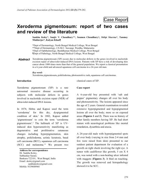
A 6-year-old boy presented with ‘salt <strong>and</strong><br />
pepper’ pigmentary changes all over his body<br />
<strong>and</strong> photosensitivity. The lesions appeared since<br />
the age <strong>of</strong> 2 years. General examination revealed<br />
extensive hyperpigmented <strong>and</strong> hypopigmented<br />
lesions all over the body; more so on exposed<br />
areas (Figures 1 <strong>and</strong> 2). There was no history <strong>of</strong><br />
other family members having XP. He had short<br />
stature with neurological problems like mental<br />
retardation, dysarthria <strong>and</strong> ataxia.<br />
A 24-year-old male with hyperpigmented spots<br />
all over body (varying in size from 2-4 mm <strong>and</strong><br />
few were raised from skin) presented to our<br />
outdoor patient department for evaluation <strong>of</strong> a<br />
growth on right cheek involving the right eye. A<br />
tumor with cauliflower like growth, 5 cm X 4<br />
cm, was noted with a non-healing ulcer infested<br />
with maggots (Figures 3). It bled on touching.<br />
The growth was removed <strong>and</strong> histopathology<br />
showed it to be SCC.<br />
279
Journal <strong>of</strong> Pakistan Association <strong>of</strong> Dermatologists 2012;22 (3):279-282.<br />
Figure 1 Case 1, poikiloderma on face.<br />
Figure 2 Case 1, poikiloderma on arm <strong>and</strong> shoulder.<br />
The sequence <strong>of</strong> pathological events in the<br />
patient were the appearance <strong>of</strong> irregular<br />
hyperpigmentation <strong>of</strong> the skin at 2 years <strong>of</strong> age,<br />
photosensitive rash with rough-surfaced growths<br />
(solar keratoses) since the age <strong>of</strong> 7 years <strong>and</strong><br />
Figure 3 Case 2, poikiloderma on face with<br />
fungating SCC infested with maggots.<br />
SCC since last 6 months. The patient did not<br />
suffer from any neurodegenerative abnormalities<br />
or impairment <strong>of</strong> intelligence. Till the time <strong>of</strong><br />
presentation, he had neither received any<br />
treatment nor did he get any education regarding<br />
protection from sun-exposure.<br />
Discussion<br />
The basic defect in XP is in nucleotide excision<br />
repair (NER), leading to deficient repair <strong>of</strong> DNA<br />
damaged by UV radiation. This extensively<br />
studied process consists <strong>of</strong> the removal <strong>and</strong><br />
replacement <strong>of</strong> damaged DNA with new DNA.<br />
Seven xeroderma <strong>pigmentosum</strong> repair genes<br />
(XPA through XPG) have been identified. A XP<br />
variant has also been described (Table 1). This<br />
variant type is clinically indistinguishable from<br />
classical XP but there is a defect in postreplication<br />
DNA repair following UV exposure,<br />
280
Journal <strong>of</strong> Pakistan Association <strong>of</strong> Dermatologists 2012;22 (3):279-282.<br />
Table 1 Classification <strong>of</strong> xeroderma <strong>pigmentosum</strong>.<br />
Type Gene Locus Description<br />
Type A, I, XPA XPA 9q22.3 Classical form <strong>of</strong> XP.<br />
Type B, II, XPB XPB 2q21 XP group B.<br />
Type C, III, XPC XPC 3p25 XP group C.<br />
Type D, IV, XPD XPD ERCC6 19q13.2-q13.3 , 10q11<br />
XP group D or De Sanctis-Cacchione<br />
syndrome. May be considered a subtype <strong>of</strong><br />
XPD.<br />
Type E, V, XPE DDB2 11p12-p11 XP group E.<br />
Type F, VI, XPF ERCC4 16p13.3-p13.13 XP group F.<br />
Type G, VII, XPG RAD2 ERCC5 13q33 XP group G <strong>and</strong> COFS syndrome type 3.<br />
Type V, XPV POLH 6p21.1-p12<br />
Box Kaplan-Meier survival curve<br />
90% probability <strong>of</strong> surviving to age 13 years<br />
80% probability <strong>of</strong> surviving to 28 years<br />
70% probability <strong>of</strong> surviving to 40 years<br />
Overall, life expectancy <strong>of</strong> patients with XP reduced<br />
by 30 years<br />
rather than nucleotide excision repair. 3<br />
As with most autosomal recessive disorders,<br />
usually no family history is present. But a<br />
history <strong>of</strong> consanguinity among parents may be<br />
elicited. The disease typically passes through 3<br />
stages. The skin is healthy at birth. Typically, at<br />
age <strong>of</strong> age 6 months, there is diffuse erythema,<br />
scaling, <strong>and</strong> freckles on light-exposed areas,<br />
appearing initially on the face. The second stage<br />
is characterized by poikiloderma (skin atrophy,<br />
telangiectasias, <strong>and</strong> mottled hyperpigmentation<br />
<strong>and</strong> hypopigmentation). The third stage is<br />
heralded by the appearance <strong>of</strong> numerous<br />
malignancies, including basal cell carcinoma,<br />
squamous cell carcinomas, malignant<br />
melanoma, <strong>and</strong> fibrosarcoma. These<br />
malignancies may occur as early as age 4-5<br />
years <strong>and</strong> are more prevalent in sun-exposed<br />
areas.<br />
Neurologic problems develop due to premature<br />
death <strong>of</strong> nerve cells <strong>and</strong> are seen in nearly 20%<br />
<strong>of</strong> patients with XP, more commonly in groups<br />
XPA <strong>and</strong> XPD. 5 The problems include loss <strong>of</strong><br />
XP variant. Due to mutation in a gene that<br />
codes for a specialized DNA polymerase<br />
called polymerase-η (eta).<br />
fine motor control, rigidity, ataxia, spasticity,<br />
hyporeflexia or areflexia, chorea, motor neuron<br />
signs or segmental demyelination, sensorineural<br />
deafness <strong>and</strong> progressive mental retardation. The<br />
first XP case with neurological signs was<br />
described by Dr. Albert Neisser. 6 In 1932,<br />
DeSanctis <strong>and</strong> Cacchione 7 helped to coin the<br />
term "De Sanctis-Cacchione syndrome" to apply<br />
to <strong>cases</strong> <strong>of</strong> XP with severe neurological<br />
deficiency.<br />
Ocular problems 5 occur in nearly 80% <strong>of</strong> <strong>cases</strong>.<br />
They include photophobia, lentigines (occur<br />
during the first decade <strong>of</strong> life, <strong>and</strong> they might<br />
transform into malignant melanoma), ectropion,<br />
symblepharon with ulceration, conjunctivitis <strong>and</strong><br />
eyelid malignancies. Our patient had SCC<br />
involving the right eye.<br />
The <strong>two</strong> most common types <strong>of</strong> cancer found in<br />
XP patients are BCC <strong>and</strong> SCC, with most<br />
tumours found on the face, head or neck. 8,9 Early<br />
detection <strong>of</strong> these malignancies is necessary<br />
because they are fast-growing, metastasize early<br />
<strong>and</strong> lead to death; most patients with XP do not<br />
live beyond the third decade because <strong>of</strong><br />
development <strong>of</strong> tumours. 10,11 Kraemer et al. 5<br />
constructed the Kaplan-Meier survival curve for<br />
patients with XP (Box 1).<br />
281
Journal <strong>of</strong> Pakistan Association <strong>of</strong> Dermatologists 2012;22 (3):279-282.<br />
The treatment <strong>of</strong> XP is challenging because it is<br />
a multiorgan <strong>and</strong> multisystem disease, <strong>and</strong><br />
because usually by the time <strong>of</strong> diagnosis,<br />
significant tissue damage has already occurred.<br />
Malignant tumours may already have developed<br />
by the third or fourth year <strong>of</strong> life. Early<br />
diagnosis <strong>and</strong> immediate implementation <strong>of</strong><br />
rigorous sun protection measures may prolong<br />
the lives <strong>of</strong> persons with XP. 12 The use <strong>of</strong><br />
sunscreens, other sun avoidance methods (e.g.<br />
protective clothing, hats, eyewear) <strong>and</strong> use <strong>of</strong><br />
systemic retinoids have been shown to minimize<br />
UV-induced damage <strong>and</strong> incidence <strong>of</strong> skin<br />
cancers in patients with XP. 13 Surgical excision<br />
<strong>of</strong> the tumours <strong>and</strong> grafting <strong>of</strong> skin from nonlight-exposed<br />
areas is the first line <strong>of</strong> treatment.<br />
A new approach to photoprotection is to repair<br />
DNA damage after UV exposure by delivering a<br />
DNA repair enzyme into the skin by means <strong>of</strong><br />
specially engineered liposomes. 14 T4<br />
endonuclease V has been shown to repair<br />
cyclobutane pyrimidine dimers resulting from<br />
DNA damage. 15 Genetic counselling <strong>of</strong> affected<br />
families is important. Amniocentesis may be<br />
done for prenatal diagnosis <strong>of</strong> XP <strong>and</strong><br />
interruption <strong>of</strong> the pregnancy. 16<br />
References<br />
1. Hebra F, Kaposi M. On diseases <strong>of</strong> the skin<br />
including exanthemata. New Sydenham Soc<br />
1874;61:252-58.<br />
2. Kaposi M. <strong>Xeroderma</strong> <strong>pigmentosum</strong> [in<br />
French]. Ann Dermatol Venereol 1883;4:29-<br />
38.<br />
3. English JS, Swerdlow AJ. The risk <strong>of</strong><br />
malignant melanoma, internal malignancy<br />
<strong>and</strong> mortality in xeroderma <strong>pigmentosum</strong><br />
patients. Br J Dermatol 1987;117:457-61.<br />
4. Gratchev A, Strein P, Utikal J, Sergij G.<br />
Molecular genetics <strong>of</strong> <strong>Xeroderma</strong><br />
<strong>pigmentosum</strong> variant. Exp Dermatol<br />
2003;12:529-36.<br />
5. Kraemer KH, Lee MM, Scotto J. <strong>Xeroderma</strong><br />
<strong>pigmentosum</strong>. Cutaneous, ocular, <strong>and</strong><br />
neurologic abnormalities in 830 published<br />
<strong>cases</strong>. Arch Dermatol 1987;123:241-50.<br />
6. Neisser A. Ueber das '<strong>Xeroderma</strong><br />
<strong>pigmentosum</strong>' (Kaposi): Lioderma<br />
essentialis cum melanosi et telangiectasia<br />
Vierteljahrschr Dermatol Syphil 1883:47-<br />
62.<br />
7. De Sanctis C, Cacchione A. <strong>Xeroderma</strong>tic<br />
idiocy. Riv Sper Freniat 1932;56:269-92.<br />
8. Kraemer KH, Lee MM, Andrews AD,<br />
Lambert WC. The role <strong>of</strong> sunlight <strong>and</strong> DNA<br />
repair in melanoma <strong>and</strong> nonmelanoma skin<br />
cancer. Arch Dermatol 1994;130:1018-21.<br />
9. Kraemer KH. Sunlight <strong>and</strong> skin cancer:<br />
Another link revealed. Proc Natl Acad Sci<br />
1997;94:11-4.<br />
10. Goyal JL, Rao VA, Srinivasan R, Argomal<br />
K. Oculocutaneous manifestations in<br />
xeroderma <strong>pigmentosum</strong>. Br J Ophthalmol<br />
1994;78:295-7.<br />
11. Masinjila H, Arnbjornsson E. Two children<br />
with xeroderma <strong>pigmentosum</strong> developing<br />
<strong>two</strong> different types <strong>of</strong> malignancies<br />
simultaneously. Pediatr Surg Int<br />
1998;13:299-300.<br />
12. Leal-Khouri S, Hruza GJ, Hruza LL, Martin<br />
AG. Management <strong>of</strong> a young patient with<br />
xeroderma <strong>pigmentosum</strong>. Pediatr Dermatol<br />
1994;11:72-5.<br />
13. Kraemer KH, DiGiovanna JJ, Moshell AN<br />
et al. Prevention <strong>of</strong> skin cancer in xeroderma<br />
<strong>pigmentosum</strong> with the use <strong>of</strong> oral<br />
isotretinoin. N Engl J Med 1988;318:1633-7.<br />
14. Yarosh DB, O'Connor A, Alas L, Potten C,<br />
Wolf P. Photoprotection by topical DNA<br />
repair enzymes: molecular correlates <strong>of</strong><br />
clinical studies. Photochem Photobiol<br />
1999;69:136-40.<br />
15. Yarosh D, Klein J, O'Connor A et al. Effect<br />
<strong>of</strong> topically applied T4 endonuclease V in<br />
liposomes on skin cancer in xeroderma<br />
<strong>pigmentosum</strong>: a r<strong>and</strong>omised study.<br />
<strong>Xeroderma</strong> Pigmentosum Study Group.<br />
Lancet 2001;357:926-9.<br />
16. Ahmed H, Hassan RY, Pindiga UH.<br />
<strong>Xeroderma</strong> <strong>pigmentosum</strong> in three<br />
consecutive siblings <strong>of</strong> a Nigerian family:<br />
Observations on oculocutaneous<br />
manifestations in black African children. Br<br />
J Ophthalmol 2001;85:110-1.<br />
282


