Reduced Tc-99m DMSA uptake in a patient with renal tubular ...
Reduced Tc-99m DMSA uptake in a patient with renal tubular ...
Reduced Tc-99m DMSA uptake in a patient with renal tubular ...

You also want an ePaper? Increase the reach of your titles
YUMPU automatically turns print PDFs into web optimized ePapers that Google loves.
Vol. 16, No. 7, 2002<br />
CASE REPORT<br />
Annals of Nuclear Medic<strong>in</strong>e Vol. 16, No. 7, 499–501, 2002<br />
<strong>Reduced</strong> <strong>Tc</strong>-<strong>99m</strong> <strong>DMSA</strong> <strong>uptake</strong> <strong>in</strong> a <strong>patient</strong> <strong>with</strong> <strong>renal</strong> <strong>tubular</strong> acidosis:<br />
Effect of acid-base imbalance<br />
INTRODUCTION<br />
RENAL TUBULAR ACIDOSIS (RTA) is described as kidneys<br />
<strong>with</strong> impaired ability to secrete hydrogen ions <strong>in</strong> the distal<br />
nephron or to reabsorb bicarbonate (HCO3) ions proximally,<br />
lead<strong>in</strong>g to chronic metabolic acidosis. 1 The primary<br />
defect <strong>in</strong> proximal RTA (pRTA) is reduced <strong>renal</strong><br />
HCO3 result<strong>in</strong>g <strong>in</strong> significant bicarbonaturia. pRTA is<br />
usually accompanied by hyperchloremic metabolic acidosis<br />
<strong>with</strong> a normal or slightly reduced serum potassium<br />
concentration, and the ability to lower ur<strong>in</strong>ary pH to below<br />
5.5 <strong>in</strong> the face of spontaneous acidemia or after acid<br />
load<strong>in</strong>g. It may appear as an isolated defect; primary or<br />
secondary, or <strong>in</strong>tegrated <strong>in</strong> a generalized proximal tubule<br />
defect, coupled <strong>with</strong> ur<strong>in</strong>ary wast<strong>in</strong>g of many solutes<br />
known as Fanconi syndrome. Experimental pathophysiologic<br />
studies suggest a defect <strong>in</strong> the proximal tubule<br />
Na,K-ATPase pump, lead<strong>in</strong>g to excessive ur<strong>in</strong>ary loss of<br />
HCO3, Na, K, glucose, phosphate and am<strong>in</strong>o acids. 1,2<br />
We here report an <strong>in</strong>fant <strong>with</strong> proximal <strong>renal</strong> <strong>tubular</strong><br />
acidosis accompanied by glucosuria and prote<strong>in</strong>uria. This<br />
Received May 7, 2002, revision accepted August 8, 2002.<br />
For repr<strong>in</strong>t contact: Meltem Caglar, M.D., Hacettepe University<br />
Medical Faculty, Department of Nuclear Medic<strong>in</strong>e, Ankara,<br />
06100, TURKEY.<br />
E-mail: mcaglar@hacettepe.edu.tr<br />
Meltem CAGLAR* and Rezan TOPALOG˘LU**<br />
*Department of Nuclear Medic<strong>in</strong>e and **Division of Pediatric Nephrology,<br />
Hacettepe University Medical Faculty, Ankara, Turkey<br />
<strong>Tc</strong>-<strong>99m</strong> dimercaptosucc<strong>in</strong>ic acid (<strong>DMSA</strong>) is used as a <strong>renal</strong> cortical imag<strong>in</strong>g agent to detect<br />
parenchymal abnormalities especially <strong>in</strong> children. Kidney <strong>uptake</strong> of <strong>DMSA</strong> provides an <strong>in</strong>dex for<br />
evaluation of a functional <strong>tubular</strong> mass, which depends on the <strong>renal</strong> blood flow and proximal <strong>tubular</strong><br />
cell membrane transport function. We here report a boy <strong>with</strong> <strong>renal</strong> <strong>tubular</strong> acidosis, which has<br />
noticeably reduced <strong>uptake</strong> on his <strong>Tc</strong>-<strong>99m</strong> <strong>DMSA</strong> sc<strong>in</strong>tigraphy, despite a totally normal <strong>Tc</strong>-<strong>99m</strong><br />
MAG-3 study. The case reported here clearly demonstrates a situation <strong>in</strong> which <strong>renal</strong> <strong>uptake</strong> of<br />
<strong>DMSA</strong> may be dissociated from a functional <strong>renal</strong> mass and the importance of acid-base balance<br />
which alters <strong>Tc</strong>-<strong>99m</strong> <strong>DMSA</strong> <strong>uptake</strong>.<br />
Key words: <strong>renal</strong> <strong>tubular</strong> acidosis, <strong>Tc</strong>-<strong>99m</strong> <strong>DMSA</strong>, <strong>Tc</strong>-<strong>99m</strong> MAG-3<br />
<strong>patient</strong> had a <strong>Tc</strong>-<strong>99m</strong> <strong>DMSA</strong> study performed due to a<br />
history of upper ur<strong>in</strong>ary tract <strong>in</strong>fection, which showed<br />
diffuse decreased <strong>uptake</strong> <strong>in</strong> the kidneys bilaterally and<br />
<strong>in</strong>creased liver activity. MAG-3 study performed 1 week<br />
later was normal. This case clearly demonstrates the<br />
difference <strong>in</strong> <strong>renal</strong> handl<strong>in</strong>g of <strong>Tc</strong>-<strong>99m</strong> <strong>DMSA</strong> and MAG-<br />
3 <strong>in</strong> metabolic acidosis.<br />
CASE REPORT<br />
A 51-day-old boy was admitted to the hospital for <strong>in</strong>tractable<br />
vomit<strong>in</strong>g and diarrhea. At adnission, he was dehydrated<br />
and had acidosis. Ur<strong>in</strong>alysis revealed a specific<br />
gravity of 1,015, ur<strong>in</strong>ary pH of 6.5, mild prote<strong>in</strong>uria and<br />
2–3 leucocytes per high power field microscopic exam<strong>in</strong>ation.<br />
Serum electrolytes measured at the time were as<br />
follows: BUN 5 mg/dl (normal: 4–20), creat<strong>in</strong><strong>in</strong>e 0.53<br />
mg/dl (normal: 0.5–1.2), uric acid 4.2 mg/dl (normal:<br />
2.7–8.5), Na 129 mEq/L (normal: 135–145), K 2.5 mEq/<br />
L (normal: 3.5–5.5), Cl 104 mEq/L (normal: 95–110),<br />
venous pH 7.22 (normal: 7.35–7.45), HCO3 content 13.8<br />
mEq/L (normal: 24–26), pCO2 36.2 mmHg (normal: 27–<br />
40). Laboratory f<strong>in</strong>d<strong>in</strong>gs demonstrated that the ur<strong>in</strong>e pH<br />
was <strong>in</strong>appropriately high despite spontaneous hyperchloremic<br />
metabolic acidosis, suggestive of <strong>renal</strong> <strong>tubular</strong><br />
acidosis. Furthermore, <strong>tubular</strong> phosphorus reabsorption<br />
was low at 90% <strong>with</strong> glycosuria and prote<strong>in</strong>uria but the<br />
Case Report 499
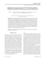
Post<br />
Fig. 1 Posterior <strong>Tc</strong>-<strong>99m</strong> <strong>DMSA</strong> scan shows decreased concentration<br />
<strong>in</strong> kidneys bilaterally and elevated liver <strong>uptake</strong> (arrow).<br />
Fig. 2 Two-m<strong>in</strong>ute <strong>in</strong>terval posterior images of <strong>Tc</strong>-<strong>99m</strong> MAG-<br />
3 images show normal concentration.<br />
ur<strong>in</strong>ary Ca/Cr ratio was normal. With all these f<strong>in</strong>d<strong>in</strong>gs,<br />
the <strong>patient</strong> was diagnosed <strong>with</strong> proximal <strong>renal</strong> <strong>tubular</strong><br />
acidosis.<br />
He was <strong>in</strong>vestigated for the presence of upper ur<strong>in</strong>ary<br />
tract <strong>in</strong>fection. His <strong>renal</strong> ultrasound did not show any<br />
abnormalities. This was followed by a <strong>Tc</strong>-<strong>99m</strong> <strong>DMSA</strong><br />
sc<strong>in</strong>tigraphy. Images were acquired 4 hours after <strong>in</strong>ject<strong>in</strong>g<br />
18 MBq of a radiopharmaceutical, by means of a<br />
500 Meltem Caglar and Rezan Topalog˘lu<br />
gamma camera (Toshiba 601, Japan) <strong>with</strong> a parallel hole,<br />
low energy, high-resolution collimator. Posterior and<br />
posterior oblique images for 300,000 counts were collected<br />
<strong>with</strong> the <strong>patient</strong> <strong>in</strong> the sup<strong>in</strong>e position. The images<br />
showed <strong>in</strong>creased liver activity <strong>with</strong> dim<strong>in</strong>ished <strong>renal</strong><br />
<strong>uptake</strong> (Fig. 1). The absolute <strong>renal</strong> <strong>uptake</strong> was 12% <strong>in</strong> the<br />
right and 10% <strong>in</strong> the left kidney.<br />
A diuretic renogram was performed <strong>with</strong> 37 MBq <strong>Tc</strong>-<br />
<strong>99m</strong> MAG-3 <strong>with</strong><strong>in</strong> one week. Images were acquired <strong>with</strong><br />
a parallel-hole, low energy, general purpose collimator <strong>in</strong><br />
the posterior view <strong>with</strong> the <strong>patient</strong> <strong>in</strong> the sup<strong>in</strong>e position.<br />
Perfusion, concentration and excretion functions of both<br />
kidneys were normal (Fig. 2). The renogram curve did not<br />
display any abnormality.<br />
DISCUSSION<br />
Our <strong>patient</strong> <strong>with</strong> <strong>renal</strong> <strong>tubular</strong> acidosis showed poor <strong>Tc</strong>-<br />
<strong>99m</strong> <strong>DMSA</strong> <strong>uptake</strong>, but a normal concentration and<br />
excretion of <strong>Tc</strong>-<strong>99m</strong> MAG-3. This boy appears to be very<br />
similar to <strong>patient</strong>s <strong>with</strong> nephronophthisis reported by<br />
Hecht et al. who had poor <strong>renal</strong> <strong>uptake</strong> of <strong>Tc</strong>-<strong>99m</strong> <strong>DMSA</strong>,<br />
but normal MAG-3 studies. 3 These authors suggested that<br />
the lack of <strong>DMSA</strong> <strong>uptake</strong> was due to failure of <strong>uptake</strong> by<br />
the tubule or defective b<strong>in</strong>d<strong>in</strong>g of the isotope <strong>with</strong><strong>in</strong> the<br />
cell.<br />
Renal handl<strong>in</strong>g of <strong>Tc</strong>-<strong>99m</strong> <strong>DMSA</strong> has been studied by<br />
Müller Suur et al. <strong>in</strong> rats. 4 They found that low fractions<br />
of prote<strong>in</strong> free <strong>Tc</strong>-<strong>99m</strong> <strong>DMSA</strong> enter the <strong>tubular</strong> lumen by<br />
glomerular filtration and are not reabsorbed by the <strong>tubular</strong><br />
epithelium. The majority of <strong>Tc</strong>-<strong>99m</strong> <strong>DMSA</strong> is removed<br />
from peri<strong>tubular</strong> capillaries, which depends on aerobic<br />
metabolism and is then bound to the cell plasma prote<strong>in</strong>s,<br />
presumably at a high b<strong>in</strong>d<strong>in</strong>g constant. 5 Autoradiography<br />
has shown that most of the <strong>Tc</strong>-<strong>99m</strong> <strong>DMSA</strong> concentrates<br />
<strong>in</strong> the cytoplasm of the proximal <strong>tubular</strong> cells. 6<br />
Many studies have documented the cl<strong>in</strong>ical utility of<br />
<strong>Tc</strong>-<strong>99m</strong> <strong>DMSA</strong> scann<strong>in</strong>g <strong>in</strong> the pediatric population, 7–9<br />
yet few data exist regard<strong>in</strong>g its ability to quantitate total<br />
<strong>renal</strong> function <strong>in</strong> children. 10,11 There is a close relationship<br />
between the function<strong>in</strong>g <strong>renal</strong> mass and the absolute<br />
number of sites available for <strong>DMSA</strong> b<strong>in</strong>d<strong>in</strong>g <strong>in</strong> the <strong>tubular</strong><br />
cells of the kidneys. 12 Bajc et al. studied a total of 282<br />
<strong>renal</strong> sc<strong>in</strong>tigrams <strong>with</strong> <strong>Tc</strong>-<strong>99m</strong> <strong>DMSA</strong> <strong>in</strong> age rang<strong>in</strong>g<br />
from 10 days to 10 years. 13 They found that average<br />
background activity was 14% of the average kidney<br />
activity at birth and decreased to approximately 6% dur<strong>in</strong>g<br />
the first year of life. Groshar et al. did not f<strong>in</strong>d a<br />
significant correlation between age and <strong>renal</strong> <strong>uptake</strong> although<br />
a significant <strong>in</strong>verse correlation was found between<br />
the percent <strong>in</strong>jected dose per cubic centimeter of<br />
<strong>renal</strong> tissue (%ID/cm 3 ) <strong>with</strong> <strong>in</strong>creas<strong>in</strong>g age. 14<br />
Organ distribution of <strong>DMSA</strong> can be altered by the<br />
method of preparation and peri<strong>tubular</strong> <strong>uptake</strong> may be<br />
<strong>in</strong>fluenced by acid base disturbances. 15–17 Yee et al. have<br />
shown that acid-base imbalance significantly alters <strong>DMSA</strong><br />
Annals of Nuclear Medic<strong>in</strong>e
k<strong>in</strong>etics. 15 In their experimental study which was performed<br />
on rats, acidosis noticeably <strong>in</strong>creased the background<br />
activity and caused a significant rise <strong>in</strong> liver<br />
accumulation. In the case reported here, the <strong>renal</strong> <strong>uptake</strong><br />
was well below the normal limits <strong>with</strong> poor <strong>renal</strong> def<strong>in</strong>ition.<br />
Based on the observation of Goodgold et al., 12 who found<br />
a close correlation (r = 0.75) between creat<strong>in</strong><strong>in</strong>e clearance<br />
and absolute <strong>DMSA</strong> <strong>uptake</strong>, a marked reduction was not<br />
expected <strong>in</strong> face of normal creat<strong>in</strong><strong>in</strong>e clearance due to<br />
immaturity of the <strong>renal</strong> cells <strong>in</strong> our <strong>patient</strong>.<br />
In contrast to <strong>DMSA</strong> <strong>uptake</strong>, <strong>Tc</strong>-<strong>99m</strong> MAG-3 sc<strong>in</strong>tigraphy<br />
was normal <strong>in</strong> our <strong>patient</strong>, probably ow<strong>in</strong>g to the<br />
differences <strong>in</strong> <strong>renal</strong> handl<strong>in</strong>g. 20% of MAG-3, which is<br />
not prote<strong>in</strong>-bound is filtered through the glomerular<br />
capillaries. MAG-3-prote<strong>in</strong> complexes pass <strong>in</strong>to the<br />
peri<strong>tubular</strong> capillary network, where dissociation occurs.<br />
Free MAG-3 is then actively transported <strong>in</strong>to the proximal<br />
<strong>tubular</strong> cells. 18 Adm<strong>in</strong>istration of probenecid decreases<br />
the transport of <strong>Tc</strong>-<strong>99m</strong> MAG-3 19 whereas it does not<br />
block the <strong>renal</strong> enzyme system that concentrates <strong>DMSA</strong> 5<br />
confirm<strong>in</strong>g the presence of a different <strong>uptake</strong> mechanisms.<br />
The case reported here clearly demonstrates a situation<br />
<strong>in</strong> which <strong>renal</strong> <strong>uptake</strong> of <strong>DMSA</strong> may be dissociated from<br />
the functional <strong>renal</strong> mass. Our f<strong>in</strong>d<strong>in</strong>gs <strong>in</strong>dicate that poor<br />
<strong>uptake</strong> of <strong>Tc</strong>-<strong>99m</strong> <strong>DMSA</strong> <strong>in</strong> comb<strong>in</strong>ation <strong>with</strong> a normal<br />
MAG-3 study would alert the nuclear medic<strong>in</strong>e physician<br />
to the possibility of various pathologic states other than<br />
<strong>tubular</strong> cell loss which may affect <strong>renal</strong> <strong>uptake</strong>. The<br />
consideration of acid-base imbalance is particularly important,<br />
especially <strong>in</strong> the evaluation of serial changes <strong>in</strong><br />
<strong>renal</strong> function.<br />
Vol. 16, No. 7, 2002<br />
REFERENCES<br />
1. Penney MD, Oleesky DA. Renal Tubular acidosis. Ann Cl<strong>in</strong><br />
Biochem 1999; 36: 408–422.<br />
2. Herr<strong>in</strong> JT. Renal <strong>tubular</strong> acidosis. In: Barrat TM, Avner ED,<br />
Harmon WE (eds). Pediatric Nephrology. Baltimore, MD;<br />
Williams & Wilk<strong>in</strong>s, 1999: 565–581.<br />
3. Hecht H, Ohlsson J, Starck SA. Poor <strong>renal</strong> <strong>uptake</strong> of<br />
Technetium-<strong>99m</strong> dimercaptosucc<strong>in</strong>ic acid and near normal<br />
technetium-mercaptoacetyltriglyc<strong>in</strong>e renogram <strong>in</strong> nephronophthisis.<br />
Pediatr Nephrol 1996; 10: 167–170.<br />
4. Müller-Suur R, Gutsche HU. Tubular Reabsorption of<br />
Technetium-<strong>99m</strong> <strong>DMSA</strong>. J Nucl Med 1995; 36: 1654–<br />
1658.<br />
5. Goldraich NP, Alvarenga AR, Goldraich IH, Ramos OL,<br />
Sigulem D. Renal accumulation of <strong>Tc</strong>-<strong>99m</strong> <strong>DMSA</strong> <strong>in</strong> the<br />
artifically perfused rat kidney. J Urol 1985; 134: 1282–<br />
1286.<br />
6. Müller-Suur R. Radiopharmaceuticals, their <strong>in</strong>tra<strong>renal</strong> handl<strong>in</strong>g<br />
and localisation. In: Murray IPC, Ell PJ (eds). Nuclear<br />
medic<strong>in</strong>e <strong>in</strong> cl<strong>in</strong>ical diagnosis and treatment, vol. I. New<br />
York; Churchill Liv<strong>in</strong>gstone, 1995: 195.<br />
7. Sty JR, Weels RG, Starshak RJ. Imag<strong>in</strong>g <strong>in</strong> acute <strong>renal</strong><br />
<strong>in</strong>fection <strong>in</strong> children. Am J Roentgenol 1987; 148: 471–477.<br />
8. Bjorgv<strong>in</strong>sson E, Majd M, Eggli ED. Diagnosis of acute<br />
pyelonephritis <strong>in</strong> children: comparison of sonography and<br />
<strong>Tc</strong>-<strong>99m</strong> <strong>DMSA</strong> sc<strong>in</strong>tigraphy. Am J Roentgenol 1991; 157:<br />
539–543.<br />
9. Majd M, Rushton HG. Renal cortical sc<strong>in</strong>tigraphy <strong>in</strong> the<br />
diagnosis of acute pyelonephritis. Sem<strong>in</strong> Nucl Med 1992;<br />
22: 98–111.<br />
10. Moris S, Chittenden SJ, Rivens I, Heary TA, Vanstone C,<br />
Meller ST. Absolute <strong>Tc</strong>-<strong>99m</strong> <strong>DMSA</strong> <strong>renal</strong> <strong>uptake</strong> <strong>in</strong> children:<br />
a study of 321 kidneys. Nucl Med Commun 1995; 16:<br />
566–571.<br />
11. Flower MA, Meller ST, Chittenden SJ, Field<strong>in</strong>g SL, Evans<br />
K, Gordon I. Absolute <strong>Tc</strong>-<strong>99m</strong> <strong>DMSA</strong> <strong>renal</strong> <strong>uptake</strong> <strong>in</strong><br />
children: optimum time to scan. Nucl Med Commun 1995;<br />
16: 572–574.<br />
12. Goodgold HM, Fletcher JM, Ste<strong>in</strong>hardt GF. Quantitative<br />
Technetium <strong>99m</strong> dimercaptosucc<strong>in</strong>ic acid <strong>renal</strong> scann<strong>in</strong>g <strong>in</strong><br />
children. Urology 1996; 47: 405–408.<br />
13. Bajc M, Wall<strong>in</strong> L. <strong>Tc</strong>-<strong>99m</strong> <strong>DMSA</strong> <strong>renal</strong> sc<strong>in</strong>tigraphy dur<strong>in</strong>g<br />
kidney maturation. Cl<strong>in</strong> Nucl Med 1995; 20: 211–214.<br />
14. Groshar D, Gorenberg M. Quantitative SPECT <strong>uptake</strong> of<br />
<strong>Tc</strong>-<strong>99m</strong> dimercaptosucc<strong>in</strong>ic acid by the kidneys <strong>in</strong> children.<br />
J Nucl Med 1999; 40: 56–59.<br />
15. Yee CA, Lee HB, Blaufox MD. Technetium-<strong>99m</strong> <strong>DMSA</strong><br />
<strong>renal</strong> <strong>uptake</strong>: <strong>in</strong>fluence of biochemical and physiologic<br />
factors. J Nucl Med 1981; 22: 1054–1058.<br />
16. Ikeda I, Inoue O, Kurata K. Chemical and biological studies<br />
on <strong>Tc</strong>-<strong>99m</strong> DMS-I: Formation of complexes by four different<br />
methods. Int J Nucl Med Bio 1977; 4: 56–65.<br />
17. Vanlic-Razumenic NM, Gorkic DA. Studies of chemical<br />
and biological properties of <strong>Tc</strong>-<strong>99m</strong> DMS (dimercapto<br />
succ<strong>in</strong>ic acid)-<strong>renal</strong> imag<strong>in</strong>g agent. Eur J Nucl Med 1976;<br />
1: 235–242.<br />
18. Eshima D, Taylor A Jr. Technetium-<strong>99m</strong> mercaptoacetyltriglyc<strong>in</strong>e:<br />
update on new <strong>Tc</strong>-<strong>99m</strong> <strong>renal</strong> <strong>tubular</strong> function<br />
agent. Sem<strong>in</strong> Nucl Med 1992; 22: 61–73.<br />
19. Frizberg AR, Kas<strong>in</strong>a S, Eshima D, Johnson DL. Synthesis<br />
and biological evaluation of technetium-<strong>99m</strong> MAG-3 as<br />
hippuran replacement 1986; 27: 111–116.<br />
Case Report 501