download report - Istituto Pasteur
download report - Istituto Pasteur
download report - Istituto Pasteur

You also want an ePaper? Increase the reach of your titles
YUMPU automatically turns print PDFs into web optimized ePapers that Google loves.
P a r t i c i p a n t s :<br />
Maria Egle De Stefano, professor; Arianna Del Signore,<br />
Lucia Leone, Loredana Lombardi, post-doc fellows.<br />
C o l l a b o r a t i o n s :<br />
<strong>Istituto</strong> Superiore di Sanità, Laboratorio di Biologia Cellulare,<br />
Roma (Dr. Tamara Petrucci); Centro di Farmacologia Cellulare e<br />
Molecolare, CNR, Dipartimento di Farmacologia Medica,<br />
Università di Milano (Dr. Cecilia Gotti); Dipartimento di<br />
Genetica e Biologia Molecolare, Sapienza-Università di Roma<br />
(Prof. Ernesto Di Mauro, Prof. Alberto Oliverio);<br />
Dipartimento di Scienze Biologiche, Università di Napoli “Federico<br />
II” (Prof. Carla Perrone Capano).<br />
Report of activity<br />
Aim of this project is the characterisation in<br />
rodent superior cervical ganglion (SCG) of the<br />
molecular mechanisms and structural changes<br />
involved in the establishment, maintenance and<br />
plasticity of the reciprocal interactions between<br />
pre- and post-ganglionic neurones and between<br />
post-ganglionic neurones and their target organs.<br />
Damage of the ganglionic connectivities, consequent<br />
to pre- and post-ganglionic nerve crush, or<br />
to the lack of dystrophin, will be used as a tool to<br />
perform our investigation.<br />
Specifically, we focused on: (i) the possible involvement<br />
of the plasminogen enzymatic cascade in the<br />
remodelling of the rat SCG intraganglionic synapses<br />
induced by pre- and post-ganglionic nerve crush;<br />
(ii) the role of dystrophin and SCG target organs in<br />
the maintenance of the structural and functional<br />
integrity of ganglionic circuits in mice.<br />
(i) Axotomy of SCG neurons is characterized by<br />
peripheral regeneration of injured axons and temporary<br />
disassembly of the intraganglionic synapses,<br />
necessary for synaptic silencing. Both events require<br />
remodelling of the extracellular matrix achieved<br />
through controlled proteolysis of its components by<br />
89<br />
Molecular recognition in biomolecules - AREA 4<br />
Neuronal response to experimental interruption<br />
of the neural circuit: a molecular and structural study<br />
in autonomic ganglia in vivo<br />
Principal investigator: Paola Paggi<br />
Professor of Physiology<br />
Dipartimento di Biologia Cellulare e dello Sviluppo<br />
Tel: (+39) 06 49912323; Fax: (+39) 06 49912351<br />
paola.paggi@uniroma1.it<br />
different enzymatic systems. Therefore, we investigated<br />
the involvement of the plasminogen enzymatic<br />
cascade in response to axotomy of rat SCG neurons.<br />
All the components of this enzymatic pathway:<br />
tissue plasminogen activator (tPA), plasminogen and<br />
their membrane receptor annexin II, as well tPA<br />
inhibitor-1 (PAI-1), are constitutively expressed in<br />
uninjured SCGs and increase significantly after SCG<br />
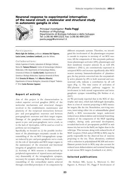
neuron axotomy. Immunolocalization of plasminogen,<br />
the key protein converted into the enzymatically<br />
active plasmin by tPA in both neuronal and nonneuronal<br />
cells, indicate a contribution by all cell<br />
types (Fig. 1). The time course of activation of<br />
tPA/plasmin enzymatic pathway suggests its<br />
involvement in both axonal regeneration and intraganglionic<br />
synapse remodelling (De Stefano et al.,<br />
2007).<br />
(ii) We previously <strong>report</strong>ed that in the SCG of dystrophic<br />
mdx mice, which lack full-length dystrophin,<br />
there is a loss of neurons projecting to SCG muscular<br />
targets, like the iris. Nonetheless, surviving neurons,<br />
innervating either iris or submandibular gland<br />
(SuGl), a SCG non-muscular target, underwent<br />
reduced axon defasciculation and terminal branching.<br />
Analysis of the components of the NGF signaling<br />
complex, during early post-natal development,<br />
revealed that levels of pro-apoptotic proNGF in mdx<br />
mouse iris, but not in the SuGl, are higher than in the<br />
wild-type. This increase, along with reduced levels of<br />
NGF receptors (TrkA and p75NTR) in SCG, may be<br />
partly responsible for the observed loss of neurons<br />
projecting to the iris. These alterations, combined<br />
with a reduction in polysialylated-NCAM and neurofilament<br />
protein levels in SCG, may also account for<br />
reduced axon defasciculation and terminal branching<br />
in mdx mouse SCG targets (Lombardi et al., 2008.).<br />
Selected publications<br />
De Stefano ME, Leone L, Moriconi C, Del<br />
Signore A, Petrucci TC, Paggi P. Involvement of