NSH Mozaic Poster 092104_062205art - Invitrogen
NSH Mozaic Poster 092104_062205art - Invitrogen
NSH Mozaic Poster 092104_062205art - Invitrogen

You also want an ePaper? Increase the reach of your titles
YUMPU automatically turns print PDFs into web optimized ePapers that Google loves.
Increase a Routine Pathology Lab’s Productivity with<br />
the use of High-Throughput MOZAIC Immunostainer<br />
Lee J.; Lau S.; Tao J., MD, QIHC; Marquez A.; Wu R. MD, PhD.<br />
INTRODUCTION<br />
Clinical laboratory services, particularly anatomic pathology labs, are increasingly relying on automation.<br />
Effective initiation of automation in laboratory services has in the past provided better patient management and<br />
future advances will certainly continue this trend.<br />
The increasing number of immunohistochemical (IHC) tests and the continuing shortage of qualified<br />
histotechnologists are common challenges for pathology laboratories. Therefore, it is critical that large runs of<br />
IHC tests can be reliably and reproducibly automated and turnaround to maximize the lab’s productivity.<br />
The type of automated IHC stainers on the market today vary in several aspects, including the technology<br />
for the processing of slides, the slide capacity per run and in the flexibility of reagents (“open” versus “closed”<br />
systems).<br />
One of the first technologies employed for the automated staining of slides was the use of the capillary gap<br />
principle. In this technology, tissue specimens on two microscope slides, or one microscope slide and a cover plate, are<br />
placed together to form a gap of defined width between them. Typically, this gap is 50 microns wide and will draw and<br />
hold a consistent volume, e.g. 100 µl, thereby contributing significantly to the reproducibility in staining. In systems<br />
employing a cover plate in place of one slide, reagents are pipetted through a small funnel located on the top of the<br />
plate. The capillary gap retains the defined amount of fluid by surface tension while the excess overflows and is<br />
automatically discarded. Washing is performed by the flow of buffer through the gaps. Sufficient fluid is retained at all<br />
times to prevent any drying of the sections.<br />
MOZAIC comes with many user-friendly features, including choice of IHC detection systems, automated<br />
CISH protocols, and a simple, convenient, easy user interface. MOZAIC can improve a pathology lab’s<br />
productivity, as each run of 96 slides, using up to 48 different primary antibodies, can be completed in less than 2 1/2<br />
hours.<br />
The purpose of our study is to verify the performance, throughput, capacity, turnaround time, and staining quality<br />
of MOZAIC.<br />
MATERIALS AND METHODS<br />
Specimens: 100 formalin-fixed, paraffin-embedded (FFPE) human cancer tissues from Zymed’s<br />
tissue bank were used in the study. 4-5 mm sections from each tissue were used.<br />
Antibodies: 80 Zymed 2 nd Gen prediluted IHC antibodies, routinely used in pathology labs, were<br />
used in the study.<br />
IHC Detection kits: Zymed Histostain ® -LAB-SA, which is labeled biotin-avidin (streptavidin),<br />
detection kits (DAB and AEC); Zymed SuperPicture Polymer detection kits (DAB and AEC)<br />
were used.<br />
CISH DNA probes: SPPT-Light ® HER2 DNA probe and SPPT-Light ® Chromosome17<br />
Centromeric DNA probe.<br />
CISH Detection kits: CISH Polymer detection kit and CISH Centromere detection kit.<br />
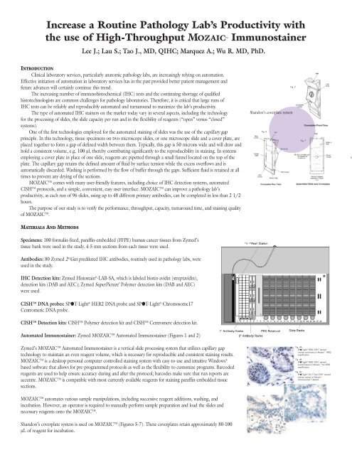
Automated Immunostainer: Zymed MOZAIC Automated Immunostainer (Figures 1 and 2)<br />
Zymed’s MOZAIC Automated Immunostainer is a vertical slide processing system that utilizes capillary gap<br />
technology to maintain an even reagent volume, which is necessary for reproducible and consistent staining results.<br />
MOZAIC is a desktop personal computer controlled staining system with easy-to-use and intuitive Windows ®<br />
based software that allows for pre-programmed protocols as well as the flexibility to customize programs. Barcoded<br />
reagents are used to help ensure accuracy during and after the protocol; barcodes make sure that run reports are<br />
accurate. MOZAIC is compatible with most currently available reagents for staining paraffin-embedded tissue<br />
sections.<br />
MOZAIC automates various sample manipulations, including successive reagent additions, washing, and<br />
incubation. However, an operator is required to manually perform sample preparation and load the slides and<br />
necessary reagents onto the MOZAIC.<br />
Shandon’s coverplate system is used on MOZAIC (Figures 5-7). These coverplates retain approximately 80-100<br />
µL of reagent for incubation.<br />
Fig. 2<br />
A.<br />
B.<br />
Shandon’s coverplate system<br />
Fig. 6<br />
C.<br />
Fig. 5<br />
Fig. 7<br />
Fig. 4A.<br />
SPPT-Light ® HER2 CISH stained<br />
ovarian carcinoma on <strong>Mozaic</strong> . HER2<br />
amplification.<br />
FIg. 4B.<br />
SPPT-Light ® HER2 CISH stained<br />
normal breast on <strong>Mozaic</strong> . No HER2<br />
amplification.<br />
FIg. 4C.<br />
SPPT-Light ® Chr.17cen CISH stained<br />
ovarian cancer on <strong>Mozaic</strong> .<br />
Chromosome17 diploid.<br />
Fi
g. 1<br />
Ancillary: Zymed MOZAIC Wash Buffer.<br />
MOZAIC staining protocols: Use MOZAIC provided staining protocols<br />
Manual staining protocols: Follow the protocols provided in the Zymed IHC and CISH detection kits product inserts.<br />
Features of Zymed MOZAIC Automated Immunostainer<br />
1. Open or closed system<br />
2. Vertical processing system<br />
3. 1 – 96 slide capacity<br />
4. 1- 48 (12 ml/vial) antibody capacity<br />
5. 7 reagent troughs (for detection, washing, counterstaining, etc.)<br />
6. Detection systems:<br />
Zymed Histostain ® LAB-SA<br />
Zymed SuperPicture Polymer<br />
Or laboratory’s own system<br />
7. Dispense volume: 100 µl – 200 µl (closed system)<br />
100 µl – 400 µl (open system)<br />
8. Minimum reagent volume/slide: 100 – 200 µl<br />
9. Reagents barcode: Yes<br />
10. Number of protocols: 6 (closed system); unlimited (open system)<br />
11. Liquid level sensor: Yes<br />
12. Audible alarm: Yes<br />
13. Temperature range: Ambient<br />
14. Software: Windows-based<br />
15. ISH and special staining: Immunostaining part of CISH<br />
RESULTS<br />
1. The specific staining for the antibodies tested was the same or stronger than the manual staining using Histostain ® LAB-SA detection system (see Figure 3).<br />
2. The specific staining for the antibodies tested was the same as the manual staining using SuperPicture Polymer detection system (see Figure 3).<br />
3. The specific staining for CISH was the same or stronger than the manual staining using CISH Polymer detection system (see Figure 4).<br />
4. Turnaround time per run<br />
SuperPicture Polymer Detection Kit (DAB): 2 hours, 30 min<br />
(Using 48 different Abs, 96 slides, including endogenous peroxidase blocking, non-overnight run)<br />
SuperPicture Polymer Detection Kit (AEC): 2 hours, 40 min<br />
(Using 48 different Abs, 96 slides, including endogenous peroxidase blocking, non-overnight run)<br />
Histostain ® LAB-SA Kit (DAB): 3 hours, 20 min<br />
(Using 48 different Abs, 96 slides, including endogenous peroxidase blocking, non-overnight run)<br />
Histostain ® LAB-SA Kit (AEC): 3 hours, 30 min<br />
(Using 48 different Abs, 96 slides, including endogenous peroxidase blocking, non-overnight run)<br />
CISH Polymer Detection Kit: 2 hours, 30 min<br />
CISH Centromere Detection Kit: 2 hours<br />
5. Good staining reproducibility and staining consistency based on three 96-slides runs using the same<br />
Fig. 3<br />
A. Rb anti-Chromogranin A<br />
stained pancreas on<br />
<strong>Mozaic</strong> using<br />
SuperPicTure Polymer<br />
Kit.<br />
B. Ms anti-MSH6 (44)<br />
stained colon on<br />
<strong>Mozaic</strong> using<br />
SuperPicTure Polymer<br />
Kit.<br />
C. Ms anti-HER2 (TAB250)<br />
stained tissue on<br />
<strong>Mozaic</strong> using<br />
SuperPicTure Polymer<br />
Kit.<br />
D. Ms anti-EGFr (31G7)<br />
stained head & neck<br />
cancer on <strong>Mozaic</strong> using Histostain ® LAB-SA<br />
kit.<br />
E. Ms anti-ER (1D5) stained<br />
tissue on <strong>Mozaic</strong> using<br />
SuperPicTure Polymer<br />
Kit.<br />
F. Ms anti-pan Cytokeratin<br />
(MAK-6 ® ) stained gastric<br />
carcinoma on <strong>Mozaic</strong> using Histostain ® A.<br />
B.<br />
C.<br />
D.<br />
LAB-SA<br />
Kit.<br />
E. F.<br />
antibodies, same tissues, same lot of reagents.<br />
6. It should be noted that with capillary gap action, air bubbles may affect staining results. To<br />
maximize performance with MOZAIC, we suggest immersing the slide and coverplate under water<br />
or buffer when applying the slide onto the coverplate.<br />
DISCUSSION<br />
Capillary gap is a proven technology that allows for the highest capacity yet small footprint<br />
immunostainer due to the vertical positioning of the slides (versus horizontal).<br />
CONCLUSION<br />
MOZAIC has high-throughput capability and is easy to operate. At