CPAP (RPA) - Intensive Care & Coordination Monitoring Unit
CPAP (RPA) - Intensive Care & Coordination Monitoring Unit
CPAP (RPA) - Intensive Care & Coordination Monitoring Unit

You also want an ePaper? Increase the reach of your titles
YUMPU automatically turns print PDFs into web optimized ePapers that Google loves.
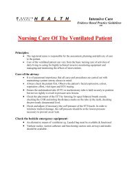
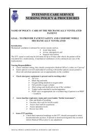
INTENSIVE CARE SERVICE<br />
NURSING POLICY& PROCEDURES<br />
NAME OF POLICY: CONTINUOUS POSITIVE AIRWAY PRESSURE<br />
VENTILATION (<strong>CPAP</strong>)<br />
GOAL: TO IMPROVE VENTILATION AND OXYGENATION BY<br />
REDUCING PATIENT’S RESPIRATORY EFFORT AND<br />
OXYGEN CONSUMPTION<br />
Introduction:<br />
Continuous positive airway pressure (<strong>CPAP</strong>) is a form of Non Invasive Ventilation (NIV) which<br />
provides positive pressure throughout the respiratory cycle (ie on inhalation and exhalation) to<br />
the spontaneous breathing patient who has sufficient respiratory drive and muscle strength. NIV<br />
<strong>CPAP</strong> may be delivered either by dedicated <strong>CPAP</strong> unit or through a ventilator with NIV<br />
functionality (<strong>CPAP</strong> may also be used on intubated patients via the ventilator).<br />
A supply of gas provides the positive pressure and a restrictive valve placed within the circuit<br />
provides PEEP. This delivers a pressure above atmospheric through the entire respiratory cycle.<br />
This positive pressure improves oxygenation and lung compliance and reduces the work of<br />
breathing (WOB) by:<br />
• Enabling the patient to take larger tidal volumes (Vt) for the same amount of effort<br />
• Increasing Functional Residual Capacity (FRC) ie preventing alveolar collapse and<br />
recruiting already collapsed alveoli, there by increasing the surface area for oxygen<br />
exchange to occur<br />
• Reducing ventilation/perfusion (V/Q) mismatch (this occurs when blood flow passes<br />
collapsed alveoli were gas exchange does not occur and so the blood returns to the<br />
heart unoxygenated)<br />
• During respiratory distress, oxygen demand increases resulting in worsening hypoxia<br />
and further respiratory distress. As <strong>CPAP</strong> improves oxygenation the WOB is<br />
decreased as the supply of oxygen meets the demand for oxygen.<br />
A <strong>CPAP</strong> circuit must contain a flow regulator, an oxygen blender, reservoir bag under pressure<br />
(2-3 times patient’s minute volume), humidifier, <strong>CPAP</strong> mask with strap and PEEP valve. The<br />
gas supply must be greater than the patient’s minute volume to allow for any increase in<br />
inspiratory effort. If the patient generates a large negative pressure the effects of <strong>CPAP</strong> will be<br />
lost if the reservoir is not sufficient.<br />
The airway pressure of <strong>CPAP</strong> applied to the thoracic cavity can reduce preload, resulting in<br />
decreased pulmonary congestion, decrease cardiac output, blood pressure and may increase<br />
intracranial pressure. It can also cause barotrauma, including alveolar damage, pneumothorax,<br />
pneumomediastinum and subcutaneous emphysema. <strong>CPAP</strong> should be used with caution in<br />
patients with chronic airways limitation disease (CAL) as they may have preexisting increased<br />
FRC and this can cause alveolar distension and rupture. <strong>CPAP</strong> circuit should always be<br />
humidified to reduce the drying effects of high-flow gases.<br />
©ROYAL PRINCE ALFRED HOSPITAL INTENSIVE CARE SERVICE
INDICATIONS FOR <strong>CPAP</strong>:<br />
• Acute respiratory failure<br />
• Pulmonary contusion and flail chest<br />
• Cardiogenic pulmonary oedema<br />
• Asthma<br />
• Chronic airways disease<br />
• Post-operative atelectasis<br />
• Obstructive sleep apnoea<br />
• Pneumonia<br />
CONTRAINDICATIONS:<br />
• Unstable facial fractures<br />
• Extensive facial lacerations<br />
• Laryngeal trauma<br />
• Recent tracheal or oesophageal<br />
anastomosis<br />
• Basal skull fracture, (use with caution)<br />
• Patients at risk of GIT bleeds/ileus<br />
USE WITH CAUTION<br />
• Patients with recent GI surgery<br />
• Decreased level of consciousness<br />
EQUIPMENT:<br />
Drager <strong>CPAP</strong> machine/Drager Ventilator with NIV function<br />
Ventilation tubing for wet circuit (humidification base in tubing pack)<br />
Sterile water bag<br />
<strong>CPAP</strong> mask and head strap<br />
PEEP valve (5,7.5, 10 or 12.5, may start with 10cmH20)<br />
Bacterial/viral filter (machine side of tubing)<br />
Test lung<br />
White rubber connector (for dedicated Drager <strong>CPAP</strong> unit only)<br />
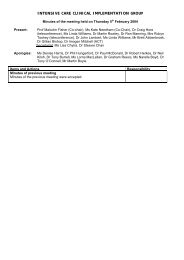
<strong>CPAP</strong> MACHINE<br />
SET UP:<br />
See pictures below for assistance<br />
• Keep “Y” connector attached to ventilator tubing<br />
• Remove white side of vent tubing<br />
• Place bacterial/viral filter to <strong>CPAP</strong> machine, then attach short arm of blue tubing from filter<br />
to humidification bath<br />
• Attach long arm of blue tubing from humidification bath to <strong>CPAP</strong> mask<br />
• Assemble blue wire of humidification bath holder to blue tubing<br />
• Using white rubber connector attach PEEP valve to exposed end of “Y” connector<br />
• Perform safety valve check (see below)<br />
• Adjust prescribed Fi02 (use flow rate of 40L/min)<br />
Equipment for Designated <strong>CPAP</strong> circuit<br />
©ROYAL PRINCE ALFRED HOSPITAL INTENSIVE CARE SERVICE
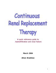
Entire designated <strong>CPAP</strong> circuit<br />
PARAMETER SETTING:<br />
• Medical orders are prescribed (Fi02, PEEP level and flow rate)<br />
• Inform patient and relatives of the procedure and management (see nursing care)<br />
• Usually the flow rate is 40L/min (see below for additional information)<br />
• Use the table on the front of the machine to set the oxygen and air flow rates to obtain the<br />
required flow rate and FI02 by turning the oxygen and air “black knobs”<br />
• Check the level of PEEP on the pressure gauge of the <strong>CPAP</strong> machine to ensure the correct<br />
level is being delivered once insitu to patient’s face<br />
• Ensure <strong>CPAP</strong> mask is air tight as possible to obtain the desired <strong>CPAP</strong> level<br />
Flow rate: A flow rate of 30-40L/min is the usual rate required. This roughly corresponds to<br />
about 1.5times the usual minute volume. This flow rate should ensure an ample reservoir of gas<br />
from which to breathe, however higher gas flows may be required in patients who are<br />
tachypnoeic and those able to generate a large negative pressure through the circuit.<br />
PEEP: PEEP is ordered in cmH20, the Drager pressure gauge measures in millibars, there is a<br />
difference of approximately 2 units between millibars and cmH20. Therefore, there will be a<br />
difference between what is ordered and what the Drager gauge measures<br />
SAFETY VALVE CHECK:<br />
• Set flow rate at approximately 20L/min (use 20L/min only for testing)<br />
• Place test lung on patient end of the circuit and occlude PEEP valve<br />
• Ensure the bellows expand to their full length<br />
• The pressure indicated on the pressure gauge must not exceed 30mbars<br />
• The safety valve at the rear will release air audibly<br />
• Ensure this check is recorded in log book<br />
©ROYAL PRINCE ALFRED HOSPITAL INTENSIVE CARE SERVICE
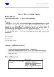
DRAGER XL & EVITA VENTILATORS:<br />
SETUP:<br />
see picture below for assistance<br />
• Keep humidifying ventilation circuit intact<br />
• Attach bacterial/viral filters to both inspiratory and expiratory arms of ventilator<br />
• Attach short arm of blue tubing to filter on inspiratory side of ventilator and other end to<br />
humidification bath<br />
• Attach long arm of blue tubing from humidification bath to <strong>CPAP</strong> mask<br />
• Assemble “blue” wire of the humidification bath holder, to blue tubing<br />
• Attach white tubing to filter on expiratory side of ventilator, attach “yellow” wire of the<br />
humidification bath holder to white tubing<br />
• Attach “Y” piece to mask<br />
• When ventilator is in “Stand By” select “Mask” and confirm. If using the EVITA ventilator<br />
(not all EVITA’s can do NIV, check for sticker to indicate) the screen will change from blue<br />
to green and the top right corner will state that the ventilator is in Non Invasive mode. If<br />
using the XL ventilator touch the “Tube/mask” tab and select “ Mask” the words “Mask<br />
Ventilation” should appear in the top right corner in orange<br />
• Check ventilator as per invasive ventilation<br />
• Touch the PEEP dials until desired parameter is reached and confirm<br />
• Attach mask to patient<br />
Setup of <strong>CPAP</strong> on Drager XL ventilator<br />
(Same set up for Drager ventilators with NIV function)<br />
©ROYAL PRINCE ALFRED HOSPITAL INTENSIVE CARE SERVICE
NURSING RESPONSIBILITIES:<br />
• Explain to patient and relatives regarding the compliance to the mask as well as:<br />
o It may be uncomfortable<br />
o Difficulty in breathing out<br />
o Tightness of the mask<br />
o Feeling of “locked in”<br />
o Possible eye swelling<br />
• Second hourly mouth and pressure relief care (whenever possible)<br />
• Padding may be required as the mask are not individualised<br />
• Regular check of equipment, as there is no integrated monitoring facilities<br />
• Hourly observations and recordings (monitor water level)<br />
• Monitor airway and respiratory function (If patient is in difficulty inform Dr.)<br />
• Monitor ABG’s<br />
• Consult with medical officer for the need of a nasogastric tube to prevent gastric distention<br />
and vomiting<br />
• Be aware of <strong>CPAP</strong> complications (see below)<br />
• Change tubing weekly and perform safety valve check<br />
• Be aware of <strong>CPAP</strong> complications (see below)<br />
COMPLICATIONS:<br />
Complication Treatment Rationale<br />
Skin necrosis (nose Apply soft dressing Cushions and protects the<br />
and chin)<br />
area<br />
Gastric insufflation Placement of nasogastric tube Relives abdominal<br />
and aspiration<br />
distension<br />
Mask intolerance and Select appropriate mask size Provides better seal and<br />
leaks<br />
comfort and prevents gas<br />
from escaping<br />
Transient hypoxia Provide alternative O2 source eg Reduces hypoxia and can<br />
nasal prongs when eating and replace mask quickly if<br />
closely monitor patient<br />
removed<br />
Hypotension Ensure patient is adequately Reduces the effects of<br />
filled. Keep PEEP less than 8cm<br />
H20 if possible.<br />
increased thoracic pressure<br />
CO2 rebreathing Ensure minimal dead space is Reduces resistance in the<br />
present and appropriate expiratory circuit and allow CO2 to<br />
valve is used<br />
escape<br />
Barotrauma Assess airway pressures and Prevents over expansion of<br />
lungs<br />
Nasal congestion and Ensure adequacy of humidified air Prevents build up and<br />
sinusitis<br />
increased viscosity of<br />
secretions<br />
Eye irritation and Avoid mask leaks, apply eye Reduced air flow into the<br />
conjunctivitis drops as appropriate<br />
eyes.<br />
©ROYAL PRINCE ALFRED HOSPITAL INTENSIVE CARE SERVICE
DISPOSAL OF EQUIPMENT:<br />
• <strong>CPAP</strong> machine is wiped down with mild detergent<br />
• The ventilation tubing, PEEP valve and humidifier chamber are disposable<br />
• All rubber connectors, hard plastic connectors and heating wires are sent to CSSD for<br />
sterilisation<br />
• <strong>CPAP</strong> mask and straps are cleaned with mild detergent<br />
REFERENCES:<br />
Bucher,L & Melander,S. (1999) Critical care nursing. W.B Saunders:Philadelphia<br />
Hill, N.S. (1997). Complications of non-invasive positive pressure ventilation. Respiratory <strong>Care</strong>, 42 (4) 433-441.<br />
Hotchkiss, J.R. & Marini, J.J. (1998). Non-invasive ventilation: An emerging supportive technique for the<br />
emergency department. Annals of Emergency Medicine, 32 (4) 470-479.<br />
Kannan, S. (1999). Practical issues in non-invasive positive pressure ventilation. <strong>Care</strong> of the Critically Ill, 15 (3)<br />
76-79.<br />
Knebel, A., Allen, M., McNemar, A. & Feigenbaum, K. (1997). A guide to non-invasive intermittent ventilatory<br />
support. Heart & Lung, 26 (4) 307-316.<br />
Mahamid, E. (2000). Non-invasive positive-pressure ventilation in acute respiratory failure. <strong>Care</strong> of the Critically<br />
Ill, 16 (2). 55-58.<br />
Marshall, A. & Pittard, M. Nursing the patient receiving Continuous Positive Airway Pressure Therapy. Australian<br />
Nurses Journal, Clinical Update. February 1998.<br />
Moore, M.J. & Schmidt, G.A. (2001). Keys to effective non-invasive ventilation, Part 1: Initial steps. Journal of<br />
Critical Illness, 16 (2) 64-70.<br />
Pierce, L.N.B. (1995). Guide to mechanical ventilation and intensive respiratory care. W.B. Saunders:<br />
Philadelphia.<br />
Py Ho, Rosa, & Boyle, M. (2000). Non-invasive positive pressure ventilation in acute respiratory failure: providing<br />
competent care. Australian Critical <strong>Care</strong>, 13 (4) 135-143.<br />
Oh, T. E. <strong>Intensive</strong> <strong>Care</strong> Mannual. Butterworths, 4 th ed. 2000.<br />
“Vital Signs” product information, 1992<br />
Occupational Health and Safety: Universal precautions taken in the preparation, administration of drug and<br />
disposal of equipment and sharps.<br />
Cross Referenced: <strong>RPA</strong>H Occ. Health & Safety Manual and Infection Control Manual<br />
NSW Infection Control Policy 98/99<br />
Revised by: Marianne O’Reilly CNS May 2003<br />
Reviewed by: Chanelle Innes CNC Cheryl Richards CNE<br />
Authorised by: Dr. Richard Totaro May 2003<br />
Revision: May 2005<br />
With the introduction of Powerchart online ordering, a clinical agreement has been set up with the Director<br />
of ICS and other Staff Specialists. Nursing Management, with the agreement of the hospital executive, have<br />
made arrangement that allows all permanently employed <strong>RPA</strong>H Nursing Staff to place orders for a variety of<br />
tests on their behalf. It is a Health Insurance Commission (HIC) directive that all orders placed by nursing<br />
staff are countersigned by the responsible MO within 14 days.<br />
©ROYAL PRINCE ALFRED HOSPITAL INTENSIVE CARE SERVICE