Diss print neu.pdf - bei DuEPublico - Universität Duisburg-Essen
Diss print neu.pdf - bei DuEPublico - Universität Duisburg-Essen
Diss print neu.pdf - bei DuEPublico - Universität Duisburg-Essen

Create successful ePaper yourself
Turn your PDF publications into a flip-book with our unique Google optimized e-Paper software.
Aspects of the Reproductive Biology of Sengis<br />
(Macroscelidea) in general<br />
and the Postnatal Development of the Short-eared Sengi<br />
(Macroscelides proboscideus) in particular<br />
Inaugural-<strong>Diss</strong>ertation<br />
zur Erlangung des Doktorgrades Dr. rer.nat.<br />
des Fachbereiches Biologie und Geographie<br />
an der <strong>Universität</strong> <strong>Duisburg</strong>-<strong>Essen</strong><br />
Vorgelegt von<br />
Gea Olbricht<br />
aus Leipzig<br />
Juli 2009
Die der vorliegenden Ar<strong>bei</strong>t zugrunde liegenden Experimente wurden im Zoologischen<br />
Garten der Stadt Wuppertal, im Zentralafrikanischen Museum Tervuren, Belgien, im<br />
Museum Alexander Koenig, Bonn und in der Anatomischen Anstalt der <strong>Universität</strong><br />
München, sowie in den südafrikanischen Museen McGregor in Kimberley und Amathole in<br />
King Williams Town durchgeführt.<br />
1. GUTACHTER:<br />
Prof. Dr. H. Burda, <strong>Universität</strong> <strong>Duisburg</strong>-<strong>Essen</strong><br />
2. GUTACHTER:<br />
Prof. Dr. B. Sures, <strong>Universität</strong> <strong>Duisburg</strong>-<strong>Essen</strong><br />
3. GUTACHTER:<br />
Dr. R. Asher, <strong>Universität</strong> Cambridge, GB<br />
VORSITZENDER DES PRÜFUNGSAUSSCHUSSES:<br />
Prof. Dr. D. Hering, <strong>Universität</strong> <strong>Duisburg</strong>-<strong>Essen</strong><br />
Tag der Disputation:<br />
03. 07. 2009
When we try to pick anything for itself,<br />
then it turns out that it is linked to everything else in the universe.<br />
John Muir<br />
Was wir wissen, ist ein Tropfen;<br />
was wir nicht wissen, ein Ozean.<br />
Isaac Newton<br />
Es ist nicht schwer zu komponieren.<br />
Aber es ist fabelhaft schwer,<br />
die überflüssigen Noten unter den Tisch fallen zu lassen.<br />
Johannes Brahms
Meiner Familie gewidmet,<br />
Dr. Alexander Sliwa mit Leona, Feline und Olivia
ACKNOWLEDGMENTS<br />
Six years came and went in the blink of an eye. Through it all, I´ve had a great deal of fun<br />
and it is a great pleasure for me to acknowledge all those who´ve helped me in this<br />
endeavour. In 2002 I approached Professor Hynek Burda of the Department of General<br />
Zoology at the University of <strong>Duisburg</strong>-<strong>Essen</strong> with the idea of initiating a study on the<br />
reproductive biology of sengis after I have had the unique opportunity of observing shorteared<br />
sengis during my time as curator at Wuppertal Zoo. It occurred to me that it was time<br />
to start a serious data collection on this amazing little creature beyond of some anecdotal<br />
information which I had noted from time to time. Prof. Burda as a small mammal specialist<br />
has always been fascinated by these unusual animals and he agreed to supervise this project. I<br />
thank him for all the advice, friendship and interest that I received from him.<br />
But first of all, my husband Dr. Alexander Sliwa deserves very special thanks for<br />
encouraging me to start this research project, providing active and moral and support and for<br />
having put up with me during my dissertation. Being an ambitious zoologist too, he inspired<br />
me many times with useful ideas in the course of numerous discussions, reviewed some early<br />
drafts and helped to design various illustrations. He followed my research progress with big<br />
interest and reported some valuable information.<br />
At the same time I would like to thank my parents, Drs. Franz and Adelheid Olbricht,<br />
who always motivated me to fight my way through, gave emotional support and helped with<br />
the children.<br />
Wuppertal Zoo provided me with generous access to my study animals, the sengis,<br />
and I would like to extend my sincerest thanks to Dr. Ulrich Schürer and his staff. Especially,<br />
the keepers of the Great Apes Department and the Felid Department were always there to<br />
advise me and provided me with valuable details of sengi husbandry and additional<br />
information on my study animals during my absence. From these departments I obtained a<br />
large number of deceased specimens. Here again my husband as the former curator at<br />
Wuppertal Zoo, Zoo veterinarian Dr. Arne Lawrenz and curator André Stadler were excellent<br />
supporters in terms of communication and technical support. I had the invaluable opportunity<br />
to share the zoo´s data bank for sengis since 1976.<br />
I am most grateful to Beryl Wilson, collection manager of the McGregor Museum in<br />
Kimberley, South Africa who shared her experience on sengis in the field with me and the
data on various sengi species in the museum collection. Together with Lucas Thibedi of the<br />
Amathole Museum in King William´s Town, South Africa, she measured some specimens<br />
available in their collections. I am also grateful to Dr. Gustav Peters of the Zoological<br />
Institute and Museum Alexander Koenig, Bonn, Germany, Wim Wendelen of the<br />
Department African Zoology of the Royal Museum for Central Africa in Tervuren, Belgium<br />
and Dr. Siegfried Czernay and Jutta Heuer of Halle Zoo, Mr. Andreas Filz of Tierpark<br />
Bernburg, and Roy Bäthe from Erfurt Zoo, all Germany, for access to their collections or<br />
donations of specimens. The Natural History Museum of the Humboldt University Berlin<br />
greatly contributed by donating a female Petromus typicus which was crucial to define the<br />
position of dorsal teats. I thank Christine Bartos and the staff from the sengi department at<br />
Philadelphia Zoo for the good communication regarding data for Rh. petersi. I gratefully<br />
acknowledge the skillful help in preparing and staining the histological sections by Astrid<br />
Sulz from the Institute for Anatomy, University of Munich. Rebecca Banasiak from the Field<br />
Museum of Natural History in Chicago, USA, provided the schematics of Figs. 2.1 and 3.1<br />
and constructed Fig. 3.2 based on photographs.<br />
I am greatly indebted to the following scientists and the co-authors of publications<br />
which will derive from the data of this thesis. Prof. Mike Perrin of the University of<br />
KwaZulu-Natal, Pietermaritzburg, RSA, Dr. William Stanley from the Field Museum of<br />
Natural History in Chicago, USA, Prof. Ulrich Welsch from the Institute for Anatomy,<br />
University of Munich, Dr. Robert Asher from the Museum of Zoology at the Cambridge<br />
University, UK, who provided additional information and advice for various aspects of the<br />
manuscript. With their scientific experience and enthusiasm they were highly supportive.<br />
Also special thanks to Dr. Galen Rathbun of the California Academy of Sciences, San<br />
Francisco, USA, and to Klaus Rudloff, curator at Tierpark Berlin-Friedrichsfelde, for the<br />
inexhaustible dialogue on the Afrotheria in general and sengis in particular.<br />
So many others have helped with the work or just helped to keep me sane. To all the<br />
graduate students in the Department of General Zoology of the University <strong>Essen</strong>-<strong>Duisburg</strong>:<br />
thanks for all the good times and all the help they have given me over the years. In particular,<br />
Dr. Sabine Begall was always there to be of assistance with statistic and special questions or<br />
just to relax my mind in times of frustration. Together with Dr. Regina Moritz she was also a<br />
never dwindling source for providing scientific articles and good spirits
LIST OF CONTENTS<br />
ACKNOWLEDGEMENTS<br />
I SUMMARY — 1<br />
II ZUSAMMENFASSUNG — 3<br />
III GENERAL INTRODUCTION — 5<br />
III.1 Fossil and extant macroscelids — 5<br />
III.2 Afrotheria - hypothesis in mammalian evolution? — 6<br />
III.3 Testicond afrotheres — 7<br />
III.4 Molecular versus morphological research — 7<br />
CHAPTER 1<br />
1. REPRODUCTIVE PATTERNS OF SENGIS (MACROSCELIDEA) – A<br />
THEORETICAL APPROACH — 9<br />
1.1 Abstract — 9<br />
1.2 Introduction — 9<br />
1.2.1 Aim of the study — 10<br />
1.3 Material and Methods — 12<br />
1.4 Results — 12<br />
1.5 Discussion — 15<br />
1.5.1 Placentation in afrotheres — 16<br />
1.5.2 Developmental status at birth — 16<br />
1.5.2.1<br />
—17<br />
Environmental factors influencing the developmental status of birth<br />
1.5.2.2 Development of precociality — 17<br />
1.5.2.3 Precocial and altricial strategies — 18<br />
1.5.3 Gestation length — 19<br />
1.5.4 Parental care — 19<br />
1.5.4.1 Lactation — 20<br />
1.5.4.2 Feeding intervals — 20<br />
1.5.5 Monogamy — 20<br />
1.5.5.1 Monogamy and parental care — 21
1.5.5.2 Mate guarding — 21<br />
1.5.5.3 Monogamy and precociality — 22<br />
1.5.6 Juvenile mortality — 22<br />
1.5.7 Litter size — 23<br />
1.5.7.1 Correlation between teat number and litter size — 23<br />
1.5.7.2 Limits of litter size — 24<br />
1.5.8 Seasonality of breeding periods — 25<br />
1.5.8.1 Male capacities — 26<br />
1.5.9 Post-partum oestrous — 26<br />
1.5.10 Poly-ovulation — 26<br />
1.5.10.1 Female capacities — 27<br />
1.5.11 Longevity and fecundity — 27<br />
1.6 Conclusions — 28<br />
CHAPTER 2<br />
2. THE TOPOGRAPHIC POSITION OF THE PENIS IN SENGIS<br />
(MACROSCELIDEA); AND COMMENTS ON PENIS TOPOLOGY IN TESTICOND<br />
MAMMALS — 32<br />
2.1 Abstract — 32<br />
2.2 Introduction — 32<br />
2.2.1 Sengis as testicond afrotheres — 33<br />
2.1.2 Taxonomic tools — 33<br />
2.1.3 Aim of the study — 34<br />
2.2 Material and Methods — 34<br />
2.2.1 Material — 34<br />
2.2.2 Methods — 34<br />
2.2.2.1 Hyracoidea — 35<br />
2.2.2.2 Statistical methods — 36<br />
2.3 Results — 36<br />
2.3.1 Sengi measurements — 36<br />
2.3.2 Hyrax accounts — 39<br />
2.4 Discussion — 39<br />
2.4.1 Distance anus-penis — 39<br />
2.4.2 Genital morphology in the Afrotheria — 40
2.4.3 Copulation posture — 40<br />
2.4.4 Hyracoidea — 41<br />
2.5 Conclusions — 41<br />
CHAPTER 3<br />
3. THE TAXONOMIC DISTRIBUTION OF MAMMARY GLANDS IN SENGIS — 42<br />
3.1 Abstract — 42<br />
3.2 Introduction — 42<br />
3.2.1 Nomenclature of teat positions — 43<br />
3.2.2 Behavioural ecology — 44<br />
3.2.3 Aim of the study — 44<br />
3.3 Material and Methods — 44<br />
3.3.1 Material — 44<br />
3.3.2 Methods — 44<br />
3.4 Results — 46<br />
3.4.1 Locations of the mammae — 46<br />
3.4.1.1 Males — 46<br />
3.4.1.2 Females — 47<br />
3.4.1.3 Formulas — 51<br />
3.5 Discussion — 52<br />
3.5.1 Teats on male sengis — 52<br />
3.5.2 Teats on female sengis and formulas — 53<br />
3.5.3 The enigma about nuchal, lateral and dorsal teats — 54<br />
3.5.3.1 Historical observations — 54<br />
3.5.3.2 The presence of dorsolateral teats — 54<br />
3.5.3.3 Defining a dorsolateral teat location — 55<br />
3.5.3.4 Functionality of teats — 55<br />
3.6 Conclusions — 56<br />
CHAPTER 4<br />
4. HISTOLOGICAL AND HISTOCHEMICAL STUDY OF THE MAMMARY<br />
GLANDS OF PETRODROMUS TETRADACTYLUS — 57<br />
4.1 Abstract — 57<br />
4.2 Introduction — 57
4.2.1 Mammae in male mammals — 58<br />
4.2.2 Aim of the study — 58<br />
4.3 Material and Methods — 59<br />
4.3.1 Material — 59<br />
4.3.2 Methods — 59<br />
4.3.2.1 Fixation, embedding and staining methods — 59<br />
4.3.2.2 Histochemistry of lectins — 60<br />
4.4 Results — 60<br />
4.4.1 General findings (light microscopy) — 60<br />
4.4.1.1 Females — 60<br />
4.4.1.2 Males — 60<br />
4.4.2 Specific histological and histochemical findings — 61<br />
4.4.2.1 Female mammary gland — 61<br />
Actin — 61<br />
PAS reaction — 61<br />
Alcian blue — 62<br />
Lectins — 63<br />
4.4.2.2 Male mammary gland — 65<br />
Lectins — 65<br />
4.4.2.3 Scent glands — 67<br />
4.5 Discussion — 69<br />
4.5.1 Histo-morphology of mammary glands in Petrodromus — 69<br />
4.5.2 The presence of mammae in male sengis — 69<br />
4.5.3 Other glands — 70<br />
4.6. Conclusions — 71<br />
CHAPTER 5<br />
5. BODY METRICS OF THE SHORT-EARED SENGI (MACROSCELIDES<br />
PROBOSCIDEUS, SMITH 1829) — 72<br />
5.1 Abstract — 72<br />
5.2. Introduction — 72<br />
5.2.1 Body metrics and other physical patterns — 73<br />
5.2.2 Growth models — 73<br />
5.2.3 Aim of the study — 74<br />
5.3 Material and Methods — 74
5.3.1 Material — 74<br />
5.3.2 Methods — 74<br />
5.3.3 Statistical analysis — 75<br />
5.3.3.1 The Gompertz growth model — 76<br />
5.4 Results — 76<br />
5.4.1 The Gompertz model — 76<br />
5.4.2 Sexual dimorphism — 78<br />
5.4.3 Correlation of body length with hind foot length and body mass — 79<br />
5.5 Discussion — 80<br />
5.5.1 Measuring scheme and statistical methods — 80<br />
5.5.2 Sexual dimorphism — 81<br />
5.5.3 Developmental stage at birth and mating system — 81<br />
5.5.4 Growth patterns and maturity — 82<br />
5.5.4.1 The Gompertz growth parameters — 82<br />
5.5.4.2 Body length against hind foot length and body mass — 82<br />
5.5.4.3 Factors influencing precociality — 83<br />
5.5.4.4 Lactation — 84<br />
5.5.4.5 Skeletal growth and sexual maturity — 85<br />
5.6 Conclusions — 85<br />
CHAPTER 6<br />
6. POST-NATAL BODY MASS DEVELOPMENT OF THE SHORT-EARED SENGI<br />
(MACROSCELIDES PROBOSCIDEUS) — 86<br />
6.1 Abstract — 86<br />
6.2 Introduction — 86<br />
6.2.1 Growth models — 86<br />
6.2.2 Aim of the study — 87<br />
6.3 Material and Methods — 87<br />
6.3.1 Material — 87<br />
6.3.2 Methods — 87<br />
6.3.2.1 Gompertz growth model and statistics — 88<br />
6.4 Results — 88<br />
6.4.1 Growth curves — 88
6.4.2 Gompertz growth parameters — 89<br />
6.4.2.1 Sex-specific growth — 90<br />
6.5 Discussion — 91<br />
6.5.1 Environmental and behavioural impact on weight development — 91<br />
6.5.2 Growth parameters — 92<br />
6.5.3 Adult body mass — 93<br />
6.5.3.1 Estimated adult weight — 93<br />
6.5.3.2 Relating body mass to sexual maturity — 94<br />
6.5.3.3 Sexual dimorphism — 94<br />
6.6 Conclusions — 94<br />
IV REFERENCES — 95<br />
V APPENDIX — 110<br />
A List of tables — 110<br />
B List of figures — 111<br />
C Staining procedures — 112<br />
D Table: Raw data on individual body measurements of male and female Macroscelides<br />
proboscideus — 115<br />
E Vita — 118<br />
F Liste der Veröffentlichungen — 120
SUMMARY 1<br />
I SUMMARY<br />
Sengis have been studied for more than a century but information on their biology is still<br />
scattered. The reproductive biology of sengis is best understood in the context of their<br />
evolutionary history. Their phylogeny has long been the subject of much speculation and<br />
controversy. This thesis aims to consolidate molecular findings of other studies with<br />
morphological methods and thus, to contribute to a better understanding of their phylogeny.<br />
Sengis are members of the Afrotheria, an African clade of mammals, and particular attention<br />
was paid in this study to their relationships to other afrotheres.<br />
Chapter 1 summarizes the knowledge on reproductive parameters across the order<br />
Macroscelidea from the literature. Various reproductive characteristics are unusual for a small<br />
mammal. The most important traits in the life history of sengis in terms of reproduction are<br />
precociality and monogamy as well as the ability of some species to poly-ovulate and to<br />
perform post-partum oestrus. Further, they have a very long lifespan with high fecundity<br />
rates.<br />
In chapters 2 and 3 I apply morphological methods to investigate phylogenetic<br />
relationships. Morphological landmarks such as the position of penis and teats were defined.<br />
The position of the penis is of utility in distinguishing between the genera Petrodromus and<br />
Macroscelides, but not between any other genera of sengi, supporting recent taxonomic<br />
conclusions regarding the relationship of these two taxa. Teat position is useful in taxonomic<br />
distinction among the three of the four genera, only between Macroscelides and Elephantulus<br />
there was no difference. Only in two species teats were found on males: Elephantulus rozeti<br />
and Petrodromus tetradactylus but not on other species examined. This supports the recent<br />
taxonomic conclusions regarding the relationship of these two taxa. The arrangement of teats<br />
was determined in a formula for each genus which distinguishes between antebrachial,<br />
abdominal and inguinal regions. No sengi exhibited teats situated dorsolaterally to the extent<br />
of those in rock-dwelling mammals such as Petromus typicus.<br />
In chapter 4 mammary tissue of a female and male P. tetradactylus was examined<br />
with different histological and histochemical methods. The results reveal a potentially<br />
functioning mammary gland in male Petrodromus with evidence of active mammary tissue.<br />
The secretory units (acini) are sexually dimorphic. In the female typical acini, milk ducts,<br />
cisternal milk sinus and a teat canal can be distinguished. The acini of the females occur in<br />
the periphery of the gland whereas acini in the male teat occur in the connective tissue of the<br />
teat. The function of mammary tissue in male Petrodromus is not clear because males of<br />
none of the sengi species contribute to the raising of their young. Apocrine scent glands were
SUMMARY 2<br />
found in both genders at the base of the teat which underlines the importance of chemical<br />
communication for sengis.<br />
In chapter 5 body measures (mass, length of body, head, ear, snout, whiskers, tail,<br />
hind foot) of captive short-eared sengis (Macroscelides proboscideus) were taken post<br />
mortem and then fitted to the 3-parameter Gompertz model There was considerable variation<br />
of the growth parameters of these body measures in terms of growth constant (K) and<br />
inflection age (I). Whiskers and snout had the fastest growth, the ears the slowest. The<br />
asymptotic value of the growth model (A) in terms of adult length of tail and ear as well as<br />
body mass was exceeded later then the sigmoidal curves suggested but nevertheless, adult<br />
size of all body parts is achieved at about sexual maturity (ca. 45 days), except hind foot<br />
length which reached its maximum earlier. No significant sexual dimorphism in the<br />
estimated adult size could be determined.<br />
Chapter 6 refines the results regarding body mass growth during the ontogeny of individual<br />
short-eared sengis which were weighed on a nearly daily basis from their first days of life<br />
until adulthood. The Gompertz growth model was used to generate the growth parameters K, I<br />
and A which were compared with data on the reproductive biology of sengis. Furthermore,<br />
the growth parameters for Macroscelides were compared with those of various species<br />
obtained from the literature.Adulthood is reached when adult size matches with sexual<br />
maturity, at about 45 days. There were no significant differences between males and females<br />
in growth or adult body mass size.
ZUSAMMENFASSUNG 3<br />
II ZUSAMMENFASSUNG<br />
Rüsselspringer oder Elefantenspitzmäuse (Macroscelidea) werden seit mehr als einem<br />
Jahrhundert wissenschaftlich untersucht, jedoch sind Informationen zu ihrer Biologie<br />
lückenhaft und zusammenhanglos. Die Fortpflanzungsbiologie von Rüsselspringern wird am<br />
besten im Kontext zu ihrer Evolutionsgeschichte verständlich. Ihre Phylogenie war lange<br />
Gegenstand von Spekulationen und Kontroversen. Diese Ar<strong>bei</strong>t hat zum Ziel, Erkenntnisse<br />
aus der Molekulargenetik mit morphologischen Methoden zu unterstützen und somit zu einem<br />
besseren Verständnis ihrer Stammesgeschichte <strong>bei</strong>tragen. Rüsselspringer gehören zu den<br />
Afrotheria, einer Gruppe von endemischen afrikanischen Säugetieren. Diese<br />
Verwandschaftsbeziehungen fanden hier besondere Berücksichtigung.<br />
Kapitel 1 faßt das bisherige Wissen über Aspekte der Fortpflanzungsbiologie der<br />
Ordnung Macroscelidea zusammen. Einige Charakteristika sind für Kleinsäuger<br />
ungewöhnlich. Zu wichtigen Reproduktionsparametern zählt, dass Rüsselspringer<br />
Nestflüchter sind und monogam leben. Daneben sind einige Arten zu Polyovulation und postpartum-<br />
Östrus befähigt. Während ihrer relativ langen Lebensdauer erreichen sie eine<br />
bemerkenswerte Geburtenrate.<br />
In den Kapiteln 2 und 3 wandte ich morphologische Methoden zur Untersuchung<br />
phylogenetischer Verwandschaftsbeziehungen an. Morphologische Meßpunkte wie die<br />
Position von Penis und Zitzen wurden festgelegt. Die Position des Penis diente der<br />
Unterscheidung der Gattungen Petrodromus und Macroscelides, jedoch nicht zu oder<br />
zwischen den anderen Gattungen. Dieses Ergebnis bestätigt <strong>neu</strong>ere taxonomische<br />
Schlussfolgerungen zur weniger engen Verwandtschaft dieser <strong>bei</strong>den Taxa. Die Lage der<br />
Zitzen führte zur taxonomischen Unterscheidung von 3 der 4 Gattungen, zwischen<br />
Elephantulus und Macroscelides besteht kein bedeutsamer Unterschied. Nur <strong>bei</strong> den <strong>bei</strong>den<br />
Arten Petrodromus tetradactylus und Elephantulus rozeti wurden <strong>bei</strong>m Männchen Zitzen<br />
gefunden, was Ergebnisse <strong>neu</strong>erer Untersuchungen zur engen Verwandschaft dieser <strong>bei</strong>den<br />
Taxa bestätigte. Die Anordnung der Zitzen definierte ich für alle 4 Gattungen in einer<br />
gattungsspezifischen Formel, wo<strong>bei</strong> zwischen antebrachialer, abdominaler und inguinaler<br />
Position unterschieden wurde. Kein Rüsselspringer zeigte dorsolaterale Zitzen, wie sie <strong>bei</strong> der<br />
Felsenratte (Petromus typicus) bekannt sind.<br />
Das Milchdrüsengewebe einer männlichen und weiblichen Rüsselratte (P.<br />
tetradactylus) wurde mit verschiedenen histologischen und histo-chemischen Methoden in<br />
Kapitel 4 untersucht. Die Egebnisse ergaben <strong>bei</strong>m Männchen eine funktionsfähige Milchdrüse
ZUSAMMENFASSUNG 4<br />
mit aktivem Milchgewebe. Die Drüsenendstücke unterschieden sich geschlechtsspezifisch.<br />
Beim Weibchen fanden sich typische Drüsenendstücke, Milchgänge sowie Milchsinus und<br />
Zitzenkanal. Die Drüsenendstücke des Weibchens befanden sich in der Drüsenperipherie, die<br />
des Männchens im Bindegewebe der Zitze. Die Funktion von Milchdrüsengewebe <strong>bei</strong> der<br />
männlichen Felsenratte bleibt unklar, da sich <strong>bei</strong> keiner Rüsselspringerart die Männchen an<br />
der Jungenaufzucht beteiligen. Apokrine Duftdrüsen wurden <strong>bei</strong> <strong>bei</strong>den Geschlechtern an der<br />
Zitzenbasis gefunden, was die große Bedeutung der olfaktorischen Kommunikation für<br />
Rüsselspringer unterstreicht.<br />
Die Kapitel 5 und 6 beschäftigen sich mit post-natalem Wachstum. In Kapitel 5<br />
wurden Körpermaße wie die Längen von Kopf-Rumpf, Ohr, Schnauze, Fibrissen, Schwanz<br />
und Hinterfuß sowie das Körpergewicht post-mortem <strong>bei</strong> zoogeborenen Kurzohr-<br />
Rüsselspringern (Macroscelides proboscideus) verschiedenen Alters vermessen und mit<br />
Hilfe des Gompertz-Wachstumsmodells analysiert. Da<strong>bei</strong> konnten erhebliche Unterschiede<br />
<strong>bei</strong>m Wachstum der verschiedenen Körpermaße im Hinblick auf die Wachstumskonstante<br />
(K) und den Zeitpunkt des schnellsten Wachstums (I) festgestellt werden. Bei Fibrissen und<br />
Schnauze konnte generell das schnellste Wachstum verzeichnet werden, <strong>bei</strong>m Ohr das<br />
langsamste. Die Annäherung an die Asymptote (A) für die adulte Länge von Schwanz und<br />
Ohr, sowie für das Adultgewicht zeigte sich in Anwendung des Wachstumsmodells später als<br />
es der sigmoidale Kurvenverlauf der direkten Meßwerte vermuten ließ. Der Hinterfuß<br />
erreichte seine Adultlänge früher als die anderen Körperteile, für die Adultmaße ungefähr<br />
<strong>bei</strong>m Einsetzen der Geschlechstreife (ca. 45. Lebenstag) ermittelt wurden. Keine Signifikanz<br />
konnte für geschlechtsspezifischen Dimorphismus der im Modell ermittelten geschätzten<br />
Adultmaße festgestellt werden.<br />
Die tägliche individuelle Gewichtszunahme von zoolebenden Kurzohr-<br />
Rüsselspringern von den ersten Lebenstagen an bis ins Erwachsenenalter war Gegenstand der<br />
Untersuchung in Kapitel 6. Die Ergebnisse des vorherigen Kapitels konnten auf der<br />
Grundlage von kompletten Meßreihen der Gewichtsentwicklung überprüft und mit Hilfe des<br />
Gompertz-Modells analysiert werden. Die Wachstumsparameter K, I und A wurden mit<br />
Aspekten der Fortpflanzungsbiologie von Rüsselspringern in Zusammenhang gesetzt und<br />
konnten gleichzeitig mit den für andere Arten aus der Literatur bekannten gleichen<br />
Wachstumsparametern verglichen werden. Es gab keine signifikanten Unterschiede zwischen<br />
Männchen und Weibchen bezüglich Wachstum oder Adultmaß <strong>bei</strong>m Körpergewicht. Das<br />
Ergebnis für das Erreichen des Adultgewichtes entsprach mit 45 Tagen dem Ergebnis des<br />
vorherigen Kapitels.
GENERAL INTRODUCTION 5<br />
III GENERAL INTRODUCTION<br />
The Macroscelidea is a monophyletic order with the family Macroscelidae which comprises<br />
16 well defined species in two subfamilies (Rovero et al. 2008): the Macroscelidinae with the<br />
genera Petrodromus, Elephantulus and Macroscelides and the Rhynchocyoninae with the<br />
only genus Rhynchocyon.<br />
The English name “elephant-shrew” originated because of the superficial similarities<br />
the Macroscelidea bear to other mammals, and in historical (and erroneous) taxonomic<br />
opinions. In particular, these animals have a very different evolutionary history than true<br />
shrews (Soricidae) and they share few life history traits. Because of this misleading nature of<br />
this name, authors (e.g. Rathbun and Woodall 2002, Skinner and Chimimba 2005) are<br />
increasingly using the African term “sengi” for elephant-shrew, which is derived from the<br />
Kiswahili word “sanje” (Eastern Africa, Rathbun and Kingdon 2006) or from the Lunda-word<br />
“Isengi” (Zambia, White and Ansell 1966). I agree with these scientists that it is appropriate<br />
to use local names and this protocol is followed here.<br />
III.1 Fossil and extant macroscelids<br />
The macroscelidean fossils from the Eocene and Oligocene (55-23 Ma) showed the early<br />
diversity in this group and thus provided new arguments for the origin and interordinal<br />
relationships of the Macroscelidea (Simons et al. 1991). Possibly, already during the early<br />
Eocene (33 Ma), ancestors of macroscelids branched off. The divergence of the two<br />
subfamilies may date from earlier Oligocene (Patterson 1965). The Rhynchocyoninae have<br />
undergone only minor evolutionary change since the early Miocene (Novacek 1984) at which<br />
time the ancestors of the Macroscelidinae were already highly specialized. The extant<br />
Rhynchocyoninae are represented by the single genus Rhynchocyon which stands apart from<br />
the Macroscelidinae in a number of characters (Corbet and Hanks 1968). It occurs in eastern<br />
and central Africa whereas the three macroscelidine genera are widely distributed from North<br />
Africa (only Elephantulus rozeti) to eastern and southern Africa.<br />
The extinct forms of sengis were herbivorous (Patterson 1965) which is considered a<br />
plesiomorphic character and a link to their paengulate relatives (Hyracoidea, Proboscidea and<br />
Sirenia). Insectivory is a later adaptation (Kerley 1995). Sengis are omnivorous, although
GENERAL INTRODUCTION 6<br />
believed to be exclusively insectivorous for a long time (Kerley 1989, 1995) and for a long<br />
time the family Macroscelidae had been placed within the former order Insectivora (Starck<br />
1948, Nowak and Paradiso 1983). Butler (1956) revised the Insectivora after comparisons<br />
with the fossil skull of Ictops and changed the status of the former family Macroscelididae<br />
into the new order Macroscelidea. Later this group was associated with the Tupaiidae<br />
(Haeckel 1866) and aligned with lagomorphs and rodents (McKenna 1975, Coldiron 1977,<br />
Novacek 1992) but no definite judgement as to possible relationships could be made with the<br />
results of these studies.<br />
A biogeographic review of sengis and the need for their conservation were published<br />
in the Databank for the Conservation and Mangement of the African Mammals (Boitani et al.<br />
1999) and the IUCN Action Plan (Nicoll and Rathbun 1990) where for six species or<br />
subspecies intermediate levels of threat were estimated.<br />
III.2 Afrotheria – hypothesis in mammalian evolution?<br />
The Afrotheria, endemic Afro-Arabian group of placental mammals includes among sengis<br />
morphologically very diverse forms like golden moles (Chrysochloridae), tenrecs<br />
(Tenrecidae), aardvarks (Tubulidentata), hyraxes (Hyracoidea), elephants (Proboscidea), and<br />
the dugongs or manatees (Sirenia) (Robinson and Seiffert 2004). The Proboscidea,<br />
Hyracoidea and Sirenia are known as Paengulata (Novacek 1992), the Chryochloridae and<br />
Tenrecidae have been also grouped together as Afrosoricida (Robinson and Seiffert 2004) or<br />
Tenrecomorpha (Carter et al. 2004). De Jong et al. (1981) were the first who suggested that<br />
aardvarks are closely related to sengis, elephants, sea cows and hyraxes based on the study of<br />
alpha A-crystallin. Later, golden moles (Stanhope et al. 1998a) and tenrecs (Stanhope et al.<br />
1998b) were included and consequently, the entire group was given the name Afrotheria.<br />
The Cretaceous afrotherian ancestor likely was a small forest-dwelling insectivore or<br />
possibly herbivore (Hedges 2001). During the mid-Cretaceous period (105-90 Ma) Africa was<br />
isolated and the diversification of the mammals - now included in the superordinal clade<br />
Afrotheria - proceeded (Stanhope et al. 1998b). The crown (extant) afrotherians appeared<br />
approximately 80 Million years ago after the stem afrotherians had about 25 million years of<br />
independent evolution (Springer et al. 2003). It has been suggested that sengis represent the<br />
earliest African lineage (Stanhope et al. 1998a).<br />
Despite of the strong molecular support for afrotherian monophyly (de Jong et al.<br />
1981, Stanhope et al. 1996, 1998a, b; Springer et al. 2003; van Dijk et al. 2001; Nikaido et al.<br />
2003) morphological features shared by these animals, particulary between the insectivoran-
GENERAL INTRODUCTION 7<br />
and ungulate-grade afotherians, have been difficult to identify (Asher and Lehmann 2008).<br />
Several similarities became apparent only recently, including non-descent of the male gonads<br />
(Werdelin and Nisonne 1992), morphology of the placenta (Mess and Carter 2006), variable<br />
vertebral count (Sánchez-Villagra et al. 2007), a concave cotylar facet of the astragalus<br />
(Tabuce et al. 2007), calcano-navicular contact (Seiffert 2007) and dental eruption (Asher and<br />
Lehmann 2008). Nishihara et al. (2006) concluded that the afrotherian lineages diverged very<br />
rapidly with the consequence that ancestral polymorphism present in the last common<br />
ancestor of Paenungulata results in incongruence. The diversification of the Macroscelidea<br />
included both slow and accelerated morphological evolution (Douady et al. 2002).<br />
One of the most pressing interpretive problems is still the uncertain phylogenetic<br />
position of the two orders aardvarks and sengis within the Afrotheria (Seiffert 2002). Sengis<br />
preserve a number of specialized paenungulate-like features in their postcranial, cranial and<br />
dental morphology but various genetic studies support close relationship of sengis with<br />
aardvarks, tenrecs and golden moles (Waddell et al. 2001).<br />
III.3 Testicond afrotheres<br />
Seiffert (2002) suggested that Afrotherians evolved from a common ancestor with a very<br />
primitive (or secondarily primitive) reproductive system, most male afrotheres are more<br />
similar to monotremes than to marsupials and most other placentals in <strong>bei</strong>ng primary<br />
testicond. Testicondy, the retention of testes in their ontogentically primary position in the<br />
abdominal cavity is a derived condition among therians and has been viewed as afrotherian<br />
synapomorphy which provides morphological support for afrotherian monophyly (Werdelin<br />
and Nilsonne 1999).<br />
III.4 Molecular versus morphological research<br />
Sengis have attracted attention of researchers for over more than a century. In recent years,<br />
systematists have been struggling to reconcile classical “morphological” methods of<br />
reconstructing evolutionary trees based on anatomical similarities and differences between<br />
living species or their extinct relatives with an avalanche of new molecular data on genetic<br />
variation among organisms (Gibbons 1991, Asher et al. 2003). As a result, the face-off<br />
between proponents of molecules and those of morphology was sometimes controversial.<br />
Balter (1997) stresses the necessity that systematists of both camps must move closely<br />
together to sort out the many remaining puzzles since only a few groups of animals have had
GENERAL INTRODUCTION 8<br />
their phylogenetic trees worked out with complete confidence and that there are warning<br />
signs that molecular evidence can lead to misleading errors. In contrast Hedges (cited as pers.<br />
comm. in Balter 1997) maintained that morphological features are more susceptible to<br />
“adaptive convergence” (homoplasy) and consequently, trees should be built with molecular<br />
data alone.<br />
This research supports molecular findings with morphological methods and is<br />
consolidating the knowledge on phylogenetic relationships.
REPRODUCTIVE PATTERNS 9<br />
CHAPTER 1<br />
1. REPRODUCTIVE PATTERNS OF SENGIS (MACROSCELIDEA) – A<br />
THEORETICAL APPROACH<br />
1.1 Abstract<br />
This theoretical approach summarizes the knowledge on reproductive parameters across the<br />
order Macroscelidea. Morphological parameters such as teat number were related to life<br />
history traits such as litter size, gestation length, developmental status at birth and mating<br />
system. The most important traits in the life history of sengis in terms of reproduction are<br />
precociality and monogamy as well as the ability of some species to poly-ovulate and to<br />
perform post-partum oestrus. Sengis have a very long lifespan. Particular attention was paid to<br />
similarities among afrotherian mammals.<br />
1.2 Introduction<br />
Nicoll and Rathbun (1990) emphasized the need for more research since sengis are not wellknown<br />
biologically and more knowledge is required for better understanding and more<br />
effective conservation. Valuable information on sengis in the wild as well as in captivity is<br />
available but there is a need to pool and summarize this knowledge to connect different<br />
parameters and subsequently to detect interrelations. Relationships between reproductive<br />
traits and life history in the Macroscelidea have not been reviewed yet. Most of the characters<br />
here are compared with other mammals such as rodents due to the lack of information on<br />
sengis and if available with other afrotheres. Tabuce et al. (2008) stress the necessity of the<br />
development of new sources of phylogenetic characters to shed light on the Afrotheria.<br />
Traits such as mammary numbers may be an important factor in the evolution of litter<br />
size (Pearl 1913a; Gilbert 1986). The correlation between mean numbers of mammae and<br />
mean litter sizes in rodents is described as the “one-half rule” (Gilbert 1986, Sherman et al.<br />
1999). The mean litter size was about one half of the mean number of mammae whereas the<br />
maximum litter sizes were approximately equal to the number of mammae. Summarizing his
REPRODUCTIVE PATTERNS 10<br />
results, Gilbert (1986) came to the conclusion that mammary number constrains litter size<br />
rather than vice versa.<br />
Considering the fact that most macroscelids have precocial young (e.g. Skinner and<br />
Smithers 1990) my interest is devoted to understanding the adaptive significance of their<br />
reproductive patterns. In addition, the energy and nutrient requirements of reproduction will<br />
be influenced by such characteristics as litter size, stage of development at birth and the<br />
duration of gestation and lactation (Neal 1986).<br />
In particular in the Macroscelidinae it is well documented that they have a relatively<br />
large number of teats in relation to small litters. This central issue is followed throughout the<br />
entire investigation and required a closer look on causal directions of correlations playing a<br />
role in this relationship. The question arose whether morphological traits influence lifestyle or<br />
whether lifestyle requires changes of these traits.<br />
Generally the discussion of causal directions of the correlates is difficult. At this point<br />
Weir and Rowlands (1973) in their review on reproductive strategies of mammals can be cited<br />
as follows: “the complexity of known mammalian reproduction is such as to confuse what<br />
may once have been the original pattern, if indeed there ever was just one. Many features in<br />
this respect do coincide, rather than there are differences from one species to the next generic,<br />
familial, or ordinal relationships“.<br />
1.2.1 Aim of the study<br />
The objective of this investigation was to find cross-species correlations between<br />
morphological parameters such as mammary number, life history traits such as litter size,<br />
developmental status of neonates (precocity or altricity), gestation length or maximum<br />
lifespan with regard to different lifestyles. Particular attention was paid to discuss<br />
reproductive traits of sengis in the context of connections and continuity within the Afrotheria<br />
and to provide a critical appraisal of the macroscelids´ position within this superorder.<br />
The following hypotheses were formulated regarding the relatively large number of teats in<br />
relation to small litter size:<br />
1. it can be explained by phylogenetic inertia;<br />
2. it can be related to the absent maternal care system and possibly to social<br />
monogamy.<br />
.
REPRODUCTIVE PATTERNS 11<br />
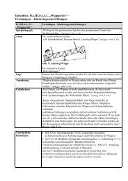
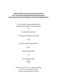
Fig. 1.1. The four sengi genera: (top) Rhynchocyon<br />
petersi, Denver Zoo (photo: A. Sliwa); (center left)<br />
Macroscelides proboscideus, Wuppertal Zoo (photo:<br />
A.Sliwa) (center right) Elephantulus brachyrhynchus,<br />
Transvaal Snake Park (photo: A.Sliwa); (bottom)<br />
Petrodromus tetradactylus, Museum of Zoology,<br />
Cambridge (photo: G. Olbricht).
REPRODUCTIVE PATTERNS 12<br />
1.3 Material and Methods<br />
An extensive bibliography is available on the Macroscelidea and many research fields are<br />
well documented. The wide range of sources over more than a century proved to be very<br />
suitable for conducting a review. Early works sometimes are controversial compared to more<br />
recent research, which allows a critical appraisal. Data on all the parameters used for Table<br />
1.1 were drawn from the extensive published literature, Fig. 1.1 presents members of all four<br />
sengi genera.<br />
1.4 Results<br />
For 14 of the 17 extant sengi species of Macroscelidea (Rathbun 2009) nine life history or<br />
reproductive traits are listed in Table 1 which could be of importance for a critical appraisal of<br />
the hypotheses.<br />
No data were available for E. fusciceps, E.pilicaudus and R. udzungwensis. Differences<br />
between the genera become evident in body mass, ovulation type, gestation time and, number<br />
of offspring.<br />
One of the diagnostic characters distinguishing the two subfamilies is the shape of the<br />
uterus. Rhynchocyon has a slightly bicornate uterus whereas its shape in all the other sengis is<br />
deeply bicornate (Corbet and Hanks 1968). Morphological differences also include teat<br />
number; while the Macroscelidinae except Petrodromus have 6 mammae, the<br />
Rhynchocyoninae and Petrodromus have 4. More details and modifications are presented in<br />
Chapter 3 which is dedicated to teat location. The special ultrastructure of the spermatozoa<br />
(Woodall and FitzGibbon 1995) is considered an ancestral feature, which divides between<br />
sengi genera.<br />
Most sengi species breed year-round but some show seasonal trends with reduced<br />
reproduction in the cool season, such as E. brachyrhynchus (Neal 1995). Singletons or twins<br />
are standard with the exception of E. rozeti where three and four young are common in some<br />
areas (Séguignes 1989). In Macroscelides some exceptional birth of triplets were noted in<br />
captivity by Olbricht et al. (2006). All members of this order produce precocial young except<br />
for Rhynchocyon which bear less developed offspring. Eye opening in R. petersi is at 11 – 12<br />
days old (Lengel 2007) while the other sengis are born with open eyes. All sengis are<br />
considered monogamous (Brown 1964, Rathbun 1979a, Kingdon 1984; Woodall and Skinner<br />
1989, Ribble and Perrin 2005). They live in pairs or singly and seldom in small groups.
REPRODUCTIVE PATTERNS 13<br />
Table 1.1 Life history characters of the Macroscelidea (pect. = pectoral, abd. = abdominal).<br />
Species Mass of<br />
adult (of<br />
neonate)<br />
in g<br />
Rhynchocyon<br />
chrysopygus<br />
(Golden-<br />
rumped sengi)<br />
R. cirnei<br />
(Chequered<br />
sengi)<br />
R. petersi<br />
(Black-and-<br />
Rufous sengi)<br />
Petrodromus<br />
tetradactylus<br />
(Four-toed<br />
sengi)<br />
Macrsocelides<br />
proboscideus<br />
(Short-eared<br />
sengi)<br />
Elephantulus<br />
brachyrhynchus<br />
(Short-snouted<br />
sengi)<br />
E. edwardii<br />
(Cape sengi)<br />
E. fuscus<br />
(Dusky e-<br />
shrew)<br />
E. intufi<br />
(Bushveld<br />
sengi)<br />
E. myurus<br />
(Eastern rock<br />
sengi)<br />
E. revoili<br />
(Somali sengi)<br />
E. rozeti (North<br />
African sengi)<br />
E. rufescens<br />
(Rufous sengi)<br />
E. rupestris<br />
(Western rock<br />
sengi)<br />
Gestation<br />
in days<br />
No. of<br />
young<br />
(of<br />
fetuses)<br />
No. of<br />
mammae<br />
Ovulation<br />
type<br />
Breeding<br />
season *<br />
540 (80) 1 42 1 1 1 4 (abd.**) 8 year-round<br />
1<br />
420 15 (2) 15 4 (abd.) 8 year-round<br />
18<br />
max.740 2<br />
(34) 2<br />
45-47 2 (2) 13<br />
2 2<br />
232 (31) 9 max.2<br />
5,18<br />
ca. 40 5<br />
46 22 (ca.8)<br />
52 5<br />
(max 9) 27<br />
52 5 , 48 6<br />
(9) 6 ,<br />
(11,9) 17<br />
(1) 13<br />
Longevity<br />
in years<br />
11 20<br />
4 (abd.) 8 oligo 13 4,5 20<br />
4 (pect.,<br />
abd.) 8<br />
(absence<br />
of abd.<br />
pair, 14 )<br />
60-61 4 max3 4 6 (pect,<br />
abd.<br />
inguinal)<br />
3,6,8,14<br />
max 2 3 ,<br />
(1-2) 13<br />
max 2 5,6<br />
(2) 13<br />
6 14 intermediate<br />
13<br />
oligo 13 Dec 3 ,June<br />
21 , Jan +<br />
July 19 ,<br />
whole year<br />
18<br />
poly 13 year-round<br />
but trends<br />
(Sep-Feb)<br />
12<br />
yearround<br />
10 ,<br />
June,<br />
Nov 3 , Oct-<br />
March 5,<br />
6 14 poly 13 Nov-Jan<br />
5 ,Sep-Dec<br />
6,28<br />
no data no data no data no data no data probably<br />
like E.<br />
49 9 ,56 5<br />
(10) 9<br />
69 9 , 80 5<br />
(8) 9<br />
50-52 9 2 3 , (1-2)<br />
13<br />
61<br />
interbirth<br />
interv. 23<br />
41 9 75 7 1-4 7 (1-<br />
max 55<br />
24 ,(10,6)<br />
26<br />
References in Table 1.1<br />
1 Rathbun 1979a<br />
2 Lengel 2007<br />
3 Shortridge 1934<br />
4 Olbricht et al. 2006<br />
5 Skinner and Smithers 1990<br />
57 26<br />
61-65 28<br />
6 3,14 intermediate<br />
13<br />
brach. 5<br />
Nov,<br />
March 3 ,<br />
Aug-<br />
March 5<br />
(2) 13 6 14 poly 13 July-Jan<br />
11 ,<br />
Sep-<br />
4) 13<br />
6 16<br />
March 5<br />
6.5 20<br />
8 20<br />
4 20<br />
5.5 20<br />
no data<br />
9 20<br />
6 14 oligo 13 Jan-Aug 13 7 4<br />
(1-2) 13 oligo 13 year-round<br />
24 ,seasonal<br />
rains 21<br />
max. 68 5 1-2 3,24 6 3, 14 oligo 13 year-round<br />
25 ,<br />
Sep,April,<br />
6 Stuart et al. 2003<br />
7 Séguignes 1989<br />
8 Fitzinger 1867<br />
9 Tripp 1972<br />
10 Leirs et al. 1995<br />
May, 3<br />
almost 8 20<br />
4 20<br />
11 van der Horst 1946<br />
12 Bernard et al. 1996<br />
13 Tripp 1971<br />
14 Corbet and Hanks 1968<br />
15 Ansell 1973
REPRODUCTIVE PATTERNS 14<br />
16 Haltenorth and Diller 1977<br />
17 Dempster et al. 1992<br />
18 Kingdon 1984<br />
19 Nowak (Walker) 1991<br />
20 Weigl 2005<br />
21 Brown 1964<br />
22 Rosenthal 1975<br />
23 van der Horst 1954<br />
24 Neal 1982<br />
25 Withers 1983<br />
26 Rathbun et al. 1981<br />
27 Yarnell and Scott 2006<br />
28 Koontz and Roeper 1983<br />
Post-partum oestrous is common but direct observations revealed an oestrous cycle of<br />
about 2 or 3 weeks for free-ranging (Sauer and Sauer 1971) and 2 weeks for captive<br />
Macroscelides (Vakhrusheva 2000). Van der Horst (1946) investigated uteri and ovaries of E.<br />
myurus jamesoni and estimated the length from anoestrous to oestrous as about 3 weeks.<br />
A modal estrous cycle of 13 days was estimated for E. rufescens with a range of 12 to<br />
49 days (Lumpkin et al. 1982).<br />
The longevity records are based on data from captivity and may represent unusually<br />
long-lived specimens. Rathbun (1979a) expected 4-5 years for Rhynchocyon chrysopygus,<br />
some years later a captive specimen reached the age of almost 11 years (Weigl 2005). This<br />
was the longest lifespan for a macroscelid ever recorded and is followed by E. intufi with 9<br />
years and M. proboscideus and E. rufescens with about 8 years.<br />
Patterns of behavioural ecology are closely related to reproductive patterns and, some<br />
of them are listed here. Rhynchocyon lives in coastal forests or in lowland and montane<br />
forests (Nicoll and Rathbun 1990). Petrodromus is likewise an inhabitant of coastal forests<br />
but also occurs in dense scrub and woodlands and in very dry scrub (Kingdon 1990). All the<br />
smaller sengis, the Macroscelidinae, prefer semi-arid savannah, bushland and woodland with<br />
Macroscelides proboscideus tolerating the most arid conditions of the Namib Desert (Sauer<br />
and Sauer 1971).<br />
Ovulation occurs spontaneously. The Macroscelidae are divided into 3 ovulation<br />
groups although a gradation rather than a complete separation between species is more<br />
appropriate (Tripp 1971):<br />
1. poly-ovulating species (31-89 corpora lutea = CL) include Elephantulus myurus<br />
jamesoni, E. edwardii and M. proboscideus,<br />
2. intermediate form (1-23 CL): E. intufi and E. brachyrhynchus and<br />
3. oligo-ovulating species (1-3 CL) are Rhynchocyon petersi, Petrodromus, E.<br />
fusciceps, E. rupestris and E. rozeti.<br />
Surprisingly, the species with the most offspring among all the Macroscelidea, E. rozeti, is<br />
oligo-ovulating. In disagreement to Tripp (1971) who found in gravid E. intufi up to 8 corpora<br />
lutea (CL) and in E. (Nasilio) brachyrhynchus up to 23 CL per ovary, van der Horst (1944)<br />
found only one or 2 CL in both species. For Petrodromus and E. rupestris (oligo-ovulating) as<br />
well as for Macroscelides and E. edwardii (in van der Horst as E. capensis, poly-ovulating)
REPRODUCTIVE PATTERNS 15<br />
both authors are in accordance. Additionally, van der Horst found in about half of the gravid<br />
females in E. intufi asymmetries of the uterus in contrast to this rare phenomenon in E.<br />
myurus jamesoni. In his study E. intufi carried an embryo in only one of the 2 horns but often<br />
2 CL were found in one ovary. In E. rupestris 3 CL were present in the 2 ovaries. The<br />
chemical constitution of the egg is a remarkable point of distinction between sengi species<br />
that belong to the same genus, e.g. the presence and absence of fat-globules in the egg (van<br />
der Horst 1944). Fat-globules are absent in poly-ovulating species such as Macroscelides and<br />
E. myurus, and present, but in low numbers in the poly-ovulating E. edwardii and clearly<br />
present in oligo-ovulating species such as E. rupestris and E. intufi. (Tripp 1971).<br />
Van der Horst (1954) confined the breeding season of E. myurus jamesoni between<br />
July and January. During his investigation females often ovulated at the end of the season<br />
without <strong>bei</strong>ng fertilized which results in a menstrual cycle, as at the end of the season also the<br />
males go into anoestrous (Stoch 1954). This species is one of the relatively few mammals that<br />
seem to menstruate regularly (van der Horst and Gillman 1941, 1942b). This uterine bleeding<br />
is of an unusual type and was considered by Patterson (1965) as a macroscelidid peculiarity.<br />
1.5 Discussion<br />
A certain homogeneity of the analyzed traits can be generally stated despite of some<br />
interspecific variance. This is in accordance with Corbet and Hanks (1968) who noted the<br />
remarkable similarity in external morphology and Rathbun (1979b) and Perrin (1995a) who<br />
observed similar life history traits. Both patterns have remained remarkable static over<br />
geological time (Rathbun and Rathbun 2006a).<br />
Due to the small litters low fecundity is expected, so that a premium is placed on traits<br />
that would increase their birth rates (Rathbun 1979b). In the following discussion I focus on<br />
potential factors controlling mammary number and litter size.<br />
There is some confusion on teat number and position. The absence of abdominal teats<br />
is described only for Petrodromus (Fitzinger 1867; Corbet and Hanks 1968). Teat number of<br />
Petrodromus was misstated with three teat pairs instead of two (Tolliver et al. 1989) and for<br />
E. myurus with two instead of three (McKerrow 1954). Corbet (1995) worked out a<br />
cladogram on the base of synapomorphies but here he surprisingly also included Petrodromus<br />
in the group with abdominal teats together with Rhynchocyon with two teat pairs in the<br />
abdominal area (Fitzinger 1867). Teat location was not described in detail nor were there<br />
exact terms to define the position. Chapter 3 will investigate this issue in more detail.
REPRODUCTIVE PATTERNS 16<br />
The intergeneric variance in mammae number stimulates the discussion whether an<br />
analysis of adaptations should be useful or whether selection in progress is to be detected. An<br />
organism may have an adaptation even if selection in progress is not operating on it now or<br />
selection in progress may be modifying an existing adaptation, creating a new adaptation<br />
(Grafen1988).<br />
1.5.1 Placentation in afrotheres<br />
Sengis, together with rock hyraxes, tenrecs and golden moles show a more recent<br />
development, the hemochorial condition elephants and aardvarks exhibit the ancient<br />
endotheliochoral placenta of the common ancestors of the Afrotheria (Starck 1949, Carter and<br />
Enders 2004). The formation of a discoid placenta in sengis and tenrecs is still a further<br />
development. The lobulated allantoic sac, as part of the fetal membranes is a feature is<br />
possibly shared by all afrotherians whereas the proliferation layer has not been described for<br />
any other mammalian orders except for sengis (Oduor-Okelo et al. 2004). Some features of<br />
the placenta such as the inner degenerative zone are present in most sengis but are absent in R.<br />
chrysopygus (Carter et al. 2004).<br />
Mammalian fetal membranes and placentas differ greatly without there <strong>bei</strong>ng any<br />
obvious relation to life style and they are an alternate source of phylogenetic information<br />
(Carter et al. 2004). The effects of selection pressures on the efficiency of placentation may<br />
stem from changes in nutritional demand, gestation length, number of embryos per pregnancy<br />
and maternal body constitution (Wildman et al. 2006). Comparison of the placental type with<br />
the brain advancement factor reveals that the haemochorial placental type includes altricial as<br />
well as precocial groups (Sacher and Staffeldt 1974) but the complex relationships do not<br />
support any simple relation between placental type and gestation schedule. The example of<br />
the guinea pig (Eisenberg 1981), <strong>bei</strong>ng also a precocial small mammal like most sengis,<br />
shows that species with haemochorial placentas pass almost the entire maternal antibody<br />
complement from the maternal circulation to the foetal circulation and pass little or none of it<br />
in the milk. Consequently, this would lead to highly developed young.<br />
The significance of the lobation of the allantois is unknown (Oduor-Okelo et al. 2004),<br />
but amniogenesis by cavitation instead of folding is recognized as character transformation on<br />
the common stem lineage of Afrotheria which helps to identify morphological characters that<br />
could be synapomorphic for this novel taxon (Mess and Carter 2006). In addition, Carter and<br />
Mess recognize the four-lobed allantoic sac and precociality as two other characer<br />
transformations on the stem lineage of Afrotheria.
REPRODUCTIVE PATTERNS 17<br />
1.5.2 Developmental status at birth<br />
Developmental length in general is a conservative trait in mammals which is primarily<br />
determined by phylogenetic factors and body size (Burda 1989b). A general controversial<br />
discussion is held about the classification itself if neonates are altricial or precocial because in<br />
reality mammalian species occur along a continuum of developmental maturity at birth<br />
(Zeveloff and Boyce 1986). Case (1978b) considers a mammal species precocial if its young<br />
possess body hair, teeth, open eyes, open auditory meatus, the ability to move forward, the<br />
ability to right itself when lying on its back and formed claws. Mammals lacking all or all but<br />
one ar altricial, those lacking some are semi-precocial or semi-altricial. In all sengis we find<br />
heavy and highly precocial neonates, except for Rhynchocyon which is considered semi-<br />
precocial following Case´s definition for precociality. The developmental stage at birth at<br />
which a young is born is a more plastic trait than the developmental length (Spalacopus<br />
cyanus, Begall et al. 1999) or a conservative rather than a plastic trait (Cryptomys sp., Begall<br />
and Burda 1998).<br />
In their analysis of the correlation between maternal investment and litter size Zeveloff<br />
and Boyce (1986) demonstrate that mammals with litter sizes < 2 tend to bear precocial young<br />
and those with more than 3 young are altricial. This agrees with small litter size and<br />
precociality in the Macroscelidinae, hyraxes and precocial rodents such as chinchillas.<br />
Nevertheless they conclude that this does not imply that litter size should be a criterion for<br />
classification of neonate development.<br />
1.5.2.1 Environmental factors influencing the developmental status at birth<br />
Growth reflects an adaptation of species for their environment (Case 1978b). Predator<br />
selection might favour an increase in neonate size where young are reared on open, exposed<br />
areas (Case 1978a). This may have largely influenced the development of precociality of<br />
sengis living in dry habitats which do not build nests. The precocial young hyraxes are also<br />
quite exposed to predators although they live in crevices in the rocks.<br />
Only Rhynchocyon occurring in dense forest can afford less advanced neonates. They<br />
build nests. On the other hand, the precocial rodent Otomys also constructs well developed<br />
nests and here the occurrence of precociality cannot be explained satisfactorily (Neal 1986).<br />
The altricial (or semi-precocial) tenrecs inhabit various habitat types (Eisenberg and Gould<br />
1970, Puschmann 2004), only for some species nest building is reported, whereas the semiprecocial<br />
aardvark young does not leave its burrow for the first 2 weeks after birth (Shoshani<br />
et al. 1988). The altricial golden moles live in burrows and so are well protected (Perrin and<br />
Fielden 1999).
REPRODUCTIVE PATTERNS 18<br />
1.5.2.2 Development of precociality<br />
There is some controversy on about the development of precociality. Following Hopson´s<br />
theory (1973) short gestation periods of small placentals followed by altricial young and<br />
intensive maternal care, including suckling is a pattern inherited by early therian ancestors.<br />
Only with the development of a complex allantoic placenta with secretory properties<br />
permitting the suspension of oestrous allowed a prolongation of the gestation period and<br />
leading to more developed neonates. Consequently, the production of advanced young is<br />
considered to be a secondary evolutionary development in mammals (Case 1978a) which<br />
evolved relatively late in mammalian history. The reproductive features of Rhynchocyon<br />
would fit into this hypothesis as it is the most ancient genus (e.g. Tabuce et al. 2008) within<br />
the order Macroscelidea. It has the shortest gestation time and has the least precocial offspring<br />
among the other sengis.<br />
On the other hand, long pregnancy is considered an ancestral trait in Bathyergidae<br />
Sumbera et al. 2003). The “primitive” taxa Ctenodactylidae and Pedetidae have relatively<br />
longer developmental times than other sciurognaths and even produce precocial young (Burda<br />
1989a). Conseuqently, longer developmental times with precocial young are considered a<br />
derived pattern in hystricognath rodents (Sumbera et al. 2003) and only in small<br />
hystricognaths altricial young are born after a long pregnancy.<br />
The majority of extant mammal species produce altricial offspring (Case 1978a). But<br />
the so-called altricial/precocial dichotomy exists within several taxa suggesting that the<br />
ancestral altricial developmental mode (Hopson 1972, Case 1978a) has been modified<br />
towards precociality more than once during mammalian evolution. That means that<br />
mammalian development patterns do not map onto continuous changes in life history patterns<br />
and a shift from altricity to precociality and vice versa during the evolution is very probable.<br />
The cost of precociality is a low intrinsic rate of natural increase and appears to<br />
depend on body size, such that differences in potential population growth rates are greatest in<br />
small-bodied mammals. This might explain why only a few species of small mammals<br />
produce precocial young (Hennemann 1984). Rhynchocyon has the shortest gestation length<br />
but produces mostly one semi-precocial offspring in contrast to Elephantulus with up to 4<br />
precocial offspring but much longer pregnancy. In order to increase the reproductive rate the<br />
Caviidae were able to shorten their developmental time (postnatal phase) (Burda 1989a) and<br />
evolved extreme precociality (Kraus et al. 2005). These authors concluded for caviomorph<br />
rodents that phylogenetic inertia is considered to constrain a shift to altricity which could also<br />
be true for sengis.
REPRODUCTIVE PATTERNS 19<br />
1.5.2.3 Precocial and altricial strategies<br />
Neal (1986) found in his analysis of reproductive characters of African small mammals<br />
including Elephantulus, that the altricial/precocial dichotomy does not fit the prediction of r-<br />
and K-selection theory but that these 2 main strategies have a mixture of r- and K-selected<br />
characteristics although species with altricial young tend to be more r-selected than species<br />
with precocial young. Consequently, reproductive effort (the proportion of total energy<br />
budget allocated to reproduction) should be a measure of an animal´s position along a<br />
continuum of “r- or K-selected” strategies (Millar 1977).<br />
1.5.3 Gestation length<br />
Generally, it is accepted that gestation length is correlated with the degree of development of<br />
the young at birth and resulting in increased development of the central nervous system<br />
(Sacher and Staffeldt 1974). Martin and Mac Larnon (1985) clearly distinguish altricial and<br />
precocial mammals by gestation period relative to maternal weight despite of some overlap<br />
between large-bodied altricial mammals and certain small-bodied precocial mammals. In<br />
contrast, Case (1978a) found no consistent correlation between adult body size and the degree<br />
of maturity at birth. Rhynchocyon sp. is the largest sengi but the least precocial. The question<br />
remains if neonatal developmental status in relation to adult body size and gestation periods<br />
are appropriate allometric patterns for phylogenetic research.<br />
However, costs and benefits of prenatal and postnatal development must be balanced<br />
no matter what the developmental status of the neonate is. On the other hand, pregnancy is<br />
energetically much less demanding than lactation and provides the offspring with a high<br />
degree of protection and environmental stability (Millar 1977). Weir and Rowlands (1973)<br />
expressed the difficulty determining causal directions with the particular example of gestation<br />
length as follows: “it is obviously impossible to tell whether the gestation length was the fixed<br />
parameter and the environmental cues existing at the time were incorporated into the mating<br />
season stimuli, or whether the gestation length was the variable upon which selection acted.”<br />
1.5.4 Parental care<br />
Fathers do not seem to be involved in infant care in captive Rhynchocyon petersi and no<br />
interaction was observed during the in-nest period. Only casual interaction occured after the<br />
infants emerged from the nest. It was not clear whether there was a male´s participation in the<br />
nest building (Baker et al. 2005). Apparently, in the course of the evolution of the mammary<br />
glands the role of the father in many mammals was reduced as is true also for sengis.
REPRODUCTIVE PATTERNS 20<br />
With regard to the high neonate birth mass in macroscelids in comparison to the<br />
mother´s weight (ca. 50% in a twin litter) it is evident that the investment during pregnancy<br />
must have reached the limit of a female´s physical capacities. This investment favours the<br />
absentee maternal care system (Rathbun 1979a, Rathbun and Rathbun 2006a) and neonates<br />
are left alone for a long period of time shortly after birth already (Sauer 1971 and 1973, Baker<br />
et al. 2005). This system could have evolved to ease the female´s burden of rearing young<br />
after this long pregnancy.<br />
Male E. myurus (Ribble and Perrin 2005) and Petrodromus (Rathbun 1979a,<br />
FitzGibbon 1995) have never been observed in the same vicinity of females with offspring.<br />
Sengis will not directly defend their young but predation is avoided in E. rufescens and R.<br />
chrysopygus partly by running away (Rathbun 1979a) which reduces the risk of predation also<br />
in mobile precocial young. Sauer (1972) emphazised the importance of a secure birth site on<br />
which the Macroscelides mother can rely on rather than helping her offspring in the case of<br />
danger. He also observed a large distance between the usual home range of the mother where<br />
she searches for food and the site the offspring are hiding.<br />
1.5.4.1 Lactation<br />
In the course of evolution reproductive hormones allowed the formation of incubation areas<br />
for further development of foetuses and further, in some mammals also glandular secretions.<br />
Lactation is considered a crucial factor in the evolution of mammalian parental investment<br />
and reproductive strategies (Eisenberg 1981). The lactation period, while maternal feeding is<br />
crucial for the surviving of the young, is shorter than the weaning time.<br />
1.5.4.2 Feeding intervals<br />
Female Macroscelides (Sauer and Sauer 1972) and R. petersi (Baker et al. 2005) return only<br />
once a night to their newborn young to nurse them. Rathbun (pers. comm.) speculated that<br />
this absentee maternal care system (Sauer 1973) may relate to the relatively large number of<br />
teats in relation to small litter size. Within 80 minutes Macroscelides young were nursed 6<br />
times which implies a large amount of milk. Further, relatively large offspring could survive<br />
longer between feedings so that the female could feed her young infrequently and spend more<br />
time feeding (Millar 1977).<br />
The guinea pig belongs to the most precocial species among small mammals but<br />
compared with sengis, the female invests much time in litters of few young (Kraus et al.<br />
2005). The suckling interval of caviomorph rodents varies quite widely from about 2 to 35
REPRODUCTIVE PATTERNS 21<br />
minutes (Alderton 1996). Species which produce their offspring in underground burrows such<br />
as the degus (Octodonta spec.) spend longer suckling them than e.g. the caviids, which remain<br />
vulnerable in the open.<br />
1.5.5 Monogamy<br />
Monogamy in sengis is considered a unique pattern which is shared by the whole clade<br />
(Ribble and Perrin 2005). There is no evidence that monogamy evolved in response to the<br />
need for paternal care and evolved more often in the absence of paternal care than in its<br />
presence (Komers and Brotherton 1997) and the authors conclude that the costs and benefits<br />
of monopolizing solitary females appear to be the primary issues in the evolution of<br />
monogamy in mammals.<br />
Monogamy in sengis may also be related to testes size as observed in other mammals<br />
(Woodall and Skinner 1988) and lack of sexual dimorphism e.g. in R. chrysopygus<br />
(FitzGibbon 1995).<br />
1.5.5.1 Monogamy and parental care<br />
Zeveloff and Boyce (1980) generally found that species with low maternal investment in the<br />
young at birth tend to be monogamous. Primates (Lemur, Tarsius, Cercopithecus),<br />
lagomorphs (Ochotona, Caprolagus), rodents (Heterocephalus, Dolichotis), carnivores<br />
(Fossa) and some ungulates are monogamous but lack paternal care.<br />
The question arises how paternal care can be defined. Male and female sengis do not<br />
have much direct contact which is defined as uniparental monogamy (Rathbun and Rathbun<br />
2006b) which is also known for the dik-dik (Brotherton and Rhodes 1996). Difficulty is<br />
experienced in deciding what the benefits of monogamous parents for offspring survival are,<br />
if there is neither direct male care nor protection from infanticide or predators. Trail<br />
maintenance activities may be one of the benefits providing easy access to the territory for<br />
foraging and predator escape (Rathbun 1979a) but this trait is not generally present in all<br />
sengis (Ribble and Perrin 2005).<br />
1.5.5 2 Mate guarding<br />
The dik-dik model (Madoqua kirkii, Brotherton and Rhodes 1996) was used to discuss mate<br />
guarding (Ribble and Perrin 2005) as the best explanation for social monogamy in sengis.<br />
They found that dik-diks and sengis share many life history traits including small litter size<br />
and highly precocious young. Male mate-guarding seems to be responsible for monogamy in<br />
E. myurus (Ribble and Perrin 2005) and R. chrysopygus (FitzGibbon 1995). Lumpkin and
REPRODUCTIVE PATTERNS 22<br />
Koontz (1986) suggested that mate guarding in view of the short duration of oestrous in<br />
sengis might be particularly adaptive which has led to monogamy (Brotherton and Manseri<br />
1997). Male mate-guarding has recently been proposed as the principal adaptive factor in the<br />
evolution of all mammalian social monogamy with parental care of young likely <strong>bei</strong>ng<br />
secondarily derived (Ribble 2003).<br />
But unlike antelopes sengis do not exhibit strong pair bonds (Rathbun and Rathbun<br />
2006a). In M. proboscideus even so few intra-pair interactions occur that Sauer (1973)<br />
characterized the species as solitary, but strongly promiscous. He still supports a latent pairbond<br />
as neighbouring partners will breed again in the next oestrous cycle. This strategy shows<br />
that male sengis are not forced into obligate monogamy such as dik-diks which have to spend<br />
much of their time with their mate to prevent them from leaving the pair´s territory (Komers<br />
1996). But even in strictly monogamous species with no evidence of extra pair paternity such<br />
as the dik-dik, males did attempt to obtain extra-pair copulations whilst females did not<br />
attempt to do so (Brotherton et al. 1997).<br />
1.5.5.3 Monogamy and precociality<br />
The tendency for monogamy related to short gestation periods as postulated by Zeveloff and<br />
Boyce (1980) however, is controversial to the relatively long gestation period in sengis.<br />
Zeveloff and Boyce (1980) also found that species with altricial young tend to be<br />
monogamous and those with precocial young tend to be polygynous. In contrast to this theory<br />
almost all sengis are precocial and monogamous whilst the aardvark produces less developed<br />
young and lives in a polygamous mating system (Shoshani et al. 1988). However, analysing<br />
data from insects, birds and mammals regarding the breeding success of males showed that<br />
the differences between monogamous and polygynous species are smaller than expected<br />
(Clutton-Brock 1988), especially when offspring survival is included in measures of male<br />
success.<br />
1.5.6 Juvenile mortality<br />
Risk-sensitive foraging behaviour is rather one of the factors to the benefit of both offspring<br />
and mother as this decreases the mortality rate as observed in Macroscelides (Lawes and<br />
Perrin 1995). The authors noted a substantial behavioural flexibility where “natural selection<br />
has acted strongly in shaping the foraging behaviour”. This means that the probability of<br />
energy shortfall or starvation is minimized and risk-prone foraging is highly unlikely which<br />
would attain a positive effect on the successful rearing of young. Van der Horst (1946)<br />
assumed an infant mortality of about 55% for wild E. myurus jamesoni which in his view is
REPRODUCTIVE PATTERNS 23<br />
low. In captive Macroscelides two third survived longer than a month (Olbricht et al. 2006)<br />
and about 80% of captive E. rufescens (Rathbun et al. 1981).<br />
Hennemann (1984) states that the evolutionary “decision” to produce precocial young<br />
may allow increased survivorship of the young in response e.g. to predation and this agrees<br />
with Case´s notion (1978a) that the relative intensity of predation on the young compared<br />
with that on the adult appears to be most important in determining the optimum size of the<br />
neonatus and its maturity at birth.<br />
1.5.7 Litter size<br />
The question arises whether adult females are breeding at or close to the theoretical limit of<br />
their capacity to produce and rear young. Partridge (1989) postulates “that number of young<br />
produced per litter is often not the maximum number that a female can produce in a breeding<br />
event but a set of trade-offs between the mothers costs of reproduction and her future<br />
reproductive potential and between number and size of young”. Because precocial young<br />
would be at the maximum mass that the adult female can carry selection for short nestling<br />
periods and concomitant longer gestation periods would lead to small litter size (Viljoen and<br />
du Toit 1985). It could also be a limiting factor, that speed is required to avoid predators in<br />
the sense that any selection against to heavy females during pregnancy would favour a smaller<br />
litter size (Millar 1977) and he concludes that litter size appears to be the adaptive variable of<br />
mammalian reproduction. Millar also found that another nonenergy factor that could favour<br />
small litters is the ability of a female with precocial young to keep track of her offspring.<br />
1.5.7.1 Correlation between teat number and litter size<br />
Phylogeny only influences litter size through homology in that a number of related taxa may<br />
have a similar litter size because a common ancestor became adapted, e.g. to a stable climax<br />
environment (Perrin 1986). Mammary number and litter size are not similar among sengis but<br />
in all genera litter size is much larger than teat number. However, there might be homology to<br />
other afrotheres. Similar to Rhynchocyon and Petrodromus mostly one young is born in the<br />
aardvark (Orycteropus afer, Shoshani et al. 1988), the Florida manatee (Trichechus manatus,<br />
Reynolds and Odell 1991) and Grant´s Golden mole (Eremitalpa granti, Perrin and Fielden<br />
1999), all females have 4 teats. Teat number and litter size within the Tenrecidae varies<br />
remarkably (Puschmann 2004).<br />
A large number of teats may be useful in connection with big litter sizes and the<br />
intensive care of dependent young. Among rodents not all species fit into the scheme of the<br />
“one-half-rule”. Comparable to sengis some similar characteristics are found in 2 other
REPRODUCTIVE PATTERNS 24<br />
precocial species: the rock hyrax (Procavia capensis, Olds and Shoshani 1982) and a rodent,<br />
(Chinchilla lanigera, Anderson and Sinha 1972) which have 6 teats but only 2 - 3 offspring.<br />
The latter authors argue about a correlation between mammary number and litter size. The<br />
naked mole rat (Heterocephalus glaber) is an altricial rodent but it is an exception to the<br />
“one-half rule” (Sherman et al. 1990). Its litter size doubles teat number.<br />
Pearl (1913 a, b) uses the term “teleological necessity” for a compensating increase in<br />
the number of mammae when the number of young born in a litter increases. But this is a<br />
rather philosophical attempt and nobody can´t imply that evolution is a directed process. In<br />
contrats, the additive genetic variance appears to be less for mammary number than for litter<br />
size (Gilbert 1986). Polar bears (Ursus maritimus) normally have 4 mammae but up to 6<br />
functional mammae are reported with less than 2 cubs as average litter size (Derocher 1990).<br />
Some of the supernumerary teats were suckled. In comparison to the Brown bear (Ursus<br />
arctos) with 6 teats and 2 cubs per litter Derocher suggested that reduction in litter size and<br />
the normal number of mammae may be related to the difficulty of raising cubs in the harsh<br />
environment of polar bears which agrees with Erickson (1960) that supernumerary teats are a<br />
throwback to an ancestral condition. The Zambian common mole rat (Cryptomys sp.) is an<br />
altricial rodent which has 3 teat pairs, the mean litter size is 2 but can be up to 6 young. In a<br />
litter of 1 or 2 offspring only the pectoral teats are suckled. If more young are born, the axial<br />
and inguinal teats are used too (Burda pers. comm.) which is in contrast to hyraxes where the<br />
inguinal teats are mostly preferred (Hoeck 1977, Fischer 1986). Nevertheless, given that the<br />
conception of phylogenetic inertia (many teats as ancestral character) applies it would be<br />
interesting to investigate if unsuckled mammae would disappear over evolutionary time as<br />
observed in domestic sows (Kim et al. 2001).<br />
However, the one-half rule does not apply for sengis. A phylogenetic interpretation of<br />
the large number of teats in sengis is somewhat hampered by uncertainty concerning their<br />
relationships to other mammalian orders. I infer that the large number of teats might be a<br />
relict from ancestral times and is explained by phylogenetic inertia. Secondly, a larger number<br />
of teats provide a larger amount of milk which may benefit precocity. Especially the<br />
Macroscelidinae are small mammals with small sized teats, barely visible even while<br />
lactating. It may be speculated that in these highly cursorial mammal the amount of milk<br />
needed is distributed among several teats rather than in a few udder-like bigger teats.<br />
1.5.7.2 Limits of litter size<br />
Litter size may be limited by one or more of several factors, depending on the habits of the<br />
animal. Litter size in mammals may be limited by the ability of females to carry large foetal
REPRODUCTIVE PATTERNS 25<br />
biomass and consequently, any selection against heavy females during pregnancy would favor<br />
a smaller litter size (Millar 1977).<br />
Mammary number in the Macroscelidea apparently does not limit litter size and<br />
subsequently reproductive success as e.g. in marsupials (Ward 1998) where teat clinging is<br />
essential of the survival of neonates. In most mammals the reduction of litter size at birth is<br />
the result of post-implantation loss such as in some diprotodont marsupials which typically<br />
ovulate more eggs and, in contrast to sengis, supernumerary neonates are produced (Ward<br />
1998). In comparison to marsupials where litter size is restricted by the number of functional<br />
teats he suggested that restriction in teat number may be analogous to the restricted<br />
implantation sites of the sengis. Here the question remains unanswered why in sengis with<br />
mostly twin litters we find 6 functional mammae and despite of the restricted area of the<br />
implantation site supernumerary litters can be produced as reported for M. proboscideus and<br />
E. rozeti.<br />
Given this conservatism of a species-typical invariant mammary number, excluding<br />
exceptions, there must be other mechanisms compensating for constantly increasing litter<br />
numbers. Some altricial rodents are known to adjust litter size by infanticide during early<br />
lactation (Perrigo 1987) whilst this has never been observed in the precocious guinea pigs<br />
(Künkele 2000). As a result, a more adequate mechanism to reduce litter size is probably fetal<br />
resorption during gestation (Peaker and Taylor 1997). Precociality as mentioned earlier also<br />
leads to smaller litters.<br />
1.5.8 Seasonality of breeding periods<br />
Although macroscelids have populated various ecological niches distinct reproductive<br />
seasonality is not found. The number of litters produced per year and the survival of young<br />
may be strongly limited by the duration of the breeding season (Case 1978b). Case (1978a)<br />
also suggested that the attainment of endothermy in the ancestors of sengis must eventually<br />
have extended the reproductive season of early macroscelids because it enabled these<br />
organisms to physically buffer seasonal changes to a greater degree.<br />
The combination of a long breeding season with a smaller litter size enables some<br />
tropical rodents to spread nutritional demands of breeding over a larger proportion of the year<br />
than is possible in temperate climates (Viljoen and du Toit 1985). E. rupestris is sexually<br />
active throughout the year assuming that they are less dependent upon rain and fog for<br />
reproduction than are e.g. the rodent species of the same area (Withers 1983). Neal (1986)<br />
noted that species bearing precocial young have relatively aseasonal breeding patterns even in<br />
very seasonal environments compared to species with altricial young because precocial
REPRODUCTIVE PATTERNS 26<br />
species may have lower energy requirements. This would give precocial species a better<br />
option to reproduce aseasonally because maximum energy expenditure during lactation is not<br />
as extreme as in rodents with altricial young (Trillmich 2000). Macroscelides adopted an<br />
omnivorous diet (Kerley 1995, Bernard et al. 1996) to deal with energy constraints which<br />
consequently allows reproduction throughout the year.<br />
Natural selection may favour rapid growth rates in highly seasonal environments<br />
(Zeveloff and Boyce 1986) which benefits the evolvement of precociality. The genus<br />
Elephantulus is physiologically and behaviourally adapted to arid and semi-arid regions of<br />
low productivity. It cannot compete against species bearing more altricial young in more<br />
productive habitats and Neal (1995) concluded that the precocial habit may be one reason<br />
why this genus is restricted to such environments. But reproductive activity is not only<br />
influenced by the environment but also by the organism´s own characteristics (Neal 1986). On<br />
the other hand, sengis exhibit a broad behavioural flexibility along with morphological and<br />
physiological features shown in observations about thermoregulation (Roxburgh and Perrin<br />
1994, Perrin 1995) including torpor (Lovegrove et al. 2001a, b), feeding behaviour (Kerley<br />
1995, Lawes and Perrin 1995) and general natural history (Sauer and Sauer 1971, 1972) to<br />
cope with environmental stochasticity (Lawes and Perrin 1995).<br />
1.5.8.1 Male capacities<br />
Woodall and Skinner (1989) described relatively small testes but extra-gonadal sperm<br />
reserves in E. myurus jamesoni compared to many similar-sized mammals. They found that<br />
this could be compensated by the active spermatogenesis throughout the year which is<br />
subsequently leading to continuous breeding during an extended breeding season. In<br />
disagreement to Stoch (1954) and van der Horst (1946) there is no cessation of breeding.<br />
Woodall and Skinner (1989) found active spermatogenesis in male E. myurus throughout the<br />
year despite of significant reductions in testis and prostate size outside the main breeding<br />
season. In contrast to other precocial mammals such as the chinchilla and the plains viscacha<br />
(Lagostomus maximus) where the males are always fertile and only the females are seasonal<br />
(Weir and Rowlands 1973), in sengis there is no complete cessation of breeding.<br />
1.5.9 Post-partum oestrous<br />
Inhibiting oestrous during lactation would not benefit the improvement of the reproductive<br />
rate for species with well developed young and short lactation (Weir and Rowlands 1973).<br />
There is no lactation anoestrous in E. myurus jamesoni (van der Horst 1946) but lactation<br />
following one pregnancy overlaps with the subsequent pregnancy (McKerrow 1954) which
REPRODUCTIVE PATTERNS 27<br />
increases the reproductive effort. Similar observations on rodents revealed that post-partum<br />
oestrous might be an indicator of high reproductive effort (Gilbert 1986).<br />
1.5.10 Poly-ovulation<br />
Natural poly-ovulation is a rare process in mammals (Weir and Rowlands 1973). In E. myurus<br />
120 eggs may be liberated from the 2 ovaries at each ovulation (van der Horst and Gillman<br />
1941a), a number which is exceeded only by a hystricomorph rodent, the plains viscacha<br />
(Lagostomus maximus) which produces 200-800 eggs (Weir 1971). This species shows some<br />
similarities to sengis, as it has precocial young, breeds throughout the year, lives in pairs and<br />
is poly-oestrous. Also, a female aardvark with one foetus and 5 more corpora lutea was found<br />
(Shoshani et al. 1988). For Tenrec ecaudatus the number of liberated eggs is not known but<br />
the litter of 31 young (Louwman 1973) is supposed to be the biggest among all the other<br />
Afrotheria. Poly-ovulation in tenrecs and sengis may represent some relict mechanism in<br />
ovulation procedures (Weir and Rowlands 1973).<br />
At first glance, it appears that some successful reproductive traits evolved in very<br />
different taxa due to environmental constraints. On the other hand, variations in the ovulation<br />
rate among sengis appear to be unrelated to environmental factors as there is an overlap of the<br />
distribution of many oligo-ovulating and poly-ovulating sengi species (Corbet and Hanks<br />
1968).<br />
1.5.10.1 Female capacities<br />
The reduction in the number of eggs from 60 to 1 (E. myurus) in each uterine horn is achieved<br />
at the latest four-cell stage and before implantation (van der Horst and Gillman 1941a). There<br />
is only one very small and well circumscribed area in each uterine horn, which enables the<br />
successful implantation of a single embryo at a time (van der Horst 1942; van der Horst and<br />
Gillman 1942c). Consequently, the final litter size is much likely to be determined entirely by<br />
the restricted implantation sites (Tripp 1971). This embryo chamber is a consistent feature of<br />
placentation in sengis that may partially account for the small litter size (Oduor-Okelo et al.<br />
2004). E. rozeti apparently is exceptional where 2 embryos are imbedded in the caudal end of<br />
each horn. Triplets were also found in M. proboscideus (Olbricht et al. 2006).<br />
This reproductive potential including potentially supernumerary young and polyovulation<br />
could help to shed some light on this question. Typical prey species stand under a<br />
strong pressure of natural selection and must be prepared for the occasional extreme rather<br />
than for the more frequent mean litter size. Sherman et al. (1999) suggested that extreme and<br />
variable litter sizes may be related to unpredictable food availability which agrees with Pearl
REPRODUCTIVE PATTERNS 28<br />
(1913a) who proposes an evolutionary “factor of safety” to achieve flexibility with<br />
environmental cues. Subsequently, a larger number of teats would support this flexibility and<br />
leaves space for further development within the evolutionary framework.<br />
1.5.11 Longevity and fecundity<br />
Two determining factors for the fecundity of individuals are lifespan and inter-litter intervals<br />
(Promislow and Harvey 1990). One strategy to produce enough offspring to balance the death<br />
rate is to extend the length of the reproduction period (Neal 1986). In fact, continuous<br />
breeding with short-inter-birth intervals and a long lifespan (Olbricht 2007) reveal the high<br />
reproductive potential of sengis despite of their low reproductive rate (small litter size, long<br />
gestation periods).<br />
Sengis have a longer lifespan as one may expect from species of that size (Jones 1985,<br />
Weigl 2005) which is linked with an extremely high fecundity rate until the end of their life<br />
(Olbricht 2007). This is also known for Zambian mole rats (Fukomys anselli), small, altricial<br />
subterranian rodents. Data from captive sengis reveal that lifespan of specimens observed in<br />
the wild or earlier studies in captivity are exceeded by far as a result of improved husbandry<br />
(Jones 1982, Weigl 2005). Van der Horst (1946) had suggested that female E. myurus and<br />
consequently all other small sengis may have only 3 pregnancies during their life time but<br />
improvement in breeding also led to high fecundity rates which was found in captive<br />
Macroscelides (Olbricht 2007) where a female had 23 litters with 46 offspring during her<br />
lifespan of 5 years and 2 months, among those the unusual birth of triplets. She had the last<br />
litter 3 months before death.<br />
Additionally, sengis have a number of behavioural and ecological traits that may<br />
represent adaptations to improved survivorship such as predator avoidance by young (Sauer<br />
1972) and by adults (Rathbun 1979b), risk-.sensitive foraging (Lawes and Perrin 1995),<br />
defending a territory (Ribble and Perrin 2005), thermoregulation (Perrin 1995) and a varied<br />
diet (Bernard et al. 1996). The “K-selected” traits of sengis require an effective quality<br />
breeding performance over a long life time. This is in line with Harvey and Zammuto (1985)<br />
and Promislow and Harvey (1990) who support an evolutionary link between low mortality<br />
rates, delayed reproduction and long lifespans.<br />
1.6 Conclusions<br />
The reproductive characteristics of the Macroscelidea are unusual as suggested by many<br />
authors (e.g. Neal 1995). Ancestral sengis were presumably not smaller in size than extant
REPRODUCTIVE PATTERNS 29<br />
species (Evans 1942, Patterson 1965, Simons et al. 1991) but probably had altricial offspring,<br />
as is suggested for other ancestral small mammals too (Derrickson 1992). Whereas the<br />
Rhynchocyoninae maintained ancestral features such as shorter gestation time, leading to less<br />
mature offspring and longer post-natal development, the Macroscelidinae extended pregnancy<br />
which is linked to precociality and shortening of the post-natal phase. The post-natal<br />
development of body metrics is presented in chapter 5 and 6.<br />
Life history traits of sengis such as fecundity and and lifespan as well as reproductive<br />
patterns such as litter size, post-partum oestrus, neonatal development and mammary number<br />
reveal a high reproductive potential and could balance the cost for the evolvement of<br />
precociality. These patterns may also have evolved in general to achieve flexibility with<br />
environmental cues which makes it difficult to determine causative factors during their<br />
phylogeny. As a result, it cannot be fully clarified whether precociality in sengis is a<br />
plesiomorphic trait.<br />
Rhynchocyon and Petrodromus have fewer teats than Elephantulus and Macroscelides.<br />
If I assume in a first hypothesis that phylogenetic inertia is the reason for this generally large<br />
mammary number relative to litter size and this is an ancient trait, so all the other<br />
physiological and behavioural traits analyzed here are adaptations to the sometimes extreme<br />
living conditions of the macroscelids. There has possibly never been a necessity to reduce the<br />
number of teats because behavioural adaptations could even derive from it as an advantage. If,<br />
for example, the maternal absentee care system can be considered as the main reason for<br />
upholding the large mammary number there could be a direct advantage of this strategy of<br />
postnatal care as it eased the dam´s work load of rearing young. In most macroscelids the<br />
highly precocial young require for a few days only an obligatory milk supply which<br />
consequently increases the fitness and the energy supply for the subsequent litter. A short but<br />
efficient milk supply is required which is secured by the large teat number but on the other<br />
hand, 6 functioning mammae are quite energy requiring. A lot of energy and effort is required<br />
but for a short period of time only and precociality is favourable to reduce the time of<br />
dependence of the offspring. Consequently, breeding success is not negatively influenced by<br />
parental care of young of different litters but is increased by short interbirth-intervals and<br />
aseasonal breeding. In domestic guinea pigs (Cavia porcellus) the peak of energy demand is<br />
low relative to that of altricial rodents and extended pregnancy is linked to a high efficiency<br />
of energy conversion into offspring (Künkele 2000). These patterns in reproductive energetics<br />
led to the speculaton that the precocial strategy might be ecologically robust and enables wild<br />
guinea pigs to reproduce year-round, even under harsh conditions. With regard to the<br />
distribution of sengis in dry habitats this aspect could also apply on macroscelids.
REPRODUCTIVE PATTERNS 30<br />
Additionally, traits such as precociality, absentee maternal care system and many teats<br />
together with the sengis´ life style help to compensate that sengis as typical small prey species<br />
are vulnerable to predation.<br />
Another hypothesis becomes important with respect to the most ancient genus<br />
Rhynchocyon which is living in dense forest. On the base of its life history I could assume that<br />
the ancient feature was 4 teats and not 6. The following conclusions would support this<br />
hypothesis. The more specialized Elephantulus-species and Macroscelides are inhabitants of<br />
open habitats. The potential to produce more offspring than the usual number of 1 or 2 is<br />
clearly evident in Macroscelides as well as in Elephantulus rozeti. As a result, an increase of<br />
mammary number as an adaptive evolutionary trait could be possible. Six teats might then be<br />
considered as a derived trait from the original 4 teats. However, it will not explain why<br />
Petrodromus also has only 4 teats.<br />
Poly-ovulation occurring in several species is another indicator for a future potential<br />
increase of the reproductive potential. Although oligo-ovulation occurs also in Elephantulusspecies,<br />
Petrodromus and Rhynchocyon do not seem to be provided with the same<br />
reproductive potential as the genera Elephantulus and Macroscelides. The gestation period is<br />
shortest in R. chrysopygus (42d) and longest in E. rozeti (75d). The highly precocial young of<br />
the latter are born in a harsh and dry environment and do not build nests. In contrast to<br />
Rhynchocyon neonates, who find the safety of a nest in dense cover, they need to develop<br />
more rapidly as they are more exposed to life threatening risks.<br />
The social monogamy found in the Macroscelidea across species and across different<br />
habitats may be the consequence of the conservation and interaction of a suite of uniquely<br />
derived traits (Rathbun and Rathbun 2006a).<br />
Continuous breeding occurs in all sengis and implies post-partum oestrous. This could<br />
be one of the reasons why the archaic Rhynchocyon could survive for 20 millions years in<br />
which time their basic anatomy and presumably their habits have changed very little<br />
(Kingdon 1990).<br />
It was to examine if these characters could be a source of phylogenetic information<br />
within the Macroscelidea and if so, that they might even give support to the Afrotheria clade.<br />
With respect to the macroseclids relationship to the Afrotheria it seems that at least neonatal<br />
development patterns are not a good tool to detect phylogenetic interrelations as “they do not<br />
appear to result from one continous axis of selection” (Derrickson 1992). In their review on<br />
afrotherian origins Robinson and Seiffert (2004) came to the inference that afrotherians,<br />
except aardvarks and paengulates are unanimous and that their phylogenetic affinities lay with<br />
a variety of other non-paengulate placental orders. Subsequently, for different afrotherian
REPRODUCTIVE PATTERNS<br />
species independently from phylogenetic relationships interspecific correlations may have<br />
evolved.<br />
Given their ancient association with the Afrotheria and their clinging to their life<br />
history traits phylogenetic inertia seems to play an important role why sengis did not evolve<br />
more specializations e.g. in their social organization (Rathbun and Rathbun 2006a).<br />
Nevertheless, sengis and probably other afrotherians too, developed a large number of<br />
adaptations to compensate for their legacy. This agrees with Robinson and Seiffert (2004)<br />
who found it very difficult to determine which taxa might preserve the morphological<br />
apomorphies that were present in the afrotherian ancestor and which taxa might have had their<br />
afrotherian synapomorphies erased. The value of reproductive parameters alone for<br />
phylogenetic research seems to be limited but together with other morphological and<br />
molecular patterns it is a valuable source of additional information.<br />
31
TOPOGRAPHIC POSITION OF PENIS 32<br />
CHAPTER 2<br />
2. THE TOPOGRAPHIC POSITION OF THE PENIS IN SENGIS, AND<br />
COMMENTS ON PENIS TOPOLOGY IN TESTICOND MAMMALS<br />
(data were partially obtained and computed in collaboration with Dr. T. William Stanley, Field Museum of<br />
Natural History, 1400 South Lake Shore Drive, Chicago, IL 60605, USA)<br />
2.1 Abstract<br />
In order to determine if the position of the penis along the longitudinal axis of the body,<br />
relative to other morphological landmarks is phylogenetically informative 44 wild born and<br />
captive specimens of all four sengi genera were measured and analyzed. The position of the<br />
penis is of utility in distinguishing between the genera Petrodromus and Macroscelides, but<br />
not between any other genera of sengi, supporting recent taxonomic conclusions regarding<br />
the relationship of these two taxa. In juvenile short-eared sengis (Macroscelides<br />
proboscideus) the position of the penis was significantly closer to the anus than in the adults<br />
of the same species.<br />
2.2 Introduction<br />
Testicular descent (testicondy) with respect to a certain variability identifies sengis as<br />
testicond afrotheres (Werdelin and Nilsonne 1999). While both historical and recent studies<br />
have focused on the natural history of the Afrotheria, limited or conflicting information is<br />
available on male sexual organs of the Macroscelidea. Woodall (1995a) reviewed the sengi<br />
male reproductive system and discussed the phylogeny of the order. Stoch (1954) described<br />
the male reproductive tract without discussing the penis. Earlier studies (Peters 1852,<br />
Carlsson 1909, Kaudern 1910) of the anatomy and histology of the penis in sengis gave<br />
accurate descriptions; however Woodall (1995b) later maintained that the results for the<br />
subject species of these studies are not representative of all genera of the order. Extending<br />
earlier studies, Woodall (1995b) and Woodall and Fitzgibbon (1995) conducted detailed
TOPOGRAPHIC POSITION OF PENIS 33<br />
examinations of the variation in morphology and histology of the penis for the various<br />
species of sengi, and found that the structure of the penis is one of the distinctive features<br />
within the group. Douady et al. (2003) investigated the amount of congruence between<br />
phylogenies based on morphology of sexual organs and those generated from analysis of<br />
molecular evidence, in a study that found that the genus Elephantulus to be diphyletic. The<br />
data also revealed close taxonomic relationship between Petrodromus tetradactylus and<br />
Elephantulus rozeti.<br />
2.2.1 Sengis as testicond afrotheres<br />
In sengis, the testes are just caudal to the kidneys, similar to other testicond mammals<br />
(Woodall and Skinner 1989, Woodall 1995a). The penis is very long and runs anteriorally<br />
under the abdominal skin – a feature not found in other mammals (Woodall 1995a, Frey<br />
1994a). It emerges just caudal to the sternum and takes up to 75% of the body length in E.<br />
rozeti and up to 50% in P. tetradactylus (Frey 1994a). This long body of the penis<br />
presumably is the reason why the genital opening is situated far anteriorally. Thus a relation<br />
between penis length and anus-penis distance can be expected. However, another afrothere<br />
has evolved a different strategy: While sengis do not have a baculum, the Tailless tenrec<br />
(Tenrec ecaudatus), with a penis that takes up to 75% of the body length, is the only<br />
afrothere which has one (Frey 1994a). That two groups of mammals with similar structure in<br />
male phalli differ in possession of an os penis begs further investigation.<br />
2.1.2 Taxonomic tools<br />
Penial morphology provided valuable information on the systematics of rodents (Altuna and<br />
Lessa 1985, Lidicker and Brylski 1987) and viverrids (Wemmer et al. 1983). Morphological<br />
features of the penis, as well as spermatozoa, are of use in taxonomic distinction among<br />
genera of the order Macroscelidea. But information on penis-anus distance in mammals in<br />
general is scarce. Difference in this has been considered as a feature of some taxonomic<br />
significance in the order Hyracoidea, (Coetzee 1966, Glover and Sale 1968) and special<br />
attention was paid in this study to this order which is also a member of the Afrotheria.<br />
While various publications regarding sengis mentioned the unusual position of the<br />
penis (Sauer 1973; Woodall 1995a), quantitative data with detailed measurements of its<br />
location relative to other landmarks such as the anus, are lacking, and the relevance of the<br />
anus-penis distance in taxonomy is unclear.
TOPOGRAPHIC POSITION OF PENIS<br />
2.1.3 Aim of the study<br />
Here we examine whether the topographic position of the penis using specimens of all four<br />
genera of the order, to determine whether the penis-anus-distance is phylogenetically<br />
informative within the order Macroscelidea. Another aspect of the paper elucidates the<br />
question why particular morphological structures may have evolved. With respect to<br />
differences in the penial morphology and topology, Frey (1994a, b) found relationship<br />
between the length of the penis, mode of locomotion and copulatory posture in mammals.<br />
The results are discussed within the context of male reproductive features in other testicond<br />
mammals, especially among the Afrotheria.<br />
2.2 Material and Methods<br />
2.2.1 Material<br />
44 specimens of eight different sengi species representing all four extant genera within the<br />
order, and from seven different locations were examined and measured (Table 2.1). Some of<br />
the specimens were wild caught, and others were born and raised in captivity. The age of<br />
captive animals was recorded when known. All specimens were preserved in fluid (either<br />
formalin and/or ethanol).<br />
2.2.2 Methods<br />
Measurements were taken using calliper and ruler, as follows: the specimen was placed on its<br />
back, and the penis (not expressed from the prepuce) was positioned against the abdomen<br />
along the longitudinal axis of the specimen with the distal tip of the prepuce oriented<br />
anteriorally. Straight-line distances were measured from the anterior tip of the lower lip of<br />
the mouth to the distal end of the prepuce (PL) and from the distal end of the prepuce to the<br />
anus (PA) (Fig 2.1).<br />
34
TOPOGRAPHIC POSITION OF PENIS<br />
The total length (AL) is the sum of these<br />
two measurements (PL + PA). Penis<br />
location was determined by computing the<br />
percentage of the distance between anus<br />
and prepuce relative to the total length<br />
(AL/PA x 100). While the quality of the<br />
material differed due to preservation<br />
methods as well as subsequent treatment<br />
(such as removal of the skull or ventral<br />
openings), the use of ratios involving<br />
penis placement relative to anus and total<br />
length allowed suitable comparisons<br />
among different specimens.<br />
Fig. 2.1 Measurement methodology to<br />
determine penis location (schematic<br />
drawing by R. Banasiak)<br />
In order to assess whether the penis position differed between adult and juvenile<br />
animals, a sample of 15 male M. proboscideus of known age was separated into adult (> 3<br />
months old, n = 8) and juvenile (< 3 months old; n = 7). Because the preparation of the<br />
specimen may present the potential for variation among measurements, the ratio of the<br />
distance of anus to penis/ anus to lower lip was used to compensate for the measurment error.<br />
Steiner and Raczynski (1976) stress out measurement errors, and urge repetition of<br />
the measurement where a single or only a few specimens are available. Therefore, some of<br />
the extreme accounts were double checked. The averages of the two measurements were<br />
subjected to further analyses.<br />
2.2.2.1 Hyracoidea<br />
Data from the literature for the Hyracoidea were computed with the same methods as used<br />
previously for sengis enabling thus comparison with the Macroscelidea. From all these<br />
accounts for body lengths (BL) of Procavia capensis (Olds and Shoshani 1982) and<br />
Heterohyrax brucei (Barry and Shoshani 2000), mean values were computed for each of the<br />
two species. These means were set in relation to the distance anus-penis of each species<br />
(Coetzee 1966, Glover and Sale 1968). The resulting percentage is comparable to the<br />
percentage which was computed for sengis.<br />
35
TOPOGRAPHIC POSITION OF PENIS<br />
2.2.2.2 Statistical methods<br />
Standard descriptive statistics (mean, range, and standard deviation) were calculated (Table<br />
2.2). The data for each genus were subjected to the Shapiro-Wilk W test to gauge whether the<br />
distribution of each set was normal. One-way analyses of variances (ANOVA) were used to<br />
determine whether the age had a significant effect on relative position of the penis in<br />
Macroscelides. To test for and assess significant taxonomic variation a one way ANOVA<br />
(effect = genus) and the post-hoc Sheffé-test to identify characters that differed significantly<br />
among groups. All analyses were conducted using Systat version 11 or SPSS 15.0.<br />
2.3 Results<br />
2.3.1 Sengi measurements<br />
Among all the sengi species measured, including both juvenile and adult individuals the<br />
position of the penis ranged from 24 to 41% along the longitudinal axis relative to the<br />
distance from anus to lower lip. The measurements for each individual is presented in Table<br />
2.1, the mean values of penis position relative to body length for the entire genus are given in<br />
Table 2.2.<br />
The relative position of the penis was 30% (24 to 35%) of the total length in juvenile<br />
M. proboscideus and 37% (34 to 41%) in adults with an average of 34% for all specimens of<br />
this species. A one-way analysis of variance demonstrated for M. proboscideus that the distal<br />
tip of the penis was significantly closer to the anus in juveniles than it was in adult specimens<br />
(F = 8.4; p
TOPOGRAPHIC POSITION OF PENIS<br />
Table 2.1. Measurements for individual male sengi specimens. The distance between the anus and penis (PA),<br />
between penis and the lower lip (PL), and total (AL; based on the sum of the first two measurements) are in mm. The<br />
penis location is represented as AL/PA X 100. Known juvenile specimens (< 3 months old) are marked with grey.<br />
ZFMK: Museum Alexander Koenig, Bonn, Germany; FMNH: Field Museum of Natural History, Chicago, USA;<br />
RMCA: Royal Museum of Central Africa, Tervuren, Belgium; German zoos include Zoo Bernburg, Zoo Wuppertal,<br />
Zoo Halle, and Zoo Erfurt.<br />
Species locality PA PL AL<br />
Penis<br />
location Museum<br />
Catalogue<br />
#<br />
Petrodromus:<br />
P. tetradactylus Djabir, Zaire 44 135 162 24% RMCA AI097M403<br />
P. tetradactylus Tanzania 48 137 185 26% FMNH 163413<br />
P. tetradactylus Tanzania 37 104 141 26% FMNH 193245<br />
P. tetradactylus Tanzania 48 123 171 28% FMNH 151462<br />
P. tetradactylus Tanzania 36 83 119 30% FMNH 169665<br />
P. tetradactylus Tanzania 49 86 135 36% FMNH 193247<br />
P. tetradactylus Tanzania 55 99 154 36% FMNH 151460<br />
Elephantulus:<br />
E. rufescens Sudan 31 75 106 29% FMNH 144103<br />
E. rufescens Tanzania 28 67 95 29% FMNH 193297<br />
E. rufescens Tanzania 31 74 105 30% FMNH 193391<br />
E. rufescens Sudan 33 54 87 38% FMNH 144070<br />
E. rufescens Sudan 37 59 96 39% FMNH 144099<br />
E. rufescens Sudan 32 55 87 37% FMNH 144072<br />
E. rufescens Sudan 34 73 107 32% FMNH 144105<br />
E. rufescens Tanzania 43 81 124 35% FMNH 193293<br />
E. rufescens Sudan 37 76 113 33% FMNH 144089<br />
E. rufescens Sudan 35 80 115 30% FMNH 144106<br />
E. rufescens Tanzania 32 71 103 31% RMCA 10833<br />
E. rufescens Sudan 34 71 105 32% FMNH 144100<br />
E. fuscipes Zaire 34 49 83 41% RMCA 1312<br />
E. brachyrhynchus Tanzania 29 60 89 32% RMCA 8903<br />
E. brachyrhynchus Tanzania 31 59 90 34% RMCA 12354<br />
E. intufi captive 35 60 95 37% ZFMK 79496<br />
Rhynchocyon:<br />
R. petersi Tanzania 60 141 201 30% FMNH 182515<br />
R. petersi Tanzania 77 120 197 39% FMNH 161395<br />
R. petersi Tanzania 71 136 207 34% FMNH 158851<br />
R. petersi Tanzania 67 110 177 38% FMNH 163866<br />
R. cirnei Tanzania 71 130 201 35% FMNH 171617<br />
R. cirnei Kisangani, Zaire 53 118 171 31% RMCA 16508<br />
Macroscelides:<br />
M. proboscideus captive 31 55 86 36% Zoo Halle 204067<br />
M. proboscideus captive 31 58 89 35% ZooWupp 20602Wr<br />
M. proboscideus captive 30 56 86 35% ZooBernb 860<br />
M. proboscideus captive 33 53 86 38% ZooWupp 20202A<br />
M. proboscideus captive 34 52 86 40% ZooWupp 20004J<br />
M. proboscideus captive 33 47 80 41% ZooWupp 204004J<br />
M. proboscideus captive 29 56 85 34.% ZooWupp 2040048<br />
M. proboscideus captive 30 53 83 36% ZooHalle 203299<br />
M. proboscideus captive 11 28 39 28% ZooWupp 206002R<br />
M. proboscideus captive 13 29 41 31% ZooWupp 206002S<br />
M. proboscideus captive 12 33 45 27% ZooWupp 20702I<br />
M. proboscideus captive 22 43 65 33% ZooHalle 205051r<br />
M. proboscideus captive 19 35 54 35% ZooErfurt no number<br />
M. proboscideus captive 13 27 40 32% ZooWupp 20702L<br />
M. proboscideus captive 8 25 33 24% ZooWupp Fetus<br />
37
TOPOGRAPHIC POSITION OF PENIS 38<br />
Table 2.2 Measurements (in mm, except for penis location which is presented as %)<br />
for the distance between the anus and penis, penis and tip of lower lip, and anus to tip<br />
of lower lip for four different genera of Macroscelidea (presented as mean ± standard<br />
deviation and (range)).<br />
Genus Anuspenis<br />
Elephantulus (n<br />
= 16)<br />
Macroscelides<br />
(n = 8)<br />
Petrodromus (n<br />
= 7)<br />
Rhynchocyon<br />
(n = 6)<br />
33.5 ± 3.6<br />
(28 – 43)<br />
31.4 ± 1.8<br />
(29 – 34)<br />
45.3 ± 6.8<br />
(36 – 55)<br />
66.5 ± 8.7<br />
(53 – 77)<br />
Penis – tip lower<br />
lip<br />
66.5 ± 10.0<br />
(49 – 81)<br />
53.8 ± 3.4<br />
(47 – 58)<br />
109.6 ± 22.3<br />
(83 – 137)<br />
125.8 ± 11.8<br />
(110 – 141)<br />
Anus – tip lower<br />
lip<br />
100.0 ± 11.6<br />
(83 – 124)<br />
85.1 ± 2.6<br />
(80 – 89)<br />
152.4 ± 22.6<br />
(119 – 185)<br />
192.3 ± 14.7<br />
(171 – 207)<br />
penis<br />
location (%)<br />
33.7 ± 3.7<br />
(29 – 41)<br />
36.9 ± 2.5<br />
(34 – 41)<br />
29.9 ± 4.4<br />
(24 – 36)<br />
34.6 ± 3.7<br />
(30 – 39)<br />
All subsequent analyses were conducted with adult animals only. In Elephantulus (n= 16,<br />
represented by four species: E. brachyrhynchus, E. fuscipes, E. intufi, E. rufescens) the penis<br />
position varied from 29 to 41%, and Rhynchocyon (n= 6, represented by two species: R. cirnei<br />
and R. petersi) ranged from 30 to 39%. While the Shapiro-Wilk W tests for all groups<br />
rendered W values that could not reject the null hypothesis of normal distribution, the result<br />
for Petrodromus close (W = 0.835, P = 0.051). The data were then subjected to a one-way<br />
ANOVA, which indicated there was significant variation in the position of the penis among<br />
the four genera examined (F = 4.77, P < 0.01). A post-hoc Sheffe´-test indicated that the only<br />
comparison between individual genera that rendered significant differences was between<br />
Petrodromus and Macroscelides (p < 0.05). Figure 2.3 graphically illustrates the overlap<br />
PENIS LOCATION<br />
0.45<br />
0.38<br />
0.32<br />
0.25<br />
Elephantulus<br />
Macroscelide<br />
Petrodromus<br />
GENUS<br />
Rhynchocyo<br />
among all four genera, at least in range.<br />
Even within the same species, there is<br />
much variation in penis position. For<br />
example in adult M. proboscideus the<br />
values range between 34 and 41%, and P.<br />
tetradactylus ranged from 24 to 36%<br />
(Table3.1)<br />
Fig. 2.3 The Box and Whisker Plot illustrates the overlap in ranges among all four genera.
TOPOGRAPHIC POSITION OF PENIS 39<br />
2.3.2 Hyrax accounts<br />
The mean for body length (BL) in H. brucei was 454mm (n = 116 in three studies of Barry<br />
and Shoshani 2000) and the distance penis-anus (PA) was 64.5 mm (n = 9, Coetzee 1966).<br />
On the bases of these measurements a percentage of 14.2 % for relative penis position was<br />
computed. The mean BL for P. capensis was 478 mm (n > 16 in two studies of Olds and<br />
Shoshani 1982) with a PA of 22.3 mm (n = 10, Coetzee 1966) which resulted in a percentage<br />
of 4.7 %.<br />
2.4 Discussion<br />
2.4.1 Distance anus-penis<br />
Unlike the penial morphology (Stoch 1954; Woodall 1995a, b) and the ultrastructure of<br />
spermatozoa (Woodall and FitzGibbon 1995), the position of the penis can only distinguish<br />
two sengi genera: Petrodromus and Macroscelides. The significant difference in the location<br />
of the penis between young and adult M. proboscideus may be due to the changing body<br />
proportions during various growth stages. As data are available only for very young M.<br />
proboscideus and some of the slightly older age classes are lacking, it cannot be determined at<br />
which age the body proportions and penis positions reach the adult state. However, adult<br />
weight is attained at 46 days after birth (Rosenthal 1975), and normally at three months the<br />
animal attains sexual maturity (Olbricht et al. 2005) although in E. rufescens sexual maturity<br />
was achieved at 50 days post partum (Rathbun et al. 1981). The distance between anus and<br />
penis of 4 cm given in Sauer and Sauer (1971) for adult M. proboscideus was slightly greater<br />
than the distance of 3.4 cm found in this study.<br />
The significant difference in the location of the penis between Macroscelides and<br />
Petrodromus corresponds to the distinctive osteological structures, such as proportional size<br />
of the bullae and the relative bizygomatic width (Evans 1942), and other morphological<br />
characters (Corbet and Hanks 1968). While Petrodromus shares some features of penis<br />
morphology with other members of the Macroscelidinae, Macroscelides is exceptional in this<br />
respect (Woodall 1995b). Considering the penis shape a marker of evolutionary history,<br />
Douady et al. (2003) used molecular methods and found significant differences between<br />
Macroscelides and the Petrodromus/E.rozeti clade. This is in contrast to allozyme and<br />
isozyme analyses where the degree of divergence between Macroscelides and Petrodromus<br />
was low (Raman and Perrin 1997). Given the intriguingly close relationship between
TOPOGRAPHIC POSITION OF PENIS 40<br />
Petrodromus and E. rozeti (Douady et al. 2003), examination of the external genitalia of male<br />
E. rozeti would be particularly interesting.<br />
2.4.2 Genital morphology in the Afrotheria<br />
Afrotherian mammals exhibit some unusual genital features including penis location and<br />
morphology. The reproductive system of male sengis show similarities with that of<br />
paengulates (hyraxes, elephants and sirenians, Woodall 1995a, Woodall and Skinner<br />
1995).This study revealed that the genital opening in male sengis is located very close to the<br />
umbilical scar which is similar to male sirenians (Reynolds and Odell 1991). But although<br />
the penis of both sengis and tenrecs is especially long, the latter is located in a false cloaca.<br />
Desert golden moles (Eremitalpa granti) evolved a unique feature among the Afrotheria with<br />
the penis <strong>bei</strong>ng situated within the cloaca (Perrin and Fielden 1999). The presence of these<br />
unusual penial features provokes the question why they have evolved and how these<br />
anatomical constraints influence the behaviour of the various afrotheres.<br />
2.4.3 Copulation posture<br />
Afrotherian mammals exhibit a longer vertebral column than other mammals (Sánchez-<br />
Villagra et al. 2007). Sengis possess a long, although shorter than paengulates, but relatively<br />
rigid lumbar region resulting from a beamlike reinforcement in two sections of the vertebral<br />
column which prevents sagittal bending. This structure is related to their mode of locomotion<br />
(Frey 1994a). Due to this lack of sagittal bending, which implies reduced mobility (Frey<br />
1994a) the approach of the male to the female genital region is difficult. Nevertheless, Sauer<br />
(1973) observed male M. proboscideus mounting dextrously despite their rotund body shape,<br />
the long thin penis rapidly finds the vagina. Lumpkin and Koontz (1986) found male E.<br />
rufescens standing almost erect on the hind limbs while the female had adopted a lordotic<br />
posture in which her hind legs were almost fully extended and her rump was elevated.<br />
Dynamic sagittal flexions are almost impossible. In sengis this is balanced by the standing<br />
copulation posture which implies a cranio-ventral penis position, whereas sirenians with their<br />
completely rigid rump copulate facing one another while swimming horizontally (Reynolds<br />
and Odell 1991). Elephants have also a relatively rigid rump which is compensated for<br />
copulation by particular genital structures in both the male and the female (Frey 1994a).<br />
Copulation in M. proboscideus took less than 10 seconds (Rosenthal 1975). The pars<br />
libera penis, the part of the penis which is not attached to the ventral semilunar line, is short
TOPOGRAPHIC POSITION OF PENIS<br />
in sengis and allows intromission only of the most anterior part of the penis tip (Frey 1994a).<br />
This short copulation time may be part of risk-sensitive behaviour. In the Tenrecinae the long<br />
penis penetrates the vagina up to the base and the baculum most probably is useful here. The<br />
mount lasts much longer than in sengis (Eisenberg and Gould 1970).<br />
2.4.4 Hyracoidea<br />
The elongate vertebral column in hyraxes is longer but less rigid than in other afrotheres and<br />
enables more parasagittal flexibility (Sánchez-Villagra et al. 2007). Therefore, they do not<br />
need a long penis. In Procavia, penis length is only 8 % in comparison to sengis with up to<br />
75% of the body length. The relative position of the penis of all sengi genera was far more<br />
anteriorally situated than in the two genera of the Hyracoidea which were examined here. In<br />
P. capensis the penis is situated closer to the anus with a distance anus–penis of 4.7% of BL<br />
in comparison to H. brucei with 14.2%. In sengis the relative penis position ranged from 29 to<br />
41%.<br />
However, the percentage cannot be compared directly with the percentage computed<br />
for the sengis since the accounts for body length (BL) were not taken in the same way and<br />
thus do not correspond to the total length (TL) taken for the sengis.<br />
2.5 Conclusions<br />
The results suggest the position of the penis is of utility in differentiating between two genera<br />
of the Macroscelidae: Petrodromus and Macroscelides. Comparisons between any other sets<br />
of sengi genera revealed broad overlap in penis position. Comparisons between adult and<br />
very young Macroscelides revealed significant differences in penis position between the two<br />
age groups. Further sampling is desirable to refine our understanding the change of penis<br />
position as sengis age. Penis location might then serve as one of many other features to<br />
determine the time of puberty. Additional samples would also further test our conclusions<br />
and place them in the context of other taxonomic studies that have utilized other<br />
morphological features and molecular data.<br />
The penis of sengis is located far cranially which is a consequence of other<br />
anatomical constraints such as a rigid lumbar region, penis length, and internal testes. They<br />
share some features of the male reproductive system with other members of the Afrotheria.<br />
41
DISTRIBUTION OF TEATS 42<br />
CHAPTER 3<br />
3. THE TAXONOMIC DISTRIBUTION OF TEATS IN SENGIS<br />
(Data were partially obtained in collaboration with Dr. T. William Stanley, Field Museum of Natural History,<br />
1400 South Lake Shore Drive, Chicago, IL 60605, USA.)<br />
3.1 Abstract<br />
The female reproductive organs of sengis are well studied but information on exact teat<br />
location is scarce and contradictory. Males and females of all four sengi genera were<br />
examined for teats. Only in two species teats were found on males: Elephantulus rozeti and<br />
Petrodromus tetradactylus but not on other species examined. This supports the recent<br />
taxonomic conclusions regarding the relationship of these two taxa. However, teat position is<br />
not useful in taxonomic distinction among the four genera.<br />
Altogether 25 female and 2 male specimens were measured and analyzed in order to<br />
determine exact teat locations along the longitudinal axis as well as in relation to the body<br />
circumference. The arrangement of teats was determined in a formula for each genus which<br />
distinguishes between antebrachial, abdominal and inguinal regions. No sengi exhibited teats<br />
situated dorsolaterally to the extent of those in rock-dwelling mammals such as Petromus<br />
typicus.<br />
3.2 Introduction<br />
Especially the molecular work on placental phylogeny has provided strong support for the<br />
monophyly of the Afrotheria as a supraordinal clade of living African mammals (Springer et<br />
al. 1997, Stanhope et al. 1998a, b) but morphological information is accumulating slowly<br />
(Asher and Lehman 2008, Tabuce et al. 2008).<br />
While both historical and recent studies have focused on the natural history of this<br />
enigmatic group, limited or conflicting information is available on the topography of the
DISTRIBUTION OF TEATS 43<br />
reproductive features such as the teats of the Macroscelidea. The number or placement of<br />
teats in females have long been utilized to distinguish among different species or genera of<br />
mammals (Corbet and Hanks 1968, Kunz et al. 1996) or generate hypotheses on the life<br />
histories of various mammals with unusual number or placement of teats such as Mastomys<br />
or Myocastor (Martin et al. 2001).<br />
Numerous publications are available with detailed descriptions of the female<br />
reproductive organs of sengis (Macroscelidea) (e.g. van der Horst 1944, e.g. van der Horst<br />
and Gillman 1941a, b, Tripp 1970, McKerrow 1994). Although there have been<br />
morphological examinations of different sengi species including recording the number and<br />
location of mammae in females (Fitzinger 1867, Shortridge 1934, Corbet and Hanks 1968,<br />
Smithers and Skinner 1990, Skinner and Chimimba 2005), this information is often<br />
ambiguous since various terms were used by different authors for the same teat location.<br />
3.2.1 Nomenclature of teat positions<br />
Terms such as axillary, antebrachial, pectoral, thoracic, abdominal, inguinal and pubic are in<br />
use with no additional information on the precise boundaries of these regions (e.g. Smithers<br />
1983, Simons 1993, Kunz et al. 1996). Mostly the number of teat pairs in each anatomical<br />
region such as pectoral (P), abdominal (A) and inguinal (I) was for example expressed for an<br />
animal with three teat pairs as P1, A1 and I1. The term antebrachial was used for P.<br />
tetradactylus (Jennings and Rathbun 2001) which means in front of the forelimb, and<br />
“nuchal” teats are published for sengis by Fitzsimons (1942) and Corbet and Hanks (1968).<br />
Although Smithers and Skinner (1990) provided an illustration for the general location of<br />
teats in female mammals and how to term those parts, for most sengi species there is a lack<br />
of illustrations or pictures which clearly show and term the exact locations of the teats. Sauer<br />
and Sauer (1972) observed a nursing female Macroscelides proboscideus in the wild but<br />
these descriptions together with illustrations and pictures of the nursing young (Rosenthal<br />
1975, Vakhrusheva 2000 – M. proboscideus, Rathbun 1979a, b - Rhynchocyon chrysopygus)<br />
can give only a vague idea. The only illustration for Elephantulus myurus jamesoni is<br />
incomplete (McKerrow 1954).<br />
Some authors make a distinction between the terms “teat” and “teat” which depends<br />
on the number of galactophores present (Blüm 1985) or the presence or absence of a<br />
common reservoir from which milk is expressed through a single duct (Martin et al. 2001).<br />
For the purpose of this study I refer to the external structure as mammae or teats. Until this<br />
study, the presence of teats in male sengis has never been mentioned to my knowledge.
DISTRIBUTION OF TEATS 44<br />
3.2.2 Behavioural ecology<br />
Sengis occupy various ecological niches including dry habitats such as the Namib Desert (M.<br />
proboscideus) to humid tropical regions such as the coastal forests of eastern Africa (R.<br />
chrysopygus). Such differences in habitat are presumably reflected in a high variability in<br />
adaptations within the order. The location of the mammae has been suggested to be adaptive<br />
and varies in accordance with specific nursing requirements (Long 1969). Such a hypothesis<br />
depends on a detailed description of the existence and location of teats on each sex of the<br />
different sengi species.<br />
3.2.3 Aim of the study<br />
In this study I examine the topographic position of teats using specimens from all four genera<br />
of the order, to determine whether 1) mammae are present in both genders, 2) there are<br />
interspecific or intergeneric differences in teat location within some genera in the order<br />
Macroscelidea, and 3) to determine whether some sengis exhibit mammae located as high up<br />
dorsolaterally as those found in rock-dwelling mammals such as Petromus.<br />
3.3 Material and Methods<br />
3.3.1 Material<br />
Altogether 26 specimens from eight different sengi species representing all four genera<br />
within the order, and from seven different locations were examined and measured (Table<br />
3.1). Some of the specimens were wild caught, and others were born in captivity. The age of<br />
captive animals was recorded, when known. All specimens were preserved in 70% ethanol or<br />
5% formalin.<br />
3.3.2 Methods<br />
Measurements were taken by myself and the three colleagues, Dr. William Stanley, Lucas<br />
Thibedi, and Beryl Wilson, using calliper and ruler. For each teat pair on one side of the<br />
longitudinal axis of the body, measurements were taken to determine the distance of each teat<br />
pair to the lower lip (NL) and anus (NA) (Fig. 3.1). Each teat pair was numbered from<br />
anterior to posterior, i.e. teat pair 1 is the most anterior pair, so the measurements were
DISTRIBUTION OF TEATS<br />
labelled NL 1 and NA 1 . NL and NA were summed for each teat, and the relative position was<br />
expressed as a percentage of NA/SUM. Both Petrodromus and Rhynchocyon had only two<br />
pairs of teats. The former lacked a pair in the posterior third of the abdominal region, so the<br />
mammae were identified as N 1 and N 2 . Rhynchocyon lacked mammae in the anterior third of<br />
the venter, so teats were labelled N 2 and N 3 .<br />
Fig. 3.1 Measurement methodology to<br />
determine teat locations (skematic<br />
drawing by R. Banasiak).<br />
45<br />
Subsequently, the location of the teats<br />
along the plane perpendicular to the<br />
longitudinal axis was determined. For this<br />
measurement, a string was run completely<br />
around the circumference of the body at<br />
the teat along the plane perpendicular to<br />
the axis of the body, and the length of the<br />
amount of string to encompass the body<br />
was measured (CN for circumference at<br />
teat). The distance between the teats of the<br />
same pair was marked on the string and<br />
measured (DN for distance between teats).<br />
The percentage resulting from DN/CN<br />
was compared among species.<br />
This method was also used to investigate if, and to what extent “lateral” or “dorsal”<br />
teats exist (i.e. those teats found in Petromus). The greater the percentage for a given teat, the<br />
more “lateral” it was<br />
While the quality of the material differed due to preservation methods as well as<br />
subsequent treatment (such as removal of the skull or ventral incisions), the use of ratios<br />
involving teat location relative to either anus and lower lip, or circumference of the body<br />
allowed suitable comparison among different specimens. Standard descriptive statistics<br />
(mean, range, and standard deviation) were calculated for measurements for each feature.
DISTRIBUTION OF TEATS 46<br />
3.4 Results<br />
3.4.1 Locations of the teats<br />
3.4.1.1 Males<br />
There was no evidence for teats in males of any of the sengis except Petrodromus<br />
tetradactylus and Elephantulus rozeti (Fig. 3.2) however, only one individual of each species<br />
was measured. In each, the teat location corresponded generally to the placement observed in<br />
females of the same species, but the male teats were much smaller than those of the females.<br />
Teats in a male Petrodromus were measured with 1.52 mm diameter on the bases of the<br />
antebrachial teate in contrast to 2.6 mm (antebrachial) and 1.56 mm (inguinal) in a female.<br />
Fig 3.2 Male teats in Elephantulus rozeti (left, FMNH 137053) and Petrodromus<br />
tetradactylus (right, FMNH 151462), photos by R. Banasiak
DISTRIBUTION OF TEATS 47<br />
3.4.1.2 Females<br />
In view of teat location and number on females of the four sengi genera, the differences<br />
are striking. Rhynchocyon and Petrodromus both have only two teat pairs but there are<br />
variances in teat position (Fig 3.3).<br />
.<br />
Fig 3.3 Two teat pairs in Rhynchocyon chrysogygus (top, photo by G. Rathbun) and<br />
Petrodromus tetradactylus (bottom)
DISTRIBUTION OF TEATS 48<br />
In contrast, female Macroscelides and Elephantulus have 3 teat pairs in the same<br />
position (Fig. 3.4.).<br />
Fig. 3.4 Three teat pairs in female Macroscelides proboscideus (top) and Elephantulus<br />
rupestris (bottom, photo by L. Thibedi, Amathole Museum). Arrows denote 4 of the six<br />
teats.<br />
The relative longitudinal positions of teats in females of the four genera examined,<br />
including six Elephantulus species (E. brachyrhynchus, E. edwardii, E. intufi, E. rozeti, E.<br />
rufescens, E. rupestris), M. proboscideus, P. tetradactylus and Rh. cirnei are given in<br />
Table 3.1 and the means are listed in Table 3.2.
DISTRIBUTION OF TEATS 49<br />
Table 3.1. Measurements of teat placement in female sengis. Teats are numbered from anterior to posterior (i.e. 3 is closest to anus); S = Source (W-wild born, C-captive); NL = distance between teat and<br />
anterior tip of lower lip; NA = distance between teat and anus; PN = relative position of teats along the longitudinal axis of the body; CN = circumference of body at plane of teat; DN = distance between<br />
the two teats of a pair; DN/CN = relative lateral position of teats<br />
Museum & # Species S NL 1 NL 2 NL 3 NA 1 NA 2 NA 3 PN 1 PN 2 PN 3 CN 1 CN 2 CN 3 DN 1 DN 2 DN 3 DN/CN 1 DN/CN 2 DN/CN 3<br />
FMNH 47749 Elephantulus<br />
brachyrhynchus<br />
W 28 48 60 63 40 29 31% 55% 67% 97 104 28 25 29% 24%<br />
RMCA 11704 E. brachyrhynchus W 31 51 67 65 41 27 32% 55% 71%<br />
RMCA 11185 E. brachyrhynchus W 31 51 68 64 40 28 33% 56% 71%<br />
McGreg E. edwardii W 29 51 74 69 51 32 30% 50% 70% 96 31 32%<br />
ZFMK 79493 E. intufi W 28 48 61 30 67%<br />
ZFMK 79497 E. intufi C 25 47 51 32 33% 59%<br />
ZFMK 79499 E. intufi C 28 44 56 52 35 25 35% 56% 69%<br />
FMNH 137052 E. rozeti C 35 56 66 57 35 26 38% 62% 72% 80 83 22 28%<br />
RMCA 7449 E. rufescens W 43 65 86 75 48 30 36% 58% 74%<br />
Amathole KM31788 E. rupestris W 78 99 96 17 23 19 22% 23% 20%<br />
Amathole KM31790 E. rupestris W 84 102 97 22 30 21 27% 29% 22%<br />
Amathole KM31791 E. rupestris W 81 108 107 19 31 23 24% 29% 21%<br />
Amathole KM31795 E. rupestris W 72 87 88 16 26 20 22% 30% 23%<br />
Amathole KM31798 E. rupestris W 73 99 92 19 29 19 26% 29% 21%<br />
ZooWupp 20301L Macroscelides<br />
proboscideus<br />
C<br />
40<br />
ZooWupp 99021F M. proboscideus C 31 54 69 58 33 26 35% 62% 73% 88 82 104 21 28 28 24% 34% 27%<br />
ZooWupp 20112F M. proboscideus C 27 49 60 65 42 33 29% 54% 65% 82 81 92 18 28 27 22% 34% 29%<br />
ZooWupp 20507I M. proboscideus C 29 52 67 58 43 29 33% 55% 70% 90 93 100 21 29<br />
ZooWupp 20202C M. proboscideus C 36 62 75 56 35 26 39% 64% 74%<br />
ZooWupp 2040047 M. proboscideus C 31 50 65 58 41 23 35% 55% 74% 93 85 102 17 25 20 18% 30% 20%<br />
ZooHalle203230 M. proboscideus C 27 53 56 36 28 33% 60%<br />
FMNH 151459 Petrodromus<br />
tetradactylus<br />
W 110 66<br />
FMNH 169664 P. tetradactylus W 48 83 89 51 35% 62% 100 117 14 17 14% 15%<br />
RMCA97009M5207 P. tetradactylus W 55 92 104 72 35% 56%<br />
RMCA AIO97M404 Rhynchocyon<br />
cirnei<br />
W 149 187 76 47 66% 80%<br />
HumbUni Berlin 58 Petromus typicus C 96 122 77 53 128 121 67 72 52 59
DISTRIBUTION OF TEATS 50<br />
The relative position of teat pair 1 (the most anterior) ranged from 29 to 39% (both values<br />
recorded in M. proboscideus), with the means for the three genera that had mammae in that<br />
general position (i.e. not including Rhynchocyon) <strong>bei</strong>ng 34% for Elephantulus (n = 8) and<br />
Macroscelides (n = 6) and 35% for Petrodromus (n = 2). The range of teat pair 2 ranged from<br />
50% (E. edwardii) to 66% (Rhynchocyon), and the mean value was 56% (Elephantulus; n =<br />
8), 58% (Macroscelides; n = 6), 59% (Petrodromus; n = 2) and 66% (Rhynchocyon; n = 1).<br />
Teat pair 3 ranged from 65% (Macroscelides) to 80% (Rhynchocyon), and the mean for the<br />
genera that had teats in that region (i.e. not including Petrodromus) was 70% (Elephantulus, n<br />
= 8), 71% (Macroscelides; n = 6) and 83% (Rhynchocyon; n = 1)<br />
Table 3.2 Horizontal and vertical location of teat pairs. The vertical value is the percentage of<br />
the location of a teat pair in relation to the total body length (distance lip-anus, see Fig, 3.1<br />
with DN 1 , NL 1 , NA 1 for pair 1). The horizontal value is the percentage of the distance<br />
between teats in a pair in relation to the circumference where the teat pair is located. N.P.<br />
means not present. Values given as mean ± standard deviation, (range) and sample size.<br />
Vertical<br />
Horizontal<br />
Genus Teat<br />
pair 1<br />
Macroscelides 34 ±<br />
3.3<br />
(29-39)<br />
n = 6<br />
Elephantulus 34 ±<br />
2.7<br />
(30-38)<br />
n = 8<br />
Petrodromus 35 ±<br />
0.0<br />
(35-35)<br />
n = 2<br />
(%)<br />
Teat<br />
pair 2<br />
58 ± 4.2<br />
(54-64)<br />
n = 6<br />
56 ± 3.5<br />
(50-62)<br />
n = 8<br />
59 ± 4.2<br />
(56-62)<br />
n = 2<br />
Rhycnchocyon 66<br />
n = 1<br />
Petromus 55<br />
n = 1<br />
Teat<br />
pair 3<br />
71 ±<br />
3.8<br />
(65-74)<br />
n = 5<br />
70 ±<br />
2.4<br />
(67-74)<br />
n = 8<br />
83<br />
n = 1<br />
70<br />
n = 1<br />
Teat<br />
pair 1<br />
21 ± 3.1<br />
(18-24)<br />
n = 3<br />
25 ± 2.6<br />
(22-28)<br />
n = 6<br />
14<br />
n = 1<br />
(%)<br />
Teat<br />
pair 2<br />
33 ± 2.3<br />
(30-34)<br />
n = 3<br />
29 ± 2.7<br />
(23-32)<br />
n = 7<br />
15<br />
n = 1<br />
52<br />
n = 1<br />
Teat<br />
pair 3<br />
25 ±<br />
4.7<br />
(20-29)<br />
n = 3<br />
22 ±<br />
1.5<br />
(20-24)<br />
n = 6<br />
59<br />
n = 1<br />
Greater variation exists for the horizontal position of the teat pairs among genera<br />
(Table 3.2). However, none of the sengi genera examined exhibited a value as large as that<br />
for Petromus typicus (52%; Fig. 3.5). The largest value for sengis was that of M.<br />
proboscideus (34%), and the smallest was P. tetradactylus (14%). Rhynchocyon was not<br />
measured, but teats are clearly contained on the venter (Fig. 3.3).
DISTRIBUTION OF TEATS 51<br />
Whether the term lateral or dorsal is applied to the most dorsal teat pair in Petromus, it is<br />
clear that there is no corresponding level of laterally elevated teats in sengis.<br />
3.4.1.3 Formulas<br />
Fig. 3.5 Dorsolateral teat in a female rodent, Petromus typicus<br />
Teat formulas refer to the longitudinal axis which meets the values for the vertical teat pairs.<br />
Definitions for pectoral (over the rib cage), abdominal (below the rib cage, anterior to the<br />
pelvic girdle), and inguinal (in the angle of the hind limbs, close to the pelvic bones) are the<br />
base for formulas of teat locations. Since applying these definitions may sometimes be<br />
difficult due to the stiff fixation of the specimen the percentages of the distance of a particular<br />
teat pairs to the lip in relation to the body length are the basis of our results. Based on the<br />
means for teat positions of the two or three teat pairs a rough arrangement could be defined<br />
for all four sengi genera as follows: the first teat pair is located at about one third of the body<br />
length starting from the lip (34 and 35%), the second pair a little less than two thirds (56, 58<br />
and 59%) and the third pair at about 3 quarters (70, 71 and 83%).<br />
The most anterior pair is located above the rib cage very high up closed to the clavicula. So<br />
the first number stands for the antebrachial position (teat pair 1), followed by abdominal (teat<br />
pair 2) and inguinal (teat pair 3). The following formulas are proposed:<br />
Rhynchocyon: 0-2-2<br />
Petrodromus: 2-2-0<br />
Macroscelides: and Elephantulus: 2-2-2
DISTRIBUTION OF TEATS 52<br />
3.5 Discussion<br />
It is clear that there is the potential for a number of measurement errors. Firstly, the quality of<br />
the material differed enormously. Standardizing measurements such as using always the<br />
same body side was not possible due to damage or distortion of the body. Measuring soft<br />
tissue bears the general problem that tissue can be stretched out too much while working on<br />
the body. This applies in particular with specimens where the skull was removed. This<br />
problem was often combined with a ventral cut thru the lip so that an accurate measuring spot<br />
was difficult to detect without stretching the upper part of the body into an abnormal<br />
position. Alternatively, the tissue can be stiff and shrunk due to various fixation methods.<br />
Steiner & Raczynski (1976) stress in their approach to identify measurement errors<br />
that a repetition of the measurement is worth only where a single or only a few specimens are<br />
of special importance. Therefore, some of the extreme accounts were double checked.<br />
3.5.1 Teats on male sengis<br />
Mammary glands in males mostly are miniature versions of those of the female (Cooper<br />
1840) and these results for male Petrodromus and E. rozeti are in line with this observation.<br />
Males of different sengi species have never been observed taking part in the care of young<br />
(Sauer 1973, Rathbun 1979a, Baker et al. 2005) and therefore, lactating males are not<br />
expected. If present, teats or teats in male mammals usually are not only small but not<br />
functional (Blüm 1985). The males of many rodents are exceptional since they are lacking<br />
teats (Mares 1988, Imperato-McGinley et al. 1986, Gilbert 2006) and male horses are not<br />
only lacking teats but most often also mammary tissue (Ellenberger and Baum 1974). If<br />
mammary glands in males are active, this is mostly considered as an abnormality. Such<br />
polymastia is known for primates (Hartman 1927) and humans (Cooper 1840). Functional<br />
teats as a normal pattern are described for the Malaysian fruit bat (Francis et al. 1994) where<br />
the males regularly feed their young. Notably, some male bats lack abdominal (pubic) teats in<br />
contrast to females (Simmons 1993). Further research will elucidate potential functionality of<br />
the mammae which were observed in male sengis (Olbricht and Welsch in prep.). More<br />
detailed information is available in chapter 4.<br />
That teats were found in males of only Petrodromus and E. rozeti is intriguing given<br />
recent taxonomic phylogenetic studies involving these taxa. Woodall (1995a, b) found<br />
similar structures in male reproductive organs between E. rozeti and Petrodromus. His
DISTRIBUTION OF TEATS 53<br />
morphological findings were subjected to molecular research which confirmed a close<br />
relationship between the two species (Douady et al 2003). The authors suggested that the<br />
basic morphology was maintained over the course of about 11 million years since the<br />
separation of Petrodromus and E. rozeti. They also presume that the aridification of the<br />
Sahara might be an explanation for the similarity in reproductive structures of these two<br />
species since in such a vicariant event there would not be strong selection for differentiation<br />
of reproductive structures. A more detailed study of the sexual structures of both<br />
Petrodromus and E. rozeti may further elucidate the relationship between these two taxa.<br />
It can only be speculated whether the existence of male teats in the two sengi species<br />
is a relict in the sense of phylogenetic inertia or whether it points to the evolutionary future of<br />
these animals. The close relationship between Petrodromus and E. rozeti supports the<br />
hypothesis of phylogenetic inertia. On the other hand, although a very ancient mammalian<br />
group, sengis generally show a high potential to adaptation, such as omnivorous diet (Kerley<br />
1995), physiological traits e.g. torpor (Lovegrove et al. 2001a, b), and reproductive traits<br />
(Sauer 1972) and have populated a number of different habitats.<br />
3.5.2 Teats on female sengis and formulas<br />
Teat positions with precise boundaries are identified for all sengi genera and formulas were<br />
developed on this basis. The following modifications were made although the relative<br />
position of teats was not useful in taxonomic distinctions between genera.<br />
Corbet and Hanks (1968) used the teat location as a diagnostic character for the<br />
distinction of the two subfamilies of Macroscelididae. Their observations reveal two pairs of<br />
abdominal mammae in the Rhynchocyoninae whereas Petrodromus lacks abdominal<br />
mammae. Smithers and Skinner (1990) had stated two pairs of pectoral teats for<br />
Petrodromus, Corbet and Hanks (1968) one antebrachial and one pectoral pair. In contrast to<br />
both, in this study one pectoral and one abdominal teat pair were found with the inguinal pair<br />
missing. For Rhynchocyon one abdominal and one inguinal pair were determined in this<br />
study.<br />
Terms for teat locations in Macroscelides and Elephantulus were modified from<br />
antebrachial, pectoral and abdominal (Corbet and Hanks 1968) to antebrachial/pectoral,<br />
abdominal and inguinal. Mc Kerrow (1954) had erroneously found only two pairs of teats<br />
instead of three in Elephantulus myurus jamesoni and determined their positions as “thoracic<br />
and abdominal pair”. In fact, her illustration showed more an antebrachial/pectoral and
DISTRIBUTION OF TEATS 54<br />
inguinal position of two teat pairs which is indeed more realistic with only the central pair<br />
missing. But she concludes in light of the large expanse of mammary tissue, that “during<br />
lactation the glands extend to the inguinal region of the body”. Her observation most<br />
probably refers to the third teat pair which was not found in that study.<br />
Furthermore, Corbet and Hanks (1968) declare the presence of nuchal, pectoral and<br />
abdominal teats for the two genera Macroscelides and Elephantulus as distinctive characters<br />
to the other two sengi genera. The term nuchal (see section below) was also used by<br />
Fitzsimons (1920). In both publications the term “nuchal” was later replaced by the term<br />
“antebrachial”.<br />
3.5.3 The enigma about nuchal, lateral and dorsal teats<br />
3.5.3.1 Historical observations<br />
Fitzsimons (1920) published the observations of Bertie van Musschenbrock who observed<br />
young rock jumping shrews (meaning sengis) “hanging on to something on top of the<br />
shoulder blades and a later examination revealed two teats, one on each side”. In these early<br />
years he determined this species “inhabiting the Cape Province both east and west“ as rock<br />
jumping shrew or rock elephant-shrew (Elephantulus rupestris typicus) but because of the<br />
confusing taxonomy of that era, there are other possibilities such as E. edwardii and E.<br />
myurus (Skinner and Chimimba 2005). Another unusual account was documented by<br />
Hufnagl (1972) in a citation of Desmond Morris who observed young E. rozeti which were<br />
“carried around by her mother, attached to her teats.” It is doubtful that the young were riding<br />
on their mother´s back <strong>bei</strong>ng attached to a dorsal teat since teat clinging cannot be expected<br />
in this highly precocial Elephantulus-species. This behaviour was never documented on<br />
another occasion. Regardless of the species, the published observation of “lateral” or “dorsal”<br />
teats in a sengi is intriguing. The results of this study do not confirm the existence of nuchal<br />
or dorsal teats.<br />
3.5.3.2 The presence of dorsolateral teats<br />
Dorsal or lateral teats are found in mammal species such as those within the rodent genera<br />
Petromus, Hystrix and Myocaster. This teat location might allow a particular nursing position<br />
that is adaptive in some habitat settings such as narrow spaces where i.e. a rock dweller lives.<br />
However, the exact terms and boundaries of these regions are ambiguous and observations<br />
sometimes are not reliable. The nutria (Myocastor coypus) rests in shallow water in reed
DISTRIBUTION OF TEATS 55<br />
nests (Puschmann 2004). Lying flat on the riverside might be comparable to a rock dweller<br />
when lying flat under rock crevices. The eight to twelve teats are so far laterated that they are<br />
conspicuous when the female is viewed from above and giving rise to the erroneous<br />
statement that they are even located in the back (Lowery 1974). Another example is the<br />
crested porcupine (Hystrix cristata) which sleeps in rock crevices (Grubb et al. 1998). It has<br />
“two lateral pairs of mammae right after the shoulders” (Mohr 1965). The dassie-rat or noki<br />
(Petromus typicus) with its strictly rock dwelling life style has two lateral and one dorsal teat<br />
pair. Habitat setting or life style of these rodents are comparable to some sengis such as the<br />
North African sengi (E. rozeti), the Western Rock sengi (E. rupestris), the Eastern Rock<br />
sengi (E. myurus) and the Cape sengi (E. edwardii). As a consequence, similar teat locations<br />
of all rock-dwellers are to be expected as an adaptation to living in rock crevices and to the<br />
absence of nests in some species but the results of this study do not correspond to these<br />
speculations.<br />
3.5.3.3 Defining a dorsolateral teat location<br />
Apart from anecdotal observations attempts have been made by scientist to determine a<br />
lateral position for mammae. Emmons and Feer (1990) illustrated teat locations for the<br />
Echimyidae but defined this region as covered by dorsal fur which is a very vague<br />
description. Using the methodology of this study for determining the lateral position of<br />
mammae, a female noki was measured (Fig. 3.5) and these data were compared with sengis.<br />
None of the sengis which were examined exhibited teats as far up laterally as Petromus<br />
(59%, Tables 3.1 and 3.2). Thus, I hypothesize that the observations of Van Musschenbrocks<br />
(as stated in Fitzsimons 1920) might have been a mis-interpretation of a relatively rare<br />
behaviour of pedal scent-marking, documented in E. rufescens (Rathbun and Redford 1981).<br />
I also speculate that rock-dwelling rodents such as Petromus are more strictly adapted to their<br />
habitat and life style than petrophile sengis and thus, their morphological traits such as teat<br />
location are more specialized. The results of this study suggest that dorsolateral teats are not<br />
present in sengis.<br />
3.5.3.4 Functionality of teats<br />
Some authors have noted the non-functionality of some teats in other mammals, e.g the<br />
anterior teat pair of small diprotodont marsupials (Ward 1998). The noki (Petromus typicus)<br />
has “two pairs of mammae situated on the sides of the body just behind the shoulders and a
DISTRIBUTION OF TEATS<br />
third pair further back on the body” (Skinner and Smithers 1990) which often is non-<br />
functional (De Graaf 1981) or individually not present (Chris and Tilde Stuart pers. comm.,<br />
Andrea Mess pers. comm.). In contrast, suckling was observed on teats of all regions in<br />
Macroscelides (Olbricht pers. observ) and E. rufescens (Rathbun pers. comm.).<br />
3.6 Conclusions<br />
The relative position of mammae was in taxonomic distinctions among the 3 of the four sengi<br />
genera: only between Macroscelides and Elephantulus there was no difference. The<br />
arrangement of teats could be determined in a formula for each genus. No sengi, including<br />
petrophile species, exhibited teats situated dorsolaterally to the extent of those in rockdwelling<br />
mammals such as Petromus typicus<br />
The results of this study support the findings of Douady et al. (2003) in finding the<br />
existence of teats in only male Elephantulus rozeti and Petrodromus tetradactylus.<br />
56
HISTOLOGY OF MAMMARY GLANDS 57<br />
CHAPTER 4<br />
4. HISTOLOGICAL AND HISTOCHEMICAL STUDY OF THE<br />
MAMMARY GLANDS IN PETRODROMUS TETRADACTYLUS<br />
(histological sections and stains were done in the Department of Anatomy, University of Munich)<br />
4.1 Abstract<br />
The mammary tissue of a female and a male wild-born Petrodromus tetradactylus was<br />
examined with different histological and histochemical methods. The results of this study<br />
reveal a potentially functioning mammary gland in male Petrodromus with evidence of<br />
active mammary tissue. The secretory units (acini) are sexually dimorphic. In females typical<br />
acini, milk ducts, cisternal milk sinus and a teat canal can be distinguished. The acini of the<br />
females occur in the periphery of the gland. Acini in the male teat occur in the connective<br />
tissue of the teat. These acini differ in morphology from those of the female, they are small<br />
and their lumen varies between narrow and comparatively wide. Additionally, in female and<br />
male Petrodromus scent glands are to be found at the base of the teat.<br />
Only in P. tetradactylus and E. rozeti of the existing 16 sengi species, males have<br />
evidence of teats. Males of none of the sengi species contribute to the raising of their young<br />
and therefore, the function of potentially functional mammary tissue in male Petrodromus is<br />
not clear.<br />
4.2 Introduction<br />
Numerous publications are available with detailed descriptions of the reproductive organs<br />
and patterns of female sengis (Macroscelidea) (e.g. van der Horst 1944, e.g. van der Horst<br />
and Gillman 1941a and b, Tripp 1971, McKerrow 1954) but little attention has been paid to<br />
the mammary glands of sengis. The presence of teats in male sengis has never been<br />
mentioned in previous studies and was documented only recently by Olbricht and Stanley
HISTOLOGY OF MAMMARY GLANDS 58<br />
(subm.), who found that out of 16 sengis species males of only in two species, Petrodromus<br />
tetradactylus and Elephantulus rozeti, there is evidence for the presence of teats.<br />
4.2.1 Mammae in male mammals<br />
In general, mammary glands are functional only in the female, though homologues of the<br />
glands are present in the male. Natural selection has never favoured the production of milk<br />
by males (Eisenberg 1981) and teats in male mammals are usually small and non-functional<br />
(Blüm 1985). Rudimentary teats are found in almost all male mammals with the exception of<br />
some rodents (Imperato-McGinley et al. 1986, Gilbert 2006, Mares 1988), whereas male<br />
horses mostly are lacking mammary tissue (Ellenberger and Baum 1974). In primates,<br />
elephants and bats several anlagen (primordia) are formed but only one pair of mammary<br />
glands continues its development and remains evident in the adult male (Blüm 1985).<br />
Mammary anlagen were found in both sexes of the marsupials Didelphis virginiana and<br />
Monodelphis domestica but the neonatal males had less than one-third of the full female<br />
complement of mammary glands (Renfree et al. 1990).<br />
On the other hand, according to Renfree et al. (1990), mammary glands can retain the<br />
ability to lactate if provided with the appropriate hormonal stimulus. When mammary glands<br />
in males become active, this is mostly considered as an abnormality. Such galactorrhoea is<br />
known from primates (Hartman 1927) including humans (Cooper 1840). Functional teats and<br />
mammary glandular tissue as a normal pattern are described for the Malaysian fruit bat<br />
(Dyacopterus spadiceus, Francis et al. 1994) where the males regularly feed their young.<br />
4.2.2 Aim of the study<br />
Teats of males are mostly miniature versions of the females of the same species (Cooper<br />
1840), which was also true for male Petrodromus and E. rozeti (Olbricht and Stanley subm.).<br />
The purpose of this study was to investigate whether mammary tissue is found in the teat of<br />
male Petrodromus and to compare it at a histological and histo-chemical level with the<br />
glandular tissue of a female.
HISTOLOGY OF MAMMARY GLANDS 59<br />
4.3 Material and Methods<br />
4.3.1 Material<br />
A female (RMCA A1.097-M-0404, ID from the Royal Muesum for Central Africa in<br />
Tervuren, Belgium or Munich 302-07, ID from the University of Munich) and male (RMCA<br />
A1.097-M-0403, Munich 216-07 and 217-07) Petrodromus tetradactylus originating from<br />
Djabir, Democratic Republic of Congo were used for histological examination. The<br />
specimens were wild born and a donation of the Royal Museum of Central Africa in<br />
Tervuren, Belgium. They were preserved in formalin. Tissue samples from the thoracic<br />
region were taken and subsequently imbedded in paraffin blocks for further examination<br />
under the microscope.<br />
4.3.2 Methods<br />
4.3.2.1 Fixation and embedding<br />
Processing of the material for histology and histochemistry was done according to standard<br />
methods (Romeis 1989, Bancroft and Stevens 1990).<br />
Because of the unknown composition of the initial fixative the specimens were<br />
refixed in 4.5% formaldehyde, and subsequently embedded in paraffin. Four µm thick<br />
paraffin sections were stained with the following methods. The detailed procedures are<br />
available in Appendix C.<br />
- Haematoxylin and eosin (HE)<br />
- Heidenhain´s azan stain<br />
- Alcian blue stain (pH 2.5)<br />
- Periodic-Acid-Schiff technique (PAS)<br />
- Immunohistochemical stain for actin<br />
For the immunohistochemical demonstration of actin (Sigma, Deisenhofen)<br />
monoclonal antibodies of mice were used. After blocking the endogene peroxidase under<br />
exclusion of light by 3% H2O2 for five minutes the following staining steps were all<br />
undertaken with Histostain Plus Kit by Zymed (Carlton Court, San Franciscso, CA, USA).<br />
After rinsing with PBS (phosphate buffered saline), three drops of the second blocking<br />
reagent (with “normal” serum) are applied 15 minutes. Directly afterwards 100 µl each of the
HISTOLOGY OF MAMMARY GLANDS 60<br />
primary antibody are applied in a dilution of 1:400. It is washed off with PBS twice after 60<br />
minutes time of influence at room temperature. Then 3 drops of biotinylated secondary<br />
antibody are applied and rinsed with PBS after 10 minutes. Afterwards streptavidin<br />
(conjugated with peroxidase) is put for 10 minutes on the sections and then rinsed twice with<br />
PBS again. At the end, 3.3-diaminobenzidine tetrahydrochloride plus 2 µl 30 % H2O2 is<br />
pipetted thereon for 10 minutes. After a rinse in Aqua dest., the sections eventually are<br />
dehydrated in ascending alcohols, cleared in xylol and mounted in DPX.<br />
4.3.2.2 Histochemistry of lectins<br />
This technique demonstrates specific carbohydrate components. The following lectins were<br />
used: Helix pomatia agglutinin (HPA, 1:50), peanut agglutinin (PNA, 1:10), and wheat germ<br />
agglutinin (WGA, 1:2000). For control the sections were preincubated in a solution with<br />
inhibitory sugars (Gal, GalNAc, GlcNAc), which resulted in marked reduction of the staining<br />
intensity.<br />
4.4 Results<br />
4.4.1 General findings (light microscopy)<br />
Although mammary tissue was found in both genders the mammary glands are sexually<br />
dimorphic. Both females and males possess tubular scent glands.<br />
4.4.1.1 Females<br />
In the female the teat contains a relatively wide teat canal, which originates from a cisternal<br />
lactiferous sinus (“ampulla”) with folded walls. This sinus is lined by an isoprismatic<br />
epithelium and gives rise to branching ducts (milkducts) which lead into lobules, which<br />
contain clusters of secretory acini. Sinus, ducts and acini are located under the cross striated<br />
sheet of skin muscle and are surrounded by connective and adipose tissue, containing blood<br />
and lymphatic vessels and nerves. This arrangement of the different components of the<br />
mammary gland generally corresponds to that of other mammals.<br />
4.4.1.2 Males<br />
In the male also a large sinus is to be found directly under the basis of the teat. The sinus is<br />
lined with cubic or prismatic epithelial cells. These glandular epithelial cells with apical
HISTOLOGY OF MAMMARY GLANDS 61<br />
protrusions apparently are active glandular cells and the lumen is filled with secretory<br />
material and cellular fragments. Glandular secretory units occur mainly in the connective<br />
tissue of the teat and differ morphologically from those of the female.<br />
4.4.2 Specific histological and histochemical findings<br />
4.4.2.1. Female mammary gland<br />
The structures of the mammary gland of the female Petrodromus (ID number 302/07) are<br />
found between cross striated dermal musculature and cross striated muscles of the thoracic<br />
and ventral body walls (Fig. 4.1). The overall morphology is that of a non-lactating gland.<br />
The teat canal is distally lined by keratinized and proximally by non-keratinized<br />
stratified squamous epithelium. The non-keratinized squamous epithelium continues into a<br />
two layered isoprismatic epithelium which lines a labyrinth of cisternal glandular spaces<br />
under the teat representing the sinus lactiferous. These cisternal spaces closely resemble the<br />
lactiferous sinus of humans. The epithelium of the cisterns partially forms high folds so that<br />
the lumen is narrowed down and can become slit-like. The apical cell layer is formed by<br />
basophilic isoprismatic to flattened cells. The cells of the basal layer are pale, myoepithelial<br />
cells.<br />
From these cisternal spaces long small ducts arise, the epithelium of which is<br />
relatively flat but also consists of two cell layers: a flat surface cell layer and a deep layer of<br />
myoepithelial cells. These ducts lead to glandular lobules, which consist of tightly packed<br />
secretory units. The secretory units are more or less oval acini (Fig. 4.2).<br />
They are composed of prismatic glandular epithelial cells and a basal layer of myoepithelial<br />
cells. The acinar lumen is narrow but well recognizable.<br />
Actin<br />
The positive demonstration of actin by immunohistochemistry shows that the basal cells of<br />
the teat canal and milk sinus are myoepithelial cells (Figs. 4.3 and 4.4). The milk sinus is<br />
collapsed (anastomosed) which is typical for non-lactating mammae.<br />
PAS reaction (<strong>neu</strong>tral glycoproteins)<br />
The apex of the glandular epithelial cells of the acini, milk ducts and cisterns of the<br />
mammary gland stained in weak to medium intensity.<br />
Alcian blue (pH 2.5)
HISTOLOGY OF MAMMARY GLANDS<br />
This stain demonstrates polyanions, e.g. mucins with electrical charges. The material within<br />
the lumen of cisterns, ducts and secretory units did not stain with Alcian blue. However, the<br />
Alcian blue stained a surprisingly large number of mast cells in the connective tissue of the<br />
skin and the glandular lobules.<br />
Fig. 4.1 Petrodromus tetradactylus, female, overview, dermal muscle (m), acini of mammary<br />
gland (a), subcutis (sc) and skin (s), Azan stain. Magnification x 120.<br />
Fig. 4.2 Petrodromus tetradactylus, female, teat, acini (a), teat canal (tc) and ducts (d).<br />
Magnification HE x 20<br />
62
HISTOLOGY OF MAMMARY GLANDS 63<br />
Fig. 4.3 Petrodromus tetradactylus, female. Demonstration of the presence of actin (brown<br />
stain) in the sphincter muscle (m) around the teat canal (tc). Magnification x 120.<br />
Fig. 4.4 P. tetradactylus, female. Demonstration of the presence of actin (brown stain) in the<br />
Lectins<br />
myoepithelial cells (my) of the anastomosed milk sinus (s). Magnification x 250.<br />
Lectin binding studies (WGA, HPA, and PNA) specify the results of the PAS reaction.<br />
a) WGA (Wheat Germ Agglutinin)
HISTOLOGY OF MAMMARY GLANDS 64<br />
A positive reaction of medium intensity was detected in the apex of the epithelial cells of the<br />
acini, the milk ducts and the cisterns. The stratified epithelium of the teat canal gave also a<br />
positive reaction of medium intensity, as did the entire epidermis. Also the myoepithelial<br />
cells were positive for WGA.<br />
b) HPA (Helix pomatia Agglutinin)<br />
Acini, milk ducts and cisterns of the mammary gland were – in contrast to the scent glands -<br />
negative for this lectin.<br />
c) PNA (Peanut Agglutinin)<br />
The epithelia of the acini, milk ducts and cisterns gave a positive reaction of medium<br />
intensity at the cellular apex. The reaction in the ductal and cisternal epithelia was slightly<br />
stronger than that of the acini. In addition, this lectin outlined clearly the capillaries<br />
surrounding the glandular tissue (Fig. 4.5).<br />
Fig. 4.5 Petrodromus tetradactylus, female. Binding of the lectin PNA (brown stain) at the<br />
apex of the acinar cells, thus outlining the lumen of the acini (a), small duct with positive<br />
reaction of the teat surface epithelium (d). Magnification x 450.
HISTOLOGY OF MAMMARY GLANDS 65<br />
4.4.2.2 Male mammary gland<br />
The teats of the male (ID numbers 216/07 and 217/07) also have a central excretory duct<br />
which distally consists of keratinized and proximally non-keratinized stratified squamous<br />
epithelium. The latter continues in the depth of the teat into a two layered isoprismatic<br />
epithelium which lines the wide cisternal space of a sinus lactiferous. The lumen of this<br />
cistern is filled with eosinophilic secretory products and necrotic cells which were probably<br />
sloughed off. The epithelial wall forms low and partially high folds. The surface epithelium<br />
is probably a secretory epithelium and the basal is a myoepithelium (positive immunostain<br />
for actin). No typical milk ducts have been observed radiating from the sinus. Acini occur in<br />
large numbers in the connective tissue of the teat (Figs. 4.6 and 4.7). These acini differ in<br />
morphology from those of the female: they are small and their lumen varies between narrow<br />
and comparatively wide. Single glandular cells contain large lipid inclusions. The glandular<br />
epithelium is mostly prismatic but can also be flattened.<br />
Lectins<br />
While PAS and Alcian Blue are negative in the sinus and acini, the lectin binding studies<br />
with WGA, HPA and PNA showed in part positive results for carbohydrates.<br />
a) WGA Wheat Germ Agglutinin<br />
The glandular cells of the acini in the teat stained in medium intensity – usually the entire<br />
cells were stained. The epithelia of the sinus were negative. However, cells and cell debris in<br />
the lumen of the sinus were positive for this lectin. The glandular cells of the scent glands<br />
showed a positive reaction at the apical membrane in medium intensity (Fig. 4.8).<br />
b) HPA Helix pomatias agglutinin<br />
The abundant acini in the teat were negative. The epithelial cells of the sinus gave a positive<br />
reaction at the apical membrane. In addition cells and cellular debris in the lumen of the sinus<br />
stained in medium intensity. The scent glands gave only a very weak positive reaction.<br />
c) PNA Peanut Agglutinin<br />
Acini and sinus of the mammary gland were negative for this lectin.
HISTOLOGY OF MAMMARY GLANDS 66<br />
Fig. 4.6 Petrodromus tetradactylus, male, teat, acini (a) and duct (d) in the connective tissue<br />
of the teat. HE magnification x 250.<br />
Fig. 4.7 P. tetradactylus, male, mammary gland, sinus (s),<br />
acini (a) in the connective tissue of the teat and opening of the teat canal (tc) where probably<br />
a small nematod is visible, HE magnification x 40.
HISTOLOGY OF MAMMARY GLANDS 67<br />
Fig. 4.8 Petrodromus tetradactylus, male, mammary acini (a) with positive WGA binding<br />
4.4.2.3 Scent glands<br />
(brown stain). Magnification x 250.<br />
The scent glands of both sexes are coiled tubular glands with a wide lumen (Figs. 4.9 and<br />
4.10). The glandular epithelium is composed of prismatic epithelial cells, often with an apical<br />
protrusion, and myoepithelial cells. The glandular cells are active cells with morphological<br />
evidence for apocrine secretion in females (formation of an apical protrusion). These cells are<br />
characterized by a strong binding of the lectins WGA and HPA which stain the entire<br />
cytoplasm. PNA, however, was negative in contrast to the mammary gland.<br />
In males WGA and HPA stained only the cellular apex in weak to medium intensity; PNA<br />
was also negative in the male.<br />
Using the PAS method the entire cytoplasm of the apical protrusions in the scent glands gave<br />
a faintly positive stain, with individual cytoplasmatic granules staining strongly.
HISTOLOGY OF MAMMARY GLANDS 68<br />
Fig. 4.9 P. tetradactylus, male, scent glands (sg), HE x 250.<br />
Fig. 4.10 P. tetradactylus, female, scent glands (sg) and positive reaction for HPA.<br />
Magnification x 250.
HISTOLOGY OF MAMMARY GLANDS 69<br />
4.5 Discussion<br />
4.5.1 Histo-morphology of mammary glands in Petrodromus<br />
The present study has shown that not only female but also male Petrodromus are in<br />
possession of mammary glandular tissue. Histological techniques such as the hematoxylineosin<br />
stain and Heidenhain´s azan stain demonstrate beyond doubt the presence of a<br />
differentiated gland with acini and ducts. In the female the histo-morphology closely<br />
resembles that of a non-lactating gland of other female mammals (Welsch et al. 2007). The<br />
well developed myoepithelial cells, as revealed by anti-actin immunohisto-chemistry<br />
underlines the close similarity. The demonstration of carbohydrates at the apex of the<br />
mammary epithelial cells (positive PAS-reaction, binding of WGA and PNA) indicates the<br />
presence of a differentiated glycocalyx which also occurs in non-lactating glands of humans<br />
(Welsch et al. 1998). The presence of PNA is an indication for secretory phenomena in the<br />
female in this study but there was no positive reaction for the male.<br />
The situation of the male mammary gland of Petrodromus offers surprising aspects.<br />
Undoubtedly glandular acini are present, however, they are in a different location and of<br />
different histology if compared with those of the female specimen. The acini occur<br />
abundantly in the connective tissue of the teat and are smaller than those of the female. The<br />
acini show some indications of secretory activity. Several glandular cells have lipid droplets<br />
in their cytoplasma. Secretory material was present in both the male and female gland which<br />
was also found in the non-lactating mammary gland of a female human (Welsch et al. 2007).<br />
As is the case in females, myoepithelial cells occur in acini of males. A common<br />
morphological feature of the female and male mammary gland is a wide cisternal space at the<br />
basis of the teats. This space corresponds to the milk-sinus of primates (Bargmann 1977).<br />
While in the female the glandular epithelium of this space had the appearance of a resting<br />
mammary gland, in the male its glandular cells were of variable height and several cells<br />
fragments were to be found in the lumen. This could indicate either an apocrine type of<br />
secretion or a process of a partial involution.<br />
4.5.2 The presence of mammae in male sengis<br />
Both in males of the bat D. spadiceus (Francis et al. 1994) and in P. tetradactylus the alveoli<br />
of the mammary tissue were more distended than in the females and in both species secretory<br />
material was found. In contrast to the fruit bats males of different sengi species have never
HISTOLOGY OF MAMMARY GLANDS 70<br />
been observed contributing to the care of young (Sauer 1973, Rathbun 1979b, Baker et al.<br />
2005) and therefore, lactating males are not to be expected.<br />
However, it is surprising that males of only two out of the eight sengi in this study, namely P.<br />
tetradactylus and Elephantulus rozeti, have evidence of mammae (Olbricht and Stanley<br />
subm.). Assuming that only these two species out of a total of all 16 sengi species are<br />
exceptional in having teats in males, it can only be speculated whether the existence of<br />
mammae is a relict due to phylogenetic inertia or whether it points to the high evolutionary<br />
potential of the Macroscelidea.<br />
4.5.3 Other glands<br />
Ancestral features of mammalian lactation are the basis for all other future modifications. In<br />
male P. tetradactylus, although not a basal macroscelid, not only mammary glands but also<br />
corresponding scent glands are found. Scent glands as well as mammary glands are apocrine<br />
glands and have their origin in sweat glands. Apocrine glands are considered to be the most<br />
primitive of the two types of scent glands (Quay 1977). Long (1969, 1972) discussed earlier<br />
hypotheses on the origin of lactation such as suckling of sweat in ancestral mammals that<br />
eventually led to a hypertrophied gland and subsequently to lactiferous structures. He<br />
concluded that placental viviparity rendered the pouch unnecessary but mammary glands<br />
persisted and developed teats. This theory would support the hypothesis that due to<br />
phylogenetic inertia teats in male Petrodromus persisted.<br />
Other apocrine glands were found in both genders in a study of Macroscelides<br />
proboscideus and five species of Elephantulus (Faurie 1996) who stressed the importance of<br />
scent communication for sengis. A large number of actively secreting gland structures of the<br />
polyptych (holocrine, exocrine) type with neighbouring monoptych (endocrine) glands were<br />
found in the head and anal region of male M. proboscideus (Montag and Zeller 2001). The<br />
apocrine scent glands at the teat in Petrodromus are certainly important for the production of<br />
breast odour which helps new-borns to find their mother´s teat. This is well known for other<br />
mammals (Brown and Macdonald 1985). It remains unclear why scent glands are present at<br />
the male´s teat. However, the importance of apocrine glands for sengis in general and the<br />
omnipotential of this type of glands may have resulted in the existence of male mammary<br />
glands with surrounding scent glands at least in two species.<br />
Further, Daly (1979) stressed the fact that the refractoriness of the male mammary<br />
gland is relative rather than absolute. Although he states that physiological barriers to the
HISTOLOGY OF MAMMARY GLANDS<br />
evolution of male lactation do not seem individually insurmountable he concludes that “there<br />
is no indication that male lactation might ever occur spontaneously in natural mammalian<br />
populations.” He further speculates that there might be a selective advantage in monogamous<br />
mammals if it ever got off the ground. And Petrodromus as well as the other sengi species<br />
are monogamous.<br />
But these two theories are only speculative and the question of the purpose of<br />
mammmae in male Petrodromus, whether this is a relict of an ancestral feature or if it is a<br />
modification, cannot be answered here. The advanced features of lactation in a particular<br />
group are germane only for that group, and not of general applicability (Hayssen 1993). To<br />
ascertain the general applicability of a particular molecular or physiological process, its<br />
ancestry must be determined. However, Petrodromus is considered the most primitive<br />
species among the Macroscelidea (e.g. Evans 1942).<br />
4.6 Conclusions<br />
It is highly desirable to examine the teats of male E. rozeti where genetic and morphological<br />
research revealed a close relationship to Petrodromus (Douady et al. 2003). Their research<br />
reveals some evidence of possibly new arrangements of the members of the genus<br />
Elephantulus which was already proposed in 1912 by Heller who used the presence or<br />
absence of glands as taxonomic tools. Additionally, more research is needed to know more<br />
about the structures of monogamy in sengis which may allow to elucidate the hypothesis of<br />
the selective advantage of having males with functioning mammary glands.<br />
71
BODY METRICS 72<br />
CHAPTER 5<br />
5. BODY METRICS OF THE SHORT-EARED SENGI<br />
(MACROSCELIDES PROBOSCIDEUS)<br />
5.1 Abstract<br />
Body measures (mass, length of head-body, ear, snout, whiskers, tail, hind foot) in altogether<br />
36 males and 33 females of captive short-eared sengis (Macroscelides proboscideus) were<br />
measured post mortem. Body measures were then fitted to the 3-parameter Gompertz model.<br />
There was a considerable variation of the studied growth parameters in terms of growth<br />
constant (K) and inflection age (I). The whiskers and the snout had the fastest growth, the ears<br />
grew relatively slow. The asymptotic values of the growth model in terms of adult length of<br />
tail and ear as well as body mass were approached later then suggested by the sigmoidal<br />
curves but nevertheless, adult size of most body parts is achieved at about sexual maturity (ca.<br />
45 days), except hind foot length which reached its maximum earlier. No significant sexual<br />
dimorphism in the estimated adult size could be determined.<br />
5.2 Introduction<br />
Growth pattern is an important life history trait (Stearns 1992). Assessment of approximate<br />
age relies on detection of age-related differences in characteristics such as body size. Age is<br />
one of the variables reflecting the structure and dynamics of a population. Knowledge on age<br />
and reproductive condition of individuals, together with data on sex ratio, is vital for<br />
evaluating the viability of a given population (Kunz et al. 1996). Consequently, established<br />
growth patterns may serve as a tool for surveys.<br />
Although small mammals are less likely to produce mature neonates than large<br />
mammals there are exceptions on both sides (Derrickson 1992): Bears (e.g. Ursus<br />
americanus, Ewer 1973) are among the largest mammals with extremely altricial young; on<br />
the other hand, the short-eared sengi (M. proboscideus), as one of the smallest members of the<br />
Macroscelidea, is highly precocial.
BODY METRICS 73<br />
5.2.1 Body metrics and other physical patterns<br />
Body mass and size change rapidly during early stages of development and, therefore, are<br />
good indicators of age. The growth rate after birth is much better known than that in the<br />
prenatal period (Kunz et al. 1996). The dimensions of different body parts can be calibrated to<br />
age by monitoring the growth of known-age individuals.<br />
In this study, standard measures were taken, as common in mammalogical practice:<br />
head-body or total length, tail length, hind foot length, ear length, and mass (e.g. DeBlase and<br />
Martin 1974, Melin et al. 2005, Kunz et al. 1996). Special attention is commonly paid to hind<br />
foot length as it is considered a diagnostic character (Krebs and Singleton 1993, Barko and<br />
Feldhamer 2002) at species and sub-species level and is important for the assessment of<br />
growth because it is less dependent of body size. However, Melin et al. (2005) argued that<br />
this standard metric is often taken routinely for its ease of aquisition. In short-eared sengis<br />
snout and whiskers are very evident body characteristics and therefore, were added to the<br />
measurement scheme.<br />
The developmental stages of mammals have been generally examined based on the<br />
degree of epiphyseal fusion, tooth eruption and, sexual maturation (Shigehara 1980). The<br />
question arose whether maturity in sengis can be evaluated by the time when these three<br />
features are completed in their development. Recent studies revealed, that complete dentition<br />
in the Afrotheria is achieved much later than sexual maturity (Asher and Lehmann 2008).<br />
Asher and Lehmann (2008) concluded that sengis finish permanent dentition after they have<br />
reached adult body size. Further, the tendency for indeterminate growth was apparantly<br />
present in both Tenrec ecaudatus and proboscideans. The attainment of adult body size in<br />
sengis prior to complete eruption of permanent teeth is not unique among mammals but it<br />
appears to depend on a combination of rapid growth and delayed eruption (Asher and<br />
Olbricht, subm.). This study was based on quantifications of skull metrics of M. proboscideus<br />
and Erinaceus europaeus.<br />
5.2.2 Growth models<br />
In addition to simple growth trajectories the growth of organism has frequently been modelled<br />
by sigmoidal functions. Many natural growth phenomena follow sigmoidal curves such as the<br />
logistic models, von Bertanlanffy´s and Gompertz´s (Zullinger et al. 1984).<br />
The Gompertz model was chosen here, because it provides several growth parameters<br />
and is considered of particular use for comparative studies (Moscarella et al. 2001). In<br />
contrast to linear growth curves obtained by average growth rate (total weight gain divided by<br />
time in which it was achieved, cf. Millar 1977), growth models allow comparison among
BODY METRICS 74<br />
species with different body size. It it is the most widely used growth curve for rodents (e.g.<br />
Begall 1997, Begall and Burda 1998, Lammers et al. 2001, Jackson and van Aarde 2003).<br />
5.2.3 Aim of the study<br />
Data on absolute age of small afrotherians are lacking in scientific collections, so the aim of<br />
this study is to identify body size measures which could provide reliable correlates to age for<br />
field studies and scientific research. It may also contribute to monitor growth and<br />
development in captive sengis. Furthermore, standard growth parameters are established for<br />
comparative studies using other mammalian species.<br />
5.3 Material and Methods<br />
5.3.1 Material<br />
The Zoological Garden Wuppertal, Germany, has bred short-eared sengis since 1988 and the<br />
breeding and longevity records in this location are exceptional (Olbricht 2007). Zoo data since<br />
the arrival of the first specimens are detailed and allow an insight into the natural history of<br />
this species over a long time period.<br />
The 36 males and 33 females which were used in this study died of natural causes. The<br />
carcasses were frozen or preserved in 70% ethanol or 5% formalin. Measuring fresh or frozen<br />
and thawed carcasses was easier and provided more reproducible results than measuring wet<br />
conserved specimens. Some developmental stages are illustrated in Fig 5.1.<br />
5.3.2 Methods<br />
Body mass, head-body-length, tail length, hind foot length, ear length, nose length, and<br />
whisker length were measured post-mortem following the commonly used protocols (e.g.,<br />
Harrison and Bates 1991). Measurements were taken to the nearest 0.1mm using a digital<br />
calliper and a string, body mass was measured before preserving the body to the nearest 0.01<br />
g using an electronic scale. For ear length the external ear (pinna) was measured. The large<br />
somewhat folded ear had to be slightly stretched. Body length was measured using a string<br />
which was run dorsally along the body midline from the dorsal base of the proboscis-like nose<br />
over the head down to the base of the tail. Hind foot length was taken from the right foot.<br />
Measurements of the proboscis were taken ventrally from the base to the tip of the nose. Tail<br />
length was measured dorsally; the base was determined by lifting the tail up. The longest<br />
whisker which became visible under macroscopic view was extracted, placed straight on<br />
white paper and then measured.
BODY METRICS<br />
C<br />
A B<br />
E F<br />
Fig. 5.1 Developmental stages of M. proboscideus (A-F, except B). (A) A pregnant female a<br />
few days before delivery attaining almost double of her body weight. (B) Very early fetus of<br />
unknown age of Elephantulus spec., wild specimen Kimberley area, RSA, donated by the<br />
McGregor Museum, photo by S. Lange. (C) Early fetus of M. proboscideus. (D) Fetus, ca. 10<br />
days before birth, (E) Newborn siblings with placenta, Zoo Leipzig, photo by C. Kern. (F)<br />
Female with young, a few days old, photo by A. Sliwa.<br />
5.3.3 Statistical analysis<br />
The Kolmogoroff-Smirnov-test was used to gauge whether the measured body parameters fit<br />
to normal distribution in adult animals. Since the data were normally distributed, parametric<br />
D<br />
75
BODY METRICS 76<br />
tests could be applied. I used a two-tailed t-test, including all data points (no means), to<br />
identify significant differences between adult females and males for each body parameter and<br />
body mass.<br />
5.3.3.1 The Gompertz growth model<br />
Data were fitted to a 3-parameter Gompertz model (cf., Begall 1997), using the following<br />
formula:<br />
M ( t)<br />
= A⋅<br />
e<br />
− K ⋅(<br />
t −I<br />
)<br />
−e<br />
where m (t) = measured parameter at time t, A = asymptotic size,<br />
K = growth constant (day -1 ), and I = inflection point (days). At the inflection point (I) the<br />
growth is the fastest. The asymptotic value (A) is an estimation of the parameter at adulthood<br />
(i.e. adult weight or adult size of the respective measured parameter).<br />
Parameters were estimated using a non-linear regression procedure of SPSS.<br />
5.4 Results<br />
5.4.1 The Gompertz model<br />
The growth parameters derived from the Gompertz equation (Table 5.1) demonstrate that<br />
there are distinctive variations within the growth of the various body parts and body mass.<br />
Table 5.1 Mean growth parameter estimates for body mass, head-body, tail, hind foot, ear,<br />
snout and whisker, derived from Gompertz equations with ABM = asymptotic value of the<br />
body mass (g), A = asymptotic value of measured parameters (except body mass) (cm); K =<br />
growth constant (days -1 ) and I = inflection point (days). The 99% and 97% values are<br />
estimates for the age when 99% and 97% of the sample size reach the closest values to the<br />
asymptote A. At day A the asymptote is exceeded for the first time in the actual data set by at<br />
least one of the specimens. All other values are means calculated over all individuals.<br />
Body<br />
parameter<br />
ABM (g)<br />
A (cm)<br />
K (day -1 )<br />
I (days)<br />
Day A<br />
99% of A<br />
(days)<br />
97% of A<br />
(days)<br />
Body mass 38.1 0.033 16.33 n. a. 155 122.13<br />
Head-Body 10.05 0.053 - 5.731 49 81.06 60.14<br />
Tail 9.73 0.072 0.551 113 64.4 49.04<br />
Hind foot 3.18 0.078 - 11.03 38 47.9 33.73<br />
Ear 2.25 0.031 - 9.96 101 138.4 102.66<br />
Snout 1.02 0.097 - 1.9 18 47.2 35.8<br />
Whisker 5.01 0.120 - 0.132 22 38.2 28.96<br />
n. a. not applicable
BODY METRICS 77<br />
The values for day A, when A was exceeded for the first time by one of the specimens,<br />
revealed that there are enormous differences in the time in which the maximum size of a<br />
particular body character is reached. While maximum snout length could be attained at day<br />
18, adult tail length is reached for the first time at day 113.<br />
These may be exceptions and to validate the data the age was calculated when most<br />
(97 % and 99 %) individuals reached adult size (Table 5.1). As expected, there were the same<br />
differences between the body measures as when calculating day A but here the earliest age to<br />
reach adult size was for whisker length with 29 days (97 %) or 38 days (99 %) and the latest<br />
for ear length with 103 days (97 %) and 138 days (99 %). However, while 99% of the<br />
specimens have reached adult size in whisker length by 38 days, ear length increases until day<br />
138.<br />
This corresponds to the growth constant (K) where whisker length has the fastest<br />
growth (K = 0.12) and ear length the slowest (K = 0.031).<br />
The time of fastest growth (I) for five body characteristics out of seven, dates back to<br />
the pre-natal phase of the ontogeny (negative values). The earliest age at which maximum<br />
growth rate (I) was achieved, was for hind foot length with 11 days before birth and the latest<br />
age was for body mass with 16 days after birth. In Table 5.2 a ranking for I and K of all body<br />
parameters (taken from Table 5.1) is presented which demonstrates that the body measure<br />
with the earliest inflection point is not necessarily the one with the fastest growth. While hind<br />
foot, ear and head-body have the earliest dates for fastest growth, fastest growth in general<br />
occurs in the development of whiskers, snout and hind foot.<br />
Table 5.2 The Gompertz parameters growth constant K (day -1 ) and inflection point I (days)<br />
for each body parameter of M. proboscideus and their ranking with “1” for fastest growth or<br />
earliest inflection date and “7” for slowest growth and latest inflection date.<br />
Body parameter Growth constant<br />
(K)<br />
Ranking for K<br />
Inflection point<br />
(I)<br />
Ranking for I<br />
Ear 0.031 7 - 9.96 2<br />
Whisker 0.120 1 - 0.132 5<br />
Hind foot 0.078 3 - 11.03 1<br />
Tail 0.072 4 0.551 6<br />
Body mass 0.033 6 16.33 7<br />
Snout 0.097 2 - 1.9 4<br />
Head-Body 0.053 5 - 5.731 3
BODY METRICS 78<br />
5.4.2 Sexual dimorphism<br />
Seven body measures were recorded for male and female M. proboscideus (Table in appendix<br />
D), their mean growth trajectories are presented in Figs. 5.2-5.8.<br />
Ear length (cm)<br />
Body length (cm)<br />
Whisker length (cm)<br />
Body mass (g)<br />
14<br />
12<br />
10<br />
2,5<br />
1,5<br />
0,5<br />
8<br />
6<br />
4<br />
2<br />
Head-Body<br />
0<br />
0 50 100 150 200<br />
Age (days)<br />
Males Females<br />
Ear length<br />
0<br />
0 50 100 150 200<br />
6<br />
5<br />
4<br />
3<br />
2<br />
1<br />
3<br />
2<br />
1<br />
Age (days)<br />
Males Females<br />
Whisker length<br />
0<br />
0 50 100 150 200<br />
60<br />
50<br />
40<br />
30<br />
20<br />
10<br />
Age (days)<br />
Males Females<br />
Body mass<br />
0<br />
0 50 100 150 200<br />
Age (days)<br />
Males Females<br />
Tail length (cm)<br />
Hind foot length (cm)<br />
Snout length (cm)<br />
14<br />
12<br />
10<br />
8<br />
6<br />
4<br />
2<br />
4<br />
3,5<br />
3<br />
2,5<br />
2<br />
1,5<br />
1<br />
0,5<br />
1,6<br />
1,4<br />
1,2<br />
0,8<br />
0,6<br />
0,4<br />
0,2<br />
Hind foot length<br />
0<br />
0 50 100 150 200<br />
1<br />
Age (days)<br />
Males Females<br />
Snout length<br />
0<br />
0 50 100 150 200<br />
Age (days)<br />
Males Females<br />
Tail length<br />
0<br />
0 50 100 150 200<br />
Age (days)<br />
Males Females<br />
Fig. 5.2 – 5.8 Length of head – body, hind foot, ear, snout, whisker, tail and, body mass of<br />
male and female M. proboscideus in relation to age. Each data plot relates to a different<br />
specimen.
BODY METRICS 79<br />
Notably, and in contrast to the growth model, adult size of various body measures is<br />
approached by some individuals at about 25 days.<br />
A t-test revealed that there were no significant differences in final size between males<br />
and females in either body mass, length of tail, whisker, snout, ear, hind foot, and head-body<br />
length. I did not observe any sex or age dependent differences in fur colour and no sex-<br />
specific ornaments or integumentary pigmentation.<br />
The asymptotic value (Table 5.1) was used to determine adult size and to indicate at<br />
what age maximum size is reached approximately. Specimens which reached or exceeded this<br />
size were considered adult for further calculations. These specimens were used for further<br />
analysis of sex-specific differences, and mean values ± standard deviations (SD) were<br />
calculated. A two-samples t-test was used to compare mean values for males and females<br />
(Table 5.3). Body mass is not considered in this table because the data set was too small. I<br />
will present the ±results for body mass which are computed on a larger data basis in chapter 6.<br />
Table 5.3 Body measures (mean adult size ± standard deviation) of adult male and female M.<br />
proboscideus (results of the t-tests are indicated by p-values).<br />
Body measure Males<br />
Mean±SD<br />
Females<br />
Mean±SD<br />
Hind foot 3.16±0.16 3.19±0.09 0.48<br />
Head-body 10.12±1.22 9.94±1.13 0.72<br />
Snout 1.01±0.09 1.01±0.1 0.94<br />
Tail 10.06±1.58 9.64±0.75 0.49<br />
Whisker 5.18±0.37 5.03±0.59 0.44<br />
Ear 2.21±0.16 2.3±0.18 0.26<br />
significance (p, all<br />
data)<br />
5.4.3 Correlation of body length with hind foot length and body mass<br />
In contrast to previous growth curves with sigmoidal trajectories the correlation between<br />
length of the head-body and hind foot as well as body length to body mass until adulthood can<br />
be best described by a positive linear curve (Figs. 5.9 and 5.10).
BODY METRICS 80<br />
Hi nd Body foot mass length (g)<br />
(cm)<br />
4<br />
50<br />
40<br />
3<br />
30<br />
220<br />
10<br />
1<br />
10,05; 38,06<br />
10,05; 3,18<br />
0<br />
0<br />
-10 0<br />
0<br />
2<br />
2<br />
4<br />
4<br />
6 8<br />
6 8<br />
Body length (cm)<br />
Body length (cm)<br />
10<br />
10<br />
12<br />
12<br />
Body mass (g)<br />
50<br />
40<br />
30<br />
20<br />
10<br />
10,05; 38,06<br />
0<br />
0 2 4 6 8 10 12<br />
-10<br />
Body length (cm)<br />
Fig. 5.9 and 5.10 Body length (head-body) versus hind foot length, and body length versus<br />
body mass of M. proboscideus. Linear regression lines were plotted into each graph. In red<br />
the asymptotes for these measures are indicated.<br />
5.5 Discussion<br />
5.5.1 Measuring scheme and statistical methods<br />
The Gompertz model should be applied, when measurements from across the entire postnatal<br />
growth period can be incorporated (cf., Begall and Burda 1998). In this study, body<br />
measurements were not taken on a nearly daily basis but only once, on the date of death of an<br />
individual. All stillbirths were recorded as “day 1” but it is not clear whether this was a<br />
premature birth or whether possibly the newborn was already one or two days old and not<br />
found at birth. Birth weight may differ in sengis as is true for other mammals too<br />
(Naaktgeboren and Slijper 1970).<br />
Modelling of other parameters reflecting growth has been successful. Specimens of<br />
different ages were available, and on this basis growth curves could be processed for<br />
Macroscelides, yet these results can only provide a rough estimate of growth for this species.<br />
Furthermore, it must be taken into account that sickness, stress and suboptimal development<br />
as primary death causes may lead to a bias in body dimensions, especially in body mass.<br />
Consequently, this body measure within this data set must be judged critically since the<br />
animals were not in optimal body condition when they died. Body mass was not considered in<br />
some of the analyses because this parameter fluctuates daily or even hourly (Freeman and<br />
Jackson 1990), depending on gut content, weather and activity just to name a few. For very<br />
young neonates ingested milk can constitute a large proportion of body mass. An analysis of<br />
body mass growth is presented in chapter 6.<br />
Maternal nutrition or body condition during early development also influences growth<br />
rates (Kunz et al. 1996). Kunz et al. (1996) emphasize that using body size to estimate the age<br />
of small animals in the field may be intriguing resulting from the difficulty of taking accurate
BODY METRICS 81<br />
linear measurements of live animals without anaesthesia. In this study a similar problem was<br />
found when measuring preserved animals such as frozen specimens or formalin or alcohol-<br />
fixed animals. This material varied in the quality of the preserved body, and sometimes I<br />
found it rather difficult to apply standard measurements. Here, bony structures such as hind<br />
foot length may be less influenced and thus are more favourable.<br />
When comparing the results computing a percentage of 97 % with the percentage of<br />
99 % of the accounts approaching the asymptotic value (Table 5.1), the 97 % estimate reveal<br />
a better approach to the real data (day A). To sum up the results, theoretical approaches and<br />
estimates processed by the Gompertz model mirror reality relatively well and serve as a useful<br />
basis for further research. Failures such as those discussed above are compensated for in an<br />
overall analysis.<br />
5.5.2 Sexual dimorphism<br />
In contrast to many rodent species (Lammers et al. 2001, Jackson and van Aarde 2003) sexspecific<br />
differences in growth could not be detected in Macroscelides. In contrast to E.<br />
brachyrhynchus, E. rupestris and E. myurus (Rautenbach and Schlitter 1977) sexual<br />
dimorphism in adult Macroseclides was not found either which is in accordance with adult E.<br />
intufi (Matson and Blood 1997). Sexual size dimorphism is strongly related to male mate<br />
competition and thus associated with a polygynous breeding system (Moscarella et al. 2001,<br />
Jackson and van Aarde 2003). Most sengis are monogamous mammals, and marked malefemale<br />
body size dimorphism or other sex-specific differences are not likely to be needed for<br />
their breeding strategy (FitzGibbon 1995). Jackson and van Aarde (2003) also suggested that<br />
body size dimorphism may help shape local communities but sengis live solitarily or in pairs<br />
making sex-specific differences unnecessary. In contrast, mole rats are monogamous, but<br />
exhibit sexual dimorphism (Begall and Burda, 1998). Here are, however, overlaps as seen<br />
also in other traits with respect to the altricial/precocial dichotomy.<br />
5.5.3 Developmental stage at birth and mating system<br />
Some authors found a relationship between precociality and mating systems (Jackson and van<br />
Aarde 2003, Derrickson 1992), others found that species with altricial young tend to be<br />
monogamous (Zeveloff and Boyce 1986). Mole rats are altricial (Burda 1989) and sengis<br />
precocial, both are monogamous. Monogamy in sengis (Brown 1964, Woodall and Skinner<br />
1989, Rathbun 1979a and 1981, Ribble and Perrin 2005, Rathbun and Rathbun 2006a) is<br />
expressed with low maternal investment so that precociality may be an advantage.
BODY METRICS 82<br />
5.5.4 Growth patterns and maturity<br />
5.5.4.1 The Gompertz growth parameters<br />
Fastest growth occurs in the prenatal phase (I) which is true for the majority of mammals.<br />
Yet, there might also be a slight bias in the data which results from taking values of different<br />
animals into account instead considering one and the same individual over the complete<br />
(postnatal) growth phase (Begall 1997) and the problem of measuring specimens of<br />
Macroscelides which were preserved in fluid.<br />
It may also be adaptive that precociality is linked to fast growth of important body parts. An<br />
example is the marsupial Dasyurus quoll where the forearm is much more developed at birth<br />
then the hind foot because it must climb on its own into the mother´s pouch (Naaktgeboren<br />
and Slijper 1970). In the short-eared sengi growth constant K indicated that adult lengths of<br />
snout and whiskers are reached much earlier than those of other body parts. Here olfactory<br />
and tactile senses are located, which are crucial for the survival of the precocial young.<br />
Olfactory communication plays a particular role in the life of sengis (Faurie 1996, Montag<br />
and Zeller 2001). The flexible proboscis-like snout or nose is always in motion and<br />
assumingly plays an important role to detect food. Long whiskers aid tactile recognition as<br />
well. Well developed vibrissae are also found in neonate altricial rodents, i.e. Fukomys anselli<br />
as a subterranean mole-rat (Burda 1989b). Thus, vibrissae may be of particular importance for<br />
both precocial and altricial mammals, whereas tail length is not as important at this stage. Tail<br />
length in Elephantulus intufi and E. rupestris of various age classes exhibited greater variation<br />
than cranial measurements (Matson and Blood 1997).<br />
Considerable skull growth in sengis takes place prior to attaining complete permanent<br />
dentition (Asher and Lehmann 2008) which may explain the early inflection point for headbody<br />
length. Asher and Lehmann (2008) also hypothesized that growth takes place faster and<br />
earlier in some afrotherians, e.g. sengis, than other mammals which could be related to the<br />
relative long gestation period. However, the authors stress that this is not true for Afrotheria<br />
as a whole clade. The earliest inflection age (I) of all body parameters was computed for hind<br />
foot length at 11 days before birth already which is certainly linked to the high importance of<br />
mobility for Macroscelides immediately after birth required for precocial neonates.<br />
5.5.4.2 Body length versus hind foot length and body mass<br />
Growth and maturity is usually assessed by means of age related ratios. I used correlations of<br />
body length versus hind foot length and body length versus body mass as a different
BODY METRICS 83<br />
approach. Burda (1989) concluded that weaning in F. anselli (rodent) depended primarily on<br />
the attained body size and not on the actual age.<br />
The linear scaling of the three parameters body length, hind foot length and body mass<br />
included individuals comprising the entire ontogenetic series as suggested by Melin et al.<br />
(2005). The correlation of hind foot length with age had revealed that adult hind foot length is<br />
achieved very early whereas body mass increases over a long period of time. Sengis share this<br />
feature with other mammals such as bats (Tadarida brasiliensis) where Hermanson and<br />
Wilkins (2008) found advanced timing in hind limb bones (carpal and tarsal bones). Growth<br />
rate of hind foot length was greater in forefoot length in fetuses of the highly precocial nutria<br />
(Myocaster coypus, Sone et al. 2008). The inflection point of the hind foot length marks the<br />
time of weaning in the Norway rat (Rattus norvegicus) (Melin et al. 2005).<br />
The regression curve of the correlation body mass versus body length was steeper than<br />
the regression curve for hind foot length versus body length.<br />
Bivariate correlations indicate that hind foot length is a good predictor of overall body<br />
size but Freeman and Jackson (1990) suggest that fieldworkers might be cautious in their use<br />
of single external metrics. Relating hind foot length to head-body length or body mass Melin<br />
et al. (2005) found hind foot length in Rattus norvegicus the worst proxy for body size<br />
because the hind foot does not grow constantly during the post-natal period. Generally none<br />
of the metrics applied in their study scales linearly to head-body length or body mass. They<br />
conclude that differential growth rates in precocial mammals may be expressed differently.<br />
5.5.4.3 Factors influencing precociality<br />
The results of this study revealed that the inflection point for some body parts occurs in the<br />
prenatal phase. I infer that rapid and early physical development, even already before birth,<br />
may facilitate precociality. The differences in the ranking between K and I for the various<br />
body measures (Table 5.3) confirm the findings of Case (1978) and Eisenberg (1981) that the<br />
selective factors regulating postnatal growth rates are different from the factors determining<br />
the degree of precociality at birth. Derrickson (1992) pointed out that the time of birth in<br />
relation of development has been modified often during mammalian evolution and that the<br />
shift in timing may affect changes in size and life history traits. Variation in neonatal maturity<br />
among birds is associated with variation in physiological anatomical and behavioural traits<br />
(Nice 1962). For example precocial birds grow at only 25% of the rate of altricial species<br />
(Case 1978b). The semi-precocial bush Karoo rat (Otomys unisulcatus) undergoes rapid<br />
postnatal development and contrary to expectations displays K-selected reproductive<br />
characteristics (Pillay 2001). Both patterns are comparable with sengis.
BODY METRICS 84<br />
5.5.4.4 Lactation period<br />
Postnatal development relates among others to the duration of the lactation time. Melin et al.<br />
(2005) argue that in precocial mammals weaning and the attainment of independent<br />
coordinated locomotion are decoupled, so differential growth rates, if they exist at all, may be<br />
expressed quite differently. The duration of lactation of wild Rhyncocyon chrysopygus was 14<br />
days, in E. rufescens 22 days (Rathbun 1976, 1979) and of captive R. petersi 45 days (Lengel<br />
2007). I recorded one individual Macroscelides in captivity (not included in the data set)<br />
which lived independently from its mother at an age of 5 days, so that weaning age is<br />
normally at a later time as survival away from the mother becomes possible. In the wild Sauer<br />
and Sauer (1972) observed a female Macroscelides offering half digested ants to her 5 day old<br />
young but suckling was observed until about 3 weeks of age.<br />
Among rodents, the highly precocial guinea pigs (Künkele and Trillmich 1997) and<br />
many hystricomorph rodents are also known to be suckled for much longer periods than it is<br />
required for the survival of the young (Weir 1974). Other reasons than physiological<br />
constraints, e.g. mother-offspring bonding or immunology are here of importance.<br />
However, weaning of sengis is much earlier than in less developed small rodents such<br />
as Spalacopus cyanus (ca. 68 days) and altricial small mammals such as Fukomys anselli<br />
(formerly known as Cryptomys hottentotus: 72-105 days, Burda 1989b; 84 days, Begall et al.<br />
1999). In contrast, hyraxes are suckled for as long as about 6 months (Olds and Shoshani<br />
1982) which is much longer than one may expect for precocial mammals. Notably, the body<br />
mass of a neonate sengi is about 25% of the mother´s body weight whereas in yellow-spotted<br />
hyraxes it is only about 10% but in some bats it is about 20% (Millar 1977). Millar (1977)<br />
concluded in his study on 100 mammal species that the size of a mammal at weaning, or<br />
independence, reflects the extent to which it is prepared for coping with the adult niche.<br />
Burda (1989b) speculates in his study on rodents that length of development is<br />
primarily determined by phylogenetic factors and body size and that there is no general effect<br />
of the habitat upon length of gestation. However, Rhynchocyon has a shorter gestation period<br />
than Macroscelides and produces the least developed young among all other sengi genera.<br />
Rhynchocyon is also considered the most ancient genus among the extant sengis (Douady et<br />
al. 2003). Here the habitat may have an effect and it can be speculated that Rhynchocyon can<br />
afford to rear altricial young in the safety of the dense forest, all the more it is the only species<br />
which builds a nest. If the hypothesis is followed that ability to give birth to precocial young<br />
is an evolutionary novelty in mammals (Hopson 1973, Case 1978a) then the habitat of<br />
Macroscelides which lives in dry regions may have had an effect on gestation length.
BODY METRICS<br />
5.5.4.5 Skeletal growth and sexual maturity<br />
Hind foot and head-body axis are skeletal parts of the body parameters and good indicators<br />
for adult size. Following the trajectories (Figs. 5.2 - 5.8) adult size in these parameters is<br />
reached earlier than the Gompertz growth parameters suggest. Modelling growth the<br />
asymptote was exceeded for the first time (day A) for hind foot at day 38 and or head-body<br />
length at day 49. In E. edwardii 95% of adult hind foot length is reached by 30 days of age<br />
(Dempster et al. 1992)<br />
Nevertheless, the findings of this study support behavioural and physiological<br />
observations of sengis. The dispersal phase was noted for Macroscelides at about 18 to 21<br />
days (Sauer and Sauer 1972). Sexual maturity is assumed to start at about 41 to 45 days after<br />
birth when young Macroscelides were rigorously chased off the parental enclosure (Rosenthal<br />
1975). Puberty in E. rufescens (Rathbun et al. 1981) usually occurs at two to three months of<br />
age but sexual maturity might be achieved with 50 days post partum already. Histological<br />
examination of the reproductive tract revealed for E. myurus jamesoni sexual maturity at<br />
about four to five weeks of age (van der Horst 1946) and investigations on teat development<br />
at five to six weeks of age (Mc Kerrow 1954).<br />
Some body parameters might postdate their growth by a wide margin. Complete<br />
eruption of permanent dentition takes place much later than adult size and sexual maturity in<br />
sengis (Asher and Lehman 2008) and this is true for some body parameters such as length of<br />
ear and tail.<br />
5.6 Conclusions<br />
The Gompertz growth model is a tool to determine growth parameters in Macroscelides<br />
despite a fragmentary measuring scheme but can only present a trend due to the lack of<br />
complete data sets over a long period of the ontogenetic development of an individual. More<br />
complete data sets for body metrics, in particular on body mass, are needed to test the<br />
partially ambiguous results of the study comparing growth parameters derived from a model<br />
with growth trajectories based directly on the measures. A more detailed analysis based on a<br />
larger data set of body mass development of M. proboscideus is presented in chapter 6.<br />
85
BODY MASS DEVELOPMENT 86<br />
CHAPTER 6<br />
6. POST-NATAL BODY MASS DEVELOPMENT IN MACROSCELIDES<br />
PROBOSCIDEUS<br />
6.1 Abstract<br />
Ten captive male and nine female short-eared sengis (Macroscelides proboscideus) were<br />
weighed on a regular basis from their first days of life until adulthood. The Gompertz growth<br />
model was applied to generate the growth parameters K, I and A which were compared with<br />
data on the reproductive biology. Furthermore, the growth parameters for Macroscelides were<br />
compared with those of various species obtained from the literature.<br />
Adulthood is reached when adult size matches with sexual maturity, at about 45 days after<br />
birth. There were no significant differences between males and females in growth parameters<br />
or adult body mass.<br />
6.2 Introduction<br />
Body mass and its development is an important growth measure, and it is widely used to<br />
compare body growth of various species (Pontier et al. 1989, Kunz and Robson 1995, Cheng<br />
and Lee 2002, Barrionuevo et al. 2004). Grand (2005) stressed the importance of non-invasive<br />
measures, such as body mass and length, to increase improve our understanding of exotic<br />
species. Data on body mass development based on the data set of chapter 5 did not completely<br />
clarify to what extent survival independently from the mother, weaning, reaching adult size of<br />
various body measures and sexual maturity are related to each other. Consequently, it is<br />
extremely difficult to determine adulthood in the short-eared sengi.<br />
Given that the degree of development are closely associated to the mating system<br />
(Derrickson 1992) and mating systems are linked to sexual dimorphism (Jackson and van<br />
Aarde 2003) it was also of interest to investigate sex specific growth patterns.<br />
6.2.1 Growth models<br />
Mammalian body growth is not linear, and fitting inappropriate models to measured data<br />
leads to a bias of estimates of growth rates (Zullinger et al. 1984). In their study on body mass<br />
increase of 331 species of mammals, Zullinger et al. (1984) found that the Gompertz model
BODY MASS DEVELOPMENT<br />
always provided the best or next best fit compared with the logistic and von Bertalanffy<br />
models. When applying growth models, Zullinger et al. (1984) generally pointed out the<br />
importance of large data sets to fit sigmoidal growth curves because it increases the statistical<br />
confidence in estimates of growth rates. Growth data cannot be fitted reasonably when the<br />
data are restricted to early growth, often prior to the inflection point or showing continued<br />
slow growth in adulthood with no clear asymptote. Consequently, information is lost when<br />
the data which are used to estimate growth rate are arbitrarily restricted.<br />
6.2.2 Aim of the study<br />
The results for body mass development obtained in chapter 5 were based on a small data set.<br />
The analysis of a larger data set used in this chapter refines the results of the previous chapter<br />
regarding body growth during the ontogeny of individual short-eared sengis. Additionally, sex<br />
specific growth patterns for body mass were examined.<br />
6.3 Material and Methods<br />
6.3.1 Material<br />
Ten male and nine female short-eared sengis born at Wuppertal Zoo, Germany, were captured<br />
and weighed almost every day, some of them from the first day of their life. When twins of<br />
the same gender were born, one of them was marked by shearing off some fur on its back. All<br />
sengis were kept under nearly standardized husbandry conditions. Three females were handraised<br />
from their first or second day of life and kept under the same husbandry conditions in<br />
my home. All of them adapted well and did not show signs of delayed physical or social<br />
development.<br />
6.3.2 Methods<br />
The time of the daily sampling was scheduled between 10 and 12 a.m. before feeding time by<br />
the keeping staff of Wuppertal Zoo. Since sengis are able to jump high, it was necessary to<br />
use a plastic box with a lid to carry each individual from its enclosure to the electronic<br />
balance (Sartorius). This box was also used to weigh the animals. The animals were<br />
accustomed to this daily routine (Fig. 6.1), so that they did not move inside the closed box.<br />
Body mass was measured to the nearest 0.01g.<br />
87
BODY MASS DEVELOPMENT 88<br />
Fig. 6.1 Data collection at Wuppertal Zoo. Catching a sengi (left), weighing routine (center),<br />
facility for M. proboscideus (right)<br />
6.3.2.1 Gompertz growth model and statistics<br />
Body mass data were fitted to the Gompertz equation (Begall 1997; Zullinger et al. 1984; and<br />
see previous chapter for details). A t-test was applied to test for significant differences<br />
between mean values for the A and K parameter of males and females.<br />
6.4 Results<br />
6.4.1 Growth curves<br />
Body mass increase of M. proboscideus shows a sigmoidal curve (Fig. 6.2). The trajectories<br />
for body mass increase of the various specimens are rather similar in shape. The numbers are<br />
ID numbers from Wuppertal Zoo, meaning F for female and M for male. There is no obvious<br />
variation among males and females or between mother-reared and hand-reared individuals. A<br />
plateau is reached at about 40 days after birth. There are two flatter curves: male M204004G<br />
died at day 19 and male M204004J was paired with a female at five weeks of age and<br />
possibly was developing differently then others due to social stress.
BODY MASS DEVELOPMENT 89<br />
Weight (g)<br />
45<br />
40<br />
35<br />
30<br />
25<br />
20<br />
15<br />
10<br />
5<br />
Body mass<br />
0<br />
0 10 20 30 40 50 60 70 80 90 100<br />
Age (days)<br />
F204004E 26.02.2004 F204004F 29.02.2004 M204004G 27.03.2004 M204004J 26.04.2004 F204004I 13.04.2004<br />
M204004C 15.02.2004 M20352G 23.12.2003 M204004B 15.02.2004 F203001T 19.05.2003<br />
Fig 6.2 Body mass increase for male and female M. proboscideus follows a sigmoidal curve.<br />
ID´s are from Wuppertal Zoo, Fxxx is for female, Mxxx for male.<br />
6.4.2 Gompertz growth parameters<br />
The growth parameters A (asymptotic value), K (growth constant) and I (inflection point)<br />
obtained for all specimens of this study together with the period of the individual data<br />
collection are listed in Table 6.1. Specimens included in Fig 6.2 are marked in bold and<br />
italics. Full data sets starting at the first three days of life until at least about three months<br />
after birth are marked in grey.<br />
There was much inter-specific variation for all body mass parameters. The time of<br />
fastest growth (I) ranged from 18 days before birth (-18) up to 16 days after birth. The growth<br />
constant K varied between 0.013 days -1 and 0.19 days -1 , the estimated adult weight (A)<br />
between 20.08 g and 48.03 g. ID M20352G was also included in Fig. 6.2 and Table 6.1 (with<br />
K = 0.12 and A = 20.08 g) but to avoid bias it was not considered in further analyses because<br />
the data collection period was too short.
BODY MASS DEVELOPMENT 90<br />
Table 6.1 Gompertz growth parameters A = asymptotic weight, K = growth constant; I =<br />
inflection point for female (Fxxxx) and male (Mxxxx) Macroscelides proboscideus; duration<br />
of data collection in days after birth; grey shading marks individuals with data set over a long<br />
period of time, bold and italics typeface for individuals included in Fig. 6.2; *: animal was not<br />
included in further analysis, hr: handraised, w: week.<br />
ID<br />
A (g)<br />
K (day -1 )<br />
I (days)<br />
Start/end<br />
data collect.<br />
(days)<br />
Remarks<br />
M203001Q 45.331 0.025 11.946 42/180<br />
M203001K 42.995 0.013 -18.39 50/195<br />
M203001O 42.154 0.024 -2.525 17/162<br />
M20352G* 20.076 0.19 -1.688 0/19 died day 19*<br />
M20352E 43.6 0.036 4.181 35/137<br />
M20352F 48.027 0.032 15.95 35/136<br />
M204004B 42.2 0.036 8.658 0/111<br />
M204004C 40.578 0.049 6.974 0/148<br />
M204004J 43.571 0.014 0.177 0/178 paired at 5 w<br />
M204004G 33.236 0.055 4.259 0/90<br />
F203001V 37.181 0.026 11.435 10/505 lost tail<br />
F203001T 36.41 0.062 7.291 2/147<br />
F203001P 46.1 0.027 10.567 23/394<br />
F204004H 43.005 0.016 7.573 6/158<br />
F204004I 47.836 0.046 16.37 6/48<br />
F205007D 43.589 0.032 12.753 3/72 sporadic data<br />
F204004D 34.885 0.058 8.250 0/144 sick day 77, hr<br />
F204004E 40.895 0.042 13.247 0/251 hr<br />
F204004F 37.159 0.048 8.028 1/254 hr<br />
6.4.2.1 Sex-specific growth<br />
Mean asymptotic weight (A) for all males was 42.4 g (SD 4.04), for all females 40.7 g (SD<br />
4.83) (Table 6.2). Mean growth constant (K) for males was 0.032 days -1 (SD 0.014) and for<br />
females 0.04 days -1 (SD 0.015). No significant differences were found between female and<br />
male M. proboscideus neither in adult body mass (p = 0.5 > 0.05) nor in growth constant K (p<br />
= 0.27).<br />
The estimated adult weight (A) computed from the data set in chapter 5 (38.06 g) was<br />
much less than the mean asymptotes for both males and females, respectively, in this study.<br />
The mean growth constant of the specimens in chapter 5 was within the range of the data for<br />
male and female Macroscelides in this study (Table 6.1) but all K-values were smaller than<br />
that for for E. rufescens (Zullinger et al. 1984) which means that average growth was slower.<br />
in the Macroscelides of both studies (chapters 5 and 6). Nevertheless, seven out of the 18<br />
samples of M. proboscideus (Table 6.1) had a similar or faster growth rate than the average of<br />
all individuals.
BODY MASS DEVELOPMENT 91<br />
Table 6.2 Means of asymptotic weight (A) and growth constant (K).<br />
Sample Asymptote A (g) Growth constant K (day -1 )<br />
Males<br />
40.78<br />
0.0315<br />
Females 42.42 0.0396<br />
Both sexes (chapter 5) 38.06 0.033<br />
E. rufescens<br />
(Zullinger et al. 1984)<br />
6.5 Discussion<br />
n.a.<br />
0.0481<br />
In contrast to the growth curves (Fig 6.2) there was much inter-specific variation for all body<br />
mass parameters obtained by applying the Gompertz growth model. Case (1978b) computed<br />
the logs of weight increase (g/day) and did not employ the Gompertz model or other growth<br />
models as did Zullinger et al. (1984) but the K-values of both studies were similar.<br />
Intraspecific variation may be the result of varying measuring intervals among the individual<br />
animals which confirms the importance of appropriate data sets. It can especially be<br />
troublesome if the choice of a time interval is restricted by patterns such as weaning. Weaning<br />
is about 21 days, the plateau for body mass growth is reached at about 40 days.<br />
Weighing data were available for 7 individuals over a long period of time (grey<br />
shading in Table 6.1) but yet, there was much individual variation and no relation between<br />
“K” and “I” was recognizable. It is very likely that there is evidence for individual variations<br />
which is described also for several measurements of E. intufi and E. rupestris (Rautenbach<br />
and Schlitter 1977, Matson and Blood 1997).<br />
6.5.1 Environmental and behavioural impact on weight development of study animals<br />
Although the data collection was standardized as far as possible there was much intraspecific<br />
variation among individual development. Weight loss in the growing young can be caused by<br />
stress factors such as removing an offspring from its parental enclosure, antagonistic<br />
behaviour of parents towards their young or between siblings as well as introduction of a new<br />
breeding partner. An example is male M204004J that was paired with a female at 5 weeks of<br />
age already. The inflection point in his weight growth was earlier than in some others (0.18<br />
days) but the growth constant was slower (0.014 day -1). Its growth curve (Fig 6.29) was flatter<br />
but its estimated adult weight (A= 43.6 g) was higher than the mean.
BODY MASS DEVELOPMENT 92<br />
Sometimes husbandry schemes such as keeper schedule changes, too large food items<br />
or suboptimal ambient temperatures affect the well-<strong>bei</strong>ng of captive animals leading to lower<br />
growth rates. Additionally, the conditions experienced during early development can have<br />
profound long-term fitness effects and may lead to delayed maturity and reduced growth rates<br />
in cavies (Kraus et al. 2005).<br />
Weight development and growth parameters varied among the hand-raised specimens<br />
but they generally did not differ significantly from the parameters and growth curves of<br />
mother-raised specimens.<br />
6.5.2 Growth parameters<br />
It is interesting to investigate the relationship between inflection age, growth constant and<br />
state of development at birth. The Brazilian free-tailed bat (Tadarida brasiliensis) has altricial<br />
young but exhibits more advanced development of hind limb bones than the Southeastern<br />
brown bat (Myotis austroriparus) which lives in the same environment (Hermanson and<br />
Wilkins 2008). Growth of Tadarida is characterized by a higher growth constant (0.112 day -<br />
1<br />
) (Zullinger et al. 1984) than the Macroscelides of chapter 5 and 6 (from 0.0315 to 0.0396<br />
day -1 ), the inflection point is with 5 days earlier than for most Macroscelides specimens. In<br />
Tadarida no mother-offspring bonds exist but the young is reared by females of the colony<br />
which may reflect a strong constraint for fast development despite of altriciality.<br />
The inflection point of E. rufescens at 11 days (Zullinger et al. 1984) ranges in the<br />
middle of the minimum and maximum obtained in this study for Macroscelides (range 0.18<br />
days to 18.39 days). If one only considers “I” of animals with the complete data sets from<br />
about birth to adulthood, there is a range from 0.177 days to 13.247 days. On the base of the<br />
data set in the previous chapter the inflection point for body mass was at 16.33 days. For E.<br />
rufescens (Zullinger et al. 1984) the growth constant K for body mass (0.0481 day -1 ) is in the<br />
middle range, if considering all individual Macroscelides (this study) but higher than the<br />
mean of the total data set of Macroscelides. I would suggest, that the large differences in the<br />
results for intraspecific “I” (this study) may reflect individual growth, caused by genetic or<br />
environmental factors. Variance in growth rate is known among precocial seals (Pinnipedia)<br />
(Case 1978b). At this point, the way of collecting of a complete data set of the same<br />
individual is not much likely to be of importance.<br />
With regard to growth constant for body mass development of small mammals<br />
(Zullinger et al. 1984; Jackson and van Aarde 2003), Macroscelides is similar to members of<br />
the Rodentia (Peromyscus, Meriones), Lagomorpha (Ochotona, Lepus), Scandentia (Tupaia,<br />
Microcebus) and the Tenrecidae (Echinops). Precocial mammals such as hares (Lepus sp.)
BODY MASS DEVELOPMENT 93<br />
have a range of “K” between 0.0191 and 0.0308 days -1 , Macroscelides is thus on the upper<br />
range. The range of “I” for Lepus spec. is between 39 and 58 days. (Zullinger et al. 1984)<br />
which is much later than Macroscelides (about 16 days maximum). In the highly precocial<br />
rodent Cavia has “K” = 0.0106 days -1 is related to “I” = 50 (Cavia aperea) or 61 (Cavia<br />
porcellus) days (Zullinger etl al. 1984).<br />
Unfortunately, growth constant (K) and inflection date (I) cannot be related on an<br />
interspecific level on the base of Zullingers results and the results of this study because of<br />
different asymptotes. Neither could a relationship be established on an intraspecific level for<br />
Macroscelides resulting from the data of this study due to strong intraspecific variation. I<br />
agree that special attention has to be paid on the taxonomic level for which postnatal growth<br />
and period are measured (Pontier et al. 1989). Much interspecific variability is related to body<br />
size, yet substantial differences in growth rate often exist between species of similar adult size<br />
(Case 1978b). He concluded that in mammals, in general, there is no detectable difference in<br />
growth rate which may be attributed to the level of maturity at birth.<br />
6.5.3 Adult body mass<br />
6.5.4.1 Estimated adult weight (A)<br />
Obesity in captive sengis was described for Macroscelides (Rosenthal 1975) and E. rufescens<br />
(Rathbun et al. 1981), but this was not an issue with the animals of this study. Rathbun et al.<br />
(1981) noticed that E. rufescens gained weight rapidly in captivity and maintained about 28.8<br />
% more body mass compared to their weight right after capture in the field. The estimated<br />
adult weight and weight gain in general may be biased by this fact but Case (1978b) stated<br />
that superfluous feeding of mammals, e.g. under captive conditions, with species-specific<br />
growth should not enhance their growth rate over maximal rates observed in nature<br />
Although the asymptotic weight (A) for Macroscelides based on the data set of chapter<br />
5 (A = 38.06 g) is within the range for the individual specimens in this study (Table 6.1), the<br />
mean asymptote (A) calculated for larger data sets in this chapter predicts a larger body mass<br />
(42.4 g for males, 40.7 g for females).<br />
The mean asymptotic weight was first approached in the actual data (Table D in<br />
appendix) at 79 days for males (M20352F with 42.74g) and, at 81 days for females<br />
(F204004E with 40.56g). Because this hand-raised female may have developed under<br />
different conditions, an additional account for females is listed here at 130 days (F203001P<br />
with 40.97g). However, the mean estimated body mass is reached far later than one may
BODY MASS DEVELOPMENT<br />
expect with regard to the growth curves in Fig. 6.2 where a tendency for the plateau becomes<br />
evident at about 45 days after birth which confirms the growth rates for E. edwardii<br />
(Dempster et al. 1992). Consequently, I infer that the Gompertz model generally<br />
overestimates body mass, as already suggested previously for neonatal body mass (Zullinger<br />
et al. 1984).<br />
6.5.4.2 Relating body mass and sexual maturity<br />
Given that the lower range of adult body mass is achieved at about 45 days after birth (Fig.<br />
6.2) this characteristic may be related to sexual maturity as suggested for other body<br />
measurements (chapter 5). E. rufescens reaches sexual maturity and adult weight at about 50<br />
days after birth (Rathbun et al. 1981).<br />
6.5.4.3 Sexual dimorphism<br />
As I supposed in chapter 5, there is no sexual dimorphism, neither in body mass growth nor in<br />
adult body mass. Differences in weight between sexes are probably not adaptive in<br />
Macroscelides.<br />
6.6 Conclusions<br />
Body mass is a useful tool to model growth and to determine the onset of adulthood. Adult<br />
body mass relates to sexual maturity. Both methods, the post-mortem (Chapter 5) as well as<br />
the daily measurement scheme employed in this study, can be applied to growth rate analysis<br />
with the Gompertz equation. Modelling growth results in a number of parameters which are<br />
useful for inter- and intraspecific comparisons. Factors such as obesity, premature birth<br />
weights and preservation methods of dead specimens may lead to outliers but are<br />
compensated. Nevertheless, comparisons with other genera confirm Case´s (1978)<br />
conclusions that there are no discernible differences in growth rate, which may be attributed<br />
to the level of maturity at birth.<br />
94
REFERENCES 95<br />
IV REFERENCES<br />
Alderton, D. 1996. Rodents of the world, Blandfort.<br />
Altuna, C.A. and E.P. Lessa. 1985. Penial morphology in Urugayan species of Ctenomys<br />
(Rodentia: Octodontidae). J. Mammal. 66: 483-488.<br />
Anderson, R.R. and K.N. Sinha. 1972. Number of mammary glands and litter size in the<br />
Golden hamster. J. Mammal. 53: 382-384.<br />
Ansell, W.F.H. 1973. Mammals of the North-Eastern Montane Areas of Zambia. The Puku 7:<br />
21.<br />
Asher, R.J., M.J.N. Novacek and J.H. Geisler. (2003). Relationships of endemic African<br />
mammals and their fossil relatives based on morphological and molecular evidence. J.<br />
Mammal. Evol. 10: 131-194.<br />
Asher, R.J. and T. Lehmann. 2008. Dental eruption in afrotherian mammals. BMC Biol. 6<br />
(14): 1-11.<br />
Baker, A.J., K. Lengel, K. McCafferty and H. Hellmuth. 2005. Black-and -rufous sengi<br />
(Rhynchocyon petersi) at the Philadelphia Zoo. Afrotherian Conserv. 3: 6-7.<br />
Balter, M. 1997. Morphologists learn to live with molecular upstarts. Science 276: 1032-1034.<br />
Bancroft, J.D. and A. Stevens. 1990. Theory and Practice of histological<br />
Techniques,:Churehill, Livingstone, Edinburgh.<br />
Bargmann, W. 1977. Histologie und mikroskopische Anatomie des Menschen, Thieme<br />
Verlag, Stuttgart.<br />
Barko, V.A. and G.A. Feldhamer. 2001. Cotton mice (Peromyscus gossypinus) in Southern<br />
Illinois: Evidence for hybridization with white-footed mice (Peromyscus leucopus). Am.<br />
Midl. Nat. 147:109-115, 2002.<br />
Barrionuevo, F.J., F. Zurita, M. Burgos, and R. Jimenez. 2004. Developmental stages and<br />
growth rate of the mole Talpa occidentalis (Insectivora, Mammalia). J. Mammal. 85:<br />
120-125.<br />
Barry, R.E. and J. Shoshani. 2000. Heterohyrax brucei. Mammal. species 645: 1-7.<br />
Begall, S. 1997. The application of the Gompertz model to describe body growth. Growth<br />
development & Aging 61: 61-67.<br />
Begall, S. and H. Burda. 1998. Reproductive characteristics and growth in the eusocial<br />
Zambian common mole-rat (Cryptoms sp., Bathyergidae). Z. Säugetierkunde 63: 297-<br />
306.<br />
Begall, S., H. Burda and M.H. Gallardo. 1999. Reproduction, postnatal dvelopment, and<br />
growth of social coruros, Spalacopus cyanus (Rodentia: Octondidae), from Chile. J.<br />
Mammal 80: 210-217.
REFERENCES 96<br />
Bernard, R.T., G.H.I. Kerley, T. Dubell and A. Davison. 1996. Reproduction in the roundeared<br />
elephant shrew (Macroscelides proboscideus) in the southern Karoo, South<br />
Africa. J. Zool. Lond. 240: 233-243.<br />
Blüm, V. 1985. Vergleichende Reproduktionsbiologie der Wirbeltiere. Springer-Verlag,<br />
Boitani, L.; F. Corsi, L. De Biase, I. D`Inzillo Carranza, M. Ravagli, G. Regiani, I. Sinibaldi<br />
and P. Trapanese. 1990. A databank for the Conservation and Management of the<br />
African Mammals. Europ. Commission, Directorate-General for Development, Division<br />
VIII/A/1 and Istituto Ecologia Applicata, Rome, Italy.<br />
Brotherton P.N.M. and A. Rhodes. 1996. Monogamy without biparental care in a dwarf<br />
antelope. Proc. Biol. Sci. 263 (1366): 23-29.<br />
Brotherton, P.N.M. and M.B. Manseri. 1997. Female dispersion and the evolution of<br />
monogamy in the dik-dik. Anim. Behav. 54: 1413-1424.<br />
Brotherton, P.N.M., J.M. Pemberton, P.E. Komers and G. Malarky. 1997. Genetic and<br />
behavioural evidence of monogamy in a mammal, Kirk´s dik-dik (Madoqua kirkii).<br />
Proc.Zool.Soc.London 264: 675-681.<br />
Brown, J.C. 1964. Observations on the elephant shrews (Macroscelididae) of Equatorial<br />
Africa. Proc.Zool.Soc.London 143: 103-119.<br />
Brown, R.E. and D.W. Macdonald. 1985. Social odours in mammals, vol. 2. Clarendon Press,<br />
Oxford.<br />
Burda, H. 1989a. Relationships among rodent taxa as indicated by reproductive biology.<br />
Zeitschrift für zool.Systematik und Evol.forsch. 27: 49-57.<br />
Burda, H. 1989b. Reproductive biology (behaviour, and postnatal development) in<br />
subterranean mole-rats, Cryptomys hottentotus (Bathergidae). Z. Säugetierkunde 54:<br />
360-376.<br />
Butler, P.M. 1956. The skull of Ictops and the classification of the Insectivora. Proc .Zool.<br />
Soc. Lond. 126: 453-481.<br />
Butler. P.M. 1995. Fossil Macroscelidea. Mammal Rev. 25: 3-14.<br />
Carlsson, A. 1909. Die Macroscelididae und ihre Beziehungen zu den übrigen Insectivoren<br />
Zoologische Jahrbücher, Abt. Systematik, Ökol. und Geographie der Tiere 28: 349-400.<br />
Carter, A.M. and A.C. Enders 2004. Comparative aspects of trophoblast development and<br />
placentation. Reprod. Biol. Endocrinol. 2: 46.<br />
Carter, A, M., A. Enders, C.H. Künkel, D. Oduor-Okelo, D. and P. Vogel. 2004. Placentation<br />
in species of phylogenetic importance: the Afrotheria. Animal Reprod. Sci. 82-83: 35-<br />
48.<br />
Case, T.J. 1978a. Endothermy and parental care in the terrestrial vertebrates. The Am.<br />
Natural. 112: 861-874.<br />
Case, T.J. 1978b. On the ovulation and adaptive significance of postnatal growth rates in the<br />
terrestrial vertebrates. The Quarterly Rev. of Biol 53: 243-282.
REFERENCES 97<br />
Cheng, H.C. and L.L. Lee. 2002. Postnatal growth, age estimation, and sexual maturity in the<br />
Formosan leaf-nosed bat (Hipposideros terasensis). J. Mammal. 83: 785-793.<br />
Clutton-Brock, T.H. 1988. Reproductive Success. In: (T. H. Clutton-Brock, ed) Reproductive<br />
Success. Chicago:Univ. of Chicago Press: 472-485.<br />
Coetzee, C.G. 1966. The relative position of the penis in southern African dassies<br />
(Hyracoidea) as a character of taxonomic importance. Zoologica Africana 2: 223-224.<br />
Coldiron, R.W. 1977. On the Jaw Muscuature and Relationships of Petrodromus tetradactylus<br />
(Mammalia, Macroscelidea). Am. Mus. Nov. 2613: 1-12.<br />
Cooper, A.P. 1840. On the anatomy of the breast. Rare Medical Books. Thomas Jefferson<br />
Univ., Philadelphia.<br />
Corbet, G.B. and J. Hanks. 1968. A revision of the elephant-shrews, family Macroscelididae.<br />
Bulletin of the British Museum of Nat. Hist. 16: 1-113.<br />
Corbet, G.B. 1995. A cladistic look at classification within the subfamily Macroscelidinae<br />
based upon morphology. Mamm. Rev. 25: 19-30.<br />
Daly, M. 1979. Why don´t male mammals lactate? J. Theor. Biol. 78: 325-345.<br />
de Graaf, G. 1981. The rodents of southern Africa. Butterworth and Co. Ltd. Durban, Pretoria.<br />
de Jong, W.W., Zweers, A. and M. Goodmann. 1981. Relationship of aardvark to elephants,<br />
hyraxes and sea cows from alpha-crystallin sequences. Nature 292: 538-540.<br />
Dempster, E.R., M.R. Perrin and R.J. Nuttal. 1992. Postnatal development of three sympatric<br />
small species of southern Africa. Z. Säugetierkunde 57: 103-111.<br />
Derocher, A.E. 1990. Supernumerary mammae and teats in the polar bear (Ursus maritimus).<br />
J. Mammal. 71: 236-237.<br />
Derrickson, E.M. 1992. Comparative reproductive strateges of altricial and precocial eutherian<br />
mammals. Funct. Ecol. 6:57-65.<br />
Douady, C.J., F. Catzeflis, J. Raman, M.S. Springer and M.J. Stanhope. 2003. The Sahara as a<br />
vicariant agent, and the role of miocene climate events, in the diversification of the<br />
mammalian order Macroscelidea (elephant shrews). Proc. Natl. Acad. Sci. USA 100:<br />
8325-8330.<br />
Eisenberg, J.F. 1981. The mammalian radiations. The University of Chicago Press, Chicago,<br />
USA.<br />
Eisenberg, J.F. and E. Gould. 1970. The Tenrecs. A study in mammalian behaviour and<br />
evolution. Smithsonian contributions to Zool. Smithsonian Inst. Press. City of<br />
Washington 27: 119-121.<br />
Ellenberger, W. and H. Baum. 1974. Handbuch der vergleichenden Anatomie der Haustiere,<br />
Springer Verlag.<br />
Erickson, A.W. 1960. Supernumerary mammae in the black bear. J. Mammal. 41: 409.
REFERENCES 98<br />
Evans, F.G. 1942. The osteology and relationships of the elephant shrews (Macroscelididae).<br />
Bull. Am. Mus. Nat. Hist. 80: 85-125.<br />
Faurie, A.S. 1996. A comparative study of communication in six taxa of southern African<br />
elephant shrews (Macroscelidea). Pietermaritzburg, MS Thesis, University of Natal,<br />
RSA. pp. 22-47.<br />
Fischer, M.S. 1986. Die Stellung der Schliefer (Hyracoidea) im phylogenetischen System der<br />
Eutheria. Courier Forsch.-Inst. Senckenberg, Frankfurt a. Main 84: 1-132.<br />
FitzGibbon, C.D. 1995. Comparative ecology of two elephant-shrew species in a Kenyan<br />
coastal forest. Mamm. Rev. 25: 19-30.<br />
Fitzinger, L. J. 1867. Über die natürliche Familie der Rohrrüßler (Macroscelides) und die<br />
derselben angehörigen Arten. Sitzungsberichte der Kaiserlichen Akademie der<br />
Wissenschaften 56: 2-28.<br />
Fitzsimons, F. W. 1920. The natural history of South Africa. Brit. Mus. Nat. Hist. London.<br />
Francis, C. M., E.L. Anthony, J.A. Brunton and T.H. Kunz. 1994. Lactation in male fruit bats.<br />
Nature 367: 691-692.<br />
Freeman, S. and W. Jackson. 1990. Univariate metrics are not adequate to measure avian body<br />
size. The Auk 107: 69-74.<br />
Frey, R. 1994a. Der Zusammenhang zwischen Lokomotionsweise, Begattungsstellung und<br />
Penislänge <strong>bei</strong> Säugetieren, I. Testiconda. Z. zool. Systematik und Evolutionsforsch. 32:<br />
163-179.<br />
Frey, R. 1994b. Der Zusammenhang zwischen Lokomotionsweise, Begattungsstellung und<br />
Penislänge <strong>bei</strong> Säugetieren, II. Testiphaena. Z. zool. Systematik und Evolutionsforsch.<br />
32: 163-179.<br />
Gibbons, A. 1991. Systematics goes molecular. Science 254: 872-874.<br />
Gilbert, A.N. 1986. Mammary number and litter size in Rodentia: The "one-half rule". Proc.<br />
Natl. Acad. Sci. USA 83: 4828-4830.<br />
Glover, T.D and Sale, J.B. 1968. The reproductive system of male rock hyrax (Procavia and<br />
Heterohyrax). J. Zool. 156: 351-362.<br />
Grafen, A. 1988. On the Uses of Lifetime Reproductive Success. In: (T. H. Clutton-Brock, ed)<br />
Reproductive Success. Univ. of Chicago Press: 454-471.<br />
Grand, T.I. 2005. Altricial and precocial mammals: a model of <strong>neu</strong>ral and muscular<br />
development. Zoo Biology 11: 3-15.<br />
Haeckel, E. 1866. Generale Morphologie der Organsimen. Bd 1. Verlag Georg Reimer,<br />
Berlin.<br />
Haltenorth T. and H. Diller. 1977. Säugetiere Afrikas und Madagaskars.. BLV<br />
Bestimmungsbuch. 1st(19): 314-320.<br />
Hartman, C. G. 1927. A case of supernumerary teat in Macaca rhesus, with remarks upon the<br />
biology of polymastia and polythelia. J. Mammal. 8: 96-106.
REFERENCES 99<br />
Harvey, P.H. and R. M. Zammuto. 1985. Patterns of mortality and age at first reproduction in<br />
natural populations of mammals. Nature 315: 319-320.<br />
Hayssen, V., A. van Tienhoven and A. van Tienhoven. 1993. Asdell´s Patterns of mammalian<br />
Reproduction. USA:Cornell University Press, Ithaca, NY. Pp. 702-704.<br />
Hedges, S.B. 2001. Afrotheria: Plate tectonics meets genomics. Proc. Natl. Acad. Sci .USA<br />
98:1-2.<br />
Heller, E. 1912. New cases of insectivores, bats, and lemurs from British East Africa.<br />
Smithsonian Miscellaneous Coll. 60: 1-10.<br />
Hennemann III., W.W. 1984. Intrinsic rates of natural increase of altricial and precocial<br />
eutherian mammals: the potential price of precociality. Oikos 43 (3): 363-368.<br />
Hermanson, J.W. and K.T. Wilkins. 2008. Growth and development of two species of bats in<br />
a shared maternity roost. Cells, Tissues, Organs 187: 24-34.<br />
Hoeck, H.N. 1977. "Teat order" in Hyrax (Procavia johnstoni and Heterohyrax brucei). Z.<br />
Säugetierkunde 42: 112-115.<br />
Hopson, J.A. 1973. Endothermy, small size, and the origin of mammalian reproduction. The<br />
Am. Natural. 107: 446-452.<br />
Hufnagl, E. 1972. Libyan mammals. Oleander Press, USA. pp 29-30.<br />
Imperato-McGinley, J. Biniendo, Z. Gedney, J. and E.D. Jr. Vaughan. 1986. Teat<br />
differentiation in fetal male rats treated with an inhibitor of the enzyme 5 alphareductase:<br />
defintion of a selective role for dihydrotestosterone. Endocrinol. 118: 132-<br />
137.<br />
Jackson, T.P. and R.J. van Aarde. 2003. Sex- and species-specific growth patterns in cryptic<br />
African rodents, Mastomys natalensis and M. coucha. J. Mammal. 84: 851-860.<br />
Jennings, M.R. and G.B. Rathbun. 2001. Petrodromus tetradactylus. Mammalian Species 682:<br />
1-6.<br />
Jones, M.L. 1982. Longevity of Captive Mammals. Zool.Garten N.F. 52: 113-128.<br />
Jones, W.T. 1985. Body size and life-history variables in heteromyids. J. Mammal. 66: 128-<br />
132.<br />
Kaudern, W. 1910. Studien über die männlichen Geschlechtsorgane von Insectivoren und<br />
Lemuriden. Zool. Jahrbücher, Abt. für Anat. 31: 1-106.<br />
Kerley, G.I.H. 1989. Diet of small mammals from the Karoo, South Africa. S. Afr. J. Wildl.<br />
Res. 19: 67-72.<br />
Kerley, G.H.I. 1995. The round-eared elephant-shrew Macroscelides proboscideus<br />
(Macroscelidea) as an omnivore. Mamm. Rev. 25: 39-44.<br />
Kim, S.W., R.A. Easter and W.L. Hurley. 2001. The regression of unsuckled mammary<br />
glands during lactation in sows: The influence of lactation stage, dietary nutrients, and<br />
litter size. J. Anim. Sci. 79: 2659-2668.
REFERENCES 100<br />
Kingdon, J. 1984. East African Mammals. The University of Chicago Press. 2 nd ed. (II Part<br />
A): 37 pp.<br />
Kingdon, J. 1990. Island Africa. Collins, London, UK.<br />
Komers, P.E. 1996. Obligate monogamy without parental care in Kirk´s dikdik. Anim. Behav.<br />
51: 131-140.<br />
Komers, P.E. and P.N.M. Brotherton. 1997. Female space use is the best predictor of<br />
monogamy in mammals. Proc. Zool. Soc. London 264: 1261-1270.<br />
Koontz, F.W. and N.J. Roeper. 1983. Elephantulus rufescens. Mammalian species 204: 1-5.<br />
Kraus, C., F. Trillmich and J. Künkele. 2005. Reproduction and growth in a precocial small<br />
mammal, Cavia magna. J. Mammal. 86: 763-772.<br />
Krebs, C.J. and G.R. Singleton. 1993. Indexes of condition for small mammals. Australian<br />
Journal of Zoology 41: 317-323.<br />
Kunz, T.H. and S.K. Robson. 1995. Postnatal growth and development in the Mexican Freetailed<br />
bat (Tadarida brasiliensis): birth size, growth rates, and age estimation. J.<br />
Mammal. 76: 769-783.<br />
Kunz, T.H., C. Wemmer and V. Hayssen. 1996. Sex, age, and reproductive condition of<br />
mammals. In: (D. Wilson, F.R. Cole, J.D. Nichols, R. Rudran, and M.S. Foster, eds)<br />
Measuring and monitoring biological diversity. Smithsonian Inst. Press, Washington<br />
and London. pp. 279-290.<br />
Künkele, J. and F. Trillmich. 1997. Are precocial young cheaper? Lactation energetics in the<br />
guinea pig. Physiol. Zool. 70: 589-596.<br />
Künkele. J. 2000. Effects of litter size on the energetics of reproduction in a highly precocial<br />
rodent, the Guinea pig. J. Mammal. 81: 691-700.<br />
Lammers, A.R., H.A. Dziech and R.Z. German. 2001. Ontogeny of sexual dimorphism in<br />
Chinchilla lanigera (Rodentia: Chinchillidae). J. Mammal. 82: 179-189.<br />
Lawes M.L. and M.R. Perrin. 1995. Risk-sensitive foraging behaviour of the round-eared<br />
elephant shrew (Macroscelides proboscideus). Behavioural Ecology and Sociobiol. 37:<br />
31-37.<br />
Leirs, H., H. Verhagen W. Verheyen and M. R. Perrin. 1995. The biology of Elephantulus<br />
brachyrhynchus in natural miombo woodland in Tanzania. Mamm. Rev. 25: 45-50.<br />
Lengel, K. 2007. American Regional Studbook for the giant elephant shrew. Philadelphia<br />
Zoo.<br />
Lidicker, W.Z. and P.V. Brylski. 1987. The conilurine rodent radiation of Australia, analyzed<br />
on the basis of phallic morphology. J. Mammal. 68: 617-641.<br />
Long, C.A. 1969. The origin and evolution of mammary glands. Bioscience 19: 519-523.<br />
Long, C.A. 1972. Two hypotheses on the origin of lactation. The Am. Natural. 106: 141-144.
REFERENCES 101<br />
Louwman, J.W.W. 1973. Breeding the tailless tenrec at Wassenaar Zoo. Int. Zoo Yearb. 13:<br />
125-126.<br />
Lovegrove, B.G., J. Raman and M. R.Perrin. 2001a. Heterothermy in elephant shrews,<br />
Elephantulus spp. (Macroscelidea): daily torpor or hibernation? J. Comp. Physiol. B<br />
171: 1-10.<br />
Lovegrove, B.G., Raman, J. and M. R. Perrin. 2001b. Daily torpor in elephant shrews<br />
(Macroscelidea: Elephantulus spp.). J. Comp. Physiol. B 171: 11-21.<br />
Lowery, Jr., G.H. 1981. The mammals of Louisiana and its adjacent waters, Louisiana State<br />
Univ. Press.<br />
Lumpkin, S., F. Koontz and J.G. Howard. 1982. The oestrous cycle of the rufous elephant<br />
shrew Elephantulus rufescens. J. Reprod. Fert. 66: 671-673.<br />
Lumpkin, S. and F. W. Koontz. 1986. Social and sexual behavior of the Rufous elephant<br />
shrew (Elephantulus rufescens) in captivity. J. Mammal. 67: 112-119.<br />
Mares, M.A. 1988. Reproduction, Growth, and Development in Argentine gerbil mice,<br />
Eligmodontia typus. J. Mamm. 69: 852-854.<br />
Martin, R.D. and A. M. MacLarnon. 1985. Gestation period, neonatal size and maternal<br />
investment in placental mammals. Nature 313: 220-223.<br />
Martin, R.E., Pine, R.H. and A.F. DeBlase. 2001. A manual of mammalogy, with keys to<br />
families of the world. McGraw Hill, Boston, Massachusetts.<br />
Matson, J.O. and B.R. Blood. 1997. Morphological variability and species limits in elephant<br />
shrews (Elephantulus intufi and E. rupestris) from Namibia. Mammalia 62: 77-93.<br />
McKenna, M.C. 1975. Toward a phylogenetic classification of the Mammalia. In: (W.P.<br />
Luckett and F.S. Szylay, eds) Phylogeny of the primates. Plenum Press, New York. pp.<br />
21-46.<br />
McKerrow, M.J. 1954. The lactation cycle of Elephantulus myurus jamesoni (Chubb). Phil.<br />
Trans. R. Soc. Lond. 238: 62-98.<br />
Melin, A.D., P.J. Bergmann and A.P. Russell. 2005. Mammalian postnatal growth estimates:<br />
the influence of weaning on the choice of a comparative metric. J. Mammal. 86: 1042-<br />
1049.<br />
Mess, A. and A.M. Carter. 2006. Evolutionary transformation of fetal membrane characters in<br />
Eutheria with special reference to Afrotheria. J. Exp. Zool. 306B: 140-163.<br />
Millar, J.S. 1977. Adaptive features of mammalian reproduction. Evolution 31: 370-386.<br />
Mohr, M. 1965. Altweltliche Stachelschweine. Die Neue Brehm-Bücherei.<br />
Montag, M. and U. Zeller. 2001. Die Duftdrüsen der kurzohrigen Elefantenspitzmaus,<br />
Macroscelides proboscideus. in: 75. Jahrestagung der Dt. Ges. für Säugetierkunde,<br />
Berlin. Dt. Ges. für Säugetierkunde:26-27.
REFERENCES 102<br />
Moscarella, R.A., M. Benado and M. Aguilera. 2001. A comparative assessment of growth<br />
curves as estimators of male and female ontogeny in Oryzomys albigularis. J. Mamm.<br />
82: 520-526.<br />
Neal, B.R. 1982. Reproductive ecology of the Rufous elephant-shrew, Elephantulus rufescens<br />
(Macrocelididae), in Kenya. Z.Säugetierkunde 47: 65-71.<br />
Neal, B.R. 1986. Reproductive characteristics of African small mammals. Cimbebasia (A) 8<br />
(14): 113-127.<br />
Neal, B.R. 1995. The ecology and reproduction of the short-snouted Elephant-shrew,<br />
Elephantulus brachyrhynchus, in Zimbabwe with a review of the reproductive ecology<br />
of the genus Elephantulus. Mammal Rev. 25: 51-60.<br />
Nice, M.M. (1962). Development behavior in precocial birds. Transactions of the Linnean<br />
Soc. 8: 1-211.<br />
Nicoll, M.E. and G.B. Rathbun. 1990. African Insectivora and Elephant-Shrews. An Action<br />
plan for their conservation. IUCN/SSC Insectivore, tree shrew and elephant-shrew<br />
specialist group. Gland, Switzerland.<br />
Nikaido, N., Nishihara, H., Humoto, Y., and N. Okada. 2003. Ancient SINESs from African<br />
Endemic Mammals. Mol.Biol.Evol. 20: 522-527.<br />
Nishihara, H., Y. Satta, M. Nikaido, J.G.M. Thewissen, M.J. Stanhope and N. Okada. 2006. A<br />
retroposon analysis of Afrotherian phylogeny. Mol. Biol. Evol. 22: 1823-1833.<br />
Novacek.M.J. 1992. Mammalian phylogeny: shaking the tree. Nature 356: 121-125.<br />
Novacek, M.J. 1984. Evolutionary stasis in the elephant shrew, Rhynchocyon. In: (N. Eldridge<br />
and S.M. Stanley, eds) Living fossils. Springer-Verlag, New York. pp. 4-22.<br />
Nowak, R.M. and J.L Paradiso. 1983. Walker's Mammals of the World. John Hopkins Univ.<br />
Press, Baltimore, MD. ...<br />
Nowak. R.M. 1991. Walker´s Mammals ofthe World. Vol. 1. The John Hopkins University<br />
Press, Baltimore and London: 180-186.<br />
Oduor-Okelo, D. Katema, R.M. and A. M. Carter. 2004. Placenta and fetal membranes of the<br />
four-toed elephant shrew, Petrodromus tetradactylus. Placenta 25: 803-809.<br />
Olbricht, G., C. Kern and G. Vakhrusheva. 2005. Einige Aspekte der Fortpflanzungsbiologie<br />
von Kurzohr-Rüsselspringern (Macroscelides proboscideus A. Smith, 1829) in<br />
Zoologischen Gärten unter besonderer Berücksichtigung von Drillingswürfen. Zool.<br />
Garten N.F. 75: 304-316.<br />
Olbricht, G. 2007. Longevity and fecundity in sengis (Macroscelidea). Afrotherian Conserv.<br />
5: 3-5.<br />
Olds, N. and J. Shoshani. 1982. Procavia capensis. Mammalian species 171: 1-7.<br />
Partridge, L. 1989. Lifetime reproductive success and life-history evolution. In: (I. Newton,<br />
ed) Lifetime reproduction in birds. London.
REFERENCES 103<br />
Patterson, B. 1965. The fossil elephant shrews (Family Macroscelididae). Bull. Mus.Comp.<br />
Zool. 133.<br />
Peaker, M. and E. Taylor. 1997. Sex ratio and litter size in the guinea pig. J. Reprod. Fert.<br />
108: 63-67.<br />
Pearl, R. 1913a. On the correlation between the number of mammae in the dam and the size of<br />
litter in mammals. I. Interracial correlation. Proc. Soc. for Exp. Biol. and Med. 10,<br />
Malden, Mass., USA: 27-30.<br />
Pearl, R. 1913b. On the correlation between number of mammae of the dam and size of litter<br />
in mammals. II. Intraracial correlation in swine. Proc. Soc. Exp. Biol. and Med. 10,<br />
Malden, Mass., USA: 31-32.<br />
Perrigo, G. 1987. Breeding and feeding strategies in deer mice and house mice when females<br />
are challenged to work for their food. Anim. Behav. 35: 1298-1316.<br />
Perrin, M.R. 1986. Some perspectives on the reproductive tactics of southern African rodents.<br />
Cimbebasia (A) 8: 63-77.<br />
Perrin, M. R. 1995a. The biology of elephant-shrews: Introduction. Mamm. Rev. 25: 1-2.<br />
Perrin, M.R. (1995). Comparative aspects of the metabolismand thermal biology of elephantshrews<br />
(Macroscelidea). Mamm. Rev. 25: 61-78.<br />
Perrin, M.R. and L.J. Fielden. 1999. Eremitalpa granti. Mammalian species 629: 1-4.<br />
Peters, W.C.H. 1852. Naturwissenschaftliche Reise nach Mossambique. Georg Reimer, Berlin<br />
Promislow, D.E. and P. H. Harvey. 1990. Living fast and dying young: a comparative analysis<br />
of life-history variation among mammals. J. Zool. 75: 224-226.<br />
Pillay, N. 2001. Reproduction and postnatal development in the bush Karoo rat Otomys<br />
unisulcatus (Muridae, Otomyinae). J. Zool. 254: 515-520.<br />
Pontier, D., J.M. Gaillard, D. Allainé, J. Trouvilliez, I. Gordon and P. Duncan. 1989. Postnatal<br />
growth rate and adult body weight in mammals: a new approach. Oecologia 80: 390-<br />
394.<br />
Puschmann, W. 2004. Zootierhaltung. Säugetiere, Verlag Harri Deutsch Frankfurt/M.,<br />
Germany.<br />
Quay, W.B. 1977. Structure and function of skin glands. In: (D. Müller-Schwarze and M.M.<br />
Mozell, eds) Chemical signals in vertebrates, New York:Plenum Press.<br />
Raman, J. and M.R. Perrin. 1997. Allozyme and isozyme variation in seven southern African<br />
elephant-shrew species. Z. Säugetierkunde 62: 108-116.<br />
Rathbun, G.B. 1979a. The social structure and ecology of elephant-shrews. Z. Tierpsych. 20<br />
(Suppl.): 1-77.<br />
Rathbun, G.B. 1979b. Rhynchocyon chrysopygus. Mammalian species 117: 1-4.<br />
Rathbun, G.B. 2009. Why is there discordant diversity insengi (Mammalia: Afrotheria.<br />
Macroscelidea) taxonomy and ecology?. Review article Afr. J. Ecol. 47: 1-13.
REFERENCES 104<br />
Rathbun G. B. and K. Redford. 1981. Pedal scent-marking in the rufous elephant-shrew. J.<br />
Mammal. 62: 635-637.<br />
Rathbun, G.B., P. Beaman and E. Maliniak. 1981. Capture, husbandry and breeding of Rufous<br />
elephant-shrews, Elephantulus rufescens. Int. Zoo Yearbook 21: 176-184.<br />
Rathbun, G.B. and P.F. Woodall. 2002. A bibliography of elephant-shrews or sengis<br />
(Macroscelidea). Mamm. Rev. 32: 66-70.<br />
Rathbun G.B. and J. Kingdon. 2006. The etymology of "sengi". Afrotherian Conserv. 4: 14-<br />
15.<br />
Rathbun, G.B. and C.D. Rathbun. 2006a. Social structure of the bushveld sengi (Elephantulus<br />
intufi) in Namibia and the evolution of monogamy in the Macroscelidea. J. Zool. Lond.<br />
(269): 391-399.<br />
Rathbun, G.B. and C.D. Rathbun. 2006b. Social monogamy in the noki or dassie-rat<br />
(Petromus typicus) in Namibia. Z. Säugetierkunde/Mamm. Biol. 71: 203-213.<br />
Rautenbach I.L. and D.A. Schlitter. Nongeographic variation in elephant shrews (Genus<br />
Elephantulus Thomas and Schwann, 1906) of Southern Africa. Annals of Carnegie<br />
Museum 46 (Article 14):223-243, 1977.<br />
Renfree, M.B., E.S. Robinson, R.V. Short and J.L. Vanderberg. 1990. Mammary glands in<br />
male marsupials: I.Primordia in neonatal opossums Didelphis virginiana and<br />
Monodelphis domestica. Development 110: 385-390.<br />
Reynolds III, J.E. and D.K. Odell. 1991. Manatees and Dugongs, Library of Congress<br />
Cataloguing-in-Publication Data, USA.<br />
Ribble, D. 2003. The evolution of social and reproductive monogamy in Peromyscus:<br />
evidence from Peromyscus californicus (the California mouse). In: Monogamy - mating<br />
strategies and partnerships in birds, humans and other mammals. U. H. Reichard and C.<br />
Boesch (eds.), Cambridge University Press: 81-92.<br />
Ribble, D.O. and M.R. Perrin. 2005. Social organization of the eastern rock elephant-shrew<br />
(Elephantulus myurus): the evidence for mate guarding. Belg. J. Zool. 135 (Suppl.):<br />
167-173.<br />
Roberts, A. 1951. The mammals of South Africa,:The "Mammmals of South Africa” Book<br />
Fund, Johannesburg.<br />
Robinson, T. and E.P. Seiffert. 2004. Afrotherian origins and interrelationships: New views<br />
and future prospects. Current topics in developmental biology 63.<br />
Romeis, B. 1989. Mikroskopische Technik, Urban and Schwarzenberg Verlag, München.<br />
Romer, A.S. and T.S. Parsons. 1983. Vergleichende Anatomie der Wirbeltiere. Paul Parey<br />
Verlag, Hamburg und Berlin.<br />
Rosenthal, M.A. 1975. The management, behavior and reproduction of the short-eared<br />
elephant shrew, Macroscelides proboscideus (Shaw). MS-Thesis, Dept. of Biology,<br />
Northeastern Univ. of Illinois.
REFERENCES 105<br />
Rovero, F., G.B. Rathbun, A. Perkin, T. Jones, D.O. Ribble, C. Leonard, R.R. Mwakisoma<br />
and N. Doggart. 2008. A new species of giant sengi or elephant-shrew (genus<br />
Rhynchocyon) highlights the exceptional biodiversity of the Udzungwa Mountains of<br />
Tanzania. J. Zool. 274: 126-133.<br />
Roxburgh, L. and M.R. Perrin. 1994. Temperature regulation and activity pattern of the<br />
round-eared elephant shrew Macroscelides proboscideus. J. therm. biol. 19: 13-22.<br />
Sacher G.A. and D.F. Staffeldt. 1974. Relation of gestation time to brain weight for placental<br />
mammals: implications for the theory of vertebrate growth. The Am. Natural. 108: 593-<br />
615.<br />
Sauer, E.G.F. 1972. Setzdistanz und Mutterfamilie <strong>bei</strong> der Kurzohrigen Elefantenspitzmaus.<br />
Mitt. der Baseler Afrika Bibliographien (4-6): 114-134.<br />
Sauer, E.G.F. 1973. Zum Sozialverhalten der Kurzohrigen Elefantenspitzmaus Macroscelides<br />
proboscideus. Zeitschrift für Säugetierkunde 38: 65-97.<br />
Sauer, E.G.F. and E.M. Sauer. 1971. Die Kurzohrige Elefantenspitzmaus in der Namib. In:<br />
(Namib und Meer). Swakopmund, Südwest-Afrika: Ges. f. wiss. Entw. und Museum: 5-<br />
43.<br />
Sauer, E.G.F. and E.M. Sauer. 1972. Zur Biologie der Kurzohrigen Elefantenspitzmaus. Z. des<br />
Kölner Zoos (15): 119-139.<br />
Sánchez-Villagra, M.R., Y. Narita and S. Kuratani. 2007. Thoracolumbar vertebral number:<br />
the first skeletal synapomorphy for afrotherian mammals. Systematics and Biodiversity<br />
5: 1-7.<br />
Seiffert, E.R. 2002. The reality of afrotherian monophyly, and some of its implications for the<br />
evolution and conservation of Afro-Arabia´s endemic placental mammals. Afrotherian<br />
Conserv. 1: 3-6.<br />
Seiffert, E.R. 2007. A new estimate of afrotherian phylogeny based on simultaneous analysis<br />
of genomic, morphological, and fossil evidence. BMC Evol. Biol. 7: 224.<br />
Séguignes, M. 1989. Contribution à l´étude de la reproduction d´Elephantulus rozeti<br />
(Insectivora, Macroscelididae). Mammalia 53 (3): 377-386.<br />
Sherman, P.W., S. Braude and J. Jarvis. 1999. Litter sizes and mammary numbers of naked<br />
mole-rats: breaking the one-half rule. J Mammalogy 80(3): 720-733.<br />
Shigehara, N. 1980. Epiphyseal union, tooth eruption, and sexual maturation in the common<br />
tree shrew, with reference to its systematic problem. Primates 21: 1-19.<br />
Shortridge, G.C. 1934. The mammals of South West Africa. Verlag Heinemann, London.<br />
Shoshani, J., C.A. Goldman and J.G.M. Thewissen. 1988. Orycteropus afer. Mammalian<br />
species (No. 300): 1-8.<br />
Simmons, N.B. 1993. Morphology, Function, and phylogenetic significance of pubic teats in<br />
bats (Mammalia: Chiroptera). A,. Mus. Novitates 3077: 1-37.
REFERENCES 106<br />
Simons, E. L., P.A. Holroyd and T.M. Brown. 1991. Early tertiary elephant-shrews from<br />
Egypt and the origins of the Macroscelidea. Proc. Natl. Acad. Sci. USA 88: 9734-9737.<br />
Skinner, J.D. and H.N. Smithers. 1990. The Mammals of the Southern African Subregion,<br />
Univ. of Pretoria, South Africa.<br />
Skinner J.D. and C.T. Chimimba. 2005. The Mammals of the Southern African Subregion,<br />
Cambridge Univ. Press, Cambridge.<br />
Smithers, H.N. 1983. The Mammals of the Southern African Subregion, University of<br />
Pretoria, South Africa.<br />
Sone, K., K. Koyasu, S. Kobayashi and S. Oda. Fetal growth and development of the coypu<br />
(Myocastor coypu): Prenatal growth, tooth eruption, and cranial ossification. Z.<br />
Säugetierkunde/Mamm.Biol. 73: 350-357.<br />
Springer, M.S., G.C. Cleven, O. Madsen, W.W. de Jong, V.D. Waddell, H.M. Amrine and<br />
M.J. Stanhope. 1997. Endemic African mammals shake the phylogenetic tree. Nature<br />
388: 61-64.<br />
Springer, M.S., W.J. Murphy, E. Eizirik and S.J. O´Brien. 2003. Placental mammal<br />
diversification and the Cretaceous-Tertiary boundary. Proc. Natl. Acad. Sci. USA 100:<br />
1056-1061.<br />
Stanhope, M.J., M.R. Smith, V.G. Waddell, C.A. Porter, M.S. Shirji and M. Goodmann. 1996.<br />
Mammalian evolution and the interphotoreceptor retinoid binding protein (IRBP) gene:<br />
Convincing evidence for several superordinal clades. J. Mol. Evol. 43: 83-92.<br />
Stanhope, M.J., O. Madsen, V.G. Waddell, G.C. Cleven, W.W. de Jong and M.S. Springer.<br />
1998a. Highly congruent molecular support for a diverse superordinal clade of endemic<br />
African mammals. Mol. Phylogen. and Evol. 9: 501-508.<br />
Stanhope, M.J., V.G. Waddell, O. Madsen, W.W. de Jong, SB. Hedges, G.C. Cleven, D. Kao<br />
and M.S. Springer. 1998b. Molecualr evidence for multiple origins of Insectivora and<br />
for a new order of endemic African insectivore mammals. Proc. Natl. Acad. Sci. USA<br />
95: 9967-9972.<br />
Starck, D. 1949. Ein Beitrag zur Kenntnis der Placentation <strong>bei</strong> den Macrosceliden. Z.<br />
Anat.Entw.gesch. 114: 319-339.<br />
Stearns, S.C. 1992. The evolution of life histories. Oxford University Press London, UK.<br />
Steiner, H.M. and J. Raczynski. 1976. Wiederholbarkeit von Messungen und individueller<br />
Messfehler <strong>bei</strong> craniometrischen Untersuchungen an Apodemus. Acta Theriol. 21: 535-<br />
541.<br />
Stoch, Z.G. 1954. The male genital system and reproductive cycle of Elephantulus myurus<br />
jamesoni (Chubb). Phyl. Trans. R. Soc. Lond. 238: 99-126.<br />
Stuart, C., T. Stuart and V. Pereboom. 2003. Aspects of the biology of the Cape Sengi,<br />
Elephantulus edwardii, from the Western Escarpment, South Africa. Afrotherian<br />
Conserv. 2:2-4.
REFERENCES 107<br />
Sumbera, R., H. Burda and W.N. Chitaukali. 2003. Reproductive biology of a solitary<br />
subterranean bathyergid rodent, the silvery mole-rat (Heliophobus argenteocinereus) J<br />
Mammology 84: 278-287.<br />
Tabuce, R., R.J. Asher and T. Lehmann. 2008. Afrotherian mammals: a review of current<br />
data. Mammalia 72: 2-14.<br />
Tolliver, D.K., L.W. Robbins, I.L. Rautenbach, D.A. Schlitter and C.G. Coetzee. 1989.<br />
Biochemical systematics of elephant shrews from southern Africa. Biochem. Syst. and<br />
Ecol. 17: 345-355.<br />
Trillmich, F. 2000. Effects of low temperature and photoperiod on reproduction in the female<br />
wild guinea pig (Cavia aprera). J. Mammal. 81: 586-594.<br />
Tripp, H.R.H. 1971. Reproduction in elephant-shrews (Macroscelididae) with special<br />
reference to ovulation and implantation. J. Reprod. Fertil. 26: 149-159.<br />
Tripp, H.R.H. 1972. Capture, Laboratory care and breeding of elephant-shrews<br />
(Macroscelididae). Laboratory animals (6): 213-224.<br />
Vakhrusheva, G. 2000. Reproductive behaviour of Short-eared elephant shrews. Mitt. BAG<br />
Kleinsäuger 2: 5-8.<br />
van der Horst, C.J. 1942. The mechanism of egg transport from the ovary to the uterus in<br />
Elephantulus. S. Afr. J. Med. Sci. 8: 41-49.<br />
van der Horst, C.J. 1944. Remarks on the systematics of Elephantulus. J. Mammal. 25: 77-82.<br />
van der Horst, C.J. 1946. Some remarks on the biology of reproduction in the female of<br />
Elephantulus, the holy animal of Set. Trans. Roy. Soc. South Africa 31: 181-194.<br />
van der Horst, C.J. and J. Gillman.. 1941a. The number of eggs and surviving embryos in<br />
Elephantulus. Anatom. Rec. 80: 443-452.<br />
van der Horst, C.J. and J. Gillman. 1941b. The menstrual cycle in Elephantulus.<br />
S.Afr.J.Med.Sci. 6: 27-47.<br />
van der Horst, C.J. and J. Gillman. 1942a. A critical analysis of the early gravid and premenstrual<br />
phenomena in the uterus of Elephantulus, Macaca and the human female. S.<br />
Afr. J. Med. Sci. 7: 134-143.<br />
van der Horst, C.J. and J. Gillman. 1942b. The life history of the corpus luteum of<br />
menstruation in Elephantulus. S. .Afr. J. Med. Sci. 7: 21-41.<br />
van der Horst, C.J. and J. Gillman. 1942c. Pre-implantation phenomena in the uterus of<br />
Elephantulus. S. Afr. J. Med. Sci. 7: 47-71.<br />
van der Horst, C.J. 1954. Elephantulus going into anoestrus; Menstruation and abortion.<br />
Phylosophical Transactions of the Royal Society of London.Series B, Biological<br />
Sciences 238 (653): 27-61.<br />
van Dijk, M.A.M., O. Madsen, F. Catzeflis, M.J. Stanhope, W.W. de Jong and M. Pagel.<br />
2001. Protein sequence signatures support the African clade of mammals. Proc. Natl.<br />
Acad. Sci. USA 98: 188-193.
REFERENCES 108<br />
Viljoen, S. and S.H.C. du Toit. 1985. Postnatal development and growth of southern African<br />
tree squirrels in the genera Funisciurus and Paraxerus. J. Mammal. 66: 119-127.<br />
Waddell, P.J., H. Kishino and R. Ota. 2001. A phylogenetic foundation for comparative<br />
mammalian genomics. Genome Inform. 12: 141-154.<br />
Ward, S.J. 1998. Numbers of teats and pre-and post-natal litter sizes in small diprotodont<br />
marsupials. J. Mammal. 79: 999-1008.<br />
Weigl, R. 2005. Longevity of mammals in captivity; from the living collections of the world.<br />
Kleine Senckenberg-Reihe. Prof. F. Mosbrugger 48: 198-199.<br />
Weir, B.J. 1971. Some notes on reproduction in the Patagonian mountain viscacha, Lagidium<br />
boxi (Mammalia: Rodentia). J. Zool. Lond. 164: 463-467.<br />
Weir, B.J. and I.W. Rowlands. 1973. Reproductive strategies of mammals. Ann. Rev. Ecol.<br />
System. 4 (4056): 139-163.<br />
Weir, B.J. 1974. Reproductive chcaracteristics of hystricomorph rodents. In (. I.W. Rowlands<br />
and B.J. Weir, eds) The biology of hystricomorph rodents. Zool. Soc. Lond. 34: 265-<br />
301.<br />
Welsch, U., F. Feuerhake, R.J. van Aarde, W. Buchheim and S. Patton. 1998. A histo- and<br />
cytophysiology of the lactating mammary gland of the African elephant (Loxodonta<br />
africana). Cell Tissue Res. 292: 377-394.<br />
Welsch, U. 2007. Secretory phenomena in the non-lactating human mammary gland. Ann.<br />
Anat. 189:131-141.<br />
Wemmer, C., J. Watling, L. Collins and K. Lang. 1983. External characters of the Sulawesi<br />
palm civet, Macrogalidia musschenbroekii Schlegel, 1879. J. Mammal. 64: 133-136.<br />
Werdelin, L. and A. Nilsonne. 1999. The evolution of the scrotum and testicular descent in<br />
mammals: a phylogenetic view. J Theoretical Biology 196: 61-72.<br />
White, C.M.N. and W.F.H. Ansell. 1966. A list of Luvale and Lunda mammal names. The<br />
Puku 4: 181-185, 1966.<br />
Wildman, D.E., C. Chen, O. Erez, L.I. Grossman, M. Goodmann and R. Romero. 2006.<br />
Evolution of the mammalian placenta revealed by phylogenetic analysis. Proc. Natl.<br />
Acad. Sci. USA 103: 3203-3208.<br />
Withers, P.C. 1983. Seasonal reproduction by small mammals of the Namib desert. Mammalia<br />
47: 195-204.<br />
Woodall, P.F. 1995a. The male reproductive-system and the phylogeny of elephant-shrews<br />
(Macroscelidea). Mamm. Rev. 25: 87-93.<br />
Woodall, P.F. 1995b. The penis of elephant shrews (Mammalia, Macroscelididae). J. Zool.<br />
Lond. 237: 399-410.<br />
Woodall, P.F. and J.D. Skinner.1989. Seasonality of reproduction in male rock elephant<br />
shrews, Elephantulus myurus. J. Zool. Lond. (217): 203-212.
REFERENCES<br />
Woodall, P.F. and C. Fitzgibbon. 1995. Ultrastructure of spermatozoa of the yellow-rumped<br />
elephant shrew Rhynchocyon chrysopygus (Mammalia, Macroscelidea) and the<br />
phylogeny of elephant shrews. Acta Zool. 76: 19-23.<br />
Yarnell, R.W. and D.M. Scott. 2006. Notes on the ecology of the short-snouted sengi<br />
(Elephantulus brachyrhynchus) at a game ranch in North-west Province, South Africa.<br />
Afrotherian Conserv. 4: 2-4.<br />
Zeveloff, S.I. and S.M. Boyce. 1980. Parental investment and mating systems in mammals.<br />
Evolution 34 (5): 973-982.<br />
Zeveloff, S.I. and S.M. Boyce. 1986. Maternal investment in mammals. Nature 321: 537-538.<br />
Zullinger, E.M., R.E. Ricklefs, K.H. Redford and G.M. Mace. 1984. Fitting sigmoidal<br />
equations to mammal growth curves. J. Mammal. 65: 607-636.<br />
109
APPENDIX LIST OF TABLES 110<br />
VI APPENDIX<br />
A LIST OF TABLES<br />
Table 1.1 Life history characters of the Macroscelidea. — 13<br />
Table 2.1 Measurements for penis location for individual male sengi specimens. — 37<br />
Table 2.2 Measurements of penis location as means, SD and range of four different genera of<br />
Macroscelidea — 38<br />
Table 3.1. Measurements of teat placement in female sengis. — 49<br />
Table 3.2 Horizontal and vertical location of teat pairs. — 50<br />
Table 5.1 Growth parameter estimates. — 76<br />
Table 5.2 Gompertz parameter ranking. — 77<br />
Table 5.3 Differences in body parameters between male and female M. proboscideus.— 79<br />
Table 6.1 Gompertz parameters for male and female M. proboscideus. — 90<br />
Table 6.2 Means of asymptotic weight and growth constant. — 91<br />
Table Appendix D. Raw data on individual body measurements of captive male and female<br />
M. proboscideus — 115
APPENDIX LIST OF FIGURES 111<br />
B LIST OF FIGURES<br />
Fig. 1.1 The four sengi genera — 11<br />
Fig. 2.1 Measurement methodology to determine penis location — 35<br />
Fig. 2.2 Penis location in Macroscelides proboscideus. — 36<br />
Fig. 2.3 The Box and Whisker Plot illustrates the overlap in ranges<br />
among all four genera —38<br />
Fig. 3.1 Measurement methodology to determine teat locations — 45<br />
Fig. 3.2 Male teats in Petrodromus and Elephantulus rozeti — 46<br />
Fig. 3.3 Two teat pairs in female Rh. chrysopygus and P. tetradactylus. — 47<br />
Fig. 3.4. Three teat pairs in female M. proboscideus and E. rupestris — 48<br />
Fig. 3.5 Dorsolateral teat in a female rodent, Petromus typicus. — 51<br />
Fig. 4.1 Petrodromus tetradactylus, female, overview. Azan stain. — 62<br />
Fig. 4.2 Petrodromus tetradactylus, female, teat. HE, acini, teat canal and ducts. — 62<br />
Fig. 4.3 Petrodromus tetradactylus, female, teat canal. Demonstration of actin. — 63<br />
Fig. 4.4 P. tetradactylus, female, milk sinus and myoepithelial cells.<br />
Demonstration of actin. — 63<br />
Fig. 4.5 Petrodromus tetradactylus, female, lining of acini. PNA. — 64<br />
Fig. 4.6 Petrodromus tetradactylus, male, teat, acini and duct. HE — 66<br />
Fig. 4.7 P. tetradactylus, male, mammary gland. HE. — 66<br />
Fig. 4.8 Petrodromus tetradactylus, male, mammary acini, WGA. — 67<br />
Fig. 4.9 P. tetradactylus, male, scent glands. HE. — 68<br />
Fig. 4.10 P. tetradactylus, female, scent glands. HPA. — 68<br />
Fig. 5.1 Developmental stages of M. proboscideus. — 75<br />
Figs. 5.2 - 5.8 Length of various body metrics of male and female M. proboscideus in<br />
relation to age. — 78<br />
Figs. 5.9 and 5.10 Body length against hind foot length and body length<br />
against body mass. — 80<br />
Fig 6.1 Data collection at Wuppertal Zoo. — 88<br />
Fig. 6.2 Body mass increase for male and female M. proboscideus. — 89
APPENDIX STAINING METHODS 112<br />
C STAINING METHODS<br />
Preparing the paraffin sections<br />
After cooling and hardening of the paraffin blocks, a microtome was used for sections of<br />
4µm. A paper strip helped to lay the individual sections into a water bath, where they could<br />
expand and thus, folds were smoothed away. After this the sections were separately put on a<br />
coated object slide and were firstly dried on a heat board and then with a temperature of 48<br />
°C in an incubator over night.<br />
Staining methods (Romeis 1989, Bancroft and Stevens 1990)<br />
Haematoxylin and eosin stain (HE)<br />
With this general staining cell nuclei are stained in blue, the cytoplasm and the muscle tissue<br />
in red. After dewaxing the sections were hydrated in descending alcohols (always five<br />
minutes in ethanol with concentrations of 100%, 96%, 80%, 70% and 50%). Then they were<br />
rinsed for five minutes in acid haemalum (1 g haematoxylin, 1000 ml Aqua dest., 200mg<br />
sodiumjodate, 50g potassium alum, 50g chloralhydrate and 1g citric acid), then firstly<br />
washed in aqua dest. and, finally under running tab water until sections “blue”. For the next<br />
step the sections were first stained in 0.1% eosin solution for three minutes before rinsing<br />
with aqua dest. After dehydrating through ascending alcohol series (only briefly in 50%,<br />
70%, 80%, 96% and 100% ethanol) and clearing 2x five minutes in xylol a mounting<br />
medium was used together with a cover glass to prepare the slide. At the end the section were<br />
left at room temperature for further drying.<br />
Alcian blue stain (pH 2.5)<br />
This method provides evidence for acid mucins which appear in bright blue. The procedure<br />
of dewaxing and hydrating through graded alcohol to water is the same as above. Then the<br />
sections are taken out of the Aqua dest. to be washed in 3% acetic acid for three minutes and<br />
finally in a solution of alcian blue (1g alcian blue 8GX in 100 ml 3% acetic acid). At the end<br />
the sections are rinsed in destilled water, passed through a dehydrating series of alcohol<br />
followed by xylene and then mounted.
APPENDIX STAINING METHODS 113<br />
Periodic-Acid-Schiff technique (PAS)<br />
With this technique aldehyde groups are formed thru oxidation of 1.2 glycol with periodic<br />
acid. The PAS-positive structures appear pink or purple whereas haematoxylin stains the<br />
nuclei blue.<br />
As described above, the sections are dewaxed and brought to distilled water as<br />
above.. After this the sections oxidize with 1% periodic acid for seven minutes at room<br />
temperature. Then they are briefly rinsed in distilled water and covered with Schiff´s solution<br />
(fuchsin sulphurous acid, purchased from Sigma, Deisenhofen, Germany, diluted 2:3) for<br />
three to five minutes. After that the sections are rinsed for one minute with sulphite water<br />
(200 ml aqua dest. with 10 ml 10% sodiumbisulphite and adding 10ml hydrochloric acid.<br />
After this they are cleared under running tab water for ten minutes before they are briefly<br />
stained with haematoxylin (se above). The sections are then stained blue again under running<br />
tab water, dehydrated through ascending alcohol series and xylene and eventually mounted.<br />
Heidenhain´s azan stain<br />
In this stain method two acid dyes are used: azocarmine and aniline blue. A cytologic stain<br />
with azocarmine is combined with a counterstain with aniline blue, after mordanting with<br />
phosphotungstic acid. To obtain good results, it is necessary to overstain with azocarmine,<br />
then slowly differentiate with aniline-alcohol to prevent counterstain from covering stained<br />
muscle, collagen, etc.The nuclei will appear red, connective tissue blue and muscle tissue red<br />
to orange.<br />
As described in the PAS technique, the sections are dewaxed and hydrated through<br />
graded alcohols to distilled water and cleared in xylene. Afterwards they are suspended for<br />
10 to 15 minutes in warm azocarmine (0.1 g azocarmine in 100 ml Aqua dest.), briefely<br />
brought to the boil, cooled down and filtrated. Then watery acetic acid is added.<br />
Afterwards the sections are rinsed thoroughly in Aqua dest. The differentiation of the<br />
connective tissue takes place shortly in aniline alcohol (0.1 ml aniline in 100 ml 96%<br />
ethanol). The differentiation is blocked in acetic acid alcohol (1ml acetic acid in 100 ml 96 %<br />
ethanol). Further differentiation and staining takes place for 30 minutes up to three hours in<br />
phosphowolframic acid solution (5 % watery solution) under microscope control. After that<br />
the sections are rinsed with Aqua dest. At the end they are treated with aniline blue-orange G<br />
(dilute 0.5 g aniline blue plus 2 g orange G in 100 ml Aqua dest., adding 8 ml acetic acid,
APPENDIX STAINING METHODS<br />
boil up and filter). After a short rinse in Aqua dest. and ethanol (96 and 100 %), the sections<br />
are cleared in xylene and mounted.<br />
Histochemistry of lectins<br />
The sections were first dewaxed and brought to water through descending alcohols, then<br />
treated for 10 minutes with 3% H2O2 to block the endogene peroxidase and eventually rinsed<br />
in PBS (phosphate buffered saline). For further blocking the sections are treated with 1%<br />
BSA (standard blocking solution) and subsequently placed in a humid chamber for 15<br />
minutes. After that time the lectin in its adequate dilution is dripped thereon. After an<br />
incubation time of one hour in the humid chamber the material is rinsed in PBS buffer. The<br />
biotinylated lectins (purchased from Sigma, Deisenhofen, Germany) are applied to the<br />
sections, followed by ABC (Avidin-biotin peroxidase complexe, produced by Vector) and<br />
then incubated at room temperature for one hour. After a rinse in PBS again, DAB<br />
(diaminobenzidine dihydrochloride) is firstly activated by H2O2 and then incubated on the<br />
sections for 10 minutes. After a rinse with Aqua dest, the nuclei are counterstained slightly<br />
with hemalum. Then the tissue sections are dehydrated through ascending alcohols to xylol<br />
again, and mounted in DPX.<br />
114
APPENDIX TABLE RAW DATA 115<br />
D TABLE<br />
Raw data on body measurements of male and female captive born Macroscelides<br />
proboscideus with known birth and death dates: Weight, length of snout, ear, whisker,<br />
hindfoot, head/body and tail. The ID numbers identify their origin (Bitterwolf and Naumann<br />
are private breeders, Zool.Gardens Bernburg, Halle, Erfurt (especially marked) and<br />
Wuppertal)<br />
ID number Sex Birth Death<br />
Death age<br />
(days) Weight Snout Ear Whisker Tarsus<br />
Head<br />
body Tail<br />
no ID m 21.03.2003 21.03.2003 0 7,85 0,585 0,87 2,115 5,18 4,02<br />
No ID w 21.03.2003 21.03.2003 0 7,68 0,545 0,80 2,065 5,01 3,86<br />
F20352D w 02.09.2003 10.11.2003 69 1,21 2,13 3,11 8,31<br />
Bitterwolf m 0 0,92 2,13 3,65<br />
Bitterwolf w 0 1,04 2,33 4,42<br />
M20352G m 23.12.2003 11.01.2004 19 1,04 2,03 4,35 2,88 9,24 7,57<br />
M203001X m 06.06.2003 08.06.2003 2 6,99 0,59 1,05 2,31 2,24 5,95<br />
M203052H m 27.12.2003 29.12.2003 2 8,65 0,66 1,11 2,68 2,44 6,46 5,06<br />
M201012J m 07.07.2001 09.07.2001 2 0,58 1,12 2,54 2,29 6,18 3,58<br />
M94005B m 18.01.1994 10.07.2001 2730 1,27 2,13 4,5 3,19 10,8 10,19<br />
F2030015 w 30.07.2003 08.11.2003 101 33,23 0,91 2,38 5,51 3,14 11,59 8,94<br />
Bitterwolf w 28.03.2002 28.05.2003 426 1,04 2,45 4,88 3,24 11,59 9,78<br />
203001S u 19.05.2003 19.05.2003 0 2,1 2,13<br />
F203001B w 05.01.2003 05.01.2003 0 8,4 0,6 1,17 2,16 2,15 6,61 3,57<br />
F20301L w 07.04.2003 07.08.2003 122 0,84 2,31 5,03 3,12 11,28 8,74<br />
Bitterwolf ID 0 0,44 2,34 2,23 4,07<br />
Bitterwolf w 0 0,47 1,19 2,35 2,18 6,41 4,05<br />
F203052B w 20.08.2003 20.08.2003 0 0,45 1,27 2,35 2,39<br />
M203052A m 20.08.2003 20.08.2003 0 0,44 1,10 2,37 2,36<br />
M203001V m 29.05.2003 01.06.2003 3 0,46 1,29 2,51 2,26 6,61 4,65<br />
M203001Y m 06.06.2003 06.06.2003 0 0,46 1,16 2,23 2,23 6,34 4,35<br />
F203001W w 06.06.2003 06.06.2003 0 0,45 1,31 2,24<br />
M2030016 m 04.08.2003 07.08.2003 3 0,48 1,11 2,14 2,14 6,06 4,45<br />
M20004J m 22.06.2000 02.04.2003 1014 1,13 2,44 5,19 3,24 9,72 9,49<br />
F201012D w 01.05.2001 05.06.2004 1131 1,11 2,21 5,06 3,17 11,1 10,61<br />
M99021I m 29.11.1999 05.10.2004 1772 38,98 1,21 2,25 4,82 3,08 11,97<br />
Naumann w 02.02.2003 16.02.2003 14 0,84 1,51 3,99 2,58 7,69 6,32
APPENDIX TABLE RAW DATA 116<br />
Naumann m 02.02.2003 16.02.2003 14 0,75 1,57 4,07 2,54 7,56 6,3<br />
F204004Y w 21.07.2004 23.07.2004 2 0,57 1,03<br />
F204004D w 26.02.2004 01.08.2004 157 1,08 2,25 5,04 3,25 9,35 10,5<br />
M206002N m 12.06.2004 04.07.2004 22 0,94 1,25 5,03 2,91 8,46 6,59<br />
M204004V m 27.06.2004 28.06.2004 1 7,3 0,52 1,24 2,17 2,34 5,55 3,8<br />
M2040041 m 08.08.2004 08.08.2004 0 8,05 0,54 1,13 2,17 2,24 6,52 4,69<br />
F20404G w 29.12.2004 16.02.2005 49 22,8 1,12 1,91 4,69 3,11 11,44 8,85<br />
Fetus w 04.06.2004 04.06.2004 0 4,79 0,37 0,67 1,44 1,68 5,27 2,78<br />
Fetus m 04.06.2004 04.06.2004 0 4,38 0,38 0,61 1,42 1,66 6,2 2,67<br />
205007A m 29.01.2005 29.01.2005 0 5,6 0,51 0,83 1,77 1,97 5,97 3,29<br />
205007B w 29.01.2005 29.01.2005 0 6,55 0,58 0,91 2,05 2,05 6,43 4,31<br />
Fetus m 24.05.2007 24.05.2007 0 2,13 0,44 0,43 1,03 3,37 1,54<br />
F20202C w 04.01.2002 25.01.2006 1482 40,33 1,16 2,02 5,44 3,29<br />
203230 Halle w 20.07.2003 30.11.2004 499 48 1,25 2,19 5,6 3,18 9,6 12,7<br />
1222 Bernb m 21.07.2005 13.12.2005 145 29,7 0,98 2,14 5,3 3,06 9,6 7,3<br />
M204004C m 15.02.2004 04.01.2005 324 38,9 1,23 2,03 5,7 3,36 11,8<br />
M204004J m 26.04.2004 18.03.2005 326 34 1,33 2,28 5,3 3,24 9,5 10,7<br />
M2040048 m 07.10.2004 25.04.2005 200 35,52 1,08 2,02 5,4 2,87 10,3 10,07<br />
F20507D w 03.02.2005 05.05.2005 91 0,98 1,91 5,1 2,79 8,3 9,8<br />
203299 Halle m 23.08.2003 02.12.2003 101 32 1,14 2,12 5,5 3,29 9,2 9,24<br />
M20507G m 24.03.2005 01.05.2005 38 31,01 1,09 1,87 3,19 9,6 8,6<br />
860 Bernb m 24.02.2003 17.07.2004 509 37 1,16 2,45 5,3 3,25 8,65 9,75<br />
F2040047 w 07.10.2004 20.04.2005 195 43,6 1,2 2,48 5,5 3,27 9,5 9,87<br />
F20301L w 07.04.2003 07.08.2003 122 3,07 8,44<br />
F20602H w 10.04.2006 14.12.2006 248 35,73 1,05 2,15 5,4 3,1 9,2 9,97<br />
M206002R m 04.08.2006 04.08.2006 0 6,27 0,523 0,59 1,77 2,01 5,4 3,14<br />
M206002S m 04.08.2006 04.08.2006 0 7 0,46 0,78 1,79 2,05 5,56 3,5<br />
M20702I m 17.03.2007 19.03.2007 2 0,49 1,01 2,58 2,19 4,92 4,13<br />
205051 Halle m 19.03.2005 05.04.2005 17 0,94 2,11 5,27 3,15 7,93 9,22<br />
F20602AC w 28.12.2006 30.12.2006 2 0,43 0,91 2,39 5,45 4,38<br />
Erfurt 2812 m 10.02.2007 26.02.2007 16 0,6 1,53 4,37 2,71 6,03 6,53<br />
1055 Bernb w 21.06.2004 23.06.2004 2 0,42 0,79 2,23 2,11 3,82 3,4<br />
F20404D w 16.12.2004 16.12.2004 0 0,45 0,97 2,02 2,07 5 3,81<br />
M202002A m 02.01.2002 07.06.2007 1982 1,35 2,16 4,82 3,24 8,44<br />
M20702L m 03.04.2007 06.04.2007 3 0,56 0,62 1,76 2,01 3,92 3,31
APPENDIX TABLE RAW DATA<br />
F20702M w 02.04.2007 02.04.2007 0 0,5 0,88 2,29 2,1 4,1 3,64<br />
2861 Erfurt w 10.06.2007 29.06.2007 19 0,84 1,74 4,44 2,9 5,73 7,35<br />
F20702Y w 17.07.2007 21.07.2007 4 5,8 0,49 0,85 2,48 2,17 4,15 3,5<br />
F20702X w 17.07.2007 17.07.2007 0 7,28 0,5 0,69 2 2,24 4,8 3,53<br />
F20507I w 05.04.2005 24.05.2007 779 1,42 2,68 5,22 3,36 8,87<br />
F206002C w 06.02.2006 24.02.2006 18 1,08 1,79 4,54 2,95 6,5 8,45<br />
204067 Halle m 30.03.2004 21.07.2004 113 1,01 2,26 5,26 3,2 9,2 9,67<br />
M20602W m 31.08.2006 01.10.2007 396 0,96 2,22 3,35 3,26 9,74 9,37<br />
117
CURRICULUM VITAE 118<br />
E VITA<br />
LEBENSLAUF<br />
PERSÖNLICHE<br />
DATEN<br />
Gea Olbricht<br />
Kurlandstr. 24, 42327 WUPPERTAL<br />
gealex@online.de<br />
Tel. 0202-744249<br />
Geburtsdatum und -ort: 02.01.1962 in Leipzig<br />
Staatsangehörigkeit: Deutsch<br />
Familienstand: Verheiratet mit Dr. Alexander Sliwa, 3 Töchter<br />
AUSBILDUNG<br />
1980 Abitur am Gymnasium Romain Rolland, Dresden<br />
1980-1982 Sprachmittlerstudium (Französisch/Arabisch) an der<br />
<strong>Universität</strong> Leipzig<br />
1984-1990 Diplomstudiengang Biologie an der Freien <strong>Universität</strong> Berlin,<br />
Thema der Diplomar<strong>bei</strong>t: "Streifgebiete und Aktivitätsmuster<br />
des<br />
Weißwedelhirsches (Odocoileus virginianus) während des<br />
Fruchtfalles"<br />
seit 2002 Promovendin an der <strong>Universität</strong> <strong>Duisburg</strong>-<strong>Essen</strong>, Abt. Allg.<br />
Zoologie,<br />
Prof. Burda<br />
BERUFLICHE ERFAHRUNGEN<br />
1980-1988 sporadische Dolmetschertätigkeiten auf verschiedenen<br />
Messen<br />
1984 5 Monate Sportanimation in einem Ferienclub auf Gran Canaria<br />
1985 3 Monate Praktikum in der Tierpflege im Zoo Berlin<br />
1987 Verhaltensstudien an Berberaffen im Affenpark Rocamadour,<br />
Frankreich<br />
1988 Tätigkeiten im Conservation and Research Center in Front<br />
Royal, Virginia, USA in Zusammenar<strong>bei</strong>t mit der Smithsonian<br />
Institution:<br />
- 3 Monate Feldassistentin für Forschungsprojekte am<br />
Weißwedelhirsch und an Kleinsäugern im Rahmen von<br />
Earthwatch,<br />
- 4 Monate Datensammlung zur Erstellung der Diplomar<strong>bei</strong>t,<br />
- Praktikum in der Tierpflege und Beobachtungen zum<br />
Paarungsverhalten von Mähnenwölfen<br />
1990 bis 1991 Angestellte im Bereich verkaufsfördernde Maßnahmen <strong>bei</strong> der<br />
Firma XP Express Parcel Systems, Berlin
CURRICULUM VITAE<br />
1991 bis 1999 wiss. Mitar<strong>bei</strong>terin im Zoologischen Garten Wuppertal<br />
seit 2000 Übersetzung (Engl.- Dt.), Fachlektorat und Autorin von<br />
Jugendsachliteratur im Bereich Natur und Tiere<br />
2004 bis 2006 freiberufliche Mitar<strong>bei</strong>terin an der Zooschule Wuppertal<br />
MITGLIEDSCHAFTEN Verband deutschsprachiger Übersetzer (VdÜ), Verband der<br />
Freizeitreiter Deutschlands (VFD); Mitglied des<br />
Zuchtausschusses im Verband der Freunde und Züchter des<br />
Berberpferdes (VFZB)<br />
SPRACHKENNTNISSE Englisch (sehr gut in Wort und Schrift)<br />
Italienisch, Französisch, Spanisch (allgemeines Verständnis)<br />
INTERESSEN Pferdeausbildung, Landwirtschaft, Reisen, Bildende Künste,<br />
Sport<br />
119<br />
Wuppertal, den 03.07.2009
VERÖFFENTLICHUNGEN 120<br />
F LISTE DER VERÖFFENTLICHUNGEN<br />
Olbricht, G. and Rathbun, G. (in prep.): Macroscelides proboscideus. Mammalian Species.<br />
Olbricht, G. and Welsch, U. (in prep.): Histological study of the mammary tissue of<br />
Petrodromus tetradactylus (Macroscelidea).<br />
Asher, R.J. and Olbricht, G. (2009): Dental ontogeny in Macroscelides proboscideus<br />
(Afrotheria) and Erinaceus europaeus (Lipotyphla). J. Mamm. Evol. 16: 99-115.<br />
Olbricht, G. and Stanley, W.T. (2009): The topographic distribution of the penis and<br />
mammary glands in sengis (Macroscelidea) and its usefulness for taxonomic<br />
distinctions. Zoosyst. Evol. 85: 297-303.<br />
Olbricht, G. (2007): Longevity and fecundity in sengis. Afrotherian Conservation.<br />
Newsletter of the IUCN Afrotheria Specialist Group 5: 3-5.<br />
Olbricht, G., Kern, C. und Vakhrusheva, G. (2006): Einige Aspekte der<br />
Fortpflanzungsbiologie von Kurzohr-Rüsselspringern (Macroscelides proboscideus<br />
A. Smith, 1829) in Zoologischen Gärten unter besonderer Berücksichtigung von<br />
Drillingswürfen. Zool. Garten N.F. 75 (5-6): 304-316.<br />
Taugner, F., Wisser, J., Lawrenz, A., Sliwa,A., Olbricht, G. and Linke, R.P. (2003): High<br />
prevelence of systemic amyloidosis in black-footed cats (Felis nigripes) as opposed to<br />
other captive feline species. In Proceedings of the 41 st International Symposium on the<br />
diseases of zoo and wild animals, Rome, Italy, May 28 – June 1, 2003.<br />
Taugner, F., Sliwa, A., Lawrenz, A., Olbricht, G. and Linke, R.P. (2002): Generalised AA-<br />
amyloidosis in black-footed cats (Felis nigripes). Proceedings of the 20th Meeting<br />
ESVP-Grugliasco, Italy, 18.-21.9.2002: 126.<br />
Taugner, F., Olbricht, G., Lawrenz, A. und Sliwa A. (2001): Generalisierte AA-Amyloidose<br />
<strong>bei</strong> Schwarzfußkatzen (Felis nigripes). 44. Tagung der Fachgruppe Pathologie“<br />
in der Dt. Vet.med. Gesellsch. in Münster, Juni 2001. Berliner u. Münchener<br />
Tierärztl. Wochenschrift, Referat No.18.<br />
Sliwa, A. und Olbricht, G. (2000): Arabische Raubtiere: Artenvielfalt in einer wenig<br />
bekannten Region. Zeitschrift d. Kölner Zoo 43(3): 107-118.<br />
Olbricht, G. (1997, 1999): Internationales Zuchtbuch für die Oman-Falbkatze (Felis<br />
silvestris gordoni). Zool. Garten der Stadt Wuppertal. ISSN 1435-2494.<br />
Olbricht, G. und Schürer, U. (1993, 1994, 1995, 1996, 1997, 1999): Int. Zuchtbuch für die<br />
Schwarzfußkatze (Felis nigripes). Zool. Garten der Stadt Wuppertal. ISSN 0944-6338.<br />
Schürer, U. und Olbricht, G. (1995/96, 1996/97, 1997/98): Int. Zuchtbuch für den Pudu<br />
(Pudu pudu). Zool. Garten der Stadt Wuppertal. ISSN 0944-6346.<br />
Olbricht, G. and Sliwa, A. (1997): In situ and ex situ observations and management of
VERÖFFENTLICHUNGEN<br />
black-footed cats (Felis nigripes). Int. Zoo Yearbook. The Zool. Society of London<br />
35: 81-89.<br />
Blanvillain, C., Berthier, J.-L., Bomsel-Demontoy, M.-C., Sempere, A., Olbricht, G. and<br />
Schwarzenberger, F. (1997): Analysis of Reproductive Data and Measurement of<br />
Fecal Progesterone Metabolites to Monitor the Ovarian Function in the Pudu (Pudu<br />
pudu). Mammalia. 61 (4): 589-602.<br />
Lawrenz,A., Marholdt, D., Olbricht, G. und Schürer, U. (1999): Infusionstherapie <strong>bei</strong> einem<br />
juvenilen Afrikanischen Elefanten (Loxodonta africana) mit Salmonellenenteritis<br />
unter Berücksichtigung der Blutparameter. Verh.ber. Erkrg. Zootiere 39:113-119.<br />
Olbricht, G. und Sliwa, A. (1995): Analyse der Jugendentwicklung von Schwarzfußkatzen<br />
(Felis nigripes) im Zoologischen Garten Wuppertal im Vergleich zur Literatur.<br />
D. Zool. Garten(N.F.)65 (4): 224-236.(Engl. Übersetzung im Int. Zuchtbuch<br />
für die Schwarzfußkatze 1995).<br />
Olbricht, G. (1995): 20 years of studbook registering - a review. in Int. Zuchtbuch für die<br />
Schwarzfußkatze. Zool. Garten der Stadt Wuppertal. 1995:21-23.<br />
Populärwissenschaftliche Artikel<br />
Olbricht, G. (2007): Haltungserfahrungen mit dem Kurzohr-Rüsselspringer (Macroscelides<br />
proboscideus). Mitteilungsblatt der Bundesar<strong>bei</strong>tsgruppe Kleinsäuger 1:6-8.<br />
Olbricht G. und Rudloff, K.(1999): Das Tierporträt. Kurzohr-Rüsselspringer (Macroscelides<br />
proboscideus). Mitteilungsblatt der BAG Kleinsäuger 2:11.<br />
Olbricht, G. (1997): Die Katzenhaltung und -zucht im Wuppertaler Zoo. ZOOmagazin<br />
Frühjahr 1997:36-42.<br />
Olbricht, G. (1995): Affen sind nicht nur lustig. ZOOmagazin Sommer 1995:36-38.<br />
ARBEITEN FÜR VERLAGE<br />
Buchautorin<br />
Nick und Yaris - Pinguinabenteuer im Zoo , Illustrationen von Ron Meier (Edition W, 2006)<br />
Pferde und Ponys (Kosmos-Verlag, 2004).<br />
Übersetzungen (aus dem Englischen)<br />
2006: TOP-Wissen: 3 Bände: Das Meer, Tödliche Gefahren, Dinosaurier (Nelson-Verlag).<br />
2005: Das Wochenend-Buch (Kapitel „Morgendliches Vogelgezwitscher“); (Pendo-Verlag).<br />
2003: Bären; Affen (Kosmosverlag); Die große farbige Enzyklopädie der Tiere (paletti:<br />
im<strong>print</strong> des Karl-Müller-Verlags).<br />
2002: Raubvögel; Wildpferde; Erforsche und Lerne: Teil 3 „Natur“ (alle Kosmosverlag).<br />
2001. Wölfe, Raubkatzen Planet Earth (tierrelevante Kapitel) (alle Kosmosverlag).<br />
Fachlektorat:<br />
2002: Der Papagei, dem ich gehörte (Joanna Burger, Piper-Verlag).<br />
2001: Krokodile; Haie (Kosmosverlag); Beach book und Bug book (Könemann-Verlag).<br />
121