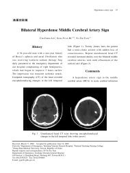
2010 American Heart Association
2010 American Heart Association
2010 American Heart Association

Create successful ePaper yourself
Turn your PDF publications into a flip-book with our unique Google optimized e-Paper software.
with a pause for ventilation). Newborns who require CPR in<br />
other settings (eg, prehospital, ED, pediatric intensive care<br />
unit [PICU], etc.), should receive CPR according to infant<br />
guidelines: 2 rescuers provide continuous chest compressions<br />
with asynchronous ventilations if an advanced airway is in<br />
place and a 15:2 ventilation-to-compression ratio if no advanced<br />
airway is in place (Class IIb, LOE C). It is reasonable<br />
to resuscitate newborns with a primary cardiac etiology of<br />
arrest, regardless of location, according to infant guidelines,<br />
with emphasis on chest compressions (Class IIa, LOE C). For<br />
further information, please refer to Part 13, “Pediatric Basic<br />
Life Support,” and Part 15, “Neonatal Resuscitation.”<br />
Extracorporeal Life Support (ECLS)<br />
Extracorporeal life support (ECLS) is a modified form of<br />
cardiopulmonary bypass used to provide prolonged delivery<br />
of oxygen to tissues. Consider early activation of ECLS for a<br />
cardiac arrest that occurs in a highly supervised environment,<br />
such as an ICU, with the clinical protocols in place and the<br />
expertise and equipment available to initiate it rapidly. ECLS<br />
should be considered only for children in cardiac arrest<br />
refractory to standard resuscitation attempts, with a potentially<br />
reversible cause of arrest (Class IIa, LOE C). 128–154<br />
When ECLS is employed during cardiac arrest, outcome for<br />
children with underlying cardiac disease is better than the<br />
outcome for children with noncardiac disease. With underlying<br />
cardiac disease, long-term survival when ECLS is initiated<br />
in a critical-care setting has been reported even after �50<br />
minutes of standard CPR. 128,129,139,147<br />
Monitoring<br />
Electrocardiography<br />
Monitor cardiac rhythm as soon as possible so both normal<br />
and abnormal cardiac rhythms are identified and followed.<br />
Continuous monitoring is helpful in tracking responses to<br />
treatment and changes in clinical condition.<br />
Echocardiography<br />
There is insufficient evidence for or against the routine use of<br />
echocardiography in pediatric cardiac arrest. When appropriately<br />
trained personnel are available, echocardiography may<br />
be considered to identify patients with potentially treatable<br />
causes of the arrest, particularly pericardial tamponade and<br />
inadequate ventricular filling (Class IIb, LOE C). 155–162 Minimize<br />
interruption of CPR while performing echocardiography.<br />
End-Tidal CO 2 (PETCO 2)<br />
Continuous capnography or capnometry monitoring, if available,<br />
may be beneficial during CPR, to help guide therapy,<br />
especially the effectiveness of chest compressions (Class IIa,<br />
LOE C). Animal and adult studies show a strong correlation<br />
between PETCO 2 and interventions that increase cardiac output<br />
during CPR or shock. 53,163–169 If the PETCO 2 is consistently<br />
�10 to 15 mm Hg, focus efforts on improving chest compressions<br />
and make sure that the victim does not receive<br />
excessive ventilation. An abrupt and sustained rise in PETCO 2<br />
in adults 170,171 and animals 110 is observed just prior to clinical<br />
identification of ROSC, so use of PETCO 2 may spare the<br />
rescuer from interrupting chest compressions for a pulse<br />
Kleinman et al Part 14: Pediatric Advanced Life Support S881<br />
check. PETCO 2 must be interpreted with caution for 1 to 2<br />
minutes after administration of epinephrine or other vasoconstrictive<br />
medications because these medications may decrease<br />
the end-tidal CO 2 level by reducing pulmonary blood flow.<br />
Vascular Access<br />
Vascular access is essential for administering medications<br />
and drawing blood samples. Obtaining peripheral venous<br />
access can be challenging in infants and children during an<br />
emergency; intraosseous (IO) access can be quickly established<br />
with minimal complications by providers with varied<br />
levels of training. 172–179 Limit the time spent attempting to<br />
establish peripheral venous access in a critically ill or injured<br />
child. 180<br />
Intraosseous (IO) Access<br />
IO access is a rapid, safe, effective, and acceptable route for<br />
vascular access in children, 172–179,181 and it is useful as the<br />
initial vascular access in cases of cardiac arrest (Class I,<br />
LOE C). All intravenous medications can be administered<br />
intraosseously, including epinephrine, adenosine, fluids,<br />
blood products, 182,183 and catecholamines. 184 Onset of action<br />
and drug levels for most drugs are comparable to venous<br />
administration. 185 IO access can be used to obtain blood<br />
samples for analysis including for type and cross match and<br />
blood gases during CPR, 186 but acid-base analysis is inaccurate<br />
after sodium bicarbonate administration via the IO<br />
cannula. 187 Use manual pressure or an infusion pump to<br />
administer viscous drugs or rapid fluid boluses; 188,189 follow<br />
each medication with a saline flush to promote entry into the<br />
central circulation.<br />
Venous Access<br />
Peripheral IV access is acceptable during resuscitation if it<br />
can be placed rapidly, but placement may be difficult in a<br />
critically ill child. Although a central venous catheter can<br />
provide more secure long-term access, its placement requires<br />
training and experience, and the procedure can be timeconsuming.<br />
Therefore central venous access is not recommended<br />
as the initial route of vascular access during an<br />
emergency. If both central and peripheral accesses are available,<br />
administer medications into the central circulation since<br />
some medications (eg, adenosine) are more effective when<br />
administered closer to the heart, and others (eg, calcium,<br />
amiodarone, procainamide, sympathomimetics) may be irritating<br />
when infused into a peripheral vein. The length of a<br />
central catheter can contribute to increased resistance, making it<br />
more difficult to push boluses of fluid rapidly through a<br />
multilumen central than a peripheral catheter.<br />
Endotracheal Drug Administration<br />
Vascular access (IO or IV) is the preferred method for drug<br />
delivery during CPR, but if it is not possible, lipid-soluble<br />
drugs, such as lidocaine, epinephrine, atropine, and naloxone<br />
(mnemonic “LEAN”) 190,191 can be administered via an endotracheal<br />
tube. 192 However, the effects may not be uniform<br />
with tracheal as compared with intravenous administration.<br />
One study of children in cardiac arrest 193 demonstrated<br />
similar ROSC and survival rates regardless of the method of<br />
Downloaded from<br />
circ.ahajournals.org at NATIONAL TAIWAN UNIV on October 18, <strong>2010</strong>