2010 American Heart Association
2010 American Heart Association 2010 American Heart Association
The most appropriate STEMI system of care starts “on the phone” with activation of EMS. Hospital-based issues include ED protocols, activation of the cardiac catheterization laboratory, and admission to the coronary intensive care unit. In PCI-capable hospitals an established “STEMI Alert” activation plan is critical. Components include prehospital ECGs and notification of the receiving facility, 45–60 and activation of the cardiac catherization team to shorten reperfusion time 54,59,82,89–92 and other hospital personnel important for treatment and resource allocation. Continuous review and quality improvement involving EMS and prehospital care providers are important to achieve ongoing optimal reperfusion time. Quality assurance, realtime feedback, and healthcare provider education can also reduce the time to therapy in STEMI. 89,93–97 Involvement of hospital leadership in the process and commitment to support rapid access to STEMI reperfusion therapy are critical factors associated with successful programs. If the emergency physician activates the STEMI reperfusion protocol, including the cardiac catheterization team, significant reductions in time to reperfusion are seen, and the rate of “false-positive” activations are infrequent, ranging from 0% to 14%. 89,93,95,96,98–107 ED Evaluation and Risk Stratification (Figure 1, Boxes 3 and 4) Focused Assessment and ECG Risk Stratification ED providers should quickly assess patients with possible ACS. Ideally within 10 minutes of ED arrival providers should obtain a targeted history while a monitor is attached to the patient and a 12-lead ECG is obtained (if not done in the prehospital setting). 108 The evaluation should focus on chest discomfort, associated signs and symptoms, prior cardiac history, risk factors for ACS, and historical features that may preclude the use of fibrinolytics or other therapies. This initial evaluation must be efficient because if the patient has STEMI, the goals of reperfusion are to administer fibrinolytics within 30 minutes of arrival (30-minute interval “door-todrug”) or to provide PCI within 90 minutes of arrival (90-minute interval “door-to-balloon”) (Class I, LOE A). Potential delay during the in-hospital evaluation period may occur from door to data, from data (ECG) to decision, and from decision to drug (or PCI). These 4 major points of in-hospital therapy are commonly referred to as the “4 D’s.” 109 All providers must focus on minimizing delays at each of these points. Prehospital transport time constitutes only 5% of delay to treatment time; ED evaluation constitutes 25% to 33% of this delay. 3,109–111 The physical examination is performed to aid diagnosis, rule out other causes of the patient’s symptoms, and evaluate the patient for complications related to ACS. Although the presence of clinical signs and symptoms may increase suspicion of ACS, evidence does not support the use of any single sign or combination of clinical signs and symptoms alone to confirm the diagnosis. 17–19,112 When the patient presents with symptoms and signs of potential ACS, the clinician uses ECG findings (Figure 1, Box 4) to classify the patient into 1 of 3 groups: O’Connor et al Part 10: Acute Coronary Syndromes S791 1. ST-segment elevation or presumed new LBBB (Box 5) is characterized by ST-segment elevation in 2 or more contiguous leads and is classified as ST-segment elevation MI (STEMI). Threshold values for ST-segment elevation consistent with STEMI are J-point elevation 0.2 mV (2 mm) in leads V2 and V3 and 0.1 mV (1 mm) in all other leads (men �40 years old); J-point elevation 0.25 mV (2.5 mm) in leads V2 and V3 and 0.1 mV (1 mm) in all other leads (men �40 years old); J-point elevation 0.15 mV (2.5 mm) in leads V2 and V3 and 0.1 mV (1 mm) in all other leads (women). 113 2. Ischemic ST-segment depression �0.5 mm (0.05 mV) or dynamic T-wave inversion with pain or discomfort (Box 9) is classified as UA/NSTEMI. Nonpersistent or transient ST-segment elevation �0.5 mm for �20 minutes is also included in this category. Threshold values for ST-segment depression consistent with ischemia are J-point depression 0.05 mV (-.5 mm) in leads V2 and V3 and -0.1 mV (-1 mm) in all other leads (men and women). 113 3. The nondiagnostic ECG with either normal or minimally abnormal (ie, nonspecific ST-segment or T-wave changes, Box 13). This ECG is nondiagnostic and inconclusive for ischemia, requiring further risk stratification. This classification includes patients with normal ECGs and those with ST-segment deviation of �0.5 mm (0.05 mV) or T-wave inversion of �0.2 mV. This category of ECG is termed nondiagnostic. The interpretation of the 12-lead ECG is a key step in this process, allowing not only for this classification but also the selection of the most appropriate diagnostic and management strategies. Not all providers are skilled in the interpretation of the ECG; as a consequence, the use of computer-aided ECG interpretation has been studied. While expert ECG intepretation is ideal, computer-aided ECG interpretation may have a role, particularly in assisting inexperienced clinicians in achieving a diagnosis (Class IIa, LOE B). Cardiac Biomarkers Serial cardiac biomarkers are often obtained during evaluation of patients suspected of ACS. Cardiac troponin is the preferred biomarker and is more sensitive than creatine kinase isoenzyme (CK-MB). Cardiac troponins are useful in diagnosis, risk stratification, and determination of prognosis. An elevated level of troponin correlates with an increased risk of death, and greater elevations predict greater risk of adverse outcome. 114 In the patients with STEMI reperfusion therapy should not be delayed pending results of biomarkers. Important limitations to these tests exist because they are insensitive during the first 4 to 6 hours of presentation unless continuous persistent pain has been present for 6 to 8 hours. For this reason cardiac biomarkers are not useful in the prehospital setting. 115–120 Clinicians should take into account the timing of symptom onset and the sensitivity, precision, and institutional norms of the assay, as well as the release kinetics and clearance of the measured biomarker. If biomarkers are initially negative within 6 hours of symptom onset, it is recommended that biomarkers should be remeasured between 6 to 12 hours after symptom onset (Class I, LOE A). A diagnosis of myocardial Downloaded from circ.ahajournals.org at NATIONAL TAIWAN UNIV on October 18, 2010
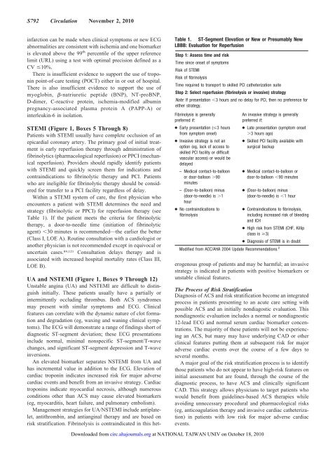
S792 Circulation November 2, 2010 infarction can be made when clinical symptoms or new ECG abnormalities are consistent with ischemia and one biomarker is elevated above the 99 th percentile of the upper reference limit (URL) using a test with optimal precision defined as a CV �10%. There is insufficient evidence to support the use of troponin point-of-care testing (POCT) either in or out of hospital. There is also insufficient evidence to support the use of myoglobin, �-natriuretic peptide (BNP), NT-proBNP, D-dimer, C-reactive protein, ischemia-modified albumin pregnancy-associated plasma protein A (PAPP-A) or interleukin-6 in isolation. STEMI (Figure 1, Boxes 5 Through 8) Patients with STEMI usually have complete occlusion of an epicardial coronary artery. The primary goal of initial treatment is early reperfusion therapy through administration of fibrinolytics (pharmacological reperfusion) or PPCI (mechanical reperfusion). Providers should rapidly identify patients with STEMI and quickly screen them for indications and contraindications to fibrinolytic therapy and PCI. Patients who are ineligible for fibrinolytic therapy should be considered for transfer to a PCI facility regardless of delay. Within a STEMI system of care, the first physician who encounters a patient with STEMI determines the need and strategy (fibrinolytic or PPCI) for reperfusion therapy (see Table 1). If the patient meets the criteria for fibrinolytic therapy, a door-to-needle time (initiation of fibrinolytic agent) �30 minutes is recommended—the earlier the better (Class I, LOE A). Routine consultation with a cardiologist or another physician is not recommended except in equivocal or uncertain cases. 89,121 Consultation delays therapy and is associated with increased hospital mortality rates (Class III, LOE B). UA and NSTEMI (Figure 1, Boxes 9 Through 12) Unstable angina (UA) and NSTEMI are difficult to distinguish initially. These patients usually have a partially or intermittently occluding thrombus. Both ACS syndromes may present with similar symptoms and ECG. Clinical features can correlate with the dynamic nature of clot formation and degradation (eg, waxing and waning clinical symptoms). The ECG will demonstrate a range of findings short of diagnostic ST-segment deviation; these ECG presentations include normal, minimal nonspecific ST-segment/T-wave changes, and significant ST-segment depression and T-wave inversions. An elevated biomarker separates NSTEMI from UA and has incremental value in addition to the ECG. Elevation of cardiac troponin indicates increased risk for major adverse cardiac events and benefit from an invasive strategy. Cardiac troponins indicate myocardial necrosis, although numerous conditions other than ACS may cause elevated biomarkers (eg, myocarditis, heart failure, and pulmonary embolism). Management strategies for UA/NSTEMI include antiplatelet, antithrombin, and antianginal therapy and are based on risk stratification. Fibrinolysis is contraindicated in this het- Table 1. ST-Segment Elevation or New or Presumably New LBBB: Evaluation for Reperfusion Step 1: Assess time and risk Time since onset of symptoms Risk of STEMI Risk of fibrinolysis Time required to transport to skilled PCI catheterization suite Step 2: Select reperfusion (fibrinolysis or invasive) strategy Note: If presentation �3 hours and no delay for PCI, then no preference for either strategy. Fibrinolysis is generally preferred if: ● Early presentation (�3 hours from symptom onset) ● Invasive strategy is not an option (eg, lack of access to skilled PCI facility or difficult vascular access) or would be delayed – Medical contact-to-balloon or door-balloon �90 minutes – (Door-to-balloon) minus (door-to-needle) is �1 hour ● No contraindications to fibrinolysis An invasive strategy is generally preferred if: ● Late presentation (symptom onset �3 hours ago) ● Skilled PCI facility available with surgical backup ● Medical contact-to-balloon or door-to-balloon �90 minutes ● (Door-to-balloon) minus (door-to-needle) is �1 hour ● Contraindications to fibrinolysis, including increased risk of bleeding and ICH ● High risk from STEMI (CHF, Killip class is �3) ● Diagnosis of STEMI is in doubt Modified from ACC/AHA 2004 Update Recommendations. 2 erogenous group of patients and may be harmful; an invasive strategy is indicated in patients with positive biomarkers or unstable clinical features. The Process of Risk Stratification Diagnosis of ACS and risk stratification become an integrated process in patients presenting to an acute care setting with possible ACS and an initially nondiagostic evaluation. This nondiagnostic evaluation includes a normal or nondiagnostic 12-lead ECG and normal serum cardiac biomarker concentrations. The majority of these patients will not be experiencing an ACS, but many may have underlying CAD or other clinical features putting them at subsequent risk for major adverse cardiac events over the course of a few days to several months. A major goal of the risk stratification process is to identify those patients who do not appear to have high-risk features on initial assessment but are found, through the course of the diagnostic process, to have ACS and clinically significant CAD. This strategy allows physicians to target patients who would benefit from guidelines-based ACS therapies while avoiding unnecessary procedural and pharmacological risks (eg, anticoagulation therapy and invasive cardiac catheterization) in patients with low risk for major adverse cardiac events. Downloaded from circ.ahajournals.org at NATIONAL TAIWAN UNIV on October 18, 2010
- Page 111 and 112: S742 Circulation November 2, 2010 O
- Page 113 and 114: S744 Circulation November 2, 2010 O
- Page 115 and 116: S746 Circulation November 2, 2010 T
- Page 117 and 118: S748 Circulation November 2, 2010 T
- Page 119 and 120: S750 Circulation November 2, 2010 p
- Page 121 and 122: S752 Circulation November 2, 2010 c
- Page 123 and 124: S754 Circulation November 2, 2010 A
- Page 125 and 126: S756 Circulation November 2, 2010 d
- Page 127 and 128: S758 Circulation November 2, 2010 R
- Page 129 and 130: S760 Circulation November 2, 2010 9
- Page 131 and 132: S762 Circulation November 2, 2010 1
- Page 133 and 134: S764 Circulation November 2, 2010 r
- Page 135 and 136: S766 Circulation November 2, 2010 3
- Page 137 and 138: Circulation 2010;122;S768-S786 DOI:
- Page 139 and 140: patients usually require an advance
- Page 141 and 142: comatose on arrival at the hospital
- Page 143 and 144: istration to maintain the arterial
- Page 145 and 146: Table 2. Common Vasoactive Drugs Dr
- Page 147 and 148: potentials (SSEPs) and select physi
- Page 149 and 150: Guidelines Part 9: Post-Cardiac Arr
- Page 151 and 152: 59. Larsson IM, Wallin E, Rubertsso
- Page 153 and 154: 142. Marcusohn E, Roguin A, Sebbag
- Page 155 and 156: 222. Rossetti AO, Oddo M, Liaudet L
- Page 157 and 158: Part 10: Acute Coronary Syndromes:
- Page 159 and 160: S788 Circulation November 2, 2010 p
- Page 161: S790 Circulation November 2, 2010 E
- Page 165 and 166: S794 Circulation November 2, 2010 T
- Page 167 and 168: S796 Circulation November 2, 2010 a
- Page 169 and 170: S798 Circulation November 2, 2010 P
- Page 171 and 172: S800 Circulation November 2, 2010 e
- Page 173 and 174: S802 Circulation November 2, 2010 p
- Page 175 and 176: S804 Circulation November 2, 2010 c
- Page 177 and 178: S806 Circulation November 2, 2010 (
- Page 179 and 180: S808 Circulation November 2, 2010 8
- Page 181 and 182: S810 Circulation November 2, 2010 a
- Page 183 and 184: S812 Circulation November 2, 2010 2
- Page 185 and 186: S814 Circulation November 2, 2010 d
- Page 187 and 188: S816 Circulation November 2, 2010 C
- Page 189 and 190: Part 11: Adult Stroke: 2010 America
- Page 191 and 192: The time-sensitive nature of stroke
- Page 193 and 194: (or the last time the patient was k
- Page 195 and 196: Table 4. Inclusion and Exclusion Ch
- Page 197 and 198: Guidelines Part 11: Stroke: Writing
- Page 199 and 200: 40. Zweifler RM, York D, et al. Acc
- Page 201 and 202: Part 12: Cardiac Arrest in Special
- Page 203 and 204: S830 Circulation November 2, 2010 P
- Page 205 and 206: S832 Circulation November 2, 2010 A
- Page 207 and 208: S834 Circulation November 2, 2010 T
- Page 209 and 210: S836 Circulation November 2, 2010 m
- Page 211 and 212: S838 Circulation November 2, 2010 b
S792 Circulation November 2, <strong>2010</strong><br />
infarction can be made when clinical symptoms or new ECG<br />
abnormalities are consistent with ischemia and one biomarker<br />
is elevated above the 99 th percentile of the upper reference<br />
limit (URL) using a test with optimal precision defined as a<br />
CV �10%.<br />
There is insufficient evidence to support the use of troponin<br />
point-of-care testing (POCT) either in or out of hospital.<br />
There is also insufficient evidence to support the use of<br />
myoglobin, �-natriuretic peptide (BNP), NT-proBNP,<br />
D-dimer, C-reactive protein, ischemia-modified albumin<br />
pregnancy-associated plasma protein A (PAPP-A) or<br />
interleukin-6 in isolation.<br />
STEMI (Figure 1, Boxes 5 Through 8)<br />
Patients with STEMI usually have complete occlusion of an<br />
epicardial coronary artery. The primary goal of initial treatment<br />
is early reperfusion therapy through administration of<br />
fibrinolytics (pharmacological reperfusion) or PPCI (mechanical<br />
reperfusion). Providers should rapidly identify patients<br />
with STEMI and quickly screen them for indications and<br />
contraindications to fibrinolytic therapy and PCI. Patients<br />
who are ineligible for fibrinolytic therapy should be considered<br />
for transfer to a PCI facility regardless of delay.<br />
Within a STEMI system of care, the first physician who<br />
encounters a patient with STEMI determines the need and<br />
strategy (fibrinolytic or PPCI) for reperfusion therapy (see<br />
Table 1). If the patient meets the criteria for fibrinolytic<br />
therapy, a door-to-needle time (initiation of fibrinolytic<br />
agent) �30 minutes is recommended—the earlier the better<br />
(Class I, LOE A). Routine consultation with a cardiologist or<br />
another physician is not recommended except in equivocal or<br />
uncertain cases. 89,121 Consultation delays therapy and is<br />
associated with increased hospital mortality rates (Class III,<br />
LOE B).<br />
UA and NSTEMI (Figure 1, Boxes 9 Through 12)<br />
Unstable angina (UA) and NSTEMI are difficult to distinguish<br />
initially. These patients usually have a partially or<br />
intermittently occluding thrombus. Both ACS syndromes<br />
may present with similar symptoms and ECG. Clinical<br />
features can correlate with the dynamic nature of clot formation<br />
and degradation (eg, waxing and waning clinical symptoms).<br />
The ECG will demonstrate a range of findings short of<br />
diagnostic ST-segment deviation; these ECG presentations<br />
include normal, minimal nonspecific ST-segment/T-wave<br />
changes, and significant ST-segment depression and T-wave<br />
inversions.<br />
An elevated biomarker separates NSTEMI from UA and<br />
has incremental value in addition to the ECG. Elevation of<br />
cardiac troponin indicates increased risk for major adverse<br />
cardiac events and benefit from an invasive strategy. Cardiac<br />
troponins indicate myocardial necrosis, although numerous<br />
conditions other than ACS may cause elevated biomarkers<br />
(eg, myocarditis, heart failure, and pulmonary embolism).<br />
Management strategies for UA/NSTEMI include antiplatelet,<br />
antithrombin, and antianginal therapy and are based on<br />
risk stratification. Fibrinolysis is contraindicated in this het-<br />
Table 1. ST-Segment Elevation or New or Presumably New<br />
LBBB: Evaluation for Reperfusion<br />
Step 1: Assess time and risk<br />
Time since onset of symptoms<br />
Risk of STEMI<br />
Risk of fibrinolysis<br />
Time required to transport to skilled PCI catheterization suite<br />
Step 2: Select reperfusion (fibrinolysis or invasive) strategy<br />
Note: If presentation �3 hours and no delay for PCI, then no preference for<br />
either strategy.<br />
Fibrinolysis is generally<br />
preferred if:<br />
● Early presentation (�3 hours<br />
from symptom onset)<br />
● Invasive strategy is not an<br />
option (eg, lack of access to<br />
skilled PCI facility or difficult<br />
vascular access) or would be<br />
delayed<br />
– Medical contact-to-balloon<br />
or door-balloon �90<br />
minutes<br />
– (Door-to-balloon) minus<br />
(door-to-needle) is �1<br />
hour<br />
● No contraindications to<br />
fibrinolysis<br />
An invasive strategy is generally<br />
preferred if:<br />
● Late presentation (symptom onset<br />
�3 hours ago)<br />
● Skilled PCI facility available with<br />
surgical backup<br />
● Medical contact-to-balloon or<br />
door-to-balloon �90 minutes<br />
● (Door-to-balloon) minus<br />
(door-to-needle) is �1 hour<br />
● Contraindications to fibrinolysis,<br />
including increased risk of bleeding<br />
and ICH<br />
● High risk from STEMI (CHF, Killip<br />
class is �3)<br />
● Diagnosis of STEMI is in doubt<br />
Modified from ACC/AHA 2004 Update Recommendations. 2<br />
erogenous group of patients and may be harmful; an invasive<br />
strategy is indicated in patients with positive biomarkers or<br />
unstable clinical features.<br />
The Process of Risk Stratification<br />
Diagnosis of ACS and risk stratification become an integrated<br />
process in patients presenting to an acute care setting with<br />
possible ACS and an initially nondiagostic evaluation. This<br />
nondiagnostic evaluation includes a normal or nondiagnostic<br />
12-lead ECG and normal serum cardiac biomarker concentrations.<br />
The majority of these patients will not be experiencing<br />
an ACS, but many may have underlying CAD or other<br />
clinical features putting them at subsequent risk for major<br />
adverse cardiac events over the course of a few days to<br />
several months.<br />
A major goal of the risk stratification process is to identify<br />
those patients who do not appear to have high-risk features on<br />
initial assessment but are found, through the course of the<br />
diagnostic process, to have ACS and clinically significant<br />
CAD. This strategy allows physicians to target patients who<br />
would benefit from guidelines-based ACS therapies while<br />
avoiding unnecessary procedural and pharmacological risks<br />
(eg, anticoagulation therapy and invasive cardiac catheterization)<br />
in patients with low risk for major adverse cardiac<br />
events.<br />
Downloaded from<br />
circ.ahajournals.org at NATIONAL TAIWAN UNIV on October 18, <strong>2010</strong>


