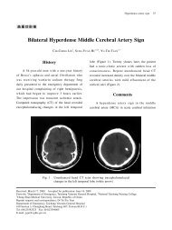
2010 American Heart Association
2010 American Heart Association
2010 American Heart Association

Create successful ePaper yourself
Turn your PDF publications into a flip-book with our unique Google optimized e-Paper software.
patients usually require an advanced airway for mechanical<br />
support of breathing. It may be necessary to replace a supraglottic<br />
airway used for initial resuscitation with an endotracheal tube,<br />
although the timing of replacement may vary. Methods for<br />
securing an advanced airway are discussed in Part 8.1: “Airway<br />
Management,” but several simple maneuvers deserve consideration.<br />
For example, rescuers and long-term hospital providers<br />
should avoid using ties that pass circumferentially around the<br />
patient’s neck, potentially obstructing venous return from the<br />
brain. They should also elevate the head of the bed 30° if<br />
tolerated to reduce the incidence of cerebral edema, aspiration,<br />
and ventilatory-associated pneumonia. Correct placement of an<br />
advanced airway, particularly during patient transport, should be<br />
monitored using waveform capnography as described in other<br />
sections of the <strong>2010</strong> AHA Guidelines for CPR and ECC.<br />
Oxygenation of the patient should be monitored continuously<br />
with pulse oximetry.<br />
Although 100% oxygen may have been used during initial<br />
resuscitation, providers should titrate inspired oxygen to the<br />
lowest level required to achieve an arterial oxygen saturation of<br />
�94%, so as to avoid potential oxygen toxicity. It is recognized<br />
that titration of inspired oxygen may not be possible immediately<br />
after out-of-hospital cardiac arrest until the patient is<br />
transported to the emergency department or, in the case of<br />
in-hospital arrest, the intensive care unit (ICU). Hyperventilation<br />
or “overbagging” the patient is common after cardiac arrest and<br />
should be avoided because of potential adverse hemodynamic<br />
effects. Hyperventilation increases intrathoracic pressure and<br />
inversely lowers cardiac output. The decrease in PaCO 2 seen with<br />
hyperventilation can also potentially decrease cerebral blood<br />
flow directly.Ventilation may be started at 10 to 12 breaths per<br />
minute and titrated to achieve a PETCO 2 of 35 to 40 mm Hg or a<br />
PaCO 2 of 40 to 45 mm Hg.<br />
Peberdy et al Part 9: Post–Cardiac Arrest Care S769<br />
Figure. Post–cardiac arrest care<br />
algorithm.<br />
The clinician should assess vital signs and monitor for<br />
recurrent cardiac arrhythmias. Continuous electrocardiographic<br />
(ECG) monitoring should continue after ROSC,<br />
during transport, and throughout ICU care until stability has<br />
been achieved. Intravenous (IV) access should be obtained if<br />
not already established and the position and function of any<br />
intravenous catheter verified. IV lines should be promptly<br />
established to replace emergent intraosseous access achieved<br />
during resuscitation. If the patient is hypotensive (systolic<br />
blood pressure �90 mm Hg), fluid boluses can be considered.<br />
Cold fluid may be used if therapeutic hypothermia is elected.<br />
Vasoactive drug infusions such as dopamine, norepinephrine,<br />
or epinephrine may be initiated if necessary and titrated to<br />
achieve a minimum systolic blood pressure of �90 mm Hg or<br />
a mean arterial pressure of �65 mm Hg.<br />
Brain injury and cardiovascular instability are the major<br />
determinants of survival after cardiac arrest. 13 Because therapeutic<br />
hypothermia is the only intervention demonstrated to<br />
improve neurological recovery, it should be considered for<br />
any patient who is unable to follow verbal commands after<br />
ROSC. The patient should be transported to a facility that<br />
reliably provides this therapy in addition to coronary reperfusion<br />
(eg, PCI) and other goal-directed postarrest care therapies.<br />
Overall the most common cause of cardiac arrest is<br />
cardiovascular disease and coronary ischemia. 14,15 Therefore,<br />
a 12-lead ECG should be obtained as soon as possible to<br />
detect ST elevation or new or presumably new left bundlebranch<br />
block. When there is high suspicion of acute myocardial<br />
infarction (AMI), local protocols for treatment of AMI<br />
and coronary reperfusion should be activated. Even in the<br />
absence of ST elevation, medical or interventional treatments<br />
may be considered for treatment of ACS 14,16,17 and should not<br />
be deferred in the presence of coma or in conjunction with<br />
Downloaded from<br />
circ.ahajournals.org at NATIONAL TAIWAN UNIV on October 18, <strong>2010</strong>