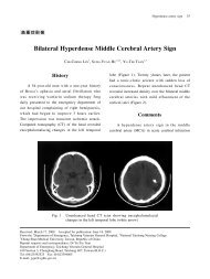
2010 American Heart Association
2010 American Heart Association
2010 American Heart Association

Create successful ePaper yourself
Turn your PDF publications into a flip-book with our unique Google optimized e-Paper software.
S740 Circulation November 2, <strong>2010</strong><br />
mended CPR parameters such as rate and depth of compression<br />
and rate of ventilation. The most simple are auditory or<br />
visual metronomes to guide providers in performing the<br />
recommended rate of chest compressions or ventilations.<br />
More sophisticated devices actually monitor chest compression<br />
rate, depth, relaxation, and pauses in real time and<br />
provide visual and auditory feedback. When recorded, this<br />
information can also be useful in providing feedback to the<br />
entire team of providers after the resuscitation has ended.<br />
This type of CPR quality monitoring is discussed in more<br />
detail in Part 5: “Adult Basic Life Support” and Part 16:<br />
“Education, Implementation and Teams.”<br />
Physiologic Parameters<br />
In humans cardiac arrest is the most critically ill condition,<br />
yet it is typically monitored by rhythm assessment using<br />
selected electocardiographic (ECG) leads and pulse checks<br />
as the only physiologic parameters to guide therapy.<br />
Animal and human studies indicate that monitoring of<br />
PETCO 2, coronary perfusion pressure (CPP), and central<br />
venous oxygen saturation (ScvO 2) provides valuable information<br />
on both the patient’s condition and response to<br />
therapy. Most importantly, PETCO 2, CPP, and ScvO 2 correlate<br />
with cardiac output and myocardial blood flow during<br />
CPR, and threshold values below which ROSC is rarely<br />
achieved have been reported. 168,190–195 Furthermore, an<br />
abrupt increase in any of these parameters is a sensitive<br />
indicator of ROSC that can be monitored without interrupting<br />
chest compressions. 91,93,167–175,177,196–201 Although no clinical<br />
study has examined whether titrating resuscitative efforts to<br />
these or other physiologic parameters improves outcome, it is<br />
reasonable to consider using these parameters when feasible<br />
to optimize chest compressions and guide vasopressor therapy<br />
during cardiac arrest (Class IIb, LOE C).<br />
Pulse<br />
Clinicians frequently try to palpate arterial pulses during chest<br />
compressions to assess the effectiveness of compressions. No<br />
studies have shown the validity or clinical utility of checking<br />
pulses during ongoing CPR. Because there are no valves in the<br />
inferior vena cava, retrograde blood flow into the venous system<br />
may produce femoral vein pulsations. 202 Thus, palpation of a<br />
pulse in the femoral triangle may indicate venous rather than<br />
arterial blood flow. Carotid pulsations during CPR do not<br />
indicate the efficacy of myocardial or cerebral perfusion during<br />
CPR. Palpation of a pulse when chest compressions are paused<br />
is a reliable indicator of ROSC but is potentially less sensitive<br />
than other physiologic measures discussed below.<br />
Healthcare providers also may take too long to check for a<br />
pulse 203,204 and have difficulty determining if a pulse is<br />
present or absent. 203–205 There is no evidence, however, that<br />
checking for breathing, coughing, or movement is superior<br />
for detection of circulation. 206 Because delays in chest compressions<br />
should be minimized, the healthcare provider<br />
should take no more than 10 seconds to check for a pulse, and<br />
if it is not felt within that time period chest compressions<br />
should be started. 205,207<br />
End-Tidal CO 2<br />
End-tidal CO 2 is the concentration of carbon dioxide in<br />
exhaled air at the end of expiration. It is typically ex-<br />
pressed as a partial pressure in mm Hg (PETCO 2). Because<br />
CO 2 is a trace gas in atmospheric air, CO 2 detected by<br />
capnography in exhaled air is produced in the body and<br />
delivered to the lungs by circulating blood. Under normal<br />
conditions PETCO 2 is in the range of 35 to 40 mm Hg.<br />
During untreated cardiac arrest CO 2 continues to be<br />
produced in the body, but there is no CO 2 delivery to the<br />
lungs. Under these conditions PETCO 2 will approach zero<br />
with continued ventilation. With initiation of CPR, cardiac<br />
output is the major determinant of CO 2 delivery to the<br />
lungs. If ventilation is relatively constant, PETCO 2 correlates<br />
well with cardiac output during CPR. The correlation<br />
between PETCO 2 and cardiac output during CPR can be<br />
transiently altered by giving IV sodium bicarbonate. 208<br />
This is explained by the fact that the bicarbonate is<br />
converted to water and CO 2, causing a transient increase in<br />
delivery of CO 2 to the lungs. Therefore, a transient rise in<br />
PETCO 2 after sodium bicarbonate therapy is expected and<br />
should not be misinterpreted as an improvement in quality<br />
of CPR or a sign of ROSC. Animal and human studies have<br />
also shown that PETCO 2 correlates with CPP and cerebral<br />
perfusion pressure during CPR. 209,210 The correlation of<br />
PETCO 2 with CPP during CPR can be altered by vasopressor<br />
therapy, especially at high doses (ie, �1 mg of<br />
epinephrine). 211–214 Vasopressors cause increased afterload,<br />
which will increase blood pressure and myocardial<br />
blood flow during CPR but will also decrease cardiac<br />
output. Therefore, a small decrease in PETCO 2 after vasopressor<br />
therapy may occur but should not be misinterpreted<br />
as a decrease in CPR quality.<br />
Persistently low PETCO 2 values (�10 mm Hg) during<br />
CPR in intubated patients suggest that ROSC is<br />
unlikely. 171,173,174,190,191,215,216 Similar data using quantitative<br />
monitoring of PETCO 2 are not available for patients with<br />
a supraglottic airway or those receiving bag-mask ventilation<br />
during CPR. One study using colorimetic end-tidal CO 2<br />
detection in nonintubated patients during CPR found that low<br />
end-tidal CO 2 was not a reliable predictor of failure to<br />
achieve ROSC. 217 An air leak during bag-mask ventilation or<br />
ventilation with a supraglottic airway could result in lower<br />
measured PETCO 2 values. Although a PETCO 2 value of<br />
�10 mm Hg in intubated patients indicates that cardiac<br />
output is inadequate to achieve ROSC, a specific target<br />
PETCO 2 value that optimizes the chance of ROSC has not been<br />
established. Monitoring PETCO 2 trends during CPR has the<br />
potential to guide individual optimization of compression<br />
depth and rate and to detect fatigue in the provider performing<br />
compressions. 201,218,219 In addition, an abrupt sustained increase<br />
in PETCO 2 during CPR is an indicator of<br />
ROSC. 91,177,196,198–201 Therefore, it is reasonable to consider<br />
using quantitative waveform capnography in intubated patients<br />
to monitor CPR quality, optimize chest compressions,<br />
and detect ROSC during chest compressions or when rhythm<br />
check reveals an organized rhythm (Class IIb, LOE C). If<br />
PETCO 2 is �10 mm Hg, it is reasonable to consider trying to<br />
improve CPR quality by optimizing chest compression parameters<br />
(Class IIb, LOE C). If PETCO 2 abruptly increases to<br />
a normal value (35 to 40 mm Hg), it is reasonable to consider<br />
that this is an indicator of ROSC (Class IIa, LOE B). The<br />
Downloaded from<br />
circ.ahajournals.org at NATIONAL TAIWAN UNIV on October 18, <strong>2010</strong>