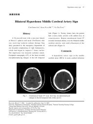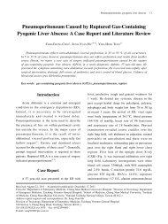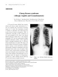
2010 American Heart Association
2010 American Heart Association
2010 American Heart Association

Create successful ePaper yourself
Turn your PDF publications into a flip-book with our unique Google optimized e-Paper software.
Circulation <strong>2010</strong>;122;S640-S656<br />
DOI: 10.1161/CIRCULATIONAHA.110.970889<br />
Circulation is published by the <strong>American</strong> <strong>Heart</strong> <strong>Association</strong>. 7272 Greenville Avenue, Dallas, TX<br />
72514<br />
Copyright © <strong>2010</strong> <strong>American</strong> <strong>Heart</strong> <strong>Association</strong>. All rights reserved. Print ISSN: 0009-7322. Online<br />
ISSN: 1524-4539<br />
Hoek<br />
Part 1: Executive Summary: <strong>2010</strong> <strong>American</strong> <strong>Heart</strong> <strong>Association</strong> Guidelines for<br />
Cardiopulmonary Resuscitation and Emergency Cardiovascular Care<br />
John M. Field, Mary Fran Hazinski, Michael R. Sayre, Leon Chameides, Stephen M.<br />
Schexnayder, Robin Hemphill, Ricardo A. Samson, John Kattwinkel, Robert A. Berg,<br />
Farhan Bhanji, Diana M. Cave, Edward C. Jauch, Peter J. Kudenchuk, Robert W.<br />
Neumar, Mary Ann Peberdy, Jeffrey M. Perlman, Elizabeth Sinz, Andrew H. Travers,<br />
Marc D. Berg, John E. Billi, Brian Eigel, Robert W. Hickey, Monica E. Kleinman,<br />
Mark S. Link, Laurie J. Morrison, Robert E. O'Connor, Michael Shuster, Clifton W.<br />
Callaway, Brett Cucchiara, Jeffrey D. Ferguson, Thomas D. Rea and Terry L. Vanden<br />
The online version of this article, along with updated information and services, is<br />
located on the World Wide Web at:<br />
http://circ.ahajournals.org/cgi/content/full/122/18_suppl_3/S640<br />
Subscriptions: Information about subscribing to Circulation is online at<br />
http://circ.ahajournals.org/subscriptions/<br />
Permissions: Permissions & Rights Desk, Lippincott Williams & Wilkins, a division of Wolters<br />
Kluwer Health, 351 West Camden Street, Baltimore, MD 21202-2436. Phone: 410-528-4050. Fax:<br />
410-528-8550. E-mail:<br />
journalpermissions@lww.com<br />
Reprints: Information about reprints can be found online at<br />
http://www.lww.com/reprints<br />
Downloaded from<br />
circ.ahajournals.org at NATIONAL TAIWAN UNIV on October 18, <strong>2010</strong>
Part 1: Executive Summary<br />
<strong>2010</strong> <strong>American</strong> <strong>Heart</strong> <strong>Association</strong> Guidelines for Cardiopulmonary<br />
Resuscitation and Emergency Cardiovascular Care<br />
John M. Field, Co-Chair*; Mary Fran Hazinski, Co-Chair*; Michael R. Sayre; Leon Chameides;<br />
Stephen M. Schexnayder; Robin Hemphill; Ricardo A. Samson; John Kattwinkel; Robert A. Berg;<br />
Farhan Bhanji; Diana M. Cave; Edward C. Jauch; Peter J. Kudenchuk; Robert W. Neumar;<br />
Mary Ann Peberdy; Jeffrey M. Perlman; Elizabeth Sinz; Andrew H. Travers; Marc D. Berg;<br />
John E. Billi; Brian Eigel; Robert W. Hickey; Monica E. Kleinman; Mark S. Link; Laurie J. Morrison;<br />
Robert E. O’Connor; Michael Shuster; Clifton W. Callaway; Brett Cucchiara; Jeffrey D. Ferguson;<br />
Thomas D. Rea; Terry L. Vanden Hoek<br />
The publication of the <strong>2010</strong> <strong>American</strong> <strong>Heart</strong> <strong>Association</strong><br />
Guidelines for Cardiopulmonary Resuscitation and<br />
Emergency Cardiovascular Care marks the 50th anniversary<br />
of modern CPR. In 1960 Kouwenhoven, Knickerbocker, and<br />
Jude documented 14 patients who survived cardiac arrest<br />
with the application of closed chest cardiac massage. 1 That<br />
same year, at the meeting of the Maryland Medical Society in<br />
Ocean City, MD, the combination of chest compressions and<br />
rescue breathing was introduced. 2 Two years later, in 1962,<br />
direct-current, monophasic waveform defibrillation was described.<br />
3 In 1966 the <strong>American</strong> <strong>Heart</strong> <strong>Association</strong> (AHA)<br />
developed the first cardiopulmonary resuscitation (CPR)<br />
guidelines, which have been followed by periodic updates. 4<br />
During the past 50 years the fundamentals of early recognition<br />
and activation, early CPR, early defibrillation, and early<br />
access to emergency medical care have saved hundreds of<br />
thousands of lives around the world. These lives demonstrate<br />
the importance of resuscitation research and clinical translation<br />
and are cause to celebrate this 50 th anniversary of CPR.<br />
Challenges remain if we are to fulfill the potential offered<br />
by the pioneer resuscitation scientists. We know that there is<br />
a striking disparity in survival outcomes from cardiac arrest<br />
across systems of care, with some systems reporting 5-fold<br />
higher survival rates than others. 5–9 Although technology,<br />
such as that incorporated in automated external defibrillators<br />
(AEDs), has contributed to increased survival from cardiac<br />
arrest, no initial intervention can be delivered to the victim of<br />
cardiac arrest unless bystanders are ready, willing, and able to<br />
act. Moreover, to be successful, the actions of bystanders and<br />
other care providers must occur within a system that coordinates<br />
and integrates each facet of care into a comprehensive<br />
whole, focusing on survival to discharge from the hospital.<br />
This executive summary highlights the major changes and<br />
most provocative recommendations in the <strong>2010</strong> AHA Guidelines<br />
for CPR and Emergency Cardiovascular Care (ECC).<br />
The scientists and healthcare providers participating in a<br />
comprehensive evidence evaluation process analyzed the<br />
sequence and priorities of the steps of CPR in light of current<br />
scientific advances to identify factors with the greatest<br />
potential impact on survival. On the basis of the strength of<br />
the available evidence, they developed recommendations<br />
to support the interventions that showed the most promise.<br />
There was unanimous support for continued emphasis on<br />
high-quality CPR, with compressions of adequate rate and<br />
depth, allowing complete chest recoil, minimizing interruptions<br />
in chest compressions and avoiding excessive<br />
ventilation. High-quality CPR is the cornerstone of a<br />
system of care that can optimize outcomes beyond return<br />
of spontaneous circulation (ROSC). Return to a prior<br />
quality of life and functional state of health is the ultimate<br />
goal of a resuscitation system of care.<br />
The <strong>2010</strong> AHA Guidelines for CPR and ECC are based on the<br />
most current and comprehensive review of resuscitation literature<br />
ever published, the <strong>2010</strong> ILCOR International Consensus<br />
on CPR and ECC Science With Treatment Recommendations. 10<br />
The <strong>2010</strong> evidence evaluation process included 356 resuscitation<br />
experts from 29 countries who reviewed, analyzed, evaluated,<br />
debated, and discussed research and hypotheses through<br />
in-person meetings, teleconferences, and online sessions (“webinars”)<br />
during the 36-month period before the <strong>2010</strong> Consensus<br />
Conference. The experts produced 411 scientific evidence reviews<br />
on 277 topics in resuscitation and emergency cardiovascular<br />
care. The process included structured evidence evaluation,<br />
analysis, and cataloging of the literature. It also included rigor-<br />
The <strong>American</strong> <strong>Heart</strong> <strong>Association</strong> requests that this document be cited as follows: Field JM, Hazinski MF, Sayre MR, Chameides L, Schexnayder SM,<br />
Hemphill R, Samson RA, Kattwinkel J, Berg RA, Bhanji F, Cave DM, Jauch EC, Kudenchuk PJ, Neumar RW, Peberdy MA, Perlman JM, Sinz E, Travers<br />
AH, Berg MD, Billi JE, Eigel B, Hickey RW, Kleinman ME, Link MS, Morrison LJ, O’Connor RE, Shuster M, Callaway CW, Cucchiara B, Ferguson<br />
JD, Rea TD, Vanden Hoek TL. Part 1: executive summary: <strong>2010</strong> <strong>American</strong> <strong>Heart</strong> <strong>Association</strong> Guidelines for Cardiopulmonary Resuscitation and<br />
Emergency Cardiovascular Care. Circulation. <strong>2010</strong>;122(suppl 3):S640–S656.<br />
*Co-chairs and equal first co-authors.<br />
(Circulation. <strong>2010</strong>;122[suppl 3]:S640–S656.)<br />
© <strong>2010</strong> <strong>American</strong> <strong>Heart</strong> <strong>Association</strong>, Inc.<br />
Circulation is available at http://circ.ahajournals.org DOI: 10.1161/CIRCULATIONAHA.110.970889<br />
Downloaded from<br />
circ.ahajournals.org at NATIONAL S640 TAIWAN UNIV on October 18, <strong>2010</strong>
ous disclosure and management of potential conflicts of interest,<br />
which are detailed in Part 2: “Evidence Evaluation and Management<br />
of Potential and Perceived Conflicts of Interest.”<br />
The recommendations in the <strong>2010</strong> Guidelines confirm the<br />
safety and effectiveness of many approaches, acknowledge<br />
ineffectiveness of others, and introduce new treatments based<br />
on intensive evidence evaluation and consensus of experts.<br />
These new recommendations do not imply that care using<br />
past guidelines is either unsafe or ineffective. In addition, it is<br />
important to note that they will not apply to all rescuers and<br />
all victims in all situations. The leader of a resuscitation<br />
attempt may need to adapt application of these recommendations<br />
to unique circumstances.<br />
New Developments in Resuscitation Science<br />
Since 2005<br />
A universal compression-ventilation ratio of 30:2 performed<br />
by lone rescuers for victims of all ages was one of the most<br />
controversial topics discussed during the 2005 International<br />
Consensus Conference, and it was a major change in the 2005<br />
AHA Guidelines for CPR and ECC. 11 In 2005 rates of<br />
survival to hospital discharge from witnessed out-of-hospital<br />
sudden cardiac arrest due to ventricular fibrillation (VF) were<br />
low, averaging �6% worldwide with little improvement in<br />
the years immediately preceding the 2005 conference. 5 Two<br />
studies published just before the 2005 International Consensus<br />
Conference documented poor quality of CPR performed<br />
in both out-of-hospital and in-hospital resuscitations. 12,13 The<br />
changes in the compression-ventilation ratio and in the<br />
defibrillation sequence (from 3 stacked shocks to 1 shock<br />
followed by immediate CPR) were recommended to minimize<br />
interruptions in chest compressions. 11–13<br />
There have been many developments in resuscitation<br />
science since 2005, and these are highlighted below.<br />
Emergency Medical Services Systems and<br />
CPR Quality<br />
Emergency medical services (EMS) systems and healthcare<br />
providers should identify and strengthen “weak links” in the<br />
Chain of Survival. There is evidence of considerable regional<br />
variation in the reported incidence and outcome from cardiac<br />
arrest within the United States. 5,14 This evidence supports the<br />
importance of accurately identifying each instance of treated<br />
cardiac arrest and measuring outcomes and suggests additional<br />
opportunities for improving survival rates in many communities.<br />
Recent studies have demonstrated improved outcome from<br />
out-of-hospital cardiac arrest, particularly from shockable<br />
rhythms, and have reaffirmed the importance of a stronger<br />
emphasis on compressions of adequate rate and depth, allowing<br />
complete chest recoil after each compression, minimizing interruptions<br />
in compressions and avoiding excessive ventilation. 15–22<br />
Implementation of new resuscitation guidelines has been<br />
shown to improve outcomes. 18,20–22 A means of expediting<br />
guidelines implementation (a process that may take from 18<br />
months to 4 years 23–26 ) is needed. Impediments to implementation<br />
include delays in instruction (eg, time needed to<br />
produce new training materials and update instructors and<br />
providers), technology upgrades (eg, reprogramming AEDs),<br />
and decision making (eg, coordination with allied agencies<br />
Field et al Part 1: Executive Summary S641<br />
and government regulators, medical direction, and participation<br />
in research).<br />
Documenting the Effects of CPR Performance by<br />
Lay Rescuers<br />
During the past 5 years there has been an effort to simplify CPR<br />
recommendations and emphasize the fundamental importance of<br />
high-quality CPR. Large observational studies from investigators<br />
in member countries of the Resuscitation Council of Asia<br />
(the newest member of ILCOR) 27,28–30 and other studies 31,32<br />
have provided important information about the positive impact<br />
of bystander CPR on survival after out-of-hospital cardiac arrest.<br />
For most adults with out-of-hospital cardiac arrest, bystander<br />
CPR with chest compression only (Hands-Only CPR) appears to<br />
achieve outcomes similar to those of conventional CPR (compressions<br />
with rescue breathing). 28–32 However, for children,<br />
conventional CPR is superior. 27<br />
CPR Quality<br />
Minimizing the interval between stopping chest compressions<br />
and delivering a shock (ie, minimizing the preshock pause)<br />
improves the chances of shock success 33,34 and patient survival.<br />
33–35 Data downloaded from CPR-sensing and feedbackenabled<br />
defibrillators provide valuable information to resuscitation<br />
teams, which can improve CPR quality. 36 These data<br />
are driving major changes in the training of in-hospital<br />
resuscitation teams and out-of-hospital healthcare providers.<br />
In-Hospital CPR Registries<br />
The National Registry of CardioPulmonary Resuscitation<br />
(NRCPR) 37 and other large databases are providing new information<br />
about the epidemiology and outcomes of in-hospital<br />
resuscitation in adults and children. 8,38–44 Although observational<br />
in nature, registries provide valuable descriptive information<br />
to better characterize cardiac arrest and resuscitation outcomes<br />
as well as identify areas for further research.<br />
Deemphasis on Devices and Advanced<br />
Cardiovascular Life Support Drugs During<br />
Cardiac Arrest<br />
At the time of the <strong>2010</strong> International Consensus Conference<br />
there were still insufficient data to demonstrate that any drugs<br />
or mechanical CPR devices improve long-term outcome after<br />
cardiac arrest. 45 Clearly further studies, adequately powered<br />
to detect clinically important outcome differences with these<br />
interventions, are needed.<br />
Importance of Post–Cardiac Arrest Care<br />
Organized post–cardiac arrest care with an emphasis on<br />
multidisciplinary programs that focus on optimizing hemodynamic,<br />
neurologic, and metabolic function (including therapeutic<br />
hypothermia) may improve survival to hospital discharge<br />
among victims who achieve ROSC following cardiac<br />
arrest either in- or out-of-hospital. 46–48 Although it is not yet<br />
possible to determine the individual effect of many of these<br />
therapies, when bundled as an integrated system of care, their<br />
deployment may well improve outcomes.<br />
Therapeutic hypothermia is one intervention that has been<br />
shown to improve outcome for comatose adult victims of<br />
Downloaded from<br />
circ.ahajournals.org at NATIONAL TAIWAN UNIV on October 18, <strong>2010</strong>
S642 Circulation November 2, <strong>2010</strong><br />
witnessed out-of-hospital cardiac arrest when the presenting<br />
rhythm was VF. 49,50 Since 2005, two nonrandomized studies<br />
with concurrent controls as well as other studies using<br />
historic controls have indicated the possible benefit of hypothermia<br />
following in- and out-of-hospital cardiac arrest from<br />
all other initial rhythms in adults. 46,51–56 Hypothermia has also<br />
been shown to be effective in improving intact neurologic<br />
survival in neonates with hypoxic-ischemic encephalopathy,<br />
57–61 and the results of a prospective multicenter pediatric<br />
study of therapeutic hypothermia after cardiac arrest are<br />
eagerly awaited.<br />
Many studies have attempted to identify comatose post–<br />
cardiac arrest patients who have no prospect for meaningful<br />
neurologic recovery, and decision rules for prognostication of<br />
poor outcome have been proposed. 62 Therapeutic hypothermia<br />
changes the specificity of prognostication decision rules<br />
that were previously established from studies of post–cardiac<br />
arrest patients not treated with hypothermia. Recent reports<br />
have documented occasional good outcomes in post–cardiac<br />
arrest patients who were treated with therapeutic hypothermia,<br />
despite neurologic exam or neuroelectrophysiologic<br />
studies that predicted poor outcome. 63,64<br />
Education and Implementation<br />
The quality of rescuer education and frequency of retraining<br />
are critical factors in improving the effectiveness of resuscitation.<br />
65–83 Ideally retraining should not be limited to 2-year<br />
intervals. More frequent renewal of skills is needed, with a<br />
commitment to maintenance of certification similar to that<br />
embraced by many healthcare-credentialing organizations.<br />
Resuscitation interventions are often performed simultaneously,<br />
and rescuers must be able to work collaboratively to<br />
minimize interruptions in chest compressions. Teamwork and<br />
leadership skills continue to be important, particularly for<br />
advanced cardiovascular life support (ACLS) and pediatric<br />
advanced life support (PALS) providers. 36,84–89<br />
Community and hospital-based resuscitation programs<br />
should systematically monitor cardiac arrests, the level of<br />
resuscitation care provided, and outcome. The cycle of<br />
measurement, interpretation, feedback, and continuous quality<br />
improvement provides fundamental information necessary<br />
to optimize resuscitation care and should help to narrow the<br />
knowledge and clinical gaps between ideal and actual resuscitation<br />
performance.<br />
Highlights of the <strong>2010</strong> Guidelines<br />
The Change From “A-B-C” to “C-A-B”<br />
The newest development in the <strong>2010</strong> AHA Guidelines for CPR<br />
and ECC is a change in the basic life support (BLS) sequence of<br />
steps from “A-B-C” (Airway, Breathing, Chest compressions) to<br />
“C-A-B” (Chest compressions, Airway, Breathing) for adults<br />
and pediatric patients (children and infants, excluding newly<br />
borns). Although the experts agreed that it is important to reduce<br />
time to first chest compressions, they were aware that a change<br />
in something as established as the A-B-C sequence would<br />
require re-education of everyone who has ever learned CPR. The<br />
<strong>2010</strong> AHA Guidelines for CPR and ECC recommend this<br />
change for the following reasons:<br />
● The vast majority of cardiac arrests occur in adults, and the<br />
highest survival rates from cardiac arrest are reported<br />
among patients of all ages with witnessed arrest and a<br />
rhythm of VF or pulseless ventricular tachycardia (VT). In<br />
these patients the critical initial elements of CPR are chest<br />
compressions and early defibrillation. 90<br />
● In the A-B-C sequence chest compressions are often<br />
delayed while the responder opens the airway to give<br />
mouth-to-mouth breaths or retrieves a barrier device or<br />
other ventilation equipment. By changing the sequence to<br />
C-A-B, chest compressions will be initiated sooner and<br />
ventilation only minimally delayed until completion of the<br />
first cycle of chest compressions (30 compressions should<br />
be accomplished in approximately 18 seconds).<br />
● Fewer than 50% of persons in cardiac arrest receive bystander<br />
CPR. There are probably many reasons for this, but one<br />
impediment may be the A-B-C sequence, which starts with<br />
the procedures that rescuers find most difficult: opening the<br />
airway and delivering rescue breaths. Starting with chest<br />
compressions might ensure that more victims receive CPR<br />
and that rescuers who are unable or unwilling to provide<br />
ventilations will at least perform chest compressions.<br />
● It is reasonable for healthcare providers to tailor the<br />
sequence of rescue actions to the most likely cause of<br />
arrest. For example, if a lone healthcare provider sees a<br />
victim suddenly collapse, the provider may assume that the<br />
victim has suffered a sudden VF cardiac arrest; once the<br />
provider has verified that the victim is unresponsive and<br />
not breathing or is only gasping, the provider should<br />
immediately activate the emergency response system, get<br />
and use an AED, and give CPR. But for a presumed victim<br />
of drowning or other likely asphyxial arrest the priority<br />
would be to provide about 5 cycles (about 2 minutes) of<br />
conventional CPR (including rescue breathing) before activating<br />
the emergency response system. Also, in newly<br />
born infants, arrest is more likely to be of a respiratory<br />
etiology, and resuscitation should be attempted with the<br />
A-B-C sequence unless there is a known cardiac etiology.<br />
Ethical Issues<br />
The ethical issues surrounding resuscitation are complex and<br />
vary across settings (in- or out-of-hospital), providers (basic or<br />
advanced), and whether to start or how to terminate CPR. Recent<br />
work suggests that acknowledgment of a verbal do-not-attemptresuscitation<br />
order (DNAR) in addition to the current standard—a<br />
written, signed, and dated DNAR document—may<br />
decrease the number of futile resuscitation attempts. 91,92 This is<br />
an important first step in expanding the clinical decision rule<br />
pertaining to when to start resuscitation in out-of-hospital cardiac<br />
arrest. However, there is insufficient evidence to support<br />
this approach without further validation.<br />
When only BLS-trained EMS personnel are available,<br />
termination of resuscitative efforts should be guided by a<br />
validated termination of resuscitation rule that reduces the<br />
transport rate of attempted resuscitations without compromising<br />
the care of potentially viable patients. 93 Advanced<br />
life support (ALS) EMS providers may use the same<br />
termination of resuscitation rule 94–99 or a derived nonvalidated<br />
rule specific to ALS providers that when applied will<br />
Downloaded from<br />
circ.ahajournals.org at NATIONAL TAIWAN UNIV on October 18, <strong>2010</strong>
decrease the number of futile transports to the emergency<br />
department (ED). 95,97–100<br />
Certain characteristics of a neonatal in-hospital cardiac<br />
arrest are associated with death, and these may be helpful in<br />
guiding physicians in the decision to start and stop a neonatal<br />
resuscitation attempt. 101–104 There is more variability in terminating<br />
resuscitation rates across systems and physicians<br />
when clinical decision rules are not followed, suggesting that<br />
these validated and generalized rules may promote uniformity<br />
in access to resuscitation attempts and full protocol care. 105<br />
Offering select family members the opportunity to be<br />
present during the resuscitation and designating staff within<br />
the team to respond to their questions and offer comfort may<br />
enhance the emotional support provided to the family during<br />
cardiac arrest and after termination of a resuscitation attempt.<br />
Identifying patients during the post–cardiac arrest period who<br />
do not have the potential for meaningful neurologic recovery is<br />
a major clinical challenge that requires further research. Caution<br />
is advised when considering limiting care or withdrawing<br />
life-sustaining therapy. Characteristics or test results that are<br />
predictive of poor outcome in post–cardiac arrest patients not<br />
treated with therapeutic hypothermia may not be as predictive of<br />
poor outcome after administration of therapeutic hypothermia.<br />
Because of the growing need for transplant tissue and organs, all<br />
provider teams who treat postarrest patients should also plan and<br />
implement a system of tissue and organ donation that is timely,<br />
effective, and supportive of family members for the subset of<br />
patients in whom brain death is confirmed or for organ donation<br />
after cardiac arrest.<br />
Resuscitation research is challenging. It must be scientifically<br />
rigorous while confronting ethical, regulatory, and public relations<br />
concerns that arise from the need to conduct such research<br />
with exception to informed consent. Regulatory requirements,<br />
community notification, and consultation requirements often<br />
impose expensive and time-consuming demands that may not<br />
only delay important research but also render it cost-prohibitive,<br />
with little significant evidence that these measures effectively<br />
address the concerns about research. 106–109<br />
Basic Life Support<br />
BLS is the foundation for saving lives following cardiac<br />
arrest. Fundamental aspects of adult BLS include immediate<br />
recognition of sudden cardiac arrest and activation of the<br />
emergency response system, early performance of highquality<br />
CPR, and rapid defibrillation when appropriate. The<br />
<strong>2010</strong> AHA Guidelines for CPR and ECC contain several<br />
important changes but also have areas of continued emphasis<br />
based on evidence presented in prior years.<br />
Key Changes in the <strong>2010</strong> AHA Guidelines for CPR<br />
and ECC<br />
● The BLS algorithm has been simplified, and “Look, Listen<br />
and Feel” has been removed from the algorithm. Performance<br />
of these steps is inconsistent and time consuming. For this<br />
reason the <strong>2010</strong> AHA Guidelines for CPR and ECC stress<br />
immediate activation of the emergency response system and<br />
starting chest compressions for any unresponsive adult victim<br />
with no breathing or no normal breathing (ie, only gasps).<br />
Field et al Part 1: Executive Summary S643<br />
● Encourage Hands-Only (compression only) CPR for the<br />
untrained lay rescuer. Hands-Only CPR is easier to perform<br />
by those with no training and can be more readily guided<br />
by dispatchers over the telephone.<br />
● Initiate chest compressions before giving rescue breaths (C-<br />
A-B rather than A-B-C). Chest compressions can be started<br />
immediately, whereas positioning the head, attaining a seal for<br />
mouth-to-mouth rescue breathing, or obtaining or assembling<br />
a bag-mask device for rescue breathing all take time. Beginning<br />
CPR with 30 compressions rather than 2 ventilations<br />
leads to a shorter delay to first compression.<br />
● There is an increased focus on methods to ensure that<br />
high-quality CPR is performed. Adequate chest compressions<br />
require that compressions be provided at the appropriate<br />
depth and rate, allowing complete recoil of the chest<br />
after each compression and an emphasis on minimizing any<br />
pauses in compressions and avoiding excessive ventilation.<br />
Training should focus on ensuring that chest compressions<br />
are performed correctly. The recommended depth of compression<br />
for adult victims has increased from a depth of 1 1 ⁄2<br />
to 2 inches to a depth of at least 2 inches.<br />
● Many tasks performed by healthcare providers during resuscitation<br />
attempts, such as chest compressions, airway management,<br />
rescue breathing, rhythm detection, shock delivery,<br />
and drug administration (if appropriate), can be performed<br />
concurrently by an integrated team of highly trained rescuers<br />
in appropriate settings. Some resuscitations start with a lone<br />
rescuer who calls for help, resulting in the arrival of additional<br />
team members. Healthcare provider training should focus on<br />
building the team as each member arrives or quickly delegating<br />
roles if multiple rescuers are present. As additional<br />
personnel arrive, responsibilities for tasks that would ordinarily<br />
be performed sequentially by fewer rescuers may now<br />
be delegated to a team of providers who should perform them<br />
simultaneously.<br />
Key Points of Continued Emphasis for the <strong>2010</strong> AHA<br />
Guidelines for CPR and ECC<br />
● Early recognition of sudden cardiac arrest in adults is based<br />
on assessing responsiveness and the absence of normal<br />
breathing. Victims of cardiac arrest may initially have<br />
gasping respirations or even appear to be having a seizure.<br />
These atypical presentations may confuse a rescuer, causing<br />
a delay in calling for help or beginning CPR. Training<br />
should focus on alerting potential rescuers to the unusual<br />
presentations of sudden cardiac arrest.<br />
● Minimize interruptions in effective chest compressions<br />
until ROSC or termination of resuscitative efforts. Any<br />
unnecessary interruptions in chest compressions (including<br />
longer than necessary pauses for rescue breathing) decreases<br />
CPR effectiveness.<br />
● Minimize the importance of pulse checks by healthcare<br />
providers. Detection of a pulse can be difficult, and even<br />
highly trained healthcare providers often incorrectly assess<br />
the presence or absence of a pulse when blood pressure is<br />
abnormally low or absent. Healthcare providers should take<br />
no more than 10 seconds to determine if a pulse is present.<br />
Chest compressions delivered to patients subsequently<br />
found not to be in cardiac arrest rarely lead to significant<br />
Downloaded from<br />
circ.ahajournals.org at NATIONAL TAIWAN UNIV on October 18, <strong>2010</strong>
S644 Circulation November 2, <strong>2010</strong><br />
injury. 110 The lay rescuer should activate the emergency<br />
response system if he or she finds an unresponsive adult.<br />
The lay rescuer should not attempt to check for a pulse and<br />
should assume that cardiac arrest is present if an adult<br />
suddenly collapses, is unresponsive, and is not breathing or<br />
not breathing normally (ie, only gasping).<br />
CPR Techniques and Devices<br />
Alternatives to conventional manual CPR have been developed<br />
in an effort to enhance perfusion during resuscitation<br />
from cardiac arrest and to improve survival. Compared with<br />
conventional CPR, these techniques and devices typically<br />
require more personnel, training, and equipment, or apply to<br />
a specific setting. Some alternative CPR techniques and<br />
devices may improve hemodynamics or short-term survival<br />
when used by well-trained providers in selected patients.<br />
Several devices have been the focus of recent clinical trials.<br />
Use of the impedance threshold device (ITD) improved ROSC<br />
and short-term survival when used in adults with out-of-hospital<br />
cardiac arrest, but there was no significant improvement in either<br />
survival to hospital discharge or neurologically-intact survival to<br />
discharge. 111 One multicenter, prospective, randomized controlled<br />
trial 112,112a comparing load-distributing band CPR (Autopulse)<br />
with manual CPR for out-of-hospital cardiac arrest<br />
demonstrated no improvement in 4-hour survival and worse<br />
neurologic outcome when the device was used. More research is<br />
needed to determine if site-specific factors 113 or experience with<br />
deployment of the device 114 influence effectiveness of the<br />
load-distributing band CPR device. Case series employing mechanical<br />
piston devices have reported variable degrees of<br />
success. 115–119<br />
To prevent delays and maximize efficiency, initial training,<br />
ongoing monitoring, and retraining programs should be<br />
offered on a frequent basis to providers using CPR devices.<br />
To date, no adjunct has consistently been shown to be<br />
superior to standard conventional (manual) CPR for out-ofhospital<br />
BLS, and no device other than a defibrillator has<br />
consistently improved long-term survival from out-ofhospital<br />
cardiac arrest.<br />
Electrical Therapies<br />
The <strong>2010</strong> AHA Guidelines for CPR and ECC have been<br />
updated to reflect new data on the use of pacing in bradycardia,<br />
and on cardioversion and defibrillation for tachycardic<br />
rhythm disturbances. Integration of AEDs into a system of<br />
care is critical in the Chain of Survival in public places<br />
outside of hospitals. To give the victim the best chance of<br />
survival, 3 actions must occur within the first moments of a<br />
cardiac arrest 120 : activation of the EMS system, 121 provision<br />
of CPR, and operation of a defibrillator. 122<br />
One area of continued interest is whether delivering a<br />
longer period of CPR before defibrillation improves outcomes<br />
in cardiac arrest. In early studies, survival was improved<br />
when 1.5 to 3 minutes of CPR preceded defibrillation<br />
for patients with cardiac arrest of �4 to 5 minutes duration<br />
prior to EMS arrival. 123,124 However, in 2 more recent<br />
randomized controlled trials, CPR performed before defibrillation<br />
did not improve outcome. 125,126 If �2 rescuers are<br />
present CPR should be performed while a defibrillator is<br />
being obtained and readied for use.<br />
The 1-shock protocol for VF has not been changed.<br />
Evidence has accumulated that even short interruptions in<br />
CPR are harmful. Thus, rescuers should minimize the interval<br />
between stopping compressions and delivering shocks and<br />
should resume CPR immediately after shock delivery.<br />
Over the last decade biphasic waveforms have been shown<br />
to be more effective than monophasic waveforms in cardioversion<br />
and defibrillation. 127–135 However, there are no clinical<br />
data comparing one specific biphasic waveform with<br />
another. Whether escalating or fixed subsequent doses of<br />
energy are superior has not been tested with different waveforms.<br />
However, if higher energy levels are available in the<br />
device at hand, they may be considered if initial shocks are<br />
unsuccessful in terminating the arrhythmia.<br />
In the last 5 to 10 years a number of randomized trials have<br />
compared biphasic with monophasic cardioversion in atrial<br />
fibrillation. The efficacy of shock energies for cardioversion of<br />
atrial fibrillation is waveform-specific and can vary from 120 to<br />
200 J depending on the defibrillator manufacturer. Thus, the<br />
recommended initial biphasic energy dose for cardioversion of<br />
atrial fibrillation is 120 to 200 J using the manufacturer’s<br />
recommended setting. 136–140 If the initial shock fails, providers<br />
should increase the dose in a stepwise fashion. Cardioversion<br />
of adult atrial flutter and other supraventricular<br />
tachycardias generally requires less energy; an initial<br />
energy of 50 J to 100 J is often sufficient. 140 If the initial<br />
shock fails, providers should increase the dose in a<br />
stepwise fashion. 141 Adult cardioversion of atrial fibrillation<br />
with monophasic waveforms should begin at 200 J and<br />
increase in a stepwise fashion if not successful.<br />
Transcutaneous pacing has also been the focus of several<br />
recent trials. Pacing is not generally recommended for patients<br />
in asystolic cardiac arrest. Three randomized controlled<br />
trials 142–144 indicate no improvement in rate of admission to<br />
hospital or survival to hospital discharge when paramedics or<br />
physicians attempted pacing in patients with cardiac arrest<br />
due to asystole in the prehospital or hospital (ED) setting.<br />
However, it is reasonable for healthcare providers to be<br />
prepared to initiate pacing in patients with bradyarrhythmias<br />
in the event the heart rate does not respond to atropine or<br />
other chronotropic (rate-accelerating) drugs. 145,146<br />
Advanced Cardiovascular Life Support<br />
ACLS affects multiple links in the Chain of Survival, including<br />
interventions to prevent cardiac arrest, treat cardiac arrest, and<br />
improve outcomes of patients who achieve ROSC after cardiac<br />
arrest. The <strong>2010</strong> AHA Guidelines for CPR and ECC continue to<br />
emphasize that the foundation of successful ACLS is good BLS,<br />
beginning with prompt high-quality CPR with minimal interruptions,<br />
and for VF/pulseless VT, attempted defibrillation within<br />
minutes of collapse. The new fifth link in the Chain of Survival<br />
and Part 9: “Post–Cardiac Arrest Care” (expanded from a<br />
subsection of the ACLS part of the 2005 AHA Guidelines for<br />
CPR and ECC) emphasize the importance of comprehensive<br />
multidisciplinary care that begins with recognition of cardiac<br />
arrest and continues after ROSC through hospital discharge and<br />
beyond. Key ACLS assessments and interventions provide an<br />
Downloaded from<br />
circ.ahajournals.org at NATIONAL TAIWAN UNIV on October 18, <strong>2010</strong>
essential bridge between BLS and long-term survival with good<br />
neurologic function.<br />
In terms of airway management the <strong>2010</strong> AHA Guidelines<br />
for CPR and ECC have a major new Class I recommendation<br />
for adults: use of quantitative waveform capnography for<br />
confirmation and monitoring of endotracheal tube placement.<br />
In addition, the use of supraglottic advanced airways continues<br />
to be supported as an alternative to endotracheal intubation<br />
for airway management during CPR. Finally, the routine<br />
use of cricoid pressure during airway management of patients<br />
in cardiac arrest is no longer recommended.<br />
There are several important changes in the <strong>2010</strong> AHA<br />
Guidelines for CPR and ECC regarding management of<br />
symptomatic arrhythmias. On the basis of new evidence of<br />
safety and potential efficacy, adenosine can now be considered<br />
for the diagnosis and treatment of stable undifferentiated<br />
wide-complex tachycardia when the rhythm is regular and the<br />
QRS waveform is monomorphic. For symptomatic or unstable<br />
bradycardia, intravenous (IV) infusion of chronotropic<br />
agents is now recommended as an equally effective alternative<br />
to external pacing when atropine is ineffective.<br />
For <strong>2010</strong> a new circular AHA ACLS Cardiac Arrest Algorithm<br />
has been introduced as an alternative to the traditional<br />
box-and-line format. Both algorithms represent restructured and<br />
simplified formats that focus on interventions that have the<br />
greatest impact on outcome. To that end, emphasis has been<br />
placed on delivery of high-quality CPR with minimal interruptions<br />
and defibrillation of VF/pulseless VT. Vascular access,<br />
drug delivery, and advanced airway placement, while still<br />
recommended, should not cause significant interruptions in chest<br />
compression or delay shocks. In addition, atropine is no longer<br />
recommended for routine use in the management of pulseless<br />
electrical activity (PEA)/asystole.<br />
Real-time monitoring and optimization of CPR quality<br />
using either mechanical parameters (eg, monitoring of chest<br />
compression rate and depth, adequacy of chest wall relaxation,<br />
length and duration of pauses in compression and<br />
number and depth of ventilations delivered) or, when feasible,<br />
physiologic parameters (partial pressure of end-tidal CO 2<br />
[PETCO 2], arterial pressure during the relaxation phase of<br />
chest compressions, or central venous oxygen saturation<br />
[ScvO 2]) are encouraged. When quantitative waveform capnography<br />
is used for adults, guidelines now include recommendations<br />
for monitoring CPR quality and detecting ROSC<br />
based on PETCO 2 values.<br />
Finally the <strong>2010</strong> AHA Guidelines for CPR and ECC continue<br />
to recognize that ACLS does not end when a patient<br />
achieves ROSC. Guidelines for post–cardiac arrest management<br />
have been significantly expanded (see Part 9) and<br />
now include a new Early Post–Cardiac Arrest Treatment<br />
Algorithm.<br />
Post–Cardiac Arrest Care<br />
The <strong>2010</strong> AHA Guidelines for CPR and ECC recognize the<br />
increased importance of systematic care and advancements in<br />
the multispecialty management of patients following ROSC<br />
and admission to the hospital that can affect neurologically<br />
intact survival. Part 9: “Post–Cardiac Arrest Care” recognizes<br />
the importance of bundled goal-oriented management and<br />
Field et al Part 1: Executive Summary S645<br />
interventions to achieve optimal outcome in victims of<br />
cardiac arrest who are admitted to a hospital following<br />
ROSC. We recommend that a comprehensive, structured,<br />
integrated, multidisciplinary system of care should be implemented<br />
in a consistent manner for the treatment of post–<br />
cardiac arrest patients.<br />
Initial and later key objectives of post–cardiac arrest care<br />
include<br />
● Optimizing cardiopulmonary function and vital organ perfusion<br />
after ROSC<br />
● Transportation to an appropriate hospital or critical-care<br />
unit with a comprehensive post–cardiac arrest treatment<br />
system of care<br />
● Identification and intervention for acute coronary syndromes<br />
(ACS)<br />
● Temperature control to optimize neurologic recovery<br />
● Anticipation, treatment, and prevention of multiple organ<br />
dysfunction<br />
The primary goal of a bundled treatment strategy for the<br />
patient after cardiac arrest includes a consistently applied<br />
comprehensive therapeutic plan delivered in a multidisciplinary<br />
environment leading to the return of normal or<br />
near-normal functional status. Patients with suspected ACS<br />
should be triaged to a facility with reperfusion capabilities<br />
and a multidisciplinary team prepared to monitor patients for<br />
multi-organ dysfunction and initiate appropriate post–cardiac<br />
arrest therapy, including hypothermia. Prognostic assessment<br />
in the setting of hypothermia is changing, and experts<br />
qualified in neurologic assessment in this patient population<br />
and integration of prognostic tools are essential for patients,<br />
caregivers, and families and are reviewed in detail in Part 9.<br />
As a guide to therapy, a new algorithm and a table of<br />
integrated goal therapy care were developed.<br />
Stabilization of the Patient With ACS<br />
The <strong>2010</strong> AHA Guidelines for CPR and ECC recommendations<br />
for the evaluation and management of ACS have been<br />
updated to define the scope of training for healthcare providers<br />
who treat patients with suspected or definite ACS within<br />
the first hours after onset of symptoms. Within this context<br />
several important strategies and components of care are<br />
defined and emphasized by these guidelines and include<br />
systems of care for patients with ST-elevation myocardial<br />
infarction (STEMI), prehospital 12-lead electrocardiograms<br />
(ECGs), triage to hospitals capable of performing percutaneous<br />
coronary intervention (PCI), and comprehensive care for<br />
patients following cardiac arrest with confirmed STEMI or<br />
suspected ACS.<br />
A well-organized approach to STEMI care requires integration<br />
of community, EMS, physician, and hospital resources<br />
in a bundled STEMI system of care. An important<br />
and key component of STEMI systems of care is the<br />
performance of prehospital 12-lead ECGs with transmission<br />
or interpretation by EMS providers and advance notification<br />
of the receiving facility. Use of prehospital 12-lead ECGs has<br />
been recommended by the AHA Guidelines for CPR and ECC<br />
since 2000 and has been documented to reduce time to<br />
Downloaded from<br />
circ.ahajournals.org at NATIONAL TAIWAN UNIV on October 18, <strong>2010</strong>
S646 Circulation November 2, <strong>2010</strong><br />
reperfusion with fibrinolytic therapy. 147–153 More recently,<br />
prehospital 12-lead ECGs have also been shown to reduce the<br />
time to primary percutaneous coronary intervention (PCI) and<br />
can facilitate triage to specific hospitals when PCI is the<br />
chosen strategy. 154–161 When EMS or ED physicians activate<br />
the cardiac care team, including the cardiac catheterization<br />
laboratory, significant reductions in reperfusion times are<br />
observed.<br />
The ACS guidelines also make new recommendations for<br />
triage of patients to PCI centers after cardiac arrest. The<br />
performance of PCI has been associated with favorable<br />
outcomes in adult patients resuscitated from cardiac arrest,<br />
and it is reasonable to include cardiac catheterization in<br />
standardized post–cardiac arrest protocols as part of an<br />
overall strategy to improve neurologically intact survival in this<br />
patient group. In patients with out-of-hospital cardiac arrest due<br />
to VF, emergent angiography with prompt revascularization of<br />
the infarct-related artery is recommended. The ECG may be<br />
insensitive or misleading following cardiac arrest, and coronary<br />
angiography after ROSC in subjects with arrest of presumed<br />
ischemic cardiac etiology may be reasonable, even in the<br />
absence of a clearly defined STEMI. Clinical findings of coma<br />
in patients before PCI are common following out-of-hospital<br />
cardiac arrest and should not be a contraindication to consideration<br />
of immediate angiography and PCI.<br />
Adult Stroke<br />
Part 11 emphasizes the early management of acute ischemic<br />
stroke in adult patients. It summarizes out-of-hospital care<br />
through the first hours of therapy. Approximately 795 000<br />
people suffer a new or repeat stroke each year, and stroke<br />
remains the third leading cause of death in the United States. By<br />
integrating public education, 911 dispatch, prehospital detection<br />
and triage, hospital stroke system development, and stroke unit<br />
management, significant improvements in stroke care have been<br />
made. Important components of the stroke system of care are<br />
summarized in Part 11.<br />
As with STEMI patients, prearrival hospital notification by<br />
the transporting EMS unit has been found to significantly<br />
increase the percentage of patients with acute stroke who<br />
receive fibrinolytic therapy. The <strong>2010</strong> AHA Guidelines for<br />
CPR and ECC recommend that every hospital with an ED<br />
have a written plan that is communicated to EMS systems<br />
describing how patients with acute stroke are to be managed<br />
in that institution. Triage of patients with acute stroke directly<br />
to designated stroke centers is a new Class I recommendation,<br />
which has been added to the Stroke Algorithm. Another new<br />
Class I recommendation is admission of the stroke patient to<br />
a dedicated stroke unit managed by a multidisciplinary team<br />
experienced in stroke care.<br />
Since publication of the 2005 AHA Guidelines for CPR and<br />
ECC, additional data have emerged extending the time<br />
window for administration of IV rtPA to select patients with<br />
acute ischemic stroke. These guidelines now recommend IV<br />
rtPA for patients who meet the eligibility criteria for the<br />
National Institute of Neurological Disorders and Stroke<br />
(NINDS) or the Third European Cooperative Acute Stroke<br />
Study (ECASS-3) if rtPA is administered by physicians in<br />
the setting of a clearly defined protocol with a knowledgeable<br />
team and institutional commitment. However, it is important<br />
to emphasize the continued time-dependent reperfusion window<br />
and that earlier treatment is better and is associated with<br />
improved outcome. Patients ineligible for standard IV fibrinolytic<br />
therapy may be considered for intra-arterial fibrinolytic<br />
therapy or mechanical revascularization at selected<br />
centers with specialized capabilities.<br />
Finally these guidelines recommend admission to a stroke unit<br />
within 3 hours of presentation to the ED. Recent studies establish<br />
that stroke unit care is superior to care in general medical wards,<br />
and positive effects of stroke unit care can persist for years. The<br />
benefits from treatment in a stroke unit are comparable to the<br />
beneficial effects achieved with IV rtPA.<br />
Overall stroke care has progressed dramatically since it<br />
was first incorporated into the ECC mission. Improvements in<br />
education, prehospital management, hospital system development,<br />
and acute treatments have lead to significant improvements<br />
in patient outcomes.<br />
Special Situations<br />
Cardiac arrest in special situations may require special<br />
treatments or procedures beyond those provided during standard<br />
BLS or ACLS. Because of difficulty in conducting<br />
randomized clinical trials in these areas or their infrequent<br />
occurrence, these unique situations call for an experienced<br />
provider to go “beyond basics,” using clinical consensus and<br />
extrapolation from typical circumstances. The topics covered<br />
in the 2005 AHA Guidelines for CPR and ECC have been<br />
reviewed, updated, and expanded to 15 specific cardiac arrest<br />
situations. These guidelines emphasize the “above and beyond”<br />
knowledge required as well as the anticipatory clinical<br />
acumen to provide timely care and unique interventions.<br />
Topics include significant periarrest features that may be<br />
important to prevent cardiac arrest or that require special<br />
post–cardiac arrest care and intervention beyond the usual<br />
care defined in these guidelines. Topics with these potentially<br />
unique features include asthma, anaphylaxis, pregnancy,<br />
morbid obesity, pulmonary embolism, electrolyte imbalance,<br />
ingestion of toxic substances, trauma, accidental hypothermia,<br />
avalanche, drowning, electric shock/lightning strikes,<br />
and special procedural situations affecting the heart, including<br />
PCI, cardiac tamponade, and cardiac surgery.<br />
Pediatric Basic Life Support<br />
The majority of pediatric cardiac arrests are asphyxial, with<br />
only approximately 5% to 15% attributable to VF. 8,9,27,162,163<br />
Animal studies 164–166 have shown that resuscitation from<br />
asphyxial arrest is best accomplished by a combination of<br />
ventilations and chest compressions. This has recently been<br />
confirmed in a large community pediatric study, 27 which not<br />
only showed that the best resuscitation results from asphyxial<br />
arrest were from a combination of ventilations and chest<br />
compressions but also that the small number of children with<br />
asphyxial arrest who received compression-only CPR had no<br />
better results than those who received no bystander CPR.<br />
Although animal studies and pediatric series support the<br />
importance of ventilation for asphyxial arrest, data in adults<br />
suggest that chest compressions are critical for resuscitation<br />
from VF arrest, with ventilations being less important. Therefore<br />
Downloaded from<br />
circ.ahajournals.org at NATIONAL TAIWAN UNIV on October 18, <strong>2010</strong>
we continue to support a combination of ventilations and chest<br />
compressions for pediatric resuscitation but emphasize that<br />
sudden witnessed cardiac arrest in the adolescent, such as might<br />
occur during an athletic event, should be treated as a VF arrest,<br />
with emphasis on chest compressions and early defibrillation.<br />
Compression-only CPR is encouraged for bystanders who are<br />
not trained in giving ventilations or are hesitant to do so.<br />
Despite the importance of providing a combination of ventilations<br />
and chest compressions for resuscitation of victims from<br />
asphyxial arrest (including most children) as described above, a<br />
switch to a C-A-B (Chest compressions, Airway, Breathing)<br />
sequence was recommended for ease of teaching. Theoretically<br />
this should delay ventilation by a maximum of about 18 seconds<br />
(less time if 2 recuers are present).<br />
There is again great emphasis on “push hard, push fast,”<br />
allowing the chest to completely recoil after each compression,<br />
minimizing interruptions in chest compressions, and<br />
avoiding excessive ventilation. To achieve effective chest<br />
compressions, rescuers are advised to compress at least one<br />
third the anterior-posterior dimension of the chest. This<br />
corresponds to approximately 1 1 ⁄2 inches (4 cm) in most<br />
infants and 2 inches (5 cm) in most children.<br />
Pediatric Advanced Life Support<br />
The following are the most important changes and reinforcements<br />
to recommendations in the 2005 AHA Guidelines for<br />
CPR and ECC:<br />
● There is additional evidence that many healthcare providers<br />
cannot quickly and reliably determine the presence or<br />
absence of a pulse in infants or children. 167 The pulse<br />
assessment is therefore again deemphasized for healthcare<br />
providers. For a child who is unresponsive and not breathing<br />
normally, if a pulse cannot be detected within 10<br />
seconds, healthcare providers should begin CPR.<br />
● More data support the safety and effectiveness of cuffed<br />
endotracheal tubes in infants and young children, and the<br />
formula for selecting the appropriately sized cuffed tube<br />
has been updated.<br />
● The safety and value of using cricoid pressure during<br />
emergency intubation has been questioned. It is therefore<br />
recommended that the application of cricoid pressure<br />
should be modified or discontinued if it impedes ventilation<br />
or the speed or ease of intubation.<br />
● Monitoring capnography/capnometry is again recommended<br />
to confirm proper endotracheal tube (and other<br />
advanced airway) position and may be useful during CPR<br />
to assess and optimize quality of chest compressions.<br />
● The optimal energy dose required for defibrillation (using<br />
either a monophasic or biphasic waveform) in infants and<br />
children is unknown. When shocks are indicated for VF or<br />
pulseless VT in infants and children, an initial energy dose<br />
of 2 to 4 J/kg of either waveform is reasonable; doses<br />
higher than 4 J/kg, especially if delivered with a biphasic<br />
defibrillator, may also be safe and effective.<br />
● On the basis of increasing evidence of potential harm from<br />
high oxygen exposure after cardiac arrest, once spontaneous<br />
circulation is restored, inspired oxygen should be<br />
titrated to limit the risk of hyperoxemia.<br />
Field et al Part 1: Executive Summary S647<br />
● New sections have been added on resuscitation of infants<br />
and children with a single ventricle, after a variety of<br />
palliative procedures, and with pulmonary hypertension.<br />
● There is recognition that for some young victims of sudden<br />
death, no cause of death is found on routine autopsy but<br />
these victims are found to have a genetic ion channel defect<br />
(channelopathy) that predisposes them to a fatal arrhythmia.<br />
It is therefore recommended that young victims of a<br />
sudden, unexpected cardiac arrest should have an unrestricted,<br />
complete autopsy when possible with appropriate<br />
preservation and genetic analysis of tissue. Detailed testing<br />
may reveal an inherited channelopathy that may also be<br />
present in surviving family members.<br />
Neonatal Resuscitation<br />
The etiology of neonatal arrests is nearly always asphyxia.<br />
Therefore, the A-B-C sequence has been retained for resuscitation<br />
of neonates unless there is a known cardiac etiology.<br />
Assessment, Supplementary Oxygen, and<br />
Peripartum Suctioning<br />
When assessing an infant’s cardiorespiratory transition and<br />
need for resuscitation, the best indicators were found to be<br />
increasing heart rate, effective respirations, and good tone.<br />
Pulse oximetry, with the probe attached to the right upper<br />
extremity, should be used to assess any need for supplementary<br />
oxygen. Studies demonstrate that healthy babies born at<br />
term start with an oxygen saturation of �60% and will take<br />
�10 minutes to reach a saturation of �90%. Hyperoxia can<br />
be toxic, particularly to the preterm infant. For babies born at<br />
term, it is best to begin resuscitation with room air rather than<br />
100% oxygen. Any supplementary oxygen administered<br />
should be regulated by blending oxygen and air, using<br />
oximetry to guide titration of the blend delivered.<br />
The role of peripartum suctioning has been deemphasized.<br />
There is no evidence to support airway suctioning in active<br />
babies, even in the presence of meconium. The available<br />
evidence does not support or refute the routine endotracheal<br />
suctioning of non-vigorous infants born through meconiumstained<br />
amniotic fluid.<br />
Chest Compressions<br />
The recommended compression-ventilation ratio remains<br />
3:1 because ventilation is critical to reversal of newborn<br />
asphyxial arrest and higher ratios may decrease minute<br />
ventilation. If the arrest is known to be of cardiac etiology,<br />
a higher ratio (15:2) should be considered. If epinephrine<br />
is indicated, a dose of 0.01 to 0.03 mg/kg should be<br />
administered IV as soon as possible. When using the<br />
endotracheal route it is likely that a larger dose (0.05<br />
mg/kg to 0.1 mg/kg) will be required.<br />
Postresuscitation Care (Post-Cardiac Arrest Care)<br />
Therapeutic hypothermia is recommended for babies born near<br />
term with evolving moderate to severe hypoxic-ischemic encephalopathy.<br />
Cooling should be initiated and conducted under<br />
clearly defined protocols with treatment in neonatal intensive<br />
care facilities and the capabilities for multidisciplinary care.<br />
Downloaded from<br />
circ.ahajournals.org at NATIONAL TAIWAN UNIV on October 18, <strong>2010</strong>
S648 Circulation November 2, <strong>2010</strong><br />
Ethics<br />
The duration of resuscitation for newborns with prolonged<br />
cardiac arrest was reviewed. In a newly born baby with no<br />
detectable heart rate that remains undetectable for 10 minutes,<br />
it is appropriate to consider stopping resuscitation. When<br />
gestation, birth weight, or congenital anomalies are associated<br />
with almost certain early death and an unacceptably high<br />
morbidity is likely among the rare survivors, resuscitation is<br />
not indicated.<br />
The role of simulation in education was assessed. The task<br />
force concluded that although it is reasonable to use simulation<br />
in resuscitation education, the most effective methodologies<br />
remain to be defined. Briefings and debriefings during learning<br />
improve acquisition of content knowledge, technical skills, or<br />
behavioral skills required for effective, safe resuscitation.<br />
Education<br />
“Education, Implementation, and Teams” is a new section in<br />
the <strong>2010</strong> AHA Guidelines for CPR and ECC. Major recommendations<br />
and points of emphasis in this new section<br />
include the following:<br />
● Bystander CPR dramatically improves survival from cardiac<br />
arrest, yet far less than half of arrest victims receive<br />
this potentially lifesaving therapy.<br />
● Methods to improve bystander willingness to perform CPR<br />
include formal training in CPR techniques, including<br />
compression-only (Hands-Only) CPR for those who may<br />
be unwilling or unable to perform conventional CPR;<br />
educating providers on the low risk of acquiring an<br />
infection by performing CPR; and specific training directed<br />
at helping providers overcome fear or panic when faced<br />
with an actual cardiac arrest victim.<br />
● EMS should provide dispatcher instructions over the telephone<br />
to help bystanders recognize victims of cardiac<br />
arrest, including victims who may still be gasping, and to<br />
encourage bystanders to provide CPR if arrest is likely.<br />
Dispatchers may also instruct untrained bystanders in the<br />
performance of compression-only (Hands-Only) CPR.<br />
● BLS skills can be learned equally well with “practice while<br />
watching” (video-based) training as through longer, traditional<br />
instructor-led courses.<br />
● To reduce the time to defibrillation for cardiac arrest<br />
victims, AED use should not be limited only to persons<br />
with formal training in their use. However, AED training<br />
does improve performance in simulation and continues to<br />
be recommended.<br />
● Training in teamwork and leadership skills should continue<br />
to be included in ALS courses.<br />
● Manikins with realistic features such as the capability to<br />
replicate chest expansion and breath sounds, generate a<br />
pulse and blood pressure, and speak may be useful for<br />
integrating the knowledge, skills, and behaviors required in<br />
ALS training. However, there is insufficient evidence to<br />
recommend their routine use in ALS courses.<br />
● Written tests should not be used exclusively to assess the<br />
competence of a participant in an advanced life support<br />
(ACLS or PALS) course (ie, there needs to be a performance<br />
assessment as well).<br />
● Formal assessment should continue to be included in<br />
resuscitation courses, both as a method of evaluating the<br />
success of the student in achieving the learning objectives<br />
and of evaluating the effectiveness of the course.<br />
● The current 2-year certification period for basic and advanced<br />
life support courses should include periodic assessment<br />
of rescuer knowledge and skills with reinforcement<br />
provided as needed. The optimal timing and method for<br />
this assessment and reinforcement are not known and<br />
warrant further investigation.<br />
● CPR prompt and feedback devices may be useful for training<br />
rescuers and may be useful as part of an overall strategy to<br />
improve the quality of CPR for actual cardiac arrests.<br />
● Debriefing is a learner-focused, nonthreatening technique<br />
to assist individual rescuers or teams to reflect on and<br />
improve performance. Debriefing should be included in<br />
advanced life support courses to facilitate learning and can<br />
be used to review performance in the clinical setting to<br />
improve subsequent performance.<br />
● Systems-based approaches to improving resuscitation performance,<br />
such as regional systems of care and rapid<br />
response systems, may be useful to reduce the variability of<br />
survival for cardiac arrest.<br />
First Aid<br />
Once again, a review of the literature on many topics relevant to first<br />
aid found that little investigation is being carried out in this field, and<br />
many recommendations have had to be extrapolated from research<br />
published in related fields. The following are new recommendations<br />
or reinforcements of previous recommendations.<br />
● Evidence suggests that, without training, laypersons and<br />
some healthcare professionals may be unable to recognize<br />
the signs and symptoms of anaphylaxis. Therefore, initial<br />
or subsequent administration of epinephrine for anaphylaxis<br />
by either of these groups may be problematic. This<br />
issue takes on added importance in view of legislation<br />
permitting the practice in some jurisdictions.<br />
● Except in diving decompression injuries, there is no evidence<br />
of any benefit of administration of oxygen by first<br />
aid providers.<br />
● The administration of aspirin by a first aid provider to a<br />
victim experiencing chest discomfort is problematic.<br />
The literature is clear on the benefit of early administration<br />
of aspirin to victims experiencing a coronary<br />
ischemic event except when there is a contraindication,<br />
such as true aspirin allergy or a bleeding disorder. Less<br />
clear, however, is whether first aid providers can recognize<br />
the signs and symptoms of an acute coronary<br />
syndrome or contraindications to aspirin and whether<br />
administration of aspirin by first aid providers delays<br />
definitive therapy in an advanced medical facility.<br />
● No evidence of benefit was found for placing an unresponsive<br />
victim who is breathing in a “recovery” position.<br />
Studies performed with volunteers appear to show that if a<br />
victim is turned because of emesis or copious secretions,<br />
the HAINES (High Arm IN Endangered Spine) position is<br />
an example of a recovery position that may have some<br />
theoretic advantages.<br />
Downloaded from<br />
circ.ahajournals.org at NATIONAL TAIWAN UNIV on October 18, <strong>2010</strong>
● Since 2005 considerable new data have emerged on the use of<br />
tourniquets to control bleeding. This experience comes primarily<br />
from the battlefields of Iraq and Afghanistan. There is<br />
no question that tourniquets do control bleeding, but if left on<br />
too long, they can cause gangrene distal to the application and<br />
systemic complications, including shock and death. Protocols<br />
for the proper use of tourniquets to control bleeding exist, but<br />
there is no experience with civilian use or how to teach the<br />
proper application of tourniquets to first aid providers. Studies<br />
have shown that not all tourniquets are the same, and some<br />
manufactured tourniquets perform better than others and<br />
better than tourniquets that are improvised.<br />
● Because of its importance, the issue of spinal stabilization<br />
was once again reviewed. Unfortunately very little new<br />
data are available, and it is still not clear whether secondary<br />
spinal cord injury is a real problem and whether the<br />
methods recommended for spinal stabilization or movement<br />
restriction are effective.<br />
● The literature regarding first aid for snake bites was once<br />
again reviewed. In the 2005 review evidence was found for<br />
a beneficial effect from pressure immobilization for neurotoxic<br />
snake bites, but it now appears that there is a benefit<br />
even for non-neurotoxic snake bites. The challenge is that<br />
the range of pressure needed under the immobilization<br />
bandage appears to be critical and may be difficult to teach<br />
or estimate in the field.<br />
● A new section on jellyfish stings has been added and new<br />
recommendations for treatment have been made.<br />
● The literature on the first aid treatment of frostbite was<br />
reviewed. There continues to be evidence of potential harm<br />
in thawing of a frozen body part if there is any chance of<br />
refreezing. The literature is mixed on the benefit of<br />
nonsteroidal anti-inflammatory agents as a first aid treatment<br />
for frostbite. Chemical warmers should not be used<br />
because they may generate temperatures capable of causing<br />
tissue injury.<br />
● Oral fluid replacement has been found to be as effective as<br />
IV fluid in exercise- or heat-induced dehydration. The best<br />
oral fluid appears to be a carbohydrate-electrolyte mixture.<br />
Conflict of Interest Management<br />
Throughout the <strong>2010</strong> evidence evaluation process the AHA and<br />
the International Liaison Committee on Resusciation (ILCOR)<br />
followed rigorous conflict of interest (COI) policies to ensure<br />
that the potential for commercial bias was minimized. The COI<br />
process was based on the successful policies and actions used in<br />
developing the 2005 International Consensus on CPR and ECC<br />
Science With Treatment Recommendations. 168,169 In 2007<br />
ILCOR modified the COI management policies to be used for<br />
the <strong>2010</strong> evidence evaluation process, further enhancing and<br />
building on the process used in 2005. Modifications ensured that<br />
commercial relationships were identified as early as possible to<br />
avoid potential conflicts by reassigning the role to a participant<br />
who had no conflicts before work began. The revisions also took<br />
into account changes in AHA policies, approved by the AHA<br />
Science Advisory and Coordinating Committee in 2009, regarding<br />
requirements for scientific statement and guideline writing<br />
group chairs and members.<br />
Field et al Part 1: Executive Summary S649<br />
The COI policies and actions for the <strong>2010</strong> evidence evaluation<br />
process 170 described in full in Part 2 of this publication applied<br />
to the entire 5-year consensus development process—before,<br />
during, and after the actual <strong>2010</strong> International Consensus Conference.<br />
The policies applied to all aspects of the evidence<br />
evaluation process, including selection of leaders and members<br />
of ILCOR task forces and writing groups, selection of topics for<br />
worksheets, selection of worksheet authors, presentation and<br />
discussion of worksheets, development of final Consensus on<br />
Science statements, and, for the AHA, creation of the <strong>2010</strong> AHA<br />
Guidelines for CPR and ECC that follow in this publication. The<br />
policies applied to all volunteers and staff involved in the<br />
process, including all leaders and members of ILCOR committees<br />
(Conference Planning Committee, Editorial Board, and<br />
Task Forces for resuscitation areas), all evidence evaluation<br />
worksheet authors, and all <strong>2010</strong> International Consensus Conference<br />
participants.<br />
As in 2005, during the entire <strong>2010</strong> International Consensus<br />
Conference every participant used his or her assigned number<br />
when speaking as a presenter, panelist, moderator, or commentator<br />
from the floor. For the duration of each speaker’s comments,<br />
a slide was displayed with the speaker’s name, institution,<br />
and any commercial relationships the speaker had disclosed so<br />
that the audience could assess the impact these relationships<br />
might have on the speaker’s input. All participants were encouraged<br />
to raise any concerns with the moderators or identified COI<br />
leads for the conference. Depending on the nature of the<br />
relationship and their role in the guidelines process, participants<br />
were restricted from some activities (ie, leading, voting, deciding,<br />
writing) that directly or indirectly related to that commercial<br />
interest. Although the focus of the evidence evaluation process<br />
was evaluation of the scientific data and translation of that<br />
evidence into treatment recommendations and guidelines, attention<br />
to potential conflicts of interest was omnipresent throughout<br />
the process, helping ensure evidence-based guidelines free of<br />
commercial influence.<br />
Summary<br />
As we mark the 50th anniversary of modern-era CPR, we<br />
must acknowledge that, despite measurable progress aimed<br />
at its prevention, cardiac arrest—both in and out of the<br />
hospital—continues to be a major public health challenge.<br />
Over these 50 years, scientific knowledge about arrest<br />
pathophysiology and resuscitation mechanisms has increased<br />
substantially. In our ongoing commitment to<br />
ensure optimal community-based care for all victims of<br />
cardiac arrest, we must continue to effectively translate the<br />
science of resuscitation into clinical care and improved<br />
resuscitation outcomes.<br />
Acknowledgments<br />
The writing group gratefully acknowledges the extraordinary dedication<br />
and contributions of the AHA ECC staff, especially Kara<br />
Robinson, as well as David Barnes, Jennifer Denton, Lana Gent,<br />
Colleen Halverson, Jody Hundley, Alicia Pederson, Tanya Semenko,<br />
and Nina Tran. In addition, the writing group acknowledges additional<br />
outstanding contributions, especially from Brenda Schoolfield,<br />
and also from Jeanette Allison, Janet Butler, Heba Costandy,<br />
Cathryn Evans, Pierce Goetz, and Sallie Young.<br />
Downloaded from<br />
circ.ahajournals.org at NATIONAL TAIWAN UNIV on October 18, <strong>2010</strong>
S650 Circulation November 2, <strong>2010</strong><br />
Guidelines Part 1: Executive Summary: Writing Group Disclosures<br />
Writing<br />
Group<br />
Member Employment Research Grant<br />
Disclosures<br />
Other Research<br />
Support Speakers’ Bureau/Honoraria<br />
Ownership<br />
Interest<br />
Consultant/Advisory<br />
Board Other<br />
John M.<br />
Penn State University COM & <strong>Heart</strong> and Vascular<br />
None None None None None None<br />
Field<br />
Institute–Professor of Medicine and Surgery. AHA ECC<br />
Senior Science Editor<br />
Mary Fran Vanderbilt University School of Nursing—Professor; AHA<br />
None None None None None None<br />
Hazinski<br />
ECC Product Development-Senior Science Editor<br />
†Significant AHA compensation to write, edit and review<br />
documents such as the <strong>2010</strong> AHA Guidelines for CPR and<br />
ECC.<br />
Michael R.<br />
Sayre<br />
The Ohio State University-Associate Professor None None None None None None<br />
Leon<br />
Emeritus Director Pediatric Cardiology, Connecticut<br />
None None None None None None<br />
Chameides Children’s Hospital; Clinical Professor, University of<br />
Connecticut<br />
Stephen M.<br />
University of Arkansas for Medical<br />
*Pharmacokinetics of proton pump inhibitors None *Contemporary Forums (nursing None None *Various medical legal<br />
Schexnayder Sciences–Professor/Division Chief; AHA Compensated<br />
in critically ill children<br />
conferences)<br />
cases involving<br />
Consultant as Associate Senior Science Editor<br />
pediatric critical care<br />
& emergency<br />
medicine<br />
Robin Emory University, Dept. of Emergency Medicine–Associate<br />
None None None None None None<br />
Hemphill<br />
Professor<br />
†Paid AHA writer<br />
Ricardo A. The University of Arizona, providing clinical care, teaching,<br />
None None None None †Consultant-<strong>American</strong><br />
None<br />
Samson and research associated with an academic pediatric<br />
<strong>Heart</strong><br />
cardiology practice. Professor of Pediatrics<br />
<strong>Association</strong>-Associate<br />
Science Editor<br />
Salary support received<br />
to devote 30% time to<br />
AHA for the<br />
development of ECC<br />
materials<br />
John<br />
Kattwinkel<br />
University of Virginia—Professor of Pediatrics None None None None None None<br />
Robert A.<br />
U of Pennsylvania-Professor Co-I, Laerdal Foundation, Sarver <strong>Heart</strong><br />
None None None. None None<br />
Berg<br />
Center (U of Arizona) Cardiac Arrest and<br />
CPR Program Support Grant, 2007–2009<br />
Farhan Montreal Children’s Hospital, McGill University; Assistant<br />
None None None None None None<br />
Bhanji<br />
Professor of Pediatrics<br />
Diana M.<br />
Cave<br />
Legacy Health System, Emergency Services, RN None None None None None None<br />
Edward C. Medical University of South Carolina; Emergency medicine †NIH (EC) IMS-3 U01 NS052220 (not<br />
None None None None *Member, DSMB Field<br />
Jauch<br />
physician, Stroke team physician, Professor<br />
related)<br />
Administration of<br />
NIH study, all money to University<br />
Stroke<br />
*NIH (Co-I) ALIAS II Study U01 NS054630<br />
Therapy–Magnesium<br />
NIH study, all money to University<br />
Trial (U01NS044364)<br />
No money involved<br />
Peter J.<br />
University of Washington–Professor of Medicine †NHLBI Resuscitation Outcomes Consortium None *Network for Continuing *Sanofi-Aventis,<br />
None †Medical-legal<br />
Kudenchuk<br />
(Principal Investigator); funding comes to<br />
Medical Education, Academy for Novartis<br />
Consultation<br />
institution<br />
Healthcare Education,<br />
Sanofi-Aventis, Pri-Med,<br />
Horiizon CME, with honoraria<br />
Robert W. University of Pennsylvania–Associate Professor of †Funding Source: NIH/NINDS Grant Number: None None None None None<br />
Neumar<br />
Emergency Medicine<br />
R21 NS054654 Funding Period 06/01/07 to<br />
06/31/<strong>2010</strong> Role on Project: Principal<br />
Investigator Title: Optimizing Therapeutic<br />
Hypothermia After Cardiac Arrest<br />
Description: The goal of this project is to<br />
evaluate how the onset and duration of<br />
therapeutic hypothermia after cardiac arrest<br />
impacts survival and neuroprotection<br />
(Continued)<br />
Downloaded from<br />
circ.ahajournals.org at NATIONAL TAIWAN UNIV on October 18, <strong>2010</strong>
Guidelines Part 1: Executive Summary: Writing Group Disclosures, Continued<br />
Writing<br />
Group<br />
Member Employment Research Grant<br />
Field et al Part 1: Executive Summary S651<br />
Other Research<br />
Support Speakers’ Bureau/Honoraria<br />
Ownership<br />
Interest<br />
Consultant/Advisory<br />
Board Other<br />
Mary Ann Virginia Commonwealth University-Professor of Internal<br />
None None None None None None<br />
Peberdy<br />
Medicine and Emergency Medicine<br />
Jeffrey M. Weill Cornell Medical College-Professor of Pediatrics *NIH Grant –Co-PI-Antimicrobial Dosing in None None None None None<br />
Perlman<br />
the NIH<br />
Elizabeth Penn State Hershey Medical Center– Professor of<br />
None None None None None None<br />
Sinz<br />
Anesthesiology and Neurosurgery<br />
*Associate Science Editor for AHA<br />
Andrew H.<br />
Travers<br />
Emergency Health Services NS-Provincial Medical Director None None None None None None<br />
Marc D. University of Arizona/University Physician’s Healthcare<br />
None None None None None None<br />
Berg (UPH)– Asso. Prof. Clinical Pediatrics Attending Intensivist,<br />
Pediatric Critical Care Medicine<br />
John E. Billi University of Michigan Medical School -Professor None None None None None None<br />
Brian Eigel <strong>American</strong> <strong>Heart</strong> <strong>Association</strong>–Director of Science, ECC<br />
Programs<br />
None None None None None None<br />
Robert W.<br />
University of Pittsburgh–MD †NIH sponsored research on the effect of None None None None *Occasional expert<br />
Hickey<br />
cyclopentenone prostaglandins upon<br />
witness in medical<br />
post-ischemic brain<br />
malpractice cases<br />
(1–2 times/yr)<br />
Monica E. Children’s Hospital Anesthesia Foundation: Not-for-profit<br />
None None None None None None<br />
Kleinman foundation– Senior Associate in Critical Care Medicine<br />
Mark S.<br />
Tufts Medical Center Academic Medical<br />
None None None None None None<br />
Link<br />
Institution-Attending Physician<br />
Laurie J.<br />
Morrison<br />
St. Michael Hospital, clinician None None None None None None<br />
Robert E. University of Virginia Health System–Professor and Chair of<br />
None None None None None None<br />
O’Connor<br />
Emergency Medicine<br />
Michael<br />
Shuster<br />
Self-employed; Emergency Physician None None None None None None<br />
Clifton W. University of Pittsburgh School of Medicine; Associate †NHLBI-Resuscitation Outcomes Consortium *Loan of cooling<br />
None †Coinventor on<br />
None None<br />
Callaway<br />
Professor<br />
equipment from<br />
patents related to<br />
UPMC Health System; Physician<br />
Medivance, Inc.,<br />
timing of<br />
a manufacturer<br />
defibrillation.<br />
of hypothermia<br />
Patents licensed to<br />
devices<br />
Medtronic ERS, by<br />
the University of<br />
Pittsburgh.<br />
*Own stock in<br />
Apple Computer,<br />
Inc.<br />
Brett University of Pennsylvania Assistant Professor of Neurology †NIH RO1-migraine imaging research None *Multiple CME talks at different None None *Occasionally serves<br />
Cucchiara<br />
institutions<br />
as expert witness for<br />
medicolegal cases<br />
Jeffrey D. Brody School of Medicine at East Carolina University–<br />
None None None None None *Currently involved as<br />
Ferguson<br />
Assistant Professor<br />
expert witness on two<br />
pending cases. Fees<br />
to date total less than<br />
$10,000 over previous<br />
12 months<br />
Thomas D. University of Washington-Associate Professor †Medtronic Foundation to develop †Philips Medical<br />
None None None None<br />
Rea<br />
community approaches to improve<br />
and<br />
resuscitation. Monies to the institution. PhysioControl<br />
†Laerdal Foundation to evaluate optimal provided<br />
approaches for bystander CPR. Monies to equipment to<br />
the institution.<br />
support research.<br />
*Philips Medical Inc<br />
Equipment went<br />
PhysioControl Inc<br />
to the institution.<br />
(Continued)<br />
Downloaded from<br />
circ.ahajournals.org at NATIONAL TAIWAN UNIV on October 18, <strong>2010</strong>
S652 Circulation November 2, <strong>2010</strong><br />
Guidelines Part 1: Executive Summary: Writing Group Disclosures, Continued<br />
Writing<br />
Group<br />
Member Employment Research Grant<br />
Terry L.<br />
Vanden<br />
Hoek<br />
The University of Chicago; Associate Professor *Principal Investigator Department of<br />
Defense, Office of Naval Research<br />
�Proteomic Development of Molecular Vital<br />
Signs: Mapping a Mitochondrial Injury<br />
Severity Score to Triage and Guide<br />
Resuscitation of Hemorrhagic Shock�<br />
9/6/04–4/31/10 $885,639 (current year)<br />
Research grant awarded to the University of<br />
Chicago<br />
References<br />
1. Kouwenhoven WB, Jude JR, Knickerbocker GG. Closed-chest cardiac<br />
massage. JAMA. 1960;173:1064–1067.<br />
2. Eisenberg M. Resuscitate! How Your Community Can Improve Survival<br />
from Sudden Cardiac Arrest. Seattle, WA: University of Washington<br />
Press; 2009.<br />
3. Lown B, Neuman J, Amarasingham R, Berkovits BV. Comparison of<br />
alternating current with direct electroshock across the closed chest. Am J<br />
Cardiol. 1962;10:223–233.<br />
4. Cardiopulmonary resuscitation: statement by the Ad Hoc Committee on<br />
Cardiopulmonary Resuscitation, of the Division of Medical Sciences,<br />
National Academy of Sciences, National Research Council. JAMA.<br />
1966;198:372–379.<br />
5. Nichol G, Thomas E, Callaway CW, Hedges J, Powell JL, Aufderheide<br />
TP, Rea T, Lowe R, Brown T, Dreyer J, Davis D, Idris A, Stiell I.<br />
Regional variation in out-of-hospital cardiac arrest incidence and<br />
outcome. JAMA. 2008;300:1423–1431.<br />
6. Mogayzel C, Quan L, Graves JR, Tiedeman D, Fahrenbruch C, Herndon<br />
P. Out-of-hospital ventricular fibrillation in children and adolescents:<br />
causes and outcomes. Ann Emerg Med. 1995;25:484–491.<br />
7. Donoghue AJ, Nadkarni V, Berg RA, Osmond MH, Wells G, Nesbitt L,<br />
Stiell IG. Out-of-hospital pediatric cardiac arrest: an epidemiologic<br />
review and assessment of current knowledge. Ann Emerg Med. 2005;<br />
46:512–522.<br />
8. Samson RA, Nadkarni VM, Meaney PA, Carey SM, Berg MD, Berg<br />
RA. Outcomes of in-hospital ventricular fibrillation in children. N Engl<br />
J Med. 2006;354:2328–2339.<br />
9. Atkins DL, Everson-Stewart S, Sears GK, Daya M, Osmond MH,<br />
Warden CR, Berg RA. Epidemiology and outcomes from out-of-hospital<br />
cardiac arrest in children: the Resuscitation Outcomes Consortium<br />
Epistry-Cardiac Arrest. Circulation. 2009;119:1484–1491.<br />
10. Hazinski MF, Nolan JP, Billi JE, Böttiger BW, Bossaert L, de Caen AR,<br />
Deakin CD, Drajer S, Eigel B, Hickey RW, Jacobs I, Kleinman ME,<br />
Kloeck W, Koster RW, Lim SH, Mancini ME, Montgomery WH,<br />
Morley PT, Morrison LJ, Nadkarni VM, O’Connor RE, Okada K,<br />
Perlman JM, Sayre MR, Shuster M, Soar J, Sunde K, Travers AH,<br />
Wyllie J, Zideman D. Part 1: executive summary: <strong>2010</strong> International<br />
Consensus on Cardiopulmonary Resuscitation and Emergency Cardiovascular<br />
Care Science With Treatment Recommendations. Circulation.<br />
<strong>2010</strong>;122(suppl 2):S250–S275.<br />
11. <strong>American</strong> <strong>Heart</strong> <strong>Association</strong> Guidelines for Cardiopulmonary Resuscitation<br />
and Emergency Cardiovascular Care. Circulation. 2005;<br />
112(Suppl):IV1–203.<br />
12. Wik L, Kramer-Johansen J, Myklebust H, Sorebo H, Svensson L,<br />
Fellows B, Steen PA. Quality of cardiopulmonary resuscitation during<br />
out-of-hospital cardiac arrest. JAMA. 2005;293:299–304.<br />
13. Abella BS, Alvarado JP, Myklebust H, Edelson DP, Barry A, O’Hearn<br />
N, Vanden Hoek TL, Becker LB. Quality of cardiopulmonary resuscitation<br />
during in-hospital cardiac arrest. JAMA. 2005;293:305–310.<br />
14. Callaway CW, Schmicker R, Kampmeyer M, Powell J, Rea TD, Daya<br />
MR, Aufderheide TP, Davis DP, Rittenberger JC, Idris AH, Nichol G.<br />
Other Research<br />
Support Speakers’ Bureau/Honoraria<br />
Ownership<br />
Interest<br />
Consultant/Advisory<br />
Board Other<br />
None None None None None<br />
This table represents the relationships of writing group members that may be perceived as actual or reasonably perceived conflicts of interest as reported on the Disclosure<br />
Questionnaire, which all members of the writing group are required to complete and submit. A relationship is considered to be “significant” if (a) the person receives $10 000<br />
or more during any 12-month period, or 5% or more of the person’s gross income; or (b) the person owns 5% or more of the voting stock or share of the entity, or owns<br />
$10 000 or more of the fair market value of the entity. A relationship is considered to be “modest” if it is less than “significant” under the preceding definition.<br />
*Modest.<br />
†Significant.<br />
Receiving hospital characteristics associated with survival after out-ofhospital<br />
cardiac arrest. Resuscitation. <strong>2010</strong>;81:524–529.<br />
15. Hollenberg J, Herlitz J, Lindqvist J, Riva G, Bohm K, Rosenqvist M,<br />
Svensson L. Improved survival after out-of-hospital cardiac arrest is<br />
associated with an increase in proportion of emergency crew–witnessed<br />
cases and bystander cardiopulmonary resuscitation. Circulation. 2008;<br />
118:389–396.<br />
16. Lund-Kordahl I, Olasveengen TM, Lorem T, Samdal M, Wik L, Sunde<br />
K. Improving outcome after out-of-hospital cardiac arrest by<br />
strengthening weak links of the local Chain of Survival: quality of<br />
advanced life support and post-resuscitation care. Resuscitation. <strong>2010</strong>;<br />
81:422–426.<br />
17. Iwami T, Nichol G, Hiraide A, Hayashi Y, Nishiuchi T, Kajino K,<br />
Morita H, Yukioka H, Ikeuchi H, Sugimoto H, Nonogi H, Kawamura T.<br />
Continuous improvements in “chain of survival” increased survival after<br />
out-of-hospital cardiac arrests: a large-scale population-based study.<br />
Circulation. 2009;119:728–734.<br />
18. Rea TD, Helbock M, Perry S, Garcia M, Cloyd D, Becker L, Eisenberg<br />
M. Increasing use of cardiopulmonary resuscitation during out-ofhospital<br />
ventricular fibrillation arrest: survival implications of guideline<br />
changes. Circulation. 2006;114:2760–2765.<br />
19. Bobrow BJ, Clark LL, Ewy GA, Chikani V, Sanders AB, Berg RA,<br />
Richman PB, Kern KB. Minimally interrupted cardiac resuscitation by<br />
emergency medical services for out-of-hospital cardiac arrest. JAMA.<br />
2008;299:1158–1165.<br />
20. Sayre MR, Cantrell SA, White LJ, Hiestand BC, Keseg DP, Koser S.<br />
Impact of the 2005 <strong>American</strong> <strong>Heart</strong> <strong>Association</strong> cardiopulmonary resuscitation<br />
and emergency cardiovascular care guidelines on out-of-hospital<br />
cardiac arrest survival. Prehosp Emerg Care. 2009;13:469–477.<br />
21. Steinmetz J, Barnung S, Nielsen SL, Risom M, Rasmussen LS.<br />
Improved survival after an out-of-hospital cardiac arrest using new<br />
guidelines. Acta Anaesthesiol Scand. 2008;52:908–913.<br />
22. Hinchey PR, Myers JB, Lewis R, De Maio VJ, Reyer E, Licatese D,<br />
Zalkin J, Snyder G. Improved out-of-hospital cardiac arrest survival<br />
after the sequential implementation of 2005 AHA guidelines for compressions,<br />
ventilations, and induced hypothermia: the Wake County<br />
Experience. Ann Emerg Med. <strong>2010</strong>; Mar 30. Epub.<br />
23. Berdowski J, Schmohl A, Tijssen JG, Koster RW. Time needed for a<br />
regional emergency medical system to implement resuscitation<br />
guidelines 2005–the Netherlands experience. Resuscitation. 2009;80:<br />
1336–1341.<br />
24. Bigham BL, Koprowicz K, Aufderheide TP, Davis DP, Donn S, Powell<br />
J, Suffoletto B, Nafziger S, Stouffer J, Idris A, Morrison LJ. Delayed<br />
prehospital implementation of the 2005 <strong>American</strong> <strong>Heart</strong> <strong>Association</strong><br />
guidelines for cardiopulmonary resuscitation and emergency cardiac<br />
care. Prehosp Emerg Care. <strong>2010</strong>;14:355–360.<br />
25. Bigham B, Aufderheide T, Davis D, Powell J, Donn S, Suffoletto B,<br />
Nafziger S, Stouffer J, Morrison LJ, the ROC Investigators. Knowledge<br />
translation in emergency medical services: a qualitative survey of<br />
barriers to guideline implementation. Resuscitation. <strong>2010</strong>; Apr 14. Epub.<br />
26. Binks AC, Murphy RE, Prout RE, Bhayani S, Griffiths CA, Mitchell T,<br />
Padkin A, Nolan JP. Therapeutic hypothermia after cardiac arrest—<br />
Downloaded from<br />
circ.ahajournals.org at NATIONAL TAIWAN UNIV on October 18, <strong>2010</strong>
implementation in UK intensive care units. Anaesthesia. <strong>2010</strong>;65:<br />
260–265.<br />
27. Kitamura T, Iwami T, Kawamura T, Nagao K, Tanaka H, Nadkarni VM,<br />
Berg RA, Hiraide A. Conventional and chest-compression-only cardiopulmonary<br />
resuscitation by bystanders for children who have out-of<br />
hospital cardiac arrests: a prospective, nationwide, population-based<br />
cohort study. Lancet. <strong>2010</strong>;375:1347–1354.<br />
28. Iwami T, Kawamura T, Hiraide A, Berg RA, Hayashi Y, Nishiuchi T,<br />
Kajino K, Yonemoto N, Yukioka H, Sugimoto H, Kakuchi H, Sase K,<br />
Yokoyama H, Nonogi H. Effectiveness of bystander-initiated<br />
cardiac-only resuscitation for patients with out-of-hospital cardiac arrest.<br />
Circulation. 2007;116:2900–2907.<br />
29. SOS-KANTO Study Group. Cardiopulmonary resuscitation by<br />
bystanders with chest compression only (SOS-KANTO): an observational<br />
study. Lancet. 2007;369(9565):920–926.<br />
30. Ong ME, Ng FS, Anushia P, Tham LP, Leong BS, Ong VY, Tiah L, Lim<br />
SH, Anantharaman V. Comparison of chest compression only and<br />
standard cardiopulmonary resuscitation for out-of-hospital cardiac arrest<br />
in Singapore. Resuscitation. 2008;78:119–126.<br />
31. Bohm K, Rosenqvist M, Herlitz J, Hollenberg J, Svensson L. Survival is<br />
similar after standard treatment and chest compression only in out-ofhospital<br />
bystander cardiopulmonary resuscitation. Circulation. 2007;<br />
116:2908–2912.<br />
32. Olasveengen TM, Wik L, Steen PA. Standard basic life support vs<br />
continuous chest compressions only in out-of-hospital cardiac arrest.<br />
Acta Anaesthesiol Scand. 2008;52:914–919.<br />
33. Edelson DP, Abella BS, Kramer-Johansen J, Wik L, Myklebust H, Barry<br />
AM, Merchant RM, Hoek TL, Steen PA, Becker LB. Effects of compression<br />
depth and pre-shock pauses predict defibrillation failure during<br />
cardiac arrest. Resuscitation. 2006;71:137–145.<br />
34. Eftestol T, Sunde K, Steen PA. Effects of interrupting precordial compressions<br />
on the calculated probability of defibrillation success during<br />
out-of-hospital cardiac arrest. Circulation. 2002;105:2270–2273.<br />
35. Christenson J, Andrusiek D, Everson-Stewart S, Kudenchuk P, Hostler<br />
D, Powell J, Callaway CW, Bishop D, Vaillancourt C, Davis D,<br />
Aufderheide TP, Idris A, Stouffer JA, Stiell I, Berg R. Chest compression<br />
fraction determines survival in patients with out-of-hospital<br />
ventricular fibrillation. Circulation. 2009;120:1241–1247.<br />
36. Edelson DP, Litzinger B, Arora V, Walsh D, Kim S, Lauderdale DS,<br />
Vanden Hoek TL, Becker LB, Abella BS. Improving in-hospital cardiac<br />
arrest process and outcomes with performance debriefing. Arch Intern<br />
Med. 2008;168:1063–1069.<br />
37. National Registry of CPR (NRCPR). http://www.nrcpr.org/. Accessed<br />
May 5, <strong>2010</strong>.<br />
38. Meaney PA, Nadkarni VM, Kern KB, Indik JH, Halperin HR, Berg RA.<br />
Rhythms and outcomes of adult in-hospital cardiac arrest. Crit Care<br />
Med. <strong>2010</strong>;38:101–108.<br />
39. Topjian AA, Localio AR, Berg RA, Alessandrini EA, Meaney PA, Pepe<br />
PE, Larkin GL, Peberdy MA, Becker LB, Nadkarni VM. Women of<br />
child-bearing age have better inhospital cardiac arrest survival outcomes<br />
than do equal-aged men. Crit Care Med. <strong>2010</strong>;38:1254–1260.<br />
40. Chan PS, Nichol G, Krumholz HM, Spertus JA, Jones PG, Peterson ED,<br />
Rathore SS, Nallamothu BK. Racial differences in survival after<br />
in-hospital cardiac arrest. JAMA. 2009;302:1195–1201.<br />
41. Kayser RG, Ornato JP, Peberdy MA. Cardiac arrest in the Emergency<br />
Department: a report from the National Registry of Cardiopulmonary<br />
Resuscitation. Resuscitation. 2008;78:151–160.<br />
42. Peberdy MA, Ornato JP, Larkin GL, Braithwaite RS, Kashner TM,<br />
Carey SM, Meaney PA, Cen L, Nadkarni VM, Praestgaard AH, Berg<br />
RA. Survival from in-hospital cardiac arrest during nights and<br />
weekends. JAMA. 2008;299:785–792.<br />
43. Nadkarni VM, Larkin GL, Peberdy MA, Carey SM, Kaye W, Mancini<br />
ME, Nichol G, Lane-Truitt T, Potts J, Ornato JP, Berg RA. First<br />
documented rhythm and clinical outcome from in-hospital cardiac arrest<br />
among children and adults. JAMA. 2006;295:50–57.<br />
44. Peberdy MA, Kaye W, Ornato JP, Larkin GL, Nadkarni V, Mancini ME,<br />
Berg RA, Nichol G, Lane-Trultt T. Cardiopulmonary resuscitation of<br />
adults in the hospital: a report of 14720 cardiac arrests from the National<br />
Registry of Cardiopulmonary Resuscitation. Resuscitation. 2003;58:<br />
297–308.<br />
45. Olasveengen TM, Sunde K, Brunborg C, Thowsen J, Steen PA, Wik L.<br />
Intravenous drug administration during out-of-hospital cardiac arrest: a<br />
randomized trial. JAMA. 2009;302:2222–2229.<br />
46. Sunde K, Pytte M, Jacobsen D, Mangschau A, Jensen LP, Smedsrud C,<br />
Draegni T, Steen PA. Implementation of a standardised treatment<br />
Field et al Part 1: Executive Summary S653<br />
protocol for post resuscitation care after out-of-hospital cardiac arrest.<br />
Resuscitation. 2007;73:29–39.<br />
47. Rittenberger JC, Guyette FX, Tisherman SA, DeVita MA, Alvarez RJ,<br />
Callaway CW. Outcomes of a hospital-wide plan to improve care of<br />
comatose survivors of cardiac arrest. Resuscitation. 2008;79:198–204.<br />
48. Gaieski DF, Band RA, Abella BS, Neumar RW, Fuchs BD, Kolansky<br />
DM, Merchant RM, Carr BG, Becker LB, Maguire C, Klair A, Hylton<br />
J, Goyal M. Early goal-directed hemodynamic optimization combined<br />
with therapeutic hypothermia in comatose survivors of out-of-hospital<br />
cardiac arrest. Resuscitation. 2009;80:418–424.<br />
49. Mild therapeutic hypothermia to improve the neurologic outcome after<br />
cardiac arrest. N Engl J Med. 2002;346:549–556.<br />
50. Bernard SA, Gray TW, Buist MD, Jones BM, Silvester W, Gutteridge G,<br />
Smith K. Treatment of comatose survivors of out-of-hospital cardiac<br />
arrest with induced hypothermia. N Engl J Med. 2002;346:557–563.<br />
51. Arrich J. Clinical application of mild therapeutic hypothermia after<br />
cardiac arrest. Crit Care Med. 2007;35:1041–1047.<br />
52. Holzer M, Mullner M, Sterz F, Robak O, Kliegel A, Losert H, Sodeck<br />
G, Uray T, Zeiner A, Laggner AN. Efficacy and safety of endovascular<br />
cooling after cardiac arrest: cohort study and Bayesian approach. Stroke.<br />
2006;37:1792–1797.<br />
53. Oddo M, Schaller MD, Feihl F, Ribordy V, Liaudet L. From evidence to<br />
clinical practice: effective implementation of therapeutic hypothermia to<br />
improve patient outcome after cardiac arrest. Crit Care Med. 2006;34:<br />
1865–1873.<br />
54. Busch M, Soreide E, Lossius HM, Lexow K, Dickstein K. Rapid implementation<br />
of therapeutic hypothermia in comatose out-of-hospital<br />
cardiac arrest survivors. Acta Anaesthesiol Scand. 2006;50:1277–1283.<br />
55. Storm C, Steffen I, Schefold JC, Krueger A, Oppert M, Jorres A, Hasper<br />
D. Mild therapeutic hypothermia shortens intensive care unit stay of<br />
survivors after out-of-hospital cardiac arrest compared to historical<br />
controls. Crit Care. 2008;12:R78.<br />
56. Don CW, Longstreth WT Jr, Maynard C, Olsufka M, Nichol G, Ray T,<br />
Kupchik N, Deem S, Copass MK, Cobb LA, Kim F. Active surface<br />
cooling protocol to induce mild therapeutic hypothermia after out-ofhospital<br />
cardiac arrest: a retrospective before-and-after comparison in a<br />
single hospital. Crit Care Med. 2009;37:3062–3069.<br />
57. Gluckman PD, Wyatt JS, Azzopardi D, Ballard R, Edwards AD, Ferriero<br />
DM, Polin RA, Robertson CM, Thoresen M, Whitelaw A, Gunn AJ.<br />
Selective head cooling with mild systemic hypothermia after neonatal<br />
encephalopathy: multicentre randomised trial. Lancet. 2005;365(9460):<br />
663–670.<br />
58. Shankaran S, Laptook AR, Ehrenkranz RA, Tyson JE, McDonald SA,<br />
Donovan EF, Fanaroff AA, Poole WK, Wright LL, Higgins RD, Finer<br />
NN, Carlo WA, Duara S, Oh W, Cotten CM, Stevenson DK, Stoll BJ,<br />
Lemons JA, Guillet R, Jobe AH. Whole-body hypothermia for neonates<br />
with hypoxic-ischemic encephalopathy. N Engl J Med. 2005;353:<br />
1574–1584.<br />
59. Azzopardi DV, Strohm B, Edwards AD, Dyet L, Halliday HL, Juszczak<br />
E, Kapellou O, Levene M, Marlow N, Porter E, Thoresen M, Whitelaw<br />
A, Brocklehurst P. Moderate hypothermia to treat perinatal asphyxial<br />
encephalopathy. N Engl J Med. 2009;361:1349–1358.<br />
60. Eicher DJ, Wagner CL, Katikaneni LP, Hulsey TC, Bass WT, Kaufman<br />
DA, Horgan MJ, Languani S, Bhatia JJ, Givelichian LM, Sankaran K,<br />
Yager JY. Moderate hypothermia in neonatal encephalopathy: safety<br />
outcomes. Pediatr Neurol. 2005;32:18–24.<br />
61. Gluckman PD, Wyatt JS, Azzopardi D, Ballard R, Edwards AD, Ferriero<br />
DM, Polin RA, Robertson CM, Thoresen M, Whitelaw A, Gunn AJ.<br />
Selective head cooling with mild systemic hypothermia after neonatal<br />
encephalopathy: multicentre randomised trial. Lancet. 2005;365:<br />
663–670.<br />
62. Wijdicks EF, Hijdra A, Young GB, Bassetti CL, Wiebe S. Practice<br />
parameter: prediction of outcome in comatose survivors after cardiopulmonary<br />
resuscitation (an evidence-based review): report of the<br />
Quality Standards Subcommittee of the <strong>American</strong> Academy of Neurology.<br />
Neurology. 2006;67:203–210.<br />
63. Rossetti AO, Oddo M, Logroscino G, Kaplan PW. Prognostication after<br />
cardiac arrest and hypothermia: a prospective study. Ann Neurol. <strong>2010</strong>;<br />
67:301–307.<br />
64. Leithner C, Ploner CJ, Hasper D, Storm C. Does hypothermia<br />
influence the predictive value of bilateral absent N20 after cardiac<br />
arrest? Neurology. <strong>2010</strong>;74:965–969.<br />
65. Smith KK, Gilcreast D, Pierce K. Evaluation of staff’s retention of<br />
ACLS and BLS skills. Resuscitation. 2008;78:59–65.<br />
Downloaded from<br />
circ.ahajournals.org at NATIONAL TAIWAN UNIV on October 18, <strong>2010</strong>
S654 Circulation November 2, <strong>2010</strong><br />
66. Woollard M, Whitfeild R, Smith A, Colquhoun M, Newcombe RG,<br />
Vetteer N, Chamberlain D. Skill acquisition and retention in automated<br />
external defibrillator (AED) use and CPR by lay responders: a prospective<br />
study. Resuscitation. 2004;60:17–28.<br />
67. Spooner BB, Fallaha JF, Kocierz L, Smith CM, Smith SC, Perkins GD.<br />
An evaluation of objective feedback in basic life support (BLS) training.<br />
Resuscitation. 2007;73:417–424.<br />
68. Einspruch EL, Lynch B, Aufderheide TP, Nichol G, Becker L. Retention<br />
of CPR skills learned in a traditional AHA <strong>Heart</strong>saver course versus 30-min<br />
video self-training: a controlled randomized study. Resuscitation. 2007;74:<br />
476–486.<br />
69. Roppolo LP, Pepe PE, Campbell L, Ohman K, Kulkarni H, Miller R,<br />
Idris A, Bean L, Bettes TN, Idris AH. Prospective, randomized trial of<br />
the effectiveness and retention of 30-min layperson training for cardiopulmonary<br />
resuscitation and automated external defibrillators: the<br />
<strong>American</strong> Airlines Study. Resuscitation. 2007;74:276–285.<br />
70. Berden HJ, Willems FF, Hendrick JM, Pijls NH, Knape JT. How<br />
frequently should basic cardiopulmonary resuscitation training be<br />
repeated to maintain adequate skills? BMJ. 1993;306(6892):1576–1577.<br />
71. Woollard M, Whitfield R, Newcombe RG, Colquhoun M, Vetter N,<br />
Chamberlain D. Optimal refresher training intervals for AED and CPR<br />
skills: a randomised controlled trial. Resuscitation. 2006;71:237–247.<br />
72. Duran R, Aladag N, Vatansever U, Kucukugurluoglu Y, Sut N, Acunas<br />
B. Proficiency and knowledge gained and retained by pediatric residents<br />
after neonatal resuscitation course. Pediatr Int. 2008;50:644–647.<br />
73. Anthonypillai F. Retention of advanced cardiopulmonary resuscitation<br />
knowledge by intensive care trained nurses. Intensive Crit Care Nurs.<br />
1992;8:180–184.<br />
74. Boonmak P, Boonmak S, Srichaipanha S, Poomsawat S. Knowledge and<br />
skill after brief ACLS training. J Med Assoc Thai. 2004;87:1311–1314.<br />
75. Kaye W, Wynne G, Marteau T, Dubin HG, Rallis SF, Simons RS, Evans<br />
TR. An advanced resuscitation training course for preregistration house<br />
officers. J R Coll Physicians Lond. 1990;24:51–54.<br />
76. Skidmore MB, Urquhart H. Retention of skills in neonatal resuscitation.<br />
Paediatr Child Health. 2001;6:31–35.<br />
77. Semeraro F, Signore L, Cerchiari EL. Retention of CPR performance in<br />
anaesthetists. Resuscitation. 2006;68:101–108.<br />
78. Trevisanuto D, Ferrarese P, Cavicchioli P, Fasson A, Zanardo V, Zacchello<br />
F. Knowledge gained by pediatric residents after neonatal resuscitation<br />
program courses. Paediatr Anaesth. 2005;15:944–947.<br />
79. Young R, King L. An evaluation of knowledge and skill retention<br />
following an in-house advanced life support course. Nurs Crit Care.<br />
2000;5:7–14.<br />
80. Duran R, Sen F, N A, Vatansever U, Acunas¸ B. Knowledge gained and<br />
retained by neonatal nurses following neonatal resuscitation program<br />
course. Turk Pediatr Ars. 2007;42:153–155.<br />
81. Grant EC, Marczinski CA, Menon K. Using pediatric advanced life<br />
support in pediatric residency training: does the curriculum need resuscitation?<br />
Pediatr Crit Care Med. 2007;8:433–439.<br />
82. O’Steen DS, Kee CC, Minick MP. The retention of advanced cardiac life<br />
support knowledge among registered nurses. J Nurs Staff Dev. 1996;12:<br />
66–72.<br />
83. Hammond F, Saba M, Simes T, Cross R. Advanced life support:<br />
retention of registered nurses’ knowledge 18 months after initial<br />
training. Aust Crit Care. 2000;13:99–104.<br />
84. Hunziker S, Buhlmann C, Tschan F, Balestra G, Legeret C, Schumacher<br />
C, Semmer NK, Hunziker P, Marsch S. Brief leadership instructions<br />
improve cardiopulmonary resuscitation in a high-fidelity simulation: a<br />
randomized controlled trial. Crit Care Med. <strong>2010</strong>;38:1086–1091.<br />
85. Thomas EJ, Taggart B, Crandell S, Lasky RE, Williams AL, Love LJ,<br />
Sexton JB, Tyson JE, Helmreich RL. Teaching teamwork during the<br />
Neonatal Resuscitation Program: a randomized trial. J Perinatol. 2007;<br />
27:409–414.<br />
86. Gilfoyle E, Gottesman R, Razack S. Development of a leadership skills<br />
workshop in paediatric advanced resuscitation. Med Teach. 2007;29:<br />
e276–e283.<br />
87. DeVita MA, Schaefer J, Lutz J, Wang H, Dongilli T. Improving medical<br />
emergency team (MET) performance using a novel curriculum and a<br />
computerized human patient simulator. Qual Saf Health Care. 2005;14:<br />
326–331.<br />
88. Makinen M, Aune S, Niemi-Murola L, Herlitz J, Varpula T, Nurmi J,<br />
Axelsson AB, Thoren AB, Castren M. Assessment of CPR-D skills of<br />
nurses in Goteborg, Sweden and Espoo, Finland: teaching leadership<br />
makes a difference. Resuscitation. 2007;72:264–269.<br />
89. Morey JC, Simon R, Jay GD, Wears RL, Salisbury M, Dukes KA, Berns<br />
SD. Error reduction and performance improvement in the emergency<br />
department through formal teamwork training: evaluation results of the<br />
MedTeams project. Health Serv Res. 2002;37:1553–1581.<br />
90. Rea TD, Cook AJ, Stiell IG, Powell J, Bigham B, Callaway CW, Chugh<br />
S, Aufderheide TP, Morrison L, Terndrup TE, Beaudoin T, Wittwer L,<br />
Davis D, Idris A, Nichol G. Predicting survival after out-of-hospital<br />
cardiac arrest: role of the Utstein data elements. Ann Emerg Med.<br />
<strong>2010</strong>;55:249–257.<br />
91. Feder S, Matheny RL, Loveless RS, Jr, Rea TD. Withholding resuscitation:<br />
a new approach to prehospital end-of-life decisions. Ann Intern<br />
Med. 2006;144:634–640.<br />
92. Kellermann A, Lynn J. Withholding resuscitation in prehospital care.<br />
Ann Intern Med. 2006;144:692–693.<br />
93. Morrison LJ, Visentin LM, Kiss A, Theriault R, Eby D, Vermeulen M,<br />
Sherbino J, Verbeek PR. Validation of a rule for termination of resuscitation<br />
in out-of-hospital cardiac arrest. N Engl J Med. 2006;355:<br />
478–487.<br />
94. Richman PB, Vadeboncoeur TF, Chikani V, Clark L, Bobrow BJ. Independent<br />
evaluation of an out-of-hospital termination of resuscitation<br />
(TOR) clinical decision rule. Acad Emerg Med. 2008;15:517–521.<br />
95. Morrison LJ, Verbeek PR, Zhan C, Kiss A, Allan KS. Validation of a<br />
universal prehospital termination of resuscitation clinical prediction rule<br />
for advanced and basic life support providers. Resuscitation. 2009;80:<br />
324–328.<br />
96. Ong ME, Jaffey J, Stiell I, Nesbitt L. Comparison of termination-ofresuscitation<br />
guidelines for basic life support: defibrillator providers in<br />
out-of-hospital cardiac arrest. Ann Emerg Med. 2006;47:337–343.<br />
97. Sasson C, Hegg AJ, Macy M, Park A, Kellermann A, McNally B.<br />
Prehospital termination of resuscitation in cases of refractory out-ofhospital<br />
cardiac arrest. JAMA. 2008;300:1432–1438.<br />
98. Ruygrok ML, Byyny RL, Haukoos JS. Validation of 3 termination of<br />
resuscitation criteria for good neurologic survival after out-of-hospital<br />
cardiac arrest. Ann Emerg Med. 2009;54:239–247.<br />
99. Skrifvars MB, Vayrynen T, Kuisma M, Castren M, Parr MJ, Silfverstople<br />
J, Svensson L, Jonsson L, Herlitz J. Comparison of Helsinki and<br />
European Resuscitation Council “do not attempt to resuscitate”<br />
guidelines, and a termination of resuscitation clinical prediction rule for<br />
out-of-hospital cardiac arrest patients found in asystole or pulseless<br />
electrical activity. Resuscitation. <strong>2010</strong>;81:679–684.<br />
100. Morrison LJ, Verbeek PR, Vermeulen MJ, Kiss A, Allan KS, Nesbitt L,<br />
Stiell I. Derivation and evaluation of a termination of resuscitation<br />
clinical prediction rule for advanced life support providers. Resuscitation.<br />
2007;74:266–275.<br />
101. De Leeuw R, Cuttini M, Nadai M, Berbik I, Hansen G, Kucinskas A,<br />
Lenoir S, Levin A, Persson J, Rebagliato M, Reid M, Schroell M, de<br />
Vonderweid U. Treatment choices for extremely preterm infants: an<br />
international perspective. J Pediatr. 2000;137:608–616.<br />
102. Jain L, Ferre C, Vidyasagar D, Nath S, Sheftel D. Cardiopulmonary<br />
resuscitation of apparently stillborn infants: survival and long-term<br />
outcome. J Pediatr. 1991;118:778–782.<br />
103. Casalaz DM, Marlow N, Speidel BD. Outcome of resuscitation following<br />
unexpected apparent stillbirth. Arch Dis Child Fetal Neonatal<br />
Ed. 1998;78:F112–F115.<br />
104. Laptook AR, Shankaran S, Ambalavanan N, Carlo WA, McDonald SA,<br />
Higgins RD, Das A. Outcome of term infants using apgar scores at 10<br />
minutes following hypoxic-ischemic encephalopathy. Pediatrics. 2009;<br />
124:1619–1626.<br />
105. Eckstein M, Stratton SJ, Chan LS. Termination of resuscitative efforts<br />
for out-of-hospital cardiac arrests. Acad Emerg Med. 2005;12:65–70.<br />
106. Weisfeldt ML, Sugarman J, Bandeen-Roche K. Toward definitive trials<br />
and improved outcomes of cardiac arrest. Circulation. <strong>2010</strong>;121:<br />
1586–1588.<br />
107. Sugarman J. Examining the provisions for research without consent in<br />
the emergency setting. Hastings Cent Rep. 2007;37:12–13.<br />
108. Tisherman SA, Powell JL, Schmidt TA, Aufderheide TP, Kudenchuk PJ,<br />
Spence J, Climer D, Kelly D, Marcantonio A, Brown T, Sopko G,<br />
Kerber R, Sugarman J, Hoyt D. Regulatory challenges for the resuscitation<br />
outcomes consortium. Circulation. 2008;118:1585–1592.<br />
109. Dickert NW, Sugarman J. Getting the ethics right regarding research in<br />
the emergency setting: lessons from the PolyHeme study. Kennedy Inst<br />
Ethics J. 2007;17:153–169.<br />
110. White L, Rogers J, Bloomingdale M, Fahrenbruch C, Culley L, Subido<br />
C, Eisenberg M, Rea T. Dispatcher-assisted cardiopulmonary resusci-<br />
Downloaded from<br />
circ.ahajournals.org at NATIONAL TAIWAN UNIV on October 18, <strong>2010</strong>
tation: risks for patients not in cardiac arrest. Circulation. <strong>2010</strong>;121:<br />
91–97.<br />
111. Cabrini L, Beccaria P, Landoni G, Biondi-Zoccai GG, Sheiban I, Cristofolini<br />
M, Fochi O, Maj G, Zangrillo A. Impact of impedance threshold<br />
devices on cardiopulmonary resuscitation: a systematic review and<br />
meta-analysis of randomized controlled studies. Crit Care Med. 2008;<br />
36:1625–1632.<br />
112. Hallstrom A, Rea TD, Sayre MR, Christenson J, Anton AR, Mosesso<br />
VN, Jr, Van Ottingham L, Olsufka M, Pennington S, White LJ, Yahn S,<br />
Husar J, Morris MF, Cobb LA. Manual chest compression vs use of an<br />
automated chest compression device during resuscitation following outof-hospital<br />
cardiac arrest: a randomized trial. JAMA. 2006;295:<br />
2620–2628.<br />
112a.Hallstrom A, Rea TD, Sayre MR, Christenson J, Cobb LA, Mosesso VN<br />
Jr, Anton AR. The ASPIRE trial investigators respond to inhomogenity<br />
and temporal effects assertion. Am J Emerg Med. August 16, <strong>2010</strong>.<br />
doi:10.1016/j.ajem.<strong>2010</strong>.07.001. Available at: http://www.ajemjournal.<br />
com/article/S0735-6757(10)00307-4/fulltext.<br />
113. Paradis N, Young G, Lemeshow S, Brewer J, Halperin H. Inhomogeneity<br />
and temporal effects in AutoPulse Assisted Prehospital International<br />
Resuscitation—an exception from consent trial terminated early.<br />
Am J Emerg Med. <strong>2010</strong>;28:391–398.<br />
114. Tomte O, Sunde K, Lorem T, Auestad B, Souders C, Jensen J, Wik L.<br />
Advanced life support performance with manual and mechanical chest<br />
compressions in a randomized, multicentre manikin study. Resuscitation.<br />
2009;80:1152–1157.<br />
115. Axelsson C, Nestin J, Svensson L, Axelsson AB, Herlitz J. Clinical<br />
consequences of the introduction of mechanical chest compression in the<br />
EMS system for treatment of out-of-hospital cardiac arrest-a pilot study.<br />
Resuscitation. 2006;71:47–55.<br />
116. Larsen AI, Hjornevik AS, Ellingsen CL, Nilsen DW. Cardiac arrest with<br />
continuous mechanical chest compression during percutaneous coronary<br />
intervention. A report on the use of the LUCAS device. Resuscitation.<br />
2007;75:454–459.<br />
117. Deakin CD, O’Neill JF, Tabor T. Does compression-only cardiopulmonary<br />
resuscitation generate adequate passive ventilation during cardiac<br />
arrest? Resuscitation. 2007;75:53–59.<br />
118. Bonnemeier H, Olivecrona G, Simonis G, Gotberg M, Weitz G, Iblher<br />
P, Gerling I, Schunkert H. Automated continuous chest compression for<br />
in-hospital cardiopulmonary resuscitation of patients with pulseless<br />
electrical activity: a report of five cases. Int J Cardiol. 2009;136:<br />
e39–e50.<br />
119. Wagner H, Terkelsen CJ, Friberg H, Harnek J, Kern K, Lassen JF,<br />
Olivecrona GK. Cardiac arrest in the catheterisation laboratory: a 5-year<br />
experience of using mechanical chest compressions to facilitate PCI<br />
during prolonged resuscitation efforts. Resuscitation. <strong>2010</strong>;81:383–387.<br />
120. Larsen MP, Eisenberg MS, Cummins RO, Hallstrom AP. Predicting<br />
survival from out-of-hospital cardiac arrest: a graphic model. Ann Emerg<br />
Med. 1993;22:1652–1658.<br />
121. Valenzuela TD, Roe DJ, Cretin S, Spaite DW, Larsen MP. Estimating<br />
effectiveness of cardiac arrest interventions: a logistic regression<br />
survival model. Circulation. 1997;96:3308–3313.<br />
122. Swor RA, Jackson RE, Cynar M, Sadler E, Basse E, Boji B, Rivera-<br />
Rivera EJ, Maher A, Grubb W, Jacobson R, et al. Bystander CPR,<br />
ventricular fibrillation, and survival in witnessed, unmonitored out-ofhospital<br />
cardiac arrest. Ann Emerg Med. 1995;25:780–784.<br />
123. Cobb LA, Fahrenbruch CE, Walsh TR, Copass MK, Olsufka M, Breskin<br />
M, Hallstrom AP. Influence of cardiopulmonary resuscitation prior to<br />
defibrillation in patients with out-of-hospital ventricular fibrillation.<br />
JAMA. 1999;281:1182–1188.<br />
124. Wik L, Hansen TB, Fylling F, Steen T, Vaagenes P, Auestad BH, Steen<br />
PA. Delaying defibrillation to give basic cardiopulmonary resuscitation<br />
to patients with out-of-hospital ventricular fibrillation: a randomized<br />
trial. JAMA. 2003;289:1389–1395.<br />
125. Baker PW, Conway J, Cotton C, Ashby DT, Smyth J, Woodman RJ,<br />
Grantham H. Defibrillation or cardiopulmonary resuscitation first for<br />
patients with out-of-hospital cardiac arrests found by paramedics to be in<br />
ventricular fibrillation? A randomised control trial. Resuscitation. 2008;<br />
79:424–431.<br />
126. Jacobs IG, Finn JC, Oxer HF, Jelinek GA. CPR before defibrillation in<br />
out-of-hospital cardiac arrest: a randomized trial. Emerg Med Australas.<br />
2005;17:39–45.<br />
127. Morrison LJ, Dorian P, Long J, Vermeulen M, Schwartz B, Sawadsky B,<br />
Frank J, Cameron B, Burgess R, Shield J, Bagley P, Mausz V, Brewer<br />
JE, Lerman BB. Out-of-hospital cardiac arrest rectilinear biphasic to<br />
Field et al Part 1: Executive Summary S655<br />
monophasic damped sine defibrillation waveforms with advanced life<br />
support intervention trial (ORBIT). Resuscitation. 2005;66:149–157.<br />
128. Schneider T, Martens PR, Paschen H, Kuisma M, Wolcke B, Gliner BE,<br />
Russell JK, Weaver WD, Bossaert L, Chamberlain D. Multicenter,<br />
randomized, controlled trial of 150-J biphasic shocks compared with<br />
200- to 360-J monophasic shocks in the resuscitation of out-of-hospital<br />
cardiac arrest victims. Optimized Response to Cardiac Arrest (ORCA)<br />
Investigators. Circulation. 2000;102:1780–1787.<br />
129. van Alem AP, Chapman FW, Lank P, Hart AA, Koster RW. A prospective,<br />
randomised and blinded comparison of first shock success of<br />
monophasic and biphasic waveforms in out-of-hospital cardiac arrest.<br />
Resuscitation. 2003;58:17–24.<br />
130. Carpenter J, Rea TD, Murray JA, Kudenchuk PJ, Eisenberg MS. Defibrillation<br />
waveform and post-shock rhythm in out-of-hospital ventricular<br />
fibrillation cardiac arrest. Resuscitation. 2003;59:189–196.<br />
131. Freeman K, Hendey GW, Shalit M, Stroh G. Biphasic defibrillation does<br />
not improve outcomes compared to monophasic defibrillation in out-ofhospital<br />
cardiac arrest. Prehosp Emerg Care. 2008;12:152–156.<br />
132. Gliner BE, White RD. Electrocardiographic evaluation of defibrillation<br />
shocks delivered to out-of-hospital sudden cardiac arrest<br />
patients. Resuscitation. 1999;41:133–144.<br />
133. White RD, Hankins DG, Bugliosi TF. Seven years’ experience with<br />
early defibrillation by police and paramedics in an emergency medical<br />
services system. Resuscitation. 1998;39:145–151.<br />
134. Cummins RO, Eisenberg MS, Bergner L, Hallstrom A, Hearne T,<br />
Murray JA. Automatic external defibrillation: evaluations of its role in<br />
the home and in emergency medical services. Ann Emerg Med.<br />
1984;13(pt 2):798–801.<br />
135. White RD, Vukov LF, Bugliosi TF. Early defibrillation by police: initial<br />
experience with measurement of critical time intervals and patient<br />
outcome. Ann Emerg Med. 1994;23:1009–1013.<br />
136. Mittal S, Ayati S, Stein KM, Schwartzman D, Cavlovich D, Tchou PJ,<br />
Markowitz SM, Slotwiner DJ, Scheiner MA, Lerman BB. Transthoracic<br />
cardioversion of atrial fibrillation: comparison of rectilinear biphasic<br />
versus damped sine wave monophasic shocks. Circulation. 2000;101:<br />
1282–1287.<br />
137. Page RL, Kerber RE, Russell JK, Trouton T, Waktare J, Gallik D, Olgin<br />
JE, Ricard P, Dalzell GW, Reddy R, Lazzara R, Lee K, Carlson M,<br />
Halperin B, Bardy GH. Biphasic versus monophasic shock waveform<br />
for conversion of atrial fibrillation: the results of an international randomized,<br />
double-blind multicenter trial. J Am Coll Cardiol. 2002;39:<br />
1956–1963.<br />
138. Scholten M, Szili-Torok T, Klootwijk P, Jordaens L. Comparison of<br />
monophasic and biphasic shocks for transthoracic cardioversion of atrial<br />
fibrillation. <strong>Heart</strong>. 2003;89:1032–1034.<br />
139. Glover BM, Walsh SJ, McCann CJ, Moore MJ, Manoharan G, Dalzell<br />
GW, McAllister A, McClements B, McEneaney DJ, Trouton TG,<br />
Mathew TP, Adgey AA. Biphasic energy selection for transthoracic<br />
cardioversion of atrial fibrillation. The BEST AF Trial. <strong>Heart</strong>. 2008;94:<br />
884–887.<br />
140. Reisinger J, Gstrein C, Winter T, Zeindlhofer E, Hollinger K, Mori M,<br />
Schiller A, Winter A, Geiger H, Siostrzonek P. Optimization of initial<br />
energy for cardioversion of atrial tachyarrhythmias with biphasic<br />
shocks. Am J Emerg Med. <strong>2010</strong>;28:159–165.<br />
141. Kerber RE, Martins JB, Kienzle MG, Constantin L, Olshansky B,<br />
Hopson R, Charbonnier F. Energy, current, and success in defibrillation<br />
and cardioversion: clinical studies using an automated impedance-based<br />
method of energy adjustment. Circulation. 1988;77:1038–1046.<br />
142. Hedges JR, Syverud SA, Dalsey WC, Feero S, Easter R, Shultz B.<br />
Prehospital trial of emergency transcutaneous cardiac pacing. Circulation.<br />
1987;76:1337–1343.<br />
143. Barthell E, Troiano P, Olson D, Stueven HA, Hendley G. Prehospital<br />
external cardiac pacing: a prospective, controlled clinical trial. Ann<br />
Emerg Med. 1988;17:1221–1226.<br />
144. Cummins RO, Graves JR, Larsen MP, Hallstrom AP, Hearne TR,<br />
Ciliberti J, Nicola RM, Horan S. Out-of-hospital transcutaneous pacing<br />
by emergency medical technicians in patients with asystolic cardiac<br />
arrest. N Engl J Med. 1993;328:1377–1382.<br />
145. Smith I, Monk TG, White PF. Comparison of transesophageal atrial<br />
pacing with anticholinergic drugs for the treatment of intraoperative<br />
bradycardia. Anesth Analg. 1994;78:245–252.<br />
146. Morrison LJ, Long J, Vermeulen M, Schwartz B, Sawadsky B, Frank J,<br />
Cameron B, Burgess R, Shield J, Bagley P, Mausz V, Brewer JE, Dorian<br />
P. A randomized controlled feasibility trial comparing safety and effec-<br />
Downloaded from<br />
circ.ahajournals.org at NATIONAL TAIWAN UNIV on October 18, <strong>2010</strong>
S656 Circulation November 2, <strong>2010</strong><br />
tiveness of prehospital pacing versus conventional treatment: ‘PrePACE.’<br />
Resuscitation. 2008;76:341–349.<br />
147. Karagounis L, Ipsen SK, Jessop MR, Gilmore KM, Valenti DA,<br />
Clawson JJ, Teichman S, Anderson JL. Impact of field-transmitted<br />
electrocardiography on time to in-hospital thrombolytic therapy in acute<br />
myocardial infarction. Am J Cardiol. 1990;66:786–791.<br />
148. Kereiakes DJ, Gibler WB, Martin LH, Pieper KS, Anderson LC.<br />
Relative importance of emergency medical system transport and the<br />
prehospital electrocardiogram on reducing hospital time delay to therapy<br />
for acute myocardial infarction: a preliminary report from the Cincinnati<br />
<strong>Heart</strong> Project. Am <strong>Heart</strong> J. 1992;123(4 Pt 1):835–840.<br />
149. Banerjee S, Rhoden WE. Fast-tracking of myocardial infarction by<br />
paramedics. J R Coll Physicians Lond. 1998;32:36–38.<br />
150. Melville MR, Gray D, Hinchley M. The potential impact of prehospital<br />
electrocardiography and telemetry on time to thrombolysis in a United<br />
Kingdom center. Ann Noninvasive Electrocardiol. 1998;3:327–332.<br />
151. Millar-Craig MW, Joy AV, Adamowicz M, Furber R, Thomas B.<br />
Reduction in treatment delay by paramedic ECG diagnosis of myocardial<br />
infarction with direct CCU admission. <strong>Heart</strong>. 1997;78:456–461.<br />
152. Brainard AH, Raynovich W, Tandberg D, Bedrick EJ. The prehospital<br />
12-lead electrocardiogram’s effect on time to initiation of reperfusion<br />
therapy: a systematic review and meta-analysis of existing literature.<br />
Am J Emerg Med. 2005;23:351–356.<br />
153. Morrison LJ, Brooks S, Sawadsky B, McDonald A, Verbeek PR. Prehospital<br />
12-lead electrocardiography impact on acute myocardial<br />
infarction treatment times and mortality: a systematic review. Acad<br />
Emerg Med. 2006;13:84–89.<br />
154. Adams GL, Campbell PT, Adams JM, Strauss DG, Wall K, Patterson J,<br />
Shuping KB, Maynard C, Young D, Corey C, Thompson A, Lee BA,<br />
Wagner GS. Effectiveness of prehospital wireless transmission of electrocardiograms<br />
to a cardiologist via hand-held device for patients with<br />
acute myocardial infarction (from the Timely Intervention in Myocardial<br />
Emergency, NorthEast Experience [TIME-NE]). Am J Cardiol. 2006;<br />
98:1160–1164.<br />
155. Afolabi BA, Novaro GM, Pinski SL, Fromkin KR, Bush HS. Use of the<br />
prehospital ECG improves door-to-balloon times in ST segment elevation<br />
myocardial infarction irrespective of time of day or day of week.<br />
Emerg Med J. 2007;24:588–591.<br />
156. Terkelsen CJ, Lassen JF, Norgaard BL, Gerdes JC, Poulsen SH, Bendix<br />
K, Ankersen JP, Gotzsche LB, Romer FK, Nielsen TT, Andersen HR.<br />
Reduction of treatment delay in patients with ST-elevation myocardial<br />
infarction: impact of pre-hospital diagnosis and direct referral to primary<br />
percutanous coronary intervention. Eur <strong>Heart</strong> J. 2005;26:770–777.<br />
157. Wall T, Albright J, Livingston B, Isley L, Young D, Nanny M, Jacobowitz<br />
S, Maynard C, Mayer N, Pierce K, Rathbone C, Stuckey T,<br />
Savona M, Leibrandt P, Brodie B, Wagner G. Prehospital ECG transmission<br />
speeds reperfusion for patients with acute myocardial infarction.<br />
N C Med J. 2000;61:104–108.<br />
158. Dhruva VN, Abdelhadi SI, Anis A, Gluckman W, Hom D, Dougan W,<br />
Kaluski E, Haider B, Klapholz M. ST-Segment Analysis Using Wireless<br />
Technology in Acute Myocardial Infarction (STAT-MI) trial. J Am Coll<br />
Cardiol. 2007;50:509–513.<br />
159. Sekulic M, Hassunizadeh B, McGraw S, David S. Feasibility of early<br />
emergency room notification to improve door-to-balloon times for<br />
patients with acute ST segment elevation myocardial infarction.<br />
Catheter Cardiovasc Interv. 2005;66:316–319.<br />
160. Swor R, Hegerberg S, McHugh-McNally A, Goldstein M, McEachin<br />
CC. Prehospital 12-lead ECG: efficacy or effectiveness? Prehosp Emerg<br />
Care. 2006;10:374–377.<br />
161. Campbell PT, Patterson J, Cromer D, Wall K, Adams GL, Albano A,<br />
Corey C, Fox P, Gardner J, Hawthorne B, Lipton J, Sejersten M,<br />
Thompson A, Wilfong S, Maynard C, Wagner G. Prehospital triage of<br />
acute myocardial infarction: wireless transmission of electrocardiograms<br />
to the on-call cardiologist via a handheld computer. J Electrocardiol.<br />
2005;38:300–309.<br />
162. Lopez-Herce J, Garcia C, Dominguez P, Carrillo A, Rodriguez-Nunez<br />
A, Calvo C, Delgado MA. Characteristics and outcome of cardiorespiratory<br />
arrest in children. Resuscitation. 2004;63:311–320.<br />
163. Rodriguez-Nunez A, Lopez-Herce J, Garcia C, Dominguez P, Carrillo<br />
A, Bellon JM. Pediatric defibrillation after cardiac arrest: initial<br />
response and outcome. Crit Care. 2006;10:R113.<br />
164. Berg RA, Hilwig RW, Kern KB, Babar I, Ewy GA. Simulated mouthto-mouth<br />
ventilation and chest compressions (bystander cardiopulmonary<br />
resuscitation) improves outcome in a swine model of prehospital<br />
pediatric asphyxial cardiac arrest. Crit Care Med. 1999;27:1893–1899.<br />
165. Berg RA, Hilwig RW, Kern KB, Ewy GA. “Bystander” chest compressions<br />
and assisted ventilation independently improve outcome from<br />
piglet asphyxial pulseless “cardiac arrest.” Circulation. 2000;101:<br />
1743–1748.<br />
166. Iglesias JM, Lopez-Herce J, Urbano J, Solana MJ, Mencia S, Del<br />
Castillo J. Chest compressions versus ventilation plus chest compressions<br />
in a pediatric asphyxial cardiac arrest animal model. Intensive<br />
Care Med. <strong>2010</strong>;36:712–716.<br />
167. Tibballs J, Weeranatna C. The influence of time on the accuracy of<br />
healthcare personnel to diagnose paediatric cardiac arrest by pulse palpation.<br />
Resuscitation. <strong>2010</strong>;81:671–675.<br />
168. Billi JE, Zideman DA, Eigel B, Nolan JP, Montgomery WH, Nadkarni<br />
VM; from the International Liaison Committee on Resuscitation and the<br />
<strong>American</strong> <strong>Heart</strong> <strong>Association</strong>. Conflict of interest management before,<br />
during, and after the 2005 International Consensus Conference on Cardiopulmonary<br />
Resuscitation and Emergency Cardiovascular Care<br />
Science With Treatment Recommendations. Circulation. 2005;<br />
112(22suppl):III 131–132.<br />
169. Billi JE, Eigel B, Montgomery WH, Nadkarni VM, Hazinski MF.<br />
Management of conflict of interest issues in the activities of the<br />
<strong>American</strong> <strong>Heart</strong> <strong>Association</strong> Emergency Cardiovascular Care Committee,<br />
2000–2005. Circulation. 2005;112(24 Suppl):IV204–205.<br />
170. Billi JE, Shuster M, Bossaert L, de Caen AR, Deakin CD, Eigel B,<br />
Hazinski MF, Hickey RW, Jacobs I, Kleinman ME, Koster RW,<br />
Mancini ME, Montgomery WH, Morley PT, Morrison LJ, Munoz H,<br />
Nadkarni VM, Nolan JP, O’Connor RE, Perlman JM, Richmond S,<br />
Sayre MR, Soar J, Wyllie J, Zideman D; for the International Liaison<br />
Committee on Resuscitation and the <strong>American</strong> <strong>Heart</strong> <strong>Association</strong>. Part<br />
4: conflict of interest management before, during, and after the <strong>2010</strong><br />
International Consensus on Cardiopulmonary Resuscitation and<br />
Emergency Cardiovascular Care Science With Treatment Recommendations.<br />
Circulation. <strong>2010</strong>;122(suppl 2):S291–S297.<br />
KEY WORDS: cardiac arrest � cardiopulmonary resuscitation � emergency<br />
� resuscitation<br />
Downloaded from<br />
circ.ahajournals.org at NATIONAL TAIWAN UNIV on October 18, <strong>2010</strong>
Circulation <strong>2010</strong>;122;S657-S664<br />
DOI: 10.1161/CIRCULATIONAHA.110.966861<br />
Circulation is published by the <strong>American</strong> <strong>Heart</strong> <strong>Association</strong>. 7272 Greenville Avenue, Dallas, TX<br />
72514<br />
Copyright © <strong>2010</strong> <strong>American</strong> <strong>Heart</strong> <strong>Association</strong>. All rights reserved. Print ISSN: 0009-7322. Online<br />
ISSN: 1524-4539<br />
Hazinski<br />
Part 2: Evidence Evaluation and Management of Potential or Perceived Conflicts<br />
of Interest: <strong>2010</strong> <strong>American</strong> <strong>Heart</strong> <strong>Association</strong> Guidelines for Cardiopulmonary<br />
Resuscitation and Emergency Cardiovascular Care<br />
Michael R. Sayre, Robert E. O'Connor, Dianne L. Atkins, John E. Billi, Clifton W.<br />
Callaway, Michael Shuster, Brian Eigel, William H. Montgomery, Robert W. Hickey,<br />
Ian Jacobs, Vinay M. Nadkarni, Peter T. Morley, Tanya I. Semenko and Mary Fran<br />
The online version of this article, along with updated information and services, is<br />
located on the World Wide Web at:<br />
http://circ.ahajournals.org/cgi/content/full/122/18_suppl_3/S657<br />
Subscriptions: Information about subscribing to Circulation is online at<br />
http://circ.ahajournals.org/subscriptions/<br />
Permissions: Permissions & Rights Desk, Lippincott Williams & Wilkins, a division of Wolters<br />
Kluwer Health, 351 West Camden Street, Baltimore, MD 21202-2436. Phone: 410-528-4050. Fax:<br />
410-528-8550. E-mail:<br />
journalpermissions@lww.com<br />
Reprints: Information about reprints can be found online at<br />
http://www.lww.com/reprints<br />
Downloaded from<br />
circ.ahajournals.org at NATIONAL TAIWAN UNIV on October 18, <strong>2010</strong>
Part 2: Evidence Evaluation and Management of Potential<br />
or Perceived Conflicts of Interest<br />
<strong>2010</strong> <strong>American</strong> <strong>Heart</strong> <strong>Association</strong> Guidelines for Cardiopulmonary<br />
Resuscitation and Emergency Cardiovascular Care<br />
Michael R. Sayre, Co-Chair*; Robert E. O’Connor, Co-Chair*; Dianne L. Atkins; John E. Billi;<br />
Clifton W. Callaway; Michael Shuster; Brian Eigel; William H. Montgomery; Robert W. Hickey;<br />
Ian Jacobs; Vinay M. Nadkarni; Peter T. Morley; Tanya I. Semenko; Mary Fran Hazinski<br />
Evidence-based medicine integrates the best available<br />
evidence and clinical expertise to deliver the finest<br />
possible patient care. 1 The victim of cardiac arrest requires<br />
immediate action, and potential rescuers must be ready to<br />
respond. Evidence must be compiled, analyzed, and discussed;<br />
clear recommendations must be established prior to<br />
the patient encounter. The <strong>2010</strong> <strong>American</strong> <strong>Heart</strong> <strong>Association</strong><br />
(AHA) Guidelines for Cardiopulmonary Resuscitation (CPR)<br />
and Emergency Cardiovascular Care (ECC) (<strong>2010</strong> AHA<br />
Guidelines for CPR and ECC) are based on a transparent,<br />
expert review of scientific evidence, informed by the clinical<br />
expertise of the writing teams. These guidelines are designed<br />
to provide rescuers and clinicians with a strategy for action<br />
that can save lives from cardiac arrest. Clinicians should<br />
always apply these evidence-based guidelines in combination<br />
with clinical judgment.<br />
The International Liaison Committee on Resuscitation<br />
(ILCOR), an international consortium of many of the world’s<br />
resuscitation councils, was formed in 1992, in part to collect,<br />
discuss, and debate scientific data on resuscitation. The<br />
majority of ILCOR’s work focuses on reviewing published,<br />
peer-reviewed evidence on resuscitation to produce sciencebased<br />
consensus summaries. 2 As one of ILCOR’s member<br />
councils, the AHA transforms international scientific consensus<br />
statements into periodic revisions of the AHA Guidelines<br />
for CPR and ECC.<br />
During production of the 1992 AHA Guidelines for CPR<br />
and ECC, an evidence evaluation process was developed to<br />
guide topic experts in conducting a thorough evidence review,<br />
distilling the evidence, and producing treatment recommendations.<br />
This evidence evaluation process was revised in<br />
2000, when an international set of CPR and ECC guidelines<br />
was developed. The evidence evaluation process was refined<br />
for the creation of the 2005 International Consensus on<br />
Cardiopulmonary Resuscitation and Emergency Cardiovascular<br />
Care Science With Treatment Recommendations<br />
(ILCOR 2005 CPR Consensus). 3 For the <strong>2010</strong> AHA Guidelines<br />
for CPR and ECC, the process was further refined, and<br />
a comprehensive description of the <strong>2010</strong> process has been<br />
published. 4 The purpose of this chapter is to briefly describe<br />
this evidence evaluation process and its translation to the<br />
<strong>2010</strong> AHA Guidelines for CPR and ECC.<br />
Evidence Evaluation Process<br />
To begin the <strong>2010</strong> review process, ILCOR representatives<br />
established six task forces: basic life support; advanced life<br />
support; acute coronary syndromes; pediatric life support;<br />
neonatal life support; and a task force for education, implementation,<br />
and teams. The AHA established two additional<br />
task forces that were not part of the ILCOR process, one for<br />
stroke and one for first aid. Two co-chairs were recruited for<br />
each task force to oversee the processes of evidence evaluation<br />
and consensus development. For most task forces, one<br />
co-chair was recruited from the AHA and the other from the<br />
international resuscitation councils. Within the advanced life<br />
support (ALS) task force, five domain subgroups were<br />
created: electrical therapy, CPR and airway devices, drugs,<br />
special situations, and post–cardiac arrest care. The ALS<br />
co-chairs designated leaders for these domains to direct<br />
completion of the evidence reviews. Three worksheet experts<br />
(Atkins, Callaway, and Jacobs) and one evidence evaluation<br />
expert (Morley) were recruited to oversee the evidence<br />
evaluation worksheets, review the search strategies, ensure<br />
correct assignment of levels of evidence (LOE), and verify<br />
completeness. The lead evidence evaluation expert, trained in<br />
the Cochrane methodology and experienced in the CPR and<br />
ECC evidence evaluation process, and the three similarly<br />
trained worksheet experts shepherded individual evidence<br />
evaluation worksheet authors through the established ILCOR<br />
process.<br />
The process included the appointment of two co-chairs<br />
(Billi and Shuster) to review conflict of interest (COI)<br />
The <strong>American</strong> <strong>Heart</strong> <strong>Association</strong> requests that this document be cited as follows: Sayre MR, O’Connor RE, Atkins DL, Billi JE, Callaway CW, Shuster<br />
M, Eigel B, Montgomery WH, Hickey RW, Jacobs I, Nadkarni VM, Morley PT, Semenko TI, Hazinski MF. Part 2: evidence evaluation and management<br />
of potential or perceived conflicts of interest: <strong>2010</strong> <strong>American</strong> <strong>Heart</strong> <strong>Association</strong> Guidelines for Cardiopulmonary Resuscitation and Emergency<br />
Cardiovascular Care. Circulation. <strong>2010</strong>;122(suppl 3):S657–S664.<br />
*Co-chairs and equal first co-authors.<br />
(Circulation. <strong>2010</strong>;122[suppl 3]:S657–S664.)<br />
© <strong>2010</strong> <strong>American</strong> <strong>Heart</strong> <strong>Association</strong>, Inc.<br />
Circulation is available at http://circ.ahajournals.org DOI: 10.1161/CIRCULATIONAHA.110.966861<br />
Downloaded from<br />
circ.ahajournals.org at NATIONAL S657 TAIWAN UNIV on October 18, <strong>2010</strong>
S658 Circulation November 2, <strong>2010</strong><br />
Table 1. Population, Intervention, Control, Outcome (PICO)<br />
Question Examples<br />
Type of Question Example Question in PICO Format<br />
Outcome In adult patients in cardiac arrest (asystole, pulseless<br />
electrical activity, pulseless VT and VF) (prehospital<br />
or in-hospital) (P), does the use of vasopressors<br />
(epinephrine, norepinephrine, others) or combination<br />
of vasopressors (I) compared with not using drugs<br />
(or a standard drug regimen) (C), improve outcomes<br />
(e.g. ROSC, survival) (O)?<br />
Diagnosis In adult and pediatric patients with presumed cardiac<br />
arrest (prehospital or in-hospital) (P), are there any<br />
factors (eg, on clinical exam) (I), as opposed to<br />
standard care (C), that increase the likelihood of<br />
diagnosing cardiac arrest (as opposed to nonarrest<br />
conditions, eg, post-seizure, hypoglycemia,<br />
intoxication) (O)?<br />
Prognosis In adult cardiac arrest (prehospital or in-hospital) (P),<br />
does the use of end-tidal CO2 (eg, absolute CO2 values or changes in waveform) (I), compared with<br />
not using end-tidal CO2 (C), accurately predict<br />
outcomes (e.g. ROSC, survival) (O)?<br />
disclosures and to manage COI issues. All task force members<br />
and co-chairs completed rigorous COI disclosures, and<br />
potential conflicts were managed as noted below.<br />
To begin the process, the evidence evaluation expert<br />
updated the 2005 evidence review worksheet for use in the<br />
<strong>2010</strong> process. The template was designed to facilitate the<br />
structured evidence reviews for the production of the final<br />
consensus on science and treatment recommendation documents.<br />
Successful completion of the evidence evaluation<br />
worksheet was required to ensure consistent application of<br />
the process by many different worksheet authors from around<br />
the world.<br />
Use of PICO Format<br />
Shortly after the 2005 AHA Guidelines for CPR and ECC<br />
were published, the task forces generated a comprehensive<br />
list of questions for evidence evaluation. Questions were<br />
selected based on controversy, new information, and previously<br />
identified knowledge gaps. 5 The clinical questions<br />
posed during the 2005 guidelines process and the knowledge<br />
gaps identified during the 2005 Consensus on Science process<br />
provided the initial basis for this list, which was<br />
supplemented during in-person meetings and conference calls<br />
among the task forces. Questions were then refined to fit the<br />
Population Intervention Comparator Outcome (PICO) format<br />
(see Table 1 for examples). 6<br />
The task forces selected and invited topic experts from<br />
around the world to serve as evidence evaluation worksheet<br />
authors. Specialty organizations were also solicited to suggest<br />
potential worksheet authors. The qualifications of each worksheet<br />
author were reviewed by the task force, and potential<br />
conflicts of interest were disclosed and evaluated by the task<br />
force co-chairs and COI co-chairs. Worksheet authors could<br />
not have any significant COI issues pertaining to their<br />
assigned worksheet. If a COI was identified, the topic was<br />
assigned to a different worksheet author. Generally two<br />
authors were invited to complete independent reviews of each<br />
PICO question. A total of 356 worksheet authors from 29<br />
countries completed 411 evidence reviews on 277 topics.<br />
After generating formal search strategies directly from the<br />
PICO questions, the worksheet authors searched, at a minimum,<br />
four databases: the Cochrane Library (The Cochrane<br />
Collaboration, Oxford, England), PubMed (National Library<br />
of Medicine, Washington, DC), Embase (Elsevier B.V.,<br />
Amsterdam, Netherlands), and an internal database of articles<br />
constructed from previous ILCOR and ILCOR council CPR<br />
guidelines development cycles. Worksheet authors were<br />
asked to review the references cited in key articles to identify<br />
other relevant articles, and authors were encouraged to review<br />
any articles that cited the key studies found. Worksheet<br />
authors then submitted their search strategies, criteria for<br />
inclusion and exclusion of articles, and initial search results<br />
for review by the task force co-chairs, worksheet experts, and<br />
an evidence evaluation expert before initiating their literature<br />
review. If necessary, the search strategy was modified and<br />
repeated based on feedback from the reviewers. The complete<br />
search strategy was documented in the evidence evaluation<br />
worksheet; this process provided transparency and enabled<br />
the worksheet authors to use the same strategy to update the<br />
literature search just prior to the <strong>2010</strong> Consensus Conference.<br />
Articles could be included in the evidence review only if the<br />
full manuscript was published or accepted for publication in<br />
a peer-reviewed journal. Abstracts and unpublished data were<br />
excluded.<br />
Classification of Evidence<br />
After a search strategy was approved, worksheet authors<br />
identified and reviewed each relevant study. Each relevant<br />
study was assigned both a numeric level of evidence (LOE)<br />
and a quality of evidence. The numeric LOE classification<br />
system was updated from the system used for the 2005<br />
process based on a review of available classification schemes<br />
(see Table 2). The levels of evidence were reduced from<br />
seven categories in 2005 to five in <strong>2010</strong> (see Table 3). The<br />
LOEs were subdivided into three major categories, depending<br />
on the type of question being asked: intervention, diagnosis,<br />
or prognosis. The quality of evidence categories were reduced<br />
from five categories in 2005 to three (good, fair, poor) in<br />
<strong>2010</strong>.<br />
Several characteristics within each LOE were defined to<br />
guide the worksheet authors. Examples included methods of<br />
randomization, blinding, similarity of groups, and equal<br />
treatment of all groups. Complete instructions for both LOE<br />
and quality of evidence were provided to the worksheet<br />
authors. 7 Worksheet authors also created a short summary of<br />
each article including the LOE, quality of evidence, direction<br />
of outcome effect for the question asked (supporting, neutral,<br />
or opposing), and outcome measured. Worksheet authors also<br />
noted industry support for the study and wrote a one- or<br />
two-sentence synopsis.<br />
Worksheet Author Summary<br />
The worksheet authors summarized the evidence in a form<br />
similar to that typically used in published systematic reviews,<br />
using the evidence evaluation worksheet “grid” to position<br />
relevant studies in three dimensions: LOE, Quality, and<br />
Downloaded from<br />
circ.ahajournals.org at NATIONAL TAIWAN UNIV on October 18, <strong>2010</strong>
Table 2. ILCOR Levels of Evidence<br />
Studies of Interventions Studies of Prognostic Tests Studies of Diagnostic Tests<br />
LOE 1: Randomized controlled trials (RCTs)<br />
or meta-analyses of RCTs<br />
LOE 2: Studies using concurrent controls<br />
without true randomization (e.g.<br />
“pseudo”-randomized)<br />
LOE P1: Inception (prospective) cohort studies (or<br />
meta-analyses of inception cohort studies), or<br />
validation of Clinical Decision Rule (CDR)<br />
LOE P2: Follow-up of untreated control groups in<br />
RCTs (or meta-analyses of follow-up studies), or<br />
derivation of CDR, or validated on split-sample<br />
only<br />
Direction of Effect. In the later section of the worksheet<br />
template, authors summarized the evidence, noting merits and<br />
shortcomings of the published literature. Finally the worksheet<br />
authors proposed draft Consensus on Science statements<br />
and draft Treatment Recommendations.<br />
Evidence Evaluation Expert and Task<br />
Force Reviews<br />
Several iterative reviews were completed for each worksheet.<br />
As noted above, the search strategies were first reviewed by<br />
the task force co-chairs and worksheet experts to confirm<br />
accuracy and completeness. Once the search strategy was<br />
approved, the worksheet authors independently performed the<br />
evidence evaluation. The evidence evaluation was again<br />
reviewed by the task force co-chairs and the worksheet<br />
experts, and the authors were asked to offer revisions when<br />
necessary. The evidence evaluation expert approved each<br />
final worksheet. For evidence reviews completed earlier than<br />
August 2009, the literature search was repeated just prior to<br />
the February <strong>2010</strong> ILCOR International Consensus on CPR<br />
and ECC Science With Treatment Recommendations Conference<br />
so that any new publications could be identified and then<br />
incorporated into the final worksheet.<br />
From 2007 to <strong>2010</strong>, the worksheet authors summarized<br />
their evidence evaluation for the task force using a standardized<br />
presentation format, either during one of six international<br />
LOE D1: Validating cohort studies (or<br />
meta-analyses of validating cohort<br />
studies), or validation of Clinical<br />
Decision Rule (CDR)<br />
LOE D2: Exploratory cohort study (or<br />
meta-analyses of follow-up studies),<br />
or derivation of CDR, or a CDR<br />
validated on a split-sample only<br />
Level of AHA<br />
Recommendation<br />
Level A<br />
LOE 3: Studies using retrospective controls LOE P3: Retrospective cohort studies LOE D3: Diagnostic case control study Level B<br />
LOE 4: Studies without a control group<br />
LOE P4: Case series LOE D4: Study of diagnostic yield (no Level C<br />
(eg, case series)<br />
reference standard)<br />
LOE 5: Studies not directly related to the LOE P5: Studies not directly related to the LOE D5: Studies not directly related to Level C<br />
specific patient/population (eg, different<br />
specific patient/population (eg, different the specific patient/population (eg,<br />
patient/population, animal models,<br />
patient/population, animal models, mechanical different patient/population, animal<br />
mechanical models etc.)<br />
models etc.)<br />
models, mechanical models etc.)<br />
Expert Opinion Expert Opinion Expert Opinion Level C<br />
Table 3. Comparison of ILCOR Levels of Evidence for 2005<br />
and <strong>2010</strong><br />
Type of Evidence 2005 Level <strong>2010</strong> Level<br />
Randomized clinical trials 1 or 2 1<br />
Meta-analyses 1 1 or 2<br />
Concurrent controls 3 2<br />
Retrospective controls 4 3<br />
Case series without controls 5 4<br />
Animal/mechanical/model 6 5<br />
Extrapolations from data collected for<br />
other purposes; theoretical analyses<br />
7 5<br />
Sayre et al Part 2: Evidence Evaluation and Management of Conflicts S659<br />
Level B<br />
face-to-face meetings or during Web conferences using Microsoft<br />
Live Meeting collaboration software. Task force<br />
co-chairs occasionally asked worksheet authors who reviewed<br />
the same question to work together after their initial<br />
review, either to reconcile different interpretations of the<br />
scientific evidence or to consider studies identified by only<br />
one author. During those meetings and Web conferences, the<br />
task forces debated and discussed the evidence presented by<br />
the worksheet authors and developed final Task Force Consensus<br />
on Science and Treatment Recommendations statements.<br />
Starting in May 2009, worksheets approved by the<br />
task forces were posted on the Internet for external review<br />
and comments from the broader resuscitation community. 7<br />
Authors of comments disclosed conflicts of interest, if any,<br />
and the task forces and worksheet authors carefully considered<br />
those comments.<br />
<strong>2010</strong> International Consensus on<br />
Science Conference<br />
Reviews culminated in the <strong>2010</strong> Consensus Conference held<br />
in Dallas, TX, in February <strong>2010</strong>. A total of 313 international<br />
experts from 30 countries attended the conference to discuss<br />
and debate the evidence evaluation reviews presented by<br />
invited worksheet authors and experts. The program provided<br />
ample time for open discussion of each topic with the<br />
audience. Prior to the meeting, each participant completed an<br />
AHA Conflict of Interest (COI) Form. Whenever anyone<br />
spoke, whether that person was speaking as a scheduled<br />
presenter, panelist, or moderator or was asking questions or<br />
making comments from the floor, the speaker’s COI disclosure<br />
was projected on a screen separate from the screen used<br />
to display presentation slides.<br />
Immediately following the conference, ILCOR Consensus<br />
on Science writing groups compiled, discussed, reviewed and<br />
edited the draft Consensus on Science and Treatment Recommendations<br />
statements of the task forces to create the <strong>2010</strong><br />
ILCOR International Consensus on CPR and ECC Science<br />
With Treatment Recommendations, published simultaneously<br />
in Circulation 8 and Resuscitation. 9 If the writing groups<br />
Downloaded from<br />
circ.ahajournals.org at NATIONAL TAIWAN UNIV on October 18, <strong>2010</strong>
S660 Circulation November 2, <strong>2010</strong><br />
Table 4. AHA Levels of Evidence 10<br />
*Data available from clinical trials or registries about the usefulness/efficacy in different subpopulations, such as gender, age, history of diabetes, history of prior<br />
myocardial infarction, history of heart failure, and prior aspirin use. A recommendation with Level of Evidence B or C does not imply that the recommendation is weak.<br />
Many important clinical questions addressed in the guidelines do not lend themselves to clinical trials. Even though randomized trials are not available, there may<br />
be a very clear clinical consensus that a particular test or therapy is useful or effective.<br />
†In 2003, the ACCF/AHA Task Force on Practice Guidelines developed a list of suggested phrases to use when writing recommendations. All guideline<br />
recommendations have been written in full sentences that express a complete thought, such that a recommendation, even if separated and presented apart from<br />
the rest of the document (including headings above sets of recommendations), would still convey the full intent of the recommendation. It is hoped that this will<br />
increase readers’ comprehension of the guidelines and will allow queries at the individual recommendation level.<br />
agreed on common treatment recommendations, those recommendations<br />
were included with the Consensus on Science<br />
statements.<br />
Development of the AHA Guidelines<br />
AHA Writing Groups<br />
In 2009 the chairs and writing group members for each<br />
chapter of the <strong>2010</strong> AHA Guidelines for CPR and ECC were<br />
nominated and required to complete an AHA conflict of<br />
interest disclosure that was reviewed by AHA staff and the<br />
AHA officers. Writing group chairs and most of the writing<br />
group members were required to be free of relevant conflicts<br />
of interest.<br />
After the <strong>2010</strong> Consensus Conference, seventeen AHA<br />
writing groups developed the <strong>2010</strong> AHA Guidelines for CPR<br />
and ECC based on the ILCOR Consensus on Science state-<br />
ments, citations, and treatment recommendations. In essence,<br />
the <strong>2010</strong> ILCOR International Consensus on CPR and ECC<br />
Science With Treatment Recommendations summarizes what<br />
is known in each subject area. The ILCOR Treatment<br />
Recommendations present the evidence-supported treatment<br />
approach for each problem. The <strong>2010</strong> AHA Guidelines for<br />
CPR and ECC expand on the details of how and when to<br />
provide treatment, and they address the training requirements<br />
for treatment providers. Other resuscitation councils around<br />
the world performed a similar process to develop their<br />
versions of the <strong>2010</strong> guidelines.<br />
In developing these guidelines, the writing groups used a<br />
recommendation system consistent with that used by the<br />
<strong>American</strong> College of Cardiology Foundation/<strong>American</strong> <strong>Heart</strong><br />
<strong>Association</strong> (ACCF/AHA) collaboration on evidence-based<br />
guidelines (see Table 4). 10 These classes represent the inte-<br />
Downloaded from<br />
circ.ahajournals.org at NATIONAL TAIWAN UNIV on October 18, <strong>2010</strong>
gration of the weight of scientific evidence with contextual<br />
factors such as expert assessment of the magnitude of benefit,<br />
usefulness, or efficacy; cost; educational and training challenges;<br />
and difficulties in implementation.<br />
AHA Classes of Recommendations and Levels<br />
of Evidence<br />
Generally for Class I recommendations, high-level prospective<br />
studies support the action or therapy, and the benefit<br />
substantially outweighs the potential for harm. An exception<br />
is possible for actions or therapies with extraordinarily large<br />
treatment effects for which expert consensus alone may<br />
suffice. 11 Under ideal conditions all CPR and ECC recommendations<br />
should be based on large, prospective, randomized,<br />
controlled clinical trials that find substantial treatment<br />
effects on long-term survival and carry a Class I label. In<br />
reality, more questions exist than there are studies attempting<br />
to answer them; and when studies have been done, they are<br />
not typically large, randomized trials on human subjects. As<br />
a result, the writing groups were often confronted with the<br />
need to make recommendations based on results from human<br />
trials that reported only intermediate outcomes, nonrandomized<br />
or retrospective observational studies, animal models, or<br />
extrapolations from studies of human subjects who were not<br />
in cardiac arrest.<br />
For Class IIa recommendations, the weight of available<br />
evidence supports the action or therapy, and the therapy is<br />
considered reasonable and generally useful. Recommendations<br />
were generally labeled Class IIb when the evidence documented<br />
only short-term benefits from the therapy or weakly positive or<br />
mixed results. Class IIb recommendations are identified by terms<br />
such as “can be considered” or “may be useful” or “usefulness/<br />
effectiveness is unknown or unclear or not well established.”<br />
Class III recommendations were reserved for interventions<br />
for which the available evidence suggests more harm than<br />
good, and experts agreed that the intervention should be<br />
avoided.<br />
“Class Indeterminate” recommendations, which were used<br />
in 2005, are not included in the <strong>2010</strong> AHA Guidelines for<br />
CPR and ECC. The elimination of the term “Class Indeterminate”<br />
is consistent with the ACCF–AHA Classes of Recommendation.<br />
When the AHA writing groups felt that the<br />
evidence was insufficient to offer a recommendation either<br />
for or against the use of a drug or intervention, no recommendation<br />
was given.<br />
The Levels of Evidence used by the ACCF/AHA Task<br />
Force on Practice Guidelines employs an alphabetic system<br />
(LOE A, B, or C) to describe the body of evidence supporting<br />
a given recommendation, in comparison to the numeric<br />
system used for the ILCOR evidence evaluation. Generally a<br />
level-A body of evidence means there are 2 or more ILCOR<br />
LOE 1 studies in support of the recommendation: multiple<br />
populations have been evaluated, or data are derived from<br />
multiple randomized clinical trials or meta-analyses. A<br />
level-B body of evidence indicates that most studies supporting<br />
the recommendation are ILCOR LOE 2 or 3 studies:<br />
limited populations have been evaluated, or data are derived<br />
from a single randomized trial or nonrandomized trial. A<br />
level-C body of evidence means that very limited populations<br />
Sayre et al Part 2: Evidence Evaluation and Management of Conflicts S661<br />
have been evaluated or that only the consensus opinions of<br />
experts, case studies, or standards of care support the<br />
recommendation.<br />
Management of Potential Conflicts of Interest<br />
Rescuers rely on the AHA ECC Guidelines development<br />
process to distill the extensive and diverse scientific evidence<br />
into straightforward recommendations on how to manage<br />
critical emergencies. They trust that the <strong>2010</strong> AHA Guidelines<br />
for CPR and ECC will be evidence based and free of<br />
commercial bias. For creation of the 2005 AHA Guidelines<br />
for CPR and ECC, the AHA and ILCOR adopted extensive<br />
conflict of interest (COI) management principles. 12,13 For<br />
<strong>2010</strong> those principles were revised to incorporate what was<br />
learned from the 2005 COI process and to incorporate new<br />
COI guidelines developed by the AHA. 14<br />
The revised COI policy governed the entire development<br />
process for the <strong>2010</strong> ILCOR International Consensus on CPR<br />
and ECC Science With Treatment Recommendations and<br />
<strong>2010</strong> AHA Guidelines for CPR and ECC, including selection<br />
of ILCOR task force and writing group leaders and members,<br />
selection of questions for review, selection of worksheet<br />
authors, creation of worksheets, presentation and discussion<br />
of worksheets, distillation into the <strong>2010</strong> ILCOR International<br />
Consensus on CPR and ECC Science With Treatment Recommendations,<br />
and development of the <strong>2010</strong> AHA Guidelines<br />
for CPR and ECC. All participants completed the detailed<br />
AHA online COI disclosure form and updated it annually and<br />
when changes occurred. Relationships were considered inactive<br />
if they terminated over 12 months prior to the AHA<br />
activity, consistent with AHA policy. The policy requires all<br />
participants to disclose all commercial relationships, including<br />
consulting agreements; speakers’ bureau memberships;<br />
membership in advisory boards; equity or stock ownership;<br />
patents or intellectual property; grant funding from industry<br />
or foundations; roles on industry-sponsored, data–safety<br />
monitoring boards;, and any other commercial relationship.<br />
Individuals with a commercial relationship were not selected<br />
to serve in roles for which they had a possible conflict.<br />
Because of their greater potential to influence discussion<br />
and outcomes, those in leadership positions (task force or<br />
writing group leaders) were held to a higher standard, having<br />
no commercial relationships with the issues or industries<br />
under discussion and review by their group. Consistent with<br />
new AHA guidelines for manuscript authorship, 15 the writing<br />
group chairs had no relevant industry relationships, the<br />
majority of the members of each writing group had no<br />
significant commercial relationships, and no two individuals<br />
with relationships with the same industry entity were permitted<br />
to serve on the same writing group (ILCOR COI policy<br />
can be found at http://ecccanadaheart.com/presenter.<br />
jhtml?identifier�3033464). Participants with limited relationships<br />
(eg, industry-funded research) were permitted to<br />
comment during discussions, with full concurrent disclosure<br />
of their relationships, but they were required to recuse<br />
themselves from voting and writing about issues related to<br />
that relationship for the <strong>2010</strong> ILCOR International Consensus<br />
on CPR and ECC Science With Treatment Recommendations<br />
and the <strong>2010</strong> AHA Guidelines for CPR and ECC. Participants<br />
Downloaded from<br />
circ.ahajournals.org at NATIONAL TAIWAN UNIV on October 18, <strong>2010</strong>
S662 Circulation November 2, <strong>2010</strong><br />
with more direct commercial relationships (eg, consultant,<br />
equity ownership) were precluded from participation in decisions,<br />
votes, or writing for any topic directly relating to the<br />
company’s business (Please see <strong>2010</strong> ILCOR International<br />
Consensus on CPR and ECC Science With Treatment Recommendations<br />
for details of the worksheet author selection<br />
process and the management of COI during the <strong>2010</strong> Consensus<br />
Conference.) 14<br />
The AHA is committed to the most transparent and influencefree<br />
evidence-based guidelines process possible. To help improve<br />
the process for the future, readers are encouraged to send<br />
their questions, suggestions, or comments to one of the authors<br />
who oversaw the COI effort (jbilli@umich.edu).<br />
Writing Group Voting Procedures<br />
Writing group members voted on every recommendation<br />
contained in these guidelines, unless they had a conflict of<br />
interest related to the topic. In the case of a conflict, the<br />
writing group member abstained from the vote and that<br />
abstention was recorded.<br />
Integration of Science Into Practice Guidelines<br />
The final <strong>2010</strong> AHA Guidelines for CPR and ECC are not<br />
intended to repeat verbatim the International Consensus on<br />
Science because that document is available online 8,9 and<br />
Disclosures<br />
Guidelines Part 2: Evidence Evaluation Process and Management of COI: Writing Group Disclosures<br />
Writing Group<br />
Member Employment Research Grant<br />
Michael R. Sayre The Ohio State University; Assoc.<br />
Professor<br />
Robert E. O’Connor University of Virginia Health<br />
System–Professor and Chair of<br />
Emergency Medicine<br />
Dianne L. Atkins University of Iowa: Medical<br />
School-Professor<br />
John E. Billi University of Michigan: Medical<br />
School-Professor<br />
Clifton W. Callaway University of Pittsburgh School of<br />
Medicine: Associate Professor;<br />
UPMC Health System—Physician<br />
†<strong>American</strong> <strong>Heart</strong><br />
<strong>Association</strong>-Work Sheet Editor<br />
for <strong>2010</strong> Guidelines. My effort on<br />
this project is paid to University<br />
of Pittsburgh as a �contracted<br />
services agreement,� and not<br />
paid directly to me<br />
because it contains a more extensive review of the literature<br />
than is needed for a guidelines document. Instead these <strong>2010</strong><br />
AHA Guidelines for CPR and ECC are intended to reflect the<br />
interpretation of the Consensus on Science by the AHA<br />
writing groups and members of the ECC Committee and its<br />
subcommittees. Whenever possible, the AHA Guidelines for<br />
CPR and ECC are consistent with the <strong>2010</strong> ILCOR International<br />
Consensus on CPR and ECC Science With Treatment<br />
Recommendations statements, and they reference the supporting<br />
science publications. However, the <strong>2010</strong> AHA Guidelines<br />
for CPR and ECC also take into consideration local resources,<br />
training and education issues, available healthcare<br />
systems, and cost-effectiveness. That translation often must<br />
balance an acknowledgment of the limitations of systems<br />
with an effort to advocate for the care most likely to improve<br />
survival from cardiac arrest.<br />
Summary<br />
In summary, the evidence review process has attempted to<br />
provide a systematic review of the scientific literature using<br />
a priori defined methods. The details and steps of the<br />
literature review are transparent and replicable. External<br />
opinions and community critique are highly valued, and the<br />
final products represent the combined labor of hundreds of<br />
participants.<br />
Other<br />
Research<br />
Support<br />
Speakers’<br />
Bureau/<br />
Honoraria<br />
Ownership<br />
Interest<br />
Consultant/<br />
Advisory Board Other<br />
None None None None None None<br />
None None None None None None<br />
None None None None None None<br />
None None None None None None<br />
†Grants to University of Pittsburgh:<br />
NHLBI-Resuscitation Outcomes<br />
Consortium HRSA-Development and<br />
Dissemination of Program Tools for<br />
Uncontrolled Donation After Cardiac<br />
Death (UDCD)<br />
None None None None None<br />
Michael Shuster Self-employed–emergency MD None None None None None None<br />
Brian Eigel <strong>American</strong> <strong>Heart</strong><br />
<strong>Association</strong>–Director of Science,<br />
ECC Programs<br />
None None None None None None<br />
William H.<br />
AHA consultant–C<strong>2010</strong><br />
None None None None None None<br />
Montgomery Conference Coordinator; self<br />
employed<br />
anesthesiologist–private practice;<br />
(Continued)<br />
Downloaded from<br />
circ.ahajournals.org at NATIONAL TAIWAN UNIV on October 18, <strong>2010</strong>
Guidelines Part 2: Evidence Evaluation Process and Management of COI: Writing Group Disclosures, Continued<br />
Writing Group<br />
Member Employment Research Grant<br />
Robert W. Hickey University of Pittsburgh–MD †NIH sponsored research on the effect of<br />
cyclopentenone prostaglandins upon<br />
post-ischemic brain<br />
Ian Jacobs Univ of Western Australia;<br />
Emergency Med. Teaching and<br />
Research-Professor;<br />
AHA-Evidence Eval. Expert<br />
Vinay M. Nadkarni University of Pennsylvania,<br />
Children’s Hospital of<br />
Philadelphia-Attending Physician,<br />
Anesthesia, Critical Care and<br />
Pediatrics<br />
Peter T. Morley University of Melbourne-Director<br />
of Medical Education;<br />
Royal Melbourne Hospital;<br />
Hospital Intensivist<br />
AHA Not for profit Evidence<br />
Evaluation Expert<br />
Tanya I. Semenko <strong>American</strong> <strong>Heart</strong><br />
<strong>Association</strong>—Science<br />
Publications Manager<br />
Mary Fran Hazinski Vanderbilt University School of<br />
Nursing—Professor; <strong>American</strong><br />
<strong>Heart</strong> <strong>Association</strong>–Senior Science<br />
Editor<br />
†Significant AHA compensation<br />
for my editing<br />
responsibilities-writing and<br />
editing of the <strong>2010</strong> AHA<br />
Guidelines for CPR and ECC<br />
References<br />
1. Sackett DL, Rosenberg WM, Gray JA, Haynes RB, Richardson WS.<br />
Evidence based medicine: what it is and what it isn’t. BMJ. 1996;312:<br />
71–72.<br />
2. Cummins RO, Chamberlain D, Montgomery WH, Kloeck WGJ, Nadkarni<br />
VM. International collaboration in resuscitation medicine. Circulation. 2005;<br />
112(suppl):III-126–III-127.<br />
Sayre et al Part 2: Evidence Evaluation and Management of Conflicts S663<br />
Other<br />
Research<br />
Support<br />
Speakers’<br />
Bureau/<br />
Honoraria<br />
Ownership<br />
Interest<br />
Consultant/<br />
Advisory Board Other<br />
None None None None None<br />
a) National Health and Medical Research<br />
Council<br />
b) The Department of Health-Western<br />
Australia<br />
c) The National <strong>Heart</strong> Foundation of<br />
Australia<br />
Funds to the Discipline of Emergency<br />
Medicine-University of Western Australia<br />
from the Ambulance Service-Western<br />
Australia and Laerdal (Australia) to<br />
maintain the Cardiac Arrest Registry for<br />
Western Australia. Our role is to<br />
independently maintain, analyze and<br />
report outcomes of cardiac arrest in<br />
Western Australia. I oversee the<br />
operation of the registry and reporting of<br />
outcomes. These funds are not used to<br />
provide any direct or indirect salary or<br />
other financial support<br />
None None None None None<br />
None None None None None None<br />
None None None None None None<br />
None None None None None None<br />
None None None None None None<br />
Table represents the relationships of writing group members that may be perceived as actual or reasonably perceived conflicts of interest as reported onthe<br />
Disclosure Questionnaire, which all members of the writing group are required to complete and submit. A relationship is considered to be “significant” if (a) the person<br />
receives $10 000 or more during any 12-month period, or 5% or more of the person’s gross income; or (b) the person owns 5% or more of the voting stock or share<br />
of the entity, or owns $10 000 or more of the fair market value of the entity. A relationship is considered to be “modest” if it is less than “significant” under the<br />
preceding definition.<br />
*Modest.<br />
†Significant.<br />
3. Zaritsky A, Morley PT. The evidence evaluation process for the 2005<br />
International Consensus Conference on Cardiopulmonary Resuscitation<br />
and Emergency Cardiovascular Care Science With Treatment Recommendations.<br />
Circulation. 2005;112(suppl):III-128–III-130.<br />
4. Morley PT, Atkins DL, Billi JE, Bossaert L, Callaway CW, de Caen AR,<br />
Deakin CD, Eigel B, Hazinski MF, Hickey RW, Jacobs I, Kleinman ME,<br />
Koster RW, Mancini ME, Montgomery WH, Morrison LJ, Nadkarni VM,<br />
Downloaded from<br />
circ.ahajournals.org at NATIONAL TAIWAN UNIV on October 18, <strong>2010</strong>
S664 Circulation November 2, <strong>2010</strong><br />
Nolan JP, O’Connor RE, Perlman JM, Sayre MR, Semenko TI, Shuster<br />
M, Soar J, Wyllie J, Zideman D. Part 3: evidence evaluation process:<br />
<strong>2010</strong> International Consensus on Cardiopulmonary Resuscitation and<br />
Emergency Cardiovascular Care Science With Treatment Recommendations.<br />
Circulation. <strong>2010</strong>;122(suppl 2):S325–S337.<br />
5. Gazmuri RJ, Nadkarni VM, Nolan JP, Arntz HR, Billi JE, Bossaert L,<br />
Deakin CD, Finn J, Hammill WW, Handley AJ, Hazinski MF, Hickey RW,<br />
Jacobs I, Jauch EC, Kloeck WG, Mattes MH, Montgomery WH, Morley<br />
P, Morrison LJ, Nichol G, O’Connor RE, Perlman J, Richmond S, Sayre M,<br />
Shuster M, Timerman S, Weil MH, Weisfeldt ML, Zaritsky A, Zideman DA.<br />
Scientific knowledge gaps and clinical research priorities for cardiopulmonary<br />
resuscitation and emergency cardiovascular care identified during the<br />
2005 International Consensus Conference on ECC [corrected] and CPR<br />
science with treatment recommendations: a consensus statement from the<br />
International Liaison Committee on Resuscitation (<strong>American</strong> <strong>Heart</strong> <strong>Association</strong>,<br />
Australian Resuscitation Council, European Resuscitation Council,<br />
<strong>Heart</strong> and Stroke Foundation of Canada, Inter<strong>American</strong> <strong>Heart</strong> Foundation,<br />
Resuscitation Council of Southern Africa, and the New Zealand Resuscitation<br />
Council); the <strong>American</strong> <strong>Heart</strong> <strong>Association</strong> Emergency Cardiovascular<br />
Care Committee; the Stroke Council; and the Cardiovascular Nursing<br />
Council. Circulation. 2007;116:2501–2512.<br />
6. Centre for Evidence Based Medicine. Asking focused questions.<br />
Available at: http://www.cebm.net/index.aspx?o�1036. Accessed April<br />
30, <strong>2010</strong>.<br />
7. <strong>American</strong> <strong>Heart</strong> <strong>Association</strong>. International Liaison Committee on Resuscitation<br />
<strong>2010</strong> Consensus on ECC and CPR Science and Treatment Recommendations.<br />
Evidence evaluation worksheets. Available at: www.<br />
heart.org/ILCOR.<br />
8. Hazinski MF, Nolan JP, Billi JE, Böttiger BW, Bossaert L, de Caen AR,<br />
Deakin CD, Drajer S, Eigel B, Hickey RW, Jacobs I, Kleinman ME,<br />
Kloeck W, Koster RW, Lim SH, Mancini ME, Montgomery WH, Morley<br />
PT, Morrison LJ, Nadkarni VM, O’Connor RE, Okada K, Perlman JM,<br />
Sayre MR, Shuster M, Soar J, Sunde K, Travers AH, Wyllie J, Zideman<br />
D. Part 1: executive summary: <strong>2010</strong> International Consensus on Cardiopulmonary<br />
Resuscitation and Emergency Cardiovascular Care Science<br />
With Treatment Recommendations. Circulation. <strong>2010</strong>;122(suppl 2):S250–S275.<br />
9. Nolan JP, Hazinski MF, Billi JE, Boettiger BW, Bossaert L, de Caen AR,<br />
Deakin CD, Drajer S, Eigel B, Hickey RW, Jacobs I, Kleinman ME,<br />
Kloeck W, Koster RW, Lim SH, Mancini ME, Montgomery WH, Morley<br />
PT, Morrison LJ, Nadkarni VM, O’Connor RE, Okada K, Perlman JM,<br />
Sayre MR, Shuster M, Soar J, Sunde K, Travers AH, Wyllie J, Zideman<br />
D. Part 1: executive summary: <strong>2010</strong> International Consensus on Cardio-<br />
pulmonary Resuscitation and Emergency Cardiovascular Care Science<br />
With Treatment Recommendations. Circulation. <strong>2010</strong>;122(suppl<br />
2):S250–S275.<br />
10. Kushner FG, Hand M, Smith SC Jr, King SB III, Anderson JL, Antman<br />
EM, Bailey SR, Bates ER, Blankenship JC, Casey DE Jr, Green LA,<br />
Hochman JS, Jacobs AK, Krumholz HM, Morrison DA, Ornato JP, Pearle<br />
DL, Peterson ED, Sloan MA, Whitlow PL, Williams DO. 2009 Focused<br />
updates: ACC/AHA guidelines for the management of patients with<br />
ST-elevation myocardial infarction (updating the 2004 guideline and<br />
2007 focused update) and ACC/AHA/SCAI guidelines on percutaneous<br />
coronary intervention (updating the 2005 guideline and 2007 focused<br />
update): a report of the <strong>American</strong> College of Cardiology Foundation/<br />
<strong>American</strong> <strong>Heart</strong> <strong>Association</strong> Task Force on Practice Guidelines [published<br />
correction appears in Circulation. <strong>2010</strong>;121:e257; dosage error in<br />
article text]. Circulation. 2009;120:2271–2306.<br />
11. Smith GC, Pell JP. Parachute use to prevent death and major trauma<br />
related to gravitational challenge: systematic review of randomised controlled<br />
trials. BMJ. 2003;327:1459–1461.<br />
12. Billi JE, Zideman DA, Eigel B, Nolan JP, Montgomery WH, Nadkarni<br />
VM; International Liaison Committee on Resuscitation and the<br />
<strong>American</strong> <strong>Heart</strong> <strong>Association</strong>. Conflict of interest management before,<br />
during, and after the 2005 International Consensus Conference on<br />
Cardiopulmonary Resuscitation and Emergency Cardiovascular Care<br />
Science With Treatment Recommendations. Circulation. 2005;<br />
112(suppl):III-131–III-132.<br />
13. 2005 <strong>American</strong> <strong>Heart</strong> <strong>Association</strong> Guidelines for Cardiopulmonary<br />
Resuscitation and Emergency Cardiovascular Care, part 1: introduction.<br />
Circulation. 2005;112(suppl):IV-1–IV-5.<br />
14. Billi JE, Shuster M, Bossaert L, de Caen A, Deakin C, Eigel B, Hazinski<br />
MF, Hickey RW, Jacobs I, Kleinman ME, Koster RW, Mancini ME,<br />
Montgomery WH, Morley PT, Morrison LJ, Munoz H, Nadkarni VM,<br />
Nolan JP, O’Connor RE, Perlman JM, Richmond S, Sayre MR, Soar J,<br />
Wyllie J, Zideman D; for the International Liaison Committee on Resuscitation<br />
and the <strong>American</strong> <strong>Heart</strong> <strong>Association</strong>. Part 4: conflict of interest<br />
management before, during, and after the <strong>2010</strong> International Consensus<br />
Conference on Cardiopulmonary Resuscitation and Emergency Cardiovascular<br />
Care Science With Treatment Recommendations. Circulation.<br />
<strong>2010</strong>;122(suppl 2):S291–S297.<br />
15. ILCOR/AHA COI. Available at: http://www.americanheart.org/<br />
presenter.jhtml?identifier�3049576.<br />
KEY WORDS: resuscitation<br />
Downloaded from<br />
circ.ahajournals.org at NATIONAL TAIWAN UNIV on October 18, <strong>2010</strong>
Part 3: Ethics: <strong>2010</strong> <strong>American</strong> <strong>Heart</strong> <strong>Association</strong> Guidelines for<br />
Cardiopulmonary Resuscitation and Emergency Cardiovascular Care<br />
Laurie J. Morrison, Gerald Kierzek, Douglas S. Diekema, Michael R. Sayre, Scott M.<br />
Silvers, Ahamed H. Idris and Mary E. Mancini<br />
Circulation <strong>2010</strong>;122;S665-S675<br />
DOI: 10.1161/CIRCULATIONAHA.110.970905<br />
Circulation is published by the <strong>American</strong> <strong>Heart</strong> <strong>Association</strong>. 7272 Greenville Avenue, Dallas, TX<br />
72514<br />
Copyright © <strong>2010</strong> <strong>American</strong> <strong>Heart</strong> <strong>Association</strong>. All rights reserved. Print ISSN: 0009-7322. Online<br />
ISSN: 1524-4539<br />
The online version of this article, along with updated information and services, is<br />
located on the World Wide Web at:<br />
http://circ.ahajournals.org/cgi/content/full/122/18_suppl_3/S665<br />
Subscriptions: Information about subscribing to Circulation is online at<br />
http://circ.ahajournals.org/subscriptions/<br />
Permissions: Permissions & Rights Desk, Lippincott Williams & Wilkins, a division of Wolters<br />
Kluwer Health, 351 West Camden Street, Baltimore, MD 21202-2436. Phone: 410-528-4050. Fax:<br />
410-528-8550. E-mail:<br />
journalpermissions@lww.com<br />
Reprints: Information about reprints can be found online at<br />
http://www.lww.com/reprints<br />
Downloaded from<br />
circ.ahajournals.org at NATIONAL TAIWAN UNIV on October 18, <strong>2010</strong>
Part 3: Ethics<br />
<strong>2010</strong> <strong>American</strong> <strong>Heart</strong> <strong>Association</strong> Guidelines for Cardiopulmonary<br />
Resuscitation and Emergency Cardiovascular Care<br />
Laurie J. Morrison, Chair; Gerald Kierzek; Douglas S. Diekema; Michael R. Sayre;<br />
Scott M. Silvers; Ahamed H. Idris; Mary E. Mancini<br />
The goals of resuscitation are to preserve life, restore health,<br />
relieve suffering, limit disability, and respect the individual’s<br />
decisions, rights, and privacy. Decisions about cardiopulmonary<br />
resuscitation (CPR) efforts are often made in seconds by<br />
rescuers who may not know the victim of cardiac arrest or<br />
whether an advance directive exists. As a result, administration<br />
of CPR may be contrary to the individual’s desires or best<br />
interests. 1–3 However, practice is evolving as more emergency<br />
physicians reportedly honor legal advance directives in decisions<br />
about resuscitation. 4–7 This section provides guidelines for<br />
healthcare providers who are faced with the difficult decision to<br />
provide or withhold emergency cardiovascular care.<br />
Ethical Principles<br />
Healthcare professionals should consider ethical, legal, and<br />
cultural factors 8,9 when caring for those in need of CPR.<br />
Although healthcare providers must play a role in resuscitation<br />
decision making, they should be guided by science, the individual<br />
patient or surrogate preferences, local policy, and legal<br />
requirements.<br />
Principle of Respect for Autonomy 10<br />
The principle of respect for autonomy is an important social<br />
value in medical ethics and law. The principle is based on<br />
society’s respect for a competent individual’s ability to make<br />
decisions about his or her own healthcare. Adults are presumed<br />
to have decision-making capability unless they are incapacitated<br />
or declared incompetent by a court of law. Truly<br />
informed decisions require that individuals receive and understand<br />
accurate information about their condition and prognosis,<br />
as well as the nature, risks, benefits, and alternatives of any<br />
proposed interventions. The individual must deliberate and<br />
choose among alternatives by linking the decision to his or her<br />
framework of values. Truly informed decisions require a strong<br />
healthcare provider–patient relationship/communication and a<br />
3-step process: (1) the patient receives and understands accurate<br />
information about his or her condition, prognosis, the nature of<br />
any proposed interventions, alternatives, and risks and benefits;<br />
(2) the patient is asked to paraphrase the information to give the<br />
provider the opportunity to assess his or her understanding and<br />
to correct any misimpressions; and (3) the patient deliberates and<br />
chooses among alternatives and justifies his or her decision. 11<br />
When decision-making capacity is temporarily impaired by<br />
factors such as active illness, treatment of these conditions may<br />
restore capacity. When the individual’s preferences are unknown<br />
or uncertain, emergency conditions should be treated until<br />
further information is available.<br />
Advance Directives, Living Wills, and<br />
Patient Self-Determination<br />
A recent study documented that more than a quarter of elderly<br />
patients require surrogate decision making at the end of life.<br />
Advance directives, living wills, and executing a durable power<br />
of attorney for health care ensure that when the patient is unable<br />
to make decisions, the preferences that the individual established<br />
in advance can guide care. These decisions are associated with<br />
less aggressive medical care near death, earlier hospice referrals<br />
for palliation, better quality of life, and caregiver’s bereavement<br />
adjustment. 12<br />
A healthcare advance directive is a legal binding document<br />
that in the United States (US) is based on the Patient Self-<br />
Determination Act of 1990. 13 It communicates the thoughts,<br />
wishes, or preferences for healthcare decisions that might need<br />
to be made during periods of incapacity. The Patient Self-<br />
Determination Act mandated that healthcare institutions should<br />
facilitate the completion of advance directives if patients desire<br />
them. 13 Advance directives can be verbal or written and may be<br />
based on conversations, written directives, living wills, or<br />
durable power of attorney for health care. The legal validity of<br />
various forms of advance directives varies from jurisdiction to<br />
jurisdiction. Courts consider written advance directives to be<br />
more trustworthy than recollections of conversations.<br />
A living will may be referred to as a “medical directive” or<br />
“declaration” or “directive to physicians,” and it provides written<br />
direction to healthcare providers about the care that the individual<br />
approves should he or she become terminally ill and be unable to<br />
make decisions. A living will constitutes evidence of the individual’s<br />
wishes, and in most areas it can be legally enforced.<br />
A durable power of attorney for health care is a legal<br />
document that appoints an authorized person to make healthcare<br />
decisions (not limited to end-of-life decisions). Simply put, a<br />
The <strong>American</strong> <strong>Heart</strong> <strong>Association</strong> requests that this document be cited as follows: Morrison LJ, Kierzek G, Diekema DS, Sayre MR, Silvers SM, Idris<br />
AH, Mancini ME. Part 3: ethics: <strong>2010</strong> <strong>American</strong> <strong>Heart</strong> <strong>Association</strong> Guidelines for Cardiopulmonary Resuscitation and Emergency Cardiovascular Care.<br />
Circulation. <strong>2010</strong>;122(suppl 3):S665–S675.<br />
(Circulation. <strong>2010</strong>;122[suppl 3]:S665–S675.)<br />
© <strong>2010</strong> <strong>American</strong> <strong>Heart</strong> <strong>Association</strong>, Inc.<br />
Circulation is available at http://circ.ahajournals.org DOI: 10.1161/CIRCULATIONAHA.110.970905<br />
Downloaded from<br />
circ.ahajournals.org at NATIONAL S665 TAIWAN UNIV on October 18, <strong>2010</strong>
S666 Circulation November 2, <strong>2010</strong><br />
living will affects the care received, and a durable power of<br />
attorney accounts for unforeseen circumstances. The latter decisions<br />
may be in conflict with the living will or advance directive;<br />
at the time of the unforeseen circumstances they are considered<br />
to be valid expressions of the patient’s best interests. 14<br />
A comprehensive healthcare advance directive combines<br />
the living will and the durable power of attorney for<br />
health care into one legally binding document.<br />
As a patient’s medical condition and desire for types of<br />
medical treatment may change over time, all types of advance<br />
directives should be revisited regularly. Most importantly the<br />
presence of an advance directive, a living will, or a durable<br />
power of attorney for health care is closely associated with<br />
ensuring that personal preferences match the actual care received,<br />
as documented in a survey of surrogates for patients of at<br />
least 60 years of age who died between 2000 and 2006 and<br />
required surrogate decision making at some point in their care. 14<br />
A Do Not Attempt Resuscitation (DNAR) order is given by<br />
a licensed physician or alternative authority as per local regulation,<br />
and it must be signed and dated to be valid. 15,16 In many<br />
settings, “Allow Natural Death” (AND) is becoming a preferred<br />
term to replace DNAR, to emphasize that the order is to allow<br />
natural consequences of a disease or injury, and to emphasize<br />
ongoing end-of-life care. 17 The DNAR order should explicitly<br />
describe the resuscitation interventions to be performed in the<br />
event of a life-threatening emergency. In most cases, a DNAR<br />
order is preceded by a documented discussion with the patient,<br />
family, or surrogate decision maker addressing the patient’s<br />
wishes about resuscitation interventions. In addition, some<br />
jurisdictions may require confirmation by a witness or a second<br />
treating physician.<br />
Surrogate Decision Makers<br />
In the event of incapacity, an adult may require a surrogate<br />
decision maker to make medical decisions. In the event that<br />
the individual has a durable power of attorney for health care, the<br />
person appointed by that document is authorized to make<br />
medical decisions within the scope of authority granted by the<br />
document. If the individual has a court-appointed guardian with<br />
authority to make healthcare decisions, the guardian becomes<br />
the authorized surrogate.<br />
If there is no court-appointed or other authority, a close<br />
relative or friend can become a surrogate decision maker. Most<br />
jurisdictions have laws that designate the legally authorized<br />
surrogate decision maker for an incompetent patient who has not<br />
identified a decision maker through a durable power of attorney<br />
for health care. Surrogate decision makers should base their<br />
decisions on the individual’s previously expressed preferences, if<br />
known; otherwise, surrogates should make decisions based on their<br />
understanding of what constitutes the best interests of the individual.<br />
Pediatric Decision Making<br />
As a general rule, minors are considered incompetent to provide<br />
legally binding consent about their health care. Parents or<br />
guardians are generally empowered to make healthcare decisions<br />
on their behalf, and in most situations, parents are given<br />
wide latitude in terms of the decisions they make on behalf of<br />
their children. Parental authority is not absolute, however, and<br />
when a parent or guardian’s decision appears to place the child<br />
at significant risk of serious harm as compared to other options,<br />
medical providers may seek to involve state agencies (eg, child<br />
protective services or a court determination) to allow treatment<br />
of the child over parental objections. 18<br />
A child should be involved in decision making at a level<br />
appropriate for the child’s maturity. Children should be asked to<br />
consent to healthcare decisions when able within the legal<br />
definition of a consenting adult based on local policy and<br />
legislation. Children �14 years of age (in Canada) and �18<br />
years of age (in the US) rarely possess the legal authority to<br />
consent to their health care except under specific legally defined<br />
situations (emancipated minors, mature minors, and for specific<br />
health conditions such as sexually transmitted diseases and<br />
pregnancy-related care). In situations where an older child will<br />
not consent, the dissent should be carefully considered by the<br />
treating provider.<br />
Principle of Futility<br />
Patients or families may ask for care that is highly unlikely to<br />
improve health outcomes. Healthcare providers, however, are<br />
not obliged to provide such care when there is scientific and<br />
social consensus that the treatment is ineffective. If the purpose<br />
of a medical treatment cannot be achieved, the treatment can be<br />
considered futile.<br />
An objective criterion for medical futility was defined in 1990<br />
for interventions and drug therapy as imparting a �1% chance<br />
of survival. 19 Although this criterion may be controversial, it<br />
remains a basis for current futility research. An obvious example<br />
of an inappropriate or futile intervention is providing CPR for a<br />
patient who has suffered irreversible death. Without objective signs<br />
of irreversible death (eg, decapitation, rigor mortis, or decomposition)<br />
and in the absence of known advance directives declining<br />
resuscitative attempts, full resuscitation should be offered.<br />
Conditions such as irreversible brain damage or brain death<br />
cannot be reliably assessed or predicted at the time of cardiac<br />
arrest. Withholding resuscitation and the discontinuation of<br />
life-sustaining treatment during or after resuscitation are ethically<br />
equivalent. In situations where the prognosis is uncertain, a<br />
trial of treatment may be initiated while further information is<br />
gathered to help determine the likelihood of survival, the<br />
patient’s preferences, and the expected clinical course (Class IIb,<br />
LOE C).<br />
Witholding and Withdrawing CPR<br />
(Termination of Resuscitative Efforts) Related<br />
to Out-of Hospital Cardiac Arrest (OHCA)<br />
Criteria for Not Starting CPR in All OHCA<br />
Basic life support (BLS) training urges all potential rescuers to<br />
immediately begin CPR without seeking consent, because any<br />
delay in care dramatically decreases the chances of survival.<br />
While the general rule is to provide emergency treatment to a<br />
victim of cardiac arrest, there are a few exceptions where<br />
withholding CPR might be appropriate, as follows:<br />
● Situations where attempts to perform CPR would place the<br />
rescuer at risk of serious injury or mortal peril<br />
● Obvious clinical signs of irreversible death (eg, rigor<br />
mortis, dependent lividity, decapitation, transection, or<br />
decomposition)<br />
● A valid, signed, and dated advance directive indicating that<br />
resuscitation is not desired, or a valid, signed, and dated<br />
DNAR order<br />
Downloaded from<br />
circ.ahajournals.org at NATIONAL TAIWAN UNIV on October 18, <strong>2010</strong>
DNAR Orders in OHCA<br />
Out-of-hospital DNAR protocols must be clearly written and<br />
easily implemented for all involved (all members of the healthcare<br />
team, patients, family members, and loved ones). DNAR<br />
documentation can take many forms (eg, written bedside orders,<br />
wallet identification cards, identification bracelets, or predefined<br />
paper documents approved by the local emergency medical<br />
services [EMS] authority). The ideal out-of-hospital DNAR<br />
documentation is portable and can be carried on the person. 16<br />
Delayed or token efforts such as so-called “slow-codes”<br />
(knowingly providing ineffective resuscitative efforts) are inappropriate.<br />
This practice compromises the ethical integrity of<br />
healthcare providers, uses deception to create a false impression,<br />
and may undermine the provider-patient relationship. The practice<br />
of “pseudo resuscitation” was self-reported by paramedics to<br />
occur in 27% of cardiac arrests in a community where a<br />
prehospital DNAR and termination-of-resuscitation protocols<br />
were not in place. 20<br />
Some EMS systems have extended the DNAR protocol to<br />
include verbal DNAR requests from family members as grounds<br />
to withhold therapy. 21,22 Paramedics withheld care to patients in<br />
cardiac arrest with a history of a terminal illness, who were<br />
under the care of a physician, and when at the time of the cardiac<br />
arrest the family requested that resuscitation not be attempted.<br />
The numbers of patients for whom resuscitation was withheld<br />
doubled after implementation (from 45 to 99 a year). This is an<br />
important first step in expanding the clinical decision rule<br />
pertaining to when to start resuscitation in OHCA, however<br />
there is insufficient evidence to support this approach without<br />
further validation.<br />
Advance Directives in OHCA<br />
Advance directives do not have to include a DNAR order, and a<br />
DNAR order is valid without an advance directive. A significant<br />
number of cardiac arrest victims for whom EMS is summoned<br />
have a terminal illness, and many have written advance directives.<br />
Laws detailing the actions of a prehospital provider in<br />
response to an out-of-hospital DNAR order vary across jurisdictions.<br />
In general, EMS professionals should initiate CPR and<br />
advanced life support if there is reasonable doubt about the<br />
validity of a DNAR order, if there is concern that the victim may<br />
have had a change of mind, or if there is a question about<br />
whether the patient intended the advance directive to be applied<br />
under the actual conditions for which EMS has been called.<br />
The DNAR order should be shown to EMS responders as<br />
soon as they arrive on the scene. If the EMS professional cannot<br />
Morrison et al Part 3: Ethics S667<br />
Figure 1. BLS termination-ofresuscitation<br />
rule for adult OHCA. 23<br />
obtain clear information about the victim’s wishes, they should<br />
not hesitate to start resuscitation. Sometimes within a few<br />
minutes of starting resuscitation, relatives or other medical<br />
personnel will arrive and confirm that the victim had clearly<br />
expressed a wish that resuscitation not be attempted. CPR or<br />
other life-support measures may be discontinued by following<br />
local directives or protocols, which may include real-time<br />
consultation with medical direction.<br />
Terminating Resuscitative Efforts in OHCA<br />
Terminating Resuscitative Efforts in Neonatal or<br />
Pediatric OHCA<br />
No predictors of neonatal or pediatric (infant or child) out-ofhospital<br />
resuscitation success or failure have been established.<br />
No validated clinical decision rules have been derived and<br />
evaluated. Further research in this area is needed.<br />
In the absence of clinical decision rules for the neonatal or<br />
pediatric OHCA victim, the responsible prehospital provider<br />
should follow BLS pediatric and advanced cardiovascular life<br />
support protocols and consult with real-time medical direction or<br />
transport the victim to the most appropriate facility per local<br />
directives.<br />
Terminating Resuscitative Efforts in Adult OHCA<br />
Terminating Resuscitative Efforts in a BLS<br />
Out-of-Hospital System<br />
Rescuers who start BLS should continue resuscitation until<br />
one of the following occurs:<br />
● Restoration of effective, spontaneous circulation<br />
● Care is transferred to a team providing advanced life<br />
support<br />
● The rescuer is unable to continue because of exhaustion,<br />
the presence of dangerous environmental hazards, or because<br />
continuation of the resuscitative efforts places others<br />
in jeopardy<br />
● Reliable and valid criteria indicating irreversible death are<br />
met, criteria of obvious death are identified, or criteria for<br />
termination of resuscitation are met.<br />
One set of reliable and valid criteria for termination of<br />
resuscitation is termed the “BLS termination of resuscitation<br />
rule” (see Figure 1). 23 All 3 of the following criteria must be<br />
present before moving to the ambulance for transport, to consider<br />
terminating BLS resuscitative attempts for adult victims of<br />
out-of-hospital cardiac arrest: (1) arrest was not witnessed by<br />
EMS provider or first responder; (2) no return of spontaneous<br />
Downloaded from<br />
circ.ahajournals.org at NATIONAL TAIWAN UNIV on October 18, <strong>2010</strong>
S668 Circulation November 2, <strong>2010</strong><br />
circulation (ROSC) after 3 full rounds of CPR and automated<br />
external defibrillator (AED) analysis; and (3) no AED shocks<br />
were delivered.<br />
The BLS termination of resuscitation rule can reduce the rate<br />
of hospital transport to 37% of cardiac arrests without compromising<br />
the care of potentially viable patients. This was prospectively<br />
validated in rural and urban EMS services 23 and externally<br />
validated in additional locations in the US, Canada, and Europe.<br />
24–29 The rule should be applied before moving to the<br />
ambulance for transport. 30 This clinical prediction rule consistently<br />
generates the highest specificity and positive predictive<br />
values when compared to previous guidelines. 29 It is recommended<br />
that regional or local EMS authorities use the BLS<br />
termination rule to develop protocols for the termination of<br />
resuscitative efforts by BLS providers for adult victims of<br />
cardiac arrest in areas where advanced life support is not<br />
available or may be significantly delayed (Class I, LOE A). The<br />
reliability and validity of this rule is uncertain if modified (Class<br />
IIb, LOE A).<br />
Implementation of the rule includes real-time contacting of<br />
medical control when the rule suggests termination. Before the<br />
protocol is implemented, EMS providers require training in<br />
sensitive communication with the family about the outcome of<br />
the resuscitative attempt. 31 This strategy will help to ensure<br />
comfort of the provider and appropriate support of the grieving<br />
family. Support for the prehospital protocol should be sought<br />
from collaborating external agencies (eg, destination hospital<br />
emergency departments [EDs], coroner, medical directors, and<br />
police) before implementation.<br />
Terminating Resuscitative Efforts in an ALS<br />
Out-of-Hospital System<br />
A different rule may be useful when the additional diagnostic<br />
and therapeutic capabilities of an advanced life support EMS<br />
response are available to the victim. The National <strong>Association</strong> of<br />
EMS Physicians (NAEMSP) suggested that resuscitative efforts<br />
could be terminated in patients who do not respond to at least 20<br />
minutes of ALS care. 32 An ALS termination of resuscitation rule<br />
was derived from a diverse population of rural and urban EMS<br />
settings. 33 This rule recommends considering terminating resuscitation<br />
when ALL of the following criteria apply before moving<br />
to the ambulance for transport (see Figure 2): (1) arrest was not<br />
witnessed; (2) no bystander CPR was provided; (3) no ROSC<br />
after full ALS care in the field; and (4) no AED shocks were<br />
delivered.<br />
Figure 2. ALS termination-ofresuscitation<br />
rule for adult OHCA. 33<br />
This rule has been retrospectively externally validated for<br />
adult patients in several regions in the US, Canada, and Europe,<br />
25,27–29 and it is reasonable to employ this rule in all ALS<br />
services (Class IIa, LOE B).<br />
Terminating Resuscitative Efforts in a Combined BLS and<br />
ALS Out-of-Hospital System<br />
In a tiered ALS- and BLS-provider system, the use of a universal<br />
rule can avoid confusion at the scene of a cardiac arrest without<br />
compromising diagnostic accuracy. 25,28,29 The BLS rule is reasonable<br />
to use in these services (Class IIa, LOE B).<br />
Termination of Resuscitative Efforts and<br />
Transport Implications<br />
Field termination reduces unnecessary transport to the hospital<br />
by 60% with the BLS rule and 40% with the ALS rule, 25<br />
reducing associated road hazards34,35 that put the provider,<br />
patient, and public at risk. In addition field termination reduces<br />
inadvertent paramedic exposure to potential biohazards and the<br />
higher cost of ED pronouncement. 36–38 More importantly the<br />
quality of CPR is compromised during transport, and survival is<br />
linked to optimizing scene care rather than rushing to<br />
hospital. 39–41<br />
Withholding and Withdrawing CPR<br />
(Termination of Resuscitative Efforts) Related<br />
To In-Hospital Cardiac Arrest<br />
Criteria for Not Starting CPR in Newly Born<br />
Infant IHCA<br />
There are prescribed recommendations to guide the initiation of<br />
resuscitative efforts in newly born infants. When gestational age,<br />
birth weight, or congenital anomalies are associated with almost<br />
certain early death and when unacceptably high morbidity is<br />
likely among the rare survivors, resuscitation is not indicated.<br />
Examples may include extreme prematurity (gestational age<br />
�23 weeks or birth weight �400 g), anencephaly, and some<br />
major chromosomal abnormalities such as trisomy 13 (Class IIb,<br />
LOE C).<br />
In conditions associated with uncertain prognosis where<br />
survival is borderline, the morbidity rate is relatively high, and<br />
the anticipated burden to the child is high, parental desires<br />
concerning initiation of resuscitation should be supported (Class<br />
IIb, LOE C).<br />
There should be a consistent and coordinated approach from<br />
the obstetric and neonatal teams in applying these guidelines and<br />
in communicating with the parents in developing an agreedupon<br />
management plan when possible.<br />
Downloaded from<br />
circ.ahajournals.org at NATIONAL TAIWAN UNIV on October 18, <strong>2010</strong>
Criteria for Not Starting CPR in Pediatric and<br />
Adult IHCA<br />
Few criteria can accurately predict the futility of continued<br />
resuscitation. In light of this uncertainty, all pediatric and adult<br />
patients who suffer cardiac arrest in the hospital setting should<br />
have resuscitative attempts initiated unless the patient has a valid<br />
DNAR order or has objective signs of irreversible death (eg,<br />
dependent lividity).<br />
DNAR Orders in IHCA<br />
Unlike other medical interventions, CPR is initiated without a<br />
physician’s order, based on implied consent for emergency<br />
treatment. A licensed physician’s order is necessary to withhold<br />
CPR in the hospital setting. Physicians should initiate a discussion<br />
about the use of CPR with all patients admitted for medical<br />
and surgical care or with their surrogates. Terminally ill patients<br />
may fear abandonment and pain more than death, so physicians<br />
should also reassure the patient and family that control of pain<br />
and other symptoms as well as other aspects of support will<br />
continue even if resuscitation is withheld.<br />
The attending physician should write the DNAR order in<br />
accordance with local policy in the patient’s chart, with a note<br />
explaining the rationale for the DNAR order, other specific<br />
limitations of care, and documenting discussions with the patient,<br />
surrogate, and family. Oral DNAR orders are not acceptable.<br />
The limitation-of-treatment order should provide explicit<br />
instructions for specific emergency interventions that may arise,<br />
including the use of vasopressor agents, mechanical ventilation,<br />
blood products, or antibiotics. The scope of a DNAR order<br />
should specify which interventions are to be withheld.<br />
It is important to emphasize that all other care should be<br />
administered without delay and as appropriate for all patients. A<br />
DNAR order does not automatically preclude interventions such<br />
as administration of parenteral fluids, nutrition, oxygen, analgesia,<br />
sedation, antiarrhythmics, or vasopressors, unless these are<br />
included in the order. Some patients may choose to accept<br />
defibrillation and chest compressions but not intubation and<br />
mechanical ventilation. DNAR orders carry no implications<br />
about other forms of treatment, and other aspects of the treatment<br />
plan should be documented separately and communicated<br />
to members of the healthcare team. DNAR orders should be<br />
reviewed periodically as per local protocol, particularly if the<br />
patient’s condition changes. 42 DNAR orders should also be<br />
reviewed before surgery by the anesthesiologist, attending surgeon,<br />
and patient or surrogate to determine their applicability in<br />
the operating suite and during the immediate postoperative<br />
recovery period. 43<br />
Terminating Resuscitative Efforts in IHCA<br />
Terminating Cardiac Arrest Resuscitative Efforts in<br />
Neonatal IHCA<br />
Noninitiation of resuscitation and discontinuation of lifesustaining<br />
treatment during or after resuscitation are ethically<br />
equivalent, and clinicians should not hesitate to withdraw support<br />
when functional survival is highly unlikely. 44 The following<br />
guidelines must be interpreted according to current regional<br />
outcomes. 45<br />
In a newly born infant with no detectable heart rate, it is<br />
appropriate to consider stopping resuscitation if the heart rate<br />
remains undetectable for 10 minutes (Class IIb, LOE C46–48 ).<br />
Morrison et al Part 3: Ethics S669<br />
The decision to continue resuscitative efforts beyond 10 minutes<br />
with no heart rate should take into consideration factors such as<br />
presumed etiology of arrest, gestational age, presence or absence<br />
of complications, and the parents’ previous expressed feelings<br />
about the acceptable risk of morbidity.<br />
In the absence of clinical decision rules to guide the termination<br />
of resuscitation in the neonatal patient, the responsible<br />
clinician should stop the resuscitative attempt if there is a high<br />
degree of certainty that the newborn will not respond to further<br />
advanced life support.<br />
Terminating Cardiac Arrest Resuscitative Efforts in<br />
Pediatric IHCA<br />
No predictors of pediatric (infant or child) resuscitative success<br />
or failure have been established. 49–51 No validated clinical<br />
decision rules to guide the termination of resuscitative efforts in<br />
pediatric cardiac arrest have been reported, and the decision to<br />
stop resuscitation may vary considerably across physicians and<br />
institutions. Further research in this area is needed.<br />
In the absence of clinical decision rules, the responsible<br />
clinician should stop the resuscitative attempt if there is a high<br />
degree of certainty that the patient will not respond to further<br />
pediatric advanced life support. Arrest characteristics to be<br />
considered by physicians making decisions may include duration<br />
of CPR, witnessed event, number of doses of epinephrine,<br />
etiology of arrest, first and subsequent rhythm, and age. 49,52–56<br />
Prolonged efforts are typically made for infants and children<br />
with recurring or refractory VF or VT, those who demonstrate<br />
some ROSC, those with drug toxicity, or those experiencing an<br />
event causing primary hypothermia. Prolonged efforts are also<br />
indicated when a decision to employ extracorporeal CPR<br />
(ECPR) has been made (see Part 14: “Pediatric Advanced Life<br />
Support”).<br />
Terminating Cardiac Arrest Resuscitative Efforts in<br />
Adult IHCA<br />
In the hospital the decision to terminate resuscitative efforts rests<br />
with the treating physician and is based on consideration of<br />
many factors, including witnessed versus unwitnessed arrest,<br />
time to CPR, initial arrest rhythm, time to defibrillation, comorbid<br />
disease, prearrest state, and whether there is ROSC at some<br />
point during the resuscitative efforts. Clinical decision rules for<br />
in-hospital termination of resuscitation may be helpful in reducing<br />
variability in decision making57 ; however, the evidence for<br />
their reliability is limited, and rules should be prospectively<br />
validated before adoption.<br />
Providing Emotional Support to the Family<br />
Providing Emotional Support to the Family<br />
During Resuscitative Efforts in Cardiac Arrest<br />
In the past, family members have often been excluded from<br />
being present during the attempted resuscitation of a child or<br />
other relative. Surveys suggest that healthcare providers hold a<br />
range of opinions about the presence of family members during<br />
resuscitative attempts. 58–69 One theoretical concern is the potential<br />
for family members to become disruptive, interfere with<br />
resuscitative procedures, or develop syncope, and another is the<br />
possibility of increased exposure to legal liability; however,<br />
these are not reported in the literature.<br />
Several surveys suggested that most family members wish to<br />
be present during a resuscitative attempt. 62–66 Family members<br />
Downloaded from<br />
circ.ahajournals.org at NATIONAL TAIWAN UNIV on October 18, <strong>2010</strong>
S670 Circulation November 2, <strong>2010</strong><br />
with no medical background have reported that being at a loved<br />
one’s side and saying goodbye during the final moments of life<br />
was comforting. 62,63,67 Family members have also reported that<br />
it helped them to adjust to the death of their loved one, 68,70 and<br />
most indicated that they would do so again. 67 Several retrospective<br />
reports note positive reactions from family members, 58–60<br />
many of whom said that they felt a sense of having helped their<br />
loved one and of easing their own grieving. 61 Most parents<br />
surveyed indicated that they wanted to be offered the option of<br />
being present during the resuscitative effort for their child. 60,71–79<br />
In the absence of data documenting harm and in light of data<br />
suggesting that it may be helpful, offering select family members<br />
the opportunity to be present during a resuscitation is reasonable<br />
and desirable (assuming that the patient, if an adult, has not<br />
raised a prior objection) (Class IIa, LOE C for adults and Class<br />
I, LOE B for pediatric patients). Parents and other family<br />
members seldom ask if they can be present unless they are<br />
encouraged to do so by healthcare providers. Resuscitation team<br />
members should be sensitive to the presence of family members<br />
during resuscitative efforts, assigning a team member to remain<br />
with the family to answer questions, clarify information, and<br />
otherwise offer comfort. 66<br />
Providing Emotional Support to the Family After<br />
Termination of Resuscitative Efforts in<br />
Cardiac Arrest<br />
Notifying family members of the death of a loved one is an<br />
important aspect of a resuscitation that should be performed<br />
compassionately, with care taken to consider the family‘s culture,<br />
religious beliefs and preconceptions surrounding death, and<br />
any guilt they may feel associated with the event or circumstances<br />
preceding the event. 80<br />
Limitation of Care and Withdrawal of<br />
Life-Sustaining Therapies<br />
Limitation of care or withdrawal of life-sustaining therapies is an<br />
emotionally complex decision for family and staff. Withholding<br />
and withdrawing life support are ethically similar. A decision to<br />
limit care or withdraw life support is justifiable if the patient is<br />
determined to be brain dead, if the physician and patient or<br />
surrogate agree that treatment goals cannot be met, or if the<br />
burden to the patient of continued treatment is believed to<br />
exceed any benefits.<br />
Patients in the end stage of an incurable disease should receive<br />
care that ensures their autonomy, comfort, and dignity. Interventions<br />
that minimize suffering and pain, dyspnea, delirium,<br />
convulsions, and other terminal complications should always be<br />
provided. For such patients it is ethically acceptable to gradually<br />
increase the doses of narcotics and sedatives to relieve pain and<br />
other suffering, even to levels that might concomitantly shorten<br />
the patient’s life. The care team should initiate plans for future<br />
care by collaborative discussions and the resolution of any<br />
conflicts with nurses, consultants, residents, fellows, the patient<br />
(when capable of participating), surrogate decision makers, and<br />
the family. Nursing and comfort care (eg, oral hygiene, skin<br />
care, patient positioning, and measures to relieve pain and<br />
suffering) must always be continued.<br />
In the absence of evidence of an incurable disease in the end<br />
stage, decisions to withdraw or limit care in the post-arrest<br />
patient are often challenging, given the difficulties of accurate<br />
prognostication, especially in the era of treatment advances such<br />
as therapeutic hypothermia.<br />
Prognostication in Neonatal and Pediatric Patients<br />
After Cardiac Arrest—Determining When to<br />
Withdraw Life-Sustaining Therapies<br />
There is insufficient evidence about clinical neurologic signs,<br />
electrophysiologic studies, biomarkers, or imaging modalities to<br />
describe an approach to prognostication in the neonatal or<br />
pediatric patient after cardiac arrest. In the absence of prognostication<br />
guidelines, the decision to withdraw life-sustaining<br />
therapies rests with the treating physician and may vary considerably<br />
across physicians and institutions. Further research in this<br />
area is needed.<br />
Prognostication in Adult Patients After Cardiac<br />
Arrest—Determining When to Withdraw<br />
Life-Sustaining Therapies<br />
There are no clinical neurologic signs, electrophysiologic studies,<br />
biomarkers, or imaging modalities that can reliably predict<br />
death or poor neurologic outcome (eg, Cerebral Performance<br />
Category of 3, 4, or 5) within the first 24 hours after cardiac<br />
arrest in patients treated with or without therapeutic hypothermia<br />
(see Part 9: “Post–Cardiac Arrest Care”). There is a tendency to<br />
withdraw care prematurely in the post-arrest patient, and this has<br />
contributed to a selection bias in the current literature on<br />
prognostic testing.<br />
Prognostic Testing in the Adult Post-Arrest Patient Not<br />
Treated With Therapeutic Hypothermia<br />
In adult post–cardiac arrest patients who are not treated with<br />
therapeutic hypothermia, it is recommended in comatose patients<br />
that pupillary light and corneal reflexes as well as<br />
vestibular-ocular reflexes and Glasgow Coma Scale (GCS)<br />
Motor Score be documented at 72 hours after sustained ROSC<br />
and thereafter at least daily (Class I, LOE B). When available,<br />
recording the unprocessed electroencephalography interpretation<br />
between 24 and 72 hours after sustained ROSC may be helpful<br />
to assist in the prediction of a poor outcome in the absence of<br />
sedatives, hypotension, accidental hypothermia, or hypoxemia;<br />
specifically the finding of generalized suppression to �20 �V, burst<br />
suppression pattern with generalized epileptic activity, or diffuse<br />
periodic complexes on a flat background (Class IIb, LOE B81 ).<br />
Prognostic Testing in the Adult Post-Arrest Patient<br />
Treated With Therapeutic Hypothermia<br />
Based on limited available evidence, potentially reliable prognosticators<br />
of poor outcome in patients treated with therapeutic<br />
hypothermia after cardiac arrest include bilateral absence of N20<br />
peak on median nerve somatosensory evoked potential �24<br />
hours after cardiac arrest82,83 and the absence of both corneal and<br />
pupillary reflexes �3 days after cardiac arrest. Limited available<br />
evidence also suggests that (1) GCS Motor Score of 2 or less at<br />
day 3 after sustained ROSC, 82 and (2) presence of status<br />
epilepticus84–86are potentially unreliable prognosticators of poor<br />
outcome in post–cardiac arrest patients treated with therapeutic<br />
hypothermia. Similarly, recovery of consciousness and cognitive<br />
functions is possible in a few post–cardiac arrest patients treated<br />
with therapeutic hypothermia despite bilateral absent or minimally<br />
present N20 responses of median nerve somatosensoryevoked<br />
potentials, suggesting that they may be unreliable as<br />
Downloaded from<br />
circ.ahajournals.org at NATIONAL TAIWAN UNIV on October 18, <strong>2010</strong>
well. 87 Serum biomarkers such as neuron-specific enolase88–90 are potentially valuable as adjunctive studies in prognostication<br />
of poor outcome in patients treated with hypothermia, but their<br />
reliability is limited by the relatively small number of patients<br />
studied and the lack of assay standardization.<br />
In the adult post–cardiac arrest patient treated with therapeutic<br />
hypothermia, it is recommended that clinical neurologic<br />
signs, electrophysiologic studies, biomarkers, and imaging<br />
be done where available at 3 days after cardiac arrest.<br />
There is limited evidence to guide decisions to withdraw<br />
life-sustaining therapy currently and the clinician should<br />
document all available prognostic testing after 72 hours<br />
post–cardiac arrest for patients treated with therapeutic hypothermia<br />
(Class I, LOE C) and use clinical judgment based<br />
on this testing to make a decision to withdraw life-sustaining<br />
therapy when appropriate.<br />
Ethics of Organ and Tissue Donation<br />
Most communities do not optimize the retrieval of organ and<br />
tissue donations; this has created protracted waiting time and<br />
greater suffering for patients awaiting organ transplantation.<br />
The Emergency Cardiovascular Care community of the <strong>American</strong><br />
<strong>Heart</strong> <strong>Association</strong> supports efforts to optimize the ethical<br />
acquisition of organ and tissue donations. Studies suggest no<br />
difference in functional outcomes of organs transplanted from<br />
patients who are determined to be brain dead as a consequence<br />
of cardiac arrest when compared with donors who are<br />
brain dead from other causes. 91–94 Therefore it is reasonable<br />
to suggest that all communities should optimize retrieval of<br />
tissue and organ donations in brain dead post–cardiac arrest<br />
patients (in-hospital) and those pronounced dead in the<br />
out-of-hospital setting (Class IIa, LOE B).<br />
Most important to this process is advance planning and<br />
infrastructure support to allow organ donation to occur in a<br />
manner sensitive to the needs of the donor’s family and<br />
without undue burden on the staff. Medical directors of EMS<br />
agencies, emergency departments (EDs), and critical care<br />
units (CCUs) should develop protocols and implementation<br />
plans with the regional organ and tissue donation program to<br />
optimize donation following a cardiac arrest death (Class I,<br />
LOE C), including<br />
● A process by which permission for organ and tissue<br />
donations will be obtained<br />
● The establishment of clearly defined guidelines for organ<br />
and tissue procurement that will be available to all healthcare<br />
providers both in and out of the hospital<br />
● Information to address the possible differences between<br />
applicable laws and societal values in procedures for organ<br />
procurement<br />
● The emotional support to be offered to providers post event<br />
● A system to acquire organ and tissue donations from individuals<br />
pronounced dead in the out-of-hospital setting. This<br />
discussion should include input from the coroner, EMS,<br />
police, and lay people representing the target community<br />
Ethics and Privacy Issues Related to<br />
Resuscitation Research<br />
Conducting clinical research in patients with cardiopulmonary<br />
arrest is challenging. In general, research involving<br />
Morrison et al Part 3: Ethics S671<br />
human subjects requires the consent of the subject or, in some<br />
cases, a legally authorized surrogate decision-maker. 95,96 This<br />
has proven to be a challenge for research involving patients in<br />
cardiac arrest because research interventions must frequently<br />
be implemented at a time when it is impossible to obtain<br />
consent. 97,98 After much public discussion and in recognition<br />
of the value of this type of human research, the United States<br />
government, through the Food and Drug Administration and<br />
the National Institutes of Health, adopted regulations that<br />
allow an exception for the need to obtain informed consent in<br />
certain limited circumstances. 99 These exceptions to informed<br />
consent for research enrollment apply only if the following<br />
conditions are met:<br />
● The subject is unconscious or incapacitated and facing a<br />
life-threatening or permanently disabling situation for<br />
which the only known therapy is investigational, unproven,<br />
or unsatisfactory.<br />
● The subject is incapable or unable to provide valid consent<br />
and the surrogate decision maker cannot be reached for<br />
permission before the time the investigational treatment<br />
must be started.<br />
● The investigational therapy offers the prospect of direct<br />
benefit to the participant, and there is no accepted therapy<br />
that is clearly superior to the experimental therapy.<br />
● The research protocol is approved by an institutional<br />
research board (IRB).<br />
In addition these regulations require that input from community<br />
representatives be sought before IRB approval in order to<br />
gain a form of “community consultation” to proceed with the<br />
research. 96,100,101 Before its initiation, public disclosure of the<br />
research and its risks and benefits must be made to the community<br />
from which potential participants will come. Public disclosure<br />
of study results is also required. This process attempts to<br />
assess the opinions and thoughts of the community in which the<br />
research will take place and enables a two-way exchange that<br />
may, in fact, modify the implementation or research design in<br />
light of the community dialogue.<br />
If a patient is enrolled in such a study, once the legal decision<br />
maker has been identified and informed of the research, the<br />
decision maker may choose to discontinue participation at any<br />
time after being fully informed of the consequences of doing so.<br />
Healthcare providers involved in training and research must<br />
be careful to protect patient privacy and the confidentiality of<br />
patient data and to minimize the collection of personal health<br />
information. Provisions to protect the privacy of patients’<br />
health information and medical records are included in the US<br />
Health Insurance Portability and Accountability Act, commonly<br />
referred to as HIPAA. For details pertaining to the US regulations<br />
see http://www.hhs.gov/ocr/privacy/hipaa/administrative/<br />
privacyrule/index.html (accessed April 22, <strong>2010</strong>).<br />
Ethics of Training on the Newly Dead<br />
The use of newly dead patients for training raises important<br />
ethical and legal issues. Obtaining consent from family members<br />
shows respect for the newly dead patient and those who will<br />
survive the patient. It may not always be possible or practical to<br />
obtain such consent immediately after the death of a patient. One<br />
argument is that presuming consent in these situations serves a<br />
Downloaded from<br />
circ.ahajournals.org at NATIONAL TAIWAN UNIV on October 18, <strong>2010</strong>
S672 Circulation November 2, <strong>2010</strong><br />
“greater good” that will benefit the living. An alternate viewpoint<br />
is that consent is unnecessary because the body is “non<br />
persona” and without autonomy or interests. These arguments,<br />
however, fail to adequately weigh the potential for harm to<br />
surviving family members who may oppose using a recently<br />
deceased loved one for the purpose of training or research. This<br />
view also ignores significant cultural differences in the acceptance<br />
or nonacceptance of the use of cadavers in medical<br />
education. The <strong>American</strong> College of Emergency Physicians<br />
practice guidelines summarizes the issues on their website,<br />
offering a more detailed discussion at http://www.acep.org/<br />
content.aspx?id�30104 (accessed April 18, <strong>2010</strong>). 102<br />
Guidelines Part 3: Ethics: Writing Group Disclosures<br />
Writing Group<br />
Member Employment Research Grant<br />
Laurie J.<br />
Morrison<br />
Gerald<br />
Kierzek<br />
Douglas S.<br />
Diekema<br />
Michael R.<br />
Sayre<br />
Scott M.<br />
Silvers<br />
Ahamed H.<br />
Idris<br />
Mary E.<br />
Mancini<br />
St Michael’s Hospital–Clinician<br />
Scientist, Director Rescu<br />
Robert and Dorothy Pitts Chair<br />
in Acute Care & Emergency<br />
Medicine Keenan Research<br />
Centre Li Ka Shing Knowledge<br />
Institute St Michael’s Hosp.<br />
Univ of Toronto<br />
Assistance Publique Hopitaux<br />
de Paris & Univ of Paris,<br />
Emerg Dept & EMS &<br />
Forensic Med, Hotel-Dieu<br />
Cochin Hosp; MD<br />
Children’s University Medical<br />
Group-Professor of Pediatrics<br />
The Ohio State University;<br />
Assoc Prof<br />
Mayo Clinic Chair Emergency<br />
Medicine<br />
UT Southwestern Medical<br />
Center at Dallas–Professor of<br />
Surgery<br />
Univ of Texas at Arlington,<br />
Professor<br />
Disclosures<br />
Ultimately, the respect for the individual should prevail over<br />
the need for healthcare providers to practice lifesaving techniques.<br />
The technical advances of high-fidelity simulation and<br />
the use of cadaver labs where consent has been obtained in<br />
advance should reduce the need for use of recently deceased<br />
patients for educational purposes.<br />
Acknowledgements<br />
Neonatal Task Force Chair Jeffrey M. Perlman for his contributions<br />
to the manuscript and Andrew H. Travers and Thomas D. Rea for<br />
their insightful review and editing.<br />
Other Research<br />
Support<br />
Speakers’<br />
Bureau/<br />
Honoraria<br />
Ownership<br />
Interest<br />
Consultant/<br />
Advisory Board Other<br />
None None None None None None<br />
†Pitts<br />
Foundation/<br />
StMichaels Hosp<br />
Toronto, 1 yr<br />
research Fellow<br />
Emerg Med CCM<br />
None None None *Steering Com (Study<br />
LMWH in traumatic<br />
injury) GSK<br />
None None None None None None<br />
none None None None None None<br />
None None None None None None<br />
†NIH grant for the<br />
Resuscitation<br />
Outcomes<br />
Consortium<br />
Dallas-Fort Worth<br />
Site. I serve as<br />
the site PI. All<br />
payments to UT<br />
SWMC, at which<br />
I’m employed<br />
*In kind support from<br />
Philips, Medtronics,<br />
and ZOLL consisting<br />
of defibrillators,<br />
software, and<br />
manikins used for<br />
training purposes<br />
None<br />
None None None None<br />
None None None None None None<br />
This table represents the relationships of writing group members that may be perceived as actual or reasonably perceived conflicts of interest as reported on the<br />
Disclosure Questionnaire, which all members of the writing group are required to complete and submit. A relationship is considered to be “significant” if (a) the person<br />
receives $10 000 or more during any 12-month period, or 5% or more of the person’s gross income; or (b) the person owns 5% or more of the voting stock or share<br />
of the entity, or owns $10 000 or more of the fair market value of the entity. A relationship is considered to be “modest” if it is less than “significant” under the<br />
preceding definition.<br />
*Modest.<br />
†Significant.<br />
Downloaded from<br />
circ.ahajournals.org at NATIONAL TAIWAN UNIV on October 18, <strong>2010</strong>
References<br />
1. Guru V, Verbeek PR, Morrison LJ. Response of paramedics to terminally<br />
ill patients with cardiac arrest: an ethical dilemma. CMAJ.<br />
1999;161:1251–1254.<br />
2. Wiese CH, Bartels UE, Zausig YA, Pfirstinger J, Graf BM, Hanekop<br />
GG. Prehospital emergency treatment of palliative care patients with<br />
cardiac arrest: a retrolective investigation. Support Care Cancer.<br />
2009.<br />
3. Miller W, Levy P, Lamba S, Zalenski RJ, Compton S. Descriptive<br />
analysis of the in-hospital course of patients who initially survive outof-hospital<br />
cardiac arrest but die in-hospital. J Palliat Med. <strong>2010</strong>;13:<br />
19–22.<br />
4. ACEP Policy Statement: Code of Ethics for Emergency Physicians. Am<br />
College of Emergency Physicians. Available at: http://www.acep.org/<br />
practres.aspx?id�29144. Accessed 5 May, <strong>2010</strong>.<br />
5. Marco CA, Bessman ES, Schoenfeld CN, Kelen GD. Ethical issues of<br />
cardiopulmonary resuscitation: current practice among emergency physicians.<br />
Acad Emerg Med. 1997;4:898–904.<br />
6. Marco CA, Bessman ES, Kelen GD. Ethical issues of cardiopulmonary<br />
resuscitation: comparison of emergency physician practices from 1995<br />
to 2007. Acad Emerg Med. 2009;16:270–273.<br />
7. ACEP statement: Code of Ethics for Emergency Physicians.<br />
8. Schmid B, Allen RS, Haley PP, Decoster J. Family matters: dyadic<br />
agreement in end-of-life medical decision making. Gerontologist. <strong>2010</strong>;<br />
50:226–237.<br />
9. Barnato AE, Anthony DL, Skinner J, Gallagher PM, Fisher ES. Racial<br />
and ethnic differences in preferences for end-of-life treatment. J Gen<br />
Intern Med. 2009;24:695–701.<br />
10. Beauchamp T, Childress J. Principles of Biomedical Ethics. 6th ed:<br />
Oxford University Press; 2008.<br />
11. Simon JR. Refusal of care: the physician-patient relationship and decisionmaking<br />
capacity. Ann Emerg Med. 2007;50:456–461.<br />
12. Wright AA, Zhang B, Ray A, Mack JW, Trice E, Balboni T, Mitchell<br />
SL, Jackson VA, Block SD, Maciejewski PK, Prigerson HG. <strong>Association</strong>s<br />
between end-of-life discussions, patient mental health, medical<br />
care near death, and caregiver bereavement adjustment. JAMA. 2008;<br />
300:1665–1673.<br />
13. Omnibus Budget Reconciliation Act of 1990, Pub. Law No. 1990;<br />
101–508.<br />
14. Silveira MJ, Kim SY, Langa KM. Advance directives and outcomes of<br />
surrogate decision making before death. N Engl J Med. <strong>2010</strong>;362:<br />
1211–1218.<br />
15. Cerminara KL, Bogin SM. A paper about a piece of paper. Regulatory<br />
action as the most effective way to promote use of physician orders for<br />
life-sustaining treatment. J Leg Med. 2008;29:479–503.<br />
16. Payne JK, Thornlow DK. Clinical perspectives on portable do-notresuscitate<br />
orders. J Gerontol Nurs. 2008;34:11–16.<br />
17. Venneman SS, Narnor-Harris P, Perish M, Hamilton M. “Allow natural<br />
death” versus “do not resuscitate”: three words that can change a life.<br />
J Med Ethics. 2008;34:2–6.<br />
18. Diekema DS. Parental refusals of medical treatment: the harm<br />
principle as threshold for state intervention. Theor Med Bioeth.<br />
2004;25:243–264.<br />
19. Schneiderman LJ, Jecker NS, Jonsen AR. Medical futility: its meaning<br />
and ethical implications. Ann Intern Med. 1990;112:949–954.<br />
20. Sherbino J, Guru V, Verbeek PR, Morrison LJ. Prehospital emergency<br />
medical services. CJEM. 2000;2:246–251.<br />
21. Kellermann A, Lynn J. Withholding resuscitation in prehospital care.<br />
Ann Intern Med. 2006;144:692–693.<br />
22. Feder S, Matheny RL, Loveless RS Jr, Rea TD. Withholding resuscitation:<br />
a new approach to prehospital end-of-life decisions. Ann Intern<br />
Med. 2006;144:634–640.<br />
23. Morrison LJ, Visentin LM, Kiss A, Theriault R, Eby D, Vermeulen M,<br />
Sherbino J, Verbeek PR. Validation of a rule for termination of resuscitation<br />
in out-of-hospital cardiac arrest. N Engl J Med. 2006;355:<br />
478–487.<br />
24. Richman PB, Vadeboncoeur TF, Chikani V, Clark L, Bobrow BJ. Independent<br />
evaluation of an out-of-hospital termination of resuscitation<br />
(TOR) clinical decision rule. Acad Emerg Med. 2008;15:517–521.<br />
25. Morrison LJ, Verbeek PR, Zhan C, Kiss A, Allan KS. Validation of a<br />
universal prehospital termination of resuscitation clinical prediction rule<br />
for advanced and basic life support providers. Resuscitation. 2009;80:<br />
324–328.<br />
Morrison et al Part 3: Ethics S673<br />
26. Ong ME, Jaffey J, Stiell I, Nesbitt L. Comparison of termination-ofresuscitation<br />
guidelines for basic life support: defibrillator providers in<br />
out-of-hospital cardiac arrest. Ann Emerg Med. 2006;47:337–343.<br />
27. Sasson C, Hegg AJ, Macy M, Park A, Kellermann A, McNally B.<br />
Prehospital termination of resuscitation in cases of refractory out-ofhospital<br />
cardiac arrest. JAMA. 2008;300:1432–1438.<br />
28. Ruygrok ML, Byyny RL, Haukoos JS. Validation of 3 termination of<br />
resuscitation criteria for good neurologic survival after out-of-hospital<br />
cardiac arrest. Ann Emerg Med. 2009;54:239–247.<br />
29. Skrifvars MB, Vayrynen T, Kuisma M, Castren M, Parr MJ, Silfverstople<br />
J, Svensson L, Jonsson L, Herlitz J. Comparison of Helsinki and<br />
European Resuscitation Council “do not attempt to resuscitate”<br />
guidelines, and a termination of resuscitation clinical prediction rule for<br />
out-of-hospital cardiac arrest patients found in asystole or pulseless<br />
electrical activity. Resuscitation. <strong>2010</strong>.<br />
30. Verbeek PR, Vermeulen MJ, Ali FH, Messenger DW, Summers J,<br />
Morrison LJ. Derivation of a termination-of-resuscitation guideline for<br />
emergency medical technicians using automated external defibrillators.<br />
Acad Emerg Med. 2002;9:671–678.<br />
31. Morrison LJ, Visentin LM, Vermeulen M, Kiss A, Theriault R, Eby D,<br />
Sherbino J, Verbeek R. Inter-rater reliability and comfort in the application<br />
of a basic life support termination of resuscitation clinical prediction<br />
rule for out of hospital cardiac arrest. Resuscitation. 2007;74:<br />
150–157.<br />
32. Bailey ED, Wydro GC, Cone DC. Termination of resuscitation in<br />
the prehospital setting for adult patients suffering nontraumatic<br />
cardiac arrest. National <strong>Association</strong> of EMS Physicians Standards<br />
and Clinical Practice Committee. Prehosp Emerg Care. 2000;4:<br />
190–195.<br />
33. Morrison LJ, Verbeek PR, Vermeulen MJ, Kiss A, Allan KS, Nesbitt L,<br />
Stiell I. Derivation and evaluation of a termination of resuscitation clinical<br />
prediction rule for advanced life support providers. Resuscitation. 2007;74:<br />
266–275.<br />
34. Auerbach PS, Morris JA Jr, Phillips JB Jr, Redlinger SR, Vaughn WK.<br />
An analysis of ambulance accidents in Tennessee. JAMA. 1987;258:<br />
1487–1490.<br />
35. Kellermann AL, Hackman BB. Terminating unsuccessful advanced<br />
cardiac life support in the field. Am J Emerg Med. 1987;5:548–549.<br />
36. Morrison LJ, Cheung MC, Redelmeier DA. Evaluating paramedic<br />
comfort with field pronouncement: development and validation of an<br />
outcome measure. Acad Emerg Med. 2003;10:633–637.<br />
37. Gray WA, Capone RJ, Most AS. Unsuccessful emergency medical<br />
resuscitation: are continued efforts in the emergency department justified?<br />
N Engl J Med. 1991;325:1393–1398.<br />
38. Suchard JR, Fenton FR, Powers RD. Medicare expenditures on unsuccessful<br />
out-of-hospital resuscitations. J Emerg Med. 1999;17:801–805.<br />
39. Olasveengen TM, Wik L, Steen PA. Quality of cardiopulmonary resuscitation<br />
before and during transport in out-of-hospital cardiac arrest.<br />
Resuscitation. 2008;76:185–190.<br />
40. Eisenberg MS, Mengert TJ. Cardiac resuscitation. N Engl J Med. 2001;<br />
344:1304–1313.<br />
41. Chung TN, Kim SW, Cho YS, Chung SP, Park I, Kim SH. Effect of<br />
vehicle speed on the quality of closed-chest compression during<br />
ambulance transport. Resuscitation. <strong>2010</strong>.<br />
42. Loertscher L, Reed DA, Bannon MP, Mueller PS. Cardiopulmonary<br />
resuscitation and do-not-resuscitate orders: a guide for clinicians. Am J<br />
Med. <strong>2010</strong>;123:4–9.<br />
43. Do Not Attempt Resuscitation (DNAR) Decisions in the Perioperative<br />
Period. London: The <strong>Association</strong> of Anaesthetists of Great Britain and<br />
Ireland; 2009.<br />
44. Paris JJ. What standards apply to resuscitation at the borderline of<br />
gestational age? J Perinatol. 2005;25:683–684.<br />
45. De Leeuw R, Cuttini M, Nadai M, Berbik I, Hansen G, Kucinskas A,<br />
Lenoir S, Levin A, Persson J, Rebagliato M, Reid M, Schroell M, de<br />
Vonderweid U. Treatment choices for extremely preterm infants: an<br />
international perspective. J Pediatr. 2000;137:608–616.<br />
46. Jain L, Ferre C, Vidyasagar D, Nath S, Sheftel D. Cardiopulmonary<br />
resuscitation of apparently stillborn infants: survival and long-term<br />
outcome. J Pediatr. 1991;118:778–782.<br />
47. Casalaz DM, Marlow N, Speidel BD. Outcome of resuscitation following<br />
unexpected apparent stillbirth. Arch Dis Child Fetal Neonatal<br />
Ed. 1998;78:F112–F115.<br />
48. Laptook AR, Shankaran S, Ambalavanan N, Carlo WA, McDonald SA,<br />
Higgins RD, Das A. Outcome of term infants using Apgar scores at 10<br />
Downloaded from<br />
circ.ahajournals.org at NATIONAL TAIWAN UNIV on October 18, <strong>2010</strong>
S674 Circulation November 2, <strong>2010</strong><br />
minutes following hypoxic-ischemic encephalopathy. Pediatrics. 2009;<br />
124:1619–1626.<br />
49. Atkins DL, Everson-Stewart S, Sears GK, Daya M, Osmond MH,<br />
Warden CR, Berg RA. Epidemiology and outcomes from out-of-hospital<br />
cardiac arrest in children: the Resuscitation Outcomes Consortium<br />
Epistry-Cardiac Arrest. Circulation. 2009;119:1484–1491.<br />
50. Perron AD, Sing RF, Branas CC, Huynh T. Predicting survival in<br />
pediatric trauma patients receiving cardiopulmonary resuscitation in the<br />
prehospital setting. Prehosp Emerg Care. 2001;5:6–9.<br />
51. Donoghue AJ, Nadkarni V, Berg RA, Osmond MH, Wells G, Nesbitt L,<br />
Stiell IG. Out-of-hospital pediatric cardiac arrest: an epidemiologic<br />
review and assessment of current knowledge. Ann Emerg Med. 2005;<br />
46:512–522.<br />
52. Reis AG, Nadkarni V, Perondi MB, Grisi S, Berg RA. A prospective<br />
investigation into the epidemiology of in-hospital pediatric cardiopulmonary<br />
resuscitation using the international Utstein reporting style.<br />
Pediatrics. 2002;109:200–209.<br />
53. Rodriguez-Nunez A, Lopez-Herce J, Garcia C, Carrillo A, Dominguez<br />
P, Calvo C, Delgado MA. Effectiveness and long-term outcome of<br />
cardiopulmonary resuscitation in paediatric intensive care units in Spain.<br />
Resuscitation. 2006;71:301–309.<br />
54. Samson RA, Nadkarni VM, Meaney PA, Carey SM, Berg MD, Berg<br />
RA. Outcomes of in-hospital ventricular fibrillation in children. N Engl<br />
J Med. 2006;354:2328–2339.<br />
55. Nadkarni VM, Larkin GL, Peberdy MA, Carey SM, Kaye W, Mancini<br />
ME, Nichol G, Lane-Truitt T, Potts J, Ornato JP, Berg RA. First<br />
documented rhythm and clinical outcome from in-hospital cardiac arrest<br />
among children and adults. JAMA. 2006;295:50–57.<br />
56. Meaney PA, Nadkarni VM, Cook EF, Testa M, Helfaer M, Kaye W,<br />
Larkin GL, Berg RA. Higher survival rates among younger patients after<br />
pediatric intensive care unit cardiac arrests. Pediatrics. 2006;118:<br />
2424–2433.<br />
57. Eckstein M, Stratton SJ, Chan LS. Termination of resuscitative<br />
efforts for out-of-hospital cardiac arrests. Acad Emerg Med. 2005;<br />
12:65–70.<br />
58. Meyers TA, Eichhorn DJ, Guzzetta CE. Do families want to be<br />
present during CPR? A retrospective survey. J Emerg Nurs. 1998;<br />
24:400–405.<br />
59. Robinson SM, Mackenzie-Ross S, Campbell Hewson GL, Egleston CV,<br />
Prevost AT. Psychological effect of witnessed resuscitation on bereaved<br />
relatives. Lancet. 1998;352:614–617.<br />
60. Boie ET, Moore GP, Brummett C, Nelson DR. Do parents want to be<br />
present during invasive procedures performed on their children in the<br />
emergency department? A survey of 400 parents. Ann Emerg Med.<br />
1999;34:70–74.<br />
61. Adams S, Whitlock M, Higgs R, Bloomfield P, Baskett PJ. Should<br />
relatives be allowed to watch resuscitation? BMJ. 1994;308:1687–1692.<br />
62. Boyd R. Witnessed resuscitation by relatives. Resuscitation. 2000;43:<br />
171–176.<br />
63. Hampe SO. Needs of the grieving spouse in a hospital setting. Nurs Res.<br />
1975;24:113–120.<br />
64. Offord RJ. Should relatives of patients with cardiac arrest be invited to<br />
be present during cardiopulmonary resuscitation? Intensive Crit Care<br />
Nurs. 1998;14:288–293.<br />
65. Shaner K, Eckle N. Implementing a program to support the option of<br />
family presence during resuscitation. The <strong>Association</strong> for the Care of<br />
Children’s Health (ACCH) Advocate. 1997;3:3–7.<br />
66. Eichhorn DJ, Meyers TA, Mitchell TG, Guzzetta CE. Opening the<br />
doors: family presence during resuscitation. J Cardiovasc Nurs. 1996;<br />
10:59–70.<br />
67. Doyle CJ, Post H, Burney RE, Maino J, Keefe M, Rhee KJ. Family<br />
participation during resuscitation: an option. Ann Emerg Med. 1987;16:<br />
673–675.<br />
68. Barratt F, Wallis DN. Relatives in the resuscitation room: their point of<br />
view. J Accid Emerg Med. 1998;15:109–111.<br />
69. Compton S, Madgy A, Goldstein M, Sandhu J, Dunne R, Swor R.<br />
Emergency medical service providers’ experience with family presence<br />
during cardiopulmonary resuscitation. Resuscitation. 2006;70:223–228.<br />
70. Beckman AW, Sloan BK, Moore GP, Cordell WH, Brizendine EJ, Boie<br />
ET, Knoop KJ, Goldman MJ, Geninatti MR. Should parents be present<br />
during emergency department procedures on children, and who should<br />
make that decision? A survey of emergency physician and nurse<br />
attitudes. Acad Emerg Med. 2002;9:154–158.<br />
71. Dudley NC, Hansen KW, Furnival RA, Donaldson AE, Van Wagenen<br />
KL, Scaife ER. The effect of family presence on the efficiency of<br />
pediatric trauma resuscitations. Ann Emerg Med. 2009;53:777–784.<br />
e773.<br />
72. Tinsley C, Hill JB, Shah J, Zimmerman G, Wilson M, Freier K,<br />
Abd-Allah S, Tinsley C, Hill JB, Shah J, Zimmerman G, Wilson M,<br />
Freier K, Abd-Allah S. Experience of families during cardiopulmonary<br />
resuscitation in a pediatric intensive care unit. Pediatrics. 2008;122:<br />
e799–e804.<br />
73. Mangurten J, Scott SH, Guzzetta CE, Clark AP, Vinson L, Sperry J,<br />
Hicks B, Voelmeck W. Effects of family presence during resuscitation<br />
and invasive procedures in a pediatric emergency department. Journal of<br />
Emergency Nursing. 2006;32:225–233.<br />
74. McGahey-Oakland PR, Lieder HS, Young A, Jefferson LS, McGahey-<br />
Oakland PR, Lieder HS, Young A, Jefferson LS. Family experiences<br />
during resuscitation at a children’s hospital emergency department.<br />
Journal of Pediatric Health Care. 2007;21:217–225.<br />
75. Jones M, Qazi M, Young KD. Ethnic differences in parent preference to<br />
be present for painful medical procedures. Pediatrics. 2005;116:<br />
e191–e197.<br />
76. Andrews R, Andrews R. Family presence during a failed major<br />
trauma resuscitation attempt of a 15-year-old boy: lessons<br />
learned.[see comment]. Journal of Emergency Nursing. 2004;30:<br />
556–558.<br />
77. Dill K, Gance-Cleveland B, Dill K, Gance-Cleveland B. With you<br />
until the end: family presence during failed resuscitation. Journal for<br />
Specialists in Pediatric Nursing: JSPN. 2005;10:204–207.<br />
78. Gold KJ, Gorenflo DW, Schwenk TL, Bratton SL, Gold KJ, Gorenflo<br />
DW, Schwenk TL, Bratton SL. Physician experience with family<br />
presence during cardiopulmonary resuscitation in children. [see<br />
comment]. Pediatric Crit Care Med. 2006;7:428–433.<br />
79. Duran CR, Oman KS, Abel JJ, Koziel VM, Szymanski D. Attitudes<br />
Toward and Beliefs About Family Presence: A Survey of Healthcare<br />
Providers, Patients’ Families, and Patients. Am Journal of Critical Care.<br />
2007;16:270–279.<br />
80. Iserson KV. Notifying survivors about sudden, unexpected deaths. West<br />
J Med. 2000;173:261–265.<br />
81. Wijdicks EF, Hijdra A, Young GB, Bassetti CL, Wiebe S. Practice<br />
parameter: prediction of outcome in comatose survivors after cardiopulmonary<br />
resuscitation (an evidence-based review): report of the Quality<br />
Standards Subcommittee of the Am Academy of Neurology. Neurology.<br />
2006;67:203–210.<br />
82. Al Thenayan E, Savard M, Sharpe M, Norton L, Young B. Predictors of<br />
poor neurologic outcome after induced mild hypothermia following<br />
cardiac arrest. Neurology. 2008;71:1535–1537.<br />
83. Tiainen M, Kovala TT, Takkunen OS, Roine RO. Somatosensory and<br />
brainstem auditory evoked potentials in cardiac arrest patients treated<br />
with hypothermia. Crit Care Med. 2005;33:1736–1740.<br />
84. Rossetti AO, Oddo M, Liaudet L, Kaplan PW. Predictors of<br />
awakening from postanoxic status epilepticus after therapeutic hypothermia.<br />
Neurology. 2009;72:744–749.<br />
85. Rossetti AO, Logroscino G, Liaudet L, Ruffieux C, Ribordy V,<br />
Schaller MD, Despland PA, Oddo M. Status epilepticus: an independent<br />
outcome predictor after cerebral anoxia. Neurology. 2007;<br />
69:255–260.<br />
86. Rossetti AO, Oddo M, Logroscino G, Kaplan PW. Prognostication after<br />
cardiac arrest and hypothermia: a prospective study. Ann Neurol. <strong>2010</strong>;<br />
67:301–307.<br />
87. Leithner C, Ploner CJ, Hasper D, Storm C. Does hypothermia<br />
influence the predictive value of bilateral absent N20 after cardiac<br />
arrest? Neurology. <strong>2010</strong>;74:965–969.<br />
88. Tiainen M, Roine RO, Pettila V, Takkunen O. Serum neuron-specific<br />
enolase and S-100B protein in cardiac arrest patients treated with hypothermia.<br />
Stroke. 2003;34:2881–2886.<br />
89. Oksanen T, Tiainen M, Skrifvars MB, Varpula T, Kuitunen A, Castren<br />
M, Pettila V. Predictive power of serum NSE and OHCA score<br />
regarding 6-month neurologic outcome after out-of-hospital ventricular<br />
fibrillation and therapeutic hypothermia. Resuscitation. 2009;80:<br />
165–170.<br />
90. Rundgren M, Karlsson T, Nielsen N, Cronberg T, Johnsson P, Friberg<br />
H. Neuron specific enolase and S-100B as predictors of outcome after<br />
cardiac arrest and induced hypothermia. Resuscitation. 2009;80:<br />
784–789.<br />
91. Adrie C, Haouache H, Saleh M, Memain N, Laurent I, Thuong M,<br />
Darques L, Guerrini P, Monchi M. An underrecognized source of organ<br />
donors: patients with brain death after successfully resuscitated cardiac<br />
arrest. Intensive Care Med. 2008;34:132–137.<br />
Downloaded from<br />
circ.ahajournals.org at NATIONAL TAIWAN UNIV on October 18, <strong>2010</strong>
92. Ali AA, Lim E, Thanikachalam M, Sudarshan C, White P, Parameshwar<br />
J, Dhital K, Large SR. Cardiac arrest in the organ donor does not<br />
negatively influence recipient survival after heart transplantation. Eur<br />
J Cardiothorac Surg. 2007;31:929–933.<br />
93. Matsumoto CS, Kaufman SS, Girlanda R, Little CM, Rekhtman Y,<br />
Raofi V, Laurin JM, Shetty K, Fennelly EM, Johnson LB, Fishbein TM.<br />
Utilization of donors who have suffered cardiopulmonary arrest and<br />
resuscitation in intestinal transplantation. Transplantation. 2008;86:<br />
941–946.<br />
94. Mercatello A, Roy P, Ng-Sing K, Choux C, Baude C, Garnier JL,<br />
Colpart JJ, Finaz J, Petit P, Moskovtchenko JF, et al. Organ transplants<br />
from out-of-hospital cardiac arrest patients. Transplant Proc. 1988;20:<br />
749–750.<br />
95. Weisfeldt ML, Sugarman J, Bandeen-Roche K. Toward definitive trials<br />
and improved outcomes of cardiac arrest. Circulation. <strong>2010</strong>;121:<br />
1586–1588.<br />
96. Dickert N, Sugarman J. Ethical goals of community consultation in<br />
research. Am J Public Health. 2005;95:1123–1127.<br />
Morrison et al Part 3: Ethics S675<br />
97. Tisherman SA, Powell JL, Schmidt TA, Aufderheide TP, Kudenchuk PJ,<br />
Spence J, Climer D, Kelly D, Marcantonio A, Brown T, Sopko G,<br />
Kerber R, Sugarman J, Hoyt D. Regulatory challenges for the resuscitation<br />
outcomes consortium. Circulation. 2008;118:1585–1592.<br />
98. Dickert NW, Sugarman J. Getting the ethics right regarding research in<br />
the emergency setting: lessons from the PolyHeme study. Kennedy Inst<br />
Ethics J. 2007;17:153–169.<br />
99. Protection of human subjects; informed consent–FDA. Final rule. Fed<br />
Regist. 1996;61:51498–51533.<br />
100. Dickert NW, Sugarman J. Community consultation: not the problem—an<br />
important part of the solution. Am J Bioeth. 2006;6:26–28.<br />
101. Shah AN, Sugarman J. Protecting research subjects under the waiver of<br />
informed consent for emergency research: experiences with efforts to<br />
inform the community. Ann Emerg Med. 2003;41:72–78.<br />
102. Marco C. Teaching Procedures Using the Newly Dead. Am College of<br />
Emergency Physicians. Ethics Committee; 2003.<br />
KEY WORDS: arrhythmia � automatic external defibrillator � cardioversion<br />
� ventricular fibrillation<br />
Downloaded from<br />
circ.ahajournals.org at NATIONAL TAIWAN UNIV on October 18, <strong>2010</strong>
Part 4: CPR Overview: <strong>2010</strong> <strong>American</strong> <strong>Heart</strong> <strong>Association</strong> Guidelines for<br />
Cardiopulmonary Resuscitation and Emergency Cardiovascular Care<br />
Andrew H. Travers, Thomas D. Rea, Bentley J. Bobrow, Dana P. Edelson, Robert A.<br />
Berg, Michael R. Sayre, Marc D. Berg, Leon Chameides, Robert E. O'Connor and<br />
Robert A. Swor<br />
Circulation <strong>2010</strong>;122;S676-S684<br />
DOI: 10.1161/CIRCULATIONAHA.110.970913<br />
Circulation is published by the <strong>American</strong> <strong>Heart</strong> <strong>Association</strong>. 7272 Greenville Avenue, Dallas, TX<br />
72514<br />
Copyright © <strong>2010</strong> <strong>American</strong> <strong>Heart</strong> <strong>Association</strong>. All rights reserved. Print ISSN: 0009-7322. Online<br />
ISSN: 1524-4539<br />
The online version of this article, along with updated information and services, is<br />
located on the World Wide Web at:<br />
http://circ.ahajournals.org/cgi/content/full/122/18_suppl_3/S676<br />
Subscriptions: Information about subscribing to Circulation is online at<br />
http://circ.ahajournals.org/subscriptions/<br />
Permissions: Permissions & Rights Desk, Lippincott Williams & Wilkins, a division of Wolters<br />
Kluwer Health, 351 West Camden Street, Baltimore, MD 21202-2436. Phone: 410-528-4050. Fax:<br />
410-528-8550. E-mail:<br />
journalpermissions@lww.com<br />
Reprints: Information about reprints can be found online at<br />
http://www.lww.com/reprints<br />
Downloaded from<br />
circ.ahajournals.org at NATIONAL TAIWAN UNIV on October 18, <strong>2010</strong>
Part 4: CPR Overview<br />
<strong>2010</strong> <strong>American</strong> <strong>Heart</strong> <strong>Association</strong> Guidelines for Cardiopulmonary<br />
Resuscitation and Emergency Cardiovascular Care<br />
Andrew H. Travers, Co-Chair*; Thomas D. Rea, Co-Chair*; Bentley J. Bobrow;<br />
Dana P. Edelson; Robert A. Berg; Michael R. Sayre; Marc D. Berg;<br />
Leon Chameides; Robert E. O’Connor; Robert A. Swor<br />
Cardiopulmonary resuscitation (CPR) is a series of lifesaving<br />
actions that improve the chance of survival<br />
following cardiac arrest. 1 Although the optimal approach to<br />
CPR may vary, depending on the rescuer, the victim, and the<br />
available resources, the fundamental challenge remains: how<br />
to achieve early and effective CPR. Given this challenge,<br />
recognition of arrest and prompt action by the rescuer<br />
continue to be priorities for the <strong>2010</strong> AHA Guidelines for<br />
CPR and ECC. This chapter provides an overview of cardiac<br />
arrest epidemiology, the principles behind each link in the<br />
Chain of Survival, an overview of the core components of<br />
CPR (see Table 1), and the approaches of the <strong>2010</strong> AHA<br />
Guidelines for CPR and ECC to improving the quality of<br />
CPR. The goal of this chapter is to integrate resuscitation<br />
science with real-world practice in order to improve the<br />
outcomes of CPR.<br />
Epidemiology<br />
Despite important advances in prevention, cardiac arrest<br />
remains a substantial public health problem and a leading<br />
cause of death in many parts of the world. 2 Cardiac arrest<br />
occurs both in and out of the hospital. In the US and Canada,<br />
approximately 350 000 people/year (approximately half of<br />
them in-hospital) suffer a cardiac arrest and receive attempted<br />
resuscitation. 3–7 This estimate does not include the substantial<br />
number of victims who suffer an arrest without attempted<br />
resuscitation. While attempted resuscitation is not always<br />
appropriate, there are many lives and life-years lost because<br />
appropriate resuscitation is not attempted.<br />
The estimated incidence of EMS-treated out-of-hospital<br />
cardiac arrest in the US and Canada is about 50 to 55/100 000<br />
persons/year and approximately 25% of these present with<br />
pulseless ventricular arrhythmias. 3,8 The estimated incidence<br />
of in-hospital cardiac arrest is 3 to 6/1000 admissions 4–6 and<br />
similarly, approximately 25% of these present with pulseless<br />
ventricular arrhythmias. 7 Cardiac arrest victims who present<br />
with ventricular fibrillation (VF) or pulseless ventricular<br />
tachycardia (VT) have a substantially better outcome com-<br />
pared with those who present with asystole or pulseless<br />
electric activity. 1,7,9<br />
The vast majority of cardiac arrest victims are adults, but<br />
thousands of infants and children suffer either an in-hospital<br />
or out-of-hospital cardiac arrest each year in the US and<br />
Canada. 7,10<br />
Cardiac arrest continues to be an all-too-common cause<br />
of premature death, and small incremental improvements<br />
in survival can translate into thousands of lives saved<br />
every year.<br />
Key Principles in Resuscitation: Strengthening<br />
the Links in the Chain of Survival<br />
Successful resuscitation following cardiac arrest requires an<br />
integrated set of coordinated actions represented by the links<br />
in the Chain of Survival (see Figure 1).<br />
The links include the following:<br />
● Immediate recognition of cardiac arrest and activation of<br />
the emergency response system<br />
● Early CPR with an emphasis on chest compressions<br />
● Rapid defibrillation<br />
● Effective advanced life support<br />
● Integrated post–cardiac arrest care<br />
Emergency systems that can effectively implement these<br />
links can achieve witnessed VF cardiac arrest survival of<br />
almost 50%. 11–14 In most emergency systems, however,<br />
survival is lower, indicating that there is an opportunity for<br />
improvement by carefully examining the links and<br />
strengthening those that are weak. 3 The individual links are<br />
interdependent, and the success of each link is dependent<br />
on the effectiveness of those that precede it.<br />
Rescuers have a wide variety of training, experience, and<br />
skills. The cardiac arrest victim’s status and response to CPR<br />
maneuvers, as well as the settings in which the arrests occur,<br />
can also be heterogeneous. The challenge is how to encourage<br />
early, effective CPR for as many victims as possible, taking<br />
The <strong>American</strong> <strong>Heart</strong> <strong>Association</strong> requests that this document be cited as follows: Travers AH, Rea TD, Bobrow BJ, Edelson DP, Berg RA, Sayre MR,<br />
Berg MD, Chameides L, O’Connor RE, Swor RA. Part 4: CPR overview: <strong>2010</strong> <strong>American</strong> <strong>Heart</strong> <strong>Association</strong> Guidelines for Cardiopulmonary<br />
Resuscitation and Emergency Cardiovascular Care. Circulation. <strong>2010</strong>;122(suppl 3):S676–S684.<br />
*Co-chairs and equal first co-authors.<br />
(Circulation. <strong>2010</strong>;122[suppl 3]:S676–S684.)<br />
© <strong>2010</strong> <strong>American</strong> <strong>Heart</strong> <strong>Association</strong>, Inc.<br />
Circulation is available at http://circ.ahajournals.org DOI: 10.1161/CIRCULATIONAHA.110.970913<br />
Downloaded from<br />
circ.ahajournals.org at NATIONAL S676 TAIWAN UNIV on October 18, <strong>2010</strong>
Table 1. Summary of Key BLS Components for Adults,<br />
Children and Infants<br />
Component Adults<br />
Recommendations<br />
Children Infants<br />
Recognition Unresponsive (for all ages)<br />
No breathing,<br />
not breathing<br />
normally (eg,<br />
only gasping)<br />
No breathing or only gasping<br />
No pulse palpated within 10 seconds (HCP Only)<br />
CPR Sequence CAB CAB CAB<br />
Compression Rate At least 100/min<br />
Compression Depth At least 2 At least 1/3 AP At least 1/3 AP<br />
inches (5 cm) Depth<br />
Depth<br />
About 2 inches<br />
(5 cm)<br />
About 1 1<br />
2<br />
inches (4 cm)<br />
Chest Wall Recoil Allow Complete Recoil Between Compressions<br />
HCPs Rotate Compressors Every 2 minutes<br />
Compression<br />
Minimize Interruptions in Chest Compressions<br />
Interruptions<br />
Attempt to limit interruptions to less than 10<br />
seconds<br />
Airway Head tilt-chin lift (HCP suspected trauma: jaw<br />
thrust)<br />
Compression to<br />
30:2<br />
30:2<br />
30:2<br />
Ventilation Ratio (1 or 2 Single Rescuer Single Rescuer<br />
(until advanced rescuers) 15:2<br />
15:2<br />
airway placed)<br />
2 HCP Rescuers 2 HCP Rescuers<br />
Ventilations: When<br />
rescuer Untrained or<br />
Trained and Not<br />
Proficient<br />
Compressions Only<br />
Ventilations with 1 breath every 6–8 seconds (8–10 breaths/min)<br />
advanced airway<br />
Asynchronous with chest compressions<br />
(HCP)<br />
About 1 second per breath<br />
Visible Chest Rise<br />
Defibrillation Attach and use AED as soon as available. Minimize<br />
interruptions in chest compressions before and after<br />
shock, resume CPR beginning with compressions<br />
immediately after each shock<br />
into account the wide range of rescuers, victims, and available<br />
resources.<br />
Solutions must be based on rigorous research and careful<br />
interpretation whenever possible. As with past guidelines,<br />
15 the process of evidence evaluation for the <strong>2010</strong><br />
AHA Guidelines for CPR and ECC was comprehensive,<br />
systematic, and transparent. The <strong>2010</strong> AHA Guidelines for<br />
CPR and ECC rest on a foundation of previous guidelines,<br />
but they are supported by new evidence whenever possible.<br />
The following sections provide an overview of the first<br />
three links in the Chain of Survival: immediate recognition of<br />
Travers et al Part 4: CPR Overview S677<br />
an arrest and activation of the emergency response system,<br />
early CPR, and rapid defibrillation. The information is<br />
provided in a manner that recognizes the real-world heterogeneity<br />
of the rescuer, victim, and resources.<br />
Conceptual Framework for CPR: Interaction of<br />
Rescuer(s) and Victim<br />
CPR traditionally has integrated chest compressions and<br />
rescue breathing with the goal of optimizing circulation and<br />
oxygenation. Rescuer and victim characteristics may influence<br />
the optimal application of the components of CPR.<br />
Rescuer<br />
Everyone can be a lifesaving rescuer for a cardiac arrest<br />
victim. CPR skills and their application depend on the<br />
rescuer’s training, experience, and confidence.<br />
Chest compressions are the foundation of CPR (see<br />
Figure 2). All rescuers, regardless of training, should<br />
provide chest compressions to all cardiac arrest victims.<br />
Because of their importance, chest compressions should be<br />
the initial CPR action for all victims regardless of age.<br />
Rescuers who are able should add ventilations to chest<br />
compressions. Highly trained rescuers working together<br />
should coordinate their care and perform chest compressions<br />
as well as ventilations in a team-based approach.<br />
Victim<br />
Most cardiac arrests in adults are sudden, resulting from a<br />
primary cardiac cause; circulation produced by chest<br />
compressions is therefore paramount. 16 In contrast, cardiac<br />
arrest in children is most often asphyxial, which requires<br />
both ventilations and chest compressions for optimal<br />
results. 17 Thus rescue breathing may be more important for<br />
children than for adults in cardiac arrest. 17<br />
Early Action: Integrating the Critical<br />
Components of CPR<br />
The Universal Adult Basic Life Support (BLS) Algorithm<br />
is a conceptual framework for all levels of rescuers in all<br />
settings. It emphasizes the key components that any<br />
rescuer can and should perform (see Figure 3).<br />
When encountering a victim of sudden adult cardiac<br />
arrest, the lone rescuer must first recognize that the victim<br />
has experienced a cardiac arrest, based on unresponsiveness<br />
and lack of normal breathing. After recognition, the<br />
rescuer should immediately activate the emergency response<br />
system, get an AED/defibrillator, if available, and<br />
start CPR with chest compressions. If an AED is not<br />
close by, the rescuer should proceed directly to CPR. If<br />
other rescuers are present, the first rescuer should direct<br />
them to activate the emergency response system and get<br />
Downloaded from<br />
circ.ahajournals.org at NATIONAL TAIWAN UNIV on October 18, <strong>2010</strong><br />
Figure 1. Chain of Survival. The links in<br />
this Chain are: Immediate recognition<br />
and activation, early CPR, rapid defibrillation,<br />
effective advanced life support<br />
and integrated post-cardiac arrest care.
S678 Circulation November 2, <strong>2010</strong><br />
the AED/defibrillator; the first rescuer should start CPR<br />
immediately.<br />
When the AED/defibrillator arrives, apply the pads, if<br />
possible, without interrupting chest compressions and turn<br />
the AED “on.” The AED will analyze the rhythm and<br />
direct the rescuer either to provide a shock (ie, attempt<br />
defibrillation) or to continue CPR.<br />
If an AED/defibrillator is not available, continue CPR<br />
without interruptions until more experienced rescuers assume<br />
care.<br />
Recognition and Activation of<br />
Emergency Response<br />
Prompt emergency activation and initiation of CPR requires<br />
rapid recognition of cardiac arrest. A cardiac arrest<br />
victim is not responsive. Breathing is absent or is not<br />
normal. 18,19 Agonal gasps are common early after sudden<br />
cardiac arrest and can be confused with normal breathing.<br />
20–23 Pulse detection alone is often unreliable, even<br />
when performed by trained rescuers, and it may require<br />
additional time. 24–27 Consequently, rescuers should start<br />
CPR immediately if the adult victim is unresponsive and<br />
not breathing or not breathing normally (ie, only gasping).<br />
The directive to “look, listen, and feel for breathing” to aid<br />
recognition is no longer recommended.<br />
Emergency dispatchers can and should assist in the assessment<br />
and direction to start CPR. 18,28,29 A healthcare professional<br />
may incorporate additional information to aid arrest<br />
recognition.<br />
Chest Compressions<br />
The prompt initiation of effective chest compressions is a<br />
fundamental aspect of cardiac arrest resuscitation. CPR improves<br />
the victim’s chance of survival by providing heart and<br />
brain circulation. Rescuers should perform chest compressions<br />
for all victims in cardiac arrest, regardless of rescuer skill level,<br />
victim characteristics, or available resources.<br />
Rescuers should focus on delivering high-quality CPR:<br />
● providing chest compressions of adequate rate (at least<br />
100/minute)<br />
● providing chest compressions of adequate depth<br />
● adults: a compression depth of at least 2 inches (5 cm)<br />
Figure 2. Building Blocks of CPR.<br />
● infants and children: a depth of least one third the<br />
anterior-posterior (AP) diameter of the chest or about<br />
1 1 ⁄2 inches (4 cm) in infants and about 2 inches (5 cm)<br />
in children<br />
● allowing complete chest recoil after each compression<br />
● minimizing interruptions in compressions<br />
● avoiding excessive ventilation<br />
If multiple rescuers are available, they should rotate the task<br />
of compressions every 2 minutes.<br />
Airway and Ventilations<br />
Opening the airway (with a head tilt–chin lift or jaw<br />
thrust) followed by rescue breaths can improve oxygenation<br />
and ventilation. However, these maneuvers can be<br />
technically challenging and require interruptions of chest<br />
compressions, particularly for a lone rescuer who has not<br />
been trained. Thus, the untrained rescuer will provide<br />
Hands-Only (compression-only) CPR (ie, compressions<br />
without ventilations), and the lone rescuer who is able<br />
should open the airway and give rescue breaths with chest<br />
compressions. Ventilations should be provided if the<br />
victim has a high likelihood of an asphyxial cause of the<br />
arrest (eg, infant, child, or drowning victim).<br />
Once an advanced airway is in place, healthcare providers<br />
will deliver ventilations at a regular rate 1 breath every<br />
6 to 8 seconds (8 to 10 breaths/minute) and chest compressions<br />
can be delivered without interruption.<br />
Defibrillation<br />
The victim’s chance of survival decreases with an increasing<br />
interval between the arrest and defibrillation. 30,31 Thus early<br />
defibrillation remains the cornerstone therapy for ventricular<br />
fibrillation and pulseless ventricular tachycardia. Community<br />
and hospital strategies should aggressively work to reduce the<br />
interval between arrest and defibrillation. 32<br />
One of the determinants of successful defibrillation is<br />
the effectiveness of chest compressions. Defibrillation<br />
outcome is improved if interruptions (for rhythm assessment,<br />
defibrillation, or advanced care) in chest compressions<br />
are kept to a minimum. 11,14,33–39<br />
Translating Resuscitation Science Into Practice<br />
In a community setting, the sole trained layperson responding<br />
to a cardiac arrest victim needs to perform an ordered<br />
Downloaded from<br />
circ.ahajournals.org at NATIONAL TAIWAN UNIV on October 18, <strong>2010</strong>
sequence of CPR steps. Laypersons can learn these skills<br />
online and in courses.<br />
In contrast, in a highly specialized environment, such as a<br />
critical care unit of a hospital, many of the individual components<br />
of CPR (compression-ventilation-defibrillation) may be<br />
managed simultaneously. This approach requires choreography<br />
among many highly-trained rescuers who work as an<br />
integrated team.<br />
In the prehospital setting, the order of the CPR components<br />
performed by the healthcare provider may switch between a<br />
sequenced and choreographed model depending on the proficiency<br />
of the provider and the availability of resources.<br />
Quality Improvement in Resuscitation<br />
Systems, Process, and Outcomes<br />
A Systems Approach<br />
Cardiac arrest is an important public health issue. Resuscitation<br />
involves a broad spectrum of individual stakeholders<br />
and groups. Individuals include victims, family mem-<br />
Travers et al Part 4: CPR Overview S679<br />
Figure 3. Simplified Adult BLS<br />
Algorithm.<br />
bers, rescuers, and healthcare providers. Key stakeholder<br />
groups include the public, emergency medical dispatchers,<br />
public safety organizations, EMS systems, hospitals, civic<br />
groups, and policy makers at the local, state, and federal<br />
levels.<br />
Because the links in the Chain of Survival are interdependent,<br />
an effective resuscitation strategy requires these<br />
individuals and groups to work in an integrated fashion and<br />
function as a system of care. 40 Fundamental to a successful<br />
resuscitation system of care is the collective appreciation<br />
of the challenges and opportunities presented by the Chain<br />
of Survival. Thus individuals and groups must work<br />
together, sharing ideas and information, to evaluate and<br />
improve their resuscitation system. Leadership and accountability<br />
are important components of this team<br />
approach.<br />
A conceptual appreciation of the system and its working<br />
components is only a starting point. Improving care<br />
requires assessment of performance. Only when performance<br />
is evaluated can participants in a system effectively<br />
Downloaded from<br />
circ.ahajournals.org at NATIONAL TAIWAN UNIV on October 18, <strong>2010</strong>
S680 Circulation November 2, <strong>2010</strong><br />
intervene to improve care. This process of quality improvement<br />
consists of an iterative and continuous cycle of<br />
(1) systematic evaluation of resuscitation care and outcome,<br />
(2) benchmarking with stakeholder feedback, and<br />
(3) strategic efforts to address identified deficiencies (see<br />
Figure 4).<br />
There is wide community and hospital variability in<br />
cardiac arrest survival. 3,8,13 High-performing systems have<br />
used this continuous quality improvement approach with<br />
great success, 41 as have systems that have more recently<br />
adopted this strategy. 42 These successes have occurred in a<br />
variety of systems, suggesting that all communities and<br />
hospitals can substantially improve care and outcomes.<br />
Since each system has different characteristics and challenges,<br />
there is no single prescriptive strategy for improvement.<br />
However, each system has an obligation to address<br />
the fundamental principles of quality improvement: measurement,<br />
benchmarking, and feedback and change.<br />
Measurement<br />
Quality improvement relies on valid assessment of resuscitation<br />
performance and outcome. The Utstein Guidelines<br />
provide useful templates for measuring key aspects of<br />
resuscitation care and outcome. 43,44 Examples of core<br />
performance measures include the rate of bystander CPR,<br />
time to defibrillation, and survival to hospital discharge.<br />
Such measures are typically assessed based on review of<br />
dispatch, EMS, and hospital records, underscoring the<br />
importance of information sharing among all the links in<br />
the system of care. Additional measures can be incorporated<br />
to meet a system’s individual quality improvement<br />
strategy. For example, individual CPR components can be<br />
measured through review of the electronic defibrillator<br />
recording and can provide a useful set of metrics for EMS<br />
and hospital providers. 45<br />
Benchmarking and Feedback<br />
These data should be systematically reviewed and compared<br />
internally to prior performance and externally to<br />
Figure 4. Quality Improvement Elements<br />
of a Resuscitation System.<br />
similar systems. Existing cardiac arrest registries can<br />
facilitate this benchmarking effort; examples include the<br />
Cardiac Arrest Registry to Enhance Survival (CARES) 46<br />
for out-of-hospital cardiac arrest and the National Registry<br />
of CardioPulmonary Resuscitation (NRCPR) 47 for inhospital<br />
cardiac arrest. The results of assessments should<br />
be regularly interpreted by all stakeholders and can iden-<br />
Table 2. Key Challenges to Improve CPR Quality for Adults,<br />
Children, and Infants<br />
CPR Component Key Challenges to Improving Quality<br />
Recognition ● Failure to recognize gasping as sign of<br />
cardiac arrest<br />
● Unreliable pulse detection<br />
Initiation of CPR ● Low bystander CPR response rates<br />
● Incorrect dispatch instructions<br />
Compression rate ● Slow compression rate<br />
Compression depth ● Shallow compression depth<br />
Chest wall recoil ● Rescuer leaning on the chest<br />
Compression ● Excessive interruptions for<br />
interruptions – rhythm/pulse checks<br />
– ventilations<br />
– defibrillation<br />
– intubation<br />
– intravenous (IV) access<br />
– other<br />
Ventilation ● Ineffective ventilations<br />
● Prolonged interruptions in compressions to<br />
deliver breaths<br />
● Excessive ventilation (especially with<br />
advanced airway)<br />
Defibrillation ● Prolonged time to defibrillator availability<br />
● Prolonged interruptions in chest compressions<br />
pre- and post-shocks<br />
Team Performance ● Delayed rotation, leading to rescuer<br />
fatigue and decay in compression quality<br />
● Poor communication among rescuers,<br />
leading to unnecessary interruptions in compressions<br />
Downloaded from<br />
circ.ahajournals.org at NATIONAL TAIWAN UNIV on October 18, <strong>2010</strong>
tify key considerations for directed efforts of improvement<br />
(see Table 2).<br />
Change<br />
The process of simply measuring and benchmarking care<br />
can positively influence outcome. 48 However, ongoing<br />
review and interpretation are necessary to identify areas<br />
for improvement. Local data may suggest the need to<br />
increase bystander CPR response rates, improve CPR<br />
performance, or shorten the time to defibrillation. Useful<br />
strategies might include programs targeting citizen awareness,<br />
education and training for citizens and professionals,<br />
and various technologic solutions. These programs need to<br />
be continually re-evaluated to ensure that potential areas<br />
for improvement are fully addressed.<br />
Important Lessons from CPR and Summary<br />
The <strong>2010</strong> AHA Guidelines for CPR and ECC mark the 50 th<br />
anniversary of modern CPR. There is general agreement<br />
that the 1960 Meeting of the Maryland Medical Society in<br />
Ocean City, MD, formally introduced the combination of<br />
chest compressions and rescue breathing. 49 Two years later<br />
(1962) direct-current, monophasic waveform defibrillation<br />
was described. 50 In 1966, the <strong>American</strong> <strong>Heart</strong> <strong>Association</strong><br />
developed the first CPR guidelines. 51 Over the past 50<br />
years, these modern-era basic life support fundamentals of<br />
early recognition and activation, early CPR, and early<br />
Guidelines Part 4: Overview of CPR Writing Group Disclosures<br />
Writing<br />
Group<br />
Member Employment Research Grant<br />
Andrew H.<br />
Travers<br />
Thomas D.<br />
Rea<br />
Bentley J.<br />
Bobrow<br />
Emergency Health<br />
Services Nova Scotia–<br />
Provincial Medical<br />
Director<br />
University of<br />
Washington: Physician,<br />
Associate Professor of<br />
Medicine; Emergency<br />
Medical Services<br />
Division - Public Health<br />
Seattle & King<br />
County–Program<br />
Medical Director<br />
Arizona Department of<br />
Health<br />
Services–Medical<br />
Director, Bureau of<br />
EMS and Trauma<br />
System; Maricopa<br />
Medical Center-Clin.<br />
Associate. Professor,<br />
Emergency Medicine<br />
Department<br />
Disclosures<br />
defibrillation have saved hundreds of thousands of lives<br />
around the world. These lives stand as a testament to the<br />
importance of resuscitation research and clinical translation.<br />
They give us cause to celebrate this 50 th anniversary<br />
of CPR.<br />
And yet we still have a long road to travel if we are to<br />
fulfill the potential offered by the Chain of Survival. There<br />
is a striking disparity in survival across systems of care for<br />
cardiac arrest. Survival disparities that were present a<br />
generation ago appear to persist. 3,8,52 Although future<br />
discoveries will offer opportunities to improve survival,<br />
we currently possess the knowledge and tools—represented<br />
by the Chain of Survival—to address many of these care<br />
gaps.<br />
The challenge is one of real-world translation across<br />
diverse systems. Since the 2005 AHA Guidelines for CPR<br />
and ECC, many instructive and encouraging examples<br />
have been published, describing ways in which that translation<br />
can be accomplished. Each system, whether in the<br />
hospital or in the community, must assess its performance<br />
and implement a strategy for improving care in cases of<br />
cardiac arrest. That strategy should support the building<br />
blocks of resuscitation: the BLS links of immediate recognition<br />
and emergency activation, early CPR, and rapid<br />
defibrillation. If we accept this imperative to act, we can<br />
achieve the full potential offered by the Chain of Survival<br />
and, in turn, improve public health.<br />
Other Research<br />
Support<br />
Speakers’<br />
Bureau/Honoraria<br />
Ownership<br />
Interest<br />
Consultant/<br />
Advisory Board Other<br />
None None None None None None<br />
*In the past, I have received modest grants from Philips Inc<br />
and PhysioControl to evaluate changes in resuscitation<br />
protocols. These investigations did not support evaluation of<br />
proprietary equipment. I am an investigator in the<br />
Resuscitation Outcomes Consortium so participate in studies<br />
evaluating dynamic CPR feedback available in a Philips<br />
defibrillator and the impedance threshold device which is<br />
developed and owned by a private company. These studies<br />
are funded primarily by the NIH and I receive no support<br />
from private industry related to these research activities. I<br />
participate in a trial of chest compression alone versus chest<br />
compression plus ventilation supported in part by the Laerdal<br />
Foundation. Collectively I receive � 5% of my salary from<br />
these research activities.<br />
†AHA. Ultra-Brief CPR Video Study, PI.<br />
*Medtronic Foundation. Arizona Statewide Resuscitation<br />
System of Care, <strong>2010</strong>–2015, PI.<br />
Travers et al Part 4: CPR Overview S681<br />
*We recently<br />
completed an<br />
AED training<br />
study for which<br />
Philips and<br />
PhysioControl<br />
provided<br />
equipment. I did<br />
not directly<br />
receive any<br />
equipment as<br />
part of the<br />
research activity.<br />
None None None *I participate<br />
as part of a<br />
DSMB in a<br />
trial sponsored<br />
by Philips Inc<br />
to evaluate<br />
quantitative VF<br />
waveform<br />
algorithm to<br />
guide care. I<br />
receive no<br />
financial<br />
support as part<br />
of the DSMB.<br />
None None None None None<br />
Downloaded from<br />
circ.ahajournals.org at NATIONAL TAIWAN UNIV on October 18, <strong>2010</strong><br />
(Continued)
S682 Circulation November 2, <strong>2010</strong><br />
Guidelines Part 4: Overview of CPR Writing Group Disclosures, Continued<br />
Writing<br />
Group<br />
Member Employment Research Grant<br />
Dana P.<br />
Edelson<br />
Robert A.<br />
Berg<br />
Michael R.<br />
Sayre<br />
Marc D.<br />
Berg<br />
Leon<br />
Chameides<br />
Robert E.<br />
O’Connor<br />
Robert A.<br />
Swor<br />
University of Chicago<br />
Assistant Professor<br />
University of<br />
Pennsylvania–<br />
Professor<br />
The Ohio State<br />
University–Associate<br />
Professor<br />
University of Arizona -<br />
Associate Professor of<br />
Clinical Pediatrics<br />
Attending Intensivist<br />
Emeritus–Director,<br />
Pediatric Cardiology,<br />
Connecticut Children’s<br />
Hospital, Clinical<br />
Professor, University of<br />
Connecticut<br />
University of Virginia<br />
Health<br />
System–Professor and<br />
Chair of Emergency<br />
Medicine<br />
William Beaumont<br />
Hospital - Hospital<br />
Emergency Physician<br />
†CURRENT RESEARCH GRANTS Pending NHLBI Career<br />
Development Award Strategies to Predict and Prevent In-Hospital<br />
Cardiac Arrest (1K23HL097157–01) This study is to validate a<br />
clinical judgment based tool for predicting impending clinical<br />
deterioration of hospitalized floor patients and compare it to<br />
previously described physiology-based tools. Role: PI (funds<br />
delivered to university). 2009–present Philips Healthcare<br />
Research Grant Advancements in CPR and Emergency Care<br />
during Hemodynamic Crisis The purpose of this project is to<br />
measure capnography and pulse pressure, using a novel<br />
plethysmographic sensor, in critically ill patients and correlate<br />
quality of CPR with these measures during CA. Role: PI (funds<br />
delivered to university) 2008–present Philips Healthcare<br />
Research Grant Q-CPR Users & Development Research Alliance<br />
The purpose of this project is to establish a multi-center registry<br />
of in-hospital resuscitation quality data and a network for clinical<br />
trials of resuscitation. Role: PI (funds delivered to university)<br />
2008–present NIH Clinical Research Loan Repayment Granted<br />
two years of student loan repayment aims to evaluate the<br />
effects of integrated team debriefing using actual performance<br />
data to improve CPR quality and patient survival following<br />
IHCA.Role: PI (funds delivered to loan servicing program)<br />
2007–present AHA Scientist Development Grant Improving CPR<br />
Quality and Patient Outcomes Using a Novel Educational<br />
Program This project aims to evaluate the effects of integrated<br />
team debriefing using actual performance data to improve CPR<br />
quality & patient survival following IHCA. Role: PI (funds delivered<br />
to university) 2008–2009<br />
NlH Agency for Healthcare Research and Quality Immersive<br />
Simulation Team Training–Impact on Rescue, Recovery and<br />
Safety Culture (5U18HS016664–02) The goal is to study the<br />
effects of simulation based training for Rapid Response Teams.<br />
Consultant (funds to univ.)<br />
Other Research<br />
Support<br />
*Philips<br />
Healthcare,<br />
Andover, MA<br />
Speakers’<br />
Bureau/Honoraria<br />
*Philips<br />
Healthcare,<br />
Andover, MA<br />
Ownership<br />
Interest<br />
Consultant/<br />
Advisory Board Other<br />
None *Triage Wireless<br />
Inc, San Diego,<br />
CA<br />
*Hanna<br />
Campbell &<br />
Powell LLP,<br />
Akron,<br />
OH-Hankton Vs<br />
Beeson<br />
None None None None None<br />
None None None None None None<br />
None None *Travel expenses<br />
reimbursed<br />
(�$4000 USD)<br />
for participation<br />
in 13th Asian<br />
Australasian<br />
Congress of<br />
Anesthesiologists,<br />
Fukuoka, Japan,<br />
June <strong>2010</strong><br />
None None None<br />
None None None None None None<br />
None None None None None None<br />
None None None None None None<br />
This table represents the relationships of writing group members that may be perceived as actual or reasonably perceived conflicts of interest as reported on the Disclosure<br />
Questionnaire, which all members of the writing group are required to complete and submit. A relationship is considered to be “significant” if (a) the person receives $ 00<br />
or more during any 12-month period, or 5% or more of the person’s gross income; or (b) the person owns 5% or more of the voting stock or share of the entity, or owns<br />
$10 000 or more of the fair market value of the entity. A relationship is considered to be “modest” if it is less than “significant” under the preceding definition.<br />
*Modest.<br />
†Significant.<br />
Downloaded from<br />
circ.ahajournals.org at NATIONAL TAIWAN UNIV on October 18, <strong>2010</strong>
References<br />
1. Sasson C, Rogers MA, Dahl J, Kellermann AL. Predictors of survival<br />
from out-of-hospital cardiac arrest: a systematic review and meta-analysis.<br />
Circ Cardiovasc Qual Outcomes. <strong>2010</strong>;3:63–81.<br />
2. Lloyd-Jones D, Adams RJ, Brown TM, Carnethon M, Dai S, De Simone<br />
G, Ferguson TB, Ford E, Furie K, Gillespie C, Go A, Greenlund K, Haase<br />
N, Hailpern S, Ho PM, Howard V, Kissela B, Kittner S, Lackland D,<br />
Lisabeth L, Marelli A, McDermott MM, Meigs J, Mozaffarian D, Mussolino<br />
M, Nichol G, Roger VL, Rosamond W, Sacco R, Sorlie P, Stafford<br />
R, Thom T, Wasserthiel-Smoller S, Wong ND, Wylie-Rosett J; <strong>American</strong><br />
<strong>Heart</strong> <strong>Association</strong> Statistics Committee and Stroke Statistics Subcommittee.<br />
<strong>Heart</strong> disease and stroke statistics–<strong>2010</strong> update: a report from the<br />
<strong>American</strong> <strong>Heart</strong> <strong>Association</strong>. Circulation. <strong>2010</strong>;121:e46–e215.<br />
3. Nichol G, Thomas E, Callaway CW, Hedges J, Powell JL, Aufderheide<br />
TP, Rea T, Lowe R, Brown T, Dreyer J, Davis D, Idris A, Stiell I.<br />
Regional variation in out-of-hospital cardiac arrest incidence and<br />
outcome. JAMA. 2008;300:1423–1431.<br />
4. Hodgetts TJ, Kenward G, Vlackonikolis I, Payne S, Castle N, Crouch R,<br />
Ineson N, Shaikh L. Incidence, location and reasons for avoidable<br />
in-hospital cardiac arrest in a district general hospital. Resuscitation.<br />
2002;54:115–123.<br />
5. Jones-Crawford JL, Parish DC, Smith BE, Dane FC. Resuscitation in the<br />
hospital: circadian variation of cardiopulmonary arrest. Am J Med. 2007;<br />
120:158–164.<br />
6. Chan PS, Jain R, Nallmothu BK, Berg RA, Sasson C. Rapid response<br />
teams: a systematic review and meta-analysis. Arch Intern Med. <strong>2010</strong>;<br />
170:18–26.<br />
7. Nadkarni VM, Larkin GL, Peberdy MA, Carey SM, Kaye W, Mancini<br />
ME, Nichol G, Lane-Truitt T, Potts J, Ornato JP, Berg RA. First documented<br />
rhythm and clinical outcome from in-hospital cardiac arrest<br />
among children and adults. JAMA. 2006;295:50–57.<br />
8. Rea TD, Eisenberg MS, Sinibaldi G, White RD. Incidence of EMStreated<br />
out-of-hospital cardiac arrest in the United States. Resuscitation.<br />
2004;63:17–24.<br />
9. Meaney PA, Nadkarni VM, Kern KB, Indik JH, Halperin HR, Berg RA.<br />
Rhythms and outcomes of adult in-hospital cardiac arrest. Crit Care Med.<br />
<strong>2010</strong>;38:101–108.<br />
10. Atkins DL, Everson-Stewart S, Sears GK, Daya M, Osmond MH, Warden<br />
CR, Berg RA. Epidemiology and outcomes from out-of-hospital cardiac<br />
arrest in children: the Resuscitation Outcomes Consortium Epistry-<br />
Cardiac Arrest. Circulation. 2009;119:1484–1491.<br />
11. Rea TD, Helbock M, Perry S, Garcia M, Cloyd D, Becker L, Eisenberg<br />
M. Increasing use of cardiopulmonary resuscitation during out-of-hospital<br />
ventricular fibrillation arrest: survival implications of guideline changes.<br />
Circulation. 2006;114:2760–2765.<br />
12. Agarwal DA, Hess EP, Atkinson EJ, White RD. Ventricular fibrillation in<br />
Rochester, Minnesota: experience over 18 years. Resuscitation. 2009;80:<br />
1253–1258.<br />
13. Chan PS, Nichol G, Krumholz HM, Spertus JA, Nallamothu BK. Hospital<br />
variation in time to defibrillation after in-hospital cardiac arrest. Arch<br />
Intern Med. 2009;169:1265–1273.<br />
14. Hinchey PR, Myers JB, Lewis R, De Maio VJ, Reyer E, Licatese D,<br />
Zalkin J, Snyder G. Improved out-of-hospital cardiac arrest survival after<br />
the sequential implementation of 2005 AHA guidelines for compressions,<br />
ventilations, and induced hypothermia: the Wake County experience. Ann<br />
Emerg Med. <strong>2010</strong>.[epub ahead of print].<br />
15. 2005 <strong>American</strong> <strong>Heart</strong> <strong>Association</strong> Guidelines for Cardiopulmonary<br />
Resuscitation and Emergency Cardiovascular Care. Part 1: Introduction.<br />
Circulation. 2005;112(24 suppl):IV-1–5.<br />
16. Rea TD, Cook AJ, Stiell IG, Powell J, Bigham B, Callaway CW, Chugh<br />
S, Aufderheide TP, Morrison L, Terndrup TE, Beaudoin T, Wittwer L,<br />
Davis D, Idris A, Nichol G. Predicting survival after out-of-hospital<br />
cardiac arrest: role of the Utstein data elements. Ann Emerg Med. <strong>2010</strong>;<br />
55:249–257.<br />
17. Kitamura T, Iwami T, Kawamura T, Nagao K, Tanaka H, Nadkarni VM,<br />
Berg RA, Hiraide A. Conventional and chest-compression-only cardiopulmonary<br />
resuscitation by bystanders for children who have out-ofhospital<br />
cardiac arrests: a prospective, nationwide, population-based<br />
cohort study. Lancet. <strong>2010</strong>;375:1347–1354.<br />
18. Berdowski J, Beekhuis F, Zwinderman AH, Tijssen JG, Koster RW.<br />
Importance of the first link: description and recognition of an out-ofhospital<br />
cardiac arrest in an emergency call. Circulation. 2009;119:<br />
2096–2102.<br />
19. Clawson J, Olola C, Scott G, Heward A, Patterson B. Effect of a Medical<br />
Priority Dispatch System key question addition in the seizure/convulsion/<br />
Travers et al Part 4: CPR Overview S683<br />
fitting protocol to improve recognition of ineffective (agonal) breathing.<br />
Resuscitation. 2008;79:257–264.<br />
20. Bobrow BJ, Zuercher M, Ewy GA, Clark L, Chikani V, Donahue D,<br />
Sanders AB, Hilwig RW, Berg RA, Kern KB. Gasping during cardiac<br />
arrest in humans is frequent and associated with improved survival.<br />
Circulation. 2008;118:2550–2554.<br />
21. Hauff SR, Rea TD, Culley LL, Kerry F, Becker L, Eisenberg MS. Factors<br />
impeding dispatcher-assisted telephone cardiopulmonary resuscitation.<br />
Ann Emerg Med. 2003;42:731–737.<br />
22. Vaillancourt C, Verma A, Trickett J, Crete D, Beaudoin T, Nesbitt L,<br />
Wells GA, Stiell IG. Evaluating the effectiveness of dispatch-assisted<br />
cardiopulmonary resuscitation instructions. Acad Emerg Med. 2007;14:<br />
877–883.<br />
23. Bohm K, Rosenqvist M, Hollenberg J, Biber B, Engerstrom L, Svensson<br />
L. Dispatcher-assisted telephone-guided cardiopulmonary resuscitation:<br />
an underused lifesaving system. Eur J Emerg Med. 2007;14:256–259.<br />
24. Eberle B, Dick WF, Schneider T, Wisser G, Doetsch S, Tzanova I.<br />
Checking the carotid pulse check: diagnostic accuracy of first responders<br />
in patients with and without a pulse. Resuscitation. 1996;33:107–116.<br />
25. Tibballs J, Russell P. Reliability of pulse palpation by healthcare personnel<br />
to diagnose pediatric cardiac arrest. Resuscitation. 2009;80:61–64.<br />
26. Chamberlain D, Smith A, Woollard M, Colquhoun M, Handley AJ,<br />
Leaves S, Kern KB. Trials of teaching methods in basic life support (3):<br />
comparison of simulated CPR performance after first training and at 6<br />
months, with a note on the value of re-training. Resuscitation. 2002;53:<br />
179–187.<br />
27. Lapostolle F, Le Toumelin P, Agostinucci JM, Catineau J, Adnet F. Basic<br />
cardiac life support providers checking the carotid pulse: performance,<br />
degree of conviction, and influencing factors. Acad Emerg Med. 2004;<br />
11:878–880.<br />
28. Rea TD, Eisenberg MS, Culley LL, Becker L. Dispatcher-assisted cardiopulmonary<br />
resuscitation and survival in cardiac arrest. Circulation.<br />
2001;104:2513–2516.<br />
29. Roppolo LP, Westfall A, Pepe PE, Nobel LL, Cowan J, Kay JJ, Idris AH.<br />
Dispatcher assessments for agonal breathing improve detection of cardiac<br />
arrest. Resuscitation. 2009;80:769–772.<br />
30. Chan PS, Krumholz HM, Nichol G, Nallamothu BK. Delayed time to<br />
defibrillation after in-hospital cardiac arrest. N Engl J Med. 2008;<br />
358:9–17.<br />
31. Valenzuela TD, Roe DJ, Nichol G, Clark LL, Spaite DW, Hardman RG.<br />
Outcomes of rapid defibrillation by security officers after cardiac arrest in<br />
casinos. N Engl J Med. 2000;343:1206–1209.<br />
32. The Public Access Defibrillation Trial Investigators. Public-access defibrillation<br />
and survival after out-of-hospital cardiac arrest. N Engl J Med.<br />
2004;351:637–646.<br />
33. Christenson J, Andrusiek D, Everson-Stewart S, Kudenchuk P, Hostler D,<br />
Powell J, Callaway CW, Bishop D, Vaillancourt C, Davis D, Aufderheide<br />
TP, Idris A, Stouffer JA, Stiell I, Berg R. Chest compression fraction<br />
determines survival in patients with out-of-hospital ventricular fibrillation.<br />
Circulation. 2009;120:1241–1247.<br />
34. Garza AG, Gratton MC, Salomone JA, Lindholm D, McElroy J, Archer<br />
R. Improved patient survival using a modified resuscitation protocol for<br />
out-of-hospital cardiac arrest. Circulation. 2009;119:2597–2605.<br />
35. Bobrow BJ, Clark LL, Ewy GA, Chikani V, Sanders AB, Berg RA,<br />
Richman PB, Kern KB. Minimally interrupted cardiac resuscitation by<br />
emergency medical services for out-of-hospital cardiac arrest. JAMA.<br />
2008;299:1158–1165.<br />
36. Kellum MJ, Kennedy KW, Barney R, Keilhauer FA, Bellino M, Zuercher<br />
M, Ewy GA. Cardiocerebral resuscitation improves neurologically intact<br />
survival of patients with out-of-hospital cardiac arrest. Ann Emerg Med.<br />
2008;52:244–252.<br />
37. Sayre MR, Cantrell SA, White LJ, Hiestand BC, Keseg DP, Koser S.<br />
Impact of the 2005 <strong>American</strong> <strong>Heart</strong> <strong>Association</strong> cardiopulmonary resuscitation<br />
and emergency cardiovascular care guidelines on out-of-hospital<br />
cardiac arrest survival. Prehosp Emerg Care. 2009;13:469–477.<br />
38. Steinmetz J, Barnung S, Nielsen SL, Risom M, Rasmussen LS. Improved<br />
survival after an out-of-hospital cardiac arrest using new guidelines. Acta<br />
Anaesthesiol Scand. 2008;52:908–913.<br />
39. Aufderheide TP, Yannopoulos D, Lick CJ, Myers B, Romig LA, Stothert<br />
JC, Barnard J, Vartanian L, Pilgrim AJ, Benditt DG. Implementing the<br />
2005 <strong>American</strong> <strong>Heart</strong> <strong>Association</strong> guidelines improves outcomes after<br />
out-of-hospital cardiac arrest. <strong>Heart</strong> Rhythm. <strong>2010</strong>.[epub ahead of print].<br />
40. Rea TD, Page RL. Community approaches to improve resuscitation after<br />
out-of-hospital sudden cardiac arrest. Circulation. <strong>2010</strong>;121:1134–1140.<br />
Downloaded from<br />
circ.ahajournals.org at NATIONAL TAIWAN UNIV on October 18, <strong>2010</strong>
S684 Circulation November 2, <strong>2010</strong><br />
41. Cobb LA, Fahrenbruch CE, Walsh TR, Copass MK, Olsufka M, Breskin<br />
M, Hallstrom AP. Influence of cardiopulmonary resuscitation prior to<br />
defibrillation in patients with out-of-hospital ventricular fibrillation.<br />
JAMA. 1999;281:1182–1188.<br />
42. Bobrow BJ, Vadeboncoeur TF, Clark L, Chikani V. Establishing Arizona’s<br />
statewide cardiac arrest reporting and educational network.<br />
Prehosp Emerg Care. 2008;12:381–387.<br />
43. Jacobs I, Nadkarni V, Bahr J, Berg RA, Billi JE, Bossaert L, Cassan P,<br />
Coovadia A, D’Este K, Finn J, Halperin H, Handley A, Herlitz J, Hickey<br />
R, Idris A, Kloeck W, Larkin GL, Mancini ME, Mason P, Mears G,<br />
Monsieurs K, Montgomery W, Morley P, Nichol G, Nolan J, Okada K,<br />
Perlman J, Shuster M, Steen PA, Sterz F, Tibballs J, Timerman S, Truitt<br />
T, Zideman D. Cardiac arrest and cardiopulmonary resuscitation outcome<br />
reports: update and simplification of the Utstein templates for resuscitation<br />
registries: a statement for healthcare professionals from a task force<br />
of the International Liaison Committee on Resuscitation (<strong>American</strong> <strong>Heart</strong><br />
<strong>Association</strong>, European Resuscitation Council, Australian Resuscitation<br />
Council, New Zealand Resuscitation Council, <strong>Heart</strong> and Stroke Foundation<br />
of Canada, Inter<strong>American</strong> <strong>Heart</strong> Foundation, Resuscitation<br />
Councils of Southern Africa). Circulation. 2004;110:3385–3397.<br />
44. Peberdy MA, Cretikos M, Abella BS, DeVita M, Goldhill D, Kloeck W,<br />
Kronick SL, Morrison LJ, Nadkarni VM, Nichol G, Nolan JP, Parr M,<br />
Tibballs J, van der Jagt EW, Young L. Recommended guidelines for<br />
monitoring, reporting, and conducting research on medical emergency<br />
team, outreach, and rapid response systems: an Utstein-style scientific<br />
statement: a scientific statement from the International Liaison Committee<br />
on Resuscitation (<strong>American</strong> <strong>Heart</strong> <strong>Association</strong>, Australian Resuscitation<br />
Council, European Resuscitation Council, <strong>Heart</strong> and Stroke<br />
Foundation of Canada, Inter<strong>American</strong> <strong>Heart</strong> Foundation, Resuscitation<br />
Council of Southern Africa, and the New Zealand Resuscitation Council);<br />
the <strong>American</strong> <strong>Heart</strong> <strong>Association</strong> Emergency Cardiovascular Care Committee;<br />
the Council on Cardiopulmonary, Perioperative, and Critical Care;<br />
and the Interdisciplinary Working Group on Quality of Care and<br />
Outcomes Research. Circulation. 2007;116:2481–2500.<br />
45. Kramer-Johansen J, Edelson DP, Losert H, Kohler K, Abella BS.<br />
Uniform reporting of measured quality of cardiopulmonary resuscitation<br />
(CPR). Resuscitation. 2007;74:406–417.<br />
46. Cardiac Arrest Registry to Enhance Survival (CARES): an out of hospital<br />
CPR registry. Available at: https://mycares.net. Accessed 5 May <strong>2010</strong>.<br />
47. National Registry of CPR (NRCPR). Available at: http://www.nrcpr.org/.<br />
Accessed 5 May <strong>2010</strong>.<br />
48. Kohli E, Ptak J, Smith R, Taylor E, Talbot EA, Kirkland KB. Variability<br />
in the Hawthorne effect with regard to hand hygiene performance in highand<br />
low-performing inpatient care units. Infect Control Hosp Epidemiol.<br />
2009;30:222–225.<br />
49. Eisenberg M. Resuscitate! How Your Community Can Improve Survival<br />
from Sudden Cardiac Arrest. Seattle: University of Washington Press;<br />
2009.<br />
50. Lown B, Neuman J, Amarasingham R, Berkovits BV. Comparison of<br />
alternating current with direct electroshock across the closed chest. Am J<br />
Cardiol. 1962;10:223–233.<br />
51. Cardiopulmonary resuscitation. JAMA. 1966;198:372–379.<br />
52. Eisenberg MS, Horwood BT, Cummins RO, Reynolds-Haertle R, Hearne<br />
TR. Cardiac arrest and resuscitation: a tale of 29 cities. Ann Emerg Med.<br />
1990;19:179–186.<br />
KEY WORDS: cardiac arrest � defibrillation � emergency department<br />
Downloaded from<br />
circ.ahajournals.org at NATIONAL TAIWAN UNIV on October 18, <strong>2010</strong>
Part 5: Adult Basic Life Support: <strong>2010</strong> <strong>American</strong> <strong>Heart</strong> <strong>Association</strong> Guidelines<br />
for Cardiopulmonary Resuscitation and Emergency Cardiovascular Care<br />
Robert A. Berg, Robin Hemphill, Benjamin S. Abella, Tom P. Aufderheide, Diana M.<br />
Cave, Mary Fran Hazinski, E. Brooke Lerner, Thomas D. Rea, Michael R. Sayre and<br />
Robert A. Swor<br />
Circulation <strong>2010</strong>;122;S685-S705<br />
DOI: 10.1161/CIRCULATIONAHA.110.970939<br />
Circulation is published by the <strong>American</strong> <strong>Heart</strong> <strong>Association</strong>. 7272 Greenville Avenue, Dallas, TX<br />
72514<br />
Copyright © <strong>2010</strong> <strong>American</strong> <strong>Heart</strong> <strong>Association</strong>. All rights reserved. Print ISSN: 0009-7322. Online<br />
ISSN: 1524-4539<br />
The online version of this article, along with updated information and services, is<br />
located on the World Wide Web at:<br />
http://circ.ahajournals.org/cgi/content/full/122/18_suppl_3/S685<br />
Subscriptions: Information about subscribing to Circulation is online at<br />
http://circ.ahajournals.org/subscriptions/<br />
Permissions: Permissions & Rights Desk, Lippincott Williams & Wilkins, a division of Wolters<br />
Kluwer Health, 351 West Camden Street, Baltimore, MD 21202-2436. Phone: 410-528-4050. Fax:<br />
410-528-8550. E-mail:<br />
journalpermissions@lww.com<br />
Reprints: Information about reprints can be found online at<br />
http://www.lww.com/reprints<br />
Downloaded from<br />
circ.ahajournals.org at NATIONAL TAIWAN UNIV on October 18, <strong>2010</strong>
Part 5: Adult Basic Life Support<br />
<strong>2010</strong> <strong>American</strong> <strong>Heart</strong> <strong>Association</strong> Guidelines for Cardiopulmonary<br />
Resuscitation and Emergency Cardiovascular Care<br />
Robert A. Berg, Chair; Robin Hemphill; Benjamin S. Abella; Tom P. Aufderheide; Diana M. Cave;<br />
Mary Fran Hazinski; E. Brooke Lerner; Thomas D. Rea; Michael R. Sayre; Robert A. Swor<br />
Basic life support (BLS) is the foundation for saving lives<br />
following cardiac arrest. Fundamental aspects of BLS<br />
include immediate recognition of sudden cardiac arrest<br />
(SCA) and activation of the emergency response system,<br />
early cardiopulmonary resuscitation (CPR), and rapid defibrillation<br />
with an automated external defibrillator (AED).<br />
Initial recognition and response to heart attack and stroke are<br />
also considered part of BLS. This section presents the <strong>2010</strong><br />
adult BLS guidelines for lay rescuers and healthcare providers.<br />
Key changes and continued points of emphasis from the<br />
2005 BLS Guidelines include the following:<br />
● Immediate recognition of SCA based on assessing unresponsiveness<br />
and absence of normal breathing (ie, the<br />
victim is not breathing or only gasping)<br />
● “Look, Listen, and Feel” removed from the BLS algorithm<br />
● Encouraging Hands-Only (chest compression only) CPR<br />
(ie, continuous chest compression over the middle of the<br />
chest) for the untrained lay-rescuer<br />
● Sequence change to chest compressions before rescue<br />
breaths (CAB rather than ABC)<br />
● Health care providers continue effective chest compressions/CPR<br />
until return of spontaneous circulation (ROSC)<br />
or termination of resuscitative efforts<br />
● Increased focus on methods to ensure that high-quality<br />
CPR (compressions of adequate rate and depth, allowing<br />
full chest recoil between compressions, minimizing interruptions<br />
in chest compressions and avoiding excessive<br />
ventilation) is performed<br />
● Continued de-emphasis on pulse check for health care<br />
providers<br />
● A simplified adult BLS algorithm is introduced with the<br />
revised traditional algorithm<br />
● Recommendation of a simultaneous, choreographed approach<br />
for chest compressions, airway management, rescue<br />
breathing, rhythm detection, and shocks (if appropriate) by<br />
an integrated team of highly-trained rescuers in appropriate<br />
settings<br />
Despite important advances in prevention, SCA continues<br />
to be a leading cause of death in many parts of the world. 1<br />
SCA has many etiologies (ie, cardiac or noncardiac causes),<br />
circumstances (eg, witnessed or unwitnessed), and settings<br />
(eg, out-of-hospital or in-hospital). This heterogeneity suggests<br />
that a single approach to resuscitation is not practical,<br />
but a core set of actions provides a universal strategy for<br />
achieving successful resuscitation. These actions are termed<br />
the links in the “Chain of Survival.” For adults they include<br />
● Immediate recognition of cardiac arrest and activation of<br />
the emergency response system<br />
● Early CPR that emphasizes chest compressions<br />
● Rapid defibrillation if indicated<br />
● Effective advanced life support<br />
● Integrated post–cardiac arrest care<br />
When these links are implemented in an effective way,<br />
survival rates can approach 50% following witnessed out-ofhospital<br />
ventricular fibrillation (VF) arrest. 2 Unfortunately<br />
survival rates in many out-of-hospital and in-hospital settings<br />
fall far short of this figure. For example, survival rates<br />
following cardiac arrest due to VF vary from approximately<br />
5% to 50% in both out-of-hospital and in-hospital settings. 3,4<br />
This variation in outcome underscores the opportunity for<br />
improvement in many settings.<br />
Recognition of cardiac arrest is not always straightforward,<br />
especially for laypersons. Any confusion on the part of a rescuer<br />
can result in a delay or failure to activate the emergency response<br />
system or to start CPR. Precious time is lost if bystanders are too<br />
confused to act. Therefore, these adult BLS Guidelines focus on<br />
recognition of cardiac arrest with an appropriate set of rescuer<br />
actions. Once the lay bystander recognizes that the victim is<br />
unresponsive, that bystander must immediately activate (or send<br />
someone to activate) the emergency response system. Once the<br />
healthcare provider recognizes that the victim is unresponsive<br />
with no breathing or no normal breathing (ie, only gasping) the<br />
healthcare provider will activate the emergency response system.<br />
After activation, rescuers should immediately begin CPR.<br />
Early CPR can improve the likelihood of survival, and yet<br />
CPR is often not provided until the arrival of professional<br />
emergency responders. 5 Chest compressions are an especially<br />
critical component of CPR because perfusion during CPR<br />
The <strong>American</strong> <strong>Heart</strong> <strong>Association</strong> requests that this document be cited as follows: Berg RA, Hemphill R, Abella BS, Aufderheide TP, Cave DM,<br />
Hazinski MF, Lerner EB, Rea TD, Sayre MR, Swor RA. Part 5: Adult basic life support: <strong>2010</strong> <strong>American</strong> <strong>Heart</strong> <strong>Association</strong> Guidelines for<br />
Cardiopulmonary Resuscitation and Emergency Cardiovascular Care. Circulation. <strong>2010</strong>;122(suppl 3):S685–S705.<br />
(Circulation. <strong>2010</strong>;122[suppl 3]:S685–S705.)<br />
© <strong>2010</strong> <strong>American</strong> <strong>Heart</strong> <strong>Association</strong>, Inc.<br />
Circulation is available at http://circ.ahajournals.org DOI: 10.1161/CIRCULATIONAHA.110.970939<br />
Downloaded from<br />
circ.ahajournals.org at NATIONAL S685 TAIWAN UNIV on October 18, <strong>2010</strong>
S686 Circulation November 2, <strong>2010</strong><br />
depends on these compressions. Therefore, chest compressions<br />
should be the highest priority and the initial action when<br />
starting CPR in the adult victim of sudden cardiac arrest. The<br />
phrase “push hard and push fast” emphasizes some of these<br />
critical components of chest compression. High-quality CPR<br />
is important not only at the onset but throughout the course of<br />
resuscitation. Defibrillation and advanced care should be<br />
interfaced in a way that minimizes any interruption in CPR. 6<br />
Rapid defibrillation is a powerful predictor of successful<br />
resuscitation following VF SCA. 7,8 Efforts to reduce the<br />
interval from collapse to defibrillation can potentially improve<br />
survival in both out-of-hospital and in-hospital settings. 8,9<br />
Depending on the setting and circumstances, earlier defibrillation<br />
may be achieved by a variety of strategies that include<br />
rescuers who are laypersons, nontraditional first responders,<br />
police, emergency medical services (EMS) professionals, and<br />
hospital professionals. 9–12 One of these strategies is the use of<br />
an AED. The AED correctly assesses heart rhythm, enabling<br />
a rescuer who is not trained in heart rhythm interpretation to<br />
accurately provide a potentially lifesaving shock to a victim<br />
of SCA. 13<br />
Immediate recognition and activation, early CPR, and<br />
rapid defibrillation (when appropriate) are the first three<br />
BLS links in the adult Chain of Survival. BLS care in the<br />
out-of-hospital setting is often provided by laypersons who<br />
may be involved in a resuscitation attempt only once in their<br />
lives. Thus, creating an effective strategy to translate BLS<br />
skills to real-world circumstances presents a challenge. This<br />
section updates the adult BLS guidelines with the goal of<br />
incorporating new scientific information while acknowledging<br />
the challenges of real-world application. Everyone, regardless<br />
of training or experience, can potentially be a<br />
lifesaving rescuer.<br />
The rest of this chapter is organized in sections that address<br />
the emergency response system, adult BLS sequence, adult<br />
BLS skills, use of an AED, special resuscitation situations,<br />
and the quality of BLS. The “Adult BLS Sequence” section<br />
provides an overview and an abridged version of the BLS<br />
sequence. The “Adult BLS Skills” section provides greater<br />
detail regarding individual CPR skills and more information<br />
about Hands-Only (compression-only) CPR. The “Special<br />
Resuscitation Situations” section addresses acute coronary<br />
syndromes, stroke, hypothermia, and foreign body airway<br />
obstruction. Because of increasing interest in monitoring and<br />
ensuring the quality of CPR, the last section focuses on the<br />
quality of BLS.<br />
Activating the Emergency Response System<br />
Emergency medical dispatch is an integral component of the<br />
EMS response. 14 Bystanders (lay responders) should immediately<br />
call their local emergency number to initiate a response<br />
anytime they find an unresponsive victim. Because dispatcher<br />
CPR instructions substantially increase the likelihood of bystander<br />
CPR performance and improve survival from cardiac<br />
arrest, all dispatchers should be appropriately trained to provide<br />
telephone CPR instructions (Class I, LOE B). 15–21<br />
When dispatchers ask bystanders to determine if breathing<br />
is present, bystanders often misinterpret agonal gasps or<br />
abnormal breathing as normal breathing. This erroneous<br />
information can result in failure by 911 dispatchers to instruct<br />
bystanders to initiate CPR for a victim of cardiac arrest. 19,22–26<br />
To help bystanders recognize cardiac arrest, dispatchers<br />
should inquire about a victim’s absence of consciousness and<br />
quality of breathing (normal versus not normal). Dispatchers<br />
should be specifically educated in recognition of abnormal<br />
breathing in order to improve recognition of gasping and<br />
cardiac arrest (Class I, LOE B). Notably, dispatchers should<br />
be aware that brief generalized seizures may be the first<br />
manifestation of cardiac arrest. 26,27 Dispatchers should recommend<br />
CPR for unresponsive victims who are not breathing<br />
normally because most are in cardiac arrest and the frequency<br />
of serious injury from chest compressions in the nonarrest<br />
group is very low (Class I, LOE B). 28 In summary, in addition<br />
to activating professional emergency responders, the dispatcher<br />
should ask straightforward questions about whether<br />
the patient is conscious and breathing normally in order to<br />
identify patients with possible cardiac arrest. The dispatcher<br />
should also provide CPR instructions to help bystanders<br />
initiate CPR when cardiac arrest is suspected.<br />
Because it is easier for rescuers receiving telephone CPR<br />
instructions to perform Hands-Only (compression-only) CPR<br />
than conventional CPR (compressions plus rescue breathing),<br />
dispatchers should instruct untrained lay rescuers to provide<br />
Hands-Only CPR for adults with SCA (Class I, LOE B). 29<br />
While Hands-Only CPR instructions have broad applicability,<br />
instances remain when rescue breaths are critically important.<br />
Dispatchers should include rescue breathing in their telephone<br />
CPR instructions to bystanders treating adult and<br />
pediatric victims with a high likelihood of an asphyxial cause<br />
of arrest (eg, drowning). 30<br />
The EMS system quality improvement process, including<br />
review of the quality of dispatcher CPR instructions provided<br />
to specific callers, is considered an important component of a<br />
high-quality lifesaving program (Class IIa, LOE B). 31–33<br />
Adult BLS Sequence<br />
The steps of BLS consist of a series of sequential assessments<br />
and actions, which are illustrated in the new simplified BLS<br />
algorithm (Figure 1). The intent of the algorithm is to present<br />
the steps of BLS in a logical and concise manner that is easy<br />
for all types of rescuers to learn, remember and perform.<br />
These actions have traditionally been presented as a sequence<br />
of distinct steps to help a single rescuer prioritize actions.<br />
However, many workplaces and most EMS and in-hospital<br />
resuscitations involve teams of providers who should perform<br />
several actions simultaneously (eg, one rescuer activates the<br />
emergency response system while another begins chest compressions,<br />
and a third either provides ventilations or retrieves<br />
the bag-mask for rescue breathing, and a fourth retrieves and<br />
sets up a defibrillator).<br />
Immediate Recognition and Activation of the<br />
Emergency Response System<br />
If a lone rescuer finds an unresponsive adult (ie, no movement<br />
or response to stimulation) or witnesses an adult who<br />
suddenly collapses, after ensuring that the scene is safe, the<br />
rescuer should check for a response by tapping the victim on<br />
the shoulder and shouting at the victim. The trained or<br />
Downloaded from<br />
circ.ahajournals.org at NATIONAL TAIWAN UNIV on October 18, <strong>2010</strong>
untrained bystander should—at a minimum—activate the<br />
community emergency response system (eg, call 911, or if in<br />
an institution with an emergency response system, call that<br />
facility’s emergency response number). If the victim also has<br />
absent or abnormal breathing (ie, only gasping), the rescuer<br />
should assume the victim is in cardiac arrest (Class I,<br />
LOE C). 19,24,34 The lay rescuer should phone the emergency<br />
response system once the rescuer finds that the victim is<br />
unresponsive—the dispatcher should be able to guide the lay<br />
rescuer through the check for breathing and the steps of CPR,<br />
if needed. The healthcare provider can check for response and<br />
look for no breathing or no normal breathing (ie, only<br />
gasping) almost simultaneously before activating the emergency<br />
response system. After activation of the emergency<br />
response system, all rescuers should immediately begin CPR<br />
(see steps below) for adult victims who are unresponsive with<br />
no breathing or no normal breathing (only gasping).<br />
When phoning 911 for help, the rescuer should be prepared<br />
to answer the dispatcher’s questions about the location of the<br />
incident, the events of the incident, the number and condition<br />
of the victim(s), and the type of aid provided. If rescuers<br />
never learned or have forgotten how to do CPR, they should<br />
also be prepared to follow the dispatcher’s instructions.<br />
Finally the rescuer making the phone call should hang up<br />
only when instructed to do so by the dispatcher.<br />
Berg et al Part 5: Adult Basic Life Support S687<br />
Figure 1. Simplified adult BLS algorithm.<br />
Pulse Check<br />
Studies have shown that both lay rescuers and healthcare<br />
providers have difficulty detecting a pulse. 35–44 Healthcare<br />
providers also may take too long to check for a pulse. 38,41<br />
● The lay rescuer should not check for a pulse and should<br />
assume that cardiac arrest is present if an adult suddenly<br />
collapses or an unresponsive victim is not breathing<br />
normally.<br />
● The healthcare provider should take no more than 10<br />
seconds to check for a pulse and, if the rescuer does not<br />
definitely feel a pulse within that time period, the rescuer<br />
should start chest compressions (Class IIa, LOE C). 45,46<br />
Early CPR<br />
Chest Compressions<br />
Chest compressions consist of forceful rhythmic applications<br />
of pressure over the lower half of the sternum. These<br />
compressions create blood flow by increasing intrathoracic<br />
pressure and directly compressing the heart. This generates<br />
blood flow and oxygen delivery to the myocardium and brain.<br />
● Effective chest compressions are essential for providing<br />
blood flow during CPR. For this reason all patients in<br />
Downloaded from<br />
circ.ahajournals.org at NATIONAL TAIWAN UNIV on October 18, <strong>2010</strong>
S688 Circulation November 2, <strong>2010</strong><br />
cardiac arrest should receive chest compressions (Class I,<br />
LOE B). 47–51<br />
● To provide effective chest compressions, push hard and<br />
push fast. It is reasonable for laypersons and healthcare<br />
providers to compress the adult chest at a rate of at least<br />
100 compressions per minute (Class IIa, LOE B) with a<br />
compression depth of at least 2 inches/5 cm (Class IIa,<br />
LOE B). Rescuers should allow complete recoil of the<br />
chest after each compression, to allow the heart to fill<br />
completely before the next compression (Class IIa, LOE B).<br />
● Rescuers should attempt to minimize the frequency and<br />
duration of interruptions in compressions to maximize the<br />
number of compressions delivered per minute (Class IIa, LOE<br />
B). A compression-ventilation ratio of 30:2 is recommended<br />
(Class IIa, LOE B).<br />
Rescue Breaths<br />
A change in the <strong>2010</strong> AHA Guidelines for CPR and ECC is<br />
to recommend the initiation of compressions before ventilations.<br />
While no published human or animal evidence demonstrates<br />
that starting CPR with 30 compressions rather than 2<br />
ventilations leads to improved outcomes, it is clear that blood<br />
flow depends on chest compressions. Therefore, delays in,<br />
and interruptions of, chest compressions should be minimized<br />
throughout the entire resuscitation. Moreover, chest compressions<br />
can be started almost immediately, while positioning<br />
the head, achieving a seal for mouth-to-mouth rescue breathing,<br />
and getting a bag-mask apparatus for rescue breathing all<br />
take time. Beginning CPR with 30 compressions rather than<br />
2 ventilations leads to a shorter delay to first compression<br />
(Class IIb, LOE C). 52–54<br />
Once chest compressions have been started, a trained<br />
rescuer should deliver rescue breaths by mouth-to-mouth or<br />
bag-mask to provide oxygenation and ventilation, as follows:<br />
● Deliver each rescue breath over 1 second (Class IIa, LOE C).<br />
● Give a sufficient tidal volume to produce visible chest rise<br />
(Class IIa, LOE C). 55<br />
● Use a compression to ventilation ratio of 30 chest compressions<br />
to 2 ventilations.<br />
Early Defibrillation With an AED<br />
After activating the emergency response system the lone<br />
rescuer should next retrieve an AED (if nearby and easily<br />
accessible) and then return to the victim to attach and use the<br />
AED. The rescuer should then provide high-quality CPR.<br />
When 2 or more rescuers are present, one rescuer should<br />
begin chest compressions while a second rescuer activates the<br />
emergency response system and gets the AED (or a manual<br />
defibrillator in most hospitals) (Class IIa, LOE C). The AED<br />
should be used as rapidly as possible and both rescuers should<br />
provide CPR with chest compressions and ventilations.<br />
Defibrillation Sequence<br />
● Turn the AED on.<br />
● Follow the AED prompts.<br />
● Resume chest compressions immediately after the shock<br />
(minimize interruptions).<br />
Rescuer Specific CPR Strategies: Putting It<br />
All Together<br />
This section summarizes the sequence of CPR interventions<br />
that should be performed by 3 prototypical rescuers after they<br />
activate the emergency response system. The specific steps<br />
that rescuers should take (Hands-Only CPR, conventional<br />
CPR with rescue breathing, CPR and AED use) are determined<br />
by the rescuer’s level of training.<br />
Untrained Lay Rescuer<br />
If a bystander is not trained in CPR, then the bystander should<br />
provide Hands-Only (chest compression only) CPR, with an<br />
emphasis on “push hard and fast,” or follow the directions of<br />
the emergency medical dispatcher. The rescuer should continue<br />
Hands-Only CPR until an AED arrives and is ready for<br />
use or healthcare providers take over care of the victim (Class<br />
IIa, LOE B).<br />
Trained Lay Rescuer<br />
All lay rescuers should, at a minimum, provide chest compressions<br />
for victims of cardiac arrest. In addition, if the<br />
trained lay rescuer is able to perform rescue breaths, he or she<br />
should add rescue breaths in a ratio of 30 compressions to 2<br />
breaths. The rescuer should continue CPR until an AED<br />
arrives and is ready for use or EMS providers take over care<br />
of the victim (Class I, LOE B).<br />
Healthcare Provider<br />
Optimally all healthcare providers should be trained in BLS.<br />
In this trained population it is reasonable for both EMS and<br />
in-hospital professional rescuers to provide chest compressions<br />
and rescue breaths for cardiac arrest victims (Class IIa,<br />
LOE B). This should be performed in cycles of 30 compressions<br />
to 2 ventilations until an advanced airway is placed;<br />
then continuous chest compressions with ventilations at a rate<br />
of 1 breath every 6 to 8 seconds (8 to 10 ventilations per<br />
minute) should be performed. Care should be taken to<br />
minimize interruptions in chest compressions when placing,<br />
or ventilating with, an advanced airway. In addition, excessive<br />
ventilation should be avoided.<br />
It is reasonable for healthcare providers to tailor the sequence<br />
of rescue actions to the most likely cause of arrest. For example,<br />
if a lone healthcare provider sees an adolescent suddenly<br />
collapse, the provider may assume that the victim has suffered a<br />
sudden cardiac arrest and call for help (phone 911 or the<br />
emergency response number), get an AED (if nearby), and<br />
return to the victim to attach and use the AED and then provide<br />
CPR. If a lone healthcare provider aids an adult drowning victim<br />
or a victim of foreign body airway obstruction who becomes<br />
unconscious, the healthcare provider may give about 5 cycles<br />
(approximately 2 minutes) of CPR before activating the emergency<br />
response system (Class IIa, LOE C).<br />
Adult BLS Skills<br />
The sequence of BLS skills for the healthcare provider is<br />
depicted in the BLS Healthcare Provider Algorithm (see<br />
Figure 2).<br />
Recognition of Arrest (Box 1)<br />
The necessary first step in the treatment of cardiac arrest is<br />
immediate recognition. Bystanders may witness the sudden<br />
Downloaded from<br />
circ.ahajournals.org at NATIONAL TAIWAN UNIV on October 18, <strong>2010</strong>
collapse of a victim or find someone who appears lifeless. At<br />
that time several steps should be initiated. Before approaching<br />
a victim, the rescuer must ensure that the scene is safe and<br />
then check for response. To do this, tap the victim on the<br />
shoulder and shout, “Are you all right?” If the victim is<br />
responsive he or she will answer, move, or moan. If the<br />
victim remains unresponsive, the lay rescuer should activate<br />
the emergency response system. The health care provider<br />
should also check for no breathing or no normal breathing (ie,<br />
only gasping) while checking for responsiveness; if the healthcare<br />
provider finds the victim is unresponsive with no breathing<br />
or no normal breathing (ie, only gasping), the rescuer should<br />
assume the victim is in cardiac arrest and immediately activate<br />
the emergency response system (Class I, LOE C 19,24,34 ).<br />
These <strong>2010</strong> AHA Guidelines for CPR and ECC deemphasize<br />
checking for breathing. Professional as well as lay<br />
rescuers may be unable to accurately determine the presence<br />
or absence of adequate or normal breathing in unresponsive<br />
victims 35,56 because the airway is not open 57 or because the<br />
victim has occasional gasps, which can occur in the first<br />
minutes after SCA and may be confused with adequate<br />
breathing. Occasional gasps do not necessarily result in<br />
adequate ventilation. The rescuer should treat the victim who<br />
has occasional gasps as if he or she is not breathing (Class I,<br />
LOE C). CPR training, both formal classroom training and<br />
“just in time” training such as that given through a dispatch<br />
Berg et al Part 5: Adult Basic Life Support S689<br />
Figure 2. BLS healthcare provider<br />
algorithm.<br />
center, should emphasize how to recognize occasional gasps<br />
and should instruct rescuers to provide CPR even when the<br />
unresponsive victim demonstrates occasional gasps (Class I,<br />
LOE B).<br />
These <strong>2010</strong> AHA Guidelines for CPR and ECC also<br />
deemphasize the pulse check as a mechanism to identify<br />
cardiac arrest. Studies have shown that both laypersons and<br />
healthcare providers have difficulty detecting a pulse. 35–44<br />
For this reason pulse check was deleted from training for lay<br />
rescuers several years ago, and is deemphasized in training for<br />
healthcare providers. The lay rescuer should assume that cardiac<br />
arrest is present and should begin CPR if an adult suddenly<br />
collapses or an unresponsive victim is not breathing or not<br />
breathing normally (ie, only gasping).<br />
Healthcare providers may take too long to check for a<br />
pulse 38,41 and have difficulty determining if a pulse is present<br />
or absent. 38,41,45 There is no evidence, however, that checking<br />
for breathing, coughing, or movement is superior for detection<br />
of circulation. 58 Because delays in chest compressions<br />
should be minimized, the healthcare provider should take no<br />
more than 10 seconds to check for a pulse; and if the rescuer<br />
does not definitely feel a pulse within that time period the<br />
rescuer should start chest compressions (Class IIa, LOE C 45,46 ).<br />
Technique: Chest Compressions (Box 4)<br />
To maximize the effectiveness of chest compressions, place<br />
the victim on a firm surface when possible, in a supine<br />
Downloaded from<br />
circ.ahajournals.org at NATIONAL TAIWAN UNIV on October 18, <strong>2010</strong>
S690 Circulation November 2, <strong>2010</strong><br />
position with the rescuer kneeling beside the victim’s chest<br />
(eg, out-of-hospital) or standing beside the bed (eg, inhospital).<br />
59 Because hospital beds are typically not firm and<br />
some of the force intended to compress the chest results in<br />
mattress displacement rather than chest compression, we have<br />
traditionally recommended the use of a backboard despite<br />
insufficient evidence for or against the use of backboards<br />
during CPR. 60–63 If a backboard is used, care should be taken<br />
to avoid delays in initiation of CPR, to minimize interruptions<br />
in CPR, and to avoid line/tube displacement. 61 Air-filled<br />
mattresses should be deflated when performing CPR. 64,65<br />
The rescuer should place the heel of one hand on the center<br />
(middle) of the victim’s chest (which is the lower half of the<br />
sternum) and the heel of the other hand on top of the first so that<br />
the hands are overlapped and parallel (Class IIa, LOE B 66–69 ).<br />
Correct performance of chest compressions requires several<br />
essential skills. The adult sternum should be depressed at<br />
least 2 inches (5 cm) (Class IIa, LOE B 70–73 ), with chest<br />
compression and chest recoil/relaxation times approximately<br />
equal (Class IIb, LOE C 74,75 ). Allow the chest to completely<br />
recoil after each compression (Class IIa, LOE B 76–80 ). In<br />
human studies of CPR in out-of-hospital 81 and in-hospital<br />
settings, 78–80 incomplete chest wall recoil was common,<br />
particularly when rescuers were fatigued. 78,81 Incomplete<br />
recoil during BLS CPR is associated with higher intrathoracic<br />
pressures and significantly decreased hemodynamics, including<br />
decreased coronary perfusion, cardiac index, myocardial<br />
blood flow, and cerebral perfusion. 76,82 Importantly, the<br />
incidence of incomplete chest wall recoil can be reduced<br />
during CPR by using electronic recording devices that provide<br />
real-time feedback. 80 Manikin studies suggest that lifting<br />
the heel of the hand slightly, but completely, off the chest can<br />
improve chest recoil. 77,81<br />
The total number of chest compressions delivered to the<br />
victim is a function of the chest compression rate and the<br />
proportion of time that chest compressions are delivered<br />
without interruption. The compression rate refers to the speed<br />
of compressions, not the actual number of compressions<br />
delivered per minute. The actual number of chest compressions<br />
delivered per minute is determined by the rate of chest<br />
compressions and the number and duration of interruptions to<br />
open the airway, deliver rescue breaths, and allow AED<br />
analysis. 83,84 The number of chest compressions delivered per<br />
minute is an important determinant of return of spontaneous<br />
circulation (ROSC) and neurologically intact survival. 6,85<br />
One study of in-hospital cardiac arrest patients 85 showed that<br />
delivery of �80 compressions/min was associated with<br />
ROSC. Extrapolation of data from an out-of-hospital observational<br />
study 6 showed improved survival to hospital discharge<br />
when at least 68 to 89 chest compressions per minute<br />
were delivered; the study also demonstrated that improved<br />
survival occurred with chest compression rates as high as<br />
120/min. It is therefore reasonable for lay rescuers and<br />
healthcare providers to perform chest compressions for adults<br />
at a rate of at least 100 compressions per minute (Class IIa,<br />
LOE B).<br />
The term “duty cycle” refers to the time spent compressing<br />
the chest as a proportion of the time between the start of 1<br />
cycle of compression and the start of the next. Coronary<br />
blood flow is determined partly by the duty cycle (reduced<br />
coronary perfusion is associated with a duty cycle of �50%)<br />
and partly by how fully the chest is relaxed at the end of each<br />
compression. 86 Although duty cycles ranging between 20%<br />
and 50% can result in adequate coronary and cerebral<br />
perfusion, 87–90 a duty cycle of 50% is recommended because<br />
it is easy to achieve with practice (Class IIb, LOE C 75 ).<br />
In 2005 3 human observational studies 91–93 showed that<br />
interruptions of chest compressions were common, averaging<br />
24% to 57% 85,91–93 of the total arrest time.<br />
The preponderance of efficacy data 94,95 suggests that limiting<br />
the frequency and duration of interruptions in chest<br />
compressions may improve clinically meaningful outcomes<br />
in cardiac arrest patients. Data are now accumulating regarding<br />
the effectiveness of these interventions in “the real<br />
world.” 2,96–102 Therefore, despite some data to the contrary, 103<br />
it is reasonable for rescuers to minimize interruption of chest<br />
compressions for checking the pulse, analyzing rhythm, or<br />
performing other activities throughout the entire resuscitation,<br />
particularly in the period immediately before and after a<br />
shock is delivered (Class IIa, LOE B 94–98 ).<br />
Additional evidence of the importance of minimizing<br />
interruptions in chest compressions comes from nonrandomized<br />
studies suggesting that survival from out-of-hospital<br />
cardiac arrest may be improved by the initial EMS provider<br />
delivery of continuous chest compressions without initial<br />
assisted ventilations, 97,98 or by EMS providers using a higher<br />
compression-to-ventilation ratio (50:2). 96 Notably, in each of<br />
these studies, the airway was opened, oxygen insufflations<br />
were provided, and assisted ventilation was recommended at<br />
some point during the EMS resuscitation. Other EMS systems<br />
have noted significant improvement in survival from out-ofhospital<br />
arrest with use of compressions-plus-ventilations<br />
with emphases on improved quality of compressions and<br />
minimization of hands-off time. 2,99 At this time there is<br />
insufficient evidence to support the removal of ventilations<br />
from CPR provided by EMS professionals.<br />
Rescuer fatigue may lead to inadequate compression rates<br />
or depth. 104–106 Significant fatigue and shallow compressions<br />
are common after 1 minute of CPR, although rescuers may<br />
not recognize that fatigue is present for �5 minutes. 105 When<br />
2 or more rescuers are available it is reasonable to switch<br />
chest compressors approximately every 2 minutes (or after<br />
about 5 cycles of compressions and ventilations at a ratio of<br />
30:2) to prevent decreases in the quality of compressions<br />
(Class IIa, LOE B). Consider switching compressors during<br />
any intervention associated with appropriate interruptions in<br />
chest compressions (eg, when an AED is delivering a shock).<br />
Every effort should be made to accomplish this switch in �5<br />
seconds. If the 2 rescuers are positioned on either side of the<br />
patient, 1 rescuer will be ready and waiting to relieve the<br />
“working compressor” every 2 minutes.<br />
Interruptions of chest compressions to palpate for a spontaneous<br />
pulse or to otherwise check for return of spontaneous<br />
circulation (ROSC) can compromise vital organ perfusion.<br />
2,94–99 Accordingly lay rescuers should not interrupt<br />
chest compressions to palpate pulses or check for ROSC<br />
(Class IIa, LOE C). In addition lay rescuers should continue<br />
Downloaded from<br />
circ.ahajournals.org at NATIONAL TAIWAN UNIV on October 18, <strong>2010</strong>
CPR until an AED arrives, the victim wakes up, or EMS<br />
personnel take over CPR (Class IIa, LOE B).<br />
Healthcare providers should interrupt chest compressions<br />
as infrequently as possible and try to limit interruptions to no<br />
longer than 10 seconds, except for specific interventions such<br />
as insertion of an advanced airway or use of a defibrillator<br />
(Class IIa, LOE C). Because of difficulties with pulse<br />
assessments, interruptions in chest compressions for a pulse<br />
check should be minimized during the resuscitation, even to<br />
determine if ROSC has occurred.<br />
Because of the difficulty in providing effective chest<br />
compressions while moving the patient during CPR, the<br />
resuscitation should generally be conducted where the patient<br />
is found (Class IIa, LOE C). This may not be possible if the<br />
environment is dangerous.<br />
Compression-Ventilation Ratio (Box 4)<br />
A compression-ventilation ratio of 30:2 is reasonable in<br />
adults, but further validation of this guideline is needed (Class<br />
IIb, LOE B 83,107–111 ). This 30:2 ratio in adults is based on a<br />
consensus among experts and on published case series. 2,99–102<br />
Further studies are needed to define the best method for<br />
coordinating chest compressions and ventilations during CPR<br />
and to define the best compression-ventilation ratio in terms<br />
of survival and neurologic outcome in patients with or<br />
without an advanced airway in place.<br />
Once an advanced airway is in place, 2 rescuers no longer<br />
need to pause chest compressions for ventilations. Instead,<br />
the compressing rescuer should give continuous chest compressions<br />
at a rate of at least 100 per minute without pauses<br />
for ventilation (Class IIa, LOE B). The rescuer delivering<br />
ventilation can provide a breath every 6 to 8 seconds (which<br />
yields 8 to 10 breaths per minute).<br />
Hands-Only CPR<br />
Only about 20% to 30% of adults with out-of-hospital cardiac<br />
arrests receive any bystander CPR. 29,48–51,112,113 Hands-Only<br />
(compression-only) bystander CPR substantially improves<br />
survival following adult out-of-hospital cardiac arrests compared<br />
with no bystander CPR. 29,48–51 Observational studies of<br />
adults with cardiac arrest treated by lay rescuers showed<br />
similar survival rates among victims receiving Hands-Only<br />
CPR versus conventional CPR with rescue breaths. 29,48–51 Of<br />
note, some healthcare providers 114–116 and laypersons 116,117<br />
indicate that reluctance to perform mouth-to-mouth ventilation<br />
for victims of cardiac arrest is a theoretical and potential<br />
barrier to performing bystander CPR. When actual bystanders<br />
were interviewed, however, such reluctance was not expressed;<br />
panic was cited as the major obstacle to laypersons<br />
performance of bystander CPR. 118 The simpler Hands-Only<br />
technique may help overcome panic and hesitation to act.<br />
How can bystander CPR be effective without rescue<br />
breathing? Initially during SCA with VF, rescue breaths are<br />
not as important as chest compressions because the oxygen<br />
level in the blood remains adequate for the first several<br />
minutes after cardiac arrest. In addition, many cardiac arrest<br />
victims exhibit gasping or agonal gasps, and gas exchange<br />
allows for some oxygenation and carbon dioxide (CO 2)<br />
elimination. 110,111,119 If the airway is open, passive chest<br />
recoil during the relaxation phase of chest compressions can<br />
Berg et al Part 5: Adult Basic Life Support S691<br />
also provide some air exchange. 19,110,111,119–122 However, at<br />
some time during prolonged CPR, supplementary oxygen<br />
with assisted ventilation is necessary. The precise interval for<br />
which the performance of Hands-Only CPR is acceptable is<br />
not known at this time. 110,111,119,123–126<br />
Laypersons should be encouraged to provide chest compressions<br />
(either Hands-Only or conventional CPR, including<br />
rescue breaths) for anyone with a presumed cardiac arrest<br />
(Class I, LOE B). No prospective study of adult cardiac arrest<br />
has demonstrated that layperson conventional CPR provides<br />
better outcomes than Hands-Only CPR when provided before<br />
EMS arrival. A recent large study of out-of-hospital pediatric<br />
cardiac arrests showed that survival was better when conventional<br />
CPR (including rescue breaths) as opposed to Hands-<br />
Only CPR was provided for children in cardiac arrest due to<br />
noncardiac causes. 30 Because rescue breathing is an important<br />
component for successful resuscitation from pediatric arrests<br />
(other than sudden, witnessed collapse of adolescents), from<br />
asphyxial cardiac arrests in both adults and children (eg,<br />
drowning, drug overdose) and from prolonged cardiac arrests,<br />
conventional CPR with rescue breathing is recommended for<br />
all trained rescuers (both in hospital and out of hospital) for<br />
those specific situations (Class IIa, LOE C 109,123,127–129 ).<br />
Managing the Airway<br />
As previously stated, a significant change in these Guidelines<br />
is to recommend the initiation of chest compressions before<br />
ventilations (CAB rather than ABC). This change reflects the<br />
growing evidence of the importance of chest compressions<br />
and the reality that setting up airway equipment takes time.<br />
The ABC mindset may reinforce the idea that compressions<br />
should wait until ventilations have begun. This mindset can<br />
occur even when more than 1 rescuer is present because<br />
“airway and breathing before ventilations” is so ingrained in<br />
many rescuers. This new emphasis on CAB helps clarify that<br />
airway maneuvers should be performed quickly and efficiently<br />
so that interruptions in chest compressions are minimized<br />
and chest compressions should take priority in the<br />
resuscitation of an adult.<br />
Open the Airway: Lay Rescuer<br />
The trained lay rescuer who feels confident that he or she can<br />
perform both compressions and ventilations should open the<br />
airway using a head tilt–chin lift maneuver (Class IIa, LOE B).<br />
For the rescuer providing Hands-Only CPR, there is insufficient<br />
evidence to recommend the use of any specific passive<br />
airway (such as hyperextending the neck to allow passive<br />
ventilation).<br />
Open the Airway: Healthcare Provider<br />
A healthcare provider should use the head tilt–chin lift<br />
maneuver to open the airway of a victim with no evidence of<br />
head or neck trauma. Although the head tilt–chin lift technique<br />
was developed using unconscious, paralyzed adult<br />
volunteers and has not been studied in victims with cardiac<br />
arrest, clinical 130 and radiographic evidence 131,132 and a case<br />
series 133 have shown it to be effective (Class IIa, LOE B).<br />
Between 0.12 and 3.7% of victims with blunt trauma have<br />
a spinal injury, 134–136 and the risk of spinal injury is increased<br />
if the victim has a craniofacial injury, 137,138 a Glasgow Coma<br />
Downloaded from<br />
circ.ahajournals.org at NATIONAL TAIWAN UNIV on October 18, <strong>2010</strong>
S692 Circulation November 2, <strong>2010</strong><br />
Scale score of �8, 139,140 or both. 138,139 For victims with<br />
suspected spinal injury, rescuers should initially use manual<br />
spinal motion restriction (eg, placing 1 hand on either side of<br />
the patient’s head to hold it still) rather than immobilization<br />
devices (Class IIb, LOE C 141,142 ). Spinal immobilization<br />
devices may interfere with maintaining a patent airway, 143,144<br />
but ultimately the use of such a device may be necessary to<br />
maintain spinal alignment during transport.<br />
If healthcare providers suspect a cervical spine injury, they<br />
should open the airway using a jaw thrust without head extension<br />
(Class IIb, LOE C 133 ). Because maintaining a patent airway<br />
and providing adequate ventilation are priorities in CPR (Class I,<br />
LOE C), use the head tilt–chin lift maneuver if the jaw thrust<br />
does not adequately open the airway.<br />
Rescue Breathing (Box 3A, 4)<br />
The <strong>2010</strong> AHA Guidelines for CPR and ECC make many of<br />
the same recommendations regarding rescue breathing as in<br />
2005:<br />
● Deliver each rescue breath over 1 second (Class IIa, LOE C).<br />
● Give a sufficient tidal volume to produce visible chest rise<br />
(Class IIa, LOE C). 55<br />
● Use a compression to ventilation ratio of 30 chest compressions<br />
to 2 ventilations.<br />
● When an advanced airway (ie, endotracheal tube, Combitube,<br />
or laryngeal mask airway [LMA]) is in place during<br />
2-person CPR, give 1 breath every 6 to 8 seconds without<br />
attempting to synchronize breaths between compressions<br />
(this will result in delivery of 8 to 10 breaths/minute).<br />
There should be no pause in chest compressions for<br />
delivery of ventilations (Class IIb, LOE C).<br />
Studies in anesthetized adults (with normal perfusion)<br />
suggest that a tidal volume of 8 to 10 mL/kg maintains<br />
normal oxygenation and elimination of CO 2. During CPR,<br />
cardiac output is �25% to 33% of normal, so oxygen uptake<br />
from the lungs and CO 2 delivery to the lungs are also<br />
reduced. As a result, a low minute ventilation (lower than<br />
normal tidal volume and respiratory rate) can maintain<br />
effective oxygenation and ventilation. 55,110,111,119 For that<br />
reason during adult CPR tidal volumes of approximately 500<br />
to 600 mL (6 to 7 mL/kg) should suffice (Class IIa, LOE<br />
B). 145–147 This is consistent with a tidal volume that produces<br />
visible chest rise.<br />
Patients with airway obstruction or poor lung compliance<br />
may require high pressures to be properly ventilated (to make<br />
the chest visibly rise). A pressure-relief valve on a resuscitation<br />
bag-mask may prevent the delivery of a sufficient tidal<br />
volume in these patients. 148 Ensure that the bag-mask device<br />
allows you to bypass the pressure-relief valve and use high<br />
pressures, if necessary, to achieve visible chest expansion. 149<br />
Excessive ventilation is unnecessary and can cause gastric<br />
inflation and its resultant complications, such as regurgitation<br />
and aspiration (Class III, LOE B 150–152 ). More important,<br />
excessive ventilation can be harmful because it increases<br />
intrathoracic pressure, decreases venous return to the heart,<br />
and diminishes cardiac output and survival. 152 In summary,<br />
rescuers should avoid excessive ventilation (too many breaths<br />
or too large a volume) during CPR (Class III, LOE B).<br />
During CPR the primary purpose of assisted ventilation is<br />
to maintain adequate oxygenation; the secondary purpose is<br />
to eliminate CO 2. However, the optimal inspired oxygen<br />
concentration, tidal volume and respiratory rate to achieve<br />
those purposes are not known. As noted above, during the<br />
first minutes of sudden VF cardiac arrest, rescue breaths are<br />
not as important as chest compressions 29,108,153 because the<br />
oxygen content in the noncirculating arterial blood remains<br />
unchanged until CPR is started; the blood oxygen content<br />
then continues to be adequate during the first several minutes<br />
of CPR. In addition, attempts to open the airway and give<br />
rescue breaths (or to access and set up airway equipment)<br />
may delay the initiation of chest compressions. 154 These<br />
issues support the CAB approach of the <strong>2010</strong> AHA Guidelines<br />
for CPR and ECC (ie, starting with Chest Compressions<br />
prior to Airway and Breathing).<br />
For victims of prolonged cardiac arrest both ventilations<br />
and compressions are important because over time oxygen in<br />
the blood is consumed and oxygen in the lungs is depleted<br />
(although the precise time course is unknown). Ventilations<br />
and compressions are also important for victims of asphyxial<br />
arrest, such as children and drowning victims, because they<br />
are hypoxemic at the time of cardiac arrest. 30,109<br />
Mouth-to-Mouth Rescue Breathing<br />
Mouth-to-mouth rescue breathing provides oxygen and ventilation<br />
to the victim. 155 To provide mouth-to-mouth rescue<br />
breaths, open the victim’s airway, pinch the victim’s nose,<br />
and create an airtight mouth-to-mouth seal. Give 1 breath<br />
over 1 second, take a “regular” (not a deep) breath, and give<br />
a second rescue breath over 1 second (Class IIb, LOE C).<br />
Taking a regular rather than a deep breath prevents the<br />
rescuer from getting dizzy or lightheaded and prevents<br />
overinflation of the victim’s lungs. The most common cause<br />
of ventilation difficulty is an improperly opened airway, 57 so<br />
if the victim’s chest does not rise with the first rescue breath,<br />
reposition the head by performing the head tilt–chin lift again<br />
and then give the second rescue breath.<br />
If an adult victim with spontaneous circulation (ie, strong<br />
and easily palpable pulses) requires support of ventilation, the<br />
healthcare provider should give rescue breaths at a rate of<br />
about 1 breath every 5 to 6 seconds, or about 10 to 12 breaths<br />
per minute (Class IIb, LOE C). Each breath should be given<br />
over 1 second regardless of whether an advanced airway is in<br />
place. Each breath should cause visible chest rise.<br />
Mouth-to–Barrier Device Breathing<br />
Some healthcare providers 114–116 and lay rescuers state that they<br />
may hesitate to give mouth-to-mouth rescue breathing and prefer<br />
to use a barrier device. The risk of disease transmission through<br />
mouth to mouth ventilation is very low, and it is reasonable to<br />
initiate rescue breathing with or without a barrier device. When<br />
using a barrier device the rescuer should not delay chest<br />
compressions while setting up the device.<br />
Mouth-to-Nose and Mouth-to-Stoma Ventilation<br />
Mouth-to-nose ventilation is recommended if ventilation<br />
through the victim’s mouth is impossible (eg, the mouth is<br />
seriously injured), the mouth cannot be opened, the victim is<br />
in water, or a mouth-to-mouth seal is difficult to achieve<br />
Downloaded from<br />
circ.ahajournals.org at NATIONAL TAIWAN UNIV on October 18, <strong>2010</strong>
(Class IIa, LOE C). A case series suggests that mouth-to-nose<br />
ventilation in adults is feasible, safe, and effective. 156<br />
Give mouth-to-stoma rescue breaths to a victim with a<br />
tracheal stoma who requires rescue breathing. A reasonable<br />
alternative is to create a tight seal over the stoma with a<br />
round, pediatric face mask (Class IIb, LOE C). There is no<br />
published evidence on the safety, effectiveness, or feasibility<br />
of mouth-to-stoma ventilation. One study of patients with<br />
laryngectomies showed that a pediatric face mask created a<br />
better peristomal seal than a standard ventilation mask. 157<br />
Ventilation With Bag and Mask<br />
Rescuers can provide bag-mask ventilation with room air or<br />
oxygen. A bag-mask device provides positive-pressure ventilation<br />
without an advanced airway; therefore a bag-mask<br />
device may produce gastric inflation and its complications.<br />
The Bag-Mask Device<br />
A bag-mask device should have the following 158 : a nonjam<br />
inlet valve; either no pressure relief valve or a pressure relief<br />
valve that can be bypassed; standard 15-mm/22-mm fittings;<br />
an oxygen reservoir to allow delivery of high oxygen concentrations;<br />
a nonrebreathing outlet valve that cannot be<br />
obstructed by foreign material and will not jam with an<br />
oxygen flow of 30 L/min; and the capability to function<br />
satisfactorily under common environmental conditions and<br />
extremes of temperature.<br />
Masks should be made of transparent material to allow<br />
detection of regurgitation. They should be capable of creating<br />
a tight seal on the face, covering both mouth and nose. Masks<br />
should be fitted with an oxygen (insufflation) inlet and have<br />
a standard 15-mm/22-mm connector. 159 They should be<br />
available in one adult and several pediatric sizes.<br />
Bag-Mask Ventilation<br />
Bag-mask ventilation is a challenging skill that requires<br />
considerable practice for competency. 160,161 Bag-mask ventilation<br />
is not the recommended method of ventilation for a<br />
lone rescuer during CPR. It is most effective when provided<br />
by 2 trained and experienced rescuers. One rescuer opens the<br />
airway and seals the mask to the face while the other squeezes<br />
the bag. Both rescuers watch for visible chest rise. 160,162<br />
The rescuer should use an adult (1 to 2 L) bag to deliver<br />
approximately 600 mL tidal volume 163–165 for adult victims.<br />
This amount is usually sufficient to produce visible chest rise<br />
and maintain oxygenation and normocarbia in apneic patients<br />
(Class IIa, LOE C 145–147 ). If the airway is open and a good,<br />
tight seal is established between face and mask, this volume<br />
can be delivered by squeezing a 1-L adult bag about two<br />
thirds of its volume or a 2-L adult bag about one third of its<br />
volume. As long as the patient does not have an advanced<br />
airway in place, the rescuers should deliver cycles of 30<br />
compressions and 2 breaths during CPR. The rescuer delivers<br />
ventilations during pauses in compressions and delivers each<br />
breath over 1 second (Class IIa, LOE C). The healthcare<br />
provider should use supplementary oxygen (O 2 concentration<br />
�40%, at a minimum flow rate of 10 to 12 L/min) when<br />
available.<br />
Ventilation With a Supraglottic Airway<br />
Supraglottic airway devices such as the LMA, the<br />
esophageal-tracheal combitube and the King airway device,<br />
Berg et al Part 5: Adult Basic Life Support S693<br />
are currently within the scope of BLS practice in a number of<br />
regions (with specific authorization from medical control).<br />
Ventilation with a bag through these devices provides an<br />
acceptable alternative to bag-mask ventilation for well-trained<br />
healthcare providers who have sufficient experience to use the<br />
devices for airway management during cardiac arrest (Class IIa,<br />
LOE B 166–171 ). It is not clear that these devices are any more or<br />
less complicated to use than a bag and mask; training is needed<br />
for safe and effective use of both the bag-mask device and each<br />
of the advanced airways. These devices are discussed in greater<br />
detail in Part 8.1 of these Guidelines.<br />
Ventilation With an Advanced Airway<br />
When the victim has an advanced airway in place during<br />
CPR, rescuers no longer deliver cycles of 30 compressions<br />
and 2 breaths (ie, they no longer interrupt compressions to<br />
deliver 2 breaths). Instead, continuous chest compressions are<br />
performed at a rate of at least 100 per minute without pauses<br />
for ventilation, and ventilations are delivered at the rate of 1<br />
breath about every 6 to 8 seconds (which will deliver<br />
approximately 8 to 10 breaths per minute).<br />
Passive Oxygen Versus Positive-Pressure Oxygen<br />
During CPR<br />
Although many studies describe outcomes after compressiononly<br />
CPR, these studies infrequently address additional techniques<br />
to improve ventilation or oxygenation. Two comparative<br />
studies 97,172 and 2 post hoc analysis studies 98,173 of<br />
passive ventilation airway techniques during cardiac arrest<br />
used the same protocol. The protocol included insertion of an<br />
oral airway and administration of oxygen with a nonrebreather<br />
mask, with interposed ventilations versus passive<br />
insufflation of oxygen during minimally interrupted chest<br />
compressions. These studies did not demonstrate a significant<br />
overall improvement in outcome measures. However, subgroup<br />
analysis showed better survival with passive oxygen<br />
insufflation among patients with witnessed VF cardiac arrest.<br />
For layperson Hands-Only CPR, evidence is insufficient to<br />
support recommending the use of any specific passive airway<br />
or ventilation technique.<br />
Cricoid Pressure<br />
Cricoid pressure is a technique of applying pressure to the<br />
victim’s cricoid cartilage to push the trachea posteriorly and<br />
compress the esophagus against the cervical vertebrae. Cricoid<br />
pressure can prevent gastric inflation and reduce the risk<br />
of regurgitation and aspiration during bag-mask ventilation,<br />
but it may also impede ventilation. Seven randomized,<br />
controlled studies demonstrated that cricoid pressure can<br />
delay or prevent the placement of an advanced airway and<br />
that aspiration can occur despite application of pressure. 174–180<br />
Additional manikin studies 181–194 found training in the maneuver<br />
to be difficult for both expert and nonexpert rescuers.<br />
Neither expert nor nonexpert rescuers demonstrated mastery<br />
of the technique, and the applied pressure was frequently<br />
inconsistent and outside of effective limits. Cricoid pressure<br />
might be used in a few special circumstances (eg, to aid in<br />
viewing the vocal cords during tracheal intubation). However,<br />
the routine use of cricoid pressure in adult cardiac arrest<br />
is not recommended (Class III, LOE B).<br />
Downloaded from<br />
circ.ahajournals.org at NATIONAL TAIWAN UNIV on October 18, <strong>2010</strong>
S694 Circulation November 2, <strong>2010</strong><br />
AED Defibrillation (Box 5, 6)<br />
All BLS providers should be trained to provide defibrillation<br />
because VF is a common and treatable initial rhythm in adults<br />
with witnessed cardiac arrest. 195 For victims with VF, survival<br />
rates are highest when immediate bystander CPR is<br />
provided and defibrillation occurs within 3 to 5 minutes<br />
of collapse. 4,5,10,11,196,197 Rapid defibrillation is the treatment<br />
of choice for VF of short duration, such as for victims of<br />
witnessed out-of-hospital cardiac arrest or for hospitalized<br />
patients whose heart rhythm is monitored (Class I, LOE A).<br />
In swine, microvascular blood flow is markedly reduced<br />
within 30 seconds of the onset of VF; chest compressions<br />
restore some of the diminished microvascular blood flow<br />
within 1 minute. 198 Performing chest compressions while<br />
another rescuer retrieves and charges a defibrillator improves<br />
the probability of survival. 6 After about 3 to 5 minutes of<br />
untreated VF, some animal models suggest that a period of<br />
chest compressions before defibrillation may be beneficial. 199<br />
In 2 randomized controlled trials in adults with out-ofhospital<br />
VF/pulseless ventricular tachycardia (VT), a period<br />
of 1 1 ⁄2 to 3 minutes of CPR by EMS before defibrillation did<br />
not improve ROSC or survival rates regardless of EMS<br />
response interval. 200,201 A third randomized controlled trial202 and a cohort clinical trial with historic controls203 also found<br />
no overall differences in outcomes. However, in two of these<br />
studies subgroups of patients with the EMS response interval<br />
intervals longer than 4 to 5 minutes showed increased<br />
survival to hospital discharge with a period of CPR prior to<br />
defibrillation. 202, 203<br />
There is insufficient evidence to recommend for or against<br />
delaying defibrillation to provide a period of CPR for patients<br />
in VF/pulseless VT out-of-hospital cardiac arrest. In settings<br />
with lay rescuer AED programs (AED onsite and available)<br />
and for in-hospital environments, or if the EMS rescuer<br />
witnesses the collapse, the rescuer should use the defibrillator<br />
as soon as it is available (Class IIa, LOE C). When more than<br />
one rescuer is available, one rescuer should provide chest<br />
compressions while another activates the emergency response<br />
system and retrieves the defibrillator. Defibrillation is discussed<br />
in further detail in Part 6: “Electrical Therapies.”<br />
Recovery Position<br />
The recovery position is used for unresponsive adult victims<br />
who clearly have normal breathing and effective circulation.<br />
This position is designed to maintain a patent airway and<br />
reduce the risk of airway obstruction and aspiration. The<br />
victim is placed on his or her side with the lower arm in front<br />
of the body.<br />
There are several variations of the recovery position, each<br />
with its own advantages. No single position is perfect for all<br />
victims. 204,205 The position should be stable, near a true lateral<br />
position, with the head dependent and with no pressure on the<br />
chest to impair breathing (Class IIa, LOE C). Studies in<br />
normal volunteers 206 show that extending the lower arm<br />
above the head and rolling the head onto the arm, while<br />
bending both legs, may be feasible for victims with known or<br />
suspected spinal injury. 207<br />
Special Resuscitation Situations<br />
Acute Coronary Syndromes<br />
In the United States coronary heart disease was responsible<br />
for 1 of every 6 hospital admissions in 2005 and 1 in every 6<br />
deaths in 2006. 208 The <strong>American</strong> <strong>Heart</strong> <strong>Association</strong> estimates<br />
that in <strong>2010</strong>, 785 000 <strong>American</strong>s will have a new coronary<br />
attack and 470 000 will have a recurrent attack. 208 Approximately<br />
70% of deaths from acute myocardial infarction<br />
(AMI) occur outside of the hospital, most within the first 4<br />
hours after the onset of symptoms. 208,209<br />
Early recognition, diagnosis, and treatment of AMI can<br />
improve outcome by limiting damage to the heart, 210 but<br />
treatment is most effective if provided within a few hours of<br />
the onset of symptoms. 211 Patients at risk for acute coronary<br />
syndromes (ACS) and their families should be taught to<br />
recognize the symptoms of ACS and to immediately activate<br />
the EMS system when symptoms appear, rather than delaying<br />
care by contacting family, calling a physician, or driving<br />
themselves to the hospital.<br />
The classic symptoms associated with ACS are chest<br />
discomfort, discomfort in other areas of the upper body,<br />
shortness of breath, sweating, nausea, and lightheadedness.<br />
The symptoms of AMI characteristically last more than 15<br />
minutes. Atypical symptoms of ACS may be more common<br />
in the elderly, women, and diabetic patients, but any patient<br />
may present with atypical signs and symptoms. 212–214 Signs<br />
and symptoms cannot be used to confirm or exclude the<br />
diagnosis of ACS because reported sensitivity ranges from<br />
35% to 92% and specificity ranges from 28% of 91%.<br />
Numerous studies do not support the use of any clinical signs<br />
and symptoms independent of electrocardiograph (ECG)<br />
tracings, cardiac biomarkers, or other diagnostic tests to rule<br />
in or rule out ACS in prehospital or emergency department<br />
(ED) settings. 215–228<br />
To improve ACS outcome, all dispatchers and EMS<br />
providers must be trained to recognize ACS symptoms, even<br />
if atypical. It is reasonable for dispatchers to advise patients<br />
with potential cardiac symptoms to chew an aspirin (160 to<br />
325 mg), providing the patient has no history of aspirin<br />
allergy and no signs of active or recent gastrointestinal<br />
bleeding (Class IIa, LOE C). 229–233<br />
EMS providers should obtain a 12-lead ECG, determine<br />
onset of ACS symptoms, and provide prearrival notification<br />
to the destination hospital. 229,234 Clinical trials have shown<br />
improved outcomes in ST-segment elevation myocardial<br />
infarction (STEMI) patients transported by EMS directly to a<br />
percutaneous coronary intervention (PCI)–capable hospital.<br />
235–237 If the patient has a STEMI on ECG and if PCI is the<br />
chosen method of reperfusion, it is reasonable to transport the<br />
patient directly to a PCI facility, bypassing closer emergency<br />
departments as necessary, in systems where time intervals<br />
between first medical contact and balloon times are less than<br />
90 minutes, and transport times are relatively short (ie, less<br />
than 30 minutes), or based on regional EMS protocols (Class<br />
IIa, LOE B).<br />
Common practice has been for basic EMT’s to administer<br />
oxygen during the initial assessment of patients with suspected<br />
ACS. However, there is insufficient evidence to<br />
Downloaded from<br />
circ.ahajournals.org at NATIONAL TAIWAN UNIV on October 18, <strong>2010</strong>
‘support or refute oxygen use in uncomplicated ACS. If the<br />
patient is dyspneic, hypoxemic, has obvious signs of heart<br />
failure, or an oxyhemoglobin saturation �94%, providers<br />
should administer oxygen and titrate therapy to provide the<br />
lowest administered oxygen concentration that will maintain<br />
the oxyhemoglobin saturation �94% (Class I, LOE C). 238 If<br />
the patient has not taken aspirin and has no history of aspirin<br />
allergy and no evidence of recent gastrointestinal bleeding,<br />
EMS providers should give the patient nonenteric aspirin<br />
(160 to 325 mg) to chew (Class I, LOE C). 229,234,239,240<br />
EMS providers can administer nitroglycerin for patients<br />
with chest discomfort and suspected ACS. Although it is<br />
reasonable to consider the early administration of nitroglycerin<br />
in select hemodynamically stable patients, insufficient<br />
evidence exists to support or refute the routine administration<br />
of nitroglycerin in the ED or prehospital setting in patients<br />
with a suspected ACS (Class IIb, LOE B). 241–243 Nitrates in<br />
all forms are contraindicated in patients with initial systolic<br />
blood pressure �90 mm Hg or �30 mm Hg below baseline<br />
and in patients with right ventricular infarction (see Part 10).<br />
Caution is advised in patients with known inferior wall<br />
STEMI, and a right-sided ECG should be performed to<br />
evaluate right ventricular infarction. Administer nitrates with<br />
extreme caution, if at all, to patients with inferior STEMI and<br />
suspected RV involvement because these patients require<br />
adequate RV preload. Nitrates are contraindicated when<br />
patients have taken a phosphodiesterase-5 (PDE-5) inhibitor<br />
within 24 hours (48 hours for tadalafil).<br />
For patients diagnosed with STEMI in the prehospital<br />
setting, EMS providers should administer appropriate analgesics,<br />
such as intravenous morphine, for persistent chest<br />
pain (Class IIa, LOE C). EMS providers may consider<br />
administering intravenous morphine for undifferentiated<br />
chest pain unresponsive to nitroglycerin (Class IIb, LOE C).<br />
However, morphine should be used with caution in unstable<br />
angina (UA)/NSTEMI due to an association with increased<br />
mortality in a large registry.<br />
Additional information about the assessment and treatment<br />
of the patient with ACS and STEMI is included in Part 10:<br />
“Acute Coronary Syndromes.”<br />
Stroke<br />
Almost 800 000 people suffer stroke each year in the United<br />
States, and stroke is a leading cause of severe, long-term<br />
disability and death. 245 Fibrinolytic therapy administered within<br />
the first hours of the onset of symptoms limits neurological<br />
injury and improves outcome in selected patients with acute<br />
ischemic stroke. 246–249 The window of opportunity is extremely<br />
limited, however. Effective therapy requires early detection of<br />
the signs of stroke, prompt activation of the EMS system and<br />
dispatch of EMS personnel; appropriate triage to a stroke center;<br />
prearrival notification; rapid triage, evaluation, and management<br />
in the ED; and rapid delivery of fibrinolytic therapy to eligible<br />
patients. For additional information about these steps, see the<br />
AHA/<strong>American</strong> Stroke <strong>Association</strong> (ASA) Guidelines for the<br />
management of acute ischemic stroke and Part 11: “Adult<br />
Stroke.” 250,251<br />
Patients at high risk for stroke, their family members, and<br />
BLS providers should learn to recognize the signs and symptoms<br />
Berg et al Part 5: Adult Basic Life Support S695<br />
of stroke and to call EMS as soon as any signs of stroke are<br />
present (Class I, LOE C). The signs and symptoms of stroke are<br />
sudden numbness or weakness of the face, arm, or leg, especially<br />
on one side of the body; sudden confusion, trouble speaking or<br />
understanding; sudden trouble seeing in one or both eyes;<br />
sudden trouble walking, dizziness, loss of balance or coordination;<br />
and sudden severe headache with no known cause. 252,253<br />
Community and professional education is essential to improve<br />
stroke recognition and early EMS activation. 254–256<br />
EMS dispatchers should be trained to suspect stroke and<br />
rapidly dispatch emergency responders. EMS personnel<br />
should be able to perform an out-of-hospital stroke assessment<br />
(Class I, LOE B 257–259 ), establish the time of symptom<br />
onset when possible, provide cardiopulmonary support, and<br />
notify the receiving hospital that a patient with possible<br />
stroke is being transported. 260–262 EMS systems should have<br />
protocols that address triaging the patient when possible<br />
directly to a stroke center (Class I, LOE B 261,263,264 ). It may be<br />
important for a family member to accompany the patient<br />
during transport to verify the time of symptom onset and<br />
provide consent for interventional therapy.<br />
Patients with acute stroke are at risk for respiratory<br />
compromise, and the combination of poor perfusion and<br />
hypoxemia will exacerbate and extend ischemic brain injury<br />
leading to worse outcomes. 265 Both out-of-hospital and inhospital<br />
medical personnel should administer supplementary<br />
oxygen to hypoxemic (ie, oxygen saturation �94%) stroke<br />
patients (Class 1, LOE C) or those with unknown oxygen<br />
saturation. There are no data to support initiation of hypertension<br />
intervention in the prehospital environment. Unless<br />
the patient is hypotensive (systolic blood pressure<br />
�90 mm Hg), prehospital intervention for blood pressure is<br />
not recommended (Class III, LOE C). Additional information<br />
about the assessment of stroke using stroke scales and the<br />
management of stroke is included in Part 11: “Adult Stroke.”<br />
Drowning<br />
Drowning is a preventable cause of death for more than 3500<br />
<strong>American</strong>s annually. 266 Over the last 25 years, the incidence<br />
of fatal drowning has declined significantly from 3.8 deaths<br />
per 100 000 population in 1970 to 1.2 in 2006. 266 The<br />
duration and severity of hypoxia sustained as a result of<br />
drowning is the single most important determinant of outcome.<br />
267,268 Rescuers should provide CPR, particularly rescue<br />
breathing, as soon as an unresponsive submersion victim is<br />
removed from the water (Class I, LOE C). When rescuing a<br />
drowning victim of any age, it is reasonable for the lone<br />
healthcare provider to give 5 cycles (about 2 minutes) of CPR<br />
before leaving the victim to activate the EMS system.<br />
Mouth-to-mouth ventilation in the water may be helpful<br />
when administered by a trained rescuer (Class IIb, LOE C 269 ).<br />
Chest compressions are difficult to perform in water; they<br />
may not be effective and they could potentially cause harm to<br />
both the rescuer and the victim. There is no evidence that<br />
water acts as an obstructive foreign body. Maneuvers to<br />
relieve foreign-body airway obstruction (FBAO) are not<br />
recommended for drowning victims because such maneuvers<br />
are not necessary and they can cause injury, vomiting,<br />
aspiration, and delay of CPR. 270<br />
Downloaded from<br />
circ.ahajournals.org at NATIONAL TAIWAN UNIV on October 18, <strong>2010</strong>
S696 Circulation November 2, <strong>2010</strong><br />
Rescuers should remove drowning victims from the water by<br />
the fastest means available and should begin resuscitation as<br />
quickly as possible. Spinal cord injury is rare among fatal<br />
drowning victims. 271 Victims with obvious clinical signs of<br />
injury, alcohol intoxication, or a history of diving into shallow<br />
water are at a higher risk of spinal cord injury, and health care<br />
providers may consider stabilization and possible immobilization<br />
of the cervical and thoracic spine for these victims. 272<br />
Hypothermia<br />
In an unresponsive victim with hypothermia, assessments of<br />
breathing and pulse are particularly difficult because heart rate<br />
and breathing may be very slow, depending on the degree of<br />
hypothermia.<br />
If the victim is unresponsive with no normal breathing, lay<br />
rescuers should begin chest compressions immediately (see Part<br />
12: “Cardiac Arrest in Special Situations”). If the adult victim is<br />
unresponsive with no breathing or no normal breathing (ie, only<br />
gasping), healthcare providers can check for a pulse, but should<br />
start CPR if a pulse is not definitely felt within 10 seconds. Do<br />
not wait to check the victim’s temperature and do not wait until<br />
the victim is rewarmed to start CPR. To prevent further heat loss,<br />
remove wet clothes from the victim; insulate or shield the victim<br />
from wind, heat, or cold; and if possible, ventilate the victim<br />
with warm, humidified oxygen.<br />
Avoid rough movement, and transport the victim to a<br />
hospital as soon as possible. If VF is detected, emergency<br />
personnel should deliver shocks using the same protocols<br />
used for the normothermic cardiac arrest victim (see Part 12:<br />
“Cardiac Arrest in Special Situations”).<br />
For the hypothermic patient in cardiac arrest, continue<br />
resuscitative efforts until the patient is evaluated by advanced<br />
care providers. In the out-of-hospital setting, passive warming<br />
can be used until active warming is available.<br />
Foreign-Body Airway Obstruction (Choking)<br />
FBAO is an uncommon, but preventable, cause of death. 273<br />
Most reported cases of FBAO occur in adults while they are<br />
eating. 274 Most reported episodes of choking in infants and<br />
children occur during eating or play when parents or childcare<br />
providers are present. The choking event is therefore<br />
commonly witnessed, and the rescuer usually intervenes<br />
while the victim is still responsive. Treatment is usually<br />
successful, and survival rates can exceed 95%. 275<br />
Recognition of Foreign-Body Airway Obstruction<br />
Because recognition of FBAO is the key to successful outcome,<br />
it is important to distinguish this emergency from fainting, heart<br />
attack, seizure, or other conditions that may cause sudden<br />
respiratory distress, cyanosis, or loss of consciousness.<br />
Foreign bodies may cause either mild or severe airway<br />
obstruction. The rescuer should intervene if the choking victim<br />
shows signs of severe airway obstruction. These include signs of<br />
poor air exchange and increased breathing difficulty, such as a<br />
silent cough, cyanosis, or inability to speak or breathe. The<br />
victim may clutch the neck, demonstrating the universal choking<br />
sign. Quickly ask, “Are you choking?” If the victim indicates<br />
“yes” by nodding his head without speaking, this will verify that<br />
the victim has severe airway obstruction.<br />
Relief of Foreign-Body Airway Obstruction<br />
When FBAO produces signs of severe airway obstruction,<br />
rescuers must act quickly to relieve the obstruction. If mild<br />
obstruction is present and the victim is coughing forcefully, do<br />
not interfere with the patient’s spontaneous coughing and breathing<br />
efforts. Attempt to relieve the obstruction only if signs of<br />
severe obstruction develop: the cough becomes silent, respiratory<br />
difficulty increases and is accompanied by stridor, or the<br />
victim becomes unresponsive. Activate the EMS system quickly<br />
if the patient is having difficulty breathing. If more than one<br />
rescuer is present, one rescuer should phone 911 while the other<br />
rescuer attends to the choking victim.<br />
The clinical data about effectiveness of maneuvers to<br />
relieve FBAO are largely retrospective and anecdotal. For<br />
responsive adults and children �1 year of age with severe<br />
FBAO, case reports show the feasibility and effectiveness of<br />
back blows or “slaps,” 276–278 abdominal thrusts, 275–277,279,280<br />
and chest thrusts. 276,281 In 1 case series of 513 choking<br />
episodes for which EMS was summoned, 275 approximately<br />
50% of the episodes of airway obstruction were relieved prior<br />
to arrival of EMS. EMS intervention with abdominal thrusts<br />
successfully relieved the obstruction in more than 85% of the<br />
remaining cases. The few patients with persistent obstruction<br />
usually responded to suction or the use of Magill forceps.<br />
Less than 4% died. 275<br />
Although chest thrusts, back slaps, and abdominal thrusts are<br />
feasible and effective for relieving severe FBAO in conscious<br />
(responsive) adults and children �1 year of age, for simplicity in<br />
training it is recommended that abdominal thrusts be applied in<br />
rapid sequence until the obstruction is relieved (Class IIb, LOE<br />
B). If abdominal thrusts are not effective, the rescuer may<br />
consider chest thrusts (Class IIb, LOE B). It is important to note<br />
that abdominal thrusts are not recommended for infants �1 year<br />
of age because thrusts may cause injuries.<br />
Chest thrusts should be used for obese patients if the<br />
rescuer is unable to encircle the victim’s abdomen. If the<br />
choking victim is in the late stages of pregnancy, the rescuer<br />
should use chest thrusts instead of abdominal thrusts.<br />
If the adult victim with FBAO becomes unresponsive, the<br />
rescuer should carefully support the patient to the ground,<br />
immediately activate (or send someone to activate) EMS, and<br />
then begin CPR. The healthcare provider should carefully<br />
lower the victim to the ground, send someone to activate the<br />
emergency response system and begin CPR (without a pulse<br />
check). After 2 minutes, if someone has not already done so,<br />
the healthcare provider should activate the emeregency response<br />
system. A randomized trial of maneuvers to open the<br />
airway in cadavers 282 and 2 prospective studies in anesthetized<br />
volunteers 281,283 showed that higher sustained airway<br />
pressures can be generated using the chest thrust rather than<br />
the abdominal thrust. Each time the airway is opened during<br />
CPR, the rescuer should look for an object in the victim’s<br />
mouth and if found, remove it. Simply looking into the mouth<br />
should not significantly increase the time needed to attempt<br />
the ventilations and proceed to the 30 chest compressions.<br />
No studies have evaluated the routine use of the finger<br />
sweep to clear an airway in the absence of visible airway<br />
obstruction. The recommendation to use the finger sweep in<br />
past guidelines was based on anecdotal reports that suggested<br />
Downloaded from<br />
circ.ahajournals.org at NATIONAL TAIWAN UNIV on October 18, <strong>2010</strong>
that it was helpful for relieving an airway obstruction.<br />
276,277,284 However, case reports have also documented<br />
harm to the victim 236,285,286 or rescuer.<br />
The Quality of BLS<br />
The quality of unprompted CPR in both in-hospital and out-of–<br />
hospital cardiac arrest events is often poor, and methods should<br />
be developed to improve the quality of CPR delivered to victims<br />
of cardiac arrest. 73,91–93,287 Several studies have demonstrated<br />
improvement in chest compression rate, depth, chest recoil,<br />
ventilation rate, and indicators of blood flow such as end-tidal<br />
CO 2 (PETCO 2) when real-time feedback or prompt devices are<br />
used to guide CPR performance. 72,73,80,288–293 However, there are<br />
no studies to date that demonstrate a significant improvement in<br />
patient survival related to the use of CPR feedback devices<br />
during actual cardiac arrest events. Other CPR feedback devices<br />
with accelerometers may overestimate compression depth when<br />
compressions are performed on a soft surface such as a mattress<br />
because the depth of sternal movement may be partly due to<br />
movement of the mattress rather than anterior-posterior (AP)<br />
compression of the chest. 62,294 Nevertheless, real-time CPR<br />
prompting and feedback technology such as visual and auditory<br />
prompting devices can improve the quality of CPR (Class IIa,<br />
LOE B).<br />
Disclosures<br />
Guidelines Part 5: Adult Basic Life Support: Writing Group Disclosures<br />
Writing Group<br />
Member Employment Research Grant Other Research Support<br />
Robert A. Berg University of<br />
Pennsylvania–Professor of<br />
Anesthesiology and Critical<br />
Care Medicine, Division<br />
Chief, Pediatric Critical<br />
Care<br />
Robin Hemphill Emory University, Dept. of<br />
Emergency Medicine–<br />
Associate Professor<br />
*Paid writer for AHA<br />
guidelines<br />
Benjamin S.<br />
Abella<br />
Tom P.<br />
Aufderheide<br />
University of<br />
Pennsylvania–Assistant<br />
Professor<br />
Medical College of<br />
Wisconsin–Professor of<br />
Emergency Medicine<br />
Diana M. Cave Legacy Health System,<br />
Emanuel Hospital,<br />
Emergency Services:<br />
Not-for-profit health<br />
system consists of 5<br />
hospitals in the Portland,<br />
Oregon metro area.<br />
Emanuel Hospital is a<br />
Level I Trauma<br />
Center.– RN, MSN;<br />
Portland Com. College,<br />
Institute for Health<br />
Professionals–Faculty/<br />
Instructor<br />
Mary Fran<br />
Hazinski<br />
Vanderbilt University School of<br />
Nursing—Professor; AHA ECC<br />
Product Development–Senior<br />
Science Editor<br />
†Significant compensation<br />
from the AHA to write and<br />
edit the AHA Guidelines and<br />
resuscitation statements and<br />
training materials<br />
Summary<br />
The critical lifesaving steps of BLS are<br />
● Immediate Recognition and Activation of the emergency<br />
response system<br />
● Early CPR and<br />
● Rapid Defibrillation for VF<br />
When an adult suddenly collapses, whoever is nearby<br />
should activate the emergency system and begin chest compressions<br />
(regardless of training). Trained lay rescuers who<br />
are able and healthcare providers should provide compressions<br />
and ventilations. Contrary to the belief of too many in<br />
this situation, CPR is not harmful. Inaction is harmful and<br />
CPR can be lifesaving. However, the quality of CPR is<br />
critical. Chest compressions should be delivered by pushing<br />
hard and fast in the center of the chest (ie, chest compressions<br />
should be of adequate rate and depth). Rescuers should allow<br />
complete chest recoil after each compression and minimize<br />
interruptions in chest compressions. They should also avoid<br />
excessive ventilation. If and when available, an AED should<br />
be applied and used without delaying chest compressions. With<br />
prompt and effective provision of these actions, lives are saved<br />
every day.<br />
Speakers’<br />
Bureau/Honoraria Ownership Interest<br />
Consultant/Advisory<br />
Board Other<br />
None None None None None None<br />
None None None None None None<br />
†Philips Healthcare-research grant for<br />
study of CPR during inhospital cardiac<br />
arrest AHA Clinical Research Program<br />
grant-research grant for study of CPR<br />
training in the community Doris Duke<br />
Foundation-research grant for study of<br />
post resuscitation injury after cardiac<br />
arrest<br />
†NIH-ROC Consortium-PI of<br />
Milwaukee site NETT-PI of Milwaukee<br />
site<br />
*ResQTrial (Advanced Circulatory<br />
Systems, Inc.)-PI of Oshkosh study<br />
site, In Kind NHLBI Trial-PI for<br />
Milwaukee site, In Kind<br />
Medtronics-Consultant<br />
JoLife-Consultant Take <strong>Heart</strong><br />
America-Board Member<br />
*Laerdal Medical Corp-inkind support<br />
of equipment for CPR research<br />
*Zoll Medical Corp.-Supplied AEDs<br />
and software capturing CPR<br />
performance data for ROC<br />
Consortium Advanced Circulatory<br />
Systems, Inc.-Supplied impedance<br />
threshold devices for ROC<br />
Consortium<br />
Berg et al Part 5: Adult Basic Life Support S697<br />
*CME lectures on<br />
topics of CPR and<br />
hypothermia after<br />
cardiac arrest<br />
*EMS Today-<br />
Compensated<br />
speaker, $2,000<br />
None None *legal review of two<br />
cardiac arrest<br />
cases, no trial<br />
appearances<br />
None *Take <strong>Heart</strong><br />
*America-Board<br />
Member<br />
*Medtronic-Consultant<br />
*JoLife-Consultant<br />
None None None None None None<br />
None None None None None None<br />
Downloaded from<br />
circ.ahajournals.org at NATIONAL TAIWAN UNIV on October 18, <strong>2010</strong><br />
None<br />
(Continued)
S698 Circulation November 2, <strong>2010</strong><br />
Guidelines Part 5: Adult Basic Life Support: Writing Group Disclosures, Continued<br />
Writing Group<br />
Member Employment Research Grant Other Research Support<br />
E. Brooke<br />
Lerner<br />
Thomas D.<br />
Rea<br />
Michael R.<br />
Sayre<br />
Medical College of<br />
Wisconsin– Associate<br />
Professor<br />
University of Washington:<br />
Physician–Associate<br />
Professor of Medicine;<br />
Emergency Medical<br />
Services Division of Public<br />
Health-Seattle & King<br />
County–Program Medical<br />
Director<br />
The Ohio State University-<br />
Associate Professor<br />
Robert A. Swor Beaumont Hospital–Director<br />
EMS Programs<br />
None †Title: Circulation Improving Resuscitation<br />
Care Trial Source: Zoll Medical<br />
Corporation Role: Consultant Principal<br />
Investigator: Lars Wik, M.D. Dates:<br />
12/2006–8/<strong>2010</strong> Total Funding to MCW:<br />
$345,000 (funding is received by my<br />
employer to support my time on this<br />
trial. My institution receives support for<br />
20% of my time and the remaining<br />
funds are used for other members of our<br />
staff and supplies. My role is to advise<br />
them on human subject protection issues<br />
and to assist with data management and<br />
report generation for the trial)<br />
*In the past, I have received unrestricted<br />
(modest) grant support from Philips Inc<br />
and PhysioControl. The topics were<br />
related to improving resuscitation<br />
generally (changing resuscitation<br />
protocols) and not specific to<br />
proprietary information or equipment. I<br />
am currently an investigator in the ROC.<br />
As part of this, I am directly involved in<br />
the Feedback Trial to evaluate dynamic<br />
fdbk available on the Philips MRX. The<br />
ROC is also evaluating the impedance<br />
threshold device. These studies are<br />
supported by the NIH primarily and I<br />
receive no support from Philips or the<br />
company that makes the impedance<br />
threshold device. I am participating in a<br />
trial of chest compression only vs chest<br />
compression plus ventilation for<br />
dispatch-assisted CPR-supported in part<br />
by Laerdal Foundation. I receive less<br />
than 5% salary support<br />
*We conducted an AED training<br />
study that recently completed where<br />
Philips and PhysioControl contributed<br />
equipment for the research. I did not<br />
receive any of this equipment<br />
References<br />
1. Lloyd-Jones D, Adams RJ, Brown TM, Carnethon M, Dai S, De Simone<br />
G, Ferguson TB, Ford E, Furie K, Gillespie C, Go A, Greenlund K,<br />
Haase N, Hailpern S, Ho PM, Howard V, Kissela B, Kittner S, Lackland<br />
D, Lisabeth L, Marelli A, McDermott MM, Meigs J, Mozaffarian D,<br />
Mussolino M, Nichol G, Roger VL, Rosamond W, Sacco R, Sorlie P,<br />
Stafford R, Thom T, Wasserthiel-Smoller S, Wong ND, Wylie-Rosett J.<br />
Executive summary: heart disease and stroke statistics–<strong>2010</strong> update: a<br />
report from the <strong>American</strong> <strong>Heart</strong> <strong>Association</strong>. Circulation. <strong>2010</strong>;121:<br />
948–954.<br />
2. Rea TD, Helbock M, Perry S, Garcia M, Cloyd D, Becker L, Eisenberg<br />
M. Increasing use of cardiopulmonary resuscitation during out-ofhospital<br />
ventricular fibrillation arrest: survival implications of guideline<br />
changes. Circulation. 2006;114:2760–2765.<br />
3. Nichol G, Thomas E, Callaway CW, Hedges J, Powell JL, Aufderheide<br />
TP, Rea T, Lowe R, Brown T, Dreyer J, Davis D, Idris A, Stiell I.<br />
Regional variation in out-of-hospital cardiac arrest incidence and<br />
outcome. JAMA. 2008;300:1423–1431.<br />
4. Chan PS, Nichol G, Krumholz HM, Spertus JA, Nallamothu BK.<br />
Hospital variation in time to defibrillation after in-hospital cardiac arrest.<br />
Arch Intern Med. 2009;169:1265–1273.<br />
5. Sasson C, Rogers MA, Dahl J, Kellermann AL. Predictors of survival<br />
from out-of-hospital cardiac arrest: a systematic review and metaanalysis.<br />
Circ Cardiovasc Qual Outcomes. <strong>2010</strong>;3:63–81.<br />
6. Christenson J, Andrusiek D, Everson-Stewart S, Kudenchuk P, Hostler<br />
D, Powell J, Callaway CW, Bishop D, Vaillancourt C, Davis D,<br />
Aufderheide TP, Idris A, Stouffer JA, Stiell I, Berg R. Chest compression<br />
fraction determines survival in patients with out-of-hospital<br />
ventricular fibrillation. Circulation. 2009;120:1241–1247.<br />
7. Valenzuela TD, Roe DJ, Cretin S, Spaite DW, Larsen MP. Estimating<br />
effectiveness of cardiac arrest interventions: a logistic regression<br />
survival model. Circulation. 1997;96:3308–3313.<br />
8. Chan PS, Krumholz HM, Nichol G, Nallamothu BK. Delayed time to<br />
defibrillation after in-hospital cardiac arrest. N Engl J Med. 2008;<br />
358:9–17.<br />
Speakers’<br />
Bureau/Honoraria Ownership Interest<br />
None *Stockholder in Medtronic,<br />
Pfizer, and General<br />
Electric<br />
Consultant/Advisory<br />
Board Other<br />
None None<br />
None None None *I serve on a DSMB<br />
for a trial sponsored<br />
by Philips to<br />
evaluate quantitative<br />
VF waveform<br />
algorhithm to guide<br />
care. I receive no<br />
support for this<br />
effort in order to<br />
minimize (eliminate)<br />
any conflict<br />
None None None None None None<br />
None None None None None None<br />
This table represents the relationships of writing group members that may be perceived as actual or reasonably perceived conflicts of interest as reported on the<br />
Disclosure Questionnaire, which all members of the writing group are required to complete and submit. A relationship is considered to be “significant” if (a) the person<br />
receives $10 000 or more during any 12-month period, or 5% or more of the person’s gross income; or (b) the person owns 5% or more of the voting stock or share<br />
of the entity, or owns $10 000 or more of the fair market value of the entity. A relationship is considered to be “modest” if it is less than “significant” under the<br />
preceding definition.<br />
*Modest.<br />
†Significant.<br />
9. Hallstrom AP, Ornato JP, Weisfeldt M, Travers A, Christenson J,<br />
McBurnie MA, Zalenski R, Becker LB, Schron EB, Proschan M. Publicaccess<br />
defibrillation and survival after out-of-hospital cardiac arrest.<br />
N Engl J Med. 2004;351:637–646.<br />
10. Valenzuela TD, Roe DJ, Nichol G, Clark LL, Spaite DW, Hardman RG.<br />
Outcomes of rapid defibrillation by security officers after cardiac arrest<br />
in casinos. N Engl J Med. 2000;343:1206–1209.<br />
11. Agarwal DA, Hess EP, Atkinson EJ, White RD. Ventricular fibrillation<br />
in Rochester, Minnesota: experience over 18 years. Resuscitation. 2009;<br />
80:1253–1258.<br />
12. Eisenberg MS, Hallstrom AP, Copass MK, Bergner L, Short F, Pierce J.<br />
Treatment of ventricular fibrillation: emergency medical technician defibrillation<br />
and paramedic services. JAMA. 1984;251:1723–1726.<br />
13. Rho RW, Page RL. The automated external defibrillator. J Cardiovasc<br />
Electrophysiol. 2007;18:896–899.<br />
14. Becker LB, Pepe PE. Ensuring the effectiveness of community-wide<br />
emergency cardiac care. Ann Emerg Med. 22(pt 2):354–365, 1993.<br />
15. Calle PA, Lagaert L, Vanhaute O, Buylaert WA. Do victims of an<br />
out-of-hospital cardiac arrest benefit from a training program for<br />
emergency medical dispatchers? Resuscitation. 1997;35:213–218.<br />
16. Emergency medical dispatching: rapid identification and treatment of<br />
acute myocardial infarction. National <strong>Heart</strong> Attack Alert Program Coordinating<br />
Committee Access to Care Subcommittee. Am J Emerg Med.<br />
1995;13:67–73.<br />
17. Hallstrom A, Cobb L, Johnson E, Copass M. Cardiopulmonary resuscitation<br />
by chest compression alone or with mouth-to-mouth ventilation.<br />
N Engl J Med. 2000;342:1546–1553.<br />
18. Culley LL, Clark JJ, Eisenberg MS, Larsen MP. Dispatcher-assisted<br />
telephone CPR: common delays and time standards for delivery. Ann<br />
Emerg Med. 1991;20:362–366.<br />
19. Berdowski J, Beekhuis F, Zwinderman AH, Tijssen JG, Koster RW.<br />
Importance of the first link: description and recognition of an out-ofhospital<br />
cardiac arrest in an emergency call. Circulation. 2009;119:<br />
2096–2102.<br />
Downloaded from<br />
circ.ahajournals.org at NATIONAL TAIWAN UNIV on October 18, <strong>2010</strong>
20. Kuisma M, Boyd J, Vayrynen T, Repo J, Nousila-Wiik M, Holmstrom<br />
P. Emergency call processing and survival from out-of-hospital ventricular<br />
fibrillation. Resuscitation. 2005;67:89–93.<br />
21. Rea TD, Eisenberg MS, Culley LL, Becker L. Dispatcher-assisted cardiopulmonary<br />
resuscitation and survival in cardiac arrest. Circulation.<br />
2001;104:2513–2516.<br />
22. Hauff SR, Rea TD, Culley LL, Kerry F, Becker L, Eisenberg MS.<br />
Factors impeding dispatcher-assisted telephone cardiopulmonary resuscitation.<br />
Ann Emerg Med. 2003;42:731–737.<br />
23. Vaillancourt C, Verma A, Trickett J, Crete D, Beaudoin T, Nesbitt L,<br />
Wells GA, Stiell IG. Evaluating the effectiveness of dispatch-assisted<br />
cardiopulmonary resuscitation instructions. Acad Emerg Med. 2007;14:<br />
877–883.<br />
24. Bohm K, Rosenqvist M, Hollenberg J, Biber B, Engerstrom L, Svensson<br />
L. Dispatcher-assisted telephone-guided cardiopulmonary resuscitation:<br />
an underused lifesaving system. Eur J Emerg Med. 2007;14:256–259.<br />
25. Hallstrom AP, Cobb LA, Johnson E, Copass MK. Dispatcher assisted CPR:<br />
implementation and potential benefit. A 12-year study. Resuscitation. 2003;<br />
57:123–129.<br />
26. Nurmi J, Pettila V, Biber B, Kuisma M, Komulainen R, Castren M.<br />
Effect of protocol compliance to cardiac arrest identification by<br />
emergency medical dispatchers. Resuscitation. 2006;70:463–469.<br />
27. Clawson J, Olola C, Heward A, Patterson B. Cardiac arrest predictability<br />
in seizure patients based on emergency medical dispatcher identification<br />
of previous seizure or epilepsy history. Resuscitation. 2007;75:298–304.<br />
28. White L, Rogers J, Bloomingdale M, Fahrenbruch C, Culley L, Subido<br />
C, Eisenberg M, Rea T. Dispatcher-assisted cardiopulmonary resuscitation:<br />
risks for patients not in cardiac arrest. Circulation.121:91–97.<br />
29. Sayre MR, Berg RA, Cave DM, Page RL, Potts J, White RD.<br />
Hands-only (compression-only) cardiopulmonary resuscitation: a call to<br />
action for bystander response to adults who experience out-of-hospital<br />
sudden cardiac arrest: a science advisory for the public from the<br />
<strong>American</strong> <strong>Heart</strong> <strong>Association</strong> Emergency Cardiovascular Care Committee.<br />
Circulation. 2008;117:2162–2167.<br />
30. Kitamura T, Iwami T, Kawamura T, Nagao K, Tanaka H, Nadkarni VM,<br />
Berg RA, Hiraide A. Conventional and chest-compression-only cardiopulmonary<br />
resuscitation by bystanders for children who have out-ofhospital<br />
cardiac arrests: a prospective, nationwide, population-based<br />
cohort study. Lancet. <strong>2010</strong>.<br />
31. Heward A, Damiani M, Hartley-Sharpe C. Does the use of the Advanced<br />
Medical Priority Dispatch System affect cardiac arrest detection? Emerg<br />
Med J. 2004;21:115–118.<br />
32. Roppolo LP, Westfall A, Pepe PE, Nobel LL, Cowan J, Kay JJ, Idris<br />
AH. Dispatcher assessments for agonal breathing improve detection of<br />
cardiac arrest. Resuscitation. 2009;80:769–772.<br />
33. Bohm K, Stalhandske B, Rosenqvist M, Ulfvarson J, Hollenberg J,<br />
Svensson L. Tuition of emergency medical dispatchers in the recognition<br />
of agonal respiration increases the use of telephone assisted CPR.<br />
Resuscitation. 2009;80:1025–1028.<br />
34. Clawson J, Olola C, Scott G, Heward A, Patterson B. Effect of a Medical<br />
Priority Dispatch System key question addition in the seizure/<br />
convulsion/fitting protocol to improve recognition of ineffective<br />
(agonal) breathing. Resuscitation. 2008;79:257–264.<br />
35. Bahr J, Klingler H, Panzer W, Rode H, Kettler D. Skills of lay people<br />
in checking the carotid pulse. Resuscitation. 1997;35:23–26.<br />
36. Brennan RT, Braslow A. Skill mastery in public CPR classes. Am J<br />
Emerg Med. 1998;16:653–657.<br />
37. Chamberlain D, Smith A, Woollard M, Colquhoun M, Handley AJ,<br />
Leaves S, Kern KB. Trials of teaching methods in basic life support :<br />
comparison of simulated CPR performance after first training and at 6<br />
months, with a note on the value of re-training. Resuscitation. 2002;53:<br />
179–187.<br />
38. Eberle B, Dick WF, Schneider T, Wisser G, Doetsch S, Tzanova I.<br />
Checking the carotid pulse check: diagnostic accuracy of first<br />
responders in patients with and without a pulse. Resuscitation. 1996;33:<br />
107–116.<br />
39. Frederick K, Bixby E, Orzel MN, Stewart-Brown S, Willett K. Will<br />
changing the emphasis from ‘pulseless’ to ‘no signs of circulation’<br />
improve the recall scores for effective life support skills in children?<br />
Resuscitation. 2002;55:255–261.<br />
40. Lapostolle F, Le Toumelin P, Agostinucci JM, Catineau J, Adnet F.<br />
Basic cardiac life support providers checking the carotid pulse: performance,<br />
degree of conviction, and influencing factors. Acad Emerg<br />
Med. 2004;11:878–880.<br />
Berg et al Part 5: Adult Basic Life Support S699<br />
41. Moule P. Checking the carotid pulse: diagnostic accuracy in students of<br />
the healthcare professions. Resuscitation. 2000;44:195–201.<br />
42. Nyman J, Sihvonen M. Cardiopulmonary resuscitation skills in nurses<br />
and nursing students. Resuscitation. 2000;47:179–184.<br />
43. Owen CJ, Wyllie JP. Determination of heart rate in the baby at birth.<br />
Resuscitation. 2004;60:213–217.<br />
44. Sarti A, Savron F, Ronfani L, Pelizzo G, Barbi E. Comparison of three<br />
sites to check the pulse and count heart rate in hypotensive infants.<br />
Paediatr Anaesth. 2006;16:394–398.<br />
45. Ochoa FJ, Ramalle-Gomara E, Carpintero JM, Garcia A, Saralegui I.<br />
Competence of health professionals to check the carotid pulse.<br />
Resuscitation. 1998;37:173–175.<br />
46. Mather C, O’Kelly S. The palpation of pulses. Anaesthesia. 1996;51:<br />
189–191.<br />
47. Olasveengen TM, Wik L, Steen PA. Standard basic life support vs.<br />
continuous chest compressions only in out-of-hospital cardiac arrest.<br />
Acta Anaesthesiol Scand. 2008;52:914–919.<br />
48. Ong ME, Ng FS, Anushia P, Tham LP, Leong BS, Ong VY, Tiah L, Lim<br />
SH, Anantharaman V. Comparison of chest compression only and<br />
standard cardiopulmonary resuscitation for out-of-hospital cardiac arrest<br />
in Singapore. Resuscitation. 2008;78:119–126.<br />
49. Bohm K, Rosenqvist M, Herlitz J, Hollenberg J, Svensson L. Survival is<br />
similar after standard treatment and chest compression only in out-ofhospital<br />
bystander cardiopulmonary resuscitation. Circulation. 2007;<br />
116:2908–2912.<br />
50. Iwami T, Kawamura T, Hiraide A, Berg RA, Hayashi Y, Nishiuchi T,<br />
Kajino K, Yonemoto N, Yukioka H, Sugimoto H, Kakuchi H, Sase K,<br />
Yokoyama H, Nonogi H. Effectiveness of bystander-initiated<br />
cardiac-only resuscitation for patients with out-of-hospital cardiac arrest.<br />
Circulation. 2007;116:2900–2907.<br />
51. SOS-KANTO Study Group. Cardiopulmonary resuscitation by<br />
bystanders with chest compression only (SOS-KANTO): an observational<br />
study. Lancet. 2007;369:920–926.<br />
52. Assar D, Chamberlain D, Colquhoun M, Donnelly P, Handley AJ,<br />
Leaves S, Kern KB. Randomised controlled trials of staged teaching for<br />
basic life support, 1: skill acquisition at bronze stage. Resuscitation.<br />
2000;45:7–15.<br />
53. Heidenreich JW, Higdon TA, Kern KB, Sanders AB, Berg RA, Niebler<br />
R, Hendrickson J, Ewy GA. Single-rescuer cardiopulmonary resuscitation:<br />
‘two quick breaths’–an oxymoron. Resuscitation. 2004;62:<br />
283–289.<br />
54. Kobayashi M, Fujiwara A, Morita H, Nishimoto Y, Mishima T, Nitta M,<br />
Hayashi T, Hotta T, Hayashi Y, Hachisuka E, Sato K. A manikin-based<br />
observational study on cardiopulmonary resuscitation skills at the Osaka<br />
Senri medical rally. Resuscitation. 2008;78:333–339.<br />
55. Baskett P, Nolan J, Parr M. Tidal volumes which are perceived to be<br />
adequate for resuscitation. Resuscitation. 1996;31:231–234.<br />
56. Ruppert M, Reith MW, Widmann JH, Lackner CK, Kerkmann R,<br />
Schweiberer L, Peter K. Checking for breathing: evaluation of the<br />
diagnostic capability of emergency medical services personnel, physicians,<br />
medical students, and medical laypersons. Ann Emerg Med.<br />
1999;34:720–729.<br />
57. Safar P, Escarraga LA, Chang F. Upper airway obstruction in the<br />
unconscious patient. J Appl Physiol. 1959;14:760–764.<br />
58. Perkins GD, Stephenson B, Hulme J, Monsieurs KG. Birmingham<br />
assessment of breathing study (BABS). Resuscitation. 2005;64:<br />
109–113.<br />
59. Handley AJ, Handley JA. Performing chest compressions in a confined<br />
space. Resuscitation. 2004;61:55–61.<br />
60. Andersen LO, Isbye DL, Rasmussen LS. Increasing compression depth<br />
during manikin CPR using a simple backboard. Acta Anaesthesiol<br />
Scand. 2007;51:747–750.<br />
61. Perkins GD, Smith CM, Augre C, Allan M, Rogers H, Stephenson B,<br />
Thickett DR. Effects of a backboard, bed height, and operator position<br />
on compression depth during simulated resuscitation. Intensive Care<br />
Med. 2006;32:1632–1635.<br />
62. Perkins GD, Kocierz L, Smith SC, McCulloch RA, Davies RP. Compression<br />
feedback devices over estimate chest compression depth when<br />
performed on a bed. Resuscitation. 2009;80:79–82.<br />
63. Noordergraaf GJ, Paulussen IW, Venema A, van Berkom PF, Woerlee<br />
PH, Scheffer GJ, Noordergraaf A. The impact of compliant surfaces on<br />
in-hospital chest compressions: effects of common mattresses and a<br />
backboard. Resuscitation. 2009;80:546–552.<br />
Downloaded from<br />
circ.ahajournals.org at NATIONAL TAIWAN UNIV on October 18, <strong>2010</strong>
S700 Circulation November 2, <strong>2010</strong><br />
64. Delvaux AB, Trombley MT, Rivet CJ, Dykla JJ, Jensen D, Smith MR,<br />
Gilbert RJ. Design and development of a cardiopulmonary resuscitation<br />
mattress. J Intensive Care Med. 2009;24:195–199.<br />
65. Perkins GD, Benny R, Giles S, Gao F, Tweed MJ. Do different mattresses<br />
affect the quality of cardiopulmonary resuscitation? Intensive<br />
Care Med. 2003;29:2330–2335.<br />
66. Kundra P, Dey S, Ravishankar M. Role of dominant hand position<br />
during external cardiac compression. Br J Anaesth. 2000;84:491–493.<br />
67. Nikandish R, Shahbazi S, Golabi S, Beygi N. Role of dominant versus<br />
non-dominant hand position during uninterrupted chest compression<br />
CPR by novice rescuers: a randomized double-blind crossover study.<br />
Resuscitation. 2008;76:256–260.<br />
68. Shin J, Rhee JE, Kim K. Is the inter-nipple line the correct hand position<br />
for effective chest compression in adult cardiopulmonary resuscitation?<br />
Resuscitation. 2007;75:305–310.<br />
69. Kusunoki S, Tanigawa K, Kondo T, Kawamoto M, Yuge O. Safety of<br />
the inter-nipple line hand position landmark for chest compression.<br />
Resuscitation. 2009;80:1175–1180.<br />
70. Babbs CF, Kemeny AE, Quan W, Freeman G. A new paradigm for<br />
human resuscitation research using intelligent devices. Resuscitation.<br />
2008;77:306–315.<br />
71. Edelson DP, Abella BS, Kramer-Johansen J, Wik L, Myklebust H, Barry<br />
AM, Merchant RM, Hoek TL, Steen PA, Becker LB. Effects of compression<br />
depth and pre-shock pauses predict defibrillation failure during<br />
cardiac arrest. Resuscitation. 2006;71:137–145.<br />
72. Kramer-Johansen J, Myklebust H, Wik L, Fellows B, Svensson L,<br />
Sorebo H, Steen PA. Quality of out-of-hospital cardiopulmonary resuscitation<br />
with real time automated feedback: a prospective interventional<br />
study. Resuscitation. 2006;71:283–292.<br />
73. Edelson DP, Litzinger B, Arora V, Walsh D, Kim S, Lauderdale DS,<br />
Vanden Hoek TL, Becker LB, Abella BS. Improving in-hospital cardiac<br />
arrest process and outcomes with performance debriefing. Arch Intern<br />
Med. 2008;168:1063–1069.<br />
74. Talley DB, Ornato JP, Clarke AM. Computer-aided characterization and<br />
optimization of the Thumper compression waveform in closed-chest<br />
CPR. Biomed Instrum Technol. 1990;24:283–288.<br />
75. Handley AJ, Handley SA. Improving CPR performance using an audible<br />
feedback system suitable for incorporation into an automated external<br />
defibrillator. Resuscitation. 2003;57:57–62.<br />
76. Yannopoulos D, McKnite S, Aufderheide TP, Sigurdsson G, Pirrallo<br />
RG, Benditt D, Lurie KG. Effects of incomplete chest wall decompression<br />
during cardiopulmonary resuscitation on coronary and cerebral<br />
perfusion pressures in a porcine model of cardiac arrest. Resuscitation.<br />
2005;64:363–372.<br />
77. Aufderheide TP, Pirrallo RG, Yannopoulos D, Klein JP, von Briesen C,<br />
Sparks CW, Deja KA, Kitscha DJ, Provo TA, Lurie KG. Incomplete<br />
chest wall decompression: A clinical evaluation of CPR performance by<br />
trained laypersons and an assessment of alternative manual chest<br />
compression-decompression techniques. Resuscitation. 2006;71:<br />
341–351.<br />
78. Sutton RM, Niles D, Nysaether J, Abella BS, Arbogast KB, Nishisaki A,<br />
Maltese MR, Donoghue A, Bishnoi R, Helfaer MA, Myklebust H,<br />
Nadkarni V. Quantitative analysis of CPR quality during in-hospital<br />
resuscitation of older children and adolescents. Pediatrics. 2009;124:<br />
494–499.<br />
79. Sutton RM, Maltese MR, Niles D, French B, Nishisaki A, Arbogast KB,<br />
Donoghue A, Berg RA, Helfaer MA, Nadkarni V. Quantitative analysis<br />
of chest compression interruptions during in-hospital resuscitation of<br />
older children and adolescents. Resuscitation. 2009;80:1259–1263.<br />
80. Niles D, Nysaether J, Sutton R, Nishisaki A, Abella BS, Arbogast K,<br />
Maltese MR, Berg RA, Helfaer M, Nadkarni V. Leaning is common<br />
during in-hospital pediatric CPR, and decreased with automated corrective<br />
feedback. Resuscitation. 2009;80:553–557.<br />
81. Aufderheide TP, Pirrallo RG, Yannopoulos D, Klein JP, von Briesen C,<br />
Sparks CW, Deja KA, Conrad CJ, Kitscha DJ, Provo TA, Lurie KG.<br />
Incomplete chest wall decompression: a clinical evaluation of CPR<br />
performance by EMS personnel and assessment of alternative manual<br />
chest compression-decompression techniques. Resuscitation. 2005;64:<br />
353–362.<br />
82. Zuercher M, Hilwig RW, Ranger-Moore J, Nysaether J, Nadkarni VM,<br />
Berg MD, Kern KB, Sutton R, Berg RA. Leaning during chest compressions<br />
impairs cardiac output and left ventricular myocardial blood<br />
flow in piglet cardiac arrest. Crit Care Med. <strong>2010</strong>;38:1141–1146.<br />
83. Babbs CF, Kern KB. Optimum compression to ventilation ratios in CPR<br />
under realistic, practical conditions: a physiological and mathematical<br />
analysis. Resuscitation. 2002;54:147–157.<br />
84. Kern KB, Hilwig RW, Berg RA, Ewy GA. Efficacy of chest<br />
compression-only BLS CPR in the presence of an occluded airway.<br />
Resuscitation. 1998;39:179–188.<br />
85. Abella BS, Sandbo N, Vassilatos P, Alvarado JP, O’Hearn N, Wigder<br />
HN, Hoffman P, Tynus K, Vanden Hoek TL, Becker LB. Chest compression<br />
rates during cardiopulmonary resuscitation are suboptimal: a<br />
prospective study during in-hospital cardiac arrest. Circulation. 2005;<br />
111:428–434.<br />
86. Wolfe JA, Maier GW, Newton JR Jr, Glower DD, Tyson GS Jr, Spratt<br />
JA, Rankin JS, Olsen CO. Physiologic determinants of coronary blood<br />
flow during external cardiac massage. J Thorac Cardiovasc Surg. 1988;<br />
95:523–532.<br />
87. Maier GW, Tyson GS Jr, Olsen CO, Kernstein KH, Davis JW, Conn EH,<br />
Sabiston DC Jr, Rankin JS. The physiology of external cardiac massage:<br />
high-impulse cardiopulmonary resuscitation. Circulation. 1984;70:<br />
86–101.<br />
88. Feneley MP, Maier GW, Kern KB, Gaynor JW, Gall SA Jr, Sanders AB,<br />
Raessler K, Muhlbaier LH, Rankin JS, Ewy GA. Influence of compression<br />
rate on initial success of resuscitation and 24 hour survival after prolonged<br />
manual cardiopulmonary resuscitation in dogs. Circulation. 1988;77:<br />
240–250.<br />
89. Halperin HR, Tsitlik JE, Guerci AD, Mellits ED, Levin HR, Shi AY,<br />
Chandra N, Weisfeldt ML. Determinants of blood flow to vital organs<br />
during cardiopulmonary resuscitation in dogs. Circulation. 1986;73:<br />
539–550.<br />
90. Handley AJ, Handley JA. The relationship between rate of chest compression<br />
and compression:relaxation ratio. Resuscitation. 1995;30:<br />
237–241.<br />
91. Wik L, Kramer-Johansen J, Myklebust H, Sorebo H, Svensson L,<br />
Fellows B, Steen PA. Quality of cardiopulmonary resuscitation during<br />
out-of-hospital cardiac arrest. JAMA. 2005;293:299–304.<br />
92. Abella BS, Alvarado JP, Myklebust H, Edelson DP, Barry A, O’Hearn<br />
N, Vanden Hoek TL, Becker LB. Quality of cardiopulmonary resuscitation<br />
during in-hospital cardiac arrest. JAMA. 2005;293:305–310.<br />
93. Valenzuela TD, Kern KB, Clark LL, Berg RA, Berg MD, Berg DD, Hilwig<br />
RW, Otto CW, Newburn D, Ewy GA. Interruptions of chest compressions<br />
during emergency medical systems resuscitation. Circulation. 2005;112:<br />
1259–1265.<br />
94. Berg RA, Hilwig RW, Berg MD, Berg DD, Samson RA, Indik JH, Kern<br />
KB. Immediate post-shock chest compressions improve outcome from<br />
prolonged ventricular fibrillation. Resuscitation. 2008;78:71–76.<br />
95. Tang W, Snyder D, Wang J, Huang L, Chang YT, Sun S, Weil MH.<br />
One-shock versus three-shock defibrillation protocol significantly improves<br />
outcome in a porcine model of prolonged ventricular fibrillation cardiac<br />
arrest. Circulation. 2006;113:2683–2689.<br />
96. Garza AG, Gratton MC, Salomone JA, Lindholm D, McElroy J, Archer<br />
R. Improved patient survival using a modified resuscitation protocol for<br />
out-of-hospital cardiac arrest. Circulation. 2009;119:2597–2605.<br />
97. Bobrow BJ, Clark LL, Ewy GA, Chikani V, Sanders AB, Berg RA,<br />
Richman PB, Kern KB. Minimally interrupted cardiac resuscitation by<br />
emergency medical services for out-of-hospital cardiac arrest. JAMA.<br />
2008;299:1158–1165.<br />
98. Kellum MJ, Kennedy KW, Barney R, Keilhauer FA, Bellino M,<br />
Zuercher M, Ewy GA. Cardiocerebral resuscitation improves neurologically<br />
intact survival of patients with out-of-hospital cardiac arrest. Ann<br />
Emerg Med. 2008;52:244–252.<br />
99. Sayre MR, Cantrell SA, White LJ, Hiestand BC, Keseg DP, Koser S.<br />
Impact of the 2005 <strong>American</strong> <strong>Heart</strong> <strong>Association</strong> cardiopulmonary resuscitation<br />
and emergency cardiovascular care guidelines on out-of-hospital<br />
cardiac arrest survival. Prehosp Emerg Care. 2009;13:469–477.<br />
100. Steinmetz J, Barnung S, Nielsen SL, Risom M, Rasmussen LS.<br />
Improved survival after an out-of-hospital cardiac arrest using new<br />
guidelines. Acta Anaesthesiol Scand. 2008;52:908–913.<br />
101. Aufderheide TP, Yannopoulos D, Lick CJ, Myers B, Romig LA,<br />
Stothert JC, Barnard J, Vartanian L, Pilgrim AJ, Benditt DG. Implementing<br />
the 2005 <strong>American</strong> <strong>Heart</strong> <strong>Association</strong> Guidelines Improves<br />
Outcomes after Out-of-Hospital Cardiac Arrest. <strong>Heart</strong> Rhythm. <strong>2010</strong>.<br />
102. Hinchey PR, Myers JB, Lewis R, De Maio VJ, Reyer E, Licatese D,<br />
Zalkin J, Snyder G. Improved Out-of-Hospital Cardiac Arrest Survival<br />
After the Sequential Implementation of 2005 AHA Guidelines for Compressions,<br />
Ventilations, and Induced Hypothermia: The Wake County<br />
Experience. Ann Emerg Med. <strong>2010</strong>.<br />
Downloaded from<br />
circ.ahajournals.org at NATIONAL TAIWAN UNIV on October 18, <strong>2010</strong>
103. Jost D, Degrange H, Verret C, Hersan O, Banville IL, Chapman FW,<br />
Lank P, Petit JL, Fuilla C, Migliani R, Carpentier JP. DEFI 2005: a<br />
randomized controlled trial of the effect of automated external defibrillator<br />
cardiopulmonary resuscitation protocol on outcome from outof-hospital<br />
cardiac arrest. Circulation. <strong>2010</strong>;121:1614–1622.<br />
104. Sugerman NT, Edelson DP, Leary M, Weidman EK, Herzberg DL,<br />
Vanden Hoek TL, Becker LB, Abella BS. Rescuer fatigue during actual<br />
in-hospital cardiopulmonary resuscitation with audiovisual feedback: a<br />
prospective multicenter study. Resuscitation. 2009;80:981–984.<br />
105. Manders S, Geijsel FE. Alternating providers during continuous chest<br />
compressions for cardiac arrest: every minute or every two minutes?<br />
Resuscitation. 2009;80:1015–1018.<br />
106. Heidenreich JW, Berg RA, Higdon TA, Ewy GA, Kern KB, Sanders<br />
AB. Rescuer fatigue: standard versus continuous chest-compression<br />
cardiopulmonary resuscitation. Acad Emerg Med. 2006;13:1020–1026.<br />
107. Dorph E, Wik L, Stromme TA, Eriksen M, Steen PA. Oxygen delivery<br />
and return of spontaneous circulation with ventilation:compression ratio<br />
2:30 versus chest compressions only CPR in pigs. Resuscitation. 2004;<br />
60:309–318.<br />
108. Berg RA, Sanders AB, Kern KB, Hilwig RW, Heidenreich JW, Porter<br />
ME, Ewy GA. Adverse hemodynamic effects of interrupting chest<br />
compressions for rescue breathing during cardiopulmonary resuscitation<br />
for ventricular fibrillation cardiac arrest. Circulation. 2001;104:<br />
2465–2470.<br />
109. Berg RA, Hilwig RW, Kern KB, Ewy GA. “Bystander” chest compressions<br />
and assisted ventilation independently improve outcome from<br />
piglet asphyxial pulseless “cardiac arrest.” Circulation. 2000;101:<br />
1743–1748.<br />
110. Berg RA, Kern KB, Hilwig RW, Berg MD, Sanders AB, Otto CW, Ewy<br />
GA. Assisted ventilation does not improve outcome in a porcine model<br />
of single-rescuer bystander cardiopulmonary resuscitation. Circulation.<br />
1997;95:1635–1641.<br />
111. Berg RA, Kern KB, Hilwig RW, Ewy GA. Assisted ventilation during<br />
‘bystander’ CPR in a swine acute myocardial infarction model does not<br />
improve outcome. Circulation. 1997;96:4364–4371.<br />
112. Vaillancourt C, Stiell IG, Wells GA. Understanding and improving low<br />
bystander CPR rates: a systematic review of the literature. CJEM.<br />
2008;10:51–65.<br />
113. Stiell IG, Wells GA, Field B, Spaite DW, Nesbitt LP, De Maio VJ,<br />
Nichol G, Cousineau D, Blackburn J, Munkley D, Luinstra-Toohey L,<br />
Campeau T, Dagnone E, Lyver M. Advanced cardiac life support in<br />
out-of-hospital cardiac arrest. N Engl J Med. 2004;351:647–656.<br />
114. Ornato JP, Hallagan LF, McMahan SB, Peeples EH, Rostafinski AG.<br />
Attitudes of BCLS instructors about mouth-to-mouth resuscitation<br />
during the AIDS epidemic. Ann Emerg Med. 1990;19:151–156.<br />
115. Brenner BE, Van DC, Cheng D, Lazar EJ. Determinants of reluctance to<br />
perform CPR among residents and applicants: the impact of experience<br />
on helping behavior. Resuscitation. 1997;35:203–211.<br />
116. Hew P, Brenner B, Kaufman J. Reluctance of paramedics and<br />
emergency medical technicians to perform mouth-to-mouth resuscitation.<br />
J Emerg Med. 1997;15:279–284.<br />
117. Sirbaugh PE, Pepe PE, Shook JE, Kimball KT, Goldman MJ, Ward MA,<br />
Mann DM. A prospective, population-based study of the demographics,<br />
epidemiology, management, and outcome of out-of-hospital pediatric<br />
cardiopulmonary arrest. Ann Emerg Med. 1999;33:174–184.<br />
118. Swor R, Khan I, Domeier R, Honeycutt L, Chu K, Compton S. CPR<br />
training and CPR performance: do CPR-trained bystanders perform<br />
CPR? Acad Emerg Med. 2006;13:596–601.<br />
119. Tang W, Weil MH, Sun S, Kette D, Gazmuri RJ, O’Connell F, Bisera<br />
J. Cardiopulmonary resuscitation by precordial compression but without<br />
mechanical ventilation. Am J Respir Crit Care Med. 1994;150(6 pt<br />
1):1709–1713.<br />
120. Bobrow BJ, Zuercher M, Ewy GA, Clark L, Chikani V, Donahue D,<br />
Sanders AB, Hilwig RW, Berg RA, Kern KB. Gasping during cardiac<br />
arrest in humans is frequent and associated with improved survival.<br />
Circulation. 2008;118:2550–2554.<br />
121. Clark JJ, Larsen MP, Culley LL, Graves JR, Eisenberg MS. Incidence of<br />
agonal respirations in sudden cardiac arrest. Ann Emerg Med. 1992;21:<br />
1464–1467.<br />
122. Bang A, Herlitz J, Martinell S. Interaction between emergency medical<br />
dispatcher and caller in suspected out-of-hospital cardiac arrest calls<br />
with focus on agonal breathing. A review of 100 tape recordings of true<br />
cardiac arrest cases. Resuscitation. 2003;56:25–34.<br />
123. Becker LB, Berg RA, Pepe PE, Idris AH, Aufderheide TP, Barnes TA,<br />
Stratton SJ, Chandra NC. A reappraisal of mouth-to-mouth ventilation<br />
Berg et al Part 5: Adult Basic Life Support S701<br />
during bystander-initiated cardiopulmonary resuscitation. A statement<br />
for healthcare professionals from the Ventilation Working Group of the<br />
Basic Life Support and Pediatric Life Support Subcommittees,<br />
<strong>American</strong> <strong>Heart</strong> <strong>Association</strong>. Resuscitation. 1997;35:189–201.<br />
124. Weil MH, Rackow EC, Trevino R, Grundler W, Falk JL, Griffel MI.<br />
Difference in acid-base state between venous and arterial blood during<br />
cardiopulmonary resuscitation. N Engl J Med. 1986;315:153–156.<br />
125. Sanders AB, Otto CW, Kern KB, Rogers JN, Perrault P, Ewy GA.<br />
Acid-base balance in a canine model of cardiac arrest. Ann Emerg Med.<br />
1988;17:667–671.<br />
126. Steen-Hansen JE. Favourable outcome after 26 minutes of “Compression<br />
only” resuscitation: a case report. Scand J Trauma Resusc<br />
Emerg Med. <strong>2010</strong>;18:19.<br />
127. Berg RA, Hilwig RW, Kern KB, Babar I, Ewy GA. Simulated mouthto-mouth<br />
ventilation and chest compressions (bystander cardiopulmonary<br />
resuscitation) improves outcome in a swine model of prehospital<br />
pediatric asphyxial cardiac arrest. Crit Care Med. 1999;27:1893–1899.<br />
128. Iglesias JM, Lopez-Herce J, Urbano J, Solana MJ, Mencia S, Del<br />
Castillo J. Chest compressions versus ventilation plus chest compressions<br />
in a pediatric asphyxial cardiac arrest animal model. Intensive<br />
Care Med. <strong>2010</strong>;36:712–716.<br />
129. Idris AH, Becker LB, Fuerst RS, Wenzel V, Rush WJ, Melker RJ, Orban<br />
DJ. Effect of ventilation on resuscitation in an animal model of cardiac<br />
arrest. Circulation. 1994;90:3063–3069.<br />
130. Guildner CW. Resuscitation: opening the airway. A comparative study<br />
of techniques for opening an airway obstructed by the tongue. JACEP.<br />
1976;5:588–590.<br />
131. Greene DG, Elam JO, Dobkin AB, Studley CL. Cinefluorographic study<br />
of hyperextension of the neck and upper airway patency. JAMA. 1961;<br />
176:570–573.<br />
132. Ruben HM, Elam JO, al. e. Investigations of pharyngeal xrays and<br />
perfomance by laymen. Anesthesiology. 1961;22:271–279.<br />
133. Elam JO, Greene DG, Schneider MA, Ruben HM, Gordon AS, Hustead<br />
RF, Benson DW, Clements JA, Ruben A. Head-tilt method of oral<br />
resuscitation. JAMA. 1960;172:812–815.<br />
134. Rhee P, Kuncir EJ, Johnson L, Brown C, Velmahos G, Martin M, Wang<br />
D, Salim A, Doucet J, Kennedy S, Demetriades D. Cervical spine injury<br />
is highly dependent on the mechanism of injury following blunt and<br />
penetrating assault. J Trauma. 2006;61:1166–1170.<br />
135. Lowery DW, Wald MM, Browne BJ, Tigges S, Hoffman JR, Mower<br />
WR. Epidemiology of cervical spine injury victims. Ann Emerg Med.<br />
2001;38:12–16.<br />
136. Milby AH, Halpern CH, Guo W, Stein SC. Prevalence of cervical spinal<br />
injury in trauma. Neurosurg Focus. 2008;25:E10.<br />
137. Mithani SK, St-Hilaire H, Brooke BS, Smith IM, Bluebond-Langner R,<br />
Rodriguez ED. Predictable patterns of intracranial and cervical spine<br />
injury in craniomaxillofacial trauma: analysis of 4786 patients. Plast<br />
Reconstr Surg. 2009;123:1293–1301.<br />
138. Hackl W, Hausberger K, Sailer R, Ulmer H, Gassner R. Prevalence of<br />
cervical spine injuries in patients with facial trauma. Oral Surg Oral<br />
Med Oral Pathol Oral Radiol Endod. 2001;92:370–376.<br />
139. Holly LT, Kelly DF, Counelis GJ, Blinman T, McArthur DL, Cryer HG.<br />
Cervical spine trauma associated with moderate and severe head injury:<br />
incidence, risk factors, and injury characteristics. J Neurosurg Spine.<br />
2002;96:285–291.<br />
140. Demetriades D, Charalambides K, Chahwan S, Hanpeter D, Alo K,<br />
Velmahos G, Murray J, Asensio J. Nonskeletal cervical spine injuries:<br />
epidemiology and diagnostic pitfalls. J Trauma. 2000;48:724–727.<br />
141. Majernick TG, Bieniek R, Houston JB, Hughes HG. Cervical spine<br />
movement during orotracheal intubation. Ann Emerg Med. 1986;15:<br />
417–420.<br />
142. Lennarson PJ, Smith DW, Sawin PD, Todd MM, Sato Y, Traynelis VC.<br />
Cervical spinal motion during intubation: efficacy of stabilization<br />
maneuvers in the setting of complete segmental instability. J Neurosurg<br />
Spine. 2001;94:265–270.<br />
143. Hastings RH, Wood PR. Head extension and laryngeal view during laryngoscopy<br />
with cervical spine stabilization maneuvers. Anesthesiology. 1994;<br />
80:825–831.<br />
144. Gerling MC, Davis DP, Hamilton RS, Morris GF, Vilke GM, Garfin SR,<br />
Hayden SR. Effects of cervical spine immobilization technique and<br />
laryngoscope blade selection on an unstable cervical spine in a cadaver<br />
model of intubation. Ann Emerg Med. 2000;36:293–300.<br />
145. Wenzel V, Keller C, Idris AH, Dorges V, Lindner KH, Brimacombe<br />
JR. Effects of smaller tidal volumes during basic life support venti-<br />
Downloaded from<br />
circ.ahajournals.org at NATIONAL TAIWAN UNIV on October 18, <strong>2010</strong>
S702 Circulation November 2, <strong>2010</strong><br />
lation in patients with respiratory arrest: good ventilation, less risk?<br />
Resuscitation. 1999;43:25–29.<br />
146. Dorges V, Ocker H, Hagelberg S, Wenzel V, Idris AH, Schmucker P.<br />
Smaller tidal volumes with room-air are not sufficient to ensure<br />
adequate oxygenation during bag-valve-mask ventilation. Resuscitation.<br />
2000;44:37–41.<br />
147. Dorges V, Ocker H, Hagelberg S, Wenzel V, Schmucker P. Optimisation<br />
of tidal volumes given with self-inflatable bags without additional<br />
oxygen. Resuscitation. 2000;43:195–199.<br />
148. Finer NN, Barrington KJ, Al-Fadley F, Peters KL. Limitations of selfinflating<br />
resuscitators. Pediatrics. 1986;77:417–420.<br />
149. Hirschman AM, Kravath RE. Venting vs ventilating. A danger of<br />
manual resuscitation bags. Chest. 1982;82:369–370.<br />
150. Berg MD, Idris AH, Berg RA. Severe ventilatory compromise due to<br />
gastric distention during pediatric cardiopulmonary resuscitation.<br />
Resuscitation. 1998;36:71–73.<br />
151. Garnett AR, Ornato JP, Gonzalez ER, Johnson EB. End-tidal carbon<br />
dioxide monitoring during cardiopulmonary resuscitation. JAMA. 1987;<br />
257:512–515.<br />
152. Aufderheide TP, Sigurdsson G, Pirrallo RG, Yannopoulos D, McKnite<br />
S, von Briesen C, Sparks CW, Conrad CJ, Provo TA, Lurie KG.<br />
Hyperventilation-induced hypotension during cardiopulmonary resuscitation.<br />
Circulation. 2004;109:1960–1965.<br />
153. Kern KB, Hilwig RW, Berg RA, Sanders AB, Ewy GA. Importance<br />
of continuous chest compressions during cardiopulmonary resuscitation:<br />
improved outcome during a simulated single lay-rescuer<br />
scenario. Circulation. 2002;105:645–649.<br />
154. Wang HE, Simeone SJ, Weaver MD, Callaway CW. Interruptions in<br />
cardiopulmonary resuscitation from paramedic endotracheal intubation.<br />
Ann Emerg Med. 2009;54:645–652 e641.<br />
155. Wenzel V, Idris AH, Banner MJ, Fuerst RS, Tucker KJ. The composition<br />
of gas given by mouth-to-mouth ventilation during CPR. Chest.<br />
1994;106:1806–1810.<br />
156. Ruben H. The immediate treatment of respiratory failure. Br J Anaesth.<br />
1964;36:542–549.<br />
157. Bhalla RK, Corrigan A, Roland NJ. Comparison of two face masks used<br />
to deliver early ventilation to laryngectomized patients. Ear Nose Throat<br />
J. 2004;83:414, 416.<br />
158. Barnes TA. Emergency ventilation techniques and related equipment.<br />
Respir Care. 1992;37:673–690, discussion 690–674.<br />
159. Johannigman JA, Branson RD, Davis K Jr, Hurst JM. Techniques of<br />
emergency ventilation: a model to evaluate tidal volume, airway<br />
pressure, and gastric insufflation. J Trauma. 1991;31:93–98.<br />
160. Elam JO. Bag-valve-mask O 2 ventilation. In: Safar P, Elam JO, eds.<br />
Advances in Cardiopulmonary Resuscitation: The Wolf Creek Conference<br />
on Cardiopulmonary Resuscitation. New York, NY: Springer-<br />
Verlag, Inc.; 1977:73–79.<br />
161. Dailey R, Young G, Simon B, Stewart R. The Airway: Emergency<br />
Management: C.V. Mosby; 1992.<br />
162. Elling R, Politis J. An evaluation of emergency medical technicians’<br />
ability to use manual ventilation devices. Ann Emerg Med. 1983;12:<br />
765–768.<br />
163. von Goedecke A, Bowden K, Wenzel V, Keller C, Gabrielli A. Effects<br />
of decreasing inspiratory times during simulated bag-valve-mask ventilation.<br />
Resuscitation. 2005;64:321–325.<br />
164. von Goedecke A, Bowden K, Keller C, Voelckel WG, Jeske HC,<br />
Wenzel V. [Decreased inspiratory time during ventilation of an unprotected<br />
airway. Effect on stomach inflation and lung ventilation in a<br />
bench model.] Anaesthesist. 2005;54:117–122.<br />
165. von Goedecke A, Paal P, Keller C, Voelckel WG, Herff H, Lindner KH,<br />
Wenzel V. [Ventilation of an unprotected airway: evaluation of a new<br />
peak-inspiratory-flow and airway-pressure-limiting bag-valve-mask.]<br />
Anaesthesist. 2006;55:629–634.<br />
166. Rumball CJ, MacDonald D. The PTL, Combitube, laryngeal mask, and<br />
oral airway: a randomized prehospital comparative study of ventilatory<br />
device effectiveness and cost-effectiveness in 470 cases of cardiorespiratory<br />
arrest. Prehosp Emerg Care. 1997;1:1–10.<br />
167. Comparison of arterial blood gases of laryngeal mask airway and bagvalve-mask<br />
ventilation in out-of-hospital cardiac arrests. Circ J. 2009;<br />
73:490–496.<br />
168. Stone BJ, Chantler PJ, Baskett PJ. The incidence of regurgitation during<br />
cardiopulmonary resuscitation: a comparison between the bag valve<br />
mask and laryngeal mask airway. Resuscitation. 1998;38:3–6.<br />
169. Atherton GL, Johnson JC. Ability of paramedics to use the Combitube<br />
in prehospital cardiac arrest. Ann Emerg Med. 1993;22:1263–1268.<br />
170. Kette F, Reffo I, Giordani G, Buzzi F, Borean V, Cimarosti R,<br />
Codiglia A, Hattinger C, Mongiat A, Tararan S. The use of laryngeal<br />
tube by nurses in out-of-hospital emergencies: Preliminary experience.<br />
Resuscitation. 2005;66:21–25.<br />
171. Timmermann A, Russo SG, Rosenblatt WH, Eich C, Barwing J,<br />
Roessler M, Graf BM. Intubating laryngeal mask airway for difficult<br />
out-of-hospital airway management: a prospective evaluation. Br J<br />
Anaesth. 2007;99:286–291.<br />
172. Kellum MJ, Kennedy KW, Ewy GA. Cardiocerebral resuscitation<br />
improves survival of patients with out-of-hospital cardiac arrest. Am J<br />
Med. 2006;119:335–340.<br />
173. Bobrow BJ, Ewy GA, Clark L, Chikani V, Berg RA, Sanders AB,<br />
Vadeboncoeur TF, Hilwig RW, Kern KB. Passive oxygen insufflation is<br />
superior to bag-valve-mask ventilation for witnessed ventricular fibrillation<br />
out-of-hospital cardiac arrest. Ann Emerg Med. 2009;54:656–662<br />
e651.<br />
174. McNelis U, Syndercombe A, Harper I, Duggan J. The effect of cricoid<br />
pressure on intubation facilitated by the gum elastic bougie. Anaesthesia.<br />
2007;62:456–459.<br />
175. Harry RM, Nolan JP. The use of cricoid pressure with the intubating<br />
laryngeal mask. Anaesthesia. 1999;54:656–659.<br />
176. Noguchi T, Koga K, Shiga Y, Shigematsu A. The gum elastic bougie<br />
eases tracheal intubation while applying cricoid pressure compared to a<br />
stylet. Can J Anaesth. 2003;50:712–717.<br />
177. Asai T, Murao K, Shingu K. Cricoid pressure applied after placement of<br />
laryngeal mask impedes subsequent fibreoptic tracheal intubation<br />
through mask. Br J Anaesth. 2000;85:256–261.<br />
178. Snider DD, Clarke D, Finucane BT. The “BURP” maneuver worsens the<br />
glottic view when applied in combination with cricoid pressure. Can J<br />
Anaesth. 2005;52:100–104.<br />
179. Smith CE, Boyer D. Cricoid pressure decreases ease of tracheal intubation<br />
using fibreoptic laryngoscopy (WuScope System). Can J<br />
Anaesth. 2002;49:614–619.<br />
180. Asai T, Barclay K, Power I, Vaughan RS. Cricoid pressure impedes<br />
placement of the laryngeal mask airway and subsequent tracheal intubation<br />
through the mask. Br J Anaesth. 1994;72:47–51.<br />
181. Domuracki KJ, Moule CJ, Owen H, Kostandoff G, Plummer JL.<br />
Learning on a simulator does transfer to clinical practice. Resuscitation.<br />
2009;80:346–349.<br />
182. Beavers RA, Moos DD, Cuddeford JD. Analysis of the application of<br />
cricoid pressure: implications for the clinician. J Perianesth Nurs. 2009;<br />
24:92–102.<br />
183. Meek T, Gittins N, Duggan JE. Cricoid pressure: knowledge and performance<br />
amongst anaesthetic assistants. Anaesthesia. 1999;54:59–62.<br />
184. Clark RK, Trethewy CE. Assessment of cricoid pressure application by<br />
emergency department staff. Emerg Med Australas. 2005;17:376–381.<br />
185. Kopka A, Robinson D. The 50 ml syringe training aid should be utilized<br />
immediately before cricoid pressure application. Eur J Emerg Med.<br />
2005;12:155–158.<br />
186. Flucker CJ, Hart E, Weisz M, Griffiths R, Ruth M. The 50-millilitre<br />
syringe as an inexpensive training aid in the application of cricoid<br />
pressure. Eur J Anaesthesiol. 2000;17:443–447.<br />
187. Shimabukuro A, Kawatani M, Nagao N, Inoue Y, Hayashida M, Hikawa<br />
Y. [Training in application of cricoid pressure.] Masui. 2006;55:<br />
742–744.<br />
188. Schmidt A, Akeson J. Practice and knowledge of cricoid pressure in<br />
southern Sweden. Acta Anaesthesiol Scand. 2001;45:1210–1214.<br />
189. Patten SP. Educating nurses about correct application of cricoid<br />
pressure. AORN J. 2006;84:449–461.<br />
190. Koziol CA, Cuddeford JD, Moos DD. Assessing the force generated<br />
with application of cricoid pressure. AORN J. 2000;72:1018–1028,<br />
1030.<br />
191. Clayton TJ, Vanner RG. A novel method of measuring cricoid force.<br />
Anaesthesia. 2002;57:326–329.<br />
192. Owen H, Follows V, Reynolds KJ, Burgess G, Plummer J. Learning to<br />
apply effective cricoid pressure using a part task trainer. Anaesthesia.<br />
2002;57:1098–1101.<br />
193. Kopka A, Crawford J. Cricoid pressure: a simple, yet effective<br />
biofeedback trainer. Eur J Anaesthesiol. 2004;21:443–447.<br />
194. Quigley P, Jeffrey P. Cricoid pressure: assessment of performance and<br />
effect of training in emergency department staff. Emerg Med Australas.<br />
2007;19:218–222.<br />
195. The Public Access Defibrillation Trial Investigators. Public-access defibrillation<br />
and survival after out-of-hospital cardiac arrest. N Engl J Med.<br />
2004;351:637–646.<br />
Downloaded from<br />
circ.ahajournals.org at NATIONAL TAIWAN UNIV on October 18, <strong>2010</strong>
196. Rea TD, Cook AJ, Stiell IG, Powell J, Bigham B, Callaway CW, Chugh<br />
S, Aufderheide TP, Morrison L, Terndrup TE, Beaudoin T, Wittwer L,<br />
Davis D, Idris A, Nichol G. Predicting survival after out-of-hospital<br />
cardiac arrest: role of the Utstein data elements. Ann Emerg Med.<br />
<strong>2010</strong>;55:249–257.<br />
197. Caffrey SL, Willoughby PJ, Pepe PE, Becker LB. Public use of<br />
automated external defibrillators. N Engl J Med. 2002;347:1242–1247.<br />
198. Fries M, Tang W, Chang YT, Wang J, Castillo C, Weil MH. Microvascular<br />
blood flow during cardiopulmonary resuscitation is predictive<br />
of outcome. Resuscitation. 2006;71:248–253.<br />
199. Stiell IG, Callaway C, Davis D, Terndrup T, Powell J, Cook A,<br />
Kudenchuk PJ, Daya M, Kerber R, Idris A, Morrison LJ, Aufderheide T.<br />
Resuscitation Outcomes Consortium (ROC) PRIMED cardiac arrest trial<br />
methods part 2: rationale and methodology for “Analyze Later vs.<br />
Analyze Early” protocol. Resuscitation. 2008;78:186–195.<br />
200. Baker PW, Conway J, Cotton C, Ashby DT, Smyth J, Woodman RJ,<br />
Grantham H. Defibrillation or cardiopulmonary resuscitation first for<br />
patients with out-of-hospital cardiac arrests found by paramedics to be in<br />
ventricular fibrillation? A randomised control trial. Resuscitation. 2008;<br />
79:424–431.<br />
201. Jacobs IG, Finn JC, Oxer HF, Jelinek GA. CPR before defibrillation in<br />
out-of-hospital cardiac arrest: a randomized trial. Emerg Med Australas.<br />
2005;17:39–45.<br />
202. Wik L, Hansen TB, Fylling F, Steen T, Vaagenes P, Auestad BH, Steen<br />
PA. Delaying defibrillation to give basic cardiopulmonary resuscitation<br />
to patients with out-of-hospital ventricular fibrillation: a randomized<br />
trial. JAMA. 2003;289:1389–1395.<br />
203. Cobb LA, Fahrenbruch CE, Walsh TR, Copass MK, Olsufka M, Breskin<br />
M, Hallstrom AP. Influence of cardiopulmonary resuscitation prior to<br />
defibrillation in patients with out-of-hospital ventricular fibrillation.<br />
JAMA. 1999;281:1182–1188.<br />
204. Handley AJ. Recovery Position. Resuscitation. 1993;26:93–95.<br />
205. Turner S, Turner I, Chapman D, Howard P, Champion P, Hatfield J,<br />
James A, Marshall S, Barber S. A comparative study of the 1992 and<br />
1997 recovery positions for use in the UK. Resuscitation. 1998;39:<br />
153–160.<br />
206. Gunn BD, Eizenberg N, Silberstein M, McMeeken JM, Tully EA,<br />
Stillman BC, Brown DJ, Gutteridge GA. How should an unconscious<br />
person with a suspected neck injury be positioned? Prehospital Disaster<br />
Med. 1995;10:239–244.<br />
207. Blake WE, Stillman BC, Eizenberg N, Briggs C, McMeeken JM. The<br />
position of the spine in the recovery position–an experimental comparison<br />
between the lateral recovery position and the modified HAINES<br />
position. Resuscitation. 2002;53:289–297.<br />
208. WRITING GROUP MEMBERS, Lloyd-Jones D, Adams RJ, Brown<br />
TM, Carnethon M, Dai S, De Simone G, Ferguson TB, Ford E, Furie K,<br />
Gillespie C, Go A, Greenlund K, Haase N, Hailpern S, Ho PM, Howard<br />
V, Kissela B, Kittner S, Lackland D, Lisabeth L, Marelli A, McDermott<br />
MM, Meigs J, Mozaffarian D, Mussolino M, Nichol G, Roger VL,<br />
Rosamond W, Sacco R, Sorlie P, Stafford R, Thom T, Wasserthiel-<br />
Smoller S, Wong ND, Wylie-Rosett J. Committee obotAHAS, Stroke<br />
Statistics Subcommittee. <strong>Heart</strong> Disease and Stroke Statistics–<strong>2010</strong><br />
Update: A Report From the <strong>American</strong> <strong>Heart</strong> <strong>Association</strong>. Circulation.<br />
<strong>2010</strong>;121:e46–e215.<br />
209. Chiriboga D, Yarzebski J, Goldberg RJ, Gore JM, Alpert JS. Temporal<br />
trends (1975 through 1990) in the incidence and case-fatality rates of<br />
primary ventricular fibrillation complicating acute myocardial<br />
infarction: a communitywide perspective. Circulation. 1994;89:<br />
998–1003.<br />
210. Anderson JL, Karagounis LA, Califf RM. Metaanalysis of five reported<br />
studies on the relation of early coronary patency grades with mortality<br />
and outcomes after acute myocardial infarction. Am J Cardiol. 1996;<br />
78:1–8.<br />
211. Raitt MH, Maynard C, Wagner GS, Cerqueira MD, Selvester RH,<br />
Weaver WD. Relation between symptom duration before thrombolytic<br />
therapy and final myocardial infarct size. Circulation. 1996;93:48–53.<br />
212. Douglas PS, Ginsburg GS. The evaluation of chest pain in women.<br />
N Engl J Med. 1996;334:1311–1315.<br />
213. Solomon CG, Lee TH, Cook EF, Weisberg MC, Brand DA, Rouan GW,<br />
Goldman L. Comparison of clinical presentation of acute myocardial<br />
infarction in patients older than 65 years of age to younger patients: the<br />
Multicenter Chest Pain Study experience. Am J Cardiol. 1989;63:<br />
772–776.<br />
214. Peberdy MA, Ornato JP. Coronary artery disease in women. <strong>Heart</strong> Dis<br />
Stroke. 1992;1:315–319.<br />
Berg et al Part 5: Adult Basic Life Support S703<br />
215. Body R, Carley S, Wibberley C, McDowell G, Ferguson J,<br />
Mackway-Jones K. The value of symptoms and signs in the emergent<br />
diagnosis of acute coronary syndromes. Resuscitation. <strong>2010</strong>;81:<br />
281–286.<br />
216. Goodacre SW, Angelini K, Arnold J, Revill S, Morris F. Clinical<br />
predictors of acute coronary syndromes in patients with undifferentiated<br />
chest pain. QJM. 2003;96:893–898.<br />
217. Goodacre S, Locker T, Morris F, Campbell S. How useful are clinical<br />
features in the diagnosis of acute, undifferentiated chest pain? Acad<br />
Emerg Med. 2002;9:203–208.<br />
218. Everts B, Karlson BW, Wahrborg P, Hedner T, Herlitz J. Localization of<br />
pain in suspected acute myocardial infarction in relation to final<br />
diagnosis, age and sex, and site and type of infarction. <strong>Heart</strong> Lung.<br />
1996;25:430–437.<br />
219. McSweeney JC, Cody M, O’Sullivan P, Elberson K, Moser DK, Garvin<br />
BJ. Women’s early warning symptoms of acute myocardial infarction.<br />
Circulation. 2003;108:2619–2623.<br />
220. Panju AA, BR Hemmelgarn, GG Guyatt, DL Simel. Is this patient<br />
having a myocardial infarction? JAMA. 1998;280:1256–1263.<br />
221. Mant J, McManus RJ, Oakes RA, Delaney BC, Barton PM, Deeks JJ,<br />
Hammersley L, Davies RC, Davies MK, Hobbs FD. Systematic review<br />
and modelling of the investigation of acute and chronic chest pain<br />
presenting in primary care. Health Technol Assess. 2004;8:iii,1–158.<br />
222. Berger JP, Buclin T, Haller E, Van Melle G, Yersin B. Right arm<br />
involvement and pain extension can help to differentiate coronary<br />
diseases from chest pain of other origin: a prospective emergency ward<br />
study of 278 consecutive patients admitted for chest pain. J Intern Med.<br />
1990;227:165–172.<br />
223. Jonsbu J, Rollag A, Aase O, Lippestad CT, Arnesen KE, Erikssen J,<br />
Koss A. Rapid and correct diagnosis of myocardial infarction: standardized<br />
case history and clinical examination provide important information<br />
for correct referral to monitored beds. J Intern Med. 1991;229:<br />
143–149.<br />
224. Hargarten KM, Aprahamian C, Stueven H, Olson DW, Aufderheide TP,<br />
Mateer JR. Limitations of prehospital predictors of acute myocardial<br />
infarction and unstable angina. Ann Emerg Med. 1987;16:1325–1329.<br />
225. Herlitz J, Hansson E, Ringvall E, Starke M, Karlson BW, Waagstein L.<br />
Predicting a life-threatening disease and death among ambulancetransported<br />
patients with chest pain or other symptoms raising suspicion<br />
of an acute coronary syndrome. Am J Emerg Med. 2002;20:588–594.<br />
226. Lee TH, Pearson SD, Johnson PA, Garcia TB, Weisberg MC, Guadagnoli<br />
E, Cook EF, Goldman L. Failure of information as an intervention<br />
to modify clinical management. A time-series trial in patients with acute<br />
chest pain. Ann Intern Med. 1995;122:434–437.<br />
227. Henrikson CA, Howell EE, Bush DE, Miles JS, Meininger GR, Friedlander<br />
T, Bushnell AC, Chandra-Strobos N. Chest pain relief by nitroglycerin<br />
does not predict active coronary artery disease. Ann Intern Med.<br />
2003;139:979–986.<br />
228. Lee TH, Rouan GW, Weisberg MC, Brand DA, Acampora D, Stasiulewicz<br />
C, Walshon J, Terranova G, Gottlieb L, Goldstein-Wayne B, et<br />
al. Clinical characteristics and natural history of patients with acute<br />
myocardial infarction sent home from the emergency room. Am J<br />
Cardiol. 1987;60:219–224.<br />
229. Freimark D, Matetzky S, Leor J, Boyko V, Barbash IM, Behar S, Hod<br />
H. Timing of aspirin administration as a determinant of survival of<br />
patients with acute myocardial infarction treated with thrombolysis.<br />
Am J Cardiol. 2002;89:381–385.<br />
230. Barbash IM, Freimark D, Gottlieb S, Hod H, Hasin Y, Battler A, Crystal<br />
E, Matetzky S, Boyko V, Mandelzweig L, Behar S, Leor J. Outcome of<br />
myocardial infarction in patients treated with aspirin is enhanced by<br />
pre-hospital administration. Cardiology. 2002;98:141–147.<br />
231. Randomised trial of intravenous streptokinase, oral aspirin, both, or<br />
neither among 17,187 cases of suspected acute myocardial infarction:<br />
ISIS-2. ISIS-2 (Second International Study of Infarct Survival) Collaborative<br />
Group. Lancet. 1988;2:349–360.<br />
232. Casaccia M, Bertello F, De Bernardi A, Sicuro M, Scacciatella P.<br />
Prehospital management of acute myocardial infarct in an experimental<br />
metropolitan system of medical emergencies [in Italian]. G Ital Cardiol.<br />
1996;26:657–672.<br />
233. Quan D, LoVecchio F, Clark B, Gallagher JV III. Prehospital use of<br />
aspirin rarely is associated with adverse events. Prehosp Disaster Med.<br />
2004;19:362–365.<br />
234. Verheugt FW, van der Laarse A, Funke-Kupper AJ, Sterkman LG,<br />
Galema TW, Roos JP. Effects of early intervention with low-dose<br />
Downloaded from<br />
circ.ahajournals.org at NATIONAL TAIWAN UNIV on October 18, <strong>2010</strong>
S704 Circulation November 2, <strong>2010</strong><br />
aspirin (100 mg) on infarct size, reinfarction and mortality in anterior<br />
wall acute myocardial infarction. Am J Cardiol. 1990;66:267–270.<br />
235. Le May MR, So DY, Dionne R, Glover CA, Froeschl MP, Wells GA,<br />
Davies RF, Sherrard HL, Maloney J, Marquis JF, O’Brien ER, Trickett<br />
J, Poirier P, Ryan SC, Ha A, Joseph PG, Labinaz M. A citywide protocol<br />
for primary PCI in ST-segment elevation myocardial infarction. N Engl<br />
J Med. 2008;358:231–240.<br />
236. Stenestrand U, Lindback J, Wallentin L. Long-term outcome of primary<br />
percutaneous coronary intervention vs prehospital and in-hospital<br />
thrombolysis for patients with ST-elevation myocardial infarction.<br />
JAMA. 2006;296:1749–1756.<br />
237. Le May MR, Davies RF, Dionne R, Maloney J, Trickett J, So D, Ha A,<br />
Sherrard H, Glover C, Marquis JF, O’Brien ER, Stiell IG, Poirier P,<br />
Labinaz M. Comparison of early mortality of paramedic-diagnosed<br />
ST-segment elevation myocardial infarction with immediate transport to<br />
a designated primary percutaneous coronary intervention center to that<br />
of similar patients transported to the nearest hospital. Am J Cardiol.<br />
2006;98:1329–1333.<br />
238. Wijesinghe M, Perrin K, Ranchord A, Simmonds M, Weatherall M,<br />
Beasley R. Routine use of oxygen in the treatment of myocardial<br />
infarction: systematic review. <strong>Heart</strong>. 2009;95:198–202.<br />
239. Haynes BE, Pritting J. A rural emergency medical technician with<br />
selected advanced skills. Prehosp Emerg Care. 1999;3:343–346.<br />
240. Funk D, Groat C, Verdile VP. Education of paramedics regarding aspirin<br />
use. Prehosp Emerg Care. 2000;4:62–64.<br />
241. Bussmann WD, Passek D, Seidel W, Kaltenbach M. Reduction of CK<br />
and CK-MB indexes of infarct size by intravenous nitroglycerin.<br />
Circulation. 1981;63:615–622.<br />
242. Charvat J, Kuruvilla T, al Amad H. Beneficial effect of intravenous<br />
nitroglycerin in patients with non-Q myocardial infarction. Cardiologia.<br />
1990;35:49–54.<br />
243. Jugdutt BI, Warnica JW. Intravenous nitroglycerin therapy to limit<br />
myocardial infarct size, expansion, and complications. Effect of timing,<br />
dosage, and infarct location. Circulation. 1988;78:906–919.<br />
244. Madsen JK, Chevalier B, Darius H, Rutsch W, Wojcik J, Schneider S,<br />
Allikmets K. Ischaemic events and bleeding in patients undergoing<br />
percutaneous coronary intervention with concomitant bivalirudin<br />
treatment. EuroIntervention. 2008;3:610–616.<br />
245. Lloyd-Jones DM, Hong Y, Labarthe D, Mozaffarian D, Appel LJ, Van<br />
Horn L, Greenlund K, Daniels S, Nichol G, Tomaselli GF, Arnett DK,<br />
Fonarow GC, Ho PM, Lauer MS, Masoudi FA, Robertson RM, Roger V,<br />
Schwamm LH, Sorlie P, Yancy CW, Rosamond WD. Defining and<br />
setting national goals for cardiovascular health promotion and disease<br />
reduction: the <strong>American</strong> <strong>Heart</strong> <strong>Association</strong>’s strategic Impact Goal<br />
through 2020 and beyond. Circulation. <strong>2010</strong>;121:586–613.<br />
246. Grotta JC, Chiu D, Lu M, Patel S, Levine SR, Tilley BC, Brott TG,<br />
Haley EC Jr, Lyden PD, Kothari R, Frankel M, Lewandowski CA,<br />
Libman R, Kwiatkowski T, Broderick JP, Marler JR, Corrigan J, Huff S,<br />
Mitsias P, Talati S, Tanne D. Agreement and variability in the interpretation<br />
of early CT changes in stroke patients qualifying for intravenous<br />
rtPA therapy. Stroke. 1999;30:1528–1533.<br />
247. Ingall TJ, O’Fallon WM, Asplund K, Goldfrank LR, Hertzberg VS,<br />
Louis TA, Christianson TJ. Findings from the reanalysis of the NINDS<br />
tissue plasminogen activator for acute ischemic stroke treatment trial.<br />
Stroke. 2004;35:2418–2424.<br />
248. Hacke W, Kaste M, Bluhmki E, Brozman M, Davalos A, Guidetti D,<br />
Larrue V, Lees KR, Medeghri Z, Machnig T, Schneider D, von Kummer<br />
R, Wahlgren N, Toni D. Thrombolysis with alteplase 3 to 4.5 hours after<br />
acute ischemic stroke. N Engl J Med. 2008;359:1317–1329.<br />
249. Hacke W, Donnan G, Fieschi C, Kaste M, von Kummer R, Broderick JP,<br />
Brott T, Frankel M, Grotta JC, Haley EC Jr, Kwiatkowski T, Levine SR,<br />
Lewandowski C, Lu M, Lyden P, Marler JR, Patel S, Tilley BC, Albers<br />
G, Bluhmki E, Wilhelm M, Hamilton S. <strong>Association</strong> of outcome with<br />
early stroke treatment: pooled analysis of ATLANTIS, ECASS, and<br />
NINDS rt-PA stroke trials. Lancet. 2004;363:768–774.<br />
250. Alberts MJ, Latchaw RE, Selman WR, Shephard T, Hadley MN, Brass<br />
LM, Koroshetz W, Marler JR, Booss J, Zorowitz RD, Croft JB, Magnis<br />
E, Mulligan D, Jagoda A, O’Connor R, Cawley CM, Connors JJ,<br />
Rose-DeRenzy JA, Emr M, Warren M, Walker MD. Recommendations<br />
for comprehensive stroke centers: a consensus statement from the Brain<br />
Attack Coalition. Stroke. 2005;36:1597–1616.<br />
251. Alberts MJ, Hademenos G, Latchaw RE, Jagoda A, Marler JR, Mayberg<br />
MR, Starke RD, Todd HW, Viste KM, Girgus M, Shephard T, Emr M,<br />
Shwayder P, Walker MD. Recommendations for the establishment of<br />
primary stroke centers. Brain Attack Coalition. JAMA. 2000;283:<br />
3102–3109.<br />
252. Barsan WG, Brott TG, Olinger CP, Adams HP Jr, Haley EC Jr, Levy<br />
DE. Identification and entry of the patient with acute cerebral infarction.<br />
Ann Emerg Med. 1988;17:1192–1195.<br />
253. Barsan WG, Brott TG, Broderick JP, Haley EC, Levy DE, Marler JR.<br />
Time of hospital presentation in patients with acute stroke. Arch Intern<br />
Med. 1993;153:2558–2561.<br />
254. Morgenstern LB, Bartholomew LK, Grotta JC, Staub L, King M, Chan<br />
W. Sustained benefit of a community and professional intervention to<br />
increase acute stroke therapy. Arch Intern Med. 2003;163:2198–2202.<br />
255. Scott PA. Enhancing community delivery of tissue plasminogen activator in<br />
stroke through community-academic collaborative clinical knowledge<br />
translation. Emerg Med Clin North Am. 2009;27:115–136, ix.<br />
256. Kleindorfer D, Khoury J, Broderick JP, Rademacher E, Woo D, Flaherty<br />
ML, Alwell K, Moomaw CJ, Schneider A, Pancioli A, Miller R, Kissela<br />
BM. Temporal trends in public awareness of stroke: warning signs, risk<br />
factors, and treatment. Stroke. 2009;40:2502–2506.<br />
257. Smith WS, Isaacs M, Corry MD. Accuracy of paramedic identification<br />
of stroke and transient ischemic attack in the field. Prehosp Emerg Care.<br />
1998;2:170–175.<br />
258. Kidwell CS, Starkman S, Eckstein M, Weems K, Saver JL. Identifying<br />
stroke in the field. Prospective validation of the Los Angeles prehospital<br />
stroke screen (LAPSS). Stroke. 2000;31:71–76.<br />
259. Smith WS, Corry MD, Fazackerley J, Isaacs SM. Improved paramedic<br />
sensitivity in identifying stroke victims in the prehospital setting.<br />
Prehosp Emerg Care. 1999;3:207–210.<br />
260. Kim SK, Lee SY, Bae HJ, Lee YS, Kim SY, Kang MJ, Cha JK.<br />
Pre-hospital notification reduced the door-to-needle time for iv t-PA in<br />
acute ischaemic stroke. Eur J Neurol. 2009;16:1331–1335.<br />
261. Quain DA, Parsons MW, Loudfoot AR, Spratt NJ, Evans MK, Russell<br />
ML, Royan AT, Moore AG, Miteff F, Hullick CJ, Attia J, McElduff P,<br />
Levi CR. Improving access to acute stroke therapies: a controlled trial of<br />
organised pre-hospital and emergency care. Med J Aust. 2008;189:<br />
429–433.<br />
262. Abdullah AR, Smith EE, Biddinger PD, Kalenderian D, Schwamm LH.<br />
Advance hospital notification by EMS in acute stroke is associated with<br />
shorter door-to-computed tomography time and increased likelihood of<br />
administration of tissue-plasminogen activator. Prehosp Emerg Care.<br />
2008;12:426–431.<br />
263. Gropen TI, Gagliano PJ, Blake CA, Sacco RL, Kwiatkowski T,<br />
Richmond NJ, Leifer D, Libman R, Azhar S, Daley MB. Quality<br />
improvement in acute stroke: the New York State Stroke Center Designation<br />
Project. Neurology. 2006;67:88–93.<br />
264. Gladstone DJ, Rodan LH, Sahlas DJ, Lee L, Murray BJ, Ween JE, Perry<br />
JR, Chenkin J, Morrison LJ, Beck S, Black SE. A citywide prehospital<br />
protocol increases access to stroke thrombolysis in Toronto. Stroke.<br />
2009;40:3841–3844.<br />
265. Langhorne P, Tong BL, Stott DJ. <strong>Association</strong> between physiological<br />
homeostasis and early recovery after stroke. Stroke. 2000;31:<br />
2518–2519.<br />
266. National Center for Injury Prevention and Control Web-based Injury<br />
Statistics Query and Reporting System (WISQARS). Centers for<br />
Disease Control and Prevention. Available at: http://www.cdc.gov/<br />
injury/wisqars/index.html.<br />
267. Youn CS, Choi SP, Yim HW, Park KN. Out-of-hospital cardiac arrest<br />
due to drowning: An Utstein Style report of 10 years of experience from<br />
St. Mary’s Hospital. Resuscitation. 2009;80:778–783.<br />
268. Suominen P, Baillie C, Korpela R, Rautanen S, Ranta S, Olkkola KT.<br />
Impact of age, submersion time and water temperature on outcome in<br />
near-drowning. Resuscitation. 2002;52:247–254.<br />
269. Perkins GD. In-water resuscitation: a pilot evaluation. Resuscitation.<br />
2005;65:321–324.<br />
270. Rosen P, Stoto M, Harley J. The use of the Heimlich maneuver in<br />
near-drowning: Institute of Medicine report. J Emerg Med. 1995;13:<br />
397–405.<br />
271. Watson RS, Cummings P, Quan L, Bratton S, Weiss NS. Cervical spine<br />
injuries among submersion victims. J Trauma. 2001;51:658–662.<br />
272. Hwang V, Shofer FS, Durbin DR, Baren JM. Prevalence of traumatic<br />
injuries in drowning and near drowning in children and adolescents.<br />
Arch Pediatr Adolesc Med. 2003;157:50–53.<br />
273. Fingerhut LA, Cox CS. Warner M International comparative analysis of<br />
injury mortality. Findings from the ICE on injury statistics. International<br />
Collaborative Effort on Injury Statistics. Adv Data. 1998(303):1–20.<br />
Downloaded from<br />
circ.ahajournals.org at NATIONAL TAIWAN UNIV on October 18, <strong>2010</strong>
274. Dolkas L, Stanley C, Smith AM, Vilke GM. Deaths associated with<br />
choking in San Diego county. J Forensic Sci. 2007;52:176–179.<br />
275. Soroudi A, Shipp HE, Stepanski BM, Ray LU, Murrin PA, Chan TC,<br />
Davis DP, Vilke GM. Adult foreign body airway obstruction in the<br />
prehospital setting. Prehosp Emerg Care. 2007;11:25–29.<br />
276. Redding JS. The choking controversy: critique of evidence on the<br />
Heimlich maneuver. Crit Care Med. 1979;7:475–479.<br />
277. Vilke GM, Smith AM, Ray LU, Steen PJ, Murrin PA, Chan TC. Airway<br />
obstruction in children aged less than 5 years: the prehospital experience.<br />
Prehosp Emerg Care. 2004;8:196–199.<br />
278. Ingalls TH. Heimlich versus a slap on the back. N Engl J Med. 1979;<br />
300:990.<br />
279. Heimlich HJ, Hoffmann KA, Canestri FR. Food-choking and drowning<br />
deaths prevented by external subdiaphragmatic compression. Physiological<br />
basis. Ann Thorac Surg. 1975;20:188–195.<br />
280. Boussuges S, Maitrerobert P, Bost M. [Use of the Heimlich Maneuver<br />
on children in the Rhone-Alpes area.] Arch Fr Pediatr. 1985;42:<br />
733–736.<br />
281. Guildner CW, Williams D, Subitch T. Airway obstructed by foreign<br />
material: the Heimlich maneuver. JACEP. 1976;5:675–677.<br />
282. Langhelle A, Sunde K, Wik L, Steen PA. Airway pressure with chest<br />
compressions versus Heimlich manoeuvre in recently dead adults with<br />
complete airway obstruction. Resuscitation. 2000;44:105–108.<br />
283. Ruben H, Macnaughton FI. The treatment of food-choking. Practitioner.<br />
1978;221:725–729.<br />
284. Brauner DJ. The Heimlich maneuver: procedure of choice? J Am Geriatr<br />
Soc. 1987;35:78.<br />
285. Hartrey R, Bingham RM. Pharyngeal trauma as a result of blind finger<br />
sweeps in the choking child. J Accid Emerg Med. 1995;12:52–54.<br />
286. Kabbani M, Goodwin SR. Traumatic epiglottis following blind finger<br />
sweep to remove a pharyngeal foreign body. Clin Pediatr (Phila).<br />
1995;34:495–497.<br />
Berg et al Part 5: Adult Basic Life Support S705<br />
287. Rea TD, Stickney RE, Doherty A, Lank P. Performance of chest compressions<br />
by laypersons during the Public Access Defibrillation Trial.<br />
Resuscitation. <strong>2010</strong>;81:293–296.<br />
288. Chiang WC, Chen WJ, Chen SY, Ko PC, Lin CH, Tsai MS, Chang WT,<br />
Chen SC, Tsan CY, Ma MH. Better adherence to the guidelines during<br />
cardiopulmonary resuscitation through the provision of audio-prompts.<br />
Resuscitation. 2005;64:297–301.<br />
289. Kern KB, Sanders AB, Raife J, Milander MM, Otto CW, Ewy GA. A<br />
study of chest compression rates during cardiopulmonary resuscitation<br />
in humans: the importance of rate-directed chest compressions. Arch<br />
Intern Med. 1992;152:145–149.<br />
290. Berg RA, Sanders AB, Milander M, Tellez D, Liu P, Beyda D. Efficacy<br />
of audio-prompted rate guidance in improving resuscitator performance<br />
of cardiopulmonary resuscitation on children. Acad Emerg Med. 1994;<br />
1:35–40.<br />
291. Abella BS, Edelson DP, Kim S, Retzer E, Myklebust H, Barry AM,<br />
O’Hearn N, Hoek TL, Becker LB. CPR quality improvement during<br />
in-hospital cardiac arrest using a real-time audiovisual feedback system.<br />
Resuscitation. 2007;73:54–61.<br />
292. Fletcher D, Galloway R, Chamberlain D, Pateman J, Bryant G,<br />
Newcombe RG. Basics in advanced life support: a role for download<br />
audit and metronomes. Resuscitation. 2008;78:127–134.<br />
293. Gruben KG, Romlein J, Halperin HR, Tsitlik JE. System for mechanical<br />
measurements during ardiopulmonary resuscitation in humans. IEEE<br />
Trans Biomed Eng. 1990;37:204–210.<br />
294. Nishisaki A, Nysaether J, Sutton R, Maltese M, Niles D, Donoghue A,<br />
Bishnoi R, Helfaer M, Perkins GD, Berg R, Arbogast K, Nadkarni V.<br />
Effect of mattress deflection on CPR quality assessment for older<br />
children and adolescents. Resuscitation. 2009;80:540–545.<br />
KEY WORDS: cardiacarrest � defibrillation � emergency<br />
Downloaded from<br />
circ.ahajournals.org at NATIONAL TAIWAN UNIV on October 18, <strong>2010</strong>
Part 6: Electrical Therapies: Automated External Defibrillators, Defibrillation,<br />
Cardioversion, and Pacing • <strong>2010</strong> <strong>American</strong> <strong>Heart</strong> <strong>Association</strong> Guidelines for<br />
Cardiopulmonary Resuscitation and Emergency Cardiovascular Care<br />
Mark S. Link, Dianne L. Atkins, Rod S. Passman, Henry R. Halperin, Ricardo A.<br />
Samson, Roger D. White, Michael T. Cudnik, Marc D. Berg, Peter J. Kudenchuk and<br />
Richard E. Kerber<br />
Circulation <strong>2010</strong>;122;S706-S719<br />
DOI: 10.1161/CIRCULATIONAHA.110.970954<br />
Circulation is published by the <strong>American</strong> <strong>Heart</strong> <strong>Association</strong>. 7272 Greenville Avenue, Dallas, TX<br />
72514<br />
Copyright © <strong>2010</strong> <strong>American</strong> <strong>Heart</strong> <strong>Association</strong>. All rights reserved. Print ISSN: 0009-7322. Online<br />
ISSN: 1524-4539<br />
The online version of this article, along with updated information and services, is<br />
located on the World Wide Web at:<br />
http://circ.ahajournals.org/cgi/content/full/122/18_suppl_3/S706<br />
Subscriptions: Information about subscribing to Circulation is online at<br />
http://circ.ahajournals.org/subscriptions/<br />
Permissions: Permissions & Rights Desk, Lippincott Williams & Wilkins, a division of Wolters<br />
Kluwer Health, 351 West Camden Street, Baltimore, MD 21202-2436. Phone: 410-528-4050. Fax:<br />
410-528-8550. E-mail:<br />
journalpermissions@lww.com<br />
Reprints: Information about reprints can be found online at<br />
http://www.lww.com/reprints<br />
Downloaded from<br />
circ.ahajournals.org at NATIONAL TAIWAN UNIV on October 18, <strong>2010</strong>
Part 6: Electrical Therapies<br />
Automated External Defibrillators, Defibrillation, Cardioversion, and Pacing<br />
<strong>2010</strong> <strong>American</strong> <strong>Heart</strong> <strong>Association</strong> Guidelines for Cardiopulmonary<br />
Resuscitation and Emergency Cardiovascular Care<br />
Mark S. Link, Chair; Dianne L. Atkins; Rod S. Passman; Henry R. Halperin; Ricardo A. Samson;<br />
Roger D. White; Michael T. Cudnik; Marc D. Berg; Peter J. Kudenchuk; Richard E. Kerber<br />
Overview<br />
This chapter presents guidelines for defibrillation with manual<br />
defibrillators and automated external defibrillators<br />
(AEDs), synchronized cardioversion, and pacing. AEDs may<br />
be used by lay rescuers and healthcare providers as part of<br />
basic life support. Manual defibrillation, cardioversion, and<br />
pacing are advanced life support therapies.<br />
Defibrillation Plus CPR:<br />
A Critical Combination<br />
Early defibrillation is critical to survival from sudden cardiac<br />
arrest (SCA) for several reasons 1 : the most frequent initial<br />
rhythm in out-of-hospital witnessed SCA is ventricular fibrillation<br />
(VF), 2 the treatment for ventricular fibrillation is defibrillation, 3<br />
the probability of successful defibrillation diminishes rapidly over<br />
time, 4 and VF tends to deteriorate to asystole over time. 1,5,6<br />
Several studies have documented the effects of time to<br />
defibrillation and the effects of bystander CPR on survival<br />
from SCA. For every minute that passes between collapse and<br />
defibrillation, survival rates from witnessed VF SCA decrease<br />
7% to 10% if no CPR is provided. 1 When bystander<br />
CPR is provided, the decrease in survival rates is more<br />
gradual and averages 3% to 4% per minute from collapse to<br />
defibrillation. 1,2,5,7 CPR can double 1,3 or triple 4 survival from<br />
witnessed SCA at most intervals to defibrillation.<br />
If bystanders provide immediate CPR, many adults in VF<br />
can survive with intact neurologic function, especially if<br />
defibrillation is performed within 5 to 10 minutes after<br />
SCA. 8,9 CPR prolongs VF, delays the onset of asystole, 10–12<br />
and extends the window of time during which defibrillation<br />
can occur. Basic CPR alone, however, is unlikely to terminate<br />
VF and restore a perfusing rhythm.<br />
New Recommendations to Integrate CPR and<br />
AED Use<br />
To treat VF SCA, rescuers must be able to rapidly integrate CPR<br />
with use of the AED. To give the victim the best chance of<br />
survival, 3 actions must occur within the first moments of a<br />
cardiac arrest 1 : activation of the emergency medical services<br />
(EMS) system, 2 provision of CPR, and operation of an AED. 3<br />
When 2 or more rescuers are present, activation of EMS and<br />
initiation of CPR can occur simultaneously.<br />
Delays to either the start of CPR or the start of defibrillation<br />
reduce survival from SCA. In the 1990s, some predicted that<br />
CPR could be rendered obsolete by the widespread development<br />
of community AED programs. However, Cobb 9 noted that as<br />
more of Seattle’s first responders were equipped with AEDs,<br />
survival rates from SCA unexpectedly fell. This decline was<br />
attributed to reduced emphasis on CPR, and there is growing<br />
evidence to support this view. Part 5: “Adult Basic Life Support”<br />
summarizes the evidence on the importance of provision of<br />
high-quality CPR (including chest compressions of adequate<br />
rate and depth, allowing full chest recoil after each compression<br />
and minimizing interruptions in compressions).<br />
Two critical questions about integration of CPR with<br />
defibrillation were evaluated during the <strong>2010</strong> International<br />
Consensus Conference on CPR and Emergency Cardiovascular<br />
Care. 13 The first question concerned whether CPR<br />
should be provided before defibrillation is attempted. The<br />
second question concerned the number of shocks to be<br />
delivered in a sequence before the rescuer resumes CPR.<br />
Shock First Versus CPR First<br />
When any rescuer witnesses an out-of-hospital arrest and an AED is<br />
immediately available on-site, the rescuer should start CPR and use<br />
the AED as soon as possible. Healthcare providers who treat cardiac<br />
arrest in hospitals and other facilities with AEDs on-site should<br />
provide immediate CPR and should use the AED/defibrillator as<br />
soon as it is available. These recommendations are designed to<br />
support early CPR and early defibrillation, particularly when an<br />
AED is available within moments of the onset of SCA.<br />
In studies in which EMS call-to-arrival intervals were 4 9 to 5 8<br />
minutes or longer, 1 1 ⁄2 to 3 minutes of CPR before defibrillation<br />
increased the rate of initial resuscitation (return of spontaneous<br />
circulation or ROSC), survival to hospital discharge, 8,9 and<br />
1-year survival 8 when compared with immediate defibrillation<br />
The <strong>American</strong> <strong>Heart</strong> <strong>Association</strong> requests that this document be cited as follows: Link MS, Atkins DL, Passman RS, Halperin HR, Samson RA, White<br />
RD, Cudnik MT, Berg MD, Kudenchuk PJ, Kerber RE. Part 6: electrical therapies: automated external defibrillators, defibrillation, cardioversion, and<br />
pacing: <strong>2010</strong> <strong>American</strong> <strong>Heart</strong> <strong>Association</strong> Guidelines for Cardiopulmonary Resuscitation and Emergency Cardiovascular Care. Circulation.<br />
<strong>2010</strong>;122(suppl 3):S706–S719.<br />
(Circulation. <strong>2010</strong>;122[suppl 3]:S706–S719.)<br />
© <strong>2010</strong> <strong>American</strong> <strong>Heart</strong> <strong>Association</strong>, Inc.<br />
Circulation is available at http://circ.ahajournals.org DOI: 10.1161/CIRCULATIONAHA.110.970954<br />
Downloaded from<br />
circ.ahajournals.org at NATIONAL S706 TAIWAN UNIV on October 18, <strong>2010</strong>
for VF SCA. However, in 2 randomized controlled trials, 14,15 a<br />
period of 1 1 ⁄2 to 3 minutes of CPR by EMS personnel before<br />
defibrillation did not improve ROSC or survival to hospital<br />
discharge in patients with out-of-hospital VF or pulseless ventricular<br />
tachycardia (VT) compared with immediate defibrillation,<br />
regardless of EMS response interval, in systems with low<br />
overall survival. In 1 retrospective before/after study, 16 immediate<br />
CPR by EMS personnel was associated with no significant<br />
difference in survival to discharge but significantly improved<br />
neurological status at 30 days or 1 year compared with immediate<br />
defibrillation in patients with out-of-hospital VF. In a<br />
retrospective observational study, 17 probability of survival was<br />
increased if chest compressions were performed during a higher<br />
proportion of the initial CPR period as compared to a lower<br />
proportion.<br />
When VF is present for more than a few minutes, the myocardium<br />
is depleted of oxygen and metabolic substrates. A brief period<br />
of chest compressions can deliver oxygen and energy substrates,<br />
increasing the likelihood that a shock may terminate VF (defibrillation)<br />
and a perfusing rhythm will return (ie, ROSC). 18<br />
When an out-of-hospital cardiac arrest is not witnessed by<br />
EMS personnel, EMS may initiate CPR while checking the ECG<br />
rhythm and preparing for defibrillation. There is insufficient<br />
evidence to determine if 1 1 ⁄2 to 3 minutes of CPR should be<br />
provided prior to defibrillation. CPR should be performed while<br />
a defibrillator is being readied (Class I, LOE B). One cycle of<br />
CPR consists of 30 compressions and 2 breaths. When compressions<br />
are delivered at a rate of about 100 per minute, 5 cycles of<br />
CPR should take roughly 2 minutes (range: about 1 1 ⁄2 to 3<br />
minutes).<br />
EMS system medical directors may consider implementing<br />
a protocol that allows EMS responders to provide CPR while<br />
preparing for defibrillation of patients found by EMS personnel<br />
to be in VF. In practice, however, CPR can be initiated<br />
while the AED is being readied.<br />
With in-hospital SCA, there is insufficient evidence to support<br />
or refute CPR before defibrillation. However, in monitored<br />
patients, the time from VF to defibrillation should be under 3<br />
minutes. When 2 or more rescuers are present, one rescuer<br />
should begin CPR while the other activates the emergency<br />
response system and prepares the defibrillator.<br />
1-Shock Protocol Versus 3-Shock Sequence<br />
At the time of the <strong>2010</strong> Consensus Conference, there were 2 new<br />
published human studies that compared a 1-shock protocol<br />
versus a 3-stacked-shock protocol for treatment of VF cardiac<br />
arrest. Evidence from these 2 well-conducted pre/post design 19,20<br />
studies suggested significant survival benefit with the singleshock<br />
defibrillation protocol compared with 3-stacked-shock<br />
protocols. If 1 shock fails to eliminate VF, the incremental<br />
benefit of another shock is low, and resumption of CPR is likely<br />
to confer a greater value than another shock. This fact, combined<br />
with the data from animal studies documenting harmful effects<br />
from interruptions to chest compressions and human studies<br />
suggesting a survival benefit with a 1-shock protocol, indicate<br />
that it is reasonable to use 1-shock for VF, then immediate CPR<br />
(Class IIa, LOE B).<br />
First-shock efficacy for biphasic shocks is comparable or<br />
better than 3 monophasic shocks. 21–25 Although the optimal<br />
Link et al Part 6: Electrical Therapies S707<br />
energy level for defibrillation using any of the monophasic or<br />
biphasic waveforms has not been determined, a recommendation<br />
for higher initial energy when using a monophasic waveform<br />
was weighed by expert consensus with consideration of the<br />
potential negative effects of a high first-shock energy versus the<br />
negative effects of prolonged VF. The consensus was that<br />
rescuers using monophasic defibrillators should give an initial<br />
shock of 360 J; if VF persists after the first shock, second and<br />
subsequent shocks of 360 J should be given. This single dose for<br />
monophasic shocks is designed to simplify instructions to<br />
rescuers but is not a mandate to recall monophasic AEDs for<br />
reprogramming. If the monophasic AED being used is programmed<br />
to deliver a different first or subsequent dose, that dose<br />
is acceptable.<br />
After shock delivery, the rescuer should not delay resumption<br />
of chest compressions to recheck the rhythm or pulse. After<br />
about 5 cycles of CPR (about 2 minutes, although this time is not<br />
firm), ideally ending with compressions, the AED should then<br />
analyze the cardiac rhythm and deliver another shock if indicated<br />
(Class I, LOE B). If a nonshockable rhythm is detected, the<br />
AED should instruct the rescuer to resume CPR immediately,<br />
beginning with chest compressions (Class I, LOE B).<br />
Concern that chest compressions in the presence of a<br />
postshock organized rhythm might provoke recurrent VF has<br />
been expressed by 1 animal and 2 human studies, 26–28 but this<br />
has not been shown to adversely affect survival if the current<br />
algorithms are followed. 19,20<br />
Furthermore, in animal studies, frequent or long interruptions<br />
in precordial chest compressions for rhythm analysis 29 or rescue<br />
breathing 30,31 were associated with postresuscitation myocardial<br />
dysfunction and reduced survival rates. Data from a prospective<br />
observational study showed that interruption in chest compressions<br />
is associated with a decreased probability of successful<br />
conversion of VF to a perfusing rhythm after shock. 32 In a recent<br />
clinical observational study of out-of-hospital CPR 33 and an inhospital<br />
study of CPR 34 by healthcare providers, chest compressions<br />
were performed only for 51% 33 to 76% 34 of total CPR time.<br />
The rhythm analysis for a 3-shock sequence performed by<br />
commercially available AEDs can result in delays of up to 37<br />
seconds between delivery of the first shock and delivery of<br />
the first postshock compression. 29 This delay is difficult to<br />
justify in light of the first-shock efficacy of �90% reported<br />
by current biphasic defibrillators. 28,35–39<br />
AED manufacturers should seek innovative methods to<br />
decrease the amount of time chest compressions are interrupted<br />
for AED operation. Training materials for lay rescuers<br />
should emphasize the importance of continued CPR until<br />
basic or advanced life support personnel take over CPR or the<br />
victim begins to move.<br />
Shortening the interval between the last compression and the<br />
shock by even a few seconds can improve shock success<br />
(defibrillation and ROSC). 18,32,40 Thus, it is reasonable for<br />
healthcare providers to practice efficient coordination between<br />
CPR and defibrillation to minimize the hands-off interval between<br />
stopping compression and administering shock (Class IIa,<br />
LOE C). For example, when 2 rescuers are present, the rescuer<br />
operating the AED should be prepared to deliver a shock as soon<br />
as the compressor removes his or her hands from the victim’s chest<br />
and all rescuers are “clear” of contact with the victim. Rescue<br />
Downloaded from<br />
circ.ahajournals.org at NATIONAL TAIWAN UNIV on October 18, <strong>2010</strong>
S708 Circulation November 2, <strong>2010</strong><br />
breathing prior to the shock will increase the time from compression<br />
to shock, and thus it is reasonable to proceed immediately to shock<br />
without rescue breathing (Class IIa, LOE B).<br />
Defibrillation Waveforms and Energy Levels<br />
The term defibrillation (shock success) is typically defined as<br />
termination of VF for at least 5 seconds following the<br />
shock. 41,42 VF frequently recurs after successful shocks, but<br />
this recurrence should not be equated with shock failure. 21,28<br />
Shock success using the typical definition of defibrillation<br />
should not be confused with resuscitation outcomes such as<br />
restoration of a perfusing rhythm (ROSC), survival to hospital<br />
admission, or survival to hospital discharge. 41,43 Since resuscitation<br />
outcomes, including survival, depend on many variables<br />
in addition to shock delivery, defibrillation programs must strive<br />
to improve patient survival, not just shock success.<br />
Modern defibrillators are classified according to 2 types of<br />
waveforms: monophasic and biphasic. Monophasic waveform<br />
defibrillators were introduced first, but biphasic waveforms are<br />
used in almost all AEDs and manual defibrillators sold today.<br />
Energy levels vary by type of device and manufacturer.<br />
Monophasic Waveform Defibrillators<br />
Monophasic waveforms deliver current of one polarity (ie,<br />
direction of current flow). Monophasic waveforms can be<br />
further categorized by the rate at which the current pulse<br />
decreases to zero. The monophasic damped sinusoidal waveform<br />
(MDS) returns to zero gradually, whereas the monophasic<br />
truncated exponential waveform (MTE) current returns abruptly<br />
(is truncated) to zero current flow.<br />
Few monophasic waveform defibrillators are being manufactured,<br />
but many are still in use, and most use MDS<br />
waveforms. As noted above, no specific waveform characteristic<br />
(either monophasic or biphasic) is consistently associated<br />
with a greater incidence of ROSC or higher survival to<br />
hospital discharge rates after cardiac arrest.<br />
Biphasic Waveform Defibrillators<br />
Data from both out-of-hospital and in-hospital studies indicate<br />
that lower-energy biphasic waveform shocks have equivalent or<br />
higher success for termination of VF than either MDS or MTE<br />
monophasic waveform shocks. 21,23,39,44–46 However, the optimal<br />
energy for first-shock biphasic waveform defibrillation has not<br />
been determined. One study 47 in which a pulsed biphasic<br />
waveform was used showed a first-shock success rate of 90%.<br />
There is no new evidence regarding the first-shock success rate<br />
with the rectilinear biphasic waveform since publication of the<br />
2005 Guidelines. Several randomized 21,23,39 and observational<br />
studies 22,48 have shown that defibrillation with biphasic waveforms<br />
of relatively low energy (�200 J) is safe and has equivalent or<br />
higher efficacy for termination of VF than monophasic waveform<br />
shocks of equivalent or higher energy. 42,49–53<br />
Evidence from 3 randomized trials 21,23,39 and 3 other human<br />
studies 22,42,54 suggests that defibrillation with biphasic waveforms<br />
improves the short-term outcome of termination of VF,<br />
but no individual study has demonstrated improved survival to<br />
discharge using biphasic waveforms when compared with studies<br />
using monophasic waveforms. There is no human study to<br />
support defibrillation with a multiphasic waveform when com-<br />
pared with any biphasic waveform. Data from animal studies<br />
suggest that multiphasic waveforms (triphasic, quadriphasic, or<br />
higher) may defibrillate at lower energies and induce less<br />
postshock myocardial dysfunction. These results are limited by<br />
studies of only short-duration VF (approximately 30 seconds)<br />
and lack of human studies for validation of these experimental<br />
observations.<br />
Biphasic waveforms are safe and have equivalent or higher<br />
efficacy for termination of VF when compared with monophasic<br />
waveforms. In the absence of biphasic defibrillators, monophasic<br />
defibrillators are acceptable (Class IIb, LOE B). Different<br />
biphasic waveforms have not been compared in humans with<br />
regard to efficacy. Therefore, for biphasic defibrillators, providers<br />
should use the manufacturer’s recommended energy dose<br />
(120 to 200 J) (Class I, LOE B). If the manufacturer’s recommended<br />
dose is not known, defibrillation at the maximal dose<br />
may be considered (Class IIb, LOE C).<br />
In pediatric defibrillation, there are limited data regarding the<br />
lowest effective dose or the upper limit for safe defibrillation.<br />
Initial monophasic doses of 2 J/kg are effective in terminating<br />
18% to 50% of VF 55–57 and 48% of VF using similar doses of<br />
biphasic energy. 57 However, even with higher energies (up to 9<br />
J/kg), defibrillation has been successful with no clear adverse<br />
effects. 58–61 Thus, for pediatric patients, it is acceptable to use an<br />
initial dose of 2 to 4 J/kg (Class IIa, LOE C), but for ease of<br />
teaching an initial dose of 2 J/kg may be considered. For<br />
refractory VF, it is reasonable to increase the dose to 4 J/kg.<br />
Subsequent energy levels should be at least 4 J/kg, and higher<br />
energy levels may be considered, not to exceed 10 J/kg or the<br />
adult maximum dose (Class IIb, LOE C).<br />
Fixed and Escalating Energy<br />
Commercially available biphasic AEDs provide either fixed<br />
or escalating energy levels. Multiple prospective human<br />
clinical studies 23,52,53 and retrospective studies 21,22,39,48,62,63<br />
have failed to identify an optimal biphasic energy level for<br />
first or subsequent shocks. Human studies 50,52 have not<br />
demonstrated evidence of harm from any biphasic waveform<br />
defibrillation energy up to 360 J, with harm defined as<br />
elevated biomarker levels, ECG findings, and reduced ejection<br />
fraction. Conversely, several animal studies have shown<br />
the potential for myocardial damage with much higher energy<br />
shocks. 64–66 Therefore, it is not possible to make a definitive<br />
recommendation for the selected energy for subsequent biphasic<br />
defibrillation attempts. However, based on available<br />
evidence, we recommend that second and subsequent energy<br />
levels should be at least equivalent and higher energy levels<br />
may be considered, if available (Class IIb, LOE B).<br />
Current-Based Defibrillation<br />
Modern defibrillators deliver current based on stored energy.<br />
Because it is accepted that defibrillation is accomplished by the<br />
passage of sufficient current through the heart, the concept of<br />
current-based defibrillation is appealing. Energy is a nonphysiologic<br />
descriptor of defibrillation despite its entrenchment in<br />
traditional jargon. Current-based defibrillation has been assessed<br />
67,68 and in 1 study was superior to energy-based defibrillation<br />
with monophasic waveforms. 69 This concept merits exploration<br />
in light of the variety of biphasic waveforms available that<br />
Downloaded from<br />
circ.ahajournals.org at NATIONAL TAIWAN UNIV on October 18, <strong>2010</strong>
deliver current in different ways. Peak current amplitude, average<br />
current, phasic duration, and phasic current flow need to be<br />
examined as determinants of shock efficacy. Transition to currentbased<br />
defibrillation is timely and should be encouraged.<br />
Clinical studies using MDS waveform shocks have tried to<br />
identify the range of current necessary to achieve defibrillation and<br />
cardioversion. The optimal current for ventricular defibrillation<br />
appears to be 30 to 40 A MDS. 67 Comparable information on<br />
current dose for biphasic waveform shocks is under investigation.<br />
Electrodes<br />
Electrode Placement<br />
Data demonstrate that 4 pad positions (anterolateral, anteroposterior,<br />
anterior-left infrascapular, and anterior-rightinfrascapular)<br />
70 are equally effective to treat atrial or ventricular<br />
arrhythmias. 71–75 There are no studies directly pertaining to<br />
placement of pads/paddles for defibrillation success with the end<br />
point of ROSC. All 4 positions are equally effective in shock<br />
success. 71–74,76–82 Any of the 4 pad positions is reasonable for<br />
defibrillation (Class IIa, LOE B). For ease of placement and<br />
education, anterolateral is a reasonable default electrode placement<br />
(Class IIa, LOE C). Alternative pad positions may be<br />
considered based on individual patient characteristics.<br />
Lateral pads/paddles should be placed under breast tissue, 83<br />
and hirsute males should be shaved prior to application of<br />
pads. 84,85 Ten studies65,81,86–93 indicated that larger pad/paddle<br />
size (8 to 12 cm diameter) lowers transthoracic impedance.<br />
Defibrillation With Implanted<br />
Cardioverter Defibrillator<br />
If the patient has an implantable cardioverter defibrillator (ICD)<br />
that is delivering shocks (ie, the patient’s muscles contract in a<br />
manner similar to that observed during external defibrillation),<br />
allow 30 to 60 seconds for the ICD to complete the treatment<br />
cycle before attaching an AED. Occasionally, the analysis and<br />
shock cycles of automatic ICDs and AEDs will conflict. 94 There<br />
is the potential for pacemaker or ICD malfunction after defibrillation<br />
when the pads are in close proximity to the device. 95,96<br />
One study with cardioversion 95 demonstrated that positioning<br />
the pads at least 8 cm away did not produce changes in pacing<br />
thresholds or sensing measurements. Pacemaker spikes with<br />
unipolar pacing may confuse AED software and may prevent<br />
VF detection. 94 The anteroposterior and anterolateral locations<br />
are acceptable in patients with these devices. In patients with<br />
ICDs or pacemakers, pad/paddle placement should not delay<br />
defibrillation. It might be reasonable to avoid placing the pads or<br />
paddles over the device (Class IIb, LOE C).<br />
Do not place AED electrode pads directly on top of a<br />
transdermal medication patch, (eg, patch containing nitroglycerin,<br />
nicotine, analgesics, hormone replacements, antihypertensives)<br />
because the patch may block delivery of energy<br />
from the electrode pad to the heart and may cause small burns<br />
to the skin. 97 If shock delivery will not be delayed, remove<br />
medication patches and wipe the area before attaching the<br />
electrode pad (Class IIb, LOE C).<br />
If an unresponsive victim is lying in water or if the victim’s<br />
chest is covered with water or the victim is extremely<br />
diaphoretic, it may be reasonable to remove the victim from<br />
water and briskly wipe the chest before attaching electrode<br />
Link et al Part 6: Electrical Therapies S709<br />
pads and attempting defibrillation (Class IIb, LOE C). AEDs<br />
can be used when the victim is lying on snow or ice (Class<br />
IIb, LOE C). Attempt to remove excess chest hair by briskly<br />
removing an electrode pad (which will remove some hair) or<br />
rapidly shaving the chest in that area provided chest compressions<br />
are not interrupted and defibrillation is not delayed.<br />
Electrode Size<br />
In 1993 the <strong>Association</strong> for the Advancement of Medical<br />
Instrumentation recommended a minimum electrode size of 50<br />
cm 2 for individual electrodes. 98 However, advances in electrode<br />
design and chemical composition may soon require modification<br />
of this recommendation. For adult defibrillation, both handheld<br />
paddle electrodes and self-adhesive pad electrodes 8 to 12 cm in<br />
diameter perform well, although defibrillation success may be<br />
higher with electrodes 12 cm in diameter rather than with those<br />
8 cm in diameter. 86,99 Small electrodes (4.3 cm) may be harmful<br />
and may cause myocardial necrosis. 88 When using handheld<br />
paddles and gel or pads, rescuers must ensure that the paddle is<br />
in full contact with the skin. Even smaller pads have been found<br />
to be effective 100 in VF of brief duration. Use of the smallest<br />
(pediatric) pads, however, can result in unacceptably high<br />
transthoracic impedance in larger children. 93 For adults, an<br />
electrode size of 8 to 12 cm is reasonable (Class IIa, LOE B).<br />
Transthoracic Impedance<br />
The average adult human impedance is �70 to 80 �. 67,86,101<br />
When transthoracic impedance is too high, a low-energy<br />
shock will not generate sufficient current to achieve defibrillation.<br />
101–103 To reduce transthoracic impedance, the defibrillator<br />
operator should use conductive materials. This is accomplished<br />
with the use of gel pads or electrode paste with<br />
paddles or through the use of self-adhesive pads. No existing<br />
data suggest that one of these modalities is better than the<br />
others in decreasing impedance.<br />
Automated External Defibrillators<br />
AEDs are sophisticated, reliable computerized devices that use<br />
voice and visual prompts to guide lay rescuers and healthcare<br />
providers to safely defibrillate VF and (pulseless) rapid ventricular<br />
tachycardia (VT) SCA. 44,46,104,105 In recent clinical trials, 33,34<br />
modified prototype AEDs recorded information about frequency<br />
and depth of chest compressions during CPR. These devices are<br />
now commercially available and can prompt rescuers to improve<br />
CPR performance.<br />
Lay Rescuer AED Programs<br />
Since 1995 the <strong>American</strong> <strong>Heart</strong> <strong>Association</strong> (AHA) has recommended<br />
the development of lay rescuer AED programs to<br />
improve survival rates from out-of-hospital SCA. 106–108 These<br />
programs are also known as public access defibrillation or PAD<br />
programs. The goal of these programs is to shorten the time from<br />
onset of SCA VF/pulseless VT until CPR and shock delivery by<br />
ensuring that AEDs and trained lay rescuers are available in<br />
public areas where SCA is likely to occur. To maximize the<br />
effectiveness of these programs, the AHA has emphasized<br />
the importance of organizing, planning, training, linking<br />
with the EMS system, and establishing a process of<br />
continuous quality improvement. 109,110<br />
Downloaded from<br />
circ.ahajournals.org at NATIONAL TAIWAN UNIV on October 18, <strong>2010</strong>
S710 Circulation November 2, <strong>2010</strong><br />
Studies of lay rescuer AED programs in airports 111 and<br />
casinos 112,113 and of first-responder programs with police officers<br />
22,44,46,63,114–116 have shown survival rates of 41% to 74%<br />
from out-of-hospital witnessed VF SCA when immediate bystander<br />
CPR is provided and defibrillation occurs within about 3<br />
to 5 minutes of collapse. 70,117a Other studies 117b,118 have demonstrated<br />
decreased time intervals from collapse to delivery of the<br />
first shock when AEDs were used during adult out-of-hospital<br />
cardiac arrest. However, if no decrease in time to defibrillation is<br />
achieved, then high survival rates are not observed. 119–121<br />
In the large prospective randomized trial Public Access<br />
Defibrillation Trial (PAD), 122 lay rescuer CPR � AED programs<br />
in targeted public settings doubled the number of survivors<br />
from out-of-hospital VF SCA when compared with programs<br />
that provided early EMS call and early CPR. The<br />
programs included a planned response, lay rescuer training, and<br />
frequent retraining/practice. In another large population-based<br />
study, AED use prior to EMS arrival resulted in a doubling of<br />
survival. 123 In a prospective population-based study of<br />
�300 000 patients, increased penetration of AEDs resulted in<br />
increased defibrillation by bystanders and increased survival<br />
compared to historical control. 124<br />
Lay rescuer AED programs will have the greatest potential<br />
impact on survival from SCA if the programs are created in<br />
locations where SCA is likely to occur. In the PAD trial,<br />
programs were established at sites with a history of at least 1<br />
out-of-hospital cardiac arrest every 2 years or where at least 1<br />
out-of-hospital SCA was predicted during the study period (ie,<br />
sites having �250 adults over 50 years of age present for �16<br />
hours/d). 122 Other data suggest that there is benefit when 1<br />
out-of-hospital arrest is likely every 5 years. 125,126<br />
CPR and AED use by public safety first responders (traditional<br />
and nontraditional) is recommended to increase survival<br />
rates for SCA (Class I, LOE B). Establishment of AED programs<br />
in public locations where there is a reasonable likelihood<br />
of witnessed cardiac arrest (eg, airports, casinos, and sports<br />
facilities) is recommended (Class I, LOE B).<br />
Because the improvement in survival rates in AED programs<br />
is affected by the time to CPR and to defibrillation, it is<br />
reasonable for sites that deploy AEDs to establish a response<br />
plan, train likely responders in CPR and AED use, maintain<br />
equipment, and coordinate with local EMS systems (Class IIa,<br />
LOE B). 109,110 Sites without these components are unlikely to<br />
demonstrate any improvement in survival rates. 126<br />
Approximately 80% of out-of-hospital cardiac arrests occur<br />
in private or residential settings. 127 One study 128 demonstrated<br />
that survival was not improved in homes of high-risk<br />
individuals equipped with AEDs compared with homes<br />
where only CPR training had been provided.<br />
AEDs are of no value for arrest not caused by VF/pulseless<br />
VT, and they are not effective for treatment of nonshockable<br />
rhythms that may develop after termination of VF. Nonperfusing<br />
rhythms are present in most patients after shock delivery,<br />
22,28,63,129 and in general, CPR is required until a perfusing<br />
rhythm returns. Therefore, the AED rescuer should be trained<br />
not only to recognize emergencies and use the AED, but also to<br />
provide CPR until the AED is retrieved and ready for shock<br />
delivery and immediately after shock delivery.<br />
The mere presence of an AED does not ensure that it will be<br />
used when SCA occurs. Even in the PAD trial, in which almost<br />
20 000 rescuers were trained to respond to SCA, lay rescuers<br />
attempted resuscitation before EMS arrival for only half of the<br />
victims of witnessed SCA, and the on-site AED was used for<br />
only 34% of the victims who experienced an arrest at locations<br />
with AED programs. 122 These findings suggest that lay rescuers<br />
need frequent practice to optimize response to emergencies.<br />
It is reasonable for lay rescuer AED programs to implement<br />
processes of continuous quality improvement (Class IIa,<br />
LOE C). These quality improvement efforts should use both<br />
routine inspections and postevent data (from AED recordings<br />
and responder reports) to evaluate the following 110,130 :<br />
● Performance of the emergency response plan, including<br />
accurate time intervals for key interventions (such as<br />
collapse to shock or no shock advisory to initiation of<br />
CPR), and patient outcome<br />
● Responder performance<br />
● AED function, including accuracy of the ECG rhythm<br />
analysis<br />
● Battery status and function<br />
● Electrode pad function and readiness, including expiration<br />
date<br />
Automated Rhythm Analysis<br />
AEDs analyze multiple features of the surface ECG signal,<br />
including frequency, amplitude, and some integration of<br />
frequency and amplitude, such as slope or wave morphology.<br />
Filters check for QRS-like signals, radio transmission, or 50or<br />
60-cycle interference, as well as loose electrodes and poor<br />
electrode contact. The AHA has recommended performance<br />
goals for AED arrhythmia analysis algorithms, specifying<br />
sensitivity and specificity for various arrhythmias. 131<br />
AEDs have been tested extensively both in vitro against<br />
libraries of recorded cardiac rhythms and clinically in many<br />
field trials in adults 131,132 and children. 133–135 They are extremely<br />
accurate in rhythm analysis. Although AEDs are not<br />
designed to deliver synchronized shocks (ie, cardioversion for<br />
VT with pulses), AEDs will recommend a (nonsynchronized)<br />
shock for monomorphic and polymorphic VT if the rate and<br />
R-wave morphology exceed preset values.<br />
Some devices are programmed to detect spontaneous<br />
movement by the patient or others. Prototype defibrillators<br />
were used in 2 recent clinical trials evaluating quality of CPR<br />
in the out-of-hospital and in-hospital settings, which led to the<br />
development of AEDs that prompt rescuers to improve the<br />
quality of CPR provided. 33,34<br />
AED Use in Children<br />
Cardiac arrest is less common in children than adults, and its<br />
causes are more diverse. 136–139 Although VF is not a common<br />
arrhythmia in children, it is observed in 5% to 15% of pediatric<br />
and adolescent arrests. 138,140–143 In these patients rapid defibrillation<br />
may improve outcomes. 143,144 The lowest-energy dose for<br />
effective defibrillation in infants and children is not known. The<br />
upper limit for safe defibrillation is also not known, but doses<br />
�4 J/kg (as high as 9 J/kg) have effectively defibrillated<br />
children 60,61 and pediatric animal models 145 with no significant<br />
Downloaded from<br />
circ.ahajournals.org at NATIONAL TAIWAN UNIV on October 18, <strong>2010</strong>
adverse effects. Based on adult clinical data 21,39 and pediatric<br />
animal models, 145–147 biphasic shocks appear to be at least as<br />
effective as monophasic shocks and are less harmful than<br />
monophasic shocks. As noted above, it is acceptable to use an<br />
initial dose of 2 to 4 J/kg (Class IIa, LOE C), but for ease of<br />
teaching an initial dose of 2 J/kg may be considered. For<br />
refractory VF, it is reasonable to increase the dose to 4 J/kg.<br />
Subsequent energy levels should be at least 4 J/kg, and higher<br />
energy levels may be considered, not to exceed 10 J/kg or the<br />
adult maximum dose (Class IIb, LOE C).<br />
Many AEDs can accurately detect VF in children of all<br />
ages 133–135 and differentiate shockable from nonshockable<br />
rhythms with a high degree of sensitivity and specificity. 133–135<br />
Some AEDs are equipped with pediatric attenuator systems<br />
(eg, pad-cable systems or a key) to reduce the delivered<br />
energy to a dose suitable for children.<br />
For children 1 to 8 years of age, it is reasonable to use a<br />
pediatric dose-attenuator system if one is available (Class IIa,<br />
LOE C). 61,148,149 If the rescuer provides CPR to a child in<br />
cardiac arrest and does not have an AED with a pediatric<br />
attenuator system, the rescuer should use a standard AED.<br />
For infants (�1 year of age), a manual defibrillator is<br />
preferred. If a manual defibrillator is not available, an AED<br />
with pediatric attenuation is desirable. If neither is available,<br />
an AED without a dose attenuator may be used. AEDs with<br />
relatively high-energy doses have been successfully used in<br />
infants with minimal myocardial damage and good neurological<br />
outcomes (Class IIb, LOE C). 150,151<br />
If an AED program is established in systems or institutions<br />
that routinely provide care to children, the program should be<br />
equipped with AEDs with a pediatric attenuator system. This<br />
statement, however, should not be interpreted as a recommendation<br />
for or against AED placement in specific locations where<br />
children are present. Ideally, healthcare systems that routinely<br />
provide care to children at risk for cardiac arrest should have<br />
available manual defibrillators capable of dose adjustment. 148<br />
In-Hospital Use of AEDs<br />
At the time of the <strong>2010</strong> Consensus Conference, there were no<br />
published in-hospital randomized trials of AEDs versus manual<br />
defibrillators. Evidence from 1 study with historic controls, 152 1<br />
case series, 153 and 2 retrospective studies 117,118 indicated higher<br />
rates of survival to hospital discharge when AEDs were used to<br />
treat adult VF or pulseless VT in the hospital. However, 1<br />
before/after study did not show an improvement in survival to<br />
discharge or ROSC when in-hospital AEDs were implemented<br />
in noncritical areas of a hospital, 154 and 1 observational study<br />
with historical controls observed no improvement in survival to<br />
discharge when comparing biphasic AEDs to standard<br />
monophasic defibrillators. 155 The Gombotz and Hanefeld studies<br />
observed a decrease in the time interval from collapse to first<br />
shock delivery as well as increased ROSC and survival.<br />
Defibrillation may be delayed when patients develop SCA in<br />
unmonitored hospital beds and in outpatient and diagnostic<br />
facilities. In such areas, several minutes may elapse before<br />
centralized response teams arrive with the defibrillator, attach it,<br />
and deliver shocks. 156 Despite limited evidence, AEDs may be<br />
considered for the hospital setting as a way to facilitate early<br />
defibrillation (a goal of �3 minutes from collapse), especially in<br />
Link et al Part 6: Electrical Therapies S711<br />
areas where staff have no rhythm recognition skills or defibrillators<br />
are used infrequently (Class IIb, LOE C).<br />
When hospitals deploy AEDs, first-responding personnel<br />
should also receive authorization and training to use an AED,<br />
with the goal of providing the first shock for any SCA within<br />
3 minutes of collapse. The objective is to make goals for<br />
in-hospital use of AEDs consistent with goals established in<br />
the out-of-hospital setting. 157 Early defibrillation capability<br />
should be available in ambulatory care facilities, as well as<br />
throughout hospital inpatient areas. Hospitals should monitor<br />
collapse-to–first shock intervals and resuscitation outcomes.<br />
Fibrillation Waveform Analysis to<br />
Predict Outcome<br />
There is evidence that VF waveforms change over time. 158,159<br />
Several retrospective case series, animal studies, and theoretical<br />
models suggest that it is possible to predict, with varying<br />
reliability, the success of attempted defibrillation by analyzing<br />
the VF waveform. 18,40,160–177 However, there are currently no<br />
prospective studies that have identified optimal waveforms<br />
and/or timing. The value of VF waveform analysis to guide<br />
defibrillation management is uncertain (Class IIb, LOE C).<br />
“Occult” Versus “False” Asystole<br />
In certain cases of cardiac arrest, it is difficult to be certain<br />
whether the rhythm is fine VF or asystole. In 1989, Losek 178<br />
published a retrospective review of initial shock delivery for 49<br />
children (infants through 19 years of age) in asystole compared<br />
with no shock delivery for 41 children in asystole and found no<br />
improvement in rhythm change, ROSC, or survival in the group<br />
that received the shocks. In 1993, the Nine City High-Dose<br />
Epinephrine Study Group published an analysis of 77 asystolic<br />
patients who received initial shock compared with 117 who<br />
received standard therapy. 179 There was a worse outcome of<br />
ROSC and survival for those who received shocks. Thus, it is not<br />
useful to shock asystole (Class III, LOE B).<br />
Fire Hazard<br />
Several case reports have described fires ignited by sparks from<br />
poorly applied defibrillator paddles in the presence of an<br />
oxygen-enriched atmosphere. 180–185 Fires have been reported<br />
when ventilator tubing is disconnected from the endotracheal<br />
tube and then left adjacent to the patient’s head, blowing oxygen<br />
across the chest during attempted defibrillation. 181,183,185 It may<br />
be reasonable for rescuers to take precautions to minimize<br />
sparking during attempted defibrillation; try to avoid defibrillation<br />
in an oxygen-enriched atmosphere (Class IIb, LOE C).<br />
The use of self-adhesive defibrillation pads and ensuring<br />
good pad–chest-wall contact will likely minimize the risk of<br />
sparks igniting during defibrillation. If manual paddles are<br />
used, gel pads are preferable to electrode pastes and gels,<br />
because the pastes and gels can spread between the 2 paddles,<br />
creating the potential for a spark (Class IIb, LOE C).<br />
Synchronized Cardioversion<br />
Synchronized cardioversion is shock delivery that is timed<br />
(synchronized) with the QRS complex. This synchronization<br />
avoids shock delivery during the relative refractory portion of<br />
the cardiac cycle, when a shock could produce VF. 186 For<br />
Downloaded from<br />
circ.ahajournals.org at NATIONAL TAIWAN UNIV on October 18, <strong>2010</strong>
S712 Circulation November 2, <strong>2010</strong><br />
additional information, see Part 8.3: “Management of Symptomatic<br />
Bradycardia and Tachycardia.”<br />
Synchronized cardioversion is recommended to treat supraventricular<br />
tachycardia due to reentry, atrial fibrillation, atrial flutter,<br />
and atrial tachycardia. Synchronized cardioversion is also recommended<br />
to treat monomorphic VT with pulses. Cardioversion is not<br />
effective for treatment of junctional tachycardia or multifocal atrial<br />
tachycardia.<br />
Synchronized cardioversion must not be used for treatment of<br />
VF as the device may not sense a QRS wave and thus a shock<br />
may not be delivered. Synchronized cardioversion should also<br />
not be used for pulseless VT or polymorphic (irregular VT).<br />
These rhythms require delivery of high-energy unsynchronized<br />
shocks (ie, defibrillation doses). Electric therapy for VT is<br />
discussed further below. For additional information see Part 8.2:<br />
“Management of Cardiac Arrest.”<br />
Supraventricular Tachycardias (Reentry Rhythms)<br />
The recommended initial biphasic energy dose for cardioversion<br />
of adult atrial fibrillation is 120 to 200 J (Class IIa, LOE<br />
A). 187–191 If the initial shock fails, providers should increase the<br />
dose in a stepwise fashion. Cardioversion of adult atrial flutter<br />
and other supraventricular tachycardias generally requires less<br />
energy; an initial energy of 50 J to 100 J is often sufficient. 191 If<br />
the initial shock fails, providers should increase the dose in a<br />
stepwise fashion. 102 Adult cardioversion of atrial fibrillation with<br />
monophasic waveforms should begin at 200 J and increase in a<br />
stepwise fashion if not successful (Class IIa, LOE B). 187–189 For<br />
cardioversion of SVT in children, use an initial dose of 0.5 to 1<br />
J/kg. If unsuccessful, increase the dose up to 2 J/kg (Class IIb,<br />
LOE C). For further information, see Part 14: “Pediatric Advanced<br />
Life Support.”<br />
Ventricular Tachycardia<br />
The energy dose and timing of shocks for treatment of VT with<br />
pulses are determined by the patient’s condition and the morphological<br />
characteristics of the VT. 192 Pulseless VT is treated as<br />
VF (see Part 8.2: “Management of Cardiac Arrest”). Management<br />
of stable VT is summarized in Part 8.3: “Management of<br />
Symptomatic Bradycardia and Tachycardia.” Unstable monomorphic<br />
(regular) VT with pulses is treated with synchronized<br />
cardioversion. Unstable polymorphic (irregular) VT with or<br />
without pulses is treated as VF using unsynchronized highenergy<br />
shocks (ie, defibrillation doses).<br />
Adult monomorphic VT (regular form and rate) with a pulse<br />
responds well to monophasic or biphasic waveform cardioversion<br />
(synchronized) shocks at initial energies of 100 J. If there is<br />
no response to the first shock, it may be reasonable to increase<br />
the dose in a stepwise fashion. No studies were identified that<br />
addressed this issue. Thus, this recommendation represents<br />
expert opinion (Class IIb, LOE C).<br />
For electric cardioversion in children the recommended<br />
starting energy dose is 0.5 to 1 J/kg. If that fails, increase the<br />
dose up to 2 J/kg (Class I, LOE C). For further information,<br />
see Part 14: “Pediatric Advanced Life Support.”<br />
Although synchronized cardioversion is preferred for treatment<br />
of an organized ventricular rhythm, for some arrhythmias<br />
synchronization is not possible. The many QRS configurations<br />
and irregular rates that comprise polymorphic ventricular<br />
tachycardia make it difficult or impossible to reliably synchronize<br />
to a QRS complex. If there is any doubt whether monomorphic or<br />
polymorphic VT is present in the unstable patient, do not delay<br />
shock delivery to perform detailed rhythm analysis—provide highenergy<br />
unsynchronized shocks (ie, defibrillation doses).<br />
The recommended shock doses for high-energy, unsynchronized<br />
shocks (defibrillation) with a biphasic or monophasic<br />
device are those presented earlier in this section (Defibrillation<br />
Waveforms and Energy Levels). After shock delivery, the<br />
healthcare provider should be prepared to provide immediate CPR<br />
(beginning with chest compressions) and follow the ACLS Cardiac<br />
Arrest Algorithm if pulseless arrest develops (for further information<br />
see Part 8.2: “Management of Cardiac Arrest”).<br />
Pacing<br />
Pacing is not recommended for patients in asystolic cardiac<br />
arrest. Randomized controlled trials 193–195 and additional studies<br />
196–202 indicate no improvement in the rate of admission to<br />
hospital or survival to hospital discharge when paramedics or<br />
physicians attempted to provide pacing in asystolic patients in<br />
the prehospital or hospital (emergency department) setting.<br />
Pacing is not effective for asystolic cardiac arrest and may delay<br />
or interrupt the delivery of chest compressions. Pacing for<br />
patients in asystole is not recommended (Class III, LOE B).<br />
In symptomatic bradycardia with a pulse, 2 randomized adult<br />
trials comparing transcutaneous pacing to drug therapy showed<br />
no difference in survival. 203,204 It is reasonable for healthcare<br />
providers to be prepared to initiate pacing in patients who do not<br />
respond to atropine (or second-line drugs if these do not delay<br />
definitive management) (Class IIa, LOE B). Immediate pacing<br />
might be considered if the patient is severely symptomatic (Class<br />
IIb, LOE C). If the patient does not respond to drugs or<br />
transcutaneous pacing, transvenous pacing is probably indicated<br />
(Class IIa, LOE C). For further information see Part 8.3:<br />
“Management of Symptomatic Bradycardia and Tachycardia.”<br />
Maintaining Devices in a State of Readiness<br />
User checklists have been developed to reduce equipment<br />
malfunction and operator errors. Failure to properly maintain<br />
the defibrillator or power supply is responsible for the<br />
majority of reported malfunctions. Many currently available<br />
defibrillators do an automated check and display readiness.<br />
Checklists are useful when designed to identify and prevent<br />
such deficiencies. It is recommended to maintain devices in a<br />
state of readiness (Class I, LOE C).<br />
Summary<br />
The recommendations for electrical therapies described in this<br />
section are designed to improve survival from SCA and lifethreatening<br />
arrhythmias. Whenever defibrillation is attempted,<br />
rescuers must coordinate high-quality CPR with defibrillation to<br />
minimize interruptions in chest compressions and to ensure<br />
immediate resumption of chest compressions after shock delivery.<br />
The high first-shock efficacy of newer biphasic defibrillators<br />
led to the recommendation of single shocks plus immediate<br />
CPR instead of 3-shock sequences that were recommended prior<br />
to 2005 to treat VF. Further data are needed to refine recommendations<br />
for energy levels for defibrillation and cardioversion<br />
using biphasic waveforms.<br />
Downloaded from<br />
circ.ahajournals.org at NATIONAL TAIWAN UNIV on October 18, <strong>2010</strong>
Guidelines Part 6: Electrical Therapies: Writing Group Disclosures<br />
Writing Group<br />
Member Employment Research Grant<br />
Disclosures<br />
Link et al Part 6: Electrical Therapies S713<br />
Other<br />
Research<br />
Support Speakers’ Bureau/Honoraria Ownership Interest<br />
Consultant/Advisory<br />
Board Other<br />
Mark S. Link Tufts Medical Center–MD None None None None None None<br />
Dianne L. University of Iowa: University and<br />
None None None None None *Serving as defense expert<br />
Atkins<br />
Medical School—Professor<br />
*Compensated works sheet<br />
editor for the Guidelines <strong>2010</strong><br />
Process. Money is paid<br />
approximately 2/3 to my<br />
institution and 1/3 to directly me.<br />
My salary from my institution is<br />
not changed by this<br />
reimbursement<br />
witness<br />
Rod S.<br />
Northwestern<br />
None None None None None None<br />
Passman University–Associate Professor<br />
Henry R.<br />
Johns Hopkins<br />
†Zoll Circulation None None *Surgivision Lexmed †Zoll Circulation *Cardiac †State of Hawaii *US<br />
Halperin<br />
University–Professor<br />
Concepts<br />
Department of Justice<br />
Ricardo A. University of Arizona: clinical<br />
None None None None None None<br />
Samson care, teaching and research with<br />
pediatric cardiology in an<br />
academic setting-Professor<br />
Roger D. White Mayo Clinic–staff physician None None None None None None<br />
Michael T. The Ohio State University Medical †Current Funding AHA Scientist None None None None None<br />
Cudnik Center–Assistant Professor, Dept Development Grant. I am the PI on<br />
of Emergency Medicine<br />
this 4 year project (July<br />
2008-June 2012) that is<br />
evaluating the impact of transport<br />
distance, transport time, and<br />
hospital level factors on survival<br />
from CA. There is no perceived<br />
conflict with this project. The<br />
money from the AHA goes to the<br />
Ohio State Research Foundation.<br />
Pending Funding R03 Small<br />
Research Grant Program, Funding<br />
Agency AHRQ. This grant is<br />
pending. It is a 1 year project<br />
designed to look at the location of<br />
current AEDs in the city of<br />
Columbus relative to the location<br />
of the out of hospital CA in order<br />
to determine the optimal location<br />
of AEDs in a community. If<br />
funded, the money will go to the<br />
Ohio State Research Foundation<br />
Marc D. Berg University of Arizona/University<br />
Physician’s Healthcare (UPH):<br />
Attending pediatric intensivist<br />
and Board Member of UPH. UPH<br />
is a physician group of the<br />
faculty of the College of<br />
Medicine. The Board oversees<br />
three distinct entities: the<br />
physician group, the UPH<br />
managed care plan, and the<br />
operations of UPH Hospital.-<br />
Associate Prof. of Clinical<br />
Pediatrics and Member, BOD<br />
None None None None None None<br />
Peter J.<br />
University of Washington - Resuscitation Outcomes None *Network for Continuing Medical *Sanofi-Aventis,<br />
None *Occasional expert witness<br />
Kudenchuk Medical Professor of Medicine; Consortium (NIH) –multicenter<br />
Education, Academy for Healthcare<br />
Novartis<br />
in medical malpractice<br />
Contracted Associate Medical study of resuscitation. Funds come<br />
Education, Sanofi-Aventis, Pri-Med,<br />
cases<br />
Director, King County Emergency<br />
Medical Services - Associate<br />
Medical Director<br />
to the University of Washington<br />
Horiizon CME, with honoraria<br />
(Continued)<br />
Downloaded from<br />
circ.ahajournals.org at NATIONAL TAIWAN UNIV on October 18, <strong>2010</strong>
S714 Circulation November 2, <strong>2010</strong><br />
Guidelines Part 6: Electrical Therapies: Writing Group Disclosures, Continued<br />
Writing Group<br />
Other<br />
Research<br />
Consultant/Advisory<br />
Member Employment Research Grant<br />
Support Speakers’ Bureau/Honoraria Ownership Interest<br />
Board Other<br />
Richard E. University of Iowa Hospitals and<br />
None None *Occasional speaker at Cardiology Grand None *See previous comments †I have served as an<br />
Kerber<br />
Clinics: Staff<br />
Rounds at other hospitals. Usual<br />
about relationships with expert witness in lawsuits<br />
Cardiologist–Professor of<br />
honorarium is $1000 for such talks,<br />
Philips (one-time) and in the past. Occasionally<br />
Medicine<br />
about 3/year. The money is paid by the<br />
Zoll (DSMB) such suits have involved<br />
institution that invites me to speak, and<br />
cardiac resuscitation,<br />
is paid to me personally. I gave a talk<br />
although not for several<br />
several months ago to Philips Medical<br />
Co. on my hypothermia research, and<br />
provided advice on aspects of<br />
defibrillator design $1000 honorarium;<br />
one-time event I am a member of a<br />
DSMB of a clinical trial of a new<br />
Resuscitation product of Zoll. There have<br />
been 2 meetings of this DSMB in the<br />
past 2 years, & expect subsequent<br />
meetings to review/discuss the trial as<br />
data are acquired. Compensation so far<br />
about $2000<br />
years.<br />
This table represents the relationships of writing group members that may be perceived as actual or reasonably perceived conflicts of interest as reported on the<br />
Disclosure Questionnaire, which all members of the writing group are required to complete and submit. A relationship is considered to be “significant” if (a) the person<br />
receives $10 000 or more during any 12-month period, or 5% or more of the person’s gross income; or (b) the person owns 5% or more of the voting stock or share<br />
of the entity, or owns $10 000 or more of the fair market value of the entity. A relationship is considered to be “modest” if it is less than “significant” under the<br />
preceding definition.<br />
*Modest.<br />
†Significant.<br />
References<br />
1. Larsen MP, Eisenberg MS, Cummins RO, Hallstrom AP. Predicting<br />
survival from out-of-hospital cardiac arrest: a graphic model. Ann Emerg<br />
Med. 1993;22:1652–1658.<br />
2. Valenzuela TD, Roe DJ, Cretin S, Spaite DW, Larsen MP. Estimating<br />
effectiveness of cardiac arrest interventions: a logistic regression<br />
survival model. Circulation. 1997;96:3308–3313.<br />
3. Swor RA, Jackson RE, Cynar M, Sadler E, Basse E, Boji B, Rivera-<br />
Rivera EJ, Maher A, Grubb W, Jacobson R, Dalbec DL. Bystander CPR,<br />
ventricular fibrillation, and survival in witnessed, unmonitored out-ofhospital<br />
cardiac arrest. Ann Emerg Med. 1995;25:780–784.<br />
4. Holmberg M, Holmberg S, Herlitz J. Incidence, duration and survival of<br />
ventricular fibrillation in out-of-hospital cardiac arrest patients in<br />
Sweden. Resuscitation. 2000;44:7–17.<br />
5. Chan PS, Krumholz HM, Nichol G, Nallamothu BK. Delayed time to<br />
defibrillation after in-hospital cardiac arrest. N Engl J Med. 2008;<br />
358:9–17.<br />
6. Kudenchuk PJ. Electrical therapies. In: Field JM, Kudenchuk JP,<br />
O’Conner RE, Vanden Hoek TL, Bresler MJ, Mattu A, Silvers SM, eds.<br />
The Textbook of Emergency Cardiovascular Care and CPR. Philadelphia,<br />
PA: Lippincott Williams & Wilkins; 2008:362–378.<br />
7. Stiell IG, Wells GA, Field B, Spaite DW, Nesbitt LP, De Maio VJ,<br />
Nichol G, Cousineau D, Blackburn J, Munkley D, Luinstra-Toohey L,<br />
Campeau T, Dagnone E, Lyver M. Advanced cardiac life support in<br />
out-of-hospital cardiac arrest. N Engl J Med. 2004;351:647–656.<br />
8. Wik L, Hansen TB, Fylling F, Steen T, Vaagenes P, Auestad BH, Steen<br />
PA. Delaying defibrillation to give basic cardiopulmonary resuscitation<br />
to patients with out-of-hospital ventricular fibrillation: a randomized<br />
trial. JAMA. 2003;289:1389–1395.<br />
9. Cobb LA, Fahrenbruch CE, Walsh TR, Copass MK, Olsufka M, Breskin<br />
M, Hallstrom AP. Influence of cardiopulmonary resuscitation prior to<br />
defibrillation in patients with out-of-hospital ventricular fibrillation.<br />
JAMA. 1999;281:1182–1188.<br />
10. Cummins RO, Eisenberg MS, Hallstrom AP, Litwin PE. Survival of<br />
out-of-hospital cardiac arrest with early initiation of cardiopulmonary<br />
resuscitation. Am J Emerg Med. 1985;3:114–119.<br />
11. Holmberg M, Holmberg S, Herlitz J. Effect of bystander cardiopulmonary<br />
resuscitation in out-of-hospital cardiac arrest patients in Sweden.<br />
Resuscitation. 2000;47:59–70.<br />
12. Waalewijn RA, Tijssen JG, Koster RW. Bystander initiated actions in<br />
out-of-hospital cardiopulmonary resuscitation: results from the<br />
Amsterdam Resuscitation Study (ARRESUST). Resuscitation. 2001;50:<br />
273–279.<br />
13. Jacobs I, Sunde K, Deakin CD, Hazinski MF, Kerber RE, Koster RW,<br />
Morrison LJ, Nolan JP, Sayre MR, on behalf of Defibrillation Chapter<br />
Collaborators. Part 6: defibrillation: <strong>2010</strong> International Consensus on<br />
Cardiopulmonary Resuscitation and Emergency Cardiovascular Care<br />
Science with Treatment Recommendations. Circulation. <strong>2010</strong>;122(suppl<br />
2):S325–S337.<br />
14. Baker PW, Conway J, Cotton C, Ashby DT, Smyth J, Woodman RJ,<br />
Grantham H. Defibrillation or cardiopulmonary resuscitation first for<br />
patients with out-of-hospital cardiac arrests found by paramedics to be in<br />
ventricular fibrillation? A randomised control trial. Resuscitation. 2008;<br />
79:424–431.<br />
15. Jacobs IG, Finn JC, Oxer HF, Jelinek GA. CPR before defibrillation in<br />
out-of-hospital cardiac arrest: a randomized trial. Emerg Med Australas.<br />
2005;17:39–45.<br />
16. Hayakawa M, Gando S, Okamoto H, Asai Y, Uegaki S, Makise H.<br />
Shortening of cardiopulmonary resuscitation time before the defibrillation<br />
worsens the outcome in out-of-hospital VF patients. Am J Emerg<br />
Med. 2009;27:470–474.<br />
17. Christenson J, Andrusiek D, Everson-Stewart S, Kudenchuk P, Hostler<br />
D, Powell J, Callaway CW, Bishop D, Vaillancourt C, Davis D,<br />
Aufderheide TP, Idris A, Stouffer JA, Stiell I, Berg R. Chest compression<br />
fraction determines survival in patients with out-of-hospital<br />
ventricular fibrillation. Circulation. 2009;120:1241–1247.<br />
18. Eftestol T, Wik L, Sunde K, Steen PA. Effects of cardiopulmonary<br />
resuscitation on predictors of ventricular fibrillation defibrillation<br />
success during out-of-hospital cardiac arrest. Circulation. 2004;110:<br />
10–15.<br />
19. Bobrow BJ, Clark LL, Ewy GA, Chikani V, Sanders AB, Berg RA,<br />
Richman PB, Kern KB. Minimally interrupted cardiac resuscitation by<br />
emergency medical services for out-of-hospital cardiac arrest. JAMA.<br />
2008;299:1158–1165.<br />
20. Rea TD, Helbock M, Perry S, Garcia M, Cloyd D, Becker L, Eisenberg<br />
M. Increasing use of cardiopulmonary resuscitation during out-ofhospital<br />
ventricular fibrillation arrest: survival implications of guideline<br />
changes. Circulation. 2006;114:2760–2765.<br />
21. van Alem AP, Chapman FW, Lank P, Hart AA, Koster RW. A prospective,<br />
randomised and blinded comparison of first shock success of<br />
monophasic and biphasic waveforms in out-of-hospital cardiac arrest.<br />
Resuscitation. 2003;58:17–24.<br />
Downloaded from<br />
circ.ahajournals.org at NATIONAL TAIWAN UNIV on October 18, <strong>2010</strong>
22. Carpenter J, Rea TD, Murray JA, Kudenchuk PJ, Eisenberg MS. Defibrillation<br />
waveform and post-shock rhythm in out-of-hospital ventricular<br />
fibrillation cardiac arrest. Resuscitation. 2003;59:189–196.<br />
23. Morrison LJ, Dorian P, Long J, Vermeulen M, Schwartz B, Sawadsky B,<br />
Frank J, Cameron B, Burgess R, Shield J, Bagley P, Mausz V, Brewer<br />
JE, Lerman BB. Out-of-hospital cardiac arrest rectilinear biphasic to<br />
monophasic damped sine defibrillation waveforms with advanced life<br />
support intervention trial (ORBIT). Resuscitation. 2005;66:149–157.<br />
24. Kudenchuk PJ, Cobb LA, Copass MK, Olsufka M, Maynard C, Nichol<br />
G. Transthoracic incremental monophasic versus biphasic defibrillation<br />
by emergency responders (TIMBER): a randomized comparison of<br />
monophasic with biphasic waveform ascending energy defibrillation for<br />
the resuscitation of out-of-hospital cardiac arrest due to ventricular<br />
fibrillation. Circulation. 2006;114:<strong>2010</strong>–2018.<br />
25. Leng CT, Paradis NA, Calkins H, Berger RD, Lardo AC, Rent KC,<br />
Halperin HR. Resuscitation after prolonged ventricular fibrillation with<br />
use of monophasic and biphasic waveform pulses for external defibrillation.<br />
Circulation. 2000;101:2968–2974.<br />
26. Osorio J, Dosdall DJ, Robichaux RP Jr, Tabereaux PB, Ideker RE. In a<br />
swine model, chest compressions cause ventricular capture and, by<br />
means of a long-short sequence, ventricular fibrillation. Circ Arrhythm<br />
Electrophysiol. 2008;1:282–289.<br />
27. Berdowski J, Tijssen JG, Koster RW. Chest compressions cause recurrence<br />
of ventricular fibrillation after the first successful conversion by<br />
defibrillation in out-of-hospital cardiac arrest. Circ Arrhythm Electrophysiol.<br />
3:72–78.<br />
28. Hess EP, White RD. Ventricular fibrillation is not provoked by chest<br />
compression during post-shock organized rhythms in out-of-hospital<br />
cardiac arrest. Resuscitation. 2005;66:7–11.<br />
29. Yu T, Weil MH, Tang W, Sun S, Klouche K, Povoas H, Bisera J.<br />
Adverse outcomes of interrupted precordial compression during<br />
automated defibrillation. Circulation. 2002;106:368–372.<br />
30. Berg RA, Sanders AB, Kern KB, Hilwig RW, Heidenreich JW, Porter<br />
ME, Ewy GA. Adverse hemodynamic effects of interrupting chest<br />
compressions for rescue breathing during cardiopulmonary resuscitation<br />
for ventricular fibrillation cardiac arrest. Circulation. 2001;104:<br />
2465–2470.<br />
31. Kern KB, Hilwig RW, Berg RA, Sanders AB, Ewy GA. Importance<br />
of continuous chest compressions during cardiopulmonary resuscitation:<br />
improved outcome during a simulated single lay-rescuer<br />
scenario. Circulation. 2002;105:645–649.<br />
32. Eftestol T, Sunde K, Steen PA. Effects of interrupting precordial compressions<br />
on the calculated probability of defibrillation success during<br />
out-of-hospital cardiac arrest. Circulation. 2002;105:2270–2273.<br />
33. Wik L, Kramer-Johansen J, Myklebust H, Sorebo H, Svensson L,<br />
Fellows B, Steen PA. Quality of cardiopulmonary resuscitation during<br />
out-of-hospital cardiac arrest. JAMA. 2005;293:299–304.<br />
34. Abella BS, Alvarado JP, Myklebust H, Edelson DP, Barry A, O’Hearn<br />
N, Vanden Hoek TL, Becker LB. Quality of cardiopulmonary resuscitation<br />
during in-hospital cardiac arrest. JAMA. 2005;293:305–310.<br />
35. Bain AC, Swerdlow CD, Love CJ, Ellenbogen KA, Deering TF, Brewer<br />
JE, Augostini RS, Tchou PJ. Multicenter study of principles-based<br />
waveforms for external defibrillation. Ann Emerg Med. 2001;37:5–12.<br />
36. Poole JE, White RD, Kanz KG, Hengstenberg F, Jarrard GT, Robinson<br />
JC, Santana V, McKenas DK, Rich N, Rosas S, Merritt S, Magnotto L,<br />
Gallagher JV III, Gliner BE, Jorgenson DB, Morgan CB, Dillon SM,<br />
Kronmal RA, Bardy GH. Low-energy impedance-compensating<br />
biphasic waveforms terminate ventricular fibrillation at high rates in<br />
victims of out-of-hospital cardiac arrest. LIFE Investigators. J Cardiovasc<br />
Electrophysiol. 1997;8:1373–1385.<br />
37. White RD, Blackwell TH, Russell JK, Snyder DE, Jorgenson DB.<br />
Transthoracic impedance does not affect defibrillation, resuscitation or<br />
survival in patients with out-of-hospital cardiac arrest treated with a<br />
non-escalating biphasic waveform defibrillator. Resuscitation. 2005;64:<br />
63–69.<br />
38. Mittal S, Ayati S, Stein KM, Knight BP, Morady F, Schwartzman D,<br />
Cavlovich D, Platia EV, Calkins H, Tchou PJ, Miller JM, Wharton JM,<br />
Sung RJ, Slotwiner DJ, Markowitz SM, Lerman BB. Comparison of a<br />
novel rectilinear biphasic waveform with a damped sine wave<br />
monophasic waveform for transthoracic ventricular defibrillation. ZOLL<br />
Investigators. J Am Coll Cardiol. 1999;34:1595–1601.<br />
39. Schneider T, Martens PR, Paschen H, Kuisma M, Wolcke B, Gliner BE,<br />
Russell JK, Weaver WD, Bossaert L, Chamberlain D. Multicenter,<br />
randomized, controlled trial of 150-J biphasic shocks compared with<br />
200- to 360-J monophasic shocks in the resuscitation of out-of-hospital<br />
Link et al Part 6: Electrical Therapies S715<br />
cardiac arrest victims. Optimized Response to Cardiac Arrest (ORCA)<br />
Investigators. Circulation. 2000;102:1780–1787.<br />
40. Eftestol T, Sunde K, Aase SO, Husoy JH, Steen PA. Predicting outcome<br />
of defibrillation by spectral characterization and nonparametric classification<br />
of ventricular fibrillation in patients with out-of-hospital cardiac<br />
arrest. Circulation. 2000;102:1523–1529.<br />
41. White RD. External defibrillation: the need for uniformity in analyzing<br />
and reporting results [editorial]. Ann Emerg Med. 1998;32:234–236.<br />
42. Gliner BE, White RD. Electrocardiographic evaluation of defibrillation<br />
shocks delivered to out-of-hospital sudden cardiac arrest<br />
patients. Resuscitation. 1999;41:133–144.<br />
43. Cummins RO, Chamberlain DA, Abramson NS, Allen M, Baskett PJ,<br />
Becker L, Bossaert L, Delooz HH, Dick WF, Eisenberg MS, Evans TR,<br />
Holmberg S, Kerber R, Mullie A, Ornato JP, Sandoe E, Skulberg A,<br />
Tunstall-Pedoe H, Swanson R, Thies WH. Recommended guidelines for<br />
uniform reporting of data from out-of-hospital cardiac arrest: the Utstein<br />
Style. A statement for health professionals from a task force of the<br />
<strong>American</strong> <strong>Heart</strong> <strong>Association</strong>, the European Resuscitation Council, the<br />
<strong>Heart</strong> and Stroke Foundation of Canada, and the Australian Resuscitation<br />
Council. Circulation. 1991;84:960–975.<br />
44. White RD, Hankins DG, Bugliosi TF. Seven years’ experience with<br />
early defibrillation by police and paramedics in an emergency medical<br />
services system. Resuscitation. 1998;39:145–151.<br />
45. Cummins RO, Eisenberg MS, Bergner L, Hallstrom A, Hearne T,<br />
Murray JA. Automatic external defibrillation: evaluations of its role in<br />
the home and in emergency medical services. Ann Emerg Med. 1984;<br />
13:798–801.<br />
46. White RD, Vukov LF, Bugliosi TF. Early defibrillation by police: initial<br />
experience with measurement of critical time intervals and patient<br />
outcome. Ann Emerg Med. 1994;23:1009–1013.<br />
47. Didon JP, Fontaine G, White RD, Jekova I, Schmid JJ, Cansell A.<br />
Clinical experience with a low-energy pulsed biphasic waveform in<br />
out-of-hospital cardiac arrest. Resuscitation. 2008;76:350–353.<br />
48. Stothert JC, Hatcher TS, Gupton CL, Love JE, Brewer JE. Rectilinear<br />
biphasic waveform defibrillation of out-of-hospital cardiac arrest.<br />
Prehosp Emerg Care. 2004;8:388–392.<br />
49. Schwarz B, Bowdle TA, Jett GK, Mair P, Lindner KH, Aldea GS,<br />
Lazzara RG, O’Grady SG, Schmitt PW, Walker RG, Chapman FW,<br />
Tacker WA. Biphasic shocks compared with monophasic damped sine<br />
wave shocks for direct ventricular defibrillation during open heart<br />
surgery. Anesthesiology. 2003;98:1063–1069.<br />
50. Higgins SL, Herre JM, Epstein AE, Greer GS, Friedman PL, Gleva ML,<br />
Porterfield JG, Chapman FW, Finkel ES, Schmitt PW, Nova RC, Greene<br />
HL. A comparison of biphasic and monophasic shocks for external<br />
defibrillation. Physio-Control Biphasic Investigators. Prehosp Emerg<br />
Care. 2000;4:305–313.<br />
51. Martens PR, Russell JK, Wolcke B, Paschen H, Kuisma M, Gliner BE,<br />
Weaver WD, Bossaert L, Chamberlain D, Schneider T. Optimal<br />
Response to Cardiac Arrest study: defibrillation waveform effects.<br />
Resuscitation. 2001;49:233–243.<br />
52. Stiell IG, Walker RG, Nesbitt LP, Chapman FW, Cousineau D, Christenson<br />
J, Bradford P, Sookram S, Berringer R, Lank P, Wells GA.<br />
BIPHASIC Trial: a randomized comparison of fixed lower versus escalating<br />
higher energy levels for defibrillation in out-of-hospital cardiac<br />
arrest. Circulation. 2007;115:1511–1517.<br />
53. Walsh SJ, McClelland AJ, Owens CG, Allen J, Anderson JM, Turner C,<br />
Adgey AA. Efficacy of distinct energy delivery protocols comparing<br />
two biphasic defibrillators for cardiac arrest. Am J Cardiol. 2004;94:<br />
378–380.<br />
54. Freeman K, Hendey GW, Shalit M, Stroh G. Biphasic defibrillation does<br />
not improve outcomes compared to monophasic defibrillation in out-ofhospital<br />
cardiac arrest. Prehosp Emerg Care. 2008;12:152–156.<br />
55. Berg MD, Samson RA, Meyer RJ, Clark LL, Valenzuela TD, Berg RA.<br />
Pediatric defibrillation doses often fail to terminate prolonged out-ofhospital<br />
ventricular fibrillation in children. Resuscitation. 2005;67:<br />
63–67.<br />
56. Rodriguez-Nunez A, Lopez-Herce J, Garcia C, Dominguez P, Carrillo<br />
A, Bellon JM. Pediatric defibrillation after cardiac arrest: initial<br />
response and outcome. Crit Care. 2006;10:R113.<br />
57. Tibballs J, Carter B, Kiraly NJ, Ragg P, Clifford M. External and<br />
internal biphasic direct current shock doses for pediatric ventricular<br />
fibrillation and pulseless ventricular tachycardia. Pediatr Crit Care Med.<br />
<strong>2010</strong>. [epub ahead of print].<br />
Downloaded from<br />
circ.ahajournals.org at NATIONAL TAIWAN UNIV on October 18, <strong>2010</strong>
S716 Circulation November 2, <strong>2010</strong><br />
58. Atkins DL, Hartley LL, York DK. Accurate recognition and effective<br />
treatment of ventricular fibrillation by automated external defibrillators<br />
in adolescents. Pediatrics. 1998;101:393–397.<br />
59. Rossano JW, Quan L, Kenney MA, Rea TD, Atkins DL. Energy<br />
doses for treatment of out-of-hospital pediatric ventricular fibrillation.<br />
Resuscitation. 2006;70:80–89.<br />
60. Gurnett CA, Atkins DL. Successful use of a biphasic waveform<br />
automated external defibrillator in a high-risk child. Am J Cardiol.<br />
2000;86:1051–1053.<br />
61. Atkins DL, Jorgenson DB. Attenuated pediatric electrode pads for<br />
automated external defibrillator use in children. Resuscitation. 2005;66:<br />
31–37.<br />
62. Gliner BE, Jorgenson DB, Poole JE, White RD, Kanz KG, Lyster TD,<br />
Leyde KW, Powers DJ, Morgan CB, Kronmal RA, Bardy GH.<br />
Treatment of out-of-hospital cardiac arrest with a low-energy<br />
impedance-compensating biphasic waveform automatic external defibrillator.<br />
The LIFE Investigators. Biomed Instrum Technol. 1998;32:<br />
631–644.<br />
63. White RD, Russell JK. Refibrillation, resuscitation and survival in outof-hospital<br />
sudden cardiac arrest victims treated with biphasic<br />
automated external defibrillators. Resuscitation. 2002;55:17–23.<br />
64. Berg RA, Samson RA, Berg MD, Chapman FW, Hilwig RW, Banville<br />
I, Walker RG, Nova RC, Anavy N, Kern KB. Better outcome after<br />
pediatric defibrillation dosage than adult dosage in a swine model of<br />
pediatric ventricular fibrillation. J Am Coll Cardiol. 2005;45:786–789.<br />
65. Killingsworth CR, Melnick SB, Chapman FW, Walker RG, Smith WM,<br />
Ideker RE, Walcott GP. Defibrillation threshold and cardiac responses<br />
using an external biphasic defibrillator with pediatric and adult adhesive<br />
patches in pediatric-sized piglets. Resuscitation. 2002;55:177–185.<br />
66. Tang W, Weil MH, Sun S, Jorgenson D, Morgan C, Klouche K, Snyder<br />
D. The effects of biphasic waveform design on post-resuscitation myocardial<br />
function. J Am Coll Cardiol. 2004;43:1228–1235.<br />
67. Lerman BB, DiMarco JP, Haines DE. Current-based versus<br />
energy-based ventricular defibrillation: a prospective study. J Am Coll<br />
Cardiol. 1988;12:1259–1264.<br />
68. Kerber RE, McPherson D, Charbonnier F, Kieso R, Hite P. Automated<br />
impedance-based energy adjustment for defibrillation: experimental<br />
studies. Circulation. 1985;71:136–140.<br />
69. Kerber RE, Kieso RA, Kienzle MG, Olshansky B, Waldo AL, Carlson<br />
MD, Wilber DJ, Aschoff AM, Birger S, Charbonnier F. Current-based<br />
transthoracic defibrillation. Am J Cardiol. 1996;78:1113–1118.<br />
70. England H, Hoffman C, Hodgman T, Singh S, Homoud M, Weinstock<br />
J, Link M, Estes NA III. Effectiveness of automated external defibrillators<br />
in high schools in greater Boston. Am J Cardiol. 2005;95:<br />
1484–1486.<br />
71. Boodhoo L, Mitchell AR, Bordoli G, Lloyd G, Patel N, Sulke N. DC<br />
cardioversion of persistent atrial fibrillation: a comparison of two protocols.<br />
Int J Cardiol. 2007;114:16–21.<br />
72. Brazdzionyte J, Babarskiene RM, Stanaitiene G. Anterior-posterior<br />
versus anterior-lateral electrode position for biphasic cardioversion of<br />
atrial fibrillation. Medicina (Kaunas). 2006;42:994–998.<br />
73. Chen CJ, Guo GB. External cardioversion in patients with persistent<br />
atrial fibrillation: a reappraisal of the effects of electrode pad position<br />
and transthoracic impedance on cardioversion success. Jpn <strong>Heart</strong> J.<br />
2003;44:921–932.<br />
74. Stanaitiene G, Babarskiene RM. [Impact of electrical shock waveform<br />
and paddle positions on efficacy of direct current cardioversion for atrial<br />
fibrillation]. Medicina (Kaunas). 2008;44:665–672.<br />
75. Krasteva V, Matveev M, Mudrov N, Prokopova R. Transthoracic<br />
impedance study with large self-adhesive electrodes in two conventional<br />
positions for defibrillation. Physiol Meas. 2006;27:1009–1022.<br />
76. Kerber RE, Martins JB, Ferguson DW, Jensen SR, Parke JD, Kieso R,<br />
Melton J. Experimental evaluation and initial clinical application of new<br />
self-adhesive defibrillation electrodes. Int J Cardiol. 1985;8:57–66.<br />
77. Garcia LA, Kerber RE. Transthoracic defibrillation: does electrode<br />
adhesive pad position alter transthoracic impedance? Resuscitation.<br />
1998;37:139–143.<br />
78. Dodd TE, Deakin CD, Petley GW, Clewlow F. External defibrillation in<br />
the left lateral position—a comparison of manual paddles with selfadhesive<br />
pads. Resuscitation. 2004;63:283–286.<br />
79. Kerber RE, Jensen SR, Grayzel J, Kennedy J, Hoyt R. Elective cardioversion:<br />
influence of paddle-electrode location and size on success rates<br />
and energy requirements. N Engl J Med. 1981;305:658–662.<br />
80. Mathew TP, Moore A, McIntyre M, Harbinson MT, Campbell NP,<br />
Adgey AA, Dalzell GW. Randomised comparison of electrode positions<br />
for cardioversion of atrial fibrillation. <strong>Heart</strong>. 1999;81:576–579.<br />
81. Camacho MA, Lehr JL, Eisenberg SR. A three-dimensional finite<br />
element model of human transthoracic defibrillation: paddle placement<br />
and size. IEEE Trans Biomed Eng. 1995;42:572–578.<br />
82. Lateef F, Lim SH, Anantharaman V, Lim CS. Changes in chest electrode<br />
impedance. Am J Emerg Med. 2000;18:381–384.<br />
83. Pagan-Carlo LA, Spencer KT, Robertson CE, Dengler A, Birkett C,<br />
Kerber RE. Transthoracic defibrillation: importance of avoiding<br />
electrode placement directly on the female breast. J Am Coll Cardiol.<br />
1996;27:449–452.<br />
84. Bissing JW, Kerber RE. Effect of shaving the chest of hirsute subjects<br />
on transthoracic impedance to self-adhesive defibrillation electrode<br />
pads. Am J Cardiol. 2000;86:587–589.<br />
85. Sado DM, Deakin CD, Petley GW, Clewlow F. Comparison of the<br />
effects of removal of chest hair with not doing so before external<br />
defibrillation on transthoracic impedance. Am J Cardiol. 2004;93:<br />
98–100.<br />
86. Kerber RE, Grayzel J, Hoyt R, Marcus M, Kennedy J. Transthoracic<br />
resistance in human defibrillation. Influence of body weight, chest size,<br />
serial shocks, paddle size and paddle contact pressure. Circulation.<br />
1981;63:676–682.<br />
87. Connell PN, Ewy GA, Dahl CF, Ewy MD. Transthoracic impedance to<br />
defibrillator discharge. Effect of electrode size and electrode-chest wall<br />
interface. J Electrocardiol. 1973;6:313-M.<br />
88. Dahl CF, Ewy GA, Warner ED, Thomas ED. Myocardial necrosis from<br />
direct current countershock: effect of paddle electrode size and time<br />
interval between discharges. Circulation. 1974;50:956–961.<br />
89. Hoyt R, Grayzel J, Kerber RE. Determinants of intracardiac current in<br />
defibrillation. Experimental studies in dogs. Circulation. 1981;64:<br />
818–823.<br />
90. Thomas ED, Ewy GA, Dahl CF, Ewy MD. Effectiveness of direct<br />
current defibrillation: role of paddle electrode size. Am <strong>Heart</strong> J. 1977;<br />
93:463–467.<br />
91. Atkins DL, Kerber RE. Pediatric defibrillation: current flow is improved<br />
by using “adult” electrode paddles. Pediatrics. 1994;94:90–93.<br />
92. Atkins DL, Sirna S, Kieso R, Charbonnier F, Kerber RE. Pediatric<br />
defibrillation: importance of paddle size in determining transthoracic<br />
impedance. Pediatrics. 1988;82:914–918.<br />
93. Samson RA, Atkins DL, Kerber RE. Optimal size of self-adhesive<br />
preapplied electrode pads in pediatric defibrillation. Am J Cardiol.<br />
1995;75:544–545.<br />
94. Monsieurs KG, Conraads VM, Goethals MP, Snoeck JP, Bossaert LL.<br />
Semi-automatic external defibrillation and implanted cardiac pacemakers:<br />
understanding the interactions during resuscitation. Resuscitation. 1995;30:<br />
127–131.<br />
95. Manegold JC, Israel CW, Ehrlich JR, Duray G, Pajitnev D, Wegener FT,<br />
Hohnloser SH. External cardioversion of atrial fibrillation in patients<br />
with implanted pacemaker or cardioverter-defibrillator systems: a randomized<br />
comparison of monophasic and biphasic shock energy application.<br />
Eur <strong>Heart</strong> J. 2007;28:1731–1738.<br />
96. Alferness CA. Pacemaker damage due to external countershock in<br />
patients with implanted cardiac pacemakers. Pacing Clin Electrophysiol.<br />
1982;5:457–458.<br />
97. Panacek EA, Munger MA, Rutherford WF, Gardner SF. Report of<br />
nitropatch explosions complicating defibrillation. Am J Emerg Med.<br />
1992;10:128–129.<br />
98. <strong>American</strong> National Standard: Automatic External Defibrillators and<br />
Remote Controlled Defibrillators (DF39). Arlington, VA: <strong>Association</strong><br />
for the Advancement of Medical Instrumentation; 1993.<br />
99. Stults KR, Brown DD, Cooley F, Kerber RE. Self-adhesive monitor/<br />
defibrillation pads improve prehospital defibrillation success. Ann<br />
Emerg Med. 1987;16:872–877.<br />
100. Wilson RF, Sirna S, White CW, Kerber RE. Defibrillation of high-risk<br />
patients during coronary angiography using self-adhesive, preapplied<br />
electrode pads. Am J Cardiol. 1987;60:380–382.<br />
101. Kerber RE, Kouba C, Martins J, Kelly K, Low R, Hoyt R, Ferguson D,<br />
Bailey L, Bennett P, Charbonnier F. Advance prediction of transthoracic<br />
impedance in human defibrillation and cardioversion: importance of<br />
impedance in determining the success of low-energy shocks. Circulation.<br />
1984;70:303–308.<br />
102. Kerber RE, Martins JB, Kienzle MG, Constantin L, Olshansky B,<br />
Hopson R, Charbonnier F. Energy, current, and success in defibrillation<br />
Downloaded from<br />
circ.ahajournals.org at NATIONAL TAIWAN UNIV on October 18, <strong>2010</strong>
and cardioversion: clinical studies using an automated impedance-based<br />
method of energy adjustment. Circulation. 1988;77:1038–1046.<br />
103. Dalzell GW, Cunningham SR, Anderson J, Adgey AA. Electrode pad<br />
size, transthoracic impedance and success of external ventricular defibrillation.<br />
Am J Cardiol. 1989;64:741–744.<br />
104. Cummins RO, Eisenberg M, Bergner L, Murray JA. Sensitivity,<br />
accuracy, and safety of an automatic external defibrillator. Lancet.<br />
1984;2:318–320.<br />
105. Davis EA, Mosesso VN Jr. Performance of police first responders in<br />
utilizing automated external defibrillation on victims of sudden cardiac<br />
arrest. Prehosp Emerg Care. 1998;2:101–107.<br />
106. Weisfeldt ML, Kerber RE, McGoldrick RP, Moss AJ, Nichol G, Ornato<br />
JP, Palmer DG, Riegel B, Smith SCJ. <strong>American</strong> <strong>Heart</strong> <strong>Association</strong><br />
Report on the Public Access Defibrillation Conference December 8–10,<br />
1994. Automatic External Defibrillation Task Force. Circulation. 1995;<br />
92:2740–2747.<br />
107. Weisfeldt ML, Kerber RE, McGoldrick RP, Moss AJ, Nichol G, Ornato<br />
JP, Palmer DG, Riegel B, Smith SC Jr. Public access defibrillation. A<br />
statement for healthcare professionals from the <strong>American</strong> <strong>Heart</strong> <strong>Association</strong><br />
Task Force on Automatic External Defibrillation. Circulation.<br />
1995;92:2763.<br />
108. Nichol G, Hallstrom AP, Ornato JP, Riegel B, Stiell IG, Valenzuela T,<br />
Wells GA, White RD, Weisfeldt ML. Potential cost-effectiveness of<br />
public access defibrillation in the United States. Circulation. 1998;97:<br />
1315–1320.<br />
109. <strong>American</strong> <strong>Heart</strong> <strong>Association</strong> in collaboration with International Liaison<br />
Committee on Resuscitation. Guidelines 2000 for Cardiopulmonary<br />
Resuscitation and Emergency Cardiovascular Care. Circulation. 2000;<br />
102(suppl):I1–I384.<br />
110. Hazinski MF, Idris AH, Kerber RE, Epstein A, Atkins D, Tang W, Lurie<br />
K. Lay rescuer automated external defibrillator (“public access defibrillation”)<br />
programs: lessons learned from an international multicenter<br />
trial: advisory statement from the <strong>American</strong> <strong>Heart</strong> <strong>Association</strong><br />
Emergency Cardiovascular Committee; the Council on Cardiopulmonary,<br />
Perioperative, and Critical Care; and the Council on Clinical<br />
Cardiology. Circulation. 2005;111:3336–3340.<br />
111. Caffrey SL, Willoughby PJ, Pepe PE, Becker LB. Public use of<br />
automated external defibrillators. N Engl J Med. 2002;347:1242–1247.<br />
112. Valenzuela TD, Bjerke HS, Clark LL, Hardman R, Spaite DW, Nichol<br />
G. Rapid defibrillation by nontraditional responders: the Casino Project.<br />
Acad Emerg Med. 1998;5:414–415.<br />
113. Valenzuela TD, Roe DJ, Nichol G, Clark LL, Spaite DW, Hardman RG.<br />
Outcomes of rapid defibrillation by security officers after cardiac arrest<br />
in casinos. N Engl J Med. 2000;343:1206–1209.<br />
114. White RD, Asplin BR, Bugliosi TF, Hankins DG. High discharge<br />
survival rate after out-of-hospital ventricular fibrillation with rapid defibrillation<br />
by police and paramedics. Ann Emerg Med. 1996;28:<br />
480–485.<br />
115. White RD. Early out-of-hospital experience with an impedancecompensating<br />
low-energy biphasic waveform automatic external defibrillator.<br />
J Interv Card Electrophysiol. 1997;1:203–208.<br />
116. White RD, Bunch TJ, Hankins DG. Evolution of a community-wide<br />
early defibrillation programme experience over 13 years using<br />
police/fire personnel and paramedics as responders. Resuscitation. 2005;<br />
65:279–283.<br />
117a.Rea TD, Olsufka M, Bemis B, White L, Yin L, Becker L, Copass M,<br />
Eisenberg M, Cobb L. A population based investigation of public access<br />
defibrillation: Rose of emergency medical services care. Resuscitation.<br />
<strong>2010</strong>;81:163–167.<br />
117b.Gombotz H, Weh B, Mitterndorfer W, Rehak P. In-hospital cardiac<br />
resuscitation outside the ICU by nursing staff equipped with automated<br />
external defibrillators—the first 500 cases. Resuscitation. 2006;70:<br />
416–422.<br />
118. Hanefeld C, Lichte C, Mentges-Schroter I, Sirtl C, Mugge A. Hospital-wide<br />
first-responder automated external defibrillator programme: 1 year experience.<br />
Resuscitation. 2005;66:167–170.<br />
119. Groh WJ, Newman MM, Beal PE, Fineberg NS, Zipes DP. Limited<br />
response to cardiac arrest by police equipped with automated external<br />
defibrillators: lack of survival benefit in suburban and rural Indiana—the<br />
police as responder automated defibrillation evaluation (PARADE).<br />
Acad Emerg Med. 2001;8:324–330.<br />
120. de Vries W, van Alem AP, de Vos R, van Oostrom J, Koster RW.<br />
Trained first-responders with an automated external defibrillator: how<br />
do they perform in real resuscitation attempts? Resuscitation. 2005;64:<br />
157–161.<br />
Link et al Part 6: Electrical Therapies S717<br />
121. Sayre MR, Evans J, White LJ, Brennan TD. Providing automated<br />
external defibrillators to urban police officers in addition to a fire<br />
department rapid defibrillation program is not effective. Resuscitation.<br />
2005;66:189–196.<br />
122. The Public Access Defibrillation Trial Investigators. Public-access defibrillation<br />
and survival after out-of-hospital cardiac arrest. N Engl J Med.<br />
2004;351:637–646.<br />
123. Weisfeldt ML, Sitlani CM, Ornato JP, Rea T, Aufderheide TP, Davis D,<br />
Dreyer J, Hess EP, Jui J, Maloney J, Sopko G, Powell J, Nichol G,<br />
Morrison LJ. Survival after application of automatic external defibrillators<br />
before arrival of the emergency medical system: evaluation in the<br />
resuscitation outcomes consortium population of 21 million. J Am Coll<br />
Cardiol. <strong>2010</strong>;55:1713–1720.<br />
124. Kitamura T, Iwami T, Kawamura T, Nagao K, Tanaka H, Hiraide A.<br />
Nationwide public-access defibrillation in Japan. N Engl J Med. <strong>2010</strong>;<br />
362:994–1004.<br />
125. Cram P, Vijan S, Fendrick AM. Cost-effectiveness of automated<br />
external defibrillator deployment in selected public locations. J Gen<br />
Intern Med. 2003;18:745–754.<br />
126. Folke F, Lippert FK, Nielsen SL, Gislason GH, Hansen ML, Schramm<br />
TK, Sorensen R, Fosbol EL, Andersen SS, Rasmussen S, Kober L,<br />
Torp-Pedersen C. Location of cardiac arrest in a city center: strategic<br />
placement of automated external defibrillators in public locations. Circulation.<br />
2009;120:510–517.<br />
127. Becker L, Eisenberg M, Fahrenbruch C, Cobb L. Public locations of<br />
cardiac arrest: implications for public access defibrillation. Circulation.<br />
1998;97:2106–2109.<br />
128. Bardy GH, Lee KL, Mark DB, Poole JE, Toff WD, Tonkin AM, Smith<br />
W, Dorian P, Packer DL, White RD, Longstreth WT Jr, Anderson J,<br />
Johnson G, Bischoff E, Yallop JJ, McNulty S, Ray LD, Clapp-Channing<br />
NE, Rosenberg Y, Schron EB. Home use of automated external defibrillators<br />
for sudden cardiac arrest. N Engl J Med. 2008;358:1793–1804.<br />
129. Weaver WD, Cobb LA, Copass MK, Hallstrom AP. Ventricular defibrillation:<br />
a comparative trial using 175-J and 320-J shocks. N Engl<br />
J Med. 1982;307:1101–1106.<br />
130. 2005 <strong>American</strong> <strong>Heart</strong> <strong>Association</strong> Guidelines for Cardiopulmonary<br />
Resuscitation and Emergency Cardiovascular Care. Circulation. 2005;<br />
112(suppl):IV1–203.<br />
131. Kerber RE, Becker LB, Bourland JD, Cummins RO, Hallstrom AP,<br />
Michos MB, Nichol G, Ornato JP, Thies WH, White RD, Zuckerman<br />
BD. Automatic external defibrillators for public access defibrillation:<br />
recommendations for specifying and reporting arrhythmia analysis<br />
algorithm performance, incorporating new waveforms, and enhancing<br />
safety. A statement for health professionals from the <strong>American</strong> <strong>Heart</strong><br />
<strong>Association</strong> Task Force on Automatic External Defibrillation, Subcommittee<br />
on AED Safety and Efficacy. Circulation. 1997;95:1677–1682.<br />
132. Dickey W, Dalzell GW, Anderson JM, Adgey AA. The accuracy of<br />
decision-making of a semi-automatic defibrillator during cardiac arrest.<br />
Eur <strong>Heart</strong> J. 1992;13:608–615.<br />
133. Atkinson E, Mikysa B, Conway JA, Parker M, Christian K, Deshpande<br />
J, Knilans TK, Smith J, Walker C, Stickney RE, Hampton DR, Hazinski<br />
MF. Specificity and sensitivity of automated external defibrillator<br />
rhythm analysis in infants and children. Ann Emerg Med. 2003;42:<br />
185–196.<br />
134. Cecchin F, Jorgenson DB, Berul CI, Perry JC, Zimmerman AA, Duncan<br />
BW, Lupinetti FM, Snyder D, Lyster TD, Rosenthal GL, Cross B,<br />
Atkins DL. Is arrhythmia detection by automatic external defibrillator<br />
accurate for children? Sensitivity and specificity of an automatic<br />
external defibrillator algorithm in 696 pediatric arrhythmias. Circulation.<br />
2001;103:2483–2488.<br />
135. Atkins DL, Scott WA, Blaufox AD, Law IH, Dick M II, Geheb F, Sobh<br />
J, Brewer JE. Sensitivity and specificity of an automated external defibrillator<br />
algorithm designed for pediatric patients. Resuscitation. 2008;<br />
76:168–174.<br />
136. Kuisma M, Suominen P, Korpela R. Paediatric out-of-hospital cardiac<br />
arrests: epidemiology and outcome. Resuscitation. 1995;30:141–150.<br />
137. Sirbaugh PE, Pepe PE, Shook JE, Kimball KT, Goldman MJ, Ward MA,<br />
Mann DM. A prospective, population-based study of the demographics,<br />
epidemiology, management, and outcome of out-of-hospital pediatric<br />
cardiopulmonary arrest. Ann Emerg Med. 1999;33:174–184.<br />
138. Hickey RW, Cohen DM, Strausbaugh S, Dietrich AM. Pediatric patients<br />
requiring CPR in the prehospital setting. Ann Emerg Med. 1995;25:<br />
495–501.<br />
139. Atkins DL, Everson-Stewart S, Sears GK, Daya M, Osmond MH,<br />
Warden CR, Berg RA. Epidemiology and outcomes from out-of-hospital<br />
Downloaded from<br />
circ.ahajournals.org at NATIONAL TAIWAN UNIV on October 18, <strong>2010</strong>
S718 Circulation November 2, <strong>2010</strong><br />
cardiac arrest in children: the Resuscitation Outcomes Consortium Epistry–Cardiac<br />
Arrest. Circulation. 2009;119:1484–1491.<br />
140. Appleton GO, Cummins RO, Larson MP, Graves JR. CPR and the single<br />
rescuer: at what age should you “call first” rather than “call fast”? Ann<br />
Emerg Med. 1995;25:492–494.<br />
141. Ronco R, King W, Donley DK, Tilden SJ. Outcome and cost at a<br />
children’s hospital following resuscitation for out-of-hospital cardiopulmonary<br />
arrest. Arch Pediatr Adolesc Med. 1995;149:210–214.<br />
142. Losek JD, Hennes H, Glaeser P, Hendley G, Nelson DB. Prehospital<br />
care of the pulseless, nonbreathing pediatric patient. Am J Emerg Med.<br />
1987;5:370–374.<br />
143. Mogayzel C, Quan L, Graves JR, Tiedeman D, Fahrenbruch C, Herndon<br />
P. Out-of-hospital ventricular fibrillation in children and adolescents:<br />
causes and outcomes. Ann Emerg Med. 1995;25:484–491.<br />
144. Safranek DJ, Eisenberg MS, Larsen MP. The epidemiology of cardiac<br />
arrest in young adults. Ann Emerg Med. 1992;21:1102–1106.<br />
145. Berg RA, Chapman FW, Berg MD, Hilwig RW, Banville I, Walker RG,<br />
Nova RC, Sherrill D, Kern KB. Attenuated adult biphasic shocks<br />
compared with weight-based monophasic shocks in a swine model of<br />
prolonged pediatric ventricular fibrillation. Resuscitation. 2004;61:<br />
189–197.<br />
146. Tang W, Weil MH, Jorgenson D, Klouche K, Morgan C, Yu T, Sun S,<br />
Snyder D. Fixed-energy biphasic waveform defibrillation in a pediatric<br />
model of cardiac arrest and resuscitation. Crit Care Med. 2002;30:<br />
2736–2741.<br />
147. Clark CB, Zhang Y, Davies LR, Karlsson G, Kerber RE. Pediatric<br />
transthoracic defibrillation: biphasic versus monophasic waveforms in<br />
an experimental model. Resuscitation. 2001;51:159–163.<br />
148. Samson RA, Berg RA, Bingham R, Biarent D, Coovadia A, Hazinski<br />
MF, Hickey RW, Nadkarni V, Nichol G, Tibballs J, Reis AG, Tse S,<br />
Zideman D, Potts J, Uzark K, Atkins D. Use of automated external<br />
defibrillators for children: an update: an advisory statement from the<br />
pediatric advanced life support task force, International Liaison Committee<br />
on Resuscitation. Circulation. 2003;107:3250–3255.<br />
149. Jorgenson D, Morgan C, Snyder D, Griesser H, Solosko T, Chan K,<br />
Skarr T. Energy attenuator for pediatric application of an automated<br />
external defibrillator. Crit Care Med. 2002;30(suppl):S145–S147.<br />
150. Bar-Cohen Y, Walsh EP, Love BA, Cecchin F. First appropriate use of<br />
automated external defibrillator in an infant. Resuscitation. 2005;67:<br />
135–137.<br />
151. Konig B, Benger J, Goldsworthy L. Automatic external defibrillation in<br />
a 6 year old. Arch Dis Child. 2005;90:310–311.<br />
152. Zafari AM, Zarter SK, Heggen V, Wilson P, Taylor RA, Reddy K,<br />
Backscheider AG, Dudley SC Jr. A program encouraging early defibrillation<br />
results in improved in-hospital resuscitation efficacy. J Am Coll<br />
Cardiol. 2004;44:846–852.<br />
153. Destro A, Marzaloni M, Sermasi S, Rossi F. Automatic external defibrillators<br />
in the hospital as well? Resuscitation. 1996;31:39–43.<br />
154. Smith M. Service is improving everywhere . . . but what about EMS?<br />
EMS Mag. 2009;38:26.<br />
155. Forcina MS, Farhat AY, O’Neil WW, Haines DE. Cardiac arrest<br />
survival after implementation of automated external defibrillator technology<br />
in the in-hospital setting. Crit Care Med. 2009;37:1229–1236.<br />
156. Kaye W, Mancini ME, Richards N. Organizing and implementing a<br />
hospital-wide first-responder automated external defibrillation program:<br />
strengthening the in-hospital chain of survival. Resuscitation. 1995;30:<br />
151–156.<br />
157. Peberdy MA, Kaye W, Ornato JP, Larkin GL, Nadkarni V, Mancini ME,<br />
Berg RA, Nichol G, Lane-Trultt T. Cardiopulmonary resuscitation of<br />
adults in the hospital: a report of 14720 cardiac arrests from the National<br />
Registry of Cardiopulmonary Resuscitation. Resuscitation. 2003;58:<br />
297–308.<br />
158. Asano Y, Davidenko JM, Baxter WT, Gray RA, Jalife J. Optical<br />
mapping of drug-induced polymorphic arrhythmias and torsade de<br />
pointes in the isolated rabbit heart. J Am Coll Cardiol. 1997;29:<br />
831–842.<br />
159. Gray RA, Jalife J, Panfilov A, Baxter WT, Cabo C, Davidenko JM,<br />
Pertsov AM. Nonstationary vortexlike reentrant activity as a mechanism<br />
of polymorphic ventricular tachycardia in the isolated rabbit heart.<br />
Circulation. 1995;91:2454–2469.<br />
160. Callaway CW, Sherman LD, Mosesso VN Jr, Dietrich TJ, Holt E,<br />
Clarkson MC. Scaling exponent predicts defibrillation success for outof-hospital<br />
ventricular fibrillation cardiac arrest. Circulation. 2001;103:<br />
1656–1661.<br />
161. Weaver WD, Cobb LA, Dennis D, Ray R, Hallstrom AP, Copass MK.<br />
Amplitude of ventricular fibrillation waveform and outcome after<br />
cardiac arrest. Ann Intern Med. 1985;102:53–55.<br />
162. Brown CG, Dzwonczyk R. Signal analysis of the human electrocardiogram<br />
during ventricular fibrillation: frequency and amplitude parameters<br />
as predictors of successful countershock. Ann Emerg Med. 1996;27:<br />
184–188.<br />
163. Callaham M, Braun O, Valentine W, Clark DM, Zegans C. Prehospital<br />
cardiac arrest treated by urban first-responders: profile of patient<br />
response and prediction of outcome by ventricular fibrillation waveform.<br />
Ann Emerg Med. 1993;22:1664–1677.<br />
164. Strohmenger HU, Lindner KH, Brown CG. Analysis of the ventricular<br />
fibrillation ECG signal amplitude and frequency parameters as predictors<br />
of countershock success in humans. Chest. 1997;111:584–589.<br />
165. Strohmenger HU, Eftestol T, Sunde K, Wenzel V, Mair M, Ulmer H,<br />
Lindner KH, Steen PA. The predictive value of ventricular fibrillation<br />
electrocardiogram signal frequency and amplitude variables in patients<br />
with out-of-hospital cardiac arrest. Anesth Analg. 2001;93:1428–1433.<br />
166. Podbregar M, Kovacic M, Podbregar-Mars A, Brezocnik M. Predicting<br />
defibrillation success by ‘genetic’ programming in patients with out-ofhospital<br />
cardiac arrest. Resuscitation. 2003;57:153–159.<br />
167. Menegazzi JJ, Callaway CW, Sherman LD, Hostler DP, Wang HE,<br />
Fertig KC, Logue ES. Ventricular fibrillation scaling exponent can guide<br />
timing of defibrillation and other therapies. Circulation. 2004;109:<br />
926–931.<br />
168. Povoas HP, Weil MH, Tang W, Bisera J, Klouche K, Barbatsis A.<br />
Predicting the success of defibrillation by electrocardiographic analysis.<br />
Resuscitation. 2002;53:77–82.<br />
169. Noc M, Weil MH, Tang W, Sun S, Pernat A, Bisera J. Electrocardiographic<br />
prediction of the success of cardiac resuscitation. Crit Care<br />
Med. 1999;27:708–714.<br />
170. Strohmenger HU, Lindner KH, Keller A, Lindner IM, Pfenninger EG.<br />
Spectral analysis of ventricular fibrillation and closed-chest cardiopulmonary<br />
resuscitation. Resuscitation. 1996;33:155–161.<br />
171. Noc M, Weil MH, Gazmuri RJ, Sun S, Biscera J, Tang W. Ventricular<br />
fibrillation voltage as a monitor of the effectiveness of cardiopulmonary<br />
resuscitation. J Lab Clin Med. 1994;124:421–426.<br />
172. Lightfoot CB, Nremt P, Callaway CW, Hsieh M, Fertig KC, Sherman<br />
LD, Menegazzi JJ. Dynamic nature of electrocardiographic waveform<br />
predicts rescue shock outcome in porcine ventricular fibrillation. Ann<br />
Emerg Med. 2003;42:230–241.<br />
173. Marn-Pernat A, Weil MH, Tang W, Pernat A, Bisera J. Optimizing<br />
timing of ventricular defibrillation. Crit Care Med. 2001;29:2360–2365.<br />
174. Hamprecht FA, Achleitner U, Krismer AC, Lindner KH, Wenzel V,<br />
Strohmenger HU, Thiel W, van Gunsteren WF, Amann A. Fibrillation<br />
power, an alternative method of ECG spectral analysis for prediction of<br />
countershock success in a porcine model of ventricular fibrillation.<br />
Resuscitation. 2001;50:287–296.<br />
175. Amann A, Achleitner U, Antretter H, Bonatti JO, Krismer AC, Lindner<br />
KH, Rieder J, Wenzel V, Voelckel WG, Strohmenger HU. Analysing<br />
ventricular fibrillation ECG-signals and predicting defibrillation success<br />
during cardiopulmonary resuscitation employing N(alpha)-histograms.<br />
Resuscitation. 2001;50:77–85.<br />
176. Brown CG, Griffith RF, Van Ligten P, Hoekstra J, Nejman G, Mitchell<br />
L, Dzwonczyk R. Median frequency—a new parameter for predicting<br />
defibrillation success rate. Ann Emerg Med. 1991;20:787–789.<br />
177. Amann A, Rheinberger K, Achleitner U, Krismer AC, Lingnau W,<br />
Lindner KH, Wenzel V. The prediction of defibrillation outcome using<br />
a new combination of mean frequency and amplitude in porcine models<br />
of cardiac arrest. Anesth Analg. 2002;95:716–722.<br />
178. Losek JD, Hennes H, Glaeser PW, Smith DS, Hendley G. Prehospital<br />
countershock treatment of pediatric asystole. Am J Emerg Med. 1989;<br />
7:571–575.<br />
179. Martin DR, Gavin T, Bianco J, Brown CG, Stueven H, Pepe PE,<br />
Cummins RO, Gonzalez E, Jastremski M. Initial countershock in the<br />
treatment of asystole. Resuscitation. 1993;26:63–68.<br />
180. Miller PH. Potential fire hazard in defibrillation. JAMA. 1972;221:192.<br />
181. Hummel RS III, Ornato JP, Weinberg SM, Clarke AM. Sparkgenerating<br />
properties of electrode gels used during defibrillation. A<br />
potential fire hazard. JAMA. 1988;260:3021–3024.<br />
182. Fires from defibrillation during oxygen administration. Health Devices.<br />
1994;23:307–309.<br />
183. Lefever J, Smith A. Risk of fire when using defibrillation in an oxygen<br />
enriched atmosphere. Medical Devices Agency Safety Notices. 1995;<br />
3:1–3.<br />
Downloaded from<br />
circ.ahajournals.org at NATIONAL TAIWAN UNIV on October 18, <strong>2010</strong>
184. Ward ME. Risk of fires when using defibrillators in an oxygen enriched<br />
atmosphere. Resuscitation. 1996;31:173.<br />
185. Theodorou AA, Gutierrez JA, Berg RA. Fire attributable to a defibrillation<br />
attempt in a neonate. Pediatrics. 2003;112:677–679.<br />
186. Lown B. Electrical reversion of cardiac arrhythmias. Br <strong>Heart</strong> J. 1967;<br />
29:469–489.<br />
187. Mittal S, Ayati S, Stein KM, Schwartzman D, Cavlovich D, Tchou PJ,<br />
Markowitz SM, Slotwiner DJ, Scheiner MA, Lerman BB. Transthoracic<br />
cardioversion of atrial fibrillation: comparison of rectilinear biphasic<br />
versus damped sine wave monophasic shocks. Circulation. 2000;101:<br />
1282–1287.<br />
188. Page RL, Kerber RE, Russell JK, Trouton T, Waktare J, Gallik D, Olgin<br />
JE, Ricard P, Dalzell GW, Reddy R, Lazzara R, Lee K, Carlson M,<br />
Halperin B, Bardy GH. Biphasic versus monophasic shock waveform<br />
for conversion of atrial fibrillation: the results of an international randomized,<br />
double-blind multicenter trial. J Am Coll Cardiol. 2002;39:<br />
1956–1963.<br />
189. Scholten M, Szili-Torok T, Klootwijk P, Jordaens L. Comparison of<br />
monophasic and biphasic shocks for transthoracic cardioversion of atrial<br />
fibrillation. <strong>Heart</strong>. 2003;89:1032–1034.<br />
190. Glover BM, Walsh SJ, McCann CJ, Moore MJ, Manoharan G, Dalzell<br />
GW, McAllister A, McClements B, McEneaney DJ, Trouton TG,<br />
Mathew TP, Adgey AA. Biphasic energy selection for transthoracic<br />
cardioversion of atrial fibrillation. The BEST AF Trial. <strong>Heart</strong>. 2008;94:<br />
884–887.<br />
191. Reisinger J, Gstrein C, Winter T, Zeindlhofer E, Hollinger K, Mori M,<br />
Schiller A, Winter A, Geiger H, Siostrzonek P. Optimization of initial<br />
energy for cardioversion of atrial tachyarrhythmias with biphasic<br />
shocks. Am J Emerg Med. <strong>2010</strong>;28:159–165.<br />
192. Kerber RE, Kienzle MG, Olshansky B, Waldo AL, Wilber D, Carlson<br />
MD, Aschoff AM, Birger S, Fugatt L, Walsh S. Ventricular tachycardia<br />
rate and morphology determine energy and current requirements for<br />
transthoracic cardioversion. Circulation. 1992;85:158–163.<br />
193. Hedges JR, Syverud SA, Dalsey WC, Feero S, Easter R, Shultz B. Prehospital<br />
trial of emergency transcutaneous cardiac pacing. Circulation.<br />
1987;76:1337–1343.<br />
Link et al Part 6: Electrical Therapies S719<br />
194. Barthell E, Troiano P, Olson D, Stueven HA, Hendley G. Prehospital<br />
external cardiac pacing: a prospective, controlled clinical trial. Ann<br />
Emerg Med. 1988;17:1221–1226.<br />
195. Cummins RO, Graves JR, Larsen MP, Hallstrom AP, Hearne TR,<br />
Ciliberti J, Nicola RM, Horan S. Out-of-hospital transcutaneous pacing<br />
by emergency medical technicians in patients with asystolic cardiac<br />
arrest. N Engl J Med. 1993;328:1377–1382.<br />
196. Ornato JP, Peberdy MA. The mystery of bradyasystole during cardiac<br />
arrest. Ann Emerg Med. 1996;27:576–587.<br />
197. Niemann JT, Adomian GE, Garner D, Rosborough JP. Endocardial and<br />
transcutaneous cardiac pacing, calcium chloride, and epinephrine in<br />
postcountershock asystole and bradycardias. Crit Care Med. 1985;13:<br />
699–704.<br />
198. Quan L, Graves JR, Kinder DR, Horan S, Cummins RO. Transcutaneous<br />
cardiac pacing in the treatment of out-of-hospital pediatric cardiac<br />
arrests. Ann Emerg Med. 1992;21:905–909.<br />
199. Dalsey WC, Syverud SA, Hedges JR. Emergency department use of<br />
transcutaneous pacing for cardiac arrests. Crit Care Med. 1985;13:<br />
399–401.<br />
200. Knowlton AA, Falk RH. External cardiac pacing during in-hospital<br />
cardiac arrest. Am J Cardiol. 1986;57:1295–1298.<br />
201. Ornato JP, Carveth WL, Windle JR. Pacemaker insertion for prehospital<br />
bradyasystolic cardiac arrest. Ann Emerg Med. 1984;13:101–103.<br />
202. White JD. Transthoracic pacing in cardiac asystole. Am J Emerg Med.<br />
1983;1:264–266.<br />
203. Smith I, Monk TG, White PF. Comparison of transesophageal atrial<br />
pacing with anticholinergic drugs for the treatment of intraoperative<br />
bradycardia. Anesth Analg. 1994;78:245–252.<br />
204. Morrison LJ, Long J, Vermeulen M, Schwartz B, Sawadsky B, Frank J,<br />
Cameron B, Burgess R, Shield J, Bagley P, Mausz V, Brewer JE, Dorian<br />
P. A randomized controlled feasibility trial comparing safety and effectiveness<br />
of prehospital pacing versus conventional treatment:<br />
“PrePACE.” Resuscitation. 2008;76:341–349.<br />
KEY WORDS: arrhythmia � automatic external defibrillator � cardioversion<br />
� ventricular fibrillation<br />
Downloaded from<br />
circ.ahajournals.org at NATIONAL TAIWAN UNIV on October 18, <strong>2010</strong>
Part 7: CPR Techniques and Devices: <strong>2010</strong> <strong>American</strong> <strong>Heart</strong> <strong>Association</strong><br />
Guidelines for Cardiopulmonary Resuscitation and Emergency Cardiovascular<br />
Care<br />
Diana M. Cave, Raul J. Gazmuri, Charles W. Otto, Vinay M. Nadkarni, Adam Cheng,<br />
Steven C. Brooks, Mohamud Daya, Robert M. Sutton, Richard Branson and Mary<br />
Fran Hazinski<br />
Circulation <strong>2010</strong>;122;S720-S728<br />
DOI: 10.1161/CIRCULATIONAHA.110.970970<br />
Circulation is published by the <strong>American</strong> <strong>Heart</strong> <strong>Association</strong>. 7272 Greenville Avenue, Dallas, TX<br />
72514<br />
Copyright © <strong>2010</strong> <strong>American</strong> <strong>Heart</strong> <strong>Association</strong>. All rights reserved. Print ISSN: 0009-7322. Online<br />
ISSN: 1524-4539<br />
The online version of this article, along with updated information and services, is<br />
located on the World Wide Web at:<br />
http://circ.ahajournals.org/cgi/content/full/122/18_suppl_3/S720<br />
Subscriptions: Information about subscribing to Circulation is online at<br />
http://circ.ahajournals.org/subscriptions/<br />
Permissions: Permissions & Rights Desk, Lippincott Williams & Wilkins, a division of Wolters<br />
Kluwer Health, 351 West Camden Street, Baltimore, MD 21202-2436. Phone: 410-528-4050. Fax:<br />
410-528-8550. E-mail:<br />
journalpermissions@lww.com<br />
Reprints: Information about reprints can be found online at<br />
http://www.lww.com/reprints<br />
Downloaded from<br />
circ.ahajournals.org at NATIONAL TAIWAN UNIV on October 18, <strong>2010</strong>
Part 7: CPR Techniques and Devices<br />
<strong>2010</strong> <strong>American</strong> <strong>Heart</strong> <strong>Association</strong> Guidelines for Cardiopulmonary<br />
Resuscitation and Emergency Cardiovascular Care<br />
Diana M. Cave, Chair; Raul J. Gazmuri; Charles W. Otto; Vinay M. Nadkarni; Adam Cheng;<br />
Steven C. Brooks; Mohamud Daya; Robert M. Sutton; Richard Branson; Mary Fran Hazinski<br />
Over the past 25 years a variety of alternatives to<br />
conventional manual CPR have been developed in an<br />
effort to enhance perfusion during attempted resuscitation<br />
from cardiac arrest and to improve survival. Compared with<br />
conventional CPR, these techniques and devices typically<br />
require more personnel, training, and equipment, or they<br />
apply to a specific setting. Application of these devices has<br />
the potential to delay or interrupt CPR, so rescuers should be<br />
trained to minimize any interruption of chest compressions or<br />
defibrillation and should be retrained as needed. Efficacy for<br />
some techniques and devices has been reported in selected<br />
settings and patient conditions; however, no alternative technique<br />
or device in routine use has consistently been shown to<br />
be superior to conventional CPR for out-of-hospital basic life<br />
support. In this section, no class of recommendation is made<br />
when there is insufficient evidence of benefit or harm,<br />
particularly if human data are extremely limited. For those<br />
devices assigned a 2005 Class of Recommendation other than<br />
Indeterminate, Classes of Recommendation were assigned<br />
when possible using the same criteria applied throughout this<br />
document (see Part 1: “Executive Summary” and Part 2:<br />
“Evidence Evaluation”).<br />
Whenever these devices are used, providers should<br />
monitor for evidence of benefit versus harm. The experts<br />
are aware of several clinical trials of the devices listed<br />
below that are under way and/or recently concluded, so<br />
readers are encouraged to monitor for the publication of<br />
additional trial results in peer-reviewed journals and AHA<br />
scientific advisory statements.<br />
CPR Techniques<br />
High-Frequency Chest Compressions<br />
High-frequency chest compression (typically at a frequency<br />
�120 per minute) has been studied as a technique<br />
for improving resuscitation from cardiac arrest. 1 The<br />
sparse human data have demonstrated mixed results. One<br />
clinical trial including 9 patients 2 and another including 23<br />
patients 3 showed that a compression frequency of 120 per<br />
minute improved hemodynamics compared to conven-<br />
tional chest compressions; no change in clinical outcome<br />
was reported. These <strong>2010</strong> AHA Guidelines for CPR and<br />
ECC recommend compressions at a rate of at least 100/<br />
min. There is insufficient evidence to recommend the<br />
routine use of high-frequency chest compressions for<br />
cardiac arrest. However, high-frequency chest compressions<br />
may be considered by adequately trained rescue<br />
personnel as an alternative (Class IIb, LOE C).<br />
Open-Chest CPR<br />
In open-chest CPR the heart is accessed through a thoracotomy<br />
(typically created through the 5 th left intercostal space)<br />
and compression is performed using the thumb and fingers, or<br />
with the palm and extended fingers against the sternum. Use<br />
of this technique generates forward blood flow and coronary<br />
perfusion pressure that typically exceed those generated by<br />
closed chest compressions.<br />
There are few human studies comparing open-chest CPR to<br />
conventional CPR in cardiac arrest and no prospective randomized<br />
trials. Several studies of open-chest CPR have<br />
demonstrated improved coronary perfusion pressure and/or<br />
return of spontaneous circulation (ROSC) for both the inhospital<br />
(eg, following cardiac surgery) 4–6 and out-ofhospital<br />
environments. 7–10<br />
Several small case series of cardiac arrest patients treated<br />
with thoracotomy and open-chest CPR after blunt 11,12 or<br />
penetrating trauma 12–14 reported survivors with mild or no<br />
neurological deficit.<br />
There is insufficient evidence of benefit or harm to<br />
recommend the routine use of open-chest CPR. However,<br />
open-chest CPR can be useful if cardiac arrest develops<br />
during surgery when the chest or abdomen is already open, or<br />
in the early postoperative period after cardiothoracic surgery<br />
(Class IIa, LOE C). A resuscitative thoracotomy to facilitate<br />
open-chest CPR may be considered in very select circumstances<br />
of adults and children with out-of-hospital cardiac<br />
arrest from penetrating trauma with short transport times to a<br />
trauma facility (Class IIb, LOE C). 15,16<br />
The <strong>American</strong> <strong>Heart</strong> <strong>Association</strong> requests that this document be cited as follows: Cave DM, Gazmuri RJ, Otto CW, Nadkarni VM, Cheng A, Brooks<br />
SC, Daya M, Sutton RM, Branson R, Hazinski MF. Part 7: CPR techniques and devices: <strong>2010</strong> <strong>American</strong> <strong>Heart</strong> <strong>Association</strong> Guidelines for<br />
Cardiopulmonary Resuscitation and Emergency Cardiovascular Care. Circulation. <strong>2010</strong>;122(suppl 3):S720–S728.<br />
(Circulation. <strong>2010</strong>;122[suppl 3]:S720–S728.)<br />
© <strong>2010</strong> <strong>American</strong> <strong>Heart</strong> <strong>Association</strong>, Inc.<br />
Circulation is available at http://circ.ahajournals.org DOI: 10.1161/CIRCULATIONAHA.110.970970<br />
Downloaded from<br />
circ.ahajournals.org at NATIONAL S720 TAIWAN UNIV on October 18, <strong>2010</strong>
Interposed Abdominal Compression-CPR<br />
The interposed abdominal compression (IAC)-CPR is a<br />
3-rescuer technique (an abdominal compressor plus the chest<br />
compressor and the rescuer providing ventilations) that includes<br />
conventional chest compressions combined with alternating<br />
abdominal compressions. The dedicated rescuer who<br />
provides manual abdominal compressions will compress the<br />
abdomen midway between the xiphoid and the umbilicus<br />
during the relaxation phase of chest compression. Hand<br />
position, depth, rhythm, and rate of abdominal compressions<br />
are similar to those for chest compressions and the force<br />
required is similar to that used to palpate the abdominal aorta.<br />
In most reports, an endotracheal tube is placed before or<br />
shortly after initiation of IAC-CPR. IAC-CPR increases<br />
diastolic aortic pressure and venous return, resulting in<br />
improved coronary perfusion pressure and blood flow to<br />
other vital organs.<br />
In 2 randomized in-hospital trials, IAC-CPR performed by<br />
trained rescuers improved short-term survival 17 and survival<br />
to hospital discharge 18 compared with conventional CPR for<br />
adult cardiac arrest. The data from these studies were combined<br />
in 2 positive meta-analyses. 19,20 However, 1 randomized<br />
controlled trial of adult out-of-hospital cardiac arrest 21<br />
did not show any survival advantage to IAC-CPR. Although<br />
there were no complications reported in adults, 19 1 pediatric<br />
case report 22 documented traumatic pancreatitis following<br />
IAC-CPR.<br />
IAC-CPR may be considered during in-hospital resuscitation<br />
when sufficient personnel trained in its use are available<br />
(Class IIb, LOE B). There is insufficient evidence to recommend<br />
for or against the use of IAC-CPR in the out-of-hospital<br />
setting or in children.<br />
“Cough” CPR<br />
“Cough” CPR describes the use of forceful voluntary coughs<br />
every 1 to 3 seconds in conscious patients shortly after the<br />
onset of a witnessed nonperfusing cardiac rhythm in a<br />
controlled environment such as the cardiac catheterization<br />
laboratory. Coughing episodically increases the intrathoracic<br />
pressure and can generate systemic blood pressures higher<br />
than those usually generated by conventional chest compressions,<br />
23,24 allowing patients to maintain consciousness 23–26 for<br />
a brief arrhythmic interval (up to 92 seconds documented in<br />
humans). 25<br />
“Cough” CPR has been reported exclusively in awake,<br />
monitored patients (predominantly in the cardiac catheterization<br />
laboratory) when arrhythmic cardiac arrest can be anticipated,<br />
the patient remains conscious and can be instructed<br />
before and coached during the event, and cardiac activity can<br />
be promptly restored. 23–33 However, not all victims are able to<br />
produce hemodynamically effective coughs. 27<br />
“Cough” CPR is not useful for unresponsive victims and<br />
should not be taught to lay rescuers. “Cough” CPR may be<br />
considered in settings such as the cardiac catheterization<br />
laboratory for conscious, supine, and monitored patients if the<br />
patient can be instructed and coached to cough forcefully<br />
every 1 to 3 seconds during the initial seconds of an<br />
arrhythmic cardiac arrest. It should not delay definitive<br />
treatment (Class IIb, LOE C).<br />
Cave et al Part 7: CPR Techniques and Devices S721<br />
Prone CPR<br />
When the patient cannot be placed in the supine position, it<br />
may be reasonable for rescuers to provide CPR with the<br />
patient in the prone position, particularly in hospitalized<br />
patients with an advanced airway in place (Class IIb, LOE<br />
C). 34–37<br />
Precordial Thump<br />
This section is new to the <strong>2010</strong> Guidelines and is based on the<br />
conclusions reached by the <strong>2010</strong> ILCOR evidence evaluation<br />
process. 38<br />
A precordial thump has been reported to convert ventricular<br />
tachyarrhythmias in 1 study with concurrent controls, 39<br />
single-patient case reports, and small case series. 40–44 However,<br />
2 larger case series found that the precordial thump was<br />
ineffective in 79 (98.8%) of 80 cases 45 and in 153 (98.7%) of<br />
155 cases of malignant ventricular arrhythmias. 46 Case reports<br />
and case series 47–49 have documented complications<br />
associated with precordial thump including sternal fracture,<br />
osteomyelitis, stroke, and triggering of malignant arrhythmias<br />
in adults and children.<br />
The precordial thump should not be used for unwitnessed<br />
out-of-hospital cardiac arrest (Class III, LOE C). The precordial<br />
thump may be considered for patients with witnessed,<br />
monitored, unstable ventricular tachycardia including pulseless<br />
VT if a defibrillator is not immediately ready for use<br />
(Class IIb, LOE C), but it should not delay CPR and shock<br />
delivery. There is insufficient evidence to recommend for or<br />
against the use of the precordial thump for witnessed onset of<br />
asystole.<br />
Percussion Pacing<br />
Percussion (eg, fist) pacing refers to the use of regular,<br />
rhythmic and forceful percussion of the chest with the<br />
rescuer’s fist in an attempt to pace the myocardium. There is<br />
little evidence supporting fist or percussion pacing in cardiac<br />
arrest based on 6 single-patient case reports 50–55 and a<br />
moderate-sized case series. 56 There is insufficient evidence to<br />
recommend percussion pacing during typical attempted resuscitation<br />
from cardiac arrest.<br />
CPR Devices<br />
Devices to Assist Ventilation<br />
Automatic and Mechanical Transport Ventilators<br />
Automatic Transport Ventilators<br />
There are very few studies evaluating the use of automatic<br />
transport ventilators (ATVs) during attempted resuscitation in<br />
patients with endotracheal intubation. During prolonged resuscitation<br />
efforts, the use of an ATV (pneumatically powered<br />
and time- or pressure-cycled) may provide ventilation<br />
and oxygenation similar to that possible with the use of a<br />
manual resuscitation bag, while allowing the Emergency<br />
Medical Services (EMS) team to perform other tasks (Class<br />
IIb, LOE C 57,58 ). Disadvantages of ATVs include the need for<br />
an oxygen source and a power source. Thus, providers should<br />
always have a bag-mask device available for manual backup.<br />
Downloaded from<br />
circ.ahajournals.org at NATIONAL TAIWAN UNIV on October 18, <strong>2010</strong>
S722 Circulation November 2, <strong>2010</strong><br />
For additional information regarding support of airway and<br />
ventilation in the adult, see ACLS Part 8.1 in these<br />
Guidelines.<br />
Manually Triggered, Oxygen-Powered,<br />
Flow-Limited Resuscitators<br />
In a study of 104 anesthetized nonarrest patients without<br />
an advanced airway in place (ie, no endotracheal tube;<br />
patients were ventilated through a mask), patients ventilated<br />
by firefighters with manually triggered, oxygenpowered,<br />
flow-limited resuscitators had less gastric inflation<br />
than those ventilated with a bag-mask device. 59<br />
Manually triggered, oxygen-powered, flow-limited resuscitators<br />
may be considered for the management of patients<br />
who do not have an advanced airway in place and for<br />
whom a mask is being used for ventilation during CPR<br />
(Class IIb, LOE C). Rescuers should avoid using the<br />
automatic mode of the oxygen-powered, flow-limited resuscitator<br />
during CPR because it may generate high<br />
positive end-expiratory pressure (PEEP) that may impede<br />
venous return during chest compressions and compromise<br />
forward blood flow (Class III, LOE C 60 ).<br />
Devices to Support Circulation<br />
Active Compression-Decompression CPR<br />
Active compression-decompression CPR (ACD-CPR) is performed<br />
with a device that includes a suction cup to actively<br />
lift the anterior chest during decompression. The application<br />
of external negative suction during the decompression phase<br />
of CPR creates negative intrathoracic pressure and thus<br />
potentially enhances venous return to the heart. When used,<br />
the device is positioned at midsternum on the chest.<br />
Results from the use of ACD-CPR have been mixed. In<br />
several studies 61–66 ACD-CPR improved ROSC and shortterm<br />
survival compared with conventional CPR. Of these<br />
studies, 3 showed improvement in neurologically intact<br />
survival. 61,64,65 In contrast, 1 Cochrane meta-analysis of 10<br />
studies involving both in-hospital arrest (826 patients) and<br />
out-of-hospital arrest (4162 patients) 67 and several other<br />
controlled trials 68–74 comparing ACD-CPR to conventional<br />
CPR showed no difference in ROSC or survival. The metaanalysis<br />
67 did not find any increase in ACD-CPR–related<br />
complications.<br />
There is insufficient evidence to recommend for or against<br />
the routine use of ACD-CPR. ACD-CPR may be considered<br />
for use when providers are adequately trained and monitored<br />
(Class IIb, LOE B).<br />
Phased Thoracic-Abdominal Compression-<br />
Decompression CPR With a Handheld Device<br />
Phased thoracic-abdominal compression-decompression CPR<br />
(PTACD-CPR) combines the concepts of IAC-CPR and<br />
ACD-CPR. A handheld device alternates chest compression<br />
and abdominal decompression with chest decompression and<br />
abdominal compression. Evidence from 1 prospective randomized<br />
clinical study of adults in cardiac arrest 75 demonstrated<br />
no improvement in survival to hospital discharge with<br />
use of PTACD-CPR during out-of-hospital cardiac arrest.<br />
There is insufficient evidence to support or refute the use of<br />
PTACD-CPR for the treatment of cardiac arrest.<br />
Impedance Threshold Device<br />
The impedance threshold device (ITD) is a pressure-sensitive<br />
valve that is attached to an endotracheal tube, supraglottic<br />
airway, or face mask. The ITD limits air entry into the lungs<br />
during the decompression phase of CPR, creating negative<br />
intrathoracic pressure and improving venous return to the<br />
heart and cardiac output during CPR. It does so without<br />
impeding positive pressure ventilation or passive exhalation.<br />
Originally, the ITD was used with a cuffed endotracheal<br />
tube during bag-tube ventilation and ACD-CPR. 76–78 The<br />
ITD and ACD-CPR devices are thought to act synergistically<br />
to enhance venous return. During ACD-CPR with or without<br />
the ITD, 1 randomized study 76 found no difference in<br />
survival, whereas another randomized study 79 found that the<br />
addition of an ITD improved short-term survival (24-hour<br />
survival and survival to ICU admission).<br />
The ITD also has been used during conventional CPR with<br />
an endotracheal tube or with a face mask, if a tight seal is<br />
maintained. 77,80,81 During conventional CPR with and without<br />
the ITD, 1 randomized trial 80 reported no difference in overall<br />
survival; however, 1 prospective cohort study 82 reported<br />
improved survival to emergency department (ED) admission<br />
with the use of the ITD. One meta-analysis of pooled data<br />
from both conventional CPR and ACD-CPR randomized<br />
trials 83 demonstrated improved ROSC and short-term survival<br />
associated with the use of an ITD in the management of<br />
adult out-of-hospital cardiac arrest patients but no significant<br />
improvement in either survival to hospital discharge or<br />
neurologically intact survival to discharge.<br />
Three cohort studies with historic controls that implemented<br />
2005 Guidelines plus ITD demonstrated improved<br />
survival to hospital discharge for out-of-hospital cardiac<br />
arrest. 84–86 It was not possible to determine the relative<br />
contribution of the ITD to the improved outcome. The use of<br />
the ITD may be considered by trained personnel as a CPR<br />
adjunct in adult cardiac arrest (Class IIb, LOE B).<br />
Mechanical Piston Devices<br />
A mechanical piston device consists of a compressed gasor<br />
electric-powered plunger mounted on a backboard; it is<br />
used to depress the sternum. Some incorporate a suction<br />
cup in the piston device while others do not. In 3<br />
studies 87–89 the use of a mechanical piston device for CPR<br />
improved end-tidal CO 2 and mean arterial pressure during<br />
adult cardiac arrest resuscitation. However, compared with<br />
manual CPR, no improvement in short- and long-term<br />
survival in adult patients was demonstrated. 87,90 Initiation<br />
and removal of the mechanical piston device were noted to<br />
increase interruptions in CPR. 91<br />
The Lund University Cardiac Arrest System (LUCAS) is<br />
a gas- (oxygen or air) or electric-powered piston device<br />
that produces a consistent chest compression rate and<br />
depth. It incorporates a suction cup attached to the sternum<br />
that returns the sternum to the starting position. There are<br />
no randomized control trials comparing the device with<br />
conventional CPR in human cardiac arrests. One case<br />
Downloaded from<br />
circ.ahajournals.org at NATIONAL TAIWAN UNIV on October 18, <strong>2010</strong>
series with concurrent controls 92 showed no benefit over<br />
conventional CPR for out-of-hospital witnessed cardiac<br />
arrest. Additional case series have reported variable success<br />
with the device. 93–98 One feasibility study reported<br />
successful deployment during diagnostic and interventional<br />
procedures. 99<br />
There is insufficient evidence to support or refute the<br />
routine use of mechanical piston devices in the treatment of<br />
cardiac arrest. Mechanical piston devices may be considered<br />
for use by properly trained personnel in specific settings for<br />
the treatment of adult cardiac arrest in circumstances (eg,<br />
during diagnostic and interventional procedures) that make<br />
manual resuscitation difficult (Class IIb, LOE C). Rescuers<br />
should attempt to limit substantial interruptions in CPR<br />
during deployment. The device should be programmed to<br />
deliver high-quality CPR, ensuring an adequate compression<br />
depth of at least 2 inches (5 cm)—this may require conversion<br />
from a percent of chest depth, a rate of at least 100<br />
compressions per minute, and a compression duration of<br />
approximately 50% of the cycle length.<br />
Load-Distributing Band CPR or Vest CPR<br />
The load-distributing band (LDB) is a circumferential chest<br />
compression device composed of a pneumatically or electrically<br />
actuated constricting band and backboard. Case series<br />
have demonstrated improved hemodynamics, 100 ROSC, 101,102<br />
and survival to hospital discharge with use of the LDB for<br />
cardiac arrest. 102 In a study using concurrent controls, 103 the<br />
use of LDB-CPR was associated with lower odds of 30-day<br />
survival (odds ratio 0.4). One multicenter prospective randomized<br />
controlled trial 104,104A comparing LDB-CPR (Autopulse<br />
device) to manual CPR for out-of-hospital cardiac<br />
arrest demonstrated no improvement in 4-hour survival and<br />
worse neurologic outcome when the device was used. These<br />
results raised concerns about possible harm with use of this<br />
device. Further studies are required to determine whether<br />
site-specific factors 105 and experience with deployment of the<br />
device 106 could influence its efficacy.<br />
The LDB may be considered for use by properly trained<br />
personnel in specific settings for the treatment of cardiac<br />
arrest (Class IIb, LOE B). However, there is insufficient<br />
evidence to support the routine use of the LDB in the<br />
treatment of cardiac arrest.<br />
Extracorporeal Techniques and Invasive<br />
Perfusion Devices<br />
Extracorporeal CPR<br />
For the purpose of these Guidelines, extracorporeal membrane<br />
oxygenation (ECMO) and cardiopulmonary bypass<br />
are considered together as different forms of extracorporeal<br />
CPR (ECPR; an alternative term may be extracorporeal<br />
life support or ECLS) when either is used for<br />
resuscitation for cardiac arrest. Both are sophisticated<br />
Cave et al Part 7: CPR Techniques and Devices S723<br />
techniques for circulating blood outside the body with or<br />
without extracorporeal oxygenation, with the goal of<br />
supporting the body’s circulation in the absence of an<br />
adequately functioning cardiac pump. The initiation of<br />
ECPR and the management of a patient on ECPR require<br />
highly trained personnel and specialized equipment.<br />
Although there are no data from randomized studies to<br />
support the routine use of ECPR, in case series and observational<br />
studies the use of ECPR for in-hospital 107,108 and<br />
out-of-hospital 109–111 cardiac arrest has been associated with<br />
improved survival when compared with conventional CPR in<br />
patients �75 years old with potentially correctable conditions.<br />
However, supportive studies consisted of small numbers<br />
of patients, and some had unbalanced comparison groups<br />
with respect to age, witnessed arrest, bystander CPR, and the<br />
quality of conventional CPR.<br />
There are no randomized studies that compare ECPR with<br />
conventional CPR for patients in cardiac arrest. However,<br />
data from several case series have demonstrated the feasibility<br />
and safety of ECPR in highly specialized centers. 108,110,111<br />
Observational studies of adults in both the in-hospital 107 and<br />
out-of-hospital 109 settings have demonstrated an association<br />
between ECPR use and improved survival when compared<br />
with conventional CPR in patients with potentially correctable<br />
conditions. These studies had small numbers of patients,<br />
and some had unbalanced comparison groups with respect to<br />
age, witness status, bystander CPR, and the quality of<br />
conventional CPR. Please refer to the Pediatrics section for<br />
discussion and specific recommendations related to the pediatric<br />
population (See Part 14: “Pediatric Advanced Life<br />
Support”).<br />
There is insufficient evidence to recommend the routine<br />
use of ECPR for patients in cardiac arrest. However, in<br />
settings where ECPR is readily available, it may be considered<br />
when the time without blood flow is brief and the<br />
condition leading to the cardiac arrest is reversible (eg,<br />
accidental hypothermia drug intoxication) or amenable to<br />
heart transplantation (eg, myocarditis) or revascularization<br />
(eg, acute myocardial infarction) (Class IIb, LOE C).<br />
Summary<br />
A variety of CPR techniques and devices may improve<br />
hemodynamics or short-term survival when used by welltrained<br />
providers in selected patients. All of these techniques<br />
and devices have the potential to delay chest compressions<br />
and defibrillation. In order to prevent delays and maximize<br />
efficiency, initial training, ongoing monitoring, and retraining<br />
programs should be offered to providers on a frequent and<br />
ongoing basis. To date, no adjunct has consistently been<br />
shown to be superior to standard conventional (manual) CPR<br />
for out-of-hospital basic life support, and no device other than<br />
a defibrillator has consistently improved long-term survival<br />
from out-of-hospital cardiac arrest.<br />
Downloaded from<br />
circ.ahajournals.org at NATIONAL TAIWAN UNIV on October 18, <strong>2010</strong>
S724 Circulation November 2, <strong>2010</strong><br />
Disclosures<br />
Guidelines Part 7: CPR Techniques and Devices: Writing Group Disclosures<br />
Writing<br />
Group<br />
Other Research<br />
Consultant/Advisory<br />
Member Employment Research Grant<br />
Support Speakers’ Bureau/Honoraria Ownership Interest<br />
Board Other<br />
Diana M. Legacy Health System, Emanuel<br />
None None None None None None<br />
Cave<br />
Hospital, Emergency<br />
Services–RN, MSN; Portland<br />
Com. College–Institute for<br />
Health Prof.-Faculty/Instructor<br />
Raul<br />
North Chicago VA Medical †Volume-Controlled Manual Ventilation during<br />
None None †Patent titled �Facilitation None None<br />
Gazmuri Center–Section Chief, Critical Resuscitation from Cardiac Arrest. Funded by<br />
of Resuscitation from<br />
Care and Professor of Medicine Dessinier Corporation. Funds come to my institution<br />
Cardiac Arrest by<br />
(Rosalind Franklin UniversityRFU) Vitamin-C<br />
Preserves Myocardial Distensibility during<br />
Resuscitation from CA. Funded by Maribor<br />
University, Slovenia. Funds come to my institution<br />
(RFU)<br />
Erythropoietin� (pending)<br />
Charles W.<br />
Otto<br />
University of Arizona–Professor None None None None None None<br />
Vinay M. University of Pennsylvania/The<br />
None None None None None *Voluntary (Unpaid)<br />
Nadkarni Children’s Hospital of<br />
member of Data<br />
Philadelphia–Attending<br />
Safety Monitoring<br />
Physician, Departement of<br />
Committee for<br />
Anesthesia, Critical Care and<br />
Automated CPR<br />
Pediatrics<br />
device trial<br />
Adam British Columbia Children’s †<strong>American</strong> <strong>Heart</strong> <strong>Association</strong> RFP - educational<br />
None None None None None<br />
Cheng<br />
Hospital: University<br />
grant. Money comes to my institution, and is<br />
Affiliated–Director, Pediatric distributed to our group of collaborative pediatric<br />
Simulation Program<br />
hospitals<br />
Steven C.<br />
University of<br />
†PI-1. Univ.of Toronto Faculty of Medicine New<br />
None None None None None<br />
Brooks Toronto–Clinician-Scientist Staff Grant. 01/07/2009–01/07/<strong>2010</strong> A pilot study<br />
to explore missed opportunities for public access<br />
defibrillation in OHCA and to determine the<br />
potential impact of emergency medical dispatchers.<br />
Role: PI $10,000 unrestricted grant administered<br />
through the research institute 2. University of<br />
Toronto Connaught New Staff Matching Grant<br />
2009–<strong>2010</strong>. 04/05/2009–03/05/2011 Development<br />
of Centres of Excellence to Improve Outcomes after<br />
OHCAt: A Pilot Study. Role: PI $23,700 unrestricted<br />
grant administered through the research institute 3.<br />
Ontario Ministry of Health and Long Term Care and<br />
the Sunnybrook Medical Services Alternative<br />
Funding Plan <strong>Association</strong>. 04/22/2009–04/21/<strong>2010</strong><br />
2008–2009 Alternative Funding Plan Phase III<br />
Innovation Fund Project Funding. Project: “Inventing<br />
the Future of Post Cardiac Arrest Care:<br />
Collaborative Development of Standardized Patient<br />
Care Pathways at Sunnybrook Health Sciences<br />
Centre.” Role: PI $100,000 unrestricted grant<br />
administered through the research institute<br />
Co-Investigator 1. National Institutes of Health<br />
Slutsky AS (PI) 01/09/2004–01/09/2009 From<br />
Bench to Bedside to Curbside. Clinical Research<br />
Consortium to improve Resuscitation. Role:<br />
Co-Investigator $2, 454, 201 US 2. Canadian<br />
Institute of Health Research Slutsky AS (PI)<br />
01/04/2005–01/10/<strong>2010</strong> Epistry component of the<br />
Resuscitation Outcomes Consortium. Role:<br />
Co-Investigator $500,001 3. Laerdal Foundation for<br />
Acute Medicine Morrison LJ (PI)<br />
01/12/2007–01/12/<strong>2010</strong> Centre Grant Program for<br />
knowledge translation projects in post resuscitation<br />
care. Role: Co-Investigator $150,000 4. <strong>Heart</strong> and<br />
Stroke Foundation of Canada. Morrison LJ & Dorian<br />
P (Co-PI’s) 01/12/2007–01/12/2009 Operating<br />
Grant in the Area of Resuscitation and Knowledge<br />
Transfer for the Strategies in Post-Arrest Care<br />
(SPARC) project. Role: Co-Investigator $200,000<br />
(Continued)<br />
Downloaded from<br />
circ.ahajournals.org at NATIONAL TAIWAN UNIV on October 18, <strong>2010</strong>
Guidelines Part 7: CPR Techniques and Devices: Writing Group Disclosures, Continued<br />
Writing<br />
Group<br />
Member Employment Research Grant<br />
Mohamud Oregon Health & Science †PI Resuscitation Outcomes Consoritum - Portland<br />
Daya University: Attending Physician– Site, NHLBI, grant is awarded directly to the<br />
Associate Professor of<br />
Emergency Medicine<br />
insitution (OHSU)<br />
Robert M.<br />
Sutton<br />
Richard<br />
Branson<br />
Mary Fran<br />
Hazinski<br />
The Children’s Hospital of<br />
Philadelphia–Critical Care<br />
Attending<br />
University of<br />
Cincinnati-Associate Professor<br />
Vanderbilt University School of<br />
Nursing—Professor; <strong>American</strong><br />
<strong>Heart</strong> <strong>Association</strong>– Senior<br />
Science Editor<br />
†Significant AHA compensation<br />
for my editing responsibilitieswriting<br />
and editing of the <strong>2010</strong><br />
AHA Guidelines for CPR and<br />
ECC<br />
*Unrestricted Research Grant Support through a<br />
Center of Excellence Grant from the Laerdal Found<br />
References<br />
1. Ornato JP, Gonzalez ER, Garnett AR, Levine RL, McClung BK. Effect<br />
of cardiopulmonary resuscitation compression rate on end-tidal carbon<br />
dioxide concentration and arterial pressure in man. Crit Care Med.<br />
1988;16:241–245.<br />
2. Swenson RD, Weaver WD, Niskanen RA, Martin J, Dahlberg S. Hemodynamics<br />
in humans during conventional and experimental methods<br />
of cardiopulmonary resuscitation. Circulation. 1988;78:630–639.<br />
3. Kern KB, Sanders AB, Raife J, Milander MM, Otto CW, Ewy GA. A<br />
study of chest compression rates during cardiopulmonary resuscitation<br />
in humans: the importance of rate-directed chest compressions. Arch<br />
Intern Med. 1992;152:145–149.<br />
4. Raman J, Saldanha RF, Branch JM, Esmore DS, Spratt PM, Farnsworth<br />
AE, Harrison GA, Chang VP, Shanahan MX. Open cardiac compression<br />
in the postoperative cardiac intensive care unit. Anaesth Intensive Care.<br />
1989;17:129–135.<br />
Cave et al Part 7: CPR Techniques and Devices S725<br />
Other Research<br />
Consultant/Advisory<br />
Support Speakers’ Bureau/Honoraria Ownership Interest<br />
Board Other<br />
None *Lectures at local, regional *Stock held in the *Philips Health Care †I am an EMS<br />
and national meetings, following health care - Consultant on 12 medical director<br />
income is directly to me, companies; Johnson and lead ECG diagnostic for 2 fire<br />
last lectures CPR update at Johnson - 250 shares algorithms and departments and<br />
the Timberline EMS Amgen - 100 shares resuscitation one 911 agency,<br />
conference, there was no Roche - 100 shares products, no this is a private<br />
honorarium but conference<br />
reimbursement for contract and the<br />
paid for my lodging Stroke<br />
this activity money comes<br />
Update in Corvallis at<br />
directly to me, this<br />
Samaritan Health,<br />
is independent of<br />
Honorarium fee was 500<br />
my employment at<br />
dollars Advanced 12 lead<br />
OHSU which is at<br />
ECG diagnostic algoritms,<br />
an 80% FTE level,<br />
Lecutre for Philips<br />
my EMS activities<br />
Healthcare at EMS today,<br />
honoarium for 2 lectures<br />
was 1000 dollars<br />
are 20% FTE<br />
None None None None<br />
None †SeQual. Sponsor of †Cardinal - makers of ICU<br />
None *Bayer *KIngs Daughters<br />
laboratory study of and home care ventialtors. I<br />
Pharmaceuticals. Hospital Ashalnd<br />
the use of oxygen am paid directly for<br />
Treatment of KY. Paid directly to<br />
concentrators in speaking. Newport Medical<br />
ventilator<br />
me<br />
conjunction with makers of ICU and home<br />
associated<br />
mechanical care ventilators. I am paid<br />
pneumonia<br />
ventilators for military directly for speaking.<br />
and mass casualty *IKARIA - manufactures and<br />
scenarios. $40,000. distributes inhaled nitric<br />
All monies are paid<br />
to the Univ. I have no<br />
financial interest in<br />
the company and do<br />
not receive any<br />
personal income<br />
oxide. I am paid directly<br />
None None None None None None<br />
This table represents the relationships of writing group members that may be perceived as actual or reasonably perceived conflicts of interest as reported on the<br />
Disclosure Questionnaire, which all members of the writing group are required to complete and submit. A relationship is considered to be “significant” if (a) the person<br />
receives $10 000 or more during any 12-month period, or 5% or more of the person’s gross income; or (b) the person owns 5% or more of the voting stock or share<br />
of the entity, or owns $10 000 or more of the fair market value of the entity. A relationship is considered to be “modest” if it is less than “significant” under the<br />
preceding definition.<br />
*Modest.<br />
†Significant.<br />
5. Anthi A, Tzelepis GE, Alivizatos P, Michalis A, Palatianos GM,<br />
Geroulanos S. Unexpected cardiac arrest after cardiac surgery:<br />
incidence, predisposing causes, and outcome of open chest cardiopulmonary<br />
resuscitation. Chest. 1998;113:15–19.<br />
6. Pottle A, Bullock I, Thomas J, Scott L. Survival to discharge following<br />
Open Chest Cardiac Compression (OCCC): a 4-year retrospective audit<br />
in a cardiothoracic specialist centre–Royal Brompton and Harefield<br />
NHS Trust, United Kingdom. Resuscitation. 2002;52:269–272.<br />
7. Takino M, Okada Y. The optimum timing of resuscitative thoracotomy<br />
for non-traumatic out-of-hospital cardiac arrest. Resuscitation. 1993;26:<br />
69–74.<br />
8. Boczar ME, Howard MA, Rivers EP, Martin GB, Horst HM, Lewandowski<br />
C, Tomlanovich MC, Nowak RM. A technique revisited:<br />
hemodynamic comparison of closed- and open-chest cardiac massage<br />
during human cardiopulmonary resuscitation. Crit Care Med. 1995;<br />
23:498–503.<br />
Downloaded from<br />
circ.ahajournals.org at NATIONAL TAIWAN UNIV on October 18, <strong>2010</strong>
S726 Circulation November 2, <strong>2010</strong><br />
9. Hachimi-Idrissi S, Leeman J, Hubloue Y, Huyghens L, Corne L. Open<br />
chest cardiopulmonary resuscitation in out-of-hospital cardiac arrest.<br />
Resuscitation. 1997;35:151–156.<br />
10. Calinas-Correia J, Phair I. Physiological variables during open chest<br />
cardiopulmonary resuscitation: results from a small series. J Accid<br />
Emerg Med. 2000;17:201–204.<br />
11. Fialka C, Sebok C, Kemetzhofer P, Kwasny O, Sterz F, Vecsei V.<br />
Open-chest cardiopulmonary resuscitation after cardiac arrest in cases of<br />
blunt chest or abdominal trauma: a consecutive series of 38 cases.<br />
J Trauma. 2004;57:809–814.<br />
12. Powell DW, Moore EE, Cothren CC, Ciesla DJ, Burch JM, Moore JB,<br />
Johnson JL. Is emergency department resuscitative thoracotomy futile<br />
care for the critically injured patient requiring prehospital cardiopulmonary<br />
resuscitation? J Am Coll Surg. 2004;199:211–215.<br />
13. Sheppard FR, Cothren CC, Moore EE, Orfanakis A, Ciesla DJ, Johnson<br />
JL, Burch JM. Emergency department resuscitative thoracotomy for<br />
nontorso injuries. Surgery. 2006;139:574–576.<br />
14. Seamon MJ, Fisher CA, Gaughan JP, Kulp H, Dempsey DT, Goldberg<br />
AJ. Emergency department thoracotomy: survival of the least expected.<br />
World J Surg. 2008;32:604–612.<br />
15. Powell RW, Gill EA, Jurkovich GJ, Ramenofsky ML. Resuscitative<br />
thoracotomy in children and adolescents. Am Surg. 1988;54:188–191.<br />
16. Rothenberg SS, Moore EE, Moore FA, Baxter BT, Moore JB, Cleveland<br />
HC. Emergency Department thoracotomy in children–a critical analysis.<br />
J Trauma. 1989;29:1322–1325.<br />
17. Sack JB, Kesselbrenner MB, Jarrad A. Interposed abdominal<br />
compression-cardiopulmonary resuscitation and resuscitation outcome<br />
during asystole and electromechanical dissociation. Circulation. 1992;<br />
86:1692–1700.<br />
18. Sack JB, Kesselbrenner MB, Bregman D. Survival from in-hospital<br />
cardiac arrest with interposed abdominal counterpulsation during cardiopulmonary<br />
resuscitation. JAMA. 1992;267:379–385.<br />
19. Babbs CF. Interposed abdominal compression CPR: a comprehensive<br />
evidence based review. Resuscitation. 2003;59:71–82.<br />
20. Babbs CF. Simplified meta-analysis of clinical trials in resuscitation.<br />
Resuscitation. 2003;57:245–255.<br />
21. Mateer JR, Stueven HA, Thompson BM, Aprahamian C, Darin JC.<br />
Pre-hospital IAC-CPR versus standard CPR: paramedic resuscitation of<br />
cardiac arrests. Am J Emerg Med. 1985;3:143–146.<br />
22. Waldman PJ, Walters BL, Grunau CF. Pancreatic injury associated with<br />
interposed abdominal compressions in pediatric cardiopulmonary resuscitation.<br />
Am J Emerg Med. 1984;2:510–512.<br />
23. Miller B, Cohen A, Serio A, Bettock D. Hemodynamics of cough<br />
cardiopulmonary resuscitation in a patient with sustained torsades de<br />
pointes/ventricular flutter. J Emerg Med. 1994;12:627–632.<br />
24. Keeble W, Tymchak WJ. Triggering of the Bezold Jarisch Reflex by<br />
reperfusion during primary PCI with maintenance of consciousness by<br />
cough CPR: a case report and review of pathophysiology. J Invasive<br />
Cardiol. 2008;20:E239–E242.<br />
25. Niemann JT, Rosborough J, Hausknecht M, Brown D, Criley JM.<br />
Cough-CPR: documentation of systemic perfusion in man and in an<br />
experimental model: a “window” to the mechanism of blood flow in<br />
external CPR. Crit Care Med. 1980;8:141–146.<br />
26. Saba SE, David SW. Sustained consciousness during ventricular fibrillation:<br />
case report of cough cardiopulmonary resuscitation. Cathet Cardiovasc<br />
Diagn. 1996;37:47–48.<br />
27. Criley JM, Blaufuss AH, Kissel GL. Cough-induced cardiac compression:<br />
self-administered form of cardiopulmonary resuscitation.<br />
JAMA. 1976;236:1246–1250.<br />
28. Wei JY, Greene HL, Weisfeldt ML. Cough-facilitated conversion of<br />
ventricular tachycardia. Am J Cardiol. 1980;45:174–176.<br />
29. Caldwell G, Millar G, Quinn E, Vincent R, Chamberlain DA. Simple<br />
mechanical methods for cardioversion: defence of the precordial thump<br />
and cough version. Br Med J (Clin Res Ed). 1985;291:627–630.<br />
30. Miller B, Lesnefsky E, Heyborne T, Schmidt B, Freeman K, Breckinridge<br />
S, Kelley K, Mann D, Reiter M. Cough-cardiopulmonary resuscitation<br />
in the cardiac catheterization laboratory: hemodynamics during<br />
an episode of prolonged hypotensive ventricular tachycardia. Cathet<br />
Cardiovasc Diagn. 1989;18:168–171.<br />
31. Rieser MJ. The use of cough-CPR in patients with acute myocardial<br />
infarction. J Emerg Med. 1992;10:291–293.<br />
32. Petelenz T, Iwinski J, Chlebowczyk J, Czyz Z, Flak Z, Fiutowski L,<br />
Zaorski K, Zeman S. Self-administered cough cardiopulmonary resuscitation<br />
(c-CPR) in patients threatened by MAS events of cardiovascular<br />
origin. Wiad Lek. 1998;51(7–8):326–336.<br />
33. Girsky MJ, Criley JM. Images in cardiovascular medicine. Cough cardiopulmonary<br />
resuscitation revisited. Circulation. 2006;114:e530–531.<br />
34. Mazer SP, Weisfeldt M, Bai D, Cardinale C, Arora R, Ma C, Sciacca<br />
RR, Chong D, Rabbani LE. Reverse CPR: a pilot study of CPR in the<br />
prone position. Resuscitation. 2003;57:279–285.<br />
35. Sun WZ, Huang FY, Kung KL, Fan SZ, Chen TL. Successful cardiopulmonary<br />
resuscitation of two patients in the prone position using reversed<br />
precordial compression. Anesthesiology. 1992;77:202–204.<br />
36. Tobias JD, Mencio GA, Atwood R, Gurwitz GS. Intraoperative cardiopulmonary<br />
resuscitation in the prone position. J Pediatr Surg. 1994;29:<br />
1537–1538.<br />
37. Brown J, Rogers J, Soar J. Cardiac arrest during surgery and ventilation<br />
in the prone position: a case report and systematic review. Resuscitation.<br />
2001;50:233–238.<br />
38. Shuster M, Lim SH, Deakin CD, Kleinman ME, Koster RW, Morrison<br />
LJ, Nolan JP, Sayre MR; on behalf of the CPR Techniques and Devices<br />
Collaborators. Part 7: CPR techniques and devices: <strong>2010</strong> International<br />
Consensus on Cardiopulmonary Resuscitation and Emergency Cardiovascular<br />
Care Science with Treatment Recommendations. Circulation.<br />
<strong>2010</strong>;122(suppl 2):S338–S344.<br />
39. Pellis T, Kette F, Lovisa D, Franceschino E, Magagnin L, Mercante WP,<br />
Kohl P. Utility of pre-cordial thump for treatment of out of hospital<br />
cardiac arrest: a prospective study. Resuscitation. 2009;80:17–23.<br />
40. Bornemann C, Scherf D. Electrocardiogram of the month. Paroxysmal<br />
ventricular tachycardia abolished by a blow to the precordium. Dis<br />
Chest. 1969;56:83–84.<br />
41. Dale KM, Lertsburapa K, Kluger J, White CM. Moxifloxacin and<br />
torsade de pointes. Ann Pharmacother. 2007;41:336–340.<br />
42. De Maio VJ, Stiell IG, Spaite DW, Ward RE, Lyver MB, Field BJ III,<br />
Munkley DP, Wells GA. CPR-only survivors of out-of-hospital cardiac<br />
arrest: implications for out-of-hospital care and cardiac arrest research<br />
methodology. Ann Emerg Med. 2001;37:602–608.<br />
43. Pennington JE, Taylor J, Lown B. Chest thump for reverting ventricular<br />
tachycardia. N Engl J Med. 1970;283:1192–1195.<br />
44. Rahner E, Zeh E. Die Regularisierung von Kammertachykardien durch<br />
präkordialen Faustschlag. (“The Regularization of Ventricular<br />
Tachycardias by Precordial Thumping.”) Med Welt. 1978;29:<br />
1659–1663.<br />
45. Amir O, Schliamser JE, Nemer S, Arie M. Ineffectiveness of precordial<br />
thump for cardioversion of malignant ventricular tachyarrhythmias.<br />
Pacing Clin Electrophysiol. 2007;30:153–156.<br />
46. Haman L, Parizek P, Vojacek J. Precordial thump efficacy in termination<br />
of induced ventricular arrhythmias. Resuscitation. 2009;80:<br />
14–16.<br />
47. Ahmar W, Morley P, Marasco S, Chan W, Aggarwal A. Sternal fracture<br />
and osteomyelitis: an unusual complication of a precordial thump.<br />
Resuscitation. 2007;75:540–542.<br />
48. Miller J, Tresch D, Horwitz L, Thompson BM, Aprahamian C, Darin JC.<br />
The precordial thump. Ann Emerg Med. 1984;13(9 Pt 2):791–794.<br />
49. Muller GI, Ulmer HE, Bauer JA. Complications of chest thump for<br />
termination of supraventricular tachycardia in children. Eur J Pediatr.<br />
1992;151:12–14.<br />
50. Chan L, Reid C, Taylor B. Effect of three emergency pacing modalities on<br />
cardiac output in cardiac arrest due to ventricular asystole. Resuscitation.<br />
2002;52:117–119.<br />
51. Dowdle JR. Ventricular standstill and cardiac percussion. Resuscitation.<br />
1996;32:31–32.<br />
52. Eich C, Bleckmann A, Schwarz SK. Percussion pacing–an almost forgotten<br />
procedure for haemodynamically unstable bradycardias? A report<br />
of three case studies and review of the literature. Br J Anaesth. 2007;<br />
98:429–433.<br />
53. Eich C, Bleckmann A, Paul T. Percussion pacing in a three-year-old girl<br />
with complete heart block during cardiac catheterization. Br J Anaesth.<br />
2005;95:465–467.<br />
54. Iseri LT, Allen BJ, Baron K, Brodsky MA. Fist pacing, a forgotten<br />
procedure in bradyasystolic cardiac arrest. Am <strong>Heart</strong> J. 1987;113:<br />
1545–1550.<br />
55. Tucker KJ, Shaburihvili TS, Gedevanishvili AT. Manual external (fist)<br />
pacing during high-degree atrioventricular block: a lifesaving intervention.<br />
Am J Emerg Med. 1995;13:53–54.<br />
56. Zeh E, Rahner E. [The manual extrathoracal stimulation of the heart.<br />
Technique and effect of the precordial thump (author’s transl)].<br />
Z Kardiol. 1978;67:299–304.<br />
Downloaded from<br />
circ.ahajournals.org at NATIONAL TAIWAN UNIV on October 18, <strong>2010</strong>
57. Weiss SJ, Ernst AA, Jones R, Ong M, Filbrun T, Augustin C, Barnum<br />
M, Nick TG. Automatic transport ventilator versus bag valve in the EMS<br />
setting: a prospective, randomized trial. South Med J. 2005;98:970–976.<br />
58. Johannigman JA, Branson RD, Johnson DJ, Davis K Jr, Hurst JM.<br />
Out-of-hospital ventilation: bag–valve device vs transport ventilator.<br />
Acad Emerg Med. 1995;2:719–724.<br />
59. Noordergraaf GJ, van Dun PJ, Kramer BP, Schors MP, Hornman HP, de<br />
Jong W, Noordergraaf A. Can first responders achieve and maintain<br />
normocapnia when sequentially ventilating with a bag-valve device and<br />
two oxygen-driven resuscitators? A controlled clinical trial in 104<br />
patients. Eur J Anaesthesiol. 2004;21:367–372.<br />
60. Hevesi ZG, Thrush DN, Downs JB, Smith RA. Cardiopulmonary resuscitation:<br />
effect of CPAP on gas exchange during chest compressions.<br />
Anesthesiology. 1999;90:1078–1083.<br />
61. Cohen TJ, Goldner BG, Maccaro PC, Ardito AP, Trazzera S, Cohen<br />
MB, Dibs SR. A comparison of active compression-decompression<br />
cardiopulmonary resuscitation with standard cardiopulmonary resuscitation<br />
for cardiac arrests occurring in the hospital. N Engl J Med.<br />
1993;329:1918–1921.<br />
62. Lurie KG, Shultz JJ, Callaham ML, Schwab TM, Gisch T, Rector T,<br />
Frascone RJ, Long L. Evaluation of active compression-decompression<br />
CPR in victims of out-of-hospital cardiac arrest. JAMA. 1994;271:<br />
1405–1411.<br />
63. Tucker KJ, Galli F, Savitt MA, Kahsai D, Bresnahan L, Redberg RF.<br />
Active compression-decompression resuscitation: effect on resuscitation<br />
success after in-hospital cardiac arrest. J Am Coll Cardiol. 1994;24:<br />
201–209.<br />
64. Plaisance P, Adnet F, Vicaut E, Hennequin B, Magne P, Prudhomme C,<br />
Lambert Y, Cantineau JP, Leopold C, Ferracci C, Gizzi M, Payen D.<br />
Benefit of active compression-decompression cardiopulmonary resuscitation<br />
as a prehospital advanced cardiac life support: a randomized<br />
multicenter study. Circulation. 1997;95:955–961.<br />
65. Plaisance P, Lurie KG, Vicaut E, Adnet F, Petit JL, Epain D, Ecollan P,<br />
Gruat R, Cavagna P, Biens J, Payen D. A comparison of standard<br />
cardiopulmonary resuscitation and active compression-decompression<br />
resuscitation for out-of-hospital cardiac arrest. French Active<br />
Compression-Decompression Cardiopulmonary Resuscitation Study<br />
Group. N Engl J Med. 1999;341:569–575.<br />
66. He Q, Wan Z, Wang L. [Random control trial of the efficacy of<br />
cardiopump on pre-hospital cardiac arrest]. Zhongguo Wei Zhong Bing<br />
Ji Jiu Yi Xue. 2003;15:292–294.<br />
67. Lafuente-Lafuente C, Melero-Bascones M. Active chest compressiondecompression<br />
for cardiopulmonary resuscitation. Cochrane Database<br />
Syst Rev. 2004;:CD002751.<br />
68. Mauer D, Schneider T, Dick W, Withelm A, Elich D, Mauer M. Active<br />
compression-decompression resuscitation: a prospective, randomized study<br />
in a two-tiered EMS system with physicians in the field. Resuscitation.<br />
1996;33:125–134.<br />
69. Stiell IG, Hebert PC, Wells GA, Laupacis A, Vandemheen K, Dreyer JF,<br />
Eisenhauer MA, Gibson J, Higginson LA, Kirby AS, Mahon JL,<br />
Maloney JP, Weitzman BN. The Ontario trial of active compressiondecompression<br />
cardiopulmonary resuscitation for in-hospital and prehospital<br />
cardiac arrest. JAMA. 1996;275:1417–1423.<br />
70. Goralski M, Villeger JL, Cami G, Linassier P, Guilles-Des-Buttes P,<br />
Fabbri P, Venot P, Tazarourte K, Cami M. Evaluation of active<br />
compression-decompression cardiopulmonary resuscitation in out-ofhospital<br />
cardiac arrest. Reanimation Urgences. 1998;7:543–550.<br />
71. Skogvoll E, Wik L. Active compression-decompression cardiopulmonary<br />
resuscitation: a population-based, prospective randomised clinical<br />
trial in out-of-hospital cardiac arrest. Resuscitation. 1999;42:163–172.<br />
72. Schwab TM, Callaham ML, Madsen CD, Utecht TA. A randomized<br />
clinical trial of active compression-decompression CPR vs standard<br />
CPR in out-of-hospital cardiac arrest in two cities. JAMA. 1995;273:<br />
1261–1268.<br />
73. Luiz T, Ellinger K, Denz C. Active compression-decompression cardiopulmonary<br />
resuscitation does not improve survival in patients with<br />
prehospital cardiac arrest in a physician-manned emergency medical<br />
system. J Cardiothorac Vasc Anesth. 1996;10:178–186.<br />
74. Nolan J, Smith G, Evans R, McCusker K, Lubas P, Parr M, Baskett P.<br />
The United Kingdom pre-hospital study of active compressiondecompression<br />
resuscitation. Resuscitation. 1998;37:119–125.<br />
75. Arntz HR, Agrawal R, Richter H, Schmidt S, Rescheleit T, Menges M,<br />
Burbach H, Schroder J, Schultheiss HP. Phased chest and abdominal<br />
compression-decompression versus conventional cardiopulmonary<br />
Cave et al Part 7: CPR Techniques and Devices S727<br />
resuscitation in out-of-hospital cardiac arrest. Circulation. 2001;104:<br />
768–772.<br />
76. Plaisance P, Lurie KG, Payen D. Inspiratory impedance during active<br />
compression-decompression cardiopulmonary resuscitation: a randomized<br />
evaluation in patients in cardiac arrest. Circulation. 2000;101:<br />
989–994.<br />
77. Plaisance P, Soleil C, Lurie KG, Vicaut E, Ducros L, Payen D. Use of<br />
an inspiratory impedance threshold device on a facemask and endotracheal<br />
tube to reduce intrathoracic pressures during the decompression<br />
phase of active compression-decompression cardiopulmonary resuscitation.<br />
Crit Care Med. 2005;33:990–994.<br />
78. Wolcke BB, Mauer DK, Schoefmann MF, Teichmann H, Provo TA,<br />
Lindner KH, Dick WF, Aeppli D, Lurie KG. Comparison of standard<br />
cardiopulmonary resuscitation versus the combination of active<br />
compression-decompression cardiopulmonary resuscitation and an<br />
inspiratory impedance threshold device for out-of-hospital cardiac<br />
arrest. Circulation. 2003;108:2201–2205.<br />
79. Plaisance P, Lurie KG, Vicaut E, Martin D, Gueugniaud PY, Petit JL,<br />
Payen D. Evaluation of an impedance threshold device in patients<br />
receiving active compression-decompression cardiopulmonary resuscitation<br />
for out of hospital cardiac arrest. Resuscitation. 2004;61:265–271.<br />
80. Aufderheide TP, Pirrallo RG, Provo TA, Lurie KG. Clinical evaluation<br />
of an inspiratory impedance threshold device during standard cardiopulmonary<br />
resuscitation in patients with out-of-hospital cardiac arrest. Crit<br />
Care Med. 2005;33:734–740.<br />
81. Pirrallo RG, Aufderheide TP, Provo TA, Lurie KG. Effect of an<br />
inspiratory impedance threshold device on hemodynamics during conventional<br />
manual cardiopulmonary resuscitation. Resuscitation. 2005;<br />
66:13–20.<br />
82. Thayne RC, Thomas DC, Neville JD, Van Dellen A. Use of an<br />
impedance threshold device improves short-term outcomes following<br />
out-of-hospital cardiac arrest. Resuscitation. 2005;67:103–108.<br />
83. Cabrini L, Beccaria P, Landoni G, Biondi-Zoccai GG, Sheiban I, Cristofolini<br />
M, Fochi O, Maj G, Zangrillo A. Impact of impedance threshold<br />
devices on cardiopulmonary resuscitation: a systematic review and<br />
meta-analysis of randomized controlled studies. Crit Care Med. 2008;<br />
36:1625–1632.<br />
84. Aufderheide T, Alexander C, Lick C, Myers B, Romig L, Vartanian L,<br />
Stothert J, McKnite S, Matsuura T, Yannopoulos D, Lurie K. From<br />
laboratory science to six emergency medical services systems: new<br />
understanding of the physiology of cardiopulmonary resuscitation<br />
increase survival rates after cardiac arrest. Crit Care Med. 2008;<br />
36(11[Suppl.]):S397–S404.<br />
85. Aufderheide TP, Yannopoulos D, Lick CJ, Myers B, Romig LA,<br />
Stothert JC, Barnard J, Vartanian L, Pilgrim AJ, Benditt DG. Implementing<br />
the 2005 <strong>American</strong> <strong>Heart</strong> <strong>Association</strong> Guidelines improves<br />
outcomes after out-of-hospital cardiac arrest. April 26, <strong>2010</strong>. doi:10.1016/<br />
j.hrthm.<strong>2010</strong>.04.022. Available at: http://www.heartrhythmjournal.com/<br />
article/PIIS1547527110003978/fulltext.<br />
86. Hinchey PR, Myers JB, Lewis R, De Maio VJ, Reyer E, Licatese D,<br />
Zalkin J, Snyder G. Improved out-of-hospital cardiac arrest survival<br />
after the sequential implementation of 2005 AHA guidelines for compressions,<br />
ventilations, and induced hypothermia: the Wake County<br />
experience. Ann Emerg Med. <strong>2010</strong>. April 1, <strong>2010</strong>. doi:10.1016/<br />
j.annemergmed.<strong>2010</strong>.01.036. Available at: http://www.annemergmed.<br />
com/article/S0196-0644(10)00116-2/fulltext.<br />
87. Dickinson ET, Verdile VP, Schneider RM, Salluzzo RF. Effectiveness<br />
of mechanical versus manual chest compressions in out-of-hospital<br />
cardiac arrest resuscitation: a pilot study. Am J Emerg Med. 1998;16:<br />
289–292.<br />
88. McDonald JL. Systolic and mean arterial pressures during manual and<br />
mechanical CPR in humans. Ann Emerg Med. 1982;11:292–295.<br />
89. Ward KR, Menegazzi JJ, Zelenak RR, Sullivan RJ, McSwain NE Jr. A<br />
comparison of chest compressions between mechanical and manual CPR<br />
by monitoring end-tidal PCO2 during human cardiac arrest. Ann Emerg<br />
Med. 1993;22:669–674.<br />
90. Taylor GJ, Rubin R, Tucker M, Greene HL, Rudikoff MT, Weisfeldt<br />
ML. External cardiac compression: a randomized comparison of<br />
mechanical and manual techniques. JAMA. 1978;240:644–646.<br />
91. Wang HC, Chiang WC, Chen SY, Ke YL, Chi CL, Yang CW, Lin PC,<br />
Ko PC, Wang YC, Tsai TC, Huang CH, Hsiung KH, Ma MH, Chen SC,<br />
Chen WJ, Lin FY. Video-recording and time-motion analyses of manual<br />
versus mechanical cardiopulmonary resuscitation during ambulance<br />
transport. Resuscitation. 2007;74:453–460.<br />
Downloaded from<br />
circ.ahajournals.org at NATIONAL TAIWAN UNIV on October 18, <strong>2010</strong>
S728 Circulation November 2, <strong>2010</strong><br />
92. Axelsson C, Nestin J, Svensson L, Axelsson AB, Herlitz J. Clinical<br />
consequences of the introduction of mechanical chest compression in the<br />
EMS system for treatment of out-of-hospital cardiac arrest-a pilot study.<br />
Resuscitation. 2006;71:47–55.<br />
93. Steen S, Liao Q, Pierre L, Paskevicius A, Sjoberg T. Evaluation of<br />
LUCAS, a new device for automatic mechanical compression and active<br />
decompression resuscitation. Resuscitation. 2002;55:285–299.<br />
94. Steen S, Sjoberg T, Olsson P, Young M. Treatment of out-of-hospital<br />
cardiac arrest with LUCAS, a new device for automatic mechanical<br />
compression and active decompression resuscitation. Resuscitation.<br />
2005;67:25–30.<br />
95. Larsen AI, Hjornevik AS, Ellingsen CL, Nilsen DW. Cardiac arrest with<br />
continuous mechanical chest compression during percutaneous coronary<br />
intervention: a report on the use of the LUCAS device. Resuscitation.<br />
2007;75:454–459.<br />
96. Deakin CD, O’Neill JF, Tabor T. Does compression-only cardiopulmonary<br />
resuscitation generate adequate passive ventilation during cardiac<br />
arrest? Resuscitation. 2007;75:53–59.<br />
97. Bonnemeier H, Olivecrona G, Simonis G, Gotberg M, Weitz G, Iblher<br />
P, Gerling I, Schunkert H. Automated continuous chest compression for<br />
in-hospital cardiopulmonary resuscitation of patients with pulseless<br />
electrical activity: a report of five cases. Int J Cardiol. 2009;136:<br />
e39–e50.<br />
98. Wagner H, Terkelsen CJ, Friberg H, Harnek J, Kern K, Lassen JF,<br />
Olivecrona GK. Cardiac arrest in the catheterisation laboratory: a 5-year<br />
experience of using mechanical chest compressions to facilitate PCI<br />
during prolonged resuscitation efforts. Resuscitation. 2009.<br />
99. Wirth S, Korner M, Treitl M, Linsenmaier U, Leidel BA, Jaschkowitz T,<br />
Reiser MF, Kanz KG. Computed tomography during cardiopulmonary<br />
resuscitation using automated chest compression devices: an initial<br />
study. Eur Radiol. 2009;19:1857–1866.<br />
100. Timerman S, Cardoso LF, Ramires JA, Halperin H. Improved hemodynamic<br />
performance with a novel chest compression device during<br />
treatment of in-hospital cardiac arrest. Resuscitation. 2004;61:273–280.<br />
101. Casner M, Andersen D, Isaacs SM. The impact of a new CPR assist<br />
device on rate of return of spontaneous circulation in out-of-hospital<br />
cardiac arrest. Prehosp Emerg Care. 2005;9:61–67.<br />
102. Ong ME, Ornato JP, Edwards DP, Dhindsa HS, Best AM, Ines CS,<br />
Hickey S, Clark B, Williams DC, Powell RG, Overton JL, Peberdy MA.<br />
Use of an automated, load-distributing band chest compression device<br />
for out-of-hospital cardiac arrest resuscitation. JAMA. 2006;295:<br />
2629–2637.<br />
103. Steinmetz J, Barnung S, Nielsen SL, Risom M, Rasmussen LS.<br />
Improved survival after an out-of-hospital cardiac arrest using new<br />
guidelines. Acta Anaesthesiol Scand. 2008;52:908–913.<br />
104. Hallstrom A, Rea TD, Sayre MR, Christenson J, Anton AR, Mosesso<br />
VN Jr, Van Ottingham L, Olsufka M, Pennington S, White LJ, Yahn S,<br />
Husar J, Morris MF, Cobb LA. Manual chest compression vs use of an<br />
automated chest compression device during resuscitation following outof-hospital<br />
cardiac arrest: a randomized trial. JAMA. 2006;295:<br />
2620–2628.<br />
104A. Hallstrom A, Rea TD, Sayre MR, Christenson J, Cobb LA, Mosesso<br />
VN Jr, Anton AR. The ASPIRE trial investigators respond to inhomogenity<br />
and temporal effects assertion. Am J Emerg Med. August 16,<br />
<strong>2010</strong>. doi:10.1016/j.ajem.<strong>2010</strong>.07.001. Available at: http://www.ajemjournal.com/article/S0735-6757(10)00307-4/fulltext.<br />
105. Paradis N, Young G, Lemeshow S, Brewer J, Halperin H. Inhomogeneity<br />
and temporal effects in ASPIRE - An Exception from Consent<br />
Trial Terminated Early. Am J Emerg Med. In Press.<br />
106. Tomte O, Sunde K, Lorem T, Auestad B, Souders C, Jensen J, Wik L.<br />
Advanced life support performance with manual and mechanical chest<br />
compressions in a randomized, multicentre manikin study. Resuscitation.<br />
2009;80:1152–1157.<br />
107. Chen YS, Lin JW, Yu HY, Ko WJ, Jerng JS, Chang WT, Chen WJ,<br />
Huang SC, Chi NH, Wang CH, Chen LC, Tsai PR, Wang SS, Hwang JJ,<br />
Lin FY. Cardiopulmonary resuscitation with assisted extracorporeal<br />
life-support versus conventional cardiopulmonary resuscitation in adults<br />
with in-hospital cardiac arrest: an observational study and propensity<br />
analysis. Lancet. 2008;372:554–561.<br />
108. Athanasuleas CL, Buckberg GD, Allen BS, Beyersdorf F, Kirsh MM.<br />
Sudden cardiac death: directing the scope of resuscitation towards the<br />
heart and brain. Resuscitation. 2006;70:44–51.<br />
109. Tanno K, Itoh Y, Takeyama Y, Nara S, Mori K, Asai Y. Utstein style<br />
study of cardiopulmonary bypass after cardiac arrest. Am J Emerg Med.<br />
2008;26:649–654.<br />
110. Chen YS, Yu HY, Huang SC, Lin JW, Chi NH, Wang CH, Wang SS,<br />
Lin FY, Ko WJ. Extracorporeal membrane oxygenation support can<br />
extend the duration of cardiopulmonary resuscitation. Crit Care Med.<br />
2008;36:2529–2535.<br />
111. Nagao K, Kikushima K, Watanabe K, Tachibana E, Tominaga Y, Tada<br />
K, Ishii M, Chiba N, Kasai A, Soga T, Matsuzaki M, Nishikawa K,<br />
Tateda Y, Ikeda H, Yagi T. Early induction of hypothermia during<br />
cardiac arrest improves neurological outcomes in patients with out-ofhospital<br />
cardiac arrest who undergo emergency cardiopulmonary bypass<br />
and percutaneous coronary intervention. Circ J. <strong>2010</strong>;74:77–85.<br />
KEY WORDS: cardiac arrest � cardiopulmonary resuscitation � emergency<br />
� ventricular fibrillation<br />
Downloaded from<br />
circ.ahajournals.org at NATIONAL TAIWAN UNIV on October 18, <strong>2010</strong>
Part 8: Adult Advanced Cardiovascular Life Support: <strong>2010</strong> <strong>American</strong> <strong>Heart</strong><br />
<strong>Association</strong> Guidelines for Cardiopulmonary Resuscitation and Emergency<br />
Cardiovascular Care<br />
Robert W. Neumar, Charles W. Otto, Mark S. Link, Steven L. Kronick, Michael<br />
Shuster, Clifton W. Callaway, Peter J. Kudenchuk, Joseph P. Ornato, Bryan McNally,<br />
Scott M. Silvers, Rod S. Passman, Roger D. White, Erik P. Hess, Wanchun Tang,<br />
Daniel Davis, Elizabeth Sinz and Laurie J. Morrison<br />
Circulation <strong>2010</strong>;122;S729-S767<br />
DOI: 10.1161/CIRCULATIONAHA.110.970988<br />
Circulation is published by the <strong>American</strong> <strong>Heart</strong> <strong>Association</strong>. 7272 Greenville Avenue, Dallas, TX<br />
72514<br />
Copyright © <strong>2010</strong> <strong>American</strong> <strong>Heart</strong> <strong>Association</strong>. All rights reserved. Print ISSN: 0009-7322. Online<br />
ISSN: 1524-4539<br />
The online version of this article, along with updated information and services, is<br />
located on the World Wide Web at:<br />
http://circ.ahajournals.org/cgi/content/full/122/18_suppl_3/S729<br />
Subscriptions: Information about subscribing to Circulation is online at<br />
http://circ.ahajournals.org/subscriptions/<br />
Permissions: Permissions & Rights Desk, Lippincott Williams & Wilkins, a division of Wolters<br />
Kluwer Health, 351 West Camden Street, Baltimore, MD 21202-2436. Phone: 410-528-4050. Fax:<br />
410-528-8550. E-mail:<br />
journalpermissions@lww.com<br />
Reprints: Information about reprints can be found online at<br />
http://www.lww.com/reprints<br />
Downloaded from<br />
circ.ahajournals.org at NATIONAL TAIWAN UNIV on October 18, <strong>2010</strong>
Part 8: Adult Advanced Cardiovascular Life Support<br />
<strong>2010</strong> <strong>American</strong> <strong>Heart</strong> <strong>Association</strong> Guidelines for Cardiopulmonary<br />
Resuscitation and Emergency Cardiovascular Care<br />
Robert W. Neumar, Chair; Charles W. Otto; Mark S. Link; Steven L. Kronick;<br />
Michael Shuster; Clifton W. Callaway; Peter J. Kudenchuk; Joseph P. Ornato; Bryan McNally;<br />
Scott M. Silvers; Rod S. Passman; Roger D. White; Erik P. Hess; Wanchun Tang;<br />
Daniel Davis; Elizabeth Sinz; Laurie J. Morrison<br />
Advanced cardiovascular life support (ACLS) impacts multiple<br />
key links in the chain of survival that include<br />
interventions to prevent cardiac arrest, treat cardiac arrest, and<br />
improve outcomes of patients who achieve return of spontaneous<br />
circulation (ROSC) after cardiac arrest. ACLS interventions<br />
aimed at preventing cardiac arrest include airway management,<br />
ventilation support, and treatment of bradyarrhythmias and<br />
tachyarrhythmias. For the treatment of cardiac arrest, ACLS<br />
interventions build on the basic life support (BLS) foundation of<br />
immediate recognition and activation of the emergency response<br />
system, early CPR, and rapid defibrillation to further increase the<br />
likelihood of ROSC with drug therapy, advanced airway management,<br />
and physiologic monitoring. Following ROSC, survival<br />
and neurologic outcome can be improved with integrated<br />
post–cardiac arrest care.<br />
Part 8 presents the <strong>2010</strong> Adult ACLS Guidelines: 8.1:<br />
“Adjuncts for Airway Control and Ventilation”; 8.2: “Management<br />
of Cardiac Arrest”; and 8.3: “Management of Symptomatic<br />
Bradycardia and Tachycardia.” Post–cardiac arrest interventions<br />
are addressed in Part 9: “Post–Cardiac Arrest Care.”<br />
Key changes from the 2005 ACLS Guidelines include<br />
● Continuous quantitative waveform capnography is recommended<br />
for confirmation and monitoring of endotracheal<br />
tube placement.<br />
● Cardiac arrest algorithms are simplified and redesigned<br />
to emphasize the importance of high-quality CPR (including<br />
chest compressions of adequate rate and depth,<br />
allowing complete chest recoil after each compression,<br />
minimizing interruptions in chest compressions and<br />
avoiding excessive ventilation).<br />
● Atropine is no longer recommended for routine use in the<br />
management of pulseless electrical activity (PEA)/asystole.<br />
● There is an increased emphasis on physiologic monitoring<br />
to optimize CPR quality and detect ROSC.<br />
● Chronotropic drug infusions are recommended as an alternative<br />
to pacing in symptomatic and unstable bradycardia.<br />
● Adenosine is recommended as a safe and potentially<br />
effective therapy in the initial management of stable<br />
undifferentiated regular monomorphic wide-complex<br />
tachycardia.<br />
Part 8.1: Adjuncts for Airway Control<br />
and Ventilation<br />
Overview of Airway Management<br />
This section highlights recommendations for the support of<br />
ventilation and oxygenation during CPR and the peri-arrest<br />
period. The purpose of ventilation during CPR is to maintain<br />
adequate oxygenation and sufficient elimination of carbon<br />
dioxide. However, research has not identified the optimal<br />
tidal volume, respiratory rate, and inspired oxygen concentration<br />
required during resuscitation from cardiac arrest.<br />
Both ventilation and chest compressions are thought to be<br />
important for victims of prolonged ventricular fibrillation<br />
(VF) cardiac arrest and for all victims with other presenting<br />
rhythms. Because both systemic and pulmonary perfusion are<br />
substantially reduced during CPR, normal ventilationperfusion<br />
relationships can be maintained with a minute<br />
ventilation that is much lower than normal. During CPR with<br />
an advanced airway in place, a lower rate of rescue breathing<br />
is needed to avoid hyperventilation.<br />
Ventilation and Oxygen Administration<br />
During CPR<br />
During low blood flow states such as CPR, oxygen delivery to<br />
the heart and brain is limited by blood flow rather than by arterial<br />
oxygen content. 1,2 Therefore, rescue breaths are less important<br />
than chest compressions during the first few minutes of resuscitation<br />
from witnessed VF cardiac arrest and could reduce CPR<br />
efficacy due to interruption in chest compressions and the<br />
increase in intrathoracic pressure that accompanies positivepressure<br />
ventilation. Thus, during the first few minutes of<br />
witnessed cardiac arrest a lone rescuer should not interrupt chest<br />
The <strong>American</strong> <strong>Heart</strong> <strong>Association</strong> requests that this document be cited as follows: Neumar RW, Otto CW, Link MS, Kronick SL, Shuster M, Callaway<br />
CW, Kudenchuk PJ, Ornato JP, McNally B, Silvers SM, Passman RS, White RD, Hess EP, Tang W, Davis D, Sinz E, Morrison LJ. Part 8: adult advanced<br />
cardiovascular life support: <strong>2010</strong> <strong>American</strong> <strong>Heart</strong> <strong>Association</strong> Guidelines for Cardiopulmonary Resuscitation and Emergency Cardiovascular Care.<br />
Circulation. <strong>2010</strong>;122(suppl 3):S729–S767.<br />
(Circulation. <strong>2010</strong>;122[suppl 3]:S729–S767.)<br />
© <strong>2010</strong> <strong>American</strong> <strong>Heart</strong> <strong>Association</strong>, Inc.<br />
Circulation is available at http://circ.ahajournals.org DOI: 10.1161/CIRCULATIONAHA.110.970988<br />
Downloaded from<br />
circ.ahajournals.org at NATIONAL S729 TAIWAN UNIV on October 18, <strong>2010</strong>
S730 Circulation November 2, <strong>2010</strong><br />
compressions for ventilation. Advanced airway placement in<br />
cardiac arrest should not delay initial CPR and defibrillation for<br />
VF cardiac arrest (Class I, LOE C).<br />
Oxygen During CPR<br />
Oxygen Administration During CPR<br />
The optimal inspired oxygen concentration during adult CPR<br />
has not been established in human or animal studies. In<br />
addition, it is unknown whether 100% inspired oxygen<br />
(FIO2�1.0) is beneficial or whether titrated oxygen is better.<br />
Although prolonged exposure to 100% inspired oxygen<br />
(FIO2�1.0) has potential toxicity, there is insufficient evidence<br />
to indicate that this occurs during brief periods of adult<br />
CPR. 3–5 Empirical use of 100% inspired oxygen during CPR<br />
optimizes arterial oxyhemoglobin content and in turn oxygen<br />
delivery; therefore, use of 100% inspired oxygen (FIO2�1.0) as soon as it becomes available is reasonable during resuscitation<br />
from cardiac arrest (Class IIa, LOE C). Management of<br />
oxygen after ROSC is discussed in Part 9: “Post-Cardiac<br />
Arrest Care.”<br />
Passive Oxygen Delivery During CPR<br />
Positive-pressure ventilation has been a mainstay of CPR but<br />
recently has come under scrutiny because of the potential for<br />
increased intrathoracic pressure to interfere with circulation<br />
due to reduced venous return to the heart. In the out-ofhospital<br />
setting, passive oxygen delivery via mask with an<br />
opened airway during the first 6 minutes of CPR provided by<br />
emergency medical services (EMS) personnel was part of a<br />
protocol of bundled care interventions (including continuous<br />
chest compressions) that resulted in improved survival. 6–8<br />
When passive oxygen delivery using a fenestrated tracheal<br />
tube (Boussignac tube) during uninterrupted physicianmanaged<br />
CPR was compared with standard CPR, there was<br />
no difference in oxygenation, ROSC, or survival to hospital<br />
admission. 9,10 Chest compressions cause air to be expelled<br />
from the chest and oxygen to be drawn into the chest<br />
passively due to the elastic recoil of the chest. In theory,<br />
because ventilation requirements are lower than normal<br />
during cardiac arrest, oxygen supplied by passive delivery is<br />
likely to be sufficient for several minutes after onset of<br />
cardiac arrest with a patent upper airway. 2 At this time there<br />
is insufficient evidence to support the removal of ventilations<br />
from CPR performed by ACLS providers.<br />
Bag-Mask Ventilation<br />
Bag-mask ventilation is an acceptable method of providing<br />
ventilation and oxygenation during CPR but is a challenging<br />
skill that requires practice for continuing competency. All<br />
healthcare providers should be familiar with the use of the<br />
bag-mask device. 11,12 Use of bag-mask ventilation is not recommended<br />
for a lone provider. When ventilations are performed by<br />
a lone provider, mouth-to-mouth or mouth-to-mask are more<br />
efficient. When a second provider is available, bag-mask ventilation<br />
may be used by a trained and experienced provider. But<br />
bag-mask ventilation is most effective when performed by 2<br />
trained and experienced providers. One provider opens the<br />
airway and seals the mask to the face while the other squeezes<br />
the bag. Bag-mask ventilation is particularly helpful when<br />
placement of an advanced airway is delayed or unsuccessful.<br />
The desirable components of a bag-mask device are listed in Part<br />
5: “Adult Basic Life Support.”<br />
The provider should use an adult (1 to 2 L) bag and the<br />
provider should deliver approximately 600 mL of tidal volume<br />
sufficient to produce chest rise over 1 second. 13 This volume of<br />
ventilation is adequate for oxygenation and minimizes the risk of<br />
gastric inflation. The provider should be sure to open the airway<br />
adequately with a head tilt–chin lift, lifting the jaw against the<br />
mask and holding the mask against the face, creating a tight seal.<br />
During CPR give 2 breaths (each 1 second) during a brief (about<br />
3 to 4 seconds) pause after every 30 chest compressions.<br />
Bag-mask ventilation can produce gastric inflation with<br />
complications, including regurgitation, aspiration, and pneumonia.<br />
Gastric inflation can elevate the diaphragm, restrict<br />
lung movement, and decrease respiratory system<br />
compliance. 14–16<br />
Airway Adjuncts<br />
Cricoid Pressure<br />
Cricoid pressure in nonarrest patients may offer some measure<br />
of protection to the airway from aspiration and gastric insufflation<br />
during bag-mask ventilation. 17–20 However, it also may<br />
impede ventilation and interfere with placement of a supraglottic<br />
airway or intubation. 21–27 The role of cricoid pressure during<br />
out-of-hospital cardiac arrest and in-hospital cardiac arrest has<br />
not been studied. If cricoid pressure is used in special circumstances<br />
during cardiac arrest, the pressure should be adjusted,<br />
relaxed, or released if it impedes ventilation or advanced airway<br />
placement. The routine use of cricoid pressure in cardiac arrest<br />
is not recommended (Class III, LOE C).<br />
Oropharyngeal Airways<br />
Although studies have not specifically considered the use of<br />
oropharyngeal airways in patients with cardiac arrest, airways<br />
may aid in the delivery of adequate ventilation with a<br />
bag-mask device by preventing the tongue from occluding the<br />
airway. Incorrect insertion of an oropharyngeal airway can<br />
displace the tongue into the hypopharynx, causing airway<br />
obstruction. To facilitate delivery of ventilations with a<br />
bag-mask device, oropharyngeal airways can be used in<br />
unconscious (unresponsive) patients with no cough or gag<br />
reflex and should be inserted only by persons trained in their<br />
use (Class IIa, LOE C).<br />
Nasopharyngeal Airways<br />
Nasopharyngeal airways are useful in patients with airway<br />
obstruction or those at risk for developing airway obstruction,<br />
particularly when conditions such as a clenched jaw prevent<br />
placement of an oral airway. Nasopharyngeal airways are<br />
better tolerated than oral airways in patients who are not<br />
deeply unconscious. Airway bleeding can occur in up to 30%<br />
of patients following insertion of a nasopharyngeal airway. 28<br />
Two case reports of inadvertent intracranial placement of a<br />
nasopharyngeal airway in patients with basilar skull fractures<br />
29,30 suggest that nasopharyngeal airways should be used<br />
with caution in patients with severe craniofacial injury.<br />
As with all adjunctive equipment, safe use of the nasopharyngeal<br />
airway requires adequate training, practice, and<br />
retraining. No studies have specifically examined the use of<br />
Downloaded from<br />
circ.ahajournals.org at NATIONAL TAIWAN UNIV on October 18, <strong>2010</strong>
nasopharyngeal airways in cardiac arrest patients. To facilitate<br />
delivery of ventilations with a bag-mask device, the<br />
nasopharyngeal airway can be used in patients with an<br />
obstructed airway. In the presence of known or suspected<br />
basal skull fracture or severe coagulopathy, an oral airway is<br />
preferred (Class IIa, LOE C).<br />
Advanced Airways<br />
Ventilation with a bag and mask or with a bag through an<br />
advanced airway (eg, endotracheal tube or supraglottic airway)<br />
is acceptable during CPR. All healthcare providers<br />
should be trained in delivering effective oxygenation and<br />
ventilation with a bag and mask. Because there are times<br />
when ventilation with a bag-mask device is inadequate,<br />
ideally ACLS providers also should be trained and experienced<br />
in insertion of an advanced airway.<br />
Providers must be aware of the risks and benefits of<br />
insertion of an advanced airway during a resuscitation attempt.<br />
Such risks are affected by the patient’s condition and<br />
the provider’s expertise in airway control. There are no<br />
studies directly addressing the timing of advanced airway<br />
placement and outcome during resuscitation from cardiac<br />
arrest. Although insertion of an endotracheal tube can be<br />
accomplished during ongoing chest compressions, intubation<br />
frequently is associated with interruption of compressions for<br />
many seconds. Placement of a supraglottic airway is a<br />
reasonable alternative to endotracheal intubation and can be<br />
done successfully without interrupting chest compressions.<br />
The provider should weigh the need for minimally interrupted<br />
compressions against the need for insertion of an<br />
endotracheal tube or supraglottic airway. There is inadequate<br />
evidence to define the optimal timing of advanced airway<br />
placement in relation to other interventions during resuscitation<br />
from cardiac arrest. In a registry study of 25 006<br />
in-hospital cardiac arrests, earlier time to invasive airway (�5<br />
minutes) was not associated with improved ROSC but was<br />
associated with improved 24-hour survival. 31 In an urban<br />
out-of-hospital setting, intubation that was achieved in �12<br />
minutes was associated with better survival than intubation<br />
achieved in �13 minutes. 32<br />
In out-of-hospital urban and rural settings, patients intubated<br />
during resuscitation had a better survival rate than<br />
patients who were not intubated, 33 whereas in an in-hospital<br />
setting, patients who required intubation during CPR had a<br />
worse survival rate. 34 A recent study 8 found that delayed<br />
endotracheal intubation combined with passive oxygen delivery<br />
and minimally interrupted chest compressions was associated<br />
with improved neurologically intact survival after<br />
out-of-hospital cardiac arrest in patients with adult witnessed<br />
VF/pulseless VT. If advanced airway placement will interrupt<br />
chest compressions, providers may consider deferring insertion<br />
of the airway until the patient fails to respond to initial<br />
CPR and defibrillation attempts or demonstrates ROSC<br />
(Class IIb, LOE C).<br />
For a patient with perfusing rhythm who requires intubation,<br />
pulse oximetry and electrocardiographic (ECG) status<br />
should be monitored continuously during airway placement.<br />
Intubation attempts should be interrupted to provide oxygenation<br />
and ventilation as needed.<br />
Neumar et al Part 8: Adult Advanced Cardiovascular Life Support S731<br />
To use advanced airways effectively, healthcare providers<br />
must maintain their knowledge and skills through frequent<br />
practice. It may be helpful for providers to master one<br />
primary method of airway control. Providers should have a<br />
second (backup) strategy for airway management and ventilation<br />
if they are unable to establish the first-choice airway<br />
adjunct. Bag-mask ventilation may serve as that backup<br />
strategy.<br />
Once an advanced airway is inserted, providers should<br />
immediately perform a thorough assessment to ensure that it<br />
is properly positioned. This assessment should not interrupt<br />
chest compressions. Assessment by physical examination<br />
consists of visualizing chest expansion bilaterally and listening<br />
over the epigastrium (breath sounds should not be heard)<br />
and the lung fields bilaterally (breath sounds should be equal<br />
and adequate). A device also should be used to confirm<br />
correct placement (see the section “Endotracheal Intubation”<br />
below).<br />
Continuous waveform capnography is recommended in<br />
addition to clinical assessment as the most reliable method of<br />
confirming and monitoring correct placement of an endotracheal<br />
tube (Class I, LOE A). Providers should observe a<br />
persistent capnographic waveform with ventilation to confirm<br />
and monitor endotracheal tube placement in the field, in the<br />
transport vehicle, on arrival at the hospital, and after any<br />
patient transfer to reduce the risk of unrecognized tube<br />
misplacement or displacement.<br />
The use of capnography to confirm and monitor correct<br />
placement of supraglottic airways has not been studied, and<br />
its utility will depend on airway design. However, effective<br />
ventilation through a supraglottic airway device should result<br />
in a capnograph waveform during CPR and after ROSC.<br />
Once an advanced airway is in place, the 2 providers<br />
should no longer deliver cycles of CPR (ie, compressions<br />
interrupted by pauses for ventilation) unless ventilation is<br />
inadequate when compressions are not paused. Instead the<br />
compressing provider should give continuous chest compressions<br />
at a rate of at least 100 per minute, without pauses for<br />
ventilation. The provider delivering ventilation should provide<br />
1 breath every 6 to 8 seconds (8 to 10 breaths per<br />
minute). Providers should avoid delivering an excessive<br />
ventilation rate because doing so can compromise venous<br />
return and cardiac output during CPR. The 2 providers should<br />
change compressor and ventilator roles approximately every<br />
2 minutes to prevent compressor fatigue and deterioration in<br />
quality and rate of chest compressions. When multiple<br />
providers are present, they should rotate the compressor role<br />
about every 2 minutes.<br />
Supraglottic Airways<br />
Supraglottic airways are devices designed to maintain an open<br />
airway and facilitate ventilation. Unlike endotracheal intubation,<br />
intubation with a supraglottic airway does not require visualization<br />
of the glottis, so both initial training and maintenance of<br />
skills are easier. Also, because direct visualization is not necessary,<br />
a supraglottic airway is inserted without interrupting<br />
compressions. Supraglottic airways that have been studied in<br />
cardiac arrest are the laryngeal mask airway (LMA), the<br />
esophageal-tracheal tube (Combitube) and the laryngeal tube<br />
Downloaded from<br />
circ.ahajournals.org at NATIONAL TAIWAN UNIV on October 18, <strong>2010</strong>
S732 Circulation November 2, <strong>2010</strong><br />
(Laryngeal Tube or King LT). When prehospital providers are<br />
trained in the use of advanced supraglottic airways such as the<br />
esophageal-tracheal tube, laryngeal tube, and the laryngeal mask<br />
airway, they appear to be able to use these devices safely and can<br />
provide ventilation that is as effective as that provided with a bag<br />
and mask or an endotracheal tube. 12,35–41<br />
Advanced airway interventions are technically complicated.<br />
Failure can occur; thus maintenance of skills through<br />
frequent experience or practice is essential. 42 It is important to<br />
remember that there is no evidence that advanced airway<br />
measures improve survival rates in the setting of out-ofhospital<br />
cardiac arrest. During CPR performed by providers<br />
trained in its use, the supraglottic airway is a reasonable<br />
alternative to bag-mask ventilation (Class IIa, LOE B) and<br />
endotracheal intubation (Class IIa, LOE A).<br />
Esophageal-Tracheal Tube<br />
The advantages of the esophageal-tracheal tube (Combitube) are<br />
similar to the advantages of the endotracheal tube when either is<br />
compared with bag-mask ventilation: isolation of the airway,<br />
reduced risk of aspiration, and more reliable ventilation. The<br />
advantages of the esophageal-tracheal tube over the endotracheal<br />
tube are related chiefly to ease of training. 12,43 Ventilation and<br />
oxygenation with the esophageal-tracheal tube compare favorably<br />
with those achieved with the endotracheal tube. 44<br />
In several controlled clinical trials involving both inhospital<br />
and out-of-hospital resuscitation of adults, providers<br />
with all levels of experience were able to insert the<br />
esophageal-tracheal tube and deliver ventilation comparable<br />
to that achieved with endotracheal intubation. 35,45–48 In a<br />
retrospective study no difference in outcome was observed in<br />
patients treated with the esophageal-tracheal tube compared<br />
with those treated with endotracheal intubation. 38 The<br />
esophageal-tracheal tube is reported to provide successful<br />
ventilation during CPR in 62% to 100% of patients. 35,45–49<br />
For healthcare professionals trained in its use, the esophagealtracheal<br />
tube is an acceptable alternative to both bag-mask<br />
ventilation (Class IIa, LOE C) or endotracheal intubation<br />
(Class IIa, LOE A) for airway management in cardiac arrest.<br />
Fatal complications may occur with use of the esophagealtracheal<br />
tube if the position of the distal lumen of the<br />
esophageal-tracheal tube in the esophagus or trachea is identified<br />
incorrectly. For this reason, confirmation of tube placement is<br />
essential. Other possible complications related to the use of the<br />
esophageal-tracheal tube are esophageal trauma, including lacerations,<br />
bruising, and subcutaneous emphysema. 45,50,51<br />
Laryngeal Tube<br />
The advantages of the laryngeal tube (Laryngeal Tube or<br />
King LT) are similar to those of the esophageal-tracheal tube;<br />
however, the laryngeal tube is more compact and less<br />
complicated to insert (unlike the esophageal-tracheal tube, the<br />
laryngeal tube can only go into the esophagus). At this time<br />
there are limited data published on the use of the laryngeal<br />
tube in cardiac arrest. 40,41,52,53 In one case series assessing 40<br />
out-of-hospital cardiac arrest patients, insertion of the laryngeal<br />
tube by trained paramedics was successful and ventilation<br />
was effective in 85% of patients. 41 For 3 patients,<br />
ventilation was ineffective because of cuff rupture; for 3 other<br />
patients, ventilation was ineffective because of massive<br />
regurgitation and aspiration before laryngeal tube placement.<br />
Another out-of-hospital assessment of 157 attempts at laryngeal<br />
tube placement revealed a 97% success rate in a mixed<br />
population of cardiac arrest and noncardiac arrest patients. 40 For<br />
healthcare professionals trained in its use, the laryngeal tube may<br />
be considered as an alternative to bag-mask ventilation (Class<br />
IIb, LOE C) or endotracheal intubation for airway management<br />
in cardiac arrest (Class IIb, LOE C).<br />
Laryngeal Mask Airway<br />
The laryngeal mask airway provides a more secure and reliable<br />
means of ventilation than the face mask. 54,55 Although the<br />
laryngeal mask airway does not ensure absolute protection<br />
against aspiration, studies have shown that regurgitation is less<br />
likely with the laryngeal mask airway than with the bag-mask<br />
device and that aspiration is uncommon. When compared with<br />
the endotracheal tube, the laryngeal mask airway provides<br />
equivalent ventilation 49,55 ; successful ventilation during CPR has<br />
been reported in 72% to 97% of patients. 36,37,44,56–58<br />
Because insertion of the laryngeal mask airway does not<br />
require laryngoscopy and visualization of the vocal cords,<br />
training in its placement and use is simpler than that for<br />
endotracheal intubation. The laryngeal mask airway also may<br />
have advantages over the endotracheal tube when access to<br />
the patient is limited, 59,60 there is a possibility of unstable<br />
neck injury, 61 or appropriate positioning of the patient for<br />
endotracheal intubation is impossible.<br />
Results from studies in anesthetized patients comparing the<br />
laryngeal mask airway with endotracheal intubation, as well<br />
as additional studies comparing it with other airways or<br />
ventilation techniques support the use of the laryngeal mask<br />
airway for airway control in a variety of settings by nurses,<br />
respiratory therapists, and EMS personnel, many of whom<br />
had not previously used this device. 12,39,44,55,62–65<br />
After successful insertion, a small proportion of patients<br />
cannot be ventilated with the laryngeal mask airway. 12,44,55 With<br />
this in mind, it is important for providers to have an alternative<br />
strategy for airway management. Providers who insert the<br />
laryngeal mask airway should receive adequate initial training<br />
and then should practice insertion of the device regularly.<br />
Success rates and the occurrence of complications should be<br />
monitored closely. For healthcare professionals trained in its use,<br />
the laryngeal mask airway is an acceptable alternative to bagmask<br />
ventilation (Class IIa, LOE B) or endotracheal intubation<br />
(Class IIa, LOE C) for airway management in cardiac arrest.<br />
Endotracheal Intubation<br />
The endotracheal tube was once considered the optimal<br />
method of managing the airway during cardiac arrest. However,<br />
intubation attempts by unskilled providers can produce<br />
complications, such as trauma to the oropharynx, interruption<br />
of compressions and ventilations for unacceptably long periods,<br />
and hypoxemia from prolonged intubation attempts or<br />
failure to recognize tube misplacement or displacement. It is<br />
now clear that the incidence of complications is unacceptably<br />
high when intubation is performed by inexperienced providers<br />
or monitoring of tube placement is inadequate. The<br />
optimal method of managing the airway during cardiac arrest<br />
will vary based on provider experience, characteristics of the<br />
Downloaded from<br />
circ.ahajournals.org at NATIONAL TAIWAN UNIV on October 18, <strong>2010</strong>
EMS or healthcare system, and the patient’s condition.<br />
Frequent experience or frequent retraining is recommended<br />
for providers who perform endotracheal intubation (Class I,<br />
LOE B). 31,66 EMS systems that perform prehospital intubation<br />
should provide a program of ongoing quality improvement<br />
to minimize complications (Class IIa, LOE B).<br />
No prospective randomized clinical trials have performed a<br />
direct comparison of bag-mask ventilation versus endotracheal<br />
intubation in adult victims of cardiac arrest. One<br />
prospective, randomized controlled trial in an EMS system<br />
with short out-of-hospital transport intervals 67 showed no<br />
survival advantage for endotracheal intubation over bag-mask<br />
ventilation in children; providers in this study had limited<br />
training and experience in intubation.<br />
The endotracheal tube keeps the airway patent, permits<br />
suctioning of airway secretions, enables delivery of a high<br />
concentration of oxygen, provides an alternative route for the<br />
administration of some drugs, facilitates delivery of a selected<br />
tidal volume, and, with use of a cuff, may protect the airway<br />
from aspiration.<br />
Indications for emergency endotracheal intubation are (1)<br />
the inability of the provider to ventilate the unconscious<br />
patient adequately with a bag and mask and (2) the absence of<br />
airway protective reflexes (coma or cardiac arrest). The<br />
provider must have appropriate training and experience in<br />
endotracheal intubation.<br />
During CPR providers should minimize the number and<br />
duration of interruptions in chest compressions, with a goal to<br />
limit interruptions to no more than 10 seconds. Interruptions for<br />
supraglottic airway placement should not be necessary at all,<br />
whereas interruptions for endotracheal intubation can be minimized<br />
if the intubating provider is prepared to begin the<br />
intubation attempt—ie, insert the laryngoscope blade with the<br />
tube ready at hand—as soon as the compressing provider pauses<br />
compressions. Compressions should be interrupted only for the<br />
time required by the intubating provider to visualize the vocal<br />
cords and insert the tube; this is ideally less than 10 seconds. The<br />
compressing provider should be prepared to resume chest<br />
compressions immediately after the tube is passed through the<br />
vocal cords. If the initial intubation attempt is unsuccessful, a<br />
second attempt may be reasonable, but early consideration<br />
should be given to using a supraglottic airway.<br />
In retrospective studies, endotracheal intubation has been<br />
associated with a 6% to 25% incidence of unrecognized tube<br />
misplacement or displacement. 68–72 This may reflect inadequate<br />
initial training or lack of experience on the part of the<br />
provider who performed intubation, or it may have resulted<br />
from displacement of a correctly positioned tube when the<br />
patient was moved. The risk of tube misplacement, displacement,<br />
or obstruction is high, 67,70 especially when the patient is<br />
moved. 73 Thus, even when the endotracheal tube is seen to<br />
pass through the vocal cords and tube position is verified by<br />
chest expansion and auscultation during positive-pressure<br />
ventilation, providers should obtain additional confirmation<br />
of placement using waveform capnography or an exhaled<br />
CO 2 or esophageal detector device (EDD). 74<br />
The provider should use both clinical assessment and<br />
confirmation devices to verify tube placement immediately<br />
after insertion and again when the patient is moved. However,<br />
Neumar et al Part 8: Adult Advanced Cardiovascular Life Support S733<br />
no single confirmation technique is completely reliable. 75,76<br />
Continuous waveform capnography is recommended in addition<br />
to clinical assessment as the most reliable method of<br />
confirming and monitoring correct placement of an endotracheal<br />
tube (Class I, LOE A).<br />
If waveform capnography is not available, an EDD or<br />
nonwaveform exhaled CO 2 monitor in addition to clinical<br />
assessment is reasonable (Class IIa, LOE B). Techniques to<br />
confirm endotracheal tube placement are further discussed<br />
below.<br />
Clinical Assessment to Confirm Tube Placement<br />
Providers should perform a thorough assessment of endotracheal<br />
tube position immediately after placement. This assessment<br />
should not require interruption of chest compressions.<br />
Assessment by physical examination consists of visualizing<br />
chest expansion bilaterally and listening over the epigastrium<br />
(breath sounds should not be heard) and the lung fields<br />
bilaterally (breath sounds should be equal and adequate). A<br />
device should also be used to confirm correct placement in<br />
the trachea (see below). If there is doubt about correct tube<br />
placement, use the laryngoscope to visualize the tube passing<br />
through the vocal cords. If still in doubt, remove the tube and<br />
provide bag-mask ventilation until the tube can be replaced.<br />
Use of Devices to Confirm Tube Placement<br />
Providers should always use both clinical assessment and<br />
devices to confirm endotracheal tube location immediately<br />
after placement and throughout the resuscitation. Two studies<br />
of patients in cardiac arrest 72,77 demonstrated 100% sensitivity<br />
and 100% specificity for waveform capnography in<br />
identifying correct endotracheal tube placement in victims of<br />
cardiac arrest. However, 3 studies demonstrated 64% sensitivity<br />
and 100% specificity when waveform capnography was<br />
first used for victims with prolonged resuscitation and transport<br />
times. 78–80 All confirmation devices should be considered<br />
adjuncts to other confirmation techniques.<br />
Exhaled CO 2 Detectors. Detection of exhaled CO 2 is one of<br />
several independent methods of confirming endotracheal tube<br />
position. Studies of waveform capnography to verify endotracheal<br />
tube position in victims of cardiac arrest have shown<br />
100% sensitivity and 100% specificity in identifying correct<br />
endotracheal tube placement. 72,77,81–88 Continuous waveform<br />
capnography is recommended in addition to clinical assessment<br />
as the most reliable method of confirming and monitoring<br />
correct placement of an endotracheal tube (Class I,<br />
LOE A).<br />
Given the simplicity of colorimetric and nonwaveform<br />
exhaled CO 2 detectors, these methods can be used in addition<br />
to clinical assessment as the initial method for confirming<br />
correct tube placement in a patient in cardiac arrest when<br />
waveform capnography is not available (Class IIa, LOE B).<br />
However, studies of colorimetric exhaled CO 2 detectors 89–94<br />
and nonwaveform PETCO 2 capnometers 77,89,90,95 indicate that<br />
the accuracy of these devices does not exceed that of auscultation<br />
and direct visualization for confirming the tracheal position<br />
of an endotracheal tube in victims of cardiac arrest.<br />
When exhaled CO 2 is detected (positive reading for CO 2)<br />
in cardiac arrest, it is usually a reliable indicator of tube<br />
Downloaded from<br />
circ.ahajournals.org at NATIONAL TAIWAN UNIV on October 18, <strong>2010</strong>
S734 Circulation November 2, <strong>2010</strong><br />
position in the trachea. False-positive readings (ie, CO 2 is<br />
detected but the tube is located in the esophagus) have been<br />
observed in animals after ingestion of large amounts of<br />
carbonated liquids before the arrest; however, the waveform<br />
does not continue during subsequent breaths. 96<br />
False-negative readings (defined in this context as failure<br />
to detect CO 2 despite tube placement in the trachea) may be<br />
present during cardiac arrest for several reasons. The most<br />
common is that blood flow and delivery of CO 2 to the lungs<br />
is low. False-negative results also have been reported in<br />
association with pulmonary embolus because pulmonary<br />
blood flow and delivery of CO 2 to the lungs are reduced. If<br />
the detector is contaminated with gastric contents or acidic<br />
drugs (eg, endotracheally administered epinephrine), a colorimetric<br />
device may display a constant color rather than<br />
breath-to-breath color change. In addition, elimination and<br />
detection of CO 2 can be drastically reduced with severe<br />
airway obstruction (eg, status asthmaticus) and pulmonary<br />
edema. 93,97,98 For these reasons, if CO 2 is not detected, we<br />
recommend that a second method be used to confirm endotracheal<br />
tube placement, such as direct visualization or the<br />
esophageal detector device.<br />
Use of CO 2-detecting devices to determine the correct<br />
placement of other advanced airways (eg, Combitube, laryngeal<br />
mask airway) has not been studied; their utility will<br />
depend on airway design. However, effective ventilation<br />
through a supraglottic airway device should result in capnograph<br />
waveform during CPR and after ROSC.<br />
Esophageal Detector Devices. The EDD consists of a bulb<br />
that is compressed and attached to the endotracheal tube. If the<br />
tube is in the esophagus (positive result for an EDD), the suction<br />
created by the EDD will collapse the lumen of the esophagus or<br />
pull the esophageal tissue against the tip of the tube, and the bulb<br />
will not re-expand. The EDD may also consist of a syringe that<br />
is attached to the endotracheal tube; the provider attempts to pull<br />
the barrel of the syringe. If the tube is in the esophagus, it will<br />
not be possible to pull the barrel (aspirate air) with the syringe.<br />
However, studies of the syringe aspiration EDD 79,99 and<br />
the self-inflating bulb EDD 78–80 indicate that the accuracy<br />
of these devices does not exceed that of auscultation and<br />
direct visualization for confirming the tracheal position of<br />
an endotracheal tube in victims of cardiac arrest. Given the<br />
simplicity of the EDD, it can be used as the initial<br />
method for confirming correct tube placement in addition<br />
to clinical assessment in the victim of cardiac arrest when<br />
waveform capnography is not available (Class IIa, LOE<br />
B).<br />
The EDD may yield misleading results in patients with<br />
morbid obesity, late pregnancy, or status asthmaticus, or<br />
when there are copious endotracheal secretions, 100,101 because<br />
the trachea tends to collapse in the presence of these conditions.<br />
There is no evidence that the EDD is accurate for the<br />
continued monitoring of endotracheal tube placement.<br />
Thoracic Impedance. Transthoracic impedance is slightly but<br />
significantly higher during inspiration than during exhalation.<br />
102 Air is a poor electric conductor. Preliminary studies<br />
suggest that changes in thoracic impedance, as measured<br />
through standard defibrillation pads, may distinguish tracheal<br />
from esophageal intubations. 103–105<br />
There are 2 published reports involving 6 patients where<br />
ventilation-induced changes in thoracic impedance disappeared<br />
after esophageal intubation. 106,107 There is little evidence<br />
for the use of thoracic impedance in diagnosing<br />
adequacy of ventilation during CPR. Treatment decisions<br />
should not be based solely on thoracic impedance measurements<br />
until further study has confirmed its utility and accuracy<br />
in this population.<br />
Postintubation Airway Management<br />
After inserting and confirming correct placement of an<br />
endotracheal tube, the provider should record the depth of<br />
the tube as marked at the front teeth or gums and secure it.<br />
There is significant potential for endotracheal tube movement<br />
with head flexion and extension 108–110 and when the<br />
patient is moved from one location to another. 111,112<br />
Continuous monitoring of endotracheal tube placement<br />
with waveform capnography is recommended as discussed<br />
above. The endotracheal tube should be secured with tape<br />
or a commercial device (Class I, LOE C). Devices and tape<br />
should be applied in a manner that avoids compression of<br />
the front and sides of the neck, which may impair venous<br />
return from the brain.<br />
One out-of-hospital study 113 and 2 studies in an intensivecare<br />
setting 114,115 indicate that backboards, commercial devices<br />
for securing the endotracheal tube, and other strategies<br />
provide equivalent methods for preventing inadvertent tube<br />
displacement when compared with traditional methods of<br />
securing the tube (tape). These devices may be considered<br />
during patient transport (Class IIb, LOE C). After tube<br />
confirmation and fixation, obtain a chest x-ray (when feasible)<br />
to confirm that the end of the endotracheal tube is<br />
properly positioned above the carina.<br />
Ventilation After Advanced Airway Placement<br />
Except for respiratory rate, it is unknown whether monitoring<br />
ventilatory parameters (eg, minute ventilation, peak pressure)<br />
during CPR will influence outcome. However, positivepressure<br />
ventilation increases intrathoracic pressure and may<br />
reduce venous return and cardiac output, especially in patients<br />
with hypovolemia or obstructive airway disease. Ventilation<br />
at high respiratory rates (�25 breaths per minute) is<br />
common during resuscitation from cardiac arrest. 116,117 In<br />
animal models, slower ventilation rates (6 to 12 breaths per<br />
minute) are associated with improved hemodynamic parameters<br />
and short-term survival. 116,118–124<br />
Because cardiac output is lower than normal during<br />
cardiac arrest, the need for ventilation is reduced. Following<br />
placement of an advanced airway, the provider delivering<br />
ventilations should perform 1 breath every 6 to 8<br />
seconds (8 to 10 breaths per minute) without pausing in<br />
applying chest compressions (unless ventilation is inadequate<br />
when compressions are not paused) (Class IIb, LOE<br />
C). Monitoring respiratory rate coupled with real-time<br />
feedback during CPR may result in better compliance with<br />
ventilation guidelines. 125<br />
Downloaded from<br />
circ.ahajournals.org at NATIONAL TAIWAN UNIV on October 18, <strong>2010</strong>
Automatic Transport Ventilators<br />
In both out-of-hospital and in-hospital settings, automatic<br />
transport ventilators (ATVs) can be useful for ventilation<br />
of adult patients in noncardiac arrest who have an advanced<br />
airway in place (Class IIb, LOE C). There are very<br />
few studies evaluating the use of ATVs attached to<br />
advanced airways during ongoing resuscitative efforts.<br />
During prolonged resuscitative efforts the use of an ATV<br />
(pneumatically powered and time- or pressure-cycled) may<br />
allow the EMS team to perform other tasks while providing<br />
adequate ventilation and oxygenation (Class IIb, LOE<br />
C). 126,127 Providers should always have a bag-mask device<br />
available for backup.<br />
Suction Devices<br />
Both portable and installed suction devices should be<br />
available for resuscitation emergencies. Portable units<br />
should provide adequate vacuum and flow for pharyngeal<br />
suction. The suction device should be fitted with largebore,<br />
nonkinking suction tubing and semirigid pharyngeal<br />
tips. Several sterile suction catheters of various sizes<br />
should be available for suctioning the lumen of the<br />
advanced airway, along with a nonbreakable collection<br />
bottle and sterile water for cleaning tubes and catheters.<br />
The installed suction unit should be powerful enough to<br />
provide an airflow of �40 L/min at the end of the delivery<br />
tube and a vacuum of �300 mm Hg when the tube is<br />
clamped. The amount of suction should be adjustable for<br />
use in children and intubated patients.<br />
Summary<br />
All basic and advanced healthcare providers should be able<br />
to provide ventilation with a bag-mask device during CPR<br />
or when the patient demonstrates cardiorespiratory compromise.<br />
Airway control with an advanced airway, which<br />
may include an endotracheal tube or a supraglottic airway<br />
device, is a fundamental ACLS skill. Prolonged interruptions<br />
in chest compressions should be avoided during<br />
advanced airway placement. All providers should be able<br />
to confirm and monitor correct placement of advanced<br />
airways; this key skill is required to ensure the safe and<br />
effective use of these devices. Training, frequency of use,<br />
and monitoring of success and complications are more<br />
important than the choice of a specific advanced airway<br />
device for use during CPR.<br />
Part 8.2: Management of Cardiac Arrest<br />
Overview<br />
This section details the general care of a patient in cardiac<br />
arrest and provides an overview of the <strong>2010</strong> ACLS Adult<br />
Cardiac Arrest Algorithms (Figures 1 and 2). Cardiac<br />
arrest can be caused by 4 rhythms: ventricular fibrillation<br />
(VF), pulseless ventricular tachycardia (VT), pulseless<br />
electric activity (PEA), and asystole. VF represents disorganized<br />
electric activity, whereas pulseless VT represents<br />
organized electric activity of the ventricular myocardium.<br />
Neither of these rhythms generates significant forward<br />
blood flow. PEA encompasses a heterogeneous group of<br />
organized electric rhythms that are associated with either<br />
Neumar et al Part 8: Adult Advanced Cardiovascular Life Support S735<br />
absence of mechanical ventricular activity or mechanical<br />
ventricular activity that is insufficient to generate a clinically<br />
detectable pulse. Asystole (perhaps better described<br />
as ventricular asystole) represents absence of detectable<br />
ventricular electric activity with or without atrial electric<br />
activity.<br />
Survival from these cardiac arrest rhythms requires both<br />
basic life support (BLS) and a system of advanced cardiovascular<br />
life support (ACLS) with integrated post–cardiac<br />
arrest care. The foundation of successful ACLS is highquality<br />
CPR, and, for VF/pulseless VT, attempted defibrillation<br />
within minutes of collapse. For victims of witnessed VF arrest,<br />
early CPR and rapid defibrillation can significantly increase the<br />
chance for survival to hospital discharge. 128–133 In comparison,<br />
other ACLS therapies such as some medications and advanced<br />
airways, although associated with an increased rate of ROSC,<br />
have not been shown to increase the rate of survival to hospital<br />
discharge. 31,33,134–138 The majority of clinical trials testing these<br />
ACLS interventions, however, preceded the recently renewed<br />
emphasis on high-quality CPR and advances in post–cardiac<br />
arrest care (see Part 9: “Post–Cardiac Arrest Care”). Therefore,<br />
it remains to be determined if improved rates of ROSC<br />
achieved with ACLS interventions might better translate into<br />
improved long-term outcomes when combined with higherquality<br />
CPR and post–cardiac arrest interventions such as<br />
therapeutic hypothermia and early percutaneous coronary<br />
intervention (PCI).<br />
The <strong>2010</strong> ACLS Adult Cardiac Arrest Algorithms (Figures<br />
1 and 2) are presented in the traditional box-and-line<br />
format and a new circular format. The 2 formats are<br />
provided to facilitate learning and memorization of the<br />
treatment recommendations discussed below. Overall these<br />
algorithms have been simplified and redesigned to emphasize<br />
the importance of high-quality CPR that is fundamental<br />
to the management of all cardiac arrest rhythms.<br />
Periodic pauses in CPR should be as brief as possible and<br />
only as necessary to assess rhythm, shock VF/VT, perform<br />
a pulse check when an organized rhythm is detected, or<br />
place an advanced airway. Monitoring and optimizing<br />
quality of CPR on the basis of either mechanical parameters<br />
(chest compression rate and depth, adequacy of<br />
relaxation, and minimization of pauses) or, when feasible,<br />
physiologic parameters (partial pressure of end-tidal CO 2<br />
[PETCO 2], arterial pressure during the relaxation phase of<br />
chest compressions, or central venous oxygen saturation<br />
[ScvO 2]) are encouraged (see “Monitoring During CPR”<br />
below). In the absence of an advanced airway, a synchronized<br />
compression–ventilation ratio of 30:2 is recommended<br />
at a compression rate of at least 100 per minute.<br />
After placement of a supraglottic airway or an endotracheal<br />
tube, the provider performing chest compressions<br />
should deliver at least 100 compressions per minute<br />
continuously without pauses for ventilation. The provider<br />
delivering ventilations should give 1 breath every 6 to 8<br />
seconds (8 to 10 breaths per minute) and should be<br />
particularly careful to avoid delivering an excessive number<br />
of ventilations (see Part 8.1: “Adjuncts for Airway<br />
Control and Ventilation”).<br />
Downloaded from<br />
circ.ahajournals.org at NATIONAL TAIWAN UNIV on October 18, <strong>2010</strong>
S736 Circulation November 2, <strong>2010</strong><br />
In addition to high-quality CPR, the only rhythm-specific<br />
therapy proven to increase survival to hospital discharge is<br />
defibrillation of VF/pulseless VT. Therefore, this intervention<br />
is included as an integral part of the CPR cycle when the<br />
rhythm check reveals VF/pulseless VT. Other ACLS inter-<br />
Figure 1. ACLS Cardiac Arrest Algorithm.<br />
ventions during cardiac arrest may be associated with an<br />
increased rate of ROSC but have not yet been proven to<br />
increase survival to hospital discharge. Therefore, they are<br />
recommended as considerations and should be performed<br />
without compromising quality of CPR or timely defibril-<br />
Downloaded from<br />
circ.ahajournals.org at NATIONAL TAIWAN UNIV on October 18, <strong>2010</strong>
lation. In other words, vascular access, drug delivery, and<br />
advanced airway placement should not cause significant<br />
interruptions in chest compression or delay defibrillation.<br />
There is insufficient evidence to recommend a specific<br />
timing or sequence (order) of drug administration and<br />
advanced airway placement during cardiac arrest. In most<br />
cases the timing and sequence of these secondary interventions<br />
will depend on the number of providers participating<br />
in the resuscitation and their skill levels. Timing<br />
and sequence will also be affected by whether vascular<br />
access has been established or an advanced airway placed<br />
before cardiac arrest.<br />
Understanding the importance of diagnosing and treating<br />
the underlying cause is fundamental to management of all<br />
cardiac arrest rhythms. During management of cardiac arrest<br />
the provider should consider the H’s and T’s to identify and<br />
treat any factor that may have caused the arrest or may be<br />
complicating the resuscitative effort (Table 1).<br />
It is common for the arrest rhythm to evolve during the<br />
course of resuscitation. In such cases management should<br />
shift smoothly to the appropriate rhythm-based strategy. In<br />
particular, providers should be prepared to deliver a timely<br />
shock when a patient who presented with asystole or PEA<br />
is found to be in VF/pulseless VT during a rhythm check.<br />
There is no evidence that the resuscitation strategy for a<br />
new cardiac arrest rhythm should necessarily be altered<br />
based on the characteristics of the previous rhythm. Medications<br />
administered during resuscitation should be monitored<br />
and total doses tabulated to avoid potential toxicity.<br />
Neumar et al Part 8: Adult Advanced Cardiovascular Life Support S737<br />
Figure 2. ACLS Cardiac Arrest Circular Algorithm.<br />
If the patient achieves ROSC, it is important to begin<br />
post–cardiac arrest care immediately to avoid rearrest and<br />
optimize the patient’s chance of long-term survival with<br />
good neurologic function (see Part 9). Finally, the reality is<br />
that the majority of resuscitative efforts do not result in<br />
ROSC. Criteria for ending unsuccessful resuscitative efforts<br />
are addressed briefly below (see “When Should<br />
Resuscitative Efforts Stop?”) and in more detail in Part 3:<br />
“Ethics.”<br />
Rhythm-Based Management of Cardiac Arrest<br />
In most cases of witnessed and unwitnessed cardiac arrest the<br />
first provider should start CPR with chest compressions and<br />
the second provider should get or turn on the defibrillator,<br />
place the adhesive pads or paddles, and check the rhythm.<br />
Paddles and electrode pads should be placed on the exposed<br />
chest in an anterior-lateral position. Acceptable alternative<br />
Table 1. Treatable Causes of Cardiac Arrest: The H’s and T’s<br />
H’s T’s<br />
Hypoxia Toxins<br />
Hypovolemia Tamponade (cardiac)<br />
Hydrogen ion (acidosis) Tension pneumothorax<br />
Hypo-/hyperkalemia Thrombosis, pulmonary<br />
Hypothermia Thrombosis, coronary<br />
For further explanation of the H’s and T’s, see Part 12: “Special Resuscitation<br />
Situations.”<br />
Downloaded from<br />
circ.ahajournals.org at NATIONAL TAIWAN UNIV on October 18, <strong>2010</strong>
S738 Circulation November 2, <strong>2010</strong><br />
positions are anterior-posterior, anterior-left infrascapular,<br />
and anterior-right infrascapular. Rhythm checks should be<br />
brief, and if an organized rhythm is observed, a pulse check<br />
should be performed. If there is any doubt about the presence<br />
of a pulse, chest compressions should be resumed immediately.<br />
If a cardiac monitor is attached to the patient at the time<br />
of arrest, the rhythm can be diagnosed before CPR is<br />
initiated.<br />
VF/Pulseless VT<br />
When a rhythm check by an automated external defibrillator<br />
(AED) reveals VF/VT, the AED will typically prompt to<br />
charge, “clear” the victim for shock delivery, and then deliver<br />
a shock, all of which should be performed as quickly as<br />
possible. CPR should be resumed immediately after shock<br />
delivery (without a rhythm or pulse check and beginning with<br />
chest compressions) and continue for 2 minutes before the<br />
next rhythm check.<br />
When a rhythm check by a manual defibrillator reveals<br />
VF/VT, the first provider should resume CPR while the<br />
second provider charges the defibrillator. Once the defibrillator<br />
is charged, CPR is paused to “clear” the patient for<br />
shock delivery. After the patient is “clear,” the second<br />
provider gives a single shock as quickly as possible to<br />
minimize the interruption in chest compressions (“hands-off<br />
interval”). The first provider resumes CPR immediately after<br />
shock delivery (without a rhythm or pulse check and beginning<br />
with chest compressions) and continues for 2 minutes.<br />
After 2 minutes of CPR the sequence is repeated, beginning<br />
with a rhythm check.<br />
The provider giving chest compressions should switch at<br />
every 2-minute cycle to minimize fatigue. CPR quality should<br />
be monitored based on mechanical or physiologic parameters<br />
(see “Monitoring During CPR” below).<br />
Defibrillation Strategies<br />
Waveform and Energy<br />
If a biphasic defibrillator is available, providers should use<br />
the manufacturer’s recommended energy dose (120 to 200 J)<br />
for terminating VF (Class I, LOE B). If the provider is<br />
unaware of the effective dose range, the provider may use the<br />
maximal dose (Class IIb, LOE C). Second and subsequent<br />
energy levels should be at least equivalent, and higher energy<br />
levels may be considered if available (Class IIb, LOE B). If<br />
a monophasic defibrillator is used, providers should deliver<br />
an initial shock of 360 J and use that dose for all subsequent<br />
shocks. If VF is terminated by a shock but then recurs later in<br />
the arrest, deliver subsequent shocks at the previously successful<br />
energy level.<br />
Automatic Versus Manual Modes for<br />
Multimodal Defibrillators<br />
Use of a multimodal defibrillator in manual mode may reduce<br />
the duration of interruption of CPR required for rhythm<br />
analysis compared with automatic mode but could increase<br />
the frequency of inappropriate shock. 139,140 Current evidence<br />
indicates that the benefit of using a multimodal defibrillator<br />
in manual instead of automatic mode during cardiac arrest is<br />
uncertain (Class IIb, LOE C).<br />
CPR Before Defibrillation<br />
During treatment of VF/pulseless VT healthcare providers<br />
must ensure that coordination between CPR and shock<br />
delivery is efficient. When VF is present for more than a<br />
few minutes, the myocardium is depleted of oxygen and<br />
metabolic substrates. A brief period of chest compressions<br />
can deliver oxygen and energy substrates and “unload” the<br />
volume-overloaded right ventricle, increasing the likelihood<br />
that a perfusing rhythm will return after shock<br />
delivery. 141<br />
Performing CPR while a defibrillator is readied for use is<br />
strongly recommended for all patients in cardiac arrest (Class<br />
I, LOE B). Analyses of VF waveform characteristics predictive<br />
of shock success have documented that the shorter the<br />
time interval between the last chest compression and shock<br />
delivery, the more likely the shock will be successful. 141 A<br />
reduction of even a few seconds in the interval from pausing<br />
compressions to shock delivery can increase the probability<br />
of shock success. 142<br />
The value of intentionally delaying defibrillation to perform<br />
CPR is less clear. One randomized controlled trial (RCT) 143 and<br />
one clinical trial 144 involving adults with out-of-hospital cardiac<br />
arrest not witnessed by EMS personnel showed that survival was<br />
improved by a period of CPR performed before the first<br />
defibrillation shock when the EMS response interval was �4to<br />
5 minutes. But 2 RCTs 145,146 demonstrated no improvement in<br />
ROSC or survival to hospital discharge in patients with out-ofhospital<br />
VF or pulseless VT who received CPR from EMS<br />
personnel for 1.5 to 3 minutes before defibrillation, regardless of<br />
EMS response interval. At this time the benefit of delaying<br />
defibrillation to perform CPR before defibrillation is unclear<br />
(Class IIb, LOE B).<br />
VF Waveform Analysis to Predict Defibrillation Success<br />
Retrospective analysis of VF waveforms in multiple clinical<br />
studies suggests that it is possible to predict the success of<br />
defibrillation from the fibrillation waveform with varying<br />
reliability. 141,147–166 No prospective human studies have specifically<br />
evaluated whether treatment altered by predicting<br />
success of defibrillation can improve successful defibrillation,<br />
rate of ROSC, or survival from cardiac arrest. The value<br />
of VF waveform analysis to guide management of defibrillation<br />
in adults with in-hospital and out-of-hospital cardiac<br />
arrest is uncertain (Class IIb, LOE C).<br />
Drug Therapy in VF/Pulseless VT<br />
When VF/pulseless VT persists after at least 1 shock and a<br />
2-minute CPR period, a vasopressor can be given with the<br />
primary goal of increasing myocardial blood flow during<br />
CPR and achieving ROSC (see “Medications for Arrest<br />
Rhythms” below for dosing) (Class IIb, LOE A). The peak<br />
effect of an intravenous (IV)/intraosseous (IO) vasopressor<br />
given as a bolus dose during CPR is delayed for at least 1 to<br />
2 minutes. The optimal timing of vasopressor administration<br />
during the 2-minute period of uninterrupted CPR has not been<br />
established. If a shock fails to generate a perfusing rhythm,<br />
then giving a vasopressor soon after the shock will optimize<br />
the potential impact of increased myocardial blood flow<br />
before the next shock. However, if a shock results in a<br />
Downloaded from<br />
circ.ahajournals.org at NATIONAL TAIWAN UNIV on October 18, <strong>2010</strong>
perfusing rhythm, a bolus dose of vasopressor at any time<br />
during the subsequent 2-minute period of CPR (before<br />
rhythm check) could theoretically have detrimental effects on<br />
cardiovascular stability. This may be avoided by using<br />
physiologic monitoring such as quantitative waveform capnography,<br />
intra-arterial pressure monitoring, and continuous<br />
central venous oxygen saturation monitoring to detect ROSC<br />
during chest compressions. 93,167–177 However, adding an additional<br />
pause for rhythm and pulse check after shock<br />
delivery but before vasopressor therapy will decrease myocardial<br />
perfusion during the critical postshock period and<br />
could reduce the chance of achieving ROSC.<br />
Amiodarone is the first-line antiarrhythmic agent given<br />
during cardiac arrest because it has been clinically demonstrated<br />
to improve the rate of ROSC and hospital admission in<br />
adults with refractory VF/pulseless VT. Amiodarone may be<br />
considered when VF/VT is unresponsive to CPR, defibrillation,<br />
and vasopressor therapy (Class IIb, LOE A). If amiodarone<br />
is unavailable, lidocaine may be considered, but in<br />
clinical studies lidocaine has not been demonstrated to<br />
improve rates of ROSC and hospital admission compared<br />
with amiodarone (Class IIb, LOE B). Magnesium sulfate<br />
should be considered only for torsades de pointes associated<br />
with a long QT interval (Class IIb, LOE B).<br />
Treating Potentially Reversible Causes of<br />
VF/Pulseless VT<br />
The importance of diagnosing and treating the underlying<br />
cause of VF/pulseless VT is fundamental to the management<br />
of all cardiac arrest rhythms. As always, the provider should<br />
recall the H’s and T’s to identify a factor that may have<br />
caused the arrest or may be complicating the resuscitative<br />
effort (see Table 1 and Part 12: “Special Resuscitation<br />
Situations”). In the case of refractory VF/pulseless VT, acute<br />
coronary ischemia or myocardial infarction should be considered<br />
as a potential etiology. Reperfusion strategies such as<br />
coronary angiography and PCI during CPR or emergency<br />
cardiopulmonary bypass have been demonstrated to be feasible<br />
in a number of case studies and case series but have not<br />
been evaluated for their effectiveness in RCTs. 178–187 Fibrinolytic<br />
therapy administered during CPR for acute coronary<br />
occlusion has not been shown to improve outcome. 188<br />
ROSC After VF/Pulseless VT<br />
If the patient has ROSC, post–cardiac arrest care should be<br />
started (Part 9). Of particular importance are treatment of<br />
hypoxemia and hypotension, early diagnosis and treatment of<br />
ST-elevation myocardial infarction (STEMI) (Class I, LOE<br />
B) and therapeutic hypothermia in comatose patients (Class I,<br />
LOE B).<br />
PEA/Asystole<br />
When a rhythm check by an AED reveals a nonshockable<br />
rhythm, CPR should be resumed immediately, beginning with<br />
chest compressions, and should continue for 2 minutes before<br />
the rhythm check is repeated. When a rhythm check using a<br />
manual defibrillator or cardiac monitor reveals an organized<br />
rhythm, a pulse check is performed. If a pulse is detected,<br />
post–cardiac arrest care should be initiated immediately (see<br />
Neumar et al Part 8: Adult Advanced Cardiovascular Life Support S739<br />
Part 9). If the rhythm is asystole or the pulse is absent (eg,<br />
PEA), CPR should be resumed immediately, beginning with<br />
chest compressions, and should continue for 2 minutes before<br />
the rhythm check is repeated. The provider performing chest<br />
compressions should switch every 2 minutes. CPR quality<br />
should be monitored on the basis of mechanical or physiologic<br />
parameters (see “Monitoring During CPR” below).<br />
Drug Therapy for PEA/Asystole<br />
A vasopressor can be given as soon as feasible with the<br />
primary goal of increasing myocardial and cerebral blood<br />
flow during CPR and achieving ROSC (see “Vasopressors”<br />
below for dosing) (Class IIb, LOE A). Available evidence<br />
suggests that the routine use of atropine during PEA or<br />
asystole is unlikely to have a therapeutic benefit (Class IIb,<br />
LOE B). For this reason atropine has been removed from the<br />
cardiac arrest algorithm.<br />
Treating Potentially Reversible Causes of PEA/Asystole<br />
PEA is often caused by reversible conditions and can be<br />
treated successfully if those conditions are identified and<br />
corrected. During each 2-minute period of CPR the provider<br />
should recall the H’s and T’s to identify factors that may have<br />
caused the arrest or may be complicating the resuscitative<br />
effort (see Table 1 and Part 12: “Special Resuscitation<br />
Situations”). Given the potential association of PEA with<br />
hypoxemia, placement of an advanced airway is theoretically<br />
more important than during VF/pulseless VT and might be<br />
necessary to achieve adequate oxygenation or ventilation.<br />
PEA caused by severe volume loss or sepsis will potentially<br />
benefit from administration of empirical IV/IO crystalloid. A<br />
patient with PEA caused by severe blood loss will potentially<br />
benefit from a blood transfusion. When pulmonary embolism<br />
is presumed or known to be the cause of cardiac arrest,<br />
empirical fibrinolytic therapy can be considered (Class IIa,<br />
LOE B; see Part 12). Finally, if tension pneumothorax is<br />
clinically suspected as the cause of PEA, initial management<br />
includes needle decompression. If available, echocardiography<br />
can be used to guide management of PEA because it<br />
provides useful information about intravascular volume status<br />
(assessing ventricular volume), cardiac tamponade, mass<br />
lesions (tumor, clot), left ventricular contractility, and regional<br />
wall motion. 189 See Part 12 for management of<br />
toxicological causes of cardiac arrest.<br />
Asystole is commonly the end-stage rhythm that follows<br />
prolonged VF or PEA, and for this reason the prognosis is<br />
generally much worse.<br />
ROSC After PEA/Asystole<br />
If the patient has ROSC, post–cardiac arrest care should be<br />
initiated (see Part 9). Of particular importance is treatment of<br />
hypoxemia and hypotension and early diagnosis and treatment<br />
of the underlying cause of cardiac arrest. Therapeutic<br />
hypothermia may be considered when the patient is comatose<br />
(Class IIb, LOE C).<br />
Monitoring During CPR<br />
Mechanical Parameters<br />
CPR quality can be improved by using a number of nonphysiologic<br />
techniques that help the provider adhere to recom-<br />
Downloaded from<br />
circ.ahajournals.org at NATIONAL TAIWAN UNIV on October 18, <strong>2010</strong>
S740 Circulation November 2, <strong>2010</strong><br />
mended CPR parameters such as rate and depth of compression<br />
and rate of ventilation. The most simple are auditory or<br />
visual metronomes to guide providers in performing the<br />
recommended rate of chest compressions or ventilations.<br />
More sophisticated devices actually monitor chest compression<br />
rate, depth, relaxation, and pauses in real time and<br />
provide visual and auditory feedback. When recorded, this<br />
information can also be useful in providing feedback to the<br />
entire team of providers after the resuscitation has ended.<br />
This type of CPR quality monitoring is discussed in more<br />
detail in Part 5: “Adult Basic Life Support” and Part 16:<br />
“Education, Implementation and Teams.”<br />
Physiologic Parameters<br />
In humans cardiac arrest is the most critically ill condition,<br />
yet it is typically monitored by rhythm assessment using<br />
selected electocardiographic (ECG) leads and pulse checks<br />
as the only physiologic parameters to guide therapy.<br />
Animal and human studies indicate that monitoring of<br />
PETCO 2, coronary perfusion pressure (CPP), and central<br />
venous oxygen saturation (ScvO 2) provides valuable information<br />
on both the patient’s condition and response to<br />
therapy. Most importantly, PETCO 2, CPP, and ScvO 2 correlate<br />
with cardiac output and myocardial blood flow during<br />
CPR, and threshold values below which ROSC is rarely<br />
achieved have been reported. 168,190–195 Furthermore, an<br />
abrupt increase in any of these parameters is a sensitive<br />
indicator of ROSC that can be monitored without interrupting<br />
chest compressions. 91,93,167–175,177,196–201 Although no clinical<br />
study has examined whether titrating resuscitative efforts to<br />
these or other physiologic parameters improves outcome, it is<br />
reasonable to consider using these parameters when feasible<br />
to optimize chest compressions and guide vasopressor therapy<br />
during cardiac arrest (Class IIb, LOE C).<br />
Pulse<br />
Clinicians frequently try to palpate arterial pulses during chest<br />
compressions to assess the effectiveness of compressions. No<br />
studies have shown the validity or clinical utility of checking<br />
pulses during ongoing CPR. Because there are no valves in the<br />
inferior vena cava, retrograde blood flow into the venous system<br />
may produce femoral vein pulsations. 202 Thus, palpation of a<br />
pulse in the femoral triangle may indicate venous rather than<br />
arterial blood flow. Carotid pulsations during CPR do not<br />
indicate the efficacy of myocardial or cerebral perfusion during<br />
CPR. Palpation of a pulse when chest compressions are paused<br />
is a reliable indicator of ROSC but is potentially less sensitive<br />
than other physiologic measures discussed below.<br />
Healthcare providers also may take too long to check for a<br />
pulse 203,204 and have difficulty determining if a pulse is<br />
present or absent. 203–205 There is no evidence, however, that<br />
checking for breathing, coughing, or movement is superior<br />
for detection of circulation. 206 Because delays in chest compressions<br />
should be minimized, the healthcare provider<br />
should take no more than 10 seconds to check for a pulse, and<br />
if it is not felt within that time period chest compressions<br />
should be started. 205,207<br />
End-Tidal CO 2<br />
End-tidal CO 2 is the concentration of carbon dioxide in<br />
exhaled air at the end of expiration. It is typically ex-<br />
pressed as a partial pressure in mm Hg (PETCO 2). Because<br />
CO 2 is a trace gas in atmospheric air, CO 2 detected by<br />
capnography in exhaled air is produced in the body and<br />
delivered to the lungs by circulating blood. Under normal<br />
conditions PETCO 2 is in the range of 35 to 40 mm Hg.<br />
During untreated cardiac arrest CO 2 continues to be<br />
produced in the body, but there is no CO 2 delivery to the<br />
lungs. Under these conditions PETCO 2 will approach zero<br />
with continued ventilation. With initiation of CPR, cardiac<br />
output is the major determinant of CO 2 delivery to the<br />
lungs. If ventilation is relatively constant, PETCO 2 correlates<br />
well with cardiac output during CPR. The correlation<br />
between PETCO 2 and cardiac output during CPR can be<br />
transiently altered by giving IV sodium bicarbonate. 208<br />
This is explained by the fact that the bicarbonate is<br />
converted to water and CO 2, causing a transient increase in<br />
delivery of CO 2 to the lungs. Therefore, a transient rise in<br />
PETCO 2 after sodium bicarbonate therapy is expected and<br />
should not be misinterpreted as an improvement in quality<br />
of CPR or a sign of ROSC. Animal and human studies have<br />
also shown that PETCO 2 correlates with CPP and cerebral<br />
perfusion pressure during CPR. 209,210 The correlation of<br />
PETCO 2 with CPP during CPR can be altered by vasopressor<br />
therapy, especially at high doses (ie, �1 mg of<br />
epinephrine). 211–214 Vasopressors cause increased afterload,<br />
which will increase blood pressure and myocardial<br />
blood flow during CPR but will also decrease cardiac<br />
output. Therefore, a small decrease in PETCO 2 after vasopressor<br />
therapy may occur but should not be misinterpreted<br />
as a decrease in CPR quality.<br />
Persistently low PETCO 2 values (�10 mm Hg) during<br />
CPR in intubated patients suggest that ROSC is<br />
unlikely. 171,173,174,190,191,215,216 Similar data using quantitative<br />
monitoring of PETCO 2 are not available for patients with<br />
a supraglottic airway or those receiving bag-mask ventilation<br />
during CPR. One study using colorimetic end-tidal CO 2<br />
detection in nonintubated patients during CPR found that low<br />
end-tidal CO 2 was not a reliable predictor of failure to<br />
achieve ROSC. 217 An air leak during bag-mask ventilation or<br />
ventilation with a supraglottic airway could result in lower<br />
measured PETCO 2 values. Although a PETCO 2 value of<br />
�10 mm Hg in intubated patients indicates that cardiac<br />
output is inadequate to achieve ROSC, a specific target<br />
PETCO 2 value that optimizes the chance of ROSC has not been<br />
established. Monitoring PETCO 2 trends during CPR has the<br />
potential to guide individual optimization of compression<br />
depth and rate and to detect fatigue in the provider performing<br />
compressions. 201,218,219 In addition, an abrupt sustained increase<br />
in PETCO 2 during CPR is an indicator of<br />
ROSC. 91,177,196,198–201 Therefore, it is reasonable to consider<br />
using quantitative waveform capnography in intubated patients<br />
to monitor CPR quality, optimize chest compressions,<br />
and detect ROSC during chest compressions or when rhythm<br />
check reveals an organized rhythm (Class IIb, LOE C). If<br />
PETCO 2 is �10 mm Hg, it is reasonable to consider trying to<br />
improve CPR quality by optimizing chest compression parameters<br />
(Class IIb, LOE C). If PETCO 2 abruptly increases to<br />
a normal value (35 to 40 mm Hg), it is reasonable to consider<br />
that this is an indicator of ROSC (Class IIa, LOE B). The<br />
Downloaded from<br />
circ.ahajournals.org at NATIONAL TAIWAN UNIV on October 18, <strong>2010</strong>
value of using quantitative waveform capnography in nonintubated<br />
patients to monitor and optimize CPR quality and<br />
detect ROSC is uncertain (Class IIb, LOE C).<br />
Coronary Perfusion Pressure and Arterial<br />
Relaxation Pressure<br />
CPP (coronary perfusion pressure�aortic relaxation<br />
[“diastolic”] pressure minus right atrial relaxation [“diastolic”]<br />
pressure) during CPR correlates with both myocardial<br />
blood flow and ROSC. 168,192,220 Relaxation pressure during CPR<br />
is the trough of the pressure waveform during the relaxation<br />
phase of chest compressions and is analogous to diastolic<br />
pressure when the heart is beating. Increased CPP correlates with<br />
improved 24-hour survival rates in animal studies 193 and is<br />
associated with improved myocardial blood flow and ROSC in<br />
animal studies of epinephrine, vasopressin, and angiotensin<br />
II. 193–195 In one human study ROSC did not occur unless a CPP<br />
�15 mm Hg was achieved during CPR. 168 However, monitoring<br />
of CPP during CPR is rarely available clinically because<br />
measurement and calculation require simultaneous recording of<br />
aortic and central venous pressure.<br />
A reasonable surrogate for CPP during CPR is arterial<br />
relaxation (“diastolic”) pressure, which can be measured<br />
using a radial, brachial, or femoral artery catheter. These<br />
closely approximate aortic relaxation pressures during CPR<br />
in humans. 211,221 The same study that identified a CPP<br />
threshold of �15 mm Hg for ROSC also reported that ROSC<br />
was not achieved if aortic relaxation “diastolic” pressure<br />
did not exceed 17 mm Hg during CPR. 168 A specific target<br />
arterial relaxation pressure that optimizes the chance of<br />
ROSC has not been established. It is reasonable to consider<br />
using arterial relaxation “diastolic” pressure to monitor<br />
CPR quality, optimize chest compressions, and guide<br />
vasopressor therapy. (Class IIb, LOE C). If the arterial<br />
relaxation “diastolic” pressure is �20 mm Hg, it is<br />
reasonable to consider trying to improve quality of CPR by<br />
optimizing chest compression parameters or giving a<br />
vasopressor or both (Class IIb, LOE C). Arterial pressure<br />
monitoring can also be used to detect ROSC during chest<br />
compressions or when a rhythm check reveals an organized<br />
rhythm (Class IIb, LOE C).<br />
Central Venous Oxygen Saturation<br />
When oxygen consumption, arterial oxygen saturation<br />
(SaO 2), and hemoglobin are constant, changes in ScvO 2<br />
reflect changes in oxygen delivery by means of changes in<br />
cardiac output. ScvO 2 can be measured continuously using<br />
oximetric tipped central venous catheters placed in the<br />
superior vena cava. ScvO 2 values normally range from 60% to<br />
80%. During cardiac arrest and CPR these values range from<br />
25% to 35%, indicating the inadequacy of blood flow<br />
produced during CPR. In one clinical study the failure to<br />
achieve ScvO 2 of 30% during CPR was associated with<br />
failure to achieve ROSC. 169 ScvO 2 also helps to rapidly detect<br />
ROSC without interrupting chest compressions to check<br />
rhythm and pulse. When available, continuous ScvO 2 monitoring<br />
is a potentially useful indicator of cardiac output and<br />
Neumar et al Part 8: Adult Advanced Cardiovascular Life Support S741<br />
oxygen delivery during CPR. Therefore, when in place before<br />
cardiac arrest, it is reasonable to consider using continuous<br />
ScvO 2 measurement to monitor quality of CPR, optimize<br />
chest compressions, and detect ROSC during chest compressions<br />
or when rhythm check reveals an organized rhythm<br />
(Class IIb, LOE C). If ScvO 2 is �30%, it is reasonable to<br />
consider trying to improve the quality of CPR by optimizing<br />
chest compression parameters (Class IIb, LOE C).<br />
Pulse Oximetry<br />
During cardiac arrest, pulse oximetry typically does not<br />
provide a reliable signal because pulsatile blood flow is<br />
inadequate in peripheral tissue beds. But the presence of a<br />
plethysmograph waveform on pulse oximetry is potentially<br />
valuable in detecting ROSC, and pulse oximetry is useful to<br />
ensure appropriate oxygenation after ROSC.<br />
Arterial Blood Gases<br />
Arterial blood gas monitoring during CPR is not a reliable<br />
indicator of the severity of tissue hypoxemia, hypercarbia<br />
(and therefore adequacy of ventilation during CPR), or tissue<br />
acidosis. 222 Routine measurement of arterial blood gases<br />
during CPR has uncertain value (Class IIb, LOE C).<br />
Echocardiography<br />
No studies specifically examine the impact of echocardiography<br />
on patient outcomes in cardiac arrest. However, a<br />
number of studies suggest that transthoracic and transesophageal<br />
echocardiography have potential utility in diagnosing<br />
treatable causes of cardiac arrest such as cardiac tamponade,<br />
pulmonary embolism, ischemia, and aortic dissection. 223–227<br />
In addition, 3 prospective studies 228–230 found that absence of<br />
cardiac motion on sonography during resuscitation of patients<br />
in cardiac arrest was highly predictive of inability to achieve<br />
ROSC: of the 341 patients from the 3 studies, 218 had no<br />
detectable cardiac activity and only 2 of these had ROSC (no<br />
data on survival-to-hospital discharge were reported). Transthoracic<br />
or transesophageal echocardiography may be considered<br />
to diagnose treatable causes of cardiac arrest and<br />
guide treatment decisions (Class IIb, LOE C).<br />
Access for Parenteral Medications During<br />
Cardiac Arrest<br />
Timing of IV/IO Access<br />
During cardiac arrest, provision of high-quality CPR and<br />
rapid defibrillation are of primary importance and drug<br />
administration is of secondary importance. After beginning<br />
CPR and attempting defibrillation for identified VF or pulseless<br />
VT, providers can establish IV or IO access. This should<br />
be performed without interrupting chest compressions. The<br />
primary purpose of IV/IO access during cardiac arrest is to<br />
provide drug therapy. Two clinical studies 134,136 reported data<br />
suggesting worsened survival for every minute that antiarrhythmic<br />
drug delivery was delayed (measured from time of<br />
dispatch). However, this finding was potentially biased by a<br />
concomitant delay in onset of other ACLS interventions. In<br />
one study 136 the interval from first shock to administration of<br />
an antiarrhythmic drug was a significant predictor of survival.<br />
Downloaded from<br />
circ.ahajournals.org at NATIONAL TAIWAN UNIV on October 18, <strong>2010</strong>
S742 Circulation November 2, <strong>2010</strong><br />
One animal study 231 reported lower CPP when delivery of a<br />
vasopressor was delayed. Time to drug administration was<br />
also a predictor of ROSC in a retrospective analysis of swine<br />
cardiac arrest. 232 Thus, although time to drug treatment<br />
appears to have importance, there is insufficient evidence to<br />
specify exact time parameters or the precise sequence with<br />
which drugs should be administered during cardiac arrest.<br />
Peripheral IV Drug Delivery<br />
If a resuscitation drug is administered by a peripheral venous<br />
route, it should be administered by bolus injection and<br />
followed with a 20-mL bolus of IV fluid to facilitate the drug<br />
flow from the extremity into the central circulation. 233 Briefly<br />
elevating the extremity during and after drug administration<br />
theoretically may also recruit the benefit of gravity to<br />
facilitate delivery to the central circulation but has not been<br />
systematically studied.<br />
IO Drug Delivery<br />
IO cannulation provides access to a noncollapsible venous<br />
plexus, enabling drug delivery similar to that achieved by<br />
peripheral venous access at comparable doses. Two prospective<br />
trials in children234 and adults235 and 6 other studies236– 242 suggest that IO access can be established efficiently; is<br />
safe and effective for fluid resuscitation, drug delivery, and<br />
blood sampling for laboratory evaluation; and is attainable in<br />
all age groups. However, many of these studies were conducted<br />
during normal perfusion states or hypovolemic shock<br />
or in animal models of cardiac arrest. Although virtually all<br />
ACLS drugs have been given intraosseously in the clinical<br />
setting without known ill effects, there is little information on<br />
the efficacy and effectiveness of such administration in<br />
clinical cardiac arrest during ongoing CPR. It is reasonable<br />
for providers to establish IO access if IV access is not readily<br />
available (Class IIa, LOE C). Commercially available kits can<br />
facilitate IO access in adults.<br />
Central IV Drug Delivery<br />
The appropriately trained provider may consider placement<br />
of a central line (internal jugular or subclavian) during<br />
cardiac arrest, unless there are contraindications (Class IIb,<br />
LOE C). The primary advantage of a central line is that peak<br />
drug concentrations are higher and drug circulation times<br />
shorter compared with drugs administered through a peripheral<br />
IV catheter. 243–245 In addition, a central line extending<br />
into the superior vena cava can be used to monitor ScvO 2 and<br />
estimate CPP during CPR, both of which are predictive of<br />
ROSC. 168,169 However, central line placement can interrupt<br />
CPR. Central venous catheterization is a relative (but not<br />
absolute) contraindication for fibrinolytic therapy in patients<br />
with acute coronary syndromes.<br />
Endotracheal Drug Delivery<br />
One study in children, 246 5 studies in adults, 247–251 and<br />
multiple animal studies 252–254 have shown that lidocaine,<br />
248,255 epinephrine, 256 atropine, 257 naloxone, and vasopressin<br />
254 are absorbed via the trachea. There are no data<br />
regarding endotracheal administration of amiodarone. Administration<br />
of resuscitation drugs into the trachea results in<br />
lower blood concentrations than when the same dose is given<br />
intravascularly. Furthermore, the results of recent animal<br />
studies 258,259 suggest that the lower epinephrine concentrations<br />
achieved when the drug is delivered endotracheally may<br />
produce transient �-adrenergic effects, resulting in vasodilation.<br />
These effects can be detrimental, causing hypotension,<br />
lower CPP and flow, and reduced potential for ROSC. Thus,<br />
although endotracheal administration of some resuscitation<br />
drugs is possible, IV or IO drug administration is preferred<br />
because it will provide more predictable drug delivery and<br />
pharmacologic effect.<br />
In one nonrandomized cohort study of out-of-hospital<br />
cardiac arrest in adults 260 using a randomized control, IV<br />
administration of atropine and epinephrine was associated<br />
with a higher rate of ROSC and survival to hospital admission<br />
than administration by the endotracheal route. Five percent of<br />
those who received IV drugs survived to hospital discharge,<br />
but no patient survived in the group receiving drugs by the<br />
endotracheal route.<br />
If IV or IO access cannot be established, epinephrine,<br />
vasopressin, and lidocaine may be administered by the<br />
endotracheal route during cardiac arrest (Class IIb, LOE B).<br />
The optimal endotracheal dose of most drugs is unknown, but<br />
typically the dose given by the endotracheal route is 2 to 2 1 ⁄2<br />
times the recommended IV dose. In 2 animal CPR studies the<br />
equipotent epinephrine dose given endotracheally was approximately<br />
3 to 10 times higher than the IV dose. 261,262<br />
Providers should dilute the recommended dose in 5 to 10 mL<br />
of sterile water or normal saline and inject the drug directly<br />
into the endotracheal tube. 256 Studies with epinephrine 263 and<br />
lidocaine 251 showed that dilution with sterile water instead of<br />
0.9% saline may achieve better drug absorption.<br />
Advanced Airway<br />
There is inadequate evidence to define the optimal timing of<br />
advanced airway placement in relation to other interventions<br />
during resuscitation from cardiac arrest. There are no prospective<br />
studies that directly address the relationship between<br />
timing or type of advanced airway placement during CPR and<br />
outcomes. In an urban out-of-hospital setting, intubation in<br />
�12 minutes has been associated with a better rate of survival<br />
than intubation in �13 minutes. 32 In a registry study of<br />
25 006 in-hospital cardiac arrests, earlier time to advanced<br />
airway (�5 minutes) was not associated with increased<br />
ROSC but was associated with improved 24-hour survival. 31<br />
In out-of-hospital urban and rural settings, patients intubated<br />
during resuscitation had better survival rates than patients<br />
who were not intubated. 33 In an in-hospital setting patients<br />
requiring intubation during CPR had worse survival rates. 34 A<br />
recent study 8 found that delayed endotracheal intubation<br />
combined with passive oxygen delivery and minimally interrupted<br />
chest compressions was associated with improved<br />
neurologically intact survival after out-of-hospital cardiac<br />
arrest in patients with witnessed VF/VT.<br />
Advantages of advanced airway placement include elimination<br />
of the need for pauses in chest compressions for<br />
Downloaded from<br />
circ.ahajournals.org at NATIONAL TAIWAN UNIV on October 18, <strong>2010</strong>
ventilation, potentially improved ventilation and oxygenation,<br />
reduction in the risk of aspiration, and ability to use<br />
quantitative waveform capnography to monitor quality of<br />
CPR, optimize chest compressions, and detect ROSC during<br />
chest compressions or when a rhythm check reveals an<br />
organized rhythm. The primary disadvantages are interruptions<br />
in chest compression during placement and the risk of<br />
unrecognized esophageal intubation.<br />
When an advanced airway (eg, endotracheal tube or supraglottic<br />
airway) is placed, 2 providers no longer deliver cycles<br />
of compressions interrupted with pauses for ventilation.<br />
Instead, the provider performing compressions should deliver<br />
at least 100 compressions per minute continuously without<br />
pauses for ventilation. The provider delivering ventilations<br />
should give 1 breath every 6 to 8 seconds (8 to 10 breaths per<br />
minute) and should be careful to avoid delivering an excessive<br />
number of ventilations.<br />
When Should Resuscitative Efforts Stop?<br />
The final decision to stop can never rest on a single<br />
parameter, such as duration of resuscitative efforts. Rather,<br />
clinical judgment and respect for human dignity must enter<br />
into decision making. In the out-of-hospital setting, cessation<br />
of resuscitative efforts in adults should follow systemspecific<br />
criteria under direct medical control. There are<br />
limited clinical data to guide this decision in neonatal and<br />
pediatric out-of-hospital or in-hospital cardiac arrest. A more<br />
detailed discussion is provided in Part 3: “Ethics.”<br />
Medications for Arrest Rhythms<br />
The primary goal of pharmacologic therapy during cardiac<br />
arrest is to facilitate restoration and maintenance of a perfusing<br />
spontaneous rhythm. Toward this goal, ACLS drug<br />
therapy during CPR is often associated with increased rates of<br />
ROSC and hospital admission but not increased rates of<br />
long-term survival with good neurologic outcome. One<br />
study 138 randomized patients to IV or no IV medications<br />
during management of adult out-of-hospital cardiac arrest.<br />
The study demonstrated higher rates of ROSC in the IV group<br />
(40% IV versus 25% no IV [odds ratio (OR) 1.99; 95%<br />
confidence interval (CI) 1.48 to 2.67]), but there was no<br />
statistical difference in survival to hospital discharge (10.5%<br />
IV versus 9.2% no IV [OR 1.16; 95% CI 0.74 to 1.82]) or<br />
survival with favorable neurologic outcome (9.8% IV versus<br />
8.1% no IV [OR 1.24; 95% CI 0.77 to 1.98]). This study was<br />
not adequately powered to detect clinically important differences<br />
in long-term outcomes. Evidence from one nonrandomized<br />
trial 137 found that the addition of ACLS interventions<br />
including IV drugs in a previously optimized BLS system<br />
with rapid defibrillation resulted in an increased rate of<br />
ROSC (18.0% with ACLS versus 12.9% before ACLS,<br />
P�0.001) and hospital admission (14.6% with ACLS versus<br />
10.9% before ACLS, P�0.001) but no statistical difference in<br />
survival to hospital discharge (5.1% with ACLS versus 5.0%<br />
before ACLS). Whether optimized high-quality CPR and<br />
advances in post–cardiac arrest care will enable the increased<br />
Neumar et al Part 8: Adult Advanced Cardiovascular Life Support S743<br />
rates of ROSC with ACLS medications to be translated into<br />
increased long-term survival remains to be determined.<br />
Vasopressors<br />
To date no placebo-controlled trials have shown that administration<br />
of any vasopressor agent at any stage during management<br />
of VF, pulseless VT, PEA, or asystole increases the<br />
rate of neurologically intact survival to hospital discharge.<br />
There is evidence, however, that the use of vasopressor<br />
agents is associated with an increased rate of ROSC.<br />
Epinephrine<br />
Epinephrine hydrochloride produces beneficial effects in<br />
patients during cardiac arrest, primarily because of its<br />
�-adrenergic receptor-stimulating (ie, vasoconstrictor) properties.<br />
264 The �-adrenergic effects of epinephrine can increase<br />
CPP and cerebral perfusion pressure during CPR. 265 The<br />
value and safety of the �-adrenergic effects of epinephrine are<br />
controversial because they may increase myocardial work<br />
and reduce subendocardial perfusion. 266<br />
There are no RCTs that adequately compare epinephrine<br />
with placebo in treatment of and outcomes related to out-ofhospital<br />
cardiac arrest. A retrospective study 267 compared<br />
epinephrine to no epinephrine for sustained VF and PEA/<br />
asystole and found improved ROSC with epinephrine but no<br />
difference in survival between the treatment groups. A<br />
meta-analysis and other studies have found improved ROSC,<br />
but none have demonstrated a survival benefit of high-dose<br />
epinephrine versus standard-dose epinephrine in cardiac<br />
arrest. 135,268–272<br />
It is reasonable to consider administering a 1 mg dose of<br />
IV/IO epinephrine every 3 to 5 minutes during adult cardiac<br />
arrest (Class IIb, LOE A). Higher doses may be indicated to<br />
treat specific problems, such as a �-blocker or calcium<br />
channel blocker overdose. Higher doses can also be considered<br />
if guided by hemodynamic monitoring such as arterial<br />
relaxation “diastolic” pressure or CPP. If IV/IO access is<br />
delayed or cannot be established, epinephrine may be given<br />
endotracheally at a dose of 2 to 2.5 mg.<br />
Vasopressin<br />
Vasopressin is a nonadrenergic peripheral vasoconstrictor<br />
that also causes coronary and renal vasoconstriction. 273<br />
Three RCTs and a meta-analysis of the trials 274–277 demonstrated<br />
no difference in outcomes (ROSC, survival to<br />
discharge, or neurologic outcome) with vasopressin (40<br />
units IV) versus epinephrine (1 mg) as a first-line vasopressor<br />
in cardiac arrest. Two RCTs 278,279 demonstrated no<br />
difference in outcomes (ROSC, survival to discharge, or<br />
neurologic) when comparing epinephrine in combination<br />
with vasopressin versus epinephrine alone in cardiac<br />
arrest. One RCT found that repeated doses of vasopressin<br />
during cardiac arrest did not improve survival rates compared<br />
with repeated doses of epinephrine. 280<br />
Because the effects of vasopressin have not been shown to<br />
differ from those of epinephrine in cardiac arrest, 1 dose of<br />
vasopressin 40 units IV/IO may replace either the first or<br />
second dose of epinephrine in the treatment of cardiac arrest<br />
(Class IIb, LOE A).<br />
Downloaded from<br />
circ.ahajournals.org at NATIONAL TAIWAN UNIV on October 18, <strong>2010</strong>
S744 Circulation November 2, <strong>2010</strong><br />
Other Vasopressors<br />
There are no alternative vasopressors (norepinephrine,<br />
phenylephrine) with proven survival benefit compared<br />
with epinephrine. 268,281,282<br />
Antiarrhythmics<br />
There is no evidence that any antiarrhythmic drug given<br />
routinely during human cardiac arrest increases survival to<br />
hospital discharge. Amiodarone, however, has been shown to<br />
increase short-term survival to hospital admission when<br />
compared with placebo or lidocaine.<br />
Amiodarone<br />
IV amiodarone affects sodium, potassium, and calcium channels<br />
and has �- and �-adrenergic blocking properties. It can<br />
be considered for treatment of VF or pulseless VT unresponsive<br />
to shock delivery, CPR, and a vasopressor. In blinded<br />
randomized controlled clinical trials in adults with refractory<br />
VF/pulseless VT in the out-of-hospital setting, 134,136 paramedic<br />
administration of amiodarone (300 mg 134 or 5 mg/kg 136 )<br />
improved hospital admission rates when compared with<br />
administration of placebo 134 or 1.5 mg/kg of lidocaine. 136<br />
Additional studies 283–287 documented consistent improvement<br />
in termination of arrhythmias when amiodarone was given to<br />
humans or animals with VF or hemodynamically unstable<br />
VT. A higher incidence of bradycardia and hypotension was<br />
reported for amiodarone in one out-of-hospital study. 134 A<br />
canine study 288 noted that administration of a vasoconstrictor<br />
before amiodarone prevented hypotension. The adverse hemodynamic<br />
effects of the IV formulation of amiodarone are<br />
attributed to vasoactive solvents (polysorbate 80 and benzyl<br />
alcohol). When administered in the absence of these solvents,<br />
an analysis of the combined data of 4 prospective clinical<br />
trials of patients with VT (some hemodynamically unstable)<br />
showed that amiodarone produced no more hypotension than<br />
lidocaine. 286 A formulation of IV amiodarone without these<br />
vasoactive solvents was approved for use in the United States.<br />
Amiodarone may be considered for VF or pulseless VT<br />
unresponsive to CPR, defibrillation, and a vasopressor therapy<br />
(Class IIb, LOE B). An initial dose of 300 mg IV/IO can<br />
be followed by 1 dose of 150 mg IV/IO. Although anecdotally<br />
administered IO without known adverse effects, there is<br />
limited experience with amiodarone given by this route.<br />
Lidocaine<br />
A retrospective review 289 demonstrated an association<br />
between improved hospital admission rates and use of<br />
lidocaine (compared with standard treatment) in patients<br />
with out-of-hospital VF cardiac arrest. But there is inadequate<br />
evidence to recommend the use of lidocaine in<br />
patients who have refractory VT/VF, defined as VT/VF not<br />
terminated by defibrillation or that continues to recur after<br />
defibrillation during out-of-hospital cardiac arrest or inhospital<br />
cardiac arrest.<br />
Lidocaine is an alternative antiarrhythmic of long-standing<br />
and widespread familiarity with fewer immediate side effects<br />
than may be encountered with other antiarrhythmics. Lidocaine,<br />
however, has no proven short- or long-term efficacy in<br />
cardiac arrest. Lidocaine may be considered if amiodarone is<br />
not available (Class IIb, LOE B). The initial dose is 1 to 1.5<br />
mg/kg IV. If VF/pulseless VT persists, additional doses of 0.5<br />
to 0.75 mg/kg IV push may be administered at 5- to<br />
10-minute intervals to a maximum dose of 3 mg/kg.<br />
Magnesium Sulfate<br />
Two observational studies 290,291 showed that IV magnesium<br />
sulfate can facilitate termination of torsades de pointes<br />
(irregular/polymorphic VT associated with prolonged QT<br />
interval). Magnesium sulfate is not likely to be effective in<br />
terminating irregular/polymorphic VT in patients with a<br />
normal QT interval. 291<br />
A number of doses of magnesium sulfate have been used<br />
clinically, and an optimal dosing regimen has not been<br />
established. When VF/pulseless VT cardiac arrest is associated<br />
with torsades de pointes, providers may administer an<br />
IV/IO bolus of magnesium sulfate at a dose of 1 to 2 g diluted<br />
in 10 mL D 5W (Class IIb, LOE C). See Part 8.3: “Management<br />
of Symptomatic Bradycardia and Tachycardia” for<br />
additional information about management of torsades de<br />
pointes not associated with cardiac arrest.<br />
Three RCTs 292–294 did not identify a significant benefit<br />
from use of magnesium compared with placebo among<br />
patients with VF arrest in the prehospital, intensive care unit,<br />
and emergency department setting, respectively. Thus, routine<br />
administration of magnesium sulfate in cardiac arrest is<br />
not recommended (Class III, LOE A) unless torsades de<br />
pointes is present.<br />
Interventions Not Recommended for Routine Use<br />
During Cardiac Arrest<br />
Atropine<br />
Atropine sulfate reverses cholinergic-mediated decreases in<br />
heart rate and atrioventricular nodal conduction. No prospective<br />
controlled clinical trials have examined the use of<br />
atropine in asystole or bradycardic PEA cardiac arrest.<br />
Lower-level clinical studies provide conflicting evidence of<br />
the benefit of routine use of atropine in cardiac arrest. 34,295–304<br />
There is no evidence that atropine has detrimental effects<br />
during bradycardic or asystolic cardiac arrest. Available<br />
evidence suggests that routine use of atropine during PEA or<br />
asystole is unlikely to have a therapeutic benefit (Class IIb,<br />
LOE B). For this reason atropine has been removed from the<br />
cardiac arrest algorithm.<br />
Sodium Bicarbonate<br />
Tissue acidosis and resulting acidemia during cardiac arrest<br />
and resuscitation are dynamic processes resulting from no<br />
blood flow during arrest and low blood flow during CPR.<br />
These processes are affected by the duration of cardiac arrest,<br />
level of blood flow, and arterial oxygen content during CPR.<br />
Restoration of oxygen content with appropriate ventilation<br />
with oxygen, support of some tissue perfusion and some<br />
cardiac output with high-quality chest compressions, then<br />
rapid ROSC are the mainstays of restoring acid-base balance<br />
during cardiac arrest.<br />
Downloaded from<br />
circ.ahajournals.org at NATIONAL TAIWAN UNIV on October 18, <strong>2010</strong>
Two studies demonstrated 305,306 increased ROSC, hospital<br />
admission, and survival to hospital discharge associated<br />
with use of bicarbonate. However, the majority of<br />
studies showed no benefit 307–309 or found a relationship<br />
with poor outcome. 304,310–312<br />
There are few data to support therapy with buffers during<br />
cardiac arrest. There is no evidence that bicarbonate improves<br />
the likelihood of defibrillation or survival rates in animals<br />
with VF cardiac arrest. A wide variety of adverse effects have<br />
been linked to administration of bicarbonate during cardiac<br />
arrest. Bicarbonate may compromise CPP by reducing systemic<br />
vascular resistance. 313 It can create extracellular alkalosis<br />
that will shift the oxyhemoglobin saturation curve and<br />
inhibit oxygen release. It can produce hypernatremia and<br />
therefore hyperosmolarity. It produces excess CO 2, which<br />
freely diffuses into myocardial and cerebral cells and may<br />
paradoxically contribute to intracellular acidosis. 314 It can<br />
exacerbate central venous acidosis and may inactivate simultaneously<br />
administered catecholamines.<br />
In some special resuscitation situations, such as preexisting<br />
metabolic acidosis, hyperkalemia, or tricyclic antidepressant<br />
overdose, bicarbonate can be beneficial (see Part 12: “Cardiac<br />
Arrest in Special Situations”). However, routine use of<br />
sodium bicarbonate is not recommended for patients in<br />
cardiac arrest (Class III, LOE B). When bicarbonate is used<br />
for special situations, an initial dose of 1 mEq/kg is typical.<br />
Whenever possible, bicarbonate therapy should be guided by<br />
the bicarbonate concentration or calculated base deficit obtained<br />
from blood gas analysis or laboratory measurement. To<br />
minimize the risk of iatrogenically induced alkalosis, providers<br />
should not attempt complete correction of the calculated<br />
base deficit. Other non–CO 2-generating buffers such as carbicarb,<br />
THAM, or tribonate have shown potential for minimizing<br />
some adverse effects of sodium bicarbonate, including<br />
CO 2 generation, hyperosmolarity, hypernatremia, hypoglycemia,<br />
intracellular acidosis, myocardial acidosis, and “overshoot”<br />
alkalosis. 315–317 But clinical experience is greatly<br />
limited and outcome studies are lacking.<br />
Calcium<br />
Studies of calcium during cardiac arrest have found variable<br />
results on ROSC, and no trial has found a beneficial effect on<br />
survival either in or out of hospital. 301,304,318–323 Routine<br />
administration of calcium for treatment of in-hospital and<br />
out-of-hospital cardiac arrest is not recommended (Class III,<br />
LOE B).<br />
Fibrinolysis<br />
Fibrinolytic therapy was proposed for use during cardiac<br />
arrest to treat both coronary thrombosis (acute coronary<br />
syndrome) with presumably complete occlusion of a proximal<br />
coronary artery and major life-threatening pulmonary<br />
embolism. Ongoing CPR is not an absolute contraindication<br />
to fibrinolysis. Initial studies were promising 324–330 and suggested<br />
benefit from fibrinolytic therapy in the treatment of<br />
victims of cardiopulmonary arrest unresponsive to standard<br />
therapy. But 2 large clinical trials 188,331 failed to show any<br />
Neumar et al Part 8: Adult Advanced Cardiovascular Life Support S745<br />
improvement in outcome with fibrinolytic therapy during<br />
CPR. One of these showed an increased risk of intracranial<br />
bleeding associated with the routine use of fibrinolytics<br />
during cardiac arrest. 188<br />
Fibrinolytic therapy should not be routinely used in cardiac<br />
arrest (Class III, LOE B). When pulmonary embolism is<br />
presumed or known to be the cause of cardiac arrest,<br />
empirical fibrinolytic therapy can be considered (Class IIa,<br />
LOE B; see Part 12).<br />
IV Fluids<br />
No published human study directly compares the outcome of<br />
routine IV fluid administration to no fluid administration<br />
during CPR. Most human and animal studies of fluid infusion<br />
during CPR did not have a control group, 332–343 and 2 animal<br />
studies showed that normothermic fluid infusion during CPR<br />
caused a decrease in CPP. 344–346 In addition to normothermic<br />
fluid, hypertonic and chilled fluids have been studied in<br />
animal and small human studies without a survival benefit.<br />
332,334,336–338,341–343 If cardiac arrest is associated with<br />
extreme volume losses, hypovolemic arrest should be suspected.<br />
These patients present with signs of circulatory shock<br />
advancing to PEA. In these settings intravascular volume<br />
should be promptly restored.<br />
Pacing<br />
Electric pacing is generally not effective in cardiac arrest, and<br />
no studies have observed a survival benefit from pacing in<br />
cardiac arrest. 347–350 Existing evidence suggests that pacing<br />
by transcutaneous, transvenous, or transmyocardial means in<br />
cardiac arrest does not improve the likelihood of ROSC or<br />
survival outcome regardless of the timing of pacing administration<br />
(early or delayed in established asystole), location of<br />
arrest (in-hospital or out-of-hospital), or primary cardiac<br />
rhythm (asystole, PEA) targeted for treatment. Electric pacing<br />
is not recommended for routine use in cardiac arrest<br />
(Class III, LOE B).<br />
Precordial Thump<br />
The potential utility of precordial thump in cardiac arrest has<br />
not been well studied. When hemodynamically unstable<br />
ventricular tachyarrhythmias were induced during electrophysiological<br />
testing, initial administration of a precordial<br />
thump appeared to be safe but rarely effective in terminating<br />
ventricular arrhythmias. 351 In a prospective observational<br />
study of patients with out-of-hospital cardiac arrest, precordial<br />
thump was associated with ROSC when administered<br />
promptly to patients with responder-witnessed asystolic arrest.<br />
When administered for VF/VT or PEA arrest it was<br />
ineffective but resulted in no apparent harm. 352 In 3 case<br />
series 353–355 VF or pulseless VT was converted to a perfusing<br />
rhythm by a precordial thump. Conversely, other case series<br />
documented deterioration in cardiac rhythm, such as rate<br />
acceleration of VT, conversion of VT to VF, or development<br />
of complete AV block or asystole following the<br />
thump. 354,356–361<br />
Downloaded from<br />
circ.ahajournals.org at NATIONAL TAIWAN UNIV on October 18, <strong>2010</strong>
S746 Circulation November 2, <strong>2010</strong><br />
The precordial thump may be considered for termination<br />
of witnessed monitored unstable ventricular tachyarrhythmias<br />
when a defibrillator is not immediately ready for use<br />
(Class IIb, LOE B), but should not delay CPR and shock<br />
delivery. There is insufficient evidence to recommend for<br />
or against the use of the precordial thump for witnessed<br />
onset of asystole, and there is insufficient evidence to<br />
recommend percussion pacing during typical attempted<br />
resuscitation from cardiac arrest.<br />
Summary<br />
Intervention to prevent cardiac arrest in critically ill<br />
patients is ideal. When cardiac arrest occurs, high-quality<br />
CPR is fundamental to the success of any subsequent<br />
ACLS intervention. During resuscitation healthcare providers<br />
must perform chest compressions of adequate rate<br />
and depth, allow complete recoil of the chest after each<br />
compression, minimize interruptions in chest compressions,<br />
and avoid excessive ventilation, especially with an<br />
advanced airway. Quality of CPR should be continuously<br />
monitored. Physiologic monitoring may prove useful to<br />
optimize resuscitative efforts. For patients in VF/pulseless<br />
VT, shocks should be delivered promptly with minimal<br />
interruptions in chest compressions. The increased rates of<br />
ROSC associated with ACLS drug therapy have yet to be<br />
translated into long-term survival benefits. However, improved<br />
quality of CPR, advances in post–cardiac arrest<br />
care, and improved overall implementation through comprehensive<br />
systems of care may provide a pathway to<br />
optimize the outcomes of cardiac arrest patients treated<br />
with ACLS interventions.<br />
Part 8.3: Management of Symptomatic<br />
Bradycardia and Tachycardia<br />
Overview<br />
This section highlights recommendations for management<br />
of patients with acute symptomatic arrhythmias. Electrocardiographic<br />
(ECG) and rhythm information should be<br />
interpreted within the context of total patient assessment.<br />
Errors in diagnosis and treatment are likely to occur if<br />
advanced cardiovascular life support (ACLS) providers<br />
base treatment decisions solely on rhythm interpretation<br />
and neglect clinical evaluation. Providers must evaluate<br />
the patient’s symptoms and clinical signs, including ventilation,<br />
oxygenation, heart rate, blood pressure, level of<br />
consciousness, and signs of inadequate organ perfusion.<br />
Unstable and symptomatic are terms typically used to<br />
describe the condition of patients with arrhythmias. Generally,<br />
unstable refers to a condition in which vital organ<br />
function is acutely impaired or cardiac arrest is ongoing or<br />
imminent. When an arrhythmia causes a patient to be<br />
unstable, immediate intervention is indicated. Symptomatic<br />
implies that an arrhythmia is causing symptoms, such as<br />
palpitations, lightheadedness, or dyspnea, but the patient is<br />
stable and not in imminent danger. In such cases more time<br />
is available to decide on the most appropriate intervention.<br />
In both unstable and symptomatic cases the provider must<br />
make an assessment as to whether it is the arrhythmia that<br />
is causing the patient to be unstable or symptomatic. For<br />
example, a patient in septic shock with sinus tachycardia of<br />
140 beats per minute is unstable; however, the arrhythmia<br />
is a physiologic compensation rather than the cause of<br />
instability. Therefore, electric cardioversion will not improve<br />
this patient’s condition. Additionally, if a patient<br />
with respiratory failure and severe hypoxemia becomes<br />
hypotensive and develops a bradycardia, the bradycardia is<br />
not the primary cause of instability. Treating the bradycardia<br />
without treating the hypoxemia is unlikely to improve<br />
the patient’s condition. It is critically important to determine<br />
the cause of the patient’s instability in order to<br />
properly direct treatment. In general, sinus tachycardia is a<br />
response to other factors and, thus, it rarely (if ever) is the<br />
cause of instability in and of itself.<br />
The <strong>2010</strong> AHA Guidlines for CPR and ECC emphasize<br />
the importance of clinical evaluation and highlight principles<br />
of therapy with algorithms that have been refined and<br />
streamlined since publication of the 2005 AHA Guidelines<br />
for CPR and ECC. 362 The key principles of arrhythmia<br />
recognition and management in adults are as follows:<br />
If bradycardia produces signs and symptoms of instability<br />
(eg, acutely altered mental status, ischemic chest<br />
discomfort, acute heart failure, hypotension, or other signs<br />
of shock that persist despite adequate airway and breathing),<br />
the initial treatment is atropine (Class IIa, LOE B). If<br />
bradycardia is unresponsive to atropine, intravenous (IV)<br />
infusion of �-adrenergic agonists with rate-accelerating<br />
effects (dopamine, epinephrine) or transcutaneous pacing<br />
(TCP) can be effective (Class IIa, LOE B) while the patient<br />
is prepared for emergent transvenous temporary pacing if<br />
required.<br />
If the tachycardic patient is unstable with severe signs and<br />
symptoms related to a suspected arrhythmia (eg, acute altered<br />
mental status, ischemic chest discomfort, acute heart failure,<br />
hypotension, or other signs of shock), immediate cardioversion<br />
should be performed (with prior sedation in the conscious<br />
patient) (Class I, LOE B). In select cases of regular<br />
narrow-complex tachycardia with unstable signs or symptoms,<br />
a trial of adenosine before cardioversion is reasonable<br />
to consider (Class IIb, LOE C).<br />
If the patient with tachycardia is stable, determine if the<br />
patient has a narrow-complex or wide-complex tachycardia,<br />
whether the rhythm is regular or irregular, and for wide<br />
complexes whether the QRS morphology is monomorphic or<br />
polymorphic. Therapy is then tailored accordingly (Table 2).<br />
Know when to call for expert consultation regarding<br />
complicated rhythm interpretation, drugs, or management<br />
decisions.<br />
A comprehensive presentation of the evaluation and management<br />
of bradyarrhythmias and tachyarrhythmias is beyond<br />
the scope of the <strong>2010</strong> AHA Guidelines for CPR and ECC. The<br />
following selected rhythm scenarios are meant to aid with the<br />
management of periarrest rhythm disorders. If cardiac arrest<br />
develops at any time, see the ACLS Cardiac Arrest Algorithms<br />
in Part 8.2: “Management of Cardiac Arrest.”<br />
Downloaded from<br />
circ.ahajournals.org at NATIONAL TAIWAN UNIV on October 18, <strong>2010</strong>
Table 2. IV Drugs Used for Tachycardia<br />
Drug Characteristics Indication(s) Dosing Side Effects<br />
Intravenous Drugs<br />
Used to Treat<br />
Supraventricular<br />
Tachyarrhythmias<br />
Adenosine Endogenous purine<br />
nucleoside; briefly<br />
depresses sinus<br />
node rate and AV<br />
node conduction;<br />
vasodilator<br />
Diltiazem,<br />
Verapamil<br />
Atenolol,<br />
Esmolol,<br />
Metoprolol,<br />
Propranolol<br />
Non-dihydropyridine<br />
calcium channel<br />
blockers; slow AV<br />
node conduction and<br />
increase AV node<br />
refractoriness;<br />
vasodilators,<br />
negative inotropes<br />
�-Blockers; reduce<br />
effects of circulating<br />
catecholamines;<br />
reduce heart rate,<br />
AV node conduction<br />
and blood pressure;<br />
negative inotropes<br />
Procainamide Sodium and<br />
potassium channel<br />
blocker<br />
Neumar et al Part 8: Adult Advanced Cardiovascular Life Support S747<br />
● Stable, narrow-complex regular tachycardias<br />
● Unstable narrow-complex regular tachycardias<br />
while preparations are made for electrical<br />
cardioversion<br />
● Stable, regular, monomorphic, wide complex<br />
tachycardia as a therapeutic and diagnostic<br />
maneuver<br />
● Stable, narrow-complex tachycardias if rhythm<br />
remains uncontrolled or unconverted by<br />
adenosine or vagal maneuvers or if SVT is<br />
recurrent<br />
● Control ventricular rate in patients with atrial<br />
fibrillation or atrial flutter<br />
● Stable, narrow-complex tachycardias if rhythm<br />
remains uncontrolled or unconverted by<br />
adenosine or vagal maneuvers or if SVT is<br />
recurrent<br />
● Control ventricular rate in patients with atrial<br />
fibrillation or atrial flutter<br />
● Certain forms of polymorphic VT (associated<br />
with acute ischemia, familial LQTS,<br />
catecholaminergic)<br />
6 mg IV as a rapid IV push<br />
followed by a 20 mL saline flush;<br />
repeat if required as 12 mg IV<br />
push<br />
Diltiazem: Initial dose 15 to 20<br />
mg (0.25 mg/kg) IV over 2<br />
minutes; additional 20 to 25 mg<br />
(0.35 mg/kg) IV in 15 minutes if<br />
needed; 5 to 15 mg/h IV<br />
maintenance infusion (titrated to<br />
AF heart rate if given for rate<br />
control)<br />
Verapamil: Initial dose 2.5 to 5<br />
mg IV given over 2 minutes; may<br />
repeat as 5 to 10 mg every 15 to<br />
30 minutes to total dose of 20 to<br />
30 mg<br />
Atenolol (�1 specific blocker) 5<br />
mg IV over 5 minutes; repeat 5<br />
mg in 10 minutes if arrhythmia<br />
persists or recurs<br />
Esmolol (�1 specific blocker with<br />
2- to 9-minute half-life) IV<br />
loading dose 500 mcg/kg (0.5<br />
mg/kg) over 1 minute, followed<br />
by an infusion of 50 mcg/kg per<br />
minute (0.05 mg/kg per minute);<br />
if response is inadequate, infuse<br />
second loading bolus of 0.5<br />
mg/kg over 1 minute and<br />
increase maintenance infusion to<br />
100 mcg/kg (0.1 mg/kg) per<br />
minute; increment; increase in<br />
this manner if required to<br />
maximum infusion rate of 300<br />
mcg/kg �0.3 mg/kg� per minute<br />
Metoprolol (�1 specific blocker) 5<br />
mg over 1 to 2 minutes repeated<br />
as required every 5 minutes to<br />
maximum dose of 15 mg<br />
Propranolol (nonselective<br />
�-blocker) 0.5 to 1 mg over 1<br />
minute, repeated up to a total<br />
dose of 0.1 mg/kg if required<br />
● Pre-excited atrial fibrillation 20 to 50 mg/min until arrhythmia<br />
suppressed, hypotension ensues,<br />
or QRS prolonged by 50%, or<br />
total cumulative dose of 17<br />
mg/kg; or 100 mg every 5<br />
minutes until arrhythmia is<br />
controlled or other conditions<br />
described above are met<br />
Hypotension,<br />
bronchospasm,<br />
chest discomfort<br />
Hypotension,<br />
bradycardia,<br />
precipitation of<br />
heart failure<br />
Hypotension,<br />
bradycardia,<br />
precipitation of<br />
heart failure<br />
Bradycardia,<br />
hypotension,<br />
torsades de<br />
pointes<br />
Downloaded from<br />
circ.ahajournals.org at NATIONAL TAIWAN UNIV on October 18, <strong>2010</strong><br />
Precautions or Special<br />
Considerations<br />
Contraindicated in<br />
patients with asthma;<br />
may precipitate atrial<br />
fibrillation, which may be<br />
very rapid in patients<br />
with WPW; thus a<br />
defibrillator should be<br />
readily available; reduce<br />
dose in post–cardiac<br />
transplant patients, those<br />
taking dipyridamole or<br />
carbamazepine and<br />
when administered via a<br />
central vein<br />
Should only be given to<br />
patients with<br />
narrow-complex<br />
tachycardias (regular or<br />
irregular). Avoid in<br />
patients with heart<br />
failure and pre-excited<br />
AF or flutter or rhythms<br />
consistent with VT<br />
Avoid in patients with<br />
asthma, obstructive<br />
airway disease,<br />
decompensated heart<br />
failure and pre-excited<br />
artrial fibrillation or<br />
flutter<br />
Avoid in patients with QT<br />
prolongation and CHF<br />
(Continued)
S748 Circulation November 2, <strong>2010</strong><br />
Table 2. Continued<br />
Drug Characteristics Indication(s) Dosing Side Effects<br />
Amiodarone Multichannel blocker<br />
(sodium, potassium,<br />
calcium channel,<br />
and noncompetitive<br />
�/�-blocker)<br />
Digoxin Cardiac glycoside<br />
with positive<br />
inotropic effects;<br />
slows AV node<br />
conduction by<br />
enhancing<br />
parasympathetic<br />
tone; slow onset of<br />
action<br />
Intravenous Drugs<br />
Used to Treat<br />
Ventricular<br />
Tachyarrhythmias<br />
Procainamide Sodium and<br />
potassium channel<br />
blocker<br />
Amiodarone Multichannel blocker<br />
(sodium, potassium,<br />
calcium channel,<br />
�- and noncompetitive<br />
�-blocker)<br />
Sotalol Potassium channel<br />
blocker and<br />
nonselective<br />
�-blocker<br />
Lidocaine Relatively weak<br />
sodium channel<br />
blocker<br />
Magnesium Cofactor in variety of<br />
cell processes<br />
including control of<br />
sodium and<br />
potassium transport<br />
● Stable irregular narrow complex tachycardia<br />
(atrial fibrillation)<br />
● Stable regular narrow-complex tachycardia<br />
● To control rapid ventricular rate due to<br />
accessory pathway conduction in pre-excited<br />
atrial arrhythmias<br />
● Stable, narrow-complex regular tachycardias if<br />
rhythm remains uncontrolled or unconverted<br />
by adenosine or vagal maneuvers or if SVT is<br />
recurrent<br />
● Control ventricular rate in patients with atrial<br />
fibrillation or atrial flutter<br />
Bradycardia<br />
This section summarizes the management of bradyarrhythmias.<br />
Following the overview of bradyarrhythmias and summary<br />
of the initial evaluation and treatment of bradycardia,<br />
drugs used in the treatment of bradycardia are presented. See<br />
the Bradycardia Algorithm, Figure 3. Box numbers in the text<br />
refer to the numbered boxes in the algorithm.<br />
Evaluation<br />
Bradycardia is defined as a heart rate of �60 beats per<br />
minute. However, when bradycardia is the cause of symptoms,<br />
the rate is generally �50 beats per minute, which is the<br />
150 mg given over 10 minutes<br />
and repeated if necessary,<br />
followed by a 1 mg/min infusion<br />
for 6 hours, followed by 0.5<br />
mg/min. Total dose over 24<br />
hours should not exceed 2.2 g.<br />
8 to 12 mcg/kg total loading<br />
dose, half of which is<br />
administered initially over 5<br />
minutes, and remaining portion<br />
as 25% fractions at 4- to 8- hour<br />
intervals<br />
● Hemodynamically stable monomorphic VT 20 to 50 mg/min until arrhythmia<br />
suppressed, hypotension ensues,<br />
or QRS prolonged by 50%, or<br />
total cumulative dose of 17<br />
mg/kg; or 100 mg every 5<br />
minutes until arrhythmia is<br />
controlled or other conditions<br />
described above are met<br />
● Hemodynamically stable monomorphic VT<br />
● Polymorphic VT with normal QT interval<br />
150 mg given over 10 minutes<br />
and repeated if necessary,<br />
followed by a 1 mg/min infusion<br />
for 6 hours, followed by 0.5<br />
mg/min. Total dose over 24<br />
hours should not exceed 2.2 g.<br />
● Hemodynamically stable monomorphic VT In clinical studies 1.5 mg/kg<br />
infused over 5 minutes; however,<br />
US package labeling recommends<br />
any dose of the drug should be<br />
infused slowly over a period of 5<br />
hours<br />
● Hemodynamically stable monomorphic VT Initial dose range from 1 to 1.5<br />
mg/kg IV; repeated if required at<br />
0.5 to 0.75 mg/kg IV every 5 to<br />
10 minutes up to maximum<br />
cumulative dose of 3 mg/kg; 1 to<br />
4 mg/min (30 to 50 mcg/kg per<br />
minute) maintenance infusion<br />
● Polymorphic VT associated with QT<br />
prolongation (torsades de pointes)<br />
Bradycardia,<br />
hypotension,<br />
phlebitis<br />
Precautions or Special<br />
Considerations<br />
Bradycardia Slow onset of action and<br />
relative low potency<br />
renders it less useful for<br />
treatment of acute<br />
arrhythmias<br />
Bradycardia,<br />
hypotension,<br />
torsades de<br />
pointes<br />
Bradycardia,<br />
hypotension,<br />
phlebitis<br />
Bradycardia,<br />
hypotension,<br />
torsades de<br />
pointes<br />
Slurred speech,<br />
altered<br />
consciousness,<br />
seizures,<br />
bradycardia<br />
1to2gIVover 15 minutes Hypotension,<br />
CNS toxicity,<br />
respiratory<br />
depression<br />
Avoid in patients with QT<br />
prolongation and CHF<br />
Avoid in patients with QT<br />
prolongation and CHF<br />
Follow magnesium levels<br />
if frequent or prolonged<br />
dosing required,<br />
particularly in patients<br />
with impaired renal<br />
function<br />
working definition of bradycardia used here (Figure 3, Box<br />
1). A slow heart rate may be physiologically normal for some<br />
patients, whereas a heart rate of �50 beats per minute may be<br />
inadequate for others. The Bradycardia Algorithm focuses on<br />
management of clinically significant bradycardia (ie, bradycardia<br />
that is inappropriate for the clinical condition).<br />
Because hypoxemia is a common cause of bradycardia,<br />
initial evaluation of any patient with bradycardia should focus<br />
on signs of increased work of breathing (tachypnea, intercostal<br />
retractions, suprasternal retractions, paradoxical abdominal<br />
breathing) and oxyhemoglobin saturation as determined<br />
by pulse oximetry (Figure 3, Box 2). If oxygenation is<br />
Downloaded from<br />
circ.ahajournals.org at NATIONAL TAIWAN UNIV on October 18, <strong>2010</strong>
inadequate or the patient shows signs of increased work of<br />
breathing, provide supplementary oxygen. Attach a monitor<br />
to the patient, evaluate blood pressure, and establish IV<br />
access. If possible, obtain a 12-lead ECG to better define the<br />
rhythm. While initiating treatment, evaluate the patient’s<br />
clinical status and identify potentially reversible causes.<br />
The provider must identify signs and symptoms of poor<br />
perfusion and determine if those signs are likely to be caused<br />
by the bradycardia (Figure 3, Box 3). If the signs and<br />
symptoms are not due to bradycardia, the provider should<br />
reassess the underlying cause of the patient’s symptoms.<br />
Remember that signs and symptoms of bradycardia may be<br />
mild; asymptomatic or minimally symptomatic patients do<br />
not necessarily require treatment (Figure 3, Box 4) unless<br />
there is suspicion that the rhythm is likely to progress to<br />
symptoms or become life-threatening (eg, Mobitz type II<br />
second-degree AV block in the setting of acute myocardial<br />
infarction [AMI]). If the bradycardia is suspected to be the<br />
cause of acute altered mental status, ischemic chest discomfort,<br />
acute heart failure, hypotension, or other signs of shock,<br />
the patient should receive immediate treatment.<br />
Atrioventricular (AV) blocks are classified as first-, second-,<br />
and third-degree. Blocks may be caused by medications or<br />
electrolyte disturbances, as well as structural problems resulting<br />
from AMI or other myocardial diseases. A first-degree AV block<br />
is defined by a prolonged PR interval (�0.20 second) and is<br />
generally benign. Second-degree AV block is divided into<br />
Mobitz types I and II. In Mobitz type I block, the block is at the<br />
AV node; the block is often transient and asymptomatic. In<br />
Neumar et al Part 8: Adult Advanced Cardiovascular Life Support S749<br />
Mobitz type II block, the block is usually below the AV node<br />
within the His-Purkinje system; this block is often symptomatic,<br />
with the potential to progress to complete (third-degree) AV<br />
block. Third-degree AV block may occur at the AV node,<br />
bundle of His, or bundle branches. When third-degree AV block<br />
is present, no impulses pass between the atria and ventricles.<br />
Third-degree AV block can be permanent or transient, depending<br />
on the underlying cause.<br />
Therapy (Figure 3, Box 5)<br />
Figure 3. Bradycardia Algorithm.<br />
Atropine<br />
Atropine remains the first-line drug for acute symptomatic<br />
bradycardia (Class IIa, LOE B). Clinical trials in adults 363–367<br />
showed that IV atropine improved heart rate, symptoms, and<br />
signs associated with bradycardia. Atropine sulfate reverses<br />
cholinergic-mediated decreases in heart rate and should be<br />
considered a temporizing measure while awaiting a transcutaneous<br />
or transvenous pacemaker for patients with symptomatic<br />
sinus bradycardia, conduction block at the level of the<br />
AV node, or sinus arrest. 367<br />
The recommended atropine dose for bradycardia is 0.5 mg<br />
IV every 3 to 5 minutes to a maximum total dose of 3 mg.<br />
Doses of atropine sulfate of �0.5 mg may paradoxically<br />
result in further slowing of the heart rate. 368 Atropine administration<br />
should not delay implementation of external pacing<br />
for patients with poor perfusion.<br />
Use atropine cautiously in the presence of acute coronary<br />
ischemia or MI; increased heart rate may worsen ischemia or<br />
increase infarction size. Atropine will likely be ineffective in<br />
Downloaded from<br />
circ.ahajournals.org at NATIONAL TAIWAN UNIV on October 18, <strong>2010</strong>
S750 Circulation November 2, <strong>2010</strong><br />
patients who have undergone cardiac transplantation because<br />
the transplanted heart lacks vagal innervation. One small<br />
uncontrolled study documented paradoxical slowing of the<br />
heart rate and high-degree AV block when atropine was<br />
administered to patients after cardiac transplantation. 369<br />
Avoid relying on atropine in type II second-degree or thirddegree<br />
AV block or in patients with third-degree AV block with<br />
a new wide-QRS complex where the location of block is likely<br />
to be in non-nodal tissue (such as in the bundle of His or more<br />
distal conduction system). These bradyarrhythmias are not likely<br />
to be responsive to reversal of cholinergic effects by atropine and<br />
are preferably treated with TCP or �-adrenergic support as<br />
temporizing measures while the patient is prepared for transvenous<br />
pacing (Figure 3, Box 6).<br />
Pacing<br />
TCP may be useful for the treatment of symptomatic bradycardias.<br />
There are limited studies comparing TCP with drug therapy<br />
for the treatment of symptomatic bradycardia. A randomized<br />
controlled trial in which atropine and glycopyrrolate were<br />
compared with TCP showed few differences in outcome and<br />
survival, although the TCP group obtained a more consistent<br />
heart rate. 363 In a study evaluating the feasibility of treatment<br />
with dopamine as compared with TCP, no differences were<br />
observed between treatment groups in survival to hospital<br />
discharge. 370 TCP is, at best, a temporizing measure. TCP is<br />
painful in conscious patients, and, whether effective or not<br />
(achieving inconsistent capture), the patient should be prepared<br />
for transvenous pacing and expert consultation should be obtained.<br />
It is reasonable for healthcare providers to initiate TCP in<br />
unstable patients who do not respond to atropine (Class IIa, LOE<br />
B). Immediate pacing might be considered in unstable patients<br />
with high-degree AV block when IV access is not available<br />
(Class IIb, LOE C). If the patient does not respond to drugs or<br />
TCP, transvenous pacing is probably indicated (Class IIa, LOE<br />
C) (Figure 3, Box 6).<br />
Alternative Drugs to Consider<br />
Although not first-line agents for treatment of symptomatic<br />
bradycardia, dopamine, epinephrine, and isoproterenol are<br />
alternatives when a bradyarrhythmia is unresponsive to or<br />
inappropriate for treatment with atropine, or as a temporizing<br />
measure while awaiting the availability of a pacemaker.<br />
Alternative drugs may also be appropriate in special circumstances<br />
such as the overdose of a �-blocker or calcium<br />
channel blocker.<br />
Dopamine. Dopamine hydrochloride is a catecholamine with<br />
both �- and �-adrenergic actions. It can be titrated to more<br />
selectively target heart rate or vasoconstriction. At lower<br />
doses dopamine has a more selective effect on inotropy and<br />
heart rate; at higher doses (�10 mcg/kg per minute), it also<br />
has vasoconstrictive effects. Dopamine infusion may be used<br />
for patients with symptomatic bradycardia, particularly if<br />
associated with hypotension, in whom atropine may be<br />
inappropriate or after atropine fails (Class IIb, LOE B). Begin<br />
dopamine infusion at 2 to 10 mcg/kg per minute and titrate to<br />
patient response. 370 Use of vasoconstrictors requires that the<br />
recipient be assessed for adequate intravascular volume and<br />
volume status supported as needed.<br />
Epinephrine. Epinephrine is a catecholamine with �- and<br />
�-adrenergic actions. Epinephrine infusion may be used for<br />
patients with symptomatic bradycardia, particularly if associated<br />
with hypotension, for whom atropine may be inappropriate or<br />
after atropine fails (Class IIb, LOE B). Begin the infusion at 2 to<br />
10 mcg/min and titrate to patient response. Use of vasoconstrictors<br />
requires that the recipient be assessed for adequate intravascular<br />
volume and volume status supported as needed.<br />
Isoproterenol. Isoproterenol is a �-adrenergic agent with �-1<br />
and �-2 effects, resulting in an increase in heart rate and<br />
vasodilation. The recommended adult dose is 2 to 10 mcg/<br />
min by IV infusion, titrated according to heart rate and<br />
rhythm response.<br />
Tachycardia<br />
This section summarizes the management of a wide variety of<br />
tachyarrhythmias. Following the overview of tachyarrhythmias<br />
and summary of the initial evaluation and treatment of<br />
tachycardia, common antiarrhythmic drugs used in the treatment<br />
of tachycardia are presented. See the Tachycardia Algorithm,<br />
Figure 4. Box numbers in the text refer to the numbered boxes<br />
in the algorithm.<br />
Classification of Tachyarrhythmias<br />
Tachycardias can be classified in several ways, based on the<br />
appearance of the QRS complex, heart rate, and regularity.<br />
ACLS professionals should be able to recognize and differentiate<br />
between sinus tachycardia, narrow-complex supraventricular<br />
tachycardia (SVT), and wide-complex tachycardia.<br />
Because ACLS providers may be unable to distinguish<br />
between supraventricular and ventricular rhythms, they<br />
should be aware that most wide-complex (broad-complex)<br />
tachycardias are ventricular in origin.<br />
● Narrow–QRS-complex (SVT) tachycardias (QRS �0.12<br />
second), in order of frequency<br />
● Sinus tachycardia<br />
● Atrial fibrillation<br />
● Atrial flutter<br />
● AV nodal reentry<br />
● Accessory pathway–mediated tachycardia<br />
● Atrial tachycardia (including automatic and reentry<br />
forms)<br />
● Multifocal atrial tachycardia (MAT)<br />
● Junctional tachycardia (rare in adults)<br />
● Wide–QRS-complex tachycardias (QRS �0.12 second)<br />
● Ventricular tachycardia (VT) and ventricular fibrillation<br />
(VF)<br />
● SVT with aberrancy<br />
● Pre-excited tachycardias (Wolff-Parkinson-White<br />
[WPW] syndrome)<br />
● Ventricular paced rhythms<br />
Irregular narrow-complex tachycardias are likely atrial<br />
fibrillation or MAT; occasionally atrial flutter is irregular.<br />
The management of atrial fibrillation and flutter is discussed<br />
in the section “Irregular Tachycardias” below.<br />
Initial Evaluation and Treatment of Tachyarrhythmias<br />
Tachycardia is defined as an arrhythmia with a rate of �100<br />
beats per minute, although, as with defining bradycardia, the<br />
Downloaded from<br />
circ.ahajournals.org at NATIONAL TAIWAN UNIV on October 18, <strong>2010</strong>
ate of a tachycardia takes on clinical significance at its<br />
greater extremes and is more likely attributable to an arrhythmia<br />
rate of �150 beats per minute (Figure 4, Box 1). A rapid<br />
heart rate is an appropriate response to a physiologic stress<br />
(eg, fever, dehydration) or other underlying conditions. When<br />
encountering patients with tachycardia, efforts should be<br />
made to determine whether the tachycardia is the primary<br />
cause of the presenting symptoms or secondary to an underlying<br />
condition that is causing both the presenting symptoms<br />
and the faster heart rate. Many experts suggest that when a<br />
heart rate is �150 beats per minute, it is unlikely that<br />
symptoms of instability are caused primarily by the<br />
tachycardia unless there is impaired ventricular function.<br />
The evaluation and management of tachyarrhythmias is<br />
depicted in the ACLS Tachycardia With Pulse Algorithm<br />
(Figure 4). Box numbers in the text refer to numbered boxes<br />
in this algorithm. If cardiac arrest develops at any time, see<br />
the ACLS Cardiac Arrest Algorithms in Part 8.2: “Management<br />
of Cardiac Arrest.”<br />
Because hypoxemia is a common cause of tachycardia,<br />
initial evaluation of any patient with tachycardia should focus<br />
Neumar et al Part 8: Adult Advanced Cardiovascular Life Support S751<br />
Figure 4. Tachycardia Algorithm.<br />
on signs of increased work of breathing (tachypnea, intercostal<br />
retractions, suprasternal retractions, paradoxical abdominal<br />
breathing) and oxyhemoglobin saturation as determined<br />
by pulse oximetry (Figure 4, Box 2). If oxygenation is<br />
inadequate or the patient shows signs of increased work of<br />
breathing, provide supplementary oxygen. Attach a monitor<br />
to the patient, evaluate blood pressure, and establish IV<br />
access. If available, obtain a 12-lead ECG to better define the<br />
rhythm, but this should not delay immediate cardioversion if<br />
the patient is unstable. While initiating treatment, evaluate the<br />
patient’s clinical status and identify potential reversible<br />
causes of the tachycardia.<br />
If signs and symptoms persist despite provision of supplementary<br />
oxygen and support of airway and ventilation, the<br />
provider should assess the patient’s degree of instability and<br />
determine if the instability is related to the tachycardia<br />
(Figure 4, Box 3). If the patient demonstrates rate-related<br />
cardiovascular compromise with signs and symptoms such as<br />
acute altered mental status, ischemic chest discomfort, acute<br />
heart failure, hypotension, or other signs of shock suspected<br />
to be due to a tachyarrhythmia, proceed to immediate syn-<br />
Downloaded from<br />
circ.ahajournals.org at NATIONAL TAIWAN UNIV on October 18, <strong>2010</strong>
S752 Circulation November 2, <strong>2010</strong><br />
chronized cardioversion (Figure 4, Box 4). However, with<br />
ventricular rates �150 beats per minute in the absence of<br />
ventricular dysfunction, it is more likely that the tachycardia<br />
is secondary to the underlying condition rather than the cause<br />
of the instability. If not hypotensive, the patient with a regular<br />
narrow-complex SVT (likely due to suspected reentry, paroxysmal<br />
supraventricular tachycardia, as described below)<br />
may be treated with adenosine while preparations are made<br />
for synchronized cardioversion (Class IIb, LOE C).<br />
If the patient with tachycardia is stable (ie, no serious signs<br />
related to the tachycardia), the provider has time to obtain a<br />
12-lead ECG, evaluate the rhythm, determine if the width of<br />
the QRS complex is �0.12 second (Figure 4, Box 5), and<br />
determine treatment options. Stable patients may await expert<br />
consultation because treatment has the potential for harm.<br />
Cardioversion<br />
If possible, establish IV access before cardioversion and<br />
administer sedation if the patient is conscious. Do not delay<br />
cardioversion if the patient is extremely unstable. For further<br />
information about defibrillation and cardioversion, see Part 6:<br />
“Electrical Therapies.”<br />
Synchronized Cardioversion and Unsynchronized Shocks<br />
(Figure 4, Box 4)<br />
Synchronized cardioversion is shock delivery that is timed<br />
(synchronized) with the QRS complex. This synchronization<br />
avoids shock delivery during the relative refractory period of<br />
the cardiac cycle when a shock could produce VF. 371 If<br />
cardioversion is needed and it is impossible to synchronize a<br />
shock, use high-energy unsynchronized shocks (defibrillation<br />
doses).<br />
Synchronized cardioversion is recommended to treat (1)<br />
unstable SVT, (2) unstable atrial fibrillation, (3) unstable<br />
atrial flutter, and (4) unstable monomorphic (regular) VT.<br />
Shock can terminate these tachyarrhythmias by interrupting<br />
the underlying reentrant pathway that is responsible for them.<br />
Waveform and Energy<br />
The recommended initial biphasic energy dose for cardioversion<br />
of atrial fibrillation is 120 to 200 J (Class IIa, LOE<br />
A). 372–376 If the initial shock fails, providers should increase<br />
the dose in a stepwise fashion.<br />
Cardioversion of atrial flutter and other SVTs generally<br />
requires less energy; an initial energy of 50 J to 100 J is often<br />
sufficient. 376 If the initial 50-J shock fails, the provider should<br />
increase the dose in a stepwise fashion. 377 Cardioversion with<br />
monophasic waveforms should begin at 200 J and increase in<br />
stepwise fashion if not successful (Class IIa, LOE B). 372–374<br />
Monomorphic VT (regular form and rate) with a pulse<br />
responds well to monophasic or biphasic waveform cardioversion<br />
(synchronized) shocks at initial energies of 100 J. If there is<br />
no response to the first shock, it may be reasonable to increase<br />
the dose in a stepwise fashion. No studies were identified that<br />
addressed this issue. Thus, this recommendation represents<br />
expert opinion (Class IIb, LOE C).<br />
Arrhythmias with a polymorphic QRS appearance (such as<br />
torsades de pointes) will usually not permit synchronization.<br />
Thus, if a patient has polymorphic VT, treat the rhythm as VF<br />
and deliver high-energy unsynchronized shocks (ie, defibril-<br />
lation doses). If there is any doubt whether monomorphic or<br />
polymorphic VT is present in the unstable patient, do not<br />
delay shock delivery to perform detailed rhythm analysis:<br />
provide high-energy unsynchronized shocks (ie, defibrillation<br />
doses). Use the ACLS Cardiac Arrest Algorithm (see Part<br />
8.2: “Management of Cardiac Arrest”).<br />
Regular Narrow-Complex Tachycardia<br />
Sinus Tachycardia<br />
Sinus tachycardia is common and usually results from a physiologic<br />
stimulus, such as fever, anemia, or hypotension/shock.<br />
Sinus tachycardia is defined as a heart rate �100 beats per<br />
minute. The upper rate of sinus tachycardia is age-related<br />
(calculated as approximately 220 beats per minute, minus the<br />
patient’s age in years) and may be useful in judging whether an<br />
apparent sinus tachycardia falls within the expected range for a<br />
patient’s age. If judged to be sinus tachycardia, no specific drug<br />
treatment is required. Instead, therapy is directed toward identification<br />
and treatment of the underlying cause. When cardiac<br />
function is poor, cardiac output can be dependent on a rapid<br />
heart rate. In such compensatory tachycardias, stroke volume is<br />
limited, so “normalizing” the heart rate can be detrimental.<br />
Supraventricular Tachycardia (Reentry SVT)<br />
Evaluation. Most SVTs are regular tachycardias that are<br />
caused by reentry, an abnormal rhythm circuit that allows a<br />
wave of depolarization to repeatedly travel in a circle in<br />
cardiac tissue. The rhythm is considered to be of supraventricular<br />
origin if the QRS complex is narrow (�120 milliseconds<br />
or �0.12 second) or if the QRS complex is wide (broad)<br />
and preexisting bundle branch block or rate-dependent aberrancy<br />
is known to be present. Reentry circuits resulting in<br />
SVT can occur in atrial myocardium (resulting in atrial<br />
fibrillation, atrial flutter, and some forms of atrial<br />
tachycardia). The reentry circuit may also reside in whole or<br />
in part in the AV node itself. This results in AV nodal reentry<br />
tachycardia (AVNRT) if both limbs of the reentry circuit<br />
involve AV nodal tissue. Alternatively, it may result in AV<br />
reentry tachycardia (AVRT) if one limb of the reentry circuit<br />
involves an accessory pathway and the other involves the AV<br />
node. The characteristic abrupt onset and termination of each<br />
of the latter groups of reentrant tachyarrhythmias (AVNRT<br />
and AVRT) led to the original name, paroxysmal supraventricular<br />
tachycardia (PSVT). This subgroup of reentry arrhythmias,<br />
due to either AVNRT or AVRT, is characterized<br />
by abrupt onset and termination and a regular rate that<br />
exceeds the typical upper limits of sinus tachycardia at rest<br />
(usually �150 beats per minute) and, in the case of an<br />
AVNRT, often presents without readily identifiable P waves<br />
on the ECG.<br />
Distinguishing the forms of reentrant SVTs that are based in<br />
atrial myocardium (such as atrial fibrillation) versus those with a<br />
reentry circuit partly or wholly based in the AV node itself<br />
(PSVT) is important because each will respond differently to<br />
therapies aimed at impeding conduction through the AV node.<br />
The ventricular rate of reentry arrhythmias based in atrial<br />
myocardium will be slowed but not terminated by drugs that<br />
slow conduction through the AV node. Conversely, reentry<br />
arrhythmias for which at least one limb of the circuit resides in<br />
the AV node (PSVT attributable to AVNRT or AVRT) can be<br />
terminated by such drugs.<br />
Downloaded from<br />
circ.ahajournals.org at NATIONAL TAIWAN UNIV on October 18, <strong>2010</strong>
Yet another group of SVTs is referred to as automatic<br />
tachycardias. These arrhythmias are not due to a circulating<br />
circuit but to an excited automatic focus. Unlike the<br />
abrupt pattern of reentry, the characteristic onset and<br />
termination of these tachyarrhythmias are more gradual<br />
and analogous to how the sinus node behaves in gradually<br />
accelerating and slowing heart rate. These automatic arrhythmias<br />
include ectopic atrial tachycardia, MAT, and<br />
junctional tachycardia. These arrhythmias can be difficult<br />
to treat, are not responsive to cardioversion, and are<br />
usually controlled acutely with drugs that slow conduction<br />
through the AV node and thereby slow ventricular rate.<br />
Therapy<br />
Vagal Maneuvers. Vagal maneuvers and adenosine are the<br />
preferred initial therapeutic choices for the termination of<br />
stable PSVT (Figure 4, Box 7). Vagal maneuvers alone<br />
(Valsalva maneuver or carotid sinus massage) will terminate<br />
up to 25% of PSVTs. 378–380 For other SVTs, vagal maneuvers<br />
and adenosine may transiently slow the ventricular rate and<br />
potentially assist rhythm diagnosis but will not usually<br />
terminate such arrhythmias.<br />
Adenosine. If PSVT does not respond to vagal maneuvers,<br />
give 6 mg of IV adenosine as a rapid IV push through a large<br />
(eg, antecubital) vein followed by a 20 mL saline flush (Class<br />
I, LOE B). If the rhythm does not convert within 1 to 2<br />
minutes, give a 12 mg rapid IV push using the method above.<br />
Because of the possibility of initiating atrial fibrillation with<br />
rapid ventricular rates in a patient with WPW, a defibrillator<br />
should be available when adenosine is administered to any<br />
patient in whom WPW is a consideration. As with vagal<br />
maneuvers, the effect of adenosine on other SVTs (such as<br />
atrial fibrillation or flutter) is to transiently slow ventricular<br />
rate (which may be useful diagnostically) but not afford their<br />
termination or meaningful lasting rate control.<br />
A number of studies 381–398 support the use of adenosine in<br />
the treatment of stable PSVT. Although 2 randomized clinical<br />
trials 383,386 documented a similar PSVT conversion rate between<br />
adenosine and calcium channel blockers, adenosine<br />
was more rapid and had fewer severe side effects than<br />
verapamil. Amiodarone as well as other antiarrhythmic<br />
agents can be useful in the termination of PSVT, but the onset<br />
of action of amiodarone is slower than that of adenosine, 399<br />
and the potential proarrhythmic risks of these agents favor the<br />
use of safer treatment alternatives.<br />
Adenosine is safe and effective in pregnancy. 400 However,<br />
adenosine does have several important drug interactions.<br />
Larger doses may be required for patients with a significant<br />
blood level of theophylline, caffeine, or theobromine. The<br />
initial dose should be reduced to 3 mg in patients taking<br />
dipyridamole or carbamazepine, those with transplanted<br />
hearts, or if given by central venous access. Side effects with<br />
adenosine are common but transient; flushing, dyspnea, and<br />
chest discomfort are the most frequently observed. 401 Adenosine<br />
should not be given to patients with asthma.<br />
After conversion, monitor the patient for recurrence and<br />
treat any recurrence of PSVT with adenosine or a longeracting<br />
AV nodal blocking agent (eg, diltiazem or �-blocker).<br />
If adenosine or vagal maneuvers disclose another form of<br />
SVT (such as atrial fibrillation or flutter), treatment with a<br />
longer-acting AV nodal blocking agent should be considered<br />
to afford more lasting control of ventricular rate.<br />
Neumar et al Part 8: Adult Advanced Cardiovascular Life Support S753<br />
Calcium Channel Blockers and �-Blockers. If adenosine or<br />
vagal maneuvers fail to convert PSVT (Figure 4, Box 7), PSVT<br />
recurs after such treatment, or these treatments disclose a<br />
different form of SVT (such as atrial fibrillation or flutter), it is<br />
reasonable to use longer-acting AV nodal blocking agents, such<br />
as the nondihydropyridine calcium channel blockers (verapamil<br />
and diltiazem) (Class IIa, LOE B) or �-blockers (Class IIa, LOE<br />
C). These drugs act primarily on nodal tissue either to terminate<br />
the reentry PSVTs that depend on conduction through the AV<br />
node or to slow the ventricular response to other SVTs by<br />
blocking conduction through the AV node. The alternate mechanism<br />
of action and longer duration of these drugs may result in<br />
more sustained termination of PSVT or afford more sustained<br />
rate control of atrial arrhythmias (such as atrial fibrillation or<br />
flutter). A number of studies have established the effectiveness<br />
of verapamil 381,383,384,386,394, 398,402–405 and diltiazem 402,406,407 in<br />
converting PSVT to normal sinus rhythm.<br />
For verapamil, give a 2.5 mg to 5 mg IV bolus over 2 minutes<br />
(over 3 minutes in older patients). If there is no therapeutic<br />
response and no drug-induced adverse event, repeated doses of<br />
5 mg to 10 mg may be administered every 15 to 30 minutes to<br />
a total dose of 20 mg. An alternative dosing regimen is to give<br />
a 5 mg bolus every 15 minutes to a total dose of 30 mg.<br />
Verapamil should be given only to patients with narrowcomplex<br />
reentry SVT or arrhythmias known with certainty to be<br />
of supraventricular origin. Verapamil should not be given to<br />
patients with wide-complex tachycardias. It should not be given<br />
to patients with impaired ventricular function or heart failure.<br />
For diltiazem, give a dose of 15 mg to 20 mg (0.25 mg/kg) IV<br />
over 2 minutes; if needed, in 15 minutes give an additional IV<br />
dose of 20 mg to 25 mg (0.35 mg/kg). The maintenance infusion<br />
dose is 5 mg/hour to 15 mg/hour, titrated to heart rate.<br />
A wide variety of IV �-blockers are available for treatment of<br />
supraventricular tachyarrhythmias. These include metoprolol,<br />
atenolol, propranolol, esmolol, and labetolol (the latter more<br />
commonly used for acute management of hypertension than for<br />
arrhythmias). In principle these agents exert their effect by<br />
antagonizing sympathetic tone in nodal tissue, resulting in<br />
slowing of conduction. Like calcium channel blockers, they also<br />
have negative inotropic effects and further reduce cardiac output<br />
in patients with heart failure. More detailed information is<br />
provided below. Side effects of �-blockers can include bradycardias,<br />
AV conduction delays, and hypotension. �-blockers<br />
should be used with caution in patients with obstructive pulmonary<br />
disease or congestive heart failure.<br />
Caution is advised when encountering pre-excited atrial fibrillation<br />
or flutter that conducts to the ventricles via both the AV<br />
node and an accessory pathway. Treatment with an AV nodal<br />
blocking agent (including adenosine, calcium blockers,<br />
�-blockers, or digoxin) is unlikely to slow the ventricular rate<br />
and in some instances may accelerate the ventricular response.<br />
Therefore, AV nodal blocking drugs should not be used for<br />
pre-excited atrial fibrillation or flutter (Class III, LOE C).<br />
Caution is also advised to avoid the combination of AV nodal<br />
blocking agents that have a longer duration of action. For<br />
example, the short elimination half-life of adenosine affords<br />
follow-up treatment, if required, with a calcium channel blocker<br />
or �-blocker. Conversely the longer half-life of a calcium<br />
channel or �-blocker means their effects will overlap; profound<br />
bradycardia can develop if they are given serially.<br />
Downloaded from<br />
circ.ahajournals.org at NATIONAL TAIWAN UNIV on October 18, <strong>2010</strong>
S754 Circulation November 2, <strong>2010</strong><br />
Although antiarrhythmic medications (eg, amiodarone, procainamide,<br />
or sotalol) can also be used to treat SVTs, the higher<br />
toxicity and risk for proarrhythmia make these medications less<br />
desirable alternatives to the described AV nodal blocking agents.<br />
A possible exception is in patients with pre-excited atrial<br />
arrhythmias; the typical AV nodal blocking drugs are contraindicated<br />
in these patients and rate control may be achieved with<br />
antiarrhythmic medications. Importantly, use of these agents for<br />
atrial-based SVTs, such as atrial fibrillation and flutter can result<br />
in their termination, which may be undesirable in the absence of<br />
precautions to prevent the thromboembolic complications that<br />
may result from such conversion.<br />
Wide-Complex Tachycardia (Figure 4, Boxes 5, 6, and 7)<br />
Evaluation<br />
The first step in the management of any tachycardia is to<br />
determine if the patient’s condition is stable or unstable (Figure<br />
4, Box 3). An unstable patient with a wide-complex tachycardia<br />
should be presumed to have VT and immediate cardioversion<br />
should be performed (Figure 4, Box 4 and see above). Precordial<br />
thump may be considered for patients with witnessed, monitored,<br />
unstable ventricular tachycardia if a defibrillator is not<br />
immediately ready for use (Class IIb, LOE C).<br />
If the patient is stable, the second step in management is to<br />
obtain a 12-lead ECG (Figure 4, Boxes 6 and 7) to evaluate<br />
the rhythm. At this point the provider should consider the<br />
need to obtain expert consultation. If the patient becomes<br />
unstable at any time, proceed with synchronized cardioversion<br />
or unsynchronized defibrillation should the arrhythmia<br />
deteriorate to VF or be due to a polymorphic VT.<br />
Wide-complex tachycardias are defined as those with a<br />
QRS �0.12 second. The most common forms of widecomplex<br />
tachycardia are<br />
● VT or VF<br />
● SVT with aberrancy<br />
● Pre-excited tachycardias (associated with or mediated by<br />
an accessory pathway)<br />
● Ventricular paced rhythms<br />
The third step in management of a tachycardia is to<br />
determine if the rhythm is regular or irregular. A regular<br />
wide-complex tachycardia is likely to be VT or SVT with<br />
aberrancy. An irregular wide-complex tachycardia may be<br />
atrial fibrillation with aberrancy, pre-excited atrial fibrillation<br />
(ie, atrial fibrillation using an accessory pathway for antegrade<br />
conduction), or polymorphic VT/torsades de pointes.<br />
Providers should consider the need for expert consultation<br />
when treating wide-complex tachycardias.<br />
Therapy for Regular Wide-Complex Tachycardias<br />
In patients with stable undifferentiated wide-QRS complex<br />
tachycardia, a reasonable approach is to try to identify the<br />
wide-complex tachycardia as SVT or VT and treat based on<br />
the algorithm for that rhythm.<br />
If the etiology of the rhythm cannot be determined, the rate is<br />
regular, and the QRS is monomorphic, recent evidence suggests<br />
that IV adenosine is relatively safe for both treatment and<br />
diagnosis 47 (Class IIb, LOE B). However, adenosine should not<br />
be given for unstable or for irregular or polymorphic wide-<br />
complex tachycardias, as it may cause degeneration of the<br />
arrhythmia to VF (Class III, LOE C). If the wide-complex<br />
tachycardia proves to be SVT with aberrancy, it will likely be<br />
transiently slowed or converted by adenosine to sinus rhythm; if<br />
due to VT there will be no effect on rhythm (except in rare cases<br />
of idiopathic VT), and the brevity of the transient adenosine<br />
effect should be reasonably tolerated hemodynamically. Because<br />
close attention to these varying responses may help to diagnose<br />
the underlying rhythm, whenever possible, continuous ECG<br />
recording is strongly encouraged to provide such written documentation.<br />
This documentation can be invaluable in helping to<br />
establish a firm rhythm diagnosis even if after the fact. Typically,<br />
adenosine is administered in a manner similar to treatment<br />
of PSVT: asa6mgrapid IV push; providers may follow the first<br />
dose with a 12 mg bolus and a second 12 mg bolus if the rate<br />
fails to convert. When adenosine is given for undifferentiated<br />
wide-complex tachycardia, a defibrillator should be available.<br />
Depending on the underlying rhythm, the response to adenosine<br />
challenge can be variable. Some studies 408–412 showed that<br />
adenosine converted an undifferentiated wide-complex<br />
tachycardia to sinus rhythm. Another study 413 showed poor rates<br />
of conversion to sinus rhythm in patients known to have VT. The<br />
following adverse effects were reported in patients with preexcited<br />
atrial fibrillation treated with adenosine: conversion to<br />
atrial fibrillation with a rapid ventricular response in one patient<br />
later found to have preexcitation, conversion to VF in one patient<br />
with known WPW, 414 conversion to VF in 4 patients with<br />
pre-excited atrial fibrillation, 415 conversion to VF in 2 patients<br />
with WPW, 416 and a single case of VF in a patient with VT. 417<br />
Verapamil is contraindicated for wide-complex<br />
tachycardias unless known to be of supraventricular origin<br />
(Class III, LOE B). Adverse effects when the rhythm was due<br />
to VT were shown in 5 small case series. 414–418 Profound<br />
hypotension was reported in 11 of 25 patients known to have<br />
VT treated with verapamil. 418<br />
For patients who are stable with likely VT, IV antiarrhythmic<br />
drugs or elective cardioversion is the preferred treatment strategy.<br />
If IV antiarrhythmics are administered, procainamide (Class<br />
IIa, LOE B), amiodarone (Class IIb, LOE B), or sotalol (Class<br />
IIb, LOE B) can be considered. Procainamide and sotalol should<br />
be avoided in patients with prolonged QT. If one of these<br />
antiarrhythmic agents is given, a second agent should not be<br />
given without expert consultation (Class III, LOE B). If antiarrhythmic<br />
therapy is unsuccessful, cardioversion or expert consultation<br />
should be considered (Class IIa, LOE C).<br />
One randomized comparison found procainamide (10 mg/kg)<br />
to be superior to lidocaine (1.5 mg/kg) for termination of<br />
hemodynamically stable monomorphic VT. 419 Procainamide can<br />
be administered at a rate of 20 to 50 mg/min until the arrhythmia<br />
is suppressed, hypotension ensues, QRS duration increases<br />
�50%, or the maximum dose of 17 mg/kg is given. Maintenance<br />
infusion is 1 to 4 mg/min. Procainamide should be avoided in<br />
patients with prolonged QT and congestive heart failure.<br />
IV sotalol (100 mg IV over 5 minutes) was found to be more<br />
effective than lidocaine (100 mg IV over 5 minutes) when<br />
administered to patients with spontaneous hemodynamically<br />
stable sustained monomorphic VT in a double-blind randomized<br />
trial within a hospital setting. 420 In a separate study of 109<br />
patients with a history of spontaneous and inducible sustained<br />
Downloaded from<br />
circ.ahajournals.org at NATIONAL TAIWAN UNIV on October 18, <strong>2010</strong>
ventricular tachyarrhythmias, infusing 1.5 mg/kg of sotalol over<br />
�5 minutes was found to be relatively safe and effective,<br />
causing hypotension in only 2 patients, both of whom responded<br />
to IV fluid. 421 Package insert recommends slow infusion, but the<br />
literature supports more rapid infusion of 1.5 mg/kg over 5<br />
minutes or less. Sotalol should be avoided in patients with a<br />
prolonged QT interval.<br />
Amiodarone is also effective in preventing recurrent<br />
monomorphic VT or treating refractory ventricular<br />
arrhythmias 286,422–424 in patients with coronary artery disease<br />
and poor ventricular function. It is given 150 mg IV over 10<br />
minutes; dosing should be repeated as needed to a maximum<br />
dose of 2.2 g IV per 24 hours. Higher doses (300 mg) were<br />
associated with an increased frequency of hypotension, although<br />
some reports 422,424 attributed the hypotension to the<br />
vasoactive solvents that are not present in a new form of the<br />
drug recently approved for use in the US.<br />
By comparison, lidocaine is less effective in terminating VT<br />
than procainamide, sotalol, and amiodarone, 286,419,420 and when<br />
given to patients with or without a history of MI with spontaneous<br />
sustained stable VT in the hospital setting. 413,425,426 Lidocaine<br />
has been reported to variably terminate VT when administered<br />
intramuscularly to patients with AMI and VT in the<br />
out-of-hospital setting. 427,428 Thus, while occasionally effective,<br />
lidocaine should be considered second-line antiarrhythmic therapy<br />
for monomorphic VT. Lidocaine can be administered at a<br />
dose of 1 to 1.5 mg/kg IV bolus. Maintenance infusion is 1 to 4<br />
mg/min (30 to 50 mcg/kg per minute).<br />
Irregular Tachycardias<br />
Atrial Fibrillation and Flutter<br />
Evaluation<br />
An irregular narrow-complex or wide-complex tachycardia is<br />
most likely atrial fibrillation (with or without aberrant conduction)<br />
with an uncontrolled ventricular response. Other diagnostic<br />
possibilities include MAT or sinus rhythm/tachycardia with<br />
frequent atrial premature beats. When there is doubt about the<br />
rhythm diagnosis and the patient is stable, a 12-lead ECG with<br />
expert consultation is recommended.<br />
Therapy<br />
General management of atrial fibrillation should focus on<br />
control of the rapid ventricular rate (rate control), conversion of<br />
hemodynamically unstable atrial fibrillation to sinus rhythm<br />
(rhythm control), or both. Patients with an atrial fibrillation<br />
duration of �48 hours are at increased risk for cardioembolic<br />
events, although shorter durations of atrial fibrillation do not<br />
exclude the possibility of such events. Electric or pharmacologic<br />
cardioversion (conversion to normal sinus rhythm) should not be<br />
attempted in these patients unless the patient is unstable. An<br />
alternative strategy is to perform cardioversion following anticoagulation<br />
with heparin and performance of transesophageal<br />
echocardiography to ensure the absence of a left atrial thrombus;<br />
see the ACC/AHA Guidelines for Management of Patients with<br />
Atrial Fibrillation. 429<br />
Rate Control<br />
Patients who are hemodynamically unstable should receive<br />
prompt electric cardioversion. More stable patients require<br />
Neumar et al Part 8: Adult Advanced Cardiovascular Life Support S755<br />
ventricular rate control as directed by patient symptoms<br />
and hemodynamics. IV �-blockers and nondihydropyridine<br />
calcium channel blockers such as diltiazem 430–433 are<br />
the drugs of choice for acute rate control in most individuals<br />
with atrial fibrillation and rapid ventricular response<br />
(Class IIa, LOE A). Digoxin 434–436 and amiodarone 437,438<br />
may be used for rate control in patients with congestive<br />
heart failure; however, the potential risk of conversion to<br />
sinus rhythm with amiodarone should be considered before<br />
treating with this agent.<br />
A wide-complex irregular rhythm should be considered preexcited<br />
atrial fibrillation. Expert consultation is advised. Avoid<br />
AV nodal blocking agents such as adenosine, calcium channel<br />
blockers, digoxin, and possibly �-blockers in patients with<br />
pre-excitation atrial fibrillation because these drugs may cause a<br />
paradoxical increase in the ventricular response. Typically,<br />
patients with pre-excited atrial fibrillation present with very<br />
rapid heart rates and require emergent electric cardioversion.<br />
When electric cardioversion is not feasible or effective, or atrial<br />
fibrillation is recurrent, use of rhythm control agents (discussed<br />
below) may be useful for both rate control and stabilization of<br />
the rhythm.<br />
Rhythm Control<br />
A variety of agents have been shown to be effective in<br />
terminating atrial fibrillation (pharmacologic or chemical<br />
cardioversion), although success between them varies and not<br />
all are available as parenteral formulations. Expert consultation<br />
is recommended.<br />
Polymorphic (Irregular) VT<br />
Polymorphic (irregular) VT requires immediate defibrillation<br />
with the same strategy used for VF.<br />
Pharmacologic treatment to prevent recurrent polymorphic<br />
VT should be directed by the underlying cause of VT and the<br />
presence or absence of a long QT interval during sinus<br />
rhythm.<br />
If a long QT interval is observed during sinus rhythm (ie, the<br />
VT is torsades de pointes), the first step is to stop medications<br />
known to prolong the QT interval. Correct electrolyte imbalance<br />
and other acute precipitants (eg, drug overdose or poisoning: see<br />
Part 12.7: “Cardiac Arrest Associated With Toxic Ingestions”).<br />
Although magnesium is commonly used to treat torsades de<br />
pointes VT (polymorphic VT associated with long QT interval),<br />
it is supported by only 2 observational studies 107,170 that showed<br />
effectiveness in patients with prolonged QT interval. One adult<br />
case series 439 showed that isoproterenol or ventricular pacing can<br />
be effective in terminating torsades de pointes associated with<br />
bradycardia and drug-induced QT prolongation. Polymorphic<br />
VT associated with familial long QT syndrome may be treated<br />
with IV magnesium, pacing, and/or �-blockers; isoproterenol<br />
should be avoided. Polymorphic VT associated with acquired<br />
long QT syndrome may be treated with IV magnesium. The<br />
addition of pacing or IV isoproterenol may be considered when<br />
polymorphic VT is accompanied by bradycardia or appears to be<br />
precipitated by pauses in rhythm.<br />
In the absence of a prolonged QT interval, the most common<br />
cause of polymorphic VT is myocardial ischemia. In this<br />
situation IV amiodarone and �-blockers may reduce the frequency<br />
of arrhythmia recurrence (Class IIb, LOE C). Myocar-<br />
Downloaded from<br />
circ.ahajournals.org at NATIONAL TAIWAN UNIV on October 18, <strong>2010</strong>
S756 Circulation November 2, <strong>2010</strong><br />
dial ischemia should be treated with �-blockers and consideration<br />
be given to expeditious cardiac catheterization with<br />
revascularization. Magnesium is unlikely to be effective in<br />
preventing polymorphic VT in patients with a normal QT<br />
interval (Class IIb, LOE C), 107 but amiodarone may be effective<br />
(Class IIb, LOE C). 440<br />
Other causes of polymorphic VT apart from ischemia and<br />
long QT syndrome are catecholaminergic VT (which may be<br />
responsive to �-blockers) and Brugada syndrome (which may<br />
be responsive to isoproterenol).<br />
Guidelines Part 8: ACLS Writing Group Disclosures<br />
Writing<br />
Group<br />
Member Employment Research Grant<br />
Robert W.<br />
Neumar<br />
Charles W.<br />
Otto<br />
Mark S.<br />
Link<br />
Steven L.<br />
Kronick<br />
Michael<br />
Shuster<br />
Clifton W.<br />
Callaway<br />
Peter J.<br />
Kudenchuk<br />
Joseph P.<br />
Ornato<br />
University of<br />
Pennsylvania-Associate<br />
Professor of<br />
Emergency Medicine<br />
University of Arizona–<br />
Professor<br />
Tufts Medical Center–<br />
Physician<br />
University of<br />
Michigan–Assistant<br />
Professor<br />
Self-employed–<br />
emergency physician<br />
University of Pittsburgh<br />
School of<br />
Medicine–Associate<br />
Professor; UPMC<br />
Health<br />
System—Physician<br />
*<strong>American</strong> <strong>Heart</strong><br />
<strong>Association</strong>-Work Sheet<br />
Editor for <strong>2010</strong><br />
Guidelines. My effort<br />
on this project is paid<br />
to University of<br />
Pittsburgh as a<br />
�contracted services<br />
agreement,� and not<br />
paid directly to me<br />
University of<br />
Washington–<br />
Professor of Medicine<br />
Richmond Ambulance<br />
Authority–Medical<br />
Director; Virginia<br />
Commonwealth<br />
University-Prof &<br />
Chmn, Emergency<br />
Medicine<br />
Disclosures<br />
Other Research<br />
Support<br />
Summary<br />
The goal of therapy for bradycardia or tachycardia is to<br />
rapidly identify and treat patients who are hemodynamically<br />
unstable or symptomatic due to the arrhythmia. Drugs or,<br />
when appropriate, pacing may be used to control unstable or<br />
symptomatic bradycardia. Cardioversion or drugs or both<br />
may be used to control unstable or symptomatic tachycardia.<br />
ACLS providers should closely monitor stable patients pending<br />
expert consultation and should be prepared to aggressively<br />
treat those with evidence of decompensation.<br />
Speakers’ Bureau/<br />
Honoraria<br />
Ownership<br />
Interest<br />
Consultant/<br />
Advisory Board Other<br />
None None None None None None<br />
None None None None None None<br />
None None None None None None<br />
None None None None None None<br />
None None None None None None<br />
†Grants to<br />
University of<br />
Pittsburgh:<br />
NHLBI-<br />
Resuscitation<br />
Outcomes<br />
Consortium<br />
HRSA-Development<br />
and Dissemination<br />
of Program Tools<br />
for Uncontrolled<br />
Donation After<br />
Cardiac Death<br />
(UDCD)<br />
†Resuscitation<br />
Outcomes<br />
Consortium<br />
(NIH/NHLBI)<br />
†Consultant and<br />
Cardiac Co-Chairman,<br />
NIH Resuscitation<br />
Outcomes<br />
Consortium Principal<br />
Investigator, VCU site<br />
for NIH Neurological<br />
Emergency<br />
Treatment Trials<br />
Network<br />
*Loan of an<br />
Arctic Sun<br />
cooling device<br />
(without<br />
disposables) to<br />
human<br />
physiology<br />
laboratory for<br />
experiments on<br />
hypothermia by<br />
Medivance, Inc.<br />
None Network for Continuing<br />
Medical Education,<br />
Academy for<br />
Healthcare Education,<br />
Sanofi-Aventis, with<br />
honoraria<br />
None *Hospital grand rounds<br />
presentations funded<br />
by ZOLL Circulation<br />
*Occasional hospital<br />
grand rounds<br />
supported by<br />
unrestricted<br />
educational grants<br />
from Squibb/Sanofi,<br />
ZOLL<br />
None †Co-inventor on<br />
patent about<br />
ventricular<br />
fibrillation<br />
waveform<br />
analysis,<br />
licensed by<br />
University of<br />
Pittsburgh to<br />
Medtronic ERS,<br />
Inc.<br />
Sanofi-Aventis,<br />
Novartis<br />
None None<br />
None None<br />
None *ZOLL Circulation<br />
Science Advisory<br />
Board (UNPAID,<br />
only receive<br />
travel<br />
reimbursement)<br />
Downloaded from<br />
circ.ahajournals.org at NATIONAL TAIWAN UNIV on October 18, <strong>2010</strong><br />
None<br />
(Continued)
Guidelines Part 8: ACLS Writing Group Disclosures, Continued<br />
Writing<br />
Group<br />
Member Employment Research Grant<br />
Bryan<br />
McNally<br />
Scott M.<br />
Silvers<br />
Rod S.<br />
Passman<br />
Roger D.<br />
White<br />
Erik P.<br />
Hess<br />
Wanchun<br />
Tang<br />
Daniel<br />
Davis<br />
Elizabeth<br />
Sinz<br />
Laurie J.<br />
Morrison<br />
Emory University–<br />
Assistant Professor of<br />
Emergency Medicine<br />
Mayo Clinic–Chair,<br />
Department of<br />
Emergency Medicine<br />
Northwestern<br />
University–Associate<br />
Professor<br />
Mayo Clinic–staff<br />
physician<br />
Mayo Clinic–Senior<br />
Associate Consultant<br />
Weil Institute of Critical<br />
Care<br />
Medicine–Professor<br />
and president<br />
UC San Diego–Faculty<br />
physician<br />
Penn State Hershey<br />
Medical<br />
Center–Professor of<br />
Anesthesiology and<br />
Neurosurgery; AHA:<br />
Paid Consultant<br />
Associate Science<br />
Editor<br />
St Michaels Hosp.<br />
Clinician Scientist<br />
Neumar et al Part 8: Adult Advanced Cardiovascular Life Support S757<br />
Other Research<br />
Support<br />
Speakers’ Bureau/<br />
Honoraria<br />
Ownership<br />
Interest<br />
Consultant/<br />
Advisory Board Other<br />
†Center for Disease<br />
Control and<br />
Prevention,<br />
CARES-Cardiac Arrest<br />
Registry to Enhance<br />
Survival, Money<br />
comes to Emory<br />
University School of<br />
Medicine as part of a<br />
cooperative<br />
agreement through<br />
<strong>American</strong> <strong>Association</strong><br />
of Medical Colleges<br />
None None None None None<br />
None None None None None None<br />
None None None None *Steering<br />
Committee<br />
member for<br />
Medtronic Crystal<br />
AF study<br />
None<br />
None None None None None None<br />
None None None None None None<br />
None None *47th Weil Critical<br />
Care Symposium:<br />
$1,500<br />
†Zoll Medical (Air<br />
Medical Advanced<br />
Monitoring<br />
Strategies)<br />
*Bispectral EEG<br />
Analyzer (Zoll<br />
Medical)<br />
*Continuous Renal<br />
Replacement Therapy<br />
Conference 2009<br />
Hospital Medicine<br />
National and Regional<br />
Meeting 2009 Grand<br />
Rounds Redding<br />
Medical Center<br />
None None None<br />
None †Cardinal Health<br />
(Development of<br />
a Prehospital<br />
Ventilator)<br />
*Derek<br />
White<br />
Law Firm<br />
John<br />
Anderson<br />
Law Firm<br />
Otorowski<br />
Johnston<br />
Diamond<br />
& Golden<br />
Law Firm<br />
None None None None None None<br />
None None None None None None<br />
This table represents the relationships of writing group members that may be perceived as actual or reasonably perceived conflicts of interest as reported on the Disclosure<br />
Questionnaire, which all members of the writing group are required to complete and submit. A relationship is considered to be “significant” if (a) the person receives $10 000<br />
or more during any 12-month period, or 5% or more of the person’s gross income; or (b) the person owns 5% or more of the voting stock or share of the entity, or owns<br />
$10 000 or more of the fair market value of the entity. A relationship is considered to be “modest” if it is less than “significant” under the preceding definition.<br />
*Modest.<br />
†Significant.<br />
Downloaded from<br />
circ.ahajournals.org at NATIONAL TAIWAN UNIV on October 18, <strong>2010</strong>
S758 Circulation November 2, <strong>2010</strong><br />
References<br />
1. Ornato JP, Garnett AR, Glauser FL. Relationship between cardiac output<br />
and the end-tidal carbon dioxide tension. Ann Emerg Med. 1990;<br />
19:1104–1106.<br />
2. Chandra NC, Gruben KG, Tsitlik JE, Brower R, Guerci AD, Halperin<br />
HH, Weisfeldt ML, Permutt S. Observations of ventilation during resuscitation<br />
in a canine model. Circulation. 1994;90:3070–3075.<br />
3. Liu Y, Rosenthal RE, Haywood Y, Miljkovic-Lolic M, Vanderhoek JY,<br />
Fiskum G. Normoxic ventilation after cardiac arrest reduces oxidation of<br />
brain lipids and improves neurological outcome. Stroke. 1998;29:<br />
1679–1686.<br />
4. Zwemer CF, Whitesall SE, D’Alecy LG. Cardiopulmonary-cerebral<br />
resuscitation with 100% oxygen exacerbates neurological dysfunction<br />
following nine minutes of normothermic cardiac arrest in dogs.<br />
Resuscitation. 1994;27:159–170.<br />
5. Lipinski CA, Hicks SD, Callaway CW. Normoxic ventilation during<br />
resuscitation and outcome from asphyxial cardiac arrest in rats.<br />
Resuscitation. 1999;42:221–229.<br />
6. Kellum MJ, Kennedy KW, Ewy GA. Cardiocerebral resuscitation<br />
improves survival of patients with out-of-hospital cardiac arrest. Am J<br />
Med. 2006;119:335–340.<br />
7. Kellum MJ, Kennedy KW, Barney R, Keilhauer FA, Bellino M,<br />
Zuercher M, Ewy GA. Cardiocerebral resuscitation improves neurologically<br />
intact survival of patients with out-of-hospital cardiac arrest. Ann<br />
Emerg Med. 2008;52:244–252.<br />
8. Bobrow BJ, Ewy GA, Clark L, Chikani V, Berg RA, Sanders AB,<br />
Vadeboncoeur TF, Hilwig RW, Kern KB. Passive oxygen insufflation is<br />
superior to bag-valve-mask ventilation for witnessed ventricular fibrillation<br />
out-of-hospital cardiac arrest. Ann Emerg Med. 2009;54:<br />
656–662.<br />
9. Saissy JM, Boussignac G, Cheptel E, Rouvin B, Fontaine D, Bargues L,<br />
Levecque JP, Michel A, Brochard L. Efficacy of continuous insufflation<br />
of oxygen combined with active cardiac compression-decompression<br />
during out-of-hospital cardiorespiratory arrest. Anesthesiology. 2000;92:<br />
1523–1530.<br />
10. Bertrand C, Hemery F, Carli P, Goldstein P, Espesson C, Ruttimann M,<br />
Macher JM, Raffy B, Fuster P, Dolveck F, Rozenberg A, Lecarpentier<br />
E, Duvaldestin P, Saissy JM, Boussignac G, Brochard L. Constant flow<br />
insufflation of oxygen as the sole mode of ventilation during out-ofhospital<br />
cardiac arrest. Intensive Care Med. 2006;32:843–851.<br />
11. Bailey AR, Hett DA. The laryngeal mask airway in resuscitation.<br />
Resuscitation. 1994;28:107–110.<br />
12. Dorges V, Wenzel V, Knacke P, Gerlach K. Comparison of different<br />
airway management strategies to ventilate apneic, nonpreoxygenated<br />
patients. Crit Care Med. 2003;31:800–804.<br />
13. Dorges V, Ocker H, Hagelberg S, Wenzel V, Idris AH, Schmucker P.<br />
Smaller tidal volumes with room-air are not sufficient to ensure<br />
adequate oxygenation during bag-valve-mask ventilation. Resuscitation.<br />
2000;44:37–41.<br />
14. Doerges V, Sauer C, Ocker H, Wenzel V, Schmucker P. Airway management<br />
during cardiopulmonary resuscitation—a comparative study of<br />
bag-valve-mask, laryngeal mask airway and combitube in a bench<br />
model. Resuscitation. 1999;41:63–69.<br />
15. Weiler N, Heinrichs W, Dick W. Assessment of pulmonary mechanics<br />
and gastric inflation pressure during mask ventilation. Prehosp Disaster<br />
Med. 1995;10:101–105.<br />
16. Ocker H, Wenzel V, Schmucker P, Dorges V. Effectiveness of various<br />
airway management techniques in a bench model simulating a cardiac<br />
arrest patient. J Emerg Med. 2001;20:7–12.<br />
17. Petito SP, Russell WJ. The prevention of gastric inflation—a neglected<br />
benefit of cricoid pressure. Anaesth Intensive Care. 1988;16:139–143.<br />
18. Lawes EG, Campbell I, Mercer D. Inflation pressure, gastric insufflation<br />
and rapid sequence induction. Br J Anaesth. 1987;59:315–318.<br />
19. Salem MR, Wong AY, Mani M, Sellick BA. Efficacy of cricoid pressure<br />
in preventing gastric inflation during bag-mask ventilation in pediatric<br />
patients. Anesthesiology. 1974;40:96–98.<br />
20. Moynihan RJ, Brock-Utne JG, Archer JH, Feld LH, Kreitzman TR. The<br />
effect of cricoid pressure on preventing gastric insufflation in infants and<br />
children. Anesthesiology. 1993;78:652–656.<br />
21. Asai T, Goy RW, Liu EH. Cricoid pressure prevents placement of the<br />
laryngeal tube and laryngeal tube-suction II. Br J Anaesth. 2007;99:<br />
282–285.<br />
22. Turgeon AF, Nicole PC, Trepanier CA, Marcoux S, Lessard MR.<br />
Cricoid pressure does not increase the rate of failed intubation by direct<br />
laryngoscopy in adults. Anesthesiology. 2005;102:315–319.<br />
23. Allman KG. The effect of cricoid pressure application on airway<br />
patency. J Clin Anesth. 1995;7:197–199.<br />
24. Brimacombe J, White A, Berry A. Effect of cricoid pressure on ease of<br />
insertion of the laryngeal mask airway. Br J Anaesth. 1993;71:800–802.<br />
25. McNelis U, Syndercombe A, Harper I, Duggan J. The effect of cricoid<br />
pressure on intubation facilitated by the gum elastic bougie.<br />
Anaesthesia. 2007;62:456–459.<br />
26. Hartsilver EL, Vanner RG. Airway obstruction with cricoid pressure.<br />
Anaesthesia. 2000;55:208–211.<br />
27. Hocking G, Roberts FL, Thew ME. Airway obstruction with cricoid<br />
pressure and lateral tilt. Anaesthesia. 2001;56:825–828.<br />
28. Stoneham MD. The nasopharyngeal airway. Assessment of position by<br />
fibreoptic laryngoscopy. Anaesthesia. 1993;48:575–580.<br />
29. Schade K, Borzotta A, Michaels A. Intracranial malposition of nasopharyngeal<br />
airway. J Trauma. 2000;49:967–968.<br />
30. Muzzi DA, Losasso TJ, Cucchiara RF. Complication from a nasopharyngeal<br />
airway in a patient with a basilar skull fracture. Anesthesiology.<br />
1991;74:366–368.<br />
31. Wong ML, Carey S, Mader TJ, Wang HE. Time to invasive airway<br />
placement and resuscitation outcomes after inhospital cardiopulmonary<br />
arrest. Resuscitation. <strong>2010</strong>;81:182–186.<br />
32. Shy BD, Rea TD, Becker LJ, Eisenberg MS. Time to intubation and<br />
survival in prehospital cardiac arrest. Prehosp Emerg Care. 2004;8:<br />
394–399.<br />
33. Jennings PA, Cameron P, Walker T, Bernard S, Smith K. Out-ofhospital<br />
cardiac arrest in Victoria: rural and urban outcomes. Med J<br />
Aust. 2006;185:135–139.<br />
34. Dumot JA, Burval DJ, Sprung J, Waters JH, Mraovic B, Karafa MT,<br />
Mascha EJ, Bourke DL. Outcome of adult cardiopulmonary resuscitations<br />
at a tertiary referral center including results of “limited” resuscitations.<br />
Arch Intern Med. 2001;161:1751–1758.<br />
35. Rabitsch W, Schellongowski P, Staudinger T, Hofbauer R, Dufek V,<br />
Eder B, Raab H, Thell R, Schuster E, Frass M. Comparison of a<br />
conventional tracheal airway with the Combitube in an urban emergency<br />
medical services system run by physicians. Resuscitation. 2003;57:<br />
27–32.<br />
36. Rumball CJ, MacDonald D. The PTL, Combitube, laryngeal mask, and<br />
oral airway: a randomized prehospital comparative study of ventilatory<br />
device effectiveness and cost-effectiveness in 470 cases of cardiorespiratory<br />
arrest. Prehosp Emerg Care. 1997;1:1–10.<br />
37. Verghese C, Prior-Willeard PF, Baskett PJ. Immediate management of<br />
the airway during cardiopulmonary resuscitation in a hospital without a<br />
resident anaesthesiologist. Eur J Emerg Med. 1994;1:123–125.<br />
38. Cady CE, Weaver MD, Pirrallo RG, Wang HE. Effect of emergency<br />
medical technician-placed Combitubes on outcomes after out-ofhospital<br />
cardiopulmonary arrest. Prehosp Emerg Care. 2009;13:<br />
495–499.<br />
39. Comparison of arterial blood gases of laryngeal mask airway and bagvalve-mask<br />
ventilation in out-of-hospital cardiac arrests. Circ J. 2009;<br />
73:490–496.<br />
40. Schalk R, Byhahn C, Fausel F, Egner A, Oberndorfer D, Walcher F,<br />
Latasch L. Out-of-hospital airway management by paramedics and<br />
emergency physicians using laryngeal tubes. Resuscitation. <strong>2010</strong>;81:<br />
323–326.<br />
41. Heuer JF, Barwing J, Eich C, Quintel M, Crozier TA, Roessler M. Initial<br />
ventilation through laryngeal tube instead of face mask in out-ofhospital<br />
cardiopulmonary arrest is effective and safe. Eur J Emerg Med.<br />
<strong>2010</strong>;17:10–15.<br />
42. Vertongen VM, Ramsay MP, Herbison P. Skills retention for insertion<br />
of the Combitube and laryngeal mask airway. Emerg Med. 2003;15:<br />
459–464.<br />
43. Lefrancois DP, Dufour DG. Use of the esophageal tracheal combitube<br />
by basic emergency medical technicians. Resuscitation. 2002;52:77–83.<br />
44. Tanigawa K, Shigematsu A. Choice of airway devices for 12,020 cases<br />
of nontraumatic cardiac arrest in Japan. Prehosp Emerg Care. 1998;2:<br />
96–100.<br />
45. Atherton GL, Johnson JC. Ability of paramedics to use the Combitube<br />
in prehospital cardiac arrest. Ann Emerg Med. 1993;22:1263–1268.<br />
46. Rumball C, Macdonald D, Barber P, Wong H, Smecher C. Endotracheal<br />
intubation and esophageal tracheal Combitube insertion by regular<br />
ambulance attendants: a comparative trial. Prehosp Emerg Care. 2004;<br />
8:15–22.<br />
47. Staudinger T, Brugger S, Roggla M, Rintelen C, Atherton GL, Johnson<br />
JC, Frass M. [Comparison of the Combitube with the endotracheal tube<br />
Downloaded from<br />
circ.ahajournals.org at NATIONAL TAIWAN UNIV on October 18, <strong>2010</strong>
in cardiopulmonary resuscitation in the prehospital phase]. Wien Klin<br />
Wochenschr. 1994;106:412–415.<br />
48. Frass M, Frenzer R, Rauscha F, Schuster E, Glogar D. Ventilation with<br />
the esophageal tracheal combitube in cardiopulmonary resuscitation:<br />
promptness and effectiveness. Chest. 1988;93:781–784.<br />
49. Samarkandi AH, Seraj MA, el Dawlatly A, Mastan M, Bakhamees HB.<br />
The role of laryngeal mask airway in cardiopulmonary resuscitation.<br />
Resuscitation. 1994;28:103–106.<br />
50. Rabitsch W, Krafft P, Lackner FX, Frenzer R, Hofbauer R, Sherif C,<br />
Frass M. Evaluation of the oesophageal-tracheal double-lumen tube<br />
(Combitube) during general anaesthesia. Wien Klin Wochenschr. 2004;<br />
116:90–93.<br />
51. Vézina D, Lessard MR, Bussieres J, Topping C, Trepanier CA. Complications<br />
associated with the use of the Esophageal-Tracheal Combitube.<br />
Can J Anaesth. 1998;45:76–80.<br />
52. Wiese CH, Semmel T, Muller JU, Bahr J, Ocker H, Graf BM. The use<br />
of the laryngeal tube disposable (LT-D) by paramedics during out-ofhospital<br />
resuscitation—an observational study concerning ERC<br />
guidelines 2005. Resuscitation. 2009;80:194–198.<br />
53. Kette F, Reffo I, Giordani G, Buzzi F, Borean V, Cimarosti R, Codiglia A,<br />
Hattinger C, Mongiat A, Tararan S. The use of laryngeal tube by nurses in<br />
out-of-hospital emergencies: preliminary experience. Resuscitation. 2005;<br />
66:21–25.<br />
54. Stone BJ, Chantler PJ, Baskett PJ. The incidence of regurgitation during<br />
cardiopulmonary resuscitation: a comparison between the bag valve<br />
mask and laryngeal mask airway. Resuscitation. 1998;38:3–6.<br />
55. The use of the laryngeal mask airway by nurses during cardiopulmonary<br />
resuscitation: results of a multicentre trial. Anaesthesia. 1994;49:3–7.<br />
56. Grantham H, Phillips G, Gilligan JE. The laryngeal mask in prehospital<br />
emergency care. Emerg Med. 1994;6:193–197.<br />
57. Kokkinis K. The use of the laryngeal mask airway in CPR.<br />
Resuscitation. 1994;27:9–12.<br />
58. Leach A, Alexander CA, Stone B. The laryngeal mask in cardiopulmonary<br />
resuscitation in a district general hospital: a preliminary communication.<br />
Resuscitation. 1993;25:245–248.<br />
59. Flaishon R, Sotman A, Ben-Abraham R, Rudick V, Varssano D,<br />
Weinbroum AA. Antichemical protective gear prolongs time to successful<br />
airway management: a randomized, crossover study in humans.<br />
Anesthesiology. 2004;100:260–266.<br />
60. Goldik Z, Bornstein J, Eden A, Ben-Abraham R. Airway management<br />
by physicians wearing anti-chemical warfare gear: comparison between<br />
laryngeal mask airway and endotracheal intubation. Eur J Anaesthesiol.<br />
2002;19:166–169.<br />
61. Pennant JH, Pace NA, Gajraj NM. Role of the laryngeal mask airway in<br />
the immobile cervical spine. J Clin Anesth. 1993;5:226–230.<br />
62. Davies PR, Tighe SQ, Greenslade GL, Evans GH. Laryngeal mask<br />
airway and tracheal tube insertion by unskilled personnel. Lancet. 1990;<br />
336:977–979.<br />
63. Reinhart DJ, Simmons G. Comparison of placement of the laryngeal<br />
mask airway with endotracheal tube by paramedics and respiratory<br />
therapists. Ann Emerg Med. 1994;24:260–263.<br />
64. Pennant JH, Walker MB. Comparison of the endotracheal tube and<br />
laryngeal mask in airway management by paramedical personnel. Anesth<br />
Analg. 1992;74:531–534.<br />
65. Yardy N, Hancox D, Strang T. A comparison of two airway aids for<br />
emergency use by unskilled personnel: the Combitube and laryngeal<br />
mask. Anaesthesia. 1999;54:181–183.<br />
66. Warner KJ, Carlbom D, Cooke CR, Bulger EM, Copass MK, Sharar SR.<br />
Paramedic training for proficient prehospital endotracheal intubation.<br />
Prehosp Emerg Care. <strong>2010</strong>;14:103–108.<br />
67. Gausche M, Lewis RJ. Out-of-hospital endotracheal intubation of<br />
children. JAMA. 2000;283:2790–2792.<br />
68. Jones JH, Murphy MP, Dickson RL, Somerville GG, Brizendine<br />
EJ. Emergency physician-verified out-of-hospital intubation: miss rates<br />
by paramedics. Acad Emerg Med. 2004;11:707–709.<br />
69. Sayre MR, Sakles JC, Mistler AF, Evans JL, Kramer AT, Pancioli AM.<br />
Field trial of endotracheal intubation by basic EMTs. Ann Emerg Med.<br />
1998;31:228–233.<br />
70. Katz SH, Falk JL. Misplaced endotracheal tubes by paramedics in an<br />
urban emergency medical services system. Ann Emerg Med. 2001;37:<br />
32–37.<br />
71. Jemmett ME, Kendal KM, Fourre MW, Burton JH. Unrecognized misplacement<br />
of endotracheal tubes in a mixed urban to rural emergency<br />
medical services setting. Acad Emerg Med. 2003;10:961–965.<br />
Neumar et al Part 8: Adult Advanced Cardiovascular Life Support S759<br />
72. Silvestri S, Ralls GA, Krauss B, Thundiyil J, Rothrock SG, Senn A,<br />
Carter E, Falk J. The effectiveness of out-of-hospital use of continuous<br />
end-tidal carbon dioxide monitoring on the rate of unrecognized misplaced<br />
intubation within a regional emergency medical services system.<br />
Ann Emerg Med. 2005;45:497–503.<br />
73. Beyer AJd, Land G, Zaritsky A. Nonphysician transport of intubated<br />
pediatric patients: a system evaluation. Crit Care Med. 1992;20:<br />
961–966.<br />
74. White SJ, Slovis CM. Inadvertent esophageal intubation in the field:<br />
reliance on a fool’s “gold standard.” Acad Emerg Med. 1997;4:89–91.<br />
75. Andersen KH, Schultz-Lebahn T. Oesophageal intubation can be undetected<br />
by auscultation of the chest. Acta Anaesthesiol Scand. 1994;38:<br />
580–582.<br />
76. Kelly JJ, Eynon CA, Kaplan JL, de Garavilla L, Dalsey WC. Use of tube<br />
condensation as an indicator of endotracheal tube placement. Ann Emerg<br />
Med. 1998;31:575–578.<br />
77. Grmec S. Comparison of three different methods to confirm tracheal<br />
tube placement in emergency intubation. Intensive Care Med. 2002;28:<br />
701–704.<br />
78. Takeda T, Tanigawa K, Tanaka H, Hayashi Y, Goto E, Tanaka K. The<br />
assessment of three methods to verify tracheal tube placement in the<br />
emergency setting. Resuscitation. 2003;56:153–157.<br />
79. Tanigawa K, Takeda T, Goto E, Tanaka K. The efficacy of esophageal<br />
detector devices in verifying tracheal tube placement: a randomized<br />
cross-over study of out-of-hospital cardiac arrest patients. Anesth Analg.<br />
2001;92:375–378.<br />
80. Tanigawa K, Takeda T, Goto E, Tanaka K. Accuracy and reliability of<br />
the self-inflating bulb to verify tracheal intubation in out-of-hospital<br />
cardiac arrest patients. Anesthesiology. 2000;93:1432–1436.<br />
81. Trikha A, Singh C, Rewari V, Arora MK. Evaluation of the SCOTI<br />
device for confirming blind nasal intubation. Anaesthesia. 1999;54:<br />
347–349.<br />
82. Tong YL, Sun M, Tang WH, Xia JY. The tracheal detecting-bulb: a new<br />
device to distinguish tracheal from esophageal intubation. Acta<br />
Anaesthesiol Sin. 2002;40:159–163.<br />
83. Zaleski L, Abello D, Gold MI. The esophageal detector device. Does it<br />
work? Anesthesiology. 1993;79:244–247.<br />
84. Holland R, Webb RK, Runciman WB. The Australian Incident Monitoring<br />
Study. Oesophageal intubation: an analysis of 2000 incident<br />
reports. Anaesth Intensive Care. 1993;21:608–610.<br />
85. Ko FY, Hsieh KS, Yu CK. Detection of airway CO2 partial pressure to<br />
avoid esophageal intubation. Zhonghua Min Guo Xiao Er Ke Yi Xue Hui<br />
Za Zhi. 1993;34:91–97.<br />
86. Linko K, Paloheimo M, Tammisto T. Capnography for detection of<br />
accidental oesophageal intubation. Acta Anaesthesiol Scand. 1983;27:<br />
199–202.<br />
87. Wayne MA, Friedland E. Prehospital use of succinylcholine: a 20-year<br />
review. Prehosp Emerg Care. 1999;3:107–109.<br />
88. Williamson JA, Webb RK, Cockings J, Morgan C. The Australian<br />
Incident Monitoring Study. The capnograph: applications and limitations—an<br />
analysis of 2000 incident reports. Anaesth Intensive Care.<br />
1993;21:551–557.<br />
89. Li J. Capnography alone is imperfect for endotracheal tube placement<br />
confirmation during emergency intubation. J Emerg Med. 2001;20:<br />
223–229.<br />
90. Anton WR, Gordon RW, Jordan TM, Posner KL, Cheney FW. A<br />
disposable end-tidal CO 2 detector to verify endotracheal intubation. Ann<br />
Emerg Med. 1991;20:271–275.<br />
91. Bhende MS, Thompson AE. Evaluation of an end-tidal CO2 detector<br />
during pediatric cardiopulmonary resuscitation. Pediatrics. 1995;95:<br />
395–399.<br />
92. MacLeod BA, Heller MB, Gerard J, Yealy DM, Menegazzi JJ. Verification<br />
of endotracheal tube placement with colorimetric end-tidal CO2<br />
detection. Ann Emerg Med. 1991;20:267–270.<br />
93. Ornato JP, Shipley JB, Racht EM, Slovis CM, Wrenn KD, Pepe PE,<br />
Almeida SL, Ginger VF, Fotre TV. Multicenter study of a portable,<br />
hand-size, colorimetric end-tidal carbon dioxide detection device. Ann<br />
Emerg Med. 1992;21:518–523.<br />
94. Varon AJ, Morrina J, Civetta JM. Clinical utility of a colorimetric<br />
end-tidal CO2 detector in cardiopulmonary resuscitation and emergency<br />
intubation. J Clin Monit. 1991;7:289–293.<br />
95. Bozeman WP, Hexter D, Liang HK, Kelen GD. Esophageal detector<br />
device versus detection of end-tidal carbon dioxide level in emergency<br />
intubation. Ann Emerg Med. 1996;27:595–599.<br />
Downloaded from<br />
circ.ahajournals.org at NATIONAL TAIWAN UNIV on October 18, <strong>2010</strong>
S760 Circulation November 2, <strong>2010</strong><br />
96. Sum Ping ST, Mehta MP, Symreng T. Accuracy of the FEF CO2<br />
detector in the assessment of endotracheal tube placement. Anesth<br />
Analg. 1992;74:415–419.<br />
97. Ward KR, Yealy DM. End-tidal carbon dioxide monitoring in<br />
emergency medicine. Part 2: clinical applications. Acad Emerg Med.<br />
1998;5:637–646.<br />
98. Tobias JD, Meyer DJ. Noninvasive monitoring of carbon dioxide during<br />
respiratory failure in toddlers and infants: end-tidal versus transcutaneous<br />
carbon dioxide. Anesth Analg. 1997;85:55–58.<br />
99. Pelucio M, Halligan L, Dhindsa H. Out-of-hospital experience with the<br />
syringe esophageal detector device. Acad Emerg Med. 1997;4:563–568.<br />
100. Baraka A, Khoury PJ, Siddik SS, Salem MR, Joseph NJ. Efficacy of the<br />
self-inflating bulb in differentiating esophageal from tracheal intubation<br />
in the parturient undergoing cesarean section. Anesth Analg. 1997;84:<br />
533–537.<br />
101. Davis DP, Stephen KA, Vilke GM. Inaccuracy in endotracheal tube<br />
verification using a Toomey syringe. J Emerg Med. 1999;17:35–38.<br />
102. Ewy GA, Hellman DA, McClung S, Taren D. Influence of ventilation<br />
phase on transthoracic impedance and defibrillation effectiveness. Crit<br />
Care Med. 1980;8:164–166.<br />
103. Mehta KH, Turley A, Peyrasse P, Janes J, Hall JE. An assessment of the<br />
ability of impedance respirometry to distinguish oesophageal from<br />
tracheal intubation. Anaesthesia. 2002;57:1090–1093.<br />
104. Yao YX, Jiang Z, Lu XH, He JH, Ma XX, Zhu JH. [A clinical study of<br />
impedance graph in verifying tracheal intubation]. Zhonghua Yi Xue Za<br />
Zhi. 2007;87:898–901.<br />
105. Absolom M, Roberts R, Bahlmann UB, Hall JE, Armstrong T, Turley A.<br />
The use of impedance respirometry to confirm tracheal intubation in<br />
children. Anaesthesia. 2006;61:1145–1148.<br />
106. Pytte M, Olasveengen TM, Steen PA, Sunde K. Misplaced and dislodged<br />
endotracheal tubes may be detected by the defibrillator during<br />
cardiopulmonary resuscitation. Acta Anaesthesiol Scand. 2007;51:<br />
770–772.<br />
107. Kramer-Johansen J, Wik L, Steen PA. Advanced cardiac life support<br />
before and after tracheal intubation—direct measurements of quality.<br />
Resuscitation. 2006;68:61–69.<br />
108. Yap SJ, Morris RW, Pybus DA. Alterations in endotracheal tube<br />
position during general anaesthesia. Anaesth Intensive Care. 1994;22:<br />
586–588.<br />
109. Sugiyama K, Yokoyama K. Displacement of the endotracheal tube<br />
caused by change of head position in pediatric anesthesia: evaluation by<br />
fiberoptic bronchoscopy. Anesth Analg. 1996;82:251–253.<br />
110. King HK. A new device: Tube Securer. An endotracheal tube holder<br />
with integrated bite-block. Acta Anaesthesiol Sin. 1997;35:257–259.<br />
111. Falk JL, Sayre MR. Confirmation of airway placement. Prehosp Emerg<br />
Care. 1999;3:273–278.<br />
112. Wang HE, Kupas DF, Paris PM, Bates RR, Yealy DM. Preliminary<br />
experience with a prospective, multi-centered evaluation of out-ofhospital<br />
endotracheal intubation. Resuscitation. 2003;58:49–58.<br />
113. Kupas DF, Kauffman KF, Wang HE. Effect of airway-securing method<br />
on prehospital endotracheal tube dislodgment. Prehosp Emerg Care.<br />
<strong>2010</strong>;14:26–30.<br />
114. Levy H, Griego L. A comparative study of oral endotracheal tube<br />
securing methods. Chest. 1993;104:1537–1540.<br />
115. Tasota FJ, Hoffman LA, Zullo TG, Jamison G. Evaluation of two<br />
methods used to stabilize oral endotracheal tubes. <strong>Heart</strong> Lung. 1987;16:<br />
140–146.<br />
116. Aufderheide TP, Sigurdsson G, Pirrallo RG, Yannopoulos D, McKnite<br />
S, von Briesen C, Sparks CW, Conrad CJ, Provo TA, Lurie KG.<br />
Hyperventilation-induced hypotension during cardiopulmonary resuscitation.<br />
Circulation. 2004;109:1960–1965.<br />
117. O’Neill JF, Deakin CD. Do we hyperventilate cardiac arrest patients?<br />
Resuscitation. 2007;73:82–85.<br />
118. Berg RA, Kern KB, Sanders AB, Otto CW, Hilwig RW, Ewy GA.<br />
Bystander cardiopulmonary resuscitation. Is ventilation necessary?<br />
Circulation. 1993;88:1907–1915.<br />
119. Berg RA, Sanders AB, Kern KB, Hilwig RW, Heidenreich JW, Porter<br />
ME, Ewy GA. Adverse hemodynamic effects of interrupting chest<br />
compressions for rescue breathing during cardiopulmonary resuscitation<br />
for ventricular fibrillation cardiac arrest. Circulation. 2001;104:<br />
2465–2470.<br />
120. Dorph E, Wik L, Steen PA. Effectiveness of ventilation-compression<br />
ratios 1:5 and 2:15 in simulated single rescuer paediatric resuscitation.<br />
Resuscitation. 2002;54:259–264.<br />
121. Hwang SO, Kim SH, Kim H, Jang YS, Zhao PG, Lee KH, Choi HJ, Shin<br />
TY. Comparison of 15:1, 15:2, and 30:2 compression-to-ventilation<br />
ratios for cardiopulmonary resuscitation in a canine model of a simulated,<br />
witnessed cardiac arrest. Acad Emerg Med. 2008;15:183–189.<br />
122. Yannopoulos D, Sigurdsson G, McKnite S, Benditt D, Lurie KG.<br />
Reducing ventilation frequency combined with an inspiratory<br />
impedance device improves CPR efficiency in swine model of cardiac<br />
arrest. Resuscitation. 2004;61:75–82.<br />
123. Yannopoulos D, Tang W, Roussos C, Aufderheide TP, Idris AH, Lurie<br />
KG. Reducing ventilation frequency during cardiopulmonary resuscitation<br />
in a porcine model of cardiac arrest. Respir Care. 2005;50:<br />
628–635.<br />
124. Yannopoulos D, Aufderheide TP, Gabrielli A, Beiser DG, McKnite SH,<br />
Pirrallo RG, Wigginton J, Becker L, Vanden Hoek T, Tang W, Nadkarni<br />
VM, Klein JP, Idris AH, Lurie KG. Clinical and hemodynamic comparison<br />
of 15:2 and 30:2 compression-to-ventilation ratios for cardiopulmonary<br />
resuscitation. Crit Care Med. 2006;34:1444–1449.<br />
125. Abella BS, Edelson DP, Kim S, Retzer E, Myklebust H, Barry AM,<br />
O’Hearn N, Hoek TL, Becker LB. CPR quality improvement during<br />
in-hospital cardiac arrest using a real-time audiovisual feedback system.<br />
Resuscitation. 2007;73:54–61.<br />
126. Weiss SJ, Ernst AA, Jones R, Ong M, Filbrun T, Augustin C, Barnum<br />
M, Nick TG. Automatic transport ventilator versus bag valve in the EMS<br />
setting: a prospective, randomized trial. South Med J. 2005;98:970–976.<br />
127. Johannigman JA, Branson RD, Johnson DJ, Davis K Jr, Hurst JM.<br />
Out-of-hospital ventilation: bag-valve device vs transport ventilator.<br />
Acad Emerg Med. 1995;2:719–724.<br />
128. Rea TD, Cook AJ, Stiell IG, Powell J, Bigham B, Callaway CW, Chugh<br />
S, Aufderheide TP, Morrison L, Terndrup TE, Beaudoin T, Wittwer L,<br />
Davis D, Idris A, Nichol G. Predicting survival after out-of-hospital<br />
cardiac arrest: role of the Utstein data elements. Ann Emerg Med.<br />
<strong>2010</strong>;55:249–257.<br />
129. Sasson C, Rogers MA, Dahl J, Kellermann AL. Predictors of survival<br />
from out-of-hospital cardiac arrest: a systematic review and meta-analysis.<br />
Circ Cardiovasc Qual Outcomes. <strong>2010</strong>;3:63–81.<br />
130. Agarwal DA, Hess EP, Atkinson EJ, White RD. Ventricular fibrillation<br />
in Rochester, Minnesota: experience over 18 years. Resuscitation. 2009;<br />
80:1253–1258.<br />
131. Chan PS, Nichol G, Krumholz HM, Spertus JA, Nallamothu BK.<br />
Hospital variation in time to defibrillation after in-hospital cardiac arrest.<br />
Arch Intern Med. 2009;169:1265–1273.<br />
132. Caffrey SL, Willoughby PJ, Pepe PE, Becker LB. Public use of<br />
automated external defibrillators. N Engl J Med. 2002;347:1242–1247.<br />
133. Valenzuela TD, Roe DJ, Nichol G, Clark LL, Spaite DW, Hardman RG.<br />
Outcomes of rapid defibrillation by security officers after cardiac arrest<br />
in casinos. N Engl J Med. 2000;343:1206–1209.<br />
134. Kudenchuk PJ, Cobb LA, Copass MK, Cummins RO, Doherty AM,<br />
Fahrenbruch CE, Hallstrom AP, Murray WA, Olsufka M, Walsh T.<br />
Amiodarone for resuscitation after out-of-hospital cardiac arrest due to<br />
ventricular fibrillation. N Engl J Med. 1999;341:871–878.<br />
135. Vandycke C, Martens P. High dose versus standard dose epinephrine in<br />
cardiac arrest—a meta-analysis. Resuscitation. 2000;45:161–166.<br />
136. Dorian P, Cass D, Schwartz B, Cooper R, Gelaznikas R, Barr A.<br />
Amiodarone as compared with lidocaine for shock-resistant ventricular<br />
fibrillation. N Engl J Med. 2002;346:884–890.<br />
137. Stiell IG, Wells GA, Field B, Spaite DW, Nesbitt LP, De Maio VJ,<br />
Nichol G, Cousineau D, Blackburn J, Munkley D, Luinstra-Toohey L,<br />
Campeau T, Dagnone E, Lyver M. Advanced cardiac life support in<br />
out-of-hospital cardiac arrest. N Engl J Med. 2004;351:647–656.<br />
138. Olasveengen TM, Sunde K, Brunborg C, Thowsen J, Steen PA, Wik L.<br />
Intravenous drug administration during out-of-hospital cardiac arrest: a<br />
randomized trial. JAMA. 2009;302:2222–2229.<br />
139. Pytte M, Pedersen TE, Ottem J, Rokvam AS, Sunde K. Comparison of<br />
hands-off time during CPR with manual and semi-automatic defibrillation<br />
in a manikin model. Resuscitation. 2007;73:131–136.<br />
140. Kramer-Johansen J, Edelson DP, Abella BS, Becker LB, Wik L, Steen PA.<br />
Pauses in chest compression and inappropriate shocks: a comparison of<br />
manual and semi-automatic defibrillation attempts. Resuscitation. 2007;73:<br />
212–220.<br />
141. Eftestol T, Wik L, Sunde K, Steen PA. Effects of cardiopulmonary<br />
resuscitation on predictors of ventricular fibrillation defibrillation<br />
success during out-of-hospital cardiac arrest. Circulation. 2004;110:<br />
10–15.<br />
Downloaded from<br />
circ.ahajournals.org at NATIONAL TAIWAN UNIV on October 18, <strong>2010</strong>
142. Eftestol T, Sunde K, Steen PA. Effects of interrupting precordial compressions<br />
on the calculated probability of defibrillation success during<br />
out-of-hospital cardiac arrest. Circulation. 2002;105:2270–2273.<br />
143. Wik L, Hansen TB, Fylling F, Steen T, Vaagenes P, Auestad BH, Steen<br />
PA. Delaying defibrillation to give basic cardiopulmonary resuscitation<br />
to patients with out-of-hospital ventricular fibrillation: a randomized<br />
trial. JAMA. 2003;289:1389–1395.<br />
144. Cobb LA, Fahrenbruch CE, Walsh TR, Copass MK, Olsufka M, Breskin<br />
M, Hallstrom AP. Influence of cardiopulmonary resuscitation prior to<br />
defibrillation in patients with out-of-hospital ventricular fibrillation.<br />
JAMA. 1999;281:1182–1188.<br />
145. Baker PW, Conway J, Cotton C, Ashby DT, Smyth J, Woodman RJ,<br />
Grantham H. Defibrillation or cardiopulmonary resuscitation first for<br />
patients with out-of-hospital cardiac arrests found by paramedics to be in<br />
ventricular fibrillation? A randomised control trial. Resuscitation. 2008;<br />
79:424–431.<br />
146. Jacobs IG, Finn JC, Oxer HF, Jelinek GA. CPR before defibrillation in<br />
out-of-hospital cardiac arrest: a randomized trial. Emerg Med Australas.<br />
2005;17:39–45.<br />
147. Box MS, Watson JN, Addison PS, Clegg GR, Robertson CE. Shock<br />
outcome prediction before and after CPR: a comparative study of<br />
manual and automated active compression-decompression CPR.<br />
Resuscitation. 2008;78:265–274.<br />
148. Brown CG, Dzwonczyk R, Martin DR. Physiologic measurement of the<br />
ventricular fibrillation ECG signal: estimating the duration of ventricular<br />
fibrillation. Ann Emerg Med. 1993;22:70–74.<br />
149. Callaway CW, Sherman LD, Mosesso VN Jr, Dietrich TJ, Holt E,<br />
Clarkson MC. Scaling exponent predicts defibrillation success for outof-hospital<br />
ventricular fibrillation cardiac arrest. Circulation. 2001;103:<br />
1656–1661.<br />
150. Eftestol T, Sunde K, Aase SO, Husoy JH, Steen PA. Predicting outcome<br />
of defibrillation by spectral characterization and nonparametric classification<br />
of ventricular fibrillation in patients with out-of-hospital cardiac<br />
arrest. Circulation. 2000;102:1523–1529.<br />
151. Eftestol T, Losert H, Kramer-Johansen J, Wik L, Sterz F, Steen PA.<br />
Independent evaluation of a defibrillation outcome predictor for out-ofhospital<br />
cardiac arrested patients. Resuscitation. 2005;67:55–61.<br />
152. Gundersen K, Kvaloy JT, Kramer-Johansen J, Olasveengen TM,<br />
Eilevstjonn J, Eftestol T. Using within-patient correlation to improve the<br />
accuracy of shock outcome prediction for cardiac arrest. Resuscitation.<br />
2008;78:46–51.<br />
153. Gunderson EP. Breast-feeding and diabetes: long-term impact on<br />
mothers and their infants. Curr Diab Rep. 2008;8:279–286.<br />
154. Gundersen K, Kvaloy JT, Kramer-Johansen J, Steen PA, Eftestol T.<br />
Development of the probability of return of spontaneous circulation in<br />
intervals without chest compressions during out-of-hospital cardiac<br />
arrest: an observational study. BMC Med. 2009;7:6.<br />
155. Jekova I, Mougeolle F, Valance A. Defibrillation shock success estimation<br />
by a set of six parameters derived from the electrocardiogram.<br />
Physiol Meas. 2004;25:1179–1188.<br />
156. Neurauter A, Eftestol T, Kramer-Johansen J, Abella BS, Sunde K,<br />
Wenzel V, Lindner KH, Eilevstjonn J, Myklebust H, Steen PA, Strohmenger<br />
HU. Prediction of countershock success using single features<br />
from multiple ventricular fibrillation frequency bands and feature combinations<br />
using neural networks. Resuscitation. 2007;73:253–263.<br />
157. Olasveengen TM, Eftestol T, Gundersen K, Wik L, Sunde K. Acute<br />
ischemic heart disease alters ventricular fibrillation waveform characteristics<br />
in out-of hospital cardiac arrest. Resuscitation. 2009;80:<br />
412–417.<br />
158. Ristagno G, Gullo A, Berlot G, Lucangelo U, Geheb E, Bisera J.<br />
Prediction of successful defibrillation in human victims of out-ofhospital<br />
cardiac arrest: a retrospective electrocardiographic analysis.<br />
Anaesth Intensive Care. 2008;36:46–50.<br />
159. Russell ME, Friedman MI, Mascioli SR, Stolz LE. Off-label use: an<br />
industry perspective on expanding use beyond approved indications.<br />
J Interv Cardiol. 2006;19:432–438.<br />
160. Snyder DE, White RD, Jorgenson DB. Outcome prediction for guidance<br />
of initial resuscitation protocol: shock first or CPR first. Resuscitation.<br />
2007;72:45–51.<br />
161. Watson JN, Uchaipichat N, Addison PS, Clegg GR, Robertson CE,<br />
Eftestol T, Steen PA. Improved prediction of defibrillation success for<br />
out-of-hospital VF cardiac arrest using wavelet transform methods.<br />
Resuscitation. 2004;63:269–275.<br />
162. Watson JN, Addison PS, Clegg GR, Steen PA, Robertson CE. Practical<br />
issues in the evaluation of methods for the prediction of shock outcome<br />
Neumar et al Part 8: Adult Advanced Cardiovascular Life Support S761<br />
success in out-of-hospital cardiac arrest patients. Resuscitation. 2006;<br />
68:51–59.<br />
163. Weaver WD, Cobb LA, Dennis D, Ray R, Hallstrom AP, Copass MK.<br />
Amplitude of ventricular fibrillation waveform and outcome after<br />
cardiac arrest. Ann Intern Med. 1985;102:53–55.<br />
164. Yang Z, Lu W, Harrison RG, Eftestol T, Steen PA. A probabilistic<br />
neural network as the predictive classifier of out-of-hospital defibrillation<br />
outcomes. Resuscitation. 2005;64:31–36.<br />
165. Jagric T, Marhl M, Stajer D, Kocjancic ST, Podbregar M, Perc M.<br />
Irregularity test for very short electrocardiogram (ECG) signals as a<br />
method for predicting a successful defibrillation in patients with ventricular<br />
fibrillation. Transl Res. 2007;149:145–151.<br />
166. Strohmenger HU, Lindner KH, Brown CG. Analysis of the ventricular<br />
fibrillation ECG signal amplitude and frequency parameters as predictors<br />
of countershock success in humans. Chest. 1997;111:584–589.<br />
167. Callaham M, Barton C. Prediction of outcome of cardiopulmonary<br />
resuscitation from end-tidal carbon dioxide concentration. Crit Care<br />
Med. 1990;18:358–362.<br />
168. Paradis NA, Martin GB, Rivers EP, Goetting MG, Appleton TJ,<br />
Feingold M, Nowak RM. Coronary perfusion pressure and the return of<br />
spontaneous circulation in human cardiopulmonary resuscitation. JAMA.<br />
1990;263:1106–1113.<br />
169. Rivers EP, Martin GB, Smithline H, Rady MY, Schultz CH, Goetting<br />
MG, Appleton TJ, Nowak RM. The clinical implications of continuous<br />
central venous oxygen saturation during human CPR. Ann Emerg Med.<br />
1992;21:1094–1101.<br />
170. Cantineau JP, Lambert Y, Merckx P, Reynaud P, Porte F, Bertrand C,<br />
Duvaldestin P. End-tidal carbon dioxide during cardiopulmonary resuscitation<br />
in humans presenting mostly with asystole: a predictor of<br />
outcome. Crit Care Med. 1996;24:791–796.<br />
171. Grmec S, Kupnik D. Does the Mainz Emergency Evaluation Scoring<br />
(MEES) in combination with capnometry (MEESc) help in the<br />
prognosis of outcome from cardiopulmonary resuscitation in a prehospital<br />
setting? Resuscitation. 2003;58:89–96.<br />
172. Grmec S, Lah K, Tusek-Bunc K. Difference in end-tidal CO2 between<br />
asphyxia cardiac arrest and ventricular fibrillation/pulseless ventricular<br />
tachycardia cardiac arrest in the prehospital setting. Crit Care. 2003;7:<br />
R139–R144.<br />
173. Grmec S, Klemen P. Does the end-tidal carbon dioxide (EtCO2) concentration<br />
have prognostic value during out-of-hospital cardiac arrest?<br />
Eur J Emerg Med. 2001;8:263–269.<br />
174. Kolar M, Krizmaric M, Klemen P, Grmec S. Partial pressure of end-tidal<br />
carbon dioxide successful predicts cardiopulmonary resuscitation in the<br />
field: a prospective observational study. Crit Care. 2008;12:R115.<br />
175. Steedman DJ, Robertson CE. Measurement of end-tidal carbon dioxide<br />
concentration during cardiopulmonary resuscitation. Arch Emerg Med.<br />
1990;7:129–134.<br />
176. Grmec S, Mally S. Timeliness of administration of vasopressors in CPR.<br />
Crit Care. 2009;13:401.<br />
177. Pokorna M, Necas E, Kratochvil J, Skripsky R, Andrlik M, Franek O. A<br />
sudden increase in partial pressure end-tidal carbon dioxide<br />
(P(ET)CO(2)) at the moment of return of spontaneous circulation.<br />
J Emerg Med. 2009;38:614–621.<br />
178. Agostoni P, Cornelis K, Vermeersch P. Successful percutaneous<br />
treatment of an intraprocedural left main stent thrombosis with the<br />
support of an automatic mechanical chest compression device. Int<br />
J Cardiol. 2008;124:e19–e21.<br />
179. Grogaard HK, Wik L, Eriksen M, Brekke M, Sunde K. Continuous<br />
mechanical chest compressions during cardiac arrest to facilitate restoration<br />
of coronary circulation with percutaneous coronary intervention.<br />
J Am Coll Cardiol. 2007;50:1093–1094.<br />
180. Steen S, Sjoberg T, Olsson P, Young M. Treatment of out-of-hospital<br />
cardiac arrest with LUCAS, a new device for automatic mechanical<br />
compression and active decompression resuscitation. Resuscitation.<br />
2005;67:25–30.<br />
181. Larsen AI, Hjornevik AS, Ellingsen CL, Nilsen DW. Cardiac arrest with<br />
continuous mechanical chest compression during percutaneous coronary<br />
intervention. A report on the use of the LUCAS device. Resuscitation.<br />
2007;75:454–459.<br />
182. Wagner H, Terkelsen CJ, Friberg H, Harnek J, Kern K, Lassen JF,<br />
Olivecrona GK. Cardiac arrest in the catheterisation laboratory: a 5-year<br />
experience of using mechanical chest compressions to facilitate PCI<br />
during prolonged resuscitation efforts. Resuscitation. <strong>2010</strong>;81:383–387.<br />
Downloaded from<br />
circ.ahajournals.org at NATIONAL TAIWAN UNIV on October 18, <strong>2010</strong>
S762 Circulation November 2, <strong>2010</strong><br />
183. Criley JM, Blaufuss AH, Kissel GL. Cough-induced cardiac compression:<br />
self-administered form of cardiopulmonary resuscitation.<br />
JAMA. 1976;236:1246–1250.<br />
184. Criley JM, Blaufuss AH, Kissel GL. Self-administered cardiopulmonary<br />
resuscitation by cough-induced cardiac compression. Trans Am Clin<br />
Climatol Assoc. 1976;87:138–146.<br />
185. Miller B, Lesnefsky E, Heyborne T, Schmidt B, Freeman K, Breckinridge<br />
S, Kelley K, Mann D, Reiter M. Cough-cardiopulmonary resuscitation<br />
in the cardiac catheterization laboratory: hemodynamics during<br />
an episode of prolonged hypotensive ventricular tachycardia. Cathet<br />
Cardiovasc Diagn. 1989;18:168–171.<br />
186. Keeble W, Tymchak WJ. Triggering of the Bezold Jarisch Reflex by<br />
reperfusion during primary PCI with maintenance of consciousness by<br />
cough CPR: a case report and review of pathophysiology. J Invasive<br />
Cardiol. 2008;20:E239–E242.<br />
187. Saba SE, David SW. Sustained consciousness during ventricular fibrillation:<br />
case report of cough cardiopulmonary resuscitation. Cathet<br />
Cardiovasc Diagn. 1996;37:47–48.<br />
188. Bottiger BW, Arntz HR, Chamberlain DA, Bluhmki E, Belmans A,<br />
Danays T, Carli PA, Adgey JA, Bode C, Wenzel V. Thrombolysis<br />
during resuscitation for out-of-hospital cardiac arrest. N Engl J Med.<br />
2008;359:2651–2662.<br />
189. Porter TR, Ornato JP, Guard CS, Roy VG, Burns CA, Nixon JV.<br />
Transesophageal echocardiography to assess mitral valve function and<br />
flow during cardiopulmonary resuscitation. Am J Cardiol. 1992;70:<br />
1056–1060.<br />
190. Levine RL, Wayne MA, Miller CC. End-tidal carbon dioxide and<br />
outcome of out-of-hospital cardiac arrest. N Engl J Med. 1997;337:<br />
301–306.<br />
191. Wayne MA, Levine RL, Miller CC. Use of end-tidal carbon dioxide to<br />
predict outcome in prehospital cardiac arrest. Ann Emerg Med. 1995;<br />
25:762–767.<br />
192. Halperin HR, Tsitlik JE, Gelfand M, Weisfeldt ML, Gruben KG, Levin<br />
HR, Rayburn BK, Chandra NC, Scott CJ, Kreps BJ, Siu CO, Guerci AD.<br />
A preliminary study of cardiopulmonary resuscitation by circumferential<br />
compression of the chest with use of a pneumatic vest. N Engl J Med.<br />
1993;329:762–768.<br />
193. Kern KB, Ewy GA, Voorhees WD, Babbs CF, Tacker WA. Myocardial<br />
perfusion pressure: a predictor of 24-hour survival during prolonged<br />
cardiac arrest in dogs. Resuscitation. 1988;16:241–250.<br />
194. Lindner KH, Prengel AW, Pfenninger EG, Lindner IM, Strohmenger<br />
HU, Georgieff M, Lurie KG. Vasopressin improves vital organ blood<br />
flow during closed-chest cardiopulmonary resuscitation in pigs.<br />
Circulation. 1995;91:215–221.<br />
195. Little CM, Angelos MG, Paradis NA. Compared to angiotensin II,<br />
epinephrine is associated with high myocardial blood flow following<br />
return of spontaneous circulation after cardiac arrest. Resuscitation.<br />
2003;59:353–359.<br />
196. Sehra R, Underwood K, Checchia P. End tidal CO2 is a quantitative<br />
measure of cardiac arrest. Pacing Clin Electrophysiol. 2003;26:<br />
515–517.<br />
197. Grmec S, Krizmaric M, Mally S, Kozelj A, Spindler M, Lesnik B.<br />
Utstein style analysis of out-of-hospital cardiac arrest—bystander CPR<br />
and end expired carbon dioxide. Resuscitation. 2007;72:404–414.<br />
198. Entholzner E, Felber A, Mielke L, Hargasser S, Breinbauer B, Hundelshausen<br />
VB, Hipp R. Assessment of end-tidal CO2 measurement in<br />
reanimation. Anasthesiol Intensivmed Notfallmed Schmerzther. 1992;27:<br />
473–476.<br />
199. Garnett AR, Ornato JP, Gonzalez ER, Johnson EB. End-tidal carbon<br />
dioxide monitoring during cardiopulmonary resuscitation. JAMA. 1987;<br />
257:512–515.<br />
200. Bhende MS, Karasic DG, Karasic RB. End-tidal carbon dioxide changes<br />
during cardiopulmonary resuscitation after experimental asphyxial<br />
cardiac arrest. Am J Emerg Med. 1996;14:349–350.<br />
201. Falk JL, Rackow EC, Weil MH. End-tidal carbon dioxide concentration<br />
during cardiopulmonary resuscitation. N Engl J Med. 1988;318:<br />
607–611.<br />
202. Connick M, Berg RA. Femoral venous pulsations during open-chest<br />
cardiac massage. Ann Emerg Med. 1994;24:1176–1179.<br />
203. Eberle B, Dick WF, Schneider T, Wisser G, Doetsch S, Tzanova I.<br />
Checking the carotid pulse check: diagnostic accuracy of first<br />
responders in patients with and without a pulse. Resuscitation. 1996;33:<br />
107–116.<br />
204. Moule P. Checking the carotid pulse: diagnostic accuracy in students of<br />
the healthcare professions. Resuscitation. 2000;44:195–201.<br />
205. Ochoa FJ, Ramalle-Gomara E, Carpintero JM, Garcia A, Saralegui I.<br />
Competence of health professionals to check the carotid pulse.<br />
Resuscitation. 1998;37:173–175.<br />
206. Perkins GD, Stephenson B, Hulme J, Monsieurs KG. Birmingham<br />
assessment of breathing study (BABS). Resuscitation. 2005;64:<br />
109–113.<br />
207. Mather C, O’Kelly S. The palpation of pulses. Anaesthesia. 1996;51:<br />
189–191.<br />
208. Okamoto H, Hoka S, Kawasaki T, Okuyama T, Takahashi S. Changes in<br />
end-tidal carbon dioxide tension following sodium bicarbonate administration:<br />
correlation with cardiac output and haemoglobin concentration.<br />
Acta Anaesthesiol Scand. 1995;39:79–84.<br />
209. Lewis LM, Stothert J, Standeven J, Chandel B, Kurtz M, Fortney J.<br />
Correlation of end-tidal CO2 to cerebral perfusion during CPR. Ann<br />
Emerg Med. 1992;21:1131–1134.<br />
210. Sanders A, Atlas M, Ewy G, Kern K, Bragg S. Expired pCO2 as an<br />
index of coronary perfusion pressure. Am J Emerg Med. 1985;3:<br />
147–149.<br />
211. Gonzalez ER, Ornato JP, Garnett AR, Levine RL, Young DS, Racht<br />
EM. Dose-dependent vasopressor response to epinephrine during CPR<br />
in human beings. Ann Emerg Med. 1989;18:920–926.<br />
212. Chase PB, Kern KB, Sanders AB, Otto CW, Ewy GA. Effects of graded<br />
doses of epinephrine on both noninvasive and invasive measures of<br />
myocardial perfusion and blood flow during cardiopulmonary resuscitation.<br />
Crit Care Med. 1993;21:413–419.<br />
213. Cantineau JP, Merckx P, Lambert Y, Sorkine M, Bertrand C,<br />
Duvaldestin P. Effect of epinephrine on end-tidal carbon dioxide<br />
pressure during prehospital cardiopulmonary resuscitation. Am J Emerg<br />
Med. 1994;12:267–270.<br />
214. Callaham M, Barton C, Matthay M. Effect of epinephrine on the ability<br />
of end-tidal carbon dioxide readings to predict initial resuscitation from<br />
cardiac arrest. Crit Care Med. 1992;20:337–343.<br />
215. Ahrens T, Schallom L, Bettorf K, Ellner S, Hurt G, O’Mara V, Ludwig J,<br />
George W, Marino T, Shannon W. End-tidal carbon dioxide measurements<br />
as a prognostic indicator of outcome in cardiac arrest. Am J<br />
Crit Care. 2001;10:391–398.<br />
216. Sanders AB, Kern KB, Otto CW, Milander MM, Ewy GA. End-tidal<br />
carbon dioxide monitoring during cardiopulmonary resuscitation: a<br />
prognostic indicator for survival. JAMA. 1989;262:1347–1351.<br />
217. Nakatani K, Yukioka H, Fujimori M, Maeda C, Noguchi H, Ishihara S,<br />
Yamanaka I, Tase C. Utility of colorimetric end-tidal carbon dioxide<br />
detector for monitoring during prehospital cardiopulmonary resuscitation.<br />
Am J Emerg Med. 1999;17:203–206.<br />
218. Ward KR, Menegazzi JJ, Zelenak RR, Sullivan RJ, McSwain NE Jr. A<br />
comparison of chest compressions between mechanical and manual CPR<br />
by monitoring end-tidal PCO2 during human cardiac arrest. Ann Emerg<br />
Med. 1993;22:669–674.<br />
219. Kalenda Z. The capnogram as a guide to the efficacy of cardiac massage.<br />
Resuscitation. 1978;6:259–263.<br />
220. Niemann JT, Criley JM, Rosborough JP, Niskanen RA, Alferness C.<br />
Predictive indices of successful cardiac resuscitation after prolonged<br />
arrest and experimental cardiopulmonary resuscitation. Ann Emerg Med.<br />
1985;14:521–528.<br />
221. Rivers EP, Lozon J, Enriquez E, Havstad SV, Martin GB, Lewandowski<br />
CA, Goetting MG, Rosenberg JA, Paradis NA, Nowak RM. Simultaneous<br />
radial, femoral, and aortic arterial pressures during human cardiopulmonary<br />
resuscitation. Crit Care Med. 1993;21:878–883.<br />
222. Weil MH, Rackow EC, Trevino R, Grundler W, Falk JL, Griffel MI.<br />
Difference in acid-base state between venous and arterial blood during<br />
cardiopulmonary resuscitation. N Engl J Med. 1986;315:153–156.<br />
223. Memtsoudis SG, Rosenberger P, Loffler M, Eltzschig HK, Mizuguchi<br />
A, Shernan SK, Fox JA. The usefulness of transesophageal echocardiography<br />
during intraoperative cardiac arrest in noncardiac surgery.<br />
Anesth Analg. 2006;102:1653–1657.<br />
224. van der Wouw PA, Koster RW, Delemarre BJ, de Vos R, Lampe-<br />
Schoenmaeckers AJ, Lie KI. Diagnostic accuracy of transesophageal<br />
echocardiography during cardiopulmonary resuscitation. J Am Coll<br />
Cardiol. 1997;30:780–783.<br />
225. Comess KA, DeRook FA, Russell ML, Tognazzi-Evans TA, Beach KW.<br />
The incidence of pulmonary embolism in unexplained sudden cardiac<br />
arrest with pulseless electrical activity. Am J Med. 2000;109:351–356.<br />
226. Niendorff DF, Rassias AJ, Palac R, Beach ML, Costa S, Greenberg M.<br />
Rapid cardiac ultrasound of inpatients suffering PEA arrest performed<br />
by nonexpert sonographers. Resuscitation. 2005;67:81–87.<br />
Downloaded from<br />
circ.ahajournals.org at NATIONAL TAIWAN UNIV on October 18, <strong>2010</strong>
227. Tayal VS, Kline JA. Emergency echocardiography to detect pericardial<br />
effusion in patients in PEA and near-PEA states. Resuscitation. 2003;<br />
59:315–318.<br />
228. Salen P, O’Connor R, Sierzenski P, Passarello B, Pancu D, Melanson S,<br />
Arcona S, Reed J, Heller M. Can cardiac sonography and capnography<br />
be used independently and in combination to predict resuscitation<br />
outcomes? Acad Emerg Med. 2001;8:610–615.<br />
229. Blaivas M, Fox JC. Outcome in cardiac arrest patients found to have<br />
cardiac standstill on the bedside emergency department echocardiogram.<br />
Acad Emerg Med. 2001;8:616–621.<br />
230. Salen P, Melniker L, Chooljian C, Rose JS, Alteveer J, Reed J, Heller M.<br />
Does the presence or absence of sonographically identified cardiac<br />
activity predict resuscitation outcomes of cardiac arrest patients? Am J<br />
Emerg Med. 2005;23:459–462.<br />
231. Wenzel V, Lindner KH, Krismer AC, Miller EA, Voelckel WG, Lingnau<br />
W. Repeated administration of vasopressin but not epinephrine<br />
maintains coronary perfusion pressure after early and late administration<br />
during prolonged cardiopulmonary resuscitation in pigs. Circulation.<br />
1999;99:1379–1384.<br />
232. Rittenberger JC, Menegazzi JJ, Callaway CW. <strong>Association</strong> of delay to<br />
first intervention with return of spontaneous circulation in a swine model<br />
of cardiac arrest. Resuscitation. 2007;73:154–160.<br />
233. Emerman CL, Pinchak AC, Hancock D, Hagen JF. The effect of bolus<br />
injection on circulation times during cardiac arrest. Am J Emerg Med.<br />
1990;8:190–193.<br />
234. Banerjee S, Singhi SC, Singh S, Singh M. The intraosseous route is a<br />
suitable alternative to intravenous route for fluid resuscitation in<br />
severely dehydrated children. Indian Pediatr. 1994;31:1511–1520.<br />
235. Brickman KR, Krupp K, Rega P, Alexander J, Guinness M. Typing and<br />
screening of blood from intraosseous access. Ann Emerg Med. 1992;21:<br />
414–417.<br />
236. Fiser RT, Walker WM, Seibert JJ, McCarthy R, Fiser DH. Tibial length<br />
following intraosseous infusion: a prospective, radiographic analysis.<br />
Pediatr Emerg Care. 1997;13:186–188.<br />
237. Ummenhofer W, Frei FJ, Urwyler A, Drewe J. Are laboratory values in<br />
bone marrow aspirate predictable for venous blood in paediatric<br />
patients? Resuscitation. 1994;27:123–128.<br />
238. Glaeser PW, Hellmich TR, Szewczuga D, Losek JD, Smith DS.<br />
Five-year experience in prehospital intraosseous infusions in children<br />
and adults. Ann Emerg Med. 1993;22:1119–1124.<br />
239. Guy J, Haley K, Zuspan SJ. Use of intraosseous infusion in the pediatric<br />
trauma patient. J Pediatr Surg. 1993;28:158–161.<br />
240. Macnab A, Christenson J, Findlay J, Horwood B, Johnson D, Jones L,<br />
Phillips K, Pollack C Jr, Robinson DJ, Rumball C, Stair T, Tiffany B,<br />
Whelan M. A new system for sternal intraosseous infusion in adults.<br />
Prehosp Emerg Care. 2000;4:173–177.<br />
241. Ellemunter H, Simma B, Trawoger R, Maurer H. Intraosseous lines in<br />
preterm and full term neonates. Arch Dis Child Fetal Neonatal Ed.<br />
1999;80:F74–F75.<br />
242. Mader TJ, Kellogg AR, Walterscheid JK, Lodding CC, Sherman LD. A<br />
randomized comparison of cardiocerebral and cardiopulmonary resuscitation<br />
using a swine model of prolonged ventricular fibrillation.<br />
Resuscitation. <strong>2010</strong>;81:596–602.<br />
243. Barsan WG, Levy RC, Weir H. Lidocaine levels during CPR: differences<br />
after peripheral venous, central venous, and intracardiac<br />
injections. Ann Emerg Med. 1981;10:73–78.<br />
244. Kuhn GJ, White BC, Swetnam RE, Mumey JF, Rydesky MF, Tintinalli<br />
JE, Krome RL, Hoehner PJ. Peripheral vs central circulation times<br />
during CPR: a pilot study. Ann Emerg Med. 1981;10:417–419.<br />
245. Emerman CL, Pinchak AC, Hancock D, Hagen JF. Effect of injection<br />
site on circulation times during cardiac arrest. Crit Care Med. 1988;16:<br />
1138–1141.<br />
246. Howard RF, Bingham RM. Endotracheal compared with intravenous<br />
administration of atropine. Arch Dis Child. 1990;65:449–450.<br />
247. Lee PL, Chung YT, Lee BY, Yeh CY, Lin SY, Chao CC. The optimal<br />
dose of atropine via the endotracheal route. Ma Zui Xue Za Zhi. 1989;<br />
27:35–38.<br />
248. Prengel AW, Lindner KH, Hahnel J, Ahnefeld FW. Endotracheal and<br />
endobronchial lidocaine administration: effects on plasma lidocaine concentration<br />
and blood gases. Crit Care Med. 1991;19:911–915.<br />
249. Schmidbauer S, Kneifel HA, Hallfeldt KK. Endobronchial application of<br />
high dose epinephrine in out of hospital cardiopulmonary resuscitation.<br />
Resuscitation. 2000;47:89.<br />
250. Raymondos K, Panning B, Leuwer M, Brechelt G, Korte T, Niehaus M,<br />
Tebbenjohanns J, Piepenbrock S. Absorption and hemodynamic effects<br />
Neumar et al Part 8: Adult Advanced Cardiovascular Life Support S763<br />
of airway administration of adrenaline in patients with severe cardiac<br />
disease. Ann Intern Med. 2000;132:800–803.<br />
251. Hahnel JH, Lindner KH, Schurmann C, Prengel A, Ahnefeld FW.<br />
Plasma lidocaine levels and PaO2 with endobronchial administration:<br />
dilution with normal saline or distilled water? Ann Emerg Med. 1990;<br />
19:1314–1317.<br />
252. Brown LK, Diamond J. The efficacy of lidocaine in ventricular fibrillation<br />
due to coronary artery ligation: endotracheal vs intravenous use.<br />
Proc West Pharmacol Soc. 1982;25:43–45.<br />
253. Jasani MS, Nadkarni VM, Finkelstein MS, Hofmann WT, Salzman SK.<br />
Inspiratory-cycle instillation of endotracheal epinephrine in porcine<br />
arrest. Acad Emerg Med. 1994;1:340–345.<br />
254. Wenzel V, Lindner KH, Prengel AW, Lurie KG, Strohmenger HU.<br />
Endobronchial vasopressin improves survival during cardiopulmonary<br />
resuscitation in pigs. Anesthesiology. 1997;86:1375–1381.<br />
255. Prengel AW, Rembecki M, Wenzel V, Steinbach G. A comparison of<br />
the endotracheal tube and the laryngeal mask airway as a route for<br />
endobronchial lidocaine administration. Anesth Analg. 2001;92:<br />
1505–1509.<br />
256. Jasani MS, Nadkarni VM, Finkelstein MS, Mandell GA, Salzman SK,<br />
Norman ME. Effects of different techniques of endotracheal epinephrine<br />
administration in pediatric porcine hypoxic-hypercarbic cardiopulmonary<br />
arrest. Crit Care Med. 1994;22:1174–1180.<br />
257. Johnston C. Endotracheal drug delivery. Pediatr Emerg Care. 1992;8:<br />
94–97.<br />
258. Efrati O, Ben-Abraham R, Barak A, Modan-Moses D, Augarten A,<br />
Manisterski Y, Barzilay Z, Paret G. Endobronchial adrenaline: should it<br />
be reconsidered? Dose response and haemodynamic effect in dogs.<br />
Resuscitation. 2003;59:117–122.<br />
259. Elizur A, Ben-Abraham R, Manisterski Y, Barak A, Efrati O, Lotan D,<br />
Barzilay Z, Paret G. Tracheal epinephrine or norepinephrine preceded<br />
by beta blockade in a dog model. Can beta blockade bestow any<br />
benefits? Resuscitation. 2003;59:271–276.<br />
260. Niemann JT, Stratton SJ, Cruz B, Lewis RJ. Endotracheal drug administration<br />
during out-of-hospital resuscitation: where are the survivors?<br />
Resuscitation. 2002;53:153–157.<br />
261. Schuttler J, Bartsch A, Ebeling BJ, Hornchen U, Kulka P, Suhling B,<br />
Stoeckel H. [Endobronchial administration of adrenaline in preclinical<br />
cardiopulmonary resuscitation]. Anasth Intensivther Notfallmed. 1987;<br />
22:63–68.<br />
262. Hornchen U, Schuttler J, Stoeckel H, Eichelkraut W, Hahn N. Endobronchial<br />
instillation of epinephrine during cardiopulmonary resuscitation.<br />
Crit Care Med. 1987;15:1037–1039.<br />
263. Naganobu K, Hasebe Y, Uchiyama Y, Hagio M, Ogawa H. A comparison<br />
of distilled water and normal saline as diluents for endobronchial<br />
administration of epinephrine in the dog. Anesth Analg. 2000;91:<br />
317–321.<br />
264. Yakaitis RW, Otto CW, Blitt CD. Relative importance of � and �<br />
adrenergic receptors during resuscitation. Crit Care Med. 1979;7:<br />
293–296.<br />
265. Michael JR, Guerci AD, Koehler RC, Shi AY, Tsitlik J, Chandra N,<br />
Niedermeyer E, Rogers MC, Traystman RJ, Weisfeldt ML. Mechanisms<br />
by which epinephrine augments cerebral and myocardial perfusion<br />
during cardiopulmonary resuscitation in dogs. Circulation. 1984;69:<br />
822–835.<br />
266. Ditchey RV, Lindenfeld J. Failure of epinephrine to improve the balance<br />
between myocardial oxygen supply and demand during closed-chest<br />
resuscitation in dogs. Circulation. 1988;78:382–389.<br />
267. Herlitz J, Ekstrom L, Wennerblom B, Axelsson A, Bang A, Holmberg S.<br />
Adrenaline in out-of-hospital ventricular fibrillation. Does it make any<br />
difference? Resuscitation. 1995;29:195–201.<br />
268. Callaham M, Madsen CD, Barton CW, Saunders CE, Pointer J. A<br />
randomized clinical trial of high-dose epinephrine and norepinephrine vs<br />
standard-dose epinephrine in prehospital cardiac arrest. JAMA. 1992;<br />
268:2667–2672.<br />
269. Gueugniaud PY, Mols P, Goldstein P, Pham E, Dubien PY, Deweerdt C,<br />
Vergnion M, Petit P, Carli P. A comparison of repeated high doses and<br />
repeated standard doses of epinephrine for cardiac arrest outside the<br />
hospital. European Epinephrine Study Group. N Engl J Med. 1998;339:<br />
1595–1601.<br />
270. Choux C, Gueugniaud PY, Barbieux A, Pham E, Lae C, Dubien PY,<br />
Petit P. Standard doses versus repeated high doses of epinephrine in<br />
cardiac arrest outside the hospital. Resuscitation. 1995;29:3–9.<br />
271. Brown CG, Martin DR, Pepe PE, Stueven H, Cummins RO, Gonzalez E,<br />
Jastremski M. A comparison of standard-dose and high-dose epineph-<br />
Downloaded from<br />
circ.ahajournals.org at NATIONAL TAIWAN UNIV on October 18, <strong>2010</strong>
S764 Circulation November 2, <strong>2010</strong><br />
rine in cardiac arrest outside the hospital. The Multicenter High-Dose<br />
Epinephrine Study Group. N Engl J Med. 1992;327:1051–1055.<br />
272. Stiell IG, Hebert PC, Weitzman BN, Wells GA, Raman S, Stark RM,<br />
Higginson LA, Ahuja J, Dickinson GE. High-dose epinephrine in adult<br />
cardiac arrest. N Engl J Med. 1992;327:1045–1050.<br />
273. Lindner KH, Strohmenger HU, Ensinger H, Hetzel WD, Ahnefeld FW,<br />
Georgieff M. Stress hormone response during and after cardiopulmonary<br />
resuscitation. Anesthesiology. 1992;77:662–668.<br />
274. Lindner A, Zierz S. [Differential sciatica pain diagnosis from the neurologic<br />
viewpoint]. Med Klin (Munich). 1997;92:335–343.<br />
275. Wenzel V, Krismer AC, Arntz HR, Sitter H, Stadlbauer KH, Lindner<br />
KH. A comparison of vasopressin and epinephrine for out-of-hospital<br />
cardiopulmonary resuscitation. N Engl J Med. 2004;350:105–113.<br />
276. Stiell IG, Hebert PC, Wells GA, Vandemheen KL, Tang AS, Higginson<br />
LA, Dreyer JF, Clement C, Battram E, Watpool I, Mason S, Klassen T,<br />
Weitzman BN. Vasopressin versus epinephrine for inhospital cardiac<br />
arrest: a randomised controlled trial. Lancet. 2001;358:105–109.<br />
277. Aung K, Htay T. Vasopressin for cardiac arrest: a systematic review and<br />
meta-analysis. Arch Intern Med. 2005;165:17–24.<br />
278. Callaway CW, Hostler D, Doshi AA, Pinchalk M, Roth RN, Lubin J,<br />
Newman DH, Kelly LJ. Usefulness of vasopressin administered with<br />
epinephrine during out-of-hospital cardiac arrest. Am J Cardiol. 2006;<br />
98:1316–1321.<br />
279. Gueugniaud PY, David JS, Chanzy E, Hubert H, Dubien PY, Mauriaucourt<br />
P, Braganca C, Billeres X, Clotteau-Lambert MP, Fuster P,<br />
Thiercelin D, Debaty G, Ricard-Hibon A, Roux P, Espesson C, Querellou<br />
E, Ducros L, Ecollan P, Halbout L, Savary D, Guillaumee F,<br />
Maupoint R, Capelle P, Bracq C, Dreyfus P, Nouguier P, Gache A,<br />
Meurisse C, Boulanger B, Lae C, Metzger J, Raphael V, Beruben A,<br />
Wenzel V, Guinhouya C, Vilhelm C, Marret E. Vasopressin and epinephrine<br />
vs. epinephrine alone in cardiopulmonary resuscitation. N Engl<br />
J Med. 2008;359:21–30.<br />
280. Mukoyama T, Kinoshita K, Nagao K, Tanjoh K. Reduced effectiveness<br />
of vasopressin in repeated doses for patients undergoing prolonged<br />
cardiopulmonary resuscitation. Resuscitation. 2009;80:755–761.<br />
281. Lindner KH, Brinkmann A, Pfenninger EG, Lurie KG, Goertz A,<br />
Lindner IM. Effect of vasopressin on hemodynamic variables, organ<br />
blood flow, and acid-base status in a pig model of cardiopulmonary<br />
resuscitation. Anesth Analg. 1993;77:427–435.<br />
282. Silfvast T, Saarnivaara L, Kinnunen A, Erosuo J, Nick L, Pesonen P,<br />
Luomanmaki K. Comparison of adrenaline and phenylephrine in outof-hospital<br />
cardiopulmonary resuscitation: a double-blind study. Acta<br />
Anaesthesiol Scand. 1985;29:610–613.<br />
283. Skrifvars MB, Kuisma M, Boyd J, Maatta T, Repo J, Rosenberg PH,<br />
Castren M. The use of undiluted amiodarone in the management of<br />
out-of-hospital cardiac arrest. Acta Anaesthesiol Scand. 2004;48:<br />
582–587.<br />
284. Petrovic T, Adnet F, Lapandry C. Successful resuscitation of ventricular<br />
fibrillation after low-dose amiodarone. Ann Emerg Med. 1998;32:<br />
518–519.<br />
285. Levine JH, Massumi A, Scheinman MM, Winkle RA, Platia EV,<br />
Chilson DA, Gomes A, Woosley RL. Intravenous amiodarone for<br />
recurrent sustained hypotensive ventricular tachyarrhythmias. Intravenous<br />
Amiodarone Multicenter Trial Group. J Am Coll Cardiol. 1996;<br />
27:67–75.<br />
286. Somberg JC, Bailin SJ, Haffajee CI, Paladino WP, Kerin NZ, Bridges D,<br />
Timar S, Molnar J. Intravenous lidocaine versus intravenous amiodarone<br />
(in a new aqueous formulation) for incessant ventricular tachycardia.<br />
Am J Cardiol. 2002;90:853–859.<br />
287. Somberg JC, Timar S, Bailin SJ, Lakatos F, Haffajee CI, Tarjan J,<br />
Paladino WP, Sarosi I, Kerin NZ, Borbola J, Bridges DE, Molnar J.<br />
Lack of a hypotensive effect with rapid administration of a new aqueous<br />
formulation of intravenous amiodarone. Am J Cardiol. 2004;93:<br />
576–581.<br />
288. Paiva EF, Perondi MB, Kern KB, Berg RA, Timerman S, Cardoso LF,<br />
Ramirez JA. Effect of amiodarone on haemodynamics during cardiopulmonary<br />
resuscitation in a canine model of resistant ventricular fibrillation.<br />
Resuscitation. 2003;58:203–208.<br />
289. Herlitz J, Ekstrom L, Wennerblom B, Axelsson A, Bang A, Lindkvist J,<br />
Persson NG, Holmberg S. Lidocaine in out-of-hospital ventricular fibrillation.<br />
Does it improve survival? Resuscitation. 1997;33:199–205.<br />
290. Manz M, Pfeiffer D, Jung W, Lueritz B. Intravenous treatment with<br />
magnesium in recurrent persistent ventricular tachycardia. New Trends<br />
in Arrhythmias. 1991;7:437–442.<br />
291. Tzivoni D, Banai S, Schuger C, Benhorin J, Keren A, Gottlieb S, Stern<br />
S. Treatment of torsade de pointes with magnesium sulfate. Circulation.<br />
1988;77:392–397.<br />
292. Allegra J, Lavery R, Cody R, Birnbaum G, Brennan J, Hartman A,<br />
Horowitz M, Nashed A, Yablonski M. Magnesium sulfate in the<br />
treatment of refractory ventricular fibrillation in the prehospital setting.<br />
Resuscitation. 2001;49:245–249.<br />
293. Thel MC, Armstrong AL, McNulty SE, Califf RM, O’Connor CM.<br />
Randomised trial of magnesium in in-hospital cardiac arrest. Duke<br />
Internal Medicine Housestaff. Lancet. 1997;350:1272–1276.<br />
294. Fatovich DM, Prentice DA, Dobb GJ. Magnesium in cardiac arrest (the<br />
magic trial). Resuscitation. 1997;35:237–241.<br />
295. Brown DC, Lewis AJ, Criley JM. Asystole and its treatment: the<br />
possible role of the parasympathetic nervous system in cardiac arrest.<br />
JACEP. 1979;8:448–452.<br />
296. Sorensen M, Engbaek J, Viby-Mogensen J, Guldager H, Molke Jensen<br />
F. Bradycardia and cardiac asystole following a single injection of<br />
suxamethonium. Acta Anaesthesiol Scand. 1984;28:232–235.<br />
297. Lovstad RZ, Granhus G, Hetland S. Bradycardia and asystolic cardiac<br />
arrest during spinal anaesthesia: a report of five cases. Acta Anaesthesiol<br />
Scand. 2000;44:48–52.<br />
298. Stueven HA, Tonsfeldt DJ, Thompson BM, Whitcomb J, Kastenson E,<br />
Aprahamian C. Atropine in asystole: human studies. Ann Emerg Med.<br />
1984;13:815–817.<br />
299. Coon GA, Clinton JE, Ruiz E. Use of atropine for brady-asystolic<br />
prehospital cardiac arrest. Ann Emerg Med. 1981;10:462–467.<br />
300. Tortolani AJ, Risucci DA, Powell SR, Dixon R. In-hospital cardiopulmonary<br />
resuscitation during asystole. Therapeutic factors associated<br />
with 24-hour survival. Chest. 1989;96:622–626.<br />
301. Stiell IG, Wells GA, Hebert PC, Laupacis A, Weitzman BN. <strong>Association</strong><br />
of drug therapy with survival in cardiac arrest: limited role of advanced<br />
cardiac life support drugs. Acad Emerg Med. 1995;2:264–273.<br />
302. Engdahl J, Bang A, Lindqvist J, Herlitz J. Can we define patients with<br />
no and those with some chance of survival when found in asystole out<br />
of hospital? Am J Cardiol. 2000;86:610–614.<br />
303. Engdahl J, Bang A, Lindqvist J, Herlitz J. Factors affecting short- and<br />
long-term prognosis among 1069 patients with out-of-hospital cardiac<br />
arrest and pulseless electrical activity. Resuscitation. 2001;51:17–25.<br />
304. van Walraven C, Stiell IG, Wells GA, Hebert PC, Vandemheen K. Do<br />
advanced cardiac life support drugs increase resuscitation rates from<br />
in-hospital cardiac arrest? The OTAC Study Group. Ann Emerg Med.<br />
1998;32:544–553.<br />
305. Bar-Joseph G, Abramson NS, Kelsey SF, Mashiach T, Craig MT, Safar<br />
P. Improved resuscitation outcome in emergency medical systems with<br />
increased usage of sodium bicarbonate during cardiopulmonary resuscitation.<br />
Acta Anaesthesiol Scand. 2005;49:6–15.<br />
306. Weaver WD, Fahrenbruch CE, Johnson DD, Hallstrom AP, Cobb LA,<br />
Copass MK. Effect of epinephrine and lidocaine therapy on outcome<br />
after cardiac arrest due to ventricular fibrillation. Circulation. 1990;82:<br />
2027–2034.<br />
307. Dybvik T, Strand T, Steen PA. Buffer therapy during out-of-hospital<br />
cardiopulmonary resuscitation. Resuscitation. 1995;29:89–95.<br />
308. Vukmir RB, Katz L. Sodium bicarbonate improves outcome in prolonged<br />
prehospital cardiac arrest. Am J Emerg Med. 2006;24:156–161.<br />
309. Aufderheide TP, Martin DR, Olson DW, Aprahamian C, Woo JW,<br />
Hendley GE, Hargarten KM, Thompson B. Prehospital bicarbonate use<br />
in cardiac arrest: a 3-year experience. Am J Emerg Med. 1992;10:4–7.<br />
310. Skovron ML, Goldberg E, Suljaga-Petchel K. Factors predicting<br />
survival for six months after cardiopulmonary resuscitation: multivariate<br />
analysis of a prospective study. Mt Sinai J Med. 1985;52:271–275.<br />
311. Delooz HH, Lewi PJ. Are inter-center differences in EMS-management<br />
and sodium-bicarbonate administration important for the outcome of<br />
CPR? The Cerebral Resuscitation Study Group. Resuscitation. 1989;<br />
17 suppl:S161–S172; discussion S199–S206.<br />
312. Roberts D, Landolfo K, Light R, Dobson K. Early predictors of mortality<br />
for hospitalized patients suffering cardiopulmonary arrest. Chest.<br />
1990;97:413–419.<br />
313. Kette F, Weil MH, Gazmuri RJ. Buffer solutions may compromise<br />
cardiac resuscitation by reducing coronary perfusion presssure. JAMA.<br />
1991;266:2121–2126.<br />
314. Graf H, Leach W, Arieff AI. Evidence for a detrimental effect of<br />
bicarbonate therapy in hypoxic lactic acidosis. Science. 1985;227:<br />
754–756.<br />
Downloaded from<br />
circ.ahajournals.org at NATIONAL TAIWAN UNIV on October 18, <strong>2010</strong>
315. Katz LM, Wang Y, Rockoff S, Bouldin TW. Low-dose Carbicarb<br />
improves cerebral outcome after asphyxial cardiac arrest in rats. Ann<br />
Emerg Med. 2002;39:359–365.<br />
316. Sun S, Weil MH, Tang W, Fukui M. Effects of buffer agents on<br />
postresuscitation myocardial dysfunction. Crit Care Med. 1996;24:<br />
2035–2041.<br />
317. Blecic S, De Backer D, Deleuze M, Vachiery JL, Vincent JL. Correction<br />
of metabolic acidosis in experimental CPR: a comparative study of<br />
sodium bicarbonate, carbicarb, and dextrose. Ann Emerg Med. 1991;20:<br />
235–238.<br />
318. Stueven HA, Thompson BM, Aprahamian C, Tonsfeldt DJ. Calcium<br />
chloride: reassessment of use in asystole. Ann Emerg Med. 1984;13:<br />
820–822.<br />
319. Stueven HA, Thompson B, Aprahamian C, Tonsfeldt DJ, Kastenson EH.<br />
The effectiveness of calcium chloride in refractory electromechanical<br />
dissociation. Ann Emerg Med. 1985;14:626–629.<br />
320. Stueven HA, Thompson B, Aprahamian C, Tonsfeldt DJ, Kastenson EH.<br />
Lack of effectiveness of calcium chloride in refractory asystole. Ann<br />
Emerg Med. 1985;14:630–632.<br />
321. Gando S, Tedo I, Tujinaga H, Kubota M. Variation in serum ionized<br />
calcium on cardiopulmonary resuscitation. J Anesth. 1988;2:154–160.<br />
322. Stueven H, Thompson BM, Aprahamian C, Darin JC. Use of calcium in<br />
prehospital cardiac arrest. Ann Emerg Med. 1983;12:136–139.<br />
323. Harrison EE, Amey BD. The use of calcium in cardiac resuscitation.<br />
Am J Emerg Med. 1983;1:267–273.<br />
324. Bottiger BW, Bode C, Kern S, Gries A, Gust R, Glatzer R, Bauer H,<br />
Motsch J, Martin E. Efficacy and safety of thrombolytic therapy after<br />
initially unsuccessful cardiopulmonary resuscitation: a prospective<br />
clinical trial. Lancet. 2001;357:1583–1585.<br />
325. Kurkciyan I, Meron G, Sterz F, Janata K, Domanovits H, Holzer M,<br />
Berzlanovich A, Bankl HC, Laggner AN. Pulmonary embolism as a<br />
cause of cardiac arrest: presentation and outcome. Arch Intern Med.<br />
2000;160:1529–1535.<br />
326. Janata K, Holzer M, Kurkciyan I, Losert H, Riedmuller E, Pikula B,<br />
Laggner AN, Laczika K. Major bleeding complications in cardiopulmonary<br />
resuscitation: the place of thrombolytic therapy in cardiac arrest<br />
due to massive pulmonary embolism. Resuscitation. 2003;57:49–55.<br />
327. Lederer W, Lichtenberger C, Pechlaner C, Kroesen G, Baubin M.<br />
Recombinant tissue plasminogen activator during cardiopulmonary<br />
resuscitation in 108 patients with out-of-hospital cardiac arrest.<br />
Resuscitation. 2001;50:71–76.<br />
328. Bozeman WP, Kleiner DM, Ferguson KL. Empiric tenecteplase is associated<br />
with increased return of spontaneous circulation and short term<br />
survival in cardiac arrest patients unresponsive to standard interventions.<br />
Resuscitation. 2006;69:399–406.<br />
329. Stadlbauer KH, Krismer AC, Arntz HR, Mayr VD, Lienhart HG,<br />
Bottiger BW, Jahn B, Lindner KH, Wenzel V. Effects of thrombolysis<br />
during out-of-hospital cardiopulmonary resuscitation. Am J Cardiol.<br />
2006;97:305–308.<br />
330. Fatovich DM, Dobb GJ, Clugston RA. A pilot randomised trial of<br />
thrombolysis in cardiac arrest (the TICA trial). Resuscitation. 2004;61:<br />
309–313.<br />
331. Abu-Laban RB, Christenson JM, Innes GD, van Beek CA, Wanger KP,<br />
McKnight RD, MacPhail IA, Puskaric J, Sadowski RP, Singer J,<br />
Schechter MT, Wood VM. Tissue plasminogen activator in cardiac<br />
arrest with pulseless electrical activity. N Engl J Med. 2002;346:<br />
1522–1528.<br />
332. Bender R, Breil M, Heister U, Dahmen A, Hoeft A, Krep H, Fischer M.<br />
Hypertonic saline during CPR: feasibility and safety of a new protocol<br />
of fluid management during resuscitation. Resuscitation. 2007;72:<br />
74–81.<br />
333. Breil M, Krep H, Sinn D, Hagendorff A, Dahmen A, Eichelkraut W,<br />
Hoeft A, Fischer M. Hypertonic saline improves myocardial blood flow<br />
during CPR, but is not enhanced further by the addition of hydroxy ethyl<br />
starch. Resuscitation. 2003;56:307–317.<br />
334. Bruel C, Parienti JJ, Marie W, Arrot X, Daubin C, Du Cheyron D,<br />
Massetti M, Charbonneau P. Mild hypothermia during advanced life<br />
support: a preliminary study in out-of-hospital cardiac arrest. Crit Care.<br />
2008;12:R31.<br />
335. D’Alecy LG, Lundy EF, Barton KJ, Zelenock GB. Dextrose containing<br />
intravenous fluid impairs outcome and increases death after eight<br />
minutes of cardiac arrest and resuscitation in dogs. Surgery. 1986;100:<br />
505–511.<br />
Neumar et al Part 8: Adult Advanced Cardiovascular Life Support S765<br />
336. Fischer M, Dahmen A, Standop J, Hagendorff A, Hoeft A, Krep H.<br />
Effects of hypertonic saline on myocardial blood flow in a porcine<br />
model of prolonged cardiac arrest. Resuscitation. 2002;54:269–280.<br />
337. Kamarainen A, Virkkunen I, Tenhunen J, Yli-Hankala A, Silfvast T.<br />
Prehospital induction of therapeutic hypothermia during CPR: a pilot<br />
study. Resuscitation. 2008;76:360–363.<br />
338. Krep H, Breil M, Sinn D, Hagendorff A, Hoeft A, Fischer M. Effects of<br />
hypertonic versus isotonic infusion therapy on regional cerebral blood<br />
flow after experimental cardiac arrest cardiopulmonary resuscitation in<br />
pigs. Resuscitation. 2004;63:73–83.<br />
339. Longstreth WT Jr, Copass MK, Dennis LK, Rauch-Matthews ME, Stark<br />
MS, Cobb LA. Intravenous glucose after out-of-hospital cardiopulmonary<br />
arrest: a community-based randomized trial. Neurology. 1993;43:<br />
2534–2541.<br />
340. Miclescu A, Basu S, Wiklund L. Methylene blue added to a hypertonichyperoncotic<br />
solution increases short-term survival in experimental<br />
cardiac arrest. Crit Care Med. 2006;34:2806–2813.<br />
341. Nordmark J, Rubertsson S. Induction of mild hypothermia with infusion<br />
of cold (4 degrees C) fluid during ongoing experimental CPR.<br />
Resuscitation. 2005;66:357–365.<br />
342. Nozari A, Safar P, Stezoski SW, Wu X, Kostelnik S, Radovsky A,<br />
Tisherman S, Kochanek PM. Critical time window for intra-arrest<br />
cooling with cold saline flush in a dog model of cardiopulmonary<br />
resuscitation. Circulation. 2006;113:2690–2696.<br />
343. Ujhelyi MR, Winecoff AP, Schur M, Frede T, Bottorff MB, Gabel M,<br />
Markel ML. Influence of hypertonic saline solution infusion on defibrillation<br />
efficacy. Chest. 1996;110:784–790.<br />
344. Ditchey RV, Lindenfeld J. Potential adverse effects of volume loading<br />
on perfusion of vital organs during closed-chest resuscitation.<br />
Circulation. 1984;69:181–189.<br />
345. Voorhees WD, Ralston SH, Kougias C, Schmitz PM. Fluid loading with<br />
whole blood or Ringer’s lactate solution during CPR in dogs. Resuscitation.<br />
1987;15:113–123.<br />
346. Yannopoulos D, Zviman M, Castro V, Kolandaivelu A, Ranjan R,<br />
Wilson RF, Halperin HR. Intra-cardiopulmonary resuscitation hypothermia<br />
with and without volume loading in an ischemic model of<br />
cardiac arrest. Circulation. 2009;120:1426–1435.<br />
347. Barthell E, Troiano P, Olson D, Stueven HA, Hendley G. Prehospital<br />
external cardiac pacing: a prospective, controlled clinical trial. Ann<br />
Emerg Med. 1988;17:1221–1226.<br />
348. Cummins RO, Graves JR, Larsen MP, Hallstrom AP, Hearne TR,<br />
Ciliberti J, Nicola RM, Horan S. Out-of-hospital transcutaneous pacing<br />
by emergency medical technicians in patients with asystolic cardiac<br />
arrest. N Engl J Med. 1993;328:1377–1382.<br />
349. Hedges JR, Syverud SA, Dalsey WC, Feero S, Easter R, Shultz B.<br />
Prehospital trial of emergency transcutaneous cardiac pacing.<br />
Circulation. 1987;76:1337–1343.<br />
350. White JD, Brown CG. Immediate transthoracic pacing for cardiac<br />
asystole in an emergency department setting. Am J Emerg Med. 1985;<br />
3:125–128.<br />
351. Haman L, Parizek P, Vojacek J. Precordial thump efficacy in termination<br />
of induced ventricular arrhythmias. Resuscitation. 2009;80:<br />
14–16.<br />
352. Pellis T, Kette F, Lovisa D, Franceschino E, Magagnin L, Mercante WP,<br />
Kohl P. Utility of pre-cordial thump for treatment of out of hospital<br />
cardiac arrest: a prospective study. Resuscitation. 2009;80:17–23.<br />
353. Befeler B. Mechanical stimulation of the heart: its therapeutic value in<br />
tachyarrhythmias. Chest. 1978;73:832–838.<br />
354. Volkmann H, Klumbies A, Kuhnert H, Paliege R, Dannberg G, Siegert<br />
K. [Terminating ventricular tachycardias by mechanical heart stimulation<br />
with precordial thumps]. Z Kardiol. 1990;79:717–724.<br />
355. Caldwell G, Millar G, Quinn E, Vincent R, Chamberlain DA. Simple<br />
mechanical methods for cardioversion: defence of the precordial thump<br />
and cough version. BMJ (Clin Res Ed). 1985;291:627–630.<br />
356. Morgera T, Baldi N, Chersevani D, Medugno G, Camerini F. Chest<br />
thump and ventricular tachycardia. Pacing Clin Electrophysiol. 1979;2:<br />
69–75.<br />
357. Rahner E, Zeh E. Die Regularisierung von Kammertachykardien durch<br />
präkordialen Faustschlag. [Regulation of ventricular tachycardia with<br />
precordial fist blow]. Med Welt. 1978;29:1659–1663.<br />
358. Gertsch M, Hottinger S, Hess T. Serial chest thumps for the treatment of<br />
ventricular tachycardia in patients with coronary artery disease. Clin<br />
Cardiol. 1992;15:181–188.<br />
359. Krijne R. Rate acceleration of ventricular tachycardia after a precordial<br />
chest thump. Am J Cardiol. 1984;53:964–965.<br />
Downloaded from<br />
circ.ahajournals.org at NATIONAL TAIWAN UNIV on October 18, <strong>2010</strong>
S766 Circulation November 2, <strong>2010</strong><br />
360. Sclarovsky S, Kracoff OH, Agmon J. Acceleration of ventricular<br />
tachycardia induced by a chest thump. Chest. 1981;80:596–599.<br />
361. Yakaitis RW, Redding JS. Precordial thumping during cardiac resuscitation.<br />
Crit Care Med. 1973;1:22–26.<br />
362. 2005 <strong>American</strong> <strong>Heart</strong> <strong>Association</strong> Guidelines for Cardiopulmonary<br />
Resuscitation and Emergency Cardiovascular Care. Circulation. 2005;<br />
112(24 suppl):IV1–IV203.<br />
363. Smith I, Monk TG, White PF. Comparison of transesophageal atrial<br />
pacing with anticholinergic drugs for the treatment of intraoperative<br />
bradycardia. Anesth Analg. 1994;78:245–252.<br />
364. Brady WJ, Swart G, DeBehnke DJ, Ma OJ, Aufderheide TP. The<br />
efficacy of atropine in the treatment of hemodynamically unstable bradycardia<br />
and atrioventricular block: prehospital and emergency<br />
department considerations. Resuscitation. 1999;41:47–55.<br />
365. Swart G, Brady WJJ, DeBehnke DJ, John OM, Aufderheide TP. Acute<br />
myocardial infarction complicated by hemodynamically unstable bradyarrhythmia:<br />
prehospital and ED treatment with atropine. Am J Emerg<br />
Med. 1999;17:647–652.<br />
366. Chadda KD, Lichstein E, Gupta PK, Choy R. Bradycardia-hypotension<br />
syndrome in acute myocardial infarction. Reappraisal of the overdrive<br />
effects of atropine. Am J Med. 1975;59:158–164.<br />
367. Chadda KD, Lichstein E, Gupta PK, Kourtesis P. Effects of atropine in<br />
patients with bradyarrhythmia complicating myocardial infarction: usefulness<br />
of an optimum dose for overdrive. Am J Med. 1977;63:503–510.<br />
368. Dauchot P, Gravenstein JS. Effects of atropine on the electrocardiogram<br />
in different age groups. Clin Pharmacol Ther. 1971;12:274–280.<br />
369. Bernheim A, Fatio R, Kiowski W, Weilenmann D, Rickli H, Rocca HP.<br />
Atropine often results in complete atrioventricular block or sinus arrest<br />
after cardiac transplantation: an unpredictable and dose-independent<br />
phenomenon. Transplantation. 2004;77:1181–1185.<br />
370. Morrison LJ, Long J, Vermeulen M, Schwartz B, Sawadsky B, Frank J,<br />
Cameron B, Burgess R, Shield J, Bagley P, Mausz V, Brewer JE, Dorian<br />
P. A randomized controlled feasibility trial comparing safety and effectiveness<br />
of prehospital pacing versus conventional treatment:<br />
‘PrePACE.’ Resuscitation. 2008;76:341–349.<br />
371. Lown B. Electrical reversion of cardiac arrhythmias. Br <strong>Heart</strong> J. 1967;<br />
29:469–489.<br />
372. Mittal S, Ayati S, Stein KM, Schwartzman D, Cavlovich D, Tchou PJ,<br />
Markowitz SM, Slotwiner DJ, Scheiner MA, Lerman BB. Transthoracic<br />
cardioversion of atrial fibrillation: comparison of rectilinear biphasic<br />
versus damped sine wave monophasic shocks. Circulation. 2000;101:<br />
1282–1287.<br />
373. Page RL, Kerber RE, Russell JK, Trouton T, Waktare J, Gallik D, Olgin<br />
JE, Ricard P, Dalzell GW, Reddy R, Lazzara R, Lee K, Carlson M,<br />
Halperin B, Bardy GH. Biphasic versus monophasic shock waveform<br />
for conversion of atrial fibrillation: the results of an international randomized,<br />
double-blind multicenter trial. J Am Coll Cardiol. 2002;39:<br />
1956–1963.<br />
374. Scholten M, Szili-Torok T, Klootwijk P, Jordaens L. Comparison of<br />
monophasic and biphasic shocks for transthoracic cardioversion of atrial<br />
fibrillation. <strong>Heart</strong>. 2003;89:1032–1034.<br />
375. Glover BM, Walsh SJ, McCann CJ, Moore MJ, Manoharan G, Dalzell<br />
GW, McAllister A, McClements B, McEneaney DJ, Trouton TG,<br />
Mathew TP, Adgey AA. Biphasic energy selection for transthoracic<br />
cardioversion of atrial fibrillation. The BEST AF Trial. <strong>Heart</strong>. 2008;94:<br />
884–887.<br />
376. Reisinger J, Gstrein C, Winter T, Zeindlhofer E, Hollinger K, Mori M,<br />
Schiller A, Winter A, Geiger H, Siostrzonek P. Optimization of initial<br />
energy for cardioversion of atrial tachyarrhythmias with biphasic<br />
shocks. Am J Emerg Med. <strong>2010</strong>;28:159–165.<br />
377. Kerber RE, Martins JB, Kienzle MG, Constantin L, Olshansky B,<br />
Hopson R, Charbonnier F. Energy, current, and success in defibrillation<br />
and cardioversion: clinical studies using an automated impedance-based<br />
method of energy adjustment. Circulation. 1988;77:1038–1046.<br />
378. Lim SH, Anantharaman V, Teo WS, Goh PP, Tan AT. Comparison of<br />
treatment of supraventricular tachycardia by Valsalva maneuver and<br />
carotid sinus massage. Ann Emerg Med. 1998;31:30–35.<br />
379. Wen ZC, Chen SA, Tai CT, Chiang CE, Chiou CW, Chang MS.<br />
Electrophysiological mechanisms and determinants of vagal maneuvers<br />
for termination of paroxysmal supraventricular tachycardia. Circulation.<br />
1998;98:2716–2723.<br />
380. Ornato JP, Hallagan LF, Reese WA, Clark RF, Tayal VS, Garnett AR,<br />
Gonzalez ER. Treatment of paroxysmal supraventricular tachycardia in<br />
the emergency department by clinical decision analysis. Am J Emerg<br />
Med. 1988;6:555–560.<br />
381. DiMarco JP, Miles W, Akhtar M, Milstein S, Sharma AD, Platia E,<br />
McGovern B, Scheinman MM, Govier WC. Adenosine for paroxysmal<br />
supraventricular tachycardia: dose ranging and comparison with verapamil:<br />
assessment in placebo-controlled, multicenter trials. The Adenosine<br />
for PSVT Study Group [published correction appears in Ann Intern<br />
Med. 1990;113:996]. Ann Intern Med. 1990;113:104–110.<br />
382. Lim SH, Anantharaman V, Teo WS, Chan YH. Slow infusion of calcium<br />
channel blockers compared with intravenous adenosine in the<br />
emergency treatment of supraventricular tachycardia. Resuscitation.<br />
2009;80:523–528.<br />
383. Cheng KA. [A randomized, multicenter trial to compare the safety and<br />
efficacy of adenosine versus verapamil for termination of paroxysmal<br />
supraventricular tachycardia]. Zhonghua Nei Ke Za Zhi. 2003;42:<br />
773–776.<br />
384. Hood MA, Smith WM. Adenosine versus verapamil in the treatment of<br />
supraventricular tachycardia: a randomized double-crossover trial. Am<br />
<strong>Heart</strong> J. 1992;123:1543–1549.<br />
385. Rankin AC, Oldroyd KG, Chong E, Dow JW, Rae AP, Cobbe SM.<br />
Adenosine or adenosine triphosphate for supraventricular tachycardias?<br />
Comparative double-blind randomized study in patients with spontaneous<br />
or inducible arrhythmias. Am <strong>Heart</strong> J. 1990;119:316–323.<br />
386. Brady WJ Jr, DeBehnke DJ, Wickman LL, Lindbeck G. Treatment of<br />
out-of-hospital supraventricular tachycardia: adenosine vs verapamil.<br />
Acad Emerg Med. 1996;3:574–585.<br />
387. Morrison LJ, Allan R, Vermeulen M, Dong SL, McCallum AL. Conversion<br />
rates for prehospital paroxysmal supraventricular tachycardia<br />
(PSVT) with the addition of adenosine: a before-and-after trial. Prehosp<br />
Emerg Care. 2001;5:353–359.<br />
388. Glatter K, Cheng J, Dorostkar P, Modin G, Talwar S, Al-Nimri M, Lee<br />
R, Saxon L, Lesh M, Scheinman M. Electrophysiologic effects of<br />
adenosine in patients with supraventricular tachycardia. Circulation.<br />
1999;99:1034–1040.<br />
389. Cairns CB, Niemann JT. Intravenous adenosine in the emergency<br />
department management of paroxysmal supraventricular tachycardia.<br />
Ann Emerg Med. 1991;20:717–721.<br />
390. Davis R, Spitalnic SJ, Jagminas L. Cost-effective adenosine dosing for<br />
the treatment of PSVT. Am J Emerg Med. 1999;17:633–634.<br />
391. Gausche M, Persse DE, Sugarman T, Shea SR, Palmer GL, Lewis RJ,<br />
Brueske PJ, Mahadevan S, Melio FR, Kuwata JH, Niemann JT. Adenosine<br />
for the prehospital treatment of paroxysmal supraventricular<br />
tachycardia. Ann Emerg Med. 1994;24:183–189.<br />
392. McIntosh-Yellin NL, Drew BJ, Scheinman MM. Safety and efficacy of<br />
central intravenous bolus administration of adenosine for termination of<br />
supraventricular tachycardia. J Am Coll Cardiol. 1993;22:741–745.<br />
393. Riccardi A, Arboscello E, Ghinatti M, Minuto P, Lerza R. Adenosine in<br />
the treatment of supraventricular tachycardia: 5 years of experience<br />
(2002–2006). Am J Emerg Med. 2008;26:879–882.<br />
394. Sellers TD, Kirchhoffer JB, Modesto TA. Adenosine: a clinical experience<br />
and comparison with verapamil for the termination of supraventricular<br />
tachycardias. Prog Clin Biol Res. 1987;230:283–299.<br />
395. Marco CA, Cardinale JF. Adenosine for the treatment of supraventricular<br />
tachycardia in the ED. Am J Emerg Med. 1994;12:485–488.<br />
396. Seet CM. Efficacy of intravenous adenosine in treatment of paroxysmal<br />
supraventricular tachycardia in the local population. Singapore Med J.<br />
1997;38:525–528.<br />
397. Tan H, Spekhorst H, Peters R, Wilde A. Adenosine induced ventricular<br />
arrhythmias in the emergency room. Pacing Clin Electrophysiol. 2001;<br />
24:450–455.<br />
398. Madsen CD, Pointer JE, Lynch TG. A comparison of adenosine and<br />
verapamil for the treatment of supraventricular tachycardia in the prehospital<br />
setting. Ann Emerg Med. 1995;25:649–655.<br />
399. Cybulski J, Kulakowski P, Makowska E, Czepiel A, Sikora-Frac M, Ceremuzynski<br />
L. Intravenous amiodarone is safe and seems to be effective in<br />
termination of paroxysmal supraventricular tachyarrhythmias. Clin Cardiol.<br />
1996;19:563–566.<br />
400. Gowda RM, Khan IA, Mehta NJ, Vasavada BC, Sacchi TJ. Cardiac<br />
arrhythmias in pregnancy: clinical and therapeutic considerations. Int<br />
J Cardiol. 2003;88:129–133.<br />
401. Camm AJ, Garratt CJ. Adenosine and supraventricular tachycardia.<br />
N Engl J Med. 1991;325:1621–1629.<br />
402. Lim SH, Anantharaman V, Teo WS. Slow-infusion of calcium channel<br />
blockers in the emergency management of supraventricular tachycardia.<br />
Resuscitation. 2002;52:167–174.<br />
403. Ferreira JF, Pamplona D, Cesar LA, Leite PF, Sosa EA, da Luz PL,<br />
Bellotti G. [Comparative study between verapamil and adenosine<br />
Downloaded from<br />
circ.ahajournals.org at NATIONAL TAIWAN UNIV on October 18, <strong>2010</strong>
triphosphate in the treatment of paroxysmal supraventricular<br />
tachycardia]. Arq Bras Cardiol. 1996;66:55–57.<br />
404. Rankin AC, Rae AP, Oldroyd KG, Cobbe SM. Verapamil or adenosine<br />
for the immediate treatment of supraventricular tachycardia. Q J Med.<br />
1990;74:203–208.<br />
405. Joshi PP, Deshmukh PK, Salkar RG. Efficacy of intravenous magnesium<br />
sulphate in supraventricular tachyarrhythmias. J Assoc Physicians India.<br />
1995;43:529–531.<br />
406. Gupta A, Naik A, Vora A, Lokhandwala Y. Comparison of efficacy of<br />
intravenous diltiazem and esmolol in terminating supraventricular<br />
tachycardia. J Assoc Physicians India. 1999;47:969–972.<br />
407. Boudonas G, Lefkos N, Efthymiadis AP, Styliadis IG, Tsapas G. Intravenous<br />
administration of diltiazem in the treatment of supraventricular<br />
tachyarrhythmias. Acta Cardiol. 1995;50:125–134.<br />
408. Marill KA, Wolfram S, Desouza IS, Nishijima DK, Kay D, Setnik GS,<br />
Stair TO, Ellinor PT. Adenosine for wide-complex tachycardia: efficacy<br />
and safety. Crit Care Med. 2009;37:2512–2518.<br />
409. Domanovits H, Laske H, Stark G, Sterz F, Schmidinger H, Schreiber W,<br />
Mullner M, Laggner AN. Adenosine for the management of patients<br />
with tachycardias—a new protocol. Eur <strong>Heart</strong> J. 1994;15:589–593.<br />
410. Ilkhanipour K, Berrol R, Yealy DM. Therapeutic and diagnostic efficacy<br />
of adenosine in wide-complex tachycardia. Ann Emerg Med. 1993;22:<br />
1360–1364.<br />
411. Rankin AC, Oldroyd KG, Chong E, Rae AP, Cobbe SM. Value and<br />
limitations of adenosine in the diagnosis and treatment of narrow and<br />
broad complex tachycardias. Br <strong>Heart</strong> J. 1989;62:195–203.<br />
412. Wilber DJ, Baerman J, Olshansky B, Kall J, Kopp D. Adenosinesensitive<br />
ventricular tachycardia. Clinical characteristics and response to<br />
catheter ablation. Circulation. 1993;87:126–134.<br />
413. Armengol RE, Graff J, Baerman JM, Swiryn S. Lack of effectiveness of<br />
lidocaine for sustained, wide QRS complex tachycardia. Ann Emerg<br />
Med. 1989;18:254–257.<br />
414. Exner DV, Muzyka T, Gillis AM. Proarrhythmia in patients with the<br />
Wolff-Parkinson-White syndrome after standard doses of intravenous<br />
adenosine. Ann Intern Med. 1995;122:351–352.<br />
415. Gupta AK, Shah CP, Maheshwari A, Thakur RK, Hayes OW,<br />
Lokhandwala YY. Adenosine induced ventricular fibrillation in Wolff-<br />
Parkinson-White syndrome. Pacing Clin Electrophysiol. 2002;25:<br />
477–480.<br />
416. Shah CP, Gupta AK, Thakur RK, Hayes OW, Mehrotra A, Lokhandwala<br />
YY. Adenosine-induced ventricular fibrillation. Indian <strong>Heart</strong> J. 2001;<br />
53:208–210.<br />
417. Parham WA, Mehdirad AA, Biermann KM, Fredman CS. Case report:<br />
adenosine induced ventricular fibrillation in a patient with stable ventricular<br />
tachycardia. J Interv Card Electrophysiol. 2001;5:71–74.<br />
418. Buxton AE, Marchlinski FE, Doherty JU, Flores B, Josephson ME.<br />
Hazards of intravenous verapamil for sustained ventricular tachycardia.<br />
Am J Cardiol. 1987;59:1107–1110.<br />
419. Gorgels AP, van den Dool A, Hofs A, Mulleneers R, Smeets JL, Vos<br />
MA, Wellens HJ. Comparison of procainamide and lidocaine in terminating<br />
sustained monomorphic ventricular tachycardia. Am J Cardiol.<br />
1996;78:43–46.<br />
420. Ho DS, Zecchin RP, Richards DA, Uther JB, Ross DL. Double-blind<br />
trial of lignocaine versus sotalol for acute termination of spontaneous<br />
sustained ventricular tachycardia. Lancet. 1994;344:18–23.<br />
421. Ho DSW, Zecchin RP, Cooper MJ, Richards DAB, Uther JB, Ross DL.<br />
Rapid intravenous infusion of d-1 sotalol: time to onset of effects on<br />
ventricular refractoriness, and safety. Eur <strong>Heart</strong> J. 1995;16:81–86.<br />
422. Marill KA, deSouza IS, Nishijima DK, Stair TO, Setnik GS, Ruskin JN.<br />
Amiodarone is poorly effective for the acute termination of ventricular<br />
tachycardia. Ann Emerg Med. 2006;47:217–224.<br />
423. Schutzenberger W, Leisch F, Kerschner K, Harringer W, Herbinger W.<br />
Clinical efficacy of intravenous amiodarone in the short term treatment<br />
of recurrent sustained ventricular tachycardia and ventricular fibrillation.<br />
Br <strong>Heart</strong> J. 1989;62:367–371.<br />
424. Tomlinson DR, Cherian P, Betts TR, Bashir Y. Intravenous amiodarone<br />
for the pharmacological termination of haemodynamically-tolerated sustained<br />
ventricular tachycardia: is bolus dose amiodarone an appropriate<br />
first-line treatment? Emerg Med J. 2008;25:15–18.<br />
425. Nasir N Jr, Taylor A, Doyle TK, Pacifico A. Evaluation of intravenous<br />
lidocaine for the termination of sustained monomorphic ventricular<br />
Neumar et al Part 8: Adult Advanced Cardiovascular Life Support S767<br />
tachycardia in patients with coronary artery disease with or without<br />
healed myocardial infarction. Am J Cardiol. 1994;74:1183–1186.<br />
426. Marill KA, Greenberg GM, Kay D, Nelson BK. Analysis of the<br />
treatment of spontaneous sustained stable ventricular tachycardia. Acad<br />
Emerg Med. 1997;4:1122–1128.<br />
427. Koster RW, Dunning AJ. Intramuscular lidocaine for prevention of<br />
lethal arrhythmias in the prehospitalization phase of acute myocardial<br />
infarction. N Engl J Med. 1985;313:1105–1110.<br />
428. Roth A, Malov N, Bloch Y, Schlesinger Z, Laniado S, Kaplinski E.<br />
Usefulness of self-administration of intramuscular lidocaine in the prehospital<br />
setting for ventricular tachyarrhythmias unassociated with acute<br />
myocardial infarction (The “SHAHAL” experience in Israel). Am J<br />
Cardiol. 1997;79:611–614.<br />
429. Fuster V, Ryden LE, Cannom DS, Crijns HJ, Curtis AB, Ellenbogen<br />
KA, Halperin JL, Le Heuzey JY, Kay GN, Lowe JE, Olsson SB,<br />
Prystowsky EN, Tamargo JL, Wann S, Smith SC Jr, Jacobs AK, Adams<br />
CD, Anderson JL, Antman EM, Halperin JL, Hunt SA, Nishimura R,<br />
Ornato JP, Page RL, Riegel B, Priori SG, Blanc JJ, Budaj A, Camm AJ,<br />
Dean V, Deckers JW, Despres C, Dickstein K, Lekakis J, McGregor K,<br />
Metra M, Morais J, Osterspey A, Tamargo JL, Zamorano JL. ACC/<br />
AHA/ESC 2006 Guidelines for the Management of Patients with Atrial<br />
Fibrillation: a report of the <strong>American</strong> College of Cardiology/<strong>American</strong><br />
<strong>Heart</strong> <strong>Association</strong> Task Force on Practice Guidelines and the European<br />
Society of Cardiology Committee for Practice Guidelines (Writing<br />
Committee to Revise the 2001 Guidelines for the Management of<br />
Patients With Atrial Fibrillation): developed in collaboration with the<br />
European <strong>Heart</strong> Rhythm <strong>Association</strong> and the <strong>Heart</strong> Rhythm Society.<br />
Circulation. 2006;114:e257–e354.<br />
430. Sticherling C, Tada H, Hsu W, Bares AC, Oral H, Pelosi F, Knight BP,<br />
Strickberger SA, Morady F. Effects of diltiazem and esmolol on cycle<br />
length and spontaneous conversion of atrial fibrillation. J Cardiovasc<br />
Pharmacol Ther. 2002;7:81–88.<br />
431. Chiladakis JA, Stathopoulos C, Davlouros P, Manolis AS. Intravenous<br />
magnesium sulfate versus diltiazem in paroxysmal atrial fibrillation. Int<br />
J Cardiol. 2001;79:287–291.<br />
432. Schreck DM, Rivera AR, Tricarico VJ. Emergency management of atrial<br />
fibrillation and flutter: intravenous diltiazem versus intravenous digoxin.<br />
Ann Emerg Med. 1997;29:135–140.<br />
433. Siu CW, Lau CP, Lee WL, Lam KF, Tse HF. Intravenous diltiazem is<br />
superior to intravenous amiodarone or digoxin for achieving ventricular<br />
rate control in patients with acute uncomplicated atrial fibrillation. Crit<br />
Care Med. 2009;37:2174–2179, quiz 2180.<br />
434. Intravenous digoxin in acute atrial fibrillation. Results of a randomized,<br />
placebo-controlled multicentre trial in 239 patients. The Digitalis in<br />
Acute Atrial Fibrillation (DAAF) Trial Group. Eur <strong>Heart</strong> J. 1997;18:<br />
649–654.<br />
435. Jordaens L, Trouerbach J, Calle P, Tavernier R, Derycke E, Vertongen<br />
P, Bergez B, Vandekerckhove Y. Conversion of atrial fibrillation to<br />
sinus rhythm and rate control by digoxin in comparison to placebo. Eur<br />
<strong>Heart</strong> J. 1997;18:643–648.<br />
436. Wattanasuwan N, Khan IA, Mehta NJ, Arora P, Singh N, Vasavada BC,<br />
Sacchi TJ. Acute ventricular rate control in atrial fibrillation: IV combination<br />
of diltiazem and digoxin vs IV diltiazem alone. Chest. 2001;<br />
119:502–506.<br />
437. Galve E, Rius T, Ballester R, Artaza MA, Arnau JM, Garcia-Dorado D,<br />
Soler-Soler J. Intravenous amiodarone in treatment of recent-onset atrial<br />
fibrillation: results of a randomized, controlled study. J Am Coll Cardiol.<br />
1996;27:1079–1082.<br />
438. Thomas SP, Guy D, Wallace E, Crampton R, Kijvanit P, Eipper V, Ross<br />
DL, Cooper MJ. Rapid loading of sotalol or amiodarone for management<br />
of recent onset symptomatic atrial fibrillation: a randomized,<br />
digoxin-controlled trial. Am <strong>Heart</strong> J. 2004;147:E3.<br />
439. Keren A, Tzivoni D, Gavish D, Levi J, Gottlieb S, Benhorin J, Stern S.<br />
Etiology, warning signs and therapy of torsade de pointes: a study of 10<br />
patients. Circulation. 1981;64:1167–1174.<br />
440. Nguyen PT, Scheinman MM, Seger J. Polymorphous ventricular<br />
tachycardia: clinical characterization, therapy, and the QT interval.<br />
Circulation. 1986;74:340–349.<br />
KEY WORDS: arrhythmia � cardiac arrest � drugs � ventricular arrhythmia<br />
� ventricular fibrillation<br />
Downloaded from<br />
circ.ahajournals.org at NATIONAL TAIWAN UNIV on October 18, <strong>2010</strong>
Circulation <strong>2010</strong>;122;S768-S786<br />
DOI: 10.1161/CIRCULATIONAHA.110.971002<br />
Circulation is published by the <strong>American</strong> <strong>Heart</strong> <strong>Association</strong>. 7272 Greenville Avenue, Dallas, TX<br />
72514<br />
Copyright © <strong>2010</strong> <strong>American</strong> <strong>Heart</strong> <strong>Association</strong>. All rights reserved. Print ISSN: 0009-7322. Online<br />
ISSN: 1524-4539<br />
Kronick<br />
Part 9: PostCardiac Arrest Care: <strong>2010</strong> <strong>American</strong> <strong>Heart</strong> <strong>Association</strong> Guidelines<br />
for Cardiopulmonary Resuscitation and Emergency Cardiovascular Care<br />
Mary Ann Peberdy, Clifton W. Callaway, Robert W. Neumar, Romergryko G.<br />
Geocadin, Janice L. Zimmerman, Michael Donnino, Andrea Gabrielli, Scott M.<br />
Silvers, Arno L. Zaritsky, Raina Merchant, Terry L. Vanden Hoek and Steven L.<br />
The online version of this article, along with updated information and services, is<br />
located on the World Wide Web at:<br />
http://circ.ahajournals.org/cgi/content/full/122/18_suppl_3/S768<br />
Subscriptions: Information about subscribing to Circulation is online at<br />
http://circ.ahajournals.org/subscriptions/<br />
Permissions: Permissions & Rights Desk, Lippincott Williams & Wilkins, a division of Wolters<br />
Kluwer Health, 351 West Camden Street, Baltimore, MD 21202-2436. Phone: 410-528-4050. Fax:<br />
410-528-8550. E-mail:<br />
journalpermissions@lww.com<br />
Reprints: Information about reprints can be found online at<br />
http://www.lww.com/reprints<br />
Downloaded from<br />
circ.ahajournals.org at NATIONAL TAIWAN UNIV on October 18, <strong>2010</strong>
Part 9: Post–Cardiac Arrest Care<br />
<strong>2010</strong> <strong>American</strong> <strong>Heart</strong> <strong>Association</strong> Guidelines for Cardiopulmonary<br />
Resuscitation and Emergency Cardiovascular Care<br />
Mary Ann Peberdy, Co-Chair*; Clifton W. Callaway, Co-Chair*; Robert W. Neumar;<br />
Romergryko G. Geocadin; Janice L. Zimmerman; Michael Donnino; Andrea Gabrielli;<br />
Scott M. Silvers; Arno L. Zaritsky; Raina Merchant; Terry L. Vanden Hoek; Steven L. Kronick<br />
There is increasing recognition that systematic post–cardiac<br />
arrest care after return of spontaneous circulation (ROSC)<br />
can improve the likelihood of patient survival with good quality<br />
of life. This is based in part on the publication of results of<br />
randomized controlled clinical trials as well as a description of<br />
the post–cardiac arrest syndrome. 1–3 Post–cardiac arrest care has<br />
significant potential to reduce early mortality caused by hemodynamic<br />
instability and later morbidity and mortality from<br />
multiorgan failure and brain injury. 3,4 This section summarizes<br />
our evolving understanding of the hemodynamic, neurological,<br />
and metabolic abnormalities encountered in patients who<br />
are initially resuscitated from cardiac arrest.<br />
The initial objectives of post–cardiac arrest care are to<br />
● Optimize cardiopulmonary function and vital organ perfusion.<br />
● After out-of-hospital cardiac arrest, transport patient to an appropriate<br />
hospital with a comprehensive post–cardiac arrest treatment<br />
system of care that includes acute coronary interventions,<br />
neurological care, goal-directed critical care, and hypothermia.<br />
● Transport the in-hospital post–cardiac arrest patient to an<br />
appropriate critical-care unit capable of providing comprehensive<br />
post–cardiac arrest care.<br />
● Try to identify and treat the precipitating causes of the<br />
arrest and prevent recurrent arrest.<br />
Subsequent objectives of post–cardiac arrest care are to<br />
● Control body temperature to optimize survival and neurological<br />
recovery<br />
● Identify and treat acute coronary syndromes (ACS)<br />
● Optimize mechanical ventilation to minimize lung injury<br />
● Reduce the risk of multiorgan injury and support organ<br />
function if required<br />
● Objectively assess prognosis for recovery<br />
● Assist survivors with rehabilitation services when required<br />
Systems of Care for Improving Post–Cardiac<br />
Arrest Outcomes<br />
Post–cardiac arrest care is a critical component of advanced life<br />
support (Figure). Most deaths occur during the first 24 hours<br />
after cardiac arrest. 5,6 The best hospital care for patients with<br />
ROSC after cardiac arrest is not completely known, but there is<br />
increasing interest in identifying and optimizing practices that<br />
are likely to improve outcomes (Table 1). 7 Positive associations<br />
have been noted between the likelihood of survival and the<br />
number of cardiac arrest cases treated at any individual hospital.<br />
8,9 Because multiple organ systems are affected after cardiac<br />
arrest, successful post–cardiac arrest care will benefit from the<br />
development of system-wide plans for proactive treatment of<br />
these patients. For example, restoration of blood pressure and<br />
gas exchange does not ensure survival and functional recovery.<br />
Significant cardiovascular dysfunction can develop, requiring<br />
support of blood flow and ventilation, including intravascular<br />
volume expansion, vasoactive and inotropic drugs, and invasive<br />
devices. Therapeutic hypothermia and treatment of the underlying<br />
cause of cardiac arrest impacts survival and neurological<br />
outcomes. Protocolized hemodynamic optimization and multidisciplinary<br />
early goal-directed therapy protocols have been<br />
introduced as part of a bundle of care to improve survival rather<br />
than single interventions. 10–12 The data suggests that proactive<br />
titration of post–cardiac arrest hemodynamics to levels intended<br />
to ensure organ perfusion and oxygenation may improve outcomes.<br />
There are multiple specific options for acheiving these<br />
goals, and it is difficult to distinguish between the benefit of<br />
protocols or any specific component of care that is most<br />
important.<br />
A comprehensive, structured, multidisciplinary system of<br />
care should be implemented in a consistent manner for the<br />
treatment of post–cardiac arrest patients (Class I, LOE B).<br />
Programs should include as part of structured interventions<br />
therapeutic hypothermia; optimization of hemodynamics and<br />
gas exchange; immediate coronary reperfusion when indicated<br />
for restoration of coronary blood flow with percutaneous<br />
coronary intervention (PCI); glycemic control; and neurological<br />
diagnosis, management, and prognostication.<br />
Overview of Post–Cardiac Arrest Care<br />
The provider of CPR should ensure an adequate airway and<br />
support breathing immediately after ROSC. Unconscious<br />
The <strong>American</strong> <strong>Heart</strong> <strong>Association</strong> requests that this document be cited as follows: Peberdy MA, Callaway CW, Neumar RW, Geocadin RG, Zimmerman<br />
JL, Donnino M, Gabrielli A, Silvers SM, Zaritsky AL, Merchant R, Vanden Hoek TL, Kronick SL. Part 9: post–cardiac arrest care: <strong>2010</strong> <strong>American</strong> <strong>Heart</strong><br />
<strong>Association</strong> Guidelines for Cardiopulmonary Resuscitation and Emergency Cardiovascular Care. Circulation. <strong>2010</strong>;122(suppl 3):S768–S786.<br />
*Co-chairs and equal first co-authors.<br />
(Circulation. <strong>2010</strong>;122[suppl 3]:S768–S786.)<br />
© <strong>2010</strong> <strong>American</strong> <strong>Heart</strong> <strong>Association</strong>, Inc.<br />
Circulation is available at http://circ.ahajournals.org DOI: 10.1161/CIRCULATIONAHA.110.971002<br />
Downloaded from<br />
circ.ahajournals.org at NATIONAL S768 TAIWAN UNIV on October 18, <strong>2010</strong>
patients usually require an advanced airway for mechanical<br />
support of breathing. It may be necessary to replace a supraglottic<br />
airway used for initial resuscitation with an endotracheal tube,<br />
although the timing of replacement may vary. Methods for<br />
securing an advanced airway are discussed in Part 8.1: “Airway<br />
Management,” but several simple maneuvers deserve consideration.<br />
For example, rescuers and long-term hospital providers<br />
should avoid using ties that pass circumferentially around the<br />
patient’s neck, potentially obstructing venous return from the<br />
brain. They should also elevate the head of the bed 30° if<br />
tolerated to reduce the incidence of cerebral edema, aspiration,<br />
and ventilatory-associated pneumonia. Correct placement of an<br />
advanced airway, particularly during patient transport, should be<br />
monitored using waveform capnography as described in other<br />
sections of the <strong>2010</strong> AHA Guidelines for CPR and ECC.<br />
Oxygenation of the patient should be monitored continuously<br />
with pulse oximetry.<br />
Although 100% oxygen may have been used during initial<br />
resuscitation, providers should titrate inspired oxygen to the<br />
lowest level required to achieve an arterial oxygen saturation of<br />
�94%, so as to avoid potential oxygen toxicity. It is recognized<br />
that titration of inspired oxygen may not be possible immediately<br />
after out-of-hospital cardiac arrest until the patient is<br />
transported to the emergency department or, in the case of<br />
in-hospital arrest, the intensive care unit (ICU). Hyperventilation<br />
or “overbagging” the patient is common after cardiac arrest and<br />
should be avoided because of potential adverse hemodynamic<br />
effects. Hyperventilation increases intrathoracic pressure and<br />
inversely lowers cardiac output. The decrease in PaCO 2 seen with<br />
hyperventilation can also potentially decrease cerebral blood<br />
flow directly.Ventilation may be started at 10 to 12 breaths per<br />
minute and titrated to achieve a PETCO 2 of 35 to 40 mm Hg or a<br />
PaCO 2 of 40 to 45 mm Hg.<br />
Peberdy et al Part 9: Post–Cardiac Arrest Care S769<br />
Figure. Post–cardiac arrest care<br />
algorithm.<br />
The clinician should assess vital signs and monitor for<br />
recurrent cardiac arrhythmias. Continuous electrocardiographic<br />
(ECG) monitoring should continue after ROSC,<br />
during transport, and throughout ICU care until stability has<br />
been achieved. Intravenous (IV) access should be obtained if<br />
not already established and the position and function of any<br />
intravenous catheter verified. IV lines should be promptly<br />
established to replace emergent intraosseous access achieved<br />
during resuscitation. If the patient is hypotensive (systolic<br />
blood pressure �90 mm Hg), fluid boluses can be considered.<br />
Cold fluid may be used if therapeutic hypothermia is elected.<br />
Vasoactive drug infusions such as dopamine, norepinephrine,<br />
or epinephrine may be initiated if necessary and titrated to<br />
achieve a minimum systolic blood pressure of �90 mm Hg or<br />
a mean arterial pressure of �65 mm Hg.<br />
Brain injury and cardiovascular instability are the major<br />
determinants of survival after cardiac arrest. 13 Because therapeutic<br />
hypothermia is the only intervention demonstrated to<br />
improve neurological recovery, it should be considered for<br />
any patient who is unable to follow verbal commands after<br />
ROSC. The patient should be transported to a facility that<br />
reliably provides this therapy in addition to coronary reperfusion<br />
(eg, PCI) and other goal-directed postarrest care therapies.<br />
Overall the most common cause of cardiac arrest is<br />
cardiovascular disease and coronary ischemia. 14,15 Therefore,<br />
a 12-lead ECG should be obtained as soon as possible to<br />
detect ST elevation or new or presumably new left bundlebranch<br />
block. When there is high suspicion of acute myocardial<br />
infarction (AMI), local protocols for treatment of AMI<br />
and coronary reperfusion should be activated. Even in the<br />
absence of ST elevation, medical or interventional treatments<br />
may be considered for treatment of ACS 14,16,17 and should not<br />
be deferred in the presence of coma or in conjunction with<br />
Downloaded from<br />
circ.ahajournals.org at NATIONAL TAIWAN UNIV on October 18, <strong>2010</strong>
S770 Circulation November 2, <strong>2010</strong><br />
Table 1. Multiple System Approach to Post–Cardiac Arrest Care<br />
Ventilation Hemodynamics Cardiovascular Neurological Metabolic<br />
● Capnography ● Frequent Blood Pressure<br />
Monitoring/Arterial-line<br />
● Continuous Cardiac Monitoring ● Serial Neurological Exam ● Serial Lactate<br />
● Rationale: Confirm secure airway ● Rationale: Maintain perfusion and ● Rationale: Detect recurrent ● Rationale: Serial examinations define ● Rationale: Confirm adequate<br />
and titrate ventilation<br />
prevent recurrent hypotension<br />
arrhythmia<br />
coma, brain injury, and prognosis perfusion<br />
● Endotracheal tube when possible ● Mean arterial pressure<br />
● No prophylactic antiarrhythmics ● Response to verbal commands or<br />
for comatose patients<br />
�65 mm Hg or systolic blood<br />
pressure �90 mm Hg<br />
physical stimulation<br />
● PETCO2�35–40 mm Hg ● Treat arrhythmias as required ● Pupillary light and corneal reflex,<br />
spontaneous eye movement<br />
● Paco2�40–45 mm Hg ● Remove reversible causes ● Gag, cough, spontaneous breaths<br />
● Chest X-ray ● Treat Hypotension ● 12-lead ECG/Troponin ● EEG Monitoring If Comatose ● Serum Potassium<br />
● Rationale: Confirm secure airway ● Rationale: Maintain perfusion ● Rationale: Detect Acute Coronary ● Rationale: Exclude seizures ● Rationale: Avoid hypokalemia which<br />
and detect causes or<br />
Syndrome/ST-Elevation<br />
promotes arrhythmias<br />
complications of arrest:<br />
Myocardial Infarction; Assess QT<br />
pneumonitis, pneumonia,<br />
pulmonary edema<br />
interval<br />
● Fluid bolus if tolerated<br />
● Dopamine 5–10 mcg/kg per min<br />
● Norepinephrine 0.1–0.5 mcg/kg<br />
per min<br />
● Epinephrine 0.1–0.5 mcg/kg per<br />
min<br />
● Anticonvulsants if seizing ● Replace to maintain K �3.5 mEq/L<br />
● Pulse Oximetry/ABG . . . ● Treat Acute Coronary Syndrome ● Core Temperature Measurement If<br />
Comatose<br />
● Urine Output, Serum Creatinine<br />
● Rationale: Maintain adequate<br />
... ● Aspirin/heparin ● Rationale: Minimize brain injury and ● Rationale: Detect acute kidney<br />
oxygenation and minimize FIO2 improve outcome<br />
injury<br />
● SpO2 �94% ... ● Transfer to acute coronary<br />
treatment center<br />
● Prevent hyperpyrexia �37.7°C ● Maintain euvolemia<br />
● PaO2�100 mm Hg ... ● Consider emergent PCI or<br />
● Induce therapeutic hypothermia if no ● Renal replacement therapy if<br />
fibrinolysis<br />
contraindications<br />
indicated<br />
● Reduce FIO2 as tolerated ... ● Cold IV fluid bolus 30 mL/kg if no<br />
contraindication<br />
● Pao2/FIO2 ratio to follow acute<br />
... ● Surface or endovascular cooling for<br />
lung injury<br />
32°C–34°C�24 hours<br />
... ● After 24 hours, slow rewarming<br />
0.25°C/hr<br />
● Mechanical Ventilation . . . ● Echocardiogram ● Consider Non-enhanced CT Scan ● Serum Glucose<br />
● Rationale: Minimize acute lung<br />
... ● Rationale: Detect global<br />
● Rationale: Exclude primary<br />
● Rationale: Detect hyperglycemia<br />
injury, potential oxygen toxicity<br />
stunning, wall-motion<br />
abnormalities, structural<br />
problems or cardiomyopathy<br />
intracranial process<br />
and hypoglycemia<br />
● Tidal Volume 6–8 mL/kg . . . ● Treat hypoglycemia (�80 mg/dL)<br />
with dextrose<br />
● Titrate minute ventilation to<br />
... ● Treat hyperglycemia to target<br />
PETCO2�35–40 mm Hg<br />
Paco2�40–45 mm Hg<br />
glucose 144–180 mg/dL<br />
● Reduce Fio2 as tolerated to keep<br />
Spo2 or Sao2 �94%<br />
... ● Local insulin protocols<br />
... ● Treat Myocardial Stunning ● Sedation/Muscle Relaxation ● Avoid Hypotonic Fluids<br />
... ● Fluids to optimize volume status ● Rationale: To control shivering, ● Rationale: May increase edema,<br />
(requires clinical judgment)<br />
agitation, or ventilator desynchrony<br />
as needed<br />
including cerebral edema<br />
... ● Dobutamine 5–10 mcg/kg per<br />
min<br />
... ● Mechanical augmentation (IABP)<br />
hypothermia. Concurrent PCI and hypothermia are safe, with<br />
good outcomes reported for some comatose patients who<br />
undergo PCI.<br />
Patients who are unconscious or unresponsive after cardiac<br />
arrest should be directed to an inpatient critical-care facility<br />
with a comprehensive care plan that includes acute cardiovascular<br />
interventions, use of therapeutic hypothermia, standardized<br />
medical goal-directed therapies, and advanced neu-<br />
rological monitoring and care. Neurological prognosis may<br />
be difficult to determine during the first 72 hours, even for<br />
patients who are not undergoing therapeutic hypothermia.<br />
This time frame for prognostication is likely to be extended in<br />
patients being cooled. 18 Many initially comatose survivors of<br />
cardiac arrest have the potential for full recovery such that<br />
they are able to lead normal lives. 1,2,19 Between 20% and 50%<br />
or more of survivors of out-of-hospital cardiac arrest who are<br />
Downloaded from<br />
circ.ahajournals.org at NATIONAL TAIWAN UNIV on October 18, <strong>2010</strong>
comatose on arrival at the hospital may have good one-year<br />
neurological outcome. 1,2,11 Therefore, it is important to place<br />
patients in a hospital critical-care unit where expert care and<br />
neurological evaluation can be performed and where appropriate<br />
testing to aid prognosis is available and performed in a<br />
timely manner.<br />
Attention should be directed to treating the precipitating<br />
cause of cardiac arrest after ROSC. The provider should<br />
initiate or request studies that will further aid in evaluation of<br />
the patient. It is important to identify and treat any cardiac,<br />
electrolyte, toxicological, pulmonary, and neurological precipitants<br />
of arrest. The clinician may find it helpful to review<br />
the H’s and T’s mnemonic to recall factors that may contribute<br />
to cardiac arrest or complicate resuscitation or postresuscitation<br />
care: hypovolemia, hypoxia, hydrogen ion (acidosis<br />
of any etiology), hyper-/hypokalemia, moderate to severe<br />
hypothermia, toxins, tamponade (cardiac), tension pneumothorax,<br />
and thrombosis of the coronary or pulmonary vasculature.<br />
For further information on treating other causes of<br />
cardiac arrest, see Part 12: “Special Resuscitation Situations.”<br />
Targeted Temperature Management<br />
Induced Hypothermia<br />
For protection of the brain and other organs, hypothermia is<br />
a helpful therapeutic approach in patients who remain comatose<br />
(usually defined as a lack of meaningful response to<br />
verbal commands) after ROSC. Questions remain about<br />
specific indications and populations, timing and duration of<br />
therapy, and methods for induction, maintenance, and subsequent<br />
reversal of hypothermia. One good randomized trial 1<br />
and a pseudorandomized trial 2 reported improved neurologically<br />
intact survival to hospital discharge when comatose<br />
patients with out-of-hospital ventricular fibrillation (VF)<br />
cardiac arrest were cooled to 32°C to 34°C for 12 or 24 hours<br />
beginning minutes to hours after ROSC. Additional studies<br />
with historical control groups show improved neurological<br />
outcome after therapeutic hypothermia for comatose survivors<br />
of VF cardiac arrest. 20,21<br />
No randomized controlled trials have compared outcome<br />
between hypothermia and normothermia for non-VF arrest.<br />
However, 6 studies with historical control groups reported a<br />
beneficial effect on outcome from use of therapeutic hypothermia<br />
in comatose survivors of out-of-hospital cardiac<br />
arrest associated with any arrest rhythm. 11,22–26 Only one<br />
study with historical controls reported better neurological<br />
outcome after VF cardiac arrest but no difference in outcome<br />
after cardiac arrest associated with other rhythms. 27 Two<br />
nonrandomized studies with concurrent controls 28,29 indicate<br />
a possible benefit of hypothermia after in- and out-of-hospital<br />
cardiac arrest associated with non-VF initial rhythms.<br />
Case series have reported the feasibility of using therapeutic<br />
hypothermia after ROSC in the setting of cardiogenic<br />
shock 23,30,31 and therapeutic hypothermia in combination with<br />
emergent PCI. 32–36 Case series also report successful use of<br />
fibrinolytic therapy for AMI after ROSC, 37,38 but data are<br />
lacking about interactions between fibrinolytics and hypothermia<br />
in this population.<br />
Peberdy et al Part 9: Post–Cardiac Arrest Care S771<br />
The impact of the timing of initiating hypothermia after<br />
cardiac arrest is not completely understood. Studies of animal<br />
models of cardiac arrest showed that short-duration hypothermia<br />
(�1 hour) achieved �10 to 20 minutes after ROSC had<br />
a beneficial effect that was lost when hypothermia was<br />
delayed. 39–41 Beyond the initial minutes of ROSC and when<br />
hypothermia is prolonged (�12 hours), the relationship<br />
between the onset of hypothermia and the resulting neuroprotection<br />
is less clear. 42,43 Two prospective clinical trials in<br />
which hypothermia was achieved within 2 hours 2 or at a<br />
median of 8 hours (interquartile range [IQR] 4 to 16 hours) 1<br />
after ROSC both demonstrated better outcomes in the<br />
hypothermia-treated than the normothermia-treated subjects.<br />
Subsequent to these studies, one registry-based case series of<br />
986 comatose post–cardiac arrest patients 35 suggested that<br />
time to initiation of cooling (IQR 1 to 1.8 hours) and time to<br />
achieving target temperature (IQR 3 to 6.7 hours) were not<br />
associated with improved neurological outcome after discharge.<br />
A case series of 49 consecutive comatose post–<br />
cardiac arrest patients 44 cooled intravascularly after out-ofhospital<br />
cardiac arrest also documented that time to target<br />
temperature (median 6.8 hours [IQR 4.5 to 9.2 hours]) was<br />
not an independent predictor of neurological outcome.<br />
The optimal duration of induced hypothermia is at least 12<br />
hours and may be �24 hours. Hypothermia was maintained<br />
for 12 2 or 24 hours 1 in the studies of out-of-hospital patients<br />
presenting in VF. Most case series of adult patients have<br />
reported 24 hours of hypothermia. The effect of a longer<br />
duration of cooling on outcome has not been studied in adults,<br />
but hypothermia for up to 72 hours was used safely in<br />
newborns. 45,46<br />
Although there are multiple methods for inducing hypothermia,<br />
no single method has proved to be optimal.<br />
Feedback-controlled endovascular catheters and surface cooling<br />
devices are available. 47–49 Other techniques (eg, cooling<br />
blankets and frequent application of ice bags) are readily<br />
available and effective but may require more labor and closer<br />
monitoring. As an adjunct, iced isotonic fluid can be infused<br />
to initiate core cooling but must be combined with a<br />
follow-up method for maintenance of hypothermia. 50–52 Although<br />
a theoretical concern is that rapid fluid loading could<br />
have adverse cardiopulmonary effects such as pulmonary<br />
edema, 9 case series indicate that cooling can be initiated<br />
safely with IV ice-cold fluids (500 mL to 30 mL/kg of saline<br />
0.9% or Ringer’s lactate). 51–59 One human case series 56<br />
showed that the deterioration in oxygenation that often occurs<br />
after ROSC was not significantly affected by the infusion of<br />
cold fluids (3427 mL � 210 mL). Two randomized controlled<br />
trials, 60,61 one study with concurrent controls, 62 and 3 case<br />
series 63,64 indicate that cooling with IV cold saline can be<br />
initiated safely in the prehospital setting.<br />
Clinicians should continuously monitor the patient’s core<br />
temperature using an esophageal thermometer, bladder catheter<br />
in nonanuric patients, or pulmonary artery catheter if one<br />
is placed for other indications. 1,2 Axillary and oral temperatures<br />
are inadequate for measurement of core temperature<br />
changes, especially during active manipulation of temperature<br />
for therapeutic hypothermia, 65,66 and true tympanic<br />
temperature probes are rarely available and often unreliable.<br />
Downloaded from<br />
circ.ahajournals.org at NATIONAL TAIWAN UNIV on October 18, <strong>2010</strong>
S772 Circulation November 2, <strong>2010</strong><br />
Bladder temperatures in anuric patients and rectal temperatures<br />
may differ from brain or core temperature. 66,67 A<br />
secondary source of temperature measurement should be<br />
considered, especially if a closed feedback cooling system is<br />
used for temperature management.<br />
A number of potential complications are associated with<br />
cooling, including coagulopathy, arrhythmias, and hyperglycemia,<br />
particularly with an unintended drop below target<br />
temperature. 35 The likelihood of pneumonia and sepsis may<br />
increase in patients treated with therapeutic hypothermia. 1,2<br />
Although these complications were not significantly different<br />
between groups in the published clinical trials, infections are<br />
common in this population, and prolonged hypothermia is<br />
known to decrease immune function. Hypothermia also<br />
impairs coagulation, and any ongoing bleeding should be<br />
controlled before decreasing temperature.<br />
In summary, we recommend that comatose (ie, lack of<br />
meaningful response to verbal commands) adult patients with<br />
ROSC after out-of-hospital VF cardiac arrest should be<br />
cooled to 32°C to 34°C (89.6°F to 93.2°F) for 12 to 24 hours<br />
(Class I, LOE B). Induced hypothermia also may be considered<br />
for comatose adult patients with ROSC after in-hospital<br />
cardiac arrest of any initial rhythm or after out-of-hospital<br />
cardiac arrest with an initial rhythm of pulseless electric<br />
activity or asystole (Class IIb, LOE B). Active rewarming<br />
should be avoided in comatose patients who spontaneously<br />
develop a mild degree of hypothermia (�32°C [89.6°F]) after<br />
resuscitation from cardiac arrest during the first 48 hours after<br />
ROSC. (Class III, LOE C).<br />
Hyperthermia<br />
After resuscitation, temperature elevation above normal can<br />
impair brain recovery. The etiology of fever after cardiac<br />
arrest may be related to activation of inflammatory cytokines<br />
in a pattern similar to that observed in sepsis. 68,69 There are no<br />
randomized controlled trials evaluating the effect of treating<br />
pyrexia with either frequent use of antipyretics or “controlled<br />
normothermia” using cooling techniques compared to no<br />
temperature intervention in post–cardiac arrest patients. Case<br />
series 70–74 and studies 75–80 suggest that there is an association<br />
between poor survival outcomes and pyrexia �37.6°C. In<br />
patients with a cerebrovascular event leading to brain ischemia,<br />
studies 75–80 demonstrate worsened short-term outcome<br />
and long-term mortality. By extrapolation this data may be<br />
relevant to the global ischemia and reperfusion of the brain<br />
that follows cardiac arrest. Patients can develop hyperthermia<br />
after rewarming posthypothermia treatment. This late hyperthermia<br />
should also be identified and treated. Providers<br />
should closely monitor patient core temperature after ROSC<br />
and actively intervene to avoid hyperthermia (Class I, LOE C).<br />
Organ-Specific Evaluation and Support<br />
The remainder of Part 9 focuses on organ-specific measures<br />
that should be included in the immediate post–cardiac arrest<br />
period.<br />
Pulmonary System<br />
Pulmonary dysfunction after cardiac arrest is common. Etiologies<br />
include hydrostatic pulmonary edema from left ven-<br />
tricular dysfunction; noncardiogenic edema from inflammatory,<br />
infective, or physical injuries; severe pulmonary<br />
atelectasis; or aspiration occurring during cardiac arrest or<br />
resuscitation. Patients often develop regional mismatch of<br />
ventilation and perfusion, contributing to decreased arterial<br />
oxygen content. The severity of pulmonary dysfunction often<br />
is measured in terms of the PaO 2/FIO 2 ratio. A PaO 2/FIO 2 ratio<br />
of �300 mm Hg usually defines acute lung injury. The acute<br />
onset of bilateral infiltrates on chest x-ray and a pulmonary<br />
artery pressure �18 mm Hg or no evidence of left atrial<br />
hypertension are common to both acute lung injury and acute<br />
respiratory distress syndrome (ARDS). A PaO 2/FIO 2 ratio<br />
�300 or �200 mm Hg separates acute lung injury from<br />
ARDS, respectively. 81 Positive end-expiratory pressure<br />
(PEEP), a lung-protective strategy for mechanical ventilation,<br />
and titrated FIO 2 are strategies that can improve pulmonary<br />
function and PaO 2 while the practitioner is determining the<br />
pathophysiology of the pulmonary dysfunction.<br />
Essential diagnostic tests in intubated patients include a<br />
chest radiograph and arterial blood gas measurements. Other<br />
diagnostic tests may be added based on history, physical<br />
examination, and clinical circumstances. Evaluation of a<br />
chest radiograph should verify the correct position of the<br />
endotracheal tube and the distribution of pulmonary infiltrates<br />
or edema and identify complications from chest compressions<br />
(eg, rib fracture, pneumothorax, and pleural effusions)<br />
or pneumonia.<br />
Providers should adjust mechanical ventilatory support<br />
based on the measured oxyhemoglobin saturation, blood gas<br />
values, minute ventilation (respiratory rate and tidal volume),<br />
and patient-ventilator synchrony. In addition, mechanical<br />
ventilatory support to reduce the work of breathing should be<br />
considered as long as the patient remains in shock. As<br />
spontaneous ventilation becomes more efficient and as concurrent<br />
medical conditions allow, the level of support may be<br />
gradually decreased.<br />
The optimal FIO 2 during the immediate period after cardiac<br />
arrest is still debated. The beneficial effect of high FIO 2 on<br />
systemic oxygen delivery should be balanced with the deleterious<br />
effect of generating oxygen-derived free radicals during the<br />
reperfusion phase. Animal data suggests that ventilations with<br />
100% oxygen (generating PaO 2 �350 mm Hg at 15 to 60<br />
minutes after ROSC) increase brain lipid peroxidation, increase<br />
metabolic dysfunctions, increase neurological degeneration, and<br />
worsen short-term functional outcome when compared with<br />
ventilation with room air or an inspired oxygen fraction titrated<br />
to a pulse oximeter reading between 94% and 96%. 82–87 One<br />
randomized prospective clinical trial compared ventilation for<br />
the first 60 minutes after ROSC with 30% oxygen (resulting in<br />
PaO 2�110�25 mm Hg at 60 minutes) or 100% oxygen (resulting<br />
in PaO 2�345�174 mm Hg at 60 minutes). 88 This small trial<br />
detected no difference in serial markers of acute brain injury,<br />
survival to hospital discharge, or percentage of patients with<br />
good neurological outcome at hospital discharge but was inadequately<br />
powered to detect important differences in survival or<br />
neurological outcome.<br />
Once the circulation is restored, monitor systemic arterial<br />
oxyhemoglobin saturation. It may be reasonable, when the<br />
appropriate equipment is available, to titrate oxygen admin-<br />
Downloaded from<br />
circ.ahajournals.org at NATIONAL TAIWAN UNIV on October 18, <strong>2010</strong>
istration to maintain the arterial oxyhemoglobin saturation<br />
�94%. Provided appropriate equipment is available, once<br />
ROSC is achieved, adjust the FIO 2 to the minimum concentration<br />
needed to achieve arterial oxyhemoglobin saturation<br />
�94%, with the goal of avoiding hyperoxia while ensuring<br />
adequate oxygen delivery. Since an arterial oxyhemoglobin<br />
saturation of 100% may correspond to a PaO 2 anywhere<br />
between �80 and 500 mm Hg, in general it is appropriate<br />
to wean FIO 2 when saturation is 100%, provided the<br />
oxyhemoglobin saturation can be maintained �94% (Class<br />
I, LOE C).<br />
Because patients may have significant metabolic acidosis<br />
after cardiac arrest, there is a temptation to institute hyperventilation<br />
to normalize blood pH. However, metabolic acidosis<br />
is likely to be reversed once adequate perfusion is<br />
restored, and there are several physiological reasons why<br />
hyperventilation may be detrimental. Minute ventilation alters<br />
the partial pressure of carbon dioxide (PaCO 2), which in<br />
turn can affect cerebral blood flow. In a normal brain a 1-mm<br />
Hg decrease in PaCO 2 results in a decrease in cerebral blood<br />
flow of approximately 2.5% to 4%; cerebral blood flow<br />
remains CO 2-reactive after cardiac arrest, 89,90 although the<br />
magnitude of the CO 2 reactivity (magnitude of change in<br />
cerebral blood flow per millimeters of mercury [mm Hg]<br />
change in PCO 2) may be diminished or suppressed for 1 to 3<br />
hours after reperfusion, 91,92 especially after prolonged ischemia<br />
(�15 minutes). 93,94 After ROSC there is an initial<br />
hyperemic blood flow response that lasts 10 to 30 minutes,<br />
followed by a more prolonged period of low blood flow. 95,96<br />
During this latter period of late hypoperfusion, a mismatch<br />
between blood flow (as a component of oxygen delivery) and<br />
oxygen requirement may occur. Hyperventilation at this stage<br />
may lower PaCO 2, cause cerebral vasoconstriction, and exacerbate<br />
cerebral ischemic injury.<br />
Physiological data in humans suggests that hyperventilation<br />
could cause additional cerebral ischemia in the post–<br />
cardiac arrest patient because sustained hypocapnia (low PCO 2)<br />
may reduce cerebral blood flow. 97,98 Transcranial Doppler<br />
measurements of the middle cerebral artery and jugular bulb<br />
oxygen saturation measurements in 10 comatose subjects<br />
after cardiac arrest revealed that hyperventilation with hypocapnia<br />
did not affect median flow velocity but did decrease<br />
jugular bulb oxygen saturation below the ischemic threshold<br />
(55%). Conversely, hypoventilation with hypercapnia produced<br />
the opposite effect. 99 In one study, controlled ventilation<br />
with specific goals to keep PaCO 2 37.6 to 45.1 mm Hg (5<br />
to 6 kPa) and SaO 2 95% to 98% as part of a bundle with<br />
multiple other goals (including hypothermia and blood pressure<br />
goals) increased survival from 26% to 56%. 11 In that<br />
study it was impossible to ascertain an independent effect of<br />
controlled ventilation separate from all other components of<br />
the bundle.<br />
Hyperventilation also may compromise systemic blood<br />
flow because of occult or auto-PEEP and is deleterious in all<br />
low-flow states, including cardiopulmonary resuscitation<br />
(CPR) 100,101 and hypovolemia. 102,103 Auto-PEEP, also known<br />
as intrinsic PEEP or gas trapping, occurs preferentially in<br />
patients with obstructive lung disease and is aggravated by<br />
hyperventilation that does not allow sufficient time for<br />
Peberdy et al Part 9: Post–Cardiac Arrest Care S773<br />
complete exhalation. A gradual increase in end-expiratory<br />
volume and pressure in the lung (hyperinflation) is transmitted<br />
to the great veins in the thorax and depresses both venous<br />
return and cardiac output. 104,105 Similar effects may occur<br />
after cardiac arrest, suggesting that hyperventilation should<br />
be avoided, especially in hypotensive patients.<br />
Other ventilatory parameters may affect the outcome of<br />
patients on mechanical ventilation after cardiac arrest, particularly<br />
when acute lung injury or ARDS develops. Over the<br />
last decade attention has focused on low-volume/high-rate<br />
ventilation. In a comparison of high- and low-tidal-volume<br />
ventilation, the death rate of patients with ARDS was reduced<br />
from 40% to 31% in the group with reduced tidal volume<br />
(VT). 106 This and subsequent studies recommend ventilating<br />
patients to maintain VT of 6 to 8 mL/kg predicted body<br />
weight and inspiratory plateau pressure �30 cm H 2O to<br />
reduce ventilator-associated lung injury. 107 Because low VT<br />
ventilation (6 mL/kg) is associated with an increased incidence<br />
of atelectasis, PEEP and other lung “recruitment<br />
maneuver” procedures may be warranted. 108 However, one<br />
study reported no difference in the rate of discharge or<br />
survival between ARDS patients receiving high- or low-<br />
PEEP regimens. 109 Furthermore, a recent historical comparison<br />
of ventilation practice after cardiac arrest reported no<br />
differences in pneumonia, oxygenation, lung compliance, and<br />
ventilator days when a low VT strategy versus a more liberal<br />
“old practice” VT was applied. 110<br />
In conclusion, post–cardiac arrest patients are at risk of<br />
acute lung injury and ARDS, but refractory hypoxemia is not<br />
a frequent mode of death after cardiac arrest. There is no<br />
reason to recommend hyperventilation and “permissive hypercapnia”<br />
(hypoventilation) for these patients, and normocapnia<br />
should be considered the standard. There is also no<br />
data to recommend unique ventilation strategies in this<br />
population different from usual care of other mechanically<br />
ventilated patients at risk for acute lung injury and ARDS.<br />
Routine hyperventilation with hypocapnia should be<br />
avoided after ROSC because it may worsen global brain<br />
ischemia by excessive cerebral vasoconstriction (Class III,<br />
LOE C). Hyperventilation or excessive tidal volumes resulting<br />
in increased intrathoracic pressure may also contribute to<br />
hemodynamic instability in certain patients. Ventilation rate<br />
and volume may be titrated to maintain high-normal PaCO 2<br />
(40 to 45 mm Hg) or PETCO 2 (35 to 40 mm Hg) while<br />
avoiding hemodynamic compromise (Class IIb, LOE C).<br />
Treatment of Pulmonary Embolism After CPR<br />
Fibrinolytic use may benefit patients with massive pulmonary<br />
emboli who have not had CPR, 111 and use of fibrinolytics to<br />
treat pulmonary embolism after CPR has been reported. 112<br />
The use of fibrinolytics during CPR has been studied, and<br />
CPR itself does not appear to pose an unacceptable risk of<br />
bleeding. 113–121 Alternatively, surgical embolectomy has also<br />
been used successfully in some patients after PE-induced<br />
cardiac arrest. 117,122–125 Mechanical thrombectomy was employed<br />
in a small case series and only one of seven patients<br />
died and pulmonary perfusion was restored in the majority<br />
(85.7%). 115 In post–cardiac arrest patients with arrest due to<br />
Downloaded from<br />
circ.ahajournals.org at NATIONAL TAIWAN UNIV on October 18, <strong>2010</strong>
S774 Circulation November 2, <strong>2010</strong><br />
presumed or known pulmonary embolism, fibrinolytics may<br />
be considered (Class IIb, LOE C).<br />
Sedation After Cardiac Arrest<br />
Patients with coma or respiratory dysfunction after ROSC are<br />
routinely intubated and maintained on mechanical ventilation<br />
for a period of time, which results in discomfort, pain, and<br />
anxiety. Intermittent or continuous sedation and/or analgesia<br />
can be used to achieve specific goals. Patients with post–<br />
cardiac arrest cognitive dysfunction may display agitation or<br />
frank delirium with purposeless movement and are at risk of<br />
self-injury. Opioids, anxiolytics, and sedative-hypnotic<br />
agents can be used in various combinations to improve<br />
patient-ventilator interaction and blunt the stress-related<br />
surge of endogenous catecholamines. Other agents with<br />
sedative and antipsychotic-tranquilizer properties, such as<br />
� 2-adrenergic agonists, 126 and butyrophenones 127 are also<br />
used based on individual clinical circumstances.<br />
If patient agitation is life-threatening, neuromuscular<br />
blocking agents can be used for short intervals with adequate<br />
sedation. Caution should be used in patients at high risk of<br />
seizures unless continuous electroencephalographic (EEG)<br />
monitoring is available. In general sedative agents should be<br />
administered cautiously with daily interruptions and titrated<br />
to the desired effect. A number of sedation scales 128–133 and<br />
motor activity scales 134 were developed to titrate these pharmacological<br />
interventions to a clinical goal.<br />
Shorter-acting medications that can be used as a single<br />
bolus or continuous infusion are usually preferred. There is<br />
little evidence to guide sedation/analgesia therapy immediately<br />
after ROSC. One observational study 135 found an<br />
association between use of sedation and development of<br />
pneumonia in intubated patients during the first 48 hours of<br />
therapy. However, the study was not designed to investigate<br />
sedation as a risk factor for either pneumonia or death in<br />
patients with cardiac arrest.<br />
Although minimizing sedation allows a better clinical<br />
estimate of neurological status, sedation, analgesia, and<br />
occasionally neuromuscular relaxation are routinely used to<br />
facilitate induced hypothermia and to control shivering. The<br />
duration of neuromuscular blocker use should be minimized<br />
and the depth of neuromuscular blockade should be monitored<br />
with a nerve twitch stimulator.<br />
It is reasonable to consider the titrated use of sedation and<br />
analgesia in critically ill patients who require mechanical<br />
ventilation or shivering suppression during induced hypothermia<br />
after cardiac arrest (Class IIb, LOE C). Duration of<br />
neuromuscular blocking agents should be kept to a minimum<br />
or avoided altogether.<br />
Cardiovascular System<br />
ACS is a common cause of cardiac arrest. 14,16,34,35,134–147 The<br />
clinician should evaluate the patient’s 12-lead ECG and<br />
cardiac markers after ROSC. A 12-lead ECG should be<br />
obtained as soon as possible after ROSC to determine<br />
whether acute ST elevation is present (Class I, LOE B).<br />
Because it is impossible to determine the final neurological<br />
status of comatose patients in the first hours after ROSC,<br />
aggressive treatment of ST-elevation myocardial infarction<br />
(STEMI) should begin as in non–cardiac arrest patients,<br />
regardless of coma or induced hypothermia. Because of the<br />
high incidence of acute coronary ischemia, consideration of<br />
emergent coronary angiography may be reasonable even in<br />
the absence of STEMI. 14,16 Notably, PCI, alone or as part of<br />
a bundle of care, is associated with improved myocardial<br />
function 14 and neurological outcomes. 11,16 Therapeutic hypothermia<br />
can be safely combined with primary PCI after<br />
cardiac arrest caused by AMI. 11,31,33–35 Other details of ACS<br />
treatment are discussed in Part 10.<br />
Patients with cardiac arrest may receive antiarrhythmic<br />
drugs such as lidocaine or amiodarone during initial resuscitation.<br />
There is no evidence to support or refute continued or<br />
prophylactic administration of these medications. 7,148–152<br />
Vasoactive Drugs for Use in Post–Cardiac<br />
Arrest Patients<br />
Vasopressors<br />
Vasoactive drugs may be administered after ROSC to support<br />
cardiac output, especially blood flow to the heart and brain.<br />
Drugs may be selected to improve heart rate (chronotropic<br />
effects), myocardial contractility (inotropic effects), or arterial<br />
pressure (vasoconstrictive effects), or to reduce afterload<br />
(vasodilator effects). Unfortunately many adrenergic drugs<br />
are not selective and may increase or decrease heart rate and<br />
afterload, increase cardiac arrhythmias, and increase myocardial<br />
ischemia by creating a mismatch between myocardial<br />
oxygen demand and delivery. Myocardial ischemia, in turn,<br />
may further decrease heart function. Some agents may also<br />
have metabolic effects that increase blood glucose, lactate,<br />
and metabolic rate. There is a paucity of data about which<br />
vasoactive drug to select first, although providers should<br />
become familiar with the differing adverse effects associated<br />
with these drugs, which might make a particular agent more<br />
or less appropriate for a specific patient. 153<br />
Specific drug infusion rates cannot be recommended because<br />
of variations in pharmacokinetics (relation between<br />
drug dose and concentration) and pharmacodynamics (relation<br />
between drug concentration and effect) in critically ill<br />
patients, 154,155 so commonly used initial dose ranges are listed<br />
in Table 2. Vasoactive drugs must be titrated at the bedside to<br />
secure the intended effect while limiting side effects. Providers<br />
must also be aware of the concentrations delivered and<br />
compatibilities with previously and concurrently administered<br />
drugs.<br />
In general, adrenergic drugs should not be mixed with<br />
sodium bicarbonate or other alkaline solutions in the IV line<br />
because there is evidence that adrenergic agents are inactivated<br />
in alkaline solutions. 156,157 Norepinephrine (levarterenol)<br />
and other catecholamines that activate �-adrenergic<br />
receptors may produce tissue necrosis if extravasation occurs.<br />
Therefore, administration through a central line is preferred<br />
whenever possible. If extravasation develops, infiltrate 5 to<br />
10 mg of phentolamine diluted in 10 to 15 mL of saline into<br />
the site of extravasation as soon as possible to prevent tissue<br />
death and sloughing.<br />
Downloaded from<br />
circ.ahajournals.org at NATIONAL TAIWAN UNIV on October 18, <strong>2010</strong>
Table 2. Common Vasoactive Drugs<br />
Drug Typical Starting Dose (Then Titrate to Effect)<br />
Epinephrine 0.1–0.5 mcg/kg/min (In 70-kg adult, 7–35 mcg/min)<br />
● Useful for symptomatic bradycardia if atropine<br />
and transcutaneous pacing fail or if pacing is not<br />
available<br />
● Used to treat severe hypotension (eg, systolic<br />
blood pressure �70 mm Hg)<br />
● Useful for anaphylaxis associated with<br />
hemodynamic instability or respiratory distress158 Norepinephrine 0.1–0.5 mcg/kg/min (In 70-kg adult, 7–35 mcg/min)<br />
● Used to treat severe hypotension (eg, systolic<br />
blood pressure �70 mm Hg) and a low total<br />
peripheral resistance<br />
● Relatively contraindicated in patients with<br />
hypovolemia. It may increase myocardial oxygen<br />
requirements, mandating cautious use in patients<br />
with ischemic heart disease<br />
● Usually induces renal and mesenteric<br />
vasoconstriction; in sepsis, however,<br />
norepinephrine improves renal blood flow and<br />
urine output159,160 Phenylephrine 0.5–2.0 mcg/kg/min (In 70-kg adult, 35–140<br />
mcg/min)<br />
● Used to treat severe hypotension (eg, systolic<br />
blood pressure �70 mm Hg) and a low total<br />
peripheral resistance<br />
Dopamine 5–10 mcg/kg/min<br />
● Used to treat hypotension, especially if it is<br />
associated with symptomatic bradycardia<br />
● Although low-dose dopamine infusion has<br />
frequently been recommended to maintain renal<br />
blood flow or improve renal function, more<br />
recent data have failed to show a beneficial<br />
effect from such therapy161,162 Dobutamine 5–10 mcg/kg/min<br />
● The (�) isomer is a potent beta-adrenergic<br />
agonist, whereas the (–) isomer is a potent<br />
alpha-1-agonist163 ● The vasodilating beta2-adrenergic effects of the<br />
(�) isomer counterbalance the vasoconstricting<br />
alpha-adrenergic effects, often leading to little<br />
change or a reduction in systemic vascular<br />
resistance<br />
Milrinone Load 50 mcg/kg over 10 minutes then infuse at 0.375<br />
mcg/kg/min<br />
● Used to treat low cardiac output<br />
● May cause less tachycardia than dobutamine<br />
Use of Vasoactive Drugs After Cardiac Arrest<br />
Hemodynamic instability is common after cardiac arrest. 6<br />
Death due to multiorgan failure is associated with a persistently<br />
low cardiac index during the first 24 hours after<br />
resuscitation. 6,164 Vasodilation may occur from loss of sympathetic<br />
tone and from metabolic acidosis. In addition, the<br />
ischemia/reperfusion of cardiac arrest and electric defibrillation<br />
both can cause transient myocardial stunning and dysfunction<br />
165 that can last many hours but may improve with<br />
use of vasoactive drugs. 158 Echocardiographic evaluation within<br />
Peberdy et al Part 9: Post–Cardiac Arrest Care S775<br />
the first 24 hours after arrest is a useful way to assess myocardial<br />
function in order to guide ongoing management. 14,17<br />
There is no proven benefit or harm associated with administration<br />
of routine IV fluids or vasoactive drugs (pressor and<br />
inotropic agents) to patients experiencing myocardial dysfunction<br />
after ROSC. Although some studies found improved<br />
outcome associated with these therapies, the outcome could not<br />
be solely ascribed to these specific interventions because they were<br />
only one component of standardized treatment protocols (eg, PCI<br />
and therapeutic hypothermia). 6,11,12,166 Invasive monitoring may be<br />
necessary to measure hemodynamic parameters accurately and to<br />
determine the most appropriate combination of medications to<br />
optimize perfusion.<br />
Fluid administration as well as vasoactive (eg, norepinephrine),<br />
inotropic (eg, dobutamine), and inodilator (eg, milrinone)<br />
agents should be titrated as needed to optimize blood<br />
pressure, cardiac output, and systemic perfusion (Class I ,<br />
LOE B). Although human studies have not established ideal<br />
targets for blood pressure or blood oxygenation, 11,12 a mean<br />
arterial pressure �65 mm Hg and an ScvO 2 �70% are<br />
generally considered reasonable goals.<br />
Although mechanical circulatory support improves hemodynamics<br />
in patients not experiencing cardiac arrest, 167–171 it<br />
has not been associated with improved clinical outcome and<br />
routine use of mechanical circulatory support after cardiac<br />
arrest is not recommended.<br />
Modifying Outcomes From Critical Illness<br />
Cardiac arrest is thought to involve multiorgan ischemic<br />
injury and microcirculatory dysfunction. 68,69,172 Implementing<br />
a protocol for goal-directed therapy using fluid and<br />
vasoactive drug administration along with monitoring of<br />
central venous oxygen saturation may improve survival from<br />
sepsis, 173 suggesting that a similar approach may benefit<br />
post–cardiac arrest patients. By analogy, studies have explored<br />
several other interventions believed to be beneficial in<br />
sepsis or other critical illness.<br />
Glucose Control<br />
The post–cardiac arrest patient is likely to develop metabolic<br />
abnormalities such as hyperglycemia that may be detrimental.<br />
Evidence from several retrospective studies 7,73,174–176 suggests<br />
an association of higher glucose levels with increased<br />
mortality or worse neurological outcomes. Variable ranges<br />
for optimum glucose values were suggested, and the studies<br />
do not provide evidence that an interventional strategy to<br />
manage glucose levels will alter outcomes. Only one study<br />
examined patients with induced hypothermia. 175<br />
The optimum blood glucose concentration and interventional<br />
strategy to manage blood glucose in the post–cardiac<br />
arrest period is unknown. A consistent finding in clinical<br />
trials of glucose control 177–185 is that intensive therapy leads<br />
to more frequent episodes of severe hypoglycemia (usually<br />
defined as blood glucose level �40 mg/dL [2.2 mmol/L]).<br />
Hypoglycemia may be associated with worse outcomes in<br />
critically ill patients. 186,187<br />
Strategies to target moderate glycemic control (144 to 180<br />
mg/dL [8 to 10 mmol/L]) may be considered in adult patients<br />
with ROSC after cardiac arrest (Class IIb, LOE B). Attempts<br />
Downloaded from<br />
circ.ahajournals.org at NATIONAL TAIWAN UNIV on October 18, <strong>2010</strong>
S776 Circulation November 2, <strong>2010</strong><br />
to control glucose concentration within a lower range (80 to<br />
110 mg/dL [4.4 to 6.1 mmol/L]) should not be implemented<br />
after cardiac arrest due to the increased risk of hypoglycemia<br />
(Class III, LOE B).<br />
Steroids<br />
Corticosteroids have an essential role in the physiological<br />
response to severe stress, including maintenance of vascular<br />
tone and capillary permeability. In the post–cardiac arrest<br />
phase, several authors report a relative adrenal insufficiency<br />
compared with the metabolic demands of the body. 188,189<br />
Relative adrenal insufficiency in the post–cardiac arrest<br />
phase was associated with higher rates of mortality. 188–190<br />
At present there are no human randomized trials investigating<br />
corticosteroid use after ROSC. One investigation<br />
combined steroid therapy with use of vasopressin, which<br />
made interpretation of results specific to steroids impossible.<br />
191 The post–cardiac arrest syndrome has similarities to<br />
septic shock, but the efficacy of corticosteroids remains<br />
controversial in patients with sepsis as well. 68,192–194 Whether<br />
the provision of corticosteroids in the post–cardiac arrest<br />
phase improves outcome remains unknown and the value of<br />
the routine use of corticosteroids for patients with ROSC<br />
following cardiac arrest is uncertain.<br />
Hemofiltration<br />
Hemofiltration has been proposed as a method to modify the<br />
humoral response to the ischemic-reperfusion injury that<br />
occurs after cardiac arrest. In a single randomized controlled<br />
trial there was no difference in 6-month survival among the<br />
groups. 195 Future investigations are required to determine<br />
whether hemofiltration will improve outcome in post–cardiac<br />
arrest patients.<br />
Central Nervous System<br />
Brain injury is a common cause of morbidity and mortality in<br />
post–cardiac arrest patients. Brain injury is the cause of death in<br />
68% of patients after out-of-hospital cardiac arrest and in 23%<br />
after in-hospital cardiac arrest. 13 The pathophysiology of post–<br />
cardiac arrest brain injury involves a complex cascade of<br />
molecular events that are triggered by ischemia and reperfusion<br />
and then executed over hours to days after ROSC. Events and<br />
conditions in the post–cardiac arrest period have the potential to<br />
exacerbate or attenuate these injury pathways and impact ultimate<br />
outcomes. Clinical manifestations of post–cardiac arrest<br />
brain injury include coma, seizures, myoclonus, various degrees<br />
of neurocognitive dysfunction (ranging from memory deficits to<br />
persistent vegetative state), and brain death. 3<br />
Seizure Management<br />
Whether there is any disease-specific management of seizures<br />
after cardiac arrest remains unknown and the true incidence<br />
of post–cardiac arrest electrographic seizures may be higher<br />
as the clinical diagnosis of seizures may not be readily<br />
apparent. It is accepted in other settings that prolonged,<br />
untreated seizures are detrimental to the brain, and seizures<br />
are common after ROSC, occurring in 5% to 20% of<br />
comatose cardiac arrest survivors with or without therapeutic<br />
hypothermia. 11,196,197<br />
An EEG for the diagnosis of seizure should be performed<br />
with prompt interpretation as soon as possible and should be<br />
monitored frequently or continuously in comatose patients<br />
after ROSC (Class I , LOE C). More clinical data are needed<br />
to define the diagnosis and management of seizures after<br />
cardiac arrest. Neuroprotective agents with anticonvulsant<br />
properties such as thiopental 196 and single-dose diazepam or<br />
magnesium or both 198 given after ROSC have not improved<br />
neurological outcome in survivors. No studies have addressed<br />
whether anticonvulsant therapy improves outcome after cardiac<br />
arrest, and several studies demonstrated that post–<br />
cardiac arrest seizures were refractory to traditional anticonvulsant<br />
agents. 199–201 The same anticonvulsant<br />
regimens for the treatment of seizures used for status<br />
epilepticus caused by other etiologies may be considered<br />
after cardiac arrest. (Class IIb, LOE C).<br />
Neuroprotective Drugs<br />
The molecular events that cause neurodegeneration after<br />
cardiac arrest occur over hours to days after ROSC. This time<br />
course suggests a potentially broad therapeutic window for<br />
neuroprotective drug therapy. However, the number of clinical<br />
trials performed to date is limited and has failed to<br />
demonstrate improved neurological outcome with potential<br />
neuroprotective drugs given after cardiac arrest.<br />
Few neuroprotective drugs have been tested in clinical<br />
trials, and only one published randomized trial 202 was performed<br />
in which a neuroprotective drug was combined with<br />
therapeutic hypothermia. No neuroprotection benefit was<br />
observed when patients (without hypothermia) were treated<br />
with thiopental, glucocorticoids, nimodipine, lidoflazine, diazepam,<br />
and magnesium sulfate. One trial using coenzyme<br />
Q10 in patients receiving hypothermia failed to show improved<br />
survival with good neurological outcome. 202 The<br />
routine use of coenzyme Q10 in patients treated with hypothermia<br />
is uncertain (Class IIb, LOE B).<br />
Prognostication of Neurological Outcome in<br />
Comatose Cardiac Arrest Survivors<br />
The goal of post–cardiac arrest management is to return patients<br />
to their prearrest functional level. However, many patients will<br />
die, remain permanently unresponsive, or remain permanently<br />
unable to perform independent activities. Early prognostication<br />
of neurological outcome is an essential component of post–<br />
cardiac arrest care. Most importantly, when decisions to limit<br />
or withdraw life-sustaining care are being considered, tools<br />
used to prognosticate poor outcome must be accurate and<br />
reliable with a false-positive rate (FPR) approaching 0%.<br />
Poor outcome is defined as death, persistent unresponsiveness,<br />
or the inability to undertake independent activities after<br />
6 months. 203 No prearrest or intra-arrest parameters (including<br />
arrest duration, bystander CPR, or presenting rhythm)<br />
alone or in combination accurately predict outcome in individual<br />
patients who achieve ROSC.<br />
A thorough neurological evaluation is needed to obtain<br />
accurate prognostic findings. No postarrest physical examination<br />
finding or diagnostic study has as yet predicted poor<br />
outcome of comatose cardiac arrest survivors during the first<br />
24 hours after ROSC. After 24 hours somatosensory evoked<br />
Downloaded from<br />
circ.ahajournals.org at NATIONAL TAIWAN UNIV on October 18, <strong>2010</strong>
potentials (SSEPs) and select physical examination findings<br />
at specific time points after ROSC in the absence of confounders<br />
(such as hypotension, seizures, sedatives, or neuromuscular<br />
blockers) are the most reliable early predictors of<br />
poor outcome in patients not undergoing therapeutic hypothermia.<br />
However, the decision to limit care should never be<br />
made on the basis of a single prognostic parameter, and<br />
expert consultation may be needed.<br />
Neurological Assessment<br />
The neurological examination is the most widely studied parameter<br />
to predict outcome in comatose post–cardiac arrest patients.<br />
Prognostication of functional outcome has not been established<br />
in noncomatose patients. Neurological examination for this<br />
purpose can be reliably undertaken only in the absence of<br />
confounding factors (hypotension, seizures, sedatives, or neuromuscular<br />
blockers). On the basis of existing studies, no clinical<br />
neurological signs reliably predict poor outcome �24 hours after<br />
cardiac arrest. 204,205 Among adult patients who are comatose and<br />
have not been treated with hypothermia, the absence of both<br />
pupillary light and corneal reflexes at �72 hours after cardiac<br />
arrest predicted poor outcome with high reliability. 204 The<br />
absence of vestibulo-ocular reflexes at �24 hours (FPR 0%,<br />
95% CI 0% to 14%) 205,206 or Glasgow Coma Scale (GCS) score<br />
�5at�72 hours (FPR 0%, 95% CI 0% to 6%) 204,207,208 are less<br />
reliable for predicting poor outcome or were studied only in<br />
limited numbers of patients. Other clinical signs, including<br />
myoclonus, 209–213 are not recommended for predicting poor<br />
outcome (Class III, LOE C).<br />
EEG<br />
No electrophysiological study reliably predicts outcome in<br />
comatose patients during the first 24 hours after ROSC. In<br />
normothermic patients without significant confounders (sedatives,<br />
hypotension, hypothermia, neuromuscular blockade,<br />
or hypoxemia), an EEG pattern showing generalized suppression<br />
to �20 �V, burst-suppression pattern associated with<br />
generalized epileptic activity, or diffuse periodic complexes<br />
on a flat background is associated with a poor outcome (FPR<br />
3%, 95% CI 0.9% to 11%). 203 One week after the initial arrest<br />
event, specific EEG findings may be useful for predicting poor<br />
outcomes in comatose cardiac arrest survivors, 161,203,204,206,214–221<br />
The prognostic accuracy of malignant EEG patterns appears to<br />
be less reliable in patients treated with hypothermia. Status<br />
epilepticus in post-ROSC patients treated with hypothermia has<br />
an FPR of 7% (95% CI 1% to 25%) to 11.5% (95% CI 3% to<br />
31%) for predicting poor outcome. 218,222<br />
In the absence of confounding factors such as sedatives,<br />
hypotension, hypothermia, neuromuscular blockade, seizures,<br />
or hypoxemia, it may be helpful to use an unprocessed EEG<br />
interpretation observed �24 hours after ROSC to assist with<br />
the prediction of a poor outcome in comatose survivors of<br />
cardiac arrest not treated with hypothermia (Class IIb, LOE B).<br />
Evoked Potentials<br />
Abnormalities in evoked potentials are associated with poor<br />
outcomes. Bilateral absence of the N20 cortical response to<br />
median nerve SSEPs predicts poor outcome (FPR 0%, 95% CI<br />
0% to 3%). 161,203 Although other evoked potential measurements<br />
Peberdy et al Part 9: Post–Cardiac Arrest Care S777<br />
(for example, Brain stem Auditory Evoked Potentials) have been<br />
associated with poor outcomes in comatose cardiac arrest survivors,<br />
they are either less reliable predictors of poor outcome than<br />
SSEPs or have not been studied in enough patients to establish<br />
their reliability. Bilateral absence of the N20 cortical response to<br />
median nerve stimulation after 24 hours predicts poor outcome<br />
in comatose cardiac arrest survivors not treated with therapeutic<br />
hypothermia (Class IIa, LOE A).<br />
The impact of therapeutic hypothermia on the prognostic<br />
accuracy of SSEPs has not been adequately studied.<br />
Neuroimaging<br />
The most studied neuroimaging modalities are magnetic<br />
resonance imaging (MRI) and computed tomography (CT) of<br />
the brain. Extensive cortical and subcortical lesions on MRI<br />
are associated with poor neurological outcome. 223–253 These<br />
studies varied widely in the MRI parameters used, sample<br />
size, and interval after arrest when testing occurred.<br />
CT imaging to detect brain injury and predict functional<br />
outcome is supported by several studies. 233,244,245,247,248,254–267<br />
The timing of CT in these studies varied widely. CT parameters<br />
associated with poor outcome were varied and included<br />
quantitative measure of gray matter:white matter Hounsfield<br />
unit ratio and qualitative description of brain structures. A<br />
nonenhanced CT scan can also provide information about<br />
structural lesions, stroke, or intracranial hemorrhage that may<br />
have contributed to cardiac arrest. 268,269<br />
Other less utilized and investigated neuroimaging modalities<br />
have included single-photon emission computed tomography,<br />
253,267,270 cerebral angiography 244 and transcranial<br />
Doppler 240 A nuclear imaging study observed that abnormal<br />
tracer uptake in the cerebral cortices was associated with poor<br />
outcome in one case report. 248<br />
Despite tremendous potential, neuroimaging has yet to be<br />
proved as an independently accurate modality for prediction<br />
of outcome in individual comatose survivors of cardiac arrest<br />
and specific neuroimaging modalities cannot be recommended<br />
for predicting poor outcome after cardiac arrest.<br />
Blood and Cerebrospinal Fluid Biomarkers<br />
There has been extensive clinical research exploring biomarkers<br />
in the blood (plasma or serum) and cerebrospinal fluid<br />
(CSF) as early predictors of poor outcome in comatose<br />
cardiac arrest survivors. Biomarkers that are predictive of<br />
neurological outcome are typically released from dying neurons<br />
or glial cells in the brain (eg, neuron-specific enolase<br />
[NSE], S100B, GFAP, CK-BB) and can be measured in the<br />
blood or CSF. The primary advantage of biomarkers is that<br />
levels are unlikely to be confounded by sedation or neuromuscular<br />
blockade, which are commonly used in the first few<br />
days after cardiac arrest. However, for most biomarkers, only<br />
an association with outcome has been reported. When using a<br />
cutoff value that results in an FPR of 0% for predicting poor<br />
outcome, the 95% CI is unacceptably high due to the small<br />
number of patients studied.<br />
The most promising and extensively studied biomarker is<br />
serum NSE, which has been reported to have a 0% FPR (95%<br />
CI 0% to 3%) for predicting poor outcome when measured<br />
between 24 and 72 hours after cardiac arrest. 203,204 Other<br />
Downloaded from<br />
circ.ahajournals.org at NATIONAL TAIWAN UNIV on October 18, <strong>2010</strong>
S778 Circulation November 2, <strong>2010</strong><br />
guidelines have recommended the use of serum NSE to<br />
predict poor outcome in patients after ROSC. 203 However, the<br />
primary limitation of serum NSE is the variability among<br />
studies in both the assays used and the cutoff value that<br />
results in an FPR of 0% for predicting poor outcome. 271–282<br />
Furthermore, interventions such as therapeutic hypothermia<br />
appear to variably alter the NSE cutoff value that is predictive<br />
of poor outcome. 283–285 Finally a number of clinical disorders,<br />
such as abdominal organ injury, have been associated with<br />
elevated NSE levels independent of cardiac arrest. 286<br />
The routine use of any serum or CSF biomarker as a sole<br />
predictor of poor outcome in comatose patients after cardiac<br />
arrest is not recommended (Class III, LOE B).<br />
Changes in Prognostication With Hypothermia<br />
There is a paucity of data about the utility of physical<br />
examination, EEG, and evoked potentials in patients who<br />
have been treated with induced hypothermia. Physical examination<br />
(motor response, pupillary light and corneal reflexes),<br />
EEG, SSEP, and imaging studies are less reliable for predicting<br />
poor outcome in patients treated with hypothermia.<br />
Durations of observation greater than 72 hours after ROSC<br />
should be considered before predicting poor outcome in<br />
patients treated with hypothermia (Class I, Level C).<br />
Organ Donation After Cardiac Arrest<br />
Despite maximal support and adequate observation, some<br />
patients will be brain-dead after cardiac arrest. Studies sug-<br />
Disclosures<br />
Guidelines Part 9: Post–Cardiac Arrest Care: Writing Group Disclosures<br />
Writing Group<br />
Member Employment Research Grant<br />
Mary Ann Peberdy Virginia Commonwealth<br />
University–Professor of Medicine &<br />
Emergency Medicine<br />
Clifton W. Callaway University of Pittsburgh School of<br />
Medicine–Associate Professor;<br />
UPMC Health System–Physician<br />
Robert W. Neumar University of<br />
Pennsylvania–Associate Professor<br />
of Emergency Medicine<br />
Romergryko G.<br />
Geocadin<br />
Janice L.<br />
Zimmerman<br />
Johns Hopkins University School of<br />
Medicine–Associate Professor of<br />
Neurology, Anesthesiology-Critical<br />
Care Medicine and Neurosurgery<br />
The Methodist Hospital Physician<br />
Organization–Head, Critical Care<br />
Division and Director, MICU<br />
Michael Donnino Harvard Medical Faculty<br />
Physicians–Physician<br />
Other Research<br />
Support<br />
gest that there is no difference in functional outcomes of<br />
organs transplanted from patients who are brain-dead as a<br />
consequence of cardiac arrest when compared with donors<br />
who are brain-dead due to other causes. 287–290 Adult patients<br />
who progress to brain death after resuscitation from cardiac<br />
arrest should be considered for organ donation (Class I, LOE B).<br />
Summary<br />
The goal of immediate post–cardiac arrest care is to optimize<br />
systemic perfusion, restore metabolic homeostasis, and support<br />
organ system function to increase the likelihood of intact<br />
neurological survival. The post–cardiac arrest period is often<br />
marked by hemodynamic instability as well as metabolic<br />
abnormalities. Support and treatment of acute myocardial<br />
dysfunction and acute myocardial ischemia can increase the<br />
probability of survival. Interventions to reduce secondary<br />
brain injury, such as therapeutic hypothermia, can improve<br />
survival and neurological recovery. Every organ system is at<br />
risk during this period, and patients are at risk of developing<br />
multiorgan dysfunction.<br />
The comprehensive treatment of diverse problems after<br />
cardiac arrest involves multidisciplinary aspects of critical<br />
care, cardiology, and neurology. For this reason, it is important<br />
to admit patients to appropriate critical-care units with a<br />
prospective plan of care to anticipate, monitor, and treat each<br />
of these diverse problems. It is also important to appreciate<br />
the relative strengths and weaknesses of different tools for<br />
estimating the prognosis of patients after cardiac arrest.<br />
Speakers’<br />
Bureau/Honoraria Ownership Interest<br />
Consultant/Advisory<br />
Board Other<br />
None None None None None None<br />
†Grants to University of Pittsburgh:<br />
NHLBI-Resuscitation Outcomes<br />
Consortium HRSA-Development<br />
and Dissemination of Program<br />
Tools for Uncontrolled Donation<br />
After Cardiac Death (UDCD)<br />
†Funding Source: NIH/NINDS Grant<br />
Number: R21 NS054654 Funding<br />
Period 06/01/07 to 06/31/<strong>2010</strong><br />
Role on Project: Principal<br />
Investigator Title: Optimizing<br />
Therapeutic Hypothermia After<br />
Cardiac Arrest Description: The<br />
goal of this project is to evaluate<br />
how the onset and duration of<br />
therapeutic hypothermia after<br />
cardiac arrest impacts survival and<br />
neuroprotection<br />
†NIH RO1 Grant: �Consequence of<br />
Cardiac Arrest: Brain Injury�<br />
*NIH R44 Grant: �Cortical Injury<br />
Monitor Phase IIB�<br />
*Loan of an Arctic Sun<br />
cooling device (without<br />
disposables) to human<br />
physiology laboratory for<br />
experiments on<br />
hypothermia by<br />
Medivance, Inc.<br />
None †Co-inventor on<br />
patent about<br />
ventricular fibrillation<br />
waveform analysis,<br />
licensed by University<br />
of Pittsburgh to<br />
Medtronic ERS, Inc.<br />
None None<br />
None None None None None<br />
None *Academic Grand<br />
Rounds <strong>American</strong><br />
Academy of Neurology<br />
None None *Society of Critical<br />
Care Medicine<br />
<strong>American</strong> College of<br />
Chest Physicians<br />
Center for Biomedical<br />
Communications<br />
†Corticosteroids in Post-cardiac<br />
Arrest Patients �Scientist<br />
Development Grant, <strong>American</strong><br />
<strong>Heart</strong> <strong>Association</strong>� Thiamine as a<br />
Metabolic Resuscitator in Septic<br />
Shock �Pending�<br />
*Thiamine Deficiency in Septic<br />
Shock �completed, NIH through<br />
Harvard Medical School� Statin<br />
Therapy in Sepsis �Eleanor Shores<br />
Grant-nonindustry�<br />
None None *Guest Editor: Neurology<br />
Clinics, Emergency<br />
Medicine Clinics, and<br />
Seminars in Neurology<br />
None None *Callaway and<br />
Associates Andrews and<br />
Kurth<br />
None None None None None<br />
Downloaded from<br />
circ.ahajournals.org at NATIONAL TAIWAN UNIV on October 18, <strong>2010</strong><br />
(Continued)
Guidelines Part 9: Post–Cardiac Arrest Care: Writing Group Disclosures, Continued<br />
Writing Group<br />
Member Employment Research Grant<br />
Andrea Gabrielli University of Florida–Professor of<br />
Anesthesiology and Surgery<br />
Scott M. Silvers Mayo Clinic Florida–Chair,<br />
Department of Emergency<br />
Medicine<br />
Arno L. Zaritsky Children’s Hospital of the King’s<br />
Daughters–Sr. VP for Clinical<br />
Services<br />
Raina Merchant University of<br />
Pennsylvania–Research fellow<br />
Terry L. Vanden<br />
Hoek<br />
University of Chicago–Associate<br />
Professor<br />
Steven L. Kronick University of Michigan Health<br />
System Healthcare institution<br />
Assistant Professor<br />
†NIH-Biomarkers and Traumatic<br />
Brain Injury<br />
References<br />
1. HACA. Hypothermia After Cardiac Arrest Study Group. Mild therapeutic<br />
hypothermia to improve the neurologic outcome after cardiac<br />
arrest. N Engl J Med. 2002;346:549–556.<br />
2. Bernard SA, Gray TW, Buist MD, Jones BM, Silvester W, Gutteridge G,<br />
Smith K. Treatment of comatose survivors of out-of-hospital cardiac<br />
arrest with induced hypothermia. N Engl J Med. 2002;346:557–563.<br />
3. Neumar RW, Nolan JP, Adrie C, Aibiki M, Berg RA, Bottiger BW,<br />
Callaway C, Clark RS, Geocadin RG, Jauch EC, Kern KB, Laurent I,<br />
Longstreth WT, Jr., Merchant RM, Morley P, Morrison LJ, Nadkarni V,<br />
Peberdy MA, Rivers EP, Rodriguez-Nunez A, Sellke FW, Spaulding C,<br />
Sunde K, Vanden Hoek T. Post-cardiac arrest syndrome: epidemiology,<br />
pathophysiology, treatment, and prognostication. A consensus statement<br />
from the International Liaison Committee on Resuscitation (<strong>American</strong><br />
<strong>Heart</strong> <strong>Association</strong>, Australian and New Zealand Council on Resuscitation,<br />
European Resuscitation Council, <strong>Heart</strong> and Stroke Foundation of<br />
Canada, Inter<strong>American</strong> <strong>Heart</strong> Foundation, Resuscitation Council of<br />
Asia, and the Resuscitation Council of Southern Africa); the <strong>American</strong><br />
<strong>Heart</strong> <strong>Association</strong> Emergency Cardiovascular Care Committee; the<br />
Council on Cardiovascular Surgery and Anesthesia; the Council on<br />
Cardiopulmonary, Perioperative, and Critical Care; the Council on<br />
Clinical Cardiology; and the Stroke Council. Circulation. 2008;118:<br />
2452–2483.<br />
4. Safar P. Resuscitation from clinical death: Pathophysiologic limits and<br />
therapeutic potentials. Crit Care Med. 1988;16:923–941.<br />
5. Negovsky VA. The second step in resuscitation–the treatment of the<br />
‘post-resuscitation disease.’ Resuscitation. 1972;1:1–7.<br />
6. Laurent I, Monchi M, Chiche JD, Joly LM, Spaulding C, Bourgeois B,<br />
Cariou A, Rozenberg A, Carli P, Weber S, Dhainaut JF. Reversible<br />
myocardial dysfunction in survivors of out-of-hospital cardiac arrest.<br />
J Am Coll Cardiol. 2002;40:2110–2116.<br />
7. Skrifvars MB, Pettila V, Rosenberg PH, Castren M. A multiple logistic<br />
regression analysis of in-hospital factors related to survival at six months<br />
in patients resuscitated from out-of-hospital ventricular fibrillation.<br />
Resuscitation. 2003;59:319–328.<br />
8. Carr BG, Kahn JM, Merchant RM, Kramer AA, Neumar RW. Interhospital<br />
variability in post-cardiac arrest mortality. Resuscitation. 2009;<br />
80:30–34.<br />
9. Callaway CW, Schmicker R, Kampmeyer M, Powell J, Rea TD, Daya<br />
MR, Aufderheide TP, Davis DP, Rittenberger JC, Idris AH, Nichol G<br />
Receiving hospital characteristics associated with survival after out-ofhospital<br />
cardiac arrest. Resuscitation; <strong>2010</strong>;81:524–529.<br />
Other Research<br />
Support<br />
Speakers’<br />
Bureau/Honoraria Ownership Interest<br />
Consultant/Advisory<br />
Board Other<br />
None None None None None<br />
None None None None None None<br />
None None None None *Data Safety Monitoring<br />
Board for NIH-sponsored<br />
clinical trial of<br />
therapeutic hypothermia<br />
after pediatric cardiac<br />
arrest<br />
None None None None None None<br />
*Principal Investigator<br />
09/06/04–04/30/10 DOD/Office of<br />
Naval Research $885,639<br />
Proteomic Development of<br />
Molecular Vital Signs: Mapping a<br />
Mitochondrial Injury Severity Score<br />
to Triage and Guide Resuscitation<br />
of Hemorrhagic Shock. This<br />
research grant is awarded to the<br />
University of Chicago<br />
Peberdy et al Part 9: Post–Cardiac Arrest Care S779<br />
None None *Hypothermia<br />
Induction Patents (3<br />
approved, 3 pending)<br />
no income<br />
10. Kirves H, Skrifvars MB, Vahakuopus M, Ekstrom K, Martikainen M,<br />
Castren M. Adherence to resuscitation guidelines during prehospital care<br />
of cardiac arrest patients. Eur J Emerg Med. 2007;14:75–81.<br />
11. Sunde K, Pytte M, Jacobsen D, Mangschau A, Jensen LP, Smedsrud C,<br />
Draegni T, Steen PA. Implementation of a standardised treatment<br />
protocol for post resuscitation care after out-of-hospital cardiac arrest.<br />
Resuscitation. 2007;73:29–39.<br />
12. Gaieski DF, Band RA, Abella BS, Neumar RW, Fuchs BD, Kolansky<br />
DM, Merchant RM, Carr BG, Becker LB, Maguire C, Klair A, Hylton<br />
J, Goyal M. Early goal-directed hemodynamic optimization combined<br />
with therapeutic hypothermia in comatose survivors of out-of-hospital<br />
cardiac arrest. Resuscitation. 2009;80:418–424.<br />
13. Laver S, Farrow C, Turner D, Nolan J. Mode of death after admission to<br />
an intensive care unit following cardiac arrest. Intensive Care Med.<br />
2004;30:2126–2128.<br />
14. Spaulding CM, Joly LM, Rosenberg A, Monchi M, Weber SN, Dhainaut<br />
JF, Carli P. Immediate coronary angiography in survivors of out-ofhospital<br />
cardiac arrest. N Engl J Med. 1997;336:1629–1633.<br />
15. Anyfantakis ZA, Baron G, Aubry P, Himbert D, Feldman LJ, Juliard<br />
JM, Ricard-Hibon A, Burnod A, Cokkinos DV, Steg PG. Acute coronary<br />
angiographic findings in survivors of out-of-hospital cardiac arrest. Am<br />
<strong>Heart</strong> J. 2009;157:312–318.<br />
16. Reynolds JC, Callaway CW, El Khoudary SR, Moore CG, Alvarez RJ,<br />
Rittenberger JC. Coronary angiography predicts improved outcome following<br />
cardiac arrest: propensity-adjusted analysis. J Intensive Care<br />
Med. 2009;24:179–186.<br />
17. Dumas F, Cariou A, Manzo-Silberman S, Grimaldi D, Vivien B,<br />
Rosencher J, Empana J-P, Carli P, Mira J-P, Jouven X, Spaulding C.<br />
Immediate percutaneous coronary intervention is associated with better<br />
survival after out-of-hospital cardiac arrest: insights from the PROCAT<br />
(Parisian Region Out of Hospital Cardiac Arrest) Registry. Circ Cardiovasc<br />
Interv. <strong>2010</strong>;3:200–207.<br />
18. Booth CM, Boone RH, Tomlinson G, Detsky AS. Is this patient dead,<br />
vegetative, or severely neurologically impaired? Assessing outcome for<br />
comatose survivors of cardiac arrest. JAMA. 2004;291:870–879.<br />
19. Bunch TJ, White RD, Gersh BJ, Meverden RA, Hodge DO, Ballman<br />
KV, Hammill SC, Shen WK, Packer DL. Long-term outcomes of outof-hospital<br />
cardiac arrest after successful early defibrillation. N Engl<br />
J Med. 2003;348:2626–2633.<br />
20. Belliard G, Catez E, Charron C, Caille V, Aegerter P, Dubourg O, Jardin<br />
F, Vieillard-Baron A. Efficacy of therapeutic hypothermia after out-ofhospital<br />
cardiac arrest due to ventricular fibrillation. Resuscitation.<br />
2007;75:252–259.<br />
None<br />
None None<br />
None None None None None *Expert Witness:<br />
Reviewed a single case<br />
for an attorney. Less<br />
than 4 hours work total<br />
This table represents the relationships of writing group members that may be perceived as actual or reasonably perceived conflicts of interest as reported on the Disclosure<br />
Questionnaire, which all members of the writing group are required to complete and submit. A relationship is considered to be “significant” if (a) the person receives $10 000<br />
or more during any 12-month period, or 5% or more of the person’s gross income; or (b) the person owns 5% or more of the voting stock or share of the entity, or owns<br />
$10 000 or more of the fair market value of the entity. A relationship is considered to be “modest” if it is less than “significant” under the preceding definition.<br />
*Modest.<br />
†Significant.<br />
Downloaded from<br />
circ.ahajournals.org at NATIONAL TAIWAN UNIV on October 18, <strong>2010</strong>
S780 Circulation November 2, <strong>2010</strong><br />
21. Castrejon S, Cortes M, Salto ML, Benittez LC, Rubio R, Juarez M,<br />
Lopez de Sa E, Bueno H, Sanchez PL, Fernandez Aviles F. Improved<br />
prognosis after using mild hypothermia to treat cardiorespiratory arrest<br />
due to a cardiac cause: comparison with a control group. Rev Esp<br />
Cardiol. 2009;62:733–741.<br />
22. Bernard SA, Jones BM, Horne MK. Clinical trial of induced hypothermia<br />
in comatose survivors of out-of-hospital cardiac arrest. Ann<br />
Emerg Med. 1997;30:146–153.<br />
23. Oddo M, Schaller MD, Feihl F, Ribordy V, Liaudet L. From evidence to<br />
clinical practice: effective implementation of therapeutic hypothermia to<br />
improve patient outcome after cardiac arrest. Crit Care Med. 2006;34:<br />
1865–1873.<br />
24. Busch M, Soreide E, Lossius HM, Lexow K, Dickstein K. Rapid implementation<br />
of therapeutic hypothermia in comatose out-of-hospital<br />
cardiac arrest survivors. Acta Anaesthesiol Scand. 2006;50:1277–1283.<br />
25. Storm C, Steffen I, Schefold JC, Krueger A, Oppert M, Jorres A, Hasper<br />
D. Mild therapeutic hypothermia shortens intensive care unit stay of<br />
survivors after out-of-hospital cardiac arrest compared to historical<br />
controls. Crit Care. 2008;12:R78.<br />
26. Don CW, Longstreth WT, Jr., Maynard C, Olsufka M, Nichol G, Ray T,<br />
Kupchik N, Deem S, Copass MK, Cobb LA, Kim F. Active surface<br />
cooling protocol to induce mild therapeutic hypothermia after out-ofhospital<br />
cardiac arrest: a retrospective before-and-after comparison in a<br />
single hospital. Crit Care Med. 2009;37:3062–3069.<br />
27. Bro-Jeppesen J, Kjaergaard J, Horsted TI, Wanscher MC, Nielsen SL,<br />
Rasmussen LS, Hassager C. The impact of therapeutic hypothermia on<br />
neurological function and quality of life after cardiac arrest. Resuscitation.<br />
2009;80:171–176.<br />
28. Arrich J. Clinical application of mild therapeutic hypothermia after<br />
cardiac arrest. Crit Care Med. 2007;35:1041–1047.<br />
29. Holzer M, Mullner M, Sterz F, Robak O, Kliegel A, Losert H, Sodeck<br />
G, Uray T, Zeiner A, Laggner AN. Efficacy and safety of endovascular<br />
cooling after cardiac arrest: cohort study and Bayesian approach. Stroke.<br />
2006;37:1792–1797.<br />
30. Skulec R, Kovarnik T, Dostalova G, Kolar J, Linhart A. Induction of<br />
mild hypothermia in cardiac arrest survivors presenting with cardiogenic<br />
shock syndrome. Acta Anaesthesiol Scand. 2008;52:188–194.<br />
31. Hovdenes J, Laake JH, Aaberge L, Haugaa H, Bugge JF. Therapeutic<br />
hypothermia after out-of-hospital cardiac arrest: experiences with<br />
patients treated with percutaneous coronary intervention and cardiogenic<br />
shock. Acta Anaesthesiol Scand. 2007;51:137–142.<br />
32. Batista LM, Lima FO, Januzzi JL, Jr., Donahue V, Snydeman C, Greer DM.<br />
Feasibility and safety of combined percutaneous coronary intervention and<br />
therapeutic hypothermia following cardiac arrest. Resuscitation. <strong>2010</strong>;81:<br />
398–403.<br />
33. Wolfrum S, Pierau C, Radke PW, Schunkert H, Kurowski V. Mild<br />
therapeutic hypothermia in patients after out-of-hospital cardiac arrest<br />
due to acute ST-segment elevation myocardial infarction undergoing<br />
immediate percutaneous coronary intervention. Crit Care Med. 2008;<br />
36:1780–1786.<br />
34. Knafelj R, Radsel P, Ploj T, Noc M. Primary percutaneous coronary<br />
intervention and mild induced hypothermia in comatose survivors of<br />
ventricular fibrillation with ST-elevation acute myocardial infarction.<br />
Resuscitation. 2007;74:227–234.<br />
35. Nielsen N, Hovdenes J, Nilsson F, Rubertsson S, Stammet P, Sunde K,<br />
Valsson F, Wanscher M, Friberg H. Outcome, timing and adverse events<br />
in therapeutic hypothermia after out-of-hospital cardiac arrest. Acta<br />
Anaesthesiol Scand. 2009;53:926–934.<br />
36. Sunde K, Pytte M, Jacobsen D, Mangschau A, Jensen LP, Smedsrud C,<br />
Draegni T, Steen PA. Implementation of a standardised treatment<br />
protocol for post resuscitation care after out-of-hospital cardiac arrest.<br />
Resuscitation. 2007;73:29–39.<br />
37. Voipio V, Kuisma M, Alaspaa A, Manttari M, Rosenberg P. Thrombolytic<br />
treatment of acute myocardial infarction after out-of-hospital<br />
cardiac arrest. Resuscitation. 2001;49:251–258.<br />
38. Weston CF, Avery P. Thrombolysis following pre-hospital cardiopulmonary<br />
resuscitation. Int J Cardiol. 1992;37:195–198.<br />
39. Kuboyama K, Safar P, Radovsky A, Tisherman SA, Stezoski SW,<br />
Alexander H. Delay in cooling negates the beneficial effect of mild<br />
resuscitative cerebral hypothermia after cardiac arrest in dogs: a prospective,<br />
randomized study. Crit Care Med. 1993;21:1348–1358.<br />
40. Abella BS, Zhao D, Alvarado J, Hamann K, Vanden Hoek TL, Becker<br />
LB. Intra-arrest cooling improves outcomes in a murine cardiac arrest<br />
model. Circulation. 2004;109:2786–2791.<br />
41. Takata K, Takeda Y, Sato T, Nakatsuka H, Yokoyama M, Morita K.<br />
Effects of hypothermia for a short period on histologic outcome and<br />
extracellular glutamate concentration during and after cardiac arrest in<br />
rats. Crit Care Med. 2005;33:1340–1345.<br />
42. Hicks SD, DeFranco DB, Callaway CW. Hypothermia during reperfusion<br />
after asphyxial cardiac arrest improves functional recovery and<br />
selectively alters stress-induced protein expression. J Cereb Blood Flow<br />
Metab. 2000;20:520–530.<br />
43. Colbourne F, Li H, Buchan AM. Indefatigable CA1 sector neuroprotection<br />
with mild hypothermia induced 6 hours after severe forebrain<br />
ischemia in rats. J Cereb Blood Flow Metab. 1999;19:742–749.<br />
44. Wolff B, Machill K, Schumacher D, Schulzki I, Werner D. Early<br />
achievement of mild therapeutic hypothermia and the neurologic<br />
outcome after cardiac arrest. Int J Cardiol. 2009;133:223–228.<br />
45. Gluckman PD, Wyatt JS, Azzopardi D, Ballard R, Edwards AD, Ferriero<br />
DM, Polin RA, Robertson CM, Thoresen M, Whitelaw A, Gunn AJ.<br />
Selective head cooling with mild systemic hypothermia after neonatal<br />
encephalopathy: multicentre randomised trial. Lancet. 2005;365(9460):<br />
663–670.<br />
46. Shankaran S, Laptook AR, Ehrenkranz RA, Tyson JE, McDonald SA,<br />
Donovan EF, Fanaroff AA, Poole WK, Wright LL, Higgins RD, Finer<br />
NN, Carlo WA, Duara S, Oh W, Cotten CM, Stevenson DK, Stoll BJ,<br />
Lemons JA, Guillet R, Jobe AH. Whole-body hypothermia for neonates<br />
with hypoxic-ischemic encephalopathy. N Engl J Med. 2005;353:<br />
1574–1584.<br />
47. Heard KJ, Peberdy MA, Sayre MR, Sanders A, Geocadin RG, Dixon<br />
SR, Larabee TM, Hiller K, Fiorello A, Paradis NA, O’Neil BJ. A<br />
randomized controlled trial comparing the Arctic Sun to standard<br />
cooling for induction of hypothermia after cardiac arrest. Resuscitation.<br />
81:9–14.<br />
48. Flint AC, Hemphill JC, Bonovich DC. Therapeutic hypothermia after<br />
cardiac arrest: performance characteristics and safety of surface cooling<br />
with or without endovascular cooling. Neurocrit Care. 2007;7:109–118.<br />
49. Pichon N, Amiel JB, Francois B, Dugard A, Etchecopar C, Vignon P.<br />
Efficacy of and tolerance to mild induced hypothermia after out-ofhospital<br />
cardiac arrest using an endovascular cooling system. Crit Care.<br />
2007;11:R71.<br />
50. Kliegel A, Losert H, Sterz F, Kliegel M, Holzer M, Uray T, Domanovits<br />
H. Cold simple intravenous infusions preceding special endovascular<br />
cooling for faster induction of mild hypothermia after cardiac arrest–a<br />
feasibility study. Resuscitation. 2005;64:347–351.<br />
51. Kliegel A, Janata A, Wandaller C, Uray T, Spiel A, Losert H, Kliegel M,<br />
Holzer M, Haugk M, Sterz F, Laggner AN. Cold infusions alone are<br />
effective for induction of therapeutic hypothermia but do not keep<br />
patients cool after cardiac arrest. Resuscitation. 2007;73:46–53.<br />
52. Kim F, Olsufka M, Carlbom D, Deem S, Longstreth WT, Jr., Hanrahan<br />
M, Maynard C, Copass MK, Cobb LA. Pilot study of rapid infusion of<br />
2 L of 4 degrees C normal saline for induction of mild hypothermia in<br />
hospitalized, comatose survivors of out-of-hospital cardiac arrest.<br />
Circulation. 2005;112:715–719.<br />
53. Kliegel A, Losert H, Sterz F, Kliegel M, Holzer M, Uray T, Domanovits<br />
H. Cold simple intravenous infusions preceding special endovascular<br />
cooling for faster induction of mild hypothermia after cardiac arrest–a<br />
feasibility study. Resuscitation. 2005;64:347–351.<br />
54. Bernard S, Buist M, Monteiro O, Smith K. Induced hypothermia using<br />
large volume, ice-cold intravenous fluid in comatose survivors of outof-hospital<br />
cardiac arrest: a preliminary report. Resuscitation. 2003;<br />
56:9–13.<br />
55. Virkkunen I, Yli-Hankala A, Silfvast T. Induction of therapeutic hypothermia<br />
after cardiac arrest in prehospital patients using ice-cold<br />
Ringer’s solution: a pilot study. Resuscitation. 2004;62:299–302.<br />
56. Jacobshagen C, Pax A, Unsold BW, Seidler T, Schmidt-Schweda S,<br />
Hasenfuss G, Maier LS. Effects of large volume, ice-cold intravenous fluid<br />
infusion on respiratory function in cardiac arrest survivors. Resuscitation.<br />
2009;80:1223–1228.<br />
57. Kilgannon JH, Roberts BW, Stauss M, Cimino MJ, Ferchau L, Chansky<br />
ME, Dellinger RP, Parrillo JE, Trzeciak S. Use of a standardized order<br />
set for achieving target temperature in the implementation of therapeutic<br />
hypothermia after cardiac arrest: a feasibility study. Acad Emerg Med.<br />
2008;15:499–505.<br />
58. Spiel AO, Kliegel A, Janata A, Uray T, Mayr FB, Laggner AN, Jilma B,<br />
Sterz F. Hemostasis in cardiac arrest patients treated with mild hypothermia<br />
initiated by cold fluids. Resuscitation. 2009;80:762–765.<br />
Downloaded from<br />
circ.ahajournals.org at NATIONAL TAIWAN UNIV on October 18, <strong>2010</strong>
59. Larsson IM, Wallin E, Rubertsson S. Cold saline infusion and ice packs<br />
alone are effective in inducing and maintaining therapeutic hypothermia<br />
after cardiac arrest. Resuscitation. <strong>2010</strong>;81:15–19.<br />
60. Kim F, Olsufka M, Longstreth WT, Jr., Maynard C, Carlbom D, Deem<br />
S, Kudenchuk P, Copass MK, Cobb LA. Pilot randomized clinical trial<br />
of prehospital induction of mild hypothermia in out-of-hospital cardiac<br />
arrest patients with a rapid infusion of 4 degrees C normal saline.<br />
Circulation. 2007;115:3064–3070.<br />
61. Kamarainen A, Virkkunen I, Tenhunen J, Yli-Hankala A, Silfvast T.<br />
Prehospital therapeutic hypothermia for comatose survivors of cardiac<br />
arrest: a randomized controlled trial. Acta Anaesthesiol Scand. 2009;53:<br />
900–907.<br />
62. Hammer L, Vitrat F, Savary D, Debaty G, Santre C, Durand M, Dessertaine<br />
G, Timsit JF. Immediate prehospital hypothermia protocol in<br />
comatose survivors of out-of-hospital cardiac arrest. Am J Emerg Med.<br />
2009;27:570–573.<br />
63. Kamarainen A, Virkkunen I, Tenhunen J, Yli-Hankala A, Silfvast T.<br />
Prehospital induction of therapeutic hypothermia during CPR: a pilot<br />
study. Resuscitation. 2008;76:360–363.<br />
64. Kamarainen A, Virkkunen I, Tenhunen J, Yli-Hankala A, Silfvast T.<br />
Induction of therapeutic hypothermia during prehospital CPR using<br />
ice-cold intravenous fluid. Resuscitation. 2008;79:205–211.<br />
65. Imamura M, Matsukawa T, Ozaki M, Sessler DI, Nishiyama T,<br />
Kumazawa T. The accuracy and precision of four infrared aural canal<br />
thermometers during cardiac surgery. Acta Anaesthesiol Scand. 1998;<br />
42:1222–1226.<br />
66. Pujol A, Fusciardi J, Ingrand P, Baudouin D, Le Guen AF, Menu P.<br />
Afterdrop after hypothermic cardiopulmonary bypass: the value of<br />
tympanic membrane temperature monitoring. J Cardiothorac Vasc<br />
Anesth. 1996;10:336–341.<br />
67. Akata T, Setoguchi H, Shirozu K, Yoshino J. Reliability of temperatures<br />
measured at standard monitoring sites as an index of brain temperature<br />
during deep hypothermic cardiopulmonary bypass conducted for<br />
thoracic aortic reconstruction. J Thorac Cardiovasc Surg. 2007;133:<br />
1559–1565.<br />
68. Adrie C, Adib-Conquy M, Laurent I, Monchi M, Vinsonneau C, Fitting<br />
C, Fraisse F, Dinh-Xuan AT, Carli P, Spaulding C, Dhainaut JF,<br />
Cavaillon JM. Successful cardiopulmonary resuscitation after cardiac<br />
arrest as a “sepsis-like” syndrome. Circulation. 2002;106:562–568.<br />
69. Adrie C, Laurent I, Monchi M, Cariou A, Dhainaou JF, Spaulding C.<br />
Postresuscitation disease after cardiac arrest: a sepsis-like syndrome?<br />
Curr Opin Crit Care. 2004;10:208–212.<br />
70. Takino M, Okada Y. Hyperthermia following cardiopulmonary resuscitation.<br />
Intensive Care Med. 1991;17:419–420.<br />
71. Zeiner A, Holzer M, Sterz F, Schorkhuber W, Eisenburger P, Havel C,<br />
Kliegel A, Laggner AN. Hyperthermia after cardiac arrest is associated<br />
with an unfavorable neurologic outcome. Arch Intern Med. 2001;161:<br />
2007–2012.<br />
72. Hickey RW, Kochanek PM, Ferimer H, Graham SH, Safar P. Hypothermia<br />
and hyperthermia in children after resuscitation from cardiac<br />
arrest. Pediatrics. 2000;106(pt 1):118–122.<br />
73. Langhelle A, Tyvold SS, Lexow K, Hapnes SA, Sunde K, Steen PA.<br />
In-hospital factors associated with improved outcome after out-ofhospital<br />
cardiac arrest. A comparison between four regions in Norway.<br />
Resuscitation. 2003;56:247–263.<br />
74. Takasu A, Saitoh D, Kaneko N, Sakamoto T, Okada Y. Hyperthermia:<br />
is it an ominous sign after cardiac arrest? Resuscitation. 2001;49:<br />
273–277.<br />
75. Wang Y, Lim LL, Levi C, Heller RF, Fisher J. Influence of admission<br />
body temperature on stroke mortality. Stroke. 2000;31:404–409.<br />
76. Diringer MN. Treatment of fever in the neurologic intensive care unit<br />
with a catheter-based heat exchange system. Crit Care Med. 2004;32:<br />
559–564.<br />
77. Diringer MN, Reaven NL, Funk SE, Uman GC. Elevated body temperature<br />
independently contributes to increased length of stay in neurologic<br />
intensive care unit patients. Crit Care Med. 2004;32:1489–1495.<br />
78. Reith J, Jorgensen HS, Pedersen PM, Nakayama H, Raaschou HO,<br />
Jeppesen LL, Olsen TS. Body temperature in acute stroke: relation to<br />
stroke severity, infarct size, mortality, and outcome. Lancet. 1996;<br />
347(8999):422–425.<br />
79. Hanchaiphiboolkul S. Body temperature and mortality in acute cerebral<br />
infarction. J Med Assoc Thai. 2005;88:26–31.<br />
80. Kammersgaard LP, Jorgensen HS, Rungby JA, Reith J, Nakayama H,<br />
Weber UJ, Houth J, Olsen TS. Admission body temperature predicts<br />
Peberdy et al Part 9: Post–Cardiac Arrest Care S781<br />
long-term mortality after acute stroke: the Copenhagen Stroke Study.<br />
Stroke. 2002;33:1759–1762.<br />
81. Bernard GR, Artigas A, Brigham KL, Carlet J, Falke K, Hudson L,<br />
Lamy M, Legall JR, Morris A, Spragg R. The Am-European Consensus<br />
Conference on ARDS. Definitions, mechanisms, relevant outcomes, and<br />
clinical trial coordination. Am J Respir Crit Care Med. 1994;149(3 Pt 1):<br />
818–824.<br />
82. Marsala J, Marsala M, Vanicky I, Galik J, Orendacova J. Post cardiac<br />
arrest hyperoxic resuscitation enhances neuronal vulnerability of the<br />
respiratory rhythm generator and some brainstem and spinal cord<br />
neuronal pools in the dog. Neurosci Lett. 1992;146:121–124.<br />
83. Zwemer CF, Whitesall SE, D’Alecy LG. Cardiopulmonary-cerebral<br />
resuscitation with 100% oxygen exacerbates neurological dysfunction following<br />
nine minutes of normothermic cardiac arrest in dogs. Resuscitation.<br />
1994;27:159–170.<br />
84. Liu Y, Rosenthal RE, Haywood Y, Miljkovic-Lolic M, Vanderhoek JY,<br />
Fiskum G. Normoxic ventilation after cardiac arrest reduces oxidation of<br />
brain lipids and improves neurological outcome. Stroke. 1998;29:<br />
1679–1686.<br />
85. Vereczki V, Martin E, Rosenthal RE, Hof PR, Hoffman GE, Fiskum G.<br />
Normoxic resuscitation after cardiac arrest protects against hippocampal<br />
oxidative stress, metabolic dysfunction, and neuronal death. J Cereb<br />
Blood Flow Metab. 2006;26:821–835.<br />
86. Richards EM, Fiskum G, Rosenthal RE, Hopkins I, McKenna MC.<br />
Hyperoxic reperfusion after global ischemia decreases hippocampal<br />
energy metabolism. Stroke. 2007;38:1578–1584.<br />
87. Richards EM, Rosenthal RE, Kristian T, Fiskum G. Postischemic<br />
hyperoxia reduces hippocampal pyruvate dehydrogenase activity. Free<br />
Radic Biol Med. 2006;40:1960–1970.<br />
88. Kuisma M, Boyd J, Voipio V, Alaspaa A, Roine RO, Rosenberg P.<br />
Comparison of 30 and the 100% inspired oxygen concentrations during<br />
early post-resuscitation period: a randomised controlled pilot study.<br />
Resuscitation. 2006;69:199–206.<br />
89. Safar P, Xiao F, Radovsky A, Tanigawa K, Ebmeyer U, Bircher N,<br />
Alexander H, Stezoski SW. Improved cerebral resuscitation from<br />
cardiac arrest in dogs with mild hypothermia plus blood flow promotion.<br />
Stroke. 1996;27:105–113.<br />
90. Kagstrom E, Smith ML, Siesjo BK. Cerebral circulatory responses to<br />
hypercapnia and hypoxia in the recovery period following complete and<br />
incomplete cerebral ischemia in the rat. Acta Physiol Scand. 1983;118:<br />
281–291.<br />
91. Krep H, Brinker G, Pillekamp F, Hossmann KA. Treatment with an<br />
endothelin type A receptor-antagonist after cardiac arrest and resuscitation<br />
improves cerebral hemodynamic and functional recovery in rats.<br />
Crit Care Med. 2000;28:2866–2872.<br />
92. Krep H, Brinker G, Schwindt W, Hossmann KA. Endothelin type<br />
A-antagonist improves long-term neurological recovery after cardiac<br />
arrest in rats. Crit Care Med. 2000;28:2873–2880.<br />
93. Nemoto EM, Snyder JV, Carroll RG, Morita H. Global ischemia in<br />
dogs: cerebrovascular CO2 reactivity and autoregulation. Stroke. 1975;<br />
6:425–431.<br />
94. Vanicky I, Marsala M, Murar J, Marsala J. Prolonged postischemic<br />
hyperventilation reduces acute neuronal damage after 15 min of cardiac<br />
arrest in the dog. Neurosci Lett. 1992;135:167–170.<br />
95. Wolfson SK, Jr., Safar P, Reich H, Clark JM, Gur D, Stezoski W, Cook<br />
EE, Krupper MA. Dynamic heterogeneity of cerebral hypoperfusion<br />
after prolonged cardiac arrest in dogs measured by the stable xenon/CT<br />
technique: a preliminary study. Resuscitation. 1992;23:1–20.<br />
96. Fischer M, Hossmann KA. No-reflow after cardiac arrest. Intensive Care<br />
Med. 1995;21:132–141.<br />
97. Ausina A, Baguena M, Nadal M, Manrique S, Ferrer A, Sahuquillo J,<br />
Garnacho A. Cerebral hemodynamic changes during sustained hypocapnia<br />
in severe head injury: can hyperventilation cause cerebral ischemia?<br />
Acta Neurochir Suppl. 1998;71:1–4.<br />
98. Yundt KD, Diringer MN. The use of hyperventilation and its impact on<br />
cerebral ischemia in the treatment of traumatic brain injury. Crit Care<br />
Clin. 1997;13:163–184.<br />
99. Buunk G, van der Hoeven JG, Meinders AE. Cerebrovascular reactivity<br />
in comatose patients resuscitated from a cardiac arrest. Stroke. 1997;28:<br />
1569–1573.<br />
100. Yannopoulos D, Aufderheide TP, Gabrielli A, Beiser DG, McKnite SH,<br />
Pirrallo RG, Wigginton J, Becker L, Vanden Hoek T, Tang W, Nadkarni<br />
VM, Klein JP, Idris AH, Lurie KG. Clinical and hemodynamic comparison<br />
of 15:2 and 30:2 compression-to-ventilation ratios for cardiopulmonary<br />
resuscitation. Crit Care Med. 2006;34:1444–1449.<br />
Downloaded from<br />
circ.ahajournals.org at NATIONAL TAIWAN UNIV on October 18, <strong>2010</strong>
S782 Circulation November 2, <strong>2010</strong><br />
101. Yannopoulos D, Tang W, Roussos C, Aufderheide TP, Idris AH, Lurie<br />
KG. Reducing ventilation frequency during cardiopulmonary resuscitation<br />
in a porcine model of cardiac arrest. Respir Care. 2005;50:<br />
628–635.<br />
102. Herff H, Paal P, von Goedecke A, Lindner KH, Severing AC, Wenzel V.<br />
Influence of ventilation strategies on survival in severe controlled hemorrhagic<br />
shock. Crit Care Med. 2008;36:2613–2620.<br />
103. Pepe PE, Raedler C, Lurie KG, Wigginton JG. Emergency ventilatory<br />
management in hemorrhagic states: elemental or detrimental? J Trauma.<br />
2003;54:1048–1055.<br />
104. Pepe PE, Marini JJ. Occult positive end-expiratory pressure in mechanically<br />
ventilated patients with airflow obstruction: the auto-PEEP effect.<br />
Am Rev Respir Dis. 1982;126:166–170.<br />
105. Franklin C, Samuel J, Hu TC. Life-threatening hypotension associated<br />
with emergency intubation and the initiation of mechanical ventilation.<br />
Am J Emerg Med. 1994;12:425–428.<br />
106. The Acute Respiratory Distress Syndrome Network. Ventilation with<br />
lower tidal volumes as compared with traditional tidal volumes for acute<br />
lung injury and the acute respiratory distress syndrome. N Engl J Med.<br />
2000;342:1301–1308.<br />
107. Tremblay LN, Slutsky AS. Ventilator-induced lung injury: from the<br />
bench to the bedside. Intensive Care Med. 2006;32:24–33.<br />
108. Borges JB, Okamoto VN, Matos GF, Caramez MP, Arantes PR, Barros<br />
F, Souza CE, Victorino JA, Kacmarek RM, Barbas CS, Carvalho CR,<br />
Amato MB. Reversibility of lung collapse and hypoxemia in early acute<br />
respiratory distress syndrome. Am J Respir Crit Care Med. 2006;174:<br />
268–278.<br />
109. Brower RG, Lanken PN, MacIntyre N, Matthay MA, Morris A, Ancukiewicz<br />
M, Schoenfeld D, Thompson BT. Higher versus lower positive<br />
end-expiratory pressures in patients with the acute respiratory distress<br />
syndrome. N Engl J Med. 2004;351:327–336.<br />
110. Wongsurakiat P, Pierson DJ, Rubenfeld GD. Changing pattern of ventilator<br />
settings in patients without acute lung injury: changes over 11<br />
years in a single institution. Chest. 2004;126:1281–1291.<br />
111. Wan S, Quinlan DJ, Agnelli G, Eikelboom JW. Thrombolysis compared<br />
with heparin for the initial treatment of pulmonary embolism: a metaanalysis<br />
of the randomized controlled trials. Circulation. 2004;110:<br />
744–749.<br />
112. Scholz KH, Hilmer T, Schuster S, Wojcik J, Kreuzer H, Tebbe U.<br />
Thrombolysis in resuscitated patients with pulmonary embolism. Dtsch<br />
Med Wochenschr. 1990;115:930–935.<br />
113. Bottiger BW, Bode C, Kern S, Gries A, Gust R, Glatzer R, Bauer H,<br />
Motsch J, Martin E. Efficacy and safety of thrombolytic therapy after<br />
initially unsuccessful cardiopulmonary resuscitation: a prospective<br />
clinical trial. Lancet. 2001;357(9268):1583–1585.<br />
114. Fatovich DM, Dobb GJ, Clugston RA. A pilot randomised trial of<br />
thrombolysis in cardiac arrest (The TICA trial). Resuscitation. 2004;61:<br />
309–313.<br />
115. Fava M, Loyola S, Bertoni H, Dougnac A. Massive pulmonary<br />
embolism: percutaneous mechanical thrombectomy during cardiopulmonary<br />
resuscitation. J Vasc Interv Radiol. 2005;16:119–123.<br />
116. Janata K, Holzer M, Kurkciyan I, Losert H, Riedmuller E, Pikula B,<br />
Laggner AN, Laczika K. Major bleeding complications in cardiopulmonary<br />
resuscitation: the place of thrombolytic therapy in cardiac arrest<br />
due to massive pulmonary embolism. Resuscitation. 2003;57:49–55.<br />
117. Konstantinov IE, Saxena P, Koniuszko MD, Alvarez J, Newman MA.<br />
Acute massive pulmonary embolism with cardiopulmonary resuscitation:<br />
management and results. Tex <strong>Heart</strong> Inst J. 2007;34:41–45; discussion<br />
45–46.<br />
118. Lederer W, Lichtenberger C, Pechlaner C, Kroesen G, Baubin M.<br />
Recombinant tissue plasminogen activator during cardiopulmonary<br />
resuscitation in 108 patients with out-of-hospital cardiac arrest. Resuscitation.<br />
2001;50:71–76.<br />
119. Lederer W, Lichtenberger C, Pechlaner C, Kinzl J, Kroesen G, Baubin<br />
M. Long-term survival and neurological outcome of patients who<br />
received recombinant tissue plasminogen activator during out-ofhospital<br />
cardiac arrest. Resuscitation. 2004;61:123–129.<br />
120. Zahorec R. Rescue systemic thrombolysis during cardiopulmonary<br />
resuscitation. Bratisl Lek Listy. 2002;103(7–8):266–269.<br />
121. Spohr F, Bottiger BW. Thrombolytic therapy during or after cardiopulmonary<br />
resuscitation: efficacy and safety of a new therapeutic approach.<br />
Minerva Anestesiol. 2003;69:357–364.<br />
122. Schmid C, Zietlow S, Wagner TO, Laas J, Borst HG. Fulminant pulmonary<br />
embolism: symptoms, diagnostics, operative technique, and<br />
results. Ann Thorac Surg. 1991;52:1102–1105.<br />
123. Dauphine C, Omari B. Pulmonary embolectomy for acute massive<br />
pulmonary embolism. Ann Thorac Surg. 2005;79:1240–1244.<br />
124. Doerge HC, Schoendube FA, Loeser H, Walter M, Messmer BJ. Pulmonary<br />
embolectomy: review of a 15-year experience and role in the<br />
age of thrombolytic therapy. Eur J Cardiothorac Surg. 1996;10:<br />
952–957.<br />
125. Ullmann M, Hemmer W, Hannekum A. The urgent pulmonary embolectomy:<br />
mechanical resuscitation in the operating theatre determines<br />
the outcome. Thorac Cardiovasc Surg. 1999;47:5–8.<br />
126. Hall JE, Uhrich TD, Barney JA, Arain SR, Ebert TJ. Sedative, amnestic,<br />
and analgesic properties of small-dose dexmedetomidine infusions.<br />
Anesth Analg. 2000;90:699–705.<br />
127. Milbrandt EB, Kersten A, Kong L, Weissfeld LA, Clermont G, Fink<br />
MP, Angus DC. Haloperidol use is associated with lower hospital<br />
mortality in mechanically ventilated patients. Crit Care Med. 2005;33:<br />
226–229, discussion 263–225.<br />
128. De Jonghe B, Cook D, Griffith L, Appere-de-Vecchi C, Guyatt G,<br />
Theron V, Vagnerre A, Outin H. Adaptation to the Intensive Care<br />
Environment (ATICE): development and validation of a new sedation<br />
assessment instrument. Crit Care Med. 2003;31:2344–2354.<br />
129. Weinert C, McFarland L. The state of intubated ICU patients: development<br />
of a two-dimensional sedation rating scale for critically ill<br />
adults. Chest. 2004;126:1883–1890.<br />
130. Ramsay MA, Savege TM, Simpson BR, Goodwin R. Controlled<br />
sedation with alphaxalone-alphadolone. Br Med J. 1974;2(5920):<br />
656–659.<br />
131. Sessler CN, Gosnell MS, Grap MJ, Brophy GM, O’Neal PV, Keane KA,<br />
Tesoro EP, Elswick RK. The Richmond Agitation-Sedation Scale:<br />
validity and reliability in adult intensive care unit patients. Am J Respir<br />
Crit Care Med. 2002;166:1338–1344.<br />
132. Riker RR, Fraser GL, Cox PM. Continuous infusion of haloperidol<br />
controls agitation in critically ill patients. Crit Care Med. 1994;22:<br />
433–440.<br />
133. de Lemos J, Tweeddale M, Chittock D. Measuring quality of sedation in<br />
adult mechanically ventilated critically ill patients: the Vancouver Interaction<br />
and Calmness Scale. Sedation Focus Group. J Clin Epidemiol.<br />
2000;53:908–919.<br />
134. Devlin JW, Boleski G, Mlynarek M, Nerenz DR, Peterson E, Jankowski<br />
M, Horst HM, Zarowitz BJ. Motor Activity Assessment Scale: a valid<br />
and reliable sedation scale for use with mechanically ventilated patients<br />
in an adult surgical intensive care unit. Crit Care Med. 1999;27:<br />
1271–1275.<br />
135. Rello J, Diaz E, Roque M, Valles J. Risk factors for developing<br />
pneumonia within 48 hours of intubation. Am J Respir Crit Care Med.<br />
1999;159:1742–1746.<br />
136. Bendz B, Eritsland J, Nakstad AR, Brekke M, Klow NE, Steen PA,<br />
Mangschau A. Long-term prognosis after out-of-hospital cardiac arrest<br />
and primary percutaneous coronary intervention. Resuscitation. 2004;<br />
63:49–53.<br />
137. Engdahl J, Abrahamsson P, Bang A, Lindqvist J, Karlsson T, Herlitz J.<br />
Is hospital care of major importance for outcome after out-of-hospital<br />
cardiac arrest? Experience acquired from patients with out-of-hospital<br />
cardiac arrest resuscitated by the same Emergency Medical Service and<br />
admitted to one of two hospitals over a 16-year period in the municipality<br />
of Goteborg. Resuscitation. 2000;43:201–211.<br />
138. Gorjup V, Radsel P, Kocjancic ST, Erzen D, Noc M. Acute ST-elevation<br />
myocardial infarction after successful cardiopulmonary resuscitation.<br />
Resuscitation. 2007;72:379–385.<br />
139. Garot P, Lefevre T, Eltchaninoff H, Morice MC, Tamion F, Abry B,<br />
Lesault PF, Le Tarnec JY, Pouges C, Margenet A, Monchi M, Laurent<br />
I, Dumas P, Garot J, Louvard Y. Six-month outcome of emergency<br />
percutaneous coronary intervention in resuscitated patients after cardiac<br />
arrest complicating ST-elevation myocardial infarction. Circulation.<br />
2007;115:1354–1362.<br />
140. Lettieri C, Savonitto S, De Servi S, Guagliumi G, Belli G, Repetto A,<br />
Piccaluga E, Politi A, Ettori F, Castiglioni B, Fabbiocchi F, De Cesare<br />
N, Sangiorgi G, Musumeci G, Onofri M, D’Urbano M, Pirelli S, Zanini<br />
R, Klugmann S. Emergency percutaneous coronary intervention in<br />
patients with ST-elevation myocardial infarction complicated by out-ofhospital<br />
cardiac arrest: early and medium-term outcome. Am <strong>Heart</strong> J.<br />
2009;157:569–575.<br />
141. Kahn JK, Glazier S, Swor R, Savas V, O’Neill WW. Primary coronary<br />
angioplasty for acute myocardial infarction complicated by out-ofhospital<br />
cardiac arrest. Am J Cardiol. 1995;75:1069–1070.<br />
Downloaded from<br />
circ.ahajournals.org at NATIONAL TAIWAN UNIV on October 18, <strong>2010</strong>
142. Marcusohn E, Roguin A, Sebbag A, Aronson D, Dragu R, Amikam S,<br />
Boulus M, Grenadier E, Kerner A, Nikolsky E, Markiewicz W, Hammerman<br />
H, Kapeliovich M. Primary percutaneous coronary intervention<br />
after out-of-hospital cardiac arrest: patients and outcomes. Isr Med<br />
Assoc J. 2007;9:257–259.<br />
143. McCullough PA, Prakash R, Tobin KJ, O’Neill WW, Thompson<br />
RJ. Application of a cardiac arrest score in patients with sudden death<br />
and ST segment elevation for triage to angiography and intervention.<br />
J Interv Cardiol. 2002;15:257–261.<br />
144. Nagao K, Hayashi N, Kanmatsuse K, Arima K, Ohtsuki J, Kikushima K,<br />
Watanabe I. Cardiopulmonary cerebral resuscitation using emergency<br />
cardiopulmonary bypass, coronary reperfusion therapy and mild hypothermia<br />
in patients with cardiac arrest outside the hospital. J Am Coll<br />
Cardiol. 2000;36:776–783.<br />
145. Peels HO, Jessurun GA, van der Horst IC, Arnold AE, Piers LH, Zijlstra<br />
F. Outcome in transferred and nontransferred patients after primary<br />
percutaneous coronary intervention for ischaemic out-of-hospital<br />
cardiac arrest. Catheter Cardiovasc Interv. 2008;71:147–151.<br />
146. Pleskot M, Babu A, Hazukova R, Stritecky J, Bis J, Matejka J, Cermakova<br />
E. Out-of-hospital cardiac arrests in patients with acute ST<br />
elevation myocardial infarctions in the East Bohemian region over the<br />
period 2002–2004. Cardiology. 2008;109:41–51.<br />
147. Quintero-Moran B, Moreno R, Villarreal S, Perez-Vizcayno MJ, Hernandez<br />
R, Conde C, Vazquez P, Alfonso F, Banuelos C, Escaned J,<br />
Fernandez-Ortiz A, Azcona L, Macaya C. Percutaneous coronary intervention<br />
for cardiac arrest secondary to ST-elevation acute myocardial<br />
infarction. Influence of immediate paramedical/medical assistance on<br />
clinical outcome. J Invasive Cardiol. 2006;18:269–272.<br />
148. Investigators A. A comparison of antiarrhythmic-drug therapy with<br />
implantable defibrillators in patients resuscitated from near-fatal ventricular<br />
arrhythmias. The Antiarrhythmics versus Implantable Defibrillators<br />
(AVID) Investigators. N Engl J Med. 1997;337:1576–1583.<br />
149. Buxton AE, Lee KL, Fisher JD, Josephson ME, Prystowsky EN, Hafley<br />
G. A randomized study of the prevention of sudden death in patients<br />
with coronary artery disease. Multicenter Unsustained Tachycardia Trial<br />
Investigators. N Engl J Med. 1999;341:1882–1890.<br />
150. Connolly SJ, Gent M, Roberts RS, Dorian P, Roy D, Sheldon RS,<br />
Mitchell LB, Green MS, Klein GJ, O’Brien B. Canadian implantable<br />
defibrillator study (CIDS): a randomized trial of the implantable cardioverter<br />
defibrillator against amiodarone. Circulation. 2000;101:<br />
1297–1302.<br />
151. Kuck KH, Cappato R, Siebels J, Ruppel R. Randomized comparison of<br />
antiarrhythmic drug therapy with implantable defibrillators in patients<br />
resuscitated from cardiac arrest: the Cardiac Arrest Study Hamburg<br />
(CASH). Circulation. 2000;102:748–754.<br />
152. Wever EFD, Hauer RNW, Van Capelle FJL, Tijssen JGP, Crijns HJGM,<br />
Algra A, Wiesfeld ACP, Bakker PFA, Robles de Medina EO. Randomized<br />
study of implantable defibrillator as first-choice therapy versus<br />
conventional strategy in postinfarct sudden death survivors. Circulation.<br />
1995;91:2195–2203.<br />
153. De Backer D, Biston P, Devriendt J, Madl C, Chochrad D, Aldecoa C,<br />
Brasseur A, Defrance P, Gottignies P, Vincent JL. Comparison of<br />
dopamine and norepinephrine in the treatment of shock. N Engl J Med.<br />
<strong>2010</strong>;362:779–789.<br />
154. Kellum JA, Pinsky MR. Use of vasopressor agents in critically ill<br />
patients. Curr Opin Crit Care. 2002;8:236–241.<br />
155. Zaritsky AL. Catecholamines, inotropic medications, and vasopressor<br />
agents. In: Chernow B, ed. The Pharmacologic Approach to the Critically<br />
Ill Patient. III ed. Baltimore, MD: Williams & Wilkins;1994:<br />
387–404.<br />
156. Grillo JA, Gonzalez ER, Ramaiya A, Karnes HT, Wells B. Chemical<br />
compatibility of inotropic and vasoactive agents delivered via a multiple<br />
line infusion system. Crit Care Med. 1995;23:1061–1066.<br />
157. Bonhomme L, Benhamou D, Comoy E, Preaux N. Stability of epinephrine<br />
in alkalinized solutions. Ann Emerg Med. 1990;19:1242–1244.<br />
158. Vasquez A, Kern KB, Hilwig RW, Heidenreich J, Berg RA, Ewy GA.<br />
Optimal dosing of dobutamine for treating post-resuscitation left ventricular<br />
dysfunction. Resuscitation. 2004;61:199–207.<br />
159. Gisvold SE, Sterz F, Abramson NS, Bar-Joseph G, Ebmeyer U, Gervais<br />
H, Ginsberg M, Katz LM, Kochanek PM, Kuboyama K, Miller B, Obrist<br />
W, Roine RO, Safar P, Sim KM, Vandevelde K, White RJ, Xiao F.<br />
Cerebral resuscitation from cardiac arrest: treatment potentials. Crit<br />
Care Med. 1996;24(2 Suppl):S69–80.<br />
160. del Zoppo GJ, Mabuchi T. Cerebral microvessel responses to focal<br />
ischemia. J Cereb Blood Flow Metab. 2003;23:879–894.<br />
Peberdy et al Part 9: Post–Cardiac Arrest Care S783<br />
161. Zandbergen EG, de Haan RJ, Stoutenbeek CP, Koelman JH, Hijdra A.<br />
Systematic review of early prediction of poor outcome in anoxicischaemic<br />
coma. Lancet. 1998;352(9143):1808–1812.<br />
162. Rothstein TL. Recovery from near death following cerebral anoxia: A<br />
case report demonstrating superiority of median somatosensory evoked<br />
potentials over EEG in predicting a favorable outcome after cardiopulmonary<br />
resuscitation. Resuscitation. 2004;60:335–341.<br />
163. Kaplan PW, Genoud D, Ho TW, Jallon P. Etiology, neurologic correlations,<br />
and prognosis in alpha coma. Clin Neurophysiol. 1999;110:<br />
205–213.<br />
164. Mullner M, Domanovits H, Sterz F, Herkner H, Gamper G, Kurkciyan<br />
I, Laggner AN. Measurement of myocardial contractility following<br />
successful resuscitation: quantitated left ventricular systolic function<br />
utilising non-invasive wall stress analysis. Resuscitation. 1998;39:<br />
51–59.<br />
165. Weaver WD, Cobb LA, Copass MK, Hallstrom AP. Ventricular defibrillation:<br />
a comparative trial using 175-J and 320-J shocks. N Engl<br />
J Med. 1982;307:1101–1106.<br />
166. Mayr V, Luckner G, Jochberger S, Wenzel V, Ulmer H, Pajk W,<br />
Knotzer H, Friesenecker B, Lindner K, Hasibeder W, Dunser M.<br />
Arginine vasopressin in advanced cardiovascular failure during the postresuscitation<br />
phase after cardiac arrest. Resuscitation. 2007;72:35–44.<br />
167. Stevenson LW, Miller LW, Desvigne-Nickens P, Ascheim DD, Parides<br />
MK, Renlund DG, Oren RM, Krueger SK, Costanzo MR, Wann LS,<br />
Levitan RG, Mancini D. Left ventricular assist device as destination for<br />
patients undergoing intravenous inotropic therapy: a subset analysis<br />
from REMATCH (Randomized Evaluation of Mechanical Assistance in<br />
Treatment of Chronic <strong>Heart</strong> Failure). Circulation. 2004;110:975–981.<br />
168. Thiele H, Sick P, Boudriot E, Diederich KW, Hambrecht R, Niebauer J,<br />
Schuler G. Randomized comparison of intra-aortic balloon support with<br />
a percutaneous left ventricular assist device in patients with revascularized<br />
acute myocardial infarction complicated by cardiogenic shock.<br />
Eur <strong>Heart</strong> J. 2005;26:1276–1283.<br />
169. Burkhoff D, Cohen H, Brunckhorst C, O’Neill WW. A randomized<br />
multicenter clinical study to evaluate the safety and efficacy of the<br />
Tandem<strong>Heart</strong> percutaneous ventricular assist device versus conventional<br />
therapy with intraaortic balloon pumping for treatment of cardiogenic<br />
shock. Am <strong>Heart</strong> J. 2006;152:469 e461–e468.<br />
170. Greenberg SB, Deshur M, Khavkin Y, Karaikovic E, Vender J. Successful<br />
resuscitation of a patient who developed cardiac arrest from<br />
pulsed saline bacitracin lavage during thoracic laminectomy and fusion.<br />
J Clin Anesth. 2008;20:294–296.<br />
171. Seyfarth M, Sibbing D, Bauer I, Frohlich G, Bott-Flugel L, Byrne R,<br />
Dirschinger J, Kastrati A, Schomig A. A randomized clinical trial to<br />
evaluate the safety and efficacy of a percutaneous left ventricular assist<br />
device versus intra-aortic balloon pumping for treatment of cardiogenic<br />
shock caused by myocardial infarction. J Am Coll Cardiol. 2008;52:<br />
1584–1588.<br />
172. Dellinger RP, Carlet JM, Masur H, Gerlach H, Calandra T, Cohen J,<br />
Gea-Banacloche J, Keh D, Marshall JC, Parker MM, Ramsay G, Zimmerman<br />
JL, Vincent JL, Levy MM. Surviving Sepsis Campaign<br />
guidelines for management of severe sepsis and septic shock. Crit Care<br />
Med. 2004;32:858–873.<br />
173. Rivers E, Nguyen B, Havstad S, Ressler J, Muzzin A, Knoblich B,<br />
Peterson E, Tomlanovich M. Early goal-directed therapy in the<br />
treatment of severe sepsis and septic shock. N Engl J Med. 2001;345:<br />
1368–1377.<br />
174. Nolan JP, Laver SR, Welch CA, Harrison DA, Gupta V, Rowan K.<br />
Outcome following admission to UK intensive care units after cardiac<br />
arrest: a secondary analysis of the ICNARC Case Mix Programme<br />
Database. Anaesthesia. 2007;62:1207–1216.<br />
175. Losert H, Sterz F, Roine RO, Holzer M, Martens P, Cerchiari E, Tiainen<br />
M, Mullner M, Laggner AN, Herkner H, Bischof MG. Strict normoglycaemic<br />
blood glucose levels in the therapeutic management of patients<br />
within 12h after cardiac arrest might not be necessary. Resuscitation.<br />
2008;76:214–220.<br />
176. Mullner M, Sterz F, Binder M, Schreiber W, Deimel A, Laggner AN.<br />
Blood glucose concentration after cardiopulmonary resuscitation<br />
influences functional neurological recovery in human cardiac arrest<br />
survivors. J Cereb Blood Flow Metab. 1997;17:430–436.<br />
177. Oksanen T, Skrifvars MB, Varpula T, Kuitunen A, Pettila V, Nurmi J,<br />
Castren M. Strict versus moderate glucose control after resuscitation<br />
from ventricular fibrillation. Intensive Care Med. 2007;33:2093–2100.<br />
178. van den Berghe G, Wouters P, Weekers F, Verwaest C, Bruyninckx F,<br />
Schetz M, Vlasselaers D, Ferdinande P, Lauwers P, Bouillon R.<br />
Downloaded from<br />
circ.ahajournals.org at NATIONAL TAIWAN UNIV on October 18, <strong>2010</strong>
S784 Circulation November 2, <strong>2010</strong><br />
Intensive insulin therapy in the critically ill patients. N Engl J Med.<br />
2001;345:1359–1367.<br />
179. Van den Berghe G, Wilmer A, Hermans G, Meersseman W, Wouters PJ,<br />
Milants I, Van Wijngaerden E, Bobbaers H, Bouillon R. Intensive<br />
insulin therapy in the medical ICU. N Engl J Med. 2006;354:449–461.<br />
180. Arabi YM, Dabbagh OC, Tamim HM, Al-Shimemeri AA, Memish ZA,<br />
Haddad SH, Syed SJ, Giridhar HR, Rishu AH, Al-Daker MO, Kahoul<br />
SH, Britts RJ, Sakkijha MH. Intensive versus conventional insulin<br />
therapy: a randomized controlled trial in medical and surgical critically<br />
ill patients. Crit Care Med. 2008;36:3190–3197.<br />
181. Brunkhorst FM, Engel C, Bloos F, Meier-Hellmann A, Ragaller M,<br />
Weiler N, Moerer O, Gruendling M, Oppert M, Grond S, Olthoff D,<br />
Jaschinski U, John S, Rossaint R, Welte T, Schaefer M, Kern P, Kuhnt<br />
E, Kiehntopf M, Hartog C, Natanson C, Loeffler M, Reinhart K.<br />
Intensive insulin therapy and pentastarch resuscitation in severe sepsis.<br />
N Engl J Med. 2008;358:125–139.<br />
182. Finfer S, Chittock DR, Su SY, Blair D, Foster D, Dhingra V, Bellomo<br />
R, Cook D, Dodek P, Henderson WR, Hebert PC, Heritier S, Heyland<br />
DK, McArthur C, McDonald E, Mitchell I, Myburgh JA, Norton R,<br />
Potter J, Robinson BG, Ronco JJ. Intensive versus conventional glucose<br />
control in critically ill patients. N Engl J Med. 2009;360:1283–1297.<br />
183. Preiser JC, Devos P, Ruiz-Santana S, Melot C, Annane D, Groeneveld<br />
J, Iapichino G, Leverve X, Nitenberg G, Singer P, Wernerman J,<br />
Joannidis M, Stecher A, Chiolero R. A prospective randomised multicentre<br />
controlled trial on tight glucose control by intensive insulin<br />
therapy in adult intensive care units: the Glucontrol study. Intensive<br />
Care Med. 2009;35:1738–1748.<br />
184. Griesdale DE, de Souza RJ, van Dam RM, Heyland DK, Cook DJ,<br />
Malhotra A, Dhaliwal R, Henderson WR, Chittock DR, Finfer S, Talmor<br />
D. Intensive insulin therapy and mortality among critically ill patients:<br />
a meta-analysis including NICE-SUGAR study data. CMAJ. 2009;180:<br />
821–827.<br />
185. Wiener RS, Wiener DC, Larson RJ. Benefits and risks of tight glucose<br />
control in critically ill adults: a meta-analysis. JAMA. 2008;300:<br />
933–944.<br />
186. Krinsley JS, Grover A. Severe hypoglycemia in critically ill patients:<br />
risk factors and outcomes. Crit Care Med. 2007;35:2262–2267.<br />
187. Arabi YM, Tamim HM, Rishu AH. Hypoglycemia with intensive insulin<br />
therapy in critically ill patients: predisposing factors and association<br />
with mortality. Crit Care Med. 2009;37:2536–2544.<br />
188. Schultz CH, Rivers EP, Feldkamp CS, Goad EG, Smithline HA, Martin<br />
GB, Fath JJ, Wortsman J, Nowak RM. A characterization of<br />
hypothalamic-pituitary-adrenal axis function during and after human<br />
cardiac arrest. Crit Care Med. 1993;21:1339–1347.<br />
189. Kim JJ, Lim YS, Shin JH, Yang HJ, Kim JK, Hyun SY, Rhoo I, Hwang<br />
SY, Lee G. Relative adrenal insufficiency after cardiac arrest: impact on<br />
postresuscitation disease outcome. Am J Emerg Med. 2006;24:684–688.<br />
190. Pene F, Hyvernat H, Mallet V, Cariou A, Carli P, Spaulding C, Dugue<br />
MA, Mira JP. Prognostic value of relative adrenal insufficiency after<br />
out-of-hospital cardiac arrest. Intensive Care Med. 2005;31:627–633.<br />
191. Mentzelopoulos SD, Zakynthinos SG, Tzoufi M, Katsios N, Papastylianou<br />
A, Gkisioti S, Stathopoulos A, Kollintza A, Stamataki E,<br />
Roussos C. Vasopressin, epinephrine, and corticosteroids for in-hospital<br />
cardiac arrest. Arch Intern Med. 2009;169:15–24.<br />
192. Minneci PC, Deans KJ, Banks SM, Eichacker PQ, Natanson C. Corticosteroids<br />
for septic shock. Ann Intern Med. 2004;141:742–743.<br />
193. Sprung CL, Annane D, Keh D, Moreno R, Singer M, Freivogel K, Weiss<br />
YG, Benbenishty J, Kalenka A, Forst H, Laterre PF, Reinhart K, Cuthbertson<br />
BH, Payen D, Briegel J. Hydrocortisone therapy for patients<br />
with septic shock. N Engl J Med. 2008;358:111–124.<br />
194. Annane D, Sebille V, Charpentier C, Bollaert PE, Francois B, Korach<br />
JM, Capellier G, Cohen Y, Azoulay E, Troche G, Chaumet-Riffaud P,<br />
Bellissant E. Effect of treatment with low doses of hydrocortisone and<br />
fludrocortisone on mortality in patients with septic shock. JAMA. 2002;<br />
288:862–871.<br />
195. Laurent I, Adrie C, Vinsonneau C, Cariou A, Chiche JD, Ohanessian A,<br />
Spaulding C, Carli P, Dhainaut JF, Monchi M. High-volume hemofiltration<br />
after out-of-hospital cardiac arrest: a randomized study. JAm<br />
Coll Cardiol. 2005;46:432–437.<br />
196. Brain Resuscitation Clinical Trial I Study Group. Randomized clinical<br />
study of thiopental loading in comatose survivors of cardiac arrest.<br />
N Engl J Med. 1986;314:397–403.<br />
197. Holzer M, Sterz F, Behringer W, Oschatz E, Kofler J, Eisenburger P,<br />
Kittler H, Konschitzky R, Laggner AN. Endothelin-1 elevates regional<br />
cerebral perfusion during prolonged ventricular fibrillation cardiac arrest<br />
in pigs. Resuscitation. 2002;55:317–327.<br />
198. Longstreth WT, Jr., Fahrenbruch CE, Olsufka M, Walsh TR, Copass<br />
MK, Cobb LA. Randomized clinical trial of magnesium, diazepam, or<br />
both after out-of-hospital cardiac arrest. Neurology. 2002;59:506–514.<br />
199. Krumholz A, Stern BJ, Weiss HD. Outcome from coma after cardiopulmonary<br />
resuscitation: relation to seizures and myoclonus. Neurology.<br />
1988;38:401–405.<br />
200. Wijdicks EF, Parisi JE, Sharbrough FW. Prognostic value of myoclonus<br />
status in comatose survivors of cardiac arrest. Ann Neurol. 1994;35:<br />
239–243.<br />
201. Hui AC, Cheng C, Lam A, Mok V, Joynt GM. Prognosis following<br />
Postanoxic Myoclonus Status epilepticus. Eur Neurol. 2005;54:10–13.<br />
202. Damian MS, Ellenberg D, Gildemeister R, Lauermann J, Simonis G,<br />
Sauter W, Georgi C. Coenzyme Q10 combined with mild hypothermia<br />
after cardiac arrest: a preliminary study. Circulation. 2004;110:<br />
3011–3016.<br />
203. Wijdicks EF, Hijdra A, Young GB, Bassetti CL, Wiebe S. Practice<br />
parameter: prediction of outcome in comatose survivors after cardiopulmonary<br />
resuscitation (an evidence-based review): report of the Quality<br />
Standards Subcommittee of the <strong>American</strong> Academy of Neurology. Neurology.<br />
2006;67:203–210.<br />
204. Zandbergen EG, Hijdra A, Koelman JH, Hart AA, Vos PE, Verbeek<br />
MM, de Haan RJ. Prediction of poor outcome within the first 3 days of<br />
postanoxic coma. Neurology. 2006;66:62–68.<br />
205. Edgren E, Hedstrand U, Nordin M, Rydin E, Ronquist G. Prediction of<br />
outcome after cardiac arrest. Crit Care Med. 1987;15:820–825.<br />
206. Young GB, Doig G, Ragazzoni A. Anoxic-ischemic encephalopathy:<br />
clinical and electrophysiological associations with outcome. Neurocrit<br />
Care. 2005;2:159–164.<br />
207. Bassetti C, Bomio F, Mathis J, Hess CW. Early prognosis in coma after<br />
cardiac arrest: a prospective clinical, electrophysiological, and biochemical<br />
study of 60 patients. J Neurol Neurosurg Psychiatry. 1996;61:<br />
610–615.<br />
208. Edgren E, Hedstrand U, Kelsey S, Sutton-Tyrrell K, Safar P. Assessment<br />
of neurological prognosis in comatose survivors of cardiac arrest. BRCT<br />
I Study Group. Lancet. 1994;343(8905):1055–1059.<br />
209. Arnoldus EP, Lammers GJ. Postanoxic coma: good recovery despite<br />
myoclonus status. Ann Neurol. 1995;38:697–698.<br />
210. Celesia GG, Grigg MM, Ross E. Generalized status myoclonicus in<br />
acute anoxic and toxic-metabolic encephalopathies. Arch Neurol. 1988;<br />
45:781–784.<br />
211. Datta S, Hart GK, Opdam H, Gutteridge G, Archer J. Post-hypoxic<br />
myoclonic status: the prognosis is not always hopeless. Crit Care<br />
Resusc. 2009;11:39–41.<br />
212. English WA, Giffin NJ, Nolan JP. Myoclonus after cardiac arrest:<br />
pitfalls in diagnosis and prognosis. Anaesthesia. 2009;64:908–911.<br />
213. Morris HR, Howard RS, Brown P. Early myoclonic status and outcome<br />
after cardiorespiratory arrest. J Neurol Neurosurg Psychiatry. 1998;64:<br />
267–268.<br />
214. Rundgren M, Rosen I, Friberg H. Amplitude-integrated EEG (aEEG)<br />
predicts outcome after cardiac arrest and induced hypothermia. Intensive<br />
Care Med. 2006;32:836–842.<br />
215. Shibata S, Imota T, Shigeomi S, Sato W, Enzan K. Use of the bispectral<br />
index during the early postresuscitative phase after out-of-hospital<br />
cardiac arrest. J Anesth. 2005;19:243–246.<br />
216. Stammet P, Werer C, Mertens L, Lorang C, Hemmer M. Bispectral<br />
index (BIS) helps predicting bad neurological outcome in comatose<br />
survivors after cardiac arrest and induced therapeutic hypothermia.<br />
Resuscitation. 2009;80:437–442.<br />
217. Ajisaka H. Early electroencephalographic findings in patients with<br />
anoxic encephalopathy after cardiopulmonary arrest and successful<br />
resusitation. J Clin Neurosci. 2004;11:616–618.<br />
218. Rossetti AO, Logroscino G, Liaudet L, Ruffieux C, Ribordy V, Schaller<br />
MD, Despland PA, Oddo M. Status epilepticus: an independent outcome<br />
predictor after cerebral anoxia. Neurology. 2007;69:255–260.<br />
219. Berkhoff M, Donati F, Bassetti C. Postanoxic alpha (theta) coma: a<br />
reappraisal of its prognostic significance. Clin Neurophysiol. 2000;111:<br />
297–304.<br />
220. Thomke F, Brand A, Weilemann SL. The temporal dynamics of postanoxic<br />
burst-suppression EEG. J Clin Neurophysiol. 2002;19:24–31.<br />
221. Fatovich DM, Jacobs IG, Celenza A, Paech MJ. An observational<br />
study of bispectral index monitoring for out of hospital cardiac arrest.<br />
Resuscitation. 2006;69:207–212.<br />
Downloaded from<br />
circ.ahajournals.org at NATIONAL TAIWAN UNIV on October 18, <strong>2010</strong>
222. Rossetti AO, Oddo M, Liaudet L, Kaplan PW. Predictors of awakening<br />
from postanoxic status epilepticus after therapeutic hypothermia.<br />
Neurology. 2009;72:744–749.<br />
223. Allen JS, Tranel D, Bruss J, Damasio H. Correlations between regional<br />
brain volumes and memory performance in anoxia. J Clin Exp Neuropsychol.<br />
2006;28:457–476.<br />
224. De Volder AG, Michel C, Guerit JM, Bol A, Georges B, de Barsy T,<br />
Laterre C. Brain glucose metabolism in postanoxic syndrome due to<br />
cardiac arrest. Acta Neurol Belg. 1994;94:183–189.<br />
225. Fujioka M, Nishio K, Miyamoto S, Hiramatsu KI, Sakaki T, Okuchi K,<br />
Taoka T, Fujioka S. Hippocampal damage in the human brain after<br />
cardiac arrest. Cerebrovasc Dis. 2000;10:2–7.<br />
226. Tommasino C, Grana C, Lucignani G, Torri G, Fazio F. Regional<br />
cerebral metabolism of glucose in comatose and vegetative state<br />
patients. J Neurosurg Anesthesiol. 1995;7:109–116.<br />
227. Lovblad K, Senn P, Walpoth BH, Walpotth BN, Mattle HP, Radanov<br />
BP, Ozdoba C, Schroth G. Increased brain tolerance for ischemia in<br />
accidental deep hypothermia and circulatory arrest. Riv Neuroradiol<br />
1998;11(SUPPL 2):224–226.<br />
228. Edgren E, Enblad P, Grenvik A, Lilja A, Valind S, Wiklund L, Hedstrand<br />
U, Stjernstrom H, Persson L, Ponten U, Langstrom B. Cerebral<br />
blood flow and metabolism after cardiopulmonary resuscitation. A<br />
pathophysiologic and prognostic positron emission tomography pilot<br />
study. Resuscitation. 2003;57:161–170.<br />
229. Grubb NR, Fox KA, Smith K, Best J, Blane A, Ebmeier KP, Glabus MF,<br />
O’Carroll RE. Memory impairment in out-of-hospital cardiac arrest<br />
survivors is associated with global reduction in brain volume, not focal<br />
hippocampal injury. Stroke. 2000;31:1509–1514.<br />
230. Gut E, Fritz R, Leyhe T, et al. MRT after cerebral hypoxia. Correlation<br />
of imaging findings with clinical outcome and functional rehabilitation.<br />
Klin Neuroradiol. 1999;9:147–152.<br />
231. Els T, Kassubek J, Kubalek R, Klisch J. Diffusion-weighted MRI during<br />
early global cerebral hypoxia: a predictor for clinical outcome? Acta<br />
Neurol Scand. 2004;110:361–367.<br />
232. Kano H, Houkin K, Harada K, Koyanagi I, Nara S, Itou Y, Imaizumi H,<br />
Asai Y, Saitou M. Neuronal cell injury in patients after cardiopulmonary<br />
resuscitation: evaluation by diffusion-weighted imaging and magnetic<br />
resonance spectroscopy. Neurosurg Rev. 2006;29:88–92.<br />
233. Nogami K, Fujii M, Kato S, Nishizaki T, Suzuki M, Yamashita S, Oda<br />
Y, Sadamitsu D, Maekawa T. Analysis of magnetic resonance imaging<br />
(MRI) morphometry and cerebral blood flow in patients with hypoxicischemic<br />
encephalopathy. J Clin Neurosci. 2004;11:376–380.<br />
234. Wijdicks EF, Campeau NG, Miller GM. MR imaging in comatose<br />
survivors of cardiac resuscitation. AJNR Am J Neuroradiol. 2001;22:<br />
1561–1565.<br />
235. Wijman CA, Mlynash M, Caulfield AF, Hsia AW, Eyngorn I, Bammer<br />
R, Fischbein N, Albers GW, Moseley M. Prognostic value of brain<br />
diffusion-weighted imaging after cardiac arrest. Ann Neurol. 2009;65:<br />
394–402.<br />
236. Wu O, Sorensen AG, Benner T, Singhal AB, Furie KL, Greer DM.<br />
Comatose patients with cardiac arrest: predicting clinical outcome with<br />
diffusion-weighted MR imaging. Radiology. 2009;252:173–181.<br />
237. Arbelaez A, Castillo M, Mukherji SK. Diffusion-weighted MR imaging<br />
of global cerebral anoxia. AJNR Am J Neuroradiol. 1999;20:999–1007.<br />
238. Barrett KM, Freeman WD, Weindling SM, Brott TG, Broderick DF,<br />
Heckman MG, Crook JE, Divertie GD, Meschia JF. Brain injury after<br />
cardiopulmonary arrest and its assessment with diffusion-weighted<br />
magnetic resonance imaging. Mayo Clin Proc. 2007;82:828–835.<br />
239. Berek K, Lechleitner P, Luef G, Felber S, Saltuari L, Schinnerl A,<br />
Traweger C, Dienstl F, Aichner F. Early determination of neurological<br />
outcome after prehospital cardiopulmonary resuscitation. Stroke. 1995;<br />
26:543–549.<br />
240. Iida K, Satoh H, Arita K, Nakahara T, Kurisu K, Ohtani M. Delayed<br />
hyperemia causing intracranial hypertension after cardiopulmonary<br />
resuscitation. Crit Care Med. 1997;25:971–976.<br />
241. Ettl A, Felber S, Birbamer G, Daxer A. Corticol blindness following<br />
cerebral hypoxia. Proton nuclear magnetic resonance imaging and spectroscopy<br />
observations. Neuroophthalmology. 1994;14:259–263.<br />
242. Greer DM. MRI in anoxic brain injury. Neurocrit Care. 2004;1:<br />
213–215.<br />
243. Kuoppamaki M, Bhatia KP, Quinn N. Progressive delayed-onset<br />
dystonia after cerebral anoxic insult in adults. Mov Disord. 2002;17:<br />
1345–1349.<br />
Peberdy et al Part 9: Post–Cardiac Arrest Care S785<br />
244. Arishima H, Ishii H, Kubota T, Maeda H, Shigemori K. [Angiographic<br />
features of anoxic encephalopathy in the acute phase: a case report]. No<br />
To Shinkei. 2003;55:977–982.<br />
245. Verslegers W, Crols R, van den Kerchove M, de Potter W, Appel B,<br />
Lowenthal A. Parkinsonian syndrome after cardiac arrest: radiological<br />
and neurochemical changes. Clin Neurol Neurosurg. 1988;90:177–179.<br />
246. Bolouri MR, Small GA. Neuroimaging of hypoxia and cocaine-induced<br />
hippocampal stroke. J Neuroimaging. 2004;14:290–291.<br />
247. Fujioka M, Okuchi K, Sakaki T, Hiramatsu K, Miyamoto S, Iwasaki S.<br />
Specific changes in human brain following reperfusion after cardiac<br />
arrest. Stroke. 1994;25:2091–2095.<br />
248. Hung GU, Lee JD, Lee JK. Bilateral cranial Tc-99m MDP uptake due to<br />
hypoxic-ischemic encephalopathy. Clin Nucl Med. 2007;32:328–329.<br />
249. Johkura K, Naito M. Wernicke’s encephalopathy-like lesions in global<br />
cerebral hypoxia. J Clin Neurosci. 2008;15:318–319.<br />
250. Konaka K, Miyashita K, Naritomi H. Changes in diffusion-weighted<br />
magnetic resonance imaging findings in the acute and subacute phases<br />
of anoxic encephalopathy. J Stroke Cerebrovasc Dis. 2007;16:82–83.<br />
251. Singhal AB, Topcuoglu MA, Koroshetz WJ. Diffusion MRI in three<br />
types of anoxic encephalopathy. J Neurol Sci. 2002;196(1–2):37–40.<br />
252. Wartenberg KE, Patsalides A, Yepes MS. Is magnetic resonance spectroscopy<br />
superior to conventional diagnostic tools in hypoxic-ischemic<br />
encephalopathy? J Neuroimaging. 2004;14:180–186.<br />
253. Zhang YX, Liu JR, Jiang B, Liu HQ, Ding MP, Song SJ, Zhang BR,<br />
Zhang H, Xu B, Chen HH, Wang ZJ, Huang JZ. Lance-Adams syndrome:<br />
a report of two cases. J Zhejiang Univ Sci. 2007;8:715–720.<br />
254. Choi SP, Park HK, Park KN, Kim YM, Ahn KJ, Choi KH, Lee WJ,<br />
Jeong SK. The density ratio of grey to white matter on computed<br />
tomography as an early predictor of vegetative state or death after<br />
cardiac arrest. Emerg Med J. 2008;25:666–669.<br />
255. De Reuck J, Decoo D, Vienne J, Strijckmans K, Lemahieu I. Significance<br />
of white matter lucencies in posthypoxic-ischemic encephalopathy:<br />
comparison of clinical status and of computed and positron<br />
emission tomographic findings. Eur Neurol. 1992;32:334–339.<br />
256. Inoue Y, Shiozaki T, Irisawa T, Mohri T, Yoshiya K, Ikegawa H, Tasaki<br />
O, Tanaka H, Shimazu T, Sugimoto H. Acute cerebral blood flow<br />
variations after human cardiac arrest assessed by stable xenon enhanced<br />
computed tomography. Curr Neurovasc Res. 2007;4:49–54.<br />
257. Nunes B, Pais J, Garcia R, Magalhaes Z, Granja C, Silva MC. Cardiac<br />
arrest: long-term cognitive and imaging analysis. Resuscitation. 2003;<br />
57:287–297.<br />
258. Yanagawa Y, Un-no Y, Sakamoto T, Okada Y. Cerebral density on CT<br />
immediately after a successful resuscitation of cardiopulmonary arrest<br />
correlates with outcome. Resuscitation. 2005;64:97–101.<br />
259. Della Corte F, Barelli A, Giordano A, Iacobucci T, Valente MR, Pennisi<br />
MA. CBF determination in post-ischemic-anoxic comatose patients.<br />
Minerva Anestesiol. 1993;59:637–641.<br />
260. Kjos BO, Brant-Zawadzki M, Young RG. Early CT findings of global<br />
central nervous system hypoperfusion. AJR. 1983;141:1227–1232.<br />
261. Morimoto Y, Kemmotsu O, Kitami K, Matsubara I, Tedo I. Acute brain<br />
swelling after out-of-hospital cardiac arrest: pathogenesis and outcome.<br />
Crit Care Med. 1993;21:104–110.<br />
262. Torbey MT, Geocadin R, Bhardwaj A. Brain arrest neurological<br />
outcome scale (BrANOS): predicting mortality and severe disability<br />
following cardiac arrest. Resuscitation. 2004;63:55–63.<br />
263. Torbey MT, Selim M, Knorr J, Bigelow C, Recht L. Quantitative<br />
analysis of the loss of distinction between gray and white matter in<br />
comatose patients after cardiac arrest. Stroke. 2000;31:2163–2167.<br />
264. Imaizumi H, Tsuruoka K, Ujike Y, Kaneko M, Namiki A. [Hypoxic<br />
brain damage after prolonged cardiac arrest during anesthesia–changes<br />
in CT and serum NSE concentration]. Masui. 1994;43:1256–1260.<br />
265. Kelsen J, Obel A. Images in clinical medicine. Fatal cerebral hypoxemia<br />
after cardiac arrest. N Engl J Med. 2003;348:817.<br />
266. Schwab SA, Richter G, Bautz WA, Uder M, Alibek S. [Hypoxic injury<br />
of all deep nuclei of the brain–a case report from computed<br />
tomography]. Rontgenpraxis. 2008;56:245–248.<br />
267. Tanaka H, Masugata H, Fukunaga R, Mandai K, Sueyoshi K, Abe H.<br />
Sequential change of heterogeneous cerebral blood blow patterns after<br />
diffuse brain ischemia. Resuscitation. 1992;24:273–281.<br />
268. Inamasu J, Miyatake S, Tomioka H, Suzuki M, Nakatsukasa M, Maeda<br />
N, Ito T, Arai K, Komura M, Kase K, Kobayashi K. Subarachnoid<br />
haemorrhage as a cause of out-of-hospital cardiac arrest: a prospective<br />
computed tomography study. Resuscitation. 2009;80:977–980.<br />
Downloaded from<br />
circ.ahajournals.org at NATIONAL TAIWAN UNIV on October 18, <strong>2010</strong>
S786 Circulation November 2, <strong>2010</strong><br />
269. Naples R, Ellison E, Brady WJ. Cranial computed tomography in the<br />
resuscitated patient with cardiac arrest. Am J Emerg Med. 2009;27:<br />
63–67.<br />
270. Heckmann JG, Lang CJ, Pfau M, Neundorfer B. Electrocerebral silence<br />
with preserved but reduced cortical brain perfusion. Eur J Emerg Med.<br />
2003;10:241–243.<br />
271. Grubb NR, Simpson C, Sherwood RA, Abraha HD, Cobbe SM,<br />
O’Carroll RE, Deary I, Fox KA. Prediction of cognitive dysfunction<br />
after resuscitation from out-of-hospital cardiac arrest using serum<br />
neuron-specific enolase and protein S-100. <strong>Heart</strong>. 2007;93:1268–1273.<br />
272. Reisinger J, Hollinger K, Lang W, Steiner C, Winter T, Zeindlhofer E,<br />
Mori M, Schiller A, Lindorfer A, Wiesinger K, Siostrzonek P. Prediction<br />
of neurological outcome after cardiopulmonary resuscitation by serial<br />
determination of serum neuron-specific enolase. Eur <strong>Heart</strong> J. 2007;28:<br />
52–58.<br />
273. Prohl J, Rother J, Kluge S, de Heer G, Liepert J, Bodenburg S, Pawlik<br />
K, Kreymann G. Prediction of short-term and long-term outcomes after<br />
cardiac arrest: a prospective multivariate approach combining biochemical,<br />
clinical, electrophysiological, and neuropsychological investigations.<br />
Crit Care Med. 2007;35:1230–1237.<br />
274. Rech TH, Vieira SR, Nagel F, Brauner JS, Scalco R. Serum neuronspecific<br />
enolase as early predictor of outcome after in-hospital cardiac<br />
arrest: a cohort study. Crit Care. 2006;10:R133.<br />
275. Pfeifer R, Borner A, Krack A, Sigusch HH, Surber R, Figulla HR.<br />
Outcome after cardiac arrest: predictive values and limitations of the<br />
neuroproteins neuron-specific enolase and protein S-100 and the<br />
Glasgow Coma Scale. Resuscitation. 2005;65:49–55.<br />
276. Meynaar IA, Straaten HM, van der Wetering J, Verlooy P, Slaats EH,<br />
Bosman RJ, van der Spoel JI, Zandstra DF. Serum neuron-specific<br />
enolase predicts outcome in post-anoxic coma: a prospective cohort<br />
study. Intensive Care Med. 2003;29:189–195.<br />
277. Zingler VC, Krumm B, Bertsch T, Fassbender K, Pohlmann-Eden B.<br />
Early prediction of neurological outcome after cardiopulmonary resuscitation:<br />
a multimodal approach combining neurobiochemical and electrophysiological<br />
investigations may provide high prognostic certainty in<br />
patients after cardiac arrest. Eur Neurol. 2003;49:79–84.<br />
278. Rosen H, Sunnerhagen KS, Herlitz J, Blomstrand C, Rosengren L.<br />
Serum levels of the brain-derived proteins S-100 and NSE predict<br />
long-term outcome after cardiac arrest. Resuscitation. 2001;49:183–191.<br />
279. Schoerkhuber W, Kittler H, Sterz F, Behringer W, Holzer M, Frossard<br />
M, Spitzauer S, Laggner AN. Time course of serum neuron-specific<br />
enolase. A predictor of neurological outcome in patients resuscitated<br />
from cardiac arrest. Stroke. 1999;30:1598–1603.<br />
280. Fogel W, Krieger D, Veith M, Adams HP, Hund E, Storch-Hagenlocher<br />
B, Buggle F, Mathias D, Hacke W. Serum neuron-specific enolase as<br />
early predictor of outcome after cardiac arrest. Crit Care Med. 1997;<br />
25:1133–1138.<br />
281. Martens P, Raabe A, Johnsson P. Serum S-100 and neuron-specific<br />
enolase for prediction of regaining consciousness after global cerebral<br />
ischemia. Stroke. 1998;29:2363–2366.<br />
282. Dauberschmidt R, Zinsmeyer J, Mrochen H, Meyer M. Changes of<br />
neuron-specific enolase concentration in plasma after cardiac arrest and<br />
resuscitation. Mol Chem Neuropathol. 1991;14:237–245.<br />
283. Tiainen M, Roine RO, Pettila V, Takkunen O. Serum neuron-specific<br />
enolase and S-100B protein in cardiac arrest patients treated with hypothermia.<br />
Stroke. 2003;34:2881–2886.<br />
284. Oksanen T, Tiainen M, Skrifvars MB, Varpula T, Kuitunen A, Castren<br />
M, Pettila V. Predictive power of serum NSE and OHCA score<br />
regarding 6-month neurologic outcome after out-of-hospital ventricular<br />
fibrillation and therapeutic hypothermia. Resuscitation. 2009;80:<br />
165–170.<br />
285. Rundgren M, Karlsson T, Nielsen N, Cronberg T, Johnsson P, Friberg<br />
H. Neuron specific enolase and S-100B as predictors of outcome after<br />
cardiac arrest and induced hypothermia. Resuscitation. 2009;80:<br />
784–789.<br />
286. Pelinka LE, Hertz H, Mauritz W, Harada N, Jafarmadar M, Albrecht M,<br />
Redl H, Bahrami S. Nonspecific increase of systemic neuron-specific<br />
enolase after trauma: clinical and experimental findings. Shock<br />
(Augusta, Ga.) 2005;24:119–123.<br />
287. Adrie C, Haouache H, Saleh M, Memain N, Laurent I, Thuong M,<br />
Darques L, Guerrini P, Monchi M. An underrecognized source of organ<br />
donors: patients with brain death after successfully resuscitated cardiac<br />
arrest. Intensive Care Med. 2008;34:132–137.<br />
288. Ali AA, Lim E, Thanikachalam M, Sudarshan C, White P, Parameshwar<br />
J, Dhital K, Large SR. Cardiac arrest in the organ donor does not<br />
negatively influence recipient survival after heart transplantation. Eur<br />
J Cardiothorac Surg. 2007;31:929–933.<br />
289. Matsumoto CS, Kaufman SS, Girlanda R, Little CM, Rekhtman Y,<br />
Raofi V, Laurin JM, Shetty K, Fennelly EM, Johnson LB, Fishbein TM.<br />
Utilization of donors who have suffered cardiopulmonary arrest and<br />
resuscitation in intestinal transplantation. Transplantation. 2008;86:<br />
941–946.<br />
290. Mercatello A, Roy P, Ng-Sing K, Choux C, Baude C, Garnier JL,<br />
Colpart JJ, Finaz J, Petit P, Moskovtchenko JF, et al. Organ transplants<br />
from out-of-hospital cardiac arrest patients. Transplant Proc. 1988;20:<br />
749–750.<br />
KEY WORDS: cardiac arrest � drug � imaging � moderate hypothermia<br />
Downloaded from<br />
circ.ahajournals.org at NATIONAL TAIWAN UNIV on October 18, <strong>2010</strong>
Part 10: Acute Coronary Syndromes: <strong>2010</strong> <strong>American</strong> <strong>Heart</strong> <strong>Association</strong><br />
Guidelines for Cardiopulmonary Resuscitation and Emergency Cardiovascular<br />
Care<br />
Robert E. O'Connor, William Brady, Steven C. Brooks, Deborah Diercks, Jonathan<br />
Egan, Chris Ghaemmaghami, Venu Menon, Brian J. O'Neil, Andrew H. Travers and<br />
Demetris Yannopoulos<br />
Circulation <strong>2010</strong>;122;S787-S817<br />
DOI: 10.1161/CIRCULATIONAHA.110.971028<br />
Circulation is published by the <strong>American</strong> <strong>Heart</strong> <strong>Association</strong>. 7272 Greenville Avenue, Dallas, TX<br />
72514<br />
Copyright © <strong>2010</strong> <strong>American</strong> <strong>Heart</strong> <strong>Association</strong>. All rights reserved. Print ISSN: 0009-7322. Online<br />
ISSN: 1524-4539<br />
The online version of this article, along with updated information and services, is<br />
located on the World Wide Web at:<br />
http://circ.ahajournals.org/cgi/content/full/122/18_suppl_3/S787<br />
Subscriptions: Information about subscribing to Circulation is online at<br />
http://circ.ahajournals.org/subscriptions/<br />
Permissions: Permissions & Rights Desk, Lippincott Williams & Wilkins, a division of Wolters<br />
Kluwer Health, 351 West Camden Street, Baltimore, MD 21202-2436. Phone: 410-528-4050. Fax:<br />
410-528-8550. E-mail:<br />
journalpermissions@lww.com<br />
Reprints: Information about reprints can be found online at<br />
http://www.lww.com/reprints<br />
Downloaded from<br />
circ.ahajournals.org at NATIONAL TAIWAN UNIV on October 18, <strong>2010</strong>
Part 10: Acute Coronary Syndromes<br />
<strong>2010</strong> <strong>American</strong> <strong>Heart</strong> <strong>Association</strong> Guidelines for Cardiopulmonary<br />
Resuscitation and Emergency Cardiovascular Care<br />
Robert E. O’Connor, Chair; William Brady; Steven C. Brooks; Deborah Diercks; Jonathan Egan;<br />
Chris Ghaemmaghami; Venu Menon; Brian J. O’Neil; Andrew H. Travers; Demetris Yannopoulos<br />
The <strong>2010</strong> AHA Guidelines for CPR and ECC for the<br />
evaluation and management of acute coronary syndromes<br />
(ACS) are intended to define the scope of training for<br />
healthcare providers who treat patients with suspected or<br />
definite ACS within the first hours after onset of symptoms.<br />
These guidelines summarize key out-of-hospital, emergency<br />
department (ED), and related initial critical-care topics that<br />
are relevant to diagnosis and initial stabilization and are not<br />
intended to guide treatment beyond the ED. Emergency<br />
providers should use these contents to supplement other<br />
recommendations from the ACC/AHA Guidelines, which are<br />
used throughout the United States and Canada. 1–3 As with any<br />
guidelines, these general recommendations must be considered<br />
within the context of local resources and their application<br />
to individual patients by knowledgeable healthcare providers.<br />
The healthcare providers managing the individual<br />
patients are best suited to determine the most appropriate<br />
treatment strategy.<br />
The primary goals of therapy for patients with ACS are to<br />
● Reduce the amount of myocardial necrosis that occurs in<br />
patients with acute myocardial infarction (AMI), thus<br />
preserving left ventricular (LV) function, preventing heart<br />
failure, and limiting other cardiovascular complications<br />
● Prevent major adverse cardiac events (MACE): death,<br />
nonfatal MI, and need for urgent revascularization<br />
● Treat acute, life-threatening complications of ACS, such as<br />
ventricular fibrillation (VF), pulseless ventricular<br />
tachycardia (VT), unstable tachycardias, symptomatic bradycardias<br />
(See Part 8: “Advanced Cardiovascular Life<br />
Support”), pulmonary edema, cardiogenic shock and mechanical<br />
complications of AMI<br />
● An overview of recommended care for the ACS patient is<br />
illustrated in Figure 1, the Acute Coronary Syndromes<br />
Algorithm. Part 10 provides details of the care highlighted<br />
in the numbered algorithm boxes; box numbers in the text<br />
correspond to the numbered boxes in the algorithm. In this<br />
part, the abbreviation “AMI” refers to acute myocardial<br />
infarction, whether associated with ST-elevation myocardial<br />
infarction (STEMI) or non-ST-elevation myocardial<br />
infarction (NSTEMI). The diagnosis and treatment of AMI,<br />
however, will often differ for patients with STEMI versus<br />
NSTEMI. Please note carefully which AMI type is being<br />
discussed.<br />
Prehospital Management<br />
Patient and Healthcare Provider Recognition of<br />
ACS (Figure 1, Box 1)<br />
Prompt diagnosis and treatment offers the greatest potential<br />
benefit for myocardial salvage in the first hours of STEMI;<br />
and early, focused management of unstable angina and<br />
NSTEMI reduces adverse events and improves outcome. 4<br />
Thus, it is imperative that healthcare providers recognize<br />
patients with potential ACS in order to initiate the evaluation,<br />
appropriate triage, and management as expeditiously as<br />
possible; in the case of STEMI, this recognition also allows<br />
for prompt notification of the receiving hospital and preparation<br />
for emergent reperfusion therapy. Potential delays to<br />
therapy occur during 3 intervals: from onset of symptoms to<br />
patient recognition, during prehospital transport, and during<br />
emergency department (ED) evaluation.<br />
Patient-based delay in recognition of ACS and activation<br />
of the emergency medical services (EMS) system often<br />
constitutes the longest period of delay to treatment. 5 With<br />
respect to the prehospital recognition of ACS, numerous<br />
issues have been identified as independent factors for<br />
prehospital treatment delay (ie, symptom-to-door time),<br />
including older age, 6 racial and ethnic minorities, 7,8 female<br />
gender, 9 lower socioeconomic status, 10,11 and solitary living<br />
arrangements. 7,12<br />
Hospital-based delays in ACS recognition range from<br />
nonclassical patient presentations and other confounding<br />
diagnostic issues to provider misinterpretation of patient data<br />
and inefficient in-hospital system of care. 9,13–16<br />
Symptoms of ACS may be used in combination with other<br />
important information (biomarkers, risk factors, ECG, and<br />
other diagnostic tests) in making triage and some treatment<br />
decisions in the out-of-hospital and ED settings. The symptoms<br />
of AMI may be more intense than angina and most often<br />
The <strong>American</strong> <strong>Heart</strong> <strong>Association</strong> requests that this document be cited as follows: O’Connor RE, Brady W, Brooks SC, Diercks D, Egan J,<br />
Ghaemmaghami C, Menon V, O’Neil BJ, Travers AH, Yannopoulos D. Part 10: acute coronary syndromes: <strong>2010</strong> <strong>American</strong> <strong>Heart</strong> <strong>Association</strong> Guidelines<br />
for Cardiopulmonary Resuscitation and Emergency Cardiovascular Care. Circulation. <strong>2010</strong>;122(suppl 3):S787–S817.<br />
(Circulation. <strong>2010</strong>;122[suppl 3]:S787–S817.)<br />
© <strong>2010</strong> <strong>American</strong> <strong>Heart</strong> <strong>Association</strong>, Inc.<br />
Circulation is available at http://circ.ahajournals.org DOI: 10.1161/CIRCULATIONAHA.110.971028<br />
Downloaded from<br />
circ.ahajournals.org at NATIONAL S787 TAIWAN UNIV on October 18, <strong>2010</strong>
S788 Circulation November 2, <strong>2010</strong><br />
persist for longer periods of time (eg, longer than 15–20<br />
minutes). The classic symptom associated with ACS is chest<br />
discomfort, but symptoms may also include discomfort in<br />
other areas of the upper body, shortness of breath, sweating,<br />
nausea, vomiting, and dizziness. Most often the patient will<br />
Figure 1. Acute Coronary Syndromes Algorithm.<br />
note chest or upper body discomfort and dyspnea as the<br />
predominant presenting symptoms accompanied by diaphoresis,<br />
nausea, vomiting, and dizziness. 17–19 Isolated diaphoresis,<br />
nausea, vomiting, or dizziness are unusual predominant<br />
presenting symptoms. 20 Atypical or unusual symptoms are<br />
Downloaded from<br />
circ.ahajournals.org at NATIONAL TAIWAN UNIV on October 18, <strong>2010</strong>
more common in women, the elderly, and diabetic patients.<br />
21–23 The physical examination of the patient with ACS<br />
is often normal.<br />
Public education campaigns increase patient awareness and<br />
knowledge of the symptoms of ACS, yet have only transient<br />
effects on time to presentation. 24,25 For patients at risk for<br />
ACS (and for their families), primary care physicians and<br />
other healthcare providers should consider discussing the<br />
appropriate use of aspirin and activation of EMS system.<br />
Furthermore, an awareness of the location of the nearest<br />
hospital that offers 24-hour emergency cardiovascular care<br />
can also be included in this discussion. Previous guidelines<br />
have recommended that the patient, family member, or<br />
companion activate the EMS system rather than call their<br />
physician or drive to the hospital if chest discomfort is<br />
unimproved or worsening 5 minutes after taking 1 nitroglycerin<br />
treatment. 2<br />
Initial EMS Care (Figure 1, Box 2)<br />
Half the patients who die of ACS do so before reaching the<br />
hospital. VF or pulseless VT is the precipitating cardiac arrest<br />
rhythm in most of these deaths, 26,27 and it is most likely to<br />
develop in the early phase of ACS evolution. 28 Communities<br />
should develop programs to respond to cardiac emergencies<br />
that include the prompt recognition of ACS symptoms by<br />
O’Connor et al Part 10: Acute Coronary Syndromes S789<br />
Figure 2. Prehospital fibrinolytic checklist.<br />
Adapted from Antman EM, et al.<br />
ACC/AHA guidelines for the management<br />
of patients with ST-elevation myocardial<br />
infarction: a report of the <strong>American</strong><br />
College of Cardiology/<strong>American</strong><br />
<strong>Heart</strong> <strong>Association</strong> Task Force on Practice<br />
Guidelines (Committee to Revise the<br />
1999 Guidelines for the Management of<br />
Patients with Acute Myocardial Infarction).<br />
Circulation. 2004;110:e82-e292, with permission<br />
from Lippincott Williams & Wilkins.<br />
Copyright 2004, <strong>American</strong> <strong>Heart</strong><br />
<strong>Association</strong>.<br />
patients and their companions as well as by healthcare and<br />
public safety providers and early activation of the EMS<br />
system. Additional features of such a program include highquality<br />
CPR for patients in cardiac arrest (see Part 5: “Adult<br />
Basic Life Support”) and rapid access to and use of an<br />
automated external defibrillator (AED) through community<br />
AED programs (see Part 6: “Electrical Therapies”). 29 Emergency<br />
dispatch center personnel should be educated in the<br />
provision of CPR instructions for lay rescuers before the<br />
arrival of EMS. EMS providers should be trained to respond<br />
to cardiovascular emergencies, including ACS and its acute<br />
complications.<br />
Emergency dispatch center personnel can provide instrutctions<br />
to the patient or caller before EMS arrival. Because<br />
aspirin should be administered as soon as possible after<br />
symptom onset to patients with suspected ACS, it is<br />
reasonable for EMS dispatchers to instruct patients with no<br />
history of aspirin allergy and without signs of active or<br />
recent gastrointestinal bleeding to chew an aspirin (160 to<br />
325 mg) while awaiting the arrival of EMS providers<br />
(Class IIa, LOE C). 30–35<br />
EMS providers should be familiar with the presentation of<br />
ACS and trained to determine the time of symptom onset.<br />
EMS providers should monitor vital signs and cardiac rhythm<br />
and be prepared to provide CPR and defibrillation if needed.<br />
Downloaded from<br />
circ.ahajournals.org at NATIONAL TAIWAN UNIV on October 18, <strong>2010</strong>
S790 Circulation November 2, <strong>2010</strong><br />
EMS providers administer oxygen during the initial assessment<br />
of patients with suspected ACS. However, there is<br />
insufficient evidence to support its routine use in uncomplicated<br />
ACS. If the patient is dyspneic, hypoxemic, or has<br />
obvious signs of heart failure, providers should titrate therapy,<br />
based on monitoring of oxyhemoglobin saturation, to<br />
�94% (Class I, LOE C). 36<br />
EMS providers should administer nonenteric aspirin (160<br />
[Class I, LOE B] to 325 mg [Class I, LOE C]). The patient<br />
should chew the aspirin tablet to hasten absorption. 30,37–39<br />
EMS providers should administer up to 3 nitroglycerin doses<br />
(tablets or spray) at intervals of 3 to 5 minutes. Nitrates in all<br />
forms are contraindicated in patients with initial systoloic<br />
blood pressure �90 mm Hg or �30 mm Hg below baseline<br />
and in patients with right ventricular infarction. 40–42 Caution<br />
is advised in patients with known inferior wall STEMI, and a<br />
right-sided ECG should be performed to evaluate RV infarction.<br />
Administer nitrates with extreme caution, if at all, to<br />
patients with inferior STEMI and suspected right ventricular<br />
(RV) involvement because these patients require adequate<br />
RV preload. Nitrates are contraindicated when patients have<br />
taken a phosphodiesterase-5 (PDE-5) inhibitor within 24<br />
hours (48 hours for tadalafil). 43 Morphine is indicated in<br />
STEMI when chest discomfort is unresponsive to nitrates<br />
(Class I, LOE C); morphine should be used with caution in<br />
unstable angina (UA)/NSTEMI due to an association with<br />
increased mortality in a large registry (Class IIa, LOE C). 44<br />
The efficacy of other analgesics is unknown.<br />
Prehospital ECGs (Figure 1, Box 2)<br />
Prehospital 12-lead ECGs speed the diagnosis, shorten the<br />
time to reperfusion (fibrinolytics 45–52 or primary percutaneous<br />
coronary intervention [PPCI] 53–60 ). EMS personnel should<br />
routinely acquire a 12-lead electrocardiogram (ECG) as soon<br />
as possible for all patients exhibiting signs and symptoms of<br />
ACS. The ECG may be transmitted for remote interpretation<br />
by a physician or screened for STEMI by properly trained<br />
paramedics, with or without the assistance of computerinterpretation.<br />
Advance notification should be provided to the<br />
receiving hospital for patients identified as having STEMI<br />
(Class I, LOE B).<br />
Implementation of 12-lead ECG diagnostic programs with<br />
concurrent medically-directed quality assurance is recommended<br />
(Class I, LOE B). Prehospital personnel can accurately<br />
identify ST-segment elevation from the 12-lead ECG. 47,50,61–74 If<br />
providers are not trained to interperet the 12-lead ECG, field<br />
transmission of the ECG or a computer report to the receiving<br />
hospital is recommended (Class I, LOE B).<br />
Prehospital Fibrinolysis<br />
Clinical trials have shown the benefit of initiating fibrinolysis<br />
as soon as possible after onset of ischemic-type chest discomfort<br />
in patients with confirmed STEMI or new or presumably<br />
new left bundle branch block (LBBB). 75,76 Several<br />
prospective studies 77–79 have documented reduced time to<br />
administration of fibrinolytics and decreased mortality rates<br />
when out-of-hospital fibrinolytics were administered to patients<br />
with STEMI. Physicians in the Grampian Region Early<br />
Anistreplase Trial (GREAT) trial administered fibrinolytic<br />
therapy to patients at home 130 minutes earlier than to<br />
patients at the hospital with both a 50% reduction in hospital<br />
mortality and greater 1-year and 5-year survival in those<br />
treated earlier. 79–81 Meta-analyses have demonstrated reduced<br />
mortality and improved outcomes with prehospital<br />
fibrinolysis regardless of the training and experience of the<br />
prehospital provider. 75,77<br />
When fibrinolysis is the chosen reperfusion strategy the<br />
fibrinolytic agent should be initiated as soon as possible,<br />
preferably within 30 minutes of first medical contact (Class I,<br />
LOE A). It is strongly recommended that systems which<br />
administer fibrinolytics in the prehospital setting include the<br />
following features: protocols using fibrinolytic checklists,<br />
12-lead ECG acquisition and interpretation, experience in<br />
advanced life support, communication with the receiving<br />
institution, medical director with training and experience in<br />
STEMI management, and continuous quality improvement<br />
(Class I, LOE C).<br />
Triage and Transfer<br />
Prehospital Triage and EMS Hospital Destination<br />
In approximately 40% of patients with a myocardial infarction,<br />
the EMS provider establishes first medical contact. 82,83<br />
In these patients, the ability to identify STEMI in the<br />
prehospital setting allows for the consideration of specific<br />
hospital destination. Direct triage from the scene to a PCIcapable<br />
hospital may reduce the time to definitive therapy<br />
and improve outcome. In a large historically controlled<br />
clinical trial, the mortality rate was significantly reduced<br />
(8.9% versus 1.9%) when transport time was less than 30<br />
minutes. 84 Increased out-of-hospital times with longer EMSinitiated<br />
diversion to a PCI-capable hospital may worsen<br />
outcomes. If PCI is the chosen method of reperfusion for the<br />
prehospital STEMI patient, it is reasonable to transport<br />
patients directly to the nearest PCI facility, bypassing closer<br />
EDs as necessary, in systems where time intervals between<br />
first medical contact and balloon times are �90 minutes and<br />
transport times are relatively short (ie, �30 minutes) (Class<br />
IIa, LOE B).<br />
In patients presenting within 2 hours of symptom onset or<br />
when delays to PCI are anticipated, fibrinolytic therapy is<br />
recommended. In these circumstances fibrinolytic therapy has<br />
equivalent or improved outcomes compared to PCI, especially<br />
when the benefit to bleeding risk is favorable (eg,<br />
young age, anterior location of MI) (Class 1, LOE B). 85,86<br />
Interfacility Transfer<br />
Hospital and ED protocols should clearly identify criteria for<br />
expeditious transfer of patients to PCI facilities. These<br />
include patients who are inelegible for fibrinolytic therapy or<br />
who are in cardiogenic shock (Class I, LOE C). 1 A door-todeparture<br />
time �30 minutes is recommended by ACC/AHA<br />
Guidelines. 2 Transfer of high-risk patients who have received<br />
primary reperfusion with fibrinolytic therapy is reasonable<br />
(Class IIa, LOE B). 87,88<br />
Systems of Care<br />
A well-organized approach to STEMI care requires integration<br />
of community, EMS, physician, and hospital resources.<br />
Downloaded from<br />
circ.ahajournals.org at NATIONAL TAIWAN UNIV on October 18, <strong>2010</strong>
The most appropriate STEMI system of care starts “on the<br />
phone” with activation of EMS. Hospital-based issues include<br />
ED protocols, activation of the cardiac catheterization laboratory,<br />
and admission to the coronary intensive care unit.<br />
In PCI-capable hospitals an established “STEMI Alert”<br />
activation plan is critical. Components include prehospital<br />
ECGs and notification of the receiving facility, 45–60 and<br />
activation of the cardiac catherization team to shorten reperfusion<br />
time 54,59,82,89–92 and other hospital personnel important<br />
for treatment and resource allocation.<br />
Continuous review and quality improvement involving<br />
EMS and prehospital care providers are important to achieve<br />
ongoing optimal reperfusion time. Quality assurance, realtime<br />
feedback, and healthcare provider education can also<br />
reduce the time to therapy in STEMI. 89,93–97 Involvement of<br />
hospital leadership in the process and commitment to support<br />
rapid access to STEMI reperfusion therapy are critical factors<br />
associated with successful programs.<br />
If the emergency physician activates the STEMI reperfusion<br />
protocol, including the cardiac catheterization team,<br />
significant reductions in time to reperfusion are seen, and the<br />
rate of “false-positive” activations are infrequent, ranging<br />
from 0% to 14%. 89,93,95,96,98–107<br />
ED Evaluation and Risk Stratification (Figure<br />
1, Boxes 3 and 4)<br />
Focused Assessment and ECG Risk Stratification<br />
ED providers should quickly assess patients with possible<br />
ACS. Ideally within 10 minutes of ED arrival providers<br />
should obtain a targeted history while a monitor is attached to<br />
the patient and a 12-lead ECG is obtained (if not done in the<br />
prehospital setting). 108 The evaluation should focus on chest<br />
discomfort, associated signs and symptoms, prior cardiac<br />
history, risk factors for ACS, and historical features that may<br />
preclude the use of fibrinolytics or other therapies. This initial<br />
evaluation must be efficient because if the patient has<br />
STEMI, the goals of reperfusion are to administer fibrinolytics<br />
within 30 minutes of arrival (30-minute interval “door-todrug”)<br />
or to provide PCI within 90 minutes of arrival<br />
(90-minute interval “door-to-balloon”) (Class I, LOE A).<br />
Potential delay during the in-hospital evaluation period<br />
may occur from door to data, from data (ECG) to decision,<br />
and from decision to drug (or PCI). These 4 major points of<br />
in-hospital therapy are commonly referred to as the “4<br />
D’s.” 109 All providers must focus on minimizing delays at<br />
each of these points. Prehospital transport time constitutes<br />
only 5% of delay to treatment time; ED evaluation constitutes<br />
25% to 33% of this delay. 3,109–111<br />
The physical examination is performed to aid diagnosis,<br />
rule out other causes of the patient’s symptoms, and evaluate<br />
the patient for complications related to ACS. Although the<br />
presence of clinical signs and symptoms may increase suspicion<br />
of ACS, evidence does not support the use of any single<br />
sign or combination of clinical signs and symptoms alone to<br />
confirm the diagnosis. 17–19,112<br />
When the patient presents with symptoms and signs of<br />
potential ACS, the clinician uses ECG findings (Figure 1,<br />
Box 4) to classify the patient into 1 of 3 groups:<br />
O’Connor et al Part 10: Acute Coronary Syndromes S791<br />
1. ST-segment elevation or presumed new LBBB (Box 5)<br />
is characterized by ST-segment elevation in 2 or more<br />
contiguous leads and is classified as ST-segment elevation<br />
MI (STEMI). Threshold values for ST-segment<br />
elevation consistent with STEMI are J-point elevation<br />
0.2 mV (2 mm) in leads V2 and V3 and 0.1 mV (1 mm)<br />
in all other leads (men �40 years old); J-point elevation<br />
0.25 mV (2.5 mm) in leads V2 and V3 and 0.1 mV<br />
(1 mm) in all other leads (men �40 years old); J-point<br />
elevation 0.15 mV (2.5 mm) in leads V2 and V3 and 0.1<br />
mV (1 mm) in all other leads (women). 113<br />
2. Ischemic ST-segment depression �0.5 mm (0.05 mV)<br />
or dynamic T-wave inversion with pain or discomfort<br />
(Box 9) is classified as UA/NSTEMI. Nonpersistent or<br />
transient ST-segment elevation �0.5 mm for �20<br />
minutes is also included in this category. Threshold<br />
values for ST-segment depression consistent with ischemia<br />
are J-point depression 0.05 mV (-.5 mm) in leads<br />
V2 and V3 and -0.1 mV (-1 mm) in all other leads (men<br />
and women). 113<br />
3. The nondiagnostic ECG with either normal or minimally<br />
abnormal (ie, nonspecific ST-segment or T-wave<br />
changes, Box 13). This ECG is nondiagnostic and<br />
inconclusive for ischemia, requiring further risk stratification.<br />
This classification includes patients with normal<br />
ECGs and those with ST-segment deviation of<br />
�0.5 mm (0.05 mV) or T-wave inversion of �0.2 mV.<br />
This category of ECG is termed nondiagnostic.<br />
The interpretation of the 12-lead ECG is a key step in this<br />
process, allowing not only for this classification but also the<br />
selection of the most appropriate diagnostic and management<br />
strategies. Not all providers are skilled in the interpretation of<br />
the ECG; as a consequence, the use of computer-aided ECG<br />
interpretation has been studied. While expert ECG intepretation<br />
is ideal, computer-aided ECG interpretation may have a<br />
role, particularly in assisting inexperienced clinicians in<br />
achieving a diagnosis (Class IIa, LOE B).<br />
Cardiac Biomarkers<br />
Serial cardiac biomarkers are often obtained during evaluation<br />
of patients suspected of ACS. Cardiac troponin is the<br />
preferred biomarker and is more sensitive than creatine<br />
kinase isoenzyme (CK-MB). Cardiac troponins are useful in<br />
diagnosis, risk stratification, and determination of prognosis.<br />
An elevated level of troponin correlates with an increased<br />
risk of death, and greater elevations predict greater risk of<br />
adverse outcome. 114<br />
In the patients with STEMI reperfusion therapy should not<br />
be delayed pending results of biomarkers. Important limitations<br />
to these tests exist because they are insensitive during<br />
the first 4 to 6 hours of presentation unless continuous<br />
persistent pain has been present for 6 to 8 hours. For this<br />
reason cardiac biomarkers are not useful in the prehospital<br />
setting. 115–120<br />
Clinicians should take into account the timing of symptom<br />
onset and the sensitivity, precision, and institutional norms of<br />
the assay, as well as the release kinetics and clearance of the<br />
measured biomarker. If biomarkers are initially negative<br />
within 6 hours of symptom onset, it is recommended that<br />
biomarkers should be remeasured between 6 to 12 hours after<br />
symptom onset (Class I, LOE A). A diagnosis of myocardial<br />
Downloaded from<br />
circ.ahajournals.org at NATIONAL TAIWAN UNIV on October 18, <strong>2010</strong>
S792 Circulation November 2, <strong>2010</strong><br />
infarction can be made when clinical symptoms or new ECG<br />
abnormalities are consistent with ischemia and one biomarker<br />
is elevated above the 99 th percentile of the upper reference<br />
limit (URL) using a test with optimal precision defined as a<br />
CV �10%.<br />
There is insufficient evidence to support the use of troponin<br />
point-of-care testing (POCT) either in or out of hospital.<br />
There is also insufficient evidence to support the use of<br />
myoglobin, �-natriuretic peptide (BNP), NT-proBNP,<br />
D-dimer, C-reactive protein, ischemia-modified albumin<br />
pregnancy-associated plasma protein A (PAPP-A) or<br />
interleukin-6 in isolation.<br />
STEMI (Figure 1, Boxes 5 Through 8)<br />
Patients with STEMI usually have complete occlusion of an<br />
epicardial coronary artery. The primary goal of initial treatment<br />
is early reperfusion therapy through administration of<br />
fibrinolytics (pharmacological reperfusion) or PPCI (mechanical<br />
reperfusion). Providers should rapidly identify patients<br />
with STEMI and quickly screen them for indications and<br />
contraindications to fibrinolytic therapy and PCI. Patients<br />
who are ineligible for fibrinolytic therapy should be considered<br />
for transfer to a PCI facility regardless of delay.<br />
Within a STEMI system of care, the first physician who<br />
encounters a patient with STEMI determines the need and<br />
strategy (fibrinolytic or PPCI) for reperfusion therapy (see<br />
Table 1). If the patient meets the criteria for fibrinolytic<br />
therapy, a door-to-needle time (initiation of fibrinolytic<br />
agent) �30 minutes is recommended—the earlier the better<br />
(Class I, LOE A). Routine consultation with a cardiologist or<br />
another physician is not recommended except in equivocal or<br />
uncertain cases. 89,121 Consultation delays therapy and is<br />
associated with increased hospital mortality rates (Class III,<br />
LOE B).<br />
UA and NSTEMI (Figure 1, Boxes 9 Through 12)<br />
Unstable angina (UA) and NSTEMI are difficult to distinguish<br />
initially. These patients usually have a partially or<br />
intermittently occluding thrombus. Both ACS syndromes<br />
may present with similar symptoms and ECG. Clinical<br />
features can correlate with the dynamic nature of clot formation<br />
and degradation (eg, waxing and waning clinical symptoms).<br />
The ECG will demonstrate a range of findings short of<br />
diagnostic ST-segment deviation; these ECG presentations<br />
include normal, minimal nonspecific ST-segment/T-wave<br />
changes, and significant ST-segment depression and T-wave<br />
inversions.<br />
An elevated biomarker separates NSTEMI from UA and<br />
has incremental value in addition to the ECG. Elevation of<br />
cardiac troponin indicates increased risk for major adverse<br />
cardiac events and benefit from an invasive strategy. Cardiac<br />
troponins indicate myocardial necrosis, although numerous<br />
conditions other than ACS may cause elevated biomarkers<br />
(eg, myocarditis, heart failure, and pulmonary embolism).<br />
Management strategies for UA/NSTEMI include antiplatelet,<br />
antithrombin, and antianginal therapy and are based on<br />
risk stratification. Fibrinolysis is contraindicated in this het-<br />
Table 1. ST-Segment Elevation or New or Presumably New<br />
LBBB: Evaluation for Reperfusion<br />
Step 1: Assess time and risk<br />
Time since onset of symptoms<br />
Risk of STEMI<br />
Risk of fibrinolysis<br />
Time required to transport to skilled PCI catheterization suite<br />
Step 2: Select reperfusion (fibrinolysis or invasive) strategy<br />
Note: If presentation �3 hours and no delay for PCI, then no preference for<br />
either strategy.<br />
Fibrinolysis is generally<br />
preferred if:<br />
● Early presentation (�3 hours<br />
from symptom onset)<br />
● Invasive strategy is not an<br />
option (eg, lack of access to<br />
skilled PCI facility or difficult<br />
vascular access) or would be<br />
delayed<br />
– Medical contact-to-balloon<br />
or door-balloon �90<br />
minutes<br />
– (Door-to-balloon) minus<br />
(door-to-needle) is �1<br />
hour<br />
● No contraindications to<br />
fibrinolysis<br />
An invasive strategy is generally<br />
preferred if:<br />
● Late presentation (symptom onset<br />
�3 hours ago)<br />
● Skilled PCI facility available with<br />
surgical backup<br />
● Medical contact-to-balloon or<br />
door-to-balloon �90 minutes<br />
● (Door-to-balloon) minus<br />
(door-to-needle) is �1 hour<br />
● Contraindications to fibrinolysis,<br />
including increased risk of bleeding<br />
and ICH<br />
● High risk from STEMI (CHF, Killip<br />
class is �3)<br />
● Diagnosis of STEMI is in doubt<br />
Modified from ACC/AHA 2004 Update Recommendations. 2<br />
erogenous group of patients and may be harmful; an invasive<br />
strategy is indicated in patients with positive biomarkers or<br />
unstable clinical features.<br />
The Process of Risk Stratification<br />
Diagnosis of ACS and risk stratification become an integrated<br />
process in patients presenting to an acute care setting with<br />
possible ACS and an initially nondiagostic evaluation. This<br />
nondiagnostic evaluation includes a normal or nondiagnostic<br />
12-lead ECG and normal serum cardiac biomarker concentrations.<br />
The majority of these patients will not be experiencing<br />
an ACS, but many may have underlying CAD or other<br />
clinical features putting them at subsequent risk for major<br />
adverse cardiac events over the course of a few days to<br />
several months.<br />
A major goal of the risk stratification process is to identify<br />
those patients who do not appear to have high-risk features on<br />
initial assessment but are found, through the course of the<br />
diagnostic process, to have ACS and clinically significant<br />
CAD. This strategy allows physicians to target patients who<br />
would benefit from guidelines-based ACS therapies while<br />
avoiding unnecessary procedural and pharmacological risks<br />
(eg, anticoagulation therapy and invasive cardiac catheterization)<br />
in patients with low risk for major adverse cardiac<br />
events.<br />
Downloaded from<br />
circ.ahajournals.org at NATIONAL TAIWAN UNIV on October 18, <strong>2010</strong>
Although the diagnosis of ACS is important and will help<br />
to guide immediate therapy, the estimation of risk for major<br />
adverse cardiac events in the immediate, short-term, and<br />
long-term time frames helps the physician determine the<br />
urgency in completing the diagnostic workup not just for<br />
ACS but also for CAD. Many patients can be managed in the<br />
outpatient setting once it is determined that they are at very<br />
low risk for short-term (30 days) major adverse cardiac<br />
events.<br />
Braunwald Risk Stratification<br />
ACC/AHA Guidelines recommend that all patients be risk<br />
stratified for the selection of an initial management strategy<br />
and site of care. 3 A well-recognized approach is the one<br />
initially proposed and later refined by Braunwald and colleagues<br />
and published in ACC/AHA Guidelines on the<br />
Management of Patients With Unstable Angina and Non-ST<br />
Segment Elevation MI. 122–126 This approach is based on a<br />
combination of historical, clinical, laboratory, and ECG<br />
variables and answers two questions: what is the likelihood<br />
that signs and symptoms represent ACS secondary to obstructive<br />
CAD, and what is the likelihood of an adverse clinical<br />
outcome?<br />
Table 2 127 is a modified version of Braunwald and colleagues’<br />
approach updated over several publications. 124,126,128<br />
Patients are initially risk-stratified according to the likelihood<br />
that symptoms are due to unstable CAD. Patients at intermediate<br />
or high risk for CAD are further classified by their risk<br />
of major adverse cardiac events. This second classification is<br />
useful for prospectively identifying patients at intermediate or<br />
high risk who can benefit from an invasive strategy and more<br />
aggressive pharmacology with antiplatelet and antithrombin<br />
agents. Other risk stratification schemes include the TIMI,<br />
GRACE, and PURSUIT risk scores developed for short- and<br />
longer-term risk assessment. 129–133 Stratification tools cannot<br />
be used to determine discharge from the ED.<br />
TIMI Risk Score<br />
The risk of major adverse cardiac events has been further<br />
studied and refined. Researchers who derived the important<br />
Thrombolysis in Myocardial Ischemia (TIMI) risk score used<br />
data from the TIMI-11B and ESSENCE (Efficacy and Safety<br />
of Subcutaneous Enoxaparin in Non–Q-Wave Coronary<br />
Events) trials for UA/NSTEMI 134,135 and from the In-TIME<br />
trial for STEMI. 136<br />
The TIMI risk score comprises 7 independent prognostic<br />
variables (Table 3). These 7 variables were significantly<br />
associated with the occurrence within 14 days of at least one<br />
of the primary end points: death, new or recurrent MI, or need<br />
for urgent revascularization. The score is derived from<br />
complex multivariate logistic regression. It is useful to note<br />
that traditional cardiac risk factors are only weakly associated<br />
with major adverse cardiac events. Aspirin use was found to<br />
be one of the most powerful predictors. 134 It is possible that<br />
aspirin use identified a subgroup of patients at higher risk or<br />
on active but failed therapy for CAD.<br />
The TIMI risk score was validated with 3 groups of<br />
patients, and 4 clinical trials showed a significant interaction<br />
between the TIMI risk score and outcome (Table 3). 136–139<br />
O’Connor et al Part 10: Acute Coronary Syndromes S793<br />
These findings confirm the value of the TIMI risk score as a<br />
guide to therapeutic decisions (Class IIa, LOE B).<br />
Indicators for Early Invasive Strategies<br />
Risk stratification (Figure 1, Boxes 9, 13, 14, 15) helps the<br />
clinician identify patients with non–ST-elevation ACS who<br />
should be managed with an early invasive strategy versus a<br />
selectively invasive one. Early coronary angiography may<br />
allow the clinician to determine whether patients are appropriate<br />
candidates for revascularization with PCI or coronary<br />
artery bypass grafting (CABG).<br />
The 2007 Focused Update of the ACC/AHA/SCAI 2005<br />
Guideline Update for Percutaneous Coronary Intervention<br />
contains the following recommendations related to the selection<br />
of early invasive PCI versus conservative strategies.<br />
1. An early invasive PCI strategy is indicated for patients<br />
with non–ST-elevation ACS who have no serious comorbidity<br />
and who have coronary lesions amenable to<br />
PCI and an elevated risk for clinical events (Class I,<br />
LOE A). (See Table 4 and Section 3.3 of the ACC/AHA<br />
2007 UA/NSTEMI Guidelines).<br />
2. An early invasive strategy (ie, diagnostic angiography<br />
with intent to perform revascularization) is indicated in<br />
non–ST-elevation ACS patients who have refractory<br />
angina or hemodynamic or electric instability (without<br />
serious comorbidities or contraindications to such procedures)<br />
(Class I, LOE B).<br />
3. In initially stabilized patients, an initially conservative<br />
(ie, a selectively invasive) strategy may be considered<br />
as a treatment strategy for non–ST-elevation ACS<br />
patients (without serious comorbidities or contraindications<br />
to such procedures) who have an elevated risk for<br />
clinical events including those with abnormal troponin<br />
elevations (Class IIb, LOE B).<br />
4. The decision to implement an initial conservative (versus<br />
initial invasive) strategy in these patients may be<br />
made by considering physician and patient preference<br />
(Class IIb, LOE C).<br />
Normal or Nondiagnostic ECG Changes (Figure 1,<br />
Boxes 13 Through 17)<br />
The majority of patients with normal or nondiagnostic ECGs<br />
do not have ACS. Patients in this category with ACS are most<br />
often at low or intermediate risk. The physician’s goal<br />
involves risk stratification (see above) to provide appropriate<br />
diagnostic or treatment strategies for an individual patient.<br />
These strategies then target patients at increased risk for<br />
benefit while avoiding risk (eg, anticoagulation therapy and<br />
invasive cardiac catheterization) in patients with low or<br />
minimal risk.<br />
The Chest Pain Unit Model<br />
Chest pain observation protocols may be employed in a<br />
dedicated space (ie, a physical chest pain unit [CPU]) or<br />
throughout an ED/hospital (ie, virtual CPU). These chest pain<br />
observation protocols are a rapid system of patient assessment<br />
that should generally include a history and physical<br />
examination, a period of observation, serial electrocardiography,<br />
and serial measurement of serum cardiac markers. In<br />
selected patients, an evaluation for inducible myocardial<br />
ischemia or anatomic coronary disease after AMI is excluded<br />
Downloaded from<br />
circ.ahajournals.org at NATIONAL TAIWAN UNIV on October 18, <strong>2010</strong>
S794 Circulation November 2, <strong>2010</strong><br />
Table 2. Likelihood That Signs and Symptoms Represent ACS Secondary to CAD<br />
High Likelihood<br />
Feature<br />
Any of the following:<br />
History Chest or left arm pain or<br />
discomfort as chief symptom<br />
reproducing prior documented<br />
angina; known history of CAD<br />
including MI<br />
Examination Transient MR murmur,<br />
hypotension, diaphoresis,<br />
pulmonary edema, or rales<br />
ECG New or presumably new transient<br />
ST-segment deviation (�1 mm) or<br />
T-wave inversion in multiple<br />
precordial leads<br />
Cardiac markers Elevated cardiac TnI, TnT, or<br />
CK-MB<br />
when indicated. Eleven randomized trials 140–150 suggest that<br />
these protocols may be used to improve accuracy in identifying<br />
patients requiring inpatient admission or further diagnostic<br />
testing and, thereby, reduce length of stay, rate of<br />
hospital admission, and health care costs while improving<br />
quality of life measures.<br />
In patients with suspicion for ACS, normal initial biomarkers,<br />
and nonischemic ECG, chest pain observation protocols<br />
may be recommended as a safe and effective strategy for<br />
evaluating patients in the ED (Class I, LOE A). There is no<br />
direct evidence demonstrating that CPUs/observation protocols<br />
reduce adverse cardiovascular outcomes, including mortality<br />
for patients presenting with possible ACS, normal<br />
serum cardiac biomarkers, and a nondiagnostic ECG.<br />
Advanced Testing to Detect Coronary Ischemia and CAD<br />
For ED/CPU patients who are suspected of having ACS, have<br />
nonischemic ECG’s and negative biomarkers, a noninvasive<br />
test for inducible myocardial ischemia or anatomic evaluation<br />
of the coronary arteries (eg, computed tomography [CT]<br />
angiography, cardiac magnetic resonance, myocardial perfusion<br />
imaging, stress echocardiography) can be useful in<br />
identifying patients suitable for discharge from the ED (Class<br />
IIa, LOE B). This strategy may be considered to increase<br />
diagnostic accuracy for ACS thereby decreasing costs, length<br />
of stay, time to diagnosis, and can provide valuable shortterm<br />
and long-term prognostic information of future major<br />
cardiac events.<br />
Myocardial perfusion scintigraphy (MPS) has a high negative<br />
predictive value (NPV) for ruling out ACS; 99% in<br />
patients presenting to the ED with acute chest pain, nondiagnostic<br />
ECG, and negative cardiac markers. MPS can also be<br />
used for risk stratification, especially in low- to intermediatelikelihood<br />
of cardiac events according to traditional cardiac<br />
markers (Class IIa, LOE B). 151–154 MPS is best utilized in<br />
patients with an intermediate probability or LOE of risk<br />
stratification.<br />
Intermediate Likelihood<br />
Absence of high-likelihood features and<br />
presence of any of the following:<br />
Chest or left arm pain or discomfort as chief<br />
symptom; age �70 years; male sex; diabetes<br />
mellitus<br />
Low Likelihood<br />
Absence of high- or intermediate-likelihood<br />
features but may have the following:<br />
Probable ischemic symptoms in absence of any<br />
intermediate-likelihood characteristics; recent<br />
cocaine use<br />
Extracardiac vascular disease Chest discomfort reproduced by palpation<br />
Fixed Q waves ST depression 0.5 to 1 mm or<br />
T-wave inversion �1 mm<br />
T-wave flattening or inversion �1 mm in leads<br />
with dominant R waves Normal ECG<br />
Normal Normal<br />
CAD indicates coronary artery disease; CK-MB, MB fraction of creatine kinase; ECG, electrocardiogram; MI, myocardial infarction; MR, mitral regurgitation; TnI,<br />
troponin I; and TnT, troponin T.<br />
Modified from Braunwald E, et al. Unstable Angina: Diagnosis and Management. 1994;3-1-AHCPR Publication No 94-0602:1-154. In the public domain. 127<br />
The use of multidetector computed tomography (MDCT)<br />
angiography (64-slice scanner) after presentation to the ED<br />
with chest discomfort, a nondiagnostic ECG, and negative<br />
cardiac biomarkers has also been demonstrated to have high<br />
sensitivity and specificity for CAD and ACS. 155,156 The use of<br />
MDCT angiography for selected low-risk patients can be<br />
useful to allow for safe early discharge from the ED (Class<br />
IIa, LOE B). 157–159<br />
It is reasonable to consider both the exposure to radiation<br />
and iodinated contrast agents when using MDCT angiography<br />
and myocardial perfusion imaging. Little evidence is<br />
available to support the use of MRI in this patient population.<br />
Safety of Discharge and Risk of Major Adverse Cardiac<br />
Events After Discharge From the ED/CPU<br />
The final step in the CPU risk-stratification process is the<br />
decision to discharge or admit the patient. No simple clinical<br />
decision rule is adequate and appropriate to identify ED chest<br />
discomfort patients with suspected ACS who can be safely<br />
discharged from the ED. 160 The use of inpatient-derived risk<br />
scoring systems are useful for prognosis (Class I, LOE A) but<br />
are not recommended to identify patients who may be safely<br />
discharged from the ED (Class III, LOE C).<br />
The Bayesian process of serial assignment of pretest risk,<br />
diagnostic testing, and reclassification into post-test risk<br />
levels based on the test results is the most reliable method to<br />
identify patients at the lowest risk for short term major<br />
adverse cardiac events and those patients in need of further<br />
evaluation for underlying CAD.<br />
Patients at low and intermediate clinical risk for ACS who<br />
have remained stable in the CPU and have negative serial<br />
ECGs, serial cardiac biomarker measurements, and noninvasive<br />
physiological or anatomic testing for ACS have very low<br />
rates of major adverse cardiac events at 30 days from ED<br />
discharge. 161–165 Patients younger than 40 years-of-age with<br />
nonclassical presentations and no significant past medical<br />
history have very low short-term rates of major adverse<br />
cardiac events when serial biomarkers and 12-lead ECGs are<br />
Downloaded from<br />
circ.ahajournals.org at NATIONAL TAIWAN UNIV on October 18, <strong>2010</strong>
Table 3. TIMI Risk Score for Patients With Unstable Angina<br />
and Non–ST-Segment Elevation MI: Predictor Variables<br />
Predictor Variable<br />
Point Value<br />
of Variable Definition<br />
Age �65 years 1<br />
�3 risk factors for<br />
CAD<br />
1 Risk factors<br />
● Family history of CAD<br />
● Hypertension<br />
● Hypercholesterolemia<br />
● Diabetes<br />
● Current smoker<br />
Aspirin use in last 7<br />
days<br />
1<br />
Recent, severe<br />
symptoms of angina<br />
1 �2 anginal events in last 24 hours<br />
Elevated cardiac<br />
1 CK-MB or cardiac-specific troponin<br />
markers<br />
level<br />
ST deviation<br />
1 ST depression �0.5 mm is<br />
�0.5 mm<br />
significant; transient ST elevation<br />
�0.5 mm for �20 minutes is<br />
treated as ST-segment depression<br />
and is high risk; ST elevation<br />
�1 mm for more than 20 minutes<br />
places these patients in the STEMI<br />
treatment category<br />
Prior coronary artery 1 Risk predictor remains valid even if<br />
stenosis �50%<br />
this information is unknown<br />
Calculated TIMI Risk<br />
Score<br />
Risk of �1 Primary End<br />
Point* in �14 Days Risk Status<br />
0 or 1 5% Low<br />
2 8% Low<br />
3 13% Intermediate<br />
4 20% Intermediate<br />
5 26% High<br />
*Primary end points: death, new or recurrent MI, or need for urgent<br />
revascularization.<br />
normal. These patients may be discharged directly from the<br />
ED/CPU if appropriate outpatient testing can be arranged<br />
within 72 hours. 3,161–163,165–167 Any system that attempts to<br />
facilitate outpatient testing should include mechanisms to<br />
ensure patient access to outpatient clinics and testing facilities<br />
and should consider nonmedical barriers to discharge from<br />
the ED that may require inpatient admission.<br />
Initial General Therapy for ACS<br />
Several initial therapeutic measures are appropriate for all<br />
patients with suspected ACS in the ED setting. These include<br />
continuous cardiac monitoring, establishment of intravenous<br />
(IV) access, and consideration of several medications discussed<br />
below.<br />
Oxygen<br />
Oxygen should be administered to patients with breathlessness,<br />
signs of heart failure, shock, or an arterial oxyhemoglobin<br />
saturation �94% (Class I, LOE C). Noninvasive moni-<br />
O’Connor et al Part 10: Acute Coronary Syndromes S795<br />
Table 4. Selection of Initial Treatment Strategy for Patients<br />
With Non-ST-Elevation ACS: Invasive Versus<br />
Conservative Strategy*<br />
Preferred Strategy Patient Characteristics<br />
Invasive ● Recurrent angina or ischemia at rest or with<br />
low-level activities despite intensive medical therapy<br />
● Elevated cardiac biomarkers (TnT or TnI)<br />
● New or presumably new ST-segment depression<br />
● Signs or symptoms of HF or new or worsening mitral<br />
regurgitation<br />
● High-risk findings from noninvasive testing<br />
● Hemodynamic instability<br />
● Sustained ventricular tachycardia<br />
● PCI within 6 months<br />
● Prior CABG<br />
● High-risk score (eg, TIMI, GRACE)<br />
● Reduced LV function (LVEF less than 40%)<br />
Conservative ● Low-risk score (eg, TIMI, GRACE)<br />
● Patient or physician preference in absence of<br />
high-risk features<br />
CABG indicates coronary artery bypass graft surgery; GRACE, Global Registry<br />
of Acute Coronary Events; HF, heart failure; LV, left ventricular; LVEF, left<br />
ventricular ejection fraction; PCI, percutaneous coronary intervention; TIMI,<br />
Thrombolysis in Myocardial Infarction; TnI, troponin I; and TnT, troponin T.<br />
*Adapted from the ACC/AHA 2007 UA/NSTEMI Guidelines.<br />
toring of blood oxygen saturation can be useful to decide on<br />
the need for oxygen administration.<br />
In the absence of compelling evidence for established<br />
benefit in uncomplicated cases, ACC/AHA Guidelines have<br />
noted that there appeared to be little justification for continuing<br />
routine oxygen use beyond 6 hours. 2 There is insufficient<br />
evidence to recommend the routine usage of oxygen therapy<br />
in patients suffering from an uncomplicated AMI or an ACS<br />
without signs of hypoxemia or heart failure. Supplementary<br />
oxygen has been shown to limit ischemic myocardial injury<br />
in animals, 168–171 but evidence of benefit from supplementary<br />
oxygen from human trials is limited. 168 A case study found<br />
improvement in ST changes with the use of oxygen in<br />
humans. 172 Others suggested harm with high-flow oxygen<br />
administration. 173,174<br />
Aspirin and Nonsteriodal<br />
Anti-Inflammatory Drugs<br />
Early administration of aspirin (acetylsalicylic acid [ASA]),<br />
has been associated with decreased mortality rates in several<br />
clinical trials. 30,32,175,176 Multiple studies support the safety of<br />
aspirin administration. Therefore, unless the patient has a<br />
known aspirin allergy or active gastrointestinal hemorrhage,<br />
nonenteric aspirin should be given as soon as possible to all<br />
patients with suspected ACS (Class I, LOE A).<br />
Aspirin produces a rapid clinical antiplatelet effect with<br />
near-total inhibition of thromboxane A2 production. It reduces<br />
coronary reocclusion and recurrent ischemic events<br />
after fibrinolytic therapy. Aspirin alone reduced death from<br />
AMI in the Second International Study of Infarct Survival<br />
(ISIS-2), and its effect was additive to that of streptokinase. 32<br />
Aspirin was found to substantially reduce vascular events in<br />
Downloaded from<br />
circ.ahajournals.org at NATIONAL TAIWAN UNIV on October 18, <strong>2010</strong>
S796 Circulation November 2, <strong>2010</strong><br />
all patients with AMI, and in high-risk patients it reduced<br />
nonfatal AMI and vascular death. 177 Aspirin is also effective<br />
in patients with NSTEMI. The recommended dose is 160 to<br />
325 mg. Chewable or soluble aspirin is absorbed more<br />
quickly than swallowed tablets. 178,179<br />
Aspirin suppositories (300 mg) are safe and can be considered<br />
for patients with severe nausea, vomiting, or disorders<br />
of the upper gastrointestinal tract.<br />
Other nonsteroidal anti-inflammatory medications<br />
(NSAIDS) are contraindicated and should be discontinued in<br />
patients who are taking these medications. NSAIDs (except<br />
for aspirin), both nonselective as well as COX-2 selective<br />
agents, should not be administered during hospitalization for<br />
STEMI because of the increased risk of mortality, reinfarction,<br />
hypertension, heart failure, and myocardial rupture<br />
associated with their use (Class III, LOE C). 180–182<br />
Nitroglycerin (or Glyceryl Trinitrate)<br />
Nitroglycerin has beneficial hemodynamic effects, including<br />
dilation of the coronary arteries (particularly in the region of<br />
plaque disruption), the peripheral arterial bed, and venous<br />
capacitance vessels. The treatment benefits of nitroglycerin<br />
are limited, however, and no conclusive evidence has been<br />
shown to support the routine use of IV, oral, or topical nitrate<br />
therapy in patients with AMI. 183 With this in mind, these<br />
agents should be carefully considered, especially in the<br />
patient with low blood pressure and when their use would<br />
preclude the use of other agents known to be beneficial, such<br />
as angiotensin-converting enzyme (ACE) inhibitors.<br />
Patients with ischemic discomfort should receive up to 3<br />
doses of sublingual or aerosol nitroglycerin at 3- to 5-minute<br />
intervals until pain is relieved or low blood pressure limits its<br />
use (Class I, LOE B). Topical nitrates are acceptable alternatives<br />
for patients who require anti-anginal therapy but who<br />
are hemodynamically stable and do not have ongoing refractory<br />
ischemic symptoms. Parenteral formulations, rather than<br />
long acting oral preparations, can be used acutely to enable<br />
titration in patients with obvious ACS, objective test abnormality,<br />
and ongoing discomfort. In patients with recurrent<br />
ischemia, nitrates are indicated in the first 24 to 48 hours.<br />
The use of nitrates in patients with hypotension (SBP<br />
�90 mm Hg or �30 mm Hg below baseline), extreme<br />
bradycardia (�50 bpm), or tachycardia in the absence of<br />
heart failure (�100 bpm) and in patients with right ventricular<br />
infarction is contraindicated (Class III, LOE C). Caution<br />
is advised in patients with known inferior wall STEMI, and a<br />
right-sided ECG should be performed to evaluate RV infarction.<br />
Administer nitrates with extreme caution, if at all, to<br />
patients with inferior-wall MI and suspected right ventricular<br />
(RV) involvement because these patients require adequate<br />
RV preload. Nitroglycerin should not be administered to<br />
patients who had taken a phosphodiesterase inhibitor (eg,<br />
sildenafil) for erectile dysfunction within 24 hours (48 hours<br />
if tadalafil use).<br />
Relief of chest discomfort with nitroglycerin is neither<br />
sensitive nor specific for ACS; gastrointestinal etiologies as<br />
well as other causes of chest discomfort can “respond” to<br />
nitroglycerin administration. 18,184–186<br />
Analgesia<br />
Providers should administer analgesics, such as intravenous<br />
morphine, for chest discomfort unresponsive to nitrates.<br />
Morphine is the preferred analgesic for patients with STEMI<br />
(Class I, LOE C). However, analysis of retrospective registry<br />
data raised a question about the potentially adverse effects of<br />
morphine in patients with UA/NSTEMI. 44 As a result, the ACC<br />
AHA UA/NSTEMI writing group reduced morphine use to a<br />
Class IIa recommendation for that patient population. 3<br />
Reperfusion Therapies (Figure 1, Box 7, 8)<br />
Acute reperfusion therapy using PPCI or fibrinolytic therapy<br />
in patients with STEMI restores flow in the infarct-related<br />
artery, limits infarct size, and translates into early mortality<br />
benefit that is sustained over the next decade. 187,188 While<br />
optimal fibrinolysis restores normal coronary flow (TIMI 3)<br />
in 50% to 60% of subjects, PPCI is able to achieve restored<br />
flow in �90% of subjects. The patency rates achieved with<br />
PPCI translates into reduced mortality and reinfarction rates<br />
as compared to fibrinolytic therapy. 189 This benefit is even<br />
greater in patients presenting with cardiogenic shock. PPCI<br />
also results in a decreased risk of intracranial hemorrhage and<br />
stroke, making it the reperfusion strategy of choice in the<br />
elderly and those at risk for bleeding complications.<br />
Fibrinolytics<br />
Early fibrinolytic therapy is a well-established treatment<br />
modality for patients with STEMI who present within 12<br />
hours of the onset of symptoms and who lack contraindications<br />
to its use. 188,190–193 Early reperfusion results in reduced<br />
mortality, and the shorter the time to reperfusion, the greater<br />
the benefit. A 47% reduction in mortality was noted when<br />
fibrinolytic therapy was provided within the first hour after<br />
onset of symptoms. 188,193<br />
The major determinants of myocardial salvage and longterm<br />
prognosis are short time to reperfusion, 190,193 complete<br />
and sustained patency of the infarct-related artery with<br />
normal (TIMI grade 3) flow, 194,195 and normal microvascular<br />
perfusion. 22,196–198<br />
In the absence of contraindications, fibrinolytic therapy is<br />
recommended for STEMI if symptom onset has been within<br />
12 hours of presentation and PCI is not available within 90<br />
minutes of first medical contact (Class I, LOE A). Patients are<br />
evaluated for risk and benefit; for absolute and relative<br />
contraindications to therapy (see Table 5).<br />
If fibrinolysis is chosen for reperfusion, the ED physician<br />
should administer fibrinolytics to eligible patients as early as<br />
possible according to a predetermined process of care developed<br />
by the ED and cardiology staff (Class I, LOE A). The<br />
goal is a door-to-needle time of less than 30 minutes with<br />
effort focused on shortening the time to therapy. Patients<br />
treated within the first 70 minutes of onset of symptoms have<br />
�50% reduction in infarct size and 75% reduction in mortality<br />
rates. 199 For fibrinolytic therapy, it is estimated that 65<br />
lives will be saved per 1000 patients treated if fibrinolytics<br />
are provided in the first hour, with a pooled total of 131 lives<br />
saved per 1000 patients treated if fibrinolytics are provided<br />
within the first 3 hours of onset of symptoms. 200 Although<br />
fibrinolytics may be beneficial if given within 12 hours after<br />
Downloaded from<br />
circ.ahajournals.org at NATIONAL TAIWAN UNIV on October 18, <strong>2010</strong>
Table 5. Fibrinolytic Therapy<br />
Contraindications and cautions for fibrinolytic use in STEMI from ACC/AHA<br />
2004 Guideline Update*<br />
Absolute Contraindications<br />
● Any prior intracranial hemorrhage<br />
● Known structural cerebral vascular lesion (eg, AVM)<br />
● Known malignant intracranial neoplasm (primary or metastatic)<br />
● Ischemic stroke within 3 months EXCEPT acute ischemic stroke within 3<br />
hours<br />
● Suspected aortic dissection<br />
● Active bleeding or bleeding diathesis (excluding menses)<br />
● Significant closed head trauma or facial trauma within 3 months<br />
Relative Contraindications<br />
● History of chronic, severe, poorly controlled hypertension<br />
● Severe uncontrolled hypertension on presentation (SBP �180 mm Hg or<br />
DBP �110 mm Hg)†<br />
● History of prior ischemic stroke �3 months, dementia, or known<br />
intracranial pathology not covered in contraindications<br />
● Traumatic or prolonged (�10 minutes) CPR or major surgery (�3 weeks)<br />
● Recent (within 2 to 4 weeks) internal bleeding<br />
● Noncompressible vascular punctures<br />
● For streptokinase/anistreplase: prior exposure (�5 days ago) or prior<br />
allergic reaction to these agents<br />
● Pregnancy<br />
● Active peptic ulcer<br />
● Current use of anticoagulants: the higher the INR, the higher the risk of<br />
bleeding<br />
CPR, cardiopulmonary resuscitation; AVM indicates arteriovenous malformation;<br />
SBP, systolic blood pressure; DBP, diastolic blood pressure; INR,<br />
International Normalized Ratio.<br />
*Viewed as advisory for clinical decision making and may not be all-inclusive<br />
or definitive.<br />
†Could be an absolute contraindication in low-risk patients with myocardial<br />
infarction.<br />
onset of symptoms, the mortality benefit is time sensitive,<br />
with shorter intervals to administration being associated with<br />
better outcomes. 201,202<br />
Patients with STEMI presenting at later times in the<br />
myocardial infarction evolution are much less likely to<br />
benefit from fibrinolysis. In fact, fibrinolytic therapy is<br />
generally not recommended for patients presenting between<br />
12 and 24 hours after onset of symptoms based on the results<br />
of the LATE and EMERAS trials, 201,204 unless continuing<br />
ischemic pain is present with continuing ST-segment elevation<br />
(Class IIb, LOE B). Fibrinolytic therapy should not be<br />
administered (Class III, LOE B) to patients who present<br />
greater than 24 hours after the onset of symptoms.<br />
Risks of Fibrinolytic Therapy<br />
Physicians who administer fibrinolytic agents must be aware<br />
of the indications, contraindications, benefits, and major risks<br />
of administration so that they are able to weigh the net clinical<br />
benefit for each patient (see Table 5). 203,204 This net clinical<br />
benefit requires integration of relative and absolute contraindications<br />
versus overall potential clinical gain.<br />
Patients who present early after symptom onset with<br />
extensive ECG changes (consistent with a large AMI) and a<br />
O’Connor et al Part 10: Acute Coronary Syndromes S797<br />
low risk of intracranial bleeding receive the greatest benefit<br />
from fibrinolytic therapy. 190 Patients who have symptoms<br />
highly suggestive of ACS and ECG findings consistent with<br />
LBBB are also appropriate candidates for intervention because<br />
they have the highest mortality rate when LBBB is due<br />
to extensive AMI. Inferior wall STEMI also benefits from<br />
fibrinolysis, yet the magnitude of this outcome improvement<br />
is markedly less robust. More extensive inferior STEMI<br />
presentations, of course, demonstrate more robust benefit<br />
when undergoing fibrinolysis; inferior wall STEMI with RV<br />
involement is such an example. Fibrinolytics have been<br />
shown to be beneficial across a spectrum of patient subgroups<br />
with comorbidities such as previous MI, diabetes,<br />
tachycardia, and hypotension. 190 Although superior to placebo,<br />
the lack of efficacy in the setting of cardiogenic shock<br />
makes referral for PPCI an optimal strategy in this setting.<br />
Although older patients (�75 years) have a higher risk of<br />
death, their absolute benefit appears to be similar to that of<br />
younger patients. The incidence of stroke does increase with<br />
advancing age, 205,206 reducing the relative benefit of fibrinolytic<br />
therapy. Older age is the most important baseline<br />
variable predicting nonhemorrhagic stroke. 206 Although 1<br />
large trial reported lower early and 1-year mortality rates with<br />
accelerated administration of tissue plasminogen activator<br />
(rtPA) in patients �85 years of age, 207 a retrospective<br />
analysis found no specific survival advantage and possible<br />
risk for patients �75 years of age. 208<br />
Intracranial Hemorrhage<br />
Fibrinolytic therapy is associated with a small but definite<br />
increase in the risk of hemorrhagic stroke, which contributes<br />
to increased mortality. 190 More intensive fibrinolytic regimens<br />
using rtPA (alteplase) and heparin pose a greater risk<br />
than streptokinase and aspirin. 200,209 Clinical factors that may<br />
help risk-stratify patients at the time of presentation are age<br />
(�65 years), low body weight (�70 kg), hypertension on<br />
presentation (�180/110 mm Hg), and use of rtPA. The<br />
number of risk factors can be used to estimate the frequency<br />
of stroke, which ranges from 0.25% with no risk factors to<br />
2.5% with 3 risk factors. 204 Several risk factor estimates are<br />
available for use by clinicians, including Simoons, 204 the<br />
Co-Operative Cardiovascular Project, 210 and the In-Time 2<br />
trial. 211<br />
Percutaneous Coronary Intervention (PCI)<br />
Coronary angioplasty with or without stent placement is the<br />
treatment of choice for the management of STEMI when it<br />
can be performed effectively with a door-to-balloon time<br />
�90 minutes by a skilled provider (performing �75 PCIs per<br />
year) at a skilled PCI facility (performing �200 PCIs<br />
annually, of which at least 36 are primary PCI for STEMI)<br />
(Class I, LOE A). 2,212,213 PPCI may also be offered to patients<br />
presenting to non-PCI centers when prompt transfer can<br />
result in an effective ballon time of �90 minutes from first<br />
medical contact as a systems goal. 214 The TRANSFER AMI<br />
trial supports the transfer of high-risk patients who receive<br />
fibrinolysis in a non-PCI center to a PCI center within 6 hours<br />
of presentation to receive routine early PCI. 87<br />
Downloaded from<br />
circ.ahajournals.org at NATIONAL TAIWAN UNIV on October 18, <strong>2010</strong>
S798 Circulation November 2, <strong>2010</strong><br />
PCI Following ROSC After Cardiac Arrest<br />
Each year in the United States, 236 000 to 325 000 patients<br />
experience out-of-hospital cardiac arrest, and the prognosis is<br />
generally grim with a median survival to discharge rate of<br />
only 8.4%. 215 Large variations in outcome have been observed<br />
across EMS systems, and this has resulted in a call for<br />
regionalization of care with a goal to optimize the utilization<br />
of proven beneficial therapies and interventions. 216 Despite<br />
the lack of data from RCTs in this situation, the performance<br />
of PCI has been associated with favorable outcomes in this<br />
setting and is supported by the observation that following<br />
early angiography, half of the studied population is noted to<br />
have an acute coronary occlusion. 217 The data are strongest<br />
for patients with out-of-hospital cardiac arrest due to VF in<br />
the setting of STEMI (or new or presumably new LBBB), and<br />
emergent angiography with prompt recanalization of the<br />
infarct-related artery is recommended (Class I, LOE B). PPCI<br />
also appears applicable in the setting of NSTEMI subjects in<br />
whom emergent revascularization may result in hemodynamic<br />
and electric stability. PPCI after ROSC in subjects with<br />
arrest of presumed ischemic cardiac etiology may be reasonable,<br />
even in the absence of a clearly defined STEMI (Class<br />
IIb, LOE B).<br />
There is concern that the poor prognosis for out-of-hospital<br />
cardiac arrest will prove detrimental to the public perception<br />
and reputation of interventional programs dedicated to treating<br />
patients following ROSC because of poorer outcome that<br />
could adversely affect mortality data for PCI programs. As a<br />
result, the AHA policy statement strongly supports a mechanism<br />
to report PCI outcomes for out-of-hospital cardiac<br />
arrest separate from PCI outcomes following STEMI, as this<br />
will remove potential barriers for interventional cardiologists<br />
to actively participate in the care of this population. 216 In<br />
contrast to PCI, randomized control trials of acute reperfusion<br />
therapy using fibrinolytic agents have been performed in<br />
subjects with out-of-hospital cardiac arrest without a favorable<br />
outcome. 218,219<br />
A 12-lead ECG should be performed as soon as possible<br />
after ROSC. Clinical findings of coma in patients prior to PCI<br />
are commonly present in patients with out-of-hospital cardiac<br />
arrest, and should not be a contraindication to consider<br />
immediate angiography and PCI. It is reasonable to include<br />
cardiac catheterization and coronary angiography in standardized<br />
post–cardiac arrest protocols as part of an overall<br />
strategy to improve neurologically intact survival in this<br />
patient group (Class IIa, LOE B) and appropriate treatment<br />
of ACS or STEMI, including PCI or fibrinolysis, should be<br />
initiated regardless of coma (Class I, LOE B). Angiography<br />
and/or PCI need not preclude or delay other therapeutic<br />
strategies including therapeutic hypothermia (Class IIa,<br />
LOE B).<br />
Cardiac angiography and PCI, when used as part of a<br />
standardized advanced post–cardiac arrest protocol, may<br />
result in improved survival to hospital discharge. 220 Acute<br />
coronary artery occlusion is frequent in survivors of out-ofhospital<br />
cardiac arrest. PCI is feasible following ROSC, and<br />
almost 50% of cardiac arrest survivors have an acute thrombotic<br />
occlusion, or culprit lesion, that is amenable to reperfusion.<br />
217,221–235 In addition, successful PCI can result in<br />
improved cardiac ejection fraction and survival. 217 Cardiac<br />
catheterization alone (without PCI) has been associated with<br />
improved neurologically intact survival. 235 Although coronary<br />
artery occlusion after cardiac arrest is associated with ST<br />
elevation or LBBB, specific ECG findings may also be<br />
conspicuously absent. 217,235<br />
Outcomes after angiography and PCI vary considerably<br />
depending on patient subsets. Survival in post–cardiac arrest<br />
patients with STEMI is as high as 70% to almost 100% with<br />
shorter durations of witnessed arrest due to VF. 221,223 A<br />
significant number of eventual survivors may initially be<br />
comatose before PCI. 221<br />
A 12-lead ECG should be performed as soon as possible<br />
after ROSC (Class I, LOE A). Appropriate treatment of ACS<br />
or STEMI, including PCI or fibrinolysis, should be initiated<br />
regardless of coma (Class I, LOE B). Coma and the use of<br />
induced hypothermia are not contraindications or reasons to<br />
delay PCI or fibrinolysis.<br />
PCI Versus Fibrinolytic Therapy<br />
For patients admitted to hospitals with PCI facilities, PPCI<br />
confers clinical benefit as compared to fibrinolysis (both in<br />
terms of death and reinfarction or stroke) for the majority<br />
of patients. 189,236 There is scant evidence for incremental<br />
benefit of PCI over fibrinolysis for specific subgroups such<br />
as post-CABG patients 235 or patients with renal failure. 238<br />
PCI is the preferred reperfusion strategy in the STEMI<br />
patient who can arrive in the catheterization laboratory<br />
with ballon inflation within 90 minutes of initial hospital<br />
arrival. As a system goal, PCI should ideally be performed<br />
within 90 minutes of first medical contact. PCI should be<br />
performed by an experienced provider (an individual who<br />
performs �75 PCI procedures per year) in a high-volume<br />
center (a laboratory that performs more than 200 PCI<br />
procedures per year, of which at least 36 are PCI for<br />
STEMI). High-risk STEMI patients, “late presenters” (ie,<br />
�3 hours since the onset of STEMI symptoms), and<br />
individuals with contraindication to fibrinolysis are all<br />
candidates for PCI as well. And, of course, if the diagnosis<br />
of STEMI is in doubt, regardless of the reason, initial<br />
coronary angiography followed by PCI is the most appropriate<br />
diagnostic and therapeutic strategy.<br />
Although PCI may offer an improved outcome over fibrinolysis,<br />
catheter-based techniques must be applied early<br />
without prolonged delay. If applied without delay by experienced<br />
providers, PCI provides improved outcome in the<br />
STEMI patient. As noted in the DANAMI-2 study, 239 PCI<br />
initiated within 3 hours of initial hospital arrival was superior<br />
to fibrinolysis. For patients admitted in hospital without PCI<br />
capabilities, there may be some benefit associated with<br />
transferring patients for PPCI versus on-site fibrinolytics in<br />
terms of reinfarction, stroke and a trend to a lower mortality<br />
in the PPCI group. 214,240 For patients with cardiogenic shock,<br />
early revascularization was associated with improved survival<br />
at six months, especially in patients younger than 75<br />
years-of-age. 241 Transfer for PCI instead of more immediate<br />
fibrinolysis has shown the combined rate of death, nonfatal<br />
MI, and stroke to be reduced by 42% if the mean transfer to<br />
PCI time could be less than 80 to 122 minutes.<br />
Downloaded from<br />
circ.ahajournals.org at NATIONAL TAIWAN UNIV on October 18, <strong>2010</strong>
If the time required to mobilize staff and arrange for PCI is<br />
prolonged or delays in transfer are anticipated, the treating<br />
physician must consider fibrinolysis, assuming that the patient<br />
is an appropriate candidate. Time delays to PCI range<br />
from 45 to 120 minutes and are associated with age, symptom<br />
duration, and location of infarction. These delays may negate<br />
the benefit of PCI over fibrinolytics. 86,242 In addition, the<br />
benefit of PCI over fibrinolytics is offset when PCI is carried<br />
out in low-volume PCI centers. 212 PCI has been shown to be<br />
superior to fibrinolysis on the combined end points of<br />
short-term death, nonfatal myocardial infarction, and stroke.<br />
Pinto and colleagues 86 have performed a very important<br />
analysis of the “PCI versus fibrinolysis” consideration in<br />
the STEMI patient. Their analysis asked the following<br />
questions for the patient with STEMI: How long should the<br />
practitioner wait for PCI in a patient who is fibrinolytic<br />
eligible? And, in this waiting period for PCI, when is the<br />
benefit of the catheter-based therapy lost and fibrinolysis<br />
becomes the preferred option? Time recommendations—<br />
essentially the answer to the above questions—are provided<br />
with respect to patient age, infarct duration, and MI<br />
anatomic location. This paper provides the emergency physician<br />
with the total elapsed time that he or she should wait<br />
for PCI, at which point the survival benefit of the invasive<br />
strategy is lost and the patient should receive a fibrinolytic<br />
agent. These times include the following:<br />
● For patients presenting within 2 hours of symptom onset:<br />
94 minutes<br />
● For patients presenting beyond 2 hours of symptom onset:<br />
190 minutes<br />
● For patients less than 65 years of age: 71 minutes<br />
● For patients greater than 65 years of age: 155 minutes<br />
● Anterior STEMI: 115 minutes<br />
● Nonanterior STEMI: 112 minutes<br />
Further analysis combined commonly encountered clinical<br />
variables in typical STEMI presentations:<br />
● Patient presentation within 2 hours of symptom onset and<br />
—anterior STEMI with age �65 years: 40 minutes<br />
—anterior STEMI with age �65 years: 107 minutes<br />
—non-anterior STEMI with age �65 years: 58 minutes<br />
—non-anterior STEMI with age �65 years: 168 minutes<br />
● Patient presentation beyond 2 hours of symptom onset and<br />
—anterior STEMI with age �65 years: 43 minutes<br />
—anterior STEMI with age �65 years: 148 minutes<br />
—nonanterior STEMI with age �65 years: 103 minutes<br />
—nonanterior STEMI with age �65 years: 179 minutes<br />
Post hoc analysis and theoretical constructs have addressed<br />
the time delay that mitigates the benefit of PPCI as compared<br />
to fibrinolytic therapy in the absence of randomized trials.<br />
The time delay has been analyzed to be between 60 and 120<br />
minutes. 86,243–243b Taking these into consideration, the recent<br />
European Society of Cardiology recommendation extended<br />
the time delay indicating that PPCI should be performed<br />
within 2 hours from first medical contact except in those<br />
O’Connor et al Part 10: Acute Coronary Syndromes S799<br />
patients with a large amount of myocardium is at risk<br />
(maximum delay of 90 minutes). 242c The ACC AHA 2009<br />
Focused STEMI Writing Group noted, “There has been<br />
discussion about whether the recommended door-to-balloon<br />
time (or first medical contact to balloon time) should be<br />
greater than 90 minutes. However, the writing group continues<br />
to believe that the focus should be on developing systems<br />
of care to increase the number of patients with timely access<br />
to PCI rather than extending the acceptable window for<br />
door-to-balloon time.” 1<br />
Delays to reperfusion therapy are not without negative<br />
consequence as noted in a subset of patients in the GRACE<br />
(Global Registry of Acute Coronary Events) database. The<br />
authors of this registry examined the outcome impact of<br />
treatment delays on STEMI patients receiving reperfusion<br />
therapy. This study involved 3959 patients from 106<br />
hospitals in 14 countries who presented within 6 hours of<br />
chest pain onset and underwent either PCI (55%) or fibrinolysis<br />
(45%). Delays in reperfusion were associated with increased<br />
mortality for both treatment strategies, yet were more pronounced<br />
in those patients receiving fibrinolysis. 243d<br />
A cooperative and interdisciplinary effort between<br />
emergency medicine and cardiology, as well as among the<br />
EMS agencies, the catheterization laboratory, and the<br />
CCU, has the potential to reduce markedly the door-totherapy<br />
time in STEMI patients and therefore limit delays<br />
in providing this time-sensitive treatment. Prior agreement<br />
between the ED and cardiovascular physicians at institutions<br />
with invasive capability must be obtained so that<br />
consideration of PCI does not introduce further delays in<br />
fibrinolytic drug administration; such cooperation can<br />
limit additional delays in the administration of fibrinolytic<br />
agents in patients who are considered for PCI in AMI.<br />
A systems of care approach involving a reperfusion team<br />
or “STEMI alert” system mobilizes hospital-based resources,<br />
optimizing the approach to the patient. This system, whether<br />
activated by data gathered in the ED or prehospital-based<br />
information, has the potential to offer time-sensitive therapies<br />
in a rapid fashion to these ill patients.<br />
In summary, for patients presenting within 12 hours of<br />
symptom onset and electrocardiographic findings consistent<br />
with STEMI, reperfusion should be initiated as soon as<br />
possible – independent of the method chosen (Class I, LOE<br />
A). Primary PCI performed at a high-volume center within<br />
90 minutes of first medical contact by an experienced<br />
operator that maintains an appropriate expert status is<br />
reasonable, as it improves morbidity and mortality as<br />
compared with immediate fibrinolysis (�30 minutes doorto-needle)<br />
(Class I, LOE A). If PCI cannot be accomplished<br />
within 90 minutes of first medical contact, independent<br />
of the need for emergent transfer, then fibrinolysis<br />
is recommended, assuming the patient lacks contraindications<br />
to such therapy (Class I, LOE B). For those patients<br />
with a contraindication to fibrinolysis, PCI is recommended<br />
despite the delay, rather than foregoing reperfusion<br />
therapy (Class I, LOE A). For those STEMI patients<br />
presenting in shock, PCI (or CABG) is the preferred<br />
reperfusion treatment. Fibrinolysis should only be consid-<br />
Downloaded from<br />
circ.ahajournals.org at NATIONAL TAIWAN UNIV on October 18, <strong>2010</strong>
S800 Circulation November 2, <strong>2010</strong><br />
ered in consultation with the cardiologist if there is a<br />
substantial delay to PCI.<br />
Complicated AMI<br />
Cardiogenic Shock, LV Failure, and Congestive<br />
<strong>Heart</strong> Failure<br />
Infarction of �40% of the LV myocardium usually results in<br />
cardiogenic shock and carries a high mortality rate. Of those<br />
who developed shock, 244 patients with ST-segment elevation<br />
developed shock significantly earlier than patients without<br />
ST-segment elevation. Cardiogenic shock and congestive<br />
heart failure are not contraindications to fibrinolysis, but PCI<br />
is preferred if the patient is at a facility with PCI capabilities.<br />
Based on the results of the SHOCK trial ACC/AHA guidelines<br />
note that PPCI is reasonable in those who develop shock<br />
within 36 hours of symptom onset and who are suitable<br />
candidates for revascularization that can be performed within<br />
18 hours of the onset of shock. 3 Although the benefits in the<br />
SHOCK trial were observed only in patients �75 years of<br />
age, selected elderly patients also appear to benefit from this<br />
strategy. The guidelines also support the use of hemodynamic<br />
support with intra-aortic balloon counterpulsation (IABP) in<br />
this setting as part of aggressive medical treatment. The IABP<br />
works synergistically with fibrinolytic agents in this setting,<br />
and the benefits observed with early revascularization strategy<br />
in the SHOCK trial were also obtained in the setting of<br />
IABP support. The use of PPCI for patients with cardiogenic<br />
shock has increased over time and contributes to the observed<br />
decrease in hospital mortality. 245,246 The majority of survivors<br />
following cardiogenic shock experience a good quality of life,<br />
and the early mortality benefit with revascularization is<br />
sustained over time. 247–249 In hospitals without PCI facilities,<br />
fibrinolytic administration needs to be considered with<br />
prompt transfer to a tertiary care facility where adjunct PCI<br />
can be performed if cardiogenic shock or ongoing ischemia<br />
ensues. 250 The ACC/AHA STEMI guidelines recommend a<br />
door-to-departure time of �30 minutes for transfer to a<br />
PCI-capable center. 3<br />
RV Infarction<br />
RV infarction or ischemia may occur in up to 50% of patients<br />
with inferior wall MI. The clinician should suspect RV<br />
infarction in patients with inferior wall infarction, hypotension,<br />
and clear lung fields. In patients with inferior wall<br />
infarction, obtain an ECG with right-sided leads. ST-segment<br />
elevation (�1 mm) in lead V4R is sensitive (sensitivity, 88%;<br />
specificity, 78%; diagnostic accuracy, 83%) for RV infarction<br />
and is a strong predictor of increased in-hospital complications<br />
and mortality. 251<br />
The in-hospital mortality rate of patients with RV dysfunction<br />
is 25% to 30%, and these patients should be routinely<br />
considered for reperfusion therapy. Fibrinolytic therapy reduces<br />
the incidence of RV dysfunction. 252 Similarly PCI is an<br />
alternative for patients with RV infarction and is preferred for<br />
patients in shock. Patients with shock caused by RV failure<br />
have a mortality rate similar to that for patients with shock<br />
due to LV failure.<br />
Patients with RV dysfunction and acute infarction are<br />
dependent on maintenance of RV “filling” pressure (RV<br />
end-diastolic pressure) to maintain cardiac output. 253 Thus,<br />
nitrates, diuretics, and other vasodilators (ACE inhibitors)<br />
should be avoided because severe hypotension may result.<br />
Hypotension is initially treated with an IV fluid bolus.<br />
Adjunctive Therapies for ACS and AMI<br />
Thienopyridines<br />
Clopidogrel<br />
Clopidogrel is an oral thienopyridine prodrug that irreversibly<br />
inhibits the adenosine diphosphate receptor on the platelet,<br />
resulting in a reduction in platelet aggregation through a<br />
different mechanism than aspirin. Since the publication of the<br />
2005 AHA Guidelines, several important clopidogrel studies<br />
have been published that document its efficacy for patients<br />
with both NSTEMI and STEMI.<br />
There is a reduction in combined event rate (cardiovascular<br />
mortality, nonfatal infarction, and nonfatal stroke) and/or<br />
mortality; with a resultant small increase in major bleeding<br />
when clopidogrel is administered by providers in the ED or in<br />
hospital to patients with NSTEMI ACS. 254–256 Patients with<br />
ACS and a rise in cardiac biomarkers or ECG changes<br />
consistent with ischemia had reduced stroke and major<br />
adverse cardiac events if clopidogrel was added to aspirin and<br />
heparin within 4 hours of hospital presentation. 257 Clopidogrel<br />
given 6 hours or more before elective PCI for patients<br />
with ACS without ST elevation reduces adverse ischemic<br />
events at 28 days. 258<br />
The Clopidogrel in Unstable angina to prevent Recurrent<br />
ischemic Events (CURE) trial documented an increased rate<br />
of bleeding (but not intracranial hemorrhage) in the 2072<br />
patients undergoing CABG within 5 to 7 days of administration.<br />
259 Although a posthoc analysis of this trial reported a<br />
trend toward life-threatening bleeding 257 and a prospective<br />
study failed to show increased bleeding in 1366 patients<br />
undergoing CABG, 260 a subsequent risk-to-benefit ratio analysis<br />
concluded that the bleeding risk with clopidogrel in<br />
patients undergoing CABG was modest. The use of clopidogrel<br />
in ACS patients with a high likelihood of needing<br />
CABG requires weighing the risk of bleeding if given against<br />
the potential for perioperative ACS events if withheld. The<br />
current ACC/AHA guidelines recommend withholding clopidogrel<br />
for 5 to 7 days in patients for whom CABG is<br />
anticipated.<br />
In patients up to 75 years of age with STEMI managed by<br />
fibrinolysis, a consistent improvement in combined event rate<br />
(cardiovascular mortality, nonfatal infarction, and nonfatal<br />
stroke) and/or mortality, with a resultant small increase in<br />
major bleeding, is observed when clopidogrel, in a 300-mg<br />
loading dose, was administered in addition to aspirin and<br />
heparin (low-molecular-weight heparin [LMWH] or unfractionated<br />
heparin [UFH]), at the time of initial management<br />
(followed by a 75 mg daily dose for up to 8 days in<br />
hospital). 260–265<br />
In patients with STEMI managed with PPCI, there is a<br />
reduction in combined event rate (cardiovascular mortality,<br />
nonfatal infarction, and nonfatal stroke) and/or mortality<br />
with a resultant small increase in major bleeding<br />
Downloaded from<br />
circ.ahajournals.org at NATIONAL TAIWAN UNIV on October 18, <strong>2010</strong>
when clopidogrel is administered by ED, hospital, or<br />
prehospital providers. 261,264–267<br />
On the basis of these findings, providers should administer<br />
a loading dose of clopidogrel in addition to standard care<br />
(aspirin, anticoagulants, and reperfusion) for patients determined<br />
to have moderate- to high-risk non-ST-segment elevation<br />
ACS and STEMl (Class I, LOE A). 257 In patients �75<br />
years of age a loading dose of clopidogrel 300 to 600 mg with<br />
non-STE ACS and STEMI, regardless of approach to<br />
management, is recommended. It is reasonable to administer<br />
a 300-mg oral dose of clopidogrel to ED patients with<br />
suspected ACS (without ECG or cardiac marker changes)<br />
who are unable to take aspirin because of hypersensitivity<br />
or major gastrointestinal intolerance (Class IIa, LOE B).<br />
Providers should administer a 300-mg oral dose of clopidogrel<br />
to ED patients up to 75 years of age with STEMI<br />
who receive aspirin, heparin, and fibrinolysis (Class I, LOE B).<br />
There is little evidence on the use of a loading dose of<br />
clopidogrel in patients aged �75 years of age with NSTEMI and<br />
STEMI treated by PPCI, and patients �75 years of age were<br />
excluded in the studies on STEMI treated by fibrinolysis,<br />
therefore the ideal dose of clopidogrel in patients over 75 years<br />
of age has yet to be delineated. In the ED the choice of<br />
immediate antiplatelet therapy (as well as protocols for STEMI<br />
and NSTEMI) should be guided by local interdisciplinary<br />
review of ongoing clinical trials, guidelines, and<br />
recommendations.<br />
Prasugrel<br />
Prasugrel is an oral thienopyridine prodrug that irreversably<br />
binds to the ADP receptor to inhibit platelet aggregation.<br />
Prasugrel may be associated with a reduction in combined<br />
event rate (cardiovascular mortality, nonfatal infarction, and<br />
nonfatal stroke) with no benefit in mortality compared to<br />
clopidogrel but with an overall resultant increase in major<br />
bleeding (as compared to clopidogrel) when administered<br />
after angiography to patients with NSTEMI undergoing<br />
PCI. 268–272 Risk factors associated with a higher rate of<br />
bleeding with prasugrel use are age �75 years, previous<br />
stroke or TIA, and body weight less than 60 kg.<br />
Small improvements in combined event rate (cardiovascular<br />
mortality, nonfatal infarction, and nonfatal stroke) and/or<br />
mortality are observed when prasugrel (compared to clopidogrel)<br />
is administered before or after angiography to patients<br />
with NSTEMI and STEMI managed with PCI. 268–271,273,274<br />
Prasugrel (60 mg oral loading dose) may be substituted for<br />
clopidogrel after angiography in patients determined to have<br />
non-ST-segment elevation ACS or STEMI who are more than<br />
12 hours after symptom onset prior to planned PCI (Class IIa,<br />
LOE B). There is no direct evidence for the use of prasugrel<br />
in the ED or prehospital settings. In patients who are not at<br />
high risk for bleeding, administration of prasugrel (60-mg<br />
oral loading dose) prior to angiography in patients determined<br />
to have STEMI �12 hours after the initial symptoms<br />
may be substituted for administration of clopidogrel<br />
(Class IIa, LOE B). Prasugrel is not recommended in<br />
STEMI patients managed with fibrinolysis or NSTEMI<br />
patients before angiography.<br />
O’Connor et al Part 10: Acute Coronary Syndromes S801<br />
Glycoprotein IIb/IIIa Inhibitors<br />
The use and efficacy of glycoprotein IIb/IIIa receptor inhibitors<br />
for treatment of patients with UA/NSTEMI has been<br />
well established. 274–279 These trials were conducted prior to<br />
contemporary conservative and invasive strategies, and ongoing<br />
questions have been investigated concerning their<br />
timing (eg, upsteam initiation) and use combined with other<br />
contemporary agents (eg, clopidogrel).<br />
Two recent studies do not support the routine use of<br />
upstream GP IIb/IIIa inhibitors. 280,281 Other studies have<br />
documented benefit largely in patients who have elevated<br />
cardiac troponin and a planned invasive strategy or specific<br />
subsets such as those patients with diabetes or significant<br />
ST-segment depression on the presenting ECG. 282–286 The<br />
current evidence supports a selective strategy for the use of<br />
GP IIb/IIIa inhibitors in the use of dual platelet inhibitor<br />
treatment of patients with planned invasive strategy taking<br />
into consideration the ACS risk of the patient and weighing<br />
this against the potential bleeding risk. There is no current<br />
evidence supporting the routine use of GP IIb/ IIIa inhibitor<br />
therapy prior to angiography in patients with STEMI and use<br />
of these agents upstream is uncertain. Use of GP IIb/ IIIa<br />
inhibitors should be guided by local interdisciplinary review<br />
of ongoing clinical trials, guidelines, and recommendations.<br />
�-Adrenergic Receptor Blockers<br />
Controversy surrounds the administration of �-adrenergic<br />
receptor blockers in the setting of ACS. Several studies<br />
have shown reduced mortality 287,288 and decreased infarct<br />
size 289–291 with early IV �-blocker use. Early �-blocker<br />
administration may help prevent dangerous arrhythmias<br />
288,290,292,293 and reduce reinfarction, but there is an<br />
increased incidence of cardiogenic shock.<br />
Recent evidence shows no particular benefit to the IV<br />
administration of �-blockers on either mortality, infarct size,<br />
prevention of arrhythmias, or reinfarction 294–301 There may<br />
be, however, a statistically significant short-term benefit to<br />
6-week mortality when IV �-blockers were given to low-risk<br />
(ie, Killip Class I) patients. 296 IV �-blockers may also be<br />
beneficial for NSTEMI. One study 302 suggested that the<br />
earlier the IV �-blockers were administered, the greater the<br />
effect seen on infarct size and mortality. Of note, none of<br />
the papers reviewed showed that �-blockers caused irreversible<br />
harm when given early in the development of suspected<br />
ACS. Balancing the evidence overall for non-ST-segment<br />
elevation ACS patients, current ACC/AHA Guidelines recommend<br />
�-blockers be initiated orally within the first 24<br />
hours after hospitalization. 3<br />
Contraindications to �-blockers are moderate to severe LV<br />
failure and pulmonary edema, bradycardia (�60 bpm), hypotension<br />
(SBP �100 mm Hg), signs of poor peripheral<br />
perfusion, second-degree or third-degree heart block, or<br />
reactive airway disease. Studies of �-blockers varied significantly<br />
in the treatment times used, with no high quality<br />
papers studying the administration of �-blockers in the<br />
prehospital setting or in the very early ED setting (ie, within<br />
the first hour of a suspected ACS).<br />
For patients with ACS, there is no evidence to support<br />
the routine administration of IV �-blockers in the prehos-<br />
Downloaded from<br />
circ.ahajournals.org at NATIONAL TAIWAN UNIV on October 18, <strong>2010</strong>
S802 Circulation November 2, <strong>2010</strong><br />
pital setting or during initial assessment in the ED. IV<br />
�-blocker therapy may be considered as reasonable in<br />
specific situations such as severe hypertension or<br />
tachyarrhythmias in patients without contraindications<br />
(Class IIa, LOE B). In the absence of contraindications, PO<br />
�-blockers should be administered within the first 24 hours<br />
to patients with suspected ACS (Class 1, LOE A). Patients<br />
with initial contraindications should be re-evaluated periodically.<br />
It is reasonable to start oral �-blockers with low<br />
doses after the patient is stabilized prior to discharge<br />
(Class IIa, LOE B).<br />
Heparins<br />
Heparin is an indirect inhibitor of thrombin that has been<br />
widely used in ACS as adjunctive therapy for fibrinolysis<br />
and in combination with aspirin and other platelet inhibitors<br />
for the treatment of non-ST-segment elevation ACS.<br />
UFH has several disadvantages, including (1) the need for<br />
IV administration; (2) the requirement for frequent monitoring<br />
of the activated partial thromboplastin time (aPTT);<br />
(3) an unpredictable anticoagulant response in individual<br />
patients; and (4) heparin can also stimulate platelet activation,<br />
causing thrombocytopenia. Because of the limitations<br />
of heparin, newer preparations of LMWH have been<br />
developed.<br />
Unfractionated Heparin Versus Low-Molecular-Weight<br />
Heparin in UA/NSTEMI<br />
Enoxaparin<br />
Eleven in-hospital randomized clinical trials, 303–313 and additional<br />
studies (including 7 meta-analyses) 314–320 document<br />
similar or improved composite outcomes (death, MI, and/or<br />
recurrent angina or recurrent ischemia or revascularization)<br />
when enoxaparin was administered instead of UFH to patients<br />
with non-ST-segment elevation ACS with an increase<br />
in the proportion of patients with minor bleeding<br />
complications.<br />
Fondaparinux<br />
There was similar 321–323 or improved 324,325 outcomes of combined<br />
end points (death, MI, urgent revascularization) without<br />
increased bleeding when fondaparinux was administered<br />
in-hospital rather than UFH in patients with non-ST-segment<br />
elevation ACS. Fondaparinux was associated with increased<br />
risk of catheter thrombosis in PCI. 324<br />
Bivalirudin<br />
No benefit in combined outcome was observed when bivalirudin<br />
was administered in hospital compared to UFH in<br />
patients with non-ST-segment elevation ACS, however less<br />
bleeding was observed with bivalirudin and no renal dosing is<br />
required. 326–329<br />
Treatment Recommendations for UA/NSTEMI<br />
For in-hospital patients with NSTEMI managed with a<br />
planned initial conservative approach, either fondaparinux<br />
(Class IIa, LOE B) or enoxaparin (Class IIa, LOE A) are<br />
reasonable alternatives to UFH or placebo. For in-hospital<br />
patients with NSTEMI managed with a planned invasive<br />
approach, either enoxaparin or UFH are reasonable choices<br />
(Class IIa, LOE A). Fondaparinux may be used in the setting<br />
of PCI, but requires co-administration of UFH and does not<br />
appear to offer an advantage over UFH alone (Class IIb, LOE<br />
A). For in-hospital patients with NSTEMI and renal insufficiency,<br />
bivalirudin or UFH may be considered (Class IIb,<br />
LOE A). For in-hospital patients with NSTEMI and increased<br />
bleeding risk, where anticoagulant therapy is not contraindicated,<br />
fondaparinux (Class IIa, LOE B) or bivalirudin (Class<br />
IIa, LOE A) are reasonable and UFH may be considered<br />
(Class IIb, LOE C) There is no specific evidence for or<br />
against anticoagulant use in NSTEMI in the prehospital<br />
setting.<br />
Unfractionated Heparin Versus Low-Molecular-Weight<br />
Heparin With Fibrinolysis in STEMI<br />
Nine randomized clinical trials 320,331–338 and additional studies<br />
(including one meta-analyses) 339 document similar or<br />
improved composite outcomes (death, MI, and/or recurrent<br />
angina or recurrent ischemia or revascularization) when<br />
enoxaparin was administered instead of UFH to patients with<br />
STEMI undergoing fibrinolysis. This must be balanced<br />
against an increase in intracranial hemorrhage in patients<br />
�75 years of age who received enoxaparin documented in<br />
one of these randomized controlled trials. 338<br />
One randomized clinical trial 340 demonstrated superiority<br />
in clinical outcomes when fondaparinux was compared to<br />
UFH in patients treated with fibrinolysis.<br />
There is insufficient evidence to provide a recommendation<br />
on bivalirudin, nadroparin, reviparin, or parnaparin for<br />
use in STEMI patients undergoing fibrinolysis.<br />
Enoxaparin<br />
For patients with STEMI managed with fibrinolysis in the<br />
hospital, it is reasonable to administer enoxaparin instead<br />
of UFH (Class IIa, LOE A). In addition, for prehospital<br />
patients with STEMI managed with fibrinolysis, adjunctive<br />
enoxaparin instead of UFH may be considered (Class<br />
IIb, LOE A). Patients initially treated with enoxaparin<br />
should not be switched to UFH and vice versa because of<br />
increased risk of bleeding (Class III, LOE C). 341 In<br />
younger patients �75 years the initial dose of enoxaparin<br />
is 30 mg IV bolus followed by 1 mg/kg SC every 12 hours<br />
(first SC dose shortly after the IV bolus) (Class IIb, LOE<br />
A). Patients �75 years may be treated with 0.75 mg/kg SC<br />
enoxaparin every 12 hours without an initial IV bolus<br />
(Class IIb, LOE B). Patients with impaired renal function<br />
(creatinine clearance �30 mL/min) may be given 1 mg/kg<br />
enoxaparin SC once daily (Class IIb, LOE B). Patients<br />
with known impaired renal function may alternatively be<br />
managed with UFH (Class IIb, LOE B).<br />
Fondaparinux<br />
Fondaparinux (initially 2.5 mg IV followed by 2.5 mg SC<br />
once daily) may be considered in the hospital for patients<br />
treated specifically with non-fibrin-specific thrombolytics (ie,<br />
streptokinase), provided the creatinine is �3 mg/dL (Class<br />
IIb, LOE B).<br />
There are insufficient data to recommend other LMWH or<br />
bivalirudin over UFH in patients treated with fibrinolysis in<br />
STEMI.<br />
Downloaded from<br />
circ.ahajournals.org at NATIONAL TAIWAN UNIV on October 18, <strong>2010</strong>
Unfractionated Heparin Versus Low-Molecular-Weight<br />
Heparin With PPCI in STEMI<br />
Two registry studies 342,343 and other studies demonstrated<br />
similar or improved outcomes when enoxaparin was compared<br />
to UFH in patients undergoing PPCI combined with a<br />
GP IIb/IIIa antagonist and thienopyridine inhibitor.<br />
One large clinical trial 340 demonstrated better outcomes in<br />
terms of acute cardiac events and bleeding using fondaparinux<br />
and PPCI. Thrombus formation on catheter material in<br />
patients on fondaparinux, however, required the addition of<br />
UFH during PCI. 324<br />
Two large randomized clinical trials resulted in less<br />
bleeding and a short- and long-term reduction in cardiac<br />
events and overall mortality with bivalirudin compared to<br />
UFH plus a glycoprotein inhibitor in patients with STEMI<br />
and PPCI. 344,345<br />
For patients with STEMI undergoing contemporary PCI<br />
(ie, additional broad use of glycoprotein IIb/IIIa inhibitors<br />
and a thienopyridine) enoxaparin may be considered a safe<br />
and effective alternative to UFH (Class IIb, LOE B). Patients<br />
initially treated with enoxaparin should not be switched to<br />
UFH and vice versa to avoid increased risk of bleeding.<br />
Fondaparinux may be considered as an alternative to UFH,<br />
however, there is an increased risk of catheter thrombi with<br />
fondaparinux alone. Additional UFH (50 to 100 U/kg bolus)<br />
may help to avoid this complication (Class IIb, LOE B), but<br />
using these two agents is not recommended over UFH alone.<br />
For fondaparinux and enoxaparin it is necessary to adjust the<br />
dose in patients with renal impairment. Bivalirudin may be<br />
considered as an alternative to UFH and GP IIb/IIIa inhibitors<br />
(Class IIb, LOE A).<br />
Calcium Channel Blockers<br />
There is little evidence that calcium channel blocking agents<br />
can be safely used as an alternative or additional therapy to<br />
�-blockers when the later are contraindicated or their maximum<br />
dose has been achieved.<br />
Calcium channel blocking agents have not been shown to<br />
reduce mortality after acute MI, and in certain patients with<br />
cardiovascular disease there are data to suggest that they are<br />
harmful. �-blockers have been used much more boadly, have<br />
a much safer profile, and appear to be a more appropriate<br />
choice for patients presenting with myocardial infarction<br />
compared to calcium channel blockers.<br />
ACE Inhibitor Therapy<br />
ACE Inhibitors and ARBs in the Hospital<br />
ACE inhibitor therapy has improved survival rates in patients<br />
with AMI, particularly when started early after the initial<br />
hospitalization. 183,346–349 Evidence from 7 large clinical trials,<br />
183,346–351 2 meta-analyses, 352,353 and 10 minor trials348,351,354–362<br />
documents consistent improvement in mortality<br />
when oral ACE inhibitors are administered in the hospital<br />
setting to patients with AMI with or without early reperfusion<br />
therapy. In these studies ACE inhibitors were not administered<br />
in the presence of hypotension (SBP �100 mm Hg or<br />
�30 mm Hg below baseline). The beneficial effects are most<br />
pronounced in patients with anterior infarction, pulmonary<br />
congestion, or LV ejection fraction �40%.<br />
O’Connor et al Part 10: Acute Coronary Syndromes S803<br />
Administration of an oral ACE inhibitor is recommended<br />
within the first 24 hours after onset of symptoms in STEMI<br />
patients with pulmonary congestion or LV ejection fraction<br />
�40%, in the absence of hypotension (SBP �100 mm Hg or<br />
�30 mm Hg below baseline) (Class I, LOE A). Oral ACE<br />
inhibitor therapy can also be useful for all other patients with<br />
AMI with or without early reperfusion therapy (Class IIa,<br />
LOE B). IV administration of ACE inhibitors is contraindicated<br />
in the first 24 hours because of risk of hypotension<br />
(Class III, LOE C).<br />
ACE Inhibitors in the Prehospital Setting<br />
Despite multiple studies that have shown a benefit of ACE<br />
inhibitors and ARBs in patients with a myocardial infarction<br />
when therapy is started during the first 24 hours of the index<br />
hospitalization, no trial specifically evaluates patients in the<br />
ED or prehospital settings. An older randomized trial showed<br />
a reduction in mortality with an increased risk of hypotension<br />
in patients treated soon after presentation in the<br />
inpatient setting. 183 Several trials showed a reduction in the<br />
rate of heart failure and mortality in patients treated soon<br />
after fibrinolysis, 363–365 and several others showed no<br />
benefit with the early or prehospital use of angiotensin<br />
converting enzyme. 364,366,367<br />
In conclusion, although ACE inhibitors and ARBs have<br />
been shown to reduce long-term risk of mortality in patients<br />
suffering an AMI, there is insufficient evidence to support the<br />
routine initiation of ACE inhibitors and ARBs in the prehospital<br />
or ED setting (Class IIb, LOE C).<br />
HMG Coenzyme A Reductase Inhibitors (Statins)<br />
A variety of studies documented consistent reduction in<br />
indicators of inflammation and complications such as reinfarction,<br />
recurrent angina, and arrhythmias when statin treatment<br />
is administered within a few days after onset of an<br />
ACS. 368–371 There is little data to suggest that this therapy<br />
should be initiated within the ED; however, early initiation<br />
(within 24 hours of presentation) of statin therapy is recommended<br />
in patients with an ACS or AMI (Class I, LOE C). If<br />
patients are already on statin therapy, continue the therapy<br />
(Class IIb, LOE C).<br />
An increase in short-term mortality and incidence of major<br />
adverse cardiac events have been reported with discontinuation<br />
of statin treatment in ACS patients at hospital admission.<br />
Statins should not be discontinued during the index hospitalization<br />
unless contraindicated (Class III, LOE C). 372–381<br />
Pretreatment with statins in patients undergoing elective<br />
percutaneous angioplasty for stable angina or hemodynamicaly<br />
stable ACS has been shown to significantly reduce<br />
biomarkers of myocardial necrosis or inflammation compared<br />
to placebo when given between 3 and 7 days prior to the<br />
procedure. 382,383<br />
Furthermore, pretreatment with atorvastatin 80 mg 12<br />
hours before and an additional 40 mg immediately before PCI<br />
for NSTEMI or documented ischemia has been shown to<br />
significanty decrease the 30 day composite of death, MI, and<br />
unplanned revascularization compared to placebo in a prospective<br />
randomized trial. There were no deaths in any of the<br />
two groups and the primary end point was driven by peripro-<br />
Downloaded from<br />
circ.ahajournals.org at NATIONAL TAIWAN UNIV on October 18, <strong>2010</strong>
S804 Circulation November 2, <strong>2010</strong><br />
cedural myocardial infarction in concordance to the previously<br />
published studies. 384<br />
In conclusion, intensive (target LDL values optimally �70<br />
mg/dL) statin treatment should be initiated within the first 24<br />
hours after onset of an ACS event (eg, immediately after<br />
hospital admission) in all patients presenting with any form of<br />
ACS unless strictly contraindicated (eg, by proven intolerance)<br />
(Class I, LOE A).<br />
It is reasonable to use statin pretreatment for patients who<br />
will be undergoing elective or urgent angioplasty in order to<br />
decrease perioperative myocardial infarction. There are no<br />
reports on risk or safety considerations of early initiation of<br />
statin treatment in ACS.<br />
Glucose-Insulin-Potassium<br />
Although glucose-insulin-potassium (GIK) therapy was formerly<br />
thought to reduce the chance of mortality during AMI<br />
by several mechanisms, recent clinical trials found that GIK<br />
did not show any benefit in STEMI. 385,386 At this time there<br />
is little evidence to suggest that this intervention is helpful<br />
(Class IIb, LOE C).<br />
Management of Arrhythmias<br />
This section discusses management of arrhythmias during<br />
acute ischemia and infarction.<br />
Ventricular Rhythm Disturbances<br />
Treatment of ventricular arrhythmias during and after AMI<br />
has been a controversial topic for three decades. Primary VF<br />
accounts for the majority of early deaths during AMI. 387–389<br />
The incidence of primary VF is highest during the first 4<br />
hours after onset of symptoms 28,390–392 but remains an important<br />
contributor to mortality during the first 24 hours. Secondary<br />
VF occurring in the setting of CHF or cardiogenic<br />
shock can also contribute to death from AMI. VF is a less<br />
common cause of death in the hospital setting with the use of<br />
fibrinolytics and percutaneous revascularization as early<br />
reperfusion strategies. Broad use of �-blockers also contributes<br />
significantly in the reduction of VF incidence in the<br />
after AMI.<br />
Although prophylaxis with lidocaine reduces the incidence<br />
of VF, an analysis of data from ISIS-3 and a meta-analysis<br />
suggest that lidocaine increased all-cause mortality rates. 393<br />
Thus, the practice of prophylactic administration of lidocaine<br />
is not recommended (Class III, LOE A).<br />
Sotalol has not been adequately studied (Class IIb, LOE C).<br />
Amiodarone in a single RCT did not appear to improve<br />
survival in low doses and may increase mortality in high<br />
doses when used early in patients with suspected myocardial<br />
infarction (Class IIb, LOE C). 394<br />
Twenty published studies including 14 RCTs and 4<br />
meta-analyses/reviews provide no good evidence that prophylactic<br />
antiarrhythmics improve outcomes (survival to<br />
discharge, 30/60 day mortality) and despite a documented<br />
decrease in the incidence of malignant ventricular arrhythmias,<br />
they may cause harm. Therefore prophylactic antiarrhythmics<br />
are not recommended for patients with suspected<br />
ACS or myocardial infarction in the prehospital or<br />
ED (Class III, LOE A).<br />
Routine IV administration of �-blockers to patients without<br />
hemodynamic or electric contraindications is associated<br />
with a reduced incidence of primary VF (Class IIb, LOE C).<br />
Low serum potassium, but not magnesium, has been<br />
associated with ventricular arrhythmias. It is prudent clinical<br />
practice to maintain serum potassium �4 mEq/L and magnesium<br />
�2 mEq/L (Class IIB, LOE A).<br />
Routine administration of magnesium to patients with MI<br />
has no significant clinical mortality benefit, particularly in<br />
patients receiving fibrinolytic therapy. 183 ISIS-4 enrolled<br />
�58 000 patients and showed a trend toward increased<br />
mortality rates when magnesium was given in-hospital for<br />
primary prophylaxis to patients within the first 4 hours of<br />
known or suspected AMI.<br />
Following an episode of VF, there is no conclusive data to<br />
support the use of lidocaine or any particular strategy for<br />
preventing VF recurrence. Further management of ventricular<br />
rhythm disturbances is discussed in Part 8.2: “Management of<br />
Cardiac Arrest” and Part 8.3: “Management of Symptomatic<br />
Bradycardia and Tachycardia.”<br />
Summary<br />
There has been tremendous progress in reducing disability<br />
and death from ACS. But many patients still die before<br />
reaching the hospital because patients and family members<br />
fail to recognize the signs of ACS and fail to activate the EMS<br />
system. Once the patient with ACS contacts the healthcare<br />
system, providers must focus on support of cardiorespiratory<br />
function, rapid transport, and early classification of the<br />
patient based on ECG characteristics. Patients with STEMI<br />
require prompt reperfusion; the shorter the interval from<br />
symptom onset to reperfusion, the greater the benefit. In the<br />
STEMI population, mechanical reperfusion with percutaenous<br />
coronary intervention improves survival and decreases<br />
major cardiovascular events compared to fibrinolysis. Patients<br />
with UA/NSTEMI (non-STEMI ACS) or nonspecific<br />
or normal ECGs require risk stratification and appropriate<br />
monitoring and therapy. Healthcare providers can improve<br />
survival rates and myocardial function of patients with ACS<br />
by providing skilled, efficient, and coordinated out-ofhospital<br />
and in-hospital care.<br />
Downloaded from<br />
circ.ahajournals.org at NATIONAL TAIWAN UNIV on October 18, <strong>2010</strong>
Guidelines Part 10: ACS Writing Group Disclosures<br />
Writing Group<br />
Member Employment Research Grant<br />
References<br />
1. Kushner FG, Hand M, Smith SC Jr, King SB III, Anderson JL, Antman<br />
EM, Bailey SR, Bates ER, Blankenship JC, Casey DE Jr, Green LA,<br />
Hochman JS, Jacobs AK, Krumholz HM, Morrison DA, Ornato JP,<br />
Pearle DL, Peterson ED, Sloan MA, Whitlow PL, Williams DO. 2009<br />
Focused Updates: ACC/AHA Guidelines for the Management of<br />
Patients With ST-Elevation Myocardial Infarction (updating the 2004<br />
Guideline and 2007 Focused Update) and ACC/AHA/SCAI Guidelines<br />
on Percutaneous Coronary Intervention (updating the 2005 Guideline<br />
and 2007 Focused Update): a report of the Am College of Cardiology<br />
Foundation/Am <strong>Heart</strong> <strong>Association</strong> Task Force on Practice Guidelines.<br />
Circulation. 2009;120:2271–2306.<br />
2. Antman EM, Anbe DT, Armstrong PW, Bates ER, Green LA, Hand M,<br />
Hochman JS, Krumholz HM, Kushner FG, Lamas GA, Mullany CJ,<br />
Ornato JP, Pearle DL, Sloan MA, Smith SC Jr, Alpert JS, Anderson JL,<br />
O’Connor et al Part 10: Acute Coronary Syndromes S805<br />
Disclosures<br />
Other Research<br />
Support Speakers’ Bureau/Honoraria<br />
Ownership<br />
Interest<br />
Consultant/Advisory<br />
Board Other<br />
Robert E.<br />
University of Virginia Health<br />
None None None None None None<br />
O’Connor<br />
System–Professor and Chair of<br />
Emergency Medicine<br />
William Brady University of Virginia None None None None None *Expert Witness: If contacted by<br />
attorneys, I will consider reviewing<br />
cases under litigation/potentially under<br />
litigation. I am not affiliated with any<br />
agency or business entity in this<br />
regard. When paid, the money is paid<br />
to my department at the University of<br />
Virginia–I can then use the money for<br />
professional issues and/or receive a<br />
certain amount in my personal pay<br />
check from the University.<br />
Steven C.<br />
University of<br />
None None None None None None<br />
Brooks<br />
Toronto–Clinician-Scientist<br />
Deborah Diercks University of California, Davis *Collaborator on Research Grant<br />
None *Speakers Bureau for Sanofi-Aventis None *Scheuring Plough,<br />
None<br />
Medical Center–Professor of Nanosphere and <strong>Heart</strong>scape<br />
and Bristol Myer Squibb<br />
<strong>Heart</strong>scape,<br />
Emergency Medicine<br />
with money going to the<br />
*Sanofi Aventis, <strong>Heart</strong>scape, Bristol<br />
Sanofi-Aventis<br />
University<br />
Myers Squibb, and Schering Plough<br />
Jonathan Egan Children’s hopital at Westmead,<br />
Sydney–Paediatric Intensivist<br />
None None None None None None<br />
Chris<br />
University of Virginia School of<br />
None None *Speaker and program chair for the None None None<br />
Ghaemmaghami medicine Faculty Department of<br />
<strong>2010</strong> national meeting of the<br />
Emergency Medicine Associate<br />
Professor and Vice Chair<br />
Society of Chest Pain Centers<br />
Venu Menon Cleveland Clinic–Director CCU None None None None None None<br />
Brian J. O’Neil Wayne State, Associate Chair †Medivance Corporation, Zoll *Somanetics<br />
Speakers bureau<br />
None Advisory board- Expert witness- *Seimien Huckabay law<br />
Circulation, <strong>Heart</strong>scape<br />
Corp<br />
†Sanofi- Aventis, Bristol Meyer<br />
*<strong>Heart</strong>scape<br />
offices<br />
Technologies<br />
Squibb<br />
Technologies; *Zoll<br />
*Glaxo-Smith Kline; Medivance Corp;<br />
<strong>Heart</strong>scape Technologies<br />
Honoraria* <strong>Heart</strong>scape Technologies<br />
Circulation<br />
Andrew H.<br />
Emergency Health<br />
None None None None None None<br />
Travers<br />
Services–Provincial Medical<br />
Director; Dalhousie<br />
University–Associate Professor;<br />
QE-II Hospital–Attending<br />
Physician<br />
Demetris<br />
University of<br />
None None None None None None<br />
Yannopoulos<br />
Minnesota–Interventional<br />
cardiologist<br />
This table represents the relationships of writing group members that may be perceived as actual or reasonably perceived conflicts of interest as reported on the<br />
Disclosure Questionnaire, which all members of the writing group are required to complete and submit. A relationship is considered to be “significant” if (a) the person<br />
receives $10 000 or more during any 12-month period, or 5% or more of the person’s gross income; or (b) the person owns 5% or more of the voting stock or share<br />
of the entity, or owns $10 000 or more of the fair market value of the entity. A relationship is considered to be “modest” if it is less than “significant” under the<br />
preceding definition<br />
*Modest.<br />
†Significant.<br />
Faxon DP, Fuster V, Gibbons RJ, Gregoratos G, Halperin JL, Hiratzka<br />
LF, Hunt SA, Jacobs AK. ACC/AHA guidelines for the management of<br />
patients with ST-elevation myocardial infarction–executive summary: a<br />
report of the Am College of Cardiology/Am <strong>Heart</strong> <strong>Association</strong> Task<br />
Force on Practice Guidelines (Writing Committee to Revise the 1999<br />
Guidelines for the Management of Patients With Acute Myocardial<br />
Infarction). Circulation. 2004;110:588–636.<br />
3. Anderson JL, Adams CD, Antman EM, Bridges CR, Califf RM, Casey<br />
DE Jr, Chavey WE II, Fesmire FM, Hochman JS, Levin TN, Lincoff<br />
AM, Peterson ED, Theroux P, Wenger NK, Wright RS, Smith SC Jr,<br />
Jacobs AK, Halperin JL, Hunt SA, Krumholz HM, Kushner FG, Lytle<br />
BW, Nishimura R, Ornato JP, Page RL, Riegel B. ACC/AHA 2007<br />
guidelines for the management of patients with unstable angina/non<br />
ST-elevation myocardial infarction: a report of the Am College of<br />
Cardiology/Am <strong>Heart</strong> <strong>Association</strong> Task Force on Practice Guidelines<br />
Downloaded from<br />
circ.ahajournals.org at NATIONAL TAIWAN UNIV on October 18, <strong>2010</strong>
S806 Circulation November 2, <strong>2010</strong><br />
(Writing Committee to Revise the 2002 Guidelines for the Management<br />
of Patients With Unstable Angina/Non ST-Elevation Myocardial<br />
Infarction): developed in collaboration with the Am College of<br />
Emergency Physicians, the Society for Cardiovascular Angiography and<br />
Interventions, and the Society of Thoracic Surgeons: endorsed by the<br />
Am <strong>Association</strong> of Cardiovascular and Pulmonary Rehabilitation and<br />
the Society for Academic Emergency Medicine. Circulation. 2007;116:<br />
e148–e304.<br />
4. Steg PG, Bonnefoy E, Chabaud S, Lapostolle F, Dubien PY, Cristofini<br />
P, Leizorovicz A, Touboul P. Impact of time to treatment on mortality<br />
after prehospital fibrinolysis or primary angioplasty: data from the<br />
CAPTIM randomized clinical trial. Circulation. 2003;108:2851–2856.<br />
5. Nallamothu BK, Bates ER, Herrin J, Wang Y, Bradley EH, Krumholz<br />
HM. Times to treatment in transfer patients undergoing primary percutaneous<br />
coronary intervention in the United States: National Registry of<br />
Myocardial Infarction (NRMI)-3/4 analysis. Circulation. 2005;111:<br />
761–767.<br />
6. Saczynski JS, Yarzebski J, Lessard D, Spencer FA, Gurwitz JH, Gore<br />
JM, Goldberg RJ. Trends in prehospital delay in patients with acute<br />
myocardial infarction (from the Worcester <strong>Heart</strong> Attack Study). Am J<br />
Cardiol . 2008;102:1589–1594.<br />
7. Lefler LL, Bondy KN. Women’s delay in seeking treatment with myocardial<br />
infarction: a meta-synthesis. J Cardiovasc Nurs. 2004;19:<br />
251–268.<br />
8. McGinn AP, Rosamond WD, Goff DC Jr, Taylor HA, Miles JS,<br />
Chambless L. Trends in prehospital delay time and use of emergency<br />
medical services for acute myocardial infarction: experience in 4 US<br />
communities from 1987–2000. Am <strong>Heart</strong> J. 2005;150:392–400.<br />
9. Jneid H, Fonarow GC, Cannon CP, Hernandez AF, Palacios IF, Maree<br />
AO, Wells Q, Bozkurt B, Labresh KA, Liang L, Hong Y, Newby LK,<br />
Fletcher G, Peterson E, Wexler L. Sex differences in medical care and<br />
early death after acute myocardial infarction. Circulation. 2008;118:<br />
2803–2810.<br />
10. Foraker RE, Rose KM, McGinn AP, Suchindran CM, Goff DC Jr,<br />
Whitsel EA, Wood JL, Rosamond WD. Neighborhood income, health<br />
insurance, and prehospital delay for myocardial infarction: the atherosclerosis<br />
risk in communities study. Arch Intern Med. 2008;168:<br />
1874–1879.<br />
11. Sari I, Acar Z, Ozer O, Erer B, Tekbas E, Ucer E, Genc A, Davutoglu<br />
V, Aksoy M. Factors associated with prolonged prehospital delay in<br />
patients with acute myocardial infarction. Turk Kardiyol Dern Ars.<br />
2008;36:156–162.<br />
12. Gibler WB, Armstrong PW, Ohman EM, Weaver WD, Stebbins AL,<br />
Gore JM, Newby LK, Califf RM, Topol EJ. Persistence of delays in<br />
presentation and treatment for patients with acute myocardial infarction:<br />
The GUSTO-I and GUSTO-III experience. Ann Emerg Med. 2002;39:<br />
123–130.<br />
13. Mehta RH, Bufalino VJ, Pan W, Hernandez AF, Cannon CP, Fonarow<br />
GC, Peterson ED. Achieving rapid reperfusion with primary percutaneous<br />
coronary intervention remains a challenge: insights from Am <strong>Heart</strong><br />
<strong>Association</strong>’s Get With the Guidelines program. Am <strong>Heart</strong> J. 2008;155:<br />
1059–1067.<br />
14. Rathore SS, Curtis JP, Chen J, Wang Y, Nallamothu BK, Epstein AJ,<br />
Krumholz HM. <strong>Association</strong> of door-to-balloon time and mortality in<br />
patients admitted to hospital with ST elevation myocardial infarction:<br />
national cohort study. BMJ. 2009;338:b1807.<br />
15. Song YB, Hahn JY, Gwon HC, Kim JH, Lee SH, Jeong MH. The impact<br />
of initial treatment delay using primary angioplasty on mortality among<br />
patients with acute myocardial infarction: from the Korea acute myocardial<br />
infarction registry. J Korean Med Sci. 2008;23:357–364.<br />
16. Moser DK, Kimble LP, Alberts MJ, Alonzo A, Croft JB, Dracup K,<br />
Evenson KR, Go AS, Hand MM, Kothari RU, Mensah GA, Morris DL,<br />
Pancioli AM, Riegel B, Zerwic JJ. Reducing delay in seeking treatment<br />
by patients with acute coronary syndrome and stroke: a scientific<br />
statement from the Am <strong>Heart</strong> <strong>Association</strong> Council on cardiovascular<br />
nursing and stroke council. Circulation. 2006;114:168–182.<br />
17. Goodacre SW, Angelini K, Arnold J, Revill S, Morris F. Clinical<br />
predictors of acute coronary syndromes in patients with undifferentiated<br />
chest pain. QJM. 2003;96:893–898.<br />
18. Goodacre S, Locker T, Morris F, Campbell S. How useful are clinical<br />
features in the diagnosis of acute, undifferentiated chest pain? Acad<br />
Emerg Med. 2002;9:203–208.<br />
19. Everts B, Karlson BW, Wahrborg P, Hedner T, Herlitz J. Localization of<br />
pain in suspected acute myocardial infarction in relation to final<br />
diagnosis, age and sex, and site and type of infarction. <strong>Heart</strong> Lung .<br />
1996;25:430–437.<br />
20. Panju AA, B.R. Hemmelgarn, G.G. Guyatt, and DLSimel. Is this patient<br />
having a myocardial infarction? JAMA. 1998;280:1256–1263.<br />
21. Douglas PS, Ginsburg GS. The evaluation of chest pain in women. N<br />
Engl J Med. 1996;334:1311–1315.<br />
22. Solomon CG, Lee TH, Cook EF, Weisberg MC, Brand DA, Rouan GW,<br />
Goldman L. Comparison of clinical presentation of acute myocardial<br />
infarction in patients older than 65 years of age to younger patients: the<br />
Multicenter Chest Pain Study experience. Am J Cardiol. 1989;63:<br />
772–776.<br />
23. Peberdy MA, Ornato JP. Coronary artery disease in women. <strong>Heart</strong> Dis<br />
Stroke. 1992;1:315–319.<br />
24. Blohm M, Herlitz J, Schroder U, Hartford M, Karlson BW, Risenfors M,<br />
Larsson E, Luepker R, Wennerblom B, Holmberg S. Reaction to a media<br />
campaign focusing on delay in acute myocardial infarction. <strong>Heart</strong> Lung.<br />
1991;20:661–666.<br />
25. Hedges JR, Feldman HA, Bittner V, Goldberg RJ, Zapka J, Osganian<br />
SK, Murray DM, Simons-Morton DG, Linares A, Williams J, Luepker<br />
RV, Eisenberg MS. Impact of community intervention to reduce patient<br />
delay time on use of reperfusion therapy for acute myocardial infarction:<br />
rapid early action for coronary treatment (REACT) trial. REACT Study<br />
Group. Acad Emerg Med. 2000;7:862–872.<br />
26. Antman EM, Berlin JA. Declining incidence of ventricular fibrillation in<br />
myocardial infarction: implications for the prophylactic use of lidocaine.<br />
Circulation. 1992;86:764–773.<br />
27. Wyman MG, Wyman RM, Cannom DS, Criley JM. Prevention of<br />
primary ventricular fibrillation in acute myocardial infarction with prophylactic<br />
lidocaine. Am J Cardiol. 2004;94:545–551.<br />
28. Chiriboga D, Yarzebski J, Goldberg RJ, Gore JM, Alpert JS. Temporal<br />
trends (1975 through 1990) in the incidence and case-fatality rates of<br />
primary ventricular fibrillation complicating acute myocardial<br />
infarction: a communitywide perspective. Circulation. 1994;89:<br />
998–1003.<br />
29. The Public Access Defibrillation Trial Investigators. Public-access defibrillation<br />
and survival after out-of-hospital cardiac arrest. N Engl J Med.<br />
2004;351:637–646.<br />
30. Freimark D, Matetzky S, Leor J, Boyko V, Barbash IM, Behar S, Hod<br />
H. Timing of aspirin administration as a determinant of survival of<br />
patients with acute myocardial infarction treated with thrombolysis.<br />
Am J Cardiol. 2002;89:381–385.<br />
31. Barbash IM, Freimark D, Gottlieb S, Hod H, Hasin Y, Battler A, Crystal<br />
E, Matetzky S, Boyko V, Mandelzweig L, Behar S, Leor J. Outcome of<br />
myocardial infarction in patients treated with aspirin is enhanced by<br />
pre-hospital administration. Cardiology. 2002;98:141–147.<br />
32. Randomised trial of intravenous streptokinase, oral aspirin, both, or<br />
neither among 17,187 cases of suspected acute myocardial infarction:<br />
ISIS-2. ISIS-2 (Second International Study of Infarct Survival) Collaborative<br />
Group. Lancet. 1988;2(8607):349–360.<br />
33. Casaccia M, Bertello F, De Bernardi A, Sicuro M, Scacciatella P.<br />
Prehospital management of acute myocardial infarct in an experimental<br />
metropolitan system of medical emergencies [in Italian]. G Ital Cardiol.<br />
1996;26:657–672.<br />
34. Quan D, LoVecchio F, Clark B, Gallagher JV, III. Prehospital use of<br />
aspirin rarely is associated with adverse events. Prehosp Disaster Med.<br />
2004;19:362–365.<br />
35. Eisenberg MJ, Topol EJ. Prehospital administration of aspirin in patients<br />
with unstable angina and acute myocardial infarction. Arch Intern Med.<br />
1996;156:1506–1510.<br />
36. Wijesinghe M, Perrin K, Ranchord A, Simmonds M, Weatherall M,<br />
Beasley R. Routine use of oxygen in the treatment of myocardial<br />
infarction: systematic review. <strong>Heart</strong>. 2009;95:198–202.<br />
37. Haynes BE, Pritting J. A rural emergency medical technician with<br />
selected advanced skills. Prehosp Emerg Care. 1999;3:343–346.<br />
38. Funk D, Groat C, Verdile VP. Education of paramedics regarding aspirin<br />
use. Prehosp Emerg Care. 2000;4:62–64.<br />
39. Verheugt FW, van der Laarse A, Funke-Kupper AJ, Sterkman LG,<br />
Galema TW, Roos JP. Effects of early intervention with low-dose<br />
aspirin (100 mg) on infarct size, reinfarction and mortality in anterior<br />
wall acute myocardial infarction. Am J Cardiol. 1990;66:267–270.<br />
40. Bussmann WD, Passek D, Seidel W, Kaltenbach M. Reduction of CK<br />
and CK-MB indexes of infarct size by intravenous nitroglycerin. Circulation.<br />
1981;63:615–622.<br />
Downloaded from<br />
circ.ahajournals.org at NATIONAL TAIWAN UNIV on October 18, <strong>2010</strong>
41. Charvat J, Kuruvilla T, al Amad H. Beneficial effect of intravenous<br />
nitroglycerin in patients with non-Q myocardial infarction. Cardiologia.<br />
1990;35:49–54.<br />
42. Jugdutt BI, Warnica JW. Intravenous nitroglycerin therapy to limit<br />
myocardial infarct size, expansion, and complications. Effect of timing,<br />
dosage, and infarct location. Circulation. 1988;78:906–919.<br />
43. Madsen JK, Chevalier B, Darius H, Rutsch W, Wojcik J, Schneider S,<br />
Allikmets K. Ischaemic events and bleeding in patients undergoing<br />
percutaneous coronary intervention with concomitant bivalirudin<br />
treatment. EuroIntervention. 2008;3:610–616.<br />
44. Meine TJ, Roe MT, Chen AY, Patel MR, Washam JB, Ohman EM,<br />
Peacock WF, Pollack CV Jr, Gibler WB, Peterson ED. <strong>Association</strong> of<br />
intravenous morphine use and outcomes in acute coronary syndromes:<br />
results from the CRUSADE Quality Improvement Initiative. Am<br />
<strong>Heart</strong> J. 2005;149:1043–1049.<br />
45. Karagounis L, Ipsen SK, Jessop MR, Gilmore KM, Valenti DA,<br />
Clawson JJ, Teichman S, Anderson JL. Impact of field-transmitted<br />
electrocardiography on time to in-hospital thrombolytic therapy in acute<br />
myocardial infarction. Am J Cardiol. 1990;66:786–791.<br />
46. Kereiakes DJ, Gibler WB, Martin LH, Pieper KS, Anderson LC.<br />
Relative importance of emergency medical system transport and the<br />
prehospital electrocardiogram on reducing hospital time delay to therapy<br />
for acute myocardial infarction: a preliminary report from the Cincinnati<br />
<strong>Heart</strong> Project. Am <strong>Heart</strong> J. 1992;123(4 Pt 1):835–840.<br />
47. Foster DB, Dufendach JH, Barkdoll CM, Mitchell BK. Prehospital<br />
recognition of AMI using independent nurse/paramedic 12-lead ECG<br />
evaluation: impact on in-hospital times to thrombolysis in a rural community<br />
hospital. The Am journal of emergency medicine. 1994;12:<br />
25–31.<br />
48. Banerjee S, Rhoden WE. Fast-tracking of myocardial infarction by<br />
paramedics. J R Coll Physicians Lond. 1998;32:36–38.<br />
49. Melville MR, Gray D, al. e. The potential impact of prehospital electrocardiography<br />
and telemetry on time to thrombolysis in a United<br />
Kingdom center. Ann Noninvasive Electrocardiol. 1998;3:327–333.<br />
50. Millar-Craig MW, Joy AV, Adamowicz M, Furber R, Thomas B.<br />
Reduction in treatment delay by paramedic ECG diagnosis of myocardial<br />
infarction with direct CCU admission. <strong>Heart</strong> . 1997;78:456–461.<br />
51. Brainard AH, Raynovich W, Tandberg D, Bedrick EJ. The prehospital<br />
12-lead electrocardiogram’s effect on time to initiation of reperfusion<br />
therapy: a systematic review and meta-analysis of existing literature.<br />
Am J Emerg Med. 2005;23:351–356.<br />
52. Morrison LJ, Brooks S, Sawadsky B, McDonald A, Verbeek PR. Prehospital<br />
12-lead electrocardiography impact on acute myocardial<br />
infarction treatment times and mortality: a systematic review. Acad<br />
Emerg Med. 2006;13:84–89.<br />
53. Adams GL, Campbell PT, Adams JM, Strauss DG, Wall K, Patterson J,<br />
Shuping KB, Maynard C, Young D, Corey C, Thompson A, Lee BA,<br />
Wagner GS. Effectiveness of prehospital wireless transmission of electrocardiograms<br />
to a cardiologist via hand-held device for patients with<br />
acute myocardial infarction (from the Timely Intervention in Myocardial<br />
Emergency, NorthEast Experience [TIME-NE]). Am J Cardiol. 2006;<br />
98:1160–1164.<br />
54. Afolabi BA, Novaro GM, Pinski SL, Fromkin KR, Bush HS. Use of the<br />
prehospital ECG improves door-to-balloon times in ST segment elevation<br />
myocardial infarction irrespective of time of day or day of week.<br />
Emerg Med J. 2007;24:588–591.<br />
55. Terkelsen CJ, Lassen JF, Norgaard BL, Gerdes JC, Poulsen SH, Bendix<br />
K, Ankersen JP, Gotzsche LB, Romer FK, Nielsen TT, Andersen HR.<br />
Reduction of treatment delay in patients with ST-elevation myocardial<br />
infarction: impact of pre-hospital diagnosis and direct referral to primary<br />
percutanous coronary intervention. Eur <strong>Heart</strong> J. 2005;26:770–777.<br />
56. Wall T, Albright J, Livingston B, Isley L, Young D, Nanny M, Jacobowitz<br />
S, Maynard C, Mayer N, Pierce K, Rathbone C, Stuckey T,<br />
Savona M, Leibrandt P, Brodie B, Wagner G. Prehospital ECG transmission<br />
speeds reperfusion for patients with acute myocardial infarction.<br />
North Carolina Medical Journal. 2000;61:104–108.<br />
57. Dhruva VN, Abdelhadi SI, Anis A, Gluckman W, Hom D, Dougan W,<br />
Kaluski E, Haider B, Klapholz M. ST-Segment Analysis Using Wireless<br />
Technology in Acute Myocardial Infarction (STAT-MI) trial. J Am Coll<br />
Cardiol. 2007;50:509–513.<br />
58. Sekulic M, Hassunizadeh B, McGraw S, David S. Feasibility of early<br />
emergency room notification to improve door-to-balloon times for<br />
patients with acute ST segment elevation myocardial infarction.<br />
Catheter Cardiovasc Interv. 2005;66:316–319.<br />
O’Connor et al Part 10: Acute Coronary Syndromes S807<br />
59. Swor R, Hegerberg S, McHugh-McNally A, Goldstein M, McEachin<br />
CC. Prehospital 12-lead ECG: efficacy or effectiveness? Prehosp Emerg<br />
Care. 2006;10:374–377.<br />
60. Campbell PT, Patterson J, Cromer D, Wall K, Adams GL, Albano A,<br />
Corey C, Fox P, Gardner J, Hawthorne B, Lipton J, Sejersten M,<br />
Thompson A, Wilfong S, Maynard C, Wagner G. Prehospital triage of<br />
acute myocardial infarction: wireless transmission of electrocardiograms<br />
to the on-call cardiologist via a handheld computer. J Electrocardiol.<br />
2005;38(4):300–309.<br />
61. Feldman JA, Brinsfield K, Bernard S, White D, Maciejko T. Real-time<br />
paramedic compared with blinded physician identification of<br />
ST-segment elevation myocardial infarction: results of an observational<br />
study. Am J Emerg Med. 2005;23:443–448.<br />
62. Le May MR, Dionne R, Maloney J, Trickett J, Watpool I, Ruest M,<br />
Stiell I, Ryan S, Davies RF. Diagnostic performance and potential<br />
clinical impact of advanced care paramedic interpretation of ST-segment<br />
elevation myocardial infarction in the field. CJEM. 2006;8:401–407.<br />
63. van’t Hof AW, Rasoul S, van de Wetering H, Ernst N, Suryapranata H,<br />
Hoorntje JC, Dambrink JH, Gosselink M, Zijlstra F, Ottervanger JP, de<br />
Boer MJ Feasibility and benefit of prehospital diagnosis, triage, and<br />
therapy by paramedics only in patients who are candidates for primary<br />
angioplasty for acute myocardial infarction. Am heart journal. 2006;<br />
151:1255 e1251–1255.<br />
64. Pitt K. Prehospital selection of patients for thrombolysis by paramedics.<br />
Emerg Med J . 2002;19:260–263.<br />
65. Trivedi K, Schuur JD, Cone DC. Can paramedics read ST-segment<br />
elevation myocardial infarction on prehospital 12-lead electrocardiograms?<br />
Prehosp Emerg Care. 2009;13:207–214.<br />
66. Whitbread M, Leah V, Bell T, Coats TJ. Recognition of ST elevation by<br />
paramedics. Emerg Med J. 2002;19:66–67.<br />
67. Lloyd G, Roberts A, Bashir I, Mumby M, Kamalvand K, Cooke R. An<br />
audit of clinical nurse practitioner led thrombolysis to improve the<br />
treatment of acute myocardial infarction. J Public Health Med. 2000;<br />
22:462–465.<br />
68. Qasim A, Malpass K, O’Gorman DJ, Heber ME. Safety and efficacy of<br />
nurse initiated thrombolysis in patients with acute myocardial infarction.<br />
BMJ. 2002;324(7349):1328–1331.<br />
69. Heath SM, Bain RJ, Andrews A, Chida S, Kitchen SI, Walters MI.<br />
Nurse initiated thrombolysis in the accident and emergency department:<br />
safe, accurate, and faster than fast track. Emerg Med J. 2003;20:<br />
418–420.<br />
70. Bouten MJ, Simoons ML, Hartman JA, van Miltenburg AJ, van der<br />
Does E, Pool J. Prehospital thrombolysis with alteplase (rt-PA) in acute<br />
myocardial infarction. Eur <strong>Heart</strong> J. 1992;13:925–931.<br />
71. Kremser AK, Lyneham J. Can Australian nurses safely assess for<br />
thrombolysis on EKG criteria? J Emerg Nurs. 2007;33:102–109.<br />
72. Wilmshurst P, Purchase A, Webb C, Jowett C, Quinn T. Improving door<br />
to needle times with nurse initiated thrombolysis. <strong>Heart</strong>. 2000;84:<br />
262–266.<br />
73. Quinn T. Can nurses safely assess suitability for thrombolytic therapy?<br />
A pilot study. Intensive Crit Care Nurs. 1995;11:126–129.<br />
74. Ioannidis JP, Salem D, Chew PW, Lau J. Accuracy and clinical effect of<br />
out-of-hospital electrocardiography in the diagnosis of acute cardiac<br />
ischemia: a meta-analysis. Ann Emerg Med. 2001;37:461–470.<br />
75. Prehospital thrombolytic therapy in patients with suspected acute myocardial<br />
infarction. The European Myocardial Infarction Project Group.<br />
N Engl J Med. 1993;329:383–389.<br />
76. Weaver WD, Cerqueira M, Hallstrom AP, Litwin PE, Martin JS,<br />
Kudenchuk PJ, Eisenberg M. Prehospital-initiated vs hospital-initiated<br />
thrombolytic therapy. The Myocardial Infarction Triage and Intervention<br />
Trial. JAMA. 1993;270:1211–1216.<br />
77. Morrison LJ, Verbeek PR, McDonald AC, Sawadsky BV, Cook DJ.<br />
Mortality and prehospital thrombolysis for acute myocardial infarction:<br />
A meta-analysis. JAMA . 2000;283:2686–2692.<br />
78. Dussoix P, Reuille O, Verin V, Gaspoz JM, Unger PF. Time savings<br />
with prehospital thrombolysis in an urban area. Eur J Emerg Med.<br />
2003;10:2–5.<br />
79. Feasibility, safety, and efficacy of domiciliary thrombolysis by general<br />
practitioners: Grampian region early anistreplase trial. GREAT Group.<br />
BMJ. 1992;305(6853):548–553.<br />
80. Rawles JM. Quantification of the benefit of earlier thrombolytic therapy:<br />
five- year results of the Grampian Region Early Anistreplase Trial<br />
(GREAT). J Am Coll Cardiol. 1997;30:1181–1186.<br />
Downloaded from<br />
circ.ahajournals.org at NATIONAL TAIWAN UNIV on October 18, <strong>2010</strong>
S808 Circulation November 2, <strong>2010</strong><br />
81. Rawles J. Halving of mortality at 1 year by domiciliary thrombolysis in<br />
the Grampian Region Early Anistreplase Trial (GREAT). J Am Coll<br />
Cardiol. 1994;23:1–5.<br />
82. Le May MR, So DY, Dionne R, Glover CA, Froeschl MP, Wells GA,<br />
Davies RF, Sherrard HL, Maloney J, Marquis JF, O’Brien ER, Trickett<br />
J, Poirier P, Ryan SC, Ha A, Joseph PG, Labinaz M. A citywide protocol<br />
for primary PCI in ST-segment elevation myocardial infarction. N Engl<br />
J Med. 2008;358:231–240.<br />
83. Stenestrand U, Lindback J, Wallentin L. Long-term outcome of primary<br />
percutaneous coronary intervention vs prehospital and in-hospital<br />
thrombolysis for patients with ST-elevation myocardial infarction.<br />
Jama. 2006;296:1749–1756.<br />
84. Le May MR, Davies RF, Dionne R, Maloney J, Trickett J, So D, Ha A,<br />
Sherrard H, Glover C, Marquis JF, O’Brien ER, Stiell IG, Poirier P,<br />
Labinaz M. Comparison of early mortality of paramedic-diagnosed<br />
ST-segment elevation myocardial infarction with immediate transport to<br />
a designated primary percutaneous coronary intervention center to that<br />
of similar patients transported to the nearest hospital. Am J Cardiol.<br />
2006;98:1329–1333.<br />
85. Bonnefoy E, Steg PG, Boutitie F, Dubien PY, Lapostolle F, Roncalli J,<br />
Dissait F, Vanzetto G, Leizorowicz A, Kirkorian G, Mercier C,<br />
McFadden EP, Touboul P. Comparison of primary angioplasty and<br />
pre-hospital fibrinolysis in acute myocardial infarction (CAPTIM) trial:<br />
a 5-year follow-up. Eur <strong>Heart</strong> J. 2009;30:1598–1606.<br />
86. Pinto DS, Kirtane AJ, Nallamothu BK, Murphy SA, Cohen DJ, Laham<br />
RJ, Cutlip DE, Bates ER, Frederick PD, Miller DP, Carrozza JP Jr,<br />
Antman EM, Cannon CP, Gibson CM. Hospital delays in reperfusion for<br />
ST-elevation myocardial infarction: implications when selecting a reperfusion<br />
strategy. Circulation. 2006;114:2019–2025.<br />
87. Cantor WJ, Fitchett D, Borgundvaag B, Ducas J, Heffernan M, Cohen<br />
EA, Morrison LJ, Langer A, Dzavik V, Mehta SR, Lazzam C, Schwartz<br />
B, Casanova A, Goodman SG. Routine early angioplasty after fibrinolysis<br />
for acute myocardial infarction. N Engl J Med. 2009;360:<br />
2705–2718.<br />
88. Di Mario C, Dudek D, Piscione F, Mielecki W, Savonitto S, Murena E,<br />
Dimopoulos K, Manari A, Gaspardone A, Ochala A, Zmudka K,<br />
Bolognese L, Steg PG, Flather M. Immediate angioplasty versus<br />
standard therapy with rescue angioplasty after thrombolysis in the<br />
Combined Abciximab REteplase Stent Study in Acute Myocardial<br />
Infarction (CARESS-in-AMI): an open, prospective, randomised, multicentre<br />
trial. Lancet. 2008;371(9612):559–568.<br />
89. Bradley EH, Herrin J, Wang Y, Barton BA, Webster TR, Mattera JA,<br />
Roumanis SA, Curtis JP, Nallamothu BK, Magid DJ, McNamara RL,<br />
Parkosewich J, Loeb JM, Krumholz HM. Strategies for reducing the<br />
door-to-balloon time in acute myocardial infarction. N Engl J Med.<br />
2006;355:2308–2320.<br />
90. Brown JP, Mahmud E, Dunford JV, Ben-Yehuda O. Effect of prehospital<br />
12-lead electrocardiogram on activation of the cardiac catheterization<br />
laboratory and door-to-balloon time in ST-segment elevation<br />
acute myocardial infarction. Am J Cardiol. 2008;101:158–161.<br />
91. Gross BW, Dauterman KW, Moran MG, Kotler TS, Schnugg SJ,<br />
Rostykus PS, Ross AM, Weaver WD. An approach to shorten time to<br />
infarct artery patency in patients with ST-segment elevation myocardial<br />
infarction. Am J Cardiol. 2007;99:1360–1363.<br />
92. van de Loo A, Saurbier B, Kalbhenn J, Koberne F, Zehender M. Primary<br />
percutaneous coronary intervention in acute myocardial infarction:<br />
direct transportation to catheterization laboratory by emergency teams<br />
reduces door-to-balloon time. Clin Cardiol. 2006;29:112–116.<br />
93. Bradley EH, Curry LA, Webster TR, Mattera JA, Roumanis SA,<br />
Radford MJ, McNamara RL, Barton BA, Berg DN, Krumholz HM.<br />
Achieving rapid door-to-balloon times: how top hospitals improve<br />
complex clinical systems. Circulation . 2006;113:1079–1085.<br />
94. Holmboe ES, Bradley EH, Mattera JA, Roumanis SA, Radford MJ,<br />
Krumholz HM. Characteristics of physician leaders working to improve<br />
the quality of care in acute myocardial infarction. Jt Comm J Qual Saf.<br />
2003;29:289–296.<br />
95. Huang RL, Donelli A, Byrd J, Mickiewicz MA, Slovis C, Roumie C,<br />
Elasy TA, Dittus RS, Speroff T, Disalvo T, Zhao D. Using quality<br />
improvement methods to improve door-to-balloon time at an academic<br />
medical center. J Invasive Cardiol. 2008;20:46–52.<br />
96. Lipton JA, Broce M, Lucas D, Mimnagh K, Matthews A, Reyes B,<br />
Burdette J, Wagner GS, Warren SG. Comprehensive hospital care<br />
improvement strategies reduce time to treatment in ST-elevation acute<br />
myocardial infarction. Crit Pathw Cardiol. 2006;5:29–33.<br />
97. Ward MR, Lo ST, Herity NA, Lee DP, Yeung AC Effect of audit on<br />
door-to-inflation times in primary angioplasty/stenting for acute myocardial<br />
infarction. Am J Cardiol. 2001;87:336–338, A339.<br />
98. Krumholz HM, Bradley EH, Nallamothu BK, Ting HH, Batchelor WB,<br />
Kline-Rogers E, Stern AF, Byrd JR, Brush JE Jr. A campaign to improve<br />
the timeliness of primary percutaneous coronary intervention: Door-to-<br />
Balloon: An Alliance for Quality. J Am Coll Cardiol Cardiovasc Interv.<br />
2008;1:97–104.<br />
99. Bradley EH, Roumanis SA, Radford MJ, Webster TR, McNamara RL,<br />
Mattera JA, Barton BA, Berg DN, Portnay EL, Moscovitz H, Parkosewich<br />
J, Holmboe ES, Blaney M, Krumholz HM. Achieving door-toballoon<br />
times that meet quality guidelines: how do successful hospitals<br />
do it? J Am Coll Cardiol. 2005;46:1236–1241.<br />
100. Jacoby J, Axelband J, Patterson J, Belletti D, Heller M. Cardiac cath lab<br />
activation by the emergency physician without prior consultation<br />
decreases door-to-balloon time. J Invasive Cardiol. 2005;17:154–155.<br />
101. Khot UN, Johnson ML, Ramsey C, Khot MB, Todd R, Shaikh SR, Berg<br />
WJ. Emergency department physician activation of the catheterization<br />
laboratory and immediate transfer to an immediately available catheterization<br />
laboratory reduce door-to-balloon time in ST-elevation myocardial<br />
infarction. Circulation. 2007;116:67–76.<br />
102. Kraft PL, Newman S, Hanson D, Anderson W, Bastani A. Emergency<br />
physician discretion to activate the cardiac catheterization team<br />
decreases door-to-balloon time for acute ST-elevation myocardial<br />
infarction. Ann Emerg Med. 2007;50:520–526.<br />
103. Kurz MC, Babcock C, Sinha S, Tupesis JP, Allegretti J. The impact of<br />
emergency physician-initiated primary percutaneous coronary intervention<br />
on mean door-to-balloon time in patients with ST-segmentelevation<br />
myocardial infarction. Ann Emerg Med. 2007;50:527–534.<br />
104. Lee CH, Ooi SB, Tay EL, Low AF, Teo SG, Lau C, Tai BC, Lim I, Lam<br />
S, Lim IH, Chai P, Tan HC. Shortening of median door-to-balloon time<br />
in primary percutaneous coronary intervention in Singapore by simple<br />
and inexpensive operational measures: clinical practice improvement<br />
program. J Interv Cardiol. 2008;21:414–423.<br />
105. Singer AJ, Shembekar A, Visram F, Schiller J, Russo V, Lawson W,<br />
Gomes CA, Santora C, Maliszewski M, Wilbert L, Dowdy E, Viccellio<br />
P, Henry MC. Emergency department activation of an interventional<br />
cardiology team reduces door-to-balloon times in ST-segment-elevation<br />
myocardial infarction. Ann Emerg Med. 2007;50:538–544.<br />
106. Thatcher JL, Gilseth TA, Adlis S. Improved efficiency in acute myocardial<br />
infarction care through commitment to emergency departmentinitiated<br />
primary PCI. J Invasive Cardiol. 2003;15:693–698.<br />
107. Zarich SW, Sachdeva R, Fishman R, Werdmann MJ, Parniawski M,<br />
Bernstein L, Dilella M. Effectiveness of a multidisciplinary quality<br />
improvement initiative in reducing door-to-balloon times in primary<br />
angioplasty. J Interv Cardiol. 2004;17:191–195.<br />
108. Emergency department: rapid identification and treatment of patients<br />
with acute myocardial infarction. National <strong>Heart</strong> Attack Alert Program<br />
Coordinating Committee, 60 Minutes to Treatment Working Group. Ann<br />
Emerg Med. 1994;23:311–329.<br />
109. Lambrew CT, Bowlby LJ, Rogers WJ, Chandra NC, Weaver WD.<br />
Factors influencing the time to thrombolysis in acute myocardial<br />
infarction. Time to Thrombolysis Substudy of the National Registry of<br />
Myocardial Infarction-1. Arch Intern Med. 1997;157:2577–2582.<br />
110. Bleeker JK, Simoons ML, Erdman RA, Leenders CM, Kruyssen HA,<br />
Lamers LM, van der Does E. Patient and doctor delay in acute myocardial<br />
infarction: a study in Rotterdam, The Netherlands. Br J Gen<br />
Pract. 1995;45(393):181–184.<br />
111. Goldberg RJ, McGovern PG, Guggina T, Savageau J, Rosamond WD,<br />
Luepker RV. Prehospital delay in patients with acute coronary heart<br />
disease: concordance between patient interviews and medical records.<br />
Am <strong>Heart</strong> J. 135(pt 1):293–299, 1998.<br />
112. McSweeney JC, Cody M, O’Sullivan P, Elberson K, Moser DK, Garvin<br />
BJ. Women’s early warning symptoms of acute myocardial infarction.<br />
Circulation. 2003;108:2619–2623.<br />
113. Wagner GS, Macfarlane P, Wellens H, Josephson M, Gorgels A, Mirvis<br />
DM, Pahlm O, Surawicz B, Kligfield P, Childers R, Gettes LS, Bailey<br />
JJ, Deal BJ, Gorgels A, Hancock EW, Kors JA, Mason JW, Okin P,<br />
Rautaharju PM, van Herpen G. AHA/ACCF/HRS recommendations for<br />
the standardization and interpretation of the electrocardiogram: part VI:<br />
acute ischemia/infarction: a scientific statement from the Am <strong>Heart</strong><br />
<strong>Association</strong> Electrocardiography and Arrhythmias Committee, Council<br />
on Clinical Cardiology; the Am College of Cardiology Foundation; and<br />
the <strong>Heart</strong> Rhythm Society. Endorsed by the International Society for<br />
Downloaded from<br />
circ.ahajournals.org at NATIONAL TAIWAN UNIV on October 18, <strong>2010</strong>
Computerized Electrocardiology. Journal of the Am College of Cardiology.<br />
2009;53:1003–1011.<br />
114. Antman EM, Tanasijevic MJ, Thompson B, Schactman M, McCabe CH,<br />
Cannon CP, Fischer GA, Fung AY, Thompson C, Wybenga D,<br />
Braunwald E. Cardiac-specific troponin I levels to predict the risk of<br />
mortality in patients with acute coronary syndromes. N Engl J Med.<br />
1996;335:1342–1349.<br />
115. Svensson L, Axelsson C, Nordlander R, Herlitz J. Elevation of biochemical<br />
markers for myocardial damage prior to hospital admission in<br />
patients with acute chest pain or other symptoms raising suspicion of<br />
acute coronary syndrome. J Intern Med. 2003;253:311–319.<br />
116. Gust R, Gust A, Bottiger BW, Bohrer H, Martin E. Bedside troponin T<br />
testing is not useful for early out-of-hospital diagnosis of myocardial<br />
infarction. Acta Anaesthesiol Scand. 1998;42:414–417.<br />
117. Newman J, Aulick N, Cheng T, Faynor S, Curtis R, Mercer D, Williams<br />
J, Hobbs G. Prehospital identification of acute coronary ischemia using<br />
a troponin T rapid assay. Prehosp Emerg Care. 1999;3:97–101.<br />
118. Svensson L, Axelsson C, Nordlander R, Herlitz J. Prognostic value of<br />
biochemical markers, 12-lead ECG and patient characteristics amongst<br />
patients calling for an ambulance due to a suspected acute coronary<br />
syndrome. J Intern Med. 2004;255:469–477.<br />
119. Schuchert A, Hamm C, Scholz J, Klimmeck S, Goldmann B, Meinertz<br />
T. Prehospital testing for troponin T in patients with suspected acute<br />
myocardial infarction. Am <strong>Heart</strong> J. 1999;138(1 Pt 1):45–48.<br />
120. Tanaka K, Seino Y, Ohbayashi K, Takano T. Cardiac emergency triage<br />
and therapeutic decisions using whole blood rapid troponin T test for<br />
patients with suspicious acute coronary syndrome. Jpn Circ J. 2001;65:<br />
424–428.<br />
121. Al-Mubarak N, Rogers WJ, Lambrew CT, Bowlby LJ, French WJ<br />
Consultation before thrombolytic therapy in acute myocardial infarction.<br />
Second National Registry of Myocardial Infarction (NRMI 2) Investigators.<br />
The Am journal of cardiology. 1999;83:89–93, A88.<br />
122. Braunwald E, Antman EM, Beasley JW, Califf RM, Cheitlin MD,<br />
Hochman JS, Jones RH, Kereiakes D, Kupersmith J, Levin TN, Pepine<br />
CJ, Schaeffer JW, Smith EE, III, Steward DE, Theroux P, Gibbons RJ,<br />
Alpert JS, Faxon DP, Fuster V, Gregoratos G, Hiratzka LF, Jacobs AK,<br />
Smith SC Jr. ACC/AHA 2002 guideline update for the management of<br />
patients with unstable angina and non-ST-segment elevation myocardial<br />
infarction–summary article: a report of the Am College of Cardiology/Am<br />
<strong>Heart</strong> <strong>Association</strong> task force on practice guidelines (Committee<br />
on the Management of Patients With Unstable Angina). J Am Coll<br />
Cardiol. 2002;40:1366–1374.<br />
123. Braunwald E, Antman EM, Beasley JW, Califf RM, Cheitlin MD,<br />
Hochman JS, Jones RH, Kereiakes D, Kupersmith J, Levin TN, Pepine<br />
CJ, Schaeffer JW, Smith EE, III, Steward DE, Theroux P, Gibbons RJ,<br />
Alpert JS, Faxon DP, Fuster V, Gregoratos G, Hiratzka LF, Jacobs AK,<br />
Smith SC Jr. ACC/AHA guideline update for the management of<br />
patients with unstable angina and non-ST-segment elevation myocardial<br />
infarction–2002: summary article: a report of the Am College of Cardiology/Am<br />
<strong>Heart</strong> <strong>Association</strong> Task Force on Practice Guidelines (Committee<br />
on the Management of Patients With Unstable Angina). Circulation.<br />
2002;106:1893–1900.<br />
124. Braunwald E, Antman EM, Beasley JW, Califf RM, Cheitlin MD,<br />
Hochman JS, Jones RH, Kereiakes D, Kupersmith J, Levin TN, Pepine<br />
CJ, Schaeffer JW, Smith EE III, Steward DE, Theroux P, Alpert JS,<br />
Eagle KA, Faxon DP, Fuster V, Gardner TJ, Gregoratos G, Russell RO,<br />
Smith SC Jr. ACC/AHA guidelines for the management of patients with<br />
unstable angina and non-ST-segment elevation myocardial infarction. A<br />
report of the Am College of Cardiology/Am <strong>Heart</strong> <strong>Association</strong> Task<br />
Force on Practice Guidelines (Committee on the Management of<br />
Patients With Unstable Angina). J Am Coll Cardiol. 2000;36:970–1062.<br />
125. Effects of tissue plasminogen activator and a comparison of early<br />
invasive and conservative strategies in unstable angina and non-Q-wave<br />
myocardial infarction. Results of the TIMI IIIB Trial. Thrombolysis in<br />
Myocardial Ischemia. Circulation. 1994;89:1545–1556.<br />
126. Scanlon PJ, Faxon DP, Audet AM, Carabello B, Dehmer GJ, Eagle KA,<br />
Legako RD, Leon DF, Murray JA, Nissen SE, Pepine CJ, Watson RM,<br />
Ritchie JL, Gibbons RJ, Cheitlin MD, Gardner TJ, Garson A Jr, Russell<br />
RO Jr, Ryan TJ, Smith SC Jr. ACC/AHA guidelines for coronary<br />
angiography: executive summary and recommendations. A report of the<br />
Am College of Cardiology/Am <strong>Heart</strong> <strong>Association</strong> Task Force on<br />
Practice Guidelines (Committee on Coronary Angiography) developed<br />
in collaboration with the Society for Cardiac Angiography and Interventions.<br />
Circulation. 1999;99:2345–2357.<br />
O’Connor et al Part 10: Acute Coronary Syndromes S809<br />
127. King SB, III, Smith SC Jr, Hirshfeld JW Jr, Jacobs AK, Morrison DA,<br />
Williams DO, Feldman TE, Kern MJ, O’Neill WW, Schaff HV,<br />
Whitlow PL, Adams CD, Anderson JL, Buller CE, Creager MA,<br />
Ettinger SM, Halperin JL, Hunt SA, Krumholz HM, Kushner FG, Lytle<br />
BW, Nishimura R, Page RL, Riegel B, Tarkington LG, Yancy CW. 2007<br />
Focused Update of the ACC/AHA/SCAI 2005 Guideline Update for<br />
Percutaneous Coronary Intervention: a report of the Am College of<br />
Cardiology/Am <strong>Heart</strong> <strong>Association</strong> Task Force on Practice Guidelines:<br />
2007 Writing Group to Review New Evidence and Update the ACC/<br />
AHA/SCAI 2005 Guideline Update for Percutaneous Coronary Intervention,<br />
Writing on Behalf of the 2005 Writing Committee. Circulation.<br />
2008;117:261–295.<br />
128. Alpert JS, Thygesen K, Antman E, Bassand JP. Myocardial infarction<br />
redefined–a consensus document of The Joint European Society of<br />
Cardiology/Am College of Cardiology Committee for the redefinition of<br />
myocardial infarction. Journal of the Am College of Cardiology. 2000;<br />
36:959–969.<br />
129. Pollack CV Jr, Sites FD, Shofer FS, Sease KL, Hollander JE. Application<br />
of the TIMI risk score for unstable angina and non-ST elevation<br />
acute coronary syndrome to an unselected emergency department chest<br />
pain population. Acad Emerg Med. 2006;13:13–18.<br />
130. Antman EM, Cohen M, Bernink PJ, McCabe CH, Horacek T, Papuchis<br />
G, Mautner B, Corbalan R, Radley D, Braunwald E. The TIMI risk score<br />
for unstable angina/non-ST elevation MI: A method for prognostication<br />
and therapeutic decision making. JAMA. 2000;284:835–842.<br />
131. Boersma E, Pieper KS, Steyerberg EW, Wilcox RG, Chang WC, Lee<br />
KL, Akkerhuis KM, Harrington RA, Deckers JW, Armstrong PW,<br />
Lincoff AM, Califf RM, Topol EJ, Simoons ML. Predictors of outcome<br />
in patients with acute coronary syndromes without persistent<br />
ST-segment elevation. Results from an international trial of 9461<br />
patients. The PURSUIT Investigators. Circulation. 2000;101:<br />
2557–2567.<br />
132. Granger CB, Goldberg RJ, Dabbous O, Pieper KS, Eagle KA, Cannon<br />
CP, Van De Werf F, Avezum A, Goodman SG, Flather MD, Fox KA.<br />
Predictors of hospital mortality in the global registry of acute coronary<br />
events. Archives of internal medicine. 2003;163:2345–2353.<br />
133. Eagle KA, Lim MJ, Dabbous OH, Pieper KS, Goldberg RJ, Van de Werf<br />
F, Goodman SG, Granger CB, Steg PG, Gore JM, Budaj A, Avezum A,<br />
Flather MD, Fox KAA, for the GRACE Investigators. A validated<br />
prediction model for all forms of acute coronary syndrome: estimating<br />
the risk of 6-month postdischarge death in an international registry.<br />
JAMA. 2004;291:2727–2733.<br />
134. Guideline for the management of patients with acute coronary syndromes<br />
without persistent ECG ST segment elevation. British Cardiac<br />
Society Guidelines and Medical Practice Committee and Royal College<br />
of Physicians Clinical Effectiveness and Evaluation Unit. <strong>Heart</strong>. 2001;<br />
85:133–142.<br />
135. Clinical policy: critical issues in the evaluation and management of adult<br />
patients presenting with suspected acute myocardial infarction or<br />
unstable angina. Am College of Emergency Physicians. Ann Emerg<br />
Med. 2000;35:521–525.<br />
136. Doukky R, Calvin JE. Risk stratification in patients with unstable angina<br />
and non-ST segment elevation myocardial infarction: evidence-based<br />
review. J Invasive Cardiol. 2002;14:215–220.<br />
137. Doukky R, Calvin JE. Part II: risk stratification in patients with unstable<br />
angina and non-ST segment elevation myocardial infarction:<br />
evidence-based review. The Journal of Invasive Cardiology. 2002;14:<br />
254–262.<br />
138. Braunwald E, Jones RH, Mark DB, Brown J, Brown L, Cheitlin MD,<br />
Concannon CA, Cowan M, Edwards C, Fuster V, et al. Diagnosing and<br />
managing unstable angina. Agency for Health Care Policy and Research.<br />
Circulation. 1994;90:613–622.<br />
139. Smith SC Jr, Dove JT, Jacobs AK, Kennedy JW, Kereiakes D, Kern MJ,<br />
Kuntz RE, Popma JJ, Schaff HV, Williams DO, Gibbons RJ, Alpert JP,<br />
Eagle KA, Faxon DP, Fuster V, Gardner TJ, Gregoratos G, Russell RO,<br />
Smith SC Jr. ACC/AHA guidelines for percutaneous coronary intervention<br />
(revision of the 1993 PTCA guidelines)-executive summary: a<br />
report of the Am College of Cardiology/Am <strong>Heart</strong> <strong>Association</strong> task<br />
force on practice guidelines (Committee to revise the 1993 guidelines<br />
for percutaneous transluminal coronary angioplasty) endorsed by the<br />
Society for Cardiac Angiography and Interventions. Circulation. 2001;<br />
103:3019–3041.<br />
140. Farkouh ME, Smars PA, Reeder GS, Zinsmeister AR, Evans RW, Meloy<br />
TD, Kopecky SL, Allen M, Allison TG, Gibbons RJ, Gabriel SE. A<br />
clinical trial of a chest-pain observation unit for patients with unstable<br />
Downloaded from<br />
circ.ahajournals.org at NATIONAL TAIWAN UNIV on October 18, <strong>2010</strong>
S810 Circulation November 2, <strong>2010</strong><br />
angina. Chest Pain Evaluation in the Emergency Room (CHEER) Investigators.<br />
N Engl J Med. 1998;339:1882–1888.<br />
141. Udelson JE, Beshansky JR, Ballin DS, Feldman JA, Griffith JL, Handler<br />
J, Heller GV, Hendel RC, Pope JH, Ruthazer R, Spiegler EJ, Woolard<br />
RH, Selker HP. Myocardial perfusion imaging for evaluation and triage<br />
of patients with suspected acute cardiac ischemia: a randomized controlled<br />
trial. JAMA. 2002;288:2693–2700.<br />
142. Bedetti G, Pasanisi EM, Tintori G, Fonseca L, Tresoldi S, Minneci C,<br />
Jambrik Z, Ghelarducci B, Orlandini A, Picano E. Stress echo in chest<br />
pain unit: the SPEED trial. International journal of cardiology. 2005;<br />
102:461–467.<br />
143. Caragher TE, Fernandez BB, Barr LA. Long-term experience with an<br />
accelerated protocol for diagnosis of chest pain. Archives of pathology &<br />
laboratory medicine. 2000;124:1434–1439.<br />
144. deFilippi CR, Rosanio S, Tocchi M, Parmar RJ, Potter MA, Uretsky BF,<br />
Runge MS. Randomized comparison of a strategy of predischarge<br />
coronary angiography versus exercise testing in low-risk patients in a<br />
chest pain unit: in-hospital and long-term outcomes. Journal of the Am<br />
College of Cardiology. 2001;37:2042–2049.<br />
145. Gomez MA, Anderson JL, Karagounis LA, Muhlestein JB, Mooers FB.<br />
An emergency department-based protocol for rapidly ruling out myocardial<br />
ischemia reduces hospital time and expense: results of a randomized<br />
study (ROMIO). J Am Coll Cardiol. 1996;28:25–33.<br />
146. Lee TH, Juarez G, Cook EF, Weisberg MC, Rouan GW, Brand DA,<br />
Goldman L. Ruling out acute myocardial infarction. A prospective<br />
multicenter validation of a 12-hour strategy for patients at low risk.<br />
N Engl J Med. 1991;324:1239–1246.<br />
147. Nucifora G, Badano LP, Sarraf-Zadegan N, Karavidas A, Trocino G,<br />
Scaffidi G, Pettinati G, Astarita C, Vysniauskas V, Gregori D, Ilerigelen<br />
B, Fioretti PM. Effect on quality of life of different accelerated diagnostic<br />
protocols for management of patients presenting to the emergency<br />
department with acute chest pain. The Am journal of cardiology. 2009;<br />
103:592–597.<br />
148. Ramakrishna G, Milavetz JJ, Zinsmeister AR, Farkouh ME, Evans RW,<br />
Allison TG, Smars PA, Gibbons RJ. Effect of exercise treadmill testing<br />
and stress imaging on the triage of patients with chest pain: CHEER<br />
substudy. Mayo Clinic proceedings. 2005;80:322–329.<br />
149. Roberts RR, Zalenski RJ, Mensah EK, Rydman RJ, Ciavarella G,<br />
Gussow L, Das K, Kampe LM, Dickover B, McDermott MF, Hart A,<br />
Straus HE, Murphy DG, Rao R. Costs of an emergency<br />
department-based accelerated diagnostic protocol vs hospitalization in<br />
patients with chest pain: a randomized controlled trial. JAMA. 1997;<br />
278:1670–1676.<br />
150. Zalenski RJ, McCarren M, Roberts R, Rydman RJ, Jovanovic B, Das K,<br />
Mendez J, el-Khadra M, Fraker L, McDermott M. An evaluation of a<br />
chest pain diagnostic protocol to exclude acute cardiac ischemia in the<br />
emergency department. Archives of internal medicine. 1997;157:<br />
1085–1091.<br />
151. Paventi S, Parafati MA, Luzio ED, Pellegrino CA. Usefulness of twodimensional<br />
echocardiography and myocardial perfusion imaging for<br />
immediate evaluation of chest pain in the emergency department. Resuscitation.<br />
2001;49:47–51.<br />
152. Conti A, Sammicheli L, Gallini C, Costanzo EN, Antoniucci D, Barletta<br />
G. Assessment of patients with low-risk chest pain in the emergency<br />
department: Head-to-head comparison of exercise stress echocardiography<br />
and exercise myocardial SPECT. Am <strong>Heart</strong> J. 2005;149:894–901.<br />
153. Gallagher MJ, Ross MA, Raff GL, Goldstein JA, O’Neill WW, O’Neil<br />
B. The diagnostic accuracy of 64-slice computed tomography coronary<br />
angiography compared with stress nuclear imaging in emergency<br />
department low-risk chest pain patients. Ann Emerg Med. 2007;49:<br />
125–136.<br />
154. Forberg JL, Hilmersson CE, Carlsson M, Arheden H, Bjork J, Hjalte K,<br />
Ekelund U. Negative predictive value and potential cost savings of acute<br />
nuclear myocardial perfusion imaging in low risk patients with suspected<br />
acute coronary syndrome: a prospective single blinded study.<br />
BMC Emerg Med. 2009;9:12.<br />
155. Athappan G, Habib M, Ponniah T, Jeyaseelan L Multi-detector computerized<br />
tomography angiography for evaluation of acute chest pain - A<br />
meta analysis and systematic review of literature. Int J Cardiol. 2009.<br />
156. Vanhoenacker PK, Decramer I, Bladt O, Sarno G, Bevernage C, Wijns<br />
W. Detection of non-ST-elevation myocardial infarction and unstable<br />
angina in the acute setting: meta-analysis of diagnostic performance of<br />
multi-detector computed tomographic angiography. BMC Cardiovasc<br />
Disord. 2007;7:39.<br />
157. Hoffmann U, Bamberg F, Chae CU, Nichols JH, Rogers IS, Seneviratne<br />
SK, Truong QA, Cury RC, Abbara S, Shapiro MD, Moloo J, Butler J,<br />
Ferencik M, Lee H, Jang IK, Parry BA, Brown DF, Udelson JE,<br />
Achenbach S, Brady TJ, Nagurney JT. Coronary computed tomography<br />
angiography for early triage of patients with acute chest pain: the<br />
ROMICAT (Rule Out Myocardial Infarction using Computer Assisted<br />
Tomography) trial. J Am Coll Cardiol. 2009;53:1642–1650.<br />
158. Hollander JE, Litt HI, Chase M, Brown AM, Kim W, Baxt WG.<br />
Computed tomography coronary angiography for rapid disposition of<br />
low-risk emergency department patients with chest pain syndromes.<br />
Acad Emerg Med. 2007;14:112–116.<br />
159. Goldstein JA, Gallagher MJ, O’Neill WW, Ross MA, O’Neil BJ, Raff<br />
GL. A randomized controlled trial of multi-slice coronary computed<br />
tomography for evaluation of acute chest pain. J Am Coll Cardiol.<br />
2007;49:863–871.<br />
160. Hess EP, Thiruganasambandamoorthy V, Wells GA, Erwin P, Jaffe AS,<br />
Hollander JE, Montori VM, Stiell IG. Diagnostic accuracy of clinical<br />
prediction rules to exclude acute coronary syndrome in the emergency<br />
department setting: a systematic review. Cjem. 2008;10:373–382.<br />
161. Hamm CW, Goldmann BU, Heeschen C, Kreymann G, Berger J,<br />
Meinertz T. Emergency room triage of patients with acute chest pain by<br />
means of rapid testing for cardiac troponin T or troponin I. N Engl<br />
J Med. 1997;337:1648–1653.<br />
162. Lai C, Noeller TP, Schmidt K, King P, Emerman CL. Short-term risk<br />
after initial observation for chest pain. J Emerg Med. 2003;25:357–362.<br />
163. Marsan RJ Jr, Shaver KJ, Sease KL, Shofer FS, Sites FD, Hollander JE.<br />
Evaluation of a clinical decision rule for young adult patients with chest<br />
pain. Acad Emerg Med. 2005;12:26–31.<br />
164. Christenson J, Innes G, McKnight D, Thompson CR, Wong H, Yu E,<br />
Boychuk B, Grafstein E, Rosenberg F, Gin K, Anis A, Singer J. A<br />
clinical prediction rule for early discharge of patients with chest pain.<br />
Ann Emerg Med. 2006;47:1–10.<br />
165. Challa PK, Smith KM, Conti CR. Initial presenting electrocardiogram as<br />
determinant for hospital admission in patients presenting to the<br />
emergency department with chest pain: a pilot investigation. Clin<br />
Cardiol. 2007;30:558–561.<br />
166. Schillinger M, Sodeck G, Meron G, Janata K, Nikfardjam M, Rauscha<br />
F, Laggner AN, Domanovits H. Acute chest pain–identification of<br />
patients at low risk for coronary events. The impact of symptoms,<br />
medical history and risk factors. Wien Klin Wochenschr. 2004;116:<br />
83–89.<br />
167. Bassan R, Pimenta L, Scofano M, Gamarski R, Volschan A. Probability<br />
stratification and systematic diagnostic approach for chest pain patients<br />
in the emergency department. Crit Pathw Cardiol. 2004;3:1–7.<br />
168. Maroko PR, Radvany P, Braunwald E, Hale SL. Reduction of infarct<br />
size by oxygen inhalation following acute coronary occlusion. Circulation.<br />
1975;52:360–368.<br />
169. Ribeiro LG, Louie EK, Davis MA, Maroko PR. Augmentation of collateral<br />
blood flow to the ischaemic myocardium by oxygen inhalation<br />
following experimental coronary artery occlusion. Cardiovasc Res.<br />
1979;13:160–166.<br />
170. Kelly RF, Hursey TL, Parrillo JE, Schaer GL. Effect of 100% oxygen<br />
administration on infarct size and left ventricular function in a canine<br />
model of myocardial infarction and reperfusion. Am <strong>Heart</strong> J. 1995;130:<br />
957–965.<br />
171. Ishikawa K, Kanamasa K, Yamakado T, Katori R. The beneficial effects<br />
of 40% and 100% O2 inhalations on acutely-induced myocardial ischemia<br />
in dogs. The Tohoku journal of experimental medicine. 1986;149:<br />
107–117.<br />
172. Madias JE, Hood WB Jr. Reduction of precordial ST-segment elevation<br />
in patients with anterior myocardial infarction by oxygen breathing.<br />
Circulation. 1976;53(3 Suppl):I198–I200.<br />
173. Rawles JM, Kenmure AC. Controlled trial of oxygen in uncomplicated<br />
myocardial infarction. BMJ. 1976;1(6018):1121–1123.<br />
174. Wilson AT, Channer KS. Hypoxaemia and supplemental oxygen<br />
therapy in the first 24 hours after myocardial infarction: the role of pulse<br />
oximetry. J R Coll Physicians Lond. 1997;31:657–661.<br />
175. Gurfinkel EP, Manos EJ, Mejail RI, Cerda MA, Duronto EA, Garcia<br />
CN, Daroca AM, Mautner B. Low molecular weight heparin versus<br />
regular heparin or aspirin in the treatment of unstable angina and silent<br />
ischemia. J Am Coll Cardiol. 1995;26:313–318.<br />
176. Collaborative meta-analysis of randomised trials of antiplatelet therapy<br />
for prevention of death, myocardial infarction, and stroke in high risk<br />
patients. BMJ. 2002;324(7329):71–86.<br />
Downloaded from<br />
circ.ahajournals.org at NATIONAL TAIWAN UNIV on October 18, <strong>2010</strong>
177. Collaborative overview of randomised trials of antiplatelet therapy–I:<br />
Prevention of death, myocardial infarction, and stroke by prolonged<br />
antiplatelet therapy in various categories of patients. Antiplatelet Trialists’<br />
Collaboration. BMJ. 1994;308(6921):81–106.<br />
178. Feldman M, Cryer B. Aspirin absorption rates and platelet inhibition<br />
times with 325-mg buffered aspirin tablets (chewed or swallowed intact)<br />
and with buffered aspirin solution. Am J Cardiol. 1999;84:404–409.<br />
179. Sagar KA, Smyth MR. A comparative bioavailability study of different<br />
aspirin formulations using on-line multidimensional chromatography.<br />
J Pharm Biomed Anal. 1999;21:383–392.<br />
180. McGettigan P, Henry D. Cardiovascular risk and inhibition of cyclooxygenase:<br />
a systematic review of the observational studies of selective<br />
and nonselective inhibitors of cyclooxygenase 2. Jama. 2006;296:<br />
1633–1644.<br />
181. Kearney PM, Baigent C, Godwin J, Halls H, Emberson JR, Patrono C.<br />
Do selective cyclo-oxygenase-2 inhibitors and traditional non-steroidal<br />
anti-inflammatory drugs increase the risk of atherothrombosis? Metaanalysis<br />
of randomised trials. BMJ (Clinical research ed. 2006;<br />
332(7553):1302–1308.<br />
182. Gibson IR, Bonfield W. Novel synthesis and characterization of an<br />
AB-type carbonate-substituted hydroxyapatite. J Biomed Mater Res.<br />
2002;59:697–708.<br />
183. ISIS-4: a randomised factorial trial assessing early oral captopril, oral<br />
mononitrate, and intravenous magnesium sulphate in 58,050 patients<br />
with suspected acute myocardial infarction. ISIS-4 (Fourth International<br />
Study of Infarct Survival) Collaborative Group. Lancet. 1995;<br />
345(8951):669–685.<br />
184. Diercks DB, Boghos E, Guzman H, Amsterdam EA, Kirk JD. Changes<br />
in the numeric descriptive scale for pain after sublingual nitroglycerin do<br />
not predict cardiac etiology of chest pain. Annals of emergency<br />
medicine. 2005;45:581–585.<br />
185. Henrikson CA, Howell EE, Bush DE, Miles JS, Meininger GR, Friedlander<br />
T, Bushnell AC, Chandra-Strobos N. Chest pain relief by nitroglycerin<br />
does not predict active coronary artery disease. Ann Intern Med.<br />
2003;139:979–986.<br />
186. Steele R, McNaughton T, McConahy M, Lam J. Chest pain in<br />
emergency department patients: if the pain is relieved by nitroglycerin,<br />
is it more likely to be cardiac chest pain? CJEM. 2006;8:164–169.<br />
187. Baigent C, Collins R, Appleby P, Parish S, Sleight P, Peto R. ISIS-2: 10<br />
year survival among patients with suspected acute myocardial infarction<br />
in randomised comparison of intravenous streptokinase, oral aspirin,<br />
both, or neither. The ISIS-2 (Second International Study of Infarct<br />
Survival) Collaborative Group. Bmj. 1998;316(7141):1337–1343.<br />
188. Franzosi MG, Santoro E, De Vita C, Geraci E, Lotto A, Maggioni AP,<br />
Mauri F, Rovelli F, Santoro L, Tavazzi L, Tognoni G. Ten-year<br />
follow-up of the first megatrial testing thrombolytic therapy in patients<br />
with acute myocardial infarction: results of the Gruppo Italiano per lo<br />
Studio della Sopravvivenza nell’Infarto-1 study. The GISSI Investigators.<br />
Circulation. 1998;98:2659–2665.<br />
189. Keeley EC, Boura JA, Grines CL. Primary angioplasty versus intravenous<br />
thrombolytic therapy for acute myocardial infarction: a quantitative<br />
review of 23 randomised trials. Lancet. 2003;361(9351):13–20.<br />
190. Indications for fibrinolytic therapy in suspected acute myocardial<br />
infarction: collaborative overview of early mortality and major morbidity<br />
results from all randomised trials of more than 1000 patients.<br />
Fibrinolytic Therapy Trialists’ (FTT) Collaborative Group. Lancet.<br />
1994;343(8893):311–322.<br />
191. A comparison of reteplase with alteplase for acute myocardial infarction.<br />
The Global Use of Strategies to Open Occluded Coronary Arteries<br />
(GUSTO III) Investigators. N Engl J Med. 1997;337:1118–1123.<br />
192. Single-bolus tenecteplase compared with front-loaded alteplase in acute<br />
myocardial infarction: the ASSENT-2 double-blind randomised trial.<br />
Assessment of the Safety and Efficacy of a New Thrombolytic Investigators.<br />
Lancet. 1999;354(9180):716–722.<br />
193. Effectiveness of intravenous thrombolytic treatment in acute myocardial<br />
infarction. Gruppo Italiano per lo Studio della Streptochinasi nell’Infarto<br />
Miocardico (GISSI). Lancet. 1986;1(8478):397–402.<br />
194. Brodie BR, Stuckey TD, Kissling G, Hansen CJ, Weintraub RA, Kelly<br />
TA. Importance of infarct-related artery patency for recovery of left<br />
ventricular function and late survival after primary angioplasty for acute<br />
myocardial infarction. J Am Coll Cardiol. 1996;28:319–325.<br />
195. Puma JA, Sketch MHJ, Thompson TD, Simes RJ, Morris DC, White<br />
HD, Topol EJ, Califf RM. Support for the open-artery hypothesis in<br />
survivors of acute myocardial infarction: analysis of 11,228 patients<br />
treated with thrombolytic therapy. Am J Cardiol. 1999;83:482–487.<br />
O’Connor et al Part 10: Acute Coronary Syndromes S811<br />
196. de Lemos JA, Antman EM, Gibson CM, McCabe CH, Giugliano RP,<br />
Murphy SA, Coulter SA, Anderson K, Scherer J, Frey MJ, Van Der<br />
Wieken R, Van De Werf F, Braunwald E. Abciximab improves both<br />
epicardial flow and myocardial reperfusion in ST-elevation myocardial<br />
infarction: observations from the TIMI 14 trial. Circulation. 2000;101:<br />
239–243.<br />
197. Claeys MJ, Bosmans J, Veenstra L, Jorens P, De R, Vrints CJ. Determinants<br />
and prognostic implications of persistent ST-segment elevation<br />
after primary angioplasty for acute myocardial infarction: importance of<br />
microvascular reperfusion injury on clinical outcome. Circulation. 1999;<br />
99:1972–1977.<br />
198. Gibson CM, Murphy SA, Rizzo MJ, Ryan KA, Marble SJ, McCabe CH,<br />
Cannon CP, Van de Werf F, Braunwald E. Relationship between TIMI<br />
frame count and clinical outcomes after thrombolytic administration.<br />
Thrombolysis In Myocardial Infarction (TIMI) Study Group. Circulation.<br />
1999;99:1945–1950.<br />
199. Brouwer MA, Martin JS, Maynard C, Wirkus M, Litwin PE, Verheugt<br />
FW, Weaver WD. Influence of early prehospital thrombolysis on mortality<br />
and event-free survival (the Myocardial Infarction Triage and<br />
Intervention [MITI] Randomized Trial). MITI Project Investigators.<br />
Am J Cardiol. 1996;78:497–502.<br />
200. An international randomized trial comparing four thrombolytic strategies<br />
for acute myocardial infarction. The GUSTO investigators. N Engl J Med.<br />
1993;329:673–682.<br />
201. Randomised trial of late thrombolysis in patients with suspected acute<br />
myocardial infarction. EMERAS (Estudio Multicentrico Estreptoquinasa<br />
Republicas de America del Sur) Collaborative Group. Lancet.<br />
1993;342(8874):767–772.<br />
202. Late Assessment of Thrombolytic Efficacy (LATE) study with alteplase<br />
6–24 hours after onset of acute myocardial infarction. Lancet. 1993;<br />
342(8874):759–766.<br />
203. Hillis LD, Forman S, Braunwald E. Risk stratification before<br />
thrombolytic therapy in patients with acute myocardial infarction. The<br />
Thrombolysis in Myocardial Infarction (TIMI) Phase II<br />
Co-Investigators. J Am Coll Cardiol. 1990;16:313–315.<br />
204. Simoons ML, Maggioni AP, Knatterud G, Leimberger JD, de Jaegere P,<br />
van Domburg R, Boersma E, Franzosi MG, Califf R, Schroder R, al. e.<br />
Individual risk assessment for intracranial haemorrhage during<br />
thrombolytic therapy. Lancet. 1993;342(8886–8887):1523–1528.<br />
205. Mahaffey KW, Granger CB, Sloan MA, Thompson TD, Gore JM,<br />
Weaver WD, White HD, Simoons ML, Barbash GI, Topol EJ, Califf<br />
RM. Risk factors for in-hospital nonhemorrhagic stroke in patients with<br />
acute myocardial infarction treated with thrombolysis: results from<br />
GUSTO-I. Circulation. 1998;97:757–764.<br />
206. Gore JM, Granger CB, Simoons ML, Sloan MA, Weaver WD, White<br />
HD, Barbash GI, Van de Werf F, Aylward PE, Topol EJ, et al. Stroke<br />
after thrombolysis. Mortality and functional outcomes in the GUSTO-I<br />
trial. Global Use of Strategies to Open Occluded Coronary Arteries.<br />
Circulation. 1995;92:2811–2818.<br />
207. White HD, Barbash GI, Califf RM, Simes RJ, Granger CB, Weaver WD,<br />
Kleiman NS, Aylward PE, Gore JM, Vahanian A, Lee KL, Ross AM,<br />
Topol EJ. Age and outcome with contemporary thrombolytic therapy.<br />
Results from the GUSTO-I trial. Global Utilization of Streptokinase and<br />
TPA for Occluded coronary arteries trial. Circulation. 1996;94:<br />
1826–1833.<br />
208. Thiemann DR, Coresh J, Schulman SP, Gerstenblith G, Oetgen WJ,<br />
Powe NR. Lack of benefit for intravenous thrombolysis in patients with<br />
myocardial infarction who are older than 75 years. Circulation. 2000;<br />
101:2239–2246.<br />
209. Collins R, Peto R, Parish S, Sleight P. ISIS-3 and GISSI-2: no survival<br />
advantage with tissue plasminogen activator over streptokinase, but a<br />
significant excess of strokes with tissue plasminogen activator in both<br />
trials. Am J Cardiol. 1993;71:1127–1130.<br />
210. Randomised placebo-controlled and balloon-angioplasty-controlled trial<br />
to assess safety of coronary stenting with use of platelet glycoprotein-<br />
IIb/IIIa blockade. The EPISTENT Investigators. Evaluation of Platelet<br />
IIb/IIIa Inhibitor for Stenting. Lancet. 1998;352(9122):87–92.<br />
211. Selker HP, Griffith JL, D’Agostino RB. A tool for judging coronary care<br />
unit admission appropriateness, valid for both real-time and retrospective<br />
use. A time-insensitive predictive instrument (TIPI) for acute<br />
cardiac ischemia: a multicenter study. Med Care. 1991;29:610–627.<br />
212. Magid DJ, Calonge BN, Rumsfeld JS, Canto JG, Frederick PD, Every<br />
NR, Barron HV. Relation between hospital primary angioplasty volume<br />
and mortality for patients with acute MI treated with primary angioplasty<br />
vs thrombolytic therapy. JAMA. 2000;284:3131–3138.<br />
Downloaded from<br />
circ.ahajournals.org at NATIONAL TAIWAN UNIV on October 18, <strong>2010</strong>
S812 Circulation November 2, <strong>2010</strong><br />
213. Canto JG, Every NR, Magid DJ, Rogers WJ, Malmgren JA, Frederick<br />
PD, French WJ, Tiefenbrunn AJ, Misra VK, Kiefe CI, Barron HV. The<br />
volume of primary angioplasty procedures and survival after acute<br />
myocardial infarction. National Registry of Myocardial Infarction 2<br />
Investigators. N Engl J Med. 2000;342:1573–1580.<br />
214. Dalby M, Bouzamondo A, Lechat P, Montalescot G. Transfer for<br />
primary angioplasty versus immediate thrombolysis in acute myocardial<br />
infarction: a meta-analysis. Circulation. 2003;108:1809–1814.<br />
215. Nichol G, Thomas E, Callaway CW, Hedges J, Powell JL, Aufderheide<br />
TP, Rea T, Lowe R, Brown T, Dreyer J, Davis D, Idris A, Stiell I.<br />
Regional variation in out-of-hospital cardiac arrest incidence and<br />
outcome. JAMA. 2008;300:1423–1431.<br />
216. Nichol G, Aufderheide TP, Eigel B, Neumar RW, Lurie KG, Bufalino<br />
VJ, Callaway CW, Menon V, Bass RR, Abella BS, Sayre M, Dougherty<br />
CM, Racht EM, Kleinman ME, O’Connor RE, Reilly JP, Ossmann EW,<br />
Peterson E. Regional systems of care for out-of-hospital cardiac arrest:<br />
A policy statement from the Am <strong>Heart</strong> <strong>Association</strong>. Circulation. <strong>2010</strong>;<br />
121:709–729.<br />
217. Spaulding CM, Joly LM, Rosenberg A, Monchi M, Weber SN, Dhainaut<br />
JF, Carli P. Immediate coronary angiography in survivors of out-ofhospital<br />
cardiac arrest. N Engl J Med. 1997;336:1629–1633.<br />
218. Abu-Laban RB, Christenson JM, Innes GD, van Beek CA, Wanger KP,<br />
McKnight RD, MacPhail IA, Puskaric J, Sadowski RP, Singer J,<br />
Schechter MT, Wood VM. Tissue plasminogen activator in cardiac<br />
arrest with pulseless electrical activity. N Engl J Med. 2002;346:<br />
1522–1528.<br />
219. Bottiger BW, Arntz HR, Chamberlain DA, Bluhmki E, Belmans A,<br />
Danays T, Carli PA, Adgey JA, Bode C, Wenzel V. Thrombolysis<br />
during resuscitation for out-of-hospital cardiac arrest. N Engl J Med.<br />
2008;359:2651–2662.<br />
220. Sunde K, Pytte M, Jacobsen D, Mangschau A, Jensen LP, Smedsrud C,<br />
Draegni T, Steen PA. Implementation of a standardised treatment<br />
protocol for post resuscitation care after out-of-hospital cardiac arrest.<br />
Resuscitation. 2007;73:29–39.<br />
221. Bendz B, Eritsland J, Nakstad AR, Brekke M, Klow NE, Steen PA,<br />
Mangschau A. Long-term prognosis after out-of-hospital cardiac arrest<br />
and primary percutaneous coronary intervention. Resuscitation. 2004;<br />
63:49–53.<br />
222. Engdahl J, Abrahamsson P, Bang A, Lindqvist J, Karlsson T, Herlitz J.<br />
Is hospital care of major importance for outcome after out-of-hospital<br />
cardiac arrest? Experience acquired from patients with out-of-hospital<br />
cardiac arrest resuscitated by the same Emergency Medical Service and<br />
admitted to one of two hospitals over a 16-year period in the municipality<br />
of Goteborg. Resuscitation. 2000;43:201–211.<br />
223. Gorjup V, Radsel P, Kocjancic ST, Erzen D, Noc M. Acute ST-elevation<br />
myocardial infarction after successful cardiopulmonary resuscitation.<br />
Resuscitation. 2007;72:379–385.<br />
224. Garot P, Lefevre T, Eltchaninoff H, Morice MC, Tamion F, Abry B,<br />
Lesault PF, Le Tarnec JY, Pouges C, Margenet A, Monchi M, Laurent<br />
I, Dumas P, Garot J, Louvard Y. Six-month outcome of emergency<br />
percutaneous coronary intervention in resuscitated patients after cardiac<br />
arrest complicating ST-elevation myocardial infarction. Circulation.<br />
2007;115:1354–1362.<br />
225. Lettieri C, Savonitto S, De Servi S, Guagliumi G, Belli G, Repetto A,<br />
Piccaluga E, Politi A, Ettori F, Castiglioni B, Fabbiocchi F, De Cesare<br />
N, Sangiorgi G, Musumeci G, Onofri M, D’Urbano M, Pirelli S, Zanini<br />
R, Klugmann S Emergency percutaneous coronary intervention in<br />
patients with ST-elevation myocardial infarction complicated by out-ofhospital<br />
cardiac arrest: early and medium-term outcome. Am <strong>Heart</strong> J.<br />
2009;157:569–575 e561.<br />
226. Kahn JK, Glazier S, Swor R, Savas V, O’Neill WW. Primary coronary<br />
angioplasty for acute myocardial infarction complicated by out-ofhospital<br />
cardiac arrest. The Am journal of cardiology. 1995;75:<br />
1069–1070.<br />
227. Knafelj R, Radsel P, Ploj T, Noc M. Primary percutaneous coronary<br />
intervention and mild induced hypothermia in comatose survivors of<br />
ventricular fibrillation with ST-elevation acute myocardial infarction.<br />
Resuscitation. 2007;74:227–234.<br />
228. Marcusohn E, Roguin A, Sebbag A, Aronson D, Dragu R, Amikam S,<br />
Boulus M, Grenadier E, Kerner A, Nikolsky E, Markiewicz W, Hammerman<br />
H, Kapeliovich M. Primary percutaneous coronary intervention<br />
after out-of-hospital cardiac arrest: patients and outcomes. Isr Med<br />
Assoc J. 2007;9:257–259.<br />
229. McCullough PA, Prakash R, Tobin KJ, O’Neill WW, Thompson<br />
RJ. Application of a cardiac arrest score in patients with sudden death<br />
and ST segment elevation for triage to angiography and intervention.<br />
J Interv Cardiol. 2002;15:257–261.<br />
230. Nagao K, Hayashi N, Kanmatsuse K, Arima K, Ohtsuki J, Kikushima K,<br />
Watanabe I. Cardiopulmonary cerebral resuscitation using emergency<br />
cardiopulmonary bypass, coronary reperfusion therapy and mild hypothermia<br />
in patients with cardiac arrest outside the hospital. J Am Coll<br />
Cardiol. 2000;36:776–783.<br />
231. Nielsen N, Hovdenes J, Nilsson F, Rubertsson S, Stammet P, Sunde K,<br />
Valsson F, Wanscher M, Friberg H. Outcome, timing and adverse events<br />
in therapeutic hypothermia after out-of-hospital cardiac arrest. Acta<br />
Anaesthesiol Scand. 2009;53:926–934.<br />
232. Peels HO, Jessurun GA, van der Horst IC, Arnold AE, Piers LH, Zijlstra<br />
F. Outcome in transferred and nontransferred patients after primary<br />
percutaneous coronary intervention for ischaemic out-of-hospital<br />
cardiac arrest. Catheter Cardiovasc Interv. 2008;71:147–151.<br />
233. Pleskot M, Babu A, Hazukova R, Stritecky J, Bis J, Matejka J, Cermakova<br />
E. Out-of-hospital cardiac arrests in patients with acute ST<br />
elevation myocardial infarctions in the East Bohemian region over the<br />
period 2002–2004. Cardiology. 2008;109:41–51.<br />
234. Quintero-Moran B, Moreno R, Villarreal S, Perez-Vizcayno MJ, Hernandez<br />
R, Conde C, Vazquez P, Alfonso F, Banuelos C, Escaned J,<br />
Fernandez-Ortiz A, Azcona L, Macaya C. Percutaneous coronary intervention<br />
for cardiac arrest secondary to ST-elevation acute myocardial<br />
infarction. Influence of immediate paramedical/medical assistance on<br />
clinical outcome. J Invasive Cardiol. 2006;18:269–272.<br />
235. Reynolds JC, Callaway CW, El Khoudary SR, Moore CG, Alvarez RJ,<br />
Rittenberger JC. Coronary angiography predicts improved outcome following<br />
cardiac arrest: propensity-adjusted analysis. J Intensive Care<br />
Med. 2009;24:179–186.<br />
236. Hartwell D, Colquitt J, Loveman E, Clegg AJ, Brodin H, Waugh N,<br />
Royle P, Davidson P, Vale L, MacKenzie L Clinical effectiveness and<br />
cost-effectiveness of immediate angioplasty for acute myocardial<br />
infarction: systematic review and economic evaluation. Health Technol<br />
Assess. 2005;9:1–99, iii–iv.<br />
237. Peterson LR, Chandra NC, French WJ, Rogers WJ, Weaver WD, Tiefenbrunn<br />
AJ. Reperfusion therapy in patients with acute myocardial<br />
infarction and prior coronary artery bypass graft surgery (National<br />
Registry of Myocardial Infarction-2). Am J Cardiol. 1999;84:<br />
1287–1291.<br />
238. Dragu R, Behar S, Sandach A, Boyko V, Kapeliovich M, Rispler S,<br />
Hammerman H. Should primary percutaneous coronary intervention be<br />
the preferred method of reperfusion therapy for patients with renal<br />
failure and ST-elevation acute myocardial infarction? Am J Cardiol.<br />
2006;97:1142–1145.<br />
239. Andersen HR, Nielsen TT, Rasmussen K, Thuesen L, Kelbaek H,<br />
Thayssen P, Abildgaard U, Pedersen F, Madsen JK, Grande P, Villadsen<br />
AB, Krusell LR, Haghfelt T, Lomholt P, Husted SE, Vigholt E,<br />
Kjaergard HK, Mortensen LS. A comparison of coronary angioplasty<br />
with fibrinolytic therapy in acute myocardial infarction. N Engl J Med.<br />
2003;349:733–742.<br />
240. Scott I, Chan J, Aroney C, Carroll G. Local thrombolysis or rapid<br />
transfer for primary angioplasty for patients presenting with ST segment<br />
elevation myocardial infarction to hospitals without angioplasty<br />
facilities. Internal medicine journal. 2004;34:373–377.<br />
241. Hochman JS, Sleeper LA, Webb JG, Sanborn TA, White HD, Talley JD,<br />
Buller CE, Jacobs AK, Slater JN, Col J, McKinlay SM, LeJemtel TH.<br />
Early revascularization in acute myocardial infarction complicated by<br />
cardiogenic shock. SHOCK Investigators. Should We Emergently<br />
Revascularize Occluded Coronaries for Cardiogenic Shock. N Engl<br />
J Med. 1999;341:625–634.<br />
242. Kent DM, Ruthazer R, Griffith JL, Beshansky JR, Grines CL, Aversano<br />
T, Concannon TW, Zalenski RJ, Selker HP. Comparison of mortality<br />
benefit of immediate thrombolytic therapy versus delayed primary<br />
angioplasty for acute myocardial infarction. Am J Cardiol. 2007;99:<br />
1384–1388.<br />
243. Nallamothu BK, Bates ER. Percutaneous coronary intervention versus<br />
fibrinolytic therapy in acute myocardial infarction: is timing (almost)<br />
everything? Am J Cardiol. 2003;92:824–826.<br />
243a.Betriu A, Masotti M. Comparison of mortality rates in acute myocardial<br />
infarction treated by percutaneous coronary intervention versus fibrinolysis.<br />
Am J Cardiol. 2005;95:100–101.<br />
243b.Boersma E. Does time matter? A pooled analysis of randomized clinical<br />
trials comparing primary percutaneous coronary intervention and<br />
in-hospital fibrinolysis in acute myocardial infarction patients. Eur<br />
<strong>Heart</strong> J. 2006;27:779–788.<br />
Downloaded from<br />
circ.ahajournals.org at NATIONAL TAIWAN UNIV on October 18, <strong>2010</strong>
243c.Van de Werf F, Bax J, Betriu A, Blomstrom-Lundqvist C, Crea F, Falk<br />
V, Filippatos G, Fox K, Huber K, Kastrati A, Rosengren A, Steg PG,<br />
Tubaro M, Verheugt F, Weidinger F, Weis M, Vahanian A, Camm J, De<br />
Caterina R, Dean V, Dickstein K, Filippatos G, Funck-Brentano C,<br />
Hellemans I, Kristensen SD, McGregor K, Sechtem U, Silber S, Tendera<br />
M, Widimsky P, Zamorano JL, Silber S, Aguirre FV, Al-Attar N,<br />
Alegria E, Andreotti F, Benzer W, Breithardt O, Danchin N, Di Mario<br />
C, Dudek D, Gulba D, Halvorsen S, Kaufmann P, Kornowski R, Lip GY,<br />
Rutten F. Management of acute myocardial infarction in patients presenting<br />
with persistent ST-segment elevation: the Task Force on the<br />
Management of ST-Segment Elevation Acute Myocardial Infarction of<br />
the European Society of Cardiology. Eur <strong>Heart</strong> J. 2008;29:2909-2945.<br />
243d.Nallamothu B, Fox KA, Kennelly BM, Van de Werf F, Gore JM, Steg<br />
PG, Granger CB, Dabbous OH, Kline-Rogers E, Eagle KA. Relationship<br />
of treatment delays and mortality in patients undergoing fibrinolysis and<br />
primary percutaneous coronary intervention. The Global Registry of<br />
Acute Coronary Events. <strong>Heart</strong> (British Cardiac Society). 2007;93:<br />
1552–1555.<br />
244. Holmes DR Jr, Bates ER, Kleiman NS, Sadowski Z, Horgan JH, Morris<br />
DC, Califf RM, Berger PB, Topol EJ. Contemporary reperfusion therapy<br />
for cardiogenic shock: the GUSTO-I trial experience. The GUSTO-I<br />
Investigators. Global Utilization of Streptokinase and Tissue Plasminogen<br />
Activator for Occluded Coronary Arteries. J Am Coll Cardiol.<br />
1995;26:668–674.<br />
245. Babaev A, Frederick PD, Pasta DJ, Every N, Sichrovsky T, Hochman<br />
JS. Trends in management and outcomes of patients with acute myocardial<br />
infarction complicated by cardiogenic shock. Jama. 2005;294:<br />
448–454.<br />
246. Goldberg RJ, Spencer FA, Gore JM, Lessard D, Yarzebski J. Thirty-year<br />
trends (1975 to 2005) in the magnitude of, management of, and hospital<br />
death rates associated with cardiogenic shock in patients with acute<br />
myocardial infarction: a population-based perspective. Circulation.<br />
2009;119:1211–1219.<br />
247. Hochman JS, Sleeper LA, Webb JG, Dzavik V, Buller CE, Aylward P,<br />
Col J, White HD. Early revascularization and long-term survival in<br />
cardiogenic shock complicating acute myocardial infarction. Jama.<br />
2006;295:2511–2515.<br />
248. Singh M, White J, Hasdai D, Hodgson PK, Berger PB, Topol EJ, Califf<br />
RM, Holmes DR Jr. Long-term outcome and its predictors among<br />
patients with ST-segment elevation myocardial infarction complicated<br />
by shock: insights from the GUSTO-I trial. J Am Coll Cardiol. 2007;<br />
50:1752–1758.<br />
249. Sleeper LA, Ramanathan K, Picard MH, Lejemtel TH, White HD,<br />
Dzavik V, Tormey D, Avis NE, Hochman JS. Functional status and<br />
quality of life after emergency revascularization for cardiogenic shock<br />
complicating acute myocardial infarction. J Am Coll Cardiol. 2005;46:<br />
266–273.<br />
250. Califf RM, Bengtson JR. Cardiogenic shock. N Engl J Med. 1994;330:<br />
1724–1730.<br />
251. Zehender M, Kasper W, Kauder E, Schonthaler M, Geibel A,<br />
Olschewski M, Just H. Right ventricular infarction as an independent<br />
predictor of prognosis after acute inferior myocardial infarction. N Engl<br />
J Med. 1993;328:981–988.<br />
252. Berger PB, Ruocco NA Jr, Ryan TJ, Jacobs AK, Zaret BL, Wackers FJ,<br />
Frederick MM, Faxon DP. Frequency and significance of right ventricular<br />
dysfunction during inferior wall left ventricular myocardial<br />
infarction treated with thrombolytic therapy (results from the<br />
thrombolysis in myocardial infarction [TIMI] II trial). The TIMI<br />
Research Group. Am J Cardiol. 1993;71:1148–1152.<br />
253. Goldstein JA, Barzilai B, Rosamond TL, Eisenberg PR, Jaffe AS.<br />
Determinants of hemodynamic compromise with severe right ventricular<br />
infarction. Circulation. 1990;82:359–368.<br />
254. Alexander D, Ou FS, Roe MT, Pollack CV Jr, Ohman EM, Cannon CP,<br />
Gibler WB, Fintel DJ, Peterson ED, Brown DL. Use of and inhospital<br />
outcomes after early clopidogrel therapy in patients not undergoing an<br />
early invasive strategy for treatment of non-ST-segment elevation myocardial<br />
infarction: results from Can Rapid risk stratification of Unstable<br />
angina patients Suppress ADverse outcomes with Early implementation<br />
of the Am College of Cardiology/Am <strong>Heart</strong> <strong>Association</strong> guidelines<br />
(CRUSADE). Am <strong>Heart</strong> J. 2008;156:606–612.<br />
255. Chan AW, Moliterno DJ, Berger PB, Stone GW, DiBattiste PM,<br />
Yakubov SL, Sapp SK, Wolski K, Bhatt DL, Topol EJ. Triple antiplatelet<br />
therapy during percutaneous coronary intervention is associated with<br />
improved outcomes including one-year survival: results from the Do<br />
O’Connor et al Part 10: Acute Coronary Syndromes S813<br />
Tirofiban and ReoProGive Similar Efficacy Outcome Trial (TARGET).<br />
J Am Coll Cardiol. 2003;42:1188–1195.<br />
256. Zeymer U, Gitt AK, Zahn R, Junger C, Bauer T, Koth O, Heer T,<br />
Wienbergen H, Gottwik M, Senges J. Clopidogrel in addition to aspirin<br />
reduces one-year major adverse cardiac and cerebrovascular events in<br />
unselected patients with non-ST segment elevation myocardial<br />
infarction. Acute Card Care. 2008;10:43–48.<br />
257. Fox KA, Mehta SR, Peters R, Zhao F, Lakkis N, Gersh BJ, Yusuf S.<br />
Benefits and risks of the combination of clopidogrel and aspirin in<br />
patients undergoing surgical revascularization for non-ST-elevation<br />
acute coronary syndrome: the Clopidogrel in Unstable angina to prevent<br />
Recurrent ischemic Events (CURE) Trial. Circulation. 2004;110:<br />
1202–1208.<br />
258. Steinhubl SR, Berger PB, Mann JT III, Fry ET, DeLago A, Wilmer C,<br />
Topol EJ. Early and sustained dual oral antiplatelet therapy following<br />
percutaneous coronary intervention: a randomized controlled trial.<br />
JAMA. 2002;288:2411–2420.<br />
259. Yusuf S, Zhao F, Mehta SR, Chrolavicius S, Tognoni G, Fox KK.<br />
Effects of clopidogrel in addition to aspirin in patients with acute<br />
coronary syndromes without ST-segment elevation. N Engl J Med.<br />
2001;345:494–502.<br />
260. Sabatine MS, Cannon CP, Gibson CM, Lopez-Sendon JL, Montalescot<br />
G, Theroux P, Claeys MJ, Cools F, Hill KA, Skene AM, McCabe CH,<br />
Braunwald E. Addition of clopidogrel to aspirin and fibrinolytic therapy<br />
for myocardial infarction with ST-segment elevation. N Engl J Med.<br />
2005;352:1179–1189.<br />
261. Sabatine MS, Cannon CP, Gibson CM, Lopez-Sendon JL, Montalescot<br />
G, Theroux P, Lewis BS, Murphy SA, McCabe CH, Braunwald E.<br />
Effect of clopidogrel pretreatment before percutaneous coronary intervention<br />
in patients with ST-elevation myocardial infarction treated with<br />
fibrinolytics: the PCI-CLARITY study. JAMA. 2005;294:1224–1232.<br />
262. Verheugt FW, Montalescot G, Sabatine MS, Soulat L, Lambert Y,<br />
Lapostolle F, Adgey J, Cannon CP. Prehospital fibrinolysis with dual<br />
antiplatelet therapy in ST-elevation acute myocardial infarction: a<br />
substudy of the randomized double blind CLARITY-TIMI 28 trial.<br />
J Thromb Thrombolysis. 2007;23:173–179.<br />
263. Chen ZM, Jiang LX, Chen YP, Xie JX, Pan HC, Peto R, Collins R, Liu<br />
LS. Addition of clopidogrel to aspirin in 45,852 patients with acute<br />
myocardial infarction: randomised placebo-controlled trial. Lancet.<br />
2005;366(9497):1607–1621.<br />
264. Zeymer U, Gitt A, Junger C, Bauer T, Heer T, Koeth O, Mark B, Zahn<br />
R, Senges J, Gottwik M. Clopidogrel in addition to aspirin reduces<br />
in-hospital major cardiac and cerebrovascular events in unselected<br />
patients with acute ST segment elevation myocardial. Thromb Haemost.<br />
2008;99:155–160.<br />
265. Zeymer U, Gitt AK, Junger C, Heer T, Wienbergen H, Koeth O, Bauer<br />
T, Mark B, Zahn R, Gottwik M, Senges J. Effect of clopidogrel on<br />
1-year mortality in hospital survivors of acute ST-segment elevation<br />
myocardial infarction in clinical practice. Eur <strong>Heart</strong> J. 2006;27:<br />
2661–2666.<br />
266. Lev EI, Kornowski R, Vaknin-Assa H, Brosh D, Fuchs S, Battler A,<br />
Assali A. Effect of clopidogrel pretreatment on angiographic and clinical<br />
outcomes in patients undergoing primary percutaneous coronary intervention<br />
for ST-elevation acute myocardial infarction. Am J Cardiol.<br />
2008;101:435–439.<br />
267. Vlaar PJ, Svilaas T, Damman K, de Smet BJ, Tijssen JG, Hillege HL,<br />
Zijlstra F. Impact of pretreatment with clopidogrel on initial patency and<br />
outcome in patients treated with primary percutaneous coronary intervention<br />
for ST-segment elevation myocardial infarction: a systematic<br />
review. Circulation. 2008;118:1828–1836.<br />
268. Wiviott SD, Braunwald E, McCabe CH, Montalescot G, Ruzyllo W,<br />
Gottlieb S, Neumann FJ, Ardissino D, De Servi S, Murphy SA,<br />
Riesmeyer J, Weerakkody G, Gibson CM, Antman EM. Prasugrel<br />
versus clopidogrel in patients with acute coronary syndromes. N Engl<br />
J Med. 2007;357:2001–2015.<br />
269. Antman EM, Wiviott SD, Murphy SA, Voitk J, Hasin Y, Widimsky P,<br />
Chandna H, Macias W, McCabe CH, Braunwald E. Early and late<br />
benefits of prasugrel in patients with acute coronary syndromes<br />
undergoing percutaneous coronary intervention: a TRITON-TIMI 38<br />
(TRial to Assess Improvement in Therapeutic Outcomes by Optimizing<br />
Platelet InhibitioN with Prasugrel-Thrombolysis In Myocardial<br />
Infarction) analysis. J Am Coll Cardiol. 2008;51:2028–2033.<br />
270. Murphy SA, Antman EM, Wiviott SD, Weerakkody G, Morocutti G,<br />
Huber K, Lopez-Sendon J, McCabe CH, Braunwald E. Reduction in<br />
recurrent cardiovascular events with prasugrel compared with clopi-<br />
Downloaded from<br />
circ.ahajournals.org at NATIONAL TAIWAN UNIV on October 18, <strong>2010</strong>
S814 Circulation November 2, <strong>2010</strong><br />
dogrel in patients with acute coronary syndromes from the<br />
TRITON-TIMI 38 trial. Eur <strong>Heart</strong> J. 2008;29:2473–2479.<br />
271. Wiviott SD, Braunwald E, Angiolillo DJ, Meisel S, Dalby AJ, Verheugt<br />
FW, Goodman SG, Corbalan R, Purdy DA, Murphy SA, McCabe CH,<br />
Antman EM. Greater clinical benefit of more intensive oral antiplatelet<br />
therapy with prasugrel in patients with diabetes mellitus in the trial to<br />
assess improvement in therapeutic outcomes by optimizing platelet<br />
inhibition with prasugrel-Thrombolysis in Myocardial Infarction 38.<br />
Circulation. 2008;118:1626–1636.<br />
272. Wiviott SD, Antman EM, Winters KJ, Weerakkody G, Murphy SA,<br />
Behounek BD, Carney RJ, Lazzam C, McKay RG, McCabe CH,<br />
Braunwald E. Randomized comparison of prasugrel (CS-747,<br />
LY640315), a novel thienopyridine P2Y12 antagonist, with clopidogrel<br />
in percutaneous coronary intervention: results of the Joint Utilization of<br />
Medications to Block Platelets Optimally (JUMBO)-TIMI 26 trial. Circulation.<br />
2005;111:3366–3373.<br />
273. Montalescot G, Wiviott SD, Braunwald E, Murphy SA, Gibson CM,<br />
McCabe CH, Antman EM. Prasugrel compared with clopidogrel in<br />
patients undergoing percutaneous coronary intervention for<br />
ST-elevation myocardial infarction (TRITON-TIMI 38): double-blind,<br />
randomised controlled trial. Lancet. 2009;373(9665):723–731.<br />
274. Wiviott SD, Braunwald E, McCabe CH, Horvath I, Keltai M, Herrman<br />
JP, Van de Werf F, Downey WE, Scirica BM, Murphy SA, Antman EM.<br />
Intensive oral antiplatelet therapy for reduction of ischaemic events<br />
including stent thrombosis in patients with acute coronary syndromes<br />
treated with percutaneous coronary intervention and stenting in the<br />
TRITON-TIMI 38 trial: a subanalysis of a randomised trial. Lancet.<br />
2008;371(9621):1353–1363.<br />
275. Boersma E, Harrington RA, Moliterno DJ, White H, Theroux P, Van de<br />
Werf F, de Torbal A, Armstrong PW, Wallentin LC, Wilcox RG, Simes<br />
J, Califf RM, Topol EJ, Simoons ML. Platelet glycoprotein IIb/IIIa<br />
inhibitors in acute coronary syndromes: a meta-analysis of all major<br />
randomised clinical trials. Lancet. 2002;359:189–198.<br />
276. Inhibition of the platelet glycoprotein IIb/IIIa receptor with tirofiban in<br />
unstable angina and non-Q-wave myocardial infarction. Platelet<br />
Receptor Inhibition in Ischemic Syndrome Management in Patients<br />
Limited by Unstable Signs and Symptoms (PRISM-PLUS) Study Investigators.<br />
N Eng J Med. 1998;338:1488–1497.<br />
277. Inhibition of platelet glycoprotein IIb/IIIa with eptifibatide in patients<br />
with acute coronary syndromes. The PURSUIT Trial Investigators.<br />
Platelet Glycoprotein IIb/IIIa in Unstable Angina: Receptor Suppression<br />
Using Integrilin Therapy. N Eng J Med. 1998;339:436–443.<br />
278. Randomised placebo-controlled trial of abciximab before and during<br />
coronary intervention in refractory unstable angina: the CAPTURE<br />
Study. Lancet. May 17 1997;349:1429–1435.<br />
279. Cannon CP, Weintraub WS, Demopoulos LA, Vicari R, Frey MJ, Lakkis<br />
N, Neumann FJ, Robertson DH, DeLucca PT, DiBattiste PM, Gibson<br />
CM, Braunwald E. Comparison of early invasive and conservative<br />
strategies in patients with unstable coronary syndromes treated with the<br />
glycoprotein IIb/IIIa inhibitor tirofiban. N Engl J Med. 2001;34:<br />
1879–1887.<br />
280. Stone GW, Ware JH, Bertrand ME, Lincoff AM, Moses JW, Ohman<br />
EM, White HD, Feit F, Colombo A, McLaurin BT, Cox DA, Manoukian<br />
SV, Fahy M, Clayton TC, Mehran R, Pocock SJ. Antithrombotic<br />
strategies in patients with acute coronary syndromes undergoing early<br />
invasive management: one-year results from the ACUITY trial. JAMA.<br />
2007;298:2497–2506.<br />
281. Giugliano RP, White JA, Bode C, Armstrong PW, Montalescot G, Lewis<br />
BS, van’t Hof A, Berdan LG, Lee KL, Strony JT, Hildemann S, Veltri<br />
E, Van de Werf F, Braunwald E, Harrington RA, Califf RM, Newby LK.<br />
Early versus delayed, provisional eptifibatide in acute coronary syndromes.<br />
N Engl J Med. 2009;360:2176–2190.<br />
282. A comparison of aspirin plus tirofiban with aspirin plus heparin for<br />
unstable angina. Platelet Receptor Inhibition in Ischemic Syndrome<br />
Management (PRISM) Study Investigators. N Engl J Med. 1998;338:<br />
1498–1505.<br />
283. Hamm CW, Heeschen C, Goldmann B, Vahanian A, Adgey J, Miguel<br />
CM, Rutsch W, Berger J, Kootstra J, Simoons ML. Benefit of abciximab<br />
in patients with refractory unstable angina in relation to serum troponin<br />
T levels. c7E3 Fab Antiplatelet Therapy in Unstable Refractory Angina<br />
(CAPTURE) Study Investigators. N Engl J Med. 1999;340:1623–1629.<br />
284. Heeschen C, Hamm CW, Goldmann B, Deu A, Langenbrink L, White<br />
HD. Troponin concentrations for stratification of patients with acute<br />
coronary syndromes in relation to therapeutic efficacy of tirofiban.<br />
PRISM Study Investigators. Platelet Receptor Inhibition in Ischemic<br />
Syndrome Management. Lancet. 1999;354:1757–1762.<br />
285. Cannon CP, Weintraub WS, Demopoulos LA, Vicari R, Frey MJ, Lakkis<br />
N, Neumann FJ, Robertson DH, DeLucca PT, DiBattiste PM, Gibson<br />
CM, Braunwald E. Comparison of early invasive and conservative<br />
strategies in patients with unstable coronary syndromes treated with the<br />
glycoprotein IIb/IIIa inhibitor tirofiban. N Engl J Med. 2001;344:<br />
1879–1887.<br />
286. Roffi M, Chew DP, Mukherjee D, Bhatt DL, White JA, Heeschen C,<br />
Hamm CW, Moliterno DJ, Califf RM, White HD, Kleiman NS, Theroux<br />
P, Topol EJ. Platelet glycoprotein IIb/IIIa inhibitors reduce mortality in<br />
diabetic patients with non-ST-segment-elevation acute coronary syndromes.<br />
Circulation. 2001;104:2767–2771.<br />
287. Randomised trial of intravenous atenolol among 16 027 cases of suspected<br />
acute myocardial infarction: ISIS-1. First International Study of<br />
Infarct Survival Collaborative Group. Lancet. 1986;2:57–66.<br />
288. Hjalmarson A, Herlitz J, Holmberg S, Ryden L, Swedberg K, Vedin A,<br />
Waagstein F, Waldenstrom A, Waldenstrom J, Wedel H, Wilhelmsen L,<br />
Wilhelmsson C. The Goteborg metoprolol trial. Effects on mortality and<br />
morbidity in acute myocardial infarction: Limitation of infarct size by<br />
beta blockers and its potential role for prognosis. Circulation. 1983;67(6<br />
Pt 2):I26–I32.<br />
289. Reduction of infarct size by the early use of intravenous timolol in acute<br />
myocardial infarction. International Collaborative Study Group. Am J<br />
Cardiol. 1984;54:14E–15E.<br />
290. Jurgensen HJ, Andersen MP, Bechsgaard P, Frederiksen J, Hansen DA,<br />
Nielsen PB, Pedersen F, Pedersen-Bjergaard O, Rasmussen SL. Effect<br />
of acute and long-term beta-adrenergic blockade with alprenolol in<br />
definite or suspected myocardial infarction. Study design, patient characteristics<br />
and conduct of the study. Acta medica Scandinavica. 1984;<br />
680:8–17.<br />
291. Galcera-Tomas J, Castillo-Soria FJ, Villegas-Garcia MM, Florenciano-<br />
Sanchez R, Sanchez-Villanueva JG, de La Rosa JA, Martinez-Caballero<br />
A, Valenti-Aldeguer JA, Jara-Perez P, Parraga-Ramirez M, Lopez-<br />
Martinez I, Inigo-Garcia L, Pico-Aracil F. Effects of early use of<br />
atenolol or captopril on infarct size and ventricular volume: A<br />
double-blind comparison in patients with anterior acute myocardial<br />
infarction. Circulation. 2001;103:813–819.<br />
292. Chen ZM, Pan HC, Chen YP, Peto R, Collins R, Jiang LX, Xie JX, Liu<br />
LS. Early intravenous then oral metoprolol in 45,852 patients with acute<br />
myocardial infarction: randomised placebo-controlled trial. Lancet.<br />
2005;366(9497):1622–1632.<br />
293. Herlitz J, Edvardsson N, Holmberg S, Ryden L, Waagstein F, Waldenstrom<br />
A, Swedberg K, Hjalmarson A Goteborg Metoprolol Trial:<br />
effects on arrhythmias. Am J Cardiol. 1984;53:27D–31D.<br />
294. Metoprolol in acute myocardial infarction (MIAMI). A randomised<br />
placebo-controlled international trial. The MIAMI Trial Research<br />
Group. Eur <strong>Heart</strong> J. 1985;6:199–226.<br />
295. Roberts R, Rogers WJ, Mueller HS, Lambrew CT, Diver DJ, Smith HC,<br />
Willerson JT, Knatterud GL, Forman S, Passamani E, et al. Immediate<br />
versus deferred beta-blockade following thrombolytic therapy in<br />
patients with acute myocardial infarction. Results of the Thrombolysis in<br />
Myocardial Infarction (TIMI) II-B Study. Circulation. 1991;83:<br />
422–437.<br />
296. Al-Reesi A, Al-Zadjali N, Perry J, Fergusson D, Al-Shamsi M,<br />
Al-Thagafi M, Stiell I. Do beta-blockers reduce short-term mortality<br />
following acute myocardial infarction? A systematic review and metaanalysis.<br />
CJEM. 2008;10:215–223.<br />
297. Yusuf S, Peto R, Lewis J, Collins R, Sleight P. Beta blockade during and<br />
after myocardial infarction: an overview of the randomized trials. Prog<br />
Cardiovasc Dis. 1985;27:335–371.<br />
298. Basu S, Senior R, Raval U, van der Does R, Bruckner T, Lahiri A.<br />
Beneficial effects of intravenous and oral carvedilol treatment in acute<br />
myocardial infarction. A placebo-controlled, randomized trial. Circulation.<br />
1997;96:183–191.<br />
299. Freemantle N, Cleland J, Young P, Mason J, Harrison J. �-Blockade<br />
after myocardial infarction: systematic review and meta regression analysis.<br />
BMJ. 1999;318(7200):1730–1737.<br />
300. Murray DP, Murray RG, Rafiqi E, Littler WA. Does acute-phase betablockade<br />
reduce mortality in acute myocardial infarction by limiting<br />
infarct size? Int J Cardiol. 1988;20:327–339.<br />
301. Heidbuchel H, Tack J, Vanneste L, Ballet A, Ector H, Van de Werf F.<br />
Significance of arrhythmias during the first 24 hours of acute myocardial<br />
infarction treated with alteplase and effect of early administration of a<br />
Downloaded from<br />
circ.ahajournals.org at NATIONAL TAIWAN UNIV on October 18, <strong>2010</strong>
eta-blocker or a bradycardiac agent on their incidence. Circulation.<br />
1994;89:1051–1059.<br />
302. Herlitz J, Hjalmarson A, Swedberg K, Ryden L, Waagstein F. Effects on<br />
mortality during five years after early intervention with metoprolol in<br />
suspected acute myocardial infarction. Acta Med Scand. 1988;223:<br />
227–231.<br />
303. TIMI-11B Investigators, Antman EM, McCabe CH, Gurfinkel EP,<br />
Turpie AG, Bernink PJ, Salein D, Bayes De Luna A, Fox K, Lablanche<br />
JM, Radley D, Premmereur J, Braunwald E. Enoxaparin prevents death<br />
and cardiac ischemic events in unstable angina/non-Q-wave myocardial<br />
infarction. Results of the thrombolysis in myocardial infarction (TIMI)<br />
11B trial. Circulation. 1999;100:1593–1601.<br />
304. Campos JV, Juarez Herrera U, Rosas Peralta M, Lupi Herrera E,<br />
Gonzalez Pacheco H, Martinez Sanchez C, Chuquiure Valenzuela E,<br />
Vieyra Herrera G, Cardozo Zepeda C, Barrera Sanchez C, Reyes Corona<br />
J, Cortina de la Rosa E, de la Pena Diaz A, Izaguirre Avila R, de la Pena<br />
Fernandez A. [Decrease of total hemorrhage with reduced doses of<br />
enoxaparin in high risk unstable angina. ENHNFAI study. (Enoxaparin<br />
vs non-fractionated heparin in unstable angina). Preliminary report].<br />
Arch Cardiol Mex. 2002;72:209–219.<br />
305. Cohen M, Demers C, Gurfinkel EP, Turpie AG, Fromell GJ, Goodman<br />
S, Langer A, Califf RM, Fox KA, Premmereur J, Bigonzi F. A comparison<br />
of low-molecular-weight heparin with unfractionated heparin for<br />
unstable coronary artery disease. Efficacy and Safety of Subcutaneous<br />
Enoxaparin in Non-Q-Wave Coronary Events Study Group. N Engl<br />
J Med. 1997;337:447–452.<br />
306. Cohen M, Theroux P, Borzak S, Frey MJ, White HD, Van Mieghem W,<br />
Senatore F, Lis J, Mukherjee R, Harris K, Bigonzi F. Randomized<br />
double-blind safety study of enoxaparin versus unfractionated heparin in<br />
patients with non-ST-segment elevation acute coronary syndromes<br />
treated with tirofiban and aspirin: the ACUTE II study. The Antithrombotic<br />
Combination Using Tirofiban and Enoxaparin. Am <strong>Heart</strong> J.<br />
2002;144:470–477.<br />
307. Goodman SG, Cohen M, Bigonzi F, Gurfinkel EP, Radley DR, Le Iouer<br />
V, Fromell GJ, Demers C, Turpie AG, Califf RM, Fox KA, Langer A.<br />
Randomized trial of low molecular weight heparin (enoxaparin) versus<br />
unfractionated heparin for unstable coronary artery disease: one-year<br />
results of the ESSENCE Study. Efficacy and Safety of Subcutaneous<br />
Enoxaparin in Non-Q Wave Coronary Events. J Am Coll Cardiol.<br />
2000;36:693–698.<br />
308. Goodman SG, Fitchett D, Armstrong PW, Tan M, Langer A. Randomized<br />
evaluation of the safety and efficacy of enoxaparin versus<br />
unfractionated heparin in high-risk patients with non-ST-segment elevation<br />
acute coronary syndromes receiving the glycoprotein IIb/IIIa<br />
inhibitor eptifibatide. Circulation. 2003;107:238–244.<br />
309. Malhotra S, Bhargava VK, Grover A, Pandhi P, Sharma YP. A randomized<br />
trial to compare the efficacy, safety, cost and platelet aggregation<br />
effects of enoxaparin and unfractionated heparin (the ESCAPEU<br />
trial). Int J Clin Pharmacol Ther. 2001;39:110–115.<br />
310. Blazing MA, de Lemos JA, White HD, Fox KA, Verheugt FW, Ardissino<br />
D, DiBattiste PM, Palmisano J, Bilheimer DW, Snapinn SM,<br />
Ramsey KE, Gardner LH, Hasselblad V, Pfeffer MA, Lewis EF,<br />
Braunwald E, Califf RM. Safety and efficacy of enoxaparin vs unfractionated<br />
heparin in patients with non-ST-segment elevation acute<br />
coronary syndromes who receive tirofiban and aspirin: a randomized<br />
controlled trial. JAMA. 2004;292:55–64.<br />
311. Ferguson JJ, Califf RM, Antman EM, Cohen M, Grines CL, Goodman<br />
S, Kereiakes DJ, Langer A, Mahaffey KW, Nessel CC, Armstrong PW,<br />
Avezum A, Aylward P, Becker RC, Biasucci L, Borzak S, Col J, Frey<br />
MJ, Fry E, Gulba DC, Guneri S, Gurfinkel E, Harrington R, Hochman<br />
JS, Kleiman NS, Leon MB, Lopez-Sendon JL, Pepine CJ, Ruzyllo W,<br />
Steinhubl SR, Teirstein PS, Toro-Figueroa L, White H. Enoxaparin vs<br />
unfractionated heparin in high-risk patients with non-ST-segment elevation<br />
acute coronary syndromes managed with an intended early<br />
invasive strategy: primary results of the SYNERGY randomized trial.<br />
JAMA. 2004;292:45–54.<br />
312. Mahaffey KW, Ferguson JJ. Exploring the role of enoxaparin in the<br />
management of high-risk patients with non-ST-elevation acute coronary<br />
syndromes: the SYNERGY trial. Am heart journal. 2005;149(4 Suppl):<br />
S81–S90.<br />
313. Mitrovska S, Jovanova S. Low-molecular weight heparin enoxaparin in<br />
the treatment of acute coronary syndromes without ST segment elevation.<br />
Bratislavske lekarske listy. 2009;110:45–48.<br />
314. Antman EM, Cohen M, Radley D, McCabe C, Rush J, Premmereur J,<br />
Braunwald E. Assessment of the treatment effect of enoxaparin for<br />
O’Connor et al Part 10: Acute Coronary Syndromes S815<br />
unstable angina/non-Q-wave myocardial infarction: TIMI 11B-essence<br />
meta-analysis. Circulation. 1999;100:1602–1608.<br />
315. Antman EM, Cohen M, McCabe C, Goodman SG, Murphy SA,<br />
Braunwald E. Enoxaparin is superior to unfractionated heparin for preventing<br />
clinical events at 1-year follow-up of TIMI 11B and ESSENCE.<br />
Eur <strong>Heart</strong> J. 2002;23:308–314.<br />
316. Magee KD, Sevcik W, Moher D, Rowe BH Low molecular weight<br />
heparins versus unfractionated heparin for acute coronary syndromes.<br />
Cochrane Database Syst Rev. 2003:CD002132.<br />
317. Petersen JL, Mahaffey KW, Hasselblad V, Antman EM, Cohen M,<br />
Goodman SG, Langer A, Blazing MA, Le-Moigne-Amrani A, de Lemos<br />
JA, Nessel CC, Harrington RA, Ferguson JJ, Braunwald E, Califf RM.<br />
Efficacy and bleeding complications among patients randomized to<br />
enoxaparin or unfractionated heparin for antithrombin therapy in non-<br />
ST-Segment elevation acute coronary syndromes: a systematic<br />
overview. JAMA. 2004;292:89–96.<br />
318. Eikelboom JW, Anand SS, Malmberg K, Weitz JI, Ginsberg JS, Yusuf<br />
S. Unfractionated heparin and low-molecular-weight heparin in acute<br />
coronary syndrome without ST elevation: a meta-analysis. Lancet. 2000;<br />
355(9219):1936–1942.<br />
319. Le Nguyen MT, Spencer FA. Low molecular weight heparin and unfractionated<br />
heparin in the early pharmacologic management of acute<br />
coronary syndromes: a meta-analysis of randomized clinical trials. J<br />
Thromb Thrombolysis. 2001;12:289–295.<br />
320. Murphy SA, Gibson CM, Morrow DA, Van de Werf F, Menown IB,<br />
Goodman SG, Mahaffey KW, Cohen M, McCabe CH, Antman EM,<br />
Braunwald E. Efficacy and safety of the low-molecular weight heparin<br />
enoxaparin compared with unfractionated heparin across the acute<br />
coronary syndrome spectrum: a meta-analysis. Eur <strong>Heart</strong> J. 2007;28:<br />
2077–2086.<br />
321. Joyner CD, Peters RJ, Afzal R, Chrolavicius S, Mehta SR, Fox KA,<br />
Granger CB, Franzosi MG, Flather M, Budaj A, Bassand JP, Yusuf S.<br />
Fondaparinux compared to enoxaparin in patients with acute coronary<br />
syndromes without ST-segment elevation: outcomes and treatment<br />
effect across different levels of risk. Am <strong>Heart</strong> J. 2009;157:502–508.<br />
322. Mehta SR, Granger CB, Eikelboom JW, Bassand JP, Wallentin L, Faxon<br />
DP, Peters RJ, Budaj A, Afzal R, Chrolavicius S, Fox KA, Yusuf S.<br />
Efficacy and safety of fondaparinux versus enoxaparin in patients with<br />
acute coronary syndromes undergoing percutaneous coronary intervention:<br />
results from the OASIS-5 trial. J Am Coll Cardiol. 2007;50:<br />
1742–1751.<br />
323. Mehta SR, Steg PG, Granger CB, Bassand JP, Faxon DP, Weitz JI,<br />
Afzal R, Rush B, Peters RJ, Natarajan MK, Velianou JL, Goodhart DM,<br />
Labinaz M, Tanguay JF, Fox KA, Yusuf S. Randomized, blinded trial<br />
comparing fondaparinux with unfractionated heparin in patients<br />
undergoing contemporary percutaneous coronary intervention: Arixtra<br />
Study in Percutaneous Coronary Intervention: a Randomized Evaluation<br />
(ASPIRE) Pilot Trial. Circulation. 2005;111:1390–1397.<br />
324. Yusuf S, Mehta SR, Chrolavicius S, Afzal R, Pogue J, Granger CB,<br />
Budaj A, Peters RJ, Bassand JP, Wallentin L, Joyner C, Fox KA.<br />
Comparison of fondaparinux and enoxaparin in acute coronary syndromes.<br />
N Engl J Med. 2006;354:1464–1476.<br />
325. Mehta SR, Boden WE, Eikelboom JW, Flather M, Steg PG, Avezum A,<br />
Afzal R, Piegas LS, Faxon DP, Widimsky P, Budaj A, Chrolavicius S,<br />
Rupprecht HJ, Jolly S, Granger CB, Fox KA, Bassand JP, Yusuf S.<br />
Antithrombotic therapy with fondaparinux in relation to interventional<br />
management strategy in patients with ST- and non-ST-segment elevation<br />
acute coronary syndromes: an individual patient-level combined<br />
analysis of the Fifth and Sixth Organization to Assess Strategies in<br />
Ischemic Syndromes (OASIS 5 and 6) randomized trials. Circulation.<br />
2008;118:2038–2046.<br />
326. Feit F, Manoukian SV, Ebrahimi R, Pollack CV, Ohman EM, Attubato<br />
MJ, Mehran R, Stone GW. Safety and efficacy of bivalirudin monotherapy<br />
in patients with diabetes mellitus and acute coronary syndromes:<br />
a report from the ACUITY (Acute Catheterization and Urgent Intervention<br />
Triage Strategy) trial. J Am Coll Cardiol. 2008;51:1645–1652.<br />
327. Lansky AJ, Mehran R, Cristea E, Parise H, Feit F, Ohman EM, White<br />
HD, Alexander KP, Bertrand ME, Desmet W, Hamon M, Stone GW.<br />
Impact of gender and antithrombin strategy on early and late clinical<br />
outcomes in patients with non-ST-elevation acute coronary syndromes<br />
(from the ACUITY trial). Am J Cardiol. 2009;103:1196–1203.<br />
328. Lopes RD, Alexander KP, Manoukian SV, Bertrand ME, Feit F, White<br />
HD, Pollack CV Jr, Hoekstra J, Gersh BJ, Stone GW, Ohman EM.<br />
Advanced age, antithrombotic strategy, and bleeding in non-ST-segment<br />
elevation acute coronary syndromes: results from the ACUITY (Acute<br />
Downloaded from<br />
circ.ahajournals.org at NATIONAL TAIWAN UNIV on October 18, <strong>2010</strong>
S816 Circulation November 2, <strong>2010</strong><br />
Catheterization and Urgent Intervention Triage Strategy) trial. Journal<br />
of the Am College of Cardiology. 2009;53:1021–1030.<br />
329. Singh S, Molnar J, Arora R. Efficacy and safety of bivalirudin versus<br />
heparins in reduction of cardiac outcomes in acute coronary syndrome<br />
and percutaneous coronary interventions. J Cardiovasc Pharmacol Ther.<br />
2007;12:283–291.<br />
330. Stone GW, Ware JH, Bertrand ME, Lincoff AM, Moses JW, Ohman<br />
EM, White HD, Feit F, Colombo A, McLaurin BT, Cox DA, Manoukian<br />
SV, Fahy M, Clayton TC, Mehran R, Pocock SJ. Antithrombotic<br />
strategies in patients with acute coronary syndromes undergoing early<br />
invasive management: one-year results from the ACUITY trial. JAMA.<br />
2007;298:2497–2506.<br />
331. Efficacy and safety of tenecteplase in combination with enoxaparin,<br />
abciximab, or unfractionated heparin: the ASSENT-3 randomised trial in<br />
acute myocardial infarction. Lancet. 2001;358(9282):605–613.<br />
332. Antman EM, Louwerenburg HW, Baars HF, Wesdorp JC, Hamer B,<br />
Bassand JP, Bigonzi F, Pisapia G, Gibson CM, Heidbuchel H,<br />
Braunwald E, Van de Werf F. Enoxaparin as adjunctive antithrombin<br />
therapy for ST-elevation myocardial infarction: results of the ENTIRE-<br />
Thrombolysis in Myocardial Infarction (TIMI) 23 Trial. Circulation.<br />
2002;105:1642–1649.<br />
333. Antman EM, Morrow DA, McCabe CH, Murphy SA, Ruda M,<br />
Sadowski Z, Budaj A, Lopez-Sendon JL, Guneri S, Jiang F, White HD,<br />
Fox KA, Braunwald E. Enoxaparin versus unfractionated heparin with<br />
fibrinolysis for ST-elevation myocardial infarction. N Engl J Med.<br />
2006;354:1477–1488.<br />
334. Eikelboom JW, Quinlan DJ, Mehta SR, Turpie AG, Menown IB, Yusuf<br />
S. Unfractionated and low-molecular-weight heparin as adjuncts to<br />
thrombolysis in aspirin-treated patients with ST-elevation acute myocardial<br />
infarction: a meta-analysis of the randomized trials. Circulation.<br />
2005;112:3855–3867.<br />
335. Theroux P, Welsh RC. Meta-analysis of randomized trials comparing<br />
enoxaparin versus unfractionated heparin as adjunctive therapy to fibrinolysis<br />
in ST-elevation acute myocardial infarction. Am J Cardiol.<br />
2003;91:860–864.<br />
336. Ross AM, Molhoek P, Lundergan C, Knudtson M, Draoui Y, Regalado L,<br />
Le Louer V, Bigonzi F, Schwartz W, De Jong E, Coyne K. Randomized<br />
comparison of enoxaparin, a low-molecular-weight heparin, with unfractionated<br />
heparin adjunctive to recombinant tissue plasminogen activator<br />
thrombolysis and aspirin: Second Trial of Heparin and Aspirin Reperfusion<br />
Therapy (HART II). Circulation. 2001;104:648–652.<br />
337. Sinnaeve PR, Alexander JH, Bogaerts K, Belmans A, Wallentin L,<br />
Armstrong P, Adgey JA, Tendera M, Diaz R, Soares-Piegas L, Vahanian<br />
A, Granger CB, Van De Werf FJ. Efficacy of tenecteplase in combination<br />
with enoxaparin, abciximab, or unfractionated heparin: one-year<br />
follow-up results of the Assessment of the Safety of a New Thrombolytic-3<br />
(ASSENT-3) randomized trial in acute myocardial infarction. Am<br />
<strong>Heart</strong> J. 2004;147:993–998.<br />
338. Wallentin L, Goldstein P, Armstrong PW, Granger CB, Adgey AA,<br />
Arntz HR, Bogaerts K, Danays T, Lindahl B, Makijarvi M, Verheugt F,<br />
Van de Werf F. Efficacy and safety of tenecteplase in combination with<br />
the low-molecular-weight heparin enoxaparin or unfractionated heparin<br />
in the prehospital setting: the Assessment of the Safety and Efficacy of<br />
a New Thrombolytic Regimen (ASSENT)-3 PLUS randomized trial in<br />
acute myocardial infarction. Circulation. 2003;108:135–142.<br />
339. Armstrong PW, Chang WC, Wallentin L, Goldstein P, Granger CB,<br />
Bogaerts K, Danays T, Van de Werf F. Efficacy and safety of unfractionated<br />
heparin versus enoxaparin: a pooled analysis of ASSENT-3 and<br />
-3 PLUS data. CMAJ. 2006;174:1421–1426.<br />
340. Yusuf S, Mehta SR, Chrolavicius S, Afzal R, Pogue J, Granger CB,<br />
Budaj A, Peters RJ, Bassand JP, Wallentin L, Joyner C, Fox KA. Effects<br />
of fondaparinux on mortality and reinfarction in patients with acute<br />
ST-segment elevation myocardial infarction: the OASIS-6 randomized<br />
trial. JAMA. 2006;295:1519–1530.<br />
341. Ferguson J. Low-molecular-weight heparins and glycoprotein IIb/IIIa<br />
antagonists in acute coronary syndromes. J Invasive Cardiol. 2004;16:<br />
136–144.<br />
342. Zeymer U, Gitt A, Junger C, Bauer T, Heer T, Koeth O, Wienbergen H,<br />
Zahn R, Senges J. Efficacy and safety of enoxaparin in unselected<br />
patients with ST-segment elevation myocardial infarction. Thromb<br />
Haemost. 2008;99:150–154.<br />
343. Zeymer U, Gitt A, Zahn R, Junger C, Bauer T, Heer T, Koeth O, Senges<br />
J. Efficacy and safety of enoxaparin in combination with and without GP<br />
IIb/IIIa inhibitors in unselected patients with ST segment elevation<br />
myocardial infarction treated with primary percutaneous coronary intervention.<br />
EuroIntervention. 2009;4:524–528.<br />
344. Stone GW, Witzenbichler B, Guagliumi G, Peruga JZ, Brodie BR, Dudek<br />
D, Kornowski R, Hartmann F, Gersh BJ, Pocock SJ, Dangas G, Wong SC,<br />
Kirtane AJ, Parise H, Mehran R. Bivalirudin during primary PCI in acute<br />
myocardial infarction. N Engl J Med. 2008;358:2218–2230.<br />
345. Mehran R, Lansky AJ, Witzenbichler B, Guagliumi G, Peruga JZ,<br />
Brodie BR, Dudek D, Kornowski R, Hartmann F, Gersh BJ, Pocock SJ,<br />
Wong SC, Nikolsky E, Gambone L, Vandertie L, Parise H, Dangas GD,<br />
Stone GW. Bivalirudin in patients undergoing primary angioplasty for<br />
acute myocardial infarction (HORIZONS-AMI): 1-year results of a<br />
randomised controlled trial. Lancet. 2009;374(9696):1149–1159.<br />
346. GISSI-3: effects of lisinopril and transdermal glyceryl trinitrate singly<br />
and together on 6-week mortality and ventricular function after acute<br />
myocardial infarction. Gruppo Italiano per lo Studio della Sopravvivenza<br />
nell’infarto Miocardico. Lancet. 1994;343(8906):1115–1122.<br />
347. Oral captopril versus placebo among 14,962 patients with suspected<br />
acute myocardial infarction: a multicenter, randomized, double-blind,<br />
placebo controlled clinical trial. Chinese Cardiac Study (CCS-1) Collaborative<br />
Group. Chin Med J (Engl). 1997;110:834–838.<br />
348. Ambrosioni E, Borghi C, Magnani B. The effect of the angiotensinconverting-enzyme<br />
inhibitor zofenopril on mortality and morbidity after<br />
anterior myocardial infarction: the Survival of Myocardial Infarction<br />
Long-Term Evaluation (SMILE) Study Investigators. N Engl J Med.<br />
1995;332:80–85.<br />
349. Borghi C, Marino P, Zardini P, Magnani B, Collatina S, Ambrosioni E.<br />
Short- and long-term effects of early fosinopril administration in patients<br />
with acute anterior myocardial infarction undergoing intravenous<br />
thrombolysis: results from the Fosinopril in Acute Myocardial Infarction<br />
Study. FAMIS Working Party. Am <strong>Heart</strong> J. 1998;136:213–225.<br />
350. Oral captopril versus placebo among 13,634 patients with suspected<br />
acute myocardial infarction: interim report from the Chinese Cardiac<br />
Study (CCS-1). Lancet. 1995;345(8951):686–687.<br />
351. Pfeffer MA, Greaves SC, Arnold JM, Glynn RJ, LaMotte FS, Lee RT,<br />
Menapace FJ Jr, Rapaport E, Ridker PM, Rouleau JL, Solomon SD,<br />
Hennekens CH. Early versus delayed angiotensin-converting enzyme<br />
inhibition therapy in acute myocardial infarction. The healing and early<br />
afterload reducing therapy trial. Circulation. 1997;95:2643–2651.<br />
352. Indications for ACE inhibitors in the early treatment of acute myocardial<br />
infarction: systematic overview of individual data from 100,000 patients<br />
in randomized trials. ACE Inhibitor Myocardial Infarction Collaborative<br />
Group. Circulation. 1998;97:2202–2212.<br />
353. Teo KK, Yusuf S, Pfeffer M, Torp-Pedersen C, Kober L, Hall A, Pogue<br />
J, Latini R, Collins R. Effects of long-term treatment with angiotensinconverting-enzyme<br />
inhibitors in the presence or absence of aspirin: a<br />
systematic review. Lancet. 2002;360(9339):1037–1043.<br />
354. Latini R, Maggioni AP, Flather M, Sleight P, Tognoni G. ACE inhibitor<br />
use in patients with myocardial infarction. Summary of evidence from<br />
clinical trials. Circulation. 1995;92:3132–3137.<br />
355. Latini R, Tognoni G, Maggioni AP, Baigent C, Braunwald E, Chen ZM,<br />
Collins R, Flather M, Franzosi MG, Kjekshus J, Kober L, Liu LS, Peto R,<br />
Pfeffer M, Pizzetti F, Santoro E, Sleight P, Swedberg K, Tavazzi L, Wang<br />
W, Yusuf S. Clinical effects of early angiotensin-converting enzyme inhibitor<br />
treatment for acute myocardial infarction are similar in the presence and<br />
absence of aspirin: systematic overview of individual data from 96,712<br />
randomized patients. Angiotensin-converting Enzyme Inhibitor Myocardial<br />
Infarction Collaborative Group. J Am Coll Cardiol. 2000;35:1801–1807.<br />
356. Lu CY [Treatment of acute myocardial infarction with oral captopril. A<br />
randomized, double blind and placebo controlled pilot study]. Zhonghua<br />
Xin Xue Guan Bing Za Zhi. 1993;21:74–76, 121–122.<br />
357. Ray SG, Pye M, Oldroyd KG, Christie J, Connelly DT, Northridge DB,<br />
Ford I, Morton JJ, Dargie HJ, Cobbe SM. Early treatment with captopril<br />
after acute myocardial infarction. Br <strong>Heart</strong> J. 1993;69:215–222.<br />
358. Di Pasquale P, Paterna S, Cannizzaro S, Bucca V. Does captopril<br />
treatment before thrombolysis in acute myocardial infarction attenuate<br />
reperfusion damage? Short-term and long-term effects. Int J Cardiol.<br />
1994;43:43–50.<br />
359. Spinar J, Vitovec J, Pluhacek L, Spinarova L, Fischerova B, Toman J.<br />
First dose hypotension after angiotensin converting enzyme inhibitor<br />
captopril and angiotensin II blocker losartan in patients with acute<br />
myocardial infarction. Int J Cardiol. 75(2–3):197–204, 2000.<br />
360. Wagner A, Herkner H, Schreiber W, Bur A, Woisetschlager C, Stix G,<br />
Laggner AN, Hirschl MM. Ramipril prior to thrombolysis attenuates the<br />
early increase of PAI-1 in patients with acute myocardial infarction.<br />
Thromb Haemost. 2002;88:180–185.<br />
Downloaded from<br />
circ.ahajournals.org at NATIONAL TAIWAN UNIV on October 18, <strong>2010</strong>
361. Mehta PM, Przyklenk K, Kloner RA Cardioprotective effects of captopril<br />
in myocardial ischaemia, ischaemia/reperfusion and infarction.<br />
Eur <strong>Heart</strong> J. 1990;11 Suppl B:94–99.<br />
362. Pfeffer MA, McMurray JJ, Velazquez EJ, Rouleau JL, Kober L, Maggioni<br />
AP, Solomon SD, Swedberg K, Van de Werf F, White H, Leimberger JD,<br />
Henis M, Edwards S, Zelenkofske S, Sellers MA, Califf RM. Valsartan,<br />
captopril, or both in myocardial infarction complicated by heart failure, left<br />
ventricular dysfunction, or both. N Engl J Med. 2003;349:1893–1906.<br />
363. Di Pasquale P, Bucca V, Scalzo S, Cannizzaro S, Giubilato A, Paterna<br />
S. Does the addition of losartan improve the beneficial effects of ACE<br />
inhibitors in patients with anterior myocardial infarction? A pilot study.<br />
<strong>Heart</strong> (British Cardiac Society). 1999;81:606–611.<br />
364. Kingma JH, van Gilst WH, Peels CH, Dambrink JH, Verheugt FW,<br />
Wielenga RP. Acute intervention with captopril during thrombolysis in<br />
patients with first anterior myocardial infarction. Results from the Captopril<br />
and Thrombolysis Study (CATS). Eur <strong>Heart</strong> J. 1994;15:898–907.<br />
365. van Gilst WH, Kingma JH, Peels KH, Dambrink JH, St John Sutton M.<br />
Which patient benefits from early angiotensin-converting enzyme inhibition<br />
after myocardial infarction? Results of one-year serial echocardiographic<br />
follow-up from the Captopril and Thrombolysis Study<br />
(CATS). J Am Coll Cardiol. 1996;28:114–121.<br />
366. de Kam PJ, Voors AA, van den Berg MP, van Veldhuisen DJ, Brouwer<br />
J, Crijns HJ, Borghi C, Ambrosioni E, Hochman JS, LeJemtel TH,<br />
Kingma JH, Sutton MS, van Gilst WH. Effect of very early angiotensin-converting<br />
enzyme inhibition on left ventricular dilation after myocardial<br />
infarction in patients receiving thrombolysis: results of a metaanalysis<br />
of 845 patients. FAMIS, CAPTIN and CATS Investigators.<br />
Journal of the Am College of Cardiology. 2000;36:2047–2053.<br />
367. Voors AA, de Kam PJ, van den Berg MP, Borghi C, Hochman JS, van<br />
Veldhuisen DJ, van Gilst WH. Acute administration of angiotensin<br />
converting enzyme inhibitors in thrombolysed myocardial infarction<br />
patients is associated with a decreased incidence of heart failure, but an<br />
increased re-infarction risk. Cardiovasc Drugs Ther. 2005;19:119–124.<br />
368. Kayikcioglu M, Can L, Kultursay H, Payzin S, Turkoglu C. Early use of<br />
pravastatin in patients with acute myocardial infarction undergoing<br />
coronary angioplasty. Acta Cardiol. 2002;57:295–302.<br />
369. Kayikcioglu M, Can L, Evrengul H, Payzin S, Kultursay H. The effect<br />
of statin therapy on ventricular late potentials in acute myocardial<br />
infarction. Int J Cardiol. 2003;90:63–72.<br />
370. Kinlay S, Schwartz GG, Olsson AG, Rifai N, Leslie SJ, Sasiela WJ,<br />
Szarek M, Libby P, Ganz P. High-dose atorvastatin enhances the decline<br />
in inflammatory markers in patients with acute coronary syndromes in<br />
the MIRACL study. Circulation. 2003;108:1560–1566.<br />
371. Correia LC, Sposito AC, Lima JC, Magalhaes LP, Passos LC, Rocha<br />
MS, D’Oliveira A, Esteves JP. Anti-inflammatory effect of atorvastatin<br />
(80 mg) in unstable angina pectoris and non-Q-wave acute myocardial<br />
infarction. Am J Cardiol. 2003;92:298–301.<br />
372. Heeschen C, Hamm CW, Laufs U, Snapinn S, Bohm M, White HD.<br />
Withdrawal of statins increases event rates in patients with acute<br />
coronary syndromes. Circulation. 2002;105:1446–1452.<br />
373. Chan AW, Bhatt DL, Chew DP, Reginelli J, Schneider JP, Topol EJ,<br />
Ellis SG. Relation of inflammation and benefit of statins after percutaneous<br />
coronary interventions. Circulation. 2003;107:1750–1756.<br />
374. Cuculi F, Radovanovic D, Eberli FR, Stauffer JC, Bertel O, Erne P. The<br />
impact of statin treatment on presentation mode and early outcomes in<br />
acute coronary syndromes. Cardiology. 2008;109:156–162.<br />
375. Daskalopoulou SS, Delaney JA, Filion KB, Brophy JM, Mayo NE,<br />
Suissa S Discontinuation of statin therapy following an acute myocardial<br />
infarction: a population-based study. Eur <strong>Heart</strong> J. 2008.<br />
376. Fonarow GC, Wright RS, Spencer FA, Fredrick PD, Dong W, Every N,<br />
French WJ. Effect of statin use within the first 24 hours of admission for<br />
acute myocardial infarction on early morbidity and mortality. Am J<br />
Cardiol. 2005;96:611–616.<br />
377. Lenderink T, Boersma E, Gitt AK, Zeymer U, Wallentin L, Van de Werf<br />
F, Hasdai D, Behar S, Simoons ML. Patients using statin treatment<br />
within 24 h after admission for ST-elevation acute coronary syndromes<br />
had lower mortality than non-users: a report from the first Euro <strong>Heart</strong><br />
Survey on acute coronary syndromes. Eur <strong>Heart</strong> J. 2006;27:1799–1804.<br />
378. Saab FA, Petrina M, Kline-Rogers E, Fang J, Otten R, Mukherjee D,<br />
Eagle KA. Early statin therapy in elderly patients presenting with acute<br />
coronary syndrome causing less heart failure. Indian <strong>Heart</strong> J. 2006;58:<br />
321–324.<br />
O’Connor et al Part 10: Acute Coronary Syndromes S817<br />
379. Spencer FA, Fonarow GC, Frederick PD, Wright RS, Every N, Goldberg<br />
RJ, Gore JM, Dong W, Becker RC, French W. Early withdrawal of statin<br />
therapy in patients with non-ST-segment elevation myocardial<br />
infarction: national registry of myocardial infarction. Arch Intern Med.<br />
2004;164:2162–2168.<br />
380. Kiyokuni M, Kosuge M, Ebina T, Hibi K, Tsukahara K, Okuda J,<br />
Iwahashi N, Maejima N, Kusama I, Komura N, Nakayama N, Umemura<br />
S, Kimura K. Effects of pretreatment with statins on infarct size in<br />
patients with acute myocardial infarction who receive fibrinolytic<br />
therapy. Circ J. 2009;73:330–335.<br />
381. Wright RS, Bybee K, Miller WL, Laudon DA, Murphy JG, Jaffe AS.<br />
Reduced risks of death and CHF are associated with statin therapy<br />
administered acutely within the first 24 h of AMI. Int J Cardiol.<br />
2006;108:314–319.<br />
382. Pasceri V, Patti G, Nusca A, Pristipino C, Richichi G, Di Sciascio G.<br />
Randomized trial of atorvastatin for reduction of myocardial damage<br />
during coronary intervention: results from the ARMYDA (Atorvastatin<br />
for Reduction of MYocardial Damage during Angioplasty) study. Circulation.<br />
2004;110:674–678.<br />
383. Briguori C, Colombo A, Airoldi F, Violante A, Focaccio A, Balestrieri<br />
P, Paolo Elia P, Golia B, Lepore S, Riviezzo G, Scarpato P, Librera M,<br />
Bonizzoni E, Ricciardelli B. Statin administration before percutaneous<br />
coronary intervention: impact on periprocedural myocardial infarction.<br />
European heart journal. 2004;25:1822–1828.<br />
384. Patti G, Pasceri V, Colonna G, Miglionico M, Fischetti D, Sardella G,<br />
Montinaro A, Di Sciascio G. Atorvastatin pretreatment improves<br />
outcomes in patients with acute coronary syndromes undergoing early<br />
percutaneous coronary intervention: results of the ARMYDA-ACS randomized<br />
trial. J Am Coll Cardiol. 2007;49:1272–1278.<br />
385. Mehta SR, Yusuf S, Diaz R, Zhu J, Pais P, Xavier D, Paolasso E, Ahmed<br />
R, Xie C, Kazmi K, Tai J, Orlandini A, Pogue J, Liu L. Effect of<br />
glucose-insulin-potassium infusion on mortality in patients with acute<br />
ST-segment elevation myocardial infarction: the CREATE-ECLA randomized<br />
controlled trial. JAMA. 2005;293:437–446.<br />
386. Timmer J Glucose-insulin-potassium study in patients with ST-elevation<br />
myocardial infarction without signs of heart failure: The Gips-II Trial.<br />
paper presentated at Late-Breaking Clinical Trials III. Paper presented<br />
at: Am College of Cardiology Scientific Sessions;, March 9,2005;<br />
Orlando, Fla.<br />
387. Pantridge JF, Geddes JS. A mobile intensive-care unit in the management<br />
of myocardial infarction. Lancet. 1967;2(7510):271–273.<br />
388. Cohen MC, Rohtla KM, Lavery CE, Muller JE, Mittleman MA. Metaanalysis<br />
of the morning excess of acute myocardial infarction and<br />
sudden cardiac death [published correction appears in Am J Cardiol.<br />
1998;81:260]. Am J Cardiol. 1997;79:1512–1516.<br />
389. Colquhoun MC, Julien DG Sudden death in the community–the arrhythmia<br />
causing cardiac arrest and results of immediate resuscitation. Resuscitation.<br />
1992;24:177A.<br />
390. Campbell RW, Murray A, Julian DG. Ventricular arrhythmias in first 12<br />
hours of acute myocardial infarction: natural history study. Br <strong>Heart</strong> J.<br />
1981;46:351–357.<br />
391. O’Doherty M, Tayler DI, Quinn E, Vincent R, Chamberlain DA. Five<br />
hundred patients with myocardial infarction monitored within one hour<br />
of symptoms. BMJ. 1983;286(6375):1405–1408.<br />
392. Lie KI, Wellens HJ, Downar E, Durrer D. Observations on patients with<br />
primary ventricular fibrillation complicating acute myocardial<br />
infarction. Circulation. 1975;52:755–759.<br />
393. MacMahon S, Collins R, Peto R, Koster RW, Yusuf S. Effects of<br />
prophylactic lidocaine in suspected acute myocardial infarction: an<br />
overview of results from the randomized, controlled trials. JAMA. 1988;<br />
260:1910–1916.<br />
394. Elizari MV, Martinez JM, Belziti C, Ciruzzi M, Perez de la Hoz R, Sinisi<br />
A, Carbajales J, Scapin O, Garguichevich J, Girotti L, Cagide A. Morbidity<br />
and mortality following early administration of amiodarone in<br />
acute myocardial infarction. GEMICA study investigators, GEMA<br />
Group, Buenos Aires, Argentina. Grupo de Estudios Multicentricos en<br />
Argentina. Eur <strong>Heart</strong> J. 2000;21:198–205. Copyright <strong>2010</strong> <strong>American</strong><br />
<strong>Heart</strong> <strong>Association</strong>, Inc., European Resuscitation Council, and International<br />
Liaison Committee on Resuscitation.<br />
KEY WORDS: acute coronary syndrome � myocardial infarction � non-STsegment<br />
elevation acute coronary syndromes<br />
Downloaded from<br />
circ.ahajournals.org at NATIONAL TAIWAN UNIV on October 18, <strong>2010</strong>
Part 11: Adult Stroke: <strong>2010</strong> <strong>American</strong> <strong>Heart</strong> <strong>Association</strong> Guidelines for<br />
Cardiopulmonary Resuscitation and Emergency Cardiovascular Care<br />
Edward C. Jauch, Brett Cucchiara, Opeolu Adeoye, William Meurer, Jane Brice,<br />
Yvonne (Yu-Feng) Chan, Nina Gentile and Mary Fran Hazinski<br />
Circulation <strong>2010</strong>;122;S818-S828<br />
DOI: 10.1161/CIRCULATIONAHA.110.971044<br />
Circulation is published by the <strong>American</strong> <strong>Heart</strong> <strong>Association</strong>. 7272 Greenville Avenue, Dallas, TX<br />
72514<br />
Copyright © <strong>2010</strong> <strong>American</strong> <strong>Heart</strong> <strong>Association</strong>. All rights reserved. Print ISSN: 0009-7322. Online<br />
ISSN: 1524-4539<br />
The online version of this article, along with updated information and services, is<br />
located on the World Wide Web at:<br />
http://circ.ahajournals.org/cgi/content/full/122/18_suppl_3/S818<br />
Subscriptions: Information about subscribing to Circulation is online at<br />
http://circ.ahajournals.org/subscriptions/<br />
Permissions: Permissions & Rights Desk, Lippincott Williams & Wilkins, a division of Wolters<br />
Kluwer Health, 351 West Camden Street, Baltimore, MD 21202-2436. Phone: 410-528-4050. Fax:<br />
410-528-8550. E-mail:<br />
journalpermissions@lww.com<br />
Reprints: Information about reprints can be found online at<br />
http://www.lww.com/reprints<br />
Downloaded from<br />
circ.ahajournals.org at NATIONAL TAIWAN UNIV on October 18, <strong>2010</strong>
Part 11: Adult Stroke<br />
<strong>2010</strong> <strong>American</strong> <strong>Heart</strong> <strong>Association</strong> Guidelines for Cardiopulmonary<br />
Resuscitation and Emergency Cardiovascular Care<br />
Edward C. Jauch, Co-Chair*; Brett Cucchiara, Co-Chair*; Opeolu Adeoye; William Meurer;<br />
Jane Brice; Yvonne (Yu-Feng) Chan; Nina Gentile; Mary Fran Hazinski<br />
Nearly 15 years of increased stroke education and organization<br />
has produced significant strides in public<br />
awareness and development of stroke systems of care. Despite<br />
these successes, though, each year 795 000 people suffer<br />
a new or repeat stroke, and stroke remains the third leading<br />
cause of death in the United States. 1 Many advances have<br />
been made in stroke prevention, treatment, and rehabilitation,<br />
but arguably the greatest gains have been in the area of stroke<br />
systems of care. Integrating public education, 911 dispatch,<br />
prehospital detection and triage, hospital stroke system development,<br />
and stroke unit management have led to significant<br />
improvements in stroke care. Not only have the rates of<br />
appropriate fibrinolytic therapy increased over the past 5<br />
years, but also overall stroke care has improved, in part<br />
through the creation of stroke centers. 2 To achieve further<br />
improvement in reducing the burden of stroke, healthcare<br />
providers, hospitals, and communities must continue to develop<br />
systems to increase the efficiency and effectiveness of<br />
stroke care. 3 The “D’s of Stroke Care” remain the major steps<br />
in diagnosis and treatment of stroke and identify the key<br />
points at which delays can occur. 4,5<br />
● Detection: Rapid recognition of stroke symptoms<br />
● Dispatch: Early activation and dispatch of emergency<br />
medical services (EMS) system by calling 911<br />
● Delivery: Rapid EMS identification, management, and<br />
transport<br />
● Door: Appropriate triage to stroke center<br />
● Data: Rapid triage, evaluation, and management within the<br />
emergency department (ED)<br />
● Decision: Stroke expertise and therapy selection<br />
● Drug: Fibrinolytic therapy, intra-arterial strategies<br />
● Disposition: Rapid admission to stroke unit, critical-care unit<br />
This chapter summarizes the early management of acute<br />
ischemic stroke in adult patients. It describes care from out-ofhospital<br />
therapy through the first hours of in-hospital therapy.<br />
For additional information about the management of acute<br />
ischemic stroke, see the <strong>American</strong> <strong>Heart</strong> <strong>Association</strong> (AHA)/<br />
<strong>American</strong> Stroke <strong>Association</strong> (ASA) guidelines for the management<br />
of acute ischemic stroke. 3,6,7<br />
Management Goals<br />
The overall goal of stroke care is to minimize acute brain injury<br />
and maximize patient recovery. The time-sensitive nature of<br />
stroke care is central to the establishment of successful stroke<br />
systems, hence the commonly used refrain “Time is Brain.” The<br />
AHA and ASA have developed a community-oriented “Stroke<br />
Chain of Survival” that links specific actions to be taken by<br />
patients and family members with recommended actions by<br />
out-of-hospital healthcare responders, ED personnel, and inhospital<br />
specialty services. These links, which are similar to<br />
those in the Adult Chain of Survival for victims of sudden<br />
cardiac arrest, include rapid recognition of stroke warning signs<br />
and activation of the emergency response system (call 911);<br />
rapid EMS dispatch, transport, and prehospital notification;<br />
triage to a stroke center; and rapid diagnosis, treatment, and<br />
disposition in the hospital.<br />
The AHA ECC stroke guidelines focus on the initial out-ofhospital<br />
and ED assessment and management of the patient with<br />
acute stroke as depicted in the algorithm Goals for Management<br />
of Patients With Suspected Stroke (Figure). The time goals of<br />
the National Institute of Neurological Disorders and Stroke<br />
(NINDS) 8 are illustrated on the left side of the algorithm as<br />
clocks. A sweep hand depicts the goal in minutes from ED arrival<br />
to task completion to remind the clinician of the time-sensitive<br />
nature of management of acute ischemic stroke.<br />
The sections below summarize the principles and goals of<br />
stroke system development and emergency assessment and management,<br />
as well as highlight new recommendations and training<br />
issues. The text refers to the numbered boxes in the algorithm.<br />
Stroke Systems of Care<br />
The regionalization of stroke care was not widely considered<br />
in the era before availability of effective acute therapies. With<br />
the NINDS recombinant tissue plasminogen activator (rtPA)<br />
trial, the crucial need for local partnerships between academic<br />
medical centers and community hospitals became a reality. 9<br />
The <strong>American</strong> <strong>Heart</strong> <strong>Association</strong> requests that this document be cited as follows: Jauch EC, Cucchiara B, Adeoye O, Meurer W, Brice J, Chan Y-F,<br />
Gentile N, Hazinski MF. Part 11: adult stroke: <strong>2010</strong> <strong>American</strong> <strong>Heart</strong> <strong>Association</strong> Guidelines for Cardiopulmonary Resuscitation and Emergency<br />
Cardiovascular Care. Circulation. <strong>2010</strong>;122(suppl 3):S818–S828.<br />
*Co-chairs and equal first co-authors.<br />
(Circulation. <strong>2010</strong>;122[suppl 3]:S818–S828.)<br />
© <strong>2010</strong> <strong>American</strong> <strong>Heart</strong> <strong>Association</strong>, Inc.<br />
Circulation is available at http://circ.ahajournals.org DOI: 10.1161/CIRCULATIONAHA.110.971044<br />
Downloaded from<br />
circ.ahajournals.org at NATIONAL S818 TAIWAN UNIV on October 18, <strong>2010</strong>
The time-sensitive nature of stroke requires such an approach,<br />
even in densely populated metropolitan centers. The idea of a<br />
“stroke-prepared” hospital emerged after the United States<br />
Food and Drug Administration (FDA) approved rtPA for<br />
stroke. In 2000 the Brain Attack Coalition provided a description<br />
of “primary stroke centers,” which would ensure that best<br />
practices for stroke care (acute and beyond) would be offered<br />
in an organized fashion. 7 The logic of having a multitiered<br />
system such as that provided for trauma was evident. Therefore,<br />
in 2005 the Brain Attack Coalition followed the statement<br />
on primary stroke centers with recommendations for<br />
comprehensive stroke centers. 6 Following the establishment<br />
Figure. Goals for management of patients with suspected stroke.<br />
Jauch et al Part 11: Adult Stroke S819<br />
of primary stroke centers and comprehensive stroke centers,<br />
the new concept of a stroke-prepared hospital has recently<br />
emerged. This stroke-prepared hospital can access stroke<br />
expertise via telemedicine. The comparison with a trauma<br />
system with Level 1, 2, and 3 centers is rational and quite<br />
intuitive to emergency care providers familiar with such<br />
configurations.<br />
Substantial progress has been made toward regionalization<br />
of stroke care. Several states have passed legislation requiring<br />
prehospital providers to triage patients with suspected stroke<br />
to designated stroke centers. This is contingent on the<br />
accuracy of dispatch, an area where further improvement is<br />
Downloaded from<br />
circ.ahajournals.org at NATIONAL TAIWAN UNIV on October 18, <strong>2010</strong>
S820 Circulation November 2, <strong>2010</strong><br />
needed. 10 The integration of EMS into regional stroke models<br />
is crucial for improvement of patient outcomes. 11 Efforts have<br />
been strong in many regions, especially in regions with<br />
relatively high population density and large critical mass of<br />
stroke centers to effectively create a model for stroke regionalization.<br />
12 Although a large proportion of the US population<br />
is now within close proximity to a stroke center, it is not clear<br />
how many stroke patients arrive at stroke-prepared hospitals.<br />
Additional work is needed to expand the reach of regional<br />
stroke networks. Healthcare professionals working in EMS,<br />
emergency medicine, or emergency nursing can also assist in<br />
this process by determining which hospitals in their community<br />
offer care concordant with the Brain Attack Coalition<br />
recommendations for primary stroke centers. 7,11,13,14<br />
Stroke Recognition and EMS Care (Box 1)<br />
Stroke Warning Signs<br />
Identifying clinical signs of possible stroke is important<br />
because recanalization strategies (intravenous [IV] fibrinolysis<br />
and intra-arterial/catheter-based approaches) must be provided<br />
within the first few hours from onset of symptoms. 9,15,16<br />
Most strokes occur at home, and just over half of all victims<br />
of acute stroke use EMS for transport to the hospital. 17–21<br />
Stroke knowledge among the lay public remains poor. 22,23<br />
These factors can delay EMS access and treatment, resulting<br />
in increased morbidity and mortality. Community and professional<br />
education is essential22,24 and has successfully<br />
increased the proportion of stroke patients treated with<br />
fibrinolytic therapy. 25–27<br />
Patient education efforts are most effective when the<br />
message is clear and succinct. The signs and symptoms of<br />
stroke include sudden weakness or numbness of the face,<br />
arm, or leg, especially on one side of the body; sudden<br />
confusion; trouble speaking or understanding; sudden trouble<br />
seeing in one or both eyes; sudden trouble walking, dizziness,<br />
loss of balance or coordination; or sudden severe headache<br />
with no known cause. Educational efforts need to couple the<br />
knowledge of the signs and symptoms of stroke with action—<br />
call 911.<br />
911 and EMS Dispatch<br />
EMS systems of care include both 911 emergency medical<br />
dispatch centers and EMS response personnel. It is imperative<br />
that the stroke system of care provide education and training<br />
to 911 and EMS personnel to minimize delays in prehospital<br />
dispatch, assessment, and transport. Emergency medical telecommunicators<br />
must identify and provide high-priority dispatch<br />
to patients with stroke symptoms. Current literature<br />
suggests that 911 telecommunicators do not recognize stroke<br />
well and that the use of scripted stroke-specific screens<br />
during a 911 call may be helpful. 10,28 Studies are ongoing to<br />
investigate the effectiveness of such a stroke assessment tool<br />
for 911 telecommunicators. 29,30<br />
In settings where ground transport to a stroke center is<br />
potentially long, air medical services may be used. Regional<br />
stroke resources work with EMS agencies to establish criteria<br />
for the use of air medical transport for patients with acute<br />
stroke and determine the most appropriate destination based<br />
on distance and the hospital’s stroke capability. As with<br />
Table 1. The Cincinnati Prehospital Stroke Scale<br />
Facial droop (have patient show teeth or smile)<br />
● Normal—both sides of face move equally<br />
● Abnormal—one side of face does not move as well as the other side<br />
Arm drift (patient closes eyes and holds both arms straight out for 10<br />
seconds)<br />
● Normal—both arms move the same or both arms do not move at all<br />
(other findings, such as pronator drift, may be helpful)<br />
● Abnormal—one arm does not move or one arm drifts down compared<br />
with the other<br />
Abnormal speech (have the patient say “you can’t teach an old dog new<br />
tricks”)<br />
● Normal—patient uses correct words with no slurring<br />
● Abnormal—patient slurs words, uses the wrong words, or is unable to<br />
speak<br />
Interpretation: If any 1 of these 3 signs is abnormal, the probability of a<br />
stroke is 72%.<br />
ground transportation, prehospital notification should be performed<br />
to ensure appropriate activation of stroke resources.<br />
Stroke Assessment Tools<br />
EMS providers can identify stroke patients with reasonable<br />
sensitivity and specificity, using abbreviated out-of-hospital<br />
tools such as the Cincinnati Prehospital Stroke Scale<br />
(CPSS) 31–34 (Table 1) or the Los Angeles Prehospital Stroke<br />
Screen (LAPSS). 35,36 The CPSS is based on physical examination<br />
only. The EMS provider checks for 3 physical<br />
findings: facial droop, arm weakness, and speech abnormalities.<br />
The presence of a single abnormality on the CPSS has<br />
a sensitivity of 59% and a specificity of 89% when scored by<br />
prehospital providers. 33 Another assessment tool, the LAPSS,<br />
requires that the provider rule out other causes of altered level<br />
of consciousness (eg, history of seizures, hypoglycemia) and<br />
then identify asymmetry in any of 3 examination categories:<br />
facial smile or grimace, grip, and arm strength. The LAPSS<br />
has a sensitivity of 93% and a specificity of 97%. 35,36<br />
With standard training in stroke recognition, paramedics<br />
demonstrated a sensitivity of 61% to 66% for identifying<br />
patients with stroke. 34,37,38 After receiving training in use of a<br />
stroke assessment tool, paramedic sensitivity for identifying<br />
patients with stroke increased to 86% to 97%. 36,39,40 We<br />
recommend that all paramedics and emergency medical<br />
technicians-basic (EMT-basic) be trained in recognition of<br />
stroke using a validated, abbreviated out-of-hospital screening<br />
tool such as the CPSS or LAPSS (Class I, LOE B).<br />
Prehospital Management and Triage (Box 2)<br />
As with any other time-sensitive acute illness, prehospital<br />
providers must perform an initial assessment and intervene if<br />
necessary to provide cardiopulmonary support. In addition,<br />
for stroke, providers must clearly establish the time of onset of<br />
symptoms. This time represents time zero for the patient. If<br />
the patient wakes from sleep or is found with symptoms of a<br />
stroke, the time of onset of symptoms is defined as the last<br />
time the patient was observed to be normal. EMS providers<br />
must be able to support cardiopulmonary function, perform<br />
rapid stroke assessment, establish time of onset of symptoms<br />
Downloaded from<br />
circ.ahajournals.org at NATIONAL TAIWAN UNIV on October 18, <strong>2010</strong>
(or the last time the patient was known to be normal), triage<br />
and transport the patient, and provide prearrival notification<br />
to the most appropriate receiving hospital. 31,41–44<br />
Patients with acute stroke are at risk for respiratory compromise<br />
from aspiration, upper airway obstruction, hypoventilation,<br />
and (rarely) neurogenic pulmonary edema. The combination of<br />
poor perfusion and hypoxemia will exacerbate and extend<br />
ischemic brain injury and has been associated with worse<br />
outcome from stroke. 45 Both out-of-hospital and in-hospital<br />
medical personnel should administer supplemental oxygen to<br />
hypoxemic (ie, oxygen saturation �94%) stroke patients (Class<br />
I, LOE C) or those with unknown oxygen saturation.<br />
Although blood pressure management is a component of<br />
the ED care of stroke patients, there are no data to support<br />
initiation of hypertension intervention in the prehospital<br />
environment. Unless the patient is hypotensive (systolic<br />
blood pressure �90 mm Hg), prehospital intervention for<br />
blood pressure is not recommended (Class III, LOE C).<br />
Transport and Destination Hospital<br />
EMS providers should consider transporting a witness, family<br />
member, or caregiver with the patient to verify the time of<br />
stroke symptom onset. En route to the facility, providers<br />
should continue to support cardiopulmonary function, monitor<br />
neurologic status, check blood glucose if possible, and<br />
provide prehospital notification.<br />
Prearrival hospital notification by the transporting EMS<br />
unit has been found to significantly increase the percentage<br />
of patients with acute stroke who receive fibrinolytic therapy.<br />
46–48 Bypass of community hospitals in favor of transporting<br />
patients directly to a stroke center has undergone<br />
investigations that merit attention. Investigators in New York,<br />
Canada, Italy, and Australia have performed before-and-after<br />
studies examining the difference in rates of rtPA administration<br />
after implementation of a hospital bypass protocol for<br />
EMS. All have found significantly larger percentages of<br />
patients with ischemic stroke treated with rtPA when patients<br />
are transported directly to stroke centers. 47,49,50 Recently<br />
investigators have begun to examine the impact of direct<br />
activation of stroke teams by EMS. 50,51<br />
EMS providers must rapidly deliver the patient to a medical<br />
facility capable of providing acute stroke care and provide<br />
prearrival notification to the receiving facility. 41,46,48 Each receiving<br />
hospital should define its capability for treating patients<br />
with acute stroke using the definitions established for strokeprepared<br />
hospitals, primary stroke centers, and comprehensive<br />
stroke centers 3,6,7 and should communicate this information to<br />
the EMS system and the community. Although not every<br />
hospital is capable of organizing the necessary resources to<br />
safely administer fibrinolytic therapy, every hospital with an ED<br />
should have a written plan that is communicated to EMS<br />
systems describing how patients with acute stroke are to be<br />
managed in that institution. The plan should detail the roles of<br />
healthcare professionals in the care of patients with acute stroke<br />
and define which patients will be treated with fibrinolytic<br />
therapy at that facility and when transfer to another hospital with<br />
a dedicated stroke unit is appropriate.<br />
The role of stroke centers and in particular stroke units<br />
continues to be defined, but a growing body of evi-<br />
Jauch et al Part 11: Adult Stroke S821<br />
dence 47,49,50,52–58 indicates a favorable benefit from triage of<br />
stroke patients directly to designated stroke centers (Class I,<br />
LOE B). EMS systems should establish a stroke destination<br />
preplan to enable EMS providers to direct patients with acute<br />
stroke to appropriate facilities. When multiple stroke hospitals<br />
are within similar transport distances, EMS personnel<br />
should consider triage to the stroke center with the highest<br />
capability of stroke care.<br />
Multiple randomized clinical trials and meta-analyses in<br />
adults 50,59–62 document consistent improvement in 1-year<br />
survival rate, functional outcome, and quality of life when<br />
patients hospitalized with acute stroke are cared for in a<br />
dedicated stroke unit by a multidisciplinary team experienced<br />
in managing stroke. Although the studies reported were<br />
conducted outside the United States at in-hospital units that<br />
provided both acute care and rehabilitation, the improved<br />
outcomes were apparent very early in stroke care. These<br />
results should be relevant to the outcome of dedicated stroke<br />
units staffed with experienced multidisciplinary teams in the<br />
United States. When such a facility is available within a<br />
reasonable transport interval, stroke patients who require<br />
hospitalization should be admitted there (Class I, LOE B).<br />
In-Hospital Care<br />
Initial ED Assessment and Stabilization (Box 3)<br />
Protocols should be used in the ED to minimize delay to<br />
definitive diagnosis and therapy: “Time is Brain.” 43 As a<br />
goal, ED personnel should assess the patient with suspected<br />
stroke within 10 minutes of arrival in the ED. General care<br />
includes assessment, cardiopulmonary support (airway,<br />
breathing, circulation), and evaluation of baseline vital signs.<br />
Administration of oxygen to hypoxemic patients with stroke<br />
(oxygen saturation �94%) is recommended (Class I, LOE C).<br />
On arrival ED personnel should establish or confirm IV<br />
access and obtain blood samples for baseline studies (eg,<br />
complete blood count, coagulation studies, blood glucose). If not<br />
already identified in the prehospital setting, ED staff should<br />
promptly identify and treat hypoglycemia. The ED physician<br />
should perform a neurologic screening assessment, order an emergent<br />
computed tomography (CT) scan of the brain, and activate the<br />
stroke team or arrange for consultation with a stroke expert.<br />
A 12-lead electrocardiogram (ECG) does not take priority<br />
over the CT scan but may identify a recent acute myocardial<br />
infarction or arrhythmias (eg, atrial fibrillation) as the cause<br />
of an embolic stroke. If the patient is hemodynamically<br />
stable, treatment of other arrhythmias, including bradycardia,<br />
premature atrial or ventricular contractions, or asymptomatic<br />
atrioventricular conduction block, may not be necessary. 63<br />
There is general agreement to recommend cardiac monitoring<br />
during the first 24 hours of evaluation in patients with acute<br />
ischemic stroke to detect atrial fibrillation and potentially<br />
life-threatening arrhythmias. 64<br />
Assessment (Box 4)<br />
The treating physician should review the patient’s history and<br />
verify time of onset of symptoms. 65–67 This may require<br />
interviewing out-of-hospital providers, witnesses, and family<br />
members to establish the time that the patient was last known<br />
to be normal. Neurologic assessment is performed, incorpo-<br />
Downloaded from<br />
circ.ahajournals.org at NATIONAL TAIWAN UNIV on October 18, <strong>2010</strong>
S822 Circulation November 2, <strong>2010</strong><br />
Table 2. Potential Approaches to Arterial Hypertension in<br />
Acute Ischemic Stroke Patients Who Are Potential Candidates<br />
for Acute Reperfusion Therapy<br />
Patient otherwise eligible for acute reperfusion therapy except that blood<br />
pressure is �185/110 mm Hg<br />
● Labetalol 10–20 mg IV over 1–2 minutes, may repeat �1, or<br />
● Nicardipine IV 5 mg/hr, titrate up by 2.5 mg/hr every 5–15 minutes,<br />
maximum 15 mg/hr; when desired blood pressure reached, lower to 3<br />
mg/hr, or<br />
● Other agents (hydralazine, enalaprilat, etc) may be considered when<br />
appropriate<br />
If blood pressure is not maintained at or below 185/110 mm Hg, do not<br />
administer rtPA<br />
Management of blood pressure during and after rtPA or other acute<br />
reperfusion therapy:<br />
● Monitor blood pressure every 15 minutes for 2 hours from the start of<br />
rtPA therapy; then every 30 minutes for 6 hours; and then every hour<br />
for 16 hours<br />
If systolic BP 180–230 mm Hg or diastolic BP 105–120 mm Hg<br />
● Labetalol 10 mg IV followed by continuous IV infusion 2–8 mg/min, or<br />
● Nicardipine IV 5 mg/h, titrate up to desired effect by 2.5 mg/hr every<br />
5–15 minutes, maximum 15 mg/h<br />
If blood pressure not controlled or diastolic BP �140 mm Hg, consider<br />
sodium nitroprusside<br />
rating either the National Institutes of Health Stroke Scale<br />
(NIHSS) or the Canadian Neurological Scale (CNS) (see the<br />
ASA website: www.strokeassociation.org).<br />
Management of hypertension in the stroke patient is<br />
dependent on fibrinolytic eligibility. For patients potentially<br />
eligible for fibrinolytic therapy, blood pressure must be<br />
�185 mm Hg systolic and �110 mm Hg diastolic to limit the<br />
risk of bleeding complications. Because the maximum interval<br />
from onset of stroke until effective treatment of stroke<br />
with rtPA is limited, most patients with sustained hypertension<br />
above these levels (ie, systolic blood pressure<br />
�185 mm Hg or diastolic blood pressure �110 mm Hg) will<br />
not be eligible for IV rtPA (Tables 2 and 3). 68<br />
Imaging (Box 5)<br />
Ideally the CT scan should be completed within 25 minutes of<br />
the patient’s arrival in the ED and should be interpreted<br />
within 45 minutes of ED arrival. Centers may perform more<br />
advanced neurologic imaging (multimodal magnetic reso-<br />
Table 3. Approach to Arterial Hypertension in Acute Ischemic<br />
Stroke Patients Who Are Not Potential Candidates for Acute<br />
Reperfusion Therapy<br />
Consider lowering blood pressure in patients with acute ischemic stroke if<br />
systolic blood pressure �220 mm Hg or diastolic blood pressure<br />
�120 mm Hg<br />
Consider blood pressure reduction as indicated for other concomitant organ<br />
system injury<br />
● Acute myocardial infarction<br />
● Congestive heart failure<br />
● Acute aortic dissection<br />
A reasonable target is to lower blood pressure by 15% to 25% within the<br />
first day<br />
nance imaging [MRI], CT perfusion, and CT angiography),<br />
but obtaining these studies should not delay initiation of IV<br />
rtPA in eligible patients. Emergent CT or MRI scans of<br />
patients with suspected stroke should be promptly evaluated<br />
by a physician with expertise in interpretation of these<br />
studies. 69 During the first few hours of an ischemic stroke the<br />
noncontrast CT scan may not indicate signs of brain ischemia.<br />
If the CT scan shows no evidence of intracerebral hemorrhage,<br />
the patient may be a candidate for fibrinolytic therapy<br />
(Boxes 6 and 8). If hemorrhage is noted on the CT scan, the<br />
patient is not a candidate for fibrinolytic therapy. Consult a<br />
neurologist or neurosurgeon and consider transfer as needed<br />
for appropriate care (Box 7).<br />
If hemorrhage is not present on the initial CT scan and the<br />
patient is not a candidate for fibrinolytic therapy for other<br />
reasons, consider administration of aspirin (Box 9) either<br />
rectally or orally after the patient is screened for dysphagia<br />
(see below). Admit the patient to a stroke unit (if available)<br />
for careful monitoring (Box 11).<br />
Fibrinolytic Therapy (Boxes 6, 8, and 10)<br />
The treating physician should review the inclusion and<br />
exclusion criteria for IV fibrinolytic therapy (Tables 4 and 5)<br />
and perform a repeat neurologic examination incorporating<br />
the NIHSS or CNS. If the patient’s neurologic signs are<br />
spontaneously clearing (ie, function is rapidly improving to<br />
normal and is near baseline), administration of fibrinolytics<br />
may not be required (Box 6). 64<br />
As with all medications, fibrinolytics have potential adverse<br />
effects. The physician must verify that there are no<br />
exclusion criteria, consider the risks and benefits to the<br />
patient, and be prepared to monitor and treat any potential<br />
complications. The major complication of IV rtPA for stroke<br />
is symptomatic intracranial hemorrhage. This complication<br />
occurred in 6.4% of the 312 patients treated in the NINDS<br />
trials 9 and 4.6% of the 1135 patients treated in 60 Canadian<br />
centers. 70 A meta-analysis of 15 published case series on the<br />
open-label use of rtPA for acute ischemic stroke in general<br />
clinical practice showed a symptomatic hemorrhage rate of<br />
5.2% of 2639 patients treated. 71 Other complications include<br />
orolingual angioedema (occurs in approximately 1.5% of<br />
patients), acute hypotension, and systemic bleeding. In one<br />
large prospective registry, major systemic bleeding was<br />
uncommon (0.4%) and usually occurred at the site of femoral<br />
puncture for acute angiography. 70,72<br />
If the patient remains a candidate for fibrinolytic therapy<br />
(Box 8), the physician should discuss the risks and potential<br />
benefits of the therapy with the patient or family if available<br />
(Box 10). After this discussion, if the patient/family elects to<br />
proceed with fibrinolytic therapy, begin the rtPA bolus and<br />
infusion as quickly as possible and begin the stroke pathway<br />
of care (see below). Careful dose calculation and removal of<br />
excess rtPA help prevent inadvertent administration of excess<br />
rtPA. Typically neither anticoagulant nor antiplatelet treatment<br />
may be administered for 24 hours after administration<br />
of rtPA until a repeat CT scan at 24 hours shows no<br />
hemorrhagic transformation.<br />
Several studies 9,15,70 have documented a higher likelihood<br />
of good to excellent functional outcome when rtPA is<br />
Downloaded from<br />
circ.ahajournals.org at NATIONAL TAIWAN UNIV on October 18, <strong>2010</strong>
Table 4. Inclusion and Exclusion Characteristics of Patients<br />
With Ischemic Stroke Who Could Be Treated With rtPA Within 3<br />
Hours From Symptom Onset<br />
Inclusion criteria<br />
● Diagnosis of ischemic stroke causing measurable neurologic deficit<br />
● Onset of symptoms �3 hours before beginning treatment<br />
● Age �18 years<br />
Exclusion criteria<br />
● Head trauma or prior stroke in previous 3 months<br />
● Symptoms suggest subarachnoid hemorrhage<br />
● Arterial puncture at noncompressible site in previous 7 days<br />
● History of previous intracranial hemorrhage<br />
● Elevated blood pressure (systolic �185 mm Hg or diastolic<br />
�110 mm Hg)<br />
● Evidence of active bleeding on examination<br />
● Acute bleeding diathesis, including but not limited to<br />
–Platelet count �100 000/mm 3<br />
–Heparin received within 48 hours, resulting in aPTT �upper limit of<br />
normal<br />
–Current use of anticoagulant with INR �1.7 or PT �15 seconds<br />
● Blood glucose concentration �50 mg/dL (2.7 mmol/L)<br />
● CT demonstrates multilobar infarction (hypodensity �1/3 cerebral<br />
hemisphere)<br />
Relative exclusion criteria<br />
Recent experience suggests that under some circumstances—with careful<br />
consideration and weighing of risk to benefit—patients may receive<br />
fibrinolytic therapy despite 1 or more relative contraindications. Consider risk<br />
to benefit of rtPA administration carefully if any of these relative<br />
contraindications is present<br />
● Only minor or rapidly improving stroke symptoms (clearing spontaneously)<br />
● Seizure at onset with postictal residual neurologic impairments<br />
● Major surgery or serious trauma within previous 14 days<br />
● Recent gastrointestinal or urinary tract hemorrhage (within previous 21<br />
days)<br />
● Recent acute myocardial infarction (within previous 3 months)<br />
rtPA indicates recombinant tissue plasminogen activator; aPTT, activated<br />
partial thromboplastin time; INR, international normalized ratio; and PT, partial<br />
thromboplastin time.<br />
administered to adult patients with acute ischemic stroke<br />
within 3 hours of onset of symptoms. These results are<br />
obtained when rtPA is administered by physicians in hospitals<br />
with a stroke protocol that rigorously adheres to the<br />
eligibility criteria and therapeutic regimen of the NINDS<br />
protocol. These results have been supported by a subsequent<br />
1-year follow-up study, 73 reanalysis of the NINDS data, 74 and<br />
a meta-analysis. 75 Evidence from prospective randomized<br />
studies 9,15,74,76 in adults also documents a greater likelihood<br />
of benefit the earlier treatment is begun. Additional analyses<br />
of the original NINDS data by an independent group of<br />
investigators confirmed the validity of the results, 74 verifying<br />
that improved outcomes in the rtPA treatment arm persist<br />
even when imbalances in the baseline stroke severity among<br />
treatment groups is corrected. 77<br />
Treatment of carefully selected patients with acute ischemic<br />
stroke with IV rtPA between 3 and 4.5 hours after onset<br />
of symptoms has also been shown to improve clinical<br />
Jauch et al Part 11: Adult Stroke S823<br />
Table 5. Additional Inclusion and Exclusion Characteristics of<br />
Patients With Ischemic Stroke Who Could Be Treated With rtPA<br />
From 3 to 4.5 Hours From Symptom Onset<br />
Inclusion criteria<br />
● Diagnosis of ischemic stroke causing measurable neurologic deficit<br />
● Onset of symptoms 3 to 4.5 hours before beginning treatment<br />
Exclusion criteria<br />
● Age �80 years<br />
● Severe stroke (NIHSS �25)<br />
● Taking an oral anticoagulant regardless of INR<br />
● History of both diabetes and prior ischemic stroke<br />
Notes<br />
● The checklist includes some FDA-approved indications and<br />
contraindications for administration of rtPA for acute ischemic stroke.<br />
Recent guideline revisions have modified the original FDA criteria. A<br />
physician with expertise in acute stroke care may modify this list<br />
● Onset time is either witnessed or last known normal<br />
● In patients without recent use of oral anticoagulants or heparin, treatment<br />
with rtPA can be initiated before availability of coagulation study results<br />
but should be discontinued if INR is �1.7 or PT is elevated by local<br />
laboratory standards<br />
● In patients without history of thrombocytopenia, treatment with rtPA can<br />
be initiated before availability of platelet count but should be discontinued<br />
if platelet count is �100 000/mm 3<br />
rtPA indicates recombinant tissue plasminogen activator; NIHSS, National<br />
Institutes of Health Stroke Scale; INR, international normalized ratio; FDA, Food<br />
and Drug Administration; and PT, partial thromboplastin time.<br />
outcome, although the degree of clinical benefit is smaller<br />
than that achieved with treatment within 3 hours. 16,78 Data<br />
supporting treatment in this time window come from a large,<br />
randomized trial (ECASS-3) that specifically enrolled patients<br />
between 3 and 4.5 hours after symptom onset, as well<br />
as a meta-analysis of prior trials. Criteria for inclusion in<br />
ECASS-3 were similar to the NINDS criteria, except that<br />
ECASS-3 excluded patients older than 80 years of age, with<br />
a baseline NIHSS �25, taking oral anticoagulants, or who<br />
had a combination of diabetes and prior stroke. At present,<br />
use of IV rtPA within the 3- to 4.5-hour window has not yet<br />
been FDA approved, although it is recommended by a current<br />
AHA/ASA science advisory. 78 Administration of IV rtPA to<br />
patients with acute ischemic stroke who meet the NINDS or<br />
ECASS-3 eligibility criteria is recommended if rtPA is<br />
administered by physicians in the setting of a clearly defined<br />
protocol, a knowledgeable team, and institutional commitment<br />
(Class I, LOE B).<br />
It is important to note that the superior outcomes reported<br />
in both community and tertiary care hospitals in clinical trials<br />
of rtPA may be difficult to replicate in hospitals with less<br />
experience in, and institutional commitment to, acute stroke<br />
care. 79,80 Failure to adhere to protocol is associated with an<br />
increased rate of complications, particularly the risk of<br />
symptomatic intracranial hemorrhage. 79,81 There is a relationship<br />
between violations of the NINDS treatment protocol and<br />
increased risk of symptomatic intracerebral hemorrhage and<br />
death. 71 In Germany there was an increased risk of death after<br />
administration of rtPA for acute ischemic stroke in hospitals<br />
that treated �5 patients per year, suggesting that clinical<br />
Downloaded from<br />
circ.ahajournals.org at NATIONAL TAIWAN UNIV on October 18, <strong>2010</strong>
S824 Circulation November 2, <strong>2010</strong><br />
experience is an important factor in ensuring adherence to<br />
protocol. 72 Adding a dedicated stroke team to a community<br />
hospital can increase the number of patients with acute stroke<br />
treated with fibrinolytic therapy and produce excellent clinical<br />
outcomes. 82 There is also strong evidence to avoid all<br />
delays and treat patients as soon as possible. These findings<br />
show that it is important to have an institutional commitment<br />
to ensure optimal patient outcomes.<br />
Evidence from 3 prospective randomized studies in adults and<br />
a meta-analysis 83–87 have demonstrated improved outcome from<br />
intra-arterial fibrinolysis. Thus, for patients with acute ischemic<br />
stroke who are not candidates for standard IV fibrinolysis,<br />
administration of intra-arterial fibrinolytics is reasonable (Class<br />
I, LOE B). To date, intra-arterial administration of fibrinolytics<br />
has not been FDA approved. In carefully selected patients,<br />
catheter-based thrombectomy is being performed at centers<br />
where resources and expertise are available. The pending ASA<br />
acute ischemic stroke guidelines will provide greater detail about<br />
intra-arterial strategies.<br />
General Stroke Care<br />
Recent studies establish that stroke unit care is superior to care<br />
in general medical wards, and the positive effects of stroke unit<br />
care can persist for years. The benefits from treatment in a stroke<br />
unit are comparable to the effects achieved with IV rtPA.<br />
Patients should be admitted to a stroke unit (if available) for<br />
careful observation (Box 11), including monitoring of blood<br />
pressure and neurologic status and physiologic optimization.<br />
General stroke care, centered on physiologic optimization, includes<br />
prevention of hypoxia, management of hypertension,<br />
optimal glucose control, maintenance of euthermia, and nutritional<br />
support. Additional efforts center on prevention of complications<br />
associated with stroke (eg, aspiration pneumonia, deep<br />
venous thrombosis, urinary tract infections) and initiation of<br />
secondary stroke prevention.<br />
Given the requirements for frequent neurologic assessment<br />
and vital sign measurements, especially after administration<br />
of IV rtPA, patients should be admitted as quickly as<br />
possible, ideally within 3 hours from arrival. 8 If the patient’s<br />
neurologic status deteriorates, an emergent CT scan is required<br />
to determine if cerebral edema or hemorrhage is<br />
responsible for the deterioration. Treatment of hemorrhage or<br />
edema should be started immediately as indicated.<br />
Blood Pressure Management<br />
Blood pressure management varies depending on whether or not<br />
fibrinolytic or intra-arterial therapies were used. Current recommendations<br />
for control of blood pressure in patients who receive<br />
IV rtPA or intra-arterial recanalization therapies are shown in<br />
Table 2. In those patients for whom recanalization is not<br />
planned, more liberal acceptance of hypertension is recommended,<br />
provided no other comorbid conditions require intervention<br />
(Table 3). Normal saline, administered at a rate of approximately<br />
75 to 100 mL/h, is used to maintain euvolemia as needed. In<br />
stroke patients who may be relatively hypovolemic, careful administration<br />
of IV normal saline boluses may be appropriate.<br />
Glycemic Control<br />
Hyperglycemia is associated with worse clinical outcome in<br />
patients with acute ischemic stroke, 88–95 but there is no direct<br />
evidence that active glucose control improves clinical outcome.<br />
96,97 There is contradictory evidence for the benefit of<br />
insulin treatment of hyperglycemia in other critically ill<br />
patients. 98,99 Current AHA/ASA recommendations call for the<br />
use of insulin when the serum glucose level is greater than 185<br />
mg/dL in patients with acute stroke (Class IIa, LOE C);<br />
however, the utility of administration of IV or subcutaneous<br />
insulin to lower blood glucose in patients with acute ischemic<br />
stroke when serum glucose is �185 mg/dL remains uncertain.<br />
Temperature Control<br />
Hyperthermia in the setting of acute cerebral ischemia is associated<br />
with increased morbidity and mortality and should be<br />
managed aggressively (treat fever �37.5°C [99.5°F]). 100–103<br />
Hypothermia has been shown to improve survival and functional<br />
outcome in patients following resuscitation from ventricular<br />
fibrillation (VF) sudden cardiac arrest; however, there are<br />
limited data on the role of hypothermia specific to acute<br />
ischemic stroke. At this time there is insufficient scientific<br />
evidence to recommend for or against the use of hypothermia in<br />
the treatment of acute ischemic stroke (Class IIb, LOE C).<br />
Dysphagia Screening<br />
All patients with stroke should be screened for dysphagia<br />
before they are given anything by mouth. A simple bedside<br />
screening evaluation involves asking the patient to sip water<br />
from a cup. If the patient can sip and swallow without<br />
difficulty, the patient is asked to take a large gulp of water<br />
and swallow. If there are no signs of coughing or aspiration<br />
after 30 seconds, then it is safe for the patient to have a<br />
thickened diet until formally assessed by a speech pathologist.<br />
Medications may be given in applesauce or jam. Any<br />
patient who fails a swallow test may be given medications<br />
such as aspirin rectally or, if appropriate for the medication,<br />
intravenously, intramuscularly, or subcutaneously.<br />
Other Stroke Management<br />
Additional stroke care includes support of the airway, oxygenation<br />
and ventilation, and nutritional support. Seizure<br />
prophylaxis is not recommended, but for patients who experience<br />
a seizure, administration of anticonvulsants is recommended<br />
to prevent more seizures. 104 In patients with severe<br />
stroke, posterior circulation stroke, and in younger patients,<br />
healthcare providers must observe for signs of increased<br />
intracranial pressure.<br />
Summary<br />
Advances in stroke care will have the greatest effect on stroke<br />
outcome if care is delivered within a regional stroke system<br />
designed to improve both efficiency and effectiveness. The<br />
ultimate goal of stroke care is to minimize ongoing injury,<br />
emergently recanalize acute vascular occlusions, and begin<br />
secondary measures to maximize functional recovery. These<br />
efforts will provide stroke patients with the greatest opportunity<br />
for a return to previous quality of life and decrease the<br />
overall societal burden of stroke.<br />
Downloaded from<br />
circ.ahajournals.org at NATIONAL TAIWAN UNIV on October 18, <strong>2010</strong>
Guidelines Part 11: Stroke: Writing Group Disclosures<br />
Writing Group<br />
Member Employment Research Grant<br />
Edward C. Jauch Medical University of South<br />
Carolina-Professor<br />
Brett Cucchiara University of Pennsylvania–Assistant<br />
Professor of Neurology<br />
Opeolu Adeoye University of Cincinnati–Assistant<br />
Professor of Emergency Medicine<br />
and Neurosurgery<br />
William Meurer University of Michigan–Assistant<br />
Professor<br />
Jane Brice University of North Carolina:<br />
Associate professor in the<br />
department of emergency<br />
medicine. Perform clinical work in<br />
the emergency department.<br />
Perform research in the areas of<br />
EMS and stroke. Teach in the<br />
School of Medicine–Associate<br />
Professor<br />
Yvonne (Yu-Feng) The Mount Sinai School of<br />
Chan<br />
Medicine–Assistant Professor of<br />
Emergency Medicine<br />
Nina Gentile Temple University–Professor,<br />
Department of Emergency Medicine<br />
Mary Fran<br />
Hazinski<br />
Vanderbilt University School of<br />
Nursing—Professor; AHA ECC<br />
Product Development–Senior<br />
Science Editor<br />
†Substantial consulting fees as a<br />
senior science editor for the AHA<br />
ECC Product Development.<br />
Disclosures<br />
Other<br />
Research<br />
Support<br />
Speakers’<br />
Bureau/<br />
Honoraria<br />
Ownership<br />
Interest<br />
Consultant/<br />
Advisory<br />
Board Other<br />
NIH trials related to stroke None None None None None<br />
†NIH RO1-migraine imaging<br />
research-significant<br />
None *Multiple CME<br />
talks at different<br />
institutions<br />
None None *Genentech EKR<br />
Therapeutics<br />
None None *Occasionally<br />
serves as expert<br />
witness for<br />
medicolegal<br />
cases<br />
None None None<br />
None None None None None None<br />
None None None None None None<br />
None None None None None None<br />
†Active Support: 5 NIH U01<br />
NS044876–03. Insulin Resistance<br />
Intervention after Stroke (IRIS) Trial.<br />
Investigation of the effect of<br />
Pioglitazone on development of<br />
diabetes and stroke recurrence<br />
after ischemic stroke or TIA. Total<br />
Award to Temple, direct costs:<br />
$184,000 2005–<strong>2010</strong> NIH NINDS<br />
U01 NS40406-04 Albumin in Acute<br />
Ischemic Stroke (ALIAS) Trial.<br />
Human Serum Albumin will be<br />
compared to placebo on improving<br />
the 3 month outcome of ischemic<br />
stroke patients when administered<br />
within 5 hours of symptom onset.<br />
Total Award to Temple, direct<br />
Jauch et al Part 11: Adult Stroke S825<br />
None None None None None<br />
costs: $225,000 2008–2011<br />
None None None None None None<br />
This table represents the relationships of writing group members that may be perceived as actual or reasonably perceived conflicts of interest as reported<br />
on the Disclosure Questionnaire, which all members of the writing group are required to complete and submit. A relationship is considered to be “significant”<br />
if (a) the person receives $10 000 or more during any 12-month period, or 5% or more of the person’s gross income; or (b) the person owns 5% or more<br />
of the voting stock or share of the entity, or owns $10 000 or more of the fair market value of the entity. A relationship is considered to be “modest” if it<br />
is less than “significant” under the preceding definition.<br />
*Modest.<br />
†Significant.<br />
Downloaded from<br />
circ.ahajournals.org at NATIONAL TAIWAN UNIV on October 18, <strong>2010</strong>
S826 Circulation November 2, <strong>2010</strong><br />
References<br />
1. Lloyd-Jones DM, Hong Y, Labarthe D, Mozaffarian D, Appel LJ, Van<br />
Horn L, Greenlund K, Daniels S, Nichol G, Tomaselli GF, Arnett DK,<br />
Fonarow GC, Ho PM, Lauer MS, Masoudi FA, Robertson RM, Roger V,<br />
Schwamm LH, Sorlie P, Yancy CW, Rosamond WD. Defining and<br />
setting national goals for cardiovascular health promotion and disease<br />
reduction: the <strong>American</strong> <strong>Heart</strong> <strong>Association</strong>’s strategic Impact Goal<br />
through 2020 and beyond. Circulation. <strong>2010</strong>;121:586–613.<br />
2. Schwamm LH, Fonarow GC, Reeves MJ, Pan W, Frankel MR, Smith<br />
EE, Ellrodt G, Cannon CP, Liang L, Peterson E, Labresh KA. Get With<br />
the Guidelines–Stroke is associated with sustained improvement in care<br />
for patients hospitalized with acute stroke or transient ischemic attack.<br />
Circulation. 2009;119:107–115.<br />
3. Schwamm LH, Pancioli A, Acker JE III, Goldstein LB, Zorowitz RD,<br />
Shephard TJ, Moyer P, Gorman M, Johnston SC, Duncan PW, Gorelick<br />
P, Frank J, Stranne SK, Smith R, Federspiel W, Horton KB, Magnis E,<br />
Adams RJ. Recommendations for the establishment of stroke systems of<br />
care: recommendations from the <strong>American</strong> Stroke <strong>Association</strong>’s Task<br />
Force on the Development of Stroke Systems. Circulation. 2005;111:<br />
1078–1091.<br />
4. Hazinski M. D-mystifying recognition and management of stroke. Curr<br />
Emerg Cardiac Care. 1996;7:8.<br />
5. Acute stroke: current treatment and paradigms. In: Cummins R, Field J,<br />
Hazinski M, eds. ACLS: Principles and Practice. Dallas, Tex: <strong>American</strong><br />
<strong>Heart</strong> <strong>Association</strong>;2003:437–482.<br />
6. Alberts MJ, Latchaw RE, Selman WR, Shephard T, Hadley MN, Brass<br />
LM, Koroshetz W, Marler JR, Booss J, Zorowitz RD, Croft JB, Magnis<br />
E, Mulligan D, Jagoda A, O’Connor R, Cawley CM, Connors JJ,<br />
Rose-DeRenzy JA, Emr M, Warren M, Walker MD. Recommendations<br />
for comprehensive stroke centers: a consensus statement from the Brain<br />
Attack Coalition. Stroke. 2005;36:1597–1616.<br />
7. Alberts MJ, Hademenos G, Latchaw RE, Jagoda A, Marler JR, Mayberg<br />
MR, Starke RD, Todd HW, Viste KM, Girgus M, Shephard T, Emr M,<br />
Shwayder P, Walker MD. Recommendations for the establishment of<br />
primary stroke centers. Brain Attack Coalition. JAMA. 2000;283:<br />
3102–3109.<br />
8. Marler J, Jones P, Emr M, eds. Setting New Directions for Stroke Care:<br />
Proceedings of a National Symposium on Rapid Identification and<br />
Treatment of Acute Stroke. Bethesda, Md: National Institute of Neurological<br />
Disorders and Stroke; 1997.<br />
9. Tissue plasminogen activator for acute ischemic stroke. The National<br />
Institute of Neurological Disorders and Stroke rt-PA Stroke Study<br />
Group. N Engl J Med. 1995;333:1581–1587.<br />
10. Buck BH, Starkman S, Eckstein M, Kidwell CS, Haines J, Huang R,<br />
Colby D, Saver JL. Dispatcher recognition of stroke using the National<br />
Academy Medical Priority Dispatch System. Stroke. 2009;40:<br />
2027–2030.<br />
11. Acker JE III, Pancioli AM, Crocco TJ, Eckstein MK, Jauch EC,<br />
Larrabee H, Meltzer NM, Mergendahl WC, Munn JW, Prentiss SM,<br />
Sand C, Saver JL, Eigel B, Gilpin BR, Schoeberl M, Solis P, Bailey JR,<br />
Horton KB, Stranne SK. Implementation strategies for emergency<br />
medical services within stroke systems of care: a policy statement from<br />
the <strong>American</strong> <strong>Heart</strong> <strong>Association</strong>/<strong>American</strong> Stroke <strong>Association</strong> Expert<br />
Panel on Emergency Medical Services Systems and the Stroke Council.<br />
Stroke. 2007;38:3097–3115.<br />
12. Gropen T, Magdon-Ismail Z, Day D, Melluzzo S, Schwamm LH.<br />
Regional implementation of the stroke systems of care model: recommendations<br />
of the northeast cerebrovascular consortium. Stroke. 2009;<br />
40:1793–1802.<br />
13. Park S, Schwamm LH. Organizing regional stroke systems of care. Curr<br />
Opin Neurol. 2008;21:43–55.<br />
14. Summers D, Leonard A, Wentworth D, Saver JL, Simpson J, Spilker JA,<br />
Hock N, Miller E, Mitchell PH. Comprehensive overview of nursing and<br />
interdisciplinary care of the acute ischemic stroke patient: a scientific<br />
statement from the <strong>American</strong> <strong>Heart</strong> <strong>Association</strong>. Stroke. 2009;40:<br />
2911–2944.<br />
15. Hacke W, Donnan G, Fieschi C, Kaste M, von Kummer R, Broderick JP,<br />
Brott T, Frankel M, Grotta JC, Haley EC Jr, Kwiatkowski T, Levine SR,<br />
Lewandowski C, Lu M, Lyden P, Marler JR, Patel S, Tilley BC, Albers<br />
G, Bluhmki E, Wilhelm M, Hamilton S. <strong>Association</strong> of outcome with<br />
early stroke treatment: pooled analysis of ATLANTIS, ECASS, and<br />
NINDS rt-PA stroke trials. Lancet. 2004;363:768–774.<br />
16. Hacke W, Kaste M, Bluhmki E, Brozman M, Davalos A, Guidetti D,<br />
Larrue V, Lees KR, Medeghri Z, Machnig T, Schneider D, von Kummer<br />
R, Wahlgren N, Toni D. Thrombolysis with alteplase 3 to 4.5 hours after<br />
acute ischemic stroke. N Engl J Med. 2008;359:1317–1329.<br />
17. Barsan WG, Brott TG, Olinger CP, Adams HP Jr, Haley EC Jr, Levy<br />
DE. Identification and entry of the patient with acute cerebral infarction.<br />
Ann Emerg Med. 1988;17:1192–1195.<br />
18. Barsan WG, Brott TG, Broderick JP, Haley EC, Levy DE, Marler JR.<br />
Time of hospital presentation in patients with acute stroke. Arch Intern<br />
Med. 1993;153:2558–2561.<br />
19. Pepe PE, Zachariah BS, Sayre MR, Floccare D. Ensuring the chain of<br />
recovery for stroke in your community. Chain of Recovery Writing<br />
Group. Prehosp Emerg Care. 1998;2:89–95.<br />
20. Evenson KR, Foraker RE, Morris DL, Rosamond WD. A comprehensive<br />
review of prehospital and in-hospital delay times in acute stroke<br />
care. Int J Stroke. 2009;4:187–199.<br />
21. Adeoye O, Lindsell C, Broderick J, Alwell K, Jauch E, Moomaw CJ,<br />
Flaherty ML, Pancioli A, Kissela B, Kleindorfer D. Emergency medical<br />
services use by stroke patients: a population-based study. Am J Emerg<br />
Med. 2009;27:141–145.<br />
22. Kleindorfer D, Khoury J, Broderick JP, Rademacher E, Woo D, Flaherty<br />
ML, Alwell K, Moomaw CJ, Schneider A, Pancioli A, Miller R, Kissela<br />
BM. Temporal trends in public awareness of stroke: warning signs, risk<br />
factors, and treatment. Stroke. 2009;40:2502–2506.<br />
23. Jones SP, Jenkinson AJ, Leathley MJ, Watkins CL. Stroke knowledge<br />
and awareness: an integrative review of the evidence. Age Ageing.<br />
<strong>2010</strong>;39:11–22.<br />
24. Lyden P, Rapp K, Babcock T, et al. Ultra-rapid identification, triage, and<br />
enrollment of stroke patients into clinical trials. J Stroke Cerebrovasc<br />
Dis. 1994;2:106–113.<br />
25. Morgenstern LB, Staub L, Chan W, Wein TH, Bartholomew LK, King<br />
M, Felberg RA, Burgin WS, Groff J, Hickenbottom SL, Saldin K,<br />
Demchuk AM, Kalra A, Dhingra A, Grotta JC. Improving delivery of<br />
acute stroke therapy: the TLL Temple Foundation Stroke Project.<br />
Stroke. 2002;33:160–166.<br />
26. Morgenstern LB, Bartholomew LK, Grotta JC, Staub L, King M, Chan<br />
W. Sustained benefit of a community and professional intervention to<br />
increase acute stroke therapy. Arch Intern Med. 2003;163:2198–2202.<br />
27. Scott PA. Enhancing community delivery of tissue plasminogen activator<br />
in stroke through community-academic collaborative clinical<br />
knowledge translation. Emerg Med Clin North Am. 2009;27:115–136.<br />
28. Rosamond WD, Evenson KR, Schroeder EB, Morris DL, Johnson AM,<br />
Brice JH. Calling emergency medical services for acute stroke: a study<br />
of 9-1-1 tapes. Prehosp Emerg Care. 2005;9:19–23.<br />
29. Liferidge AT, Brice JH, Overby BA, Evenson KR. Ability of laypersons<br />
to use the Cincinnati Prehospital Stroke Scale. Prehosp Emerg Care.<br />
2004;8:384–387.<br />
30. Hurwitz AS, Brice JH, Overby BA, Evenson KR. Directed use of the<br />
Cincinnati Prehospital Stroke Scale by laypersons. Prehosp Emerg<br />
Care. 2005;9:292–296.<br />
31. Kothari R, Barsan W, Brott T, Broderick J, Ashbrock S. Frequency and<br />
accuracy of prehospital diagnosis of acute stroke. Stroke. 1995;26:<br />
937–941.<br />
32. Kothari R, Hall K, Brott T, Broderick J. Early stroke recognition:<br />
developing an out-of-hospital NIH Stroke Scale. Acad Emerg Med.<br />
1997;4:986–990.<br />
33. Kothari RU, Pancioli A, Liu T, Brott T, Broderick J. Cincinnati Prehospital<br />
Stroke Scale: reproducibility and validity. Ann Emerg Med.<br />
1999;33:373–378.<br />
34. Smith WS, Isaacs M, Corry MD. Accuracy of paramedic identification<br />
of stroke and transient ischemic attack in the field. Prehosp Emerg Care.<br />
1998;2:170–175.<br />
35. Kidwell CS, Saver JL, Schubert GB, Eckstein M, Starkman S. Design<br />
and retrospective analysis of the Los Angeles Prehospital Stroke Screen<br />
(LAPSS). Prehosp Emerg Care. 1998;2:267–273.<br />
36. Kidwell CS, Starkman S, Eckstein M, Weems K, Saver JL. Identifying<br />
stroke in the field. Prospective validation of the Los Angeles prehospital<br />
stroke screen (LAPSS). Stroke. 2000;31:71–76.<br />
37. Ellison SR, Gratton MC, Schwab RA, Ma OJ. Prehospital dispatch<br />
assessment of stroke. Mo Med. 2004;101:64–66.<br />
38. Wojner AW, Morgenstern L, Alexandrov AV, Rodriguez D, Persse D,<br />
Grotta JC. Paramedic and emergency department care of stroke: baseline<br />
data from a citywide performance improvement study. Am J Crit Care.<br />
2003;12:411–417.<br />
39. Smith WS, Corry MD, Fazackerley J, Isaacs SM. Improved paramedic<br />
sensitivity in identifying stroke victims in the prehospital setting.<br />
Prehosp Emerg Care. 1999;3:207–210.<br />
Downloaded from<br />
circ.ahajournals.org at NATIONAL TAIWAN UNIV on October 18, <strong>2010</strong>
40. Zweifler RM, York D, et al. Accuracy of paramedic diagnosis of stroke.<br />
J Stroke Cerebrovasc Dis. 1998;7:446–448.<br />
41. Sayre MR, Swor RA, Honeykutt LK. Prehospital identification and<br />
treatment. In: Emr M, ed. Setting New Directions for Stroke Care:<br />
Proceedings of a National Symposium on Rapid Identification and<br />
Treatment of Acute Stroke. Bethesda, Md: National Institute of Neurological<br />
Disorders and Stroke;1997:35–44.<br />
42. Zachariah B, Dunford J, Van Cott CC. Dispatch life support and the<br />
acute stroke patient: making the right call. In: Proceedings of the<br />
National Institute of Neurological Disorders and Stroke. Bethesda, Md:<br />
National Institute of Neurological Disorders and Stroke; 1991:29–33.<br />
43. A systems approach to immediate evaluation and management of<br />
hyperacute stroke. Experience at eight centers and implications for<br />
community practice and patient care. The National Institute of Neurological<br />
Disorders and Stroke (NINDS) rt-PA Stroke Study Group.<br />
Stroke. 1997;28:1530–1540.<br />
44. Crocco TJ, Grotta JC, Jauch EC, Kasner SE, Kothari RU, Larmon BR,<br />
Saver JL, Sayre MR, Davis SM. EMS management of acute stroke–prehospital<br />
triage (resource document to NAEMSP position statement).<br />
Prehosp Emerg Care. 2007;11:313–317.<br />
45. Langhorne P, Tong BL, Stott DJ. <strong>Association</strong> between physiological<br />
homeostasis and early recovery after stroke. Stroke. 2000;31:<br />
2518–2519.<br />
46. Kim SK, Lee SY, Bae HJ, Lee YS, Kim SY, Kang MJ, Cha JK.<br />
Pre-hospital notification reduced the door-to-needle time for IV t-PA in<br />
acute ischaemic stroke. Eur J Neurol. 2009;16:1331–1335.<br />
47. Quain DA, Parsons MW, Loudfoot AR, Spratt NJ, Evans MK, Russell<br />
ML, Royan AT, Moore AG, Miteff F, Hullick CJ, Attia J, McElduff P,<br />
Levi CR. Improving access to acute stroke therapies: a controlled trial of<br />
organised pre-hospital and emergency care. Med J Aust. 2008;189:<br />
429–433.<br />
48. Abdullah AR, Smith EE, Biddinger PD, Kalenderian D, Schwamm LH.<br />
Advance hospital notification by EMS in acute stroke is associated with<br />
shorter door-to-computed tomography time and increased likelihood of<br />
administration of tissue-plasminogen activator. Prehosp Emerg Care.<br />
2008;12:426–431.<br />
49. Gropen TI, Gagliano PJ, Blake CA, Sacco RL, Kwiatkowski T,<br />
Richmond NJ, Leifer D, Libman R, Azhar S, Daley MB. Quality<br />
improvement in acute stroke: the New York State Stroke Center Designation<br />
Project. Neurology. 2006;67:88–93.<br />
50. Gladstone DJ, Rodan LH, Sahlas DJ, Lee L, Murray BJ, Ween JE, Perry<br />
JR, Chenkin J, Morrison LJ, Beck S, Black SE. A citywide prehospital<br />
protocol increases access to stroke thrombolysis in Toronto. Stroke.<br />
2009;40:3841–3844.<br />
51. Douglas VC, Tong DC, Gillum LA, Zhao S, Brass LM, Dostal J,<br />
Johnston SC. Do the Brain Attack Coalition’s criteria for stroke centers<br />
improve care for ischemic stroke? Neurology. 2005;64:422–427.<br />
52. Chapman KM, Woolfenden AR, Graeb D, Johnston DC, Beckman J,<br />
Schulzer M, Teal PA. Intravenous tissue plasminogen activator for acute<br />
ischemic stroke: a Canadian hospital’s experience. Stroke. 2000;31:<br />
2920–2924.<br />
53. Merino JG, Silver B, Wong E, Foell B, Demaerschalk B, Tamayo A,<br />
Poncha F, Hachinski V. Extending tissue plasminogen activator use to<br />
community and rural stroke patients. Stroke. 2002;33:141–146.<br />
54. Riopelle RJ, Howse DC, Bolton C, Elson S, Groll DL, Holtom D, Brunet<br />
DG, Jackson AC, Melanson M, Weaver DF. Regional access to acute<br />
ischemic stroke intervention. Stroke. 2001;32:652–655.<br />
55. Cross DT III, Tirschwell DL, Clark MA, Tuden D, Derdeyn CP, Moran<br />
CJ, Dacey RG Jr. Mortality rates after subarachnoid hemorrhage: variations<br />
according to hospital case volume in 18 states. J Neurosurg.<br />
2003;99:810–817.<br />
56. Domeier R, Scott P, Wagner C. From research to the road: the development<br />
of EMS specialty triage. Air Med J. 2004;23:28–31.<br />
57. Pepe PE, Zachariah BS, Sayre MR, Floccare D. Ensuring the chain of<br />
recovery for stroke in your community. Acad Emerg Med. 1998;5:<br />
352–358.<br />
58. Wojner-Alexandrov AW, Alexandrov AV, Rodriguez D, Persse D,<br />
Grotta JC. Houston paramedic and emergency stroke treatment and<br />
outcomes study (HoPSTO). Stroke. 2005;36:1512–1518.<br />
59. Collaborative systematic review of the randomised trials of organised<br />
inpatient (stroke unit) care after stroke. Stroke Unit Trialists’ Collaboration.<br />
BMJ. 1997;314:1151–1159.<br />
60. How do stroke units improve patient outcomes? A collaborative systematic<br />
review of the randomized trials. Stroke Unit Trialists Collaboration.<br />
Stroke. 1997;28:2139–2144.<br />
Jauch et al Part 11: Adult Stroke S827<br />
61. Organised inpatient (stroke unit) care for stroke. Cochrane Database<br />
Syst Rev. 2002:CD000197.<br />
62. Ma RH, Wang YJ, Zhao XQ, Wang CX, Yang ZH, Qu H. [The impact<br />
of stroke unit on early outcome of cerebral infarction patients].<br />
Zhonghua Nei Ke Za Zhi. 2004;43:183–185.<br />
63. Oppenheimer SM, Cechetto DF, Hachinski VC. Cerebrogenic cardiac<br />
arrhythmias: cerebral electrocardiographic influences and their role in<br />
sudden death. Arch Neurol. 1990;47:513–519.<br />
64. Adams HP Jr, Adams RJ, Brott T, del Zoppo GJ, Furlan A, Goldstein<br />
LB, Grubb RL, Higashida R, Kidwell C, Kwiatkowski TG, Marler JR,<br />
Hademenos GJ. Guidelines for the early management of patients with<br />
ischemic stroke: a scientific statement from the Stroke Council of the<br />
<strong>American</strong> Stroke <strong>Association</strong>. Stroke. 2003;34:1056–1083.<br />
65. LaMonte MP, Bahouth MN, Hu P, Pathan MY, Yarbrough KL,<br />
Gunawardane R, Crarey P, Page W. Telemedicine for acute stroke:<br />
triumphs and pitfalls. Stroke. 2003;34:725–728.<br />
66. Rymer MM, Thurtchley D, Summers D. Expanded modes of tissue<br />
plasminogen activator delivery in a comprehensive stroke center<br />
increases regional acute stroke interventions. Stroke. 2003;34:e58–e60.<br />
67. Audebert HJ, Kukla C, Clarmann von Claranau S, Kuhn J, Vatankhah B,<br />
Schenkel J, Ickenstein GW, Haberl RL, Horn M. Telemedicine for safe<br />
and extended use of thrombolysis in stroke: the Telemedic Pilot Project<br />
for Integrative Stroke Care (TEMPiS) in Bavaria. Stroke. 2005;36:<br />
287–291.<br />
68. Adams HP Jr, del Zoppo G, Alberts MJ, Bhatt DL, Brass L, Furlan A,<br />
Grubb RL, Higashida RT, Jauch EC, Kidwell C, Lyden PD, Morgenstern<br />
LB, Qureshi AI, Rosenwasser RH, Scott PA, Wijdicks EF.<br />
Guidelines for the early management of adults with ischemic stroke: a<br />
guideline from the <strong>American</strong> <strong>Heart</strong> <strong>Association</strong>/<strong>American</strong> Stroke <strong>Association</strong><br />
Stroke Council, Clinical Cardiology Council, Cardiovascular<br />
Radiology and Intervention Council, and the Atherosclerotic Peripheral<br />
Vascular Disease and Quality of Care Outcomes in Research Interdisciplinary<br />
Working Groups. Stroke. 2007;38:1655–1711.<br />
69. Latchaw RE, Alberts MJ, Lev MH, Connors JJ, Harbaugh RE,<br />
Higashida RT, Hobson R, Kidwell CS, Koroshetz WJ, Mathews V,<br />
Villablanca P, Warach S, Walters B. Recommendations for imaging of<br />
acute ischemic stroke: a scientific statement from the <strong>American</strong> <strong>Heart</strong><br />
<strong>Association</strong>. Stroke. 2009;40:3646–3678.<br />
70. Hill MD, Buchan AM. Thrombolysis for acute ischemic stroke: results<br />
of the Canadian Alteplase for Stroke Effectiveness Study. Canadian<br />
Alteplase for Stroke Effectiveness Study (CASES) Investigators. CMAJ.<br />
2005;172:1307.<br />
71. Graham GD. Tissue plasminogen activator for acute ischemic stroke in<br />
clinical practice: a meta-analysis of safety data. Stroke. 2003;34:<br />
2847–2850.<br />
72. Heuschmann PU, Berger K, Misselwitz B, Hermanek P, Leffmann C,<br />
Adelmann M, Buecker-Nott HJ, Rother J, Neundoerfer B,<br />
Kolominsky-Rabas PL. Frequency of thrombolytic therapy in patients<br />
with acute ischemic stroke and the risk of in-hospital mortality: the<br />
German Stroke Registers Study Group. Stroke. 2003;34:1106–1113.<br />
73. Kwiatkowski TG, Libman RB, Frankel M, Tilley BC, Morgenstern LB,<br />
Lu M, Broderick JP, Lewandowski CA, Marler JR, Levine SR, Brott T.<br />
Effects of tissue plasminogen activator for acute ischemic stroke at one<br />
year. National Institute of Neurological Disorders and Stroke Recombinant<br />
Tissue Plasminogen Activator Stroke Study Group. N Engl<br />
J Med. 1999;340:1781–1787.<br />
74. Ingall TJ, O’Fallon WM, Asplund K, Goldfrank LR, Hertzberg VS,<br />
Louis TA, Christianson TJ. Findings from the reanalysis of the NINDS<br />
tissue plasminogen activator for acute ischemic stroke treatment trial.<br />
Stroke. 2004;35:2418–2424.<br />
75. Wardlaw JM, Zoppo G, Yamaguchi T, Berge E. Thrombolysis for acute<br />
ischaemic stroke. Cochrane Database Syst Rev. 2003:CD000213.<br />
76. Marler JR, Tilley BC, Lu M, Brott TG, Lyden PC, Grotta JC, Broderick<br />
JP, Levine SR, Frankel MP, Horowitz SH, Haley EC Jr, Lewandowski<br />
CA, Kwiatkowski TP. Early stroke treatment associated with better<br />
outcome: the NINDS rt-PA stroke study. Neurology. 2000;55:<br />
1649–1655.<br />
77. Kwiatkowski T, Libman R, Tilley BC, Lewandowski C, Grotta JC,<br />
Lyden P, Levine SR, Brott T. The impact of imbalances in baseline<br />
stroke severity on outcome in the National Institute of Neurological<br />
Disorders and Stroke Recombinant Tissue Plasminogen Activator<br />
Stroke Study. Ann Emerg Med. 2005;45:377–384.<br />
78. Del Zoppo GJ, Saver JL, Jauch EC, Adams HP Jr. Expansion of the time<br />
window for treatment of acute ischemic stroke with intravenous tissue<br />
Downloaded from<br />
circ.ahajournals.org at NATIONAL TAIWAN UNIV on October 18, <strong>2010</strong>
S828 Circulation November 2, <strong>2010</strong><br />
plasminogen activator: a science advisory from the <strong>American</strong> <strong>Heart</strong><br />
<strong>Association</strong>/<strong>American</strong> Stroke <strong>Association</strong>. Stroke. 2009;40:2945–2948.<br />
79. Katzan IL, Furlan AJ, Lloyd LE, Frank JI, Harper DL, Hinchey JA,<br />
Hammel JP, Qu A, Sila CA. Use of tissue-type plasminogen activator for<br />
acute ischemic stroke: the Cleveland area experience. JAMA. 2000;283:<br />
1151–1158.<br />
80. Bravata DM, Kim N, Concato J, Krumholz HM, Brass LM. Thrombolysis<br />
for acute stroke in routine clinical practice. Arch Intern Med.<br />
2002;162:1994–2001.<br />
81. Lopez-Yunez AM, Bruno A, Williams LS, Yilmaz E, Zurru C, Biller J.<br />
Protocol violations in community-based rTPA stroke treatment are associated<br />
with symptomatic intracerebral hemorrhage. Stroke. 2001;32:<br />
12–16.<br />
82. Lattimore SU, Chalela J, Davis L, DeGraba T, Ezzeddine M, Haymore<br />
J, Nyquist P, Baird AE, Hallenbeck J, Warach S. Impact of establishing<br />
a primary stroke center at a community hospital on the use of<br />
thrombolytic therapy: the NINDS Suburban Hospital Stroke Center<br />
experience. Stroke. 2003;34:e55–e57.<br />
83. Furlan A, Higashida R, Wechsler L, Gent M, Rowley H, Kase C, Pessin<br />
M, Ahuja A, Callahan F, Clark WM, Silver F, Rivera F. Intra-arterial<br />
prourokinase for acute ischemic stroke. The PROACT II study: a randomized<br />
controlled trial. Prolyse in Acute Cerebral Thromboembolism.<br />
JAMA. 1999;282:2003–2011.<br />
84. Combined intravenous and intra-arterial recanalization for acute ischemic<br />
stroke: the Interventional Management of Stroke Study. Stroke.<br />
2004;35:904–911.<br />
85. Ogawa A, Mori E, Minematsu K, Taki W, Takahashi A, Nemoto S,<br />
Miyamoto S, Sasaki M, Inoue T. Randomized trial of intraarterial<br />
infusion of urokinase within 6 hours of middle cerebral artery stroke: the<br />
middle cerebral artery embolism local fibrinolytic intervention trial<br />
(MELT) Japan. Stroke. 2007;38:2633–2639.<br />
86. del Zoppo GJ, Higashida RT, Furlan AJ, Pessin MS, Rowley HA, Gent<br />
M. PROACT: a phase II randomized trial of recombinant pro-urokinase<br />
by direct arterial delivery in acute middle cerebral artery stroke.<br />
PROACT Investigators. Prolyse in Acute Cerebral Thromboembolism.<br />
Stroke. 1998;29:4–11.<br />
87. Saver JL. Intra-arterial fibrinolysis for acute ischemic stroke: the<br />
message of melt. Stroke. 2007;38:2627–2628.<br />
88. Alvarez-Sabin J, Molina CA, Montaner J, Arenillas JF, Huertas R, Ribo<br />
M, Codina A, Quintana M. Effects of admission hyperglycemia on<br />
stroke outcome in reperfused tissue plasminogen activator–treated<br />
patients. Stroke. 2003;34:1235–1241.<br />
89. Baird TA, Parsons MW, Phanh T, Butcher KS, Desmond PM, Tress<br />
BM, Colman PG, Chambers BR, Davis SM. Persistent poststroke hyperglycemia<br />
is independently associated with infarct expansion and worse<br />
clinical outcome. Stroke. 2003;34:2208–2214.<br />
90. Parsons MW, Barber PA, Desmond PM, Baird TA, Darby DG, Byrnes<br />
G, Tress BM, Davis SM. Acute hyperglycemia adversely affects stroke<br />
outcome: a magnetic resonance imaging and spectroscopy study. Ann<br />
Neurol. 2002;52:20–28.<br />
91. Capes SE, Hunt D, Malmberg K, Pathak P, Gerstein HC. Stress hyperglycemia<br />
and prognosis of stroke in nondiabetic and diabetic patients: a<br />
systematic overview. Stroke. 2001;32:2426–2432.<br />
92. Bhalla A, Sankaralingam S, Tilling K, Swaminathan R, Wolfe C, Rudd<br />
A. Effect of acute glycaemic index on clinical outcome after acute<br />
stroke. Cerebrovasc Dis. 2002;13:95–101.<br />
93. Bruno A, Biller J, Adams HP Jr, Clarke WR, Woolson RF, Williams LS,<br />
Hansen MD. Acute blood glucose level and outcome from ischemic<br />
stroke. Trial of ORG 10172 in Acute Stroke Treatment (TOAST) Investigators.<br />
Neurology. 1999;52:280–284.<br />
94. Celik Y, Utku U, Asil T, Balci K. Factors affecting haemorrhagic<br />
transformation in middle cerebral artery infarctions. J Clin Neurosci.<br />
2004;11:656–658.<br />
95. Williams LS, Rotich J, Qi R, Fineberg N, Espay A, Bruno A, Fineberg<br />
SE, Tierney WR. Effects of admission hyperglycemia on mortality and<br />
costs in acute ischemic stroke. Neurology. 2002;59:67–71.<br />
96. Scott JF, Robinson GM, French JM, O’Connell JE, Alberti KG, Gray<br />
CS. Glucose potassium insulin infusions in the treatment of acute stroke<br />
patients with mild to moderate hyperglycemia: the Glucose Insulin in<br />
Stroke Trial (GIST). Stroke. 1999;30:793–799.<br />
97. Gray CS, Hildreth AJ, Alberti GK, O’Connell JE. Poststroke hyperglycemia:<br />
natural history and immediate management. Stroke. 2004;35:<br />
122–126.<br />
98. van den Berghe G, Wouters P, Weekers F, Verwaest C, Bruyninckx F,<br />
Schetz M, Vlasselaers D, Ferdinande P, Lauwers P, Bouillon R.<br />
Intensive insulin therapy in the critically ill patients. N Engl J Med.<br />
2001;345:1359–1367.<br />
99. Finfer S, Chittock DR, Su SY, Blair D, Foster D, Dhingra V, Bellomo<br />
R, Cook D, Dodek P, Henderson WR, Hebert PC, Heritier S, Heyland<br />
DK, McArthur C, McDonald E, Mitchell I, Myburgh JA, Norton R,<br />
Potter J, Robinson BG, Ronco JJ. Intensive versus conventional glucose<br />
control in critically ill patients. N Engl J Med. 2009;360:1283–1297.<br />
100. Hajat C, Hajat S, Sharma P. Effects of poststroke pyrexia on stroke<br />
outcome: a meta-analysis of studies in patients. Stroke. 2000;31:<br />
410–414.<br />
101. Azzimondi G, Bassein L, Nonino F, Fiorani L, Vignatelli L, Re G,<br />
D’Alessandro R. Fever in acute stroke worsens prognosis. A prospective<br />
study. Stroke. 1995;26:2040–2043.<br />
102. Reith J, Jorgensen HS, Pedersen PM, Nakayama H, Raaschou HO,<br />
Jeppesen LL, Olsen TS. Body temperature in acute stroke: relation to<br />
stroke severity, infarct size, mortality, and outcome. Lancet. 1996;347:<br />
422–425.<br />
103. Boysen G, Christensen H. Stroke severity determines body temperature<br />
in acute stroke. Stroke. 2001;32:413–417.<br />
104. Adams HJ, Brott T, Crowell R, Furlan A, Gomez C, Grotta J, Helgason<br />
C, Marler J, Woolson R, Zivin J, Feinberg W, Mayberg M. Guidelines<br />
for the management of patients with acute ischemic stroke: a statement<br />
for healthcare professionals from a special writing group of the Stroke<br />
Council, <strong>American</strong> <strong>Heart</strong> <strong>Association</strong>. Stroke. 1994;25:1901–1914.<br />
KEY WORDS: emergency department � hemorrhage � ischemic stroke<br />
� stroke<br />
Downloaded from<br />
circ.ahajournals.org at NATIONAL TAIWAN UNIV on October 18, <strong>2010</strong>
Part 12: Cardiac Arrest in Special Situations: <strong>2010</strong> <strong>American</strong> <strong>Heart</strong> <strong>Association</strong><br />
Guidelines for Cardiopulmonary Resuscitation and Emergency Cardiovascular<br />
Care<br />
Terry L. Vanden Hoek, Laurie J. Morrison, Michael Shuster, Michael Donnino,<br />
Elizabeth Sinz, Eric J. Lavonas, Farida M. Jeejeebhoy and Andrea Gabrielli<br />
Circulation <strong>2010</strong>;122;S829-S861<br />
DOI: 10.1161/CIRCULATIONAHA.110.971069<br />
Circulation is published by the <strong>American</strong> <strong>Heart</strong> <strong>Association</strong>. 7272 Greenville Avenue, Dallas, TX<br />
72514<br />
Copyright © <strong>2010</strong> <strong>American</strong> <strong>Heart</strong> <strong>Association</strong>. All rights reserved. Print ISSN: 0009-7322. Online<br />
ISSN: 1524-4539<br />
The online version of this article, along with updated information and services, is<br />
located on the World Wide Web at:<br />
http://circ.ahajournals.org/cgi/content/full/122/18_suppl_3/S829<br />
Subscriptions: Information about subscribing to Circulation is online at<br />
http://circ.ahajournals.org/subscriptions/<br />
Permissions: Permissions & Rights Desk, Lippincott Williams & Wilkins, a division of Wolters<br />
Kluwer Health, 351 West Camden Street, Baltimore, MD 21202-2436. Phone: 410-528-4050. Fax:<br />
410-528-8550. E-mail:<br />
journalpermissions@lww.com<br />
Reprints: Information about reprints can be found online at<br />
http://www.lww.com/reprints<br />
Downloaded from<br />
circ.ahajournals.org at NATIONAL TAIWAN UNIV on October 18, <strong>2010</strong>
Part 12: Cardiac Arrest in Special Situations<br />
<strong>2010</strong> <strong>American</strong> <strong>Heart</strong> <strong>Association</strong> Guidelines for Cardiopulmonary<br />
Resuscitation and Emergency Cardiovascular Care<br />
Terry L. Vanden Hoek, Chair; Laurie J. Morrison; Michael Shuster; Michael Donnino; Elizabeth Sinz;<br />
Eric J. Lavonas; Farida M. Jeejeebhoy; Andrea Gabrielli<br />
This section of the <strong>2010</strong> AHA Guidelines for CPR and<br />
ECC addresses cardiac arrest in situations that require<br />
special treatments or procedures beyond those provided<br />
during basic life support (BLS) and advanced cardiovascular<br />
life support (ACLS). We have included 15 specific<br />
cardiac arrest situations. The first several sections discuss<br />
cardiac arrest associated with internal physiological or<br />
metabolic conditions, such as asthma (12.1), anaphylaxis<br />
(12.2), pregnancy (12.3), morbid obesity (12.4), pulmonary<br />
embolism (PE) (12.5), and electrolyte imbalance<br />
(12.6).<br />
The next several sections relate to resuscitation and treatment<br />
of cardiac arrest associated with external or environmentally<br />
related circumstances, such as ingestion of toxic<br />
substances (12.7), trauma (12.8), accidental hypothermia<br />
(12.9), avalanche (12.10), drowning (12.11), and electric<br />
shock/lightning strikes (12.12).<br />
The last 3 sections review management of cardiac arrest<br />
that may occur during special situations affecting the heart,<br />
including percutaneous coronary intervention (PCI) (12.13),<br />
cardiac tamponade (12.14), and cardiac surgery (12.15).<br />
Part 12.1: Cardiac Arrest Associated<br />
With Asthma<br />
Asthma is responsible for more than 2 million visits to the<br />
emergency department (ED) in the United States each year,<br />
with 1 in 4 patients requiring admission to a hospital. 1<br />
Annually there are 5,000 to 6,000 asthma-related deaths in<br />
the United States, many occurring in the prehospital<br />
setting. 2 Severe asthma accounts for approximately 2% to<br />
20% of admissions to intensive care units, with up to one<br />
third of these patients requiring intubation and mechanical<br />
ventilation. 3 This section focuses on the evaluation and<br />
treatment of patients with near-fatal asthma.<br />
Several consensus groups have developed excellent guidelines<br />
for the management of asthma that are available on the<br />
World Wide Web:<br />
● http://www.nhlbi.nih.gov/about/naepp<br />
● http://www.ginasthma.com<br />
Pathophysiology<br />
The pathophysiology of asthma consists of 3 key abnormalities:<br />
● Bronchoconstriction<br />
● Airway inflammation<br />
● Mucous plugging<br />
Complications of severe asthma, such as tension pneumothorax,<br />
lobar atelectasis, pneumonia, and pulmonary edema,<br />
can contribute to fatalities. Severe asthma exacerbations are<br />
commonly associated with hypercarbia and acidemia, hypotension<br />
due to decreased venous return, and depressed mental<br />
status, but the most common cause of death is asphyxia.<br />
Cardiac causes of death are less common. 4<br />
Clinical Aspects of Severe Asthma<br />
Wheezing is a common physical finding, although the severity<br />
of wheezing does not correlate with the degree of airway<br />
obstruction. The absence of wheezing may indicate critical<br />
airway obstruction, whereas increased wheezing may indicate<br />
a positive response to bronchodilator therapy.<br />
Oxygen saturation (SaO 2) levels may not reflect progressive<br />
alveolar hypoventilation, particularly if oxygen is being administered.<br />
Note that SaO 2 may fall initially during therapy because<br />
� 2-agonists produce both bronchodilation and vasodilation and<br />
initially may increase intrapulmonary shunting.<br />
Other causes of wheezing are pulmonary edema, 5 chronic<br />
obstructive pulmonary disease (COPD), pneumonia, anaphylaxis,<br />
6 foreign bodies, PE, bronchiectasis, and subglottic mass. 7<br />
Initial Stabilization<br />
Patients with severe life-threatening asthma require urgent<br />
and aggressive treatment with simultaneous administration of<br />
oxygen, bronchodilators, and steroids. Healthcare providers<br />
must monitor these patients closely for deterioration. Although<br />
the pathophysiology of life-threatening asthma consists<br />
of bronchoconstriction, inflammation, and mucous plugging,<br />
only bronchoconstriction and inflammation are<br />
amenable to drug treatment.<br />
The <strong>American</strong> <strong>Heart</strong> <strong>Association</strong> requests that this document be cited as follows: Vanden Hoek TL, Morrison LJ, Shuster M, Donnino M, Sinz E,<br />
Lavonas EJ, Jeejeebhoy FM, Gabrielli A. Part 12: cardiac arrest in special situations: <strong>2010</strong> <strong>American</strong> <strong>Heart</strong> <strong>Association</strong> guidelines for cardiopulmonary<br />
resuscitation and emergency cardiovascular care. Circulation. <strong>2010</strong>;122(suppl 3):S829–S861.<br />
(Circulation. <strong>2010</strong>;122[suppl 3]:S829–S861.)<br />
© <strong>2010</strong> <strong>American</strong> <strong>Heart</strong> <strong>Association</strong>, Inc.<br />
Circulation is available at http://circ.ahajournals.org DOI: 10.1161/CIRCULATIONAHA.110.971069<br />
Downloaded from<br />
circ.ahajournals.org at NATIONAL S829 TAIWAN UNIV on October 18, <strong>2010</strong>
S830 Circulation November 2, <strong>2010</strong><br />
Primary Therapy<br />
Oxygen<br />
Oxygen should be provided to all patients with severe asthma,<br />
even those with normal oxygenation. As noted above, successful<br />
treatment with � 2-agonists may cause an initial decrease in<br />
oxygen saturation because the resultant bronchodilation can<br />
initially increase the ventilation-perfusion mismatch.<br />
Inhaled � 2-Agonists<br />
Short-acting �-agonists provide rapid, dose-dependent bronchodilation<br />
with minimal side effects. Because the dose delivered<br />
depends on the patient’s lung volume and inspiratory flow<br />
rate, the same dose can be used in most patients regardless of age<br />
or size. Studies have shown no difference in the effects of<br />
continuous versus intermittent administration of nebulized albuterol<br />
8,9 ; however, continuous administration was more effective<br />
in a subset of patients with severe exacerbations of asthma. 8 A<br />
Cochrane meta-analysis showed no overall difference between<br />
the effects of albuterol delivered by metered-dose inhaler spacer<br />
or nebulizer. 10 If prior use of a metered-dose inhaler has not been<br />
effective, use of a nebulizer is reasonable.<br />
Although albuterol is sometimes administered intravenously<br />
(IV) in severe asthma, a systematic review of 15 clinical trials<br />
found that IV � 2-agonists, administered by either bolus or<br />
infusion, did not lead to significant improvements in any clinical<br />
outcome measure. 9<br />
Levalbuterol is the R-isomer of albuterol. Comparisons with<br />
albuterol have produced mixed results, with some studies showing<br />
a slightly improved bronchodilator effect in the treatment of<br />
acute asthma in the ED. 11 There is no evidence that levalbuterol<br />
should be favored over albuterol.<br />
One of the most common adjuncts used with �-agonist<br />
treatment, particularly in the first hours of treatment, include<br />
anticholinergic agents (see “Adjunctive Therapies” below for<br />
more detail). When combined with short-acting �-agonists,<br />
anticholinergic agents such as ipratropium can produce a<br />
clinically modest improvement in lung function compared<br />
with short-acting �-agonists alone. 12,13<br />
Corticosteroids<br />
Systemic corticosteroids are the only treatment for the inflammatory<br />
component of asthma proven to be effective for acute<br />
asthma exacerbations. Because the antiinflammatory effects<br />
after administration may not be apparent for 6 to 12 hours,<br />
corticosteroids should be administered early. The early use of<br />
systemic steroids hastens the resolution of airflow obstruction<br />
and may reduce admission to the hospital. 14 Although there may<br />
be no difference in clinical effects between oral and IV formulations<br />
of corticosteroids, 15,16 the IV route is preferable in<br />
patients with severe asthma. In adults a typical initial dose of<br />
methylprednisolone is 125 mg (dose range: 40 mg to 250 mg);<br />
a typical dose of dexamethasone is 10 mg.<br />
Adjunctive Therapies<br />
Anticholinergics<br />
Ipratropium bromide is an anticholinergic bronchodilator pharmacologically<br />
related to atropine. The nebulizer dose is 500<br />
mcg. 15,16 Ipratropium bromide has a slow onset of action<br />
(approximately 20 minutes), with peak effectiveness at 60 to 90<br />
minutes and no systemic side effects. The drug is typically given<br />
only once because of its prolonged onset of action, but some<br />
studies have shown that repeat doses of 250 mcg or 500 mcg<br />
every 20 minutes may be beneficial. 17 A recent meta-analysis<br />
indicated a reduced number of hospital admissions associated<br />
with treatment with ipratropium bromide, particularly in patients<br />
with severe exacerbations. 18<br />
Magnesium Sulfate<br />
When combined with nebulized �-adrenergic agents and corticosteroids,<br />
IV magnesium sulfate can moderately improve pulmonary<br />
function in patients with asthma. 19 Magnesium causes<br />
relaxation of bronchial smooth muscle independent of serum<br />
magnesium level, with only minor side effects (flushing, lightheadedness).<br />
A Cochrane meta-analysis of 7 studies concluded<br />
that IV magnesium sulfate improves pulmonary function and<br />
reduces hospital admissions, particularly for patients with the<br />
most severe exacerbations of asthma. 20 The use of nebulized<br />
magnesium sulfate as an adjunct to nebulized �-adrenergic<br />
agents has been reported in a small case series to improve FEV1<br />
and SpO 2, 21 although a prior meta-analysis demonstrated only a<br />
trend toward improved pulmonary function with nebulized<br />
magnesium. 22 For those with severe refractory asthma, providers<br />
may consider IV magnesium at the standard adult dose of 2 g<br />
administered over 20 minutes.<br />
Epinephrine or Terbutaline<br />
Epinephrine and terbutaline are adrenergic agents that can be<br />
given subcutaneously to patients with acute severe asthma. The<br />
dose of subcutaneous epinephrine (concentration 1:1000) is 0.01<br />
mg/kg, divided into 3 doses of approximately 0.3 mg administered<br />
at 20-minute intervals. Although the nonselective adrenergic<br />
properties of epinephrine may cause an increase in heart rate,<br />
myocardial irritability, and increased oxygen demand, its use is<br />
well-tolerated, even in patients �35 years of age. 23 Terbutaline<br />
is given in a subcutaneous dose of 0.25 mg, which can be<br />
repeated every 20 minutes for 3 doses. There is no evidence that<br />
subcutaneous epinephrine or terbutaline has advantages over<br />
inhaled � 2-agonists. Epinephrine has been administered IV<br />
(initiated at 0.25 mcg/min to 1 mcg/min continuous infusion) in<br />
severe asthma; however, 1 retrospective investigation indicated a<br />
4% incidence of serious side effects. There is no evidence of<br />
improved outcomes with IV epinephrine compared with selective<br />
inhaled � 2-agonists. 24<br />
Ketamine<br />
Ketamine is a parenteral, dissociative anesthetic with bronchodilatory<br />
properties that also can stimulate copious bronchial<br />
secretions. One case series 25 suggested substantial efficacy,<br />
whereas 2 published randomized trials in children 26,27 found no<br />
benefit of ketamine when compared with standard care. Ketamine<br />
has sedative and analgesic properties that may be useful if<br />
intubation is planned.<br />
Heliox<br />
Heliox is a mixture of helium and oxygen (usually a 70:30<br />
helium to oxygen ratio mix) that is less viscous than ambient air.<br />
Heliox has been shown to improve the delivery and deposition<br />
of nebulized albuterol 28 ; however, a recent meta-analysis of<br />
clinical trials did not support its use as initial treatment for<br />
Downloaded from<br />
circ.ahajournals.org at NATIONAL TAIWAN UNIV on October 18, <strong>2010</strong>
patients with acute asthma. 29 Because the heliox mixture requires<br />
at least 70% helium for effect, it cannot be used if the<br />
patient requires �30% oxygen.<br />
Methylxanthines<br />
Although once considered a mainstay in the treatment of acute<br />
asthma, methylxanthines are no longer recommended because of<br />
their erratic pharmacokinetics, known side effects, and lack of<br />
evidence of benefit. 30<br />
Leukotriene Antagonists<br />
Leukotriene antagonists improve lung function and decrease the<br />
need for short-acting � 2-agonists for long-term asthma therapy,<br />
but their effectiveness during acute exacerbations of asthma is<br />
unproven.<br />
Inhaled Anesthetics<br />
Case reports in adults 31 and children 32 suggest a benefit of the<br />
potent inhalation anesthetics sevoflurane and isoflurane for<br />
patients with life-threatening asthma unresponsive to maximal<br />
conventional therapy. These agents may have direct bronchodilator<br />
effects. In addition, the anesthetic effect of these drugs<br />
increases the ease of mechanical ventilation and reduces oxygen<br />
demand and carbon dioxide production. This therapy requires<br />
expert consultation in an intensive care setting, and its effectiveness<br />
has not been evaluated in randomized clinical studies.<br />
Assisted Ventilation<br />
Noninvasive Positive-Pressure Ventilation<br />
Noninvasive positive-pressure ventilation (NIPPV) may offer<br />
short-term support for patients with acute respiratory failure and<br />
may delay or eliminate the need for endotracheal intubation.<br />
33–35 This therapy requires that the patient is alert and has<br />
adequate spontaneous respiratory effort. Bilevel positive<br />
airway pressure (BiPAP), the most common method of<br />
delivering NIPPV, allows for separate control of inspiratory<br />
and expiratory pressures.<br />
Endotracheal Intubation With Mechanical Ventilation<br />
Endotracheal intubation is indicated for patients who present<br />
with apnea, coma, persistent or increasing hypercapnia, exhaustion,<br />
severe distress, and depression of mental status. Clinical<br />
judgment is necessary to assess the need for immediate endotracheal<br />
intubation for these critically ill patients. Endotracheal<br />
intubation does not solve the problem of small airway constriction<br />
in patients with severe asthma; thus, therapy directed toward<br />
relief of bronchoconstriction should be continued. Mechanical<br />
ventilation in the asthmatic patient can be difficult and associated<br />
risks require careful management. Intubation and positivepressure<br />
ventilation can trigger further bronchoconstriction and<br />
complications such as breath stacking that result from incomplete<br />
expiration, air trapping, and buildup of positive endexpiratory<br />
pressure (ie, intrinsic or auto-PEEP). This breath<br />
stacking can cause barotrauma. Decreasing tidal volume may<br />
avoid auto-PEEP and high peak airway pressures. Optimal<br />
ventilator management requires expert consultation and ongoing<br />
careful review of ventilation flow and pressure curves. Although<br />
endotracheal intubation introduces risks, it should be performed<br />
when necessary based on clinical condition.<br />
Rapid sequence intubation is the technique of choice and<br />
should be performed by an expert in airway management. The<br />
Vanden Hoek et al Part 12: Cardiac Arrest in Special Situations S831<br />
provider should use the largest endotracheal tube available<br />
(usually 8 or 9 mm) to decrease airway resistance. Immediately<br />
after intubation, endotracheal tube placement should be confirmed<br />
by clinical examination and waveform capnography. A<br />
chest radiograph should then be performed.<br />
Troubleshooting After Intubation<br />
When severe bronchoconstriction is present, breath stacking<br />
(so-called auto-PEEP) can develop during positive-pressure<br />
ventilation, leading to complications such as hyperinflation,<br />
tension pneumothorax, and hypotension. During manual or<br />
mechanical ventilation, a slower respiratory rate should be used<br />
with smaller tidal volumes (eg, 6 to 8 mL/kg), 36 shorter inspiratory<br />
time (eg, adult inspiratory flow rate 80 to 100 mL/min), and<br />
longer expiratory time (eg, inspiratory to expiratory ratio 1:4 or<br />
1:5) than generally would be provided to patients without<br />
asthma. 37 Management of mechanical ventilation will vary<br />
based on patient-ventilation characteristics. Expert consultation<br />
should be obtained.<br />
Mild hypoventilation (permissive hypercapnia) reduces the<br />
risk of barotrauma. Hypercapnia is typically well tolerated. 38,39<br />
Sedation is often required to optimize ventilation, decrease<br />
ventilator dyssynchrony (and therefore auto-PEEP), and minimize<br />
barotrauma after intubation. Because delivery of inhaled<br />
medications may be inadequate before intubation, the provider<br />
should continue to administer inhaled albuterol treatments<br />
through the endotracheal tube.<br />
Four common causes of acute deterioration in any intubated<br />
patient are recalled by the mnemonic DOPE (tube Displacement,<br />
tube Obstruction, Pneumothorax, Equipment failure).<br />
Auto-PEEP is another common cause of deterioration in patients<br />
with asthma. If the asthmatic patient’s condition deteriorates or<br />
if it is difficult to ventilate the patient, check the ventilator for<br />
leaks or malfunction; verify endotracheal tube position; eliminate<br />
tube obstruction (eliminate any mucous plugs and kinks);<br />
evaluate for auto-PEEP; and rule out a pneumothorax.<br />
High-end expiratory pressure can be reduced quickly by<br />
separating the patient from the ventilator circuit; this will allow<br />
PEEP to dissipate during passive exhalation. If auto-PEEP<br />
results in significant hypotension, assisting with exhalation by<br />
pressing on the chest wall after disconnection of the ventilator<br />
circuit will allow active exhalation and should lead to immediate<br />
resolution of hypotension. To minimize auto-PEEP, decrease the<br />
respiratory rate or tidal volume or both. If auto-PEEP persists<br />
and the patient displays ventilator dyssynchrony despite adequate<br />
sedation, paralytic agents may be considered.<br />
In exceedingly rare circumstances, aggressive treatment for<br />
acute respiratory failure due to severe asthma will not provide<br />
adequate gas exchange. There are case reports that describe<br />
successful use of extracorporeal membrane oxygenation<br />
(ECMO) in adult and pediatric patients 40–43 with severe asthma<br />
after other aggressive measures have failed to reverse hyoxemia<br />
and hypercarbia.<br />
BLS Modifications<br />
BLS treatment of cardiac arrest in asthmatic patients is<br />
unchanged.<br />
Downloaded from<br />
circ.ahajournals.org at NATIONAL TAIWAN UNIV on October 18, <strong>2010</strong>
S832 Circulation November 2, <strong>2010</strong><br />
ACLS Modifications<br />
When cardiac arrest occurs in the patient with acute asthma,<br />
standard ACLS guidelines should be followed.<br />
Case series and case reports describe a novel technique of<br />
cardiopulmonary resuscitation (CPR) termed “lateral chest compressions”;<br />
however, there is insufficient evidence to recommend<br />
this technique over standard techniques. 44–50<br />
The adverse effect of auto-PEEP on coronary perfusion<br />
pressure and capacity for successful defibrillation has been<br />
described in patients in cardiac arrest without asthma. 51,52 Moreover,<br />
the adverse effect of auto-PEEP on hemodynamics in<br />
asthmatic patients who are not in cardiac arrest has also been<br />
well-described. 53–56 Therefore, since the effects of auto-PEEP in<br />
an asthmatic patient with cardiac arrest are likely quite severe, a<br />
ventilation strategy of low respiratory rate and tidal volume is<br />
reasonable (Class IIa, LOE C). During arrest a brief disconnection<br />
from the bag mask or ventilator may be considered, and<br />
compression of the chest wall to relieve air-trapping can be<br />
effective (Class IIa, LOE C).<br />
For all asthmatic patients with cardiac arrest, and especially<br />
for patients in whom ventilation is difficult, the possible diagnosis<br />
of a tension pneumothorax should be considered and<br />
treated (Class I, LOE C).<br />
Part 12.2: Cardiac Arrest Associated<br />
With Anaphylaxis<br />
Anaphylaxis is an allergic reaction characterized by multisystem<br />
involvement, including skin, airway, vascular system, and gastrointestinal<br />
tract. Severe cases may result in complete obstruction<br />
of the airway and cardiovascular collapse from vasogenic<br />
shock. Anaphylaxis accounts for about 500 to 1000 deaths per<br />
year in the United States. 57<br />
The term classic anaphylaxis refers to hypersensitivity reactions<br />
mediated by the immunoglobulins IgE and IgG. Prior<br />
sensitization to an allergen produces antigen-specific immunoglobulins.<br />
Subsequent reexposure to the allergen provokes the<br />
anaphylactic reaction, although many anaphylactic reactions<br />
occur with no documented prior exposure. Pharmacological<br />
agents, latex, foods, and stinging insects are among the most<br />
common causes of anaphylaxis described.<br />
Signs and Symptoms<br />
The initial symptoms of anaphylaxis are often nonspecific<br />
and include tachycardia, faintness, cutaneous flushing, urticaria,<br />
diffuse or localized pruritus, and a sensation of impending<br />
doom. Urticaria is the most common physical finding.<br />
The patient may be agitated or anxious and may appear either<br />
flushed or pale.<br />
A common early sign of respiratory involvement is rhinitis.<br />
As respiratory compromise becomes more severe, serious upper<br />
airway (laryngeal) edema may cause stridor and lower airway<br />
edema (asthma) may cause wheezing. Upper airway edema can<br />
also be a sign in angiotensin converting enzyme inhibitorinduced<br />
angioedema or C1 esterase inhibitor deficiency with<br />
spontaneous laryngeal edema. 58–60<br />
Cardiovascular collapse is common in severe anaphylaxis. If<br />
not promptly corrected, vasodilation and increased capillary<br />
permeability, causing decreased preload and relative hypovolemia<br />
of up to 37% of circulating blood volume, can rapidly lead<br />
to cardiac arrest. 61,62 Myocardial ischemia and acute myocardial<br />
infarction, malignant arrhythmias, and cardiovascular depression<br />
can also contribute to rapid hemodynamic deterioration and<br />
cardiac arrest. 63 Additionally, cardiac dysfunction may result<br />
from underlying disease or development of myocardial ischemia<br />
due to hypotension or following administration of<br />
epinephrine. 64,65<br />
There are no randomized controlled trials evaluating alternative<br />
treatment algorithms for cardiac arrest due to anaphylaxis.<br />
Evidence is limited to case reports and extrapolations from<br />
nonfatal cases, interpretation of pathophysiology, and consensus<br />
opinion. Providers must be aware that urgent support of airway,<br />
breathing, and circulation is essential in suspected anaphylactic<br />
reactions.<br />
Because of limited evidence, the management of cardiac<br />
arrest secondary to anaphylaxis should be treated with standard<br />
BLS and ACLS. The following therapies are largely consensusbased<br />
but commonly used and widely accepted in the management<br />
of the patient with anaphylaxis who is not in cardiac arrest.<br />
BLS Modifications<br />
Airway<br />
Early and rapid advanced airway management is critical and<br />
should not be unnecessarily delayed. Given the potential for the<br />
rapid development of oropharyngeal or laryngeal edema, 66<br />
immediate referral to a health professional with expertise in<br />
advanced airway placement is recommended (Class I, LOE C).<br />
Circulation<br />
The intramuscular (IM) administration of epinephrine (epinephrine<br />
autoinjectors, eg, the EpiPen) in the anterolateral aspect<br />
of the middle third of the thigh provides the highest peak blood<br />
levels. 67 Absorption and subsequent achievement of maximum<br />
plasma concentration after subcutaneous administration is<br />
slower than the IM route and may be significantly delayed with<br />
shock. 67<br />
Epinephrine 68 should be administered early by IM injection to<br />
all patients with signs of a systemic allergic reaction, especially<br />
hypotension, airway swelling, or difficulty breathing (Class I,<br />
LOE C). The recommended dose is 0.2 to 0.5 mg (1:1000) IM<br />
to be repeated every 5 to 15 minutes in the absence of clinical<br />
improvement (Class I, LOE C). 69 The adult epinephrine IM<br />
auto-injector will deliver 0.3 mg of epinephrine and the pediatric<br />
epinephrine IM auto-injector will deliver 0.15 mg of epinephrine.<br />
In both anaphylaxis and cardiac arrest the immediate use of<br />
an epinephrine autoinjector is recommended if available (Class I,<br />
LOE C).<br />
ACLS Modifications<br />
Airway<br />
Early recognition of the potential for a difficult airway in<br />
anaphylaxis is paramount in patients who develop hoarseness,<br />
lingual edema, stridor, or oropharyngeal swelling. Planning for<br />
advanced airway management, including a surgical airway, 70 is<br />
recommended (Class I, LOE C).<br />
Fluid Resuscitation<br />
In a prospective evaluation of volume resuscitation after diagnostic<br />
sting challenge, repeated administration of 1000-mL<br />
Downloaded from<br />
circ.ahajournals.org at NATIONAL TAIWAN UNIV on October 18, <strong>2010</strong>
olus doses of isotonic crystalloid (eg, normal saline) titrated to<br />
systolic blood pressure above 90 mm Hg was used successfully<br />
in patients whose hypotension did not respond immediately to<br />
vasoactive drugs. 61,71 Vasogenic shock from anaphylaxis may<br />
require aggressive fluid resuscitation (Class IIa, LOE C).<br />
Vasopressors<br />
There are no human trials establishing the role of epinephrine or<br />
preferred route of administration in anaphylactic shock managed<br />
by ACLS providers. 68 In an animal study of profound anaphylactic<br />
shock, IV epinephrine restored blood pressure to baseline;<br />
however, the effect was limited to the first 15 minutes after<br />
shock, and no therapeutic effect was observed with the same<br />
dose of epinephrine administered IM or subcutaneously. 72<br />
Therefore, when an IV line is in place, it is reasonable to<br />
consider the IV route as an alternative to IM administration of<br />
epinephrine in anaphylactic shock (Class IIa, LOE C).<br />
For patients not in cardiac arrest, IV epinephrine 0.05 to 0.1<br />
mg (5% to 10% of the epinephrine dose used routinely in cardiac<br />
arrest) has been used successfully in patients with anaphylactic<br />
shock. 73 Because fatal overdose of epinephrine has been reported,<br />
64,71,74,75 close hemodynamic monitoring is recommended<br />
(Class I, LOE B).<br />
In a study of animals sensitized by ragweed, a continuous IV<br />
infusion of epinephrine maintained a mean arterial pressure at<br />
70% of preshock levels better than no treatment or bolus<br />
epinephrine treatment (IV, subcutaneous, or IM). 76 Furthermore,<br />
a recent human study suggests that careful titration of a continuous<br />
infusion of IV epinephrine (5 to 15 mcg/min), based on<br />
severity of reaction and in addition to crystalloid infusion, may<br />
be considered in treatment of anaphylactic shock. 71 Therefore,<br />
IV infusion of epinephrine is a reasonable alternative to IV<br />
boluses for treatment of anaphylaxis in patients not in cardiac<br />
arrest (Class IIa, LOE C) and may be considered in postarrest<br />
management (Class IIb, LOE C).<br />
Recently vasopressin has been used successfully in patients<br />
with anaphylaxis (with or without cardiac arrest) who did not<br />
respond to standard therapy. 77–79 Other small case series described<br />
successful results with administration of alternative<br />
�-agonists such as norepinephrine, 80 methoxamine, 81,82 and<br />
metaraminol. 83–85 Alternative vasoactive drugs (vasopressin,<br />
norepinephrine, methoxamine, and metaraminol) may be<br />
considered in cardiac arrest secondary to anaphylaxis that<br />
does not respond to epinephrine (Class IIb, LOE C). No<br />
randomized controlled trials have evaluated epinephrine versus<br />
the use of alternative vasoactive drugs for cardiac arrest<br />
due to anaphylaxis.<br />
Other Interventions<br />
There are no prospective randomized clinical studies evaluating<br />
the use of other therapeutic agents in anaphylactic shock or<br />
cardiac arrest. Adjuvant use of antihistamines (H1 and H2<br />
antagonist), 86,87 inhaled �-adrenergic agents, 88 and IV corticosteroids<br />
89 has been successful in management of the patient with<br />
anaphylaxis and may be considered in cardiac arrest due to<br />
anaphylaxis (Class IIb, LOE C).<br />
Extracorporeal Support of Circulation<br />
Cardiopulmonary bypass has been successful in isolated case<br />
reports of anaphylaxis followed by cardiac arrest. 90,91 Use of<br />
Vanden Hoek et al Part 12: Cardiac Arrest in Special Situations S833<br />
these advanced techniques may be considered in clinical situations<br />
where the required professional skills and equipment are<br />
immediately available (Class IIb, LOE C).<br />
Part 12.3: Cardiac Arrest Associated<br />
With Pregnancy<br />
Scope of the Problem<br />
The Confidential Enquiries into Maternal and Child Health<br />
(CEMACH) data set constitutes the largest population-based<br />
data set on this target population. 92 The overall maternal mortality<br />
rate was calculated at 13.95 deaths per 100 000 maternities.<br />
There were 8 cardiac arrests with a frequency calculated at<br />
0.05 per 1000 maternities, or 1:20 000. The frequency of cardiac<br />
arrest in pregnancy is on the rise with previous reports estimating<br />
the frequency to be 1:30 000 maternities. 93 Despite pregnant<br />
women being younger than the traditional cardiac arrest patient,<br />
the survival rates are poorer, with one case series reporting a<br />
survival rate of 6.9%. 93,94<br />
During attempted resuscitation of a pregnant woman, providers<br />
have 2 potential patients: the mother and the fetus. The best<br />
hope of fetal survival is maternal survival. For the critically ill<br />
pregnant patient, rescuers must provide appropriate resuscitation<br />
based on consideration of the physiological changes caused by<br />
pregnancy.<br />
Key Interventions to Prevent Arrest<br />
The following interventions are the standard of care for<br />
treating the critically ill pregnant patient (Class I, LOE C):<br />
● Place the patient in the full left-lateral position to relieve<br />
possible compression of the inferior vena cava. Uterine<br />
obstruction of venous return can produce hypotension and<br />
may precipitate arrest in the critically ill patient. 95,96<br />
● Give 100% oxygen.<br />
● Establish intravenous (IV) access above the diaphragm.<br />
● Assess for hypotension; maternal hypotension that warrants<br />
therapy has been defined as a systolic blood pressure<br />
�100 mm Hg or �80% of baseline. 97,98 Maternal hypotension<br />
can result in reduced placental perfusion. 99–102 In<br />
the patient who is not in arrest, both crystalloid and colloid<br />
solutions have been shown to increase preload. 103<br />
● Consider reversible causes of critical illness and treat<br />
conditions that may contribute to clinical deterioration as<br />
early as possible.<br />
Resuscitation of the Pregnant Patient in Cardiac<br />
Arrest (Figure 1)<br />
There are no randomized controlled trials evaluating the effect of<br />
specialized obstetric resuscitation versus standard care in pregnant<br />
patients in cardiac arrest. There are reports in the literature<br />
of patients not in arrest that describe the science behind important<br />
physiological changes that occur in pregnancy that may<br />
influence treatment recommendations and guidelines for resuscitation<br />
from cardiac arrest in pregnancy.<br />
BLS Modifications<br />
Patient Positioning<br />
Patient position has emerged as an important strategy to improve<br />
the quality of CPR and resultant compression force and output.<br />
Downloaded from<br />
circ.ahajournals.org at NATIONAL TAIWAN UNIV on October 18, <strong>2010</strong>
S834 Circulation November 2, <strong>2010</strong><br />
The pregnant uterus can compress the inferior vena cava,<br />
impeding venous return and thereby reducing stroke volume and<br />
cardiac output. Reports of noncardiac arrest parturients indicate<br />
that left-lateral tilt results in improved maternal hemodynamics<br />
of blood pressure, cardiac output, and stroke volume 96,98,104 ; and<br />
improved fetal parameters of oxygenation, nonstress test, and<br />
fetal heart rate. 100–102<br />
Although chest compressions in the left-lateral tilt position are<br />
feasible in a manikin study, 105 they result in less forceful chest<br />
compressions than are possible in the supine position. 106 Two<br />
studies found no improvement in maternal hemodynamic or fetal<br />
parameters with 10° to 20° left-lateral tilt in patients not in<br />
arrest. 107,108 One study reported more aortic compression at 15°<br />
left-lateral tilt compared with a full left-lateral tilt. 97 In addition,<br />
aortic compression has been found at �30° of tilt, 109 however<br />
the majority of these patients were in labor.<br />
If left-lateral tilt is used to improve maternal hemodynamics<br />
during cardiac arrest, the degree of tilt should be maximized.<br />
However, at a tilt �30° the patient may slide or roll off the<br />
inclined plane, 106 so this degree of tilt may not be practical<br />
during resuscitation. Although important, the degree of tilt is<br />
difficult to estimate reliably; 1 study reported that the degree of<br />
table tilt is often overestimated. 110 Using a fixed, hard wedge of<br />
a predetermined angle may help.<br />
Figure 1. Maternal cardiac<br />
arrest algorithm.<br />
Two studies in pregnant women not in arrest found that<br />
manual left uterine displacement, which is done with the patient<br />
supine, is as good as or better than left-lateral tilt in relieving<br />
aortocaval compression (as assessed by the incidence of hypotension<br />
and use of ephedrine). 111,112<br />
Therefore, to relieve aortocaval compression during chest<br />
compressions and optimize the quality of CPR, it is reasonable<br />
to perform manual left uterine displacement in the supine<br />
position first (Class IIa, LOE C). Left uterine displacement can<br />
be performed from either the patient’s left side with the<br />
2-handed technique (Figure 2) or the patient’s right side with the<br />
1-handed technique (Figure 3), depending on the positioning of<br />
the resuscitation team. If this technique is unsuccessful, and an<br />
appropriate wedge is readily available, then providers may<br />
consider placing the patient in a left-lateral tilt of 27° to 30°, 106<br />
using a firm wedge to support the pelvis and thorax (Figure 4)<br />
(Class IIb, LOE C).<br />
If chest compressions remain inadequate after lateral uterine<br />
displacement or left-lateral tilt, immediate emergency cesarean<br />
section should be considered. (See “Emergency Cesarean Section<br />
in Cardiac Arrest,” below.)<br />
Airway<br />
Airway management is more difficult during pregnancy<br />
(see “ACLS Modifications: Airway,” below), and placing the<br />
Downloaded from<br />
circ.ahajournals.org at NATIONAL TAIWAN UNIV on October 18, <strong>2010</strong>
Figure 2. . Left uterine displacement with 2-handed technique.<br />
patient in a tilt may increase the difficulty. In addition, altered<br />
airway anatomy increases the risks of aspiration and rapid<br />
desaturation. Therefore, optimal use of bag-mask ventilation<br />
and suctioning, while preparing for advanced airway placement<br />
(see “ACLS Modifications”) is critical.<br />
Breathing<br />
Pregnant patients can develop hypoxemia rapidly because of<br />
decreased functional residual capacity and increased oxygen<br />
demand. One study in normal pregnancy reported increased<br />
intrapulmonary shunting of 12.8% to 15.3% compared with the<br />
nonpregnant state, in which the normal value is 2% to 5%, 113<br />
which further increases the risk of hypoxemia. Ventilation<br />
volumes may need to be reduced because the mother’s diaphragm<br />
is elevated. Providers should be prepared to support<br />
oxygenation and ventilation and monitor oxygen saturation<br />
closely.<br />
Circulation<br />
Chest compressions should be performed slightly higher on the<br />
sternum than normally recommended to adjust for the elevation<br />
Figure 3. Left uterine displacement using 1-handed technique.<br />
Vanden Hoek et al Part 12: Cardiac Arrest in Special Situations S835<br />
Figure 4. Patient in a 30° left-lateral tilt using a firm wedge to<br />
support pelvis and thorax.<br />
of the diaphragm and abdominal contents caused by the gravid<br />
uterus.<br />
Defibrillation<br />
Use of an AED on a pregnant victim has not been studied but is<br />
reasonable.<br />
ACLS Modifications<br />
There should be no delay in delivering usual treatments during<br />
the management of cardiac arrest in pregnancy.<br />
Airway<br />
Pregnancy results in changes in airway mucosa, including<br />
edema, friability, hypersecretion, and hyperemia. 114,115 In addition,<br />
1 study found that the upper airway in the third trimester of<br />
pregnancy is smaller compared with that of nonpregnant women<br />
and women in the postpartum period. 116 Therefore, airway<br />
management of the pregnant patient may be more difficult than<br />
airway management of the nonpregnant patient.<br />
There is significant literature recognizing the issue of failed<br />
intubation in obstetric anesthesia as a major cause of maternal<br />
morbidity and mortality. 117,118 All providers involved in a<br />
resuscitation attempt should be aware of the increased risk for<br />
pregnancy-related complications in airway management. Intubation<br />
with an endotracheal tube or supraglottic airway should be<br />
performed only by experienced providers if possible.<br />
Cheun et al 119 found that during apnea desaturation in<br />
pregnant patients is significantly faster than in nonpregnant<br />
patients. Bag-mask ventilation with 100% oxygen<br />
before intubation is especially important in pregnancy<br />
(Class IIa, LOE B). 120<br />
Circulation<br />
Changes in Pharmacokinetics<br />
One clinical pharmacokinetic study discovered an increase in the<br />
rate of glomerular filtration and volume of plasma during normal<br />
pregnancy. 121 There is no evidence, however, that current<br />
Downloaded from<br />
circ.ahajournals.org at NATIONAL TAIWAN UNIV on October 18, <strong>2010</strong>
S836 Circulation November 2, <strong>2010</strong><br />
medications or doses should be altered during management of<br />
cardiac arrest in pregnancy; therefore, current recommended<br />
drug dosages for use in resuscitation of adults should also be<br />
used in resuscitation of the pregnant patient.<br />
Defibrillation<br />
Defibrillation should be performed at the recommended<br />
ACLS defibrillation doses (Class I, LOE C). 122<br />
Although there are no studies documenting maternal or fetal<br />
complications with defibrillation, there are case reports 123–130<br />
and case series 131–133 that describe potential harm to the fetus<br />
when an accidental electric shock (lightning, electric circuit) is<br />
delivered directly to the mother. After a pregnant woman<br />
receives an electric shock, the range of clinical presentations<br />
varies from the mother feeling only a strange sensation with no<br />
fetal effects to fetal death either immediately or a few days after<br />
the shock. Risk factors for adverse fetal outcomes include the<br />
magnitude of current and duration of contact. The greatest<br />
predictor of risk for adverse fetal outcome is if the current travels<br />
through the uterus, because amniotic fluid most likely transmits<br />
current in a manner similar to that transmitted via other body<br />
fluids, which could increase the risk of fetal death or burns.<br />
Although there is a small risk of inducing fetal arrhythmias,<br />
cardioversion and defibrillation on the external chest are considered<br />
safe at all stages of pregnancy. 134–136<br />
Some experts have raised concern that electric arcing may<br />
occur if fetal monitors are attached during defibrillation of a<br />
pregnant woman, but there is no evidence to support this.<br />
Overall it is reasonable to assume that if the shock is delivered<br />
to the mother’s thorax, there is very low to no risk of electric<br />
arcing to fetal monitors. If internal or external fetal monitors are<br />
attached during cardiac arrest in a pregnant woman, it is<br />
reasonable to remove them (Class IIb, LOE C).<br />
Treatment of Reversible Causes<br />
The same reversible causes of cardiac arrest that occur in<br />
nonpregnant women can occur during pregnancy. Providers<br />
should be familiar with pregnancy-specific diseases and procedural<br />
complications and during resuscitation attempts should try<br />
to identify common and reversible causes of cardiac arrest in<br />
pregnancy. 92<br />
Cardiac Disease<br />
Cardiac disease is the primary cause of maternal mortality,<br />
according to the 2003 to 2005 Confidential Enquiries into<br />
Maternal and Child Health report. 92 For example, the number of<br />
deaths from cardiac disease was 2.27 per 100,000 pregnancies,<br />
whereas the number of deaths from thrombosis and thromboembolism<br />
was 1.94 per 100,000 pregnancies. 92 The number of<br />
cardiac deaths during pregnancy has increased steadily since<br />
1991. The most common causes of maternal death from cardiac<br />
disease are myocardial infarction, followed by aortic dissection.<br />
92 A study completed in California also found that the<br />
incidence of myocardial infarction in pregnancy increased<br />
throughout the 1990s. 137 In addition, a nationwide review of<br />
myocardial infarction in pregnancy in the United States found<br />
that the risk of myocardial infarction in pregnancy is 3 to 4 times<br />
that of nonpregnant women of reproductive age. 138<br />
Women are deferring pregnancy to older ages, increasing the<br />
chance that they will have atherosclerotic heart disease. Because<br />
fibrinolytics are relatively contraindicated in pregnancy, PCI is<br />
the reperfusion strategy of choice for ST-elevation myocardial<br />
infarction.<br />
The number of babies born with congenital heart disease who<br />
now survive to adulthood has increased exponentially over the<br />
last 3 decades. 139,140 It is estimated that 85% of neonates born<br />
with congenital heart disease will survive to adulthood. Therefore,<br />
more women with congenital heart disease are surviving to<br />
have children, which translates into higher risk for a cardiac<br />
event during pregnancy. In fact, illnesses related to congenital<br />
heart disease and pulmonary hypertension are the third most<br />
common cause of maternal cardiac deaths. 92<br />
Magnesium Sulfate Toxicity<br />
Patients with magnesium toxicity present with cardiac effects<br />
ranging from ECG interval changes (prolonged PR, QRS and<br />
QT intervals) at magnesium levels of 2.5–5 mmol/L to AV nodal<br />
conduction block, bradycardia, hypotension and cardiac arrest at<br />
levels of 6–10 mmol/L. Neurological effects ranging from loss<br />
of tendon reflexes, sedation, severe muscular weakness, and<br />
respiratory depression are seen at levels of 4–5 mmol/L. Other<br />
signs of magnesium toxicity include gastrointestinal symptoms<br />
(nausea and vomiting), skin changes (flushing), and electrolyte/<br />
fluid abnormalities (hypophosphatemia, hyperosmolar dehydration).<br />
Patients with renal failure and metabolic derangements can<br />
develop toxicity after relatively lower magnesium doses.<br />
Iatrogenic overdose is possible in the pregnant woman who<br />
receives magnesium sulfate, particularly if the woman becomes<br />
oliguric. Empirical calcium administration may be lifesaving in<br />
these cases. 141–143<br />
Preeclampsia/Eclampsia<br />
Preeclampsia/eclampsia develops after the 20th week of gestation<br />
and can produce severe hypertension and ultimately diffuse<br />
organ-system failure. If untreated, maternal and fetal morbidity<br />
and mortality may result.<br />
Life-Threatening Pulmonary Embolism (PE)<br />
Successful use of fibrinolytics in pregnant women has been<br />
reported for massive, life-threatening PE 144–146 and ischemic<br />
stroke. 147 Pregnant women in cardiac arrest with<br />
suspected PE should be treated in accordance with the<br />
ACLS guidelines (see Part 12.5: “Cardiac Arrest Associated<br />
With Pulmonary Embolism”).<br />
Amniotic Fluid Embolism<br />
Clinicians have reported successful use of cardiopulmonary<br />
bypass for pregnant women with a life-threatening amniotic<br />
fluid embolism during labor and delivery. 148 The use of perimortem<br />
cesarean section has resulted in maternal and neonatal<br />
survival. 149<br />
Anesthetic Complications<br />
Anesthesia-related maternal morbidity and mortality continue to<br />
be a major concern, which has led to development of specialized<br />
obstetric anesthesia techniques. 118 Cardiac arrest may result<br />
from spinal shock as a result of regional anesthesia. Induction of<br />
general anesthesia may lead to loss of airway control or<br />
pulmonary aspiration, and emergence from anesthesia can be<br />
associated with hypoventilation or airway obstruction, leading to<br />
cardiac arrest. 150–155<br />
Downloaded from<br />
circ.ahajournals.org at NATIONAL TAIWAN UNIV on October 18, <strong>2010</strong>
Maternal Cardiac Arrest Not Immediately<br />
Reversed by BLS and ACLS<br />
Emergency Cesarean Section in Cardiac Arrest<br />
Resuscitation team leaders should activate the protocol for an<br />
emergency cesarean delivery as soon as cardiac arrest is identified<br />
in a pregnant woman with an obviously gravid uterus. By<br />
the time the physician is ready to deliver the baby, standard<br />
ACLS should be underway and immediately reversible causes of<br />
cardiac arrest should be ruled out. When the gravid uterus is<br />
large enough to cause maternal hemodynamic changes due to<br />
aortocaval compression, emergency cesarean section should be<br />
considered, regardless of fetal viability.<br />
What Defines a Gravid Uterus With the Potential to<br />
Cause Aortocaval Compression?<br />
A study found that maternal aortocaval compression can occur<br />
for singleton pregnancies at �20 weeks of gestational age. 156<br />
However, the exact gestational age at which aortocaval compression<br />
occurs is not consistent, especially with multiplegestation<br />
pregnancies or intrauterine growth retardation, and<br />
gestational age and number of fetuses may not always be known<br />
in the emergency situation. Fundal height is often used to<br />
estimate gestational age. In a singleton gestation, by 20 weeks<br />
fundal height is approximately at the level of the umbilicus 157 ;<br />
however the fundus may reach the umbilicus between 15 and 19<br />
weeks of gestation. 158 Fundal height may also be skewed by<br />
other factors such as abdominal distention 157 and increased body<br />
mass index; therefore fundal height may be a poor predictor of<br />
gestational age.<br />
One review of emergency cesarean sections in maternal<br />
cardiac arrest before the third trimester concluded that if the<br />
fundus extends above the level of the umbilicus, aortocaval<br />
compression can occur, and emergency cesarean section should<br />
be performed regardless of gestational age. 158<br />
Two cases of maternal cardiac arrest in early pregnancy of 13<br />
to 15 weeks were reported in which the mother was resuscitated<br />
without an emergency cesarean section being performed and the<br />
pregnancy continued to successful delivery of a live infant at<br />
term. 159,160 Not every pregnant woman in cardiac arrest is a<br />
candidate for an emergency cesarean section; the decision<br />
depends on whether or not the gravid uterus is thought to<br />
interfere with maternal hemodynamics.<br />
Why Perform an Emergency Cesarean Section in<br />
Cardiac Arrest?<br />
Several case reports of emergency cesarean section in maternal<br />
cardiac arrest indicate a return of spontaneous circulation or<br />
improvement in maternal hemodynamic status only after the<br />
uterus has been emptied. 94–96,143,149,161–166 In a case series of 38<br />
cases of perimortem cesarean section, 12 of 20 women for<br />
whom maternal outcome was recorded had return of spontaneous<br />
circulation immediately after delivery. No cases of worsened<br />
maternal status after cesarean section were reported. 166 The<br />
critical point to remember is that both mother and infant may die<br />
if the provider cannot restore blood flow to the mother’s heart. 94<br />
The Importance of Timing With Emergency<br />
Cesarean Section<br />
The 5-minute window that providers have to determine if<br />
cardiac arrest can be reversed by BLS and ACLS was first<br />
Vanden Hoek et al Part 12: Cardiac Arrest in Special Situations S837<br />
described in 1986 and has been perpetuated in specialty guidelines.<br />
143,166 The rescue team is not required to wait 5 minutes<br />
before initiating emergency hysterotomy, and there are circumstances<br />
that support an earlier start. 157 For instance, in an<br />
obvious nonsurvivable injury, 166,167–169 when the maternal<br />
prognosis is grave and resuscitative efforts appear futile,<br />
moving straight to an emergency cesarean section may be<br />
appropriate, especially if the fetus is viable.<br />
Many reports document long intervals between an urgent<br />
decision for hysterotomy and actual delivery of the infant, far<br />
exceeding the obstetric guideline of 30 minutes for patients not<br />
in arrest. 170,171 Very few cases of perimortem cesarean section<br />
fall within the recommended 5-minute period. 94,166 Survival of<br />
the mother has been reported with perimortem cesarean section<br />
performed up to 15 minutes after the onset of maternal cardiac<br />
arrest. 94,172–174 If emergency cesarean section cannot be performed<br />
by the 5-minute mark, it may be advisable to prepare to<br />
evacuate the uterus while the resuscitation continues. (Class IIb,<br />
LOE C).<br />
At �24 to 25 weeks of gestation, the best survival rate for the<br />
infant occurs when the infant is delivered no more than 5<br />
minutes after the mother’s heart stops beating. 175–178 Typically<br />
this requires that the provider begin the hysterotomy about 4<br />
minutes after cardiac arrest. At gestational ages �30 weeks,<br />
infant survival has been seen even when delivery occurred after<br />
5 minutes from onset of maternal cardiac arrest. 166 In a recent<br />
retrospective cohort series, neonatal survival was documented<br />
when delivery occurred within 30 minutes after onset of maternal<br />
cardiac arrest. 94<br />
When there is an obvious gravid uterus, the emergency<br />
cesarean section team should be activated at the onset of<br />
maternal cardiac arrest (Class I, LOE B). Emergency cesarean<br />
section may be considered at 4 minutes after onset of maternal<br />
cardiac arrest if there is no return of spontaneous circulation<br />
(Class IIb, LOE C).<br />
Institutional Preparation for Maternal<br />
Cardiac Arrest<br />
Experts and organizations have emphasized the importance<br />
of preparation. 143,179 Providers at medical centers must<br />
review whether performance of an emergency hysterotomy<br />
is feasible, and if so, they must identify the best means of<br />
accomplishing this procedure rapidly. Team planning<br />
should be done in collaboration with the obstetric, neonatal,<br />
emergency, anesthesiology, intensive care, and cardiac<br />
arrest services (Class I, LOE C).<br />
Post–Cardiac Arrest Care<br />
One case report showed that post–cardiac arrest hypothermia<br />
can be used safely and effectively in early pregnancy without<br />
emergency cesarean section (with fetal heart monitoring), with<br />
favorable maternal and fetal outcome after a term delivery. 159 No<br />
cases in the literature have reported the use of therapeutic<br />
hypothermia with perimortem cesarean section. Therapeutic<br />
hypothermia may be considered on an individual basis after<br />
cardiac arrest in a comatose pregnant patient based on current<br />
recommendations for the nonpregnant patient (Class IIb, LOE<br />
C). During therapeutic hypothermia of the pregnant patient, it is<br />
recommended that the fetus be continuously monitored for<br />
Downloaded from<br />
circ.ahajournals.org at NATIONAL TAIWAN UNIV on October 18, <strong>2010</strong>
S838 Circulation November 2, <strong>2010</strong><br />
bradycardia as a potential complication, and obstetric and<br />
neonatal consultation should be sought (Class I, LOE C).<br />
Part 12.4: Cardiac Arrest in the<br />
Morbidly Obese<br />
Morbid obesity can provide challenges during the resuscitation<br />
attempt. Airway management may be more challenging, and<br />
changes to the thorax may make resuscitative efforts more<br />
demanding. Evidence from 2 case studies, 180,181 1 case series, 182<br />
and 1 related clinical study 183 indicated no differences in<br />
survival based on patient weight. However, one large case series<br />
demonstrated lower survival for morbidly obese children who<br />
required in-hospital pediatric CPR. 184<br />
BLS and ACLS Modifications<br />
No modifications to standard BLS or ACLS care have been<br />
proven efficacious, although techniques may need to be adjusted<br />
due to the physical attributes of individual patients.<br />
Part 12.5: Cardiac Arrest Associated With<br />
Pulmonary Embolism<br />
Pulmonary embolism (PE) can result in cardiovascular collapse<br />
and cardiac arrest. Although cardiac arrest caused by PE often<br />
presents as pulseless electric activity (PEA), not all cases of PEA<br />
are caused by PE.<br />
ACLS Modifications<br />
In patients with cardiac arrest and without known PE, routine<br />
fibrinolytic treatment given during CPR shows no benefit 185,186<br />
and is not recommended (Class III, LOE A).<br />
In patients with cardiac arrest and presumed PE, however, the<br />
use of fibrinolytics during CPR may improve the patient’s<br />
chance of survival. 187–194 Despite the potential to increase the<br />
risk of severe bleeding, fibrinolytics may improve survival to<br />
discharge and long-term neurological function in patients with<br />
presumed PE-induced cardiac arrest. 193–196 Emergency echocardiography<br />
may be helpful in determining the presence of<br />
thrombus or PE.<br />
In a small number of patients, percutaneous mechanical<br />
thromboembolectomy during CPR has been performed successfully.<br />
189 Surgical embolectomy has also been used successfully<br />
in some patients with PE-induced cardiac arrest. 191,197,198<br />
In patients with cardiac arrest due to presumed or known PE,<br />
it is reasonable to administer fibrinolytics (Class IIa, LOE B).<br />
Survival has been described with percutaneous mechanical<br />
thrombectomy or surgical embolectomy with or without prior<br />
treatment with fibrinolysis.<br />
Part 12.6: Cardiac Arrest Associated With<br />
Life-Threatening Electrolyte Disturbances<br />
Electrolyte abnormalities can be associated with cardiovascular<br />
emergencies and may cause or contribute to cardiac arrest,<br />
hinder resuscitative efforts, and affect hemodynamic recovery<br />
after cardiac arrest. An evidence-based review in <strong>2010</strong> focused<br />
on electrolyte abnormalities most often associated with cardiac<br />
arrest.<br />
Early consideration may be given to using selective methods<br />
of therapeutic management in addition to standard ACLS protocols<br />
that can be provided rapidly and have been shown to be<br />
effective in patients with cardiovascular instability as outlined<br />
below. Current BLS and ACLS should be used to manage<br />
cardiac arrest associated with all electrolyte disturbances.<br />
Potassium (K�)<br />
Potassium is maintained mainly in the intracellular compartment<br />
through the action of the Na�/K� ATPase pump. The magnitude<br />
of the potassium gradient across cell membranes determines<br />
excitability of nerve and muscle cells, including the<br />
myocardium.<br />
Potassium is tightly regulated. Under normal conditions potential<br />
differences across membranes, especially cardiac, are not<br />
affected by alterations in potassium level. Rapid or significant<br />
changes in serum concentrations of potassium result from the<br />
shifting of potassium from one space to another and may have<br />
life-threatening consequences.<br />
Hyperkalemia<br />
Hyperkalemia is one of the few potentially lethal electrolyte<br />
disturbances. Severe hyperkalemia (defined as a serum potassium<br />
concentration �6.5 mmol/L) occurs most commonly from<br />
renal failure or from release of potassium from cells and can<br />
cause cardiac arrhythmias and cardiac arrest. In 1 retrospective<br />
in-hospital study of 29 063 patients, hyperkalemia was found to<br />
be directly responsible for sudden cardiac arrest in 7 cases. 199<br />
Acute kidney injury was present in all the arrest cases, accompanied<br />
by acute pancreatitis in 3 cases and acute hepatic failure<br />
in 2 cases. Overall renal failure and drug treatment were the most<br />
common causes of hyperkalemia, with the most severe cases<br />
occurring when excessive IV potassium was administered to a<br />
patient with renal insufficiency.<br />
Although severe hyperkalemia may cause flaccid paralysis,<br />
paresthesia, depressed deep tendon reflexes, or respiratory difficulties,<br />
200–202 the first indicator of hyperkalemia may be the<br />
presence of peaked T waves (tenting) on the electrocardiogram<br />
(ECG). As serum potassium rises, the ECG may progressively<br />
develop flattened or absent P waves, a prolonged PR interval,<br />
widened QRS complex, deepened S waves, and merging of S<br />
and T waves (Figure 5). If hyperkalemia is left untreated, a<br />
sine-wave pattern, idioventricular rhythms, and asystolic cardiac<br />
arrest may develop. 203,204<br />
ACLS Modifications in Management of Severe<br />
Cardiotoxicity or Cardiac Arrest Due to Hyperkalemia<br />
Treatment of severe hyperkalemia aims at protecting the heart<br />
from the effects of hyperkalemia by antagonizing the effect of<br />
potassium on excitable cell membranes, forcing potassium into<br />
cells to remove it promptly from the circulation, and removing<br />
potassium from the body. Therapies that shift potassium will act<br />
rapidly but are temporary and thus may need to be repeated. In<br />
order of urgency, treatment includes the following:<br />
� Stabilize myocardial cell membrane:<br />
� Calcium chloride (10%): 5 to 10 mL (500 to 1000 mg)<br />
IV over 2 to 5 minutes or calcium gluconate (10%): 15<br />
to 30 mL IV over 2 to 5 minutes<br />
� Shift potassium into cells:<br />
� Sodium bicarbonate: 50 mEq IV over 5 minutes<br />
Downloaded from<br />
circ.ahajournals.org at NATIONAL TAIWAN UNIV on October 18, <strong>2010</strong>
Figure 5. ECG changes in hyperkalemia.<br />
� Glucose plus insulin: mix 25 g (50 mL of D50) glucose<br />
and 10 U regular insulin and give IV over 15 to 30<br />
minutes<br />
� Nebulized albuterol: 10 to 20 mg nebulized over 15<br />
minutes<br />
� Promote potassium excretion:<br />
� Diuresis: furosemide 40 to 80 mg IV<br />
� Kayexalate: 15 to 50 g plus sorbitol per oral or per<br />
rectum<br />
� Dialysis<br />
When cardiac arrest occurs secondary to hyperkalemia, it may<br />
be reasonable to administer adjuvant IV therapy as outlined<br />
above for cardiotoxicity in addition to standard ACLS (Class IIb,<br />
LOE C).<br />
ACLS Modifications in Management of Severe<br />
Cardiotoxicity Due to Hypokalemia<br />
Life-threatening hypokalemia is uncommon but can occur in the<br />
setting of gastrointestinal and renal losses and is associated with<br />
hypomagnesemia. Severe hypokalemia will alter cardiac tissue<br />
excitability and conduction. Hypokalemia can produce ECG<br />
changes such as U waves, T-wave flattening, and arrhythmias<br />
(especially if the patient is taking digoxin), particularly ventricular<br />
arrhythmias, 205,206 which, if left untreated, deteriorate to<br />
PEA or asystole.<br />
Several studies reported an association with hypokalemia and<br />
development of ventricular fibrillation, 207–210 whereas a single<br />
animal study reported that hypokalemia lowered the ventricular<br />
fibrillation threshold. 211 However, the management of hypokalemia<br />
in the setting of cardiotoxicity, specifically torsades de<br />
Vanden Hoek et al Part 12: Cardiac Arrest in Special Situations S839<br />
pointes, is largely based on historical case reports that report<br />
slow infusion of potassium over hours. 212 The effect of bolus<br />
administration of potassium for cardiac arrest suspected to be<br />
secondary to hypokalemia is unknown and ill advised (Class III,<br />
LOE C).<br />
Sodium (Na � )<br />
Sodium is the major intravascular ion that influences serum<br />
osmolality. Sodium abnormalities are unlikely to lead to cardiac<br />
arrest, and there are no specific recommendations for either<br />
checking or treating sodium during cardiac arrest. Disturbances<br />
in sodium level are unlikely to be the primary cause of severe<br />
cardiovascular instability.<br />
Magnesium (Mg �� )<br />
Magnesium is an essential electrolyte and an important cofactor<br />
for multiple enzymes, including ATPase. Magnesium is necessary<br />
for the movement of sodium, potassium, and calcium into<br />
and out of cells and plays an important role in stabilizing<br />
excitable membranes. The presence of a low plasma magnesium<br />
concentration has been associated with poor prognosis in cardiac<br />
arrest patients. 208,213–216<br />
Hypermagnesemia<br />
Hypermagnesemia is defined as a serum magnesium concentration<br />
�2.2 mEq/L (normal: 1.3 to 2.2 mEq/L). Neurological<br />
symptoms of hypermagnesemia include muscular weakness,<br />
paralysis, ataxia, drowsiness, and confusion. Hypermagnesemia<br />
can produce vasodilation and hypotension. 217 Extremely high<br />
serum magnesium levels may produce a depressed level of<br />
consciousness, bradycardia, cardiac arrhythmias, hypoventilation,<br />
and cardiorespiratory arrest. 208,215,216<br />
ACLS Modifications in Management of Cardiac Arrest<br />
and Severe Cardiotoxicity Due to Hypermagnesemia<br />
Administration of calcium (calcium chloride [10%] 5 to 10 mL<br />
or calcium gluconate [10%] 15 to 30 mL IV over 2 to 5 minutes)<br />
may be considered during cardiac arrest associated with hypermagnesemia<br />
(Class IIb, LOE C).<br />
Hypomagnesemia<br />
Hypomagnesemia, defined as a serum magnesium concentration<br />
�1.3 mEq/L, is far more common than hypermagnesemia.<br />
Hypomagnesemia usually results from decreased absorption or<br />
increased loss of magnesium from either the kidneys or intestines<br />
(diarrhea). Alterations in thyroid hormone function, certain<br />
medications (eg, pentamidine, diuretics, alcohol), and malnourishment<br />
can also induce hypomagnesemia.<br />
ACLS Modifications in Management of Cardiac Arrest<br />
and Severe Cardiotoxicity Due to Hypomagnesemia<br />
Hypomagnesemia can be associated with polymorphic ventricular<br />
tachycardia, including torsades de pointes, a pulseless form<br />
(polymorphic) of ventricular tachycardia. For cardiotoxicity and<br />
cardiac arrest, IV magnesium 1 to 2gofMgSO 4 bolus IV push<br />
is recommended (Class I, LOE C).<br />
Calcium (Ca �� )<br />
Calcium abnormality as an etiology of cardiac arrest is rare.<br />
There are no studies evaluating the treatment of hypercalcemia<br />
or hypocalcemia during arrest. However, empirical use of<br />
Downloaded from<br />
circ.ahajournals.org at NATIONAL TAIWAN UNIV on October 18, <strong>2010</strong>
S840 Circulation November 2, <strong>2010</strong><br />
calcium (calcium chloride [10%] 5 to 10 mL OR calcium<br />
gluconate [10%] 15 to 30 mL IV over 2 to 5 minutes) may be<br />
considered when hyperkalemia or hypermagnesemia is suspected<br />
as the cause of cardiac arrest (Class IIb, LOE C).<br />
Part 12.7: Cardiac Arrest Associated With<br />
Toxic Ingestions<br />
Poisoning has been likened to trauma on the cellular level,<br />
destroying the natural workings of a victim’s physiology. 218<br />
Severe poisoning alters the function of a cellular receptor, ion<br />
channel, organelle, or chemical pathway to the extent that critical<br />
organ systems can no longer support life.<br />
As with any patient in cardiac arrest, management of the<br />
patient with a toxic exposure begins with support of airway,<br />
breathing, and circulation. Cardiac arrest due to toxicity is<br />
managed in accordance with the current standards of BLS and<br />
ACLS. With few exceptions, there are no unique antidotes or<br />
toxin-specific interventions that are recommended during resuscitation<br />
from cardiac arrest.<br />
Once return of spontaneous circulation is achieved, urgent<br />
consultation with a medical toxicologist or certified regional<br />
poison center is recommended, as the postarrest management of<br />
the critically poisoned patient may benefit from a thorough<br />
understanding of the toxic agent. Consultation is also recommended<br />
early in the management of a patient with potentially<br />
life-threatening poisoning, when appropriate interventions might<br />
prevent deterioration to cardiac arrest. In the United States a<br />
certified poison center can be reached by calling 1-800-222-<br />
1222; in Canada, call 1-800-268-9017.<br />
It is extremely difficult to conduct clinical trials of acute<br />
life-threatening poisoning. Challenges include the infrequency<br />
with which most specific conditions occur, the heterogeneity of<br />
presentation, and ethical challenges related to withholding established<br />
care from patients who are unable to provide informed<br />
consent because the patient has an altered mental status, the<br />
patient is suicidal, or there is a lack of time to explain treatment<br />
alternatives. 219<br />
The majority of questions addressing cardiac arrest due to<br />
drug toxicity remain unanswered. Epidemiological studies are<br />
required to document the incidence rate of cardiac arrests<br />
secondary to drug toxicity and the safety and efficacy baseline<br />
rates for current therapeutic strategies. This section presents<br />
recommendations for the care of the patient with a toxicological<br />
problem causing cardiac arrest or severe cardiovascular instability<br />
(respiratory depression, hypotension, life-threatening alterations<br />
of cardiac conduction, etc). Some recommendations are<br />
evidence-based, but most research in this area consists of case<br />
reports, small case series, animal studies, and pharmacokinetic<br />
studies in healthy volunteers. Virtually no toxicology research<br />
involves human cardiac arrest. Thus, many of these recommendations<br />
are based on expert consensus, and further research is<br />
needed to validate them.<br />
Initial Approach to the Critically Poisoned Patient<br />
Management of the critically poisoned patient begins with<br />
airway protection, support of respiration and circulation, and<br />
rapid assessment. Patients may or may not be able to provide an<br />
accurate history of exposure to a toxic substance. Whenever<br />
possible, history gathering should include questioning of persons<br />
who accompany the patient, evaluation of containers, review of<br />
pharmacy records, and examination of the patient’s prior medical<br />
record. 220 Many patients who ingest medications in a suicide<br />
attempt take more than 1 substance, and the number of substances<br />
ingested is greater in fatal than in nonfatal suicide<br />
attempts. 221 Comprehensive toxicology laboratory testing is<br />
virtually never available in a time frame that supports early<br />
resuscitation decisions. 222<br />
Poisoned patients may deteriorate rapidly. Care for all adult<br />
patients who are critically ill or under evaluation for possible<br />
toxin exposure or ingestion, particularly when the history is<br />
uncertain, should begin in a monitored treatment area where the<br />
development of central nervous system depression, hemodynamic<br />
instability, or seizures can be rapidly recognized and<br />
addressed. 223<br />
Gastrointestinal decontamination, once a mainstay in the<br />
management of ingested toxins, has a less significant role in<br />
poisoning treatment today. With rare exceptions, gastric lavage,<br />
whole bowel irrigation, and administration of syrup of ipecac are<br />
no longer recommended. 224–226 Administration of single-dose<br />
activated charcoal to adsorb ingested toxins is generally recommended<br />
for the ingestion of life-threatening poisons for which<br />
no adequate antidotal therapy is available and when the charcoal<br />
can be administered within 1 hour of poisoning. 228 Multipledose<br />
activated charcoal should be considered for patients who<br />
have ingested a life-threatening amount of specific toxins (eg,<br />
carbamazepine, dapsone, phenobarbital, quinine, or theophylline)<br />
for which a benefit of this strategy has been established. 229<br />
Charcoal should not be administered for ingestions of caustic<br />
substances, metals, or hydrocarbons. 228<br />
Charcoal should only be administered to patients with an<br />
intact or protected airway. In patients who are at risk for<br />
aspiration, endotracheal intubation and head-of-bed elevation<br />
should be performed before charcoal administration. 229,230 Because<br />
the decision to perform gastrointestinal decontamination is<br />
complex, multifactorial, and associated with risk, expert advice<br />
can be helpful.<br />
Toxidromes<br />
A “toxidrome” is a clinical syndrome—a constellation of signs,<br />
symptoms, and laboratory findings—suggestive of the effects of<br />
a specific toxin. By recognizing these presentations, the clinician<br />
can establish a working diagnosis that guides initial management.<br />
Some common toxidromes are presented in the Table.<br />
Practically every sign and symptom observed in poisoning<br />
can be produced by natural disease, and many clinical<br />
presentations associated with natural disease can be mimicked<br />
by some poison. 231 It is important to maintain a<br />
broad differential diagnosis, particularly when the history<br />
of toxic chemical exposure is unclear.<br />
Opioid Toxicity<br />
There are no data to support the use of specific antidotes in the<br />
setting of cardiac arrest due to opioid overdose. Resuscitation<br />
from cardiac arrest should follow standard BLS and ACLS<br />
algorithms.<br />
Naloxone is a potent antagonist of the binding of opioid<br />
medications to their receptors in the brain and spinal cord.<br />
Administration of naloxone can reverse central nervous system<br />
Downloaded from<br />
circ.ahajournals.org at NATIONAL TAIWAN UNIV on October 18, <strong>2010</strong>
Table. Common Toxidromes*<br />
Tachycardia and/or<br />
Hypertension<br />
Cardiac Signs<br />
Bradycardia and/or<br />
Hypotension<br />
Cardiac Conduction<br />
Delays (Wide QRS)<br />
Amphetamines Beta blockers Cocaine<br />
Anticholinergic drugs Calcium channel<br />
blockers<br />
Cyclic antidepressants<br />
Antihistamines Clonidine Local anesthetics<br />
Cocaine Digoxin and related<br />
glycosides<br />
Propoxyphene<br />
Theophylline/caffeine Organophosphates Antiarrhythmics (e.g.,<br />
Withdrawal states<br />
and carbamates quinidine, flecainide)<br />
Seizures<br />
CNS/Metabolic Signs<br />
CNS and/or Respiratory<br />
Depression<br />
Antidepressants<br />
Metabolic Acidosis<br />
Cyclic antidepressants (several classes) Cyanide<br />
Isoniazid Benzodiazepines Ethylene glycol<br />
Selective and non-selective<br />
norepinephrine reuptake<br />
inhibitors (eg, bupropion) Carbon monoxide Metformin<br />
Withdrawal states Ethanol Methanol<br />
Methanol<br />
Opioids<br />
Oral hypoglycemics<br />
Salicylates<br />
*Differential diagnosis lists are partial.<br />
and respiratory depression caused by opioid overdose. Naloxone<br />
has no role in the management of cardiac arrest.<br />
In the patient with known or suspected opioid overdose with<br />
respiratory depression who is not in cardiac arrest, ventilation<br />
should be assisted by a bag mask, 232–238 followed by administration<br />
of naloxone and placement of an advanced airway if there<br />
is no response to naloxone (Class I, LOE A).<br />
Administration of naloxone can produce fulminate opioid<br />
withdrawal in opioid-dependent individuals, leading to agitation,<br />
hypertension, and violent behavior. For this reason, naloxone<br />
administration should begin with a low dose (0.04 to 0.4 mg),<br />
with repeat dosing or dose escalation to 2 mg if the initial<br />
response is inadequate. 239 Some patients may require much<br />
higher doses to reverse intoxication with atypical opioids, such<br />
as propoxyphene, or following massive overdose ingestions.<br />
240,241 Naloxone can be given IV, 235,236,242,243 IM, 232,235,236<br />
intranasally, 232,242 and into the trachea. 244<br />
The duration of action of naloxone is approximately 45 to 70<br />
minutes, but respiratory depression caused by ingestion of a<br />
long-acting opioid (eg, methadone) may last longer. Thus, the<br />
clinical effects of naloxone may not last as long as those of the<br />
opioid, and repeat doses of naloxone may be needed.<br />
Patients with life-threatening central nervous system or respiratory<br />
depression reversed by naloxone administration should be<br />
observed for resedation. Although a brief period of observation<br />
may be appropriate for patients with morphine or heroin overdose,<br />
245 a longer period of observation may be required to safely<br />
discharge a patient with life-threatening overdose of a longacting<br />
or sustained-release opioid. 239,246<br />
Vanden Hoek et al Part 12: Cardiac Arrest in Special Situations S841<br />
Benzodiazepines<br />
There are no data to support the use of specific antidotes in the<br />
setting of cardiac arrest due to benzodiazepine overdose. Resuscitation<br />
from cardiac arrest should follow standard BLS and<br />
ACLS algorithms.<br />
Flumazenil is a potent antagonist of the binding of benzodiazepines<br />
to their central nervous system receptors. Administration<br />
of flumazenil can reverse central nervous system and<br />
respiratory depression caused by benzodiazepine overdose.<br />
Flumazenil has no role in the management of cardiac arrest.<br />
The administration of flumazenil to patients with undifferentiated<br />
coma confers risk and is not recommended (Class III, LOE<br />
B). Flumazenil administration can precipitate seizures in<br />
benzodiazepine-dependent patients and has been associated with<br />
seizures, arrhythmia, and hypotension in patients with coingestion<br />
of certain medications, such as tricyclic antidepressants.<br />
247,248 However, flumazenil may be used safely to<br />
reverse excessive sedation known to be due to the use of<br />
benzodiazepines in a patient without known contraindications<br />
(eg, procedural sedation). 249<br />
�-Blockers<br />
There are no data to support the use of specific antidotes in the<br />
setting of cardiac arrest due to �-blocker overdose. Resuscitation<br />
from cardiac arrest should follow standard BLS and ACLS<br />
algorithms.<br />
�-Blocker medication overdose may cause such severe inhibition<br />
of �-adrenergic receptors that high-dose vasopressors<br />
cannot effectively restore blood pressure, cardiac output, or<br />
perfusion. Therapeutic options in the treatment of refractory<br />
hemodynamic instability due to �-blocker overdose include<br />
administration of glucagon, high-dose insulin, or IV calcium<br />
salts.<br />
Glucagon<br />
Administration of glucagon may be helpful for severe cardiovascular<br />
instability associated with �-blocker toxicity that is<br />
refractory to standard measures, including vasopressors. The<br />
recommended dose of glucagon is a bolus of 3 to 10 mg,<br />
administered slowly over 3 to 5 minutes, followed by an infusion<br />
of 3 to 5 mg/h (0.05 to 0.15 mg/kg followed by an infusion of<br />
0.05 to 0.10 mg/kg per hour) (Class IIb, LOE C). 250–262 The<br />
infusion rate is titrated to achieve an adequate hemodynamic<br />
response (appropriate mean arterial pressure and evidence of<br />
good perfusion). Because the amount of glucagon required to<br />
sustain this therapy may exceed 100 mg in a 24-hour period,<br />
plans should be made early to ensure that an adequate supply of<br />
glucagon is available. Glucagon commonly causes vomiting. In<br />
patients with central nervous system depression, the airway must<br />
be protected before glucagon administration. Animal studies<br />
have suggested that the concomitant use of dopamine alone or in<br />
combination with isoproterenol and milrinone may decrease the<br />
effectiveness of glucagon. 263–265<br />
Insulin<br />
Animal studies suggest that high-dose IV insulin, accompanied<br />
by IV dextrose supplementation and electrolyte monitoring, may<br />
improve hemodynamic stability and survival in �-blocker overdose<br />
by improving myocardial energy utilization. 266,267 A single<br />
human case report 268 showed improved hemodynamic stability<br />
Downloaded from<br />
circ.ahajournals.org at NATIONAL TAIWAN UNIV on October 18, <strong>2010</strong>
S842 Circulation November 2, <strong>2010</strong><br />
and survival to discharge following administration of high-dose<br />
insulin in refractory shock due to a massive overdose of<br />
metoprolol. Administration of high-dose insulin in patients with<br />
shock refractory to other measures may be considered (Class IIb,<br />
LOE C).<br />
Although the ideal human dose has not been determined, a<br />
commonly used protocol calls for IV administration of 1 U/kg<br />
regular insulin as a bolus, accompanied by 0.5 g/kg dextrose,<br />
followed by continuous infusions of 0.5 to 1 U/kg per hour of<br />
insulin and 0.5 g/kg per hour of dextrose. 269 The insulin infusion<br />
is titrated as needed to achieve adequate hemodynamic response,<br />
whereas the dextrose infusion is titrated to maintain serum<br />
glucose concentrations of 100 to 250 mg/dL (5.5 to 14 mmol/L).<br />
Very frequent serum glucose monitoring (up to every 15<br />
minutes) may be needed during the initial phase of dextrose<br />
titration. Sustained infusions of concentrated dextrose solutions<br />
(�10%) require central venous access. Insulin causes potassium<br />
to shift into the cells. Moderate hypokalemia is common during<br />
high-dose insulin-euglycemia therapy, and animals treated with<br />
aggressive potassium repletion developed asystole. 266 To avoid<br />
overly aggressive potassium repletion, 1 human protocol targets<br />
potassium levels of 2.5 to 2.8 mEq/L. 269<br />
Calcium<br />
One human case report 270 and a large-animal study 271 suggest<br />
that calcium may be helpful in �-blocker overdose. Administration<br />
of calcium in patients with shock refractory to other<br />
measures may be considered (Class IIb, LOE C).<br />
One approach is to administer 0.3 mEq/kg of calcium (0.6<br />
mL/kg of 10% calcium gluconate solution or 0.2 mL/kg of<br />
10% calcium chloride solution) IV over 5 to 10 minutes,<br />
followed by an infusion of 0.3 mEq/kg per hour. 269 The<br />
infusion rate is titrated to adequate hemodynamic response.<br />
Serum ionized calcium levels should be monitored, and<br />
severe hypercalcemia (ionized calcium levels greater than<br />
twice the upper limits of normal) should be avoided. Sustained<br />
infusions of IV calcium require central venous access.<br />
Other Therapies<br />
Case reports have suggested that in patients who remain<br />
critically hypotensive despite maximal vasopressor therapy,<br />
specific interventions using intra-aortic balloon counterpulsation,<br />
ventricular assist devices, and extracorporeal membrane<br />
oxygenation or other extra corporeal life support<br />
(ECLS) devices may be lifesaving. 272–274 While evidence<br />
remains weak, at least two human case reports indicate a<br />
possible benefit from lipid emulsion infusion for overdose by<br />
�-blockers. 275,276 Animal studies are mixed. 277–280 Because<br />
this area of therapy is rapidly evolving, 281–283 prompt consultation<br />
with a medical toxicologist or other specialists with<br />
up-to-date knowledge is recommended when managing<br />
treatment-refractory hypotension from �-blocker overdosage.<br />
Calcium Channel Blockers<br />
There are no data to support the use of specific antidotes in<br />
the setting of cardiac arrest due to calcium channel blocker<br />
overdose. Resuscitation from cardiac arrest should follow<br />
standard BLS and ACLS algorithms.<br />
Calcium channel blocker overdose also may cause lifethreatening<br />
hypotension and bradycardia that are refractory to<br />
standard agents. Treatment with high-dose insulin has been<br />
described in a number of clinical case reports 284–295 and<br />
animal studies. 296–299 High-dose insulin, in the doses listed in<br />
the �-blocker section above, may be effective for restoring<br />
hemodynamic stability and improving survival in the setting<br />
of severe cardiovascular toxicity associated with toxicity<br />
from a calcium channel blocker overdose (Class IIb, LOE B).<br />
Limited evidence supports the use of calcium in the<br />
treatment of hemodynamically unstable calcium channel<br />
blocker overdose refractory to other treatments.<br />
285,286,289,290,292–294,297,300–303 Administration of calcium<br />
in patients with shock refractory to other measures may be<br />
considered (Class IIb, LOE C).<br />
There is insufficient and conflicting evidence to recommend<br />
the use of glucagon 289,290,294,296,297,300,303–306 in the<br />
treatment of hemodynamically unstable calcium channel<br />
blocker overdose.<br />
Digoxin and Related Cardiac Glycosides<br />
Digoxin poisoning can cause severe bradycardia and lifethreatening<br />
arrhythmias, including ventricular tachycardia,<br />
ventricular fibrillation, and high degrees of AV nodal blockade.<br />
Other plant- and animal-derived cardiac glycosides may<br />
produce similar effects, including those found in oleander,<br />
lily-of-the-valley, toad skin, and some herbal medications.<br />
There are no data to support the use of specific antidotes in<br />
the setting of cardiac arrest due to digoxin overdose. Resuscitation<br />
from cardiac arrest should follow standard BLS and<br />
ACLS algorithms, with specific antidotes used in the postcardiac<br />
arrest phase if severe cardiotoxicity is encountered.<br />
Antidigoxin Fab antibodies should be administered to<br />
patients with severe life-threatening cardiac glycoside toxicity<br />
(Class I, LOE B). 307–316 One vial of antidigoxin Fab is<br />
standardized to neutralize 0.5 mg of digoxin. Although the<br />
ideal dose is unknown, a reasonable strategy is as follows:<br />
● If the ingested dose of digoxin is known, administer 2 vials<br />
of Fab for every milligram of digoxin ingested.<br />
● In cases of chronic digoxin toxicity or when the ingested<br />
dose is not known, calculate the number of vials to<br />
administer by using the following formula: serum digoxin<br />
concentration (ng/mL)�weight (kg)/100.<br />
● In critical cases in which therapy is required before a serum<br />
digoxin level can be obtained or in cases of life-threatening<br />
toxicity due to cardiac glycosides, administer empirically<br />
10 to 20 vials.<br />
Hyperkalemia is a marker of severity in acute cardiac<br />
glycoside poisoning and is associated with poor prognosis. 317<br />
Antidigoxin Fab may be administered empirically to patients<br />
with acute poisoning from digoxin or related cardiac glycosides<br />
whose serum potassium level exceeds 5.0 mEq/L. 318<br />
Cocaine<br />
There are no data to support the use of cocaine-specific<br />
interventions in the setting of cardiac arrest due to cocaine<br />
overdose. Resuscitation from cardiac arrest should follow<br />
standard BLS and ACLS algorithms, with specific antidotes<br />
used in the postresuscitation phase if severe cardiotoxicity or<br />
neurotoxicity is encountered. A single case series demon-<br />
Downloaded from<br />
circ.ahajournals.org at NATIONAL TAIWAN UNIV on October 18, <strong>2010</strong>
strated excellent overall and neurologically intact survival<br />
(55%) in patients with cardiac arrest associated with cocaine<br />
overdose who were treated with standard therapy. 319<br />
Cocaine-induced tachycardia and hypertension are predominantly<br />
caused by central nervous system stimulation.<br />
Treatment strategies are extrapolated from acute coronary<br />
syndrome studies, small case series, and experiments in<br />
cocaine-naïve human volunteers. It may be reasonable to try<br />
agents that have shown efficacy in the management of acute<br />
coronary syndrome in patients with severe cardiovascular<br />
toxicity. �-Blockers (phentolamine), 320 benzodiazepines<br />
(lorazepam, diazepam), 321 calcium channel blockers (verapamil),<br />
322 morphine, 323 and sublingual nitroglycerin 324,325 may<br />
be used as needed to control hypertension, tachycardia, and<br />
agitation (Class IIb, LOE B). The available data do not<br />
support the use of 1 agent over another in the treatment of<br />
cardiovascular toxicity due to cocaine (Class IIb, LOE B).<br />
There is clear evidence that cocaine can precipitate acute<br />
coronary syndromes. 326 For cocaine-induced hypertension or<br />
chest discomfort, benzodiazepines, nitroglycerin, and/or morphine<br />
can be beneficial (Class IIa, LOE B). 321,324,327 Because<br />
the effects of cocaine and other stimulant medications are<br />
transient, drugs and doses should be chosen carefully to<br />
minimize the risk of producing hypotension after the offending<br />
agent has been metabolized. Catheterization laboratory<br />
studies demonstrate that cocaine administration leads to<br />
reduced coronary artery diameter. This effect is reversed by<br />
morphine, 323 nitroglycerin, 325 phentolamine, 320 and verapamil<br />
322 ; is not changed by labetalol 328 ; and is exacerbated by<br />
propranolol. 329 Several studies suggest that administration of<br />
�-blockers may worsen cardiac perfusion and/or produce<br />
paradoxical hypertension when cocaine is present. 329,330 Although<br />
contradictory evidence exists, 331,332 current recommendations<br />
are that pure �-blocker medications in the setting<br />
of cocaine are not indicated (Class IIb, LOE C). 333<br />
In severe overdose, cocaine acts as a Vaughan-Williams<br />
class Ic antiarrhythmic, producing wide-complex tachycardia<br />
through several mechanisms, including blockade of cardiac<br />
sodium channels. 107 Although there is no human evidence in<br />
cocaine poisoning, extrapolation from evidence in the treatment<br />
of wide-complex tachycardia caused by other class Ic<br />
agents (flecainide) and tricyclic antidepressants suggests that<br />
administration of hypertonic sodium bicarbonate may be<br />
beneficial. 334 A typical treatment strategy used for these other<br />
sodium channel blockers involves administration of 1 mL/kg<br />
of sodium bicarbonate solution (8.4%, 1 mEq/mL) IV as a<br />
bolus, repeated as needed until hemodynamic stability is<br />
restored and QRS duration is �120 ms. 335–342 Current evidence<br />
neither supports nor refutes a role for lidocaine in the<br />
management of wide-complex tachycardia caused by cocaine.<br />
Cyclic Antidepressants<br />
Many drugs can prolong the QRS interval in overdose. These<br />
include Vaughan-Williams class Ia and Ic antiarrhythmics<br />
(eg, procainamide, quinidine, flecainide), cyclic antidepressants<br />
(eg, amitriptyline), and cocaine. Type Ia and Ic antiarrhythmics<br />
were not reviewed in <strong>2010</strong>. Similar to the type Ia<br />
antiarrhythmics, cyclic antidepressants block cardiac sodium<br />
Vanden Hoek et al Part 12: Cardiac Arrest in Special Situations S843<br />
channels, leading to hypotension and wide-complex arrhythmia<br />
in overdose.<br />
Cardiac arrest caused by cyclic antidepressant toxicity<br />
should be managed by current BLS and ACLS treatment<br />
guidelines. A small case series of cardiac arrest patients<br />
demonstrated improvement with sodium bicarbonate and<br />
epinephrine, 343 but the concomitant use of physostigmine in<br />
the prearrest period in this study reduces the ability to<br />
generalize this study. Administration of sodium bicarbonate<br />
for cardiac arrest due to cyclic antidepressant overdose may<br />
be considered (Class IIb, LOE C).<br />
Therapeutic strategies for treatment of severe cyclic antidepressant<br />
cardiotoxicity include increasing serum sodium,<br />
increasing serum pH, or doing both simultaneously. The<br />
relative contributions of hypernatremia and alkalemia are<br />
controversial, but in practice most experience involves administration<br />
of hypertonic sodium bicarbonate solution (8.4%<br />
solution, 1 mEq/mL). Sodium bicarbonate boluses of 1<br />
mL/kg may be administered as needed to achieve hemodynamic<br />
stability (adequate mean arterial blood pressure and<br />
perfusion) and QRS narrowing (Class IIb, LOE C). 335–342<br />
Serum sodium levels and pH should be monitored, and severe<br />
hypernatremia (sodium �155 mEq/L) and alkalemia (pH<br />
�7.55) should be avoided. A number of vasopressors and<br />
inotropes have been associated with improvement in the<br />
treatment of tricyclic-induced hypotension, ie, epinephrine,<br />
239,344,345 norepinephrine, 345–348 dopamine, 348–350 and<br />
dobutamine. 349<br />
Local Anesthetic Toxicity<br />
Inadvertent intravascular administration of local anesthetics,<br />
such as bupivacaine, mepivacaine, or lidocaine, can produce<br />
refractory seizures and rapid cardiovascular collapse leading<br />
to cardiac arrest. Clinical case reports 351–355 and controlled<br />
animal studies 356–360 have suggested that rapid IV infusion of<br />
lipids may reverse this toxicity either by redistributing the<br />
local anesthetic away from its site of action or by augmenting<br />
metabolic pathways within the cardiac myocyte.<br />
Case reports have shown return of spontaneous circulation<br />
in patients with prolonged cardiac arrest unresponsive to<br />
standard ACLS measures, 361,362 suggesting a role for administration<br />
of IV lipids during cardiac arrest. Although ideal<br />
dosing has not been determined, because dosage varied across<br />
all studies, it may be reasonable to consider 1.5 mL/kg of<br />
20% long-chain fatty acid emulsion as an initial bolus,<br />
repeated every 5 minutes until cardiovascular stability is<br />
restored (Class IIb, LOE C). 363 After the patient is stabilized,<br />
some papers suggest a maintenance infusion of 0.25 mL/kg<br />
per minute for at least 30 to 60 minutes. A maximum<br />
cumulative dose of 12 mL/kg has been proposed. 363<br />
Some animal data suggest that lipid infusion alone may<br />
be more effective than standard doses of epinephrine or<br />
vasopressin. 357,360 Although there is limited evidence to<br />
change routine care for severe cardiotoxicity, several<br />
professional societies advocate protocolized clinical<br />
use. 364–366 Because this is a rapidly evolving clinical<br />
area, 367,368 prompt consultation with a medical toxicologist,<br />
anesthesiologist, or other specialist with up-to-date<br />
knowledge is strongly recommended.<br />
Downloaded from<br />
circ.ahajournals.org at NATIONAL TAIWAN UNIV on October 18, <strong>2010</strong>
S844 Circulation November 2, <strong>2010</strong><br />
Carbon Monoxide<br />
Apart from complications from deliberate drug abuse, carbon<br />
monoxide is the leading cause of unintentional poisoning<br />
death in the United States. 369 In addition to reducing the<br />
ability of hemoglobin to deliver oxygen, carbon monoxide<br />
causes direct cellular damage to the brain and myocardium. 370<br />
Survivors of carbon monoxide poisoning are at risk for<br />
lasting neurological injury. 370<br />
Several studies have suggested that very few patients who<br />
develop cardiac arrest from carbon monoxide poisoning<br />
survive to hospital discharge, regardless of treatment administered<br />
following return of spontaneous circulation. 371–373<br />
Routine care of patients in cardiac arrest and severe cardiotoxicity<br />
from carbon monoxide poisoning should comply<br />
with standard BLS and ACLS recommendations.<br />
Hyperbaric Oxygen<br />
Two studies suggest that neurological outcomes were improved<br />
in patients with carbon monoxide toxicity of all<br />
severity (excluding “moribund” patients) 374 and mild to<br />
moderate severity (excluding loss of consciousness and cardiac<br />
instability) 375 who received hyperbaric oxygen therapy<br />
for carbon monoxide poisoning. Other studies found no<br />
difference in neurologically intact survival. 376,377 A systematic<br />
review 378,379 and a recent evidence-based clinical policy<br />
review 380 concluded that, based on the available evidence,<br />
improvement in neurologically intact survival following<br />
treatment for carbon monoxide poisoning with hyperbaric<br />
oxygen is possible but unproven.<br />
Hyperbaric oxygen therapy is associated with a low incidence<br />
of severe side effects. Because hyperbaric oxygen<br />
therapy appears to confer little risk, 380 the available data<br />
suggest that hyperbaric oxygen therapy may be helpful in<br />
treatment of acute carbon monoxide poisoning in patients<br />
with severe toxicity (Class IIb, LOE C).<br />
Patients with carbon monoxide poisoning who develop a<br />
cardiac injury have an increased risk of cardiovascular and<br />
all-cause mortality for at least 7 years after the event, even if<br />
hyperbaric oxygen is administered. 381,382 Although data about<br />
effective interventions in this population are lacking, it is<br />
reasonable to advise enhanced follow-up for these patients.<br />
On the basis of this conflicting evidence, the routine<br />
transfer of patients to a hyperbaric treatment facility following<br />
resuscitation from severe cardiovascular toxicity should<br />
be carefully considered, weighing the risk of transport against<br />
the possible improvement in neurologically intact survival.<br />
Cyanide<br />
Cyanide is a surprisingly common chemical. In addition to<br />
industrial sources, cyanide can be found in jewelry cleaners,<br />
electroplating solutions, and as a metabolic product of the<br />
putative antitumor drug amygdalin (laetrile). Cyanide is a<br />
major component of fire smoke, and cyanide poisoning must<br />
be considered in victims of smoke inhalation who have<br />
hypotension, central nervous system depression, metabolic<br />
acidosis, or soot in the nares or respiratory secretions. 383<br />
Cyanide poisoning causes rapid cardiovascular collapse,<br />
which manifests as hypotension, lactic acidosis, central apnea,<br />
and seizures.<br />
Patients in cardiac arrest 383–385 or those presenting with<br />
cardiovascular instability 383–389 caused by known or suspected<br />
cyanide poisoning should receive cyanide-antidote<br />
therapy with a cyanide scavenger (either IV hydroxocobalamin<br />
or a nitrate such as IV sodium nitrite and/or inhaled<br />
amyl nitrite), followed as soon as possible by IV sodium<br />
thiosulfate. 387,390,391<br />
Both hydroxocobalamin 383–389 and sodium nitrite 387,390,391<br />
serve to rapidly and effectively bind cyanide in the serum and<br />
reverse the effects of cyanide toxicity. Because nitrites induce<br />
methemoglobin formation 390 and can cause hypotension, 392<br />
hydroxocobalamin has a safety advantage, particularly in<br />
children and victims of smoke inhalation who might also<br />
have carbon monoxide poisoning. A detailed comparison of<br />
these measures has been recently published. 393<br />
Sodium thiosulfate serves as a metabolic cofactor, enhancing<br />
the detoxification of cyanide to thiocyanate.<br />
Thiosulfate administration enhances the effectiveness of<br />
cyanide scavengers in animal experimentation 394–397 and<br />
has been used successfully in humans with both hydroxocobalamin<br />
383,389 and sodium nitrite. 387,390,391 Sodium thiosulfate<br />
is associated with vomiting but has no other significant<br />
toxicity. 398 Therefore, based on the best evidence available, a<br />
treatment regimen of 100% oxygen and hydroxocobalamin,<br />
with or without sodium thiosulfate, is recommended (Class I,<br />
LOE B).<br />
Part 12.8: Cardiac Arrest Associated<br />
With Trauma<br />
BLS and ACLS for the trauma patient are fundamentally the<br />
same as that for the patient with primary cardiac arrest, with<br />
focus on support of airway, breathing, and circulation. In<br />
addition, reversible causes of cardiac arrest need to considered.<br />
While CPR in the pulseless trauma patient has overall<br />
been considered futile, several reversible causes of cardiac<br />
arrest in the context of trauma are correctible and their prompt<br />
treatment could be life-saving. These include hypoxia, hypovolemia,<br />
diminished cardiac output secondary to pneumothorax<br />
or pericardial tamponade, and hypothermia.<br />
BLS Modifications<br />
When multisystem trauma is present or trauma involves<br />
the head and neck, the cervical spine must be stabilized. A<br />
jaw thrust should be used instead of a head tilt–chin lift to<br />
establish a patent airway. If breathing is inadequate and the<br />
patient’s face is bloody, ventilation should be provided<br />
with a barrier device, a pocket mask, or a bag-mask device<br />
while maintaining cervical spine stabilization. Stop any<br />
visible hemorrhage using direct compression and appropriate<br />
dressings. If the patient is completely unresponsive despite<br />
rescue breathing, provide standard CPR and defibrillation as<br />
indicated.<br />
ACLS Modifications<br />
After initiation of BLS care, if bag-mask ventilation is<br />
inadequate, an advanced airway should be inserted while<br />
maintaining cervical spine stabilization. If insertion of an<br />
advanced airway is not possible and ventilation remains<br />
Downloaded from<br />
circ.ahajournals.org at NATIONAL TAIWAN UNIV on October 18, <strong>2010</strong>
inadequate, experienced providers should consider a<br />
cricothyrotomy.<br />
A unilateral decrease in breath sounds during positivepressure<br />
ventilation should prompt the rescuer to consider the<br />
possibility of pneumothorax, hemothorax, or rupture of the<br />
diaphragm.<br />
When the airway, oxygenation, and ventilation are adequate,<br />
evaluate and support circulation. Control ongoing<br />
bleeding where possible and replace lost volume if the losses<br />
appear to have significantly compromised circulating blood<br />
volume. Cardiac arrest resuscitation will likely be ineffective<br />
in the presence of uncorrected severe hypovolemia.<br />
Treatment of PEA requires identification and treatment of<br />
reversible causes, such as severe hypovolemia, hypothermia,<br />
cardiac tamponade, or tension pneumothorax. 399 Development<br />
of bradyasystolic rhythms often indicates the presence<br />
of severe hypovolemia, severe hypoxemia, or cardiorespiratory<br />
failure. Ventricular fibrillation (VF) and pulseless ventricular<br />
tachycardia (VT) are treated with CPR and defibrillation.<br />
For treatment recommendations regarding cardiac<br />
tamponade in traumatic cardiac arrest, see Part 12.14: “Cardiac<br />
Arrest Caused by Cardiac Tamponade.”<br />
Resuscitative thoracotomy may be indicated in selected<br />
patients. A review of the literature from 1966 to 1999, carried<br />
out by the <strong>American</strong> College of Surgeons Committee on<br />
Trauma, found a survival rate of 7.8% (11.2% for penetrating<br />
injuries and 1.6% for blunt lesions) in trauma victims who<br />
would otherwise have 100% mortality. 400 Practitioners should<br />
consult the guidelines for withholding or terminating resuscitation,<br />
which were developed for victims of traumatic<br />
cardiac arrest by a joint committee of the National <strong>Association</strong><br />
of EMS Physicians and the <strong>American</strong> College of Surgeons<br />
Committee on Trauma. 401,402<br />
Commotio Cordis<br />
Commotio cordis is VF triggered by a blow to the anterior<br />
chest during a cardiac repolarization. 403,404 Blunt cardiac<br />
injury may result in cardiac contusion with injured myocardium<br />
and risk of ECG changes and arrhythmias. Even a small<br />
blow to the anterior chest during a cardiac repolarization,<br />
such as that imparted by the strike of a baseball or hockey<br />
puck, may trigger VF, so-called commotio cordis. 405 Events<br />
causing commotio cordis are most commonly seen in young<br />
persons up to 18 years of age who are engaged in sports but<br />
may occur during daily activities. Prompt recognition that a<br />
precordial blow may cause VF is critical. Rapid defibrillation<br />
is often life-saving for these frequently young victims of<br />
cardiac arrest. Provision of immediate BLS care using an<br />
automated external defibrillator (AED) and ACLS for VF in<br />
this setting is appropriate.<br />
Part 12.9: Cardiac Arrest in<br />
Accidental Hypothermia<br />
Unintentional or accidental hypothermia is a serious and<br />
preventable health problem. Severe hypothermia (body temperature<br />
�30°C [86°F]) is associated with marked depression<br />
of critical body functions, which may make the victim appear<br />
clinically dead during the initial assessment. Therefore, lifesaving<br />
procedures should be initiated unless the victim is<br />
Vanden Hoek et al Part 12: Cardiac Arrest in Special Situations S845<br />
obviously dead (eg, rigor mortis, decomposition, hemisection,<br />
decapitation). The victim should be transported as soon<br />
as possible to a center where aggressive rewarming during<br />
resuscitation is possible.<br />
Initial Care for Victims of<br />
Accidental Hypothermia<br />
When the victim is extremely cold but has maintained a<br />
perfusing rhythm, the rescuer should focus on interventions<br />
that prevent further loss of heat and begin to rewarm the<br />
victim immediately. Additional interventions include the<br />
following:<br />
● Preventing additional evaporative heat loss by removing<br />
wet garments and insulating the victim from further environmental<br />
exposures. Passive rewarming is generally adequate<br />
for patients with mild hypothermia (temperature<br />
�34°C [93.2°F]).<br />
● For patients with moderate (30°C to 34°C [86°F to 93.2°F])<br />
hypothermia with a perfusing rhythm, external warming<br />
techniques are appropriate. 406 Passive rewarming alone<br />
will be inadequate for these patients. 407<br />
● For patients with severe hypothermia (�30°C [86°F]) with<br />
a perfusing rhythm, core rewarming is often used, although<br />
some have reported successful rewarming with active<br />
external warming techniques. 408,409 Active external warming<br />
techniques include forced air or other efficient surfacewarming<br />
devices.<br />
● Patients with severe hypothermia and cardiac arrest can<br />
be rewarmed most rapidly with cardiopulmonary bypass.<br />
406,410–415 Alternative effective core rewarming<br />
techniques include warm-water lavage of the thoracic<br />
cavity 413,416–420 and extracorporeal blood warming with<br />
partial bypass. 421–423<br />
● Adjunctive core rewarming techniques include warmed<br />
IV or intraosseous (IO) fluids and warm humidified<br />
oxygen. 424 Heat transfer with these measures is not<br />
rapid, and should be considered supplementary to active<br />
warming techniques.<br />
● Do not delay urgent procedures such as airway management<br />
and insertion of vascular catheters. Although these<br />
patients may exhibit cardiac irritability, this concern should<br />
not delay necessary interventions.<br />
Beyond these critical initial steps, the treatment of severe<br />
hypothermia (temperature �30°C [86°F]) in the field remains<br />
controversial. Many providers do not have the time or<br />
equipment to assess core body temperature or to institute<br />
aggressive rewarming techniques, although these methods<br />
should be initiated when available.<br />
BLS Modifications<br />
When the victim is hypothermic, pulse and respiratory rates<br />
may be slow or difficult to detect, 425,426 and the ECG may<br />
even show asystole. If the hypothermic victim has no signs of<br />
life, begin CPR without delay. If the victim is not breathing,<br />
start rescue breathing immediately.<br />
The temperature at which defibrillation should first be<br />
attempted in the severely hypothermic patient and the number<br />
of defibrillation attempts that should be made have not been<br />
Downloaded from<br />
circ.ahajournals.org at NATIONAL TAIWAN UNIV on October 18, <strong>2010</strong>
S846 Circulation November 2, <strong>2010</strong><br />
established. There are case reports of refractory ventricular<br />
arrhythmias with severe hypothermia; however, in a recent<br />
animal model it was found that an animal with a temperature<br />
of as low as 30°C had a better response to defibrillation than<br />
did normothermic animals in arrest. 427,428<br />
If VT or VF is present, defibrillation should be attempted.<br />
If VT or VF persists after a single shock, the value of<br />
deferring subsequent defibrillations until a target temperature<br />
is achieved is uncertain. It may be reasonable to perform<br />
further defibrillation attempts according to the standard BLS<br />
algorithm concurrent with rewarming strategies (Class IIb,<br />
LOE C).<br />
To prevent further loss of core heat, remove wet garments<br />
and protect the victim from additional environmental exposure.<br />
Insofar as possible, this should be done while providing<br />
initial BLS therapies. Rewarming should be attempted when<br />
feasible.<br />
ACLS Modifications<br />
For unresponsive patients or those in arrest, advanced airway<br />
insertion is appropriate as recommended in the standard<br />
ACLS guidelines. Advanced airway management enables<br />
effective ventilation with warm, humidified oxygen and<br />
reduces the likelihood of aspiration in patients in periarrest.<br />
ACLS management of cardiac arrest due to hypothermia<br />
focuses on aggressive active core rewarming techniques as<br />
the primary therapeutic modality. Conventional wisdom indicates<br />
that the hypothermic heart may be unresponsive to<br />
cardiovascular drugs, pacemaker stimulation, and defibrillation;<br />
however, the data to support this are essentially theoretical.<br />
429 In addition, drug metabolism may be reduced, and<br />
there is a theoretical concern that medications could accumulate<br />
to toxic levels in the peripheral circulation if given<br />
repeatedly to the severely hypothermic victim. For these<br />
reasons, previous guidelines suggest withholding IV drugs if<br />
the victim’s core body temperature is �30°C (86°F).<br />
In the last decade a number of animal investigations have<br />
been performed evaluating both vasopressors and antiarrhythmic<br />
medications that could challenge some of this conventional<br />
wisdom. 430–435 In a meta-analysis of these studies,<br />
Wira et al 436 found that vasopressor medications (ie, epinephrine<br />
or vasopressin) increased rates of return of spontaneous<br />
circulation (ROSC) when compared with placebo (62%<br />
versus 17%; P�0.0001, n�77). Coronary perfusion pressures<br />
were increased in groups that received vasopressors compared<br />
with placebo. But groups given antiarrhythmics showed<br />
no improvement in ROSC when compared with control<br />
groups, although sample sizes were relatively small (n�34<br />
and n�40, respectively).<br />
One small-animal investigation suggested that the application<br />
of standard normothermic ACLS algorithms using both<br />
drugs (ie, epinephrine and amiodarone) and defibrillation<br />
improved ROSC compared with a placebo arm of defibrillation<br />
only (91% versus 30%; P�0.01; n�21). Human trials of<br />
medication use in accidental hypothermia do not exist,<br />
although case reports of survival with use of intra-arrest<br />
medication have been reported. 414,418,437<br />
Given the lack of human evidence and relatively small<br />
number of animal investigations, the recommendation for<br />
administration or withholding of medications is not clear. It<br />
may be reasonable to consider administration of a vasopressor<br />
during cardiac arrest according to the standard ACLS<br />
algorithm concurrent with rewarming strategies (Class IIb,<br />
LOE C).<br />
After ROSC<br />
After ROSC, patients should continue to be warmed to a goal<br />
temperature of approximately 32° to 34°C; this can be<br />
maintained according to standard postarrest guidelines for<br />
mild to moderate hypothermia in patients for whom induced<br />
hypothermia is appropriate. For those with contraindications<br />
to induced hypothermia, rewarming can continue to normal<br />
temperatures.<br />
Because severe hypothermia is frequently preceded by<br />
other disorders (eg, drug overdose, alcohol use, or trauma),<br />
the clinician must look for and treat these underlying conditions<br />
while simultaneously treating hypothermia.<br />
Withholding and Cessation of Resuscitative Efforts<br />
Multiple case reports indicate survival from accidental hypothermia<br />
even with prolonged CPR and downtimes. 410,422<br />
Thus, patients with severe accidental hypothermia and cardiac<br />
arrest may benefit from resuscitation even in cases of<br />
prolonged downtime and prolonged CPR. Low serum potassium<br />
may indicate hypothermia, and not hypoxemia, as the<br />
primary cause of the arrest. 438 Patients should not be considered<br />
dead before warming has been provided.<br />
Part 12.10: Cardiac Arrest in<br />
Avalanche Victims<br />
Avalanche-related deaths are on the rise in North America<br />
due to winter recreational activities, including backcountry<br />
skiing and snowboarding, helicopter and snowcat skiing,<br />
snowmobiling, out-of-bounds skiing, ice climbing, mountaineering,<br />
and snowshoeing. The most common causes of<br />
avalanche-related death are asphyxia, trauma, and hypothermia,<br />
or combinations of the 3. Rescue and resuscitation<br />
strategies focus on management of asphyxia and hypothermia,<br />
because most field research has been done on these 2<br />
conditions.<br />
Avalanches occur in areas that are difficult to access by<br />
rescuers in a timely manner, and burials frequently involve<br />
multiple victims. The decision to initiate full resuscitative<br />
measures should be determined by the number of victims,<br />
resources available, and likelihood of survival. Studies of<br />
avalanche victims demonstrate a progressive nonlinear reduction<br />
in survival as the time of avalanche burial lengthens.<br />
439–442 The likelihood of survival is minimal when<br />
avalanche victims are buried �35 minutes with an obstructed<br />
airway and in cardiac arrest on extrication 440,441,443–449 or are<br />
buried for any length of time and in cardiac arrest on<br />
extrication with an obstructed airway and an initial core<br />
temperature of �32°C. 441–443,447,450<br />
It may be difficult to know with any certainty how long an<br />
avalanche victim has been buried. The core temperature at<br />
time of extrication provides a proxy for duration of burial. A<br />
case series 450 of buried avalanche victims showed a maximum<br />
cooling rate of 8°C per hour, whereas a case report 447<br />
described a maximum cooling rate of 9°C per hour. These<br />
Downloaded from<br />
circ.ahajournals.org at NATIONAL TAIWAN UNIV on October 18, <strong>2010</strong>
cooling rates suggest that at 35 minutes of burial, the core<br />
temperature may drop as low as 32°C.<br />
If information on the duration of burial or the state of the<br />
airway on extrication is not available to the receiving physician,<br />
a serum potassium level of �8 mmol/L on hospital<br />
admission is a prognostic marker for ROSC 444 and survival to<br />
hospital discharge. 443,450 High potassium values are associated<br />
with asphyxia, 443,450–452 and there is an inverse correlation<br />
between admission K� and survival to discharge in<br />
all-cause hypothermic patients. 443,453–456 In a series of 32<br />
avalanche survivors the highest serum K� was 6.4 mmol/<br />
L, 450 but there is a single case report of a 31- month-old child<br />
with a K� of 11.8 mmol/L presenting with hypothermia from<br />
exposure unrelated to an avalanche who survived. 457 This<br />
suggests that the upper survivable limit of potassium is<br />
unknown for children who are hypothermic and victims of<br />
avalanche.<br />
Full resuscitative measures, including extracorporeal rewarming<br />
when available, are recommended for all avalanche<br />
victims without the characteristics outlined above that deem<br />
them unlikely to survive or with any obvious lethal traumatic<br />
injury (Class I, LOE C).<br />
Part 12.11: Drowning<br />
Each year drowning is responsible for more than 500 000<br />
deaths worldwide. 458 Drowning is a leading preventable<br />
cause of unintentional morbidity and mortality. 459.460 All<br />
victims of drowning who require any form of resuscitation<br />
(including rescue breathing alone) should be transported to<br />
the hospital for evaluation and monitoring, even if they<br />
appear to be alert and demonstrate effective cardiorespiratory<br />
function at the scene (Class I, LOE C).<br />
A number of terms are used to describe drowning. 461 To aid<br />
in use of consistent terminology and uniform reporting of<br />
data, use of the Utstein definitions and style of data reporting<br />
specific to drowning is recommended. 462,463<br />
Although survival is uncommon in victims who have<br />
undergone prolonged submersion and require prolonged resuscitation,<br />
464,465 successful resuscitation with full neurological<br />
recovery has occurred occasionally after prolonged submersion<br />
in icy water 466–469 and, in some instances, warm<br />
water. 470,471 For this reason, scene resuscitation should be<br />
initiated and the victim transported to the ED unless there is<br />
obvious death (eg, rigor mortis, decomposition, hemisection,<br />
decapitation, lividity).<br />
BLS Modifications<br />
The most important and detrimental consequence of submersion<br />
is hypoxia; therefore, oxygenation, ventilation, and<br />
perfusion should be restored as rapidly as possible. This will<br />
require immediate bystander CPR plus activation of the EMS<br />
system. With the <strong>2010</strong> AHA Guidelines for CPR and ECC,<br />
CPR now begins with chest compressions in a C-A-B<br />
sequence. However, the guidelines recommend individualization<br />
in sequence based upon the presumed etiology of the<br />
arrest. CPR for drowning victims should use the traditional<br />
A-B-C approach in view of the hypoxic nature of the arrest.<br />
Victims with only respiratory arrest usually respond after a<br />
few artificial breaths are given.<br />
Vanden Hoek et al Part 12: Cardiac Arrest in Special Situations S847<br />
Recovery From the Water<br />
When attempting to rescue a drowning victim, the rescuer<br />
should get to the victim as quickly as possible. It is crucial,<br />
however, that the rescuer pays constant attention to his or her<br />
own personal safety during the rescue process.<br />
The reported incidence of cervical spine injury in drowning<br />
victims is low (0.009%). 472,473 Unnecessary cervical spine immobilization<br />
can impede adequate opening of the airway and<br />
delay delivery of rescue breaths. Routine stabilization of the<br />
cervical spine in the absence of circumstances that suggest a<br />
spinal injury is not recommended (Class III, LOE B). 473,474<br />
Rescue Breathing<br />
The first and most important treatment of the drowning victim is<br />
the immediate provision of ventilation. Prompt initiation of<br />
rescue breathing increases the victim’s chance of survival. 475<br />
Rescue breathing is usually performed once the unresponsive<br />
victim is in shallow water or out of the water. Mouth-to-nose<br />
ventilation may be used as an alternative to mouth-to-mouth<br />
ventilation if it is difficult for the rescuer to pinch the victim’s<br />
nose, support the head, and open the airway in the water.<br />
Management of the drowning victim’s airway and breathing<br />
is similar to that recommended for any victim of cardiopulmonary<br />
arrest. Some victims aspirate no water because they<br />
develop laryngospasm or breath-holding. 465,476 Even if water is<br />
aspirated, there is no need to clear the airway of aspirated water,<br />
because only a modest amount of water is aspirated by the<br />
majority of drowning victims, and aspirated water is rapidly<br />
absorbed into the central circulation. 465,477 Attempts to remove<br />
water from the breathing passages by any means other than<br />
suction (eg, abdominal thrusts or the Heimlich maneuver) are<br />
unnecessary and potentially dangerous. 477 The routine use of<br />
abdominal thrusts or the Heimlich maneuver for drowning<br />
victims is not recommended (Class III, LOE C).<br />
Chest Compressions<br />
As soon as the unresponsive victim is removed from the water,<br />
the rescuer should open the airway, check for breathing, and if<br />
there is no breathing, give 2 rescue breaths that make the chest<br />
rise (if this was not done previously in the water). After delivery<br />
of 2 effective breaths, the lay rescuer should immediately begin<br />
chest compressions and provide cycles of compressions and<br />
ventilations according to the BLS guidelines. Once the victim is<br />
out of the water, if he or she is unresponsive and not breathing<br />
after delivery of 2 rescue breaths, rescuers should attach an AED<br />
and attempt defibrillation if a shockable rhythm is identified. It<br />
is only necessary to dry the chest area before applying the<br />
defibrillation pads and using the AED. If hypothermia is present,<br />
follow the recommendations in Part 12.9: “Cardiac Arrest in<br />
Accidental Hypothermia.”<br />
Vomiting by the Victim During Resuscitation<br />
The victim may vomit when the rescuer performs chest compressions<br />
or rescue breathing. In fact, in a 10-year study in<br />
Australia, two thirds of victims who received rescue breathing<br />
and 86% of those who required compressions and ventilations<br />
vomited. 478 If vomiting occurs, turn the victim to the side and<br />
remove the vomitus using your finger, a cloth, or suction. If<br />
spinal cord injury is suspected, the victim should be logrolled so<br />
Downloaded from<br />
circ.ahajournals.org at NATIONAL TAIWAN UNIV on October 18, <strong>2010</strong>
S848 Circulation November 2, <strong>2010</strong><br />
that the head, neck, and torso are turned as a unit to protect the<br />
cervical spine.<br />
ACLS Modifications<br />
Victims in cardiac arrest may present with asystole, PEA, or<br />
pulseless VT/VF. For treatment of these rhythms, follow the<br />
appropriate PALS or ACLS guidelines. Case reports of pediatric<br />
patients document the use of surfactant for fresh water–induced<br />
respiratory distress, but further research is needed. 479–482 The<br />
use of extracorporeal membrane oxygenation in patients with<br />
severe hypothermia after submersion has been documented in<br />
case reports. 468,469,483<br />
Part 12.12: Cardiac Arrest Associated With<br />
Electric Shock and Lightning Strikes<br />
Injuries from electric shock and lightning strike result from the<br />
direct effects of current on the heart and brain, cell membranes,<br />
and vascular smooth muscle. Additional injuries result from the<br />
conversion of electric energy into heat energy as current passes<br />
through body tissues. 484<br />
Electric Shock<br />
Fatal electrocutions may occur with household current; however,<br />
high-tension current generally causes the most serious injuries.<br />
485 Contact with alternating current (the type of current<br />
commonly present in most North <strong>American</strong> households and<br />
commercial settings) may cause tetanic skeletal muscle contractions,<br />
“locking” the victim to the source of the electricity and<br />
thereby leading to prolonged exposure. The frequency of alternating<br />
current increases the likelihood of current flow through<br />
the heart during the relative refractory period, which is the<br />
“vulnerable period” of the cardiac cycle. This exposure can<br />
precipitate VF, which is analogous to the R-on-T phenomenon<br />
that occurs in nonsynchronized cardioversion. 486<br />
Lightning Strike<br />
The National Weather Service estimates that an average of 70<br />
deaths and 630 injuries occur due to lightning strikes in the<br />
United States each year. 487 Lightning strike injuries can vary<br />
widely, even among groups of people struck at the same time.<br />
Symptoms are mild in some victims, whereas fatal injuries occur<br />
in others. 488,489<br />
The primary cause of death in victims of lightning strike is<br />
cardiac arrest, which may be associated with primary VF or<br />
asystole. 488–491 Lightning acts as an instantaneous, massive<br />
direct-current shock, simultaneously depolarizing the entire<br />
myocardium. 489,492 In many cases intrinsic cardiac automaticity<br />
may spontaneously restore organized cardiac activity and a<br />
perfusing rhythm. However, concomitant respiratory arrest due<br />
to thoracic muscle spasm and suppression of the respiratory<br />
center may continue after ROSC. Unless ventilation is supported,<br />
a secondary hypoxic (asphyxial) cardiac arrest will<br />
develop. 493<br />
Lightning also can have myriad effects on the cardiovascular<br />
system, producing extensive catecholamine release or autonomic<br />
stimulation. The victim may develop hypertension, tachycardia,<br />
nonspecific ECG changes (including prolongation of the QT<br />
interval and transient T-wave inversion), and myocardial necrosis<br />
with release of creatinine kinase-MB fraction.<br />
Lightning can produce a wide spectrum of peripheral and<br />
central neurological injuries. The current can produce brain<br />
hemorrhages, edema, and small-vessel and neuronal injury.<br />
Hypoxic encephalopathy can result from cardiac arrest.<br />
Victims are most likely to die of lightning injury if they<br />
experience immediate respiratory or cardiac arrest and no<br />
treatment is provided. Patients who do not suffer respiratory or<br />
cardiac arrest, and those who respond to immediate treatment,<br />
have an excellent chance of recovery. Therefore, when multiple<br />
victims are struck simultaneously by lightning, rescuers should<br />
give the highest priority to patients in respiratory or cardiac<br />
arrest.<br />
For victims in cardiac arrest, treatment should be early,<br />
aggressive, and persistent. Victims with respiratory arrest may<br />
require only ventilation and oxygenation to avoid secondary<br />
hypoxic cardiac arrest. Resuscitation attempts may have high<br />
success rates and efforts may be effective even when the interval<br />
before the resuscitation attempt is prolonged. 493<br />
BLS Modifications<br />
The rescuer must first be certain that rescue efforts will not put<br />
him or her in danger of electric shock. When the scene is safe (ie,<br />
the danger of shock has been removed), determine the victim’s<br />
cardiorespiratory status. If spontaneous respiration or circulation<br />
is absent, immediately initiate standard BLS resuscitation care,<br />
including the use of an AED to identify and treat VT or VF.<br />
Maintain spinal stabilization during extrication and treatment<br />
if there is a likelihood of head or neck trauma. 494,495 Both<br />
lightning and electric shock often cause multiple trauma, including<br />
injury to the spine, 495 muscular strains, internal injuries from<br />
being thrown, and fractures caused by the tetanic response of<br />
skeletal muscles. 496 Remove smoldering clothing, shoes, and<br />
belts to prevent further thermal damage.<br />
ACLS Modifications<br />
No modification of standard ACLS care is required for victims<br />
of electric injury or lightning strike, with the exception of paying<br />
attention to possible cervical spine injury. Establishing an airway<br />
may be difficult for patients with electric burns of the face,<br />
mouth, or anterior neck. Extensive soft-tissue swelling may<br />
develop rapidly, complicating airway control measures. Thus,<br />
early intubation should be performed for patients with evidence<br />
of extensive burns even if the patient has begun to breathe<br />
spontaneously.<br />
For victims with significant tissue destruction and in whom a<br />
pulse is regained, rapid IV fluid administration is indicated to<br />
counteract distributive/hypovolemic shock and to correct ongoing<br />
fluid losses due to third spacing. Fluid administration should<br />
be adequate to maintain diuresis and facilitate excretion of<br />
myoglobin, potassium, and other byproducts of tissue destruction<br />
(this is particularly true for patients with electric injury). 492<br />
Regardless of the extent of external injuries after electrothermal<br />
shock, the underlying tissue damage can be far more extensive.<br />
Downloaded from<br />
circ.ahajournals.org at NATIONAL TAIWAN UNIV on October 18, <strong>2010</strong>
Part 12.13: Cardiac Arrest During<br />
Percutaneous Coronary Intervention<br />
During both elective and emergent percutaneous coronary<br />
intervention (PCI), there is risk of cardiac arrest. Although<br />
high-quality chest compressions improve the chance of successful<br />
resuscitation and survival, it is difficult to perform<br />
effective, high-quality chest compressions during PCI. Therefore,<br />
resuscitation adjuncts have been explored for the treatment<br />
of cardiac arrest during PCI. There are no randomized<br />
controlled trials evaluating alternative treatment strategies as<br />
opposed to standard care for cardiac arrest during PCI.<br />
Mechanical CPR During PCI<br />
Mechanical chest compression devices have been used successfully<br />
in an animal model 497 and adult humans 497–501 to<br />
provide maintenance of circulation in cardiac arrest while<br />
continuing a percutaneous coronary procedure. It is reasonable<br />
to use mechanical CPR during PCI (Class IIa, LOE C).<br />
Emergency Cardiopulmonary Bypass<br />
One case series 502 describes the use of emergency cardiopulmonary<br />
bypass to stabilize and facilitate emergency coronary<br />
angioplasty in patients with cardiac arrest unresponsive to<br />
ACLS during PCI. It is reasonable to use emergency cardiopulmonary<br />
bypass during PCI (Class IIb, LOE C).<br />
Cough CPR<br />
Multiple case reports 503–507 describe the use of cough CPR to<br />
temporarily maintain adequate blood pressure and level of<br />
consciousness in patients who develop ventricular arrhythmias<br />
during PCI while definitive therapy for malignant<br />
arrhythmias is instituted. It is reasonable to use cough CPR<br />
during PCI (Class IIa, LOE C).<br />
Intracoronary Verapamil<br />
One large case series 508 describes the successful use of<br />
intracoronary verapamil to terminate reperfusion-induced VT<br />
following mechanical revascularization therapy. Verapamil<br />
was not successful in terminating VF.<br />
Part 12.14: Cardiac Arrest Caused by<br />
Cardiac Tamponade<br />
Cardiac tamponade can be a life-threatening event. Increasing<br />
fluid and pressure in the pericardium reduces atrial and<br />
ventricular filling. As filling is reduced, stroke volume and<br />
cardiac output fall, with associated hypotension leading to<br />
cardiac arrest. Rapid diagnosis and drainage of the pericardial<br />
fluid are required to avoid cardiovascular collapse.<br />
Pericardiocentesis guided by echocardiography is a safe<br />
and effective method of relieving tamponade in a nonarrest<br />
setting, especially when used in conjunction with a pericardial<br />
drain, and may obviate the need for subsequent operating<br />
room treatment. 509–513 In the arrest setting, in the absence of<br />
echocardiography, emergency pericardiocentesis without imaging<br />
guidance can be beneficial (Class IIa, LOE C).<br />
Emergency department thoracotomy may improve survival<br />
compared with pericardiocentesis in patients with pericardial<br />
Vanden Hoek et al Part 12: Cardiac Arrest in Special Situations S849<br />
tamponade secondary to trauma who are in cardiac arrest or<br />
who are prearrest, 514–516 especially if gross blood causes<br />
clotting that blocks a pericardiocentesis needle (Class IIb,<br />
LOE C). 517<br />
Part 12.15: Cardiac Arrest Following<br />
Cardiac Surgery<br />
The incidence of cardiac arrest following cardiac surgery is in<br />
the range of 1–3%. Causes include conditions that may be<br />
readily reversed such as ventricular fibrillation, hypovolemia,<br />
cardiac tamponade, or tension pneumothorax. Pacing wires, if<br />
present, may reverse symptomatic bradycardia or asystole. A<br />
recent review may be helpful for those seeking additional<br />
information. 518<br />
Resternotomy<br />
Studies of patients with cardiac arrest after cardiac surgery<br />
who are treated with resternotomy and internal cardiac<br />
compression have reported improved outcome compared with<br />
a standard protocol 519–529 when patients are treated by experienced<br />
personnel in intensive care units. Findings of similar<br />
quality studies 530–534 reported no difference in outcomes<br />
when resternotomy was compared with standard management<br />
of cardiac arrest after cardiac surgery. Resternotomy performed<br />
outside an intensive care unit generally has a very<br />
poor outcome. 519,526,533<br />
For patients with cardiac arrest following cardiac surgery,<br />
it is reasonable to perform resternotomy in an appropriately<br />
staffed and equipped intensive care unit (Class IIa, LOE B).<br />
Despite rare case reports describing damage to the heart<br />
possibly due to external chest compressions, 535,536 chest<br />
compressions should not be withheld if emergency resternotomy<br />
is not immediately available (Class IIa, LOE C).<br />
Mechanical Circulatory Support<br />
Nine case series have reported survival of some post–cardiac<br />
surgery patients during cardiac arrest refractory to standard<br />
resuscitation measures following the use of extracorporeal<br />
membrane oxygenation 537–541 and cardiopulmonary bypass.<br />
529,542–544 In post–cardiac surgery patients who are refractory<br />
to standard resuscitation procedures, mechanical<br />
circulatory support (eg, extracorporeal membrane oxygenation<br />
and cardiopulmonary bypass) may be effective in improving<br />
outcome (Class IIb, LOE B).<br />
Pharmacological Intervention<br />
Rebound hypertension following administration of pressors<br />
during resuscitation has the potential to induce significant<br />
bleeding in this group of patients. Results from a single study<br />
of epinephrine 545 and another study evaluating the choice of<br />
antiarrhythmics 546 in patients with cardiac arrest following<br />
cardiac surgery were neutral. There is insufficient evidence<br />
on epinephrine dose, antiarrhythmic use, and other routine<br />
pharmacological interventions to recommend deviating from<br />
standard resuscitation guidelines when cardiac arrest occurs<br />
after cardiac surgery.<br />
Downloaded from<br />
circ.ahajournals.org at NATIONAL TAIWAN UNIV on October 18, <strong>2010</strong>
S850 Circulation November 2, <strong>2010</strong><br />
Disclosures<br />
Guidelines Part 12: Cardiac Arrest in Special Situations: Writing Group Disclosures<br />
Writing Group<br />
Member Employment Research Grant<br />
Terry L. Vanden<br />
Hoek<br />
Laurie J.<br />
Morrison<br />
The University of<br />
Chicago-Associate<br />
Professor<br />
St. Michaels Clinician<br />
scientist<br />
Michael Shuster Self-employed–emergency<br />
physician<br />
Michael Donnino Harvard Medical Faculty<br />
Physicians–Physician<br />
Elizabeth Sinz Penn State Hershey<br />
Medical Center–Professor<br />
of Anesthesiology and<br />
Neurosurgery;<br />
AHA–Associate Science<br />
Editor<br />
Eric J. Lavonas Rocky Mountain Poison &<br />
Drug Center; (RMPDC)<br />
Denver, Colo. Associate<br />
Director<br />
Farida M.<br />
Jeejeebhoy<br />
Self employed<br />
cardiologist, affiliate with<br />
University Health<br />
Network/Mt Sinai and<br />
University of Toronto<br />
Other<br />
Research<br />
Support<br />
Speakers’ Bureau/<br />
Honoraria<br />
Ownership<br />
Interest<br />
Consultant/<br />
Advisory Board Other<br />
*Vanden Hoek, Principal Investigator, Department<br />
of Defense, Office of Naval Research, �Proteomic<br />
Development of Molecular Vital Signs: Mapping a<br />
Mitochondrial Injury Severity Score to Triage and<br />
Guide Resuscitation of Hemorrhagic Shock.�<br />
Research grant awarded to the University of<br />
Chicago<br />
None None None None None<br />
None None None None None None<br />
None None None None None None<br />
†Corticosteroids in Post-arrest Shock (<strong>American</strong><br />
<strong>Heart</strong> <strong>Association</strong>, Scientist Development Grant);<br />
Thiamine as a Metabolic Resuscitator in Septic<br />
Shock (NIH pending); *Statins in Sepsis (Eleanor<br />
Shore) Clinical Correlates to Influenza Genome<br />
(NIH); Thiamine Deficiency in Critically Ill<br />
(Harvard Medical School/NIH); Thiamine for<br />
Congestive <strong>Heart</strong> Failure (Baystate Incubator<br />
Fund-NON-industry, academic hospital funding)<br />
None None None None None<br />
None None None None None None<br />
†RMPDC performed research related to<br />
hydroxocobalamin prior to its licensure in the<br />
United States. This occurred prior to my arrival<br />
at RMPDC. RMPDC-DH performed work related<br />
to the development of hydroxocobalamin<br />
(CyanoKit, Dey LP) as a cyanide antidote.<br />
Various projects were completed in 2001, 2005,<br />
and 2006. Some of the sponsors of this<br />
research (EMD; Merck KGA) either no longer<br />
exist or no longer have an interest in<br />
hydroxocobalamin. I was not involved in this<br />
research, which was performed long before my<br />
arrival. RMPDC-DH does not have any current or<br />
pending hydroxocobalamin-related projects.<br />
Neither I nor any other DHHA employee derives<br />
personal financial benefit from these<br />
relationships. I don’t get a bonus of any sort. My<br />
salary is supported by general institutional funds<br />
and an unrelated research endowment. Also, my<br />
performance evaluation is not related the<br />
performance of any of these contracts. My role:<br />
PI on one portion of the project, collaborator on<br />
the rest 2008–2009 (ongoing)<br />
None None None None None<br />
None None None None None None<br />
Andrea Gabrielli University of<br />
†NIH-Biomarkers in Traumatic Brain Injury None None None None None<br />
Florida–Professor of<br />
Anesthesiology and<br />
Surgery<br />
This table represents the relationships of writing group members that may be perceived as actual or reasonably perceived conflicts of interest as reported on the<br />
Disclosure Questionnaire, which all members of the writing group are required to complete and submit. A relationship is considered to be “significant” if (a) the person<br />
receives $10 000 or more during any 12-month period, or 5% or more of the person’s gross income; or (b) the person owns 5% or more of the voting stock or share<br />
of the entity, or owns $10 000 or more of the fair market value of the entity. A relationship is considered to be “modest” if it is less than “significant” under the<br />
preceding definition.<br />
*Modest.<br />
†Significant.<br />
Downloaded from<br />
circ.ahajournals.org at NATIONAL TAIWAN UNIV on October 18, <strong>2010</strong>
References<br />
1. Kenyon N, Albertson TE. Status asthmaticus: from the emergency<br />
department to the intensive care unit. Clin Rev Allergy Immunol. 2001;<br />
20:271–292.<br />
2. Division of Data Services. New Asthma Estimates: Tracking Prevalence,<br />
Health Care, and Mortality. Hyattsville, Md: National Center for Health<br />
Statistics; 2001.<br />
3. McFadden ER Jr. Acute severe asthma. Am J Respir Crit Care Med.<br />
2003;168:740–759.<br />
4. McFadden ER Jr, Warren EL. Observations on asthma mortality. Ann<br />
Intern Med. 1997;127:142–147.<br />
5. Jorge S, Becquemin MH, Delerme S, Bennaceur M, Isnard R, Achkar R,<br />
Riou B, Boddaert J, Ray P. Cardiac asthma in elderly patients:<br />
incidence, clinical presentation and outcome. BMC Cardiovasc Disord.<br />
2007;7:16.<br />
6. Rainbow J, Browne GJ. Fatal asthma or anaphylaxis? Emerg Med J.<br />
2002;19:415–417.<br />
7. Kokturk N, Demir N, Kervan F, Dinc E, Koybasioglu A, Turktas H. A<br />
subglottic mass mimicking near-fatal asthma: a challenge of diagnosis.<br />
J Emerg Med. 2004;26:57–60.<br />
8. Camargo CA Jr, Spooner CH, Rowe BH. Continuous versus intermittent<br />
beta-agonists in the treatment of acute asthma. Cochrane Database Syst<br />
Rev. 2003;No. 4:CD001115.<br />
9. Travers A, Jones AP, Kelly K, Barker SJ, Camargo CA, Rowe BH.<br />
Intravenous beta2-agonists for acute asthma in the emergency<br />
department. Cochrane Database Syst Rev. 2001:CD002988.<br />
10. Cates CJ, Crilly JA, Rowe BH. Holding chambers (spacers) versus<br />
nebulisers for beta-agonist treatment of acute asthma. Cochrane<br />
Database Syst Rev. 2006;No. 2:CD000052.<br />
11. Kelly HW. Levalbuterol for asthma: a better treatment? Curr Allergy<br />
Asthma Rep. 2007;7:310–314.<br />
12. Aaron SD. The use of ipratropium bromide for the management of acute<br />
asthma exacerbation in adults and children: a systematic review.<br />
J Asthma. 2001;38:521–530.<br />
13. Rodrigo G, Rodrigo C, Burschtin O. A meta-analysis of the effects of<br />
ipratropium bromide in adults with acute asthma. Am J Med. 1999;107:<br />
363–370.<br />
14. Edmonds ML, Camargo CA Jr, Pollack CV Jr, Rowe BH. Early use of<br />
inhaled corticosteroids in the emergency department treatment of acute<br />
asthma. Cochrane Database Syst Rev. 2003;No. 3:CD002308.<br />
15. Harrison BD, Stokes TC, Hart GJ, Vaughan DA, Ali NJ, Robinson AA.<br />
Need for intravenous hydrocortisone in addition to oral prednisolone in<br />
patients admitted to hospital with severe asthma without ventilatory<br />
failure. Lancet. 1986;1:181–184.<br />
16. Ratto D, Alfaro C, Sipsey J, Glovsky MM, Sharma OP. Are intravenous<br />
corticosteroids required in status asthmaticus? JAMA. 1988;260:<br />
527–529.<br />
17. Plotnick LH, Ducharme FM. Acute asthma in children and adolescents:<br />
should inhaled anticholinergics be added to beta(2)-agonists? Am J<br />
Respir Med. 2003;2:109–115.<br />
18. Rodrigo GJ, Castro-Rodriguez JA. Anticholinergics in the treatment of<br />
children and adults with acute asthma: a systematic review with metaanalysis.<br />
Thorax. 2005;60:740–746.<br />
19. Silverman RA, Osborn H, Runge J, Gallagher EJ, Chiang W, Feldman<br />
J, Gaeta T, Freeman K, Levin B, Mancherje N, Scharf S. IV magnesium<br />
sulfate in the treatment of acute severe asthma: a multicenter randomized<br />
controlled trial. Chest. 2002;122:489–497.<br />
20. Rowe BH, Bretzlaff JA, Bourdon C, Bota GW, Camargo CA Jr. Magnesium<br />
sulfate for treating exacerbations of acute asthma in the<br />
emergency department. Cochrane Database Syst Rev. 2000;No.<br />
1:CD001490.<br />
21. Gallegos-Solórzanó MC, Pérez-Padilla R, Hernández-Zenteno RJ. “Usefulness<br />
of inhaled magnesium sulfate in the coadjuvant management of<br />
severe asthma crisis in an emergency department. Pulm Pharmacol<br />
Ther. <strong>2010</strong>;23:432–437.<br />
22. Blitz M, Blitz S, Hughes R, Diner B, Beasley R, Knopp J, Rowe BH.<br />
Aerosolized magnesium sulfate for acute asthma: a systematic review.<br />
Chest. 2005;128:337–344.<br />
23. Cydulka R, Davison R, Grammer L, Parker M, Mathews JIV. The use of<br />
epinephrine in the treatment of older adult asthmatics. Ann Emerg Med.<br />
1988;17:322–326.<br />
24. Putland M, Kerr D, Kelly AM. Adverse events associated with the use<br />
of intravenous epinephrine in emergency department patients presenting<br />
with severe asthma. Ann Emerg Med. 2006;47:559–563.<br />
Vanden Hoek et al Part 12: Cardiac Arrest in Special Situations S851<br />
25. Petrillo TM, Fortenberry JD, Linzer JF, Simon HK. Emergency<br />
department use of ketamine in pediatric status asthmaticus. J Asthma.<br />
2001;38:657–664.<br />
26. Howton JC, Rose J, Duffy S, Zoltanski T, Levitt MA. Randomized,<br />
double-blind, placebo-controlled trial of intravenous ketamine in acute<br />
asthma. Ann Emerg Med. 1996;27:170–175.<br />
27. Allen JY, Macias CG. The efficacy of ketamine in pediatric emergency<br />
department patients who present with acute severe asthma. Ann Emerg<br />
Med. 2005;46:43–50.<br />
28. Hess DR, Acosta FL, Ritz RH, Kacmarek RM, Camargo CA Jr. The<br />
effect of heliox on nebulizer function using a beta-agonist bronchodilator.<br />
Chest. 1999;115:184–189.<br />
29. Rodrigo G, Pollack C, Rodrigo C, Rowe BH. Heliox for nonintubated<br />
acute asthma patients. Cochrane Database Syst Rev. 2006;No.<br />
4:CD002884.<br />
30. Parameswaran K, Belda J, Rowe BH. Addition of intravenous aminophylline<br />
to � 2-agonists in adults with acute asthma. Cochrane Database<br />
Syst Rev. 2000;No. 4:CD002742.<br />
31. Schultz TE. Sevoflurane administration in status asthmaticus: a case<br />
report. AANA J. 2005;73:35–36.<br />
32. Wheeler DS, Clapp CR, Ponaman ML, Bsn HM, Poss WB. Isoflurane<br />
therapy for status asthmaticus in children: a case series and protocol.<br />
Pediatr Crit Care Med. 2000;1:55–59.<br />
33. British Thoracic Society Standards of Care Committee. Non-invasive<br />
ventilation in acute respiratory failure. Thorax. 2002;57:192–211.<br />
34. Soroksky A, Stav D, Shpirer I. A pilot prospective, randomized,<br />
placebo-controlled trial of bilevel positive airway pressure in acute<br />
asthmatic attack. Chest. 2003;123:1018–1025.<br />
35. Ram FS, Wellington S, Rowe BH, Wedzicha JA. Non-invasive positive<br />
pressure ventilation for treatment of respiratory failure due to severe<br />
acute exacerbations of asthma. Cochrane Database Syst Rev. 2005;No.<br />
3:CD004360.<br />
36. Marik PE, Varon J, Fromm R Jr. The management of acute severe<br />
asthma. J Emerg Med. 2002;23:257–268.<br />
37. Brenner B, Corbridge T, Kazzi A. Intubation and mechanical ventilation<br />
of the asthmatic patient in respiratory failure. Proc Am Thorac Soc.<br />
2009;6:371–379.<br />
38. Darioli R, Perret C. Mechanical controlled hypoventilation in status<br />
asthmaticus. Am Rev Respir Dis. 1984;129:385–387.<br />
39. Tuxen DV. Permissive hypercapnic ventilation. Am J Respir Crit Care<br />
Med. 1994;150:870–874.<br />
40. Mikkelsen ME, Woo YJ, Sager JS, Fuchs BD, Christie JD. Outcomes<br />
using extracorporeal life support for adult respiratory failure due to<br />
status asthmaticus. Asaio J. 2009;55(1):47–52.<br />
41. Leiba A, Bar-Yosef S, Bar-Dayan Y, Weiss Y, Segal E, Paret G, Vardi<br />
A. Early administration of extracorporeal life support for near fatal<br />
asthma. Isr Med Assoc J. 2003;5(8):600–602.<br />
42. Conrad SA, Green R, Scott LK. Near-fatal pediatric asthma managed<br />
with pumpless arteriovenous carbon dioxide removal. Criti Care Med.<br />
2007;35:2624–2629.<br />
43. Elliot SC, Paramasivam K, Oram J, Bodenham AR, Howell SJ, Mallick<br />
A. Pumpless extracorporeal carbon dioxide removal for life-threatening<br />
asthma. Crit Care Med. 2007;35:945–948.<br />
44. Barker P. Resuscitation in status asthmaticus. Med J Aust. 1985;<br />
142:238.<br />
45. Diament RH, Sloan JP. Failed resuscitation in acute severe asthma: a<br />
medical indication for emergency thoracotomy? Arch Emerg Med. 1987;<br />
4:233–235.<br />
46. Eason J, Tayler D, Cottam S, Edwards R, Beard C, Peachey T, Lanigan<br />
C, Knibb A, Dimond J. Manual chest compression for total bronchospasm.<br />
Lancet. 1991;337:366.<br />
47. Fisher MM, Bowey CJ, Ladd-Hudson K. External chest compression in<br />
acute asthma: a preliminary study. Crit Care Med. 1989;17:686–687.<br />
48. Fisher MM, Whaley AP, Pye RR. External chest compression in the<br />
management of acute severe asthma: a technique in search of evidence.<br />
Prehosp Disaster Med. 2001;16:124–127.<br />
49. Mostert JW. Lung massage for total bronchospasm: a case report. S Afr<br />
Med J. 1960;34:703–704.<br />
50. Smolnikoff VP. Total bronchospasm and lung massage. Anaesthesia.<br />
1960;15:40–44.<br />
51. Deakin CD, McLaren RM, Petley GW, Clewlow F, Dalrymple-Hay MJ.<br />
Effects of positive end-expiratory pressure on transthoracic impedance:<br />
implications for defibrillation. Resuscitation. 1998;37:9–12.<br />
52. Voelckel WG, Lurie KG, Zielinski T, McKnite S, Plaisance P, Wenzel<br />
V, Lindner KH. The effects of positive end-expiratory pressure during<br />
Downloaded from<br />
circ.ahajournals.org at NATIONAL TAIWAN UNIV on October 18, <strong>2010</strong>
S852 Circulation November 2, <strong>2010</strong><br />
active compression decompression cardiopulmonary resuscitation with<br />
the inspiratory threshold valve. Anesth Analg. 2001;92:967–974.<br />
53. Tuxen DV. Detrimental effects of positive end-expiratory pressure<br />
during controlled mechanical ventilation of patients with severe airflow<br />
obstruction. Am Rev Respir Dis. 1989;140:5–9.<br />
54. Myles PS, Madder H, Morgan EB. Intraoperative cardiac arrest after<br />
unrecognized dynamic hyperinflation. Br J Anaesth. 1995;74:340–342.<br />
55. Mercer M. Cardiac arrest after unrecognized dynamic inflation. Br J<br />
Anaesth. 1995;75:252.<br />
56. Rosengarten PL, Tuxen DV, Dziukas L, Scheinkestel C, Merrett K,<br />
Bowes G. Circulatory arrest induced by intermittent positive pressure<br />
ventilation in a patient with severe asthma. Anaesth Intensive Care.<br />
1991;19:118–121.<br />
57. Neugut AI, Ghatak AT, Miller RL. Anaphylaxis in the United States: an<br />
investigation into its epidemiology. Arch Intern Med. 2001;161:15–21.<br />
58. Banerji A, Clark S, Blanda M, LoVecchio F, Snyder B, Camargo CA Jr.<br />
Multicenter study of patients with angiotensin-converting enzyme inhibitor-induced<br />
angioedema who present to the emergency department.<br />
Ann Allergy Asthma Immunol. 2008;100:327–332.<br />
59. Agah R, Bandi V, Guntupalli KK. Angioedema: the role of ACE inhibitors<br />
and factors associated with poor clinical outcome. Intensive Care<br />
Med. 1997;23:793–796.<br />
60. Bork K, Hardt J, Schicketanz KH, Ressel N. Clinical studies of sudden<br />
upper airway obstruction in patients with hereditary angioedema due to<br />
C1 esterase inhibitor deficiency. Arch Intern Med. 2003;163:<br />
1229–1235.<br />
61. Fisher M. Blood volume replacement in acute anaphylactic cardiovascular<br />
collapse related to anaesthesia. Br J Anaesth. 1977;49:1023–1026.<br />
62. Nicolas F, Villers D, Blanloeil Y. Hemodynamic pattern in anaphylactic<br />
shock with cardiac arrest. Crit Care Med. 1984;12:144–145.<br />
63. Raper RF, Fisher MM. Profound reversible myocardial depression after<br />
anaphylaxis. Lancet. 1988;1:386–388.<br />
64. Pumphrey RS. Lessons for management of anaphylaxis from a study of<br />
fatal reactions. Clin Exp Allergy. 2000;30:1144–1150.<br />
65. Pumphrey RS. Fatal anaphylaxis in the UK, 1992–2001. Novartis Found<br />
Symp. 2004;257:116–128.<br />
66. Yilmaz R, Yuksekbas O, Erkol Z, Bulut ER, Arslan MN. Postmortem<br />
findings after anaphylactic reactions to drugs in Turkey. Am J Forensic<br />
Med Pathol. 2009;30:346–349.<br />
67. Simons FE, Gu X, Simons KJ. Epinephrine absorption in adults: intramuscular<br />
versus subcutaneous injection. J Allergy Clin Immunol. 2001;<br />
108:871–873.<br />
68. Sheikh A, Shehata YA, Brown SG, Simons FE. Adrenaline (epinephrine)<br />
for the treatment of anaphylaxis with and without shock. Cochrane<br />
Database Syst Rev. 2008;No. 4:CD006312.<br />
69. Korenblat P, Lundie MJ, Dankner RE, Day JH. A retrospective study of<br />
epinephrine administration for anaphylaxis: how many doses are<br />
needed? Allergy Asthma Proc. 1999;20:383–386.<br />
70. Yunginger JW, Sweeney KG, Sturner WQ, Giannandrea LA, Teigland<br />
JD, Bray M, Benson PA, York JA, Biedrzycki L, Squillace DL, Helm<br />
RM. Fatal food-induced anaphylaxis. JAMA. 1988;260:1450–1452.<br />
71. Brown SG, Blackman KE, Stenlake V, Heddle RJ. Insect sting anaphylaxis;<br />
prospective evaluation of treatment with intravenous<br />
adrenaline and volume resuscitation. Emerg Med J. 2004;21:149–154.<br />
72. Bautista E, Simons FE, Simons KJ, Becker AB, Duke K, Tillett M,<br />
Kepron W, Mink SN. Epinephrine fails to hasten hemodynamic<br />
recovery in fully developed canine anaphylactic shock. Int Arch Allergy<br />
Immunol. 2002;128:151–164.<br />
73. Bochner BS, Lichtenstein LM. Anaphylaxis. N Engl J Med. 1991;324:<br />
1785–1790.<br />
74. Pumphrey R. Anaphylaxis: can we tell who is at risk of a fatal reaction?<br />
Curr Opin Allergy Clin Immunol. 2004;4:285–290.<br />
75. Johnston SL, Unsworth J, Gompels MM. Adrenaline given outside the<br />
context of life threatening allergic reactions. BMJ. 2003;326:589–590.<br />
76. Mink SN, Simons FE, Simons KJ, Becker AB, Duke K. Constant<br />
infusion of epinephrine, but not bolus treatment, improves haemodynamic<br />
recovery in anaphylactic shock in dogs. Clin Exp Allergy.<br />
2004;34:1776–1783.<br />
77. Kill C, Wranze E, Wulf H. Successful treatment of severe anaphylactic<br />
shock with vasopressin: two case reports. Int Arch Allergy Immunol.<br />
2004;134:260–261.<br />
78. Williams SR, Denault AY, Pellerin M, Martineau R. Vasopressin for<br />
treatment of shock following aprotinin administration. Can J Anaesth.<br />
2004;51:169–172.<br />
79. Schummer C, Wirsing M, Schummer W. The pivotal role of vasopressin<br />
in refractory anaphylactic shock. Anesth Analg. 2008;107:620–624.<br />
80. Kluger MT. The Bispectral Index during an anaphylactic circulatory<br />
arrest. Anaesth Intensive Care. 2001;29:544–547.<br />
81. McBrien ME, Breslin DS, Atkinson S, Johnston JR. Use of methoxamine<br />
in the resuscitation of epinephrine-resistant electromechanical<br />
dissociation. Anaesthesia. 2001;56:1085–1089.<br />
82. Rocq N, Favier JC, Plancade D, Steiner T, Mertes PM. Successful use<br />
of terlipressin in post-cardiac arrest resuscitation after an epinephrineresistant<br />
anaphylactic shock to suxamethonium. Anesthesiology. 2007;<br />
107:166–167.<br />
83. Green R, Ball A. Alpha-agonists for the treatment of anaphylactic shock.<br />
Anaesthesia. 2005;60:621–622.<br />
84. Heytman M, Rainbird A. Use of alpha-agonists for management of<br />
anaphylaxis occurring under anaesthesia: case studies and review.<br />
Anaesthesia. 2004;59:1210–1215.<br />
85. Higgins DJ, Gayatri P. Methoxamine in the management of severe<br />
anaphylaxis. Anaesthesia. 1999;54:1126.<br />
86. Simons FE. Advances in H1-antihistamines. N Engl J Med. 2004;351:<br />
2203–2217.<br />
87. Sheikh A, Ten Broek V, Brown SG, Simons FE. H1-antihistamines for<br />
the treatment of anaphylaxis: Cochrane systematic review. Allergy.<br />
2007;62:830–837.<br />
88. Gibbs MW, Kuczkowski KM, Benumof JL. Complete recovery from<br />
prolonged cardio-pulmonary resuscitation following anaphylactic<br />
reaction to readministered intravenous cefazolin. Acta Anaesthesiol<br />
Scand. 2003;47:230–232.<br />
89. Choo KJ, Simons FE, Sheikh A. Glucocorticoids for the treatment of<br />
anaphylaxis. Cochrane Database Syst Rev. <strong>2010</strong>;3:CD007596.<br />
90. Allen SJ, Gallagher A, Paxton LD. Anaphylaxis to rocuronium. Anaesthesia.<br />
2000;55:1223–1224.<br />
91. Lafforgue E, Sleth JC, Pluskwa F, Saizy C. Successful extracorporeal<br />
resuscitation of a probable perioperative anaphylactic shock due to<br />
atracurium [in French]. Ann Fr Anesth Reanim. 2005;24:551–555.<br />
92. Lewis G, ed. The Confidential Enquiry into Maternal and Child Health<br />
(CEMACH). Saving mothers’ lives: reviewing maternal deaths to make<br />
motherhood safer—2003–2005. The Seventh Report on Confidential<br />
Enquiries into Maternal Deaths in the United Kingdom. London:<br />
CEMACH. 2007.<br />
93. Department of Health, Welsh Office, Scottish Office Department of<br />
Health, Department of Health and Social Services, Northern Ireland.<br />
Why mothers die. Report on confidential enquiries into maternal deaths<br />
in the United Kingdom 2000–2002. London (UK): The Stationery<br />
Office; 2004.<br />
94. Dijkman A, Huisman CM, Smit M, Schutte JM, Zwart JJ, van<br />
Roosmalen JJ, Oepkes D. Cardiac arrest in pregnancy: increasing use of<br />
perimortem caesarean section due to emergency skills training? BJOG.<br />
<strong>2010</strong>;117:282–287.<br />
95. Page-Rodriguez A, Gonzalez-Sanchez JA. Perimortem cesarean section<br />
of twin pregnancy: case report and review of the literature. Acad Emerg<br />
Med. 1999;6:1072–1074.<br />
96. Cardosi RJ, Porter KB. Cesarean delivery of twins during maternal<br />
cardiopulmonary arrest. Obstet Gynecol. 1998;92(pt 2):695–697.<br />
97. Rees SG, Thurlow JA, Gardner IC, Scrutton MJ, Kinsella SM. Maternal<br />
cardiovascular consequences of positioning after spinal anaesthesia for<br />
Caesarean section: left 15 degree table tilt vs. left lateral. Anaesthesia.<br />
2002;57:15–20.<br />
98. Mendonca C, Griffiths J, Ateleanu B, Collis RE. Hypotension following<br />
combined spinal-epidural anaesthesia for Caesarean section: left lateral<br />
position vs. tilted supine position. Anaesthesia. 2003;58:428–431.<br />
99. Alahuhta S, Jouppila P. How to maintain uteroplacental perfusion during<br />
obstetric anaesthesia. Acta Anaesthesiol Scand. 1997;110:106–108.<br />
100. Carbonne B, Benachi A, Leveque ML, Cabrol D, Papiernik E. Maternal<br />
position during labor: effects on fetal oxygen saturation measured by<br />
pulse oximetry. Obstet Gynecol. 1996;88:797–800.<br />
101. Tamas P, Szilagyi A, Jeges S, Vizer M, Csermely T, Ifi Z, Balint A,<br />
Szabo I. Effects of maternal central hemodynamics on fetal heart rate<br />
patterns. Acta Obstet Gynecol Scand. 2007;86:711–714.<br />
102. Abitbol MM. Supine position in labor and associated fetal heart rate<br />
changes. Obstet Gynecol. 1985;65:481–486.<br />
103. Tamilselvan P, Fernando R, Bray J, Sodhi M, Columb M. The effects of<br />
crystalloid and colloid preload on cardiac output in the parturient<br />
undergoing planned cesarean delivery under spinal anesthesia: a randomized<br />
trial. Anesth Analg. 2009;109:1916–1921.<br />
Downloaded from<br />
circ.ahajournals.org at NATIONAL TAIWAN UNIV on October 18, <strong>2010</strong>
104. Bamber JH, Dresner M. Aortocaval compression in pregnancy: the<br />
effect of changing the degree and direction of lateral tilt on maternal<br />
cardiac output. Anesth Analg. 2003;97:256–258.<br />
105. Goodwin AP, Pearce AJ. The human wedge: a manoeuvre to relieve<br />
aortocaval compression during resuscitation in late pregnancy. Anaesthesia.<br />
1992;47:433–434.<br />
106. Rees GA, Willis BA. Resuscitation in late pregnancy. Anaesthesia.<br />
1988;43:347–349.<br />
107. Ellington C, Katz VL, Watson WJ, Spielman FJ. The effect of lateral tilt<br />
on maternal and fetal hemodynamic variables. Obstet Gynecol. 1991;<br />
77:201–203.<br />
108. Matorras R, Tacuri C, Nieto A, Gutierrez de Teran G, Cortes J. Lack of<br />
benefits of left tilt in emergent cesarean sections: a randomized study of<br />
cardiotocography, cord acid-base status and other parameters of the<br />
mother and the fetus. J Perinat Med. 1998;26:284–292.<br />
109. Kinsella SM, Whitwam JG, Spencer JA. Aortic compression by the<br />
uterus: identification with the Finapres digital arterial pressure<br />
instrument. Br J Obstet Gynaecol. 1990;97:700–705.<br />
110. Jones SJ, Kinsella SM, Donald FA. Comparison of measured and<br />
estimated angles of table tilt at Caesarean section. Br J Anaesth. 2003;<br />
90:86–87.<br />
111. Kundra P, Khanna S, Habeebullah S, Ravishankar M. Manual displacement<br />
of the uterus during Caesarean section. Anaesthesia. 2007;<br />
62:460–465.<br />
112. Amaro A, Capelli E, Cardoso M, Rosa M, Carvalho J. Manual left<br />
uterine displacement or modified Crawford’s edge: a comparative study<br />
in spinal anesthesia for cesarean delivery. Rev Bras Anest. 1998;48:<br />
99–104.<br />
113. Hankins GD, Harvey CJ, Clark SL, Uckan EM, Van Hook JW. The<br />
effects of maternal position and cardiac output on intrapulmonary shunt<br />
in normal third-trimester pregnancy. Obstet Gynecol. 1996;88:327–330.<br />
114. Elkus R, Popovich J Jr. Respiratory physiology in pregnancy. Clin Chest<br />
Med. 1992;13:555–565.<br />
115. Lapinsky SE, Kruczynski K, Slutsky AS. Critical care in the pregnant<br />
patient. Am J Respir Crit Care Med. 1995;152:427–455.<br />
116. Izci B, Vennelle M, Liston WA, Dundas KC, Calder AA, Douglas NJ.<br />
Sleep-disordered breathing and upper airway size in pregnancy and<br />
post-partum. Eur Respir J. 2006;27:321–327.<br />
117. Vasdev GM, Harrison BA, Keegan MT, Burkle CM. Management of the<br />
difficult and failed airway in obstetric anesthesia. J Anesth. 2008;22:<br />
38–48.<br />
118. Marx GF, Berman JA. Anesthesia-related maternal mortality. Bull N Y<br />
Acad Med. 1985;61:323–330.<br />
119. Cheun JK, Choi KT. Arterial oxygen desaturation rate following<br />
obstructive apnea in parturients. J Korean Med Sci. 1992;7:6–10.<br />
120. Norris MC, Dewan DM. Preoxygenation for cesarean section: a comparison<br />
of two techniques. Anesthesiology. 1985;62:827–829.<br />
121. Varga I, Rigo J Jr, Somos P, Joo JG, Nagy B. Analysis of maternal<br />
circulation and renal function in physiologic pregnancies: parallel examinations<br />
of the changes in the cardiac output and the glomerular filtration<br />
rate. J Matern Fetal Med. 2000;9:97–104.<br />
122. Nanson J, Elcock D, Williams M, Deakin CD. Do physiological changes<br />
in pregnancy change defibrillation energy requirements? Br J Anaesth.<br />
2001;87:237–239.<br />
123. Toongsuwan S. Post mortem caesarean section following death by<br />
electrocution. AustNZJObstet Gynaecol. 1972;12:265–266.<br />
124. Hrozek D. Intrauterine death of the fetus in a mother shocked by an<br />
electric current (case report) [in German]. Zentralbl Gynakol. 1963;85:<br />
203–204.<br />
125. Esteve H. Abortion and electrocution: an exceptional industrial accident<br />
[in French]. Arch Mal Prof. 1971;32:559–562.<br />
126. Steer RG. Delayed fetal death following electrical injury in the first<br />
trimester. AustNZJObstet Gynaecol. 1992;32:377–378.<br />
127. Mehl LE. Electrical injury from Tasering and miscarriage. Acta Obstet<br />
Gynecol Scand. 1992;71:118–123.<br />
128. Peppler RD, Labranche FJ Jr, Comeaux JJ. Intrauterine death of a fetus<br />
in a mother shocked by an electrical current: a case report. J La State<br />
Med Soc. 1973;124:37–38.<br />
129. Jaffe R, Fejgin M, Ben Aderet N. Fetal death in early pregnancy due to<br />
electric current. Acta Obstet Gynecol Scand. 1986;65:283.<br />
130. Yoong AF. Electrical shock sustained in pregnancy followed by placental<br />
abruption. Postgrad Med J. 1990;66:563–564.<br />
131. Rees WD. Pregnant woman struck by lightning. BMJ. 1965;1:103–104.<br />
132. Fatovich DM. Electric shock in pregnancy. J Emerg Med. 1993;11:<br />
175–177.<br />
Vanden Hoek et al Part 12: Cardiac Arrest in Special Situations S853<br />
133. Leiberman JR, Mazor M, Molcho J, Haiam E, Maor E, Insler V.<br />
Electrical accidents during pregnancy. Obstet Gynecol. 1986;67:<br />
861–863.<br />
134. Brown O, Davidson N, Palmer J. Cardioversion in the third trimester of<br />
pregnancy. AustNZJObstet Gynaecol. 2001;41:241–242.<br />
135. Adamson DL, Nelson-Piercy C. Managing palpitations and arrhythmias<br />
during pregnancy. <strong>Heart</strong>. 2007;93:1630–1636.<br />
136. Goldman RD, Einarson A, Koren G. Electric shock during pregnancy.<br />
Can Fam Physician. 2003;49:297–298.<br />
137. Ladner HE, Danielsen B, Gilbert WM. Acute myocardial infarction in<br />
pregnancy and the puerperium: a population-based study. Obstet<br />
Gynecol. 2005;105:480–484.<br />
138. James AH, Jamison MG, Biswas MS, Brancazio LR, Swamy GK, Myers<br />
ER. Acute myocardial infarction in pregnancy: a United States<br />
population-based study. Circulation. 2006;113:1564–1571.<br />
139. Marelli AJ, Therrien J, Mackie AS, Ionescu-Ittu R, Pilote L. Planning<br />
the specialized care of adult congenital heart disease patients: from<br />
numbers to guidelines; an epidemiologic approach. Am <strong>Heart</strong> J. 2009;<br />
157:1–8.<br />
140. Warnes CA, Williams RG, Bashore TM, Child JS, Connolly HM,<br />
Dearani JA, Del Nido P, Fasules JW, Graham TP Jr, Hijazi ZM, Hunt<br />
SA, King ME, Landzberg MJ, Miner PD, Radford MJ, Walsh EP, Webb<br />
GD. ACC/AHA 2008 guidelines for the management of adults with<br />
congenital heart disease: executive summary: a report of the <strong>American</strong><br />
College of Cardiology/<strong>American</strong> <strong>Heart</strong> <strong>Association</strong> Task Force on<br />
Practice Guidelines (writing committee to develop guidelines for the<br />
management of adults with congenital heart disease). Circulation. 2008;<br />
118:2395–2451.<br />
141. Poole JH, Long J. Maternal mortality: a review of current trends. Crit<br />
Care Nurs Clin North Am. 2004;16:227–230.<br />
142. Munro PT. Management of eclampsia in the accident and emergency<br />
department. J Accid Emerg Med. 2000;17:7–11.<br />
143. McDonnell NJ. Cardiopulmonary arrest in pregnancy: two case reports<br />
of successful outcomes in association with perimortem Caesarean<br />
delivery. Br J Anaesth. 2009;103:406–409.<br />
144. Turrentine MA, Braems G, Ramirez MM. Use of thrombolytics for the<br />
treatment of thromboembolic disease during pregnancy. Obstet Gynecol<br />
Surv. 1995;50:534–541.<br />
145. Thabut G, Thabut D, Myers RP, Bernard-Chabert B, Marrash-Chahla R,<br />
Mal H, Fournier M. Thrombolytic therapy of pulmonary embolism: a<br />
meta-analysis. J Am Coll Cardiol. 2002;40:1660–1667.<br />
146. Patel RK, Fasan O, Arya R. Thrombolysis in pregnancy. Thromb<br />
Haemost. 2003;90:1216–1217.<br />
147. Dapprich M, Boessenecker W. Fibrinolysis with alteplase in a pregnant<br />
woman with stroke. Cerebrovasc Dis. 2002;13:290.<br />
148. Stanten RD, Iverson LI, Daugharty TM, Lovett SM, Terry C, Blumenstock<br />
E. Amniotic fluid embolism causing catastrophic pulmonary<br />
vasoconstriction: diagnosis by transesophageal echocardiogram and<br />
treatment by cardiopulmonary bypass. Obstet Gynecol. 2003;102:<br />
496–498.<br />
149. Stehr SN, Liebich I, Kamin G, Koch T, Litz RJ. Closing the gap between<br />
decision and delivery: amniotic fluid embolism with severe cardiopulmonary<br />
and haemostatic complications with a good outcome. Resuscitation.<br />
2007;74:377–381.<br />
150. Mhyre JM, Riesner MN, Polley LS, Naughton NN. A series of anesthesiarelated<br />
maternal deaths in Michigan, 1985–2003. Anesthesiology. 2007;106:<br />
1096–1104.<br />
151. D’Angelo R. Anesthesia-related maternal mortality: a pat on the back or<br />
a call to arms? Anesthesiology. 2007;106:1082–1084.<br />
152. Hawkins JL, Koonin LM, Palmer SK, Gibbs CP. Anesthesia-related<br />
deaths during obstetric delivery in the United States, 1979–1990.<br />
Anesthesiology. 1997;86:277–284.<br />
153. Fisher RS, Roberts GS, Grabowski CJ, Cohen S. Altered lower esophageal<br />
sphincter function during early pregnancy. Gastroenterology. 1978;74:<br />
1233–1237.<br />
154. Dodds WJ, Dent J, Hogan WJ. Pregnancy and the lower esophageal<br />
sphincter. Gastroenterology. 1978;74:1334–1336.<br />
155. Baron TH, Ramirez B, Richter JE. Gastrointestinal motility disorders<br />
during pregnancy. Ann Intern Med. 1993;118:366–375.<br />
156. Ueland K, Novy MJ, Peterson EN, Metcalfe J. Maternal cardiovascular<br />
dynamics, IV: the influence of gestational age on the maternal cardiovascular<br />
response to posture and exercise. Am J Obstet Gynecol. 1969;<br />
104:856–864.<br />
157. Stallard TC, Burns B. Emergency delivery and perimortem C-section.<br />
Emerg Med Clin North Am. 2003;21:679–693.<br />
Downloaded from<br />
circ.ahajournals.org at NATIONAL TAIWAN UNIV on October 18, <strong>2010</strong>
S854 Circulation November 2, <strong>2010</strong><br />
158. Mackway-Jones K. Towards evidence based emergency medicine: best<br />
BETs from the Manchester Royal Infirmary. Emerg Med J. 2003;<br />
20:464.<br />
159. Rittenberger JC, Kelly E, Jang D, Greer K, Heffner A. Successful<br />
outcome utilizing hypothermia after cardiac arrest in pregnancy: a case<br />
report. Crit Care Med. 2008;36:1354–1356.<br />
160. Selden BS, Burke TJ. Complete maternal and fetal recovery after prolonged<br />
cardiac arrest. Ann Emerg Med. 1988;17:346–349.<br />
161. McCartney CJL, Dark A. Caesarean delivery during cardiac arrest in late<br />
pregnancy. Anaesthesia. 1998;53:310–311.<br />
162. Lurie S, Mamet Y. Caesarean delivery during maternal cardiopulmonary<br />
resuscitation for status asthmaticus. Emerg Med J. 2003;20:296–297.<br />
163. O’Connor RL, Sevarino FB. Cardiopulmonary arrest in the pregnant<br />
patient: a report of a successful resuscitation. J Clin Anesth. 1994;6:<br />
66–68.<br />
164. Finegold H, Darwich A, Romeo R, Vallejo M, Ramanathan S. Successful<br />
resuscitation after maternal cardiac arrest by immediate cesarean<br />
section in the labor room. Anesthesiology. 2002;96:1278.<br />
165. Parker J, Balis N, Chester S, Adey D. Cardiopulmonary arrest in pregnancy:<br />
successful resuscitation of mother and infant following<br />
immediate caesarean section in labour ward. Aust N Z J Obstet<br />
Gynaecol. 1996;36:207–210.<br />
166. Katz V, Balderston K, DeFreest M. Perimortem cesarean delivery: were<br />
our assumptions correct? Am J Obstet Gynecol. 2005;192:1916–1920.<br />
167. Lanoix R, Akkapeddi V, Goldfeder B. Perimortem cesarean section:<br />
case reports and recommendations. Acad Emerg Med. 1995;2:<br />
1063–1067.<br />
168. Tang G, Nada W, Gyaneshwar R, Crooke D. Perimortem Caesarean<br />
section: two case reports and a management protocol. AustNZJObstet<br />
Gynaecol. 2000;40:405–408.<br />
169. Lopez-Zeno JA, Carlo WA, O’Grady JP, Fanaroff AA. Infant survival<br />
following delayed postmortem cesarean delivery. Obstet Gynecol.<br />
1990;76(pt 2):991–992.<br />
170. MacKenzie IZ, Cooke I. What is a reasonable time from decision-todelivery<br />
by caesarean section? Evidence from 415 deliveries. BJOG.<br />
2002;109:498–504.<br />
171. Helmy WH, Jolaoso AS, Ifaturoti OO, Afify SA, Jones MH. The<br />
decision-to-delivery interval for emergency caesarean section: is 30<br />
minutes a realistic target? BJOG. 2002;109:505–508.<br />
172. Kam CW. Perimortem caesarean sections (PMCS). J Accid Emerg Med.<br />
1994;11:57–58.<br />
173. Kupas DF, Harter SC, Vosk A. Out-of-hospital perimortem cesarean<br />
section. Prehosp Emerg Care. 1998;2:206–208.<br />
174. Kazandi M, Mgoyi L, Gundem G, Hacivelioglu S, Yucebilgin S,<br />
Ozkinay E. Post-mortem Caesarean section performed 30 minutes after<br />
maternal cardiopulmonary arrest. AustNZJObstet Gynaecol. 2004;44:<br />
351–353.<br />
175. Katz VL, Dotters DJ, Droegemueller W. Perimortem cesarean delivery.<br />
Obstet Gynecol. 1986;68:571–576.<br />
176. Oates S, Williams GL, Rees GA. Cardiopulmonary resuscitation in late<br />
pregnancy. BMJ. 1988;297:404–405.<br />
177. Strong THJ, Lowe RA. Perimortem cesarean section. Am J Emerg Med.<br />
1989;7:489–494.<br />
178. Boyd R, Teece S. Towards evidence based emergency medicine: best<br />
BETs from the Manchester Royal Infirmary: perimortem caesarean<br />
section. Emerg Med J. 2002;19:324–325.<br />
179. Morris S, Stacey M. Resuscitation in pregnancy. BMJ. 2003;327:<br />
1277–1279.<br />
180. Bunch TJ, White RD, Lopez-Jimenez F, Thomas RJ. <strong>Association</strong> of<br />
body weight with total mortality and with ICD shocks among survivors<br />
of ventricular fibrillation in out-of-hospital cardiac arrest. Resuscitation.<br />
2008;77:351–355.<br />
181. White RD, Blackwell TH, Russell JK, Jorgenson DB. Body weight does<br />
not affect defibrillation, resuscitation, or survival in patients with outof-hospital<br />
cardiac arrest treated with a nonescalating biphasic<br />
waveform defibrillator. Crit Care Med. 2004;32:S387–S392.<br />
182. DeSilva RA, Lown B. Energy requirement for defibrillation of a<br />
markedly overweight patient. Circulation. 1978;57:827–830.<br />
183. White RD, Blackwell TH, Russell JK, Snyder DE, Jorgenson DB.<br />
Transthoracic impedance does not affect defibrillation, resuscitation or<br />
survival in patients with out-of-hospital cardiac arrest treated with a<br />
non-escalating biphasic waveform defibrillator. Resuscitation. 2005;64:<br />
63–69.<br />
184. Srinivasan V, Nadkarni VM, Helfaer MA, Carey SM, Berg RA.<br />
Childhood obesity and survival after in-hospital pediatric cardiopulmonary<br />
resuscitation. Pediatrics. <strong>2010</strong>:125:e481–e488.<br />
185. Bottiger BW, Arntz HR, Chamberlain DA, Bluhmki E, Belmans A,<br />
Danays T, Carli PA, Adgey JA, Bode C, Wenzel V. Thrombolysis<br />
during resuscitation for out-of-hospital cardiac arrest. N Engl J Med.<br />
2008;359:2651–2662.<br />
186. Abu-Laban RB, Christenson JM, Innes GD, van Beek CA, Wanger KP,<br />
McKnight RD, MacPhail IA, Puskaric J, Sadowski RP, Singer J,<br />
Schechter MT, Wood VM. Tissue plasminogen activator in cardiac<br />
arrest with pulseless electrical activity. N Engl J Med. 2002;346:<br />
1522–1528.<br />
187. Bottiger BW, Bode C, Kern S, Gries A, Gust R, Glatzer R, Bauer H,<br />
Motsch J, Martin E. Efficacy and safety of thrombolytic therapy after<br />
initially unsuccessful cardiopulmonary resuscitation: a prospective<br />
clinical trial. Lancet. 2001;357:1583–1585.<br />
188. Fatovich DM, Dobb GJ, Clugston RA. A pilot randomised trial of<br />
thrombolysis in cardiac arrest (the TICA trial). Resuscitation. 2004;61:<br />
309–313.<br />
189. Fava M, Loyola S, Bertoni H, Dougnac A. Massive pulmonary<br />
embolism: percutaneous mechanical thrombectomy during cardiopulmonary<br />
resuscitation. J Vasc Interv Radiol. 2005;16:119–123.<br />
190. Janata K, Holzer M, Kurkciyan I, Losert H, Riedmuller E, Pikula B,<br />
Laggner AN, Laczika K. Major bleeding complications in cardiopulmonary<br />
resuscitation: the place of thrombolytic therapy in cardiac arrest<br />
due to massive pulmonary embolism. Resuscitation. 2003;57:49–55.<br />
191. Konstantinov IE, Saxena P, Koniuszko MD, Alvarez J, Newman MA.<br />
Acute massive pulmonary embolism with cardiopulmonary resuscitation:<br />
management and results. Tex <strong>Heart</strong> Inst J. 2007;34:41–45.<br />
192. Lederer W, Lichtenberger C, Pechlaner C, Kroesen G, Baubin M.<br />
Recombinant tissue plasminogen activator during cardiopulmonary<br />
resuscitation in 108 patients with out-of-hospital cardiac arrest.<br />
Resuscitation. 2001;50:71–76.<br />
193. Lederer W, Lichtenberger C, Pechlaner C, Kinzl J, Kroesen G, Baubin<br />
M. Long-term survival and neurological outcome of patients who<br />
received recombinant tissue plasminogen activator during out-ofhospital<br />
cardiac arrest. Resuscitation. 2004;61:123–129.<br />
194. Zahorec R. Rescue systemic thrombolysis during cardiopulmonary<br />
resuscitation. Bratisl Lek Listy. 2002;103:266–269.<br />
195. Li X, Fu QL, Jing XL, Li YJ, Zhan H, Ma ZF, Liao XX. A meta-analysis<br />
of cardiopulmonary resuscitation with and without the administration of<br />
thrombolytic agents. Resuscitation. 2006;70:31–36.<br />
196. Varriale P, Maldonado JM. Echocardiographic observations during in<br />
hospital cardiopulmonary resuscitation. Critical Care Medicine. 1997;<br />
25:1717–1720.<br />
197. Schmid C, Zietlow S, Wagner TO, Laas J, Borst HG. Fulminant pulmonary<br />
embolism: symptoms, diagnostics, operative technique, and<br />
results. Ann Thorac Surg. 1991;52:1102–1105.<br />
198. Dauphine C, Omari B. Pulmonary embolectomy for acute massive<br />
pulmonary embolism. Ann Thorac Surg. 2005;79:1240–1244.<br />
199. Paice B, Gray JM, McBride D, Donnelly T, Lawson DH. Hyperkalaemia<br />
in patients in hospital. Br Med J (Clin Res Ed). 1983;286:1189–1192.<br />
200. Weiner ID, Wingo CS. Hyperkalemia: a potential silent killer. JAmSoc<br />
Nephrol. 1998;9:1535–1543.<br />
201. Weiner M, Epstein FH. Signs and symptoms of electrolyte disorders.<br />
Yale J Biol Med. 1970;43:76–109.<br />
202. Rastegar A, Soleimani M. Hypokalaemia and hyperkalaemia. Postgrad<br />
Med J. 2001;77:759–764.<br />
203. Mattu A, Brady WJ, Robinson DA. Electrocardiographic manifestations<br />
of hyperkalemia. Am J Emerg Med. 2000;18:721–729.<br />
204. Frohnert PP, Giuliani ER, Friedberg M, Johnson WJ, Tauxe WN. Statistical<br />
investigation of correlations between serum potassium levels and<br />
electrocardiographic findings in patients on intermittent hemodialysis<br />
therapy. Circulation. 1970;41:667–676.<br />
205. Gennari FJ. Hypokalemia. N Engl J Med. 1998;339:451–458.<br />
206. Slovis C, Jenkins R. ABC of clinical electrocardiography: conditions not<br />
primarily affecting the heart [published corrections appear in BMJ.<br />
2007;334(7603) doi: 10.1136/bmj.39219.615243.AE and BMJ. 2002;<br />
325:259]. BMJ. 2002;324:1320–1323.<br />
207. Clausen TG, Brocks K, Ibsen H. Hypokalemia and ventricular arrhythmias<br />
in acute myocardial infarction. Acta Med Scand. 1988;224:<br />
531–537.<br />
208. Higham PD, Adams PC, Murray A, Campbell RW. Plasma potassium,<br />
serum magnesium and ventricular fibrillation: a prospective study.<br />
Q J Med. 1993;86:609–617.<br />
Downloaded from<br />
circ.ahajournals.org at NATIONAL TAIWAN UNIV on October 18, <strong>2010</strong>
209. Nordrehaug JE. Malignant arrhythmia in relation to serum potassium in<br />
acute myocardial infarction. Am J Cardiol. 1985;56:20D–23D.<br />
210. Nordrehaug JE, von der Lippe G. Hypokalaemia and ventricular fibrillation<br />
in acute myocardial infarction. Br <strong>Heart</strong> J. 1983;50:525–529.<br />
211. Obeid AI, Verrier RL, Lown B. Influence of glucose, insulin, and<br />
potassium on vulnerability to ventricular fibrillation in the canine heart.<br />
Circ Res. 1978;43:601–608.<br />
212. Curry P, Fitchett D, Stubbs W, Krikler D. Ventricular arrhythmias and<br />
hypokalaemia. Lancet. 1976;2:231–233.<br />
213. Buylaert WA, Calle PA, Houbrechts HN. Serum electrolyte disturbances<br />
in the post-resuscitation period. Resuscitation. 1989;17(suppl):<br />
S189–S196.<br />
214. Cannon LA, Heiselman DE, Dougherty JM, Jones J. Magnesium levels<br />
in cardiac arrest victims: relationship between magnesium levels and<br />
successful resuscitation. Ann Emerg Med. 1987;16:1195–1199.<br />
215. McDonnell NJ, Muchatuta NA, Paech MJ. Acute magnesium toxicity in<br />
an obstetric patient undergoing general anaesthesia for caesarean<br />
delivery. Int J Obstet Anesth. <strong>2010</strong>;19:226–231.<br />
216. James MF. Cardiopulmonary arrest in pregnancy. Br J Anaesth. <strong>2010</strong>;<br />
104:115.<br />
217. Mordes JP, Swartz R, Arky RA. Extreme hypermagnesemia as a cause<br />
of refractory hypotension. Ann Intern Med. 1975;83:657–658.<br />
218. Trestrail JH. Criminal Poisoning: Investigational Guide for Law<br />
Enforcement, Toxicologists, Forensic Scientists, and Attorneys. 2nd ed.<br />
Totowa, NJ: Humana; 2007.<br />
219. Courtney DM, Neumar RW, Venkatesh AK, Kaji AH, Cairns CB,<br />
Lavonas E, Richardson LD. Unique characteristics of emergency care<br />
research: scope, populations, and infrastructure. Acad Emerg Med. 2009;<br />
16:990–994.<br />
220. Matsika MD, Tournier M, Lagnaoui R, Pehourcq F, Molimard M,<br />
Begaud B, Verdoux H, Moore N. Comparison of patient questionnaires<br />
and plasma assays in intentional drug overdoses. Basic Clin Pharmacol<br />
Toxicol. 2004;95:31–37.<br />
221. Neeleman J, Wessely S. Drugs taken in fatal and non-fatal selfpoisoning:<br />
a study in south London. Acta Psychiatr Scand. 1997;95:<br />
283–287.<br />
222. Wu AH, McKay C, Broussard LA, Hoffman RS, Kwong TC, Moyer TP,<br />
Otten EM, Welch SL, Wax P. National Academy of Clinical Biochemistry<br />
Laboratory Medicine Practice Guidelines: recommendations<br />
for the use of laboratory tests to support poisoned patients who present<br />
to the emergency department. Clin Chem. 2003;49:357–379.<br />
223. Shannon MW. A general approach to poisoning. In: Shannon MW,<br />
Borron SW, Burns MJ, eds. Haddad and Winchester’s Clinical Management<br />
of Poisoning and Drug Overdose. 4th ed. Philadelphia, Pa:<br />
Saunders/Elsevier; 2007:13–30.<br />
224. Position paper: ipecac syrup. J Toxicol Clin Toxicol. 2004;42:133–143.<br />
225. Vale JA, Kulig K; <strong>American</strong> Academy of Clinical Toxicology;<br />
European <strong>Association</strong> of Poisons Centres and Clinical Toxicologists.<br />
Position paper: gastric lavage. J Toxicol Clin Toxicol. 2004;42:933–943.<br />
226. Position paper: whole bowel irrigation. J Toxicol Clin Toxicol. 2004;<br />
42:843–854.<br />
227. Deleted in proof.<br />
228. Chyka PA, Seger D, Krenzelok EP, Vale JA; <strong>American</strong> Academy of<br />
Clinical Toxicology; European <strong>Association</strong> of Poisons Centres and Clinical<br />
Toxicologists. Position paper: single-dose activated charcoal. Clin Toxicol<br />
(Phila). 2005;43:61–87.<br />
229. <strong>American</strong> Academy of Clinical Toxicology; European <strong>Association</strong> of<br />
Poisons Centres and Clinical Toxicologists. Position statement and<br />
practice guidelines on the use of multi-dose activated charcoal in the<br />
treatment of acute poisoning. J Toxicol Clin Toxicol. 1999;37:731–751.<br />
230. Metheny NA. Preventing respiratory complications of tube feedings:<br />
evidence-based practice. Am J Crit Care. 2006;15:360–369.<br />
231. Adelson L. Poison and the pathologist. JAMA. 1964;187:918–920.<br />
232. Kelly AM, Kerr D, Dietze P, Patrick I, Walker T, Koutsogiannis Z.<br />
Randomised trial of intranasal versus intramuscular naloxone in prehospital<br />
treatment for suspected opioid overdose. Med J Aust. 2005;182:<br />
24–27.<br />
233. Rupreht J, Dworacek B, Oosthoek H, Dzoljic MR, Valkenburg M.<br />
Physostigmine versus naloxone in heroin-overdose. J Toxicol Clin<br />
Toxicol. 1983;21:387–397.<br />
234. Wanger K, Brough L, Macmillan I, Goulding J, MacPhail I, Christenson<br />
JM. Intravenous vs subcutaneous naloxone for out-of-hospital management<br />
of presumed opioid overdose. Acad Emerg Med. 1998;5:<br />
293–299.<br />
Vanden Hoek et al Part 12: Cardiac Arrest in Special Situations S855<br />
235. Leach M. Naloxone: a new therapeutic and diagnostic agent for<br />
emergency use. JACEP. 1973;2:21–23.<br />
236. Sporer KA, Firestone J, Isaacs SM. Out-of-hospital treatment of opioid<br />
overdoses in an urban setting. Acad Emerg Med. 1996;3:660–667.<br />
237. Yealy DM, Paris PM, Kaplan RM, Heller MB, Marini SE. The safety of<br />
prehospital naloxone administration by paramedics. Ann Emerg Med.<br />
1990;19:902–905.<br />
238. Mills CA, Flacke JW, Flacke WE, Bloor BC, Liu MD. Narcotic reversal<br />
in hypercapnic dogs: comparison of naloxone and nalbuphine. Can J<br />
Anaesth. 1990;37:238–244.<br />
239. Clarke SF, Dargan PI, Jones AL. Naloxone in opioid poisoning: walking<br />
the tightrope. Emerg Med J. 2005;22:612–616.<br />
240. Moore RA, Rumack BH, Conner CS, Peterson RG. Naloxone: underdosage<br />
after narcotic poisoning. Am J Dis Child. 1980;134:156–158.<br />
241. Schneir AB, Vadeboncoeur TF, Offerman SR, Barry JD, Ly BT,<br />
Williams SR, Clark RF. Massive OxyContin ingestion refractory to<br />
naloxone therapy. Ann Emerg Med. 2002;40:425–428.<br />
242. Robertson TM, Hendey GW, Stroh G, Shalit M. Intranasal naloxone is<br />
a viable alternative to intravenous naloxone for prehospital narcotic<br />
overdose. Prehosp Emerg Care. 2009;13:512–515.<br />
243. Evans LE, Swainson CP, Roscoe P, Prescott LF. Treatment of drug<br />
overdosage with naloxone, a specific narcotic antagonist. Lancet. 1973;<br />
1:452–455.<br />
244. Greenberg MI, Roberts JR, Baskin SI. Endotracheal naloxone reversal of<br />
morphine-induced respiratory depression in rabbits. Ann Emerg Med.<br />
1980;9:289–292.<br />
245. Vilke GM, Sloane C, Smith AM, Chan TC. Assessment for deaths in<br />
out-of-hospital heroin overdose patients treated with naloxone who<br />
refuse transport. Acad Emerg Med. 2003;10:893–896.<br />
246. Etherington J, Christenson J, Innes G, Grafstein E, Pennington S,<br />
Spinelli JJ, Gao M, Lahiffe B, Wanger K, Fernandes C. Is early discharge<br />
safe after naloxone reversal of presumed opioid overdose?<br />
CJEM. 2000;2:156–162.<br />
247. The Flumazenil in Benzodiazepine Intoxication Multicenter Study<br />
Group. Treatment of benzodiazepine overdose with flumazenil. Clin<br />
Ther. 1992;14:978–995.<br />
248. Lheureux P, Vranckx M, Leduc D, Askenasi R. Flumazenil in mixed<br />
benzodiazepine/tricyclic antidepressant overdose: a placebo-controlled<br />
study in the dog. Am J Emerg Med. 1992;10:184–188.<br />
249. Pitetti RD, Singh S, Pierce MC. Safe and efficacious use of procedural<br />
sedation and analgesia by nonanesthesiologists in a pediatric emergency<br />
department. Arch Pediatr Adolesc Med. 2003;157:1090–1096.<br />
250. Fahed S, Grum DF, Papadimos TJ. Labetalol infusion for refractory<br />
hypertension causing severe hypotension and bradycardia: an issue of<br />
patient safety. Patient Saf Surg. 2008;2:13.<br />
251. Fernandes CM, Daya MR. Sotalol-induced bradycardia reversed by<br />
glucagon. Can Fam Physician. 1995;41:659–660, 663–665.<br />
252. Frishman W, Jacob H, Eisenberg E, Ribner H. Clinical pharmacology of<br />
the new beta-adrenergic blocking drugs, part 8: self-poisoning with<br />
beta-adrenoceptor blocking agents: recognition and management. Am<br />
<strong>Heart</strong> J. 1979;98:798–811.<br />
253. Gabry AL, Pourriat JL, Hoang TD, Lapandry C. Cardiogenic shock<br />
caused by metoprolol poisoning: reversibility with high doses of<br />
glucagon and isoproterenol [in French]. Presse Med. 1985;14:229.<br />
254. Hazouard E, Ferrandiere M, Lesire V, Joye F, Perrotin D, de Toffol B.<br />
Peduncular hallucinosis related to propranolol self-poisoning: efficacy<br />
of intravenous glucagon. Intensive Care Med. 1999;25:336–337.<br />
255. Khan MI, Miller MT. Beta-blocker toxicity: the role of glucagon: report<br />
of 2 cases. S Afr Med J. 1985;67:1062–1063.<br />
256. Moller BH. Massive intoxication with metoprolol. BMJ. 1976;1:222.<br />
Letter.<br />
257. O’Mahony D, O’Leary P, Molloy MG. Severe oxprenolol poisoning: the<br />
importance of glucagon infusion. Hum Exp Toxicol. 1990;9:101–103.<br />
258. Wallin CJ, Hulting J. Massive metoprolol poisoning treated with prenalterol.<br />
Acta Med Scand. 1983;214:253–255.<br />
259. Weinstein RS. Recognition and management of poisoning with betaadrenergic<br />
blocking agents. Ann Emerg Med. 1984;13:1123–1131.<br />
260. Alderfliegel F, Leeman M, Demaeyer P, Kahn RJ. Sotalol poisoning<br />
associated with asystole. Intensive Care Med. 1993;19:57–58.<br />
261. Kenyon CJ, Aldinger GE, Joshipura P, Zaid GJ. Successful resuscitation<br />
using external cardiac pacing in beta adrenergic antagonist-induced<br />
bradyasystolic arrest. Ann Emerg Med. 1988;17:711–713.<br />
262. Freestone S, Thomas HM, Bhamra RK, Dyson EH. Severe atenolol<br />
poisoning: treatment with prenalterol. Hum Toxicol. 1986;5:343–345.<br />
Downloaded from<br />
circ.ahajournals.org at NATIONAL TAIWAN UNIV on October 18, <strong>2010</strong>
S856 Circulation November 2, <strong>2010</strong><br />
263. Toet AE, Wemer J, Vleeming W, te Biesebeek JD, Meulenbelt J, de<br />
Wildt DJ. Experimental study of the detrimental effect of dopamine/<br />
glucagon combination in d,l-propranolol intoxication. Hum Exp Toxicol.<br />
1996;15:411–421.<br />
264. Toet AE, te Biesebeek JD, Vleeming W, Wemer J, Meulenbelt J, de<br />
Wildt DJ. Reduced survival after isoprenaline/dopamine in d,l-propranolol<br />
intoxicated rats. Hum Exp Toxicol. 1996;15:120–128.<br />
265. Sato S, Tsuji MH, Okubo N, Nishimoto C, Naito H. Combined use of<br />
glucagon and milrinone may not be preferable for severe propranolol<br />
poisoning in the canine model. J Toxicol Clin Toxicol. 1995;33:<br />
337–342.<br />
266. Kerns W II, Schroeder D, Williams C, Tomaszewski C, Raymond R.<br />
Insulin improves survival in a canine model of acute beta-blocker<br />
toxicity. Ann Emerg Med. 1997;29:748–757.<br />
267. Holger JS, Engebretsen KM, Fritzlar SJ, Patten LC, Harris CR,<br />
Flottemesch TJ. Insulin versus vasopressin and epinephrine to treat<br />
beta-blocker toxicity. Clin Toxicol (Phila). 2007;45:396–401.<br />
268. Page C, Hacket LP, Isbister GK. The use of high-dose insulin-glucose<br />
euglycemia in beta-blocker overdose: a case report. J Med Toxicol.<br />
2009;5:139–143.<br />
269. Kerns W II. Management of beta-adrenergic blocker and calcium<br />
channel antagonist toxicity. Emerg Med Clin North Am. 2007;25:<br />
309–331.<br />
270. Pertoldi F, D’Orlando L, Mercante WP. Electromechanical dissociation<br />
48 hours after atenolol overdose: usefulness of calcium chloride. Ann<br />
Emerg Med. 1998;31:777–781.<br />
271. Love JN, Hanfling D, Howell JM. Hemodynamic effects of calcium<br />
chloride in a canine model of acute propranolol intoxication. Ann Emerg<br />
Med. 1996;28:1–6.<br />
272. McVey FK, Corke CF. Extracorporeal circulation in the management of<br />
massive propranolol overdose. Anaesthesia. 1991;46:744–746.<br />
273. Lane AS, Woodward AC, Goldman MR. Massive propranolol overdose<br />
poorly responsive to pharmacologic therapy: use of the intra-aortic<br />
balloon pump. Ann Emerg Med. 1987;16:1381–1383.<br />
274. Rooney M, Massey KL, Jamali F, Rosin M, Thomson D, Johnson DH.<br />
Acebutolol overdose treated with hemodialysis and extracorporeal<br />
membrane oxygenation. J Clin Pharmacol. 1996;36:760–763.<br />
275. Stellpflug SJ, Harris CR, Engebretsen KM, Cole JB, Holger JS. Intentional<br />
overdose with cardiac arrest treated with intravenous fat emulsion<br />
and high-dose insulin. Clin Toxicol. <strong>2010</strong>:48:227–229.<br />
276. Zimmer BW, Marcus RJ, Sawyer K, Harchelroad F. Salicylate intoxication<br />
as a cause of pseudohyperchloremia. Am J Kidney Dis. 2008;<br />
51(2):346–347.<br />
277. Cave G, Harvey M. Lipid emulsion may augment early blood pressure<br />
recovery in a rabbit model of atenolol toxicity. J Med Toxicol. 2009;5:<br />
50–51.<br />
278. Cave G, Harvey MG, Castle CD. The role of fat emulsion therapy in a<br />
rodent model of propranolol toxicity: a preliminary study. J Med<br />
Toxicol. 2006;2(1):4–7.<br />
279. Harvey MG, Cave GR. Intralipid infusion ameliorates propranololinduced<br />
hypotension in rabbits. J Med Toxicol. 2008;4:71–76.<br />
280. Browne A, Harvey M, Cave G. Intravenous lipid emulsion does not<br />
augment blood pressure recovery in a rabbit model of metoprolol<br />
toxicity. J Med Toxicol. <strong>2010</strong>:ePub.<br />
281. Turner-Lawrence DE, Kerns Ii W. Intravenous fat emulsion: a potential<br />
novel antidote. J Med Toxicol. 2008;4:109–114.<br />
282. Cave G, Harvey M. Intravenous lipid emulsion as antidote beyond local<br />
anesthetic toxicity: a systematic review. Acad Emerg Med. 2009;16:<br />
815–824.<br />
283. Jamaty C, Bailey B, Larocque A, Notebaert E, Sanogo K, Chauny JM.<br />
Lipid emulsions in the treatment of acute poisoning: a systematic review<br />
of human and animal studies. Clin Toxicol. <strong>2010</strong>:48:1–27.<br />
284. Boyer EW, Duic PA, Evans A. Hyperinsulinemia/euglycemia therapy<br />
for calcium channel blocker poisoning. Pediatr Emerg Care. 2002;18:<br />
36–37.<br />
285. Cohen E, Du D, Joyce D, Kapernick EA, Volovik Y, Kelly JW, Dillin<br />
A. Temporal requirements of insulin/IGF-1 signaling for proteotoxicity<br />
protection. Aging Cell. <strong>2010</strong>;9:126–134.<br />
286. Greene SL, Gawarammana I, Wood DM, Jones AL, Dargan PI. Relative<br />
safety of hyperinsulinaemia/euglycaemia therapy in the management of<br />
calcium channel blocker overdose: a prospective observational study.<br />
Intensive Care Med. 2007;33:2019–2024.<br />
287. Harris NS. Case records of the Massachusetts General Hospital: case<br />
24–2006: a 40-year-old woman with hypotension after an overdose of<br />
amlodipine. N Engl J Med. 2006;355:602–611.<br />
288. Johansen KK, Belhage B. A 48-year-old woman’s survival from a<br />
massive verapamil overdose [in Danish]. Ugeskr Laeger. 2007;169:<br />
4074–4075.<br />
289. Kanagarajan K, Marraffa JM, Bouchard NC, Krishnan P, Hoffman RS,<br />
Stork CM. The use of vasopressin in the setting of recalcitrant hypotension<br />
due to calcium channel blocker overdose. Clin Toxicol (Phila).<br />
2007;45:56–59.<br />
290. Marques M, Gomes E, de Oliveira J. Treatment of calcium channel<br />
blocker intoxication with insulin infusion: case report and literature<br />
review. Resuscitation. 2003;57:211–213.<br />
291. Ortiz-Munoz L, Rodriguez-Ospina LF, Figueroa-Gonzalez M. Hyperinsulinemic-euglycemic<br />
therapy for intoxication with calcium channel<br />
blockers. Bol Asoc Med P R. 2005;97(pt 2):182–189.<br />
292. Patel NP, Pugh ME, Goldberg S, Eiger G. Hyperinsulinemic euglycemia<br />
therapy for verapamil poisoning: case report. Am J Crit Care. 2007;16:<br />
518–529.<br />
293. Rasmussen L, Husted SE, Johnsen SP. Severe intoxication after an<br />
intentional overdose of amlodipine. Acta Anaesthesiol Scand. 2003;47:<br />
1038–1040.<br />
294. Smith SW, Ferguson KL, Hoffman RS, Nelson LS, Greller HA. Prolonged<br />
severe hypotension following combined amlodipine and valsartan<br />
ingestion. Clin Toxicol (Phila). 2008;46:470–474.<br />
295. Yuan TH, Kerns WPI, Tomaszewski CA, Ford MD, Kline JA. Insulinglucose<br />
as adjunctive therapy for severe calcium channel antagonist<br />
poisoning. J Toxicol Clin Toxicol. 1999;37:463–474.<br />
296. Kline JA, Tomaszewski CA, Schroeder JD, Raymond RM. Insulin is a<br />
superior antidote for cardiovascular toxicity induced by verapamil in the<br />
anesthetized canine. J Pharmacol Exp Ther. 1993;267:744–750.<br />
297. Kline JA, Leonova E, Raymond RM. Beneficial myocardial metabolic<br />
effects of insulin during verapamil toxicity in the anesthetized canine.<br />
Crit Care Med. 1995;23:1251–1263.<br />
298. Kline JA, Leonova E, Williams TC, Schroeder JD, Watts JA. Myocardial<br />
metabolism during graded intraportal verapamil infusion in<br />
awake dogs. J Cardiovasc Pharmacol. 1996;27:719–726.<br />
299. Kline JA, Raymond RM, Schroeder JD, Watts JA. The diabetogenic<br />
effects of acute verapamil poisoning. Toxicol Appl Pharmacol. 1997;<br />
145:357–362.<br />
300. Durward A, Guerguerian AM, Lefebvre M, Shemie SD. Massive diltiazem<br />
overdose treated with extracorporeal membrane oxygenation.<br />
Pediatr Crit Care Med. 2003;4:372–376.<br />
301. Fiszer M, Kolacinski Z, Rechcinski T. The application of 4-aminopyridine<br />
in calcium channel inhibitors acute poisoning [in Polish]. Przegl<br />
Lek. 2007;64:293–297.<br />
302. Pfaender M, Casetti PG, Azzolini M, Baldi ML, Valli A. Successful<br />
treatment of a massive atenolol and nifedipine overdose with CVVHDF.<br />
Minerva Anestesiol. 2008;74:97–100.<br />
303. Sabatier J, Pouyet T, Shelvey G, Cavero I. Antagonistic effects of<br />
epinephrine, glucagon and methylatropine but not calcium chloride<br />
against atrio-ventricular conduction disturbances produced by high<br />
doses of diltiazem, in conscious dogs. Fundam Clin Pharmacol. 1991;<br />
5:93–106.<br />
304. Stone CK, May WA, Carroll R. Treatment of verapamil overdose with<br />
glucagon in dogs. Ann Emerg Med. 1995;25:369–374.<br />
305. Stone CK, Thomas SH. Cardiopulmonary resuscitation (CPR) performance.<br />
Prehosp Disaster Med. 1996;11:120.<br />
306. Tuncok Y, Apaydin S, Kalkan S, Ates M, Guven H. The effects of<br />
amrinone and glucagon on verapamil-induced cardiovascular toxicity in<br />
anaesthetized rats. Int J Exp Pathol. 1996;77:207–212.<br />
307. Eddleston M, Rajapakse S, Rajakanthan, Jayalath S, Sjostrom L, Santharaj<br />
W, Thenabadu PN, Sheriff MH, Warrell DA. Anti-digoxin Fab<br />
fragments in cardiotoxicity induced by ingestion of yellow oleander: a<br />
randomised controlled trial. Lancet. 2000;355:967–972.<br />
308. Smith TW, Butler VP Jr, Haber E, Fozzard H, Marcus FI, Bremner WF,<br />
Schulman IC, Phillips A. Treatment of life-threatening digitalis intoxication<br />
with digoxin-specific Fab antibody fragments: experience in 26<br />
cases. N Engl J Med. 1982;307:1357–1362.<br />
309. Wenger TL, Butler VPJ, Haber E, Smith TW. Treatment of 63 severely<br />
digitalis-toxic patients with digoxin-specific antibody fragments. JAm<br />
Coll Cardiol. 1985;5(suppl):118A–123A.<br />
310. Antman EM, Wenger TL, Butler VP Jr, Haber E, Smith TW. Treatment<br />
of 150 cases of life-threatening digitalis intoxication with digoxinspecific<br />
Fab antibody fragments: final report of a multicenter study.<br />
Circulation. 1990;81:1744–1752.<br />
Downloaded from<br />
circ.ahajournals.org at NATIONAL TAIWAN UNIV on October 18, <strong>2010</strong>
311. Woolf AD, Wenger T, Smith TW, Lovejoy FHJ. The use of digoxinspecific<br />
Fab fragments for severe digitalis intoxication in children.<br />
N Engl J Med. 1992;326:1739–1744.<br />
312. Hickey AR, Wenger TL, Carpenter VP, Tilson HH, Hlatky MA, Furberg<br />
CD, Kirkpatrick CH, Strauss HC, Smith TW. Digoxin immune Fab<br />
therapy in the management of digitalis intoxication: safety and efficacy<br />
results of an observational surveillance study. J Am Coll Cardiol. 1991;<br />
17:590–598.<br />
313. Wenger TL. Experience with digoxin immune Fab (ovine) in patients<br />
with renal impairment. Am J Emerg Med. 1991;9(suppl 1):21–23.<br />
314. Woolf AD, Wenger TL, Smith TW, Lovejoy FHJ. Results of multicenter<br />
studies of digoxin-specific antibody fragments in managing<br />
digitalis intoxication in the pediatric population. Am J Emerg Med.<br />
1991;9(suppl 1):16–20.<br />
315. Taboulet P, Baud FJ, Bismuth C, Vicaut E. Acute digitalis intoxication:<br />
is pacing still appropriate? J Toxicol Clin Toxicol. 1993;31:261–273.<br />
316. Lapostolle F, Borron SW, Verdier C, Taboulet P, Guerrier G, Adnet F,<br />
Clemessy JL, Bismuth C, Baud FJ. Digoxin-specific Fab fragments as<br />
single first-line therapy in digitalis poisoning. Crit Care Med. 2008;36:<br />
3014–3018.<br />
317. Bismuth C, Gaultier M, Conso F, Efthymiou ML. Hyperkalemia in acute<br />
digitalis poisoning: prognostic significance and therapeutic implications.<br />
Clin Toxicol. 1973;6:153–162.<br />
318. Lapostolle F, Borron SW. Digitalis. In: Shannon MW, Borron SW,<br />
Burns MJ, eds. Haddad and Winchester’s Clinical Management of<br />
Poisoning and Drug Overdose. Philadelphia, Pa: Saunders/Elsevier;<br />
2007:949–962.<br />
319. Hsue PY, McManus D, Selby V, Ren X, Pillutla P, Younes N, Goldschlager<br />
N, Waters DD. Cardiac arrest in patients who smoke crack<br />
cocaine. Am J Cardiol. 2007;99:822–824.<br />
320. Lange RA, Cigarroa RG, Yancy CW Jr, Willard JE, Popma JJ, Sills MN,<br />
McBride W, Kim AS, Hillis LD. Cocaine-induced coronary-artery vasoconstriction.<br />
N Engl J Med. 1989;321:1557–1562.<br />
321. Baumann BM, Perrone J, Hornig SE, Shofer FS, Hollander JE. Randomized,<br />
double-blind, placebo-controlled trial of diazepam, nitroglycerin,<br />
or both for treatment of patients with potential cocaine-associated<br />
acute coronary syndromes. Acad Emerg Med. 2000;7:878–885.<br />
322. Negus BH, Willard JE, Hillis LD, Glamann DB, Landau C, Snyder RW,<br />
Lange RA. Alleviation of cocaine-induced coronary vasoconstriction<br />
with intravenous verapamil. Am J Cardiol. 1994;73:510–513.<br />
323. Saland KE, Hillis LD, Lange RA, Cigarroa JE. Influence of morphine<br />
sulfate on cocaine-induced coronary vasoconstriction. Am J Cardiol.<br />
2002;90:810–811.<br />
324. Hollander JE, Hoffman RS, Gennis P, Fairweather P, DiSano MJ,<br />
Schumb DA, Feldman JA, Fish SS, Dyer S, Wax P, Whelan C,<br />
Schwarzwald E. Nitroglycerin in the treatment of cocaine associated<br />
chest pain: clinical safety and efficacy. J Toxicol Clin Toxicol. 1994;<br />
32:243–256.<br />
325. Brogan WCI, Lange RA, Kim AS, Moliterno DJ, Hillis LD. Alleviation<br />
of cocaine-induced coronary vasoconstriction by nitroglycerin. JAm<br />
Coll Cardiol. 1991;18:581–586.<br />
326. Mittleman MA, Mintzer D, Maclure M, Tofler GH, Sherwood JB,<br />
Muller JE. Triggering of myocardial infarction by cocaine. Circulation.<br />
1999;99:2737–2741.<br />
327. Honderick T, Williams D, Seaberg D, Wears R. A prospective, randomized,<br />
controlled trial of benzodiazepines and nitroglycerine or nitroglycerine<br />
alone in the treatment of cocaine-associated acute coronary<br />
syndromes. Am J Emerg Med. 2003;21:39–42.<br />
328. Boehrer JD, Moliterno DJ, Willard JE, Hillis LD, Lange RA. Influence<br />
of labetalol on cocaine-induced coronary vasoconstriction in humans.<br />
Am J Med. 1993;94:608–610.<br />
329. Lange RA, Cigarroa RG, Flores ED, McBride W, Kim AS, Wells PJ,<br />
Bedotto JB, Danziger RS, Hillis LD. Potentiation of cocaine-induced<br />
coronary vasoconstriction by beta-adrenergic blockade. Ann Intern Med.<br />
1990;112:897–903.<br />
330. Sand IC, Brody SL, Wrenn KD, Slovis CM. Experience with esmolol for<br />
the treatment of cocaine-associated cardiovascular complications. Am J<br />
Emerg Med. 1991;9:161–163.<br />
331. Dattilo PB, Hailpern SM, Fearon K, Sohal D, Nordin C. Beta-blockers<br />
are associated with reduced risk of myocardial infarction after cocaine<br />
use. Ann Emerg Med. 2008;51:117–125.<br />
332. Vongpatanasin W, Mansour Y, Chavoshan B, Arbique D, Victor RG.<br />
Cocaine stimulates the human cardiovascular system via a central<br />
mechanism of action. Circulation. 1999;100:497–502.<br />
Vanden Hoek et al Part 12: Cardiac Arrest in Special Situations S857<br />
333. McCord J, Jneid H, Hollander JE, de Lemos JA, Cercek B, Hsue P,<br />
Gibler WB, Ohman EM, Drew B, Philippides G, Newby LK. Management<br />
of cocaine-associated chest pain and myocardial infarction: a<br />
scientific statement from the <strong>American</strong> <strong>Heart</strong> <strong>Association</strong> Acute Cardiac<br />
Care Committee of the Council on Clinical Cardiology. Circulation.<br />
2008;117:1897–1907.<br />
334. Wood DM, Dargan PI, Hoffman RS. Management of cocaine-induced<br />
cardiac arrhythmias due to cardiac ion channel dysfunction. Clin Toxicol<br />
(Phila). 2009;47:14–23.<br />
335. Hoffman JR, Votey SR, Bayer M, Silver L. Effect of hypertonic sodium<br />
bicarbonate in the treatment of moderate-to-severe cyclic antidepressant<br />
overdose. Am J Emerg Med. 1993;11:336–341.<br />
336. Koppel C, Wiegreffe A, Tenczer J. Clinical course, therapy, outcome<br />
and analytical data in amitriptyline and combined amitriptyline/<br />
chlordiazepoxide overdose. Hum Exp Toxicol. 1992;11:458–465.<br />
337. Brown TC. Tricyclic antidepressant overdosage: experimental studies on<br />
the management of circulatory complications. Clin Toxicol. 1976;9:<br />
255–272.<br />
338. Hedges JR, Baker PB, Tasset JJ, Otten EJ, Dalsey WC, Syverud SA.<br />
Bicarbonate therapy for the cardiovascular toxicity of amitriptyline in an<br />
animal model. J Emerg Med. 1985;3:253–260.<br />
339. Knudsen K, Abrahamsson J. Epinephrine and sodium bicarbonate independently<br />
and additively increase survival in experimental amitriptyline<br />
poisoning. Crit Care Med. 1997;25:669–674.<br />
340. Nattel S, Mittleman M. Treatment of ventricular tachyarrhythmias<br />
resulting from amitriptyline toxicity in dogs. J Pharmacol Exp Ther.<br />
1984;231:430–435.<br />
341. Pentel P, Benowitz N. Efficacy and mechanism of action of sodium<br />
bicarbonate in the treatment of desipramine toxicity in rats. J Pharmacol<br />
Exp Ther. 1984;230:12–19.<br />
342. Sasyniuk BI, Jhamandas V, Valois M. Experimental amitriptyline intoxication:<br />
treatment of cardiac toxicity with sodium bicarbonate. Ann<br />
Emerg Med. 1986;15:1052–1059.<br />
343. Pentel P, Peterson CD. Asystole complicating physostigmine treatment<br />
of tricyclic antidepressant overdose. Ann Emerg Med. 1980;9:588–590.<br />
344. Knudsen K, Abrahamsson J. Effects of epinephrine and norepinephrine<br />
on hemodynamic parameters and arrhythmias during a continuous<br />
infusion of amitriptyline in rats. J Toxicol Clin Toxicol. 1993;31:<br />
461–471.<br />
345. Knudsen K, Abrahamsson J. Effects of epinephrine, norepinephrine,<br />
magnesium sulfate, and milrinone on survival and the occurrence of<br />
arrhythmias in amitriptyline poisoning in the rat. Crit Care Med. 1994;<br />
22:1851–1855.<br />
346. Tran TP, Panacek EA, Rhee KJ, Foulke GE. Response to dopamine vs<br />
norepinephrine in tricyclic antidepressant-induced hypotension. Acad<br />
Emerg Med. 1997;4:864–868.<br />
347. Tobis JM, Aronow WS. Effect of amitriptyline antidotes on repetitive<br />
extrasystole threshold. Clin Pharmacol Ther. 1980;27:602–606.<br />
348. Vernon DD, Banner W Jr, Garrett JS, Dean JM. Efficacy of dopamine<br />
and norepinephrine for treatment of hemodynamic compromise in amitriptyline<br />
intoxication. Crit Care Med. 1991;19:544–549.<br />
349. Follmer CH, Lum BK. Protective action of diazepam and of sympathomimetic<br />
amines against amitryptyline-induced toxicity. J Pharmacol<br />
Exp Ther. 1982;222:424–429.<br />
350. Sangster B, de Groot G, Borst C, de Wildt D. Dopamine and isoproterenol<br />
in imipramine intoxication in the dog. J Toxicol Clin Toxicol.<br />
1985;23:407–420.<br />
351. Foxall GL, Hardman JG, Bedforth NM. Three-dimensional, multiplanar,<br />
ultrasound-guided, radial nerve block. Reg Anesth Pain Med. 2007;32:<br />
516–521.<br />
352. Shah S, Gopalakrishnan S, Apuya J, Martin T. Use of Intralipid in an<br />
infant with impending cardiovascular collapse due to local anesthetic<br />
toxicity. J Anesth. 2009;23:439–441.<br />
353. Zimmer C, Piepenbrink K, Riest G, Peters J. Cardiotoxic and neurotoxic<br />
effects after accidental intravascular bupivacaine administration: therapy<br />
with lidocaine propofol and lipid emulsion [in German]. Anaesthesist.<br />
2007;56:449–453.<br />
354. Litz RJ, Roessel T, Heller AR, Stehr SN. Reversal of central nervous<br />
system and cardiac toxicity after local anesthetic intoxication by lipid<br />
emulsion injection. Anesth Analg. 2008;106:1575–1577.<br />
355. Ludot H, Tharin JY, Belouadah M, Mazoit JX, Malinovsky JM. Successful<br />
resuscitation after ropivacaine and lidocaine-induced ventricular<br />
arrhythmia following posterior lumbar plexus block in a child. Anesth<br />
Analg. 2008;106:1572–1574.<br />
Downloaded from<br />
circ.ahajournals.org at NATIONAL TAIWAN UNIV on October 18, <strong>2010</strong>
S858 Circulation November 2, <strong>2010</strong><br />
356. Cave G, Harvey MG, Winterbottom T. Evaluation of the <strong>Association</strong> of<br />
Anaesthetists of Great Britain and Ireland lipid infusion protocol in<br />
bupivacaine induced cardiac arrest in rabbits. Anaesthesia. 2009;64:<br />
732–737.<br />
357. DiGregorio RV, Fung HB. Rapid dosing of critical care infusions: the<br />
dopamine and norepinephrine “clocks.” J Emerg Nurs. 2009;35:<br />
165–168.<br />
358. Weinberg GL, VadeBoncouer T, Ramaraju GA, Garcia-Amaro MF,<br />
Cwik MJ. Pretreatment or resuscitation with a lipid infusion shifts the<br />
dose-response to bupivacaine-induced asystole in rats. Anesthesiology.<br />
1998;88:1071–1075.<br />
359. Weinberg G, Ripper R, Feinstein DL, Hoffman W. Lipid emulsion<br />
infusion rescues dogs from bupivacaine-induced cardiac toxicity. Reg<br />
Anesth Pain Med. 2003;28:198–202.<br />
360. Weinberg GL, Di Gregorio G, Ripper R, Kelly K, Massad M,<br />
Edelman L, Schwartz D, Shah N, Zheng S, Feinstein DL. Resuscitation<br />
with lipid versus epinephrine in a rat model of bupivacaine<br />
overdose. Anesthesiology. 2008;108:907–913.<br />
361. Litz RJ, Popp M, Stehr SN, Koch T. Successful resuscitation of a patient<br />
with ropivacaine-induced asystole after axillary plexus block using lipid<br />
infusion. Anaesthesia. 2006;61:800–801.<br />
362. Rosenblatt MA, Abel M, Fischer GW, Itzkovich CJ, Eisenkraft JB.<br />
Successful use of a 20% lipid emulsion to resuscitate a patient after a<br />
presumed bupivacaine-related cardiac arrest. Anesthesiology. 2006;105:<br />
217–218.<br />
363. Civetta JM, Gabel JC. Flow directed-pulmonary artery catheterization in<br />
surgical patients: indications and modifications of technic. Ann Surg.<br />
1972;176:753–756.<br />
364. <strong>Association</strong> of Anaesthetists of Great Britain and Ireland. Guidelines for the<br />
Management of Severe Local Anaesthetic Toxicity. <strong>2010</strong>. Available from:<br />
http://www.aagbi.org/publications/guidelines/docs/la_toxicity_<strong>2010</strong>.pdf.<br />
Accessed January 31, <strong>2010</strong>.<br />
365. United Kingdom Resuscitation Council. Cardiac arrest or cardiovascular<br />
collapse caused by local anaesthetic 2008; Available from: http://<br />
www.resus.org.uk/pages/caLocalA.htm.<br />
366. Neal JM, Bernards CM, Butterworth JF, DiGregorio G, Drasner K,<br />
Hejtmanek MR, Mulroy MF, Rosenquist RW, Weinberg GL. ASRA<br />
Practice Advisory on Local Anesthetic Systemic Toxicity. Regional Anesthesia<br />
and Pain Medicine <strong>2010</strong>;35:152–161. Available at: http://<br />
journals.lww.com/rapm/Fulltext/<strong>2010</strong>/03000/ASRA_Practice_<br />
Advisory_on_Local_Anesthetic.7.aspx. Accessed July 14, <strong>2010</strong>.<br />
367. Turner-Lawrence DE, Kerns W II. Intravenous fat emulsion: a potential<br />
novel antidote. J Med Toxicol. 2008;4:109–114.<br />
368. Picard J, Ward SC, Zumpe R, Meek T, Barlow J, Harrop-Griffiths W.<br />
Guidelines and the adoption of ’lipid rescue’ therapy for local anaesthetic<br />
toxicity. Anaesthesia. 2009;64(2):122–125.<br />
369. Centers for Disease Control and Prevention. Unintentional poisoning<br />
deaths: United States, 1999–2004. MMWR Morb Mortal Wkly Rep.<br />
2007;56:93–96.<br />
370. Weaver LK. Clinical practice: carbon monoxide poisoning. N Engl<br />
J Med. 2009;360:1217–1225.<br />
371. Hampson NB, Zmaeff JL. Outcome of patients experiencing cardiac<br />
arrest with carbon monoxide poisoning treated with hyperbaric oxygen.<br />
Ann Emerg Med. 2001;38:36–41.<br />
372. Sloan EP, Murphy DG, Hart R, Cooper MA, Turnbull T, Barreca RS,<br />
Ellerson B. Complications and protocol considerations in carbon<br />
monoxide-poisoned patients who require hyperbaric oxygen therapy:<br />
report from a ten-year experience. Ann Emerg Med. 1989;18:629–634.<br />
373. Chou KJ, Fisher JL, Silver EJ. Characteristics and outcome of children<br />
with carbon monoxide poisoning with and without smoke exposure<br />
referred for hyperbaric oxygen therapy. Pediatr Emerg Care. 2000;16:<br />
151–155.<br />
374. Weaver LK, Hopkins RO, Chan KJ, Churchill S, Elliott CG, Clemmer<br />
TP, Orme JF Jr, Thomas FO, Morris AH. Hyperbaric oxygen for acute<br />
carbon monoxide poisoning. N Engl J Med. 2002;347:1057–1067.<br />
375. Thom SR, Taber RL, Mendiguren II, Clark JM, Hardy KR, Fisher AB.<br />
Delayed neuropsychologic sequelae after carbon monoxide poisoning:<br />
prevention by treatment with hyperbaric oxygen. Ann Emerg Med.<br />
1995;25:474–480.<br />
376. Scheinkestel CD, Bailey M, Myles PS, Jones K, Cooper DJ, Millar IL,<br />
Tuxen DV. Hyperbaric or normobaric oxygen for acute carbon<br />
monoxide poisoning: a randomised controlled clinical trial. Med J Aust.<br />
1999;170:203–210.<br />
377. Raphael JC, Elkharrat D, Jars-Guincestre MC, Chastang C, Chasles V,<br />
Vercken JB, Gajdos P. Trial of normobaric and hyperbaric oxygen for<br />
acute carbon monoxide intoxication. Lancet. 1989;2:414–419.<br />
378. Juurlink DN, Buckley NA, Stanbrook MB, Isbister GK, Bennett M,<br />
McGuigan MA. Hyperbaric oxygen for carbon monoxide poisoning.<br />
Cochrane Database Syst Rev. 2005;No. 1:CD002041.<br />
379. Buckley NA, Isbister GK, Stokes B, Juurlink DN. Hyperbaric oxygen<br />
for carbon monoxide poisoning: a systematic review and critical analysis<br />
of the evidence. Toxicol Rev. 2005;24:75–92.<br />
380. Wolf SJ, Lavonas EJ, Sloan EP, Jagoda AS. Clinical policy: critical<br />
issues in the management of adult patients presenting to the emergency<br />
department with acute carbon monoxide poisoning. Ann Emerg Med.<br />
2008;51:138–152.<br />
381. Satran D, Henry CR, Adkinson C, Nicholson CI, Bracha Y, Henry TD.<br />
Cardiovascular manifestations of moderate to severe carbon monoxide<br />
poisoning. J Am Coll Cardiol. 2005;45:1513–1516.<br />
382. Henry CR, Satran D, Lindgren B, Adkinson C, Nicholson CI, Henry TD.<br />
Myocardial injury and long-term mortality following moderate to severe<br />
carbon monoxide poisoning. JAMA. 2006;295:398–402.<br />
383. Baud FJ, Barriot P, Toffis V, Riou B, Vicaut E, Lecarpentier Y, Bourdon<br />
R, Astier A, Bismuth C. Elevated blood cyanide concentrations in<br />
victims of smoke inhalation. N Engl J Med. 1991;325:1761–1766.<br />
384. Borron SW, Baud FJ, Barriot P, Imbert M, Bismuth C. Prospective study<br />
of hydroxocobalamin for acute cyanide poisoning in smoke inhalation.<br />
Ann Emerg Med. 2007;49:794–801, 801.e1–e2.<br />
385. Fortin JL, Giocanti JP, Ruttimann M, Kowalski JJ. Prehospital administration<br />
of hydroxocobalamin for smoke inhalation-associated cyanide<br />
poisoning: 8 years of experience in the Paris Fire Brigade. Clin Toxicol<br />
(Phila). 2006;44(suppl 1):37–44.<br />
386. Borron SW, Baud FJ, Megarbane B, Bismuth C. Hydroxocobalamin for<br />
severe acute cyanide poisoning by ingestion or inhalation. Am J Emerg<br />
Med. 2007;25:551–558.<br />
387. Espinoza OB, Perez M, Ramirez MS. Bitter cassava poisoning in eight<br />
children: a case report. Vet Hum Toxicol. 1992;34:65.<br />
388. Houeto P, Hoffman JR, Imbert M, Levillain P, Baud FJ. Relation of<br />
blood cyanide to plasma cyanocobalamin concentration after a fixed<br />
dose of hydroxocobalamin in cyanide poisoning. Lancet. 1995;346:<br />
605–608.<br />
389. Pontal P, Bismuth C, Garnier R. Therapeutic attitude in cyanide poisoning:<br />
retrospective study of 24 non-lethal cases. Vet Hum Toxicol.<br />
1982;24:286–287.<br />
390. Kirk MA, Gerace R, Kulig KW. Cyanide and methemoglobin kinetics in<br />
smoke inhalation victims treated with the cyanide antidote kit. Ann<br />
Emerg Med. 1993;22:1413–1418.<br />
391. Chen KK, Rose CL. Nitrite and thiosulfate therapy in cyanide poisoning.<br />
JAMA. 1952;149:113–119.<br />
392. Kiese M, Weger N. Formation of ferrihaemoglobin with aminophenols<br />
in the human for the treatment of cyanide poisoning. Eur J Pharmacol.<br />
1969;7:97–105.<br />
393. Hall AH, Saiers J, Baud F. Which cyanide antidote? Critical Reviews in<br />
Toxicology. 2009;39:541–552.<br />
394. Hobel M, Engeser P, Nemeth L, Pill J. The antidote effect of thiosulphate<br />
and hydroxocobalamin in formation of nitroprusside intoxication<br />
of rabbits. Arch Toxicol. 1980;46:207–213.<br />
395. Mengel K, Kramer W, Isert B, Friedberg KD. Thiosulphate and hydroxocobalamin<br />
prophylaxis in progressive cyanide poisoning in<br />
guinea-pigs. Toxicology. 1989;54:335–342.<br />
396. Friedberg KD, Shukla UR. The efficiency of aquocobalamine as an<br />
antidote in cyanide poisoning when given alone or combined with<br />
sodium thiosulfate. Arch Toxicol. 1975;33:103–113.<br />
397. Hall AH, Rumack BH. Hydroxycobalamin/sodium thiosulfate as a<br />
cyanide antidote. J Emerg Med. 1987;5:115–121.<br />
398. Forsyth JC, Mueller PD, Becker CE, Osterloh J, Benowitz NL, Rumack<br />
BH, Hall AH. Hydroxocobalamin as a cyanide antidote: safety, efficacy<br />
and pharmacokinetics in heavily smoking normal volunteers. J Toxicol<br />
Clin Toxicol. 1993;31:277–294.<br />
399. Kloeck WG. A practical approach to the aetiology of pulseless electrical<br />
activity: a simple 10-step training mnemonic. Resuscitation. 1995;30:<br />
157–159.<br />
400. Working Group, Ad Hoc Subcommittee on Outcomes, <strong>American</strong><br />
College of Surgeons–Committee on Trauma. Practice management<br />
guidelines for emergency department thoracotomy. J Am Coll Surg.<br />
2001;193:303–309.<br />
401. Hopson LR, Hirsh E, Delgado J, Domeier RM, McSwain NE, Krohmer<br />
J. Guidelines for withholding or termination of resuscitation in pre-<br />
Downloaded from<br />
circ.ahajournals.org at NATIONAL TAIWAN UNIV on October 18, <strong>2010</strong>
hospital traumatic cardiopulmonary arrest: joint position statement of<br />
the National <strong>Association</strong> of EMS Physicians and the <strong>American</strong> College<br />
of Surgeons Committee on Trauma. J Am Coll Surg. 2003;196:106–112.<br />
402. Advanced Trauma Life Support for Doctors. 7 th Ed. Chicago: <strong>American</strong><br />
College of Surgeons; 2004.<br />
403. Maron BJ, Estes NA III. Commotio cordis. N Engl J Med. <strong>2010</strong>;362:<br />
917–927.<br />
404. Maron BJ, Doerer JJ, Haas TS, Estes NA, Hodges JS, Link MS.<br />
Commotio cordis and the epidemiology of sudden death in competitive<br />
lacrosse. Pediatrics. 2009;124:966–971.<br />
405. Link MS, Maron BJ, Wang PJ, VanderBrink BA, Zhu W, Estes NA III.<br />
Upper and lower limits of vulnerability to sudden arrhythmic death with<br />
chest-wall impact (commotio cordis). J Am Coll Cardiol. 2003;41:<br />
99–104.<br />
406. Sheridan RL, Goldstein MA, Stoddard FJ Jr, Walker TG. Case records<br />
of the Massachusetts General Hospital: case 41–2009: a 16-year-old boy<br />
with hypothermia and frostbite. N Engl J Med. 2009;361:2654–2662.<br />
407. Larach MG. Accidental hypothermia. Lancet. 1995;345:493–498.<br />
408. Kornberger E, Schwarz B, Lindner KH, Mair P. Forced air surface<br />
rewarming in patients with severe accidental hypothermia. Resuscitation.<br />
1999;41:105–111.<br />
409. Roggla M, Frossard M, Wagner A, Holzer M, Bur A, Roggla G. Severe<br />
accidental hypothermia with or without hemodynamic instability:<br />
rewarming without the use of extracorporeal circulation. Wien Klin<br />
Wochenschr. 2002;114:315–320.<br />
410. Gilbert M, Busund R, Skagseth A, Nilsen PÅ, Solbø JP. Resuscitation<br />
from accidental hypothermia of 13.7°C with circulatory arrest. Lancet.<br />
2000;355:375–376.<br />
411. Coleman E, Doddakula K, Meeke R, Marshall C, Jahangir S, Hinchion<br />
J. An atypical case of successful resuscitation of an accidental profound<br />
hypothermia patient, occurring in a temperate climate. Perfusion. <strong>2010</strong>;<br />
25:103–106.<br />
412. Walpoth BH, Walpoth-Aslan BN, Mattle HP, Radanov BP, Schroth G,<br />
Schaeffler L, Fischer AP, von Segesser L, Althaus U. Outcome of<br />
survivors of accidental deep hypothermia and circulatory arrest treated<br />
with extracorporeal blood warming. N Engl J Med. 1997;337:<br />
1500–1505.<br />
413. Althaus U, Aeberhard P, Schupbach P, Nachbur BH, Muhlemann W.<br />
Management of profound accidental hypothermia with cardiorespiratory<br />
arrest. Ann Surg. 1982;195:492–495.<br />
414. Dobson JA, Burgess JJ. Resuscitation of severe hypothermia by extracorporeal<br />
rewarming in a child. J Trauma. 1996;40:483–485.<br />
415. Farstad M, Andersen KS, Koller ME, Grong K, Segadal L, Husby P.<br />
Rewarming from accidental hypothermia by extracorporeal circulation:<br />
a retrospective study. Eur J Cardiothorac Surg. 2001;20:58–64.<br />
416. Kangas E, Niemela H, Kojo N. Treatment of hypothermic circulatory<br />
arrest with thoracotomy and pleural lavage. Ann Chir Gynaecol. 1994;<br />
83:258–260.<br />
417. Plaisier BR. Thoracic lavage in accidental hypothermia with cardiac<br />
arrest: report of a case and review of the literature. Resuscitation.<br />
2005;66:99–104.<br />
418. Winegard C. Successful treatment of severe hypothermia and prolonged<br />
cardiac arrest with closed thoracic cavity lavage. J Emerg Med. 1997;<br />
15:629–632.<br />
419. Walters DT. Closed thoracic cavity lavage for hypothermia with cardiac<br />
arrest. Ann Emerg Med. 1991;20:439–440.<br />
420. Hall KN, Syverud SA. Closed thoracic cavity lavage in the treatment of<br />
severe hypothermia in human beings. Ann Emerg Med. 1990;19:<br />
204–206.<br />
421. Oberhammer R, Beikircher W, Hormann C, Lorenz I, Pycha R, Adler-<br />
Kastner L, Brugger H. Full recovery of an avalanche victim with<br />
profound hypothermia and prolonged cardiac arrest treated by extracorporeal<br />
re-warming. Resuscitation. 2008;76:474–480.<br />
422. Tiruvoipati R, Balasubramanian SK, Khoshbin E, Hadjinikolaou L,<br />
Sosnowski AW, Firmin RK. Successful use of venovenous extracorporeal<br />
membrane oxygenation in accidental hypothermic cardiac arrest.<br />
ASAIO J. 2005;51:474–476.<br />
423. Scaife ER, Connors RC, Morris SE, Nichol PF, Black RE, Matlak ME,<br />
Hansen K, Bolte RG. An established extracorporeal membrane oxygenation<br />
protocol promotes survival in extreme hypothermia. J Pediatr<br />
Surg. 2007;42:2012–2016.<br />
424. Weinberg AD. The role of inhalation rewarming in the early management<br />
of hypothermia. Resuscitation. 1998;36:101–104.<br />
425. Steinman AM. Cardiopulmonary resuscitation and hypothermia. Circulation.<br />
1986;74(pt 2):IV-29–IV-32.<br />
Vanden Hoek et al Part 12: Cardiac Arrest in Special Situations S859<br />
426. Danzl DF, Pozos RS, Auerbach PS, Glazer S, Goetz W, Johnson E, Jui<br />
J, Lilja P, Marx JA, Miller J, Mills W Jr, Nowak R, Shields R, Vicario<br />
S, Wayne M. Multicenter hypothermia survey. Ann Emerg Med. 1987;<br />
16:1042–1055.<br />
427. Incagnoli P, Bourgeois B, Teboul A, Laborie JM. Resuscitation from<br />
accidental hypothermia of 22 degrees C with circulatory arrest:<br />
importance of prehospital management [in French]. Ann Fr Anesth<br />
Reanim. 2006;25:535–538.<br />
428. Boddicker KA, Zhang Y, Zimmerman MB, Davies LR, Kerber RE.<br />
Hypothermia improves defibrillation success and resuscitation outcomes<br />
from ventricular fibrillation. Circulation. 2005;111:3195–3201.<br />
429. Reuler JB. Hypothermia: pathophysiology, clinical settings, and management.<br />
Ann Intern Med. 1978;89:519–527.<br />
430. Elenbaas RM, Mattson K, Cole H, Steele M, Ryan J, Robinson W.<br />
Bretylium in hypothermia-induced ventricular fibrillation in dogs. Ann<br />
Emerg Med. 1984;13:994–999.<br />
431. Kornberger E, Lindner KH, Mayr VD, Schwarz B, Rackwitz KS, Wenzel<br />
V, Krismer AC, Mair P. Effects of epinephrine in a pig model of hypothermic<br />
cardiac arrest and closed-chest cardiopulmonary resuscitation<br />
combined with active rewarming. Resuscitation. 2001;50:301–308.<br />
432. Schwarz B, Mair P, Raedler C, Deckert D, Wenzel V, Lindner KH.<br />
Vasopressin improves survival in a pig model of hypothermic cardiopulmonary<br />
resuscitation. Crit Care Med. 2002;30:1311–1314.<br />
433. Schwarz B, Mair P, Wagner-Berger H, Stadlbauer KH, Girg S, Wenzel V,<br />
Lindner KH. Neither vasopressin nor amiodarone improve CPR outcome in<br />
an animal model of hypothermic cardiac arrest. Acta Anaesthesiol Scand.<br />
2003;47:1114–1118.<br />
434. Stoner J, Martin G, O’Mara K, Ehlers J, Tomlanovich M. Amiodarone<br />
and bretylium in the treatment of hypothermic ventricular fibrillation in<br />
a canine model. Acad Emerg Med. 2003;10:187–191.<br />
435. Wira C, Martin G, Stoner J, Margolis K, Donnino M. Application of<br />
normothermic cardiac arrest algorithms to hypothermic cardiac arrest in<br />
a canine model. Resuscitation. 2006;69:509–516.<br />
436. Wira CR, Becker JU, Martin G, Donnino MW. Anti-arrhythmic and<br />
vasopressor medications for the treatment of ventricular fibrillation in<br />
severe hypothermia: a systematic review of the literature. Resuscitation.<br />
2008;78:21–29.<br />
437. Lienhart HG, John W, Wenzel V. Cardiopulmonary resuscitation of a<br />
near-drowned child with a combination of epinephrine and vasopressin.<br />
Pediatr Crit Care Med. 2005;6:486–488.<br />
438. Kjaergaard B, Jakobsen LK, Nielsen C, Knudsen PJ, Kristensen SR,<br />
Larsson A. Low plasma potassium in deep hypothermic cardiac arrest<br />
indicates that cardiac arrest is secondary to hypothermia: a porcine<br />
study. Eur J Emerg Med. <strong>2010</strong>;17:131–135.<br />
439. Falk M, Brugger H, Adler-Kastner L. Avalanche survival chances.<br />
Nature. 1994;368:21.<br />
440. Buser O, Etter HJ, Jaccard C. Probability of dying in an avalanche [in<br />
German]. Z Unfallchir Versicherungsmed. 1993;suppl 1:263–271.<br />
441. Brugger H, Falk M. New perspectives of avalanche disasters: phase<br />
classification using pathophysiologic considerations [in German]. Wien<br />
Klin Wochenschr. 1992;104:167–173.<br />
442. Brugger H, Durrer B, Adler-Kastner L, Falk M, Tschirky F. Field<br />
management of avalanche victims. Resuscitation. 2001;51:7–15.<br />
443. Locher T, Walpoth B, Pfluger D, Althaus U. Accidental hypothermia in<br />
Switzerland (1980–1987): case reports and prognostic factors [in<br />
German]. Schweiz Med Wochenschr. 1991;121:1020–1028.<br />
444. Mair P, Kornberger E, Furtwaengler W, Balogh D, Antretter H. Prognostic<br />
markers in patients with severe accidental hypothermia and cardiocirculatory<br />
arrest. Resuscitation. 1994;27:47–54.<br />
445. Grosse AB, Grosse CA, Steinbach LS, Zimmermann H, Anderson S.<br />
Imaging findings of avalanche victims. Skeletal Radiol. 2007;36:<br />
515–521.<br />
446. Stalsberg H, Albretsen C, Gilbert M, Kearney M, Moestue E, Nordrum<br />
I, Rostrup M, Orbo A. Mechanism of death in avalanche victims.<br />
Virchows Arch A Pathol Anat Histopathol. 1989;414:415–422.<br />
447. Oberhammer R, Beikircher W, Hormann C, Lorenz I, Pycha R, Adler-<br />
Kastner L, Brugger H. Full recovery of an avalanche victim with<br />
profound hypothermia and prolonged cardiac arrest treated by extracorporeal<br />
re-warming. Resuscitation. 2008;76:474–480.<br />
448. Radwin MI, Grissom CK. Technological advances in avalanche<br />
survival. Wilderness Environ Med. 2002;13:143–152.<br />
449. Paal P, Ellerton J, Sumann G, Demetz F, Mair P, Brugger H. Basic life<br />
support ventilation in mountain rescue: official recommendations of the<br />
International Commission for Mountain Emergency Medicine (ICAR<br />
MEDCOM). High Alt Med Biol. 2007;8:147–154.<br />
Downloaded from<br />
circ.ahajournals.org at NATIONAL TAIWAN UNIV on October 18, <strong>2010</strong>
S860 Circulation November 2, <strong>2010</strong><br />
450. Locher T, Walpoth BH. Differential diagnosis of circulatory failure in<br />
hypothermic avalanche victims: retrospective analysis of 32 avalanche<br />
accidents [in German]. Praxis (Bern 1994). 1996;85:1275–1282.<br />
451. Farstad M, Andersen KS, Koller ME, Grong K, Segadal L, Husby P.<br />
Rewarming from accidental hypothermia by extracorporeal circulation:<br />
a retrospective study. Eur J Cardiothorac Surg. 2001;20:58–64.<br />
452. Schaller MD, Fischer AP, Perret CH. Hyperkalemia: a prognostic factor<br />
during acute severe hypothermia. JAMA. 1990;264:1842–1845.<br />
453. Danzl DF, Pozos RS, Auerbach PS, Glazer S, Goetz W, Johnson E, Jui J,<br />
Lilja P, Marx JA, Miller J, Mills W Jr, Nowak R, Shields R, Vicario S,<br />
Wayne M. Multicenter hypothermia survey. Ann Emerg Med. 1987;16:<br />
1042–1055.<br />
454. Ruttmann E, Weissenbacher A, Ulmer H, Muller L, Hofer D, Kilo J,<br />
Rabl W, Schwarz B, Laufer G, Antretter H, Mair P. Prolonged extracorporeal<br />
membrane oxygenation-assisted support provides improved<br />
survival in hypothermic patients with cardiocirculatory arrest. J Thorac<br />
Cardiovasc Surg. 2007;134:594–600.<br />
455. Silfvast T, Pettila V. Outcome from severe accidental hypothermia in<br />
Southern Finland: a 10-year review. Resuscitation. 2003;59:285–290.<br />
456. Hauty MG, Esrig BC, Hill JG, Long WB. Prognostic factors in severe<br />
accidental hypothermia: experience from the Mt. Hood tragedy.<br />
J Trauma. 1987;27:1107–1112.<br />
457. Dobson JA, Burgess JJ. Resuscitation of severe hypothermia by extracorporeal<br />
rewarming in a child. J Trauma. 1996;40:483–485.<br />
458. Peden MM, McGee K. The epidemiology of drowning worldwide. Inj<br />
Control Saf Promot. 2003;10:195–199.<br />
459. Warner DS, Bierens JJ, Beerman SB, Katz LM. Drowning: a cry for<br />
help. Anesthesiology. 2009;110:1211–1213.<br />
460. Joost, JLM Handbook on Drowning. Berlin: Springer; 2004.<br />
461. Papa L, Hoelle R, Idris A. Systematic review of definitions for drowning<br />
incidents. Resuscitation. 2005;65:255–264.<br />
462. Idris AH, Berg RA, Bierens J, Bossaert L, Branche CM, Gabrielli A, Graves<br />
SA, Handley AJ, Hoelle R, Morley PT, Papa L, Pepe PE, Quan L, Szpilman<br />
D, Wigginton JG, Modell JH. Recommended guidelines for uniform<br />
reporting of data from drowning: the “Utstein style.” Resuscitation. 2003;<br />
59:45–57.<br />
463. Youn CS, Choi SP, Yim HW, Park KN. Out-of-hospital cardiac arrest<br />
due to drowning: an Utstein Style report of 10 years of experience from<br />
St. Mary’s Hospital. Resuscitation. 2009;80:778–783.<br />
464. Quan L, Wentz KR, Gore EJ, Copass MK. Outcome and predictors of<br />
outcome in pediatric submersion victims receiving prehospital care in<br />
King County, Washington. Pediatrics. 1990;86:586–593.<br />
465. Modell JH, Davis JH. Electrolyte changes in human drowning victims.<br />
Anesthesiology. 1969;30:414–420.<br />
466. Southwick FS, Dalglish PH Jr. Recovery after prolonged asystolic<br />
cardiac arrest in profound hypothermia: a case report and literature<br />
review. JAMA. 1980;243:1250–1253.<br />
467. Siebke H, Rød T, Breivik H, Lind B. Survival after 40 minutes’ submersion<br />
without cerebral sequelae. Lancet. 1975;1:1275–1277.<br />
468. Bolte RG, Black PG, Bowers RS, Thorne JK, Corneli HM. The use of<br />
extracorporeal rewarming in a child submerged for 66 minutes. JAMA.<br />
1988;260:377–379.<br />
469. Gilbert M, Busund R, Skagseth A, Nilsen PÅ, Solbø JP. Resuscitation<br />
from accidental hypothermia of 13.7°C with circulatory arrest. Lancet.<br />
2000;355:375–376.<br />
470. Szpilman D, Soares M. In-water resuscitation: is it worthwhile?<br />
Resuscitation. 2004;63:25–31.<br />
471. Allman FD, Nelson WB, Pacentine GA, McComb G. Outcome following<br />
cardiopulmonary resuscitation in severe pediatric near-drowning.<br />
Am J Dis Child. 1986;140:571–575.<br />
472. Weinstein MD, Krieger BP. Near-drowning: epidemiology, pathophysiology,<br />
and initial treatment. J Emerg Med. 1996;14:461–467.<br />
473. Watson RS, Cummings P, Quan L, Bratton S, Weiss NS. Cervical spine<br />
injuries among submersion victims. J Trauma. 2001;51:658–662.<br />
474. Hwang V, Shofer FS, Durbin DR, Baren JM. Prevalence of traumatic<br />
injuries in drowning and near drowning in children and adolescents.<br />
Arch Pediatr Adolesc Med. 2003;157:50–53.<br />
475. Kyriacou DN, Arcinue EL, Peek C, Kraus JF. Effect of immediate<br />
resuscitation on children with submersion injury. Pediatrics.<br />
1994;94(pt 1):137–142.<br />
476. Modell JH. Drowning. N Engl J Med. 1993;328:253–256.<br />
477. Rosen P, Stoto M, Harley J. The use of the Heimlich maneuver in<br />
near-drowning: Institute of Medicine report. J Emerg Med. 1995;13:<br />
397–405.<br />
478. Manolios N, Mackie I. Drowning and near-drowning on Australian<br />
beaches patrolled by life-savers: a 10-year study, 1973–1983. Med J<br />
Aust. 1988;148:165–167, 170–171.<br />
479. Onarheim H, Vik V. Porcine surfactant (Curosurf) for acute respiratory<br />
failure after near-drowning in 12 year old. Acta Anaesthesiol Scand.<br />
2004;48:778–781.<br />
480. Staudinger T, Bankier A, Strohmaier W, Weiss K, Locker GJ, Knapp S,<br />
Roggla M, Laczika K, Frass M. Exogenous surfactant therapy in a<br />
patient with adult respiratory distress syndrome after near drowning.<br />
Resuscitation. 1997;35:179–182.<br />
481. Suzuki H, Ohta T, Iwata K, Yamaguchi K, Sato T. Surfactant therapy for<br />
respiratory failure due to near-drowning. Eur J Pediatr. 1996;155:<br />
383–384.<br />
482. Cubattoli L, Franchi F, Coratti G. Surfactant therapy for acute respiratory<br />
failure after drowning: two children victim of cardiac arrest.<br />
Resuscitation. 2009;80:1088–1089.<br />
483. Thalmann M, Trampitsch E, Haberfellner N, Eisendle E, Kraschl R,<br />
Kobinia G. Resuscitation in near drowning with extracorporeal<br />
membrane oxygenation. Ann Thorac Surg. 2001;72:607–608.<br />
484. Fish RM, Geddes LA. Conduction of electrical current to and through<br />
the human body: a review. Eplasty. 2009;9:e44.<br />
485. Budnick LD. Bathtub-related electrocutions in the United States, 1979 to<br />
1982. JAMA. 1984;252:918–920.<br />
486. Geddes LA, Bourland JD, Ford G. The mechanism underlying sudden<br />
death from electric shock. Med Instrum. 1986;20:303–315.<br />
487. Medical aspects of lightning. National Weather Service Web site.<br />
Available at: www.lightningsafety.noaa.gov/medical.html. Accessed<br />
May 7, <strong>2010</strong>.<br />
488. Patten BM. Lightning and electrical injuries. Neurol Clin. 1992;10:<br />
1047–1058.<br />
489. Browne BJ, Gaasch WR. Electrical injuries and lightning. Emerg Med<br />
Clin North Am. 1992;10:211–229.<br />
490. Kleiner JP, Wilkin JH. Cardiac effects of lightning stroke. JAMA.<br />
1978;240:2757–2759.<br />
491. Lichtenberg R, Dries D, Ward K, Marshall W, Scanlon P. Cardiovascular<br />
effects of lightning strikes. J Am Coll Cardiol. 1993;21:<br />
531–536.<br />
492. Cooper MA. Emergent care of lightning and electrical injuries. Semin<br />
Neurol. 1995;15:268–278.<br />
493. Milzman DP, Moskowitz L, Hardel M. Lightning strikes at a mass<br />
gathering. South Med J. 1999;92:708–710.<br />
494. Duclos PJ, Sanderson LM. An epidemiological description of lightningrelated<br />
deaths in the United States. Int J Epidemiol. 1990;19:673–679.<br />
495. Epperly TD, Stewart JR. The physical effects of lightning injury. J Fam<br />
Pract. 1989;29:267–272.<br />
496. Whitcomb D, Martinez JA, Daberkow D. Lightning injuries. South Med J.<br />
2002;95:1331–1334.<br />
497. Grogaard HK, Wik L, Eriksen M, Brekke M, Sunde K. Continuous<br />
mechanical chest compressions during cardiac arrest to facilitate restoration<br />
of coronary circulation with percutaneous coronary intervention.<br />
J Am Coll Cardiol. 2007;50:1093–1094.<br />
498. Agostoni P, Cornelis K, Vermeersch P. Successful percutaneous<br />
treatment of an intraprocedural left main stent thrombosis with the<br />
support of an automatic mechanical chest compression device. Int<br />
J Cardiol. 2008;124:e19–e21.<br />
499. Steen S, Sjoberg T, Olsson P, Young M. Treatment of out-of-hospital<br />
cardiac arrest with LUCAS, a new device for automatic mechanical<br />
compression and active decompression resuscitation. Resuscitation.<br />
2005;67:25–30.<br />
500. Larsen AI, Hjornevik AS, Ellingsen CL, Nilsen DW. Cardiac arrest with<br />
continuous mechanical chest compression during percutaneous coronary<br />
intervention: a report on the use of the LUCAS device. Resuscitation.<br />
2007;75:454–459.<br />
501. Wagner H, Terkelsen CJ, Friberg H, Harnek J, Kern K, Lassen JF,<br />
Olivecrona GK. Cardiac arrest in the catheterisation laboratory: a 5-year<br />
experience of using mechanical chest compressions to facilitate PCI<br />
during prolonged resuscitation efforts. Resuscitation. <strong>2010</strong>;81:383–387.<br />
502. Shawl FA, Domanski MJ, Wish MH, Davis M, Punja S, Hernandez<br />
TJ. Emergency cardiopulmonary bypass support in patients with cardiac<br />
arrest in the catheterization laboratory. Cathet Cardiovasc Diagn. 1990;<br />
19:8–12.<br />
503. Criley JM, Blaufuss AH, Kissel GL. Cough-induced cardiac compression:<br />
self-administered form of cardiopulmonary resuscitation.<br />
JAMA. 1976;236:1246–1250.<br />
Downloaded from<br />
circ.ahajournals.org at NATIONAL TAIWAN UNIV on October 18, <strong>2010</strong>
504. Criley JM, Blaufuss AH, Kissel GL. Self-administered cardiopulmonary<br />
resuscitation by cough-induced cardiac compression. Trans Am Clin<br />
Climatol Assoc. 1976;87:138–146.<br />
505. Miller B, Lesnefsky E, Heyborne T, Schmidt B, Freeman K, Breckinridge<br />
S, Kelley K, Mann D, Reiter M. Cough-cardiopulmonary resuscitation<br />
in the cardiac catheterization laboratory: hemodynamics during<br />
an episode of prolonged hypotensive ventricular tachycardia. Cathet<br />
Cardiovasc Diagn. 1989;18:168–171.<br />
506. Keeble W, Tymchak WJ. Triggering of the Bezold Jarisch reflex by<br />
reperfusion during primary PCI with maintenance of consciousness by<br />
cough CPR: a case report and review of pathophysiology. J Invasive<br />
Cardiol. 2008;20:E239–E242.<br />
507. Saba SE, David SW. Sustained consciousness during ventricular fibrillation:<br />
case report of cough cardiopulmonary resuscitation. Cathet Cardiovasc<br />
Diagn. 1996;37:47–48.<br />
508. Kato M, Dote K, Sasaki S, Takemoto H, Habara S, Hasegawa D. Intracoronary<br />
verapamil rapidly terminates reperfusion tachyarrhythmias in<br />
acute myocardial infarction. Chest. 2004;126:702–708.<br />
509. Maggiolini S, Bozzano A, Russo P, Vitale G, Osculati G, Cantu E,<br />
Achilli F, Valagussa F. Echocardiography-guided pericardiocentesis<br />
with probe-mounted needle: report of 53 cases. J Am Soc Echocardiogr.<br />
2001;14:821–824.<br />
510. Salem K, Mulji A, Lonn E. Echocardiographically guided pericardiocentesis:<br />
the gold standard for the management of pericardial effusion<br />
and cardiac tamponade. Can J Cardiol. 1999;15:1251–1255.<br />
511. Susini G, Pepi M, Sisillo E, Bortone F, Salvi L, Barbier P, Fiorentini C.<br />
Percutaneous pericardiocentesis versus subxiphoid pericardiotomy in<br />
cardiac tamponade due to postoperative pericardial effusion. J Cardiothorac<br />
Vasc Anesth. 1993;7:178–183.<br />
512. Tsang TS, Barnes ME, Gersh BJ, Bailey KR, Seward JB. Outcomes of<br />
clinically significant idiopathic pericardial effusion requiring intervention.<br />
Am J Cardiol. 2003;91:704–707.<br />
513. Tsang TS, Enriquez-Sarano M, Freeman WK, Barnes ME, Sinak LJ,<br />
Gersh BJ, Bailey KR, Seward JB. Consecutive 1127 therapeutic echocardiographically<br />
guided pericardiocenteses: clinical profile, practice<br />
patterns, and outcomes spanning 21 years. Mayo Clin Proc. 2002;77:<br />
429–436.<br />
514. Coats TJ, Keogh S, Clark H, Neal M. Prehospital resuscitative thoracotomy<br />
for cardiac arrest after penetrating trauma: rationale and case<br />
series. J Trauma. 2001;50:670–673.<br />
515. Powell DW, Moore EE, Cothren CC, Ciesla DJ, Burch JM, Moore JB,<br />
Johnson JL. Is emergency department resuscitative thoracotomy futile<br />
care for the critically injured patient requiring prehospital cardiopulmonary<br />
resuscitation? J Am Coll Surg. 2004;199:211–215.<br />
516. Lewis G, Knottenbelt JD. Should emergency room thoracotomy be<br />
reserved for cases of cardiac tamponade? Injury. 1991;22:5–6.<br />
517. Wang JC, Jiang P, Huang J, Qian GS. The protective effects and<br />
mechanisms of peroxisome proliferator-activated receptor-gamma<br />
agonist in rats with acute lung injury [in Chinese]. Zhonghua Jie He He<br />
Hu Xi Za Zhi. 2008;31:425–430.<br />
518. Dunning J, Fabbri A, Kohl PH, Levine A, Lockowandt U, Mackay J,<br />
Pavie AJ, Strang T, Versteegh MI, Nashef SA, EACTS Clinical<br />
Guidelines Committee. Guideline for resuscitation in cardiac arrest after<br />
cardiac surgery. Eur J Cardiothorac Surg. 2009;36:3–28.<br />
519. Mackay JH, Powell SJ, Charman SC, Rozario C. Resuscitation after<br />
cardiac surgery: are we ageist? Eur J Anaesthesiol. 2004;21:66–71.<br />
520. Raman J, Saldanha RF, Branch JM, Esmore DS, Spratt PM, Farnsworth<br />
AE, Harrison GA, Chang VP, Shanahan MX. Open cardiac compression<br />
in the postoperative cardiac intensive care unit. Anaesth Intensive Care.<br />
1989;17:129–135.<br />
521. Karhunen JP, Sihvo EI, Suojaranta-Ylinen RT, Ramo OJ, Salminen US.<br />
Predictive factors of hemodynamic collapse after coronary artery bypass<br />
grafting: a case-control study. J Cardiothorac Vasc Anesth. 2006;20:<br />
143–148.<br />
522. Anthi A, Tzelepis GE, Alivizatos P, Michalis A, Palatianos GM,<br />
Geroulanos S. Unexpected cardiac arrest after cardiac surgery:<br />
incidence, predisposing causes, and outcome of open chest cardiopulmonary<br />
resuscitation. Chest. 1998;113:15–19.<br />
523. Dimopoulou I, Anthi A, Michalis A, Tzelepis GE. Functional status and<br />
quality of life in long-term survivors of cardiac arrest after cardiac<br />
surgery. Crit Care Med. 2001;29:1408–1411.<br />
524. el-Banayosy A, Brehm C, Kizner L, Hartmann D, Kortke H, Korner<br />
MM, Minami K, Reichelt W, Korfer R. Cardiopulmonary resuscitation<br />
after cardiac surgery: a two-year study. J Cardiothorac Vasc Anesth.<br />
1998;12:390–392.<br />
Vanden Hoek et al Part 12: Cardiac Arrest in Special Situations S861<br />
525. Fairman RM, Edmunds LH Jr. Emergency thoracotomy in the surgical<br />
intensive care unit after open cardiac operation. Ann Thorac Surg.<br />
1981;32:386–391.<br />
526. Mackay JH, Powell SJ, Osgathorp J, Rozario CJ. Six-year prospective<br />
audit of chest reopening after cardiac arrest. Eur J Cardiothorac Surg.<br />
2002;22:421–425.<br />
527. Ngaage DL, Cowen ME. Survival of cardiorespiratory arrest after<br />
coronary artery bypass grafting or aortic valve surgery. Ann Thorac<br />
Surg. 2009;88:64–68.<br />
528. Kriaras I, Anthi A, Michelopoulos A, Karakatsani A, Tzelepis G, Papadimitriou<br />
L, Geroulanos S. Antimicrobial protection in cardiac surgery<br />
patients undergoing open chest CPR. Resuscitation. 1996;31:10–11.<br />
529. Rousou JA, Engelman RM, Flack JE III, Deaton DW, Owen SG.<br />
Emergency cardiopulmonary bypass in the cardiac surgical unit can be<br />
a lifesaving measure in postoperative cardiac arrest. Circulation.<br />
1994;90(pt 2):II-280–II-284.<br />
530. Beyersdorf F, Kirsh M, Buckberg GD, Allen BS. Warm glutamate/<br />
aspartate-enriched blood cardioplegic solution for perioperative sudden<br />
death. J Thorac Cardiovasc Surg. 1992;104:1141–1147.<br />
531. Feng WC, Bert AA, Browning RA, Singh AK. Open cardiac massage<br />
and periresuscitative cardiopulmonary bypass for cardiac arrest following<br />
cardiac surgery. J Cardiovasc Surg. 1995;36:319–321.<br />
532. Wahba A, Gotz W, Birnbaum DE. Outcome of cardiopulmonary resuscitation<br />
following open heart surgery. Scand Cardiovasc J. 1997;31:<br />
147–149.<br />
533. Pottle A, Bullock I, Thomas J, Scott L. Survival to discharge following<br />
open chest cardiac compression (OCCC): a 4-year retrospective audit in<br />
a cardiothoracic specialist centre: Royal Brompton and Harefield NHS<br />
Trust, United Kingdom. Resuscitation. 2002;52:269–272.<br />
534. Kaiser GC, Naunheim KS, Fiore AC, Harris HH, McBride LR, Pennington<br />
DG, Barner HB, Willman VL. Reoperation in the intensive care<br />
unit. Ann Thorac Surg. 1990;49:903–907.<br />
535. Bohrer H, Gust R, Bottiger BW. Cardiopulmonary resuscitation after<br />
cardiac surgery. J Cardiothorac Vasc Anesth. 1995;9:352.<br />
536. Ricci M, Karamanoukian HL, D’Ancona G, Jajkowski MR, Bergsland J,<br />
Salerno TA. Avulsion of an H graft during closed-chest cardiopulmonary<br />
resuscitation after minimally invasive coronary artery bypass graft<br />
surgery. J Cardiothorac Vasc Anesth. 2000;14:586–587.<br />
537. Chen YS, Chao A, Yu HY, Ko WJ, Wu IH, Chen RJ, Huang SC, Lin<br />
FY, Wang SS. Analysis and results of prolonged resuscitation in cardiac<br />
arrest patients rescued by extracorporeal membrane oxygenation. JAm<br />
Coll Cardiol. 2003;41:197–203.<br />
538. Dalton HJ, Siewers RD, Fuhrman BP, Del Nido P, Thompson AE,<br />
Shaver MG, Dowhy M. Extracorporeal membrane oxygenation for<br />
cardiac rescue in children with severe myocardial dysfunction. Crit Care<br />
Med. 1993;21:1020–1028.<br />
539. Ghez O, Feier H, Ughetto F, Fraisse A, Kreitmann B, Metras D.<br />
Postoperative extracorporeal life support in pediatric cardiac surgery:<br />
recent results. ASAIO J. 2005;51:513–516.<br />
540. del Nido PJ, Dalton HJ, Thompson AE, Siewers RD. Extracorporeal<br />
membrane oxygenator rescue in children during cardiac arrest after<br />
cardiac surgery. Circulation. 1992;86(suppl):II-300–II-304.<br />
541. Duncan BW, Ibrahim AE, Hraska V, del Nido PJ, Laussen PC, Wessel<br />
DL, Mayer JE Jr, Bower LK, Jonas RA. Use of rapid-deployment<br />
extracorporeal membrane oxygenation for the resuscitation of pediatric<br />
patients with heart disease after cardiac arrest. J Thorac Cardiovasc<br />
Surg. 1998;116:305–311.<br />
542. Newsome LR, Ponganis P, Reichman R, Nakaji N, Jaski B, Hartley M.<br />
Portable percutaneous cardiopulmonary bypass: use in supported coronary<br />
angioplasty, aortic valvuloplasty, and cardiac arrest. J Cardiothorac Vasc<br />
Anesth. 1992;6:328–331.<br />
543. Parra DA, Totapally BR, Zahn E, Jacobs J, Aldousany A, Burke RP,<br />
Chang AC. Outcome of cardiopulmonary resuscitation in a pediatric<br />
cardiac intensive care unit. Crit Care Med. 2000;28:3296–3300.<br />
544. Overlie PA. Emergency use of cardiopulmonary bypass. J Interv<br />
Cardiol. 1995;8:239–247.<br />
545. Cipolotti G, Paccagnella A, Simini G. Successful cardiopulmonary<br />
resuscitation using high doses of epinephrine. Int J Cardiol. 1991;33:<br />
430–431.<br />
546. Kron IL, DiMarco JP, Harman PK, Crosby IK, Mentzer RM Jr, Nolan<br />
SP, Wellons HA Jr. Unanticipated postoperative ventricular<br />
tachyarrhythmias. Ann Thorac Surg. 1984;38:317–322.<br />
KEY WORDS: cardiac arrest � defibrillation � emergency<br />
Downloaded from<br />
circ.ahajournals.org at NATIONAL TAIWAN UNIV on October 18, <strong>2010</strong>
Circulation <strong>2010</strong>;122;S862-S875<br />
DOI: 10.1161/CIRCULATIONAHA.110.971085<br />
Circulation is published by the <strong>American</strong> <strong>Heart</strong> <strong>Association</strong>. 7272 Greenville Avenue, Dallas, TX<br />
72514<br />
Copyright © <strong>2010</strong> <strong>American</strong> <strong>Heart</strong> <strong>Association</strong>. All rights reserved. Print ISSN: 0009-7322. Online<br />
ISSN: 1524-4539<br />
Hazinski<br />
Part 13: Pediatric Basic Life Support: <strong>2010</strong> <strong>American</strong> <strong>Heart</strong> <strong>Association</strong><br />
Guidelines for Cardiopulmonary Resuscitation and Emergency Cardiovascular<br />
Care<br />
Marc D. Berg, Stephen M. Schexnayder, Leon Chameides, Mark Terry, Aaron<br />
Donoghue, Robert W. Hickey, Robert A. Berg, Robert M. Sutton and Mary Fran<br />
The online version of this article, along with updated information and services, is<br />
located on the World Wide Web at:<br />
http://circ.ahajournals.org/cgi/content/full/122/18_suppl_3/S862<br />
Subscriptions: Information about subscribing to Circulation is online at<br />
http://circ.ahajournals.org/subscriptions/<br />
Permissions: Permissions & Rights Desk, Lippincott Williams & Wilkins, a division of Wolters<br />
Kluwer Health, 351 West Camden Street, Baltimore, MD 21202-2436. Phone: 410-528-4050. Fax:<br />
410-528-8550. E-mail:<br />
journalpermissions@lww.com<br />
Reprints: Information about reprints can be found online at<br />
http://www.lww.com/reprints<br />
Downloaded from<br />
circ.ahajournals.org at NATIONAL TAIWAN UNIV on October 18, <strong>2010</strong>
Part 13: Pediatric Basic Life Support<br />
<strong>2010</strong> <strong>American</strong> <strong>Heart</strong> <strong>Association</strong> Guidelines for Cardiopulmonary<br />
Resuscitation and Emergency Cardiovascular Care<br />
Marc D. Berg, Chair; Stephen M. Schexnayder; Leon Chameides; Mark Terry; Aaron Donoghue;<br />
Robert W. Hickey; Robert A. Berg; Robert M. Sutton; Mary Fran Hazinski<br />
For best survival and quality of life, pediatric basic life<br />
support (BLS) should be part of a community effort<br />
that includes prevention, early cardiopulmonary resuscitation<br />
(CPR), prompt access to the emergency response<br />
system, and rapid pediatric advanced life support (PALS),<br />
followed by integrated post–cardiac arrest care. These 5<br />
links form the <strong>American</strong> <strong>Heart</strong> <strong>Association</strong> (AHA) pediatric<br />
Chain of Survival (Figure 1), the first 3 links of which<br />
constitute pediatric BLS.<br />
Rapid and effective bystander CPR can be associated<br />
with successful return of spontaneous circulation (ROSC)<br />
and neurologically intact survival in children following<br />
out-of-hospital cardiac arrest. 1–3 Bystander resuscitation<br />
may have the greatest impact for out-of-hospital respiratory<br />
arrest, 4 because survival rates �70% have been<br />
reported with good neurologic outcome. 5,6 Bystander resuscitation<br />
may also have substantial impact on survival<br />
from primary ventricular fibrillation (VF), because survival<br />
rates of 20% to 30% have been documented in<br />
children with sudden out-of-hospital witnessed VF. 7<br />
Overall about 6% 8 of children who suffer an out-ofhospital<br />
cardiac arrest and 8% of those who receive<br />
prehospital emergency response resuscitation survive, but<br />
many suffer serious permanent brain injury as a result of<br />
their arrest. 7,9–14 Out-of-hospital survival rates and neurological<br />
outcome can be improved with prompt bystander<br />
CPR, 3,6,15–17 but only about one third to one half of infants<br />
and children who suffer cardiac arrest receive bystander<br />
CPR. 3,9,12,18 Infants are less likely to survive out-ofhospital<br />
cardiac arrest (4%) than children (10%) or adolescents<br />
(13%), presumably because many infants included<br />
in the arrest figure are found dead after a substantial period<br />
of time, most from sudden infant death syndrome (SIDS). 8<br />
As in adults, survival is greater in pediatric patients with<br />
an initial rhythm of VF or pulseless ventricular tachycardia<br />
(VT) than in those with asystole or pulseless electric<br />
activity. 7,8<br />
Results of in-hospital resuscitation are better with an<br />
overall survival of 27%. 19–21 The 2008 pediatric data from<br />
the National Registry of CardioPulmonary Resuscitation<br />
(NRCPR) recorded an overall survival of 33% for pulseless<br />
arrests among the 758 cases of in-hospital pediatric<br />
arrests that occurred in the participating hospitals. Pediatric<br />
patients with VF/pulseless VT had a 34% survival to<br />
discharge, while patients with pulseless electric activity<br />
had a 38% survival. The worst outcome was in patients<br />
with asystole, only 24% of whom survived to hospital<br />
discharge. Infants and children with a pulse, but poor<br />
perfusion and bradycardia who required CPR, had the best<br />
survival (64%) to discharge. Children are more likely to<br />
survive in-hospital arrests than adults, 19 and infants have a<br />
higher survival rate than children. 20<br />
Prevention of Cardiopulmonary Arrest<br />
In infants, the leading causes of death are congenital<br />
malformations, complications of prematurity, and SIDS. In<br />
children over 1 year of age, injury is the leading cause of<br />
death. Survival from traumatic cardiac arrest is rare,<br />
emphasizing the importance of injury prevention in reducing<br />
deaths. 22,23 Motor vehicle crashes are the most common<br />
cause of fatal childhood injuries; targeted interventions,<br />
such as the use of child passenger safety seats, can<br />
reduce the risk of death. Resources for the prevention of<br />
motor vehicle-related injuries are detailed on the US<br />
National Highway Traffic Safety Administration’s website<br />
at www.nhtsa.gov. The World Health Organization provides<br />
information on the prevention of violence and<br />
injuries at www.who.int/violence_injury_prevention/en/.<br />
ABC or CAB?<br />
The recommended sequence of CPR has previously been<br />
known by the initials “ABC”: Airway, Breathing/ventilation,<br />
and Chest compressions (or Circulation). The <strong>2010</strong><br />
AHA Guidelines for CPR and ECC recommend a CAB<br />
sequence (chest compressions, airway, breathing/ventilations).<br />
This section will review some of the rationale for<br />
making the change for children as well as for adults.<br />
During cardiac arrest high-quality CPR, particularly<br />
high-quality chest compressions are essential to generate<br />
The <strong>American</strong> <strong>Heart</strong> <strong>Association</strong> requests that this document be cited as follows: Berg MD, Schexnayder SM, Chameides L, Terry M, Donoghue A,<br />
Hickey RW, Berg RA, Sutton RM, Hazinski MF. Part 13: pediatric basic life support: <strong>2010</strong> <strong>American</strong> <strong>Heart</strong> <strong>Association</strong> Guidelines for Cardiopulmonary<br />
Resuscitation and Emergency Cardiovascular Care. Circulation. <strong>2010</strong>;122(suppl 3):S862–S875.<br />
(Circulation. <strong>2010</strong>;122[suppl 3]:S862–S875.)<br />
© <strong>2010</strong> <strong>American</strong> <strong>Heart</strong> <strong>Association</strong>, Inc.<br />
Circulation is available at http://circ.ahajournals.org DOI: 10.1161/CIRCULATIONAHA.110.971085<br />
Downloaded from<br />
circ.ahajournals.org at NATIONAL S862 TAIWAN UNIV on October 18, <strong>2010</strong>
lood flow to vital organs and to achieve ROSC. The<br />
arguments in favor of starting with chest compressions are<br />
as follows:<br />
● The vast majority of victims who require CPR are adults<br />
with VF cardiac arrest in whom compressions are more<br />
important than ventilations. 24 They have a better outcome if<br />
chest compressions are started as early as possible with<br />
minimal interruptions. Beginning CPR with 30 compressions<br />
rather than 2 ventilations leads to a shorter delay to<br />
first compression in adult studies. 25–27<br />
● All rescuers should be able to start chest compressions<br />
almost immediately. In contrast, positioning the head and<br />
attaining a seal for mouth-to-mouth or a bag-mask apparatus<br />
for rescue breathing take time and delays the initiation<br />
of chest compressions.<br />
Asphyxial cardiac arrest is more common than VF cardiac<br />
arrest in infants and children, and ventilations are extremely<br />
important in pediatric resuscitation. Animal studies 28–30 and a<br />
recent large pediatric study 3 show that resuscitation results<br />
for asphyxial arrest are better with a combination of ventilations<br />
and chest compressions. It is, however, unknown<br />
whether it makes a difference if the sequence begins with<br />
ventilations (ABC) or with chest compressions (CAB). Starting<br />
CPR with 30 compressions followed by 2 ventilations<br />
should theoretically delay ventilations by only about 18<br />
seconds for the lone rescuer and by an even a shorter interval<br />
for 2 rescuers. The CAB sequence for infants and children is<br />
recommended in order to simplify training with the hope that<br />
more victims of sudden cardiac arrest will receive bystander<br />
CPR. It offers the advantage of consistency in teaching<br />
rescuers, whether their patients are infants, children, or<br />
adults.<br />
For the purposes of these guidelines<br />
● Infant BLS guidelines apply to infants�approximately 1<br />
year of age.<br />
● Child BLS guidelines apply to children approximately 1<br />
year of age until puberty. For teaching purposes puberty is<br />
defined as breast development in females and the presence<br />
of axillary hair in males.<br />
● Adult BLS guidelines (see Part 5) apply at and beyond<br />
puberty.<br />
BLS Sequence for Lay Rescuers<br />
These guidelines delineate a series of skills as a sequence<br />
of distinct steps depicted in the Pediatric BLS Algorithm,<br />
Berg et al Part 13: Pediatric Basic Life Support S863<br />
Figure 1. Pediatric Chain of Survival.<br />
but they should be performed simultaneously (eg, starting<br />
CPR and activating the emergency response system) when<br />
there is more than 1 rescuer.<br />
Safety of Rescuer and Victim<br />
Always make sure that the area is safe for you and the<br />
victim. Although provision of CPR carries a theoretical<br />
risk of transmitting infectious disease, the risk to the<br />
rescuer is very low. 31<br />
Assess Need for CPR<br />
To assess the need for CPR, the lay rescuer should assume<br />
that cardiac arrest is present if the victim is unresponsive<br />
and not breathing or only gasping.<br />
Check for Response<br />
Gently tap the victim and ask loudly, “Are you okay?” Call<br />
the child’s name if you know it. If the child is responsive, he<br />
or she will answer, move, or moan. Quickly check to see if<br />
the child has any injuries or needs medical assistance. If you<br />
are alone and the child is breathing, leave the child to phone<br />
the emergency response system, but return quickly and<br />
recheck the child’s condition frequently. Children with respiratory<br />
distress often assume a position that maintains airway<br />
patency and optimizes ventilation. Allow the child with<br />
respiratory distress to remain in a position that is most<br />
comfortable. If the child is unresponsive, shout for help.<br />
Check for Breathing<br />
If you see regular breathing, the victim does not need CPR.<br />
If there is no evidence of trauma, turn the child onto the<br />
side (recovery position), which helps maintain a patent<br />
airway and decreases risk of aspiration.<br />
If the victim is unresponsive and not breathing (or only<br />
gasping), begin CPR. Sometimes victims who require CPR<br />
will gasp, which may be misinterpreted as breathing. Treat<br />
the victim with gasps as though there is no breathing and<br />
begin CPR. Formal training as well as “just in time”<br />
training, such as that provided by an emergency response<br />
system dispatcher, should emphasize how to recognize the<br />
difference between gasping and normal breathing; rescuers<br />
should be instructed to provide CPR even when the<br />
unresponsive victim has occasional gasps (Class IIa,<br />
LOE C).<br />
Downloaded from<br />
circ.ahajournals.org at NATIONAL TAIWAN UNIV on October 18, <strong>2010</strong>
S864 Circulation November 2, <strong>2010</strong><br />
Figure 2. Two-finger chest compression technique in infant<br />
(1 rescuer).<br />
Start Chest Compressions<br />
During cardiac arrest, high-quality chest compressions<br />
generate blood flow to vital organs and increase the<br />
likelihood of ROSC. If the infant or child is unresponsive<br />
and not breathing, give 30 chest compressions.<br />
The following are characteristics of high-quality CPR:<br />
● Chest compressions of appropriate rate and depth. “Push<br />
fast”: push at a rate of at least 100 compressions per<br />
minute. “Push hard”: push with sufficient force to depress<br />
at least one third the anterior-posterior (AP) diameter of the<br />
chest or approximately 1 1 ⁄2 inches (4 cm) in infants and 2<br />
inches (5 cm) in children (Class I, LOE C). Inadequate<br />
compression depth is common 32–34 even by health care<br />
providers.<br />
● Allow complete chest recoil after each compression to<br />
allow the heart to refill with blood.<br />
● Minimize interruptions of chest compressions.<br />
● Avoid excessive ventilation.<br />
For best results, deliver chest compressions on a firm<br />
surface. 35,36<br />
For an infant, lone rescuers (whether lay rescuers or<br />
healthcare providers) should compress the sternum with 2<br />
fingers (Figure 2) placed just below the intermammary line<br />
(Class IIb, LOE C). 37–41 Do not compress over the xiphoid or<br />
ribs. Rescuers should compress at least one third the depth of<br />
the chest, or about 4 cm (1.5 inches).<br />
For a child, lay rescuers and healthcare providers should<br />
compress the lower half of the sternum at least one third of<br />
the AP dimension of the chest or approximately 5 cm (2<br />
inches) with the heel of 1 or 2 hands. Do not press on the<br />
xiphoid or the ribs. There are no data to determine if the 1or<br />
2-hand method produces better compressions and better<br />
outcome (Class IIb, LOE C). In a child manikin study,<br />
higher chest compression pressures were obtained 42 with<br />
less rescuer fatigue 43 with the 2-hand technique. Because<br />
children and rescuers come in all sizes, rescuers may use<br />
either 1 or 2 hands to compress the child’s chest. Whichever<br />
you use, make sure to achieve an adequate compression<br />
depth with complete release after each compression.<br />
After each compression, allow the chest to recoil completely<br />
(Class IIb, LOE B) because complete chest re-<br />
expansion improves the flow of blood returning to the<br />
heart and thereby blood flow to the body during CPR. 44–46<br />
During pediatric CPR incomplete chest wall recoil is<br />
common, particularly when rescuers become fatigued.<br />
32,47,48 Incomplete recoil during CPR is associated<br />
with higher intrathoracic pressures and significantly decreased<br />
venous return, coronary perfusion, blood flow, and<br />
cerebral perfusion. 45,46 Manikin studies suggest that techniques<br />
to lift the heel of the hand slightly, but completely,<br />
off the chest can improve chest recoil, but this technique<br />
has not been studied in humans. 44,49 Automated CPR<br />
feedback devices hold promise as monitors of CPR quality<br />
parameters, including chest recoil, by providing real-time,<br />
corrective feedback to the rescuer. However, there is<br />
currently insufficient evidence for or against their use in<br />
infants and children.<br />
Rescuer fatigue can lead to inadequate compression rate,<br />
depth, and recoil. 32,47,50 The quality of chest compressions<br />
may deteriorate within minutes even when the rescuer<br />
denies feeling fatigued. 51,52 Rescuers should therefore<br />
rotate the compressor role approximately every 2 minutes<br />
to prevent compressor fatigue and deterioration in quality<br />
and rate of chest compressions. Recent data suggest that<br />
when feedback devices are used and compressions are<br />
effective, some rescuers may be able to effectively continue<br />
past the 2-minute interval. 47 The switch should be<br />
accomplished as quickly as possible (ideally in less than 5<br />
seconds) to minimize interruptions in chest compressions.<br />
Resuscitation outcomes in infants and children are best<br />
if chest compressions are combined with ventilations (see<br />
below), but if a rescuer is not trained in providing<br />
ventilations, or is unable to do so, the lay rescuer should<br />
continue with chest compressions (“Hands-Only” or<br />
compression-only CPR) until help arrives.<br />
Open the Airway and Give Ventilations<br />
For the lone rescuer a compression-to-ventilation ratio of<br />
30:2 is recommended. After the initial set of 30 compressions,<br />
open the airway and give 2 breaths. In an unresponsive<br />
infant or child, the tongue may obstruct the airway and<br />
interfere with ventilations. 53–55 Open the airway using a<br />
head tilt–chin lift maneuver for both injured and noninjured<br />
victims (Class I, LOE B).<br />
To give breaths to an infant, use a mouth-to-mouth-andnose<br />
technique; to give breaths to a child, use a mouth-tomouth<br />
technique. 56 Make sure the breaths are effective (ie,<br />
the chest rises). Each breath should take about 1 second. If<br />
the chest does not rise, reposition the head, make a better<br />
seal, and try again. 56 It may be necessary to move the<br />
child’s head through a range of positions to provide<br />
optimal airway patency and effective rescue breathing.<br />
In an infant, if you have difficulty making an effective seal<br />
over the mouth and nose, try either mouth-to-mouth or<br />
mouth-to-nose ventilation (Class IIb, LOE C). 57–59 If you use<br />
the mouth-to-mouth technique, pinch the nose closed. If you<br />
use the mouth-to-nose technique, close the mouth. In either<br />
case make sure the chest rises when you give a breath. If you<br />
are the only rescuer, provide 2 effective ventilations using as<br />
Downloaded from<br />
circ.ahajournals.org at NATIONAL TAIWAN UNIV on October 18, <strong>2010</strong>
short a pause in chest compressions as possible after each set<br />
of 30 compressions (Class IIa, LOE C).<br />
Coordinate Chest Compressions and Breathing<br />
After giving 2 breaths, immediately give 30 compressions.<br />
The lone rescuer should continue this cycle of 30 compressions<br />
and 2 breaths for approximately 2 minutes (about<br />
5 cycles) before leaving the victim to activate the emergency<br />
response system and obtain an automated external<br />
defibrillator (AED) if one is nearby.<br />
The ideal compression-to-ventilation ratio in infants and<br />
children is unknown. The following have been considered<br />
in recommending a compression-to-ventilation ratio of<br />
30:2 for single rescuers:<br />
● Evidence from manikin studies shows that lone rescuers<br />
cannot deliver the desired number of compressions per<br />
minute with the compression-to-ventilation ratio of 5:1 that<br />
was previously recommended (2000 and earlier). 60–63 For<br />
the lone rescuer, manikin studies show that a ratio of 30:2<br />
yields more chest compressions than a 15:2 ratio with no,<br />
or minimal, increase in rescuer fatigue. 64–68<br />
● Volunteers recruited at an airport to perform single-rescuer<br />
layperson CPR on an adult manikin had less “no flow time”<br />
(ie, arrest time without chest compressions, when no blood<br />
flow is generated) with 30:2 compared with a 15:2 ratio. 69<br />
● An observational human study 70 comparing resuscitations<br />
by firefighters prior to and following the change from 15:2<br />
to 30:2 compression-to-ventilation ratio reported more<br />
chest compressions per minute with a 30:2 ratio; ROSC<br />
was unchanged.<br />
● Animal studies 71–73 show that coronary perfusion pressure,<br />
a major determinant of success in resuscitation, rapidly<br />
declines when chest compressions are interrupted; once<br />
compressions are resumed, several chest compressions are<br />
needed to restore coronary perfusion pressure. Thus, frequent<br />
interruptions of chest compressions prolong the<br />
duration of low coronary perfusion pressure and flow.<br />
● Manikin studies, 25,69,74 as well as in- and out-of-hospital<br />
adult human studies, 33,34,75 have documented long interruptions<br />
in chest compressions. Adult studies 76–78 have also<br />
demonstrated that these interruptions reduce the likelihood<br />
of ROSC.<br />
Activate Emergency Response System<br />
If there are 2 rescuers, one should start CPR immediately<br />
and the other should activate the emergency response<br />
system (in most locales by phoning 911) and obtain an<br />
AED, if one is available. Most infants and children with<br />
cardiac arrest have an asphyxial rather than a VF arrest<br />
3,9,12 ; therefore 2 minutes of CPR are recommended<br />
before the lone rescuer activates the emergency response<br />
system and gets an AED if one is nearby. The lone rescuer<br />
should then return to the victim as soon as possible and use<br />
the AED (if available) or resume CPR, starting with chest<br />
compressions. Continue with cycles of 30 compressions to<br />
2 ventilations until emergency response rescuers arrive or<br />
the victim starts breathing spontaneously.<br />
Berg et al Part 13: Pediatric Basic Life Support S865<br />
BLS Sequence for Healthcare Providers and<br />
Others Trained in 2-Rescuer CPR<br />
For the most part the sequence of BLS for healthcare<br />
providers is similar to that for laypeople with some variation<br />
as indicated below (see Figure 3). Healthcare providers are<br />
more likely to work in teams and less likely to be lone<br />
rescuers. Activities described as a series of individual sequences<br />
are often performed simultaneously (eg, chest compressions<br />
and preparing for rescue breathing) so there is less<br />
significance regarding which is performed first.<br />
It is reasonable for healthcare providers to tailor the<br />
sequence of rescue actions to the most likely cause of arrest.<br />
For example, if the arrest is witnessed and sudden (eg, sudden<br />
collapse in an adolescent or a child identified at high risk for<br />
arrhythmia or during an athletic event), the healthcare provider<br />
may assume that the victim has suffered a sudden<br />
VF–cardiac arrest and as soon as the rescuer verifies that the<br />
child is unresponsive and not breathing (or only gasping) the<br />
rescuer should immediately phone the emergency response<br />
system, get the AED and then begin CPR and use the AED.<br />
(Class IIa LOE C). 2,7,79<br />
Assess the Need for CPR (BOX 1)<br />
If the victim is unresponsive and is not breathing (or only<br />
gasping), send someone to activate the emergency response<br />
system.<br />
Pulse Check (BOX 3)<br />
If the infant or child is unresponsive and not breathing (gasps<br />
do not count as breathing), healthcare providers may take up<br />
to 10 seconds to attempt to feel for a pulse (brachial in an<br />
infant and carotid or femoral in a child). If, within 10 seconds,<br />
you don’t feel a pulse or are not sure if you feel a pulse, begin<br />
chest compressions (Class IIa, LOE C). It can be difficult to<br />
feel a pulse, especially in the heat of an emergency, and<br />
studies show that healthcare providers, 80 as well as lay<br />
rescuers, are unable to reliably detect a pulse. 81–95<br />
Inadequate Breathing With Pulse<br />
If there is a palpable pulse �60 per minute but there is<br />
inadequate breathing, give rescue breaths at a rate of about<br />
12 to 20 breaths per minute (1 breath every 3 to 5 seconds)<br />
until spontaneous breathing resumes (Box 3A). Reassess<br />
the pulse about every 2 minutes (Class IIa, LOE B) but<br />
spend no more than 10 seconds doing so.<br />
Bradycardia With Poor Perfusion<br />
If the pulse is �60 per minute and there are signs of poor<br />
perfusion (ie, pallor, mottling, cyanosis) despite support of<br />
oxygenation and ventilation, begin chest compressions.<br />
Because cardiac output in infancy and childhood largely<br />
depends on heart rate, profound bradycardia with poor<br />
perfusion is an indication for chest compressions because<br />
cardiac arrest is imminent and beginning CPR prior to full<br />
cardiac arrest results in improved survival. 96 The absolute<br />
heart rate at which chest compressions should be initiated<br />
is unknown; the recommendation to provide chest compressions<br />
for a heart rate �60 per minute with signs of<br />
Downloaded from<br />
circ.ahajournals.org at NATIONAL TAIWAN UNIV on October 18, <strong>2010</strong>
S866 Circulation November 2, <strong>2010</strong><br />
poor perfusion is based on ease of teaching and retention<br />
of skills. For additional information see “Bradycardia” in<br />
Part 14: “Pediatric Advanced Life Support.”<br />
Chest Compressions (BOX 4)<br />
If the infant or child is unresponsive, not breathing, and<br />
has no pulse (or you are unsure whether there is a pulse),<br />
start chest compressions (see “Start Chest Compressions”<br />
in “BLS Sequence for Lay Rescuers”). The only difference<br />
in chest compressions for the healthcare provider is in<br />
chest compression for infants.<br />
The lone healthcare provider should use the 2-finger<br />
chest compression technique for infants. The 2-thumb–<br />
encircling hands technique (Figure 4) is recommended<br />
when CPR is provided by 2 rescuers. Encircle the infant’s<br />
chest with both hands; spread your fingers around the<br />
Figure 3. Pediatric BLS Algorithm.<br />
thorax, and place your thumbs together over the lower<br />
third of the sternum. 37–41,97–103 Forcefully compress the<br />
sternum with your thumbs. In the past, it has been<br />
recommended that the thorax be squeezed at the time of<br />
chest compression, but there is no data that show benefit<br />
from a circumferential squeeze. The 2-thumb–encircling<br />
hands technique is preferred over the 2-finger technique<br />
because it produces higher coronary artery perfusion<br />
pressure, results more consistently in appropriate depth or<br />
force of compression, 99–102 and may generate higher systolic<br />
and diastolic pressures. 97,98,103,104 If you cannot physically<br />
encircle the victim’s chest, compress the chest with<br />
2 fingers (see “Chest Compressions” above).<br />
Ventilations (Box 4)<br />
After 30 compressions (15 compressions if 2 rescuers),<br />
open the airway with a head tilt–chin lift and give 2<br />
Downloaded from<br />
circ.ahajournals.org at NATIONAL TAIWAN UNIV on October 18, <strong>2010</strong>
Figure 4. Two thumb-encircling hands chest compression in<br />
infant (2 rescuers).<br />
breaths. If there is evidence of trauma that suggests spinal<br />
injury, use a jaw thrust without head tilt to open the airway<br />
(Class IIb LOE C). 55,105,106 Because maintaining a patent<br />
airway and providing adequate ventilation is important in<br />
pediatric CPR, use a head tilt–chin lift maneuver if the jaw<br />
thrust does not open the airway.<br />
Coordinate Chest Compressions and Ventilations<br />
A lone rescuer uses a compression-to-ventilation ratio of<br />
30:2. For 2-rescuer infant and child CPR, one provider<br />
should perform chest compressions while the other keeps<br />
the airway open and performs ventilations at a ratio of<br />
15:2. Deliver ventilations with minimal interruptions in<br />
chest compressions (Class IIa, LOE C). If an advanced<br />
airway is in place, cycles of compressions and ventilations<br />
are no longer delivered. Instead the compressing rescuer<br />
should deliver at least 100 compressions per minute<br />
continuously without pauses for ventilation. The ventilation<br />
rescuer delivers 8 to 10 breaths per minute (a breath<br />
every 6 to 8 seconds), being careful to avoid excessive<br />
ventilation in the stressful environment of a pediatric<br />
arrest.<br />
Defibrillation (Box 6)<br />
VF can be the cause of sudden collapse 7,107 or may develop<br />
during resuscitation attempts. 19,108 Children with sudden<br />
witnessed collapse (eg, a child collapsing during an athletic<br />
event) are likely to have VF or pulseless VT and need<br />
immediate CPR and rapid defibrillation. VF and pulseless<br />
VT are referred to as “shockable rhythms” because they<br />
respond to electric shocks (defibrillation).<br />
Many AEDs have high specificity in recognizing pediatric<br />
shockable rhythms, and some are equipped to decrease<br />
(or attenuate) the delivered energy to make them<br />
suitable for infants and children �8 years of age. 109–111 For<br />
infants a manual defibrillator is preferred when a shockable<br />
rhythm is identified by a trained healthcare provider<br />
(Class IIb, LOE C). The recommended first energy dose<br />
for defibrillation is 2 J/kg. If a second dose is required, it<br />
should be doubled to 4 J/kg. If a manual defibrillator is not<br />
available, an AED equipped with a pediatric attenuator is<br />
Berg et al Part 13: Pediatric Basic Life Support S867<br />
preferred for infants. An AED with a pediatric attenuator is also<br />
preferred for children �8 year of age. If neither is available, an<br />
AED without a dose attenuator may be used (Class IIb, LOE C).<br />
AEDs that deliver relatively high energy doses have been<br />
successfully used in infants with minimal myocardial damage<br />
and good neurological outcomes. 112,113<br />
Rescuers should coordinate chest compressions and shock<br />
delivery to minimize the time between compressions and<br />
shock delivery and to resume CPR, beginning with compressions,<br />
immediately after shock delivery. The AED will<br />
prompt the rescuer to re-analyze the rhythm about every 2<br />
minutes. Shock delivery should ideally occur as soon as<br />
possible after compressions.<br />
Defibrillation Sequence Using an AED<br />
Turn the AED on.<br />
● Follow the AED prompts.<br />
● End CPR cycle (for analysis and shock) with compressions,<br />
if possible<br />
● Resume chest compressions immediately after the shock.<br />
Minimize interruptions in chest compressions.<br />
Hands-Only (Compression-Only) CPR<br />
Optimal CPR for infants and children includes both compressions<br />
and ventilations (Class I LOE B). Animal studies<br />
71–73,114,115 demonstrated that chest compressions alone,<br />
without ventilations, are sufficient to resuscitate VFinduced<br />
cardiac arrest. In contrast, in asphyxial cardiac<br />
arrest, 3 animal studies 28–30 showed that ventilations, when<br />
added to chest compressions, improved outcome. One<br />
large pediatric study demonstrated that bystander CPR<br />
with chest compressions and mouth-to-mouth rescue<br />
breathing is more effective than compressions alone when<br />
the arrest was from a noncardiac etiology. 3 In fact,<br />
although the numbers are small, outcomes from chest<br />
compressions-only CPR were no better than if no bystander<br />
resuscitation was provided for asphyxial arrest. In<br />
contrast, bystander CPR with compressions-only was as<br />
effective as compressions plus mouth-to-mouth rescue<br />
breathing for the 29% of arrests of cardiac etiology. 3 Thus<br />
ventilations are more important during resuscitation from<br />
asphyxia-induced arrest, the most common etiology in<br />
infants and children, than during resuscitation from VF or<br />
pulseless VT. But even in asphyxial arrest, fewer ventilations<br />
are needed to maintain an adequate ventilationperfusion<br />
ratio in the presence of reduced cardiac output<br />
and, consequently, low pulmonary blood flow, achieved by<br />
chest compressions. Optimal CPR in infants and children<br />
includes both compressions and ventilations, but compressions<br />
alone are preferable to no CPR (Class 1 LOE B).<br />
Breathing Adjuncts<br />
Barrier Devices<br />
Despite its safety, 31 some healthcare providers116–118 and<br />
lay rescuers9,119,120 may hesitate to give mouth-to-mouth<br />
rescue breathing without a barrier device. Barrier devices<br />
have not reduced the low risk of transmission of infec-<br />
Downloaded from<br />
circ.ahajournals.org at NATIONAL TAIWAN UNIV on October 18, <strong>2010</strong>
S868 Circulation November 2, <strong>2010</strong><br />
Figure 5. The EC clamp technique of bag-mask ventilations. Three<br />
fingers of one hand lift the jaw (they form the “E”) while the thumb<br />
and index finger hold the mask to the face (making a “C”).<br />
tion, 31 and some may increase resistance to air flow. 121,122<br />
If you use a barrier device, do not delay rescue breathing.<br />
If there is any delay in obtaining a barrier device or<br />
ventilation equipment, give mouth-to-mouth ventilation (if<br />
willing and able) or continue chest compressions alone.<br />
Bag-Mask Ventilation (Healthcare Providers)<br />
Bag-mask ventilation is an essential CPR technique for<br />
healthcare providers. Bag-mask ventilation requires training<br />
and periodic retraining in the following skills: selecting the<br />
correct mask size, opening the airway, making a tight seal<br />
between the mask and face, delivering effective ventilation,<br />
and assessing the effectiveness of that ventilation.<br />
Use a self-inflating bag with a volume of at least 450 to 500<br />
mL 123 for infants and young children, as smaller bags may not<br />
deliver an effective tidal volume or the longer inspiratory<br />
times required by full-term neonates and infants. 124 In older<br />
children or adolescents, an adult self-inflating bag (1000 mL)<br />
may be needed to reliably achieve chest rise.<br />
A self-inflating bag delivers only room air unless<br />
supplementary oxygen is attached, but even with an<br />
oxygen inflow of 10 L/min, the concentration of delivered<br />
oxygen varies from 30% to 80% and is affected by the tidal<br />
volume and peak inspiratory flow rate. 125 To deliver a high<br />
oxygen concentration (60% to 95%), attach an oxygen<br />
reservoir to the self-inflating bag. Maintain an oxygen flow<br />
of 10 to 15 L/min into a reservoir attached to a pediatric<br />
bag 125 and a flow of at least 15 L/min into an adult bag.<br />
Effective bag-mask ventilation requires a tight seal between<br />
the mask and the victim’s face. Open the airway by<br />
lifting the jaw toward the mask making a tight seal and<br />
squeeze the bag until the chest rises (see Figure 5). Because<br />
effective bag-mask ventilation requires complex steps, bagmask<br />
ventilation is not recommended for a lone rescuer<br />
during CPR. During CPR the lone rescuer should use mouth-<br />
to-barrier device techniques for ventilation. Bag-mask ventilation<br />
can be provided effectively during 2-person CPR.<br />
Precautions<br />
Healthcare providers often deliver excessive ventilation<br />
during CPR, 34,126,127 particularly when an advanced airway<br />
is in place. Excessive ventilation is harmful because it<br />
● Increases intrathoracic pressure and impedes venous return<br />
and therefore decreases cardiac output, cerebral blood flow,<br />
and coronary perfusion. 127<br />
● Causes air trapping and barotrauma in patients with smallairway<br />
obstruction.<br />
● Increases the risk of regurgitation and aspiration in patients<br />
without an advanced airway.<br />
Avoid excessive ventilation (Class III, LOE C); use only<br />
the force and tidal volume necessary to just make the chest<br />
rise. Give each breath slowly, over approximately 1 second,<br />
and watch for chest rise. If the chest does not rise, reopen the<br />
airway, verify that there is a tight seal between the mask and<br />
the face (or between the bag and the advanced airway), and<br />
reattempt ventilation.<br />
Because effective bag-mask ventilation requires complex<br />
steps, bag-mask ventilation is not recommended for<br />
ventilation by a lone rescuer during CPR.<br />
Patients with airway obstruction or poor lung compliance<br />
may require high inspiratory pressures to be properly<br />
ventilated (sufficient to produce chest rise). A pressurerelief<br />
valve may prevent the delivery of a sufficient tidal<br />
volume in these patients. 125 Make sure that the bag-mask<br />
device allows you to bypass the pressure-relief valve and<br />
use high pressures, if necessary, to achieve visible chest<br />
expansion. 128<br />
Two-Person Bag-Mask Ventilation<br />
If skilled rescuers are available, a 2-person technique may<br />
provide more effective bag-mask-ventilation than a singleperson<br />
technique. 129 A 2-person technique may be required<br />
to provide effective bag-mask ventilation when there is<br />
significant airway obstruction, poor lung compliance, 128 or<br />
difficulty in creating a tight seal between the mask and the<br />
face. One rescuer uses both hands to open the airway and<br />
maintain a tight mask-to-face seal while the other compresses<br />
the ventilation bag. Both rescuers should observe<br />
the chest to ensure chest rise. Because the 2-person<br />
technique may be more effective, be careful to avoid<br />
delivering too high a tidal volume that may contribute to<br />
excessive ventilation.<br />
Gastric Inflation and Cricoid Pressure<br />
Gastric inflation may interfere with effective ventilation 130<br />
and cause regurgitation. To minimize gastric inflation<br />
● Avoid creation of excessive peak inspiratory pressures by<br />
delivering each breath over approximately 1 second. 131<br />
● Cricoid pressure may be considered, but only in an unresponsive<br />
victim if there is an additional healthcare provider. 132–134<br />
Avoid excessive cricoid pressure so as not to obstruct the<br />
trachea. 135<br />
Downloaded from<br />
circ.ahajournals.org at NATIONAL TAIWAN UNIV on October 18, <strong>2010</strong>
Oxygen<br />
Animal and theoretical data suggest possible adverse<br />
effects of 100% oxygen, 136–139 but studies comparing<br />
various concentrations of oxygen during resuscitation have<br />
been performed only in the newborn period. 137,139–145 Until<br />
additional information becomes available, it is reasonable<br />
for healthcare providers to use 100% oxygen during<br />
resuscitation. Once circulation is restored, monitor systemic<br />
oxygen saturation, It may be reasonable, when<br />
appropriate equipment is available, to titrate oxygen administration<br />
to maintain the oxyhemoglobin saturation<br />
�94%. Provided appropriate equipment is available, once<br />
ROSC is achieved, adjust the FIO 2 to the minimum<br />
concentration needed to achieve transcutaneous or arterial<br />
oxygen saturation of at least 94% with the goal of avoiding<br />
hypreroxia while ensuring adequate oxygen delivery.<br />
Since an oxygen saturation of 100% may correspond to a<br />
PaO 2 anywhere between �80 and 500 mm Hg, in general<br />
it is appropriate to wean the FIO 2 for a saturation of 100%,<br />
provided the oxyhemoglobin saturation can be maintained<br />
�94% (Class IIb, LOE C). Whenever possible, humidify<br />
oxygen to prevent mucosal drying and thickening of<br />
pulmonary secretions.<br />
Oxygen Masks<br />
Simple oxygen masks can provide an oxygen concentration<br />
of 30% to 50% to a victim who is breathing spontaneously.<br />
To deliver a higher concentration of oxygen, use a tightfitting<br />
nonrebreathing mask with an oxygen inflow rate of<br />
approximately 15 L/min to maintain inflation of the<br />
reservoir bag.<br />
Nasal Cannulas<br />
Infant- and pediatric-size nasal cannulas are suitable for<br />
children with spontaneous breathing. The concentration of<br />
delivered oxygen depends on the child’s size, respiratory<br />
rate, and respiratory effort, 146 but the concentration of<br />
inspired oxygen is limited unless a high-flow device is<br />
used.<br />
Other CPR Techniques and Adjuncts<br />
There is insufficient data in infants and children to<br />
recommend for or against the use of the following:<br />
mechanical devices to compress the chest, active<br />
compression-decompression CPR, interposed abdominal<br />
compression CPR (IAC-CPR), the impedance threshold<br />
device, or pressure sensor accelerometer (feedback) devices.<br />
For further information, see Part 7: “CPR Devices”<br />
for adjuncts in adults.<br />
Foreign-Body Airway Obstruction (Choking)<br />
Epidemiology and Recognition<br />
More than 90% of childhood deaths from foreign-body<br />
aspiration occur in children �5 years of age; 65% of the<br />
victims are infants. Liquids are the most common cause of<br />
choking in infants, 147 whereas balloons, small objects, and<br />
foods (eg, hot dogs, round candies, nuts, and grapes) are<br />
Berg et al Part 13: Pediatric Basic Life Support S869<br />
the most common causes of foreign-body airway obstruction<br />
(FBAO) in children. 148–151<br />
Signs of FBAO include a sudden onset of respiratory<br />
distress with coughing, gagging, stridor, or wheezing.<br />
Sudden onset of respiratory distress in the absence of fever<br />
or other respiratory symptoms (eg, antecedent cough,<br />
congestion) suggests FBAO rather than an infectious cause<br />
of respiratory distress, such as croup.<br />
Relief of FBAO<br />
FBAO may cause mild or severe airway obstruction. When<br />
the airway obstruction is mild, the child can cough and<br />
make some sounds. When the airway obstruction is severe,<br />
the victim cannot cough or make any sound.<br />
● If FBAO is mild, do not interfere. Allow the victim to clear<br />
the airway by coughing while you observe for signs of<br />
severe FBAO.<br />
● If the FBAO is severe (ie, the victim is unable to make a<br />
sound) you must act to relieve the obstruction.<br />
For a child perform subdiaphragmatic abdominal thrusts<br />
(Heimlich maneuver) 152,153 until the object is expelled or the<br />
victim becomes unresponsive. For an infant, deliver repeated<br />
cycles of 5 back blows (slaps) followed by 5 chest compressions<br />
154–156 until the object is expelled or the victim becomes<br />
unresponsive. Abdominal thrusts are not recommended for<br />
infants because they may damage the infant’s relatively large<br />
and unprotected liver.<br />
If the victim becomes unresponsive, start CPR with<br />
chest compressions (do not perform a pulse check). After<br />
30 chest compressions, open the airway. If you see a<br />
foreign body, remove it but do not perform blind finger<br />
sweeps because they may push obstructing objects farther<br />
into the pharynx and may damage the oropharynx. 157–159<br />
Attempt to give 2 breaths and continue with cycles of chest<br />
compressions and ventilations until the object is expelled.<br />
After 2 minutes, if no one has already done so, activate the<br />
emergency response system.<br />
Special Resuscitation Situations<br />
Children With Special Healthcare Needs<br />
Children with special healthcare needs may require emergency<br />
care for complications of chronic conditions (eg, obstruction of<br />
a tracheostomy), failure of support technology (eg, ventilator<br />
malfunction), progression of underlying disease, or events unrelated<br />
to those special needs. 160 Care is often complicated by a<br />
lack of medical information, a comprehensive plan of medical<br />
care, a list of current medications, and lack of clarity in limitation<br />
of resuscitation orders such as “Do Not Attempt Resuscitation<br />
(DNAR)” or “Allow Natural Death (AND).” Parents and childcare<br />
providers of children with special healthcare needs are<br />
encouraged to keep copies of medical information at home,<br />
with the child, and at the child’s school or child-care<br />
facility. School nurses should have copies and should<br />
maintain a readily available list of children with DNAR/<br />
AND orders. 160,161 An Emergency Information Form (EIF)<br />
developed by the <strong>American</strong> Academy of Pediatrics and the<br />
Downloaded from<br />
circ.ahajournals.org at NATIONAL TAIWAN UNIV on October 18, <strong>2010</strong>
S870 Circulation November 2, <strong>2010</strong><br />
<strong>American</strong> College of Emergency Physicians 162 is available<br />
online (www.aap.org/advocacy/EIFTemp09.pdf).<br />
Advanced Directives<br />
If a decision to limit or withhold resuscitative efforts is<br />
made, the physician must write an order clearly detailing<br />
the limits of any attempted resuscitation. A separate order<br />
must be written for the out-of-hospital setting. Regulations<br />
regarding out-of-hospital DNAR or AND directives vary<br />
from state to state.<br />
When a child with a chronic or potentially lifethreatening<br />
condition is discharged from the hospital,<br />
parents, school nurses, and home healthcare providers<br />
should be informed about the reason for hospitalization,<br />
a summary of the hospital course, and how to recognize<br />
signs of deterioration. They should receive specific instructions<br />
about CPR and whom to contact. 161<br />
Ventilation With a Tracheostomy or Stoma<br />
Everyone involved with the care of a child with a tracheostomy<br />
(parents, school nurses, and home healthcare<br />
providers) should know how to assess patency of the<br />
airway, clear the airway, change the tracheostomy tube,<br />
and perform CPR using the artificial airway.<br />
Use the tracheostomy tube for ventilation and verify<br />
adequacy of airway and ventilation by watching for chest<br />
expansion. If the tracheostomy tube does not allow effective<br />
ventilation even after suctioning, replace it. If you are<br />
still unable to achieve chest rise, remove the tracheostomy<br />
tube and attempt alternative ventilation methods, such as<br />
mouth-to-stoma ventilation or bag-mask ventilation<br />
through the nose and mouth (while you or someone else<br />
occludes the tracheal stoma).<br />
Trauma<br />
The principles of BLS resuscitation for the injured child<br />
are the same as those for the ill child, but some aspects<br />
require emphasis.<br />
The following are important aspects of resuscitation of<br />
pediatric victims of trauma:<br />
● Anticipate airway obstruction by dental fragments, blood,<br />
or other debris. Use a suction device if necessary.<br />
● Stop all external bleeding with direct pressure.<br />
● When the mechanism of injury is compatible with spinal<br />
injury, minimize motion of the cervical spine and movement<br />
of the head and neck.<br />
● Professional rescuers should open and maintain the airway<br />
with a jaw thrust and try not to tilt the head. If a jaw thrust<br />
does not open the airway, use a head tilt–chin lift, because<br />
a patent airway is necessary. If there are 2 rescuers, 1 can<br />
manually restrict cervical spine motion while the other<br />
rescuer opens the airway.<br />
● To limit spine motion, secure at least the thighs, pelvis, and<br />
shoulders to the immobilization board. Because of the<br />
disproportionately large size of the head in infants and<br />
young children, optimal positioning may require recessing<br />
the occiput 163 or elevating the torso to avoid undesirable<br />
backboard-induced cervical flexion. 163,164<br />
● If possible, transport children with potential for serious<br />
trauma to a trauma center with pediatric expertise.<br />
Drowning<br />
Outcome after drowning is determined by the duration of<br />
submersion, the water temperature, and how promptly and<br />
effectively CPR is provided. 1,16,165 Neurologically intact<br />
survival has been reported after prolonged submersion in<br />
icy waters. 166,167 Start resuscitation by safely removing the<br />
victim from the water as rapidly as possible. If you have<br />
special training, start rescue breathing while the victim is<br />
still in the water 168 if doing so will not delay removing the<br />
victim from the water. Do not attempt chest compressions<br />
in the water.<br />
After removing the victim from the water start CPR if<br />
the victim is unresponsive and is not breathing. If you are<br />
alone, continue with 5 cycles (about 2 minutes) of compressions<br />
and ventilations before activating the emergency<br />
response system and getting an AED. If 2 rescuers are<br />
present, send the second rescuer to activate the emergency<br />
response system immediately and get the AED while you<br />
continue CPR.<br />
The Quality of BLS<br />
Immediate CPR can improve survival from cardiac arrest<br />
in children, but not enough children receive high-quality<br />
CPR. We must increase the number of laypersons who<br />
learn, remember, and perform CPR, and must improve the<br />
quality of CPR provided by lay rescuers and healthcare<br />
providers alike.<br />
Healthcare systems that deliver CPR should implement<br />
processes of performance improvement. These include<br />
monitoring the time required for recognition and activation<br />
of the emergency response system, the quality of CPR<br />
delivered at the scene of cardiac arrest, other process-ofcare<br />
measures (eg, initial rhythm, bystander CPR, and<br />
response intervals), and patient outcome up to hospital<br />
discharge (see Part 4: “Overview of CPR”). This evidence<br />
should be used to optimize the quality of CPR delivered.<br />
Downloaded from<br />
circ.ahajournals.org at NATIONAL TAIWAN UNIV on October 18, <strong>2010</strong>
Disclosures<br />
Guidelines Part 13: Pediatric BLS Writing Group Disclosures<br />
Writing Group<br />
Member Employment Research Grant<br />
Marc D. Berg University of Arizona/University<br />
Physician’s Healthcare (UPH)–<br />
Associate Professor of Clinical<br />
Pediatrics and Member, Board<br />
of Directors<br />
Stephen M.<br />
Schexnayder<br />
University of Arkansas for<br />
Medical Sciences—Professor/<br />
Division Chief; AHA<br />
Compensated Consultant as<br />
Associate Senior Science<br />
Editor.†<br />
References<br />
1. Kyriacou DN, Arcinue EL, Peek C, Kraus JF. Effect of immediate resuscitation<br />
on children with submersion injury. Pediatrics. 1994;94(pt 1):137–142.<br />
2. Hickey RW, Cohen DM, Strausbaugh S, Dietrich AM. Pediatric patients<br />
requiring CPR in the prehospital setting. Ann Emerg Med. 1995;25:495–501.<br />
3. Kitamura T, Iwami T, Kawamura T, Nagao K, Tanaka H, Nadkarni VM,<br />
Berg RA, Hiraide A. Conventional and chest-compression-only cardiopulmonary<br />
resuscitation by bystanders for children who have out-ofhospital<br />
cardiac arrests: a prospective, nationwide, population-based<br />
cohort study. Lancet. <strong>2010</strong>;375(9723):1347–1354.<br />
4. Kuisma M, Alaspaa A. Out-of-hospital cardiac arrests of non-cardiac<br />
origin: epidemiology and outcome. Eur <strong>Heart</strong> J. 1997;18:1122–1128.<br />
Other Research<br />
Support<br />
Speakers’<br />
Bureau/Honoraria<br />
Ownership<br />
Interest<br />
Consultant/<br />
Advisory Board Other<br />
None None None None None None<br />
*Pharmacokinetics of Proton<br />
Pump Inhibitors<br />
Berg et al Part 13: Pediatric Basic Life Support S871<br />
None *Contemporary<br />
Forums<br />
None None<br />
Leon Chameides Emeritus Director Pediatric<br />
Cardiology, Connecticut<br />
Children’s Medical Center<br />
Clinical Professor, University of<br />
Connecticut<br />
None None None None None None<br />
Mark Terry Johnson County<br />
Med-Act–Deputy Chief<br />
Operations<br />
None None None None None None<br />
Aaron Donoghue University of<br />
Pennsylvania–Assistant<br />
Professor of Pediatrics<br />
None None None None None None<br />
Robert W. Hickey University of Pittsburgh—MD †Salary support on NIH grant to<br />
investigate the role of<br />
cylcopentenone prostaglandins in<br />
hypoxic-ischemic brain injury<br />
None None None None<br />
Robert A. Berg University of Pennsylvania<br />
Professor<br />
None None None None None None<br />
Robert M. Sutton The Children’s Hospital of<br />
Philadelphia–Critical Care<br />
Attending<br />
None None None None None None<br />
Mary Fran Hazinski Vanderbilt University School of<br />
Nursing—Professor; AHA ECC<br />
Product Development—Senior<br />
Science Editor. †Significant<br />
compensation from the AHA to<br />
provide protected time to edit,<br />
review, write for the<br />
development of the <strong>2010</strong> AHA<br />
Guidelines for CPR and ECC<br />
and the <strong>2010</strong> International<br />
Liaison Committee on<br />
Resuscitation Consensus on<br />
CPR and ECC Science with<br />
Treatment Recommendation<br />
None None None None None None<br />
This table represents the relationships of writing group members that may be perceived as actual or reasonably perceived conflicts of interest as reported on the<br />
Disclosure Questionnaire, which all members of the writing group are required to complete and submit. A relationship is considered to be “significant” if (a) the person<br />
receives $10 000 or more during any 12-month period, or 5% or more of the person’s gross income; or (b) the person owns 5% or more of the voting stock or share<br />
of the entity, or owns $10 000 or more of the fair market value of the entity. A relationship is considered to be “modest” if it is less than “significant” under the<br />
preceding definition.<br />
*Modest.<br />
†Significant.<br />
5. Friesen RM, Duncan P, Tweed WA, Bristow G. Appraisal of<br />
pediatric cardiopulmonary resuscitation. Can Med Assoc J. 1982;<br />
126:1055–1058.<br />
6. Lopez-Herce J, Garcia C, Rodriguez-Nunez A, Dominguez P, Carrillo<br />
A, Calvo C, Delgado MA. Long-term outcome of paediatric cardiorespiratory<br />
arrest in Spain. Resuscitation. 2005;64:79–85.<br />
7. Mogayzel C, Quan L, Graves JR, Tiedeman D, Fahrenbruch C, Herndon<br />
P. Out-of-hospital ventricular fibrillation in children and adolescents:<br />
causes and outcomes. Ann Emerg Med. 1995;25:484–491.<br />
8. Atkins DL, Everson-Stewart S, Sears GK, Daya M, Osmond MH,<br />
Warden CR, Berg RA. Epidemiology and outcomes from out-of-hospital<br />
cardiac arrest in children: the Resuscitation Outcomes Consortium<br />
Epistry-Cardiac Arrest. Circulation. 2009;119:1484–1491.<br />
Downloaded from<br />
circ.ahajournals.org at NATIONAL TAIWAN UNIV on October 18, <strong>2010</strong>
S872 Circulation November 2, <strong>2010</strong><br />
9. Sirbaugh PE, Pepe PE, Shook JE, Kimball KT, Goldman MJ,<br />
Ward MA, Mann DM. A prospective, population-based study of the<br />
demographics, epidemiology, management, and outcome of out-ofhospital<br />
pediatric cardiopulmonary arrest. Ann Emerg Med. 1999;33:<br />
174–184.<br />
10. Schindler MB, Bohn D, Cox PN, McCrindle BW, Jarvis A, Edmonds J,<br />
Barker G. Outcome of out-of-hospital cardiac or respiratory arrest in<br />
children. N Engl J Med. 1996;335:1473–1479.<br />
11. O’Rourke PP. Outcome of children who are apneic and pulseless in the<br />
emergency room. Crit Care Med. 1986;14:466–468.<br />
12. Young KD, Seidel JS. Pediatric cardiopulmonary resuscitation: a collective<br />
review. Ann Emerg Med. 1999;33:195–205.<br />
13. Dieckmann RA, Vardis R. High-dose epinephrine in pediatric out-ofhospital<br />
cardiopulmonary arrest. Pediatrics. 1995;95:901–913.<br />
14. Herlitz J, Engdahl J, Svensson L, Young M, Angquist KA, Holmberg S.<br />
Characteristics and outcome among children suffering from out of<br />
hospital cardiac arrest in Sweden. Resuscitation. 2005;64:37–40.<br />
15. Lopez-Herce J, Garcia C, Dominguez P, Carrillo A, Rodriguez-Nunez<br />
A, Calvo C, Delgado MA. Characteristics and outcome of cardiorespiratory<br />
arrest in children. Resuscitation. 2004;63:311–320.<br />
16. Suominen P, Baillie C, Korpela R, Rautanen S, Ranta S, Olkkola KT.<br />
Impact of age, submersion time and water temperature on outcome in<br />
near-drowning. Resuscitation. 2002;52:247–254.<br />
17. Kuisma M, Suominen P, Korpela R. Paediatric out-of-hospital cardiac<br />
arrests: epidemiology and outcome. Resuscitation. 1995;30:141–150.<br />
18. Pell JP, Sirel JM, Marsden AK, Ford I, Walker NL, Cobbe SM. Presentation,<br />
management, and outcome of out of hospital cardiopulmonary<br />
arrest: comparison by underlying aetiology. <strong>Heart</strong>. 2003;89:839–842.<br />
19. Nadkarni VM, Larkin GL, Peberdy MA, Carey SM, Kaye W, Mancini<br />
ME, Nichol G, Lane-Truitt T, Potts J, Ornato JP, Berg RA. First<br />
documented rhythm and clinical outcome from in-hospital cardiac arrest<br />
among children and adults. JAMA. 2006;295:50–57.<br />
20. Meaney PA, Nadkarni VM, Cook EF, Testa M, Helfaer M, Kaye W,<br />
Larkin GL, Berg RA. Higher survival rates among younger patients after<br />
pediatric intensive care unit cardiac arrests. Pediatrics. 2006;118:<br />
2424–2433.<br />
21. Tibballs J, Kinney S. A prospective study of outcome of in-patient<br />
paediatric cardiopulmonary arrest. Resuscitation. 2006;71:310–318.<br />
22. Crewdson K, Lockey D, Davies G. Outcome from paediatric cardiac<br />
arrest associated with trauma. Resuscitation. 2007;75:29–34.<br />
23. Donoghue AJ, Nadkarni V, Berg RA, Osmond MH, Wells G, Nesbitt L,<br />
Stiell IG. Out-of-hospital pediatric cardiac arrest: an epidemiologic<br />
review and assessment of current knowledge. Ann Emerg Med. 2005;<br />
46:512–522.<br />
24. Rea TD, Cook AJ, Stiell IG, Powell J, Bigham B, Callaway CW, Chugh<br />
S, Aufderheide TP, Morrison L, Terndrup TE, Beaudoin T, Wittwer L,<br />
Davis D, Idris A, Nichol G. Predicting survival after out-of-hospital<br />
cardiac arrest: role of the Utstein data elements. Ann Emerg Med.<br />
<strong>2010</strong>;55:249–257.<br />
25. Assar D, Chamberlain D, Colquhoun M, Donnelly P, Handley AJ,<br />
Leaves S, Kern KB. Randomised controlled trials of staged teaching for<br />
basic life support, 1: skill acquisition at bronze stage. Resuscitation.<br />
2000;45:7–15.<br />
26. Heidenreich JW, Higdon TA, Kern KB, Sanders AB, Berg RA, Niebler<br />
R, Hendrickson J, Ewy GA. Single-rescuer cardiopulmonary resuscitation:<br />
‘two quick breaths’–an oxymoron. Resuscitation. 2004;62:<br />
283–289.<br />
27. Kobayashi M, Fujiwara A, Morita H, Nishimoto Y, Mishima T, Nitta M,<br />
Hayashi T, Hotta T, Hayashi Y, Hachisuka E, Sato K. A manikin-based<br />
observational study on cardiopulmonary resuscitation skills at the Osaka<br />
Senri medical rally. Resuscitation. 2008;78:333–339.<br />
28. Berg RA, Hilwig RW, Kern KB, Babar I, Ewy GA. Simulated mouthto-mouth<br />
ventilation and chest compressions (bystander cardiopulmonary<br />
resuscitation) improves outcome in a swine model of prehospital<br />
pediatric asphyxial cardiac arrest. Crit Care Med. 1999;27:1893–1899.<br />
29. Berg RA, Hilwig RW, Kern KB, Ewy GA. “Bystander” chest compressions<br />
and assisted ventilation independently improve outcome from<br />
piglet asphyxial pulseless “cardiac arrest.” Circulation. 2000;101:<br />
1743–1748.<br />
30. Iglesias JM, Lopez-Herce J, Urbano J, Solana MJ, Mencia S, Del<br />
Castillo J. Chest compressions versus ventilation plus chest compressions<br />
in a pediatric asphyxial cardiac arrest animal model. Intensive<br />
Care Med. <strong>2010</strong>;36:712–716.<br />
31. Mejicano GC, Maki DG. Infections acquired during cardiopulmonary<br />
resuscitation: estimating the risk and defining strategies for prevention.<br />
Ann Intern Med. 1998;129:813–828.<br />
32. Sutton RM, Niles D, Nysaether J, Abella BS, Arbogast KB, Nishisaki A,<br />
Maltese MR, Donoghue A, Bishnoi R, Helfaer MA, Myklebust H,<br />
Nadkarni V. Quantitative analysis of CPR quality during in-hospital<br />
resuscitation of older children and adolescents. Pediatrics. 2009;124:<br />
494–499.<br />
33. Wik L, Kramer-Johansen J, Myklebust H, Sorebo H, Svensson L,<br />
Fellows B, Steen PA. Quality of cardiopulmonary resuscitation during<br />
out-of-hospital cardiac arrest. JAMA. 2005;293:299–304.<br />
34. Abella BS, Alvarado JP, Myklebust H, Edelson DP, Barry A, O’Hearn<br />
N, Vanden Hoek TL, Becker LB. Quality of cardiopulmonary resuscitation<br />
during in-hospital cardiac arrest. JAMA. 2005;293:305–310.<br />
35. Nishisaki A, Nysaether J, Sutton R, Maltese M, Niles D, Donoghue A,<br />
Bishnoi R, Helfaer M, Perkins GD, Berg R, Arbogast K, Nadkarni V.<br />
Effect of mattress deflection on CPR quality assessment for older<br />
children and adolescents. Resuscitation. 2009;80:540–545.<br />
36. Noordergraaf GJ, Paulussen IW, Venema A, van Berkom PF, Woerlee<br />
PH, Scheffer GJ, Noordergraaf A. The impact of compliant surfaces on<br />
in-hospital chest compressions: effects of common mattresses and a<br />
backboard. Resuscitation. 2009;80:546–552.<br />
37. Clements F, McGowan J. Finger position for chest compressions in<br />
cardiac arrest in infants. Resuscitation. 2000;44:43–46.<br />
38. Finholt DA, Kettrick RG, Wagner HR, Swedlow DB. The heart is under<br />
the lower third of the sternum: implications for external cardiac<br />
massage. Am J Dis Child. 1986;140:646–649.<br />
39. Phillips GW, Zideman DA. Relation of infant heart to sternum: its<br />
significance in cardiopulmonary resuscitation. Lancet. 1986;1:<br />
1024–1025.<br />
40. Orlowski JP. Optimum position for external cardiac compression in<br />
infants and young children. Ann Emerg Med. 1986;15:667–673.<br />
41. Shah NM, Gaur HK. Position of heart in relation to sternum and nipple<br />
line at various ages. Indian Pediatr. 1992;29:49–53.<br />
42. Stevenson AG, McGowan J, Evans AL, Graham CA. CPR for children:<br />
one hand or two? Resuscitation. 2005;64:205–208.<br />
43. Peska E, Kelly AM, Kerr D, Green D. One-handed versus two-handed chest<br />
compressions in paediatric cardio-pulmonary resuscitation. Resuscitation.<br />
2006;71:65–69.<br />
44. Aufderheide TP, Pirrallo RG, Yannopoulos D, Klein JP, von Briesen C,<br />
Sparks CW, Deja KA, Conrad CJ, Kitscha DJ, Provo TA, Lurie KG.<br />
Incomplete chest wall decompression: a clinical evaluation of CPR<br />
performance by EMS personnel and assessment of alternative manual<br />
chest compression-decompression techniques. Resuscitation. 2005;64:<br />
353–362.<br />
45. Yannopoulos D, McKnite S, Aufderheide TP, Sigurdsson G, Pirrallo<br />
RG, Benditt D, Lurie KG. Effects of incomplete chest wall decompression<br />
during cardiopulmonary resuscitation on coronary and cerebral<br />
perfusion pressures in a porcine model of cardiac arrest. Resuscitation.<br />
2005;64:363–372.<br />
46. Zuercher M, Hilwig RW, Ranger-Moore J, Nysaether J, Nadkarni VM,<br />
Berg MD, Kern KB, Sutton R, Berg RA. Leaning during chest compressions<br />
impairs cardiac output and left ventricular myocardial blood<br />
flow in piglet cardiac arrest. Crit Care Med. <strong>2010</strong>;38:1141–1146.<br />
47. Sutton RM, Maltese MR, Niles D, French B, Nishisaki A, Arbogast KB,<br />
Donoghue A, Berg RA, Helfaer MA, Nadkarni V. Quantitative analysis<br />
of chest compression interruptions during in-hospital resuscitation of<br />
older children and adolescents. Resuscitation. 2009;80:1259–1263.<br />
48. Niles D, Nysaether J, Sutton R, Nishisaki A, Abella BS, Arbogast K,<br />
Maltese MR, Berg RA, Helfaer M, Nadkarni V. Leaning is common<br />
during in-hospital pediatric CPR, and decreased with automated corrective<br />
feedback. Resuscitation. 2009;80:553–557.<br />
49. Aufderheide TP, Pirrallo RG, Yannopoulos D, Klein JP, von Briesen C,<br />
Sparks CW, Deja KA, Kitscha DJ, Provo TA, Lurie KG. Incomplete<br />
chest wall decompression: A clinical evaluation of CPR performance by<br />
trained laypersons and an assessment of alternative manual chest<br />
compression-decompression techniques. Resuscitation. 2006;71:<br />
341–351.<br />
50. Ashton A, McCluskey A, Gwinnutt CL, Keenan AM. Effect of rescuer<br />
fatigue on performance of continuous external chest compressions over<br />
3 min. Resuscitation. 2002;55:151–155.<br />
51. Ochoa FJ, Ramalle-Gomara E, Lisa V, Saralegui I. The effect of rescuer<br />
fatigue on the quality of chest compressions. Resuscitation. 1998;37:<br />
149–152.<br />
Downloaded from<br />
circ.ahajournals.org at NATIONAL TAIWAN UNIV on October 18, <strong>2010</strong>
52. Hightower D, Thomas SH, Stone CK, Dunn K, March JA. Decay in<br />
quality of closed-chest compressions over time. Ann Emerg Med. 1995;<br />
26:300–303.<br />
53. Ruben HM, Elam JO, et al. Investigations of pharyngeal xrays and<br />
perfomance by laymen. Anesthesiology. 1961;22:271–279.<br />
54. Safar P, Aguto-Escarraga L. Compliance in apneic anesthetized adults.<br />
Anesthesiology. 1959;20:283–289.<br />
55. Elam JO, Greene DG, Schneider MA, Ruben HM, Gordon AS, Hustead<br />
RF, Benson DW, Clements JA, Ruben A. Head-tilt method of oral<br />
resuscitation. JAMA. 1960;172:812–815.<br />
56. Zideman DA. Paediatric and neonatal life support. Br J Anaesth. 1997;<br />
79:178–187.<br />
57. Tonkin SL, Davis SL, Gunn TR. Nasal route for infant resuscitation by<br />
mothers. Lancet. 1995;345:1353–1354.<br />
58. Segedin E, Torrie J, Anderson B. Nasal airway versus oral route for<br />
infant resuscitation. Lancet. 1995;346:382.<br />
59. Tonkin SL, Gunn AJ. Failure of mouth-to-mouth resuscitation in cases<br />
of sudden infant death. Resuscitation. 2001;48:181–184.<br />
60. Dorph E, Wik L, Steen PA. Effectiveness of ventilation-compression<br />
ratios 1:5 and 2:15 in simulated single rescuer paediatric resuscitation.<br />
Resuscitation. 2002;54:259–264.<br />
61. Greingor JL. Quality of cardiac massage with ratio compressionventilation<br />
5/1 and 15/2. Resuscitation. 2002;55:263–267.<br />
62. Kinney SB, Tibballs J. An analysis of the efficacy of bag-valve-mask<br />
ventilation and chest compression during different compression-ventilation<br />
ratios in manikin-simulated paediatric resuscitation. Resuscitation. 2000;43:<br />
115–120.<br />
63. Srikantan SK, Berg RA, Cox T, Tice L, Nadkarni VM. Effect of<br />
one-rescuer compression/ventilation ratios on cardiopulmonary resuscitation<br />
in infant, pediatric, and adult manikins. Pediatr Crit Care Med.<br />
2005;6:293–297.<br />
64. Betz AE, Callaway CW, Hostler D, Rittenberger JC. Work of CPR<br />
during two different compression to ventilation ratios with real-time<br />
feedback. Resuscitation. 2008;79:278–282.<br />
65. Haque IU, Udassi JP, Udassi S, Theriaque DW, Shuster JJ, Zaritsky AL.<br />
Chest compression quality and rescuer fatigue with increased compression<br />
to ventilation ratio during single rescuer pediatric CPR. Resuscitation.<br />
2008;79:82–89.<br />
66. Bjorshol CA, Soreide E, Torsteinbo TH, Lexow K, Nilsen OB, Sunde K.<br />
Quality of chest compressions during 10 min of single-rescuer basic life<br />
support with different compression: ventilation ratios in a manikin<br />
model. Resuscitation. 2008;77:95–100.<br />
67. Deschilder K, De Vos R, Stockman W. The effect on quality of chest<br />
compressions and exhaustion of a compression–ventilation ratio of 30:2<br />
versus 15:2 during cardiopulmonary resuscitation–a randomised trial.<br />
Resuscitation. 2007;74:113–118.<br />
68. Yannopoulos D, Aufderheide TP, Gabrielli A, Beiser DG, McKnite SH,<br />
Pirrallo RG, Wigginton J, Becker L, Vanden Hoek T, Tang W, Nadkarni<br />
VM, Klein JP, Idris AH, Lurie KG. Clinical and hemodynamic comparison<br />
of 15:2 and 30:2 compression-to-ventilation ratios for cardiopulmonary<br />
resuscitation. Crit Care Med. 2006;34:1444–1449.<br />
69. Odegaard S, Saether E, Steen PA, Wik L. Quality of lay person CPR<br />
performance with compression: ventilation ratios 15:2, 30:2 or continuous<br />
chest compressions without ventilations on manikins. Resuscitation.<br />
2006;71:335–340.<br />
70. Hostler D, Rittenberger JC, Roth R, Callaway CW. Increased chest<br />
compression to ventilation ratio improves delivery of CPR. Resuscitation.<br />
2007;74:446–452.<br />
71. Berg RA, Sanders AB, Kern KB, Hilwig RW, Heidenreich JW, Porter<br />
ME, Ewy GA. Adverse hemodynamic effects of interrupting chest<br />
compressions for rescue breathing during cardiopulmonary resuscitation<br />
for ventricular fibrillation cardiac arrest. Circulation. 2001;104:<br />
2465–2470.<br />
72. Kern KB, Hilwig RW, Berg RA, Sanders AB, Ewy GA. Importance<br />
of continuous chest compressions during cardiopulmonary resuscitation:<br />
improved outcome during a simulated single lay-rescuer<br />
scenario. Circulation. 2002;105:645–649.<br />
73. Ewy GA, Zuercher M, Hilwig RW, Sanders AB, Berg RA, Otto CW,<br />
Hayes MM, Kern KB. Improved neurological outcome with continuous<br />
chest compressions compared with 30:2 compressions-to-ventilations<br />
cardiopulmonary resuscitation in a realistic swine model of out-ofhospital<br />
cardiac arrest. Circulation. 2007;116:2525–2530.<br />
74. Heidenreich JW, Sanders AB, Higdon TA, Kern KB, Berg RA, Ewy<br />
GA. Uninterrupted chest compression CPR is easier to perform and<br />
remember than standard CPR. Resuscitation. 2004;63:123–130.<br />
Berg et al Part 13: Pediatric Basic Life Support S873<br />
75. Valenzuela TD, Kern KB, Clark LL, Berg RA, Berg MD, Berg DD, Hilwig<br />
RW, Otto CW, Newburn D, Ewy GA. Interruptions of chest compressions<br />
during emergency medical systems resuscitation. Circulation. 2005;112:<br />
1259–1265.<br />
76. Abella BS, Sandbo N, Vassilatos P, Alvarado JP, O’Hearn N, Wigder<br />
HN, Hoffman P, Tynus K, Vanden Hoek TL, Becker LB. Chest compression<br />
rates during cardiopulmonary resuscitation are suboptimal: a<br />
prospective study during in-hospital cardiac arrest. Circulation. 2005;<br />
111:428–434.<br />
77. Eftestol T, Sunde K, Steen PA. Effects of interrupting precordial compressions<br />
on the calculated probability of defibrillation success during<br />
out-of-hospital cardiac arrest. Circulation. 2002;105:2270–2273.<br />
78. Christenson J, Andrusiek D, Everson-Stewart S, Kudenchuk P, Hostler<br />
D, Powell J, Callaway CW, Bishop D, Vaillancourt C, Davis D,<br />
Aufderheide TP, Idris A, Stouffer JA, Stiell I, Berg R. Chest compression<br />
fraction determines survival in patients with out-of-hospital<br />
ventricular fibrillation. Circulation. 2009;120:1241–1247.<br />
79. Appleton GO, Cummins RO, Larson MP, Graves JR. CPR and the single<br />
rescuer: at what age should you “call first” rather than “call fast”? Ann<br />
Emerg Med. 1995;25:492–494.<br />
80. Tibballs J, Russell P. Reliability of pulse palpation by healthcare personnel<br />
to diagnose paediatric cardiac arrest. Resuscitation. 2009;80:<br />
61–64.<br />
81. Bahr J, Klingler H, Panzer W, Rode H, Kettler D. Skills of lay people<br />
in checking the carotid pulse. Resuscitation. 1997;35:23–26.<br />
82. Brearley S, Shearman CP, Simms MH. Peripheral pulse palpation: an<br />
unreliable physical sign. Ann R Coll Surg Engl. 1992;74:169–171.<br />
83. Cavallaro DL, Melker RJ. Comparison of two techniques for detecting<br />
cardiac activity in infants. Crit Care Med. 1983;11:189–190.<br />
84. Inagawa G, Morimura N, Miwa T, Okuda K, Hirata M, Hiroki K. A<br />
comparison of five techniques for detecting cardiac activity in infants.<br />
Paediatr Anaesth. 2003;13:141–146.<br />
85. Kamlin CO, O’Donnell CP, Everest NJ, Davis PG, Morley CJ. Accuracy of<br />
clinical assessment of infant heart rate in the delivery room. Resuscitation.<br />
2006;71:319–321.<br />
86. Lee CJ, Bullock LJ. Determining the pulse for infant CPR: time for a<br />
change? Mil Med. 1991;156:190–193.<br />
87. Mather C, O’Kelly S. The palpation of pulses. Anaesthesia. 1996;51:<br />
189–191.<br />
88. Ochoa FJ, Ramalle-Gomara E, Carpintero JM, Garcia A, Saralegui I. Competence<br />
of health professionals to check the carotid pulse. Resuscitation.<br />
1998;37:173–175.<br />
89. Owen CJ, Wyllie JP. Determination of heart rate in the baby at birth.<br />
Resuscitation. 2004;60:213–217.<br />
90. Sarti A, Savron F, Casotto V, Cuttini M. <strong>Heart</strong>beat assessment in<br />
infants: a comparison of four clinical methods. Pediatr Crit Care Med.<br />
2005;6:212–215.<br />
91. Sarti A, Savron F, Ronfani L, Pelizzo G, Barbi E. Comparison of three<br />
sites to check the pulse and count heart rate in hypotensive infants.<br />
Paediatr Anaesth. 2006;16:394–398.<br />
92. Tanner M, Nagy S, Peat JK. Detection of infant’s heart beat/pulse by<br />
caregivers: a comparison of 4 methods. J Pediatr. 2000;137:429–430.<br />
93. Whitelaw CC, Goldsmith LJ. Comparison of two techniques for determining<br />
the presence of a pulse in an infant. Acad Emerg Med. 1997;4:<br />
153–154.<br />
94. Dick WF, Eberle B, Wisser G, Schneider T. The carotid pulse check<br />
revisited: what if there is no pulse? Crit Care Med. 2000;28(11 Suppl):<br />
N183–185.<br />
95. Eberle B, Dick WF, Schneider T, Wisser G, Doetsch S, Tzanova I.<br />
Checking the carotid pulse check: diagnostic accuracy of first<br />
responders in patients with and without a pulse. Resuscitation. 1996;33:<br />
107–116.<br />
96. Donoghue A, Berg RA, Hazinski MF, Praestgaard AH, Roberts K,<br />
Nadkarni VM. Cardiopulmonary resuscitation for bradycardia with poor<br />
perfusion versus pulseless cardiac arrest. Pediatrics. 2009;124:<br />
1541–1548.<br />
97. David R. Closed chest cardiac massage in the newborn infant. Pediatrics.<br />
1988;81:552–554.<br />
98. Todres ID, Rogers MC. Methods of external cardiac massage in the<br />
newborn infant. J Pediatr. 1975;86:781–782.<br />
99. Menegazzi JJ, Auble TE, Nicklas KA, Hosack GM, Rack L, Goode JS.<br />
Two-thumb versus two-finger chest compression during CRP in a swine<br />
infant model of cardiac arrest. Ann Emerg Med. 1993;22:240–243.<br />
Downloaded from<br />
circ.ahajournals.org at NATIONAL TAIWAN UNIV on October 18, <strong>2010</strong>
S874 Circulation November 2, <strong>2010</strong><br />
100. Houri PK, Frank LR, Menegazzi JJ, Taylor R. A randomized, controlled<br />
trial of two-thumb vs two-finger chest compression in a swine infant<br />
model of cardiac arrest. Prehosp Emerg Care. 1997;1:65–67.<br />
101. Dorfsman ML, Menegazzi JJ, Wadas RJ, Auble TE. Two-thumb vs<br />
two-finger chest compression in an infant model of prolonged cardiopulmonary<br />
resuscitation. Acad Emerg Med. 2000;7:1077–1082.<br />
102. Whitelaw CC, Slywka B, Goldsmith LJ. Comparison of a two-finger<br />
versus two-thumb method for chest compressions by healthcare providers<br />
in an infant mechanical model. Resuscitation. 2000;43:213–216.<br />
103. Thaler MM, Stobie GH. An improved technique of external caridac<br />
compression in infants and young children. N Engl J Med. 1963;269:<br />
606–610.<br />
104. Ishimine P, Menegazzi J, Weinstein D. Evaluation of two-thumb chest<br />
compression with thoracic squeeze in a swine model of infant cardiac<br />
arrest. Acad Emerg Med. 1998;5:397.<br />
105. Roth B, Magnusson J, Johansson I, Holmberg S, Westrin P. Jaw lift: a<br />
simple and effective method to open the airway in children. Resuscitation.<br />
1998;39:171–174.<br />
106. Bruppacher H, Reber A, Keller JP, Geiduschek J, Erb TO, Frei FJ. The<br />
effects of common airway maneuvers on airway pressure and flow in<br />
children undergoing adenoidectomies. Anesth Analg. 2003;97:29–34.<br />
107. Atkins DL, Jorgenson DB. Attenuated pediatric electrode pads for<br />
automated external defibrillator use in children. Resuscitation. 2005;66:<br />
31–37.<br />
108. Samson RA, Nadkarni VM, Meaney PA, Carey SM, Berg MD, Berg<br />
RA. Outcomes of in-hospital ventricular fibrillation in children. N Engl<br />
J Med. 2006;354:2328–2339.<br />
109. Atkinson E, Mikysa B, Conway JA, Parker M, Christian K, Deshpande<br />
J, Knilans TK, Smith J, Walker C, Stickney RE, Hampton DR, Hazinski<br />
MF. Specificity and sensitivity of automated external defibrillator<br />
rhythm analysis in infants and children. Ann Emerg Med. 2003;42:<br />
185–196.<br />
110. Cecchin F, Jorgenson DB, Berul CI, Perry JC, Zimmerman AA, Duncan<br />
BW, Lupinetti FM, Snyder D, Lyster TD, Rosenthal GL, Cross B, Atkins<br />
DL. Is arrhythmia detection by automatic external defibrillator accurate for<br />
children? Sensitivity and specificity of an automatic external defibrillator<br />
algorithm in 696 pediatric arrhythmias. Circulation. 2001;103:2483–2488.<br />
111. Atkins DL, Scott WA, Blaufox AD, Law IH, Dick M II, Geheb F, Sobh<br />
J, Brewer JE. Sensitivity and specificity of an automated external defibrillator<br />
algorithm designed for pediatric patients. Resuscitation. 2008;<br />
76:168–174.<br />
112. Bar-Cohen Y, Walsh EP, Love BA, Cecchin F. First appropriate use of<br />
automated external defibrillator in an infant. Resuscitation. 2005;67:<br />
135–137.<br />
113. Konig B, Benger J, Goldsworthy L. Automatic external defibrillation in<br />
a 6 year old. Arch Dis Child. 2005;90:310–311.<br />
114. Dorph E, Wik L, Stromme TA, Eriksen M, Steen PA. Oxygen delivery<br />
and return of spontaneous circulation with ventilation:compression ratio<br />
2:30 versus chest compressions only CPR in pigs. Resuscitation. 2004;<br />
60:309–318.<br />
115. Kern KB, Hilwig RW, Berg RA, Ewy GA. Efficacy of chest<br />
compression-only BLS CPR in the presence of an occluded airway.<br />
Resuscitation. 1998;39:179–188.<br />
116. Ornato JP, Hallagan LF, McMahan SB, Peeples EH, Rostafinski AG.<br />
Attitudes of BCLS instructors about mouth-to-mouth resuscitation<br />
during the AIDS epidemic. Ann Emerg Med. 1990;19:151–156.<br />
117. Brenner BE, Van DC, Cheng D, Lazar EJ. Determinants of reluctance to<br />
perform CPR among residents and applicants: the impact of experience<br />
on helping behavior. Resuscitation. 1997;35:203–211.<br />
118. Hew P, Brenner B, Kaufman J. Reluctance of paramedics and<br />
emergency medical technicians to perform mouth-to-mouth resuscitation.<br />
J Emerg Med. 1997;15:279–284.<br />
119. Locke CJ, Berg RA, Sanders AB, Davis MF, Milander MM, Kern KB,<br />
Ewy GA. Bystander cardiopulmonary resuscitation. Concerns about<br />
mouth-to-mouth contact. Arch Intern Med. 1995;155:938–943.<br />
120. Shibata K, Taniguchi T, Yoshida M, Yamamoto K. Obstacles to<br />
bystander cardiopulmonary resuscitation in Japan. Resuscitation. 2000;<br />
44:187–193.<br />
121. Terndrup TE, Warner DA. Infant ventilation and oxygenation by basic<br />
life support providers: comparison of methods. Prehospital Disaster<br />
Med. 1992;7:35–40.<br />
122. Hess D, Ness C, Oppel A, Rhoads K. Evaluation of mouth-to-mask<br />
ventilation devices. Respir Care. 1989;34:191–195.<br />
123. Terndrup TE, Kanter RK, Cherry RA. A comparison of infant ventilation<br />
methods performed by prehospital personnel. Ann Emerg Med.<br />
1989;18:607–611.<br />
124. Field D, Milner AD, Hopkin IE. Efficiency of manual resuscitators at<br />
birth. Arch Dis Child. 1986;61:300–302.<br />
125. Finer NN, Barrington KJ, Al-Fadley F, Peters KL. Limitations of selfinflating<br />
resuscitators. Pediatrics. 1986;77:417–420.<br />
126. Kern KB, Sanders AB, Raife J, Milander MM, Otto CW, Ewy GA. A<br />
study of chest compression rates during cardiopulmonary resuscitation<br />
in humans: the importance of rate-directed chest compressions. Arch<br />
Intern Med. 1992;152:145–149.<br />
127. Aufderheide TP, Sigurdsson G, Pirrallo RG, Yannopoulos D, McKnite<br />
S, von Briesen C, Sparks CW, Conrad CJ, Provo TA, Lurie KG.<br />
Hyperventilation-induced hypotension during cardiopulmonary resuscitation.<br />
Circulation. 2004;109:1960–1965.<br />
128. Hirschman AM, Kravath RE. Venting vs ventilating. A danger of<br />
manual resuscitation bags. Chest. 1982;82:369–370.<br />
129. Davidovic L, LaCovey D, Pitetti RD. Comparison of 1- versus 2-person<br />
bag-valve-mask techniques for manikin ventilation of infants and<br />
children. Ann Emerg Med. 2005;46:37–42.<br />
130. Berg MD, Idris AH, Berg RA. Severe ventilatory compromise due to gastric<br />
distention during pediatric cardiopulmonary resuscitation. Resuscitation.<br />
1998;36:71–73.<br />
131. Gausche M, Lewis RJ, Stratton SJ, Haynes BE, Gunter CS, Goodrich<br />
SM, Poore PD, McCollough MD, Henderson DP, Pratt FD, Seidel JS.<br />
Effect of out-of-hospital pediatric endotracheal intubation on survival<br />
and neurological outcome: a controlled clinical trial. JAMA. 2000;283:<br />
783–790.<br />
132. Moynihan RJ, Brock-Utne JG, Archer JH, Feld LH, Kreitzman TR. The<br />
effect of cricoid pressure on preventing gastric insufflation in infants and<br />
children. Anesthesiology. 1993;78:652–656.<br />
133. Salem MR, Wong AY, Mani M, Sellick BA. Efficacy of cricoid pressure<br />
in preventing gastric inflation during bag- mask ventilation in pediatric<br />
patients. Anesthesiology. 1974;40:96–98.<br />
134. Sellick BA. Cricoid pressure to control regurgitation of stomach<br />
contents during induction of anaesthesia. Lancet. 1961;2:404–406.<br />
135. Hartsilver EL, Vanner RG. Airway obstruction with cricoid pressure.<br />
Anaesthesia. 2000;55:208–211.<br />
136. Lipinski CA, Hicks SD, Callaway CW. Normoxic ventilation during resuscitation<br />
and outcome from asphyxial cardiac arrest in rats. Resuscitation.<br />
1999;42:221–229.<br />
137. Liu Y, Rosenthal RE, Haywood Y, Miljkovic-Lolic M, Vanderhoek JY,<br />
Fiskum G. Normoxic ventilation after cardiac arrest reduces oxidation of<br />
brain lipids and improves neurological outcome. Stroke. 1998;29:<br />
1679–1686.<br />
138. Lefkowitz W. Oxygen and resuscitation: beyond the myth. Pediatrics.<br />
2002;109:517–519.<br />
139. Zwemer CF, Whitesall SE, D’Alecy LG. Cardiopulmonary-cerebral resuscitation<br />
with 100% oxygen exacerbates neurological dysfunction following<br />
nine minutes of normothermic cardiac arrest in dogs. Resuscitation. 1994;<br />
27:159–170.<br />
140. Balan IS, Fiskum G, Hazelton J, Cotto-Cumba C, Rosenthal RE.<br />
Oximetry-guided reoxygenation improves neurological outcome after<br />
experimental cardiac arrest. Stroke. 2006;37:3008–3013.<br />
141. Marsala J, Marsala M, Vanicky I, Galik J, Orendacova J. Post cardiac<br />
arrest hyperoxic resuscitation enhances neuronal vulnerability of the<br />
respiratory rhythm generator and some brainstem and spinal cord<br />
neuronal pools in the dog. Neurosci Lett. 1992;146:121–124.<br />
142. Richards EM, Rosenthal RE, Kristian T, Fiskum G. Postischemic<br />
hyperoxia reduces hippocampal pyruvate dehydrogenase activity. Free<br />
Radic Biol Med. 2006;40:1960–1970.<br />
143. Richards EM, Fiskum G, Rosenthal RE, Hopkins I, McKenna MC.<br />
Hyperoxic reperfusion after global ischemia decreases hippocampal<br />
energy metabolism. Stroke. 2007;38:1578–1584.<br />
144. Vereczki V, Martin E, Rosenthal RE, Hof PR, Hoffman GE, Fiskum G.<br />
Normoxic resuscitation after cardiac arrest protects against hippocampal<br />
oxidative stress, metabolic dysfunction, and neuronal death. J Cereb<br />
Blood Flow Metab. 2006;26:821–835.<br />
145. Feet BA, Yu XQ, Rootwelt T, Oyasaeter S, Saugstad OD. Effects of<br />
hypoxemia and reoxygenation with 21% or 100% oxygen in newborn<br />
piglets: extracellular hypoxanthine in cerebral cortex and femoral<br />
muscle. Crit Care Med. 1997;25:1384–1391.<br />
146. Finer NN, Bates R, Tomat P. Low flow oxygen delivery via nasal<br />
cannula to neonates. Pediatr Pulmonol. 1996;21:48–51.<br />
Downloaded from<br />
circ.ahajournals.org at NATIONAL TAIWAN UNIV on October 18, <strong>2010</strong>
147. Vilke GM, Smith AM, Ray LU, Steen PJ, Murrin PA, Chan TC. Airway<br />
obstruction in children aged less than 5 years: the prehospital experience.<br />
Prehosp Emerg Care. 2004;8:196–199.<br />
148. Morley RE, Ludemann JP, Moxham JP, Kozak FK, Riding KH. Foreign<br />
body aspiration in infants and toddlers: recent trends in British<br />
Columbia. J Otolaryngol. 2004;33:37–41.<br />
149. Harris CS, Baker SP, Smith GA, Harris RM. Childhood asphyxiation by<br />
food. A national analysis and overview. JAMA. 1984;251:2231–2235.<br />
150. Rimell FL, Thome AJ, Stool S, Reilly JS, Rider G, Stool D, Wilson CL.<br />
Characteristics of objects that cause choking in children. JAMA. 1995;<br />
274:1763–1766.<br />
151. Prevention of choking among children. Pediatrics. <strong>2010</strong>;125:601–607.<br />
152. Heimlich HJ. A life-saving maneuver to prevent food-choking. JAMA.<br />
1975;234:398–401.<br />
153. Sternbach G, Kiskaddon RT. Henry Heimlich: a life-saving maneuver<br />
for food choking. J Emerg Med. 1985;3:143–148.<br />
154. Langhelle A, Sunde K, Wik L, Steen PA. Airway pressure with chest<br />
compressions versus Heimlich manoeuvre in recently dead adults with<br />
complete airway obstruction. Resuscitation. 2000;44:105–108.<br />
155. Redding JS. The choking controversy: critique of evidence on the<br />
Heimlich maneuver. Crit Care Med. 1979;7:475–479.<br />
156. Guildner CW, Williams D, Subitch T. Airway obstructed by foreign<br />
material: the Heimlich maneuver. JACEP. 1976;5:675–677.<br />
157. Kabbani M, Goodwin SR. Traumatic epiglottis following blind finger<br />
sweep to remove a pharyngeal foreign body. Clin Pediatr (Phila).<br />
1995;34:495–497.<br />
158. Hartrey R, Bingham RM. Pharyngeal trauma as a result of blind finger<br />
sweeps in the choking child. J Accid Emerg Med. 1995;12:52–54.<br />
159. Gjoni D, Mbamalu D, Banerjee A, James K. An unusual complication of<br />
an attempt to open the airway in a choking child. Br J Hosp Med (Lond).<br />
2009;70:595.<br />
Berg et al Part 13: Pediatric Basic Life Support S875<br />
160. Spaite DW, Conroy C, Tibbitts M, Karriker KJ, Seng M, Battaglia N,<br />
Criss EA, Valenzuela TD, Meislin HW. Use of emergency medical<br />
services by children with special health care needs. Prehosp Emerg<br />
Care. 2000;4:19–23.<br />
161. Schultz-Grant LD, Young-Cureton V, Kataoka-Yahiro M. Advance<br />
directives and do not resuscitate orders: nurses’ knowledge and the level<br />
of practice in school settings. J Sch Nurs. 1998;14:4–10, 12–13.<br />
162. Policy statement–emergency information forms and emergency preparedness<br />
for children with special health care needs. Pediatrics. <strong>2010</strong>;<br />
125:829–837.<br />
163. Herzenberg JE, Hensinger RN, Dedrick DK, Phillips WA. Emergency<br />
transport and positioning of young children who have an injury of the<br />
cervical spine. The standard backboard may be hazardous. J Bone Joint<br />
Surg Am. 1989;71:15–22.<br />
164. Nypaver M, Treloar D. Neutral cervical spine positioning in children.<br />
Ann Emerg Med. 1994;23:208–211.<br />
165. Graf WD, Cummings P, Quan L, Brutocao D. Predicting outcome in<br />
pediatric submersion victims. Ann Emerg Med. 1995;26:312–319.<br />
166. Modell JH, Idris AH, Pineda JA, Silverstein JH. Survival after prolonged<br />
submersion in freshwater in Florida. Chest. 2004;125:<br />
1948–1951.<br />
167. Mehta SR, Srinivasan KV, Bindra MS, Kumar MR, Lahiri AK. Near<br />
drowning in cold water. J Assoc Physicians India. 2000;48:674–676.<br />
168. Szpilman D, Soares M. In-water resuscitation–is it worthwhile?<br />
Resuscitation. 2004;63:25–31.<br />
KEY WORDS: automatic external defibrillator � cardiopulmonary resuscitation<br />
� pediatrics<br />
Downloaded from<br />
circ.ahajournals.org at NATIONAL TAIWAN UNIV on October 18, <strong>2010</strong>
Part 14: Pediatric Advanced Life Support: <strong>2010</strong> <strong>American</strong> <strong>Heart</strong> <strong>Association</strong><br />
Guidelines for Cardiopulmonary Resuscitation and Emergency Cardiovascular<br />
Care<br />
Monica E. Kleinman, Leon Chameides, Stephen M. Schexnayder, Ricardo A. Samson,<br />
Mary Fran Hazinski, Dianne L. Atkins, Marc D. Berg, Allan R. de Caen, Ericka L.<br />
Fink, Eugene B. Freid, Robert W. Hickey, Bradley S. Marino, Vinay M. Nadkarni,<br />
Lester T. Proctor, Faiqa A. Qureshi, Kennith Sartorelli, Alexis Topjian, Elise W. van<br />
der Jagt and Arno L. Zaritsky<br />
Circulation <strong>2010</strong>;122;S876-S908<br />
DOI: 10.1161/CIRCULATIONAHA.110.971101<br />
Circulation is published by the <strong>American</strong> <strong>Heart</strong> <strong>Association</strong>. 7272 Greenville Avenue, Dallas, TX<br />
72514<br />
Copyright © <strong>2010</strong> <strong>American</strong> <strong>Heart</strong> <strong>Association</strong>. All rights reserved. Print ISSN: 0009-7322. Online<br />
ISSN: 1524-4539<br />
The online version of this article, along with updated information and services, is<br />
located on the World Wide Web at:<br />
http://circ.ahajournals.org/cgi/content/full/122/18_suppl_3/S876<br />
Subscriptions: Information about subscribing to Circulation is online at<br />
http://circ.ahajournals.org/subscriptions/<br />
Permissions: Permissions & Rights Desk, Lippincott Williams & Wilkins, a division of Wolters<br />
Kluwer Health, 351 West Camden Street, Baltimore, MD 21202-2436. Phone: 410-528-4050. Fax:<br />
410-528-8550. E-mail:<br />
journalpermissions@lww.com<br />
Reprints: Information about reprints can be found online at<br />
http://www.lww.com/reprints<br />
Downloaded from<br />
circ.ahajournals.org at NATIONAL TAIWAN UNIV on October 18, <strong>2010</strong>
Part 14: Pediatric Advanced Life Support<br />
<strong>2010</strong> <strong>American</strong> <strong>Heart</strong> <strong>Association</strong> Guidelines for Cardiopulmonary<br />
Resuscitation and Emergency Cardiovascular Care<br />
Monica E. Kleinman, Chair; Leon Chameides; Stephen M. Schexnayder; Ricardo A. Samson;<br />
Mary Fran Hazinski; Dianne L. Atkins; Marc D. Berg; Allan R. de Caen; Ericka L. Fink;<br />
Eugene B. Freid; Robert W. Hickey; Bradley S. Marino; Vinay M. Nadkarni; Lester T. Proctor;<br />
Faiqa A. Qureshi; Kennith Sartorelli; Alexis Topjian; Elise W. van der Jagt; Arno L. Zaritsky<br />
In contrast to adults, cardiac arrest in infants and children<br />
does not usually result from a primary cardiac cause. More<br />
often it is the terminal result of progressive respiratory failure<br />
or shock, also called an asphyxial arrest. Asphyxia begins<br />
with a variable period of systemic hypoxemia, hypercapnea,<br />
and acidosis, progresses to bradycardia and hypotension, and<br />
culminates with cardiac arrest. 1<br />
Another mechanism of cardiac arrest, ventricular fibrillation<br />
(VF) or pulseless ventricular tachycardia (VT), is the<br />
initial cardiac rhythm in approximately 5% to 15% of<br />
pediatric in-hospital and out-of-hospital cardiac arrests; 2–9 it<br />
is reported in up to 27% of pediatric in-hospital arrests at<br />
some point during the resuscitation. 6 The incidence of VF/<br />
pulseless VT cardiac arrest rises with age. 2,4 Increasing<br />
evidence suggests that sudden unexpected death in young<br />
people can be associated with genetic abnormalities in myocyte<br />
ion channels resulting in abnormalities in ion flow (see<br />
“Sudden Unexplained Deaths,” below).<br />
Since <strong>2010</strong> marks the 50th anniversary of the introduction<br />
of cardiopulmonary resuscitation (CPR), 10 it seems appropriate<br />
to review the progressive improvement in outcome of<br />
pediatric resuscitation from cardiac arrest. Survival from<br />
in-hospital cardiac arrest in infants and children in the 1980s<br />
was around 9%. 11,12 Approximately 20 years later, that figure<br />
had increased to 17%, 13,14 and by 2006, to 27%. 15–17 In<br />
contrast to those favorable results from in-hospital cardiac<br />
arrest, overall survival to discharge from out-of-hospital<br />
cardiac arrest in infants and children has not changed substantially<br />
in 20 years and remains at about 6% (3% for infants<br />
and 9% for children and adolescents). 7,9<br />
It is unclear why the improvement in outcome from<br />
in-hospital cardiac arrest has occurred, although earlier recognition<br />
and management of at-risk patients on general<br />
inpatient units and more aggressive implementation of<br />
evidence-based resuscitation guidelines may have played a<br />
role. Implementation of a formal pediatric medical emergency<br />
team (MET) or rapid response team (RRT) as part of an<br />
emergency response system for a deteriorating inpatient has<br />
been shown to significantly decrease the incidence of cardiac<br />
and respiratory arrests, as well as hospital mortality rates in<br />
some large children’s hospitals. 18–21 Such teams, often consisting<br />
of providers with expertise in assessment and initial<br />
management of acutely ill patients (critical-care nurses,<br />
respiratory therapists, and critical-care physicians), decreased<br />
the number of cardiac and respiratory arrests by as much as<br />
72% 18 and hospital mortality by as much as 35% in institutions<br />
where the effect was studied. 19 Although it is possible<br />
that most of the impact is due to a decrease in respiratory<br />
arrests, this cannot be confirmed by the available published<br />
data. Implementation of a pediatric MET/RRT may be<br />
beneficial in facilities where children with high risk illnesses<br />
are present on general inpatient units (Class IIa, LOE B).<br />
Despite the improved outcome of in-hospital CPR, a<br />
majority of children with in-hospital cardiac arrest and an<br />
even larger percentage of children with out-of-hospital cardiac<br />
arrest do not survive, or they are severely incapacitated<br />
if they do. Several studies, discussed later in this document,<br />
showed that the presence of family members during resuscitation<br />
has helped them deal with the inevitable trauma and<br />
grief following the death of a child. Therefore, whenever<br />
possible, provide family members with the option of being<br />
present during resuscitation of an infant or child (Class I,<br />
LOE B).<br />
BLS Considerations During PALS<br />
Pediatric advanced life support (PALS) usually takes place<br />
in the setting of an organized response in an advanced<br />
healthcare environment. In these circumstances, multiple<br />
responders are rapidly mobilized and are capable of simultaneous<br />
coordinated action. Resuscitation teams may also<br />
have access to invasive patient monitoring that may<br />
provide additional information during the performance of<br />
basic life support (BLS).<br />
The <strong>American</strong> <strong>Heart</strong> <strong>Association</strong> requests that this document be cited as follows: Kleinman ME, Chameides L, Schexnayder SM, Samson RA, Hazinski<br />
MF, Atkins DL, Berg MD, de Caen AR, Fink EL, Freid EB, Hickey RW, Marino BS, Nadkarni VM, Proctor LT, Qureshi FA, Sartorelli K, Topjian A,<br />
van der Jagt EW, Zaritsky AL. Part 14: pediatric advanced life support: <strong>2010</strong> <strong>American</strong> <strong>Heart</strong> <strong>Association</strong> Guidelines for Cardiopulmonary Resuscitation<br />
and Emergency Cardiovascular Care. Circulation. <strong>2010</strong>;122(suppl 3):S876–S908.<br />
(Circulation. <strong>2010</strong>;122[suppl 3]:S876–S908.)<br />
© <strong>2010</strong> <strong>American</strong> <strong>Heart</strong> <strong>Association</strong>, Inc.<br />
Circulation is available at http://circ.ahajournals.org DOI: 10.1161/CIRCULATIONAHA.110.971101<br />
Downloaded from<br />
circ.ahajournals.org at NATIONAL S876 TAIWAN UNIV on October 18, <strong>2010</strong>
Simultaneous Actions<br />
BLS (whether for a child or adult) is presented as a series of<br />
sequential events with the assumption that there is only one<br />
responder, but PALS usually takes place in an environment<br />
where many rescuers are rapidly mobilized and actions are<br />
performed simultaneously. The challenge is to organize the<br />
rescuers into an efficient team. Important considerations for<br />
the greatest chance of a successful resuscitation from cardiac<br />
arrest include the following:<br />
● Chest compressions should be immediately started by one<br />
rescuer, while a second rescuer prepares to start ventilations<br />
with a bag and mask. Ventilation is extremely<br />
important in pediatrics because of the large percentage of<br />
asphyxial arrests in which best results are obtained by a<br />
combination of chest compressions and ventilations. 8 Unfortunately<br />
ventilations are sometimes delayed because<br />
equipment (bag, mask, oxygen, airway) must be mobilized.<br />
Chest compressions require only the hands of a willing<br />
rescuer. Therefore, start CPR with chest compressions<br />
immediately, while a second rescuer prepares to provide<br />
ventilations (Class I, LOE C).<br />
● The effectiveness of PALS is dependent on high-quality<br />
CPR, which requires an adequate compression rate (at least<br />
100 compressions/min), an adequate compression depth (at<br />
least one third of the AP diameter of the chest or approximately<br />
1 1 ⁄2 inches [4 cm] in infants and approximately 2<br />
inches [5 cm] in children), allowing complete recoil of the<br />
chest after each compression, minimizing interruptions in<br />
compressions, and avoiding excessive ventilation. Reasons<br />
for not performing high-quality CPR include rescuer inattention<br />
to detail, rescuer fatigue, and long or frequent<br />
interruptions to secure the airway, check the heart rhythm,<br />
and move the patient. 22 Optimal chest compressions are<br />
best delivered with the victim on a firm surface. 23,24<br />
● While one rescuer performs chest compressions and another<br />
performs ventilations, other rescuers should obtain a<br />
monitor/defibrillator, establish vascular access, and calculate<br />
and prepare the anticipated medications.<br />
Monitored Patients<br />
Many in-hospital patients, especially if they are in an ICU, are<br />
monitored and some have an advanced airway and are<br />
receiving mechanical ventilation. If the patient has an indwelling<br />
arterial catheter, use the waveform as feedback to<br />
evaluate hand position and chest compression depth. A minor<br />
adjustment of hand position or depth of compression can<br />
significantly improve the amplitude of the arterial waveform,<br />
reflecting better chest compression-induced stroke volume.<br />
The arterial waveform may also be useful in identification of<br />
return of spontaneous circulation (ROSC). If the patient’s<br />
end-tidal CO 2 (PETCO 2) is being monitored, it can be used to<br />
evaluate the quality of chest compressions; it can also provide<br />
an indication of ROSC (see below).<br />
Respiratory Failure<br />
Respiratory failure is characterized by inadequate ventilation,<br />
insufficient oxygenation, or both. Anticipate respiratory failure<br />
if any of the following signs is present:<br />
Kleinman et al Part 14: Pediatric Advanced Life Support S877<br />
● An increased respiratory rate, particularly with signs of<br />
distress (eg, increased respiratory effort including nasal<br />
flaring, retractions, seesaw breathing, or grunting)<br />
● An inadequate respiratory rate, effort, or chest excursion<br />
(eg, diminished breath sounds or gasping), especially if<br />
mental status is depressed<br />
● Cyanosis with abnormal breathing despite supplementary<br />
oxygen<br />
Shock<br />
Shock results from inadequate blood flow and oxygen delivery<br />
to meet tissue metabolic demands. The most common<br />
type of shock in children is hypovolemic, including shock due<br />
to hemorrhage. Distributive, cardiogenic, and obstructive<br />
shock occur less frequently. Shock progresses over a continuum<br />
of severity, from a compensated to a decompensated<br />
state. Compensatory mechanisms include tachycardia and<br />
increased systemic vascular resistance (vasoconstriction) in<br />
an effort to maintain cardiac output and perfusion pressure<br />
respectively. Decompensation occurs when compensatory<br />
mechanisms fail and results in hypotensive shock.<br />
Typical signs of compensated shock include<br />
● Tachycardia<br />
● Cool and pale distal extremities<br />
● Prolonged (�2 seconds) capillary refill (despite warm<br />
ambient temperature)<br />
● Weak peripheral pulses compared with central pulses<br />
● Normal systolic blood pressure<br />
As compensatory mechanisms fail, signs of inadequate<br />
end-organ perfusion develop. In addition to the above, these<br />
signs include<br />
● Depressed mental status<br />
● Decreased urine output<br />
● Metabolic acidosis<br />
● Tachypnea<br />
● Weak central pulses<br />
● Deterioration in color (eg, mottling, see below)<br />
Decompensated shock is characterized by signs and symptoms<br />
consistent with inadequate delivery of oxygen to tissues<br />
(pallor, peripheral cyanosis, tachypnea, mottling of the skin,<br />
decreased urine output, metabolic acidosis, depressed mental<br />
status), weak or absent peripheral pulses, weak central pulses,<br />
and hypotension.<br />
Learn to integrate the signs of shock because no single sign<br />
confirms the diagnosis. For example:<br />
● Capillary refill time alone is not a good indicator of<br />
circulatory volume, but a capillary refill time �2 seconds is<br />
a useful indicator of moderate dehydration when combined<br />
with decreased urine output, absent tears, dry mucous<br />
membranes, and a generally ill appearance. Capillary refill<br />
time is influenced by ambient temperature, 25 site, and age<br />
and its interpretation can be influenced by lighting. 26<br />
● Tachycardia is a common sign of shock, but it can also<br />
result from other causes, such as pain, anxiety, and fever.<br />
Downloaded from<br />
circ.ahajournals.org at NATIONAL TAIWAN UNIV on October 18, <strong>2010</strong>
S878 Circulation November 2, <strong>2010</strong><br />
● Pulses are weak in hypovolemic and cardiogenic shock, but<br />
may be bounding in anaphylactic, neurogenic, and septic<br />
shock.<br />
● Blood pressure may be normal in a child with compensated<br />
shock but may decline rapidly when the child decompensates.<br />
Like the other signs, hypotension must be interpreted<br />
within the context of the entire clinical picture.<br />
There are several sources of data that use large populations<br />
to identify the 5th percentile for systolic blood pressure at<br />
various ages. 27,28 For purposes of these guidelines, hypotension<br />
is defined as a systolic blood pressure:<br />
● �60 mm Hg in term neonates (0 to 28 days)<br />
● �70 mm Hg in infants (1 month to 12 months)<br />
● �70 mm Hg � (2 � age in years) in children 1 to 10 years<br />
● �90 mm Hg in children �10 years of age<br />
Airway<br />
Oropharyngeal and Nasopharyngeal Airways<br />
Oropharyngeal and nasopharyngeal airways help maintain an<br />
open airway by displacing the tongue or soft palate from the<br />
pharyngeal air passages. Oropharyngeal airways are used in<br />
unresponsive victims who do not have a gag reflex. Make<br />
sure to select the correct size: an oropharyngeal airway that is<br />
too small may push the base of the tongue farther into the<br />
airway; one that is too large may obstruct the airway.<br />
Nasopharyngeal airways can be used in children who do<br />
have a gag reflex. Pay careful attention to proper diameter<br />
and length. A nasopharyngeal airway that is too short may not<br />
maintain an open airway, while one that is too long may<br />
obstruct it. A small-diameter nasopharyngeal airway may be<br />
obstructed easily by secretions. It may therefore require<br />
frequent suctioning.<br />
Laryngeal Mask Airway (LMA)<br />
Although several supraglottic devices have been used in<br />
children, clinical studies of devices other than the LMA in<br />
pediatric patients are limited. When bag-mask ventilation (see<br />
“Bag-Mask Ventilation,” below) is unsuccessful and when<br />
endotracheal intubation is not possible, the LMA is acceptable<br />
when used by experienced providers to provide a patent<br />
airway and support ventilation (Class IIa, LOE C). 29–37 LMA<br />
insertion is associated with a higher incidence of complications<br />
in young children compared with older children and<br />
adults. 38–43<br />
Oxygen<br />
It is reasonable to ventilate with 100% oxygen during CPR<br />
because there is insufficient information on the optimal<br />
inspired oxygen concentration (Class IIa, LOE C). Once the<br />
circulation is restored, monitor systemic oxygen saturation. It<br />
may be reasonable, when the appropriate equipment is<br />
available, to titrate oxygen administration to maintain the<br />
oxyhemoglobin saturation �94%. Provided appropriate<br />
equipment is available, once ROSC is achieved, adjust the<br />
FIO 2 to the minimum concentration needed to achieve an<br />
arterial oxyhemoglobin saturation at least 94%, with the goal<br />
of avoiding hyperoxia while ensuring adequate oxygen delivery.<br />
Since an arterial oxyhemoglobin saturation of 100%<br />
may correspond to a PaO 2 anywhere between �80 and<br />
500 mmHg, in general it is appropriate to wean the FIO 2 when<br />
saturation is 100%, provided the oxyhemoglobin saturation<br />
can be maintained �94% (Class IIb, LOE C). Remember that<br />
adequate oxygen delivery requires not only adequate arterial<br />
oxyhemoglobin saturation but also adequate hemoglobin<br />
concentration and cardiac output.<br />
Pulse Oximetry<br />
If the patient has a perfusing rhythm, monitor oxyhemoglobin<br />
saturation continuously with a pulse oximeter because clinical<br />
recognition of hypoxemia is not reliable. 44 Pulse oximetry<br />
may, however, also be unreliable in patients with poor peripheral<br />
perfusion, carbon monoxide poisoning, or methemoglobinemia.<br />
Bag-Mask Ventilation<br />
Bag-mask ventilation can be as effective, and may be safer,<br />
than endotracheal tube ventilation for short periods during<br />
out-of-hospital resuscitation. 45–52 In the prehospital setting it<br />
is reasonable to ventilate and oxygenate infants and children<br />
with a bag-mask device, especially if transport time is short<br />
(Class IIa, LOE B). Bag-mask ventilation requires training<br />
and periodic retraining in selecting a correct mask size, maintaining<br />
an open airway, providing a tight seal between mask and<br />
face, providing ventilation, and assessing effectiveness of ventilation<br />
(see Part 13, “Pediatric Basic Life Support”).<br />
Precautions<br />
Use only the force and tidal volume needed to just make the<br />
chest rise visibly (Class I, LOE C); avoid delivering excessive<br />
ventilation during cardiac arrest (Class III, LOE C). Evidence<br />
shows that cardiac arrest victims frequently receive excessive<br />
ventilation. 22,53–55 Excessive ventilation during cardiac arrest<br />
increases intrathoracic pressure, which impedes venous return,<br />
thus reducing cardiac output and cerebral and coronary<br />
blood flow. These effects will reduce the likelihood of<br />
ROSC. 54 In addition, excessive ventilation may cause air<br />
trapping and barotrauma in patients with small airway obstruction.<br />
It also increases the risk of stomach inflation,<br />
regurgitation, and aspiration.<br />
If the infant or child is not intubated, pause after 30 chest<br />
compressions (1 rescuer) or after 15 chest compressions (2<br />
rescuers) to give 2 ventilations (mouth-to-mouth, mouth-tomask,<br />
or bag-mask). Deliver each breath with an inspiratory<br />
time of approximately 1 second. If the infant or child is<br />
intubated, ventilate at a rate of about 1 breath every 6 to 8<br />
seconds (8 to 10 times per minute) without interrupting chest<br />
compressions (Class I, LOE C). It may be reasonable to do<br />
the same if an LMA is in place (Class IIb, LOE C).<br />
In the victim with a perfusing rhythm but absent or<br />
inadequate respiratory effort, give 1 breath every 3 to 5<br />
seconds (12 to 20 breaths per minute), using the higher rate<br />
for the younger child (Class I, LOE C). One way to achieve<br />
that rate with a ventilating bag is to use the mnemonic<br />
“squeeze-release-release” at a normal speaking rate. 45,56<br />
Two-Person Bag-Mask Ventilation<br />
A 2-person ventilation technique may be preferable when<br />
personnel are available and may be more effective than<br />
ventilation by a single rescuer if the patient has significant<br />
Downloaded from<br />
circ.ahajournals.org at NATIONAL TAIWAN UNIV on October 18, <strong>2010</strong>
airway obstruction, poor lung compliance, or the rescuer has<br />
difficulty in creating a tight mask-to-face seal. 57,58 One<br />
rescuer uses both hands to maintain an open airway with a<br />
jaw thrust and a tight mask-to-face seal while the other<br />
compresses the ventilation bag. Both rescuers should observe<br />
the victim’s chest to ensure chest rise.<br />
Gastric Inflation<br />
Gastric inflation may interfere with effective ventilation 59 and<br />
cause regurgitation, aspiration of stomach contents, and<br />
further ventilatory compromise. The risk of gastric inflation<br />
can be decreased by<br />
● Avoiding excessive peak inspiratory pressures by ventilating<br />
slowly and giving only enough tidal volume to just<br />
achieve visible chest rise. 45<br />
● Applying cricoid pressure in an unresponsive victim to<br />
reduce air entry into the stomach (Class IIa, LOE B). 60–62<br />
This may require a third rescuer if cricoid pressure cannot<br />
be applied by the rescuer who is securing the bag to the<br />
face. Avoid excessive cricoid pressure so as not to obstruct<br />
the trachea (Class III, LOE B). 63<br />
● Passing a nasogastric or orogastric tube to relieve gastric<br />
inflation, especially if oxygenation and ventilation are<br />
compromised. Pass the tube after intubation because a<br />
gastric tube interferes with gastroesophageal sphincter<br />
function, allowing regurgitation during intubation. If a<br />
gastrostomy tube is present, vent it during bag-mask<br />
ventilation to allow gastric decompression.<br />
Ventilation With an Endotracheal Tube<br />
Endotracheal intubation in infants and children requires<br />
special training because the pediatric airway anatomy differs<br />
from that of the adult. The likelihood of successful endotracheal<br />
tube placement with minimal complications is related to<br />
the length of training, supervised experience in the operating<br />
room and in the field, 64,65 adequate ongoing experience, 66 and<br />
use of rapid sequence intubation (RSI). 67,68<br />
Rapid Sequence Intubation (RSI)<br />
To facilitate emergency intubation and reduce the incidence<br />
of complications, skilled, experienced providers may use<br />
sedatives, neuromuscular blocking agents, and other medications<br />
to rapidly sedate and neuromuscularly block the pediatric<br />
patient. 69<br />
Use RSI only if you are trained, and have experience using<br />
these medications and are proficient in the evaluation and<br />
management of the pediatric airway. If you use RSI you must<br />
have a secondary plan to manage the airway in the event that<br />
you cannot achieve intubation.<br />
Actual body weight, rather than ideal body weight,<br />
should be used for some non-resuscitation medications (eg,<br />
succinylcholine). 70–85<br />
Cricoid Pressure During Intubation<br />
There is insufficient evidence to recommend routine cricoid<br />
pressure application to prevent aspiration during endotracheal<br />
intubation in children. Do not continue cricoid pressure if it<br />
interferes with ventilation or the speed or ease of intubation<br />
(Class III, LOE C). 86,87<br />
Kleinman et al Part 14: Pediatric Advanced Life Support S879<br />
Cuffed Versus Uncuffed Endotracheal Tubes<br />
Both cuffed and uncuffed endotracheal tubes are acceptable<br />
for intubating infants and children (Class IIa, LOE C). In the<br />
operating room, cuffed endotracheal tubes are associated with<br />
a higher likelihood of correct selection of tube size, thus<br />
achieving a lower reintubation rate with no increased risk of<br />
perioperative complications. 88–90 In intensive care settings the<br />
risk of complications in infants and in children is no greater<br />
with cuffed tubes than with noncuffed tubes. 91–93 Cuffed<br />
endotracheal tubes may decrease the risk of aspiration. 94 If<br />
cuffed endotracheal tubes are used, cuff inflating pressure<br />
should be monitored and limited according to manufacturer’s<br />
instruction (usually less than 20 to 25 cm H 2O).<br />
In certain circumstances (eg, poor lung compliance, high<br />
airway resistance, or a large glottic air leak) a cuffed<br />
endotracheal tube may be preferable to an uncuffed tube,<br />
provided that attention is paid to endotracheal tube size,<br />
position, and cuff inflation pressure (Class IIa, LOE B). 88,91,92<br />
Endotracheal Tube Size<br />
Length-based resuscitation tapes are helpful and more accurate<br />
than age-based formula estimates of endotracheal tube<br />
size for children up to approximately 35 kg, 77,95,96 even for<br />
children with short stature. 97<br />
In preparation for intubation with either a cuffed or an<br />
uncuffed endotracheal tube, confirm that tubes with an internal<br />
diameter (ID) 0.5 mm smaller and 0.5 mm larger than the<br />
estimated size are available. During intubation, if the endotracheal<br />
tube meets resistance, place a tube 0.5 mm smaller instead.<br />
Following intubation, if there is a large glottic air leak that<br />
interferes with oxygenation or ventilation, consider replacing the<br />
tube with one that is 0.5 mm larger, or place a cuffed tube of the<br />
same size if an uncuffed tube was used originally. Note that<br />
replacement of a functional endotracheal tube is associated with<br />
risk; the procedure should be undertaken in an appropriate<br />
setting by experienced personnel.<br />
If an uncuffed endotracheal tube is used for emergency<br />
intubation, it is reasonable to select a 3.5-mm ID tube for infants<br />
up to one year of age and a 4.0-mm ID tube for patients between<br />
1 and 2 years of age. After age 2, uncuffed endotracheal tube<br />
size can be estimated by the following formula:<br />
Uncuffed endotracheal tube ID (mm)�4�(age/4)<br />
If a cuffed tube is used for emergency intubation of an infant<br />
less than 1 year of age, it is reasonable to select a 3.0 mm ID<br />
tube. For children between 1 and 2 years of age, it is<br />
reasonable to use a cuffed endotracheal tube with an internal<br />
diameter of 3.5 mm (Class IIa, LOE B). 89,98–100 After age 2 it<br />
is reasonable to estimate tube size with the following formula<br />
(Class IIa, LOE B: 89,98–101 ):<br />
Cuffed endotracheal tube ID (mm)�3.5�(age/4)<br />
Verification of Endotracheal Tube Placement<br />
There is a risk of endotracheal tube misplacement (ie, in the<br />
esophagus, the pharynx above the vocal cords, or a mainstem<br />
bronchus) and an ongoing risk of displacement or obstruction,<br />
45,102 especially during patient transport. 103 Since no<br />
single confirmation technique, including clinical signs 104 or<br />
Downloaded from<br />
circ.ahajournals.org at NATIONAL TAIWAN UNIV on October 18, <strong>2010</strong>
S880 Circulation November 2, <strong>2010</strong><br />
the presence of water vapor in the tube, 105 is completely<br />
reliable, use both clinical assessment and confirmatory devices<br />
to verify proper tube placement immediately after<br />
intubation, again after securing the endotracheal tube, during<br />
transport, and each time the patient is moved (eg, from<br />
gurney to bed) (Class I, LOE B).<br />
The following are methods for confirming correct position:<br />
● Look for bilateral chest movement and listen for equal<br />
breath sounds over both lung fields, especially over the<br />
axillae.<br />
● Listen for gastric insufflation sounds over the stomach.<br />
They should not be present if the tube is in the trachea. 104<br />
● Check for exhaled CO 2 (see “Exhaled or End-Tidal CO 2<br />
Monitoring,” below).<br />
● If there is a perfusing rhythm, check oxyhemoglobin<br />
saturation with a pulse oximeter. Remember that following<br />
hyperoxygenation, the oxyhemoglobin saturation detected<br />
by pulse oximetry may not decline for as long as 3 minutes<br />
even without effective ventilation. 106,107<br />
● If you are still uncertain, perform direct laryngoscopy and<br />
visualize the endotracheal tube to confirm that it lies<br />
between the vocal cords.<br />
● In hospital settings, perform a chest x-ray to verify that the<br />
tube is not in a bronchus and to identify proper position in<br />
the midtrachea.<br />
After intubation, secure the tube; there is insufficient<br />
evidence to recommend any single method. After securing the<br />
tube, maintain the patient’s head in a neutral position; neck<br />
flexion may push the tube farther into the airway, and<br />
extension may pull the tube out of the airway. 108,109<br />
If an intubated patient’s condition deteriorates, consider the<br />
following possibilities (mnemonic DOPE):<br />
● Displacement of the tube<br />
● Obstruction of the tube<br />
● Pneumothorax<br />
● Equipment failure<br />
Exhaled or End-Tidal CO 2 Monitoring<br />
When available, exhaled CO 2 detection (capnography or<br />
colorimetry) is recommended as confirmation of tracheal tube<br />
position for neonates, infants, and children with a perfusing<br />
cardiac rhythm in all settings (eg, prehospital, emergency<br />
department [ED], ICU, ward, operating room) (Class I,<br />
LOE C) 110–114 and during intrahospital or interhospital transport<br />
(Class IIb, LOE C). 115,116 Remember that a color change<br />
or the presence of a capnography waveform confirms tube<br />
position in the airway but does not rule out right mainstem<br />
bronchus intubation. During cardiac arrest, if exhaled CO 2 is<br />
not detected, confirm tube position with direct laryngoscopy<br />
(Class IIa, LOE C) 110,117–120 because the absence of CO 2 may<br />
reflect very low pulmonary blood flow rather than tube<br />
misplacement.<br />
Confirmation of endotracheal tube position by colorimetric<br />
end-tidal CO 2 detector may be altered by the following:<br />
● If the detector is contaminated with gastric contents or<br />
acidic drugs (eg, endotracheally administered epinephrine),<br />
a consistent color rather than a breath-to-breath color<br />
change may be seen.<br />
● An intravenous (IV) bolus of epinephrine 121 may transiently<br />
reduce pulmonary blood flow and exhaled CO 2<br />
below the limits of detection. 120<br />
● Severe airway obstruction (eg, status asthmaticus) and<br />
pulmonary edema may impair CO 2 elimination below the<br />
limits of detection. 120,122–124<br />
● A large glottic air leak may reduce exhaled tidal volume<br />
through the tube and dilute CO 2 concentration.<br />
Esophageal Detector Device (EDD)<br />
If capnography is not available, an esophageal detector device<br />
(EDD) may be considered to confirm endotracheal tube<br />
placement in children weighing �20 kg with a perfusing<br />
rhythm (Class IIb, LOE B), 125,126 but the data are insufficient<br />
to make a recommendation for or against its use in children<br />
during cardiac arrest.<br />
Transtracheal Catheter Oxygenation<br />
and Ventilation<br />
Transtracheal catheter oxygenation and ventilation may be<br />
considered for patients with severe airway obstruction above<br />
the level of the cricoid cartilage if standard methods to<br />
manage the airway are unsuccessful. Note that transtracheal<br />
ventilation primarily supports oxygenation as tidal volumes<br />
are usually too small to effectively remove carbon dioxide.<br />
This technique is intended for temporary use while a more<br />
effective airway is obtained. Attempt this procedure only<br />
after proper training and with appropriate equipment (Class<br />
IIb, LOE C). 127<br />
Suction Devices<br />
A properly sized suction device with an adjustable suction<br />
regulator should be available. Do not insert the suction<br />
catheter beyond the end of the endotracheal tube to avoid<br />
injuring the mucosa. Use a maximum suction force of -80 to<br />
-120 mm Hg for suctioning the airway via an endotracheal<br />
tube. Higher suction pressures applied through large-bore<br />
noncollapsible suction tubing and semirigid pharyngeal tips<br />
are used to suction the mouth and pharynx.<br />
CPR Guidelines for Newborns With Cardiac<br />
Arrest of Cardiac Origin<br />
Recommendations for infants differ from those for the newly<br />
born (ie, in the delivery room and during the first hours after<br />
birth) and newborns (during their initial hospitalization and in<br />
the NICU). The compression-to-ventilation ratio differs<br />
(newly born and newborns – 3:1; infant two rescuer - 15:2)<br />
and how to provide ventilations in the presence of an<br />
advanced airway differs (newly born and newborns – pause<br />
after 3 compressions; infants – no pauses for ventilations).<br />
This presents a dilemma for healthcare providers who may<br />
also care for newborns outside the NICU. Because there are<br />
no definitive scientific data to help resolve this dilemma, for<br />
ease of training we recommend that newborns (intubated or<br />
not) who require CPR in the newborn nursery or NICU<br />
receive CPR using the same technique as for the newly born<br />
in the delivery room (ie, 3:1 compression-to-ventilation ratio<br />
Downloaded from<br />
circ.ahajournals.org at NATIONAL TAIWAN UNIV on October 18, <strong>2010</strong>
with a pause for ventilation). Newborns who require CPR in<br />
other settings (eg, prehospital, ED, pediatric intensive care<br />
unit [PICU], etc.), should receive CPR according to infant<br />
guidelines: 2 rescuers provide continuous chest compressions<br />
with asynchronous ventilations if an advanced airway is in<br />
place and a 15:2 ventilation-to-compression ratio if no advanced<br />
airway is in place (Class IIb, LOE C). It is reasonable<br />
to resuscitate newborns with a primary cardiac etiology of<br />
arrest, regardless of location, according to infant guidelines,<br />
with emphasis on chest compressions (Class IIa, LOE C). For<br />
further information, please refer to Part 13, “Pediatric Basic<br />
Life Support,” and Part 15, “Neonatal Resuscitation.”<br />
Extracorporeal Life Support (ECLS)<br />
Extracorporeal life support (ECLS) is a modified form of<br />
cardiopulmonary bypass used to provide prolonged delivery<br />
of oxygen to tissues. Consider early activation of ECLS for a<br />
cardiac arrest that occurs in a highly supervised environment,<br />
such as an ICU, with the clinical protocols in place and the<br />
expertise and equipment available to initiate it rapidly. ECLS<br />
should be considered only for children in cardiac arrest<br />
refractory to standard resuscitation attempts, with a potentially<br />
reversible cause of arrest (Class IIa, LOE C). 128–154<br />
When ECLS is employed during cardiac arrest, outcome for<br />
children with underlying cardiac disease is better than the<br />
outcome for children with noncardiac disease. With underlying<br />
cardiac disease, long-term survival when ECLS is initiated<br />
in a critical-care setting has been reported even after �50<br />
minutes of standard CPR. 128,129,139,147<br />
Monitoring<br />
Electrocardiography<br />
Monitor cardiac rhythm as soon as possible so both normal<br />
and abnormal cardiac rhythms are identified and followed.<br />
Continuous monitoring is helpful in tracking responses to<br />
treatment and changes in clinical condition.<br />
Echocardiography<br />
There is insufficient evidence for or against the routine use of<br />
echocardiography in pediatric cardiac arrest. When appropriately<br />
trained personnel are available, echocardiography may<br />
be considered to identify patients with potentially treatable<br />
causes of the arrest, particularly pericardial tamponade and<br />
inadequate ventricular filling (Class IIb, LOE C). 155–162 Minimize<br />
interruption of CPR while performing echocardiography.<br />
End-Tidal CO 2 (PETCO 2)<br />
Continuous capnography or capnometry monitoring, if available,<br />
may be beneficial during CPR, to help guide therapy,<br />
especially the effectiveness of chest compressions (Class IIa,<br />
LOE C). Animal and adult studies show a strong correlation<br />
between PETCO 2 and interventions that increase cardiac output<br />
during CPR or shock. 53,163–169 If the PETCO 2 is consistently<br />
�10 to 15 mm Hg, focus efforts on improving chest compressions<br />
and make sure that the victim does not receive<br />
excessive ventilation. An abrupt and sustained rise in PETCO 2<br />
in adults 170,171 and animals 110 is observed just prior to clinical<br />
identification of ROSC, so use of PETCO 2 may spare the<br />
rescuer from interrupting chest compressions for a pulse<br />
Kleinman et al Part 14: Pediatric Advanced Life Support S881<br />
check. PETCO 2 must be interpreted with caution for 1 to 2<br />
minutes after administration of epinephrine or other vasoconstrictive<br />
medications because these medications may decrease<br />
the end-tidal CO 2 level by reducing pulmonary blood flow.<br />
Vascular Access<br />
Vascular access is essential for administering medications<br />
and drawing blood samples. Obtaining peripheral venous<br />
access can be challenging in infants and children during an<br />
emergency; intraosseous (IO) access can be quickly established<br />
with minimal complications by providers with varied<br />
levels of training. 172–179 Limit the time spent attempting to<br />
establish peripheral venous access in a critically ill or injured<br />
child. 180<br />
Intraosseous (IO) Access<br />
IO access is a rapid, safe, effective, and acceptable route for<br />
vascular access in children, 172–179,181 and it is useful as the<br />
initial vascular access in cases of cardiac arrest (Class I,<br />
LOE C). All intravenous medications can be administered<br />
intraosseously, including epinephrine, adenosine, fluids,<br />
blood products, 182,183 and catecholamines. 184 Onset of action<br />
and drug levels for most drugs are comparable to venous<br />
administration. 185 IO access can be used to obtain blood<br />
samples for analysis including for type and cross match and<br />
blood gases during CPR, 186 but acid-base analysis is inaccurate<br />
after sodium bicarbonate administration via the IO<br />
cannula. 187 Use manual pressure or an infusion pump to<br />
administer viscous drugs or rapid fluid boluses; 188,189 follow<br />
each medication with a saline flush to promote entry into the<br />
central circulation.<br />
Venous Access<br />
Peripheral IV access is acceptable during resuscitation if it<br />
can be placed rapidly, but placement may be difficult in a<br />
critically ill child. Although a central venous catheter can<br />
provide more secure long-term access, its placement requires<br />
training and experience, and the procedure can be timeconsuming.<br />
Therefore central venous access is not recommended<br />
as the initial route of vascular access during an<br />
emergency. If both central and peripheral accesses are available,<br />
administer medications into the central circulation since<br />
some medications (eg, adenosine) are more effective when<br />
administered closer to the heart, and others (eg, calcium,<br />
amiodarone, procainamide, sympathomimetics) may be irritating<br />
when infused into a peripheral vein. The length of a<br />
central catheter can contribute to increased resistance, making it<br />
more difficult to push boluses of fluid rapidly through a<br />
multilumen central than a peripheral catheter.<br />
Endotracheal Drug Administration<br />
Vascular access (IO or IV) is the preferred method for drug<br />
delivery during CPR, but if it is not possible, lipid-soluble<br />
drugs, such as lidocaine, epinephrine, atropine, and naloxone<br />
(mnemonic “LEAN”) 190,191 can be administered via an endotracheal<br />
tube. 192 However, the effects may not be uniform<br />
with tracheal as compared with intravenous administration.<br />
One study of children in cardiac arrest 193 demonstrated<br />
similar ROSC and survival rates regardless of the method of<br />
Downloaded from<br />
circ.ahajournals.org at NATIONAL TAIWAN UNIV on October 18, <strong>2010</strong>
S882 Circulation November 2, <strong>2010</strong><br />
Table 1. Medications for Pediatric Resuscitation<br />
Medication Dose Remarks<br />
Adenosine 0.1 mg/kg (maximum 6 mg)<br />
Second dose: 0.2 mg/kg (maximum 12 mg)<br />
Amiodarone 5 mg/kg IV/IO; may repeat twice up to 15 mg/kg<br />
Maximum single dose 300 mg<br />
Atropine 0.02 mg/kg IV/IO<br />
0.04–0.06 mg/kg ET*<br />
Repeat once if needed<br />
Minimum dose: 0.1 mg<br />
Maximum single dose:<br />
0.5 mg<br />
Calcium Chloride<br />
(10%)<br />
20 mg/kg IV/IO (0.2 mL/kg)<br />
Maximum single dose 2 g<br />
drug delivery, while three studies of adults in cardiac arrest<br />
194–196 demonstrated reduced ROSC and survival to hospital<br />
discharge with tracheal administration of epinephrine<br />
compared to vascular delivery. If CPR is in progress, stop<br />
chest compressions briefly, administer the medications, and<br />
follow with a flush of at least 5 mL of normal saline and 5<br />
consecutive positive-pressure ventilations. 197 Optimal endotracheal<br />
doses of medications are unknown; in general expert<br />
consensus recommends doubling or tripling the dose of<br />
lidocaine, atropine or naloxone given via the ETT. For<br />
epinephrine, a dose ten times the intravenous dose (0.1 mg/kg<br />
or 0.1 mL/kg of 1:1000 concentration) is recommended (see<br />
Table 1).<br />
The effectiveness of endotracheal epinephrine during cardiac<br />
arrest is controversial. Some studies showed it to be as<br />
effective as vascular administration 193,198,199 while other studies<br />
have not found it to be as effective. 194–196,200 Animal<br />
studies 201–206 suggested that a higher dose of epinephrine is<br />
required for endotracheal than for intravascular administration<br />
because the lower epinephrine concentrations achieved<br />
Monitor ECG<br />
Rapid IV/IO bolus with flush<br />
Monitor ECG and blood pressure; adjust administration rate to urgency<br />
(IV push during cardiac arrest, more slowly–over 20–60 minutes with<br />
perfusing rhythm). Expert consultation strongly recommended prior to<br />
use when patient has a perfusing rhythm<br />
Use caution when administering with other drugs that prolong QT<br />
(obtain expert consultation)<br />
Higher doses may be used with organophosphate poisoning<br />
Administer slowly<br />
Epinephrine 0.01 mg/kg (0.1 mL/kg 1:10,000) IV/IO<br />
0.1 mg/kg (0.1 mL/kg 1:1000) ET*<br />
Maximum dose 1 mg IV/IO; 2.5 mg ET<br />
May repeat every 3–5 minutes<br />
Glucose 0.5–1 g/kg IV/IO Newborn: 5–10 mL/kg D10W Infants and Children: 2–4 mL/kg D25W Adolescents: 1–2 mL/kg D50W Lidocaine Bolus: 1 mg/kg IV/IO<br />
Infusion: 20–50 mcg/kg/minute<br />
Magnesium Sulfate 25–50 mg/kg IV/IO over 10–20 minutes, faster in<br />
torsades de pointes<br />
Maximum dose 2 g<br />
Naloxone Full Reversal:<br />
�5 yor�20 kg: 0.1 mg/kg IV/IO/ET*<br />
�5y or �20 kg: 2 mg IV/IO/ET*<br />
Procainamide 15 mg/kg IV/IO<br />
Adult Dose: 20 mg/min IV infusion to total maximum<br />
dose of 17 mg/kg<br />
Sodium<br />
bicarbonate<br />
IV indicates intravenous; IO, intraosseous; and ET, via endotracheal tube.<br />
*Flush with 5 mL of normal saline and follow with 5 ventilations.<br />
Use lower doses to reverse respiratory depression associated with<br />
therapeutic opioid use (1–5 mcg/kg titrate to effect)<br />
Monitor ECG and blood pressure;<br />
Give slowly–over 30–60 minutes. Use caution when administering<br />
with other drugs that prolong QT (obtain expert consultation)<br />
1 mEq/kg per dose IV/IO slowly After adequate ventilation<br />
when the drug is delivered by the endotracheal route may<br />
produce predominant transient peripheral � 2-adrenergic vasodilating<br />
effects. These effects can be detrimental, and cause<br />
hypotension, lower coronary artery perfusion pressure and<br />
flow, and a reduced potential for ROSC.<br />
Non-lipid-soluble drugs (eg, sodium bicarbonate and calcium)<br />
may injure the airway; they should not be administered<br />
via the endotracheal route.<br />
Emergency Fluids and Medications<br />
Estimating Weight<br />
In the out-of-hospital setting, a child’s weight is often<br />
unknown, and even experienced personnel may not be able to<br />
estimate it accurately. 74 Tapes with precalculated doses<br />
printed at various patient lengths have been clinically validated<br />
74,77,95 and are more accurate than age-based or observer<br />
(parent or provider) estimate-based methods in the prediction<br />
of body weight. 70–77 Body habitus may also be an important<br />
consideration. 70,72,78,79<br />
Downloaded from<br />
circ.ahajournals.org at NATIONAL TAIWAN UNIV on October 18, <strong>2010</strong>
Medication Dose Calculation<br />
To calculate the dose of resuscitation medications, use the<br />
child’s weight if it is known. If the child’s weight is unknown,<br />
it is reasonable to use a body length tape with precalculated<br />
doses (Class IIa, LOE C). 70–77<br />
It is unclear if an adjustment in the calculation of resuscitation<br />
medications is needed in obese children. Use of the<br />
actual body weight in calculation of drug doses in obese<br />
patients may result in potentially toxic doses. Length-based<br />
tapes estimate the 50th percentile weight for length (ie, ideal<br />
body weight), which may, theoretically, result in inadequate<br />
doses of some medications in obese patients. Despite these<br />
theoretical considerations, there are no data regarding the<br />
safety or efficacy of adjusting the doses of resuscitation<br />
medications in obese patients. Therefore, regardless of the<br />
patient’s habitus, use the actual body weight for calculating<br />
initial resuscitation drug doses or use a body length tape with<br />
precalculated doses (Class IIb, LOE C).<br />
For subsequent doses of resuscitation drugs in both nonobese<br />
and obese patients, expert providers may consider<br />
adjusting doses to achieve the desired therapeutic effect. In<br />
general, the dose administered to a child should not exceed<br />
the standard dose recommended for adult patients.<br />
Medications (See Table 1)<br />
Adenosine<br />
Adenosine causes a temporary atrioventricular (AV) nodal<br />
conduction block and interrupts reentry circuits that involve<br />
the AV node. The drug has a wide safety margin because of<br />
its short half-life. Adenosine should be given only IV or IO,<br />
followed by a rapid saline flush to promote drug delivery to<br />
the central circulation. If adenosine is given IV, it should be<br />
administered as close to the heart as possible. (See also<br />
“Arrhythmia.”)<br />
Amiodarone<br />
Amiodarone slows AV conduction, prolongs the AV refractory<br />
period and QT interval, and slows ventricular conduction<br />
(widens the QRS). Expert consultation is strongly recommended<br />
prior to administration of amiodarone to a pediatric<br />
patient with a perfusing rhythm. (See also “Arrhythmia.”)<br />
Precautions<br />
Monitor blood pressure and electrocardiograph (ECG) during<br />
intravenous administration of amiodarone. If the patient has a<br />
perfusing rhythm, administer the drug as slowly (over 20 to<br />
60 minutes) as the patient’s clinical condition allows; if the<br />
patient is in VF/pulseless VT, give the drug as a rapid bolus.<br />
Amiodarone causes hypotension through its vasodilatory<br />
property, and the severity is related to the infusion rate;<br />
hypotension is less common with the aqueous form of<br />
amiodarone. 207 Decrease the infusion rate if there is prolongation<br />
of the QT interval or heart block; stop the infusion if<br />
the QRS widens to �50% of baseline or hypotension develops.<br />
Other potential complications of amiodarone include<br />
bradycardia and torsades de pointes ventricular tachycardia.<br />
Amiodarone should not be administered together with another<br />
drug that causes QT prolongation, such as procainamide,<br />
without expert consultation.<br />
Kleinman et al Part 14: Pediatric Advanced Life Support S883<br />
Atropine<br />
Atropine sulfate is a parasympatholytic drug that accelerates<br />
sinus or atrial pacemakers and increases the speed of AV<br />
conduction.<br />
Precautions<br />
Small doses of atropine (�0.1 mg) may produce paradoxical<br />
bradycardia because of its central effect. 208 Larger than<br />
recommended doses may be required in special circumstances<br />
such as organophosphate poisoning 209 or exposure to<br />
nerve gas agents.<br />
Calcium<br />
Calcium administration is not recommended for pediatric cardiopulmonary<br />
arrest in the absence of documented hypocalcemia,<br />
calcium channel blocker overdose, hypermagnesemia, or<br />
hyperkalemia (Class III, LOE B). Routine calcium administration<br />
in cardiac arrest provides no benefit 210–221 and may be<br />
harmful. 210–212<br />
If calcium administration is indicated during cardiac arrest,<br />
either calcium chloride or calcium gluconate may be considered.<br />
Hepatic dysfunction does not appear to alter the ability<br />
of calcium gluconate to raise serum calcium levels. 222 In<br />
critically ill children, calcium chloride may be preferred<br />
because it results in a greater increase in ionized calcium<br />
during the treatment of hypocalcemia. 222A In the nonarrest<br />
setting, if the only venous access is peripheral, calcium<br />
gluconate is recommended because it has a lower osmolality<br />
than calcium chloride and is therefore less irritating to the<br />
vein.<br />
Epinephrine<br />
The �-adrenergic-mediated vasoconstriction of epinephrine<br />
increases aortic diastolic pressure and thus coronary perfusion<br />
pressure, a critical determinant of successful resuscitation<br />
from cardiac arrest. 223,224 At low doses, the �-adrenergic<br />
effects may predominate, leading to decreased systemic<br />
vascular resistance; in the doses used during cardiac arrest,<br />
the vasoconstrictive �-effects predominate.<br />
Precautions<br />
● Do not administer catecholamines and sodium bicarbonate<br />
simultaneously through an IV catheter or tubing because<br />
alkaline solutions such as the bicarbonate inactivate the<br />
catecholamines.<br />
● In patients with a perfusing rhythm, epinephrine causes<br />
tachycardia; it may also cause ventricular ectopy,<br />
tachyarrhythmias, vasoconstriction, and hypertension.<br />
Glucose<br />
Because infants have a relatively high glucose requirement<br />
and low glycogen stores, they may develop hypoglycemia<br />
when energy requirements rise. 225 Check blood glucose<br />
concentration during the resuscitation and treat hypoglycemia<br />
promptly (Class I, LOE C). 226<br />
Lidocaine<br />
Lidocaine decreases automaticity and suppresses ventricular<br />
arrhythmias, 227 but is not as effective as amiodarone for<br />
improving ROSC or survival to hospital admission among<br />
Downloaded from<br />
circ.ahajournals.org at NATIONAL TAIWAN UNIV on October 18, <strong>2010</strong>
S884 Circulation November 2, <strong>2010</strong><br />
adult patients with VF refractory to shocks and epinephrine.<br />
228 Neither lidocaine nor amiodarone has been shown to<br />
improve survival to hospital discharge.<br />
Precautions<br />
Lidocaine toxicity includes myocardial and circulatory depression,<br />
drowsiness, disorientation, muscle twitching, and<br />
seizures, especially in patients with poor cardiac output and<br />
hepatic or renal failure. 229,230<br />
Magnesium<br />
Magnesium is indicated for the treatment of documented<br />
hypomagnesemia or for torsades de pointes (polymorphic VT<br />
associated with long QT interval). There is insufficient<br />
evidence to recommend for or against the routine administration<br />
of magnesium during cardiac arrest. 231–233<br />
Precautions<br />
Magnesium produces vasodilation and may cause hypotension<br />
if administered rapidly.<br />
Procainamide<br />
Procainamide prolongs the refractory period of the atria and<br />
ventricles and depresses conduction velocity.<br />
Precautions<br />
There is limited clinical data on using procainamide in infants<br />
and children. 234–236 Infuse procainamide very slowly (over 30<br />
to 60 minutes) while monitoring the ECG and blood pressure.<br />
Decrease the infusion rate if there is prolongation of the QT<br />
interval, or heart block; stop the infusion if the QRS widens<br />
to �50% of baseline or hypotension develops. Do not<br />
administer together with another drug causing QT prolongation,<br />
such as amiodarone, without expert consultation. Prior<br />
to using procainamide for a hemodynamically stable patient,<br />
expert consultation is strongly recommended.<br />
Sodium Bicarbonate<br />
Routine administration of sodium bicarbonate is not recommended<br />
in cardiac arrest (Class III, LOE B). 212,237,238 Sodium<br />
bicarbonate may be administered for treatment of some<br />
toxidromes (see “Toxicological Emergencies,” below) or<br />
special resuscitation situations such as hyperkalemic cardiac<br />
arrest.<br />
Precautions<br />
During cardiac arrest or severe shock, arterial blood gas<br />
analysis may not accurately reflect tissue and venous acidosis.<br />
239,240 Excessive sodium bicarbonate may impair tissue<br />
oxygen delivery; 241 cause hypokalemia, hypocalcemia, hypernatremia,<br />
and hyperosmolality; 242,243 decrease the VF<br />
threshold; 244 and impair cardiac function.<br />
Vasopressin<br />
There is insufficient evidence to make a recommendation for<br />
or against the routine use of vasopressin during cardiac arrest.<br />
Pediatric 245–247 and adult 248,249 case series/reports suggested<br />
that vasopressin 245 or its long-acting analog, terlipressin,<br />
246,247 may be effective in refractory cardiac arrest when<br />
standard therapy fails. A large pediatric NRCPR case series,<br />
however, suggested that vasopressin is associated with lower<br />
ROSC, and a trend toward lower 24-hour and discharge<br />
survival. 250 A preponderance of controlled trials in adults do<br />
not demonstrate a benefit. 251–256<br />
Pulseless Arrest<br />
In the text below, box numbers identify the corresponding<br />
step in the algorithm (Figure 1).<br />
● (Step 1) As soon as the child is found to be unresponsive<br />
with no breathing, call for help, send for a defibrillator<br />
(manual or AED), and start CPR (with supplementary<br />
oxygen if available). Attach ECG monitor or AED pads as<br />
soon as available. Throughout resuscitation, emphasis<br />
should be placed on provision of high-quality CPR (providing<br />
chest compressions of adequate rate and depth,<br />
allowing complete chest recoil after each compression,<br />
minimizing interruptions in compressions and avoiding<br />
excessive ventilation).<br />
● While CPR is being given, determine the child’s cardiac<br />
rhythm from the ECG or, if you are using an AED, the<br />
device will tell you whether the rhythm is “shockable” (eg,<br />
VF or rapid VT) or “not shockable” (eg, asystole or PEA).<br />
It may be necessary to temporarily interrupt chest compressions<br />
to determine the child’s rhythm. Asystole and bradycardia<br />
with a wide QRS are most common in asphyxial<br />
arrest. 1 VF and PEA are less common 13 but VF is more<br />
likely to be present in older children with sudden witnessed<br />
arrest.<br />
“Nonshockable Rhythm”: Asystole/PEA (Step 9)<br />
PEA is an organized electric activity—most commonly slow,<br />
wide QRS complexes—without palpable pulses. Less frequently<br />
there is a sudden impairment of cardiac output with<br />
an initially normal rhythm but without pulses and with poor<br />
perfusion. This subcategory, formerly known as electromechanical<br />
dissociation (EMD), may be more reversible than<br />
asystole. For asystole and PEA:<br />
● (Step 10) Continue CPR with as few interruptions in chest<br />
compressions as possible. A second rescuer obtains vascular<br />
access and delivers epinephrine, 0.01 mg/kg (0.1 mL/kg<br />
of 1:10 000 solution) maximum of 1 mg (10 mL), while<br />
CPR is continued. The same epinephrine dose is repeated<br />
every 3 to 5 minutes (Class I, LOE B). There is no survival<br />
benefit from high-dose epinephrine, and it may be harmful,<br />
particularly in asphyxia (Class III, LOE B). 257–261 Highdose<br />
epinephrine may be considered in exceptional circumstances,<br />
such as �-blocker overdose (Class IIb, LOE C).<br />
● Once an advanced airway is in place, 1 rescuer should give<br />
continuous chest compressions at a rate of at least 100 per<br />
minute without pause for ventilation. The second rescuer<br />
delivers ventilations at a rate of 1 breath every 6 to 8<br />
seconds (about 8 to 10 breaths per minute). Rotate the<br />
compressor role approximately every 2 minutes to prevent<br />
compressor fatigue and deterioration in quality and rate of<br />
chest compressions. Check rhythm every 2 minutes with<br />
minimal interruptions in chest compressions. If the rhythm<br />
is “nonshockable” continue with cycles of CPR and epinephrine<br />
administration until there is evidence of ROSC or<br />
you decide to terminate the effort. If at any time the rhythm<br />
Downloaded from<br />
circ.ahajournals.org at NATIONAL TAIWAN UNIV on October 18, <strong>2010</strong>
ecomes “shockable,” give a shock (Step 7) and immediately<br />
resume chest compressions for 2 minutes before<br />
rechecking the rhythm. Minimize time between chest<br />
compressions and shock delivery (ie, check rhythm and<br />
deliver shocks immediately after compressions rather than<br />
after rescue breaths, if possible) and between shock delivery<br />
and resumption of chest compressions.<br />
● Search for and treat reversible causes.<br />
“Shockable Rhythm”: VF/Pulseless VT (Step 2)<br />
Defibrillation is the definitive treatment for VF (Class I,<br />
LOE B) with an overall survival rate of 17% to 20%. 4,262,263<br />
Survival is better in primary than in secondary VF. 6 In adults,<br />
the probability of survival declines by 7% to 10% for each<br />
minute of arrest without CPR and defibrillation. 264 Survival is<br />
better if early, high-quality CPR is provided with minimal<br />
interruptions. Outcome of shock delivery is best if rescuers<br />
minimize the time between last compression and shock<br />
delivery, so rescuers should be prepared to coordinate (brief)<br />
interruptions in chest compressions to deliver shocks, and<br />
Kleinman et al Part 14: Pediatric Advanced Life Support S885<br />
Figure 1. PALS Pulseless Arrest Algorithm.<br />
should resume compressions immediately after shock<br />
delivery.<br />
Defibrillators<br />
Defibrillators are either manual or automated (AED), with<br />
monophasic or biphasic waveforms. For further information<br />
see Part 6, “Electrical Therapies: Automated External Defibrillators,<br />
Defibrillation, Cardioversion, and Pacing.”<br />
AEDs in institutions caring for children at risk for arrhythmias<br />
and cardiac arrest (eg, hospitals, EDs) must be capable<br />
of recognizing pediatric cardiac rhythms and should ideally<br />
have a method of adjusting the energy level for children.<br />
The following should be considered when using a manual<br />
defibrillator:<br />
Paddle Size<br />
In general, manual defibrillators have two sizes of hand-held<br />
paddles: adult and infant. The infant paddles may slide over<br />
or be located under the adult paddles. Manual defibrillators<br />
can also be used with hands-free pads that are self adhesive.<br />
Downloaded from<br />
circ.ahajournals.org at NATIONAL TAIWAN UNIV on October 18, <strong>2010</strong>
S886 Circulation November 2, <strong>2010</strong><br />
Use the largest paddles or self-adhering electrodes 265–267 that<br />
will fit on the child’s chest without touching (when possible,<br />
leave about 3 cm between the paddles or electrodes). Paddles<br />
and self-adhering pads appear to be equally effective. 268<br />
Self-adhering pads should be pressed firmly on the chest so<br />
that the gel on the pad completely touches the child’s chest.<br />
An appropriate paddle or self-adhesive pad size is<br />
● “Adult” size (8 to 10 cm) for children �10 kg<br />
(� approximately 1 year)<br />
● “Infant” size for infants �10 kg<br />
Interface<br />
The electrode–chest wall interface is part of the self-adhesive<br />
pad; in contrast, electrode gel must be applied liberally on<br />
manually applied paddles. Do not use saline-soaked pads,<br />
ultrasound gel, bare paddles, or alcohol pads.<br />
Paddle Position<br />
Follow package directions for placement of self-adhesive<br />
AED or monitor/defibrillator pads.<br />
Place manual paddles over the right side of the upper chest<br />
and the apex of the heart (to the left of the nipple over the left<br />
lower ribs) so the heart is between the two paddles. Apply<br />
firm pressure. There is no advantage in an anterior-posterior<br />
position of the paddles. 268<br />
Energy Dose<br />
The lowest energy dose for effective defibrillation and the<br />
upper limit for safe defibrillation in infants and children are<br />
not known; more data are needed. It has been observed that in<br />
children with VF, an initial monophasic dose of 2 J/kg is only<br />
effective in terminating ventricular fibrillation 18% to 50% of<br />
the time, 269,270 while similar doses of biphasic shocks are<br />
effective 48% of the time. 268 Children with out-of-hospital<br />
VF cardiac arrest often receive more than 2 J/kg, 271,272 and<br />
one in-hospital cardiac arrest study 268 showed that children<br />
received doses between 2.5 and 3.2 J/kg to achieve ROSC.<br />
Energy doses �4 J/kg (up to 9 J/kg) have effectively<br />
defibrillated children 272–274 and pediatric animals 275 with<br />
negligible adverse effects. Based on data from adult studies<br />
276,277 and pediatric animal models, 278–280 biphasic shocks<br />
appear to be at least as effective as monophasic shocks and<br />
less harmful.<br />
It is acceptable to use an initial dose of 2 to 4 J/kg (Class<br />
IIa, LOE C), but for ease of teaching an initial dose of 2 J/kg<br />
may be considered (Class IIb, LOE C). For refractory VF, it<br />
is reasonable to increase the dose to 4 J/kg (Class IIa, LOE C).<br />
Subsequent energy levels should be at least 4 J/kg, and higher<br />
energy levels may be considered, not to exceed 10 J/kg or the<br />
adult maximum dose (Class IIb, LOE C).<br />
AEDs<br />
Many AEDs can accurately detect VF in children of all<br />
ages. 271,281–283 They can differentiate “shockable” from “nonshockable”<br />
rhythms with a high degree of sensitivity and<br />
specificity. 281,282 It is recommended that systems and institutions<br />
that have AED programs and that care for children<br />
should use AEDs with a high specificity to recognize pediatric<br />
shockable rhythms and a pediatric attenuating system<br />
that can be used for infants and children up to approximately<br />
25 kg (approximately 8 years of age). 274,284 If an AED with an<br />
attenuator is not available, use an AED with standard electrodes<br />
(Class IIa, LOE C).<br />
In infants �1 year of age a manual defibrillator is preferred.<br />
If a manual defibrillator is not available, an AED with<br />
a dose attenuator may be used. An AED without a dose<br />
attenuator may be used if neither a manual defibrillator nor<br />
one with a dose attenuator is available (Class IIb, LOE C).<br />
Integration of Defibrillation With Resuscitation<br />
Sequence<br />
The following are important considerations:<br />
● Provide CPR until the defibrillator is ready to deliver a<br />
shock; after shock delivery, resume CPR, beginning with<br />
chest compressions. Minimize interruptions of chest compressions.<br />
In adults with prolonged arrest 285,286 and in<br />
animal models, 287 defibrillation is more likely to be successful<br />
after a period of effective chest compressions.<br />
Ideally chest compressions should be interrupted only for<br />
ventilations (until an advanced airway is in place), rhythm<br />
check, and shock delivery. If a “shockable” rhythm is still<br />
present, continue chest compressions after a rhythm check<br />
(when possible) while the defibrillator is charging (so chest<br />
compressions are delivered until shock delivery).<br />
● (Step 3) Give 1 shock (2 J/kg) as quickly as possible and<br />
immediately resume CPR, beginning with chest compressions.<br />
If 1 shock fails to eliminate VF, the incremental<br />
benefit of another immediate shock is low, and resumption<br />
of CPR is likely to confer a greater value than another<br />
shock. CPR may provide coronary perfusion, increasing<br />
the likelihood of defibrillation with a subsequent shock. It<br />
is important to minimize the time between chest compressions<br />
and shock delivery and between shock delivery and<br />
resumption of postshock compressions.<br />
● (Step 4) Continue CPR for about 2 minutes. In in-hospital<br />
settings with continuous invasive monitoring, this sequence<br />
may be modified at the expert provider’s discretion (see,<br />
also Part 8.2: “Management of Cardiac Arrest”). If sufficient<br />
rescuers are present, obtain vascular (IO or IV) access.<br />
● After 2 minutes of CPR, check the rhythm; recharge the<br />
defibrillator to a higher dose (4 J/kg).<br />
● (Step 5) If a “shockable” rhythm persists, give another<br />
shock (4 J/kg). If rhythm is “nonshockable,” continue with<br />
the asystole/PEA algorithm (Steps 10 and 11).<br />
● (Step 6) Immediately resume chest compressions. Continue<br />
CPR for approximately 2 minutes. During CPR give<br />
epinephrine 0.01 mg/kg (0.1 mL/kg of 1:10 000 concentration),<br />
maximum of 1 mg (Class I, LOE B) every 3 to 5<br />
minutes. It is helpful if a third rescuer prepares the drug<br />
doses before the rhythm is checked so epinephrine can be<br />
administered as soon as possible. Epinephrine should be<br />
administered during chest compressions, but the timing of<br />
drug administration is less important than the need to<br />
minimize interruptions in chest compressions. Just prior to<br />
the rhythm check, the rescuer operating the defibrillator<br />
should prepare to recharge the defibrillator (4 J/kg or more<br />
Downloaded from<br />
circ.ahajournals.org at NATIONAL TAIWAN UNIV on October 18, <strong>2010</strong>
with a maximum dose not to exceed 10 J/kg or the adult<br />
dose, whichever is lower).<br />
● Check the rhythm<br />
● (Step 7) If the rhythm is “shockable,” deliver another shock<br />
(4 J/kg or more with a maximum dose not to exceed 10 J/kg<br />
or the adult dose, whichever is lower) and immediately<br />
resume CPR (beginning with chest compressions).<br />
● (Step 8) While continuing CPR, give amiodarone (Class<br />
IIb, LOE C) 228,288–290 or lidocaine if amiodarone is not<br />
available.<br />
● If at any time the rhythm check shows a “nonshockable”<br />
rhythm, proceed to the “Pulseless Arrest” sequence (Steps<br />
10 or 11).<br />
● Once an advanced airway is in place, 2 rescuers no longer<br />
deliver cycles of CPR (ie, compressions interrupted by<br />
pauses for ventilation). Instead, the compressing rescuer<br />
gives continuous chest compressions at a rate of at least<br />
100 per minute without pause for ventilation. The rescuer<br />
delivering ventilation provides about 1 breath every 6 to 8<br />
seconds (8 to 10 breaths per minute). Two or more rescuers<br />
should rotate the compressor role approximately every 2<br />
minutes to prevent compressor fatigue and deterioration in<br />
quality and rate of chest compressions.<br />
● If defibrillation successfully restores an organized rhythm<br />
(or there is other evidence of ROSC, such as an abrupt rise<br />
in PETCO 2 or visible pulsations on an arterial waveform),<br />
check the child’s pulse to determine if a perfusing rhythm<br />
is present. If a pulse is present, continue with postresuscitation<br />
care.<br />
Kleinman et al Part 14: Pediatric Advanced Life Support S887<br />
Figure 2. PALS Bradycardia Algorithm.<br />
● If defibrillation is successful but VF recurs, resume CPR<br />
and give another bolus of amiodarone before trying to<br />
defibrillate with the previously successful shock dose.<br />
● Search for and treat reversible causes<br />
Torsades de Pointes<br />
This polymorphic VT is associated with a long QT interval,<br />
which may be congenital or may result from toxicity with<br />
type IA antiarrhythmics (eg, procainamide, quinidine, and<br />
disopyramide) or type III antiarrhythmics (eg, sotalol and<br />
amiodarone), tricyclic antidepressants (see below), digitalis,<br />
or drug interactions. 291,292<br />
Treatment<br />
Torsades de pointes VT typically deteriorates rapidly to VF<br />
or pulseless VT, so providers should initiate CPR and proceed<br />
with defibrillation when pulseless arrest develops (see<br />
above). Regardless of the cause, treat torsades de pointes with<br />
a rapid (over several minutes) IV infusion of magnesium<br />
sulfate (25 to 50 mg/kg; maximum single dose 2 g).<br />
Bradycardia<br />
Box numbers in the text below refer to the corresponding<br />
boxes in the PALS Bradycardia Algorithm (see Figure 2).<br />
This algorithm applies to the care of the infant or child with<br />
bradycardia and cardiorespiratory compromise, but a palpable<br />
pulse. If at any time the patient develops pulseless arrest, see<br />
the PALS Pulseless Arrest Algorithm.<br />
Emergency treatment of bradycardia is indicated when the<br />
rhythm results in hemodynamic compromise.<br />
Downloaded from<br />
circ.ahajournals.org at NATIONAL TAIWAN UNIV on October 18, <strong>2010</strong>
S888 Circulation November 2, <strong>2010</strong><br />
● (Box 1) Support a patent airway, breathing, and circulation<br />
as needed. Administer oxygen, attach an ECG monitor/<br />
defibrillator, and obtain vascular access.<br />
● (Box 2) Reassess the patient to determine if bradycardia<br />
persists and is still causing cardiorespiratory symptoms<br />
despite adequate oxygenation and ventilation.<br />
● (Box 4a) If pulses, perfusion, and respirations are adequate,<br />
no emergency treatment is necessary. Monitor and proceed<br />
with evaluation.<br />
● (Box 3) If heart rate is �60 beats per minute with poor<br />
perfusion despite effective ventilation with oxygen, start CPR.<br />
● (Box 4) After 2 minutes reevaluate the patient to determine<br />
if bradycardia and signs of hemodynamic compromise<br />
persist. Verify that the support is adequate (eg, check<br />
airway, oxygen source, and effectiveness of ventilation).<br />
● (Box 5) Medications and pacing:<br />
– Continue to support airway, ventilation, oxygenation, and<br />
chest compressions (Class I, LOE B). If bradycardia<br />
persists or responds only transiently, give epinephrine IV<br />
(or IO) 0.01 mg/kg (0.1 mL/kg of 1:10,000 solution) or if<br />
IV/IO access not available, give endotracheally 0.1 mg/kg<br />
(0.1 mL/kg of 1:1,000 solution) (Class I, LOE B).<br />
Figure 3. PALS Tachycardia Algorithm.<br />
– If bradycardia is due to increased vagal tone or<br />
primary AV conduction block (ie, not secondary to<br />
factors such as hypoxia), give IV/IO atropine 0.02<br />
mg/kg or an endotracheal dose of 0.04 to 0.06 mg/kg<br />
(Class I, LOE C).<br />
– Emergency transcutaneous pacing may be lifesaving<br />
if the bradycardia is due to complete heart block or<br />
sinus node dysfunction unresponsive to ventilation,<br />
oxygenation, chest compressions, and medications,<br />
especially if it is associated with congenital or acquired<br />
heart disease (Class IIb, LOE C). 293 Pacing is not useful<br />
for asystole 293,294 or bradycardia due to postarrest hypoxic/<br />
ischemic myocardial insult or respiratory failure.<br />
Tachycardia<br />
The box numbers in the text below correspond to the<br />
numbered boxes in the Tachycardia Algorithm (see Figure 3).<br />
● If there are signs of poor perfusion and pulses are not<br />
palpable, proceed with the PALS Pulseless Arrest Algorithm<br />
(see Figure 1).<br />
● (Box 1) If pulses are palpable and the patient has adequate<br />
perfusion<br />
Downloaded from<br />
circ.ahajournals.org at NATIONAL TAIWAN UNIV on October 18, <strong>2010</strong>
–Assess and support airway, breathing, and circulation<br />
–Provide oxygen.<br />
–Attach monitor/defibrillator.<br />
–Obtain vascular access.<br />
–Evaluate 12-lead ECG and assess QRS duration (Box 2).<br />
Narrow-Complex (0.09 Second) Tachycardia (Box 9)<br />
Wide-complex tachycardia often originates in the ventricles<br />
(ventricular tachycardia) but may be supraventricular in<br />
origin. 310<br />
Because all arrhythmia therapies have a potential for<br />
serious adverse effects, consultation with an expert in pediatric<br />
arrhythmias is strongly recommended before treating<br />
children who are hemodynamically stable.<br />
The following are important considerations in treating widecomplex<br />
tachycardia in hemodynamically stable patients:<br />
● Adenosine may be useful in differentiating SVT from VT<br />
and converting wide-complex tachycardia of supraventricular<br />
origin (Box 12). Adenosine should be considered only<br />
if the rhythm is regular and the QRS is monomorphic. Do<br />
not use adenosine in patients with known Wolff-Parkinson-<br />
White syndrome and wide-complex tachycardia.<br />
● Consider electric cardioversion after sedation using a<br />
starting energy dose of 0.5 to 1 J/kg. If that fails, increase<br />
the dose to 2 J/kg (Class IIb, LOE C) (Box 11).<br />
● Consider pharmacologic conversion with either intravenous<br />
amiodarone (5 mg/kg over 20 to 60 minutes) or<br />
procainamide (15 mg/kg given over 30 to 60 minutes)<br />
while monitoring ECG and blood pressure. Stop or slow<br />
the infusion if there is a decline in blood pressure or the<br />
QRS widens (Box 13). Expert consultation is strongly<br />
recommended prior to administration.<br />
In hemodynamically unstable patients:<br />
● Electric cardioversion is recommended using a starting<br />
energy dose of 0.5 to 1 J/kg. If that fails, increase the dose<br />
to 2 J/kg (Class 1, LOE C).<br />
Special Resuscitation Situations<br />
Septic Shock<br />
● There appears to be no clinically important difference in<br />
survival of children who are treated for septic shock with<br />
colloid compared with those who are treated with isotonic<br />
crystalloid solutions. 311–314 Although colloid may be beneficial<br />
as part of a protocol-driven strategy, 315 it is reasonable<br />
to use isotonic crystalloid solution as the initial fluid<br />
for the treatment of septic shock (Class IIa, LOE C).<br />
● Monitoring the central venous (superior vena cava) oxygen<br />
saturation (ScvO2) may be useful to titrate therapy in<br />
infants and children with septic shock. Protocol-driven or<br />
“goal-directed” therapy, with a target ScvO2 �70% appears<br />
to improve patient survival in severe sepsis (Class<br />
IIb, LOE B). 316–318<br />
● Early assisted ventilation may be considered as part of a<br />
protocol-driven strategy for septic shock (Class IIb,<br />
LOE C). 315,319<br />
● Etomidate has been shown to facilitate endotracheal intubation<br />
in infants and children with minimal hemodynamic<br />
effect, 320–322 but do not use it routinely in pediatric patients<br />
with evidence of septic shock (Class III, LOE B). Adrenal<br />
Downloaded from<br />
circ.ahajournals.org at NATIONAL TAIWAN UNIV on October 18, <strong>2010</strong>
S890 Circulation November 2, <strong>2010</strong><br />
suppression is seen after administration of etomidate in<br />
children 323 and adults. 324 In children and adults with septic<br />
shock, etomidate administration is associated with a higher<br />
mortality rate. 323,325<br />
Hypovolemic Shock<br />
● Use an isotonic crystalloid solution (eg, lactated Ringer’s<br />
solution or normal saline) 326,327 as the initial fluid for the<br />
treatment of shock (Class I, LOE A). There is no added<br />
benefit in using colloid (eg, albumin) during the early<br />
phase of resuscitation. 328,329<br />
● Treat signs of shock with a bolus of 20 mL/kg of isotonic<br />
crystalloid even if blood pressure is normal (Class IIb, LOE C).<br />
Crystalloids may have an associated survival benefit over<br />
colloid for children with shock secondary to general trauma,<br />
traumatic brain injury, and burns. 329–332 There is no evidence<br />
to support the use of a specific isotonic crystalloid. Give<br />
additional boluses (20 mL/kg) if systemic perfusion fails to<br />
improve. There are insufficient data to make a recommendation<br />
for or against use of hypertonic saline for shock associated<br />
with head injuries or hypovolemia. 333,334<br />
● There is insufficient evidence in infants and children to<br />
make a recommendation about the best timing or extent of<br />
volume resuscitation for children with hemorrhagic shock<br />
following trauma.<br />
Trauma<br />
Some aspects of trauma resuscitation require emphasis because<br />
improperly performed resuscitation is a major cause of<br />
preventable pediatric deaths. 335<br />
Common errors in pediatric trauma resuscitation include<br />
failure to open and maintain the airway, failure to provide<br />
appropriate fluid resuscitation, and failure to recognize and<br />
treat internal bleeding. Involve a qualified surgeon early and,<br />
if possible, transport a child with multisystem trauma to a<br />
trauma center with pediatric expertise.<br />
The following are special aspects of trauma resuscitation:<br />
● When the mechanism of injury is compatible with cervical<br />
spinal injury, restrict motion of the cervical spine and avoid<br />
traction or movement of the head and neck. Open and<br />
maintain the airway with a jaw thrust, and do not tilt the<br />
head.<br />
● If the airway cannot be opened with a jaw thrust, use a head<br />
tilt–chin lift because you must establish a patent airway.<br />
Because of the disproportionately large head of infants and<br />
young children, optimal positioning may require recessing<br />
the occiput 336 or elevating the torso to avoid undesirable<br />
backboard-induced cervical flexion. 336,337<br />
● Do not routinely hyperventilate even in case of head injury<br />
(Class III, LOE C). 338,339 Intentional brief hyperventilation<br />
may be used as a temporizing rescue therapy if there are<br />
signs of impending brain herniation (eg, sudden rise in<br />
measured intracranial pressure, dilation of one or both<br />
pupils with decreased response to light, bradycardia, and<br />
hypertension).<br />
● Suspect thoracic injury in all thoraco-abdominal trauma,<br />
even in the absence of external injuries. Tension pneumo-<br />
thorax, hemothorax, or pulmonary contusion may impair<br />
oxygenation and ventilation.<br />
● If the patient has maxillofacial trauma or if you suspect a<br />
basilar skull fracture, insert an orogastric rather than a<br />
nasogastric tube (Class IIa, LOE C). 340<br />
● In the very select circumstances of children with cardiac<br />
arrest from penetrating trauma with short transport times,<br />
consider performing resuscitative thoracotomy (Class IIb,<br />
LOE C). 341,342<br />
● Consider intra-abdominal hemorrhage, tension pneumothorax,<br />
pericardial tamponade, and spinal cord injury in<br />
infants and children, and intracranial hemorrhage in infants,<br />
as causes of shock. 343,344<br />
Single Ventricle<br />
Standard prearrest and arrest resuscitation procedures should<br />
be followed for infants and children with single ventricle<br />
anatomy following Stage I palliation or in the infant or<br />
neonate with a univentricular heart and a shunt to augment<br />
pulmonary blood flow. Heparin may be considered for infants<br />
with a systemic-pulmonary artery shunt or right ventricularpulmonary<br />
artery shunt. Following resuscitation from cardiac<br />
arrest, oxygen administration should be adjusted to balance<br />
systemic and pulmonary blood flow, targeting an oxyhemoglobin<br />
saturation (SpO 2) of approximately 80%. End-tidal<br />
CO 2 (PETCO 2) in the single-ventricle patient during cardiac<br />
arrest may not be a reliable indicator of CPR quality because<br />
pulmonary blood flow changes rapidly and does not necessarily<br />
reflect cardiac output during CPR. 345<br />
Neonates in a prearrest state due to elevated pulmonaryto-systemic<br />
flow ratio prior to Stage I repair might benefit<br />
from a PaCO 2 of 50 to 60 mm Hg, which can be achieved<br />
during mechanical ventilation by reducing minute ventilation,<br />
increasing the inspired fraction of CO 2, or administering<br />
opioids with or without chemical paralysis (Class IIb,<br />
LOE B). 346,347 Neonates in a low cardiac output state following<br />
stage I repair may benefit from systemic vasodilators such as<br />
�-adrenergic antagonists (eg, phenoxybenzamine) to treat or<br />
ameliorate increased systemic vascular resistance, improve<br />
systemic oxygen delivery, and reduce the likelihood of<br />
cardiac arrest (Class IIa, LOE B). 348–350 Other drugs that<br />
reduce systemic vascular resistance (eg, milrinone or nipride)<br />
351 may also be considered for patients with excessive<br />
Qp:Qs (Class IIa, LOE B). 352 Following Stage I repair,<br />
evaluation of oxygen delivery and extraction (eg, using<br />
central venous oxygen saturation [ScvO 2] and near-infrared<br />
spectroscopy) may help identify evolving changes in hemodynamics<br />
that may herald impending cardiac arrest. 353–355<br />
During cardiopulmonary arrest, it is reasonable to consider<br />
extracorporeal membrane oxygenation (ECMO) for patients<br />
with single ventricle anatomy who have undergone Stage I<br />
procedure (Class IIa, LOE B). 129,132,152,356,357<br />
Hypoventilation may improve oxygen delivery in patients<br />
in a prearrest state with Fontan or hemi-Fontan/bidirectional<br />
Glenn (BDG) physiology (Class IIa, LOE B). 358–361 Negativepressure<br />
ventilation may improve cardiac output (Class IIa,<br />
LOE C). 362,363 During cardiopulmonary arrest, it is reasonable<br />
to consider extracorporeal membrane oxygenation (ECMO) for<br />
patients with Fontan physiology (Class IIa, LOE C). 364 It is<br />
Downloaded from<br />
circ.ahajournals.org at NATIONAL TAIWAN UNIV on October 18, <strong>2010</strong>
unclear at this time whether patients with hemi-Fontan/BDG<br />
physiology in cardiac arrest might benefit from ECMO.<br />
Pulmonary Hypertension<br />
Standard PALS, including oxygenation and ventilation,<br />
should be provided to patients with pulmonary hypertension<br />
and a cardiopulmonary arrest. It may be beneficial to attempt<br />
to correct hypercarbia. Administration of a bolus of isotonic<br />
fluid may be useful to maintain preload to the systemic<br />
ventricle. If intravenous or inhaled therapy to decrease<br />
pulmonary hypertension has been interrupted, reinstitute it<br />
(Class IIa, LOE C). Consider administering inhaled nitric<br />
oxide (iNO) or aerosolized prostacyclin or analogue to reduce<br />
pulmonary vascular resistance (Class IIa, LOE C). If iNO is<br />
not available, consider giving an intravenous bolus of prostacyclin<br />
(Class IIa, LOE C). 365–367 ECMO may be beneficial<br />
if instituted early in the resuscitation (Class IIa, LOE C). 368<br />
Children With Special Healthcare Needs<br />
Children with special healthcare needs 369 may require emergency<br />
care for chronic conditions (eg, obstruction of a<br />
tracheostomy), failure of support technology (eg, ventilator<br />
failure), progression of their underlying disease, or events<br />
unrelated to those special needs. 370<br />
For additional information about CPR see Part 13: “Pediatric<br />
Basic Life Support.”<br />
Ventilation With a Tracheostomy or Stoma<br />
Parents, school nurses, and home healthcare providers should<br />
know how to assess patency of the airway, clear the airway,<br />
replace the tracheostomy tube, and perform CPR using the<br />
artificial airway in a child with a tracheostomy.<br />
Parents and providers should be able to ventilate via a<br />
tracheostomy tube and verify effectiveness by assessing chest<br />
expansion. If, after suctioning, the chest does not expand with<br />
ventilation, remove the tracheostomy tube and replace it or<br />
insert a same-sized endotracheal tube, if available, into the<br />
tracheal stoma. If a clean tube is unavailable, perform<br />
mouth-to-stoma or mask-to-stoma ventilations. If the upper<br />
airway is patent, bag-mask ventilation via the nose and mouth<br />
may be effective if the tracheal stoma is manually occluded.<br />
Toxicological Emergencies<br />
Overdose with local anesthetics, cocaine, narcotics, tricyclic<br />
antidepressants, calcium channel blockers, and �-adrenergic<br />
blockers may require specific treatment modalities in addition<br />
to the usual resuscitative measures.<br />
Local Anesthetic<br />
Local anesthetics are used topically, intravenously, subcutaneously,<br />
and in epidural or other catheters for delivery of<br />
regional analgesia. The toxicity of local anesthetics is well<br />
recognized in children; they may cause changes in mental<br />
status, seizures, arrhythmias, or even cardiac arrest in settings<br />
of overdose or inadvertent vascular administration. Multiple<br />
case reports, including some pediatric reports, have described<br />
successful treatment of local anesthetic toxicity with intravenous<br />
lipid emulsion. 371<br />
Kleinman et al Part 14: Pediatric Advanced Life Support S891<br />
Cocaine<br />
Acute coronary syndrome, manifested by chest pain and<br />
cardiac rhythm disturbances (including VT and VF), is the<br />
most frequent cocaine-related reason for hospitalization in<br />
adults. 372,373 Cocaine also may prolong the action potential<br />
and QRS duration and impairs myocardial contractility. 374,375<br />
Treatment<br />
● Hyperthermia, which may result from cocaine-induced<br />
hypermetabolism, is associated with an increase in toxicity; 376<br />
therefore treat elevated temperature aggressively.<br />
● For coronary vasospasm consider nitroglycerin (Class IIa,<br />
LOE C), 377,378 a benzodiazepine, and phentolamine (an<br />
�-adrenergic antagonist) (Class IIb, LOE C). 379,380<br />
● Do not give �-adrenergic blockers (Class III, LOE C), 376<br />
● For ventricular arrhythmia, consider sodium bicarbonate<br />
(1 to 2 mEq/kg) administration (Class IIb, LOE C) 381,382 in<br />
addition to standard treatment.<br />
● To prevent arrhythmias secondary to myocardial infarction,<br />
consider a lidocaine bolus followed by a lidocaine infusion<br />
(Class IIb, LOE C).<br />
Tricyclic Antidepressants and Other Sodium<br />
Channel Blockers<br />
Toxic doses cause cardiovascular abnormalities, including<br />
intraventricular conduction delays, heart block, bradycardia,<br />
prolongation of the QT interval, ventricular arrhythmias<br />
(including torsades de pointes, VT, and VF), hypotension,<br />
seizures, 375,383 and a depressed level of consciousness.<br />
Treatment<br />
● Give 1 to 2 mEq/kg intravenous boluses of sodium bicarbonate<br />
until arterial pH is �7.45; then provide an infusion<br />
of 150 mEq NaHCO3 per liter of D5W to maintain<br />
alkalosis. In cases of severe intoxication increase the pH to<br />
7.50 to 7.55. 375,384 Do not administer Class IA (quinidine,<br />
procainamide), Class IC (flecainide, propafenone), or Class<br />
III (amiodarone and sotalol) antiarrhythmics, which may<br />
exacerbate cardiac toxicity (Class III, LOE C). 384<br />
● For hypotension, give boluses (10 mL/kg each) of normal<br />
saline. If hypotension persists, epinephrine and norepinephrine<br />
are more effective than dopamine in raising blood<br />
pressure. 385,386<br />
● Consider ECMO if high-dose vasopressors do not maintain<br />
blood pressure. 387,388<br />
Calcium Channel Blockers<br />
Manifestations of toxicity include hypotension, ECG changes<br />
(prolongation of the QT interval, widening of the QRS, and<br />
right bundle branch block), arrhythmias (bradycardia, SVT,<br />
VT, torsades de pointes, and VF), 389 seizures, and altered<br />
mental status.<br />
Treatment<br />
● Treat mild hypotension with small boluses (5 to 10 mL/kg)<br />
of normal saline because myocardial depression may limit<br />
the amount of fluid the patient can tolerate.<br />
Downloaded from<br />
circ.ahajournals.org at NATIONAL TAIWAN UNIV on October 18, <strong>2010</strong>
S892 Circulation November 2, <strong>2010</strong><br />
● The effectiveness of calcium administration is variable<br />
(Class IIb, LOE C). 389–393 Infuse 20 mg/kg (0.2 mL/kg) of<br />
10% calcium chloride intravenously over 5 to 10 minutes;<br />
if there is a beneficial effect, give an infusion of 20 to 50<br />
mg/kg per hour. Monitor serum ionized calcium concentration<br />
to prevent hypercalcemia. It is preferable to administer<br />
calcium chloride via a central venous catheter; use<br />
caution when infusing into a peripheral IV because infiltration<br />
can cause severe tissue injury. If no central venous<br />
catheter is available, infuse calcium gluconate through a<br />
secure peripheral IV. For bradycardia and hypotension,<br />
consider vasopressors and inotropes such as norepinephrine<br />
or epinephrine (Class IIb, LOE C). 392<br />
● There are insufficient data to recommend for or against an<br />
infusion of insulin and glucose 394–397 or sodium bicarbonate.<br />
Beta-Adrenergic Blockers<br />
Toxic doses of �-adrenergic blockers cause bradycardia,<br />
heart block, and decreased cardiac contractility, and some<br />
(eg, propranolol and sotalol) may also prolong the QRS and<br />
the QT intervals. 397–400<br />
Treatment<br />
● High-dose epinephrine infusion may be effective (Class<br />
IIb, LOE C). 400,401<br />
● Consider glucagon (Class IIb, LOE C). 397,400,402,403 In<br />
adolescents infuse 5 to 10 mg of glucagon over several<br />
minutes followed by an IV infusion of 1 to 5 mg/hour.<br />
● Consider an infusion of glucose and insulin (Class IIb,<br />
LOE C). 394<br />
● There are insufficient data to make a recommendation for<br />
or against using calcium (Class IIb, LOE C). 390,404,405<br />
● Calcium may be considered if glucagon and catecholamines<br />
are ineffective (Class IIb, LOE C).<br />
Opioids<br />
Narcotics may cause hypoventilation, apnea, bradycardia, and<br />
hypotension in addition to depressed responsiveness.<br />
Treatment<br />
● Support of oxygenation and ventilation is the initial treatment<br />
for severe respiratory depression from any cause<br />
(Class I).<br />
● Naloxone reverses the respiratory depression of narcotic<br />
overdose (Class I, LOE B), 406–410 but in persons with<br />
long-term addictions or cardiovascular disease, naloxone<br />
may markedly increase heart rate and blood pressure and<br />
cause acute pulmonary edema, cardiac arrhythmias (including<br />
asystole), and seizures. Ventilation before administration<br />
of naloxone appears to reduce these adverse effects. 411<br />
Intramuscular administration of naloxone may lower the<br />
risk by slowing the onset of drug effect.<br />
Postresuscitation Stabilization (Post Cardiac<br />
Arrest Care)<br />
The goals of postresuscitation care are to preserve neurologic<br />
function, prevent secondary organ injury, diagnose and treat<br />
the cause of illness, and enable the patient to arrive at a<br />
pediatric tertiary-care facility in an optimal physiologic state.<br />
Frequent reassessment of the patient is necessary because<br />
cardiorespiratory status may deteriorate.<br />
Respiratory System<br />
● Data suggest that hyperoxemia (ie, a high PaO 2) enhances<br />
the oxidative injury observed following ischemiareperfusion.<br />
Therefore, one goal of the postresuscitation<br />
phase is to reduce the risk of oxidative injury while maintaining<br />
adequate oxygen delivery. A practical way to achieve that<br />
goal is to reduce the FIO 2 to reduce the PaO 2 while ensuring<br />
adequate arterial oxygen content. Specifically, use the lowest<br />
inspired oxygen concentration that will maintain the arterial<br />
oxyhemoglobin saturation �94%. Provided appropriate<br />
equipment is available, once ROSC is achieved, adjust the<br />
FIO 2 to the minimum concentration needed to achieve<br />
transcutaneous or arterial oxygen saturation at least 94%,<br />
with the goal of avoiding hyperoxia while ensuring adequate<br />
oxygen delivery. Since an arterial oxyhemoglobin<br />
saturation of 100% may correspond to a PaO 2 anywhere<br />
between �80 and 500 mmHg, in general it is appropriate to<br />
wean the FIO 2 for a saturation of 100%, provided the<br />
oxyhemoglobin saturation can be maintained �94%.<br />
● In addition to the usual clinical signs of adequate perfusion,<br />
laboratory parameters of adequate oxygen delivery over time<br />
include resolution of metabolic acidosis, reduced lactate<br />
concentration, and normalization of venous oxygen saturation.<br />
● Assist ventilation if there is significant respiratory compromise<br />
(tachypnea, respiratory distress with agitation or decreased<br />
responsiveness, poor air exchange, cyanosis, hypoxemia).<br />
If the patient is already intubated, verify tube<br />
position, patency, and security. In the hospital setting,<br />
consider obtaining arterial blood gases 10 to 15 minutes<br />
after establishing the initial mechanical ventilator settings<br />
and make appropriate adjustments. Ideally, correlate blood<br />
gases with capnographic end-tidal CO 2 concentration<br />
(PETCO 2) to enable noninvasive monitoring of ventilation.<br />
● Control pain and discomfort with analgesics (eg, fentanyl<br />
or morphine) and sedatives (eg, lorazepam or midazolam).<br />
Neuromuscular blocking agents (eg, vecuronium or pancuronium)<br />
with analgesia or sedation, or both, may improve<br />
oxygenation and ventilation in case of patient-ventilator<br />
dyssynchrony or severely compromised pulmonary function.<br />
Neuromuscular blockers, however, can mask seizures<br />
and impede neurologic examinations.<br />
● Monitor exhaled CO 2 (PETCO 2), especially during transport<br />
and diagnostic procedures (Class IIa, LOE B). 116,412,413<br />
● Insert a gastric tube to relieve and help prevent gastric<br />
inflation.<br />
Cardiovascular System<br />
● Monitor heart rate and blood pressure. Repeat clinical<br />
evaluations at frequent intervals until the patient is stable.<br />
Consider monitoring urine output with an indwelling catheter.<br />
A 12-lead ECG may be helpful in establishing the<br />
cause of the cardiac arrest.<br />
● Remove the IO access after alternative (preferably 2)<br />
secure venous catheters are placed. Monitor venous or<br />
arterial blood gas analysis and serum electrolytes, glucose,<br />
Downloaded from<br />
circ.ahajournals.org at NATIONAL TAIWAN UNIV on October 18, <strong>2010</strong>
Table 2. Medications to Maintain Cardiac Output and for<br />
Postresuscitation Stabilization<br />
Medication Dose Range Comment<br />
Inamrinone 0.75–1 mg/kg IV/IO over<br />
5 minutes; may repeat � 2<br />
then: 5–10 mcg/kg per<br />
minute<br />
Inodilator<br />
Dobutamine 2–20 mcg/kg per minute<br />
IV/IO<br />
Inotrope; vasodilator<br />
Dopamine 2–20 mcg/kg per minute Inotrope; chronotrope; renal<br />
IV/IO<br />
and splanchnic vasodilator<br />
in low doses; pressor in<br />
high doses<br />
Epinephrine 0.1–1 mcg/kg per minute Inotrope; chronotrope;<br />
IV/IO<br />
vasodilator in low doses;<br />
pressor in higher doses<br />
Milrinone Loading dose: 50 mcg/kg<br />
IV/IO over 10–60 min<br />
then 0.25–0.75 mcg/kg per<br />
minute<br />
Inodilator<br />
Norepinephrine<br />
0.1–2 mcg/kg per minute Vasopressor<br />
Sodium Initial: 0.5–1 mcg/kg per<br />
Vasodilator<br />
nitroprusside minute; titrate to effect up<br />
to 8 mcg/kg per minute<br />
Prepare only in D5W IV indicates intravenous; and IO, intraosseous.<br />
Alternative formula for verifying dose during continuous infusion:<br />
Infusion rate<br />
[weight (kg)�dose (mcg/kg per min)�60 (min/hour)]<br />
(mL/h)� .<br />
concentration(mcg/mL)<br />
and calcium concentrations. A chest x-ray should be<br />
performed to evaluate endotracheal tube position, heart<br />
size, and pulmonary status. Consider obtaining arterial<br />
lactate and central venous oxygen saturation to assess<br />
adequacy of tissue oxygen delivery.<br />
Drugs Used to Maintain Cardiac Output (Table 2)<br />
Myocardial dysfunction and vascular instability are common<br />
following resuscitation from cardiac arrest. 414–419 Systemic<br />
and pulmonary vascular resistances are often increased initially,<br />
except in some cases of septic shock. 420 The postarrest<br />
effects on the cardiovascular system may evolve over time,<br />
with an initial hyperdynamic state replaced by worsening<br />
cardiac function. Therefore in infants and children with<br />
documented or suspected cardiovascular dysfunction after<br />
cardiac arrest, it is reasonable to administer vasoactive drugs<br />
titrated to improve myocardial function and organ perfusion.<br />
There are no studies evaluating the benefit of specific vasoactive<br />
agents after ROSC in infants and children. In animal<br />
studies after resuscitation from cardiac arrest418,419,421–424 and<br />
post–cardiac surgical experience in children352 and adults, 425–<br />
428 hemodynamic improvement was associated with administration<br />
of selected vasoactive agents. Each drug and dose<br />
must be tailored to the patient because clinical response is<br />
variable. Infuse all vasoactive drugs into a secure IV line. The<br />
potential adverse effects of catecholamines include local ischemia<br />
and ulceration, tachycardia, atrial and ventricular tachyarrhythmias,<br />
hypertension, and metabolic changes (hyperglycemia, increased<br />
lactate concentration, 429 and hypokalemia).<br />
Kleinman et al Part 14: Pediatric Advanced Life Support S893<br />
Epinephrine<br />
Low-dose infusions (�0.3 mcg/kg per minute) generally<br />
produce �-adrenergic actions (tachycardia, potent inotropy,<br />
and decreased systemic vascular resistance). Higher-dose<br />
infusions (�0.3 mcg/kg per minute) cause �-adrenergic<br />
vasoconstriction. 430,431 Because there is great interpatient<br />
variability in response, 432,433 titrate the drug to the desired<br />
effect. Epinephrine or norepinephrine may be preferable to<br />
dopamine in patients (especially infants) with marked circulatory<br />
instability and decompensated shock. 434<br />
Dopamine<br />
Dopamine can produce direct dopaminergic effects and indirect<br />
�- and �-adrenergic effects through stimulation of norepinephrine<br />
release. Titrate dopamine to treat shock that is unresponsive<br />
to fluids and when systemic vascular resistance is low (Class IIb,<br />
LOE C). 420,435 Typically a dose of 2 to 20 mcg/kg per minute is<br />
used. Although low-dose dopamine infusion has been frequently<br />
recommended to maintain renal blood flow or improve renal<br />
function, data do not show benefit from such therapy. 436,437 At<br />
higher doses (�5 mcg/kg per minute), dopamine stimulates<br />
cardiac �-adrenergic receptors, but this effect may be reduced in<br />
infants and in patients with chronic congestive heart failure.<br />
Infusion rates �20 mcg/kg per minute may result in excessive<br />
vasoconstriction. 430,431 In one study in single ventricle postoperative<br />
cardiac patients, dopamine increased oxygen consumption<br />
while not improving blood pressure or cardiac output. 438<br />
Dobutamine Hydrochloride<br />
Dobutamine has a relatively selective effect on �1- and<br />
�2-adrenergic receptors due to effects of the two isomers; one<br />
is an �-adrenergic agonist, and the other is an �-adrenergic<br />
antagonist. 439 Dobutamine increases myocardial contractility<br />
and can decrease peripheral vascular resistance. Titrate the<br />
infusion 432,440,441 to improve cardiac output and blood pressure<br />
due to poor myocardial function. 441<br />
Norepinephrine<br />
Norepinephrine is a potent vasopressor promoting peripheral<br />
vasoconstriction. Titrate the infusion to treat shock with low<br />
systemic vascular resistance (septic, anaphylactic, spinal, or<br />
vasodilatory) unresponsive to fluid.<br />
Sodium Nitroprusside<br />
Sodium nitroprusside increases cardiac output by decreasing<br />
vascular resistance (afterload). If hypotension is related to<br />
poor myocardial function, consider using a combination of<br />
sodium nitroprusside to reduce afterload and an inotrope to<br />
improve contractility. Fluid administration may be required<br />
secondary to vasodilatory effects.<br />
Inodilators<br />
Inodilators (inamrinone and milrinone) augment cardiac output<br />
with little effect on myocardial oxygen demand. It is reasonable<br />
to use an inodilator in a highly monitored setting for treatment of<br />
myocardial dysfunction with increased systemic or pulmonary<br />
vascular resistance (Class IIa, LOE B 352,442–444 ). Administration<br />
of fluids may be required secondary to vasodilatory effects.<br />
Inodilators have a long half-life with a delay in reaching a<br />
steady-state hemodynamic effect after the infusion rate is<br />
changed (18 hours with inamrinone and 4.5 hours with<br />
Downloaded from<br />
circ.ahajournals.org at NATIONAL TAIWAN UNIV on October 18, <strong>2010</strong>
S894 Circulation November 2, <strong>2010</strong><br />
milrinone). In cases of toxicity the cardiovascular effects may<br />
persist for several hours even after the infusion is discontinued.<br />
Neurologic System<br />
A primary goal of resuscitation is to preserve brain function.<br />
Limit the risk of secondary neuronal injury by adhering to the<br />
following precautions:<br />
● Do not routinely provide excessive ventilation or hyperventilation.<br />
Hyperventilation has no benefit and may impair<br />
neurologic outcome by adversely affecting cardiac<br />
output and cerebral perfusion. 445 Intentional brief hyperventilation<br />
may be used as temporizing rescue therapy in<br />
response to signs of impending cerebral herniation (eg,<br />
sudden rise in measured intracranial pressure, dilated<br />
pupil[s] not responsive to light, bradycardia, hypertension).<br />
● Therapeutic hypothermia (32°C to 34°C) may be considered<br />
for children who remain comatose after resuscitation<br />
from cardiac arrest (Class IIb, LOE C). 446,447 It is reasonable<br />
for adolescents resuscitated from sudden, witnessed,<br />
out-of-hospital VF cardiac arrest (Class IIa, LOE C).<br />
Although there are no randomized studies in the pediatric<br />
population on the effect of therapeutic hypothermia, it is of<br />
benefit in adults following witnessed out-of-hospital VF<br />
arrest 448,449 and in asphyxiated newborns. 450,451<br />
● The ideal method and duration of cooling and rewarming<br />
are not known. Prevent shivering by providing sedation and, if<br />
needed, neuromuscular blockade, recognizing that this can<br />
mask seizure activity. Closely watch for signs of infection.<br />
Other potential complications of hypothermia include diminished<br />
cardiac output, arrhythmia, pancreatitis, coagulopathy,<br />
thrombocytopenia, hypophosphatemia, hypovolemia from<br />
cold diuresis, hypokalemia, and hypomagnesemia.<br />
● Monitor temperature continuously, if possible, and treat<br />
fever (�38°C) aggressively with antipyretics and cooling<br />
devices because fever adversely influences recovery from<br />
ischemic brain injury (Class IIa, LOE C). 452–458<br />
● Treat postischemic seizures aggressively; search for a<br />
correctable metabolic cause such as hypoglycemia or<br />
electrolyte imbalance.<br />
● Avoid rewarming from 32 to 34°C faster than 0.5°C per 2<br />
hours unless the patient requires rapid rewarming for<br />
clinical reasons.<br />
Renal System<br />
Decreased urine output (�1 mL/kg per hour in infants and<br />
children or �30 mL/hour in adolescents) may be caused by<br />
prerenal conditions (eg, dehydration, inadequate systemic perfusion),<br />
renal ischemic damage, or a combination of factors.<br />
Avoid nephrotoxic medications and adjust the dose of medications<br />
excreted by the kidneys until you have checked renal function.<br />
Interhospital Transport<br />
Ideally postresuscitation care should be provided by a trained<br />
team from a pediatric tertiary care facility. Contact such a<br />
team as early as possible during the resuscitation attempt and<br />
coordinate transportation with the receiving unit. 459 Transport<br />
team members should be trained and experienced in the care<br />
of critically ill and injured children 103,460 and supervised by a<br />
pediatric emergency medicine or pediatric critical care physician.<br />
The mode of transport and composition of the team should<br />
be established for each system based on the care required by<br />
each patient. 461 Monitor exhaled CO2 (qualitative colorimetric<br />
detector or capnography) during interhospital or intrahospital<br />
transport of intubated patients (Class IIa, LOE B). 116,413<br />
Family Presence During Resuscitation<br />
Family presence during CPR is increasingly common, and<br />
most parents would like to be given the opportunity to be<br />
present during resuscitation of their child. 462–471 Studies show<br />
that family members who are present at a resuscitation would<br />
recommend it to others. 462,463,465,471,472 Parents of chronically<br />
ill children are comfortable with medical equipment and<br />
emergency procedures, but even family members with no<br />
medical background who were at the side of a loved one to<br />
say goodbye during the final moments of life believe that<br />
their presence was beneficial to the patient, 462–464,466,471–476<br />
comforting for them, 462–465,468–471,476 and helpful in their<br />
adjustment 463–465,472,473,476,477 and grieving process. 477 Standardized<br />
psychological examinations suggest that, compared<br />
with those not present, family members present during attempted<br />
resuscitations have less anxiety and depression and more constructive<br />
grieving behavior. 477 Parents or family members often<br />
fail to ask, but healthcare providers should offer the opportunity<br />
in most situations. 474,478,479 Whenever possible, provide family<br />
members with the option of being present during resuscitation of<br />
an infant or child (Class I, LOE B). 474,478,479<br />
Family presence during resuscitation, in general, is not disruptive,<br />
464,472,475,476,480,481 and does not create stress among staff<br />
or negatively affect their performance. 462,464,480,482 If the presence<br />
of family members creates undue staff stress or is considered<br />
detrimental to the resuscitation, 483 then family members<br />
should be respectfully asked to leave (Class IIa, LOE C).<br />
Members of the resuscitation team must be sensitive to the<br />
presence of family members, and one person should be assigned<br />
to remain with the family to comfort, answer questions, and<br />
support the family. 484<br />
Termination of Resuscitative Efforts<br />
There are no reliable predictors of outcome to guide when to<br />
terminate resuscitative efforts in children.<br />
Clinical variables associated with survival include length<br />
of CPR, number of doses of epinephrine, age, witnessed<br />
versus unwitnessed cardiac arrest, and the first and subsequent<br />
rhythm. 6,7,11–13,15,16,151,485–489 None of these associations,<br />
however, predict outcome. Witnessed collapse, bystander<br />
CPR, and a short interval from collapse to arrival of<br />
professionals improve the chances of a successful resuscitation.<br />
Intact survival has been documented after unusually<br />
prolonged in-hospital resuscitation. 13,133,134,151,490,491<br />
Sudden Unexplained Deaths<br />
Increasing evidence demonstrates that some cases of sudden<br />
infant death syndrome (SIDS) and sudden death in older children<br />
and young adults may be associated with genetic mutations<br />
causing cardiac ion channelopathies. Channelopathies are dysfunctional<br />
myocyte ion channels that result in abnormal movement<br />
of electrolytes into and/or out of the cell and predispose the<br />
Downloaded from<br />
circ.ahajournals.org at NATIONAL TAIWAN UNIV on October 18, <strong>2010</strong>
heart to arrhythmia. 492–501 Mutations causing cardiac ion channelopathies<br />
are found in 2% to 10% of victims 492–498 and in 14%<br />
to 20% of young adults with sudden death in whom the cause of<br />
death is not evident in a routine autopsy. 499–501 Clinical and<br />
laboratory (eg, ECG, molecular-genetic screening) investigations<br />
of first- and second-degree relatives of patients with sudden<br />
unexplained death reported inherited, arrhythmogenic disease in<br />
22% to 53% of families. 502–505<br />
Therefore when sudden unexplained cardiac arrest occurs in<br />
children and young adults, obtain a complete past medical and<br />
family history (including a history of syncopal episodes, sei-<br />
Guidelines Part 14: PALS Writing Group Disclosures<br />
Writing<br />
Group<br />
Member Employment Research Grant<br />
Monica E.<br />
Kleinman<br />
Leon<br />
Chameides<br />
Stephen M.<br />
Schexnayder<br />
Ricardo A.<br />
Samson<br />
Mary Fran<br />
Hazinski<br />
Dianne L.<br />
Atkins<br />
Marc D.<br />
Berg<br />
Allan R. de<br />
Caen<br />
Children’s Hospital Anesthesia<br />
Foundation–Senior<br />
Associate in Critical Care<br />
Medicine<br />
Emeritus Director Pediatric<br />
Cardiology, Clinical Professor,<br />
University of Connecticut<br />
University of Arkansas for<br />
Medical Sciences—Professor/<br />
Division Chief; †AHA<br />
Compensated Consultant as<br />
Associate Senior Science Editor<br />
The University of Arizona: clinical<br />
care, teaching and research<br />
related to the field of Pediatric<br />
Cardiology in academic<br />
setting-Professor<br />
Vanderbilt University School of<br />
Nursing—Professor; AHA ECC<br />
Product Development—Senior<br />
Science Editor- †Significant<br />
compensation as a paid AHA<br />
consultant to help develop and<br />
edit the <strong>2010</strong> AHA Guidelines for<br />
CPR and ECC.<br />
University of Iowa—Professor<br />
*Compensated worksheet editor<br />
for the <strong>2010</strong> AHA Guidelines.<br />
Money is divided 2/3 to my<br />
institution and 1/3 to me.<br />
University of Arizona - Staff<br />
Intensivist; Asso. Prof. Clinical<br />
Pediatrics, Attending Intensivist,<br />
Pediatric Critical Care Medicine<br />
Self employed, pediatric<br />
intensivist<br />
Disclosures<br />
zures, unexplained accidents or drownings, or sudden unexpected<br />
death at �50 years old) and review previous ECGs.<br />
All infants, children, and young adults with sudden unexpected<br />
death should, where resources allow, have an unrestricted,<br />
complete autopsy, preferably performed by a pathologist<br />
with training and experience in cardiovascular pathology.<br />
Consider appropriate preservation and genetic analysis of tissue<br />
to determine the presence of a channelopathy. Refer families of<br />
patients that do not have a cause of death found on autopsy to a<br />
healthcare provider or center with expertise in arrhythmias<br />
(Class I, LOE C).<br />
Other<br />
Research<br />
Support Speakers’ Bureau/ Honoraria<br />
Ownership<br />
Interest<br />
Consultant/ Advisory<br />
Board Other<br />
None None None None None None<br />
None None None None None None<br />
*Pharmacokinetics of Proton<br />
Pumps inhibitors in Critically<br />
Ill patients<br />
Kleinman et al Part 14: Pediatric Advanced Life Support S895<br />
None None None None *Expert witness in<br />
several cases<br />
involving pediatric<br />
critical care &<br />
emergency<br />
medicine<br />
None None None None None None<br />
None None None None None None<br />
None None None None None *Defense expert<br />
witness for episode<br />
of ventricular<br />
fibrillation in a 2<br />
year old child.<br />
Attorney are<br />
Buckley and<br />
Thereoux of<br />
Princeton, New<br />
Jersey<br />
None None Travel expenses defrayed with an<br />
honorarium of $4000 for speaking<br />
at 13th Asian Australasian<br />
Congress of Anesthesiologists,<br />
Fukuoka, Japan 6/<strong>2010</strong><br />
None None None<br />
None None None None None *Medical expert for<br />
Canadian Medical<br />
ProtectiveAssoc<br />
(Continued)<br />
Downloaded from<br />
circ.ahajournals.org at NATIONAL TAIWAN UNIV on October 18, <strong>2010</strong>
S896 Circulation November 2, <strong>2010</strong><br />
Guidelines Part 14: PALS Writing Group Disclosures, Continued<br />
Writing<br />
Group<br />
Member Employment Research Grant<br />
Ericka L.<br />
Fink<br />
Eugene B.<br />
Freid<br />
Robert W.<br />
Hickey<br />
Bradley S.<br />
Marino<br />
Vinay M.<br />
Nadkarni<br />
Lester T.<br />
Proctor<br />
Faiqa A.<br />
Qureshi<br />
Kennith<br />
Sartorelli<br />
Alexis<br />
Topjian<br />
Elise W. van<br />
der Jagt<br />
Arno L.<br />
Zaritsky<br />
Children’s Hospital of Pittsburgh<br />
of UPMC–Assistant Professor<br />
Nemours Childrens<br />
Clinics–Anesthesiologist and<br />
Intensivist<br />
University of Pittsburgh–Pediatric<br />
Emergency Medicine Physician<br />
Cincinnati Children’s Hospital<br />
Medical Center–Associate<br />
Professor of Pediatrics<br />
University of Pennsylvania,<br />
Children’s Hospital of<br />
Philadelphia–Attending Physician,<br />
Pediatric Critical Care<br />
University of Wisconsin-Madison<br />
College of Medicine and Public<br />
Health–Professor<br />
Children’s Specialty<br />
Group—Partner<br />
University of Vermont–Associate<br />
Professor of Surgery<br />
University of<br />
Pennsylvania–Assistant Professor<br />
University of Rochester–Professor<br />
of Pediatrics and Critical Care<br />
Childen’s Hospital of The King’s<br />
Daughters-Sr. VP for Clinical<br />
Services<br />
Other<br />
Research<br />
Support Speakers’ Bureau/ Honoraria<br />
Ownership<br />
Interest<br />
Consultant/ Advisory<br />
Board Other<br />
†National Institutes of<br />
Health, NINDS K23, Laerdal<br />
Foundation, and Children’s<br />
Hospital of Pittsburgh<br />
Clinical and Translational<br />
Science Institute grants to<br />
study duration of<br />
hypothermia after pediatric<br />
cardiac arrest.<br />
None None None None None<br />
None None *$1500.00 from University of North<br />
Carolina to Nemours Childrens<br />
Clinics for 3 lectures at annual<br />
anesthesiology conferencelectures<br />
related to anesthesia<br />
management of patients with<br />
cancer, operating room ventilators<br />
& postoperative nausea/vomiting.<br />
No direct conflicts with Pediatric<br />
Life support topics<br />
None None None<br />
†NIH sponsored research on<br />
the effect of cyclopentenone<br />
prostaglandins upon<br />
post-ischemic brain.<br />
None None None None *Occasional expert<br />
witness (1–2 times<br />
per year)<br />
None None None None None None<br />
†NIH RO1: Coinvestigator,<br />
Therapeutic Hypothermia<br />
After Pediatric Cardiac<br />
Arrest Center of Excellence<br />
Grant, PI, Laerdal<br />
Foundation for Acute Care<br />
Medicine AHRQ: Agency for<br />
Healthcare Research and<br />
Quality: PI, Tracheal<br />
Intubation Safety in<br />
Pediatric ICUs<br />
*NHTSA: Coinvestigator,<br />
Chest compression<br />
characteristics in children<br />
None None None None *Volunteer (no<br />
salary or<br />
remuneration),<br />
World Federation<br />
of Pediatric<br />
Intensive and<br />
Critical Care<br />
Societies<br />
Volunteer (no<br />
salary), Data Safety<br />
and Monitoring<br />
Board, CIRC study<br />
None None None None None None<br />
None None None None None None<br />
None None None None None None<br />
*Site principal investigator<br />
at the Children’s hospital of<br />
Philadelphia for the<br />
“Therpaeutic Hypothermia<br />
after Pediatric Cardiac<br />
Arrest� funded via an NIH<br />
U01<br />
None None None None None<br />
None None None None None None<br />
None None None None *Data Safety Monitoring<br />
Board for NIH-funded<br />
pediatric hypothermia<br />
after cardiac arrest<br />
research project<br />
This table represents the relationships of writing group members that may be perceived as actual or reasonably perceived conflicts of interest as reported on the Disclosure<br />
Questionnaire, which all members of the writing group are required to complete and submit. A relationship is considered to be “significant” if (a) the person receives $10 000<br />
or more during any 12-month period, or 5% or more of the person’s gross income; or (b) the person owns 5% or more of the voting stock or share of the entity, or owns<br />
$10 000 or more of the fair market value of the entity. A relationship is considered to be “modest” if it is less than “significant” under the preceding definition.<br />
*Modest.<br />
†Significant.<br />
Downloaded from<br />
circ.ahajournals.org at NATIONAL TAIWAN UNIV on October 18, <strong>2010</strong><br />
None
References<br />
1. Young KD, Seidel JS. Pediatric cardiopulmonary resuscitation: a collective<br />
review. Ann Emerg Med. 1999;33:195–205.<br />
2. Appleton GO, Cummins RO, Larson MP, Graves JR. CPR and the single<br />
rescuer: at what age should you “call first” rather than “call fast”? Ann<br />
Emerg Med. 1995;25:492–494.<br />
3. Hickey RW, Cohen DM, Strausbaugh S, Dietrich AM. Pediatric patients<br />
requiring CPR in the prehospital setting. Ann Emerg Med. 1995;25:<br />
495–501.<br />
4. Mogayzel C, Quan L, Graves JR, Tiedeman D, Fahrenbruch C, Herndon<br />
P. Out-of-hospital ventricular fibrillation in children and adolescents:<br />
causes and outcomes. Ann Emerg Med. 1995;25:484–491.<br />
5. Donoghue AJ, Nadkarni V, Berg RA, Osmond MH, Wells G, Nesbitt L,<br />
Stiell IG. Out-of-hospital pediatric cardiac arrest: an epidemiologic<br />
review and assessment of current knowledge. Ann Emerg Med. 2005;<br />
46:512–522.<br />
6. Samson RA, Nadkarni VM, Meaney PA, Carey SM, Berg MD, Berg<br />
RA. Outcomes of in-hospital ventricular fibrillation in children. N Engl<br />
J Med. 2006;354:2328–2339.<br />
7. Atkins DL, Everson-Stewart S, Sears GK, Daya M, Osmond MH,<br />
Warden CR, Berg RA. Epidemiology and outcomes from out-of-hospital<br />
cardiac arrest in children: the Resuscitation Outcomes Consortium<br />
Epistry-Cardiac Arrest. Circulation. 2009;119:1484–1491.<br />
8. Kitamura T, Iwami T, Kawamura T, Nagao K, Tanaka H, Nadkarni VM,<br />
Berg RA, Hiraide A. Conventional and chest-compression-only cardiopulmonary<br />
resuscitation by bystanders for children who have out-ofhospital<br />
cardiac arrests: a prospective, nationwide, population-based<br />
cohort study. Lancet. <strong>2010</strong>;375:1347–1354.<br />
9. Park CB, Shin SD, Suh GJ, Ahn KO, Cha WC, Song KJ, Kim SJ, Lee<br />
EJ, Ong ME. Pediatric out-of-hospital cardiac arrest in Korea: A<br />
nationwide population-based study. Resuscitation. <strong>2010</strong>;81:512–517.<br />
10. Kouwenhoven WB, Jude JR, Knickerbocker GG. Closed-chest cardiac<br />
massage. JAMA. 1960;173:1064–1067.<br />
11. Zaritsky A, Nadkarni V, Getson P, Kuehl K. CPR in children. Ann<br />
Emerg Med. 1987;16:1107–1111.<br />
12. Gillis J, Dickson D, Rieder M, Steward D, Edmonds J. Results of<br />
inpatient pediatric resuscitation. Crit Care Med. 1986;14:469–471.<br />
13. Reis AG, Nadkarni V, Perondi MB, Grisi S, Berg RA. A prospective<br />
investigation into the epidemiology of in-hospital pediatric cardiopulmonary<br />
resuscitation using the international Utstein reporting style.<br />
Pediatrics. 2002;109:200–209.<br />
14. Suominen P, Olkkola KT, Voipio V, Korpela R, Palo R, Rasanen J.<br />
Utstein style reporting of in-hospital paediatric cardiopulmonary resuscitation.<br />
Resuscitation. 2000;45:17–25.<br />
15. Nadkarni VM, Larkin GL, Peberdy MA, Carey SM, Kaye W, Mancini<br />
ME, Nichol G, Lane-Truitt T, Potts J, Ornato JP, Berg RA. First<br />
documented rhythm and clinical outcome from in-hospital cardiac arrest<br />
among children and adults. JAMA. 2006;295:50–57.<br />
16. Meaney PA, Nadkarni VM, Cook EF, Testa M, Helfaer M, Kaye W,<br />
Larkin GL, Berg RA. Higher survival rates among younger patients after<br />
pediatric intensive care unit cardiac arrests. Pediatrics. 2006;118:<br />
2424–2433.<br />
17. Tibballs J, Kinney S. A prospective study of outcome of in-patient<br />
paediatric cardiopulmonary arrest. Resuscitation. 2006;71:310–318.<br />
18. Sharek PJ, Parast LM, Leong K, Coombs J, Earnest K, Sullivan J,<br />
Frankel LR, Roth SJ. Effect of a rapid response team on hospital-wide<br />
mortality and code rates outside the ICU in a Children’s Hospital.<br />
JAMA. 2007;298:2267–2274.<br />
19. Tibballs J, Kinney S. Reduction of hospital mortality and of preventable<br />
cardiac arrest and death on introduction of a pediatric medical<br />
emergency team. Pediatr Crit Care Med. 2009;10:306–312.<br />
20. Brilli RJ, Gibson R, Luria JW, Wheeler TA, Shaw J, Linam M, Kheir J,<br />
McLain P, Lingsch T, Hall-Haering A, McBride M. Implementation of<br />
a medical emergency team in a large pediatric teaching hospital prevents<br />
respiratory and cardiopulmonary arrests outside the intensive care unit.<br />
Pediatr Crit Care Med. 2007;8:236–246.<br />
21. Hunt EA, Zimmer KP, Rinke ML, Shilkofski NA, Matlin C, Garger C,<br />
Dickson C, Miller MR. Transition from a traditional code team to a<br />
medical emergency team and categorization of cardiopulmonary arrests<br />
in a children’s center. Arch Pediatr Adolesc Med. 2008;162:117–122.<br />
22. Abella BS, Alvarado JP, Myklebust H, Edelson DP, Barry A, O’Hearn<br />
N, Vanden Hoek TL, Becker LB. Quality of cardiopulmonary resuscitation<br />
during in-hospital cardiac arrest. JAMA. 2005;293:305–310.<br />
23. Nishisaki A, Nysaether J, Sutton R, Maltese M, Niles D, Donoghue A,<br />
Bishnoi R, Helfaer M, Perkins GD, Berg R, Arbogast K, Nadkarni V.<br />
Kleinman et al Part 14: Pediatric Advanced Life Support S897<br />
Effect of mattress deflection on CPR quality assessment for older<br />
children and adolescents. Resuscitation. 2009;80:540–545.<br />
24. Noordergraaf GJ, Paulussen IW, Venema A, van Berkom PF, Woerlee<br />
PH, Scheffer GJ, Noordergraaf A. The impact of compliant surfaces on<br />
in-hospital chest compressions: effects of common mattresses and a<br />
backboard. Resuscitation. 2009;80:546–552.<br />
25. Raju NV, Maisels MJ, Kring E, Schwarz-Warner L. Capillary refill time<br />
in the hands and feet of normal newborn infants. Clin Pediatr. 1999;<br />
38:139–144.<br />
26. Brown LH, Prasad NH, Whitley TW. Adverse lighting condition effects<br />
on the assessment of capillary refill. Am J Emerg Med. 1994;12:46–47.<br />
27. Haque IU, Zaritsky AL. Analysis of the evidence for the lower limit of<br />
systolic and mean arterial pressure in children. Pediatr Crit Care Med.<br />
2007;8:138–144.<br />
28. Zubrow AB, Hulman S, Kushner H, Falkner B. Determinants of blood<br />
pressure in infants admitted to neonatal intensive care units: a prospective<br />
multicenter study. Philadelphia Neonatal Blood Pressure Study<br />
Group. J Perinatol. 1995;15:470–479.<br />
29. Carenzi B, Corso RM, Stellino V, Carlino GD, Tonini C, Rossini L,<br />
Gentili G. Airway management in an infant with congenital centrofacial<br />
dysgenesia. Br J Anaesth. 2002;88:726–728.<br />
30. Fraser J, Hill C, McDonald D, Jones C, Petros A. The use of the<br />
laryngeal mask airway for inter-hospital transport of infants with type 3<br />
laryngotracheo-oesophageal clefts. Intensive Care Med. 1999;25:<br />
714–716.<br />
31. Iohom G, Lyons B, Casey W. Airway management in a baby with<br />
femoral hypoplasia-unusual facies syndrome. Paediatr Anaesth. 2002;<br />
12:461–464.<br />
32. Johr M, Berger TM, Ruppen W, Schlegel C. Congenital laryngotracheooesophageal<br />
cleft: successful ventilation with the Laryngeal Mask<br />
Airway. Paediatr Anaesth. 2003;13:68–71.<br />
33. Leal-Pavey YR. Use of the LMA classic to secure the airway of a<br />
premature neonate with Smith-Lemli-Opitz syndrome: a case report.<br />
AANA J. 2004;72:427–430.<br />
34. Russell P, Chambers N, du Plessis J, Vijayasekeran S. Emergency use of<br />
a size 1 laryngeal mask airway in a ventilated neonate with an undiagnosed<br />
type IV laryngotracheo-oesophageal cleft. Paediatr Anaesth.<br />
2008;18:658–662.<br />
35. Scheller B, Schalk R, Byhahn C, Peter N, L’Allemand N, Kessler P,<br />
Meininger D. Laryngeal tube suction II for difficult airway management<br />
in neonates and small infants. Resuscitation. 2009;80:805–810.<br />
36. Stocks RM, Egerman R, Thompson JW, Peery M. Airway management<br />
of the severely retrognathic child: use of the laryngeal mask airway. Ear<br />
Nose Throat J. 2002;81:223–226.<br />
37. Yao CT, Wang JN, Tai YT, Tsai TY, Wu JM. Successful management of<br />
a neonate with Pierre-Robin syndrome and severe upper airway obstruction<br />
by long term placement of a laryngeal mask airway. Resuscitation. 2004;<br />
61:97–99.<br />
38. Lopez-Gil M, Brimacombe J, Alvarez M. Safety and efficacy of the<br />
laryngeal mask airway. A prospective survey of 1400 children. Anaesthesia.<br />
1996;51:969–972.<br />
39. Lopez-Gil M, Brimacombe J, Cebrian J, Arranz J. Laryngeal mask<br />
airway in pediatric practice: a prospective study of skill acquisition by<br />
anesthesia residents. Anesthesiology. 1996;84:807–811.<br />
40. Park C, Bahk JH, Ahn WS, Do SH, Lee KH. The laryngeal mask airway<br />
in infants and children. Can J Anaesth. 2001;48:413–417.<br />
41. Bagshaw O. The size 1.5 laryngeal mask airway (LMA) in paediatric<br />
anaesthetic practice. Paediatr Anaesth. 2002;12:420–423.<br />
42. Harnett M, Kinirons B, Heffernan A, Motherway C, Casey W. Airway<br />
complications in infants: comparison of laryngeal mask airway and the<br />
facemask-oral airway. Can J Anaesth. 2000;47:315–318.<br />
43. Flick RP, Wilder RT, Pieper SF, van Koeverden K, Ellison KM,<br />
Marienau ME, Hanson AC, Schroeder DR, Sprung J. Risk factors for<br />
laryngospasm in children during general anesthesia. Paediatr Anaesth.<br />
2008;18:289–296.<br />
44. Brown LH, Manring EA, Kornegay HB, Prasad NH. Can prehospital<br />
personnel detect hypoxemia without the aid of pulse oximeters? Am J<br />
Emerg Med. 1996;14:43–44.<br />
45. Gausche M, Lewis RJ, Stratton SJ, Haynes BE, Gunter CS, Goodrich<br />
SM, Poore PD, McCollough MD, Henderson DP, Pratt FD, Seidel JS.<br />
Effect of out-of-hospital pediatric endotracheal intubation on survival<br />
and neurological outcome: a controlled clinical trial. JAMA. 2000;283:<br />
783–790.<br />
Downloaded from<br />
circ.ahajournals.org at NATIONAL TAIWAN UNIV on October 18, <strong>2010</strong>
S898 Circulation November 2, <strong>2010</strong><br />
46. Stockinger ZT, McSwain NE, Jr. Prehospital endotracheal intubation for<br />
trauma does not improve survival over bag-valve-mask ventilation.<br />
J Trauma. 2004;56:531–536.<br />
47. Pitetti R, Glustein JZ, Bhende MS. Prehospital care and outcome of<br />
pediatric out-of-hospital cardiac arrest. Prehosp Emerg Care. 2002;6:<br />
283–290.<br />
48. Gerritse BM, Draaisma JM, Schalkwijk A, van Grunsven PM, Scheffer<br />
GJ. Should EMS-paramedics perform paediatric tracheal intubation in<br />
the field? Resuscitation. 2008;79:225–229.<br />
49. A prospective multicenter evaluation of prehospital airway management<br />
performance in a large metropolitan region. Prehosp Emerg Care. 2009;<br />
13:304–310.<br />
50. Garza AG, Algren DA, Gratton MC, Ma OJ. Populations at risk for<br />
intubation nonattempt and failure in the prehospital setting. Prehosp<br />
Emerg Care. 2005;9:163–166.<br />
51. Hon KL, Olsen H, Totapally B, Leung TF. Hyperventilation at referring<br />
hospitals is common before transport in intubated children with neurological<br />
diseases. Pediatr Emerg Care. 2005;21:662–666.<br />
52. Wang HE, Lave JR, Sirio CA, Yealy DM. Paramedic intubation errors:<br />
isolated events or symptoms of larger problems? Health Aff (Millwood).<br />
2006;25:501–509.<br />
53. Kern KB, Sanders AB, Raife J, Milander MM, Otto CW, Ewy GA. A<br />
study of chest compression rates during cardiopulmonary resuscitation<br />
in humans: the importance of rate-directed chest compressions. Arch<br />
Intern Med. 1992;152:145–149.<br />
54. Aufderheide TP, Sigurdsson G, Pirrallo RG, Yannopoulos D, McKnite<br />
S, von Briesen C, Sparks CW, Conrad CJ, Provo TA, Lurie KG.<br />
Hyperventilation-induced hypotension during cardiopulmonary resuscitation.<br />
Circulation. 2004;109:1960–1965.<br />
55. Wik L, Kramer-Johansen J, Myklebust H, Sorebo H, Svensson L,<br />
Fellows B, Steen PA. Quality of cardiopulmonary resuscitation during<br />
out-of-hospital cardiac arrest. JAMA. 2005;293:299–304.<br />
56. Gausche-Hill M, Lewis RJ, Gunter CS, Henderson DP, Haynes BE,<br />
Stratton SJ. Design and implementation of a controlled trial of pediatric<br />
endotracheal intubation in the out-of-hospital setting. Ann Emerg Med.<br />
2000;36:356–365.<br />
57. Jesudian MC, Harrison RR, Keenan RL, Maull KI. Bag-valve-mask<br />
ventilation; two rescuers are better than one: preliminary report. Crit<br />
Care Med. 1985;13:122–123.<br />
58. Davidovic L, LaCovey D, Pitetti RD. Comparison of 1- versus 2-person<br />
bag-valve-mask techniques for manikin ventilation of infants and<br />
children. Ann Emerg Med. 2005;46:37–42.<br />
59. Berg MD, Idris AH, Berg RA. Severe ventilatory compromise due to<br />
gastric distention during pediatric cardiopulmonary resuscitation.<br />
Resuscitation. 1998;36:71–73.<br />
60. Moynihan RJ, Brock-Utne JG, Archer JH, Feld LH, Kreitzman TR. The<br />
effect of cricoid pressure on preventing gastric insufflation in infants and<br />
children. Anesthesiology. 1993;78:652–656.<br />
61. Salem MR, Wong AY, Mani M, Sellick BA. Efficacy of cricoid pressure<br />
in preventing gastric inflation during bag-mask ventilation in pediatric<br />
patients. Anesthesiology. 1974;40:96–98.<br />
62. Sellick BA. Cricoid pressure to control regurgitation of stomach<br />
contents during induction of anaesthesia. Lancet. 1961;2:404–406.<br />
63. Hartsilver EL, Vanner RG. Airway obstruction with cricoid pressure.<br />
Anaesthesia. 2000;55:208–211.<br />
64. Sirbaugh PE, Pepe PE, Shook JE, Kimball KT, Goldman MJ, Ward MA,<br />
Mann DM. A prospective, population-based study of the demographics,<br />
epidemiology, management, and outcome of out-of-hospital pediatric<br />
cardiopulmonary arrest. Ann Emerg Med. 1999;33:174–184.<br />
65. Brownstein DR, Quan L, Orr R, Wentz KR, Copass MK. Paramedic<br />
intubation training in a pediatric operating room. Am J Emerg Med.<br />
1992;10:418–420.<br />
66. Vilke GM, Steen PJ, Smith AM, Chan TC. Out-of-hospital pediatric<br />
intubation by paramedics: the San Diego experience. J Emerg Med.<br />
2002;22:71–74.<br />
67. Ma OJ, Atchley RB, Hatley T, Green M, Young J, Brady W. Intubation<br />
success rates improve for an air medical program after implementing the<br />
use of neuromuscular blocking agents. Am J Emerg Med. 1998;16:<br />
125–127.<br />
68. Sing RF, Rotondo MF, Zonies DH, Schwab CW, Kauder DR, Ross SE,<br />
Brathwaite CC. Rapid sequence induction for intubation by an aeromedical<br />
transport team: a critical analysis. Am J Emerg Med. 1998;16:<br />
598–602.<br />
69. Sagarin MJ, Chiang V, Sakles JC, Barton ED, Wolfe RE, Vissers RJ,<br />
Walls RM. Rapid sequence intubation for pediatric emergency airway<br />
management. Pediatr Emerg Care. 2002;18:417–423.<br />
70. Black K, Barnett P, Wolfe R, Young S. Are methods used to estimate<br />
weight in children accurate? Emerg Med (Fremantle). 2002;14:<br />
160–165.<br />
71. Chan GM, Moyer-Mileur L, Rallison L. An easy and accurate method of<br />
estimating newborn birthweight for resuscitation. Am J Perinatol.<br />
9(5–6):371–373, 1992.<br />
72. Garland JS, Kishaba RG, Nelson DB, Losek JD, Sobocinski KA. A rapid<br />
and accurate method of estimating body weight. Am J Emerg Med.<br />
1986;4:390–393.<br />
73. Krieser D, Nguyen K, Kerr D, Jolley D, Clooney M, Kelly AM. Parental<br />
weight estimation of their child’s weight is more accurate than other<br />
weight estimation methods for determining children’s weight in an<br />
emergency department? Emerg Med J. 2007;24:756–759.<br />
74. Lubitz DS, Seidel JS, Chameides L, Luten RC, Zaritsky AL, Campbell<br />
FW. A rapid method for estimating weight and resuscitation drug<br />
dosages from length in the pediatric age group. Ann Emerg Med. 1988;<br />
17:576–581.<br />
75. Varghese A, Vasudevan VK, Lewin S, Indumathi CK, Dinakar C, Rao<br />
SD. Do the length-based (Broselow) Tape, APLS, Argall and Nelson’s<br />
formulae accurately estimate weight of Indian children? Indian Pediatr.<br />
2006;43:889–894.<br />
76. Vilke GM, Marino A, Fisher R, Chan TC. Estimation of pediatric patient<br />
weight by EMT-PS. J Emerg Med. 2001;21:125–128.<br />
77. Hofer CK, Ganter M, Tucci M, Klaghofer R, Zollinger A. How reliable<br />
is length-based determination of body weight and tracheal tube size in<br />
the paediatric age group? The Broselow tape reconsidered. Br J Anaesth.<br />
2002;88:283–285.<br />
78. DuBois D, Baldwin S, King WD. Accuracy of weight estimation<br />
methods for children. Pediatr Emerg Care. 2007;23:227–230.<br />
79. Yamamoto LG, Inaba AS, Young LL, Anderson KM. Improving<br />
length-based weight estimates by adding a body habitus (obesity) icon.<br />
Am J Emerg Med. 2009;27:810–815.<br />
80. Johnson TN. The problems in scaling adult drug doses to children. Arch<br />
Dis Child. 2008;93:207–211.<br />
81. Mahmood I. Prediction of drug clearance in children: impact of allometric<br />
exponents, body weight, and age. Ther Drug Monit. 2007;29:<br />
271–278.<br />
82. Edginton AN, Schmitt W, Willmann S. Development and evaluation of<br />
a generic physiologically based pharmacokinetic model for children.<br />
Clin Pharmacokinet. 2006;45:1013–1034.<br />
83. Gill MA, Ueda CT. Novel method for the determination of pediatric<br />
dosages. Am J Hosp Pharm. 1976;33:389–392.<br />
84. Rodriguez W, Selen A, Avant D, Chaurasia C, Crescenzi T, Gieser G, Di<br />
Giacinto J, Huang SM, Lee P, Mathis L, Murphy D, Murphy S, Roberts<br />
R, Sachs HC, Suarez S, Tandon V, Uppoor RS. Improving pediatric<br />
dosing through pediatric initiatives: what we have learned. Pediatrics.<br />
2008;121:530–539.<br />
85. Traub SL, Kichen L. Estimating ideal body mass in children. Am J Hosp<br />
Pharm. 1983;40:107–110.<br />
86. Ellis DY, Harris T, Zideman D. Cricoid pressure in emergency<br />
department rapid sequence tracheal intubations: a risk-benefit analysis.<br />
Ann Emerg Med. 2007;50:653–665.<br />
87. Walker RW, Ravi R, Haylett K. Effect of cricoid force on airway calibre<br />
in children: a bronchoscopic assessment. Br J Anaesth. <strong>2010</strong>;104:71–74.<br />
88. Khine HH, Corddry DH, Kettrick RG, Martin TM, McCloskey JJ, Rose JB,<br />
Theroux MC, Zagnoev M. Comparison of cuffed and uncuffed endotracheal<br />
tubes in young children during general anesthesia. Anesthesiology. 1997;<br />
86:627–631; discussion 627A.<br />
89. Weiss M, Dullenkopf A, Fischer JE, Keller C, Gerber AC. Prospective<br />
randomized controlled multi-centre trial of cuffed or uncuffed endotracheal<br />
tubes in small children. Br J Anaesth. 2009;103:867–873.<br />
90. Bordet F, Allaouchiche B, Lansiaux S, Combet S, Pouyau A, Taylor P,<br />
Bonnard C, Chassard D. Risk factors for airway complications during<br />
general anaesthesia in paediatric patients. Paediatr Anaesth. 2002;12:<br />
762–769.<br />
91. Newth CJ, Rachman B, Patel N, Hammer J. The use of cuffed versus<br />
uncuffed endotracheal tubes in pediatric intensive care. J Pediatr. 2004;<br />
144:333–337.<br />
92. Deakers TW, Reynolds G, Stretton M, Newth CJ. Cuffed endotracheal<br />
tubes in pediatric intensive care. J Pediatr. 1994;125:57–62.<br />
Downloaded from<br />
circ.ahajournals.org at NATIONAL TAIWAN UNIV on October 18, <strong>2010</strong>
93. Mhanna MJ, Zamel YB, Tichy CM, Super DM. The “air leak” test<br />
around the endotracheal tube, as a predictor of postextubation stridor, is<br />
age dependent in children. Crit Care Med. 2002;30:2639–2643.<br />
94. Browning DH, Graves SA. Incidence of aspiration with endotracheal<br />
tubes in children. J Pediatr. 1983;102:582–584.<br />
95. Luten RC, Wears RL, Broselow J, Zaritsky A, Barnett TM, Lee T,<br />
Bailey A, Vally R, Brown R, Rosenthal B. Length-based endotracheal<br />
tube and emergency equipment in pediatrics. Ann Emerg Med. 1992;21:<br />
900–904.<br />
96. Davis D, Barbee L, Ririe D. Pediatric endotracheal tube selection: a<br />
comparison of age-based and height-based criteria. AANA J. 1998;66:<br />
299–303.<br />
97. Daugherty RJ, Nadkarni V, Brenn BR. Endotracheal tube size estimation<br />
for children with pathological short stature. Pediatr Emerg Care. 2006;<br />
22:710–717.<br />
98. Dullenkopf A, Gerber AC, Weiss M. Fit and seal characteristics of a new<br />
paediatric tracheal tube with high volume-low pressure polyurethane<br />
cuff. Acta Anaesthesiol Scand. 2005;49:232–237.<br />
99. Dullenkopf A, Kretschmar O, Knirsch W, Tomaske M, Hug M, Stutz K,<br />
Berger F, Weiss M. Comparison of tracheal tube cuff diameters with<br />
internal transverse diameters of the trachea in children. Acta Anaesthesiol<br />
Scand. 2006;50:201–205.<br />
100. Salgo B, Schmitz A, Henze G, Stutz K, Dullenkopf A, Neff S, Gerber<br />
AC, Weiss M. Evaluation of a new recommendation for improved<br />
cuffed tracheal tube size selection in infants and small children. Acta<br />
Anaesthesiol Scand. 2006;50:557–561.<br />
101. Duracher C, Schmautz E, Martinon C, Faivre J, Carli P, Orliaguet G.<br />
Evaluation of cuffed tracheal tube size predicted using the Khine<br />
formula in children. Paediatr Anaesth. 2008;18:113–118.<br />
102. Katz SH, Falk JL. Misplaced endotracheal tubes by paramedics in an<br />
urban emergency medical services system. Ann Emerg Med. 2001;37:<br />
32–37.<br />
103. Beyer AJd, Land G, Zaritsky A. Nonphysician transport of intubated<br />
pediatric patients: a system evaluation. Crit Care Med. 1992;20:<br />
961–966.<br />
104. Andersen KH, Schultz-Lebahn T. Oesophageal intubation can be undetected<br />
by auscultation of the chest. Acta Anaesthesiol Scand. 1994;38:<br />
580–582.<br />
105. Kelly JJ, Eynon CA, Kaplan JL, de Garavilla L, Dalsey WC. Use of tube<br />
condensation as an indicator of endotracheal tube placement. Ann Emerg<br />
Med. 1998;31:575–578.<br />
106. Poirier MP, Gonzalez Del-Rey JA, McAneney CM, DiGiulio GA.<br />
Utility of monitoring capnography, pulse oximetry, and vital signs in the<br />
detection of airway mishaps: a hyperoxemic animal model. Am J Emerg<br />
Med. 1998;16:350–352.<br />
107. Birmingham PK, Cheney FW, Ward RJ. Esophageal intubation: a<br />
review of detection techniques. Anesth Analg. 1986;65:886–891.<br />
108. Donn SM, Kuhns LR. Mechanism of endotracheal tube movement with<br />
change of head position in the neonate. Pediatr Radiol. 1980;9:37–40.<br />
109. Hartrey R, Kestin IG. Movement of oral and nasal tracheal tubes as a<br />
result of changes in head and neck position. Anaesthesia. 1995;50:<br />
682–687.<br />
110. Bhende MS, Karasic DG, Karasic RB. End-tidal carbon dioxide changes<br />
during cardiopulmonary resuscitation after experimental asphyxial<br />
cardiac arrest. Am J Emerg Med. 1996;14:349–350.<br />
111. Kelly JS, Wilhoit RD, Brown RE, James R. Efficacy of the FEF<br />
colorimetric end-tidal carbon dioxide detector in children. Anesth Analg.<br />
1992;75:45–50.<br />
112. Hosono S, Inami I, Fujita H, Minato M, Takahashi S, Mugishima H. A<br />
role of end-tidal CO monitoring for assessment of tracheal intubations in<br />
very low birth weight infants during neonatal resuscitation at birth.<br />
J Perinat Med. 2009;37:79–84.<br />
113. O’Donnell CP, Kamlin CO, Davis PG, Morley CJ. Endotracheal intubation<br />
attempts during neonatal resuscitation: success rates, duration,<br />
and adverse effects. Pediatrics. 2006;117:e16–21.<br />
114. Salthe J, Kristiansen SM, Sollid S, Oglaend B, Soreide E. Capnography<br />
rapidly confirmed correct endotracheal tube placement during resuscitation<br />
of extremely low birthweight babies (�1000 g). Acta Anaesthesiol<br />
Scand. 2006;50:1033–1036.<br />
115. Campbell RC, Boyd CR, Shields RO, Odom JW, Corse KM. Evaluation<br />
of an end-tidal carbon dioxide detector in the aeromedical setting. J Air<br />
Med Transp. 1990;9:13–15.<br />
116. Bhende MS, Allen WD, Jr. Evaluation of a Capno-Flo resuscitator<br />
during transport of critically ill children. Pediatr Emerg Care. 2002;18:<br />
414–416.<br />
Kleinman et al Part 14: Pediatric Advanced Life Support S899<br />
117. Bhende MS, Thompson AE, Orr RA. Utility of an end-tidal carbon<br />
dioxide detector during stabilization and transport of critically ill<br />
children. Pediatrics. 89(pt 1):1042–1044, 1992.<br />
118. Bhende MS, Thompson AE. Evaluation of an end-tidal CO2 detector<br />
during pediatric cardiopulmonary resuscitation. Pediatrics. 1995;95:<br />
395–399.<br />
119. Cardoso MM, Banner MJ, Melker RJ, Bjoraker DG. Portable devices<br />
used to detect endotracheal intubation during emergency situations: a<br />
review. Crit Care Med. 1998;26:957–964.<br />
120. Ornato JP, Shipley JB, Racht EM, Slovis CM, Wrenn KD, Pepe PE,<br />
Almeida SL, Ginger VF, Fotre TV. Multicenter study of a portable,<br />
hand-size, colorimetric end-tidal carbon dioxide detection device. Ann<br />
Emerg Med. 1992;21:518–523.<br />
121. Cantineau JP, Merckx P, Lambert Y, Sorkine M, Bertrand C,<br />
Duvaldestin P. Effect of epinephrine on end-tidal carbon dioxide<br />
pressure during prehospital cardiopulmonary resuscitation. Am J Emerg<br />
Med. 1994;12:267–270.<br />
122. Ward KR, Yealy DM. End-tidal carbon dioxide monitoring in<br />
emergency medicine. Part 2: Clinical applications. Acad Emerg Med.<br />
1998;5:637–646.<br />
123. Hand IL, Shepard EK, Krauss AN, Auld PA. Discrepancies between<br />
transcutaneous and end-tidal carbon dioxide monitoring in the critically<br />
ill neonate with respiratory distress syndrome. Crit Care Med. 1989;17:<br />
556–559.<br />
124. Tobias JD, Meyer DJ. Noninvasive monitoring of carbon dioxide during<br />
respiratory failure in toddlers and infants: end-tidal versus transcutaneous<br />
carbon dioxide. Anesth Analg. 1997;85:55–58.<br />
125. Sharieff GQ, Rodarte A, Wilton N, Bleyle D. The self-inflating bulb as<br />
an airway adjunct: is it reliable in children weighing less than 20<br />
kilograms? Acad Emerg Med. 2003;10:303–308.<br />
126. Sharieff GQ, Rodarte A, Wilton N, Silva PD, Bleyle D. The selfinflating<br />
bulb as an esophageal detector device in children weighing<br />
more than twenty kilograms: A comparison of two techniques. Ann<br />
Emerg Med. 2003;41:623–629.<br />
127. Klain M, Keszler H, Brader E. High frequency jet ventilation in CPR.<br />
Crit Care Med. 1981;9:421–422.<br />
128. Morris MC, Wernovsky G, Nadkarni VM. Survival outcomes after<br />
extracorporeal cardiopulmonary resuscitation instituted during active<br />
chest compressions following refractory in-hospital pediatric cardiac<br />
arrest. Pediatr Crit Care Med. 2004;5:440–446.<br />
129. Alsoufi B, Al-Radi OO, Nazer RI, Gruenwald C, Foreman C, Williams<br />
WG, Coles JG, Caldarone CA, Bohn DG, Van Arsdell GS. Survival<br />
outcomes after rescue extracorporeal cardiopulmonary resuscitation in<br />
pediatric patients with refractory cardiac arrest. J Thorac Cardiovasc<br />
Surg. 2007;134:952–959 e952.<br />
130. Huang SC, Wu ET, Chen YS, Chang CI, Chiu IS, Wang SS, Lin FY, Ko<br />
WJ. Extracorporeal membrane oxygenation rescue for cardiopulmonary<br />
resuscitation in pediatric patients. Crit Care Med. 2008;36:1607–1613.<br />
131. Allan CK, Thiagarajan RR, Armsby LR, del Nido PJ, Laussen PC.<br />
Emergent use of extracorporeal membrane oxygenation during pediatric<br />
cardiac catheterization. Pediatr Crit Care Med. 2006;7:212–219.<br />
132. Chan T, Thiagarajan RR, Frank D, Bratton SL. Survival after extracorporeal<br />
cardiopulmonary resuscitation in infants and children with heart<br />
disease. J Thorac Cardiovasc Surg. 2008;136:984–992.<br />
133. del Nido PJ, Dalton HJ, Thompson AE, Siewers RD. Extracorporeal<br />
membrane oxygenator rescue in children during cardiac arrest after<br />
cardiac surgery. Circulation. 1992;86(5 Suppl):II300–304.<br />
134. Duncan BW, Ibrahim AE, Hraska V, del Nido PJ, Laussen PC, Wessel<br />
DL, Mayer JE, Jr., Bower LK, Jonas RA. Use of rapid-deployment<br />
extracorporeal membrane oxygenation for the resuscitation of pediatric<br />
patients with heart disease after cardiac arrest. J Thorac Cardiovasc<br />
Surg. 1998;116:305–311.<br />
135. Hoskote A, Bohn D, Gruenwald C, Edgell D, Cai S, Adatia I, Van<br />
Arsdell G. Extracorporeal life support after staged palliation of a functional<br />
single ventricle: subsequent morbidity and survival. J Thorac<br />
Cardiovasc Surg. 2006;131:1114–1121.<br />
136. Ibrahim AE, Duncan BW, Blume ED, Jonas RA. Long-term follow-up<br />
of pediatric cardiac patients requiring mechanical circulatory support.<br />
Ann Thorac Surg. 2000;69:186–192.<br />
137. Prodhan P, Fiser RT, Dyamenahalli U, Gossett J, Imamura M, Jaquiss<br />
RD, Bhutta AT. Outcomes after extracorporeal cardiopulmonary resuscitation<br />
(ECPR) following refractory pediatric cardiac arrest in the<br />
intensive care unit. Resuscitation. 2009;80:1124–1129.<br />
Downloaded from<br />
circ.ahajournals.org at NATIONAL TAIWAN UNIV on October 18, <strong>2010</strong>
S900 Circulation November 2, <strong>2010</strong><br />
138. Thiagarajan RR, Laussen PC, Rycus PT, Bartlett RH, Bratton SL.<br />
Extracorporeal membrane oxygenation to aid cardiopulmonary resuscitation<br />
in infants and children. Circulation. 2007;116:1693–1700.<br />
139. Lequier L, Joffe AR, Robertson CM, Dinu IA, Wongswadiwat Y, Anton<br />
NR, Ross DB, Rebeyka IM. Two-year survival, mental, and motor<br />
outcomes after cardiac extracorporeal life support at less than five years<br />
of age. J Thorac Cardiovasc Surg. 2008;136:976–983 e973.<br />
140. Mahle WT, Forbess JM, Kirshbom PM, Cuadrado AR, Simsic JM,<br />
Kanter KR. Cost-utility analysis of salvage cardiac extracorporeal<br />
membrane oxygenation in children. J Thorac Cardiovasc Surg. 2005;<br />
129:1084–1090.<br />
141. Aharon AS, Drinkwater DC, Jr., Churchwell KB, Quisling SV, Reddy<br />
VS, Taylor M, Hix S, Christian KG, Pietsch JB, Deshpande JK,<br />
Kambam J, Graham TP, Chang PA. Extracorporeal membrane oxygenation<br />
in children after repair of congenital cardiac lesions. Ann Thorac<br />
Surg. 72:2095–2101, 2001; discussion 2101–2092.<br />
142. Barrett CS, Bratton SL, Salvin JW, Laussen PC, Rycus PT, Thiagarajan<br />
RR. Neurological injury after extracorporeal membrane oxygenation use<br />
to aid pediatric cardiopulmonary resuscitation. Pediatr Crit Care Med.<br />
2009;10:445–451.<br />
143. Baslaim G, Bashore J, Al-Malki F, Jamjoom A. Can the outcome of<br />
pediatric extracorporeal membrane oxygenation after cardiac surgery be<br />
predicted? Ann Thorac Cardiovasc Surg. 2006;12:21–27.<br />
144. Ghez O, Feier H, Ughetto F, Fraisse A, Kreitmann B, Metras D.<br />
Postoperative extracorporeal life support in pediatric cardiac surgery:<br />
recent results. ASAIO J. 2005;51:513–516.<br />
145. Cochran JB, Tecklenburg FW, Lau YR, Habib DM. Emergency cardiopulmonary<br />
bypass for cardiac arrest refractory to pediatric advanced life<br />
support. Pediatr Emerg Care. 1999;15:30–32.<br />
146. Dalton HJ, Siewers RD, Fuhrman BP, Del Nido P, Thompson AE,<br />
Shaver MG, Dowhy M. Extracorporeal membrane oxygenation for<br />
cardiac rescue in children with severe myocardial dysfunction. Crit Care<br />
Med. 1993;21:1020–1028.<br />
147. del Nido PJ. Extracorporeal membrane oxygenation for cardiac support<br />
in children. Ann Thorac Surg. 61:336–339, 1996; discussion 340–341.<br />
148. Ghez O, Fouilloux V, Charpentier A, Fesquet P, Lion F, Lebrun L,<br />
Commandeur M, Fraisse A, Metras D, Kreitmann B. Absence of rapid<br />
deployment extracorporeal membrane oxygenation (ECMO) team does<br />
not preclude resuscitation ecmo in pediatric cardiac patients with good<br />
results. ASAIO J. 2007;53:692–695.<br />
149. Jaggers JJ, Forbess JM, Shah AS, Meliones JN, Kirshbom PM, Miller<br />
CE, Ungerleider RM. Extracorporeal membrane oxygenation for infant<br />
postcardiotomy support: significance of shunt management. Ann Thorac<br />
Surg. 2000;69:1476–1483.<br />
150. Kelly RB, Porter PA, Meier AH, Myers JL, Thomas NJ. Duration of<br />
cardiopulmonary resuscitation before extracorporeal rescue: how long is<br />
not long enough? ASAIO J. 2005;51:665–667.<br />
151. Parra DA, Totapally BR, Zahn E, Jacobs J, Aldousany A, Burke RP,<br />
Chang AC. Outcome of cardiopulmonary resuscitation in a pediatric<br />
cardiac intensive care unit. Crit Care Med. 2000;28:3296–3300.<br />
152. Ravishankar C, Dominguez TE, Kreutzer J, Wernovsky G, Marino BS,<br />
Godinez R, Priestley MA, Gruber PJ, Gaynor WJ, Nicolson SC, Spray<br />
TL, Tabbutt S. Extracorporeal membrane oxygenation after stage I<br />
reconstruction for hypoplastic left heart syndrome. Pediatr Crit Care<br />
Med. 2006;7:319–323.<br />
153. Shah SA, Shankar V, Churchwell KB, Taylor MB, Scott BP, Bartilson<br />
R, Byrne DW, Christian KG, Drinkwater DC. Clinical outcomes of 84<br />
children with congenital heart disease managed with extracorporeal<br />
membrane oxygenation after cardiac surgery. ASAIO J. 2005;51:<br />
504–507.<br />
154. Thourani VH, Kirshbom PM, Kanter KR, Simsic J, Kogon BE, Wagoner<br />
S, Dykes F, Fortenberry J, Forbess JM. Venoarterial extracorporeal<br />
membrane oxygenation (VA-ECMO) in pediatric cardiac support. Ann<br />
Thorac Surg. 82:138–144, 2006; discussion 144–135.<br />
155. Blaivas M, Fox JC. Outcome in cardiac arrest patients found to have<br />
cardiac standstill on the bedside emergency department echocardiogram.<br />
Acad Emerg Med. 2001;8:616–621.<br />
156. Menaker J, Cushman J, Vermillion JM, Rosenthal RE, Scalea TM.<br />
Ultrasound-diagnosed cardiac tamponade after blunt abdominal traumatreated<br />
with emergent thoracotomy. J Emerg Med. 2007;32:99–103.<br />
157. Niendorff DF, Rassias AJ, Palac R, Beach ML, Costa S, Greenberg M.<br />
Rapid cardiac ultrasound of inpatients suffering PEA arrest performed<br />
by nonexpert sonographers. Resuscitation. 2005;67:81–87.<br />
158. Querellou E, Meyran D, Petitjean F, Le Dreff P, Maurin O. Ventricular<br />
fibrillation diagnosed with trans-thoracic echocardiography. Resuscitation.<br />
2009;80:1211–1213.<br />
159. Salen P, Melniker L, Chooljian C, Rose JS, Alteveer J, Reed J, Heller M.<br />
Does the presence or absence of sonographically identified cardiac<br />
activity predict resuscitation outcomes of cardiac arrest patients? Am J<br />
Emerg Med. 2005;23:459–462.<br />
160. Salen P, O’Connor R, Sierzenski P, Passarello B, Pancu D, Melanson S,<br />
Arcona S, Reed J, Heller M. Can cardiac sonography and capnography<br />
be used independently and in combination to predict resuscitation<br />
outcomes? Acad Emerg Med. 2001;8:610–615.<br />
161. Tayal VS, Kline JA. Emergency echocardiography to detect pericardial<br />
effusion in patients in PEA and near-PEA states. Resuscitation. 2003;<br />
59:315–318.<br />
162. Varriale P, Maldonado JM. Echocardiographic observations during<br />
inhospital cardiopulmonary resuscitation. Crit Care Med. 1997;25:<br />
1717–1720.<br />
163. Li Y, Ristagno G, Bisera J, Tang W, Deng Q, Weil MH. Electrocardiogram<br />
waveforms for monitoring effectiveness of chest compression<br />
during cardiopulmonary resuscitation. Crit Care Med. 2008;36:<br />
211–215.<br />
164. Ristagno G, Tang W, Chang YT, Jorgenson DB, Russell JK, Huang L,<br />
Wang T, Sun S, Weil MH. The quality of chest compressions during<br />
cardiopulmonary resuscitation overrides importance of timing of defibrillation.<br />
Chest. 2007;132:70–75.<br />
165. Berg RA, Sanders AB, Milander M, Tellez D, Liu P, Beyda D. Efficacy<br />
of audio-prompted rate guidance in improving resuscitator performance<br />
of cardiopulmonary resuscitation on children. Acad Emerg Med. 1994;<br />
1:35–40.<br />
166. Ornato JP, Gonzalez ER, Garnett AR, Levine RL, McClung BK. Effect<br />
of cardiopulmonary resuscitation compression rate on end-tidal carbon<br />
dioxide concentration and arterial pressure in man. Crit Care Med.<br />
1988;16:241–245.<br />
167. Guly UM, Robertson CE. Active decompression improves the haemodynamic<br />
state during cardiopulmonary resuscitation. Br <strong>Heart</strong> J. 1995;<br />
73:372–376.<br />
168. Rubertsson S, Karlsten R. Increased cortical cerebral blood flow with<br />
LUCAS; a new device for mechanical chest compressions compared to<br />
standard external compressions during experimental cardiopulmonary<br />
resuscitation. Resuscitation. 2005;65:357–363.<br />
169. Wik L, Naess PA, Ilebekk A, Nicolaysen G, Steen PA. Effects of various<br />
degrees of compression and active decompression on haemodynamics,<br />
end-tidal CO2, and ventilation during cardiopulmonary resuscitation of<br />
pigs. Resuscitation. 1996;31:45–57.<br />
170. Grmec S, Krizmaric M, Mally S, Kozelj A, Spindler M, Lesnik B.<br />
Utstein style analysis of out-of-hospital cardiac arrest–bystander CPR<br />
and end expired carbon dioxide. Resuscitation. 2007;72:404–414.<br />
171. Pokorna M, Necas E, Kratochvil J, Skripsky R, Andrlik M, Franek O. A<br />
Sudden Increase in Partial Pressure End-tidal Carbon Dioxide<br />
(P(ET)CO2) at the Moment of Return of Spontaneous Circulation.<br />
J Emerg Med. 2009.<br />
172. Rosetti VA, Thompson BM, Miller J, Mateer JR, Aprahamian C.<br />
Intraosseous infusion: an alternative route of pediatric intravascular<br />
access. Ann Emerg Med. 1985;14:885–888.<br />
173. Brunette DD, Fischer R. Intravascular access in pediatric cardiac arrest.<br />
Am J Emerg Med. 1988;6:577–579.<br />
174. Seigler RS, Tecklenburg FW, Shealy R. Prehospital intraosseous<br />
infusion by emergency medical services personnel: a prospective study.<br />
Pediatrics. 1989;84:173–177.<br />
175. Glaeser PW, Hellmich TR, Szewczuga D, Losek JD, Smith DS.<br />
Five-year experience in prehospital intraosseous infusions in children<br />
and adults. Ann Emerg Med. 1993;22:1119–1124.<br />
176. Ellemunter H, Simma B, Trawoger R, Maurer H. Intraosseous lines in<br />
preterm and full term neonates. Arch Dis Child Fetal Neonatal Ed.<br />
1999;80:F74–F75.<br />
177. Claudet I, Baunin C, Laporte-Turpin E, Marcoux MO, Grouteau E,<br />
Cahuzac JP. Long-term effects on tibial growth after intraosseous<br />
infusion: a prospective, radiographic analysis. Pediatr Emerg Care.<br />
2003;19:397–401.<br />
178. Fiorito BA, Mirza F, Doran TM, Oberle AN, Cruz EC, Wendtland CL,<br />
Abd-Allah SA. Intraosseous access in the setting of pediatric critical<br />
care transport. Pediatr Crit Care Med. 2005;6:50–53.<br />
179. Horton MA, Beamer C. Powered intraosseous insertion provides safe<br />
and effective vascular access for pediatric emergency patients. Pediatr<br />
Emerg Care. 2008;24:347–350.<br />
Downloaded from<br />
circ.ahajournals.org at NATIONAL TAIWAN UNIV on October 18, <strong>2010</strong>
180. Kanter RK, Zimmerman JJ, Strauss RH, Stoeckel KA. Pediatric<br />
emergency intravenous access. Evaluation of a protocol. Am J Dis Child.<br />
1986;140:132–134.<br />
181. Banerjee S, Singhi SC, Singh S, Singh M. The intraosseous route is a<br />
suitable alternative to intravenous route for fluid resuscitation in<br />
severely dehydrated children. Indian Pediatr. 1994;31:1511–1520.<br />
182. Fiser DH. Intraosseous infusion. N Engl J Med. 1990;322:1579–1581.<br />
183. Guy J, Haley K, Zuspan SJ. Use of intraosseous infusion in the pediatric<br />
trauma patient. J Pediatr Surg. 1993;28:158–161.<br />
184. Berg RA. Emergency infusion of catecholamines into bone marrow.<br />
Am J Dis Child. 1984;138:810–811.<br />
185. Andropoulos DB, Soifer SJ, Schreiber MD. Plasma epinephrine concentrations<br />
after intraosseous and central venous injection during cardiopulmonary<br />
resuscitation in the lamb. J Pediatr. 1990;116:312–315.<br />
186. Johnson L, Kissoon N, Fiallos M, Abdelmoneim T, Murphy S. Use of<br />
intraosseous blood to assess blood chemistries and hemoglobin during<br />
cardiopulmonary resuscitation with drug infusions. Crit Care Med.<br />
1999;27:1147–1152.<br />
187. Abdelmoneim T, Kissoon N, Johnson L, Fiallos M, Murphy S.<br />
Acid-base status of blood from intraosseous and mixed venous sites<br />
during prolonged cardiopulmonary resuscitation and drug infusions. Crit<br />
Care Med. 1999;27:1923–1928.<br />
188. Orlowski JP, Porembka DT, Gallagher JM, Lockrem JD, VanLente F.<br />
Comparison study of intraosseous, central intravenous, and peripheral<br />
intravenous infusions of emergency drugs. Am J Dis Child. 1990;144:<br />
112–117.<br />
189. Warren DW, Kissoon N, Sommerauer JF, Rieder MJ. Comparison of<br />
fluid infusion rates among peripheral intravenous and humerus, femur,<br />
malleolus, and tibial intraosseous sites in normovolemic and hypovolemic<br />
piglets. Ann Emerg Med. 1993;22:183–186.<br />
190. Ward JTJ. Endotracheal drug therapy. Am J Emerg Med. 1983;1:71–82.<br />
191. Johnston C. Endotracheal drug delivery. Pediatr Emerg Care. 1992;8:<br />
94–97.<br />
192. Efrati O, Ben-Abraham R, Barak A, Modan-Moses D, Augarten A,<br />
Manisterski Y, Barzilay Z, Paret G. Endobronchial adrenaline: should it<br />
be reconsidered? Dose response and haemodynamic effect in dogs.<br />
Resuscitation. 2003;59:117–122.<br />
193. Guay J, Lortie L. An evaluation of pediatric in-hospital advanced life<br />
support interventions using the pediatric Utstein guidelines: a review of<br />
203 cardiorespiratory arrests. Can J Anaesth. 2004;51:373–378.<br />
194. Niemann JT, Stratton SJ. Endotracheal versus intravenous epinephrine<br />
and atropine in out-of-hospital “primary” and postcountershock asystole.<br />
Crit Care Med. 2000;28:1815–1819.<br />
195. Niemann JT, Stratton SJ, Cruz B, Lewis RJ. Endotracheal drug administration<br />
during out-of-hospital resuscitation: where are the survivors?<br />
Resuscitation. 2002;53:153–157.<br />
196. Quinton DN, O’Byrne G, Aitkenhead AR. Comparison of endotracheal<br />
and peripheral intravenous adrenaline in cardiac arrest: is the endotracheal<br />
route reliable? Lancet. 1987;1:828–829.<br />
197. Jasani MS, Nadkarni VM, Finkelstein MS, Mandell GA, Salzman SK,<br />
Norman ME. Effects of different techniques of endotracheal epinephrine<br />
administration in pediatric porcine hypoxic-hypercarbic cardiopulmonary<br />
arrest. Crit Care Med. 1994;22:1174–1180.<br />
198. Battin M, Page B, Knight D. Is there still a place for endotracheal<br />
adrenaline in neonatal resuscitation? J Paediatr Child Health. 2007;<br />
43:504.<br />
199. Lindemann R. Resuscitation of the newborn. Endotracheal administration<br />
of epinephrine. Acta Paediatr Scand. 1984;73:210–212.<br />
200. Barber CA, Wyckoff MH. Use and efficacy of endotracheal versus<br />
intravenous epinephrine during neonatal cardiopulmonary resuscitation<br />
in the delivery room. Pediatrics. 2006;118:1028–1034.<br />
201. Efrati O, Barak A, Ben-Abraham R, Modan-Moses D, Berkovitch M,<br />
Manisterski Y, Lotan D, Barzilay Z, Paret G. Should vasopressin replace<br />
adrenaline for endotracheal drug administration? Crit Care Med. 2003;<br />
31:572–576.<br />
202. Elizur A, Ben-Abraham R, Manisterski Y, Barak A, Efrati O, Lotan D,<br />
Barzilay Z, Paret G. Tracheal epinephrine or norepinephrine preceded<br />
by beta blockade in a dog model. Can beta blockade bestow any<br />
benefits? Resuscitation. 2003;59:271–276.<br />
203. Manisterski Y, Vaknin Z, Ben-Abraham R, Efrati O, Lotan D,<br />
Berkovitch M, Barak A, Barzilay Z, Paret G. Endotracheal epinephrine:<br />
a call for larger doses. Anesth Analg. 2002;95:1037–1041.<br />
204. Orlowski JP, Gallagher JM, Porembka DT. Endotracheal epinephrine is<br />
unreliable. Resuscitation. 1990;19:103–113.<br />
Kleinman et al Part 14: Pediatric Advanced Life Support S901<br />
205. Paret G, Vaknin Z, Ezra D, Peleg E, Rosenthal T, Vardi A, Mayan H,<br />
Barzilay Z. Epinephrine pharmacokinetics and pharmacodynamics following<br />
endotracheal administration in dogs: the role of volume of<br />
diluent. Resuscitation. 1997;35:77–82.<br />
206. Vaknin Z, Manisterski Y, Ben-Abraham R, Efrati O, Lotan D, Barzilay<br />
Z, Paret G. Is endotracheal adrenaline deleterious because of the beta<br />
adrenergic effect? Anesth Analg. 2001;92:1408–1412.<br />
207. Somberg JC, Bailin SJ, Haffajee CI, Paladino WP, Kerin NZ, Bridges D,<br />
Timar S, Molnar J. Intravenous lidocaine versus intravenous amiodarone<br />
(in a new aqueous formulation) for incessant ventricular tachycardia.<br />
Am J Cardiol. 2002;90:853–859.<br />
208. Dauchot P, Gravenstein JS. Effects of atropine on the electrocardiogram<br />
in different age groups. Clin Pharmacol Ther. 1971;12:274–280.<br />
209. Zwiener RJ, Ginsburg CM. Organophosphate and carbamate poisoning<br />
in infants and children [published correction appears in Pediatrics.<br />
1988;81:683]. Pediatrics. 1988;81:121–126.<br />
210. Srinivasan V, Morris MC, Helfaer MA, Berg RA, Nadkarni VM.<br />
Calcium use during in-hospital pediatric cardiopulmonary resuscitation:<br />
a report from the National Registry of Cardiopulmonary Resuscitation.<br />
Pediatrics. 2008;121:e1144–1151.<br />
211. de Mos N, van Litsenburg RR, McCrindle B, Bohn DJ, Parshuram CS.<br />
Pediatric in-intensive-care-unit cardiac arrest: incidence, survival, and<br />
predictive factors. Crit Care Med. 2006;34:1209–1215.<br />
212. Meert KL, Donaldson A, Nadkarni V, Tieves KS, Schleien CL, Brilli<br />
RJ, Clark RS, Shaffner DH, Levy F, Statler K, Dalton HJ, van der Jagt<br />
EW, Hackbarth R, Pretzlaff R, Hernan L, Dean JM, Moler FW. Multicenter<br />
cohort study of in-hospital pediatric cardiac arrest. Pediatr Crit<br />
Care Med. 2009;10:544–553.<br />
213. Blecic S, De Backer D, Huynh CH, Deleuze M, Domb M, Luypaert P,<br />
Vincent JL. Calcium chloride in experimental electromechanical dissociation:<br />
a placebo-controlled trial in dogs. Crit Care Med. 1987;15:<br />
324–327.<br />
214. Harrison EE, Amey BD. The use of calcium in cardiac resuscitation.<br />
Am J Emerg Med. 1983;1:267–273.<br />
215. Niemann JT, Adomian GE, Garner D, Rosborough JP. Endocardial and<br />
transcutaneous cardiac pacing, calcium chloride, and epinephrine in<br />
postcountershock asystole and bradycardias. Crit Care Med. 1985;13:<br />
699–704.<br />
216. Ornato JP, Gonzales ER, Morkunas AR, Coyne MR, Beck CL.<br />
Treatment of presumed asystole during pre-hospital cardiac arrest: superiority<br />
of electrical countershock. Am J Emerg Med. 1985;3:395–399.<br />
217. Redding JS, Haynes RR, Thomas JD. Drug therapy in resuscitation from<br />
electromechanical dissociation. Crit Care Med. 1983;11:681–684.<br />
218. Redding JS, Pearson JW. Evaluation of drugs for cardiac resuscitation.<br />
Anesthesiology. 1963;24:203–207.<br />
219. Stueven H, Thompson BM, Aprahamian C, Darin JC. Use of calcium in<br />
prehospital cardiac arrest. Ann Emerg Med. 1983;12:136–139.<br />
220. Stueven HA, Thompson B, Aprahamian C, Tonsfeldt DJ, Kastenson EH.<br />
The effectiveness of calcium chloride in refractory electromechanical<br />
dissociation. Ann Emerg Med. 1985;14:626–629.<br />
221. Stueven HA, Thompson B, Aprahamian C, Tonsfeldt DJ, Kastenson EH.<br />
Lack of effectiveness of calcium chloride in refractory asystole. Ann<br />
Emerg Med. 1985;14:630–632.<br />
222. Martin TJ, Kang Y, Robertson KM, Virji MA, Marquez JM. Ionization<br />
and hemodynamic effects of calcium chloride and calcium gluconate in<br />
the absence of hepatic function. Anesthesiology. 1990;73:62–65.<br />
222a.Broner CW, Stidham GL, Westenkirchner DF, Watson DC. A prospective,<br />
randomized. double-blind comparison of calcium chloride and<br />
calcium gluconate therapies for hypocalcemia in critically ill children.<br />
J Pediatr. 1990;117:986–989.<br />
223. Niemann JT, Criley JM, Rosborough JP, Niskanen RA, Alferness C.<br />
Predictive indices of successful cardiac resuscitation after prolonged<br />
arrest and experimental cardiopulmonary resuscitation. Ann Emerg Med.<br />
1985;14:521–528.<br />
224. Sanders A, Ewy G, Taft T. Prognostic and therapeutic importance of the<br />
aortic diastolic pressure in resuscitation from cardiac arrest. Crit Care<br />
Med. 1984;12:871–873.<br />
225. Losek JD. Hypoglycemia and the ABC’S (sugar) of pediatric resuscitation.<br />
Ann Emerg Med. 2000;35:43–46.<br />
226. Beiser DG, Carr GE, Edelson DP, Peberdy MA, Hoek TL. Derangements<br />
in blood glucose following initial resuscitation from in-hospital cardiac<br />
arrest: a report from the national registry of cardiopulmonary resuscitation.<br />
Resuscitation. 2009;80:624–630.<br />
Downloaded from<br />
circ.ahajournals.org at NATIONAL TAIWAN UNIV on October 18, <strong>2010</strong>
S902 Circulation November 2, <strong>2010</strong><br />
227. Bigger JT, Jr., Mandel WJ. Effect of lidocaine on the electrophysiological<br />
properties of ventricular muscle and purkinje fibers. J Clin Invest.<br />
1970;49:63–77.<br />
228. Dorian P, Cass D, Schwartz B, Cooper R, Gelaznikas R, Barr A.<br />
Amiodarone as compared with lidocaine for shock-resistant ventricular<br />
fibrillation. N Engl J Med. 2002;346:884–890.<br />
229. Wilson FC, Harpur J, Watson T, Morrow JI. Adult survivors of severe<br />
cerebral hypoxia–case series survey and comparative analysis. Neuro<br />
Rehabilitation. 2003;18:291–298.<br />
230. Thomson PD, Melmon KL, Richardson JA, Cohn K, Steinbrunn W,<br />
Cudihee R, Rowland M. Lidocaine pharmacokinetics in advanced heart<br />
failure, liver disease, and renal failure in humans. Ann Intern Med.<br />
1973;78:499–508.<br />
231. Allegra J, Lavery R, Cody R, Birnbaum G, Brennan J, Hartman A,<br />
Horowitz M, Nashed A, Yablonski M. Magnesium sulfate in the<br />
treatment of refractory ventricular fibrillation in the prehospital setting.<br />
Resuscitation. 2001;49:245–249.<br />
232. Hassan TB, Jagger C, Barnett DB. A randomised trial to investigate the<br />
efficacy of magnesium sulphate for refractory ventricular fibrillation.<br />
Emerg Med J. 2002;19:57–62.<br />
233. Thel MC, Armstrong AL, McNulty SE, Califf RM, O’Connor CM.<br />
Randomised trial of magnesium in in-hospital cardiac arrest. Duke<br />
Internal Medicine Housestaff. Lancet. 1997;350:1272–1276.<br />
234. Luedtke SA, Kuhn RJ, McCaffrey FM. Pharmacologic management of<br />
supraventricular tachycardias in children. Part 1: Wolff-<br />
Parkinson-White and atrioventricular nodal reentry. Ann Pharmacother.<br />
1997;31:1227–1243.<br />
235. Luedtke SA, Kuhn RJ, McCaffrey FM. Pharmacologic management of<br />
supraventricular tachycardias in children, part 2: atrial flutter, atrial<br />
fibrillation, and junctional and atrial ectopic tachycardia. Ann Pharmacother.<br />
1997;31:1347–1359.<br />
236. Chang PM, Silka MJ, Moromisato DY, Bar-Cohen Y. Amiodarone<br />
versus procainamide for the acute treatment of recurrent supraventricular<br />
tachycardia in pediatric patients. Circ Arrhythm Electrophysiol.<br />
<strong>2010</strong>;3:134–140.<br />
237. Vukmir RB, Katz L. Sodium bicarbonate improves outcome in prolonged<br />
prehospital cardiac arrest. Am J Emerg Med. 2006;24:156–161.<br />
238. Lokesh L, Kumar P, Murki S, Narang A. A randomized controlled trial<br />
of sodium bicarbonate in neonatal resuscitation-effect on immediate<br />
outcome. Resuscitation. 2004;60:219–223.<br />
239. Weil MH, Rackow EC, Trevino R, Grundler W, Falk JL, Griffel MI.<br />
Difference in acid-base state between venous and arterial blood during<br />
cardiopulmonary resuscitation. N Engl J Med. 1986;315:153–156.<br />
240. Steedman DJ, Robertson CE. Acid-base changes in arterial and central<br />
venous blood during cardiopulmonary resuscitation. Arch Emerg Med.<br />
1992;9:169–176.<br />
241. Wayne MA, Delbridge TR, Ornato JP, Swor RA, Blackwell T. Concepts<br />
and application of prehospital ventilation. Prehosp Emerg Care. 2001;<br />
5:73–78.<br />
242. Mattar JA, Weil MH, Shubin H, Stein L. Cardiac arrest in the critically<br />
ill. II. Hyperosmolal states following cardiac arrest. Am J Med. 1974;<br />
56:162–168.<br />
243. Aufderheide TP, Martin DR, Olson DW, Aprahamian C, Woo JW,<br />
Hendley GE, Hargarten KM, Thompson B. Prehospital bicarbonate use<br />
in cardiac arrest: a 3-year experience. Am J Emerg Med. 1992;10:4–7.<br />
244. Bishop RL, Weisfeldt ML. Sodium bicarbonate administration during<br />
cardiac arrest. Effect on arterial pH PCO2, and osmolality. JAMA.<br />
1976;235:506–509.<br />
245. Mann K, Berg RA, Nadkarni V. Beneficial effects of vasopressin in<br />
prolonged pediatric cardiac arrest: a case series. Resuscitation. 2002;52:<br />
149–156.<br />
246. Matok I, Vardi A, Augarten A, Efrati O, Leibovitch L, Rubinshtein M,<br />
Paret G. Beneficial effects of terlipressin in prolonged pediatric cardiopulmonary<br />
resuscitation: a case series. Crit Care Med. 2007;35:<br />
1161–1164.<br />
247. Gil-Anton J, Lopez-Herce J, Morteruel E, Carrillo A, Rodriguez-Nunez<br />
A. Pediatric cardiac arrest refractory to advanced life support: is there a<br />
role for terlipressin? Pediatr Crit Care Med. <strong>2010</strong>;11:139–141.<br />
248. Lindner KH, Prengel AW, Brinkmann A, Strohmenger HU, Lindner IM,<br />
Lurie KG. Vasopressin administration in refractory cardiac arrest. Ann<br />
Intern Med. 1996;124:1061–1064.<br />
249. Lee CC, Kim GW, Kim SH, Crupi RS. Cases of aminophylline and<br />
vasopressin use after failed prehospital resuscitation of cardiac arrest.<br />
Prehosp Emerg Care. 2001;5:304–307.<br />
250. Duncan JM, Meaney P, Simpson P, Berg RA, Nadkarni V, Schexnayder<br />
S. Vasopressin for in-hospital pediatric cardiac arrest: results from the<br />
<strong>American</strong> <strong>Heart</strong> <strong>Association</strong> National Registry of Cardiopulmonary<br />
Resuscitation. Pediatr Crit Care Med. 2009;10:191–195.<br />
251. Callaway CW, Hostler D, Doshi AA, Pinchalk M, Roth RN, Lubin J,<br />
Newman DH, Kelly LJ. Usefulness of vasopressin administered with<br />
epinephrine during out-of-hospital cardiac arrest. Am J Cardiol. 2006;<br />
98:1316–1321.<br />
252. Gueugniaud PY, David JS, Chanzy E, Hubert H, Dubien PY, Mauriaucourt<br />
P, Braganca C, Billeres X, Clotteau-Lambert MP, Fuster P,<br />
Thiercelin D, Debaty G, Ricard-Hibon A, Roux P, Espesson C, Querellou<br />
E, Ducros L, Ecollan P, Halbout L, Savary D, Guillaumee F,<br />
Maupoint R, Capelle P, Bracq C, Dreyfus P, Nouguier P, Gache A,<br />
Meurisse C, Boulanger B, Lae C, Metzger J, Raphael V, Beruben A,<br />
Wenzel V, Guinhouya C, Vilhelm C, Marret E. Vasopressin and epinephrine<br />
vs. epinephrine alone in cardiopulmonary resuscitation. N Engl<br />
J Med. 2008;359:21–30.<br />
253. Lindner KH, Dirks B, Strohmenger HU, Prengel AW, Lindner IM, Lurie<br />
KG. Randomised comparison of epinephrine and vasopressin in patients<br />
with out-of-hospital ventricular fibrillation. Lancet. 1997;349:535–537.<br />
254. Mukoyama T, Kinoshita K, Nagao K, Tanjoh K. Reduced effectiveness<br />
of vasopressin in repeated doses for patients undergoing prolonged<br />
cardiopulmonary resuscitation. Resuscitation. 2009;80:755–761.<br />
255. Stiell IG, Hebert PC, Wells GA, Vandemheen KL, Tang AS, Higginson<br />
LA, Dreyer JF, Clement C, Battram E, Watpool I, Mason S, Klassen T,<br />
Weitzman BN. Vasopressin versus epinephrine for inhospital cardiac<br />
arrest: a randomised controlled trial. Lancet. 2001;358:105–109.<br />
256. Wenzel V, Krismer AC, Arntz HR, Sitter H, Stadlbauer KH, Lindner<br />
KH. A comparison of vasopressin and epinephrine for out-of-hospital<br />
cardiopulmonary resuscitation. N Engl J Med. 2004;350:105–113.<br />
257. Perondi MB, Reis AG, Paiva EF, Nadkarni VM, Berg RA. A comparison<br />
of high-dose and standard-dose epinephrine in children with<br />
cardiac arrest. N Engl J Med. 2004;350:1722–1730.<br />
258. Rodriguez Nunez A, Garcia C, Lopez-Herce Cid J. [Is high-dose epinephrine<br />
justified in cardiorespiratory arrest in children?]. An Pediatr<br />
(Barc). 2005;62:113–116.<br />
259. Patterson MD, Boenning DA, Klein BL, Fuchs S, Smith KM,<br />
Hegenbarth MA, Carlson DW, Krug SE, Harris EM. The use of<br />
high-dose epinephrine for patients with out-of-hospital cardiopulmonary<br />
arrest refractory to prehospital interventions. Pediatr Emerg Care. 2005;<br />
21:227–237.<br />
260. Dieckmann RA, Vardis R. High-dose epinephrine in pediatric out-ofhospital<br />
cardiopulmonary arrest. Pediatrics. 1995;95:901–913.<br />
261. Carpenter TC, Stenmark KR. High-dose epinephrine is not superior to<br />
standard-dose epinephrine in pediatric in-hospital cardiopulmonary<br />
arrest. Pediatrics. 1997;99:403–408.<br />
262. Herlitz J, Engdahl J, Svensson L, Young M, Angquist KA, Holmberg S.<br />
Characteristics and outcome among children suffering from out of<br />
hospital cardiac arrest in Sweden. Resuscitation. 2005;64:37–40.<br />
263. Safranek DJ, Eisenberg MS, Larsen MP. The epidemiology of cardiac<br />
arrest in young adults. Ann Emerg Med. 1992;21:1102–1106.<br />
264. Larsen MP, Eisenberg MS, Cummins RO, Hallstrom AP. Predicting<br />
survival from out-of-hospital cardiac arrest: a graphic model. Ann Emerg<br />
Med. 1993;22:1652–1658.<br />
265. Atkins DL, Sirna S, Kieso R, Charbonnier F, Kerber RE. Pediatric<br />
defibrillation: importance of paddle size in determining transthoracic<br />
impedance. Pediatrics. 1988;82:914–918.<br />
266. Atkins DL, Kerber RE. Pediatric defibrillation: current flow is improved<br />
by using “adult” electrode paddles. Pediatrics. 1994;94:90–93.<br />
267. Samson RA, Atkins DL, Kerber RE. Optimal size of self-adhesive<br />
preapplied electrode pads in pediatric defibrillation. Am J Cardiol.<br />
1995;75:544–545.<br />
268. Tibballs J, Carter B, Kiraly NJ, Ragg P, Clifford M. External and<br />
internal biphasic direct current shock doses for pediatric ventricular<br />
fibrillation and pulseless ventricular tachycardia. Pediatr Crit Care Med.<br />
<strong>2010</strong>. In press.<br />
269. Berg MD, Samson RA, Meyer RJ, Clark LL, Valenzuela TD, Berg RA.<br />
Pediatric defibrillation doses often fail to terminate prolonged out-ofhospital<br />
ventricular fibrillation in children. Resuscitation. 2005;67:<br />
63–67.<br />
270. Rodriguez-Nunez A, Lopez-Herce J, Garcia C, Dominguez P, Carrillo<br />
A, Bellon JM. Pediatric defibrillation after cardiac arrest: initial<br />
response and outcome. Crit Care. 2006;10:R113.<br />
Downloaded from<br />
circ.ahajournals.org at NATIONAL TAIWAN UNIV on October 18, <strong>2010</strong>
271. Atkins DL, Hartley LL, York DK. Accurate recognition and effective<br />
treatment of ventricular fibrillation by automated external defibrillators<br />
in adolescents. Pediatrics. 1998;101(3 Pt 1):393–397.<br />
272. Rossano JW, Quan L, Kenney MA, Rea TD, Atkins DL. Energy doses<br />
for treatment of out-of-hospital pediatric ventricular fibrillation.<br />
Resuscitation. 2006;70:80–89.<br />
273. Gurnett CA, Atkins DL. Successful use of a biphasic waveform<br />
automated external defibrillator in a high-risk child. Am J Cardiol.<br />
2000;86:1051–1053.<br />
274. Atkins DL, Jorgenson DB. Attenuated pediatric electrode pads for<br />
automated external defibrillator use in children. Resuscitation. 2005;66:<br />
31–37.<br />
275. Berg RA, Chapman FW, Berg MD, Hilwig RW, Banville I, Walker RG,<br />
Nova RC, Sherrill D, Kern KB. Attenuated adult biphasic shocks<br />
compared with weight-based monophasic shocks in a swine model of<br />
prolonged pediatric ventricular fibrillation. Resuscitation. 2004;61:<br />
189–197.<br />
276. Schneider T, Martens PR, Paschen H, Kuisma M, Wolcke B, Gliner BE,<br />
Russell JK, Weaver WD, Bossaert L, Chamberlain D. Multicenter,<br />
randomized, controlled trial of 150-J biphasic shocks compared with<br />
200- to 360-J monophasic shocks in the resuscitation of out-of-hospital<br />
cardiac arrest victims. Optimized Response to Cardiac Arrest (ORCA)<br />
Investigators. Circulation. 2000;102:1780–1787.<br />
277. van Alem AP, Chapman FW, Lank P, Hart AA, Koster RW. A prospective,<br />
randomised and blinded comparison of first shock success of<br />
monophasic and biphasic waveforms in out-of-hospital cardiac arrest.<br />
Resuscitation. 2003;58:17–24.<br />
278. Berg RA, Samson RA, Berg MD, Chapman FW, Hilwig RW, Banville<br />
I, Walker RG, Nova RC, Anavy N, Kern KB. Better outcome after<br />
pediatric defibrillation dosage than adult dosage in a swine model of<br />
pediatric ventricular fibrillation. J Am Coll Cardiol. 2005;45:786–789.<br />
279. Clark CB, Zhang Y, Davies LR, Karlsson G, Kerber RE. Pediatric<br />
transthoracic defibrillation: biphasic versus monophasic waveforms in<br />
an experimental model. Resuscitation. 2001;51:159–163.<br />
280. Tang W, Weil MH, Jorgenson D, Klouche K, Morgan C, Yu T, Sun S,<br />
Snyder D. Fixed-energy biphasic waveform defibrillation in a pediatric<br />
model of cardiac arrest and resuscitation. Crit Care Med. 2002;30:<br />
2736–2741.<br />
281. Atkinson E, Mikysa B, Conway JA, Parker M, Christian K, Deshpande<br />
J, Knilans TK, Smith J, Walker C, Stickney RE, Hampton DR, Hazinski<br />
MF. Specificity and sensitivity of automated external defibrillator<br />
rhythm analysis in infants and children. Ann Emerg Med. 2003;42:<br />
185–196.<br />
282. Cecchin F, Jorgenson DB, Berul CI, Perry JC, Zimmerman AA, Duncan<br />
BW, Lupinetti FM, Snyder D, Lyster TD, Rosenthal GL, Cross B,<br />
Atkins DL. Is arrhythmia detection by automatic external defibrillator<br />
accurate for children? Sensitivity and specificity of an automatic external<br />
defibrillator algorithm in 696 pediatric arrhythmias. Circulation. 2001;103:<br />
2483–2488.<br />
283. Atkins DL, Scott WA, Blaufox AD, Law IH, Dick M, II, Geheb F, Sobh<br />
J, Brewer JE. Sensitivity and specificity of an automated external defibrillator<br />
algorithm designed for pediatric patients. Resuscitation. 2008;<br />
76:168–174.<br />
284. Samson RA, Berg RA, Bingham R, Biarent D, Coovadia A, Hazinski<br />
MF, Hickey RW, Nadkarni V, Nichol G, Tibballs J, Reis AG, Tse S,<br />
Zideman D, Potts J, Uzark K, Atkins D. Use of automated external<br />
defibrillators for children: an update: an advisory statement from the<br />
pediatric advanced life support task force, International Liaison Committee<br />
on Resuscitation. Circulation. 2003;107:3250–3255.<br />
285. Cobb LA, Fahrenbruch CE, Walsh TR, Copass MK, Olsufka M, Breskin<br />
M, Hallstrom AP. Influence of cardiopulmonary resuscitation prior to<br />
defibrillation in patients with out-of-hospital ventricular fibrillation.<br />
JAMA. 1999;281:1182–1188.<br />
286. Wik L, Hansen TB, Fylling F, Steen T, Vaagenes P, Auestad BH, Steen<br />
PA. Delaying defibrillation to give basic cardiopulmonary resuscitation<br />
to patients with out-of-hospital ventricular fibrillation: a randomized<br />
trial. JAMA. 2003;289:1389–1395.<br />
287. Yakaitis RW, Ewy GA, Otto CW, Taren DL, Moon TE. Influence of<br />
time and therapy on ventricular defibrillation in dogs. Crit Care Med.<br />
1980;8:157–163.<br />
288. Kudenchuk PJ, Cobb LA, Copass MK, Cummins RO, Doherty AM,<br />
Fahrenbruch CE, Hallstrom AP, Murray WA, Olsufka M, Walsh T.<br />
Amiodarone for resuscitation after out-of-hospital cardiac arrest due to<br />
ventricular fibrillation. N Engl J Med. 1999;341:871–878.<br />
Kleinman et al Part 14: Pediatric Advanced Life Support S903<br />
289. Perry JC, Fenrich AL, Hulse JE, Triedman JK, Friedman RA, Lamberti<br />
JJ. Pediatric use of intravenous amiodarone: efficacy and safety in<br />
critically ill patients from a multicenter protocol. J Am Coll Cardiol.<br />
1996;27:1246–1250.<br />
290. Perry JC, Knilans TK, Marlow D, Denfield SW, Fenrich AL, Friedman<br />
RA. Intravenous amiodarone for life-threatening tachyarrhythmias in<br />
children and young adults. J Am Coll Cardiol. 1993;22:95–98.<br />
291. van Haarst AD, van ’t Klooster GA, van Gerven JM, Schoemaker RC,<br />
van Oene JC, Burggraaf J, Coene MC, Cohen AF. The influence of<br />
cisapride and clarithromycin on QT intervals in healthy volunteers. Clin<br />
Pharmacol Ther. 1998;64:542–546.<br />
292. Ray WA, Murray KT, Meredith S, Narasimhulu SS, Hall K, Stein CM.<br />
Oral erythromycin and the risk of sudden death from cardiac causes.<br />
N Engl J Med. 2004;351:1089–1096.<br />
293. Beland MJ, Hesslein PS, Finlay CD, Faerron-Angel JE, Williams WG,<br />
Rowe RD. Noninvasive transcutaneous cardiac pacing in children.<br />
Pacing Clin Electrophysiol. 1987;10:1262–1270.<br />
294. Quan L, Graves JR, Kinder DR, Horan S, Cummins RO. Transcutaneous<br />
cardiac pacing in the treatment of out-of-hospital pediatric cardiac<br />
arrests. Ann Emerg Med. 1992;21:905–909.<br />
295. Sreeram N, Wren C. Supraventricular tachycardia in infants: response to<br />
initial treatment. Arch Dis Child. 1990;65:127–129.<br />
296. Aydin M, Baysal K, Kucukoduk S, Cetinkaya F, Yaman S. Application<br />
of ice water to the face in initial treatment of supraventricular<br />
tachycardia. Turk J Pediatr. 1995;37:15–17.<br />
297. Ornato JP, Hallagan LF, Reese WA, Clark RF, Tayal VS, Garnett AR,<br />
Gonzalez ER. Treatment of paroxysmal supraventricular tachycardia in<br />
the emergency department by clinical decision analysis [published correction<br />
appears in Am J Emerg Med. 1990;8:85]. Am J Emerg Med.<br />
1988;6:555–560.<br />
298. Lim SH, Anantharaman V, Teo WS, Goh PP, Tan AT. Comparison of<br />
treatment of supraventricular tachycardia by Valsalva maneuver and<br />
carotid sinus massage. Ann Emerg Med. 1998;31:30–35.<br />
299. Waxman MB, Wald RW, Sharma AD, Huerta F, Cameron DA. Vagal<br />
techniques for termination of paroxysmal supraventricular tachycardia.<br />
Am J Cardiol. 1980;46:655–664.<br />
300. Balaguer Gargallo M, Jordan Garcia I, Caritg Bosch J, Cambra Lasaosa<br />
FJ, Prada Hermogenes F, Palomaque Rico A. [Supraventricular<br />
tachycardia in infants and children]. An Pediatr (Barc). 2007;67:<br />
133–138.<br />
301. Dixon J, Foster K, Wyllie J, Wren C. Guidelines and adenosine dosing<br />
in supraventricular tachycardia. Arch Dis Child. 2005;90:1190–1191.<br />
302. Moghaddam M, Mohammad Dalili S, Emkanjoo Z. Efficacy of Adenosine<br />
for Acute Treatment of Supraventricular Tachycardia in Infants<br />
and Children. J Teh Univ <strong>Heart</strong> Ctr. 2008;3:157–162.<br />
303. Riccardi A, Arboscello E, Ghinatti M, Minuto P, Lerza R. Adenosine in<br />
the treatment of supraventricular tachycardia: 5 years of experience<br />
(2002–2006). Am J Emerg Med. 2008;26:879–882.<br />
304. Ertan C, Atar I, Gulmez O, Atar A, Ozgul A, Aydinalp A, Muderrisoglu<br />
H, Ozin B. Adenosine-induced ventricular arrhythmias in patients with<br />
supraventricular tachycardias. Ann Noninvasive Electrocardiol. 2008;<br />
13:386–390.<br />
305. Lim SH, Anantharaman V, Teo WS, Chan YH. Slow infusion of calcium<br />
channel blockers compared with intravenous adenosine in the<br />
emergency treatment of supraventricular tachycardia. Resuscitation.<br />
2009;80:523–528.<br />
306. Holdgate A, Foo A. Adenosine versus intravenous calcium channel<br />
antagonists for the treatment of supraventricular tachycardia in adults.<br />
Cochrane Database Syst Rev. 2006;:CD005154.<br />
307. Ng GY, Hampson Evans DC, Murdoch LJ. Cardiovascular collapse after<br />
amiodarone administration in neonatal supraventicular tachycardia. Eur<br />
J Emerg Med. 2003;10:323–325.<br />
308. Saul JP, Scott WA, Brown S, Marantz P, Acevedo V, Etheridge SP,<br />
Perry JC, Triedman JK, Burriss SW, Cargo P, Graepel J, Koskelo EK,<br />
Wang R. Intravenous amiodarone for incessant tachyarrhythmias in<br />
children: a randomized, double-blind, antiarrhythmic drug trial. Circulation.<br />
2005;112:3470–3477.<br />
309. Haas NA, Camphausen CK. Acute hemodynamic effects of intravenous<br />
amiodarone treatment in pediatric patients with cardiac surgery. Clin Res<br />
Cardiol. 2008;97:801–810.<br />
310. Benson D, Jr., Smith W, Dunnigan A, Sterba R, Gallagher J. Mechanisms<br />
of regular wide QRS tachycardia in infants and children. Am J<br />
Cardiol. 1982;49:1778–1788.<br />
311. Dung NM, Day NPJ, Tam DTH, Loan HT, Chau HTT, Minh LN, Diet<br />
TV, Bethell DB, Kneen R, Hien TT, White NJ, Farrar JJ. Fluid<br />
Downloaded from<br />
circ.ahajournals.org at NATIONAL TAIWAN UNIV on October 18, <strong>2010</strong>
S904 Circulation November 2, <strong>2010</strong><br />
replacement in dengue shock syndrome: A randomized, double-blind<br />
comparison of four intravenous-fluid regimens. Clin Infect Dis. 1999;<br />
29:787–794.<br />
312. Ngo NT, Cao XT, Kneen R, Wills B, Nguyen VM, Nguyen TQ, Chu<br />
VT, Nguyen TT, Simpson JA, Solomon T, White NJ, Farrar J. Acute<br />
management of dengue shock syndrome: a randomized double-blind<br />
comparison of 4 intravenous fluid regimens in the first hour. Clin Infect<br />
Dis. 2001;32:204–213.<br />
313. Wills BA, Nguyen MD, Ha TL, Dong TH, Tran TN, Le TT, Tran VD,<br />
Nguyen TH, Nguyen VC, Stepniewska K, White NJ, Farrar JJ. Comparison<br />
of three fluid solutions for resuscitation in dengue shock<br />
syndrome. N Engl J Med. 2005;353:877–889.<br />
314. Upadhyay M, Singhi S, Murlidharan J, Kaur N, Majumdar S. Randomized<br />
evaluation of fluid resuscitation with crystalloid (saline) and<br />
colloid (polymer from degraded gelatin in saline) in pediatric septic<br />
shock. Indian Pediatr. 2005;42:223–231.<br />
315. Booy R, Habibi P, Nadel S, de Munter C, Britto J, Morrison A, Levin M.<br />
Reduction in case fatality rate from meningococcal disease associated<br />
with improved healthcare delivery. Arch Dis Child. 2001;85:386–390.<br />
316. de Oliveira CF, de Oliveira DS, Gottschald AF, Moura JD, Costa GA,<br />
Ventura AC, Fernandes JC, Vaz FA, Carcillo JA, Rivers EP, Troster EJ.<br />
ACCM/PALS haemodynamic support guidelines for paediatric septic<br />
shock: an outcomes comparison with and without monitoring central<br />
venous oxygen saturation. Intensive Care Med. 2008;34:1065–1075.<br />
317. Rivers E, Nguyen B, Havstad S, Ressler J, Muzzin A, Knoblich B,<br />
Peterson E, Tomlanovich M. Early goal-directed therapy in the<br />
treatment of severe sepsis and septic shock. N Engl J Med. 2001;345:<br />
1368–1377.<br />
318. Nguyen HB, Corbett SW, Steele R, Banta J, Clark RT, Hayes SR,<br />
Edwards J, Cho TW, Wittlake WA. Implementation of a bundle of<br />
quality indicators for the early management of severe sepsis and septic<br />
shock is associated with decreased mortality. Crit Care Med. 2007;35:<br />
1105–1112.<br />
319. Ledingham IM, McArdle CS. Prospective study of the treatment of<br />
septic shock. Lancet. 1978;1:1194–1197.<br />
320. Zuckerbraun NS, Pitetti RD, Herr SM, Roth KR, Gaines BA, King C.<br />
Use of etomidate as an induction agent for rapid sequence intubation in<br />
a pediatric emergency department. Acad Emerg Med. 2006;13:602–609.<br />
321. Sokolove PE, Price DD, Okada P. The safety of etomidate for<br />
emergency rapid sequence intubation of pediatric patients. Pediatr<br />
Emerg Care. 2000;16:18–21.<br />
322. Guldner G, Schultz J, Sexton P, Fortner C, Richmond M. Etomidate for<br />
rapid-sequence intubation in young children: hemodynamic effects and<br />
adverse events. Acad Emerg Med. 2003;10:134–139.<br />
323. den Brinker M, Hokken-Koelega AC, Hazelzet JA, de Jong FH, Hop<br />
WC, Joosten KF. One single dose of etomidate negatively influences<br />
adrenocortical performance for at least 24h in children with meningococcal<br />
sepsis. Intensive Care Med. 2008;34:163–168.<br />
324. Jabre P, Combes X, Lapostolle F, Dhaouadi M, Ricard-Hibon A, Vivien<br />
B, Bertrand L, Beltramini A, Gamand P, Albizzati S, Perdrizet D, Lebail<br />
G, Chollet-Xemard C, Maxime V, Brun-Buisson C, Lefrant JY, Bollaert<br />
PE, Megarbane B, Ricard JD, Anguel N, Vicaut E, Adnet F. Etomidate<br />
versus ketamine for rapid sequence intubation in acutely ill patients: a<br />
multicentre randomised controlled trial. Lancet. 2009;374:293–300.<br />
325. Sprung CL, Annane D, Keh D, Moreno R, Singer M, Freivogel K, Weiss<br />
YG, Benbenishty J, Kalenka A, Forst H, Laterre PF, Reinhart K, Cuthbertson<br />
BH, Payen D, Briegel J. Hydrocortisone therapy for patients<br />
with septic shock. N Engl J Med. 2008;358:111–124.<br />
326. Schierhout G, Roberts I. Fluid resuscitation with colloid or crystalloid<br />
solutions in critically ill patients: a systematic review of randomised<br />
trials. BMJ. 1998;316:961–964.<br />
327. Human albumin administration in critically ill patients: systematic<br />
review of randomised controlled trials. Cochrane Injuries Group Albumin<br />
Reviewers. BMJ. 1998;317:235–240.<br />
328. Alderson P, Schierhout G, Roberts I, Bunn F. Colloids versus crystalloids<br />
for fluid resuscitation in critically ill patients. In: The Cochrane<br />
Library. Oxford: Update Software. 2003(Issue 3).<br />
329. Finfer S, Bellomo R, Boyce N, French J, Myburgh J, Norton R. A<br />
comparison of albumin and saline for fluid resuscitation in the intensive<br />
care unit. N Engl J Med. 2004;350:2247–2256.<br />
330. Choi PT, Yip G, Quinonez LG, Cook DJ. Crystalloids vs. colloids in<br />
fluid resuscitation: a systematic review. Crit Care Med. 1999;27:<br />
200–210.<br />
331. Myburgh J, Cooper DJ, Finfer S, Bellomo R, Norton R, Bishop N, Kai<br />
Lo S, Vallance S. Saline or albumin for fluid resuscitation in patients<br />
with traumatic brain injury. N Engl J Med. 2007;357:874–884.<br />
332. Huang PP, Stucky FS, Dimick AR, Treat RC, Bessey PQ, Rue LW.<br />
Hypertonic sodium resuscitation is associated with renal failure and<br />
death. Ann Surg. 221:543–554, 1995; discussion 554–547.<br />
333. Simma B, Burger R, Falk M, Sacher P, Fanconi S. A prospective,<br />
randomized, and controlled study of fluid management in children with<br />
severe head injury: lactated Ringer’s solution versus hypertonic saline.<br />
Crit Care Med. 1998;26:1265–1270.<br />
334. Bunn F, Roberts I, Tasker R, Akpa E. Hypertonic versus isotonic<br />
crystalloid for fluid resuscitation in critically ill patients. In: The<br />
Cochrane Library. Oxford: Update Software. 2003(Issue 3).<br />
335. Dykes EH, Spence LJ, Young JG, Bohn DJ, Filler RM, Wesson DE.<br />
Preventable pediatric trauma deaths in a metropolitan region. J Pediatr<br />
Surg. 1989;24:107–110.<br />
336. Herzenberg JE, Hensinger RN, Dedrick DK, Phillips WA. Emergency<br />
transport and positioning of young children who have an injury of the<br />
cervical spine. The standard backboard may be hazardous. J Bone Joint<br />
Surg Am. 1989;71:15–22.<br />
337. Nypaver M, Treloar D. Neutral cervical spine positioning in children.<br />
Ann Emerg Med. 1994;23:208–211.<br />
338. Muizelaar JP, Marmarou A, Ward JD, Kontos HA, Choi SC, Becker DP,<br />
Gruemer H, Young HF. Adverse effects of prolonged hyperventilation<br />
in patients with severe head injury: a randomized clinical trial. J Neurosurg.<br />
1991;75:731–739.<br />
339. Skippen P, Seear M, Poskitt K, Kestle J, Cochrane D, Annich G, Handel<br />
J. Effect of hyperventilation on regional cerebral blood flow in headinjured<br />
children. Crit Care Med. 1997;25:1402–1409.<br />
340. Baskaya MK. Inadvertent intracranial placement of a nasogastric tube in<br />
patients with head injuries. Surg Neurol. 1999;52:426–427.<br />
341. Powell RW, Gill EA, Jurkovich GJ, Ramenofsky ML. Resuscitative<br />
thoracotomy in children and adolescents. Am Surg. 1988;54:188–191.<br />
342. Rothenberg SS, Moore EE, Moore FA, Baxter BT, Moore JB, Cleveland<br />
HC. Emergency Department thoracotomy in children–a critical analysis.<br />
J Trauma. 1989;29:1322–1325.<br />
343. Ramenofsky ML, Luterman A, Quindlen E, Riddick L, Curreri PW.<br />
Maximum survival in pediatric trauma: the ideal system. J Trauma.<br />
1984;24:818–823.<br />
344. Luterman A, Ramenofsky M, Berryman C, Talley MA, Curreri PW.<br />
Evaluation of prehospital emergency medical service (EMS): defining<br />
areas for improvement. J Trauma. 1983;23:702–707.<br />
345. Matthews IL, Bjornstad PG, Kaldestad RH, Heiberg L, Thaulow E,<br />
Gronn M. The impact of shunt size on lung function in infants with<br />
univentricular heart physiology. Pediatr Crit Care Med. 2009;10:<br />
60–65.<br />
346. Ramamoorthy C, Tabbutt S, Kurth CD, Steven JM, Montenegro LM,<br />
Durning S, Wernovsky G, Gaynor JW, Spray TL, Nicolson SC. Effects<br />
of inspired hypoxic and hypercapnic gas mixtures on cerebral oxygen<br />
saturation in neonates with univentricular heart defects. Anesthesiology.<br />
2002;96:283–288.<br />
347. Tabbutt S, Ramamoorthy C, Montenegro LM, Durning SM, Kurth CD,<br />
Steven JM, Godinez RI, Spray TL, Wernovsky G, Nicolson SC. Impact<br />
of inspired gas mixtures on preoperative infants with hypoplastic left<br />
heart syndrome during controlled ventilation. Circulation. 2001;104(12<br />
Suppl 1):I159–164.<br />
348. De Oliveira NC, Van Arsdell GS. Practical use of alpha blockade<br />
strategy in the management of hypoplastic left heart syndrome following<br />
stage one palliation with a Blalock-Taussig shunt. Semin Thorac Cardiovasc<br />
Surg Pediatr Card Surg Annu. 2004;7:11–15.<br />
349. Hoffman GM, Tweddell JS, Ghanayem NS, Mussatto KA, Stuth EA,<br />
Jaquis RD, Berger S. Alteration of the critical arteriovenous oxygen<br />
saturation relationship by sustained afterload reduction after the<br />
Norwood procedure. J Thorac Cardiovasc Surg. 2004;127:738–745.<br />
350. Tweddell JS, Hoffman GM, Mussatto KA, Fedderly RT, Berger S,<br />
Jaquiss RD, Ghanayem NS, Frisbee SJ, Litwin SB. Improved survival of<br />
patients undergoing palliation of hypoplastic left heart syndrome:<br />
lessons learned from 115 consecutive patients. Circulation. 2002;<br />
106(12 Suppl 1):I82–89.<br />
351. Motta P, Mossad E, Toscana D, Zestos M, Mee R. Comparison of<br />
phenoxybenzamine to sodium nitroprusside in infants undergoing<br />
surgery. J Cardiothorac Vasc Anesth. 2005;19:54–59.<br />
352. Hoffman TM, Wernovsky G, Atz AM, Kulik TJ, Nelson DP, Chang AC,<br />
Bailey JM, Akbary A, Kocsis JF, Kaczmarek R, Spray TL, Wessel DL.<br />
Efficacy and safety of milrinone in preventing low cardiac output<br />
Downloaded from<br />
circ.ahajournals.org at NATIONAL TAIWAN UNIV on October 18, <strong>2010</strong>
syndrome in infants and children after corrective surgery for congenital<br />
heart disease. Circulation. 2003;107:996–1002.<br />
353. Charpie JR, Dekeon MK, Goldberg CS, Mosca RS, Bove EL, Kulik TJ.<br />
Postoperative hemodynamics after Norwood palliation for hypoplastic<br />
left heart syndrome. Am J Cardiol. 2001;87:198–202.<br />
354. Hoffman GM, Mussatto KA, Brosig CL, Ghanayem NS, Musa N,<br />
Fedderly RT, Jaquiss RD, Tweddell JS. Systemic venous oxygen saturation<br />
after the Norwood procedure and childhood neurodevelopmental<br />
outcome. J Thorac Cardiovasc Surg. 2005;130:1094–1100.<br />
355. Johnson BA, Hoffman GM, Tweddell JS, Cava JR, Basir M, Mitchell<br />
ME, Scanlon MC, Mussatto KA, Ghanayem NS. Near-infrared spectroscopy<br />
in neonates before palliation of hypoplastic left heart<br />
syndrome. Ann Thorac Surg. 87:571–577, 2009; discussion 577–579.<br />
356. Raymond TT, Cunnyngham CB, Thompson MT, Thomas JA, Dalton<br />
HJ, Nadkarni VM. Outcomes among neonates, infants, and children<br />
after extracorporeal cardiopulmonary resuscitation for refractory<br />
in-hospital pediatric cardiac arrest: A report from the National Registry<br />
of CardioPulmonary Resuscitation*. Pediatr Crit Care Med. 2009.<br />
357. Tajik M, Cardarelli MG. Extracorporeal membrane oxygenation after<br />
cardiac arrest in children: what do we know? Eur J Cardiothorac Surg.<br />
2008;33:409–417.<br />
358. Hoskote A, Li J, Hickey C, Erickson S, Van Arsdell G, Stephens D,<br />
Holtby H, Bohn D, Adatia I. The effects of carbon dioxide on oxygenation<br />
and systemic, cerebral, and pulmonary vascular hemodynamics<br />
after the bidirectional superior cavopulmonary anastomosis. J Am Coll<br />
Cardiol. 2004;44:1501–1509.<br />
359. Li J, Hoskote A, Hickey C, Stephens D, Bohn D, Holtby H, Van Arsdell<br />
G, Redington AN, Adatia I. Effect of carbon dioxide on systemic<br />
oxygenation, oxygen consumption, and blood lactate levels after bidirectional<br />
superior cavopulmonary anastomosis. Crit Care Med. 2005;<br />
33:984–989.<br />
360. Fogel MA, Durning S, Wernovsky G, Pollock AN, Gaynor JW,<br />
Nicolson S. Brain versus lung: hierarchy of feedback loops in singleventricle<br />
patients with superior cavopulmonary connection. Circulation.<br />
2004;110(11 Suppl 1):II147–152.<br />
361. Bradley SM, Simsic JM, Mulvihill DM. Hypoventilation improves oxygenation<br />
after bidirectional superior cavopulmonary connection.<br />
J Thorac Cardiovasc Surg. 2003;126:1033–1039.<br />
362. Shekerdemian LS, Shore DF, Lincoln C, Bush A, Redington AN.<br />
Negative-pressure ventilation improves cardiac output after right heart<br />
surgery. Circulation. 1996;94(9 Suppl):II49–55.<br />
363. Shekerdemian LS, Bush A, Shore DF, Lincoln C, Redington AN. Cardiopulmonary<br />
interactions after Fontan operations: augmentation of<br />
cardiac output using negative pressure ventilation. Circulation. 1997;<br />
96:3934–3942.<br />
364. Booth KL, Roth SJ, Thiagarajan RR, Almodovar MC, del Nido PJ,<br />
Laussen PC. Extracorporeal membrane oxygenation support of the<br />
Fontan and bidirectional Glenn circulations. Ann Thorac Surg. 2004;77:<br />
1341–1348.<br />
365. Rimensberger PC, Spahr-Schopfer I, Berner M, Jaeggi E, Kalangos A,<br />
Friedli B, Beghetti M. Inhaled nitric oxide versus aerosolized iloprost in<br />
secondary pulmonary hypertension in children with congenital heart<br />
disease: vasodilator capacity and cellular mechanisms. Circulation.<br />
2001;103:544–548.<br />
366. Limsuwan A, Wanitkul S, Khosithset A, Attanavanich S, Samankatiwat<br />
P. Aerosolized iloprost for postoperative pulmonary hypertensive crisis<br />
in children with congenital heart disease. Int J Cardiol. 2008;129:<br />
333–338.<br />
367. Morris K, Beghetti M, Petros A, Adatia I, Bohn D. Comparison of<br />
hyperventilation and inhaled nitric oxide for pulmonary hypertension<br />
after repair of congenital heart disease. Crit Care Med. 2000;28:<br />
2974–2978.<br />
368. Dhillon R, Pearson GA, Firmin RK, Chan KC, Leanage R. Extracorporeal<br />
membrane oxygenation and the treatment of critical pulmonary<br />
hypertension in congenital heart disease. Eur J Cardiothorac Surg.<br />
1995;9:553–556.<br />
369. Policy statement–emergency information forms and emergency preparedness<br />
for children with special health care needs. Pediatrics. <strong>2010</strong>;<br />
125:829–837.<br />
370. Spaite DW, Conroy C, Tibbitts M, Karriker KJ, Seng M, Battaglia N,<br />
Criss EA, Valenzuela TD, Meislin HW. Use of emergency medical<br />
services by children with special health care needs. Prehosp Emerg<br />
Care. 2000;4:19–23.<br />
371. Ludot H, Tharin JY, Belouadah M, Mazoit JX, Malinovsky JM. Successful<br />
resuscitation after ropivacaine and lidocaine-induced ventricular<br />
Kleinman et al Part 14: Pediatric Advanced Life Support S905<br />
arrhythmia following posterior lumbar plexus block in a child [published<br />
erratum appears in Anesth Analg. 2008;107:238]. Anesth Analg. 2008;<br />
106:1572–1574.<br />
372. Hollander JE, Hoffman RS, Gennis P, Fairweather P, DiSano MJ,<br />
Schumb DA, Feldman JA, Fish SS, Dyer S, Wax P, et al. Prospective<br />
multicenter evaluation of cocaine-associated chest pain. Cocaine Associated<br />
Chest Pain (COCHPA) Study Group. Acad Emerg Med. 1994;1:<br />
330–339.<br />
373. Brody SL, Slovis CM, Wrenn KD. Cocaine-related medical problems:<br />
consecutive series of 233 patients. Am J Med. 1990;88:325–331.<br />
374. Bauman JL, Grawe JJ, Winecoff AP, Hariman RJ. Cocaine-related<br />
sudden cardiac death: a hypothesis correlating basic science and clinical<br />
observations. J Clin Pharmacol. 1994;34:902–911.<br />
375. Kolecki PF, Curry SC. Poisoning by sodium channel blocking agents.<br />
Crit Care Clin. 1997;13:829–848.<br />
376. Lange RA, Cigarroa RG, Flores ED, McBride W, Kim AS, Wells PJ,<br />
Bedotto JB, Danziger RS, Hillis LD. Potentiation of cocaine-induced<br />
coronary vasoconstriction by beta-adrenergic blockade. Ann Intern Med.<br />
1990;112:897–903.<br />
377. Brogan WCI, Lange RA, Kim AS, Moliterno DJ, Hillis LD. Alleviation<br />
of cocaine-induced coronary vasoconstriction by nitroglycerin. JAm<br />
Coll Cardiol. 1991;18:581–586.<br />
378. Hollander JE, Hoffman RS, Gennis P, Fairweather P, DiSano MJ,<br />
Schumb DA, Feldman JA, Fish SS, Dyer S, Wax P, et al. Nitroglycerin<br />
in the treatment of cocaine associated chest pain–clinical safety and<br />
efficacy. J Toxicol Clin Toxicol. 1994;32:243–256.<br />
379. Hoffman RS, Hollander JE. Evaluation of patients with chest pain after<br />
cocaine use. Crit Care Clin. 1997;13:809–828.<br />
380. Lange RA, Cigarroa RG, Yancy CW, Jr., Willard JE, Popma JJ, Sills<br />
MN, McBride W, Kim AS, Hillis LD. Cocaine-induced coronary-artery<br />
vasoconstriction. N Engl J Med. 1989;321:1557–1562.<br />
381. Kerns W, II, Garvey L, Owens J. Cocaine-induced wide complex dysrhythmia.<br />
J Emerg Med. 1997;15:321–329.<br />
382. Beckman KJ, Parker RB, Hariman RJ, Gallastegui JL, Javaid JI,<br />
Bauman JL. Hemodynamic and electrophysiological actions of cocaine:<br />
effects of sodium bicarbonate as an antidote in dogs. Circulation. 1991;<br />
83:1799–1807.<br />
383. Dziukas LJ, Vohra J. Tricyclic antidepressant poisoning. Med J Aust.<br />
1991;154:344–350.<br />
384. Liebelt EL. Targeted management strategies for cardiovascular toxicity<br />
from tricyclic antidepressant overdose: the pivotal role for alkalinization<br />
and sodium loading. Pediatr Emerg Care. 1998;14:293–298.<br />
385. Teba L, Schiebel F, Dedhia HV, Lazzell VA. Beneficial effect of<br />
norepinephrine in the treatment of circulatory shock caused by tricyclic<br />
antidepressant overdose. Am J Emerg Med. 1988;6:566–568.<br />
386. Tran TP, Panacek EA, Rhee KJ, Foulke GE. Response to dopamine vs<br />
norepinephrine in tricyclic antidepressant-induced hypotension. Acad<br />
Emerg Med. 1997;4:864–868.<br />
387. Williams JM, Hollingshed MJ, Vasilakis A, Morales M, Prescott JE,<br />
Graeber GM. Extracorporeal circulation in the management of severe<br />
tricyclic antidepressant overdose. Am J Emerg Med. 1994;12:456–458.<br />
388. Larkin GL, Graeber GM, Hollingsed MJ. Experimental amitriptyline<br />
poisoning: treatment of severe cardiovascular toxicity with cardiopulmonary<br />
bypass. Ann Emerg Med. 1994;23:480–486.<br />
389. Ramoska EA, Spiller HA, Winter M, Borys D. A one-year evaluation of<br />
calcium channel blocker overdoses: toxicity and treatment. Ann Emerg<br />
Med. 1993;22:196–200.<br />
390. Henry M, Kay MM, Viccellio P. Cardiogenic shock associated with<br />
calcium-channel and beta blockers: reversal with intravenous calcium<br />
chloride. Am J Emerg Med. 1985;3:334–336.<br />
391. Howarth DM, Dawson AH, Smith AJ, Buckley N, Whyte IM. Calcium<br />
channel blocking drug overdose: an Australian series. Hum Exp Toxicol.<br />
1994;13:161–166.<br />
392. Horowitz BZ, Rhee KJ. Massive verapamil ingestion: a report of two<br />
cases and a review of the literature. Am J Emerg Med. 1989;7:624–631.<br />
393. Watling SM, Crain JL, Edwards TD, Stiller RA. Verapamil overdose:<br />
case report and review of the literature. Ann Pharmacother. 1992;26:<br />
1373–1378.<br />
394. Kerns W, II, Schroeder D, Williams C, Tomaszewski C, Raymond R.<br />
Insulin improves survival in a canine model of acute beta-blocker<br />
toxicity. Ann Emerg Med. 1997;29:748–757.<br />
395. Kline JA, Tomaszewski CA, Schroeder JD, Raymond RM. Insulin is a<br />
superior antidote for cardiovascular toxicity induced by verapamil in the<br />
anesthetized canine. J Pharmacol Exp Ther. 1993;267:744–750.<br />
Downloaded from<br />
circ.ahajournals.org at NATIONAL TAIWAN UNIV on October 18, <strong>2010</strong>
S906 Circulation November 2, <strong>2010</strong><br />
396. Yuan TH, Kerns WPI, Tomaszewski CA, Ford MD, Kline JA. Insulinglucose<br />
as adjunctive therapy for severe calcium channel antagonist<br />
poisoning. J Toxicol Clin Toxicol. 1999;37:463–474.<br />
397. Kerns W, II, Kline J, Ford MD. Beta-blocker and calcium channel<br />
blocker toxicity. Emerg Med Clin North Am. 1994;12:365–390.<br />
398. Lewis M, Kallenbach J, Germond C, Zaltzman M, Muller F, Steyn J,<br />
Zwi S. Survival following massive overdose of adrenergic blocking<br />
agents (acebutolol and labetalol). Eur <strong>Heart</strong> J. 1983;4:328–332.<br />
399. Cruickshank JM, Neil-Dwyer G, Cameron MM, McAinsh J. beta-Adrenoreceptor-blocking<br />
agents and the blood-brain barrier. Clin Sci. 1980;<br />
59(suppl 6):453s–455s.<br />
400. Weinstein RS. Recognition and management of poisoning with betaadrenergic<br />
blocking agents. Ann Emerg Med. 1984;13:1123–1131.<br />
401. Avery GJd, Spotnitz HM, Rose EA, Malm JR, Hoffman BF. Pharmacologic<br />
antagonism of beta-adrenergic blockade in dogs, I: hemodynamic<br />
effects of isoproterenol, dopamine, and epinephrine in acute propranolol<br />
administration. J Thorac Cardiovasc Surg. 1979;77:267–276.<br />
402. Zaritsky AL, Horowitz M, Chernow B. Glucagon antagonism of calcium<br />
channel blocker-induced myocardial dysfunction. Crit Care Med. 1988;<br />
16:246–251.<br />
403. Mofenson HC, Caraccio TR, Laudano J. Glucagon for propranolol<br />
overdose. JAMA. 1986;255:2025–2026.<br />
404. Love JN, Hanfling D, Howell JM. Hemodynamic effects of calcium<br />
chloride in a canine model of acute propranolol intoxication. Ann Emerg<br />
Med. 1996;28:1–6.<br />
405. Haddad LM. Resuscitation after nifedipine overdose exclusively with<br />
intravenous calcium chloride. Am J Emerg Med. 1996;14:602–603.<br />
406. McGuire W, Fowlie PW. Naloxone for narcotic exposed newborn<br />
infants: systematic review. Arch Dis Child Fetal Neonatal Ed. 2003;88:<br />
F308–F311.<br />
407. Chernick V, Manfreda J, De Booy V, Davi M, Rigatto H, Seshia M.<br />
Clinical trial of naloxone in birth asphyxia. J Pediatr. 1988;113:<br />
519–525.<br />
408. Fischer CG, Cook DR. The respiratory and narcotic antagonistic effects<br />
of naloxone in infants. Anesth Analg. 1974;53:849–852.<br />
409. Kattwinkel J, Niermeyer S, Nadkarni V, Tibballs J, Phillips B, Zideman<br />
D, Van Reempts P, Osmond M. An advisory statement from the<br />
Pediatric Working Group of the International Liaison Committee on<br />
Resuscitation. Middle East J Anesthesiol. 2001;16:315–351.<br />
410. Am Academy of Pediatrics Committee on Drugs: Naloxone dosage and<br />
route of administration for infants and children: addendum to emergency<br />
drug doses for infants and children. Pediatrics. 1990;86:484–485.<br />
411. Mills CA, Flacke JW, Flacke WE, Bloor BC, Liu MD. Narcotic reversal<br />
in hypercapnic dogs: comparison of naloxone and nalbuphine. Can J<br />
Anaesth. 1990;37:238–244.<br />
412. Tobias JD, Lynch A, Garrett J. Alterations of end-tidal carbon dioxide<br />
during the intrahospital transport of children. Pediatr Emerg Care.<br />
1996;12:249–251.<br />
413. Singh S, Allen WD, Jr., Venkataraman ST, Bhende MS. Utility of a<br />
novel quantitative handheld microstream capnometer during transport of<br />
critically ill children. Am J Emerg Med. 2006;24:302–307.<br />
414. Hildebrand CA, Hartmann AG, Arcinue EL, Gomez RJ, Bing<br />
RJ. Cardiac performance in pediatric near-drowning. Crit Care Med.<br />
1988;16:331–335.<br />
415. Checchia PA, Sehra R, Moynihan J, Daher N, Tang W, Weil MH.<br />
Myocardial injury in children following resuscitation after cardiac arrest.<br />
Resuscitation. 2003;57:131–137.<br />
416. Laurent I, Monchi M, Chiche JD, Joly LM, Spaulding C, Bourgeois B,<br />
Cariou A, Rozenberg A, Carli P, Weber S, Dhainaut JF. Reversible<br />
myocardial dysfunction in survivors of out-of-hospital cardiac arrest.<br />
J Am Coll Cardiol. 2002;40:2110–2116.<br />
417. Mayr V, Luckner G, Jochberger S, Wenzel V, Ulmer H, Pajk W,<br />
Knotzer H, Friesenecker B, Lindner K, Hasibeder W, Dunser M.<br />
Arginine vasopressin in advanced cardiovascular failure during the postresuscitation<br />
phase after cardiac arrest. Resuscitation. 2007;72:35–44.<br />
418. Kern KB, Hilwig RW, Berg RA, Rhee KH, Sanders AB, Otto CW, Ewy<br />
GA. Postresuscitation left ventricular systolic and diastolic dysfunction:<br />
treatment with dobutamine. Circulation. 1997;95:2610–2613.<br />
419. Meyer RJ, Kern KB, Berg RA, Hilwig RW, Ewy GA. Post-resuscitation<br />
right ventricular dysfunction: delineation and treatment with dobutamine.<br />
Resuscitation. 2002;55:187–191.<br />
420. Ceneviva G, Paschall JA, Maffei F, Carcillo JA. Hemodynamic support<br />
in fluid-refractory pediatric septic shock. Pediatrics. 1998;102:e19.<br />
421. Huang L, Weil MH, Sun S, Cammarata G, Cao L, Tang W. Levosimendan<br />
improves postresuscitation outcomes in a rat model of CPR.<br />
J Lab Clin Med. 2005;146:256–261.<br />
422. Huang L, Weil MH, Tang W, Sun S, Wang J. Comparison between<br />
dobutamine and levosimendan for management of postresuscitation<br />
myocardial dysfunction. Crit Care Med. 2005;33:487–491.<br />
423. Studer W, Wu X, Siegemund M, Marsch S, Seeberger M, Filipovic M.<br />
Influence of dobutamine on the variables of systemic haemodynamics,<br />
metabolism, and intestinal perfusion after cardiopulmonary resuscitation<br />
in the rat. Resuscitation. 2005;64:227–232.<br />
424. Vasquez A, Kern KB, Hilwig RW, Heidenreich J, Berg RA, Ewy GA.<br />
Optimal dosing of dobutamine for treating post-resuscitation left ventricular<br />
dysfunction. Resuscitation. 2004;61:199–207.<br />
425. Alvarez J, Bouzada M, Fernandez AL, Caruezo V, Taboada M,<br />
Rodriguez J, Ginesta V, Rubio J, Garcia-Bengoechea JB, Gonzalez-<br />
Juanatey JR. [Hemodynamic effects of levosimendan compared with<br />
dobutamine in patients with low cardiac output after cardiac surgery].<br />
Rev Esp Cardiol. 2006;59:338–345.<br />
426. Jorgensen K, Bech-Hanssen O, Houltz E, Ricksten SE. Effects of levosimendan<br />
on left ventricular relaxation and early filling at maintained<br />
preload and afterload conditions after aortic valve replacement for aortic<br />
stenosis. Circulation. 2008;117:1075–1081.<br />
427. Lobato EB, Willert JL, Looke TD, Thomas J, Urdaneta F. Effects of<br />
milrinone versus epinephrine on left ventricular relaxation after cardiopulmonary<br />
bypass following myocardial revascularization: assessment<br />
by color m-mode and tissue Doppler. J Cardiothorac Vasc Anesth.<br />
2005;19:334–339.<br />
428. Nijhawan N, Nicolosi AC, Montgomery MW, Aggarwal A, Pagel PS,<br />
Warltier DC. Levosimendan enhances cardiac performance after cardiopulmonary<br />
bypass: a prospective, randomized placebo-controlled trial.<br />
J Cardiovasc Pharmacol. 1999;34:219–228.<br />
429. Levy B, Bollaert PE, Charpentier C, Nace L, Audibert G, Bauer P, Nabet<br />
P, Larcan A. Comparison of norepinephrine and dobutamine to epinephrine<br />
for hemodynamics, lactate metabolism, and gastric tonometric variables<br />
in septic shock: a prospective, randomized study. Intensive Care<br />
Med. 1997;23:282–287.<br />
430. Zaritsky A, Chernow B. Use of catecholamines in pediatrics. J Pediatr.<br />
1984;105:341–350.<br />
431. Steinberg C, Notterman DA. Pharmacokinetics of cardiovascular drugs<br />
in children. Inotropes and vasopressors. Clin Pharmacokinet. 1994;27:<br />
345–367.<br />
432. Berg RA, Padbury JF. Sulfoconjugation and renal excretion contribute<br />
to the interpatient variation of exogenous catecholamine clearance in<br />
critically ill children. Crit Care Med. 1997;25:1247–1251.<br />
433. Fisher DG, Schwartz PH, Davis AL. Pharmacokinetics of exogenous<br />
epinephrine in critically ill children. Crit Care Med. 1993;21:111–117.<br />
434. De Backer D, Biston P, Devriendt J, Madl C, Chochrad D, Aldecoa C,<br />
Brasseur A, Defrance P, Gottignies P, Vincent JL. Comparison of<br />
dopamine and norepinephrine in the treatment of shock. N Engl J Med.<br />
<strong>2010</strong>;362:779–789.<br />
435. Ushay HM, Notterman DA. Pharmacology of pediatric resuscitation.<br />
Pediatr Clin North Am. 1997;44:207–233.<br />
436. Bellomo R, Chapman M, Finfer S, Hickling K, Myburgh J. Low-dose<br />
dopamine in patients with early renal dysfunction: a placebo-controlled<br />
randomised trial. Australian and New Zealand Intensive Care Society<br />
(ANZICS) Clinical Trials Group. Lancet. 2000;356:2139–2143.<br />
437. Zacharias M, Conlon NP, Herbison GP, Sivalingam P, Walker RJ,<br />
Hovhannisyan K. Interventions for protecting renal function in the<br />
perioperative period. Cochrane Database Syst Rev. 2008;:CD003590.<br />
438. Li J, Zhang G, Holtby H, Humpl T, Caldarone CA, Van Arsdell GS,<br />
Redington AN. Adverse effects of dopamine on systemic hemodynamic<br />
status and oxygen transport in neonates after the Norwood procedure.<br />
J Am Coll Cardiol. 2006;48:1859–1864.<br />
439. Ruffolo RR, Jr., Spradlin TA, Pollock GD, Waddell JE, Murphy PJ.<br />
Alpha and beta adrenergic effects of the stereoisomers of dobutamine.<br />
J Pharmacol Exp Ther. 1981;219:447–452.<br />
440. Habib DM, Padbury JF, Anas NG, Perkin RM, Minegar C. Dobutamine<br />
pharmacokinetics and pharmacodynamics in pediatric intensive care<br />
patients. Crit Care Med. 1992;20:601–608.<br />
441. Martinez AM, Padbury JF, Thio S. Dobutamine pharmacokinetics and<br />
cardiovascular responses in critically ill neonates. Pediatrics. 1992;89:<br />
47–51.<br />
442. Barton P, Garcia J, Kouatli A, Kitchen L, Zorka A, Lindsay C, Lawless<br />
S, Giroir B. Hemodynamic effects of i.v. milrinone lactate in pediatric<br />
Downloaded from<br />
circ.ahajournals.org at NATIONAL TAIWAN UNIV on October 18, <strong>2010</strong>
patients with septic shock: a prospective, double-blinded, randomized,<br />
placebo-controlled, interventional study. Chest. 1996;109:1302–1312.<br />
443. Bailey JM, Miller BE, Lu W, Tosone SR, Kanter KR, Tam VK. The<br />
pharmacokinetics of milrinone in pediatric patients after cardiac surgery.<br />
Anesthesiology. 1999;90:1012–1018.<br />
444. Abdallah I, Shawky H. A randomised controlled trial comparing milrinone<br />
and epinephrine as inotropes in paediatric patients undergoing<br />
total correction of Tetralogy of Fallot. Egyptian Journal of Anaesthesia.<br />
2003;19:323–329.<br />
445. Buunk G, van der Hoeven JG, Meinders AE. Cerebrovascular reactivity<br />
in comatose patients resuscitated from a cardiac arrest. Stroke. 1997;28:<br />
1569–1573.<br />
446. Doherty DR, Parshuram CS, Gaboury I, Hoskote A, Lacroix J, Tucci M,<br />
Joffe A, Choong K, Farrell R, Bohn DJ, Hutchison JS. Hypothermia<br />
therapy after pediatric cardiac arrest. Circulation. 2009;119:1492–1500.<br />
447. Fink EL, Clark RS, Kochanek PM, Bell MJ, Watson RS. A tertiary care<br />
center’s experience with therapeutic hypothermia after pediatric cardiac<br />
arrest. Pediatr Crit Care Med. <strong>2010</strong>;11:66–74.<br />
448. Bernard SA, Gray TW, Buist MD, Jones BM, Silvester W, Gutteridge G,<br />
Smith K. Treatment of comatose survivors of out-of-hospital cardiac<br />
arrest with induced hypothermia. N Engl J Med. 2002;346:557–563.<br />
449. Hypothermia After Cardiac Arrest Study Group. Mild therapeutic hypothermia<br />
to improve the neurologic outcome after cardiac arrest. N Engl<br />
J Med. 2002;346:549–556.<br />
450. Gluckman PD, Wyatt JS, Azzopardi D, Ballard R, Edwards AD, Ferriero<br />
DM, Polin RA, Robertson CM, Thoresen M, Whitelaw A, Gunn AJ.<br />
Selective head cooling with mild systemic hypothermia after neonatal<br />
encephalopathy: multicentre randomised trial. Lancet. 2005;365:<br />
663–670.<br />
451. Shankaran S, Laptook AR, Ehrenkranz RA, Tyson JE, McDonald SA,<br />
Donovan EF, Fanaroff AA, Poole WK, Wright LL, Higgins RD, Finer<br />
NN, Carlo WA, Duara S, Oh W, Cotten CM, Stevenson DK, Stoll BJ,<br />
Lemons JA, Guillet R, Jobe AH. Whole-body hypothermia for neonates<br />
with hypoxic-ischemic encephalopathy. N Engl J Med. 2005;353:<br />
1574–1584.<br />
452. Zeiner A, Holzer M, Sterz F, Schorkhuber W, Eisenburger P, Havel C,<br />
Kliegel A, Laggner AN. Hyperthermia after cardiac arrest is associated<br />
with an unfavorable neurologic outcome. Arch Intern Med. 2001;161:<br />
2007–2012.<br />
453. Takasu A, Saitoh D, Kaneko N, Sakamoto T, Okada Y. Hyperthermia:<br />
is it an ominous sign after cardiac arrest? Resuscitation. 2001;49:<br />
273–277.<br />
454. Ginsberg MD, Busto R. Combating hyperthermia in acute stroke: a<br />
significant clinical concern. Stroke. 1998;29:529–534.<br />
455. Hickey RW, Kochanek PM, Ferimer H, Alexander HL, Garman RH,<br />
Graham SH. Induced hyperthermia exacerbates neurologic neuronal<br />
histologic damage after asphyxial cardiac arrest in rats. Crit Care Med.<br />
2003;31:531–535.<br />
456. Laptook A, Tyson J, Shankaran S, McDonald S, Ehrenkranz R, Fanaroff<br />
A, Donovan E, Goldberg R, O’Shea TM, Higgins RD, Poole WK.<br />
Elevated temperature after hypoxic-ischemic encephalopathy: risk factor<br />
for adverse outcomes. Pediatrics. 2008;122:491–499.<br />
457. Kim Y, Busto R, Dietrich WD, Kraydieh S, Ginsberg MD. Delayed<br />
postischemic hyperthermia in awake rats worsens the histopathological<br />
outcome of transient focal cerebral ischemia. Stroke. 27:2274–2280,<br />
1996; discussion 2281.<br />
458. Baena RC, Busto R, Dietrich WD, Globus MY, Ginsberg MD. Hyperthermia<br />
delayed by 24 hours aggravates neuronal damage in rat hippocampus<br />
following global ischemia. Neurology. 1997;48:768–773.<br />
459. Henning R. Emergency transport of critically ill children: stabilisation<br />
before departure. Med J Aust. 1992;156:117–124.<br />
460. Edge WE, Kanter RK, Weigle CG, Walsh RF. Reduction of morbidity<br />
in interhospital transport by specialized pediatric staff. Crit Care Med.<br />
1994;22:1186–1191.<br />
461. Woodward G, ed. Guidelines for Air and Ground Transport of Neonatal<br />
and Pediatric Patients. III ed; 2006. Am Academy of Pediatrics Section<br />
on Transport Medicine.<br />
462. Dudley NC, Hansen KW, Furnival RA, Donaldson AE, Van Wagenen<br />
KL, Scaife ER. The effect of family presence on the efficiency of<br />
pediatric trauma resuscitations. Ann Emerg Med. 2009;53:777–784<br />
e773.<br />
463. Tinsley C, Hill JB, Shah J, Zimmerman G, Wilson M, Freier K,<br />
Abd-Allah S, Tinsley C, Hill JB, Shah J, Zimmerman G, Wilson M,<br />
Freier K, Abd-Allah S. Experience of families during cardiopulmonary<br />
Kleinman et al Part 14: Pediatric Advanced Life Support S907<br />
resuscitation in a pediatric intensive care unit. Pediatrics. 2008;122:<br />
e799–804.<br />
464. Mangurten J, Scott SH, Guzzetta CE, Clark AP, Vinson L, Sperry J,<br />
Hicks B, Voelmeck W, Mangurten J, Scott SH, Guzzetta CE, Clark AP,<br />
Vinson L, Sperry J, Hicks B, Voelmeck W. Effects of family presence<br />
during resuscitation and invasive procedures in a pediatric emergency<br />
department. Journal of Emergency Nursing. 2006;32:225–233.<br />
465. McGahey-Oakland PR, Lieder HS, Young A, Jefferson LS, McGahey-<br />
Oakland PR, Lieder HS, Young A, Jefferson LS. Family experiences<br />
during resuscitation at a children’s hospital emergency department.<br />
Journal of Pediatric Health Care. 2007;21:217–225.<br />
466. Jones M, Qazi M, Young KD. Ethnic differences in parent preference to be<br />
present for painful medical procedures. Pediatrics. 2005;116:e191–197.<br />
467. Boie ET, Moore GP, Brummett C, Nelson DR. Do parents want to be<br />
present during invasive procedures performed on their children in the<br />
emergency department? A survey of 400 parents. Ann Emerg Med.<br />
1999;34:70–74.<br />
468. Andrews R, Andrews R. Family presence during a failed major trauma<br />
resuscitation attempt of a 15-year-old boy: lessons learned.[see<br />
comment]. Journal of Emergency Nursing. 2004;30:556–558.<br />
469. Dill K, Gance-Cleveland B, Dill K, Gance-Cleveland B. With you until<br />
the end: family presence during failed resuscitation. Journal for Specialists<br />
in Pediatric Nursing: JSPN. 2005;10:204–207.<br />
470. Gold KJ, Gorenflo DW, Schwenk TL, Bratton SL, Gold KJ, Gorenflo<br />
DW, Schwenk TL, Bratton SL. Physician experience with family<br />
presence during cardiopulmonary resuscitation in children.[see<br />
comment]. Pediatric Crit Care Med. 2006;7:428–433.<br />
471. Duran CR, Oman KS, Abel JJ, Koziel VM, Szymanski D. Attitudes<br />
Toward and Beliefs About Family Presence: A Survey of Healthcare<br />
Providers, Patients’ Families, and Patients. Am Journal of Critical Care.<br />
2007;16:270–279.<br />
472. Doyle CJ, Post H, Burney RE, Maino J, Keefe M, Rhee KJ. Family<br />
participation during resuscitation: an option. Ann Emerg Med. 1987;16:<br />
673–675.<br />
473. Hanson C, Strawser D. Family presence during cardiopulmonary resuscitation:<br />
Foote Hospital emergency department’s nine-year perspective.<br />
J Emerg Nurs. 1992;18:104–106.<br />
474. Meyers TA, Eichhorn DJ, Guzzetta CE. Do families want to be present<br />
during CPR? A retrospective survey. J Emerg Nurs. 1998;24:400–405.<br />
475. Meyers TA, Eichhorn DJ, Guzzetta CE, Clark AP, Klein JD, Taliaferro<br />
E, Calvin A. Family presence during invasive procedures and resuscitation.<br />
Am J Nurs. 100:32–42, 2000; quiz 43.<br />
476. Holzhauser K, Finucane J, De Vries S. Family Presence During Resuscitation:<br />
A Randomised Controlled Trial Of The Impact Of Family<br />
Presence. Australasian Emergency Nursing Journal. 2005;8:139–147.<br />
477. Robinson SM, Mackenzie-Ross S, Campbell Hewson GL, Egleston CV,<br />
Prevost AT. Psychological effect of witnessed resuscitation on bereaved<br />
relatives. Lancet. 1998;352:614–617.<br />
478. Boyd R. Witnessed resuscitation by relatives. Resuscitation. 2000;43:<br />
171–176.<br />
479. Offord RJ. Should relatives of patients with cardiac arrest be invited to<br />
be present during cardiopulmonary resuscitation? Intensive Crit Care<br />
Nurs. 1998;14:288–293.<br />
480. O’Connell KJ, Farah MM, Spandorfer P, Zorc JJ, O’Connell KJ, Farah<br />
MM, Spandorfer P, Zorc JJ. Family presence during pediatric trauma<br />
team activation: an assessment of a structured program. Pediatrics.<br />
2007;120:e565–574.<br />
481. Engel KG, Barnosky AR, Berry-Bovia M, Desmond JS, Ubel PA, Engel<br />
KG, Barnosky AR, Berry-Bovia M, Desmond JS, Ubel PA. Provider<br />
experience and attitudes toward family presence during resuscitation<br />
procedures. Journal of Palliative Medicine. 2007;10:1007–1009.<br />
482. Boyd R, White S. Does witnessed cardiopulmonary resuscitation alter<br />
perceived stress in accident and emergency staff? Eur J Emerg Med.<br />
2000;7:51–53.<br />
483. Compton S, Madgy A, Goldstein M, Sandhu J, Dunne R, Swor R.<br />
Emergency medical service providers’ experience with family presence<br />
during cardiopulmonary resuscitation. Resuscitation. 2006;70:223–228.<br />
484. Eichhorn DJ, Meyers TA, Mitchell TG, Guzzetta CE. Opening the<br />
doors: family presence during resuscitation. J Cardiovasc Nurs. 1996;<br />
10:59–70.<br />
485. Innes PA, Summers CA, Boyd IM, Molyneux EM. Audit of paediatric<br />
cardiopulmonary resuscitation. Arch Dis Child. 1993;68:487–491.<br />
486. Slonim AD, Patel KM, Ruttimann UE, Pollack MM. Cardiopulmonary<br />
resuscitation in pediatric intensive care units. Crit Care Med. 1997;25:<br />
1951–1955.<br />
Downloaded from<br />
circ.ahajournals.org at NATIONAL TAIWAN UNIV on October 18, <strong>2010</strong>
S908 Circulation November 2, <strong>2010</strong><br />
487. Young KD, Gausche-Hill M, McClung CD, Lewis RJ. A prospective,<br />
population-based study of the epidemiology and outcome of out-ofhospital<br />
pediatric cardiopulmonary arrest. Pediatrics. 2004;114:<br />
157–164.<br />
488. Suominen P, Baillie C, Korpela R, Rautanen S, Ranta S, Olkkola KT.<br />
Impact of age, submersion time and water temperature on outcome in<br />
near-drowning. Resuscitation. 2002;52:247–254.<br />
489. Rodriguez-Nunez A, Lopez-Herce J, Garcia C, Carrillo A, Dominguez<br />
P, Calvo C, Delgado MA. Effectiveness and long-term outcome of<br />
cardiopulmonary resuscitation in paediatric intensive care units in Spain.<br />
Resuscitation. 2006;71:301–309.<br />
490. Lopez-Herce J, Garcia C, Dominguez P, Carrillo A, Rodriguez-Nunez<br />
A, Calvo C, Delgado MA. Characteristics and outcome of cardiorespiratory<br />
arrest in children. Resuscitation. 2004;63:311–320.<br />
491. Lopez-Herce J, Garcia C, Rodriguez-Nunez A, Dominguez P, Carrillo<br />
A, Calvo C, Delgado MA. Long-term outcome of paediatric cardiorespiratory<br />
arrest in Spain. Resuscitation. 2005;64:79–85.<br />
492. Ackerman MJ, Siu BL, Sturner WQ, Tester DJ, Valdivia CR, Makielski<br />
JC, Towbin JA. Postmortem molecular analysis of SCN5A defects in<br />
sudden infant death syndrome. JAMA. 2001;286:2264–2269.<br />
493. Arnestad M, Crotti L, Rognum TO, Insolia R, Pedrazzini M, Ferrandi C,<br />
Vege A, Wang DW, Rhodes TE, George AL, Jr., Schwartz PJ. Prevalence<br />
of long-QT syndrome gene variants in sudden infant death<br />
syndrome. Circulation. 2007;115:361–367.<br />
494. Cronk LB, Ye B, Kaku T, Tester DJ, Vatta M, Makielski JC, Ackerman<br />
MJ. Novel mechanism for sudden infant death syndrome: persistent late<br />
sodium current secondary to mutations in caveolin-3. <strong>Heart</strong> Rhythm.<br />
2007;4:161–166.<br />
495. Millat G, Kugener B, Chevalier P, Chahine M, Huang H, Malicier D,<br />
Rodriguez-Lafrasse C, Rousson R. Contribution of long-QT syndrome<br />
genetic variants in sudden infant death syndrome. Pediatr Cardiol.<br />
2009;30:502–509.<br />
496. Otagiri T, Kijima K, Osawa M, Ishii K, Makita N, Matoba R, Umetsu K,<br />
Hayasaka K. Cardiac ion channel gene mutations in sudden infant death<br />
syndrome. Pediatr Res. 2008;64:482–487.<br />
497. Plant LD, Bowers PN, Liu Q, Morgan T, Zhang T, State MW, Chen W,<br />
Kittles RA, Goldstein SA. A common cardiac sodium channel variant<br />
associated with sudden infant death in African <strong>American</strong>s, SCN5A<br />
S1103Y. J Clin Invest. 2006;116:430–435.<br />
498. Tester DJ, Dura M, Carturan E, Reiken S, Wronska A, Marks AR,<br />
Ackerman MJ. A mechanism for sudden infant death syndrome (SIDS):<br />
stress-induced leak via ryanodine receptors. <strong>Heart</strong> Rhythm. 2007;4:<br />
733–739.<br />
499. Albert CM, Nam EG, Rimm EB, Jin HW, Hajjar RJ, Hunter DJ, MacRae<br />
CA, Ellinor PT. Cardiac sodium channel gene variants and sudden<br />
cardiac death in women. Circulation. 2008;117:16–23.<br />
500. Chugh SS, Senashova O, Watts A, Tran PT, Zhou Z, Gong Q, Titus JL,<br />
Hayflick SJ. Postmortem molecular screening in unexplained sudden<br />
death. J Am Coll Cardiol. 2004;43:1625–1629.<br />
501. Tester DJ, Spoon DB, Valdivia HH, Makielski JC, Ackerman MJ.<br />
Targeted mutational analysis of the RyR2-encoded cardiac ryanodine<br />
receptor in sudden unexplained death: a molecular autopsy of 49<br />
medical examiner/coroner’s cases. Mayo Clin Proc. 2004;79:<br />
1380–1384.<br />
502. Behr E, Wood DA, Wright M, Syrris P, Sheppard MN, Casey A, Davies<br />
MJ, McKenna W. Cardiological assessment of first-degree relatives in<br />
sudden arrhythmic death syndrome. Lancet. 2003;362:1457–1459.<br />
503. Behr ER, Dalageorgou C, Christiansen M, Syrris P, Hughes S, Tome<br />
Esteban MT, Rowland E, Jeffery S, McKenna WJ. Sudden arrhythmic<br />
death syndrome: familial evaluation identifies inheritable heart disease<br />
in the majority of families. Eur <strong>Heart</strong> J. 2008;29:1670–1680.<br />
504. Hofman N, Tan HL, Clur SA, Alders M, van Langen IM, Wilde AA.<br />
Contribution of inherited heart disease to sudden cardiac death in<br />
childhood. Pediatrics. 2007;120:e967–973.<br />
505. Tan HL, Hofman N, van Langen IM, van der Wal AC, Wilde AA.<br />
Sudden unexplained death: heritability and diagnostic yield of cardiological<br />
and genetic examination in surviving relatives. Circulation.<br />
2005;112:207–213.<br />
KEY WORDS: arrhythmia � cardiopulmonary resuscitation � pediatrics<br />
Downloaded from<br />
circ.ahajournals.org at NATIONAL TAIWAN UNIV on October 18, <strong>2010</strong>
Part 15: Neonatal Resuscitation: <strong>2010</strong> <strong>American</strong> <strong>Heart</strong> <strong>Association</strong> Guidelines<br />
for Cardiopulmonary Resuscitation and Emergency Cardiovascular Care<br />
John Kattwinkel, Jeffrey M. Perlman, Khalid Aziz, Christopher Colby, Karen<br />
Fairchild, John Gallagher, Mary Fran Hazinski, Louis P. Halamek, Praveen Kumar,<br />
George Little, Jane E. McGowan, Barbara Nightengale, Mildred M. Ramirez, Steven<br />
Ringer, Wendy M. Simon, Gary M. Weiner, Myra Wyckoff and Jeanette Zaichkin<br />
Circulation <strong>2010</strong>;122;S909-S919<br />
DOI: 10.1161/CIRCULATIONAHA.110.971119<br />
Circulation is published by the <strong>American</strong> <strong>Heart</strong> <strong>Association</strong>. 7272 Greenville Avenue, Dallas, TX<br />
72514<br />
Copyright © <strong>2010</strong> <strong>American</strong> <strong>Heart</strong> <strong>Association</strong>. All rights reserved. Print ISSN: 0009-7322. Online<br />
ISSN: 1524-4539<br />
The online version of this article, along with updated information and services, is<br />
located on the World Wide Web at:<br />
http://circ.ahajournals.org/cgi/content/full/122/18_suppl_3/S909<br />
Subscriptions: Information about subscribing to Circulation is online at<br />
http://circ.ahajournals.org/subscriptions/<br />
Permissions: Permissions & Rights Desk, Lippincott Williams & Wilkins, a division of Wolters<br />
Kluwer Health, 351 West Camden Street, Baltimore, MD 21202-2436. Phone: 410-528-4050. Fax:<br />
410-528-8550. E-mail:<br />
journalpermissions@lww.com<br />
Reprints: Information about reprints can be found online at<br />
http://www.lww.com/reprints<br />
Downloaded from<br />
circ.ahajournals.org at NATIONAL TAIWAN UNIV on October 18, <strong>2010</strong>
Part 15: Neonatal Resuscitation<br />
<strong>2010</strong> <strong>American</strong> <strong>Heart</strong> <strong>Association</strong> Guidelines for Cardiopulmonary<br />
Resuscitation and Emergency Cardiovascular Care<br />
John Kattwinkel, Co-Chair*; Jeffrey M. Perlman, Co-Chair*; Khalid Aziz; Christopher Colby;<br />
Karen Fairchild; John Gallagher; Mary Fran Hazinski; Louis P. Halamek; Praveen Kumar;<br />
George Little; Jane E. McGowan; Barbara Nightengale; Mildred M. Ramirez; Steven Ringer;<br />
Wendy M. Simon; Gary M. Weiner; Myra Wyckoff; Jeanette Zaichkin<br />
The following guidelines are an interpretation of the<br />
evidence presented in the <strong>2010</strong> International Consensus<br />
on Cardiopulmonary Resuscitation and Emergency<br />
Cardiovascular Care Science With Treatment Recommendations1<br />
). They apply primarily to newly born infants<br />
undergoing transition from intrauterine to extrauterine life,<br />
but the recommendations are also applicable to neonates<br />
who have completed perinatal transition and require resuscitation<br />
during the first few weeks to months following<br />
birth. Practitioners who resuscitate infants at birth or at any<br />
time during the initial hospital admission should consider<br />
following these guidelines. For the purposes of these<br />
guidelines, the terms newborn and neonate are intended to<br />
apply to any infant during the initial hospitalization. The<br />
term newly born is intended to apply specifically to an<br />
infant at the time of birth.<br />
Approximately 10% of newborns require some assistance<br />
to begin breathing at birth. Less than 1% require extensive<br />
resuscitative measures. 2,3 Although the vast majority of<br />
newly born infants do not require intervention to make the<br />
transition from intrauterine to extrauterine life, because of the<br />
large total number of births, a sizable number will require<br />
some degree of resuscitation.<br />
Those newly born infants who do not require resuscitation<br />
can generally be identified by a rapid assessment of the<br />
following 3 characteristics:<br />
● Term gestation?<br />
● Crying or breathing?<br />
● Good muscle tone?<br />
If the answer to all 3 of these questions is “yes,” the baby<br />
does not need resuscitation and should not be separated from<br />
the mother. The baby should be dried, placed skin-to-skin<br />
with the mother, and covered with dry linen to maintain<br />
temperature. Observation of breathing, activity, and color<br />
should be ongoing.<br />
If the answer to any of these assessment questions is “no,”<br />
the infant should receive one or more of the following 4<br />
categories of action in sequence:<br />
A. Initial steps in stabilization (provide warmth, clear<br />
airway if necessary, dry, stimulate)<br />
B. Ventilation<br />
C. Chest compressions<br />
D. Administration of epinephrine and/or volume expansion<br />
Approximately 60 seconds (“the Golden Minute”) are<br />
allotted for completing the initial steps, reevaluating, and<br />
beginning ventilation if required (see Figure). The decision<br />
to progress beyond the initial steps is determined by<br />
simultaneous assessment of 2 vital characteristics: respirations<br />
(apnea, gasping, or labored or unlabored breathing)<br />
and heart rate (whether greater than or less than 100 beats<br />
per minute). Assessment of heart rate should be done by<br />
intermittently auscultating the precordial pulse. When a<br />
pulse is detectable, palpation of the umbilical pulse can<br />
also provide a rapid estimate of the pulse and is more<br />
accurate than palpation at other sites. 4,5<br />
A pulse oximeter can provide a continuous assessment of<br />
the pulse without interruption of other resuscitation measures,<br />
but the device takes 1 to 2 minutes to apply, and it may not<br />
function during states of very poor cardiac output or perfusion.<br />
Once positive pressure ventilation or supplementary<br />
oxygen administration is begun, assessment should consist of<br />
simultaneous evaluation of 3 vital characteristics: heart rate,<br />
respirations, and the state of oxygenation, the latter optimally<br />
determined by a pulse oximeter as discussed under “Assessment<br />
of Oxygen Need and Administration of Oxygen” below.<br />
The most sensitive indicator of a successful response to each<br />
step is an increase in heart rate.<br />
Anticipation of Resuscitation Need<br />
Anticipation, adequate preparation, accurate evaluation, and<br />
prompt initiation of support are critical for successful neona-<br />
The <strong>American</strong> <strong>Heart</strong> <strong>Association</strong> requests that this document be cited as follows: Kattwinkel J, Perlman JM, Aziz K, Colby C, Fairchild K, Gallagher<br />
J, Hazinski MF, Halamek LP, Kumar P, Little G, McGowan JE, Nightengale B, Ramirez MM, Ringer S, Simon WM, Weiner GM, Wyckoff M, Zaichkin<br />
J. Part 15: neonatal resuscitation: <strong>2010</strong> <strong>American</strong> <strong>Heart</strong> <strong>Association</strong> Guidelines for Cardiopulmonary Resuscitation and Emergency Cardiovascular Care.<br />
Circulation. <strong>2010</strong>;122(suppl 3):S909–S919.<br />
*Co-chairs and equal first co-authors.<br />
(Circulation. <strong>2010</strong>;122[suppl 3]:S909–S919.)<br />
© <strong>2010</strong> <strong>American</strong> <strong>Heart</strong> <strong>Association</strong>, Inc.<br />
Circulation is available at http://circ.ahajournals.org DOI: 10.1161/CIRCULATIONAHA.110.971119<br />
Downloaded from<br />
circ.ahajournals.org at NATIONAL S909 TAIWAN UNIV on October 18, <strong>2010</strong>
S910 Circulation November 2, <strong>2010</strong><br />
tal resuscitation. At every delivery there should be at least 1<br />
person whose primary responsibility is the newly born. This<br />
person must be capable of initiating resuscitation, including<br />
administration of positive-pressure ventilation and chest compressions.<br />
Either that person or someone else who is promptly<br />
available should have the skills required to perform a complete<br />
resuscitation, including endotracheal intubation and<br />
administration of medications. 6 Several studies have demonstrated<br />
that a cesarean section performed under regional<br />
anesthesia at 37 to 39 weeks, without antenatally identified<br />
risk factors, versus a similar vaginal delivery performed at<br />
term, does not increase the risk of the baby requiring<br />
endotracheal intubation. 7–10<br />
With careful consideration of risk factors, the majority of<br />
newborns who will need resuscitation can be identified before<br />
Figure. Newborn Resuscitation Algorithm.<br />
birth. If the possible need for resuscitation is anticipated,<br />
additional skilled personnel should be recruited and the<br />
necessary equipment prepared. Identifiable risk factors and<br />
the necessary equipment for resuscitation are listed in the<br />
Textbook of Neonatal Resuscitation, 6th Edition (<strong>American</strong><br />
Academy of Pediatrics, in press). 11 If a preterm delivery<br />
(�37 weeks of gestation) is expected, special preparations<br />
will be required. Preterm babies have immature lungs that<br />
may be more difficult to ventilate and are also more vulnerable<br />
to injury by positive-pressure ventilation. Preterm babies<br />
also have immature blood vessels in the brain that are prone<br />
to hemorrhage; thin skin and a large surface area, which<br />
contribute to rapid heat loss; increased susceptibility to<br />
infection; and increased risk of hypovolemic shock related to<br />
small blood volume.<br />
Downloaded from<br />
circ.ahajournals.org at NATIONAL TAIWAN UNIV on October 18, <strong>2010</strong>
Initial Steps<br />
The initial steps of resuscitation are to provide warmth by<br />
placing the baby under a radiant heat source, positioning the<br />
head in a “sniffing” position to open the airway, clearing the<br />
airway if necessary with a bulb syringe or suction catheter,<br />
drying the baby, and stimulating breathing. Recent studies<br />
have examined several aspects of these initial steps. These<br />
studies are summarized below.<br />
Temperature Control<br />
Very low-birth-weight (�1500 g) preterm babies are likely to<br />
become hypothermic despite the use of traditional techniques<br />
for decreasing heat loss. 12 For this reason additional warming<br />
techniques are recommended (eg, prewarming the delivery<br />
room to 26°C, 13 covering the baby in plastic wrapping (food<br />
or medical grade, heat-resistant plastic) (Class I, LOE A 14,15 ),<br />
placing the baby on an exothermic mattress (Class IIb, LOE<br />
B 16 ), and placing the baby under radiant heat (Class IIb, LOE<br />
C 17 ). The infant’s temperature must be monitored closely<br />
because of the slight, but described risk of hyperthermia when<br />
these techniques are used in combination (Class IIb, LOE<br />
B 16 ). Other techniques for maintaining temperature during<br />
stabilization of the baby in the delivery room have been used<br />
(eg, prewarming the linen, drying and swaddling, placing the<br />
baby skin-to-skin with the mother and covering both with a<br />
blanket) and are recommended, but they have not been<br />
studied specifically (Class IIb, LOE C). All resuscitation<br />
procedures, including endotracheal intubation, chest compression,<br />
and insertion of intravenous lines, can be performed<br />
with these temperature-controlling interventions in place<br />
(Class IIb, LOE C).<br />
Infants born to febrile mothers have been reported to have<br />
a higher incidence of perinatal respiratory depression, neonatal<br />
seizures, and cerebral palsy and an increased risk of<br />
mortality. 18,19 Animal studies indicate that hyperthermia during<br />
or after ischemia is associated with progression of<br />
cerebral injury. Lowering the temperature reduces neuronal<br />
damage. 20 Hyperthermia should be avoided (Class IIb, LOE<br />
C). The goal is to achieve normothermia and avoid iatrogenic<br />
hyperthermia.<br />
Clearing the Airway<br />
When Amniotic Fluid Is Clear<br />
There is evidence that suctioning of the nasopharynx can<br />
create bradycardia during resuscitation21,22 and that suctioning<br />
of the trachea in intubated babies receiving mechanical<br />
ventilation in the neonatal intensive care unit<br />
(NICU) can be associated with deterioration of pulmonary<br />
compliance and oxygenation and reduction in cerebral<br />
blood flow velocity when performed routinely (ie, in the<br />
absence of obvious nasal or oral secretions). 23,24 However,<br />
there is also evidence that suctioning in the presence of<br />
secretions can decrease respiratory resistance. 25 Therefore<br />
it is recommended that suctioning immediately following<br />
birth (including suctioning with a bulb syringe) should be<br />
reserved for babies who have obvious obstruction to<br />
spontaneous breathing or who require positive-pressure<br />
ventilation (PPV) (Class IIb, LOE C).<br />
Kattwinkel et al Part 15: Neonatal Resuscitation S911<br />
When Meconium is Present<br />
Aspiration of meconium before delivery, during birth, or<br />
during resuscitation can cause severe meconium aspiration<br />
syndrome (MAS). Historically a variety of techniques have<br />
been recommended to reduce the incidence of MAS. Suctioning<br />
of the oropharynx before delivery of the shoulders was<br />
considered routine until a randomized controlled trial demonstrated<br />
it to be of no value. 26 Elective and routine endotracheal<br />
intubation and direct suctioning of the trachea were<br />
initially recommended for all meconium-stained newborns<br />
until a randomized controlled trial demonstrated that there<br />
was no value in performing this procedure in babies who were<br />
vigorous at birth. 27 Although depressed infants born to<br />
mothers with meconium-stained amniotic fluid (MSAF) are<br />
at increased risk to develop MAS, 28,29 tracheal suctioning has<br />
not been associated with reduction in the incidence of MAS<br />
or mortality in these infants. 30,31 The only evidence that direct<br />
tracheal suctioning of meconium may be of value was based<br />
on comparison of suctioned babies with historic controls, and<br />
there was apparent selection bias in the group of intubated<br />
babies included in those studies. 32–34<br />
In the absence of randomized, controlled trials, there is<br />
insufficient evidence to recommend a change in the current<br />
practice of performing endotracheal suctioning of nonvigorous<br />
babies with meconium-stained amniotic fluid (Class IIb,<br />
LOE C). However, if attempted intubation is prolonged and<br />
unsuccessful, bag-mask ventilation should be considered,<br />
particularly if there is persistent bradycardia.<br />
Assessment of Oxygen Need and Administration<br />
of Oxygen<br />
There is a large body of evidence that blood oxygen levels in<br />
uncompromised babies generally do not reach extrauterine<br />
values until approximately 10 minutes following birth. Oxyhemoglobin<br />
saturation may normally remain in the 70% to<br />
80% range for several minutes following birth, thus resulting<br />
in the appearance of cyanosis during that time. Other studies<br />
have shown that clinical assessment of skin color is a very<br />
poor indicator of oxyhemoglobin saturation during the immediate<br />
neonatal period and that lack of cyanosis appears to be<br />
a very poor indicator of the state of oxygenation of an<br />
uncompromised baby following birth.<br />
Optimal management of oxygen during neonatal resuscitation<br />
becomes particularly important because of the evidence<br />
that either insufficient or excessive oxygenation can be<br />
harmful to the newborn infant. Hypoxia and ischemia are<br />
known to result in injury to multiple organs. Conversely there<br />
is growing experimental evidence, as well as evidence from<br />
studies of babies receiving resuscitation, that adverse outcomes<br />
may result from even brief exposure to excessive<br />
oxygen during and following resuscitation.<br />
Pulse Oximetry<br />
Numerous studies have defined the percentiles of oxygen<br />
saturation as a function of time from birth in uncompromised<br />
babies born at term (see table in Figure). This includes<br />
saturations measured from both preductal and postductal<br />
sites, following both operative and vaginal deliveries, and<br />
those occurring at sea level and at altitude. 35–40<br />
Downloaded from<br />
circ.ahajournals.org at NATIONAL TAIWAN UNIV on October 18, <strong>2010</strong>
S912 Circulation November 2, <strong>2010</strong><br />
Newer pulse oximeters, which employ probes designed<br />
specifically for neonates, have been shown to provide<br />
reliable readings within 1 to 2 minutes following birth. 41–43<br />
These oximeters are reliable in the large majority of<br />
newborns, both term and preterm, and requiring resuscitation<br />
or not, as long as there is sufficient cardiac output and<br />
skin blood flow for the oximeter to detect a pulse. It is<br />
recommended that oximetry be used when resuscitation<br />
can be anticipated, 2 when positive pressure is administered<br />
for more than a few breaths, when cyanosis is persistent, or<br />
when supplementary oxygen is administered (Class I,<br />
LOE B).<br />
To appropriately compare oxygen saturations to similar<br />
published data, the probe should be attached to a preductal<br />
location (ie, the right upper extremity, usually the wrist or<br />
medial surface of the palm). 43 There is some evidence that<br />
attaching the probe to the baby before connecting the probe to<br />
the instrument facilitates the most rapid acquisition of signal<br />
(Class IIb, LOE C). 42<br />
Administration of Supplementary Oxygen<br />
Two meta-analyses of several randomized controlled trials<br />
comparing neonatal resuscitation initiated with room air<br />
versus 100% oxygen showed increased survival when resuscitation<br />
was initiated with air. 44,45 There are no studies in term<br />
infants comparing outcomes when resuscitations are initiated<br />
with different concentrations of oxygen other than 100% or<br />
room air. One study in preterm infants showed that initiation<br />
of resuscitation with a blend of oxygen and air resulted in less<br />
hypoxemia or hyperoxemia, as defined by the investigators,<br />
than when resuscitation was initiated with either air or 100%<br />
oxygen followed by titration with an adjustable blend of air<br />
and oxygen. 46<br />
In the absence of studies comparing outcomes of neonatal<br />
resuscitation initiated with other oxygen concentrations<br />
or targeted at various oxyhemoglobin saturations, it<br />
is recommended that the goal in babies being resuscitated<br />
at birth, whether born at term or preterm, should be an<br />
oxygen saturation value in the interquartile range of<br />
preductal saturations (see table in Figure) measured in<br />
healthy term babies following vaginal birth at sea level<br />
(Class IIb, LOE B). These targets may be achieved by<br />
initiating resuscitation with air or a blended oxygen and<br />
titrating the oxygen concentration to achieve an SpO 2 in the<br />
target range as described above using pulse oximetry<br />
(Class IIb, LOE C). If blended oxygen is not available,<br />
resuscitation should be initiated with air (Class IIb, LOE<br />
B). If the baby is bradycardic (HR �60 per minute) after<br />
90 seconds of resuscitation with a lower concentration of<br />
oxygen, oxygen concentration should be increased to<br />
100% until recovery of a normal heart rate (Class IIb,<br />
LOE B).<br />
Positive-Pressure Ventilation (PPV)<br />
If the infant remains apneic or gasping, or if the heart rate<br />
remains �100 per minute after administering the initial steps,<br />
start PPV.<br />
Initial Breaths and Assisted Ventilation<br />
Initial inflations following birth, either spontaneous or assisted,<br />
create a functional residual capacity (FRC). 47–50 The<br />
optimal pressure, inflation time, and flow rate required to<br />
establish an effective FRC when PPV is administered during<br />
resuscitation have not been determined. Evidence from animal<br />
studies indicates that preterm lungs are easily injured by<br />
large-volume inflations immediately after birth. 51,52 Assisted<br />
ventilation rates of 40 to 60 breaths per minute are commonly<br />
used, but the relative efficacy of various rates has not been<br />
investigated.<br />
The primary measure of adequate initial ventilation is<br />
prompt improvement in heart rate. 53 Chest wall movement<br />
should be assessed if heart rate does not improve. The initial<br />
peak inflating pressures needed are variable and unpredictable<br />
and should be individualized to achieve an increase in<br />
heart rate or movement of the chest with each breath.<br />
Inflation pressure should be monitored; an initial inflation<br />
pressure of 20 cm H 2O may be effective, but �30 to 40 cm<br />
H 2O may be required in some term babies without spontaneous<br />
ventilation (Class IIb, LOE C). 48,50,54 If circumstances<br />
preclude the use of pressure monitoring, the minimal inflation<br />
required to achieve an increase in heart rate should be used.<br />
There is insufficient evidence to recommend an optimum<br />
inflation time. In summary, assisted ventilation should be<br />
delivered at a rate of 40 to 60 breaths per minute to promptly<br />
achieve or maintain a heart rate �100 per minute (Class IIb,<br />
LOE C).<br />
The use of colorimetric CO 2 detectors during mask ventilation<br />
of small numbers of preterm infants in the intensive<br />
care unit and in the delivery room has been reported, and such<br />
detectors may help to identify airway obstruction. 55,56 However,<br />
it is unclear whether the use of CO 2 detectors during<br />
mask ventilation confers additional benefit above clinical<br />
assessment alone (Class IIb, LOE C).<br />
End-Expiratory Pressure<br />
Many experts recommend administration of continuous<br />
positive airway pressure (CPAP) to infants who are breathing<br />
spontaneously, but with difficulty, following birth,<br />
although its use has been studied only in infants born<br />
preterm. A multicenter randomized clinical trial of newborns<br />
at 25 to 28 weeks gestation with signs of respiratory<br />
distress showed no significant difference in the outcomes<br />
of death or oxygen requirement at 36 weeks postmenstrual<br />
age between infants started on CPAP versus those intubated<br />
and placed on mechanical ventilation in the delivery<br />
room. Starting infants on CPAP reduced the rates of<br />
intubation and mechanical ventilation, surfactant use, and<br />
duration of ventilation, but increased the rate of pneumothorax.<br />
57 Spontaneously breathing preterm infants who<br />
have respiratory distress may be supported with CPAP or<br />
with intubation and mechanical ventilation (Class IIb, LOE<br />
B). The most appropriate choice may be guided by local<br />
expertise and preferences. There is no evidence to support<br />
or refute the use of CPAP in the delivery room in the term<br />
baby with respiratory distress.<br />
Although positive end–expiratory pressure (PEEP) has<br />
been shown to be beneficial and its use is routine during<br />
Downloaded from<br />
circ.ahajournals.org at NATIONAL TAIWAN UNIV on October 18, <strong>2010</strong>
mechanical ventilation of neonates in intensive care units,<br />
there have been no studies specifically examining PEEP<br />
versus no PEEP when PPV is used during establishment of<br />
an FRC following birth. Nevertheless, PEEP is likely to be<br />
beneficial and should be used if suitable equipment is<br />
available (Class IIb, LOE C). PEEP can easily be given<br />
with a flow-inflating bag or T-piece resuscitator, but it<br />
cannot be given with a self-inflating bag unless an optional<br />
PEEP valve is used. There is, however, some evidence that<br />
such valves often deliver inconsistent end-expiratory<br />
pressures. 58,59<br />
Assisted-Ventilation Devices<br />
Effective ventilation can be achieved with either a flowinflating<br />
or self-inflating bag or with a T-piece mechanical<br />
device designed to regulate pressure. 60–63 The pop-off<br />
valves of self-inflating bags are dependent on the flow rate<br />
of incoming gas, and pressures generated may exceed the<br />
value specified by the manufacturer. Target inflation<br />
pressures and long inspiratory times are more consistently<br />
achieved in mechanical models when T-piece devices are<br />
used rather than bags, 60,61 although the clinical implications<br />
of these findings are not clear (Class IIb, LOE C). It<br />
is likely that inflation pressures will need to change as<br />
compliance improves following birth, but the relationship<br />
of pressures to delivered volume and the optimal volume to<br />
deliver with each breath as FRC is being established have<br />
not been studied. Resuscitators are insensitive to changes<br />
in lung compliance, regardless of the device being used<br />
(Class IIb, LOE C). 64<br />
Laryngeal Mask Airways<br />
Laryngeal mask airways that fit over the laryngeal inlet have<br />
been shown to be effective for ventilating newborns weighing<br />
more than 2000 g or delivered �34 weeks gestation (Class<br />
IIb, LOE B 65–67 ). There are limited data on the use of these<br />
devices in small preterm infants, ie, � 2000 g or �34 weeks<br />
(Class IIb, LOE C 65–67 ). A laryngeal mask should be considered<br />
during resuscitation if facemask ventilation is unsuccessful<br />
and tracheal intubation is unsuccessful or not feasible<br />
(Class IIa, LOE B). The laryngeal mask has not been<br />
evaluated in cases of meconium-stained fluid, during chest<br />
compressions, or for administration of emergency intratracheal<br />
medications.<br />
Endotracheal Tube Placement<br />
Endotracheal intubation may be indicated at several points<br />
during neonatal resuscitation:<br />
● Initial endotracheal suctioning of nonvigorous meconiumstained<br />
newborns<br />
● If bag-mask ventilation is ineffective or prolonged<br />
● When chest compressions are performed<br />
● For special resuscitation circumstances, such as congenital<br />
diaphragmatic hernia or extremely low birth weight<br />
The timing of endotracheal intubation may also depend on<br />
the skill and experience of the available providers.<br />
After endotracheal intubation and administration of<br />
intermittent positive pressure, a prompt increase in heart<br />
Kattwinkel et al Part 15: Neonatal Resuscitation S913<br />
rate is the best indicator that the tube is in the tracheobronchial<br />
tree and providing effective ventilation. 53 Exhaled<br />
CO 2 detection is effective for confirmation of<br />
endotracheal tube placement in infants, including very<br />
low-birth-weight infants (Class IIa, LOE B 68–71 ). A positive<br />
test result (detection of exhaled CO 2) in patients with<br />
adequate cardiac output confirms placement of the endotracheal<br />
tube within the trachea, whereas a negative test<br />
result (ie, no CO 2 detected) strongly suggests esophageal<br />
intubation. 68–72 Exhaled CO 2 detection is the recommended<br />
method of confirmation of endotracheal tube<br />
placement (Class IIa, LOE B). However, it should be noted<br />
that poor or absent pulmonary blood flow may give<br />
false-negative results (ie, no CO 2 detected despite tube<br />
placement in the trachea). A false-negative result may thus<br />
lead to unnecessary extubation and reintubation of critically<br />
ill infants with poor cardiac output.<br />
Other clinical indicators of correct endotracheal tube placement<br />
are condensation in the endotracheal tube, chest movement,<br />
and presence of equal breath sounds bilaterally, but<br />
these indicators have not been systematically evaluated in<br />
neonates (Class 11b, LOE C).<br />
Chest Compressions<br />
Chest compressions are indicated for a heart rate that is<br />
�60 per minute despite adequate ventilation with supplementary<br />
oxygen for 30 seconds. Because ventilation is the<br />
most effective action in neonatal resuscitation and because<br />
chest compressions are likely to compete with effective<br />
ventilation, rescuers should ensure that assisted ventilation<br />
is being delivered optimally before starting chest<br />
compressions.<br />
Compressions should be delivered on the lower third of the<br />
sternum to a depth of approximately one third of the anteriorposterior<br />
diameter of the chest (Class IIb, LOE C 73–75 ). Two<br />
techniques have been described: compression with 2 thumbs<br />
with fingers encircling the chest and supporting the back (the<br />
2 thumb–encircling hands technique) or compression with 2<br />
fingers with a second hand supporting the back. Because the<br />
2 thumb–encircling hands technique may generate higher<br />
peak systolic and coronary perfusion pressure than the<br />
2-finger technique, 76–80 the 2 thumb–encircling hands technique<br />
is recommended for performing chest compressions in<br />
newly born infants (Class IIb, LOE C). The 2-finger technique<br />
may be preferable when access to the umbilicus is<br />
required during insertion of an umbilical catheter, although it<br />
is possible to administer the 2 thumb–encircling hands<br />
technique in intubated infants with the rescuer standing at the<br />
baby’s head, thus permitting adequate access to the umbilicus<br />
(Class IIb, LOE C).<br />
Compressions and ventilations should be coordinated to<br />
avoid simultaneous delivery. 81 The chest should be permitted<br />
to reexpand fully during relaxation, but the rescuer’s thumbs<br />
should not leave the chest (Class IIb, LOE C). There should<br />
be a 3:1 ratio of compressions to ventilations with 90<br />
compressions and 30 breaths to achieve approximately 120<br />
events per minute to maximize ventilation at an achievable<br />
rate. Thus each event will be allotted approximately 1/2<br />
Downloaded from<br />
circ.ahajournals.org at NATIONAL TAIWAN UNIV on October 18, <strong>2010</strong>
S914 Circulation November 2, <strong>2010</strong><br />
second, with exhalation occurring during the first compression<br />
after each ventilation (Class IIb, LOE C).<br />
There is evidence from animals and non-neonatal studies<br />
that sustained compressions or a compression ratio of 15:2<br />
or even 30:2 may be more effective when the arrest is of<br />
primary cardiac etiology. One study in children suggests<br />
that CPR with rescue breathing is preferable to chest<br />
compressions alone when the arrest is of noncardiac<br />
etiology. 82 It is recommended that a 3:1 compression to<br />
ventilation ratio be used for neonatal resuscitation where<br />
compromise of ventilation is nearly always the primary<br />
cause, but rescuers should consider using higher ratios (eg,<br />
15:2) if the arrest is believed to be of cardiac origin (Class<br />
IIb, LOE C).<br />
Respirations, heart rate, and oxygenation should be<br />
reassessed periodically, and coordinated chest compressions<br />
and ventilations should continue until the spontaneous<br />
heart rate is �60 per minute (Class IIb, LOE C).<br />
However, frequent interruptions of compressions should<br />
be avoided, as they will compromise artificial maintenance<br />
of systemic perfusion and maintenance of coronary blood<br />
flow (Class IIb, LOE C).<br />
Medications<br />
Drugs are rarely indicated in resuscitation of the newly<br />
born infant. Bradycardia in the newborn infant is usually<br />
the result of inadequate lung inflation or profound hypoxemia,<br />
and establishing adequate ventilation is the most<br />
important step toward correcting it. However, if the heart<br />
rate remains �60 per minute despite adequate ventilation<br />
(usually with endotracheal intubation) with 100% oxygen<br />
and chest compressions, administration of epinephrine<br />
or volume expansion, or both, may be indicated. Rarely,<br />
buffers, a narcotic antagonist, or vasopressors may be<br />
useful after resuscitation, but these are not recommended<br />
in the delivery room.<br />
Rate and Dose of Epinephrine Administration<br />
Epinephrine is recommended to be administered intravenously<br />
(Class IIb, LOE C). Past guidelines recommended that<br />
initial doses of epinephrine be given through an endotracheal<br />
tube because the dose can be administered more quickly than<br />
when an intravenous route must be established. However,<br />
animal studies that showed a positive effect of endotracheal<br />
epinephrine used considerably higher doses than are currently<br />
recommended, 83,84 and the one animal study that used currently<br />
recommended doses via endotracheal tube showed no<br />
effect. 85 Given the lack of supportive data for endotracheal<br />
epinephrine, the IV route should be used as soon as venous<br />
access is established (Class IIb, LOE C).<br />
The recommended IV dose is 0.01 to 0.03 mg/kg per<br />
dose. Higher IV doses are not recommended because<br />
animal 86,87 and pediatric 88,89 studies show exaggerated<br />
hypertension, decreased myocardial function, and worse<br />
neurological function after administration of IV doses in<br />
the range of 0.1 mg/kg. If the endotracheal route is used,<br />
doses of 0.01 or 0.03 mg/kg will likely be ineffective.<br />
Therefore, IV administration of 0.01 to 0.03 mg/kg per<br />
dose is the preferred route. While access is being obtained,<br />
administration of a higher dose (0.05 to 0.1 mg/kg) through<br />
the endotracheal tube may be considered, but the safety<br />
and efficacy of this practice have not been evaluated (Class<br />
IIb, LOE C). The concentration of epinephrine for either<br />
route should be 1:10,000 (0.1 mg/mL).<br />
Volume Expansion<br />
Volume expansion should be considered when blood loss is<br />
known or suspected (pale skin, poor perfusion, weak pulse)<br />
and the baby’s heart rate has not responded adequately to<br />
other resuscitative measures (Class IIb, LOE C). 90 An isotonic<br />
crystalloid solution or blood is recommended for<br />
volume expansion in the delivery room (Class IIb, LOE C).<br />
The recommended dose is 10 mL/kg, which may need to be<br />
repeated. When resuscitating premature infants, care should<br />
be taken to avoid giving volume expanders rapidly, because<br />
rapid infusions of large volumes have been associated with<br />
intraventricular hemorrhage (Class IIb, LOE C).<br />
Postresuscitation Care<br />
Babies who require resuscitation are at risk for deterioration<br />
after their vital signs have returned to normal. Once<br />
adequate ventilation and circulation have been established,<br />
the infant should be maintained in, or transferred to an<br />
environment where close monitoring and anticipatory care<br />
can be provided.<br />
Naloxone<br />
Administration of naloxone is not recommended as part of<br />
initial resuscitative efforts in the delivery room for newborns<br />
with respiratory depression. <strong>Heart</strong> rate and oxygenation<br />
should be restored by supporting ventilation.<br />
Glucose<br />
Newborns with lower blood glucose levels are at increased<br />
risk for brain injury and adverse outcomes after a hypoxicischemic<br />
insult, although no specific glucose level associated<br />
with worse outcome has been identified. 91,92 Increased glucose<br />
levels after hypoxia or ischemia were not associated<br />
with adverse effects in a recent pediatric series 93 or in animal<br />
studies, 94 and they may be protective. 95 However, there are no<br />
randomized controlled trials that examine this question. Due<br />
to the paucity of data, no specific target glucose concentration<br />
range can be identified at present. Intravenous glucose<br />
infusion should be considered as soon as practical after<br />
resuscitation, with the goal of avoiding hypoglycemia (Class<br />
IIb, LOE C).<br />
Induced Therapeutic Hypothermia<br />
Several randomized controlled multicenter trials of induced<br />
hypothermia (33.5°C to 34.5°C) of newborns �36 weeks<br />
gestational age, with moderate to severe hypoxic-ischemic<br />
encephalopathy as defined by strict criteria, showed that those<br />
babies who were cooled had significantly lower mortality and<br />
less neurodevelopmental disability at 18-month follow-up<br />
than babies who were not cooled. 96–98 The randomized trials<br />
produced similar results using different methods of cooling<br />
(selective head versus systemic). 96–100 It is recommended that<br />
infants born at �36 weeks gestation with evolving moderate<br />
Downloaded from<br />
circ.ahajournals.org at NATIONAL TAIWAN UNIV on October 18, <strong>2010</strong>
to severe hypoxic-ischemic encephalopathy should be offered<br />
therapeutic hypothermia. The treatment should be implemented<br />
according to the studied protocols, which currently<br />
include commencement within 6 hours following birth, continuation<br />
for 72 hours, and slow rewarming over at least 4<br />
hours. Therapeutic hypothermia should be administered under<br />
clearly defined protocols similar to those used in published<br />
clinical trials and in facilities with the capabilities for<br />
multidisciplinary care and longitudinal follow-up (Class IIa,<br />
LOE A). Studies suggest that there may be some associated<br />
adverse effects, such as thrombocytopenia and increased need<br />
for inotropic support.<br />
Guidelines for Withholding and<br />
Discontinuing Resuscitation<br />
For neonates at the margins of viability or those with<br />
conditions which predict a high risk of mortality or<br />
morbidity, attitudes and practice vary according to region<br />
and availability of resources. Studies indicate that parents<br />
desire a larger role in decisions to initiate resuscitation and<br />
continue life support of severely compromised newborns.<br />
Opinions among neonatal providers vary widely regarding<br />
the benefits and disadvantages of aggressive therapies in<br />
such newborns.<br />
Withholding Resuscitation<br />
It is possible to identify conditions associated with high<br />
mortality and poor outcome in which withholding resuscitative<br />
efforts may be considered reasonable, particularly when<br />
there has been the opportunity for parental agreement (Class<br />
IIb, LOE C 101,102 ).<br />
A consistent and coordinated approach to individual cases<br />
by the obstetric and neonatal teams and the parents is an<br />
important goal. Noninitiation of resuscitation and discontinuation<br />
of life-sustaining treatment during or after resuscitation<br />
are ethically equivalent, and clinicians should not hesitate<br />
to withdraw support when functional survival is highly<br />
unlikely. 103 The following guidelines must be interpreted<br />
according to current regional outcomes:<br />
● When gestation, birth weight, or congenital anomalies<br />
are associated with almost certain early death and when<br />
unacceptably high morbidity is likely among the rare<br />
survivors, resuscitation is not indicated. Examples include<br />
extreme prematurity (gestational age �23 weeks<br />
or birth weight �400 g), anencephaly, and some major<br />
chromosomal abnormalities, such as trisomy 13 (Class<br />
IIb, LOE C).<br />
● In conditions associated with a high rate of survival and<br />
acceptable morbidity, resuscitation is nearly always indicated.<br />
This will generally include babies with gestational<br />
age �25 weeks and those with most congenital malformations<br />
(Class IIb, LOE C).<br />
● In conditions associated with uncertain prognosis in which<br />
survival is borderline, the morbidity rate is relatively high,<br />
and the anticipated burden to the child is high, parental<br />
desires concerning initiation of resuscitation should be<br />
supported (Class IIb, LOE C).<br />
Kattwinkel et al Part 15: Neonatal Resuscitation S915<br />
Assessment of morbidity and mortality risks should take<br />
into consideration available data, and may be augmented by<br />
use of published tools based on data from specific populations.<br />
Decisions should also take into account changes in<br />
medical practice that may occur over time.<br />
Mortality and morbidity data by gestational age compiled<br />
from data collected by perinatal centers in the US and<br />
several other countries may be found on the Neonatal<br />
Resuscitation Program (NRP) website (www.aap.org/nrp).<br />
A link to a computerized tool to estimate mortality and<br />
morbidity from a population of extremely low-birth-weight<br />
babies born in a network of regional perinatal centers may<br />
be found at that site. However, unless conception occurred<br />
via in vitro fertilization, techniques used for obstetric<br />
dating are accurate to only �3 to 4 days if applied in the<br />
first trimester and to only �1 to 2 weeks subsequently.<br />
Estimates of fetal weight are accurate to only �15% to<br />
20%. Even small discrepancies of 1 or 2 weeks between<br />
estimated and actual gestational age or a 100- to 200-g<br />
difference in birth weight may have implications for survival<br />
and long-term morbidity. Also, fetal weight can be misleading if<br />
there has been intrauterine growth restriction, and outcomes may<br />
be less predictable. These uncertainties underscore the importance<br />
of not making firm commitments about withholding or<br />
providing resuscitation until you have the opportunity to examine<br />
the baby after birth.<br />
Discontinuing Resuscitative Efforts<br />
In a newly born baby with no detectable heart rate, it is<br />
appropriate to consider stopping resuscitation if the heart<br />
rate remains undetectable for 10 minutes (Class IIb, LOE<br />
C 104–106 ). The decision to continue resuscitation efforts<br />
beyond 10 minutes with no heart rate should take into<br />
consideration factors such as the presumed etiology of the<br />
arrest, the gestation of the baby, the presence or absence of<br />
complications, the potential role of therapeutic hypothermia,<br />
and the parents’ previously expressed feelings about<br />
acceptable risk of morbidity.<br />
Structure of Educational Programs to Teach<br />
Neonatal Resuscitation<br />
Studies have demonstrated that use of simulation-based<br />
learning methodologies enhances performance in both<br />
real-life clinical situations and simulated resuscitations, 107–<br />
110 although a few studies have found no differences when<br />
compared to standard or other nonsimulated training. 111,112<br />
Also, studies examining briefings or debriefings of resuscitation<br />
team performance have generally shown improved<br />
knowledge or skills. 113–118 Interpretation of data is complicated<br />
by the heterogeneity and limitations of the studies,<br />
including a paucity of data about clinical outcomes. Based<br />
on available evidence, it is recommended that the AAP/<br />
AHA Neonatal Resuscitation Program adopt simulation,<br />
briefing, and debriefing techniques in designing an education<br />
program for the acquisition and maintenance of the<br />
skills necessary for effective neonatal resuscitation (Class<br />
IIb, LOE C).<br />
Downloaded from<br />
circ.ahajournals.org at NATIONAL TAIWAN UNIV on October 18, <strong>2010</strong>
S916 Circulation November 2, <strong>2010</strong><br />
Disclosures<br />
Guidelines Part 15: Neonatal Resuscitation Writing Group Disclosures<br />
Writing<br />
Group<br />
Member Employment Research Grant Other Research Support Speakers’ Bureau/ Honoraria<br />
John<br />
Kattwinkel<br />
Jeffrey M.<br />
Perlman<br />
University of Virginia–Professor<br />
of Pediatrics<br />
Weill Cornell-Professor of<br />
Pediatrics<br />
Khalid Aziz University of Alberta– Associate<br />
Professor of Pediatrics<br />
Christopher<br />
Colby<br />
Karen<br />
Fairchild<br />
John<br />
Gallagher<br />
Mary Fran<br />
Hazinski<br />
Louis P.<br />
Halamek<br />
Praveen<br />
Kumar<br />
George<br />
Little<br />
Jane E.<br />
McGowan<br />
Barbara<br />
Nightengale<br />
Mildred M.<br />
Ramirez<br />
Steven<br />
Ringer<br />
Wendy M.<br />
Simon<br />
Gary M.<br />
Weiner<br />
Ownership<br />
Interest Consultant/ Advisory Board Other<br />
None None None None None None<br />
†NIH-NIH- Improving antimicrobial<br />
prescribing practices in the NICU<br />
None None None None None<br />
None None None None None None<br />
Mayo Clinic–physician None None None None None None<br />
University of Virginia Health<br />
System–Associate Professor of<br />
Pediatrics<br />
Univ. Hosp of Cleveland-Crit<br />
Care Coordinator of Ped.Resp<br />
Care<br />
Vanderbilt University School of<br />
Nursing—Professor; AHA ECC<br />
Product Development-Senior<br />
Science Editor<br />
†Significant AHA compensation<br />
to write, edit and review<br />
documents such as the <strong>2010</strong><br />
AHA Guidelines for CPR and<br />
ECC.<br />
Stanford University–Associate<br />
Professor<br />
PEDIATRIC FACULTY<br />
FOUNDATION-<br />
ATTENDING NEONATOLOGIST<br />
Dartmouth College- Ped.<br />
Professor; Dartmouth Hitchcock<br />
Medfont. Center Neonatologist<br />
St Christopher’s Pediatric<br />
Associate/<br />
Tenet Healthcare–Attending<br />
neonatologist; medical director,<br />
NICU<br />
Univ.Health Assoc,Nurse<br />
Practitioner<br />
Univ of Texas Med School<br />
Houston-Physician<br />
Brigham and Women’s<br />
Hospital–Chief, Newborn<br />
Medicine<br />
<strong>American</strong> Academy of<br />
Pediatrics–Director, Life Support<br />
Programs<br />
St. Joseph Mercy Hospital-Ann<br />
Arbor Michigan–Attending<br />
Neonatologist<br />
None None None None None None<br />
None None None None None None<br />
None None None None None None<br />
†Laerdal Foundation: The Laerdal<br />
Foundation (not company)<br />
provided a grant to the Center for<br />
Advanced Pediatric and Perinatal<br />
Education at Packard Children’s<br />
Hospital at Stanford during the<br />
academic years 2006–07,<br />
2007–08, 2008–09; I develop<br />
simulation-based training<br />
programs and conduct research at<br />
CAPE. This support was provided<br />
directly to my institution.<br />
None *I have received � 10 honoraria<br />
in amounts of $500 or less from<br />
speaking at various academic<br />
meetings in the past 24 months;<br />
none of these meetings were<br />
conducted by for-profit entities.<br />
None *Laerdal Medical Advanced<br />
Medical Simulation Both of<br />
these companies reimburse<br />
me directly.<br />
*I provide medical<br />
consultation to the legal<br />
profession for which I am<br />
reimbursed directly.<br />
None None None None None None<br />
None None None None None None<br />
None None None None None * reviewed records of cases<br />
involving neonatal<br />
resuscitation on one or two<br />
occasions over the past 5<br />
years.<br />
*As co-editor for Textbook<br />
of Neonatal Resuscitation<br />
6th edition, to be published<br />
by the AAP, being paid a<br />
total of $4000 over 3 years<br />
by the AAP.<br />
None None None None None None<br />
None None *Signed as consultant for<br />
Cytokine Pharmasciences, Inc.,<br />
for a lecture in Mexico City.<br />
Product Propress for cervical<br />
rippening. $2,000 Money to Univ.<br />
None None *Vermont Oxford Neonatal<br />
Network, $1000, comes to me<br />
None None *Expert for Current expert<br />
case of triplets and preterm<br />
delivery. Money to the<br />
university &lquote;09<br />
None *Alere $2000, consultation<br />
Dey Pharamaceutical $1000<br />
Consultation Forrest<br />
Pharmaceuticals $1500<br />
Grant Review Committee<br />
†Several Attorneys, serving<br />
as expert witness in<br />
Medical malpractice cases<br />
None None None None None None<br />
None †Received equipment on-loan<br />
(3 resuscitation mannequins, 2<br />
sets of video recording<br />
equipment) from Laerdal<br />
Medical Corporation to be<br />
used to complete a research<br />
project evaluating educational<br />
methods for teaching neonatal<br />
resuscitation. The value of the<br />
on-loan equipment is<br />
approximately $35,000.<br />
None None None None<br />
Downloaded from<br />
circ.ahajournals.org at NATIONAL TAIWAN UNIV on October 18, <strong>2010</strong><br />
(Continued)
Guidelines Part 15: Neonatal Resuscitation Writing Group Disclosures, Continued<br />
Writing<br />
Group<br />
Member Employment Research Grant Other Research Support Speakers’ Bureau/ Honoraria<br />
Myra<br />
Wyckoff<br />
Jeanette<br />
Zaichkin<br />
UT Southwestern Medical<br />
Center–Associate Professor of<br />
Pediatrics<br />
Seattle Children’s<br />
Hospital–Neonatal Outreach<br />
Coordinator<br />
†<strong>American</strong> Academy of Pediatrics<br />
Neonatal Research<br />
Grant-Ergonomics of Neonatal CPR<br />
2008–2009<br />
†Received a SimNewB<br />
neonatal simulator for help in<br />
Beta testing prior to final<br />
production<br />
References<br />
1. <strong>2010</strong> International Consensus on Cardiopulmonary Resuscitation and<br />
Emergency Cardiovascular Care Science with Treatment Recommendations.<br />
Circulation. In Press.<br />
2. Perlman JM, Risser R. Cardiopulmonary resuscitation in the delivery room:<br />
associated clinical events. Arch Pediatr Adolesc Med. 1995;149:20–25.<br />
3. Barber CA, Wyckoff MH. Use and efficacy of endotracheal versus<br />
intravenous epinephrine during neonatal cardiopulmonary resuscitation<br />
in the delivery room. Pediatrics. 2006;118:1028–1034.<br />
4. Owen CJ, Wyllie JP. Determination of heart rate in the baby at birth.<br />
Resuscitation. 2004;60:213–217.<br />
5. Kamlin CO, Dawson JA, O’Donnell CP, Morley CJ, Donath SM, Sekhon<br />
J, Davis PG. Accuracy of pulse oximetry measurement of heart rate of<br />
newborn infants in the delivery room. J Pediatr. 2008;152:756–760.<br />
6. Am Academy of Pediatrics, Am College of Obstetricians and Gynecologists.<br />
In: Lockwood C, Lemons J, eds. Guidelines for Perinatal Care.<br />
6th ed. Elk Grove Village, IL: Am Academy of Pediatrics;2007:205.<br />
7. Annibale DJ, Hulsey TC, Wagner CL, Southgate WM. Comparative<br />
neonatal morbidity of abdominal and vaginal deliveries after uncomplicated<br />
pregnancies. Arch Pediatr Adolesc Med. 1995;149:862–867.<br />
8. Atherton N, Parsons SJ, Mansfield P. Attendance of paediatricians at<br />
elective Caesarean sections performed under regional anaesthesia: is it<br />
warranted? J Paediatr Child Health. 2006;42:332–336.<br />
9. Gordon A, McKechnie EJ, Jeffery H. Pediatric presence at cesarean section:<br />
justified or not? Am J Obstet Gynecol. 2005;193(3 Pt 1):599–605.<br />
10. Parsons SJ, Sonneveld S, Nolan T. Is a paediatrician needed at all<br />
Caesarean sections? J Paediatr Child Health. 1998;34:241–244.<br />
11. Kattwinkel J, ed. Textbook of Neonatal Resuscitation. 6th ed. Elk Grove<br />
Village: Am Academy of Pediatrics; In Press.<br />
12. Cramer K, Wiebe N, Hartling L, Crumley E, Vohra S. Heat loss prevention:<br />
a systematic review of occlusive skin wrap for premature<br />
neonates. J Perinatol. 2005;25:763–769.<br />
13. Kent AL, Williams J. Increasing ambient operating theatre temperature<br />
and wrapping in polyethylene improves admission temperature in premature<br />
infants. J Paediatr Child Health. 2008;44:325–331.<br />
14. Vohra S, Frent G, Campbell V, Abbott M, Whyte R. Effect of polyethylene<br />
occlusive skin wrapping on heat loss in very low birth weight<br />
infants at delivery: a randomized trial. J Pediatr. 1999;134:547–551.<br />
15. Vohra S, Roberts RS, Zhang B, Janes M, Schmidt B. Heat Loss Prevention<br />
(HeLP) in the delivery room: A randomized controlled trial of<br />
polyethylene occlusive skin wrapping in very preterm infants. J Pediatr.<br />
2004;145:750–753.<br />
16. Singh A, Duckett J, Newton T, Watkinson M. Improving neonatal unit<br />
admission temperatures in preterm babies: exothermic mattresses,<br />
polythene bags or a traditional approach? J Perinatol. <strong>2010</strong>;30:45–49.<br />
17. Meyer MP, Bold GT. Admission temperatures following radiant warmer<br />
or incubator transport for preterm infants �28 weeks: a randomised<br />
study. Arch Dis Child Fetal Neonatal Ed. 2007;92:F295–F297.<br />
Kattwinkel et al Part 15: Neonatal Resuscitation S917<br />
*Speaker at Symposia on<br />
Neonatal Care from University of<br />
Miami-honoraria paid to me<br />
Speaker at Symposia on<br />
Neonatal Care from<br />
Columbia/Cornell-honoraria paid<br />
directly to me Speaker for Grand<br />
Rounds from University of<br />
Oklahoma-honoraria paid directly<br />
to me<br />
None None *I receive honoraria directly to<br />
me from the AAP as<br />
compensation for editorial<br />
activities for NRP instructor ms.<br />
Ownership<br />
Interest Consultant/ Advisory Board Other<br />
None None None<br />
None None None<br />
This table represents the relationships of writing group members that may be perceived as actual or reasonably perceived conflicts of interest as reported on the Disclosure<br />
Questionnaire, which all members of the writing group are required to complete and submit. A relationship is considered to be “significant” if (a) the person receives $10 000<br />
or more during any 12-month period, or 5% or more of the person’s gross income; or (b) the person owns 5% or more of the voting stock or share of the entity, or owns<br />
$10 000 or more of the fair market value of the entity. A relationship is considered to be “modest” if it is less than “significant” under the preceding definition.<br />
*Modest.<br />
†Significant.<br />
18. Petrova A, Demissie K, Rhoads GG, Smulian JC, Marcella S, Ananth<br />
CV. <strong>Association</strong> of maternal fever during labor with neonatal and infant<br />
morbidity and mortality. Obstet Gynecol. 2001;98:20–27.<br />
19. Lieberman E, Lang J, Richardson DK, Frigoletto FD, Heffner LJ, Cohen<br />
A. Intrapartum maternal fever and neonatal outcome. Pediatrics.<br />
2000;105(1 Pt 1):8–13.<br />
20. Coimbra C, Boris-Moller F, Drake M, Wieloch T. Diminished neuronal<br />
damage in the rat brain by late treatment with the antipyretic drug<br />
dipyrone or cooling following cerebral ischemia. Acta Neuropathol.<br />
1996;92:447–453.<br />
21. Gungor S, Kurt E, Teksoz E, Goktolga U, Ceyhan T, Baser I. Oronasopharyngeal<br />
suction versus no suction in normal and term infants<br />
delivered by elective cesarean section: a prospective randomized controlled<br />
trial. Gynecol Obstet Invest. 2006;61:9–14.<br />
22. Waltman PA, Brewer JM, Rogers BP, May WL. Building evidence for<br />
practice: a pilot study of newborn bulb suctioning at birth. J Midwifery<br />
Womens Health. 2004;49:32–38.<br />
23. Perlman JM, Volpe JJ. Suctioning in the preterm infant: effects on<br />
cerebral blood flow velocity, intracranial pressure, and arterial blood<br />
pressure. Pediatrics. 1983;72:329–334.<br />
24. Simbruner G, Coradello H, Fodor M, Havelec L, Lubec G, Pollak A.<br />
Effect of tracheal suction on oxygenation, circulation, and lung<br />
mechanics in newborn infants. Arch Dis Child. 1981;56:326–330.<br />
25. Prendiville A, Thomson A, Silverman M. Effect of tracheobronchial<br />
suction on respiratory resistance in intubated preterm babies. Arch Dis<br />
Child. 1986;61:1178–1183.<br />
26. Vain NE, Szyld EG, Prudent LM, Wiswell TE, Aguilar AM, Vivas NI.<br />
Oropharyngeal and nasopharyngeal suctioning of meconium-stained<br />
neonates before delivery of their shoulders: multicentre, randomised<br />
controlled trial. Lancet. 2004;364:597–602.<br />
27. Wiswell TE, Gannon CM, Jacob J, Goldsmith L, Szyld E, Weiss K,<br />
Schutzman D, Cleary GM, Filipov P, Kurlat I, Caballero CL, Abassi S,<br />
Sprague D, Oltorf C, Padula M. Delivery room management of the<br />
apparently vigorous meconium-stained neonate: results of the multicenter,<br />
international collaborative trial. Pediatrics. 2000;105(1 Pt 1):1–7.<br />
28. Rossi EM, Philipson EH, Williams TG, Kalhan SC. Meconium aspiration<br />
syndrome: intrapartum and neonatal attributes. Am J Obstet<br />
Gynecol. 1989;161:1106–1110.<br />
29. Usta IM, Mercer BM, Sibai BM. Risk factors for meconium aspiration<br />
syndrome. Obstet Gynecol. 1995;86:230–234.<br />
30. Gupta V, Bhatia BD, Mishra OP. Meconium stained amniotic fluid:<br />
antenatal, intrapartum and neonatal attributes. Indian Pediatr. 1996;33:<br />
293–297.<br />
31. Al Takroni AM, Parvathi CK, Mendis KB, Hassan S, Reddy I, Kudair<br />
HA. Selective tracheal suctioning to prevent meconium aspiration<br />
syndrome. Int J Gynaecol Obstet. 1998;63:259–263.<br />
32. Carson BS, Losey RW, Bowes WA, Jr, Simmons MA. Combined<br />
obstetric and pediatric approach to prevent meconium aspiration<br />
syndrome. Am J Obstet Gynecol. 1976;126:712–715.<br />
Downloaded from<br />
circ.ahajournals.org at NATIONAL TAIWAN UNIV on October 18, <strong>2010</strong>
S918 Circulation November 2, <strong>2010</strong><br />
33. Ting P, Brady JP. Tracheal suction in meconium aspiration. Am J Obstet<br />
Gynecol. 1975;122:767–771.<br />
34. Gregory GA, Gooding CA, Phibbs RH, Tooley WH. Meconium aspiration<br />
in infants–a prospective study. J Pediatr. 1974;85:848–852.<br />
35. Toth B, Becker A, Seelbach-Gobel B. Oxygen saturation in healthy<br />
newborn infants immediately after birth measured by pulse oximetry.<br />
Arch Gynecol Obstet. 2002;266:105–107.<br />
36. Gonzales GF, Salirrosas A. Arterial oxygen saturation in healthy<br />
newborns delivered at term in Cerro de Pasco (4340 m) and Lima (150<br />
m). Reprod Biol Endocrinol. 2005;3:46.<br />
37. Altuncu E, Ozek E, Bilgen H, Topuzoglu A, Kavuncuoglu S. Percentiles<br />
of oxygen saturations in healthy term newborns in the first minutes of<br />
life. Eur J Pediatr. 2008;167:687–688.<br />
38. Kamlin CO, O’Donnell CP, Davis PG, Morley CJ. Oxygen saturation in<br />
healthy infants immediately after birth. J Pediatr. 2006;148:585–589.<br />
39. Mariani G, Dik PB, Ezquer A, Aguirre A, Esteban ML, Perez C, Fernandez<br />
Jonusas S, Fustinana C. Pre-ductal and post-ductal O2 saturation in healthy<br />
term neonates after birth. J Pediatr. 2007;150:418–421.<br />
40. Rabi Y, Yee W, Chen SY, Singhal N. Oxygen saturation trends immediately<br />
after birth. J Pediatr. 2006;148:590–594.<br />
41. Hay WW, Jr, Rodden DJ, Collins SM, Melara DL, Hale KA, Fashaw<br />
LM. Reliability of conventional and new pulse oximetry in neonatal<br />
patients. J Perinatol. 2002;22:360–366.<br />
42. O’Donnell CP, Kamlin CO, Davis PG, Morley CJ. Feasibility of and<br />
delay in obtaining pulse oximetry during neonatal resuscitation.<br />
J Pediatr. 2005;147:698–699.<br />
43. Dawson JA, Kamlin CO, Wong C, te Pas AB, O’Donnell CP, Donath<br />
SM, Davis PG, Morley CJ. Oxygen saturation and heart rate during<br />
delivery room resuscitation of infants �30 weeks’ gestation with air or<br />
100% oxygen. Arch Dis Child Fetal Neonatal Ed. 2009;94:F87–F91.<br />
44. Davis PG, Tan A, O’Donnell CP, Schulze A. Resuscitation of newborn<br />
infants with 100% oxygen or air: a systematic review and meta-analysis.<br />
Lancet. 2004;364:1329–1333.<br />
45. Rabi Y, Rabi D, Yee W. Room air resuscitation of the depressed<br />
newborn: a systematic review and meta-analysis. Resuscitation. 2007;<br />
72:353–363.<br />
46. Escrig R, Arruza L, Izquierdo I, Villar G, Saenz P, Gimeno A, Moro M,<br />
Vento M. Achievement of targeted saturation values in extremely low<br />
gestational age neonates resuscitated with low or high oxygen concentrations:<br />
a prospective, randomized trial. Pediatrics. 2008;121:875–881.<br />
47. Karlberg P, Koch G. Respiratory studies in newborn infants. III. Development<br />
of mechanics of breathing during the first week of life. A<br />
longitudinal study. Acta Paediatr. 1962;(Suppl 135):121–129.<br />
48. Vyas H, Milner AD, Hopkin IE, Boon AW. Physiologic responses to<br />
prolonged and slow-rise inflation in the resuscitation of the asphyxiated<br />
newborn infant. J Pediatr. 1981;99:635–639.<br />
49. Vyas H, Field D, Milner AD, Hopkin IE. Determinants of the first<br />
inspiratory volume and functional residual capacity at birth. Pediatr<br />
Pulmonol. 1986;2:189–193.<br />
50. Boon AW, Milner AD, Hopkin IE. Lung expansion, tidal exchange, and<br />
formation of the functional residual capacity during resuscitation of<br />
asphyxiated neonates. J Pediatr. 1979;95:1031–1036.<br />
51. Hillman NH, Moss TJ, Kallapur SG, Bachurski C, Pillow JJ, Polglase<br />
GR, Nitsos I, Kramer BW, Jobe AH. Brief, large tidal volume ventilation<br />
initiates lung injury and a systemic response in fetal sheep. Am J<br />
Respir Crit Care Med. 2007;176:575–581.<br />
52. Polglase GR, Hooper SB, Gill AW, Allison BJ, McLean CJ, Nitsos I, Pillow<br />
JJ, Kluckow M. Cardiovascular and pulmonary consequences of airway<br />
recruitment in preterm lambs. J Appl Physiol. 2009;106:1347–1355.<br />
53. Dawes GS. Foetal and Neonatal Physiology. A Comparative Study of the<br />
Changes at Birth. Chicago: Year Book Medical Publishers, Inc; 1968.<br />
54. Lindner W, Vossbeck S, Hummler H, Pohlandt F. Delivery room management<br />
of extremely low birth weight infants: spontaneous breathing or<br />
intubation? Pediatrics. 1999;103(5 Pt 1):961–967.<br />
55. Leone TA, Lange A, Rich W, Finer NN. Disposable colorimetric carbon<br />
dioxide detector use as an indicator of a patent airway during noninvasive<br />
mask ventilation. Pediatrics. 2006;118:e202–204.<br />
56. Finer NN, Rich W, Wang C, Leone T. Airway obstruction during mask<br />
ventilation of very low birth weight infants during neonatal resuscitation.<br />
Pediatrics. 2009;123:865–869.<br />
57. Morley CJ, Davis PG, Doyle LW, Brion LP, Hascoet JM, Carlin JB.<br />
Nasal CPAP or intubation at birth for very preterm infants. N Engl<br />
J Med. 2008;358:700–708.<br />
58. Kelm M, Proquitte H, Schmalisch G, Roehr CC. Reliability of two<br />
common PEEP-generating devices used in neonatal resuscitation. Klin<br />
Padiatr. 2009;221:415–418.<br />
59. Morley CJ, Dawson JA, Stewart MJ, Hussain F, Davis PG. The effect of<br />
a PEEP valve on a Laerdal neonatal self-inflating resuscitation bag.<br />
J Paediatr Child Health. 46(1–2):51–56, <strong>2010</strong>.<br />
60. Oddie S, Wyllie J, Scally A. Use of self-inflating bags for neonatal<br />
resuscitation. Resuscitation. 2005;67:109–112.<br />
61. Hussey SG, Ryan CA, Murphy BP. Comparison of three manual ventilation<br />
devices using an intubated mannequin. Arch Dis Child Fetal<br />
Neonatal Ed. 2004;89:F490–493.<br />
62. Finer NN, Rich W, Craft A, Henderson C. Comparison of methods of<br />
bag and mask ventilation for neonatal resuscitation. Resuscitation. 2001;<br />
49:299–305.<br />
63. Bennett S, Finer NN, Rich W, Vaucher Y. A comparison of three<br />
neonatal resuscitation devices. Resuscitation. 2005;67:113–118.<br />
64. Kattwinkel J, Stewart C, Walsh B, Gurka M, Paget-Brown A.<br />
Responding to compliance changes in a lung model during manual<br />
ventilation: perhaps volume, rather than pressure, should be displayed.<br />
Pediatrics. 2009;123:e465–470.<br />
65. Trevisanuto D, Micaglio M, Pitton M, Magarotto M, Piva D, Zanardo V.<br />
Laryngeal mask airway: is the management of neonates requiring positive<br />
pressure ventilation at birth changing? Resuscitation. 2004;62:151–157.<br />
66. Gandini D, Brimacombe JR. Neonatal resuscitation with the laryngeal<br />
mask airway in normal and low birth weight infants. Anesth Analg.<br />
1999;89:642–643.<br />
67. Esmail N, Saleh M, et al. Laryngeal mask airway versus endotracheal<br />
intubation for Apgar score improvement in neonatal resuscitation.<br />
Egyptian Journal of Anesthesiology. 2002;18:115–121.<br />
68. Hosono S, Inami I, Fujita H, Minato M, Takahashi S, Mugishima H. A<br />
role of end-tidal CO monitoring for assessment of tracheal intubations in<br />
very low birth weight infants during neonatal resuscitation at birth.<br />
J Perinat Med. 2009;37:79–84.<br />
69. Repetto JE, Donohue P-CP, Baker SF, Kelly L, Nogee LM. Use of<br />
capnography in the delivery room for assessment of endotracheal tube<br />
placement. J Perinatol. 2001;21:284–287.<br />
70. Roberts WA, Maniscalco WM, Cohen AR, Litman RS, Chhibber A. The<br />
use of capnography for recognition of esophageal intubation in the<br />
neonatal intensive care unit. Pediatr Pulmonol. 1995;19:262–268.<br />
71. Aziz HF, Martin JB, Moore JJ. The pediatric disposable end-tidal carbon<br />
dioxide detector role in endotracheal intubation in newborns.<br />
J Perinatol. 1999;19:110–113.<br />
72. Garey DM, Ward R, Rich W, Heldt G, Leone T, Finer NN. Tidal volume<br />
threshold for colorimetric carbon dioxide detectors available for use in<br />
neonates. Pediatrics. 2008;121:e1524–1527.<br />
73. Orlowski JP. Optimum position for external cardiac compression in<br />
infants and young children. Ann Emerg Med. 1986;15:667–673.<br />
74. Phillips GW, Zideman DA. Relation of infant heart to sternum: its significance<br />
in cardiopulmonary resuscitation. Lancet. 1986;1:1024–1025.<br />
75. Braga MS, Dominguez TE, Pollock AN, Niles D, Meyer A, Myklebust<br />
H, Nysaether J, Nadkarni V. Estimation of optimal CPR chest compression<br />
depth in children by using computer tomography. Pediatrics.<br />
2009;124:e69–e74.<br />
76. Menegazzi JJ, Auble TE, Nicklas KA, Hosack GM, Rack L, Goode JS.<br />
Two-thumb versus two-finger chest compression during CRP in a swine<br />
infant model of cardiac arrest. Ann Emerg Med. 1993;22:240–243.<br />
77. Houri PK, Frank LR, Menegazzi JJ, Taylor R. A randomized, controlled<br />
trial of two-thumb vs two-finger chest compression in a swine infant<br />
model of cardiac arrest. Prehosp Emerg Care. 1997;1:65–67.<br />
78. Udassi JP, Udassi S, Theriaque DW, Shuster JJ, Zaritsky AL, Haque IU.<br />
Effect of alternative chest compression techniques in infant and child on<br />
rescuer performance. Pediatr Crit Care Med. 2009;10:328–333.<br />
79. David R. Closed chest cardiac massage in the newborn infant.<br />
Pediatrics. 1988;81:552–554.<br />
80. Thaler MM, Stobie GH. An improved technique of external caridac<br />
compression in infants and young children. N Engl J Med. 1963;269:<br />
606–610.<br />
81. Berkowitz ID, Chantarojanasiri T, Koehler RC, Schleien CL, Dean JM,<br />
Michael JR, Rogers MC, Traystman RJ. Blood flow during cardiopulmonary<br />
resuscitation with simultaneous compression and ventilation in<br />
infant pigs. Pediatr Res. 1989;26:558–564.<br />
82. Kitamura T, Iwami T, Kawamura T, Nagao K, Tanaka H, Nadkarni VM,<br />
Berg RA, Hiraide A. Conventional and chest-compression-only cardiopulmonary<br />
resuscitation by bystanders for children who have out-of-<br />
Downloaded from<br />
circ.ahajournals.org at NATIONAL TAIWAN UNIV on October 18, <strong>2010</strong>
hospital cardiac arrests: a prospective, nationwide, population-based<br />
cohort study. Lancet. <strong>2010</strong>;375:1347–1354.<br />
83. Mielke LL, Frank C, Lanzinger MJ, Wilhelm MG, Entholzner EK,<br />
Hargasser SR, Hipp RF. Plasma catecholamine levels following tracheal<br />
and intravenous epinephrine administration in swine. Resuscitation.<br />
1998;36:187–192.<br />
84. Roberts JR, Greenberg MI, Knaub MA, Kendrick ZV, Baskin SI. Blood<br />
levels following intravenous and endotracheal epinephrine administration.<br />
JACEP. 1979;8:53–56.<br />
85. Hornchen U, Schuttler J, Stoeckel H, Eichelkraut W, Hahn N. Endobronchial<br />
instillation of epinephrine during cardiopulmonary resuscitation.<br />
Crit Care Med. 1987;15:1037–1039.<br />
86. Berg RA, Otto CW, Kern KB, Hilwig RW, Sanders AB, Henry CP, Ewy<br />
GA. A randomized, blinded trial of high-dose epinephrine versus<br />
standard-dose epinephrine in a swine model of pediatric asphyxial<br />
cardiac arrest. Crit Care Med. 1996;24:1695–1700.<br />
87. Burchfield DJ, Preziosi MP, Lucas VW, Fan J. Effects of graded doses<br />
of epinephrine during asphxia-induced bradycardia in newborn lambs.<br />
Resuscitation. 1993;25:235–244.<br />
88. Perondi MB, Reis AG, Paiva EF, Nadkarni VM, Berg RA. A comparison<br />
of high-dose and standard-dose epinephrine in children with<br />
cardiac arrest. N Engl J Med. 2004;350:1722–1730.<br />
89. Patterson MD, Boenning DA, Klein BL, Fuchs S, Smith KM, Hegenbarth<br />
MA, Carlson DW, Krug SE, Harris EM. The use of high-dose epinephrine<br />
for patients with out-of-hospital cardiopulmonary arrest refractory to prehospital<br />
interventions. Pediatr Emerg Care. 2005;21:227–237.<br />
90. Wyckoff MH, Perlman JM, Laptook AR. Use of volume expansion<br />
during delivery room resuscitation in near-term and term infants.<br />
Pediatrics. 2005;115:950–955.<br />
91. Salhab WA, Wyckoff MH, Laptook AR, Perlman JM. Initial hypoglycemia<br />
and neonatal brain injury in term infants with severe fetal<br />
acidemia. Pediatrics. 2004;114:361–366.<br />
92. Ondoa-Onama C, Tumwine JK. Immediate outcome of babies with low<br />
Apgar score in Mulago Hospital, Uganda. East Afr Med J. 2003;80:22–29.<br />
93. Klein GW, Hojsak JM, Schmeidler J, Rapaport R. Hyperglycemia and<br />
outcome in the pediatric intensive care unit. J Pediatr. 2008;153:<br />
379–384.<br />
94. LeBlanc MH, Huang M, Patel D, Smith EE, Devidas M. Glucose given<br />
after hypoxic ischemia does not affect brain injury in piglets. Stroke.<br />
25:1443–1447, 1994; discussion 1448.<br />
95. Hattori H, Wasterlain CG. Posthypoxic glucose supplement reduces<br />
hypoxic-ischemic brain damage in the neonatal rat. Ann Neurol. 1990;<br />
28:122–128.<br />
96. Gluckman PD, Wyatt JS, Azzopardi D, Ballard R, Edwards AD, Ferriero<br />
DM, Polin RA, Robertson CM, Thoresen M, Whitelaw A, Gunn AJ.<br />
Selective head cooling with mild systemic hypothermia after neonatal<br />
encephalopathy: multicentre randomised trial. Lancet. 2005;365:663–670.<br />
97. Shankaran S, Laptook AR, Ehrenkranz RA, Tyson JE, McDonald SA,<br />
Donovan EF, Fanaroff AA, Poole WK, Wright LL, Higgins RD, Finer NN,<br />
Carlo WA, Duara S, Oh W, Cotten CM, Stevenson DK, Stoll BJ, Lemons<br />
JA, Guillet R, Jobe AH. Whole-body hypothermia for neonates with<br />
hypoxic-ischemic encephalopathy. N Engl J Med. 2005;353:1574–1584.<br />
98. Azzopardi DV, Strohm B, Edwards AD, Dyet L, Halliday HL, Juszczak<br />
E, Kapellou O, Levene M, Marlow N, Porter E, Thoresen M, Whitelaw<br />
A, Brocklehurst P. Moderate hypothermia to treat perinatal asphyxial<br />
encephalopathy. N Engl J Med. 2009;361:1349–1358.<br />
99. Eicher DJ, Wagner CL, Katikaneni LP, Hulsey TC, Bass WT, Kaufman<br />
DA, Horgan MJ, Languani S, Bhatia JJ, Givelichian LM, Sankaran K,<br />
Yager JY. Moderate hypothermia in neonatal encephalopathy: safety<br />
outcomes. Pediatr Neurol. 2005;32:18–24.<br />
100. Lin ZL, Yu HM, Lin J, Chen SQ, Liang ZQ, Zhang ZY. Mild hypothermia<br />
via selective head cooling as neuroprotective therapy in term neonates with<br />
perinatal asphyxia: an experience from a single neonatal intensive care unit.<br />
J Perinatol. 2006;26:180–184.<br />
Kattwinkel et al Part 15: Neonatal Resuscitation S919<br />
101. Field DJ, Dorling JS, Manktelow BN, Draper ES. Survival of extremely<br />
premature babies in a geographically defined population: prospective<br />
cohort study of 1994–9 compared with 2000–5. BMJ. 2008;336:<br />
1221–1223.<br />
102. Tyson JE, Parikh NA, Langer J, Green C, Higgins RD. Intensive care for<br />
extreme prematurity–moving beyond gestational age. N Engl J Med.<br />
2008;358:1672–1681.<br />
103. Paris JJ. What standards apply to resuscitation at the borderline of<br />
gestational age? J Perinatol. 2005;25:683–684.<br />
104. Jain L, Ferre C, Vidyasagar D, Nath S, Sheftel D. Cardiopulmonary<br />
resuscitation of apparently stillborn infants: survival and long-term<br />
outcome. J Pediatr. 1991;118:778–782.<br />
105. Casalaz DM, Marlow N, Speidel BD. Outcome of resuscitation following<br />
unexpected apparent stillbirth. Arch Dis Child Fetal Neonatal<br />
Ed. 1998;78:F112–F115.<br />
106. Laptook AR, Shankaran S, Ambalavanan N, Carlo WA, McDonald SA,<br />
Higgins RD, Das A. Outcome of term infants using apgar scores at 10<br />
minutes following hypoxic-ischemic encephalopathy. Pediatrics. 2009;<br />
124:1619–1626.<br />
107. Knudson MM, Khaw L, Bullard MK, Dicker R, Cohen MJ, Staudenmayer<br />
K, Sadjadi J, Howard S, Gaba D, Krummel T. Trauma<br />
training in simulation: translating skills from SIM time to real time.<br />
J Trauma. 64:255–263, 2008; discussion 263–254.<br />
108. Wayne DB, Didwania A, Feinglass J, Fudala MJ, Barsuk JH, McGaghie<br />
WC. Simulation-based education improves quality of care during<br />
cardiac arrest team responses at an academic teaching hospital: a casecontrol<br />
study. Chest. 2008;133:56–61.<br />
109. Kory PD, Eisen LA, Adachi M, Ribaudo VA, Rosenthal ME, Mayo PH.<br />
Initial airway management skills of senior residents: simulation training<br />
compared with traditional training. Chest. 2007;132:1927–1931.<br />
110. Schwid HA, Rooke GA, Michalowski P, Ross BK. Screen-based anesthesia<br />
simulation with debriefing improves performance in a mannequin-based<br />
anesthesia simulator. Teach Learn Med. 2001;13:92–96.<br />
111. Shapiro MJ, Morey JC, Small SD, Langford V, Kaylor CJ, Jagminas L,<br />
Suner S, Salisbury ML, Simon R, Jay GD. Simulation based teamwork<br />
training for emergency department staff: does it improve clinical team<br />
performance when added to an existing didactic teamwork curriculum?<br />
Qual Saf Health Care. 2004;13:417–421.<br />
112. Cherry RA, Williams J, George J, Ali J. The effectiveness of a human<br />
patient simulator in the ATLS shock skills station. J Surg Res. 2007;<br />
139:229–235.<br />
113. Savoldelli GL, Naik VN, Park J, Joo HS, Chow R, Hamstra SJ. Value of<br />
debriefing during simulated crisis management: oral versus videoassisted<br />
oral feedback. Anesthesiology. 2006;105:279–285.<br />
114. Edelson DP, Litzinger B, Arora V, Walsh D, Kim S, Lauderdale DS,<br />
Vanden Hoek TL, Becker LB, Abella BS. Improving in-hospital cardiac<br />
arrest process and outcomes with performance debriefing. Arch Intern<br />
Med. 2008;168:1063–1069.<br />
115. DeVita MA, Schaefer J, Lutz J, Wang H, Dongilli T. Improving medical<br />
emergency team (MET) performance using a novel curriculum and a computerized<br />
human patient simulator. Qual Saf Health Care. 2005;14:<br />
326–331.<br />
116. Wayne DB, Butter J, Siddall VJ, Fudala MJ, Linquist LA, Feinglass J,<br />
Wade LD, McGaghie WC. Simulation-based training of internal<br />
medicine residents in advanced cardiac life support protocols: a randomized<br />
trial. Teach Learn Med. 2005;17:210–216.<br />
117. Clay AS, Que L, Petrusa ER, Sebastian M, Govert J. Debriefing in the<br />
intensive care unit: a feedback tool to facilitate bedside teaching. Crit<br />
Care Med. 2007;35:738–754.<br />
118. Blum RH, Raemer DB, Carroll JS, Dufresne RL, Cooper JB. A method<br />
for measuring the effectiveness of simulation-based team training for<br />
improving communication skills. Anesth Analg. 2005;100:1375–1380,.<br />
KEY WORDS: cardiopulmonary resuscitation<br />
Downloaded from<br />
circ.ahajournals.org at NATIONAL TAIWAN UNIV on October 18, <strong>2010</strong>
Part 16: Education, Implementation, and Teams: <strong>2010</strong> <strong>American</strong> <strong>Heart</strong><br />
<strong>Association</strong> Guidelines for Cardiopulmonary Resuscitation and Emergency<br />
Cardiovascular Care<br />
Farhan Bhanji, Mary E. Mancini, Elizabeth Sinz, David L. Rodgers, Mary Ann<br />
McNeil, Theresa A. Hoadley, Reylon A. Meeks, Melinda Fiedor Hamilton, Peter A.<br />
Meaney, Elizabeth A. Hunt, Vinay M. Nadkarni and Mary Fran Hazinski<br />
Circulation <strong>2010</strong>;122;S920-S933<br />
DOI: 10.1161/CIRCULATIONAHA.110.971135<br />
Circulation is published by the <strong>American</strong> <strong>Heart</strong> <strong>Association</strong>. 7272 Greenville Avenue, Dallas, TX<br />
72514<br />
Copyright © <strong>2010</strong> <strong>American</strong> <strong>Heart</strong> <strong>Association</strong>. All rights reserved. Print ISSN: 0009-7322. Online<br />
ISSN: 1524-4539<br />
The online version of this article, along with updated information and services, is<br />
located on the World Wide Web at:<br />
http://circ.ahajournals.org/cgi/content/full/122/18_suppl_3/S920<br />
Subscriptions: Information about subscribing to Circulation is online at<br />
http://circ.ahajournals.org/subscriptions/<br />
Permissions: Permissions & Rights Desk, Lippincott Williams & Wilkins, a division of Wolters<br />
Kluwer Health, 351 West Camden Street, Baltimore, MD 21202-2436. Phone: 410-528-4050. Fax:<br />
410-528-8550. E-mail:<br />
journalpermissions@lww.com<br />
Reprints: Information about reprints can be found online at<br />
http://www.lww.com/reprints<br />
Downloaded from<br />
circ.ahajournals.org at NATIONAL TAIWAN UNIV on October 18, <strong>2010</strong>
Part 16: Education, Implementation, and Teams<br />
<strong>2010</strong> <strong>American</strong> <strong>Heart</strong> <strong>Association</strong> Guidelines for Cardiopulmonary<br />
Resuscitation and Emergency Cardiovascular Care<br />
Farhan Bhanji, Chair; Mary E. Mancini; Elizabeth Sinz; David L. Rodgers; Mary Ann McNeil;<br />
Theresa A. Hoadley; Reylon A. Meeks; Melinda Fiedor Hamilton; Peter A. Meaney;<br />
Elizabeth A. Hunt; Vinay M. Nadkarni; Mary Fran Hazinski<br />
Cardiac arrest occurs in a wide variety of settings, from<br />
the unanticipated event in the out-of-hospital setting to<br />
anticipated arrests in the intensive care unit. Outcome from<br />
cardiac arrest is a function of many factors including the<br />
willingness of bystanders to perform cardiopulmonary resuscitation<br />
(CPR), the ability of rescuers to integrate knowledge<br />
and psychomotor skills, the quality of performance delivered<br />
by individual rescuers and teams, and the efficiency and<br />
effectiveness of post–cardiac arrest care.<br />
The Chain of Survival is a metaphor used to organize and<br />
describe the integrated set of time-sensitive, coordinated<br />
actions necessary to maximize survival from cardiac arrest.<br />
The use of evidence-based education and implementation<br />
strategies can optimize the links of that chain.<br />
Strengthening the Chain of Survival in the prehospital<br />
setting requires focus on prevention and immediate recognition<br />
of cardiac arrest, increasing the likelihood of high-quality<br />
bystander CPR and early defibrillation, and improving regional<br />
systems of care. In the hospital setting, organized<br />
efforts targeting early identification and prevention of deterioration<br />
in patients at risk can decrease the incidence of<br />
cardiac arrest. The challenge for resuscitation programs is<br />
twofold: to ensure that providers acquire and maintain the<br />
necessary knowledge, skills, and team behavior to maximize<br />
resuscitation outcome; and to assist response systems in<br />
developing, implementing, and sustaining an evidence-based<br />
Chain of Survival.<br />
Maximizing survival from cardiac arrest requires improvement<br />
in resuscitation education and the implementation of<br />
systems that support the delivery of high-quality resuscitation<br />
and postarrest care, including mechanisms to systematically<br />
evaluate resuscitation performance. Well-designed resuscitation<br />
education can encourage the delivery of high-quality<br />
CPR. In addition continuous quality improvement processes<br />
should close the feedback loop and narrow the gap between<br />
ideal and actual performance. Community- and hospitalbased<br />
resuscitation programs should systematically monitor<br />
cardiac arrests, the level of resuscitation care provided, and<br />
outcomes. The cycle of measurement, benchmarking, feed-<br />
back, and change provides fundamental information necessary<br />
to optimize resuscitation care and maximize survival.<br />
This chapter reviews key educational issues that affect the<br />
quality of resuscitation performance and describes major<br />
implementation and team-related issues shown to improve<br />
outcomes. The information is organized into four major<br />
categories: willingness to perform CPR, educational design,<br />
improving resuscitation quality, and issues related to implementation<br />
and outcomes.<br />
While important concepts identified in the <strong>2010</strong> International<br />
Liaison Committee on Resuscitation (ILCOR) and<br />
<strong>American</strong> <strong>Heart</strong> <strong>Association</strong> (AHA) evidence evaluation<br />
process are applied below, 1,2 this document does not include<br />
all education, implementation, and team-related topics contained<br />
within the <strong>2010</strong> International Consensus on Cardiopulmonary<br />
Resuscitation and Emergency Cardiac Care Science<br />
With Treatment Recommendations. 1,2<br />
Willingness to Perform<br />
Without immediate initiation of CPR, most victims of cardiac<br />
arrest will die. Bystander CPR can significantly improve<br />
survival rates from cardiac arrest, 3 but recent evidence<br />
indicates that only 15% to 30% of victims of out-of-hospital<br />
arrest receive CPR before EMS arrival. 4 Strategies to increase<br />
the incidence of bystander-initiated CPR and the use of<br />
automated external defibrillators (AEDs) are addressed in this<br />
section.<br />
Barriers to Bystander CPR<br />
Commonly cited reasons for reluctance to perform lifesaving<br />
maneuvers include concern for injuring the victim, 5–7 fear of<br />
performing CPR incorrectly, 6,8–11 physical limitations, 12 fear<br />
of liability, 12 fear of infection, 10 or victim characteristics. 13–16<br />
Opportunities exist to overcome many of these barriers<br />
through education and encouragement to perform when the<br />
bystander is faced with a victim in cardiac arrest.<br />
In a study of actual bystanders interviewed following a 911<br />
call in which the EMS dispatcher encouraged performance of<br />
CPR, nonresponders most frequently cited panic (37.5%) and<br />
The <strong>American</strong> <strong>Heart</strong> <strong>Association</strong> requests that this document be cited as follows: Bhanji F, Mancini ME, Sinz E, Rodgers DL, McNeil MA, Hoadley<br />
TA, Meeks RA, Hamilton MF, Meaney PA, Hunt EA, Nadkarni VM, Hazinski MF. Part 16: education, implementation, and teams: <strong>2010</strong> <strong>American</strong> <strong>Heart</strong><br />
<strong>Association</strong> Guidelines for Cardiopulmonary Resuscitation and Emergency Cardiovascular Care. Circulation. <strong>2010</strong>;122(suppl 3):S920–S933.<br />
(Circulation. <strong>2010</strong>;122[suppl 3]:S920–S933.)<br />
© <strong>2010</strong> <strong>American</strong> <strong>Heart</strong> <strong>Association</strong>, Inc.<br />
Circulation is available at http://circ.ahajournals.org DOI: 10.1161/CIRCULATIONAHA.110.971135<br />
Downloaded from<br />
circ.ahajournals.org at NATIONAL S920 TAIWAN UNIV on October 18, <strong>2010</strong>
fear of hurting the patient (9.1%) as the reasons they were<br />
unable to perform. 6 In 2 studies reviewing actual emergencies,<br />
bystanders encountered practical and understandable<br />
barriers to performance (eg, physical limitations, inability to<br />
listen to instructions and perform skills at the same time, and<br />
system delays) more often than panic or stress, although both<br />
were important factors. 17,18 Because panic can significantly<br />
impair a bystander’s ability to perform in an emergency, it<br />
may be reasonable for CPR training to address the possibility<br />
of panic and encourage learners to consider how they will<br />
overcome it (Class IIb LOE C).<br />
Actual bystanders 6 and surveys of the general public report<br />
that people more recently trained in CPR techniques expressed<br />
greater willingness to attempt resuscitation than those<br />
without recent training. 16,19–21 Short, self-directed video instruction<br />
is an effective and cost-efficient strategy for training<br />
rescuers. 22–33<br />
Fear of harming the victim or fear of personal injury may<br />
reduce willingness to undertake basic life support training or<br />
to perform CPR. However infection resulting from CPR<br />
performance is extremely rare and limited to a few case<br />
reports. 34–44 Educating the public about the low risks to the<br />
rescuer and victim may increase willingness to perform CPR.<br />
Some rescuers, including healthcare providers, may be<br />
more likely to initiate CPR if they have access to barrier<br />
devices. Despite the low risk of infections, it is reasonable to<br />
teach rescuers about the use of barrier devices emphasizing<br />
that CPR should not be delayed for their use (Class IIa, LOE C).<br />
Rescuers who are not willing to perform mouth-to-mouth<br />
ventilations may be willing to perform Hands-Only (chest<br />
compression-only) CPR. 5,9,10,13,19,21,45–47 CPR training programs<br />
should teach compression-only CPR as an alternative<br />
to conventional CPR for rescuers when they are unwilling or<br />
unable to provide conventional CPR (Class I, LOE B).<br />
Barriers to Recognition of Cardiac Arrest<br />
Victims of out-of-hospital cardiac arrest who are gasping<br />
have a higher survival rate compared to victims who are not<br />
gasping. 48 Gasping is commonly misinterpreted as a sign of<br />
life that may prevent rescuers from initiating resuscitation.<br />
Potential rescuers can be taught to recognize gasping and<br />
initiate CPR. 49 Rescuers should be taught to initiate CPR if<br />
the adult victim is unresponsive and is not breathing or not<br />
breathing normally (eg, only gasping) (Class I, LOE B).<br />
Dispatcher telephone instructions and support has been<br />
shown to increase willingness to perform CPR. 16,50,51 In order<br />
to increase bystander willingness to perform CPR, dispatchers<br />
should provide telephone CPR instructions to callers<br />
reporting an adult who is unresponsive and not breathing or<br />
not breathing normally (ie, only gasping) (Class I, LOE B).<br />
Physical and Psychological Concerns for Rescuers<br />
Correct performance of chest compressions is physically<br />
demanding. 52–54 In the few reports of injuries to CPR providers,<br />
most of the injuries are musculoskeletal in nature. 55–59<br />
Case reports have described occasional complaints of shortness<br />
of breath 60–62 ; other isolated events 63,64 ; hand puncture<br />
wound from a sternal wire 65 ; nerve injury 66 ; pneumothorax 67 ;<br />
and one death due to a myocardial infarction. 68 It is reason-<br />
Bhanji et al Part 16: Education, Implementation, and Teams S921<br />
able that participants undertaking CPR training be advised of<br />
the vigorous physical activity required during the skills<br />
portion of the training program (Class IIa, LOE B).<br />
CPR training and performance are positive experiences for<br />
most providers. 69 However, firsthand observation of an actual<br />
cardiac arrest and attempting resuscitation can be stressful.<br />
17,55,56,70 Rescuers who suffer postevent adverse psychological<br />
effects may benefit from support or psychological<br />
counseling.<br />
Barriers to AED Use<br />
Some rescuers may be intimidated by the idea of delivering a<br />
shock, but AEDs are safe, 71,72 and adverse events are<br />
rare. 55,73–77 Although AEDs can be used effectively with no<br />
prior training, even brief training increases the willingness of<br />
a bystander to use an AED and improves his or her performance.<br />
78–80 To maximize willingness to use an AED, publicaccess<br />
defibrillation training should continue to be encouraged<br />
for the lay public (Class I, LOE B).<br />
In summary, although the factors influencing willingness<br />
to perform CPR are myriad, many obstacles can be overcome<br />
with education. Although the precise number of trained<br />
volunteers needed to optimize the chance that a specific<br />
victim will receive CPR is not known, it is reasonable to<br />
assume that maximizing the number of people trained in a<br />
community and providing instructions and encouragement at<br />
the time an event occurs will improve the odds that a<br />
bystander will engage in resuscitation efforts.<br />
Education Design<br />
Evidence-based guidelines for instruction, as well as the<br />
development of cost-effective courses, are required to improve<br />
training of providers and ultimately improve resuscitation<br />
performance and patient outcomes.<br />
Course Design<br />
The appropriate application of learning theories combined<br />
with research into program effectiveness has resulted in<br />
substantial changes to the AHA Emergency Cardiovascular<br />
Care (ECC) courses over the past quarter century. Since the<br />
development of the first ECC Guidelines in 1966, 81 the AHA<br />
has established itself as a leader in resuscitation science.<br />
However, the AHA’s involvement in resuscitation education<br />
and training programs predates the development of formal<br />
ECC guidelines. In 1973, the AHA first endorsed training of<br />
the lay public in CPR. 82 Subsequently, Advanced Cardiac<br />
Life Support (ACLS) was introduced in 1974, 83,84 followed<br />
by Pediatric Advanced Life Support (PALS) in 1988. 85<br />
In 2004 the AHA established the ECC Education Subcommittee<br />
with members including experts in curriculum and<br />
instructional design. Over time, the Education Subcommittee<br />
endorsed several educational principles as core concepts (see<br />
Table 1). Consistent with established methodologies for<br />
program evaluation, 86 the effectiveness of resuscitation<br />
courses should be evaluated (Class I, LOE C). Although<br />
participant satisfaction is important, program evaluation<br />
should extend beyond this end point and assess learners’<br />
acquisition and retention of knowledge and skills. Evidence<br />
that learners integrate what they learn into actual practice and<br />
Downloaded from<br />
circ.ahajournals.org at NATIONAL TAIWAN UNIV on October 18, <strong>2010</strong>
S922 Circulation November 2, <strong>2010</strong><br />
Table 1. Core AHA ECC Educational Concepts<br />
● Simplification - Course content should be simplified in both the<br />
presentation of the content and the breadth of content in a single course<br />
in order to facilitate accomplishment of course objectives. 22,24,25<br />
● Consistency - Course content and skill demonstrations should be presented in<br />
a consistent manner. Video-mediated, practice-while-watching instruction is<br />
the preferred method for basic psychomotor skill training because it reduces<br />
instructor variability and potential distractions that deviate from the intended<br />
course agenda. 22–24,27–29,33<br />
● Objectives-Based - Cognitive, 87 psychomotor, 88 and affective objectives89 should be included in all courses.<br />
● Hands-on Practice - Substantial hands-on practice is needed to meet<br />
psychomotor skill performance objectives. 22,24,26,28,33,90,91<br />
● Contextual - Adult learning principles92 should be applied to all ECC<br />
courses with emphasis on creating relevant training scenarios that can be<br />
applied practically to the learners’ real-world setting, such as having<br />
hospital-based learners practice CPR on a bed instead of the floor.<br />
● Competency-based - Successful course completion should be based on<br />
the ability of the learner to demonstrate achievement of course objectives<br />
rather than attendance in a course/program for a specific time period. 27<br />
● Practice to Mastery - Key skills and course content should be repeated<br />
with deliberate practice93 to build toward mastery. 94,95<br />
● Assessment - Evaluative strategies should assess competence and<br />
promote learning. Learning objectives96 must be clear and measurable<br />
and serve as the basis of evaluation.<br />
whether that ultimately improves patient outcomes would<br />
constitute more robust forms of program evaluation.<br />
Strategies for Basic Life Support (BLS) Courses<br />
Studies have demonstrated that lay rescuer CPR skills can be<br />
acquired and retained at least as well (sometimes better)<br />
through interactive computer- and video-based synchronous<br />
practice instruction when compared with instructor-led<br />
courses. 22–33 Short video instruction combined with synchronous<br />
hands-on practice is an effective alternative to instructor-led<br />
basic life support courses (Class I, LOE A).<br />
AED Training Requirement<br />
Manikin-based studies have demonstrated that AEDs can be<br />
correctly operated without prior training. 79,97 Allowing the<br />
use of AEDs by untrained bystanders can be beneficial and<br />
may be lifesaving (Class IIa, LOE B). Because even minimal<br />
training has been shown to improve performance in simulated<br />
cardiac arrests, 26,78–80,98 training opportunities should be made<br />
available and promoted for the lay rescuer (Class I, LOE B).<br />
Strategies for Advanced Life Support<br />
(ALS) Courses<br />
Resuscitation and education literature have demonstrated that<br />
precourse preparatory strategies including computer-assisted<br />
learning tutorials, 99–104 written self-instructional materials,<br />
105,106 video reviews, 105 preparatory courses, 107,108 textbook<br />
reading, 109 and pretests 110,111 enhance knowledge acquisition<br />
or reduce classroom time. It is reasonable to include<br />
precourse preparatory strategies in advanced life support<br />
courses (Class IIa, LOE B).<br />
Teamwork has been reported to impact patient outcomes in<br />
a variety of clinical situations. 112–117 Teamwork and leader-<br />
ship training have been shown to improve subsequent resuscitation<br />
performance in simulation studies 118–123 and actual<br />
clinical performance. 124 As a result teamwork and leadership<br />
skills training should be included in advanced life support<br />
courses (Class I, LOE B).<br />
Realistic Manikins<br />
Some manikins utilized in resuscitation training have realistic<br />
features such as the ability to replicate chest expansion and<br />
breath sounds, to provide exhaled carbon dioxide, to generate<br />
a pulse and blood pressure, and to speak or make sounds. Two<br />
studies reported that training with such manikins improved<br />
clinical performance. 125,126 Thirteen studies showed an improvement<br />
in end-of-course skills when realistic manikins<br />
were used, 49,125,127–137 while six studies showed equal performance<br />
with lower technology manikins. 138–143 Use of more<br />
realistic manikins in training may incur substantially higher<br />
financial costs. 144<br />
Eight studies showed equal knowledge acquisition with<br />
realistic manikins when compared with lower-technology<br />
manikins. 128,130,138,142–146 Three studies indicated that learner<br />
satisfaction was greater with realistic manikins. 130,138,142<br />
There is insufficient evidence to recommend for or against<br />
the routine use of more realistic manikins to improve skills<br />
performance in actual resuscitations. Realistic manikins may<br />
be useful for integrating the knowledge, skills, and behaviors<br />
in ALS training (Class IIa, LOE B). Further research is<br />
needed to confirm if such technology improves resuscitation<br />
performance in the clinical setting and to determine if it can<br />
improve survival from cardiac arrest.<br />
Course Delivery Formats<br />
Course delivery formats other than the standard 2-day ACLS<br />
or PALS provider course may achieve equivalent or better<br />
knowledge or skills acquisition. These formats include interactive<br />
multimedia courses 99,147,148 ; case-based presentations<br />
149 ; integration of ACLS or PALS content into a larger<br />
curriculum such as medical student or resident training<br />
137,150,151 ; noncomputer-based, self-directed learning 152 ;<br />
problem-based learning 153,154 ; or combination of resuscitation<br />
courses with other programs such as Advanced Trauma Life<br />
Support (ATLS). 155 It is reasonable to consider alternative<br />
course scheduling formats for advanced life support courses<br />
(eg, ACLS or PALS), provided acceptable programmatic<br />
evaluation is conducted and learners meet course objectives<br />
(Class IIa, LOE B).<br />
Post-Course Assessment<br />
Studies have shown poor correlation between written tests<br />
used in resuscitation courses and clinical skills evaluations.<br />
156–159 A written test should not be used exclusively to<br />
assess learner competence following an advanced life support<br />
course (Class I, LOE B).<br />
Assessment used as an instructional tool at the end of<br />
resuscitation training has been shown to improve retention of<br />
skills at 2 weeks 160 and showed a trend toward improvement<br />
at six months. 161 End-of-course assessment may be useful in<br />
helping learners retain skills (Class IIb, LOE C).<br />
Downloaded from<br />
circ.ahajournals.org at NATIONAL TAIWAN UNIV on October 18, <strong>2010</strong>
Training Intervals<br />
Training intervals for AHA basic and advanced life support<br />
programs have traditionally been time-specific, with a maximum<br />
2-year interval recommended. The AHA ECC Program<br />
Administration Manual 162 notes that the course completion<br />
card “certifies that the individual has successfully completed<br />
the objectives and skills evaluations in accordance with the<br />
curriculum of the AHA for (course title).”<br />
Reflecting the emerging trends supporting continuous<br />
maintenance of competence and continuing professional development<br />
in the healthcare professions, 163,164 there is support<br />
to move away from a time-related certification standard and<br />
toward a more competency-based approach to resuscitation<br />
education.<br />
There is substantial evidence that basic and advanced life<br />
support skills decay rapidly after initial training. Basic skills<br />
have been shown to deteriorate when assessed at 1 to 6<br />
months 24,27,165–167 or 7 to 12 months 168,169 following training.<br />
Advanced life support providers demonstrated similar decays<br />
in knowledge or skills when assessed at 3 to 6 months, 165,170–178 7<br />
to 12 months, 179,180 and more than 12 months. 181 These<br />
studies were heterogeneous with respect to participant composition,<br />
course length, course format, instructor type, and<br />
frequency of participant involvement in actual resuscitations.<br />
The majority reflected teaching methodologies in use prior to<br />
the most recent AHA course design updates in 2005.<br />
In one study a 2-hour class was sufficient for participants<br />
to acquire and retain BLS skills for an extended time period,<br />
provided a brief re-evaluation was performed after 6<br />
months. 182 Four studies showed minimal or no deterioration<br />
of skills or knowledge at 6, 79 12, 183,184 or 17 months 185 after<br />
course completion.<br />
While the optimal mechanism for maintenance of competence<br />
is not known, the need to move toward more frequent<br />
assessment and reinforcement of skills is clear. Skill performance<br />
should be assessed during the 2-year certification with<br />
reinforcement provided as needed (Class I, LOE B). The<br />
optimal timing and method for this assessment and reinforcement<br />
are not known.<br />
Further research is needed to determine if modifications to<br />
initial training will alter the decay curve of CPR skills.<br />
Additional research is also needed to determine what time<br />
interval, mechanism of assessment, and method for refresher<br />
training will minimize decay in CPR skills. Innovative<br />
concepts to reduce the decay of skills and knowledge may<br />
include continuous maintenance of competency programs<br />
that employ frequent short-duration interactions with content<br />
and skills after an initial course, or they may include guided<br />
debriefings after real-life events that focus on response<br />
improvement.<br />
Instructors and participants should be aware that successful<br />
completion of any AHA ECC course is only the<br />
first step toward attaining and maintaining competence.<br />
AHA ECC courses should be part of a larger continuing<br />
education and continuous quality improvement process<br />
that reflects the needs and practices of individuals or<br />
systems.<br />
Bhanji et al Part 16: Education, Implementation, and Teams S923<br />
Improving Resuscitation Skills<br />
Checklists/Cognitive Aids<br />
The quality of resuscitation is a major determinant of patient<br />
outcome. Simulation studies of basic life support, 186–190<br />
advanced life support, 191,192 and anesthetic emergencies193,194 demonstrated improved performance when checklists or cognitive<br />
aids were used. However, 1 simulation study demonstrated<br />
delayed completion of 2 cycles of CPR195 when<br />
individuals not adept at cell phone operation used a cell<br />
phone-based cognitive aid. In clinical practice, physicians<br />
perceived checklists to be useful. 196,197 The impact of cognitive<br />
aids or checklists on patient outcomes is unknown.<br />
Checklists or cognitive aids, such as the AHA algorithms,<br />
may be considered for use during actual resuscitation (Class<br />
IIb, LOE C). Specific checklists and cognitive aids should be<br />
evaluated to determine if they achieve the desired effect and<br />
do not result in negative consequences such as delayed<br />
response. Further research on the optimal design is warranted.<br />
CPR Prompt or Feedback Devices<br />
Training in CPR skills using a feedback device improves<br />
learning and/or retention. 167,183,198–203 The use of a CPR<br />
feedback device can be effective for training (Class IIa, LOE A).<br />
The use of feedback devices or prompts, such as metronomes,<br />
has consistently improved performance of CPR in<br />
manikin-based studies. 204–215 In clinical practice, the use of<br />
feedback devices has resulted in improved CPR performance<br />
compared to historic or concurrent nonrandomized controls.<br />
216–220 However, two manikin-based studies demonstrated<br />
variable reliability of feedback devices depending on<br />
the support surface (eg, floor or mattress) on which CPR is<br />
performed. 221,222 CPR prompt and feedback devices can be<br />
useful as part of an overall strategy to improve the quality of<br />
CPR during actual resuscitations (Class IIa, LOE B); effect<br />
on patient survival has not been demonstrated.<br />
Debriefing<br />
Debriefing is a learner-focused, nonthreatening technique to<br />
assist individual rescuers or teams to reflect on, and improve,<br />
performance. 223 In manikin-based studies, debriefing as part<br />
of the learning strategy resulted in improved performance in<br />
post-debriefing simulated scenarios, 121,203,224–226 and it improved<br />
adherence to resuscitation guidelines in clinical settings.<br />
126 Debriefing as a technique to facilitate learning<br />
should be included in all advanced life support courses (Class<br />
I, LOE B).<br />
Debriefing of cardiac arrest events, either in isolation 124 or<br />
as part of an organized response system, 227 improves subsequent<br />
CPR performance in-hospital and results in higher rate<br />
of return of spontaneous circulation (ROSC). Debriefing of<br />
actual resuscitation events can be a useful strategy to improve<br />
future performance (Class IIa, LOE C). Additional research<br />
on how best to teach and implement postevent debriefing is<br />
warranted.<br />
Implementation and Outcomes<br />
Systems Approach and Feedback Loop<br />
Organized, cohesive resuscitation programs can improve<br />
survival from cardiac arrest by strengthening the links in the<br />
Downloaded from<br />
circ.ahajournals.org at NATIONAL TAIWAN UNIV on October 18, <strong>2010</strong>
S924 Circulation November 2, <strong>2010</strong><br />
Table 2. System Components to Prevent or Improve Survival<br />
from In-Hospital Cardiac Arrest<br />
System-level components to reduce the incidence of, and improving survival<br />
from, in-hospital cardiac arrest may include231,260 :<br />
● Systematic education on patient deterioration and its detection.<br />
● Frequent monitoring of vital signs and assessment of at-risk<br />
hospitalized patients.<br />
● Consistent use of predefined calling criteria or early warning scores.<br />
● A notification system of calling for assistance.<br />
● Rapid and effective clinical response to calls.<br />
● Administrative support for program initiation and continuous quality<br />
improvement.<br />
chain of survival. 228–230 In this section some of the key<br />
systems-based initiatives that may improve patient outcomes<br />
are presented.<br />
Rapid Response Teams (RRTs) and Medical<br />
Emergency Teams (METs)<br />
RRTs and METs respond to patients who are deteriorating in<br />
noncritical-care settings; such teams may represent one piece<br />
of a rapid response system (RRS). A RRS has several<br />
components, 231 including an “afferent arm” (ie, event detection<br />
and response triggering arm); an “efferent arm” (ie, a<br />
planned response arm, such as the RRT); a qualitymonitoring<br />
arm; and an administrative support arm.<br />
Some studies have demonstrated a reduction in cardiac<br />
arrest rates for adult patients after implementation of various<br />
components of a RRS, 232–247 while others have failed to show<br />
such a difference. 248–253<br />
In pediatric settings the implementation of RRSs has resulted<br />
in the prevention of respiratory arrest, 254 a decreased total<br />
number of arrests, 255,256 better survival from cardiac arrest, 256–258<br />
and reduction in hospital-wide mortality. 256,257,259 Implementation<br />
of a pediatric MET/RRT may be beneficial in facilities<br />
where children with high-risk illnesses are present on general<br />
inpatient units (Class IIa, LOE B).<br />
Although conflicting evidence exists, expert consensus<br />
recommends the systematic identification of patients at risk<br />
of cardiac arrest, an organized response to such patients, and<br />
evaluation of outcomes to foster continuous quality improvement<br />
(Class I, LOE C). System components that are potentially<br />
important in reducing the incidence of, and improving<br />
survival from, in-hospital cardiac arrest are summarized in<br />
Table 2.<br />
Regional Systems of (Emergency)<br />
Cardiovascular Care<br />
There is wide variability in survival to hospital discharge,<br />
one-month survival, and length of critical-care stay among<br />
hospitals caring for patients after resuscitation from cardiac<br />
arrest. 261–267 Hospitals with larger patient volumes (�50 ICU<br />
cardiac arrest admissions/year) had a better survival to<br />
hospital discharge than low-volume centers (�20 ICU–<br />
cardiac arrest admissions/yr) for patients treated for either inor<br />
out-of-hospital cardiac arrest. 265<br />
Implementation of comprehensive packages of post–cardiac<br />
arrest care that included therapeutic hypothermia and<br />
percutaneous coronary intervention 268–270 has been shown to<br />
improve survival from cardiac arrest. Two small studies<br />
demonstrated trends toward improved survival that were not<br />
statistically significant when comprehensive packages of<br />
post–cardiac arrest care were introduced. 271,272<br />
Although there is no direct evidence that regional systems<br />
of care for cardiac resuscitation improve outcome, extrapolation<br />
from research in other time-sensitive conditions, such<br />
as acute coronary syndromes, 273 stroke, 274,275 and trauma, 276<br />
suggests there may be a benefit to such a system. In <strong>2010</strong> the<br />
AHA published a policy statement calling for the development<br />
of regional systems of care as a strategy to reduce the<br />
variability in survival for out-of-hospital cardiac arrest. 277 It<br />
is reasonable that regional systems of care be considered as<br />
part of an overall approach to improve survival from cardiac<br />
arrest (Class IIa, LOE C).<br />
Resuscitation Training in<br />
Limited-Resource Communities<br />
Many AHA instructors are involved in training in limitedresource<br />
environments in the United States and throughout<br />
the world. The vast majority of participants enjoy training and<br />
feel more comfortable after educational programs regardless<br />
of the type of training provided. 278–290<br />
Improvements in provider performance and patient outcomes<br />
following training in resource-limited environments<br />
are inconsistent, and important characteristics of students and<br />
training environment, as well as outcomes (cognitive, psychomotor<br />
skills, operational performance, patient outcome,<br />
and cost-effectiveness), are inconsistently measured. Resuscitation<br />
training, when appropriately adapted to the local<br />
providers’ clinical environment and resources, has significantly<br />
reduced mortality in developing countries. 284,291–294<br />
The evidence from the trauma education is most compelling,<br />
and less clear with neonatal 295,296 and adult cardiac resuscitation<br />
training programs. 297 Patient outcome studies were<br />
often limited by study design, but 1 large, multicenter trial<br />
failed to show improvement in neonatal survival after newborn<br />
resuscitation training. 298<br />
There is no strong evidence to support any specific<br />
instruction method as preferable for all clinical environments<br />
and training subject experience. There is anecdotal evidence<br />
that successful resuscitation training in developing countries<br />
requires local adaptation to clinical environments, 280,299–301<br />
utilizing existing and sustainable resources for both care and<br />
training, 282,300–302 and a dedicated local infrastructure. 289,299<br />
Summary<br />
Optimizing the links in the Chain of Survival improves<br />
outcomes and saves lives. The use of evidence-based education<br />
and implementation strategies will allow organizations<br />
and communities to strengthen these links in the most<br />
effective and efficient manner.<br />
Acknowledgments<br />
The writing group would like to thank the members of the Education<br />
Subcommittee of <strong>American</strong> <strong>Heart</strong> <strong>Association</strong> Emergency Cardiovascular<br />
Care for their valuable contributions in the development of<br />
this manuscript.<br />
Downloaded from<br />
circ.ahajournals.org at NATIONAL TAIWAN UNIV on October 18, <strong>2010</strong>
Disclosures<br />
Guidelines Part 16: Education Implementation and Teams Writing Group Disclosures<br />
Writing<br />
Group<br />
Member Employment Research Grant<br />
Farhan<br />
Bhanji<br />
Mary E.<br />
Mancini<br />
Elizabeth<br />
Sinz<br />
David L.<br />
Rodgers<br />
Mary Ann<br />
McNeil<br />
Theresa<br />
A. Hoadley<br />
Reylon A.<br />
Meeks<br />
Melinda<br />
Fiedor<br />
Hamilton<br />
Peter A.<br />
Meaney<br />
Elizabeth<br />
A. Hunt<br />
Vinay M.<br />
Nadkarni<br />
Montreal Children’s Hospital,<br />
McGill University–Assistant<br />
Professor of Pediatrics<br />
University of Texas at<br />
Arlington—Professor<br />
Penn State Hershey Medical<br />
Center–Professor of<br />
Anesthesiology and<br />
Neurosurgery; *<strong>American</strong><br />
<strong>Heart</strong> <strong>Association</strong>; Associate<br />
Science Editor<br />
University of Minnesota<br />
Medical school- Director,<br />
Department of Emergency<br />
Medicine<br />
OSF St Francis College of<br />
Nursing; Assist. Professor;<br />
Proctor Hosp-TC coordinator<br />
Blank Children’s<br />
Hosp./Pleasant Hill<br />
FD/Southwest CC, DMACC<br />
Children’s Hospital of<br />
Pittsburgh of<br />
UPMC–Assistant Professor of<br />
CCM and Pediatrics<br />
University of Pennsylvania,<br />
Children’s Hospital of<br />
Philadelphia– Assistant<br />
Professor<br />
Johns Hopkins University<br />
School of Medicine–<br />
Director, Johns Hopkins<br />
Medicine Simulation Center<br />
University of Pennsylvania<br />
School of Medicine,<br />
Children’s Hospital of<br />
Philadelphia– Attending<br />
Physician, Anesthesia,<br />
Critical Care and Pediatrics<br />
Other Research<br />
Support<br />
Speakers’<br />
Bureau/Honoraria<br />
Ownership<br />
Interest<br />
Consultant/Advisory<br />
Board Other<br />
None None None None None None<br />
None None *In the past two years<br />
have received<br />
honoraria from<br />
Datascope for<br />
presentations at two<br />
national teaching<br />
institutes for the<br />
<strong>American</strong> <strong>Association</strong><br />
of Critical Care Nurses<br />
and Emergency<br />
Nursing<br />
<strong>Association</strong>–Topic �<br />
Improving The Chain of<br />
Survival.<br />
*I am listed on a<br />
patent held by my<br />
University for a<br />
sensing device<br />
that could be<br />
used for CPR.<br />
Device has not<br />
been produced. I<br />
receive no<br />
royalties.<br />
None None<br />
None None None None None None<br />
Clinical Educator, the Center<br />
for Simulation, Advanced<br />
Education and Innovation,<br />
Children’s Hospital of<br />
Philadelphia<br />
None None None None †Spouse (Robin Roberts)<br />
is an employee of the<br />
<strong>American</strong> <strong>Heart</strong><br />
<strong>Association</strong>.<br />
None None None None None None<br />
None None None None None None<br />
None None None None None None<br />
None None None None None None<br />
*Laerdal Foundation, Research<br />
grant�Development and<br />
Validation of a Quantitative<br />
Measurement Device to Assess<br />
Technical Basic Life Support<br />
Skills in Resource Limited<br />
Settings.� No direct support to<br />
investigator Operation Smile,<br />
Educational development grant,<br />
�Pilot Training Proposal: Acute<br />
Care Training for the<br />
Peri-Operative Pediatrician�. No<br />
direct support to investigator<br />
*Laerdal Corporation,<br />
Research equipment<br />
(study mankins) for<br />
�Development and<br />
Validation of a<br />
Quantitative<br />
Measurement Device<br />
to Assess Technical<br />
Basic Life Support<br />
Skills in Resource<br />
Limited Settings.� No<br />
direct support to<br />
investigator<br />
None None None None<br />
None None None None None None<br />
†AHRQ: Agency for Healthcare,<br />
Research and Quality: PI, Grant<br />
for Evaluation of Safety of<br />
Pediatric Tracheal Intubation<br />
and Just in Time Simulation<br />
Education Intervention<br />
Bhanji et al Part 16: Education, Implementation, and Teams S925<br />
None None None None None<br />
Downloaded from<br />
circ.ahajournals.org at NATIONAL TAIWAN UNIV on October 18, <strong>2010</strong><br />
(Continued)
S926 Circulation November 2, <strong>2010</strong><br />
Guidelines Part 16: Education Implementation and Teams Writing Group Disclosures, Continued<br />
Writing<br />
Group<br />
Member Employment Research Grant<br />
Mary Fran<br />
Hazinski<br />
Vanderbilt Univ. School of<br />
Nursing—Professor; AHA<br />
ECC Product<br />
Development—Senior<br />
Science Editor<br />
†I receive significant<br />
compensation as a<br />
consultant and senior<br />
science editor for the AHA<br />
ECC Product Development<br />
Other Research<br />
Support<br />
References<br />
1. Morley PT, Atkins DL, Billi JE, Bossaert L, Callaway CW, de Caen AR,<br />
Deakin CD, Eigel B, Hazinski MF, Hickey RW, Jacobs I, Kleinman ME,<br />
Koster RW, Mancini ME, Montgomery WH, Morrison LJ, Nadkarni<br />
VM, Nolan JP, O’Connor RE, Perlman JM, Sayre MR, Semenko TI,<br />
Shuster M, Soar J, Wyllie J, Zideman D. Part 3: evidence evaluation<br />
process: <strong>2010</strong> International Consensus on Cardiopulmonary Resuscitation<br />
and Emergency Cardiovascular Care Science With Treatment<br />
Recommendations. Circulation. <strong>2010</strong>;122(suppl 2):S283–S290.<br />
2. Hazinski MF, Nolan JP, Billi JE, Böttiger BW, Bossaert L, De Caen AR,<br />
Deakin CD, Drajer S, Eigel B, Hickey RW, Jacobs I, Kleinman ME,<br />
Kloeck W, Koster RW, Lim SH, Mancini ME, Montgomery WH,<br />
Morley PT, Morrison LJ, Nadkarni VM, O’Connor RE, Okada K,<br />
Perlman JM, Sayre MR, Shuster M, Soar J, Sunde K, Travers AH,<br />
Wyllie J, Zideman D. Part 1: executive summary: <strong>2010</strong> International<br />
Consensus on Cardiopulmonary Resuscitation and Emergency Cardiovascular<br />
Care Science With Treatment Recommendations. Circulation.<br />
<strong>2010</strong>;122(suppl 2):S250–S275.<br />
3. Sasson C, Rogers MA, Dahl J, Kellermann AL. Predictors of survival<br />
from out-of-hospital cardiac arrest: a systematic review and meta-analysis.<br />
Circ Cardiovasc Qual Outcomes. <strong>2010</strong>;3:63–81.<br />
4. Abella BS, Aufderheide TP, Eigel B, Hickey RW, Longstreth WT Jr,<br />
Nadkarni V, Nichol G, Sayre MR, Sommargren CE, Hazinski MF.<br />
Reducing barriers for implementation of bystander-initiated cardiopulmonary<br />
resuscitation: a scientific statement from the <strong>American</strong> <strong>Heart</strong><br />
<strong>Association</strong> for healthcare providers, policymakers, and community<br />
leaders regarding the effectiveness of cardiopulmonary resuscitation.<br />
Circulation. 2008;117:704–709.<br />
5. Hubble MW, Bachman M, Price R, Martin N, Huie D. Willingness of<br />
high school students to perform cardiopulmonary resuscitation and<br />
automated external defibrillation. Prehosp Emerg Care. 2003;7:<br />
219–224.<br />
6. Swor R, Khan I, Domeier R, Honeycutt L, Chu K, Compton S. CPR<br />
training and CPR performance: do CPR-trained bystanders perform<br />
CPR? Acad Emerg Med. 2006;13:596–601.<br />
7. Moser DK, Dracup K, Doering LV. Effect of cardiopulmonary resuscitation<br />
training for parents of high-risk neonates on perceived anxiety,<br />
control, and burden. <strong>Heart</strong> Lung. 1999;28:326–333.<br />
8. Omi W, Taniguchi T, Kaburaki T, Okajima M, Takamura M, Noda T,<br />
Ohta K, Itoh H, Goto Y, Kaneko S, Inaba H. The attitudes of Japanese<br />
high school students toward cardiopulmonary resuscitation. Resuscitation.<br />
2008;78:340–345.<br />
9. Shibata K, Taniguchi T, Yoshida M, Yamamoto K. Obstacles to<br />
bystander cardiopulmonary resuscitation in Japan. Resuscitation. 2000;<br />
44:187–193.<br />
10. Taniguchi T, Omi W, Inaba H. Attitudes toward the performance of<br />
bystander cardiopulmonary resuscitation in Japan. Resuscitation. 2007;<br />
75:82–87.<br />
11. Dwyer T. Psychological factors inhibit family members’ confidence to<br />
initiate CPR. Prehosp Emerg Care. 2008;12:157–161.<br />
Speakers’<br />
Bureau/Honoraria<br />
Ownership<br />
Interest<br />
Consultant/Advisory<br />
Board Other<br />
None None None None None None<br />
This table represents the relationships of writing group members that may be perceived as actual or reasonably perceived conflicts of interest as reported on the<br />
Disclosure Questionnaire, which all members of the writing group are required to complete and submit. A relationship is considered to be “significant” if (a) the person<br />
receives $10 000 or more during any 12-month period, or 5% or more of the person’s gross income; or (b) the person owns 5% or more of the voting stock or share<br />
of the entity, or owns $10 000 or more of the fair market value of the entity. A relationship is considered to be “modest” if it is less than “significant” under the<br />
preceding definition.<br />
*Modest.<br />
†Significant.<br />
12. Coons SJ, Guy MC. Performing bystander CPR for sudden cardiac<br />
arrest: behavioral intentions among the general adult population in<br />
Arizona. Resuscitation. 2009;80:334–340.<br />
13. Caves ND, Irwin MG. Attitudes to basic life support among medical<br />
students following the 2003 SARS outbreak in Hong Kong. Resuscitation.<br />
2006;68:93–100.<br />
14. Johnston TC, Clark MJ, Dingle GA, FitzGerald G. Factors influencing<br />
Queenslanders’ willingness to perform bystander cardiopulmonary<br />
resuscitation. Resuscitation. 2003;56:67–75.<br />
15. Boucek CD, Phrampus P, Lutz J, Dongilli T, Bircher NG. Willingness<br />
to perform mouth-to-mouth ventilation by health care providers: a<br />
survey. Resuscitation. 2009;80:849–853.<br />
16. Axelsson A, Thoren A, Holmberg S, Herlitz J. Attitudes of trained<br />
Swedish lay rescuers toward CPR performance in an emergency: a<br />
survey of 1012 recently trained CPR rescuers. Resuscitation. 2000;44:<br />
27–36.<br />
17. Riegel B, Mosesso VN, Birnbaum A, Bosken L, Evans LM, Feeny D,<br />
Holohan J, Jones CD, Peberdy MA, Powell J. Stress reactions and perceived<br />
difficulties of lay responders to a medical emergency. Resuscitation. 2006;<br />
70:98–106.<br />
18. Lerner EB, Sayre MR, Brice JH, White LJ, Santin AJ, Billittier AJ IV,<br />
Cloud SD. Cardiac arrest patients rarely receive chest compressions<br />
before ambulance arrival despite the availability of pre-arrival CPR<br />
instructions. Resuscitation. 2008;77:51–56.<br />
19. Donohoe RT, Haefeli K, Moore F. Public perceptions and experiences of<br />
myocardial infarction, cardiac arrest and CPR in London. Resuscitation.<br />
2006;71:70–79.<br />
20. Kuramoto N, Morimoto T, Kubota Y, Maeda Y, Seki S, Takada K,<br />
Hiraide A. Public perception of and willingness to perform bystander<br />
CPR in Japan. Resuscitation. 2008;79:475–481.<br />
21. Jelinek GA, Gennat H, Celenza T, O’Brien D, Jacobs I, Lynch D.<br />
Community attitudes towards performing cardiopulmonary resuscitation<br />
in Western Australia. Resuscitation. 2001;51:239–246.<br />
22. Lynch B, Einspruch EL, Nichol G, Becker LB, Aufderheide TP, Idris A.<br />
Effectiveness of a 30-min CPR self-instruction program for lay<br />
responders: a controlled randomized study. Resuscitation. 2005;67:<br />
31–43.<br />
23. Todd KH, Braslow A, Brennan RT, Lowery DW, Cox RJ, Lipscomb LE,<br />
Kellermann AL. Randomized, controlled trial of video self-instruction<br />
versus traditional CPR training. Ann Emerg Med. 1998;31:364–369.<br />
24. Einspruch EL, Lynch B, Aufderheide TP, Nichol G, Becker L. Retention<br />
of CPR skills learned in a traditional AHA <strong>Heart</strong>saver course versus<br />
30-min video self-training: a controlled randomized study. Resuscitation.<br />
2007;74:476–486.<br />
25. Todd KH, Heron SL, Thompson M, Dennis R, O’Connor J, Kellermann<br />
AL. Simple CPR: a randomized, controlled trial of video selfinstructional<br />
cardiopulmonary resuscitation training in an African<br />
<strong>American</strong> church congregation. Ann Emerg Med. 1999;34:730–737.<br />
26. Reder S, Cummings P, Quan L. Comparison of three instructional<br />
methods for teaching cardiopulmonary resuscitation and use of an<br />
automatic external defibrillator to high school students. Resuscitation.<br />
2006;69:443–453.<br />
Downloaded from<br />
circ.ahajournals.org at NATIONAL TAIWAN UNIV on October 18, <strong>2010</strong>
27. Roppolo LP, Pepe PE, Campbell L, Ohman K, Kulkarni H, Miller R,<br />
Idris A, Bean L, Bettes TN, Idris AH. Prospective, randomized trial of<br />
the effectiveness and retention of 30-min layperson training for cardiopulmonary<br />
resuscitation and automated external defibrillators: the<br />
<strong>American</strong> Airlines Study. Resuscitation. 2007;74:276–285.<br />
28. Batcheller AM, Brennan RT, Braslow A, Urrutia A, Kaye W. Cardiopulmonary<br />
resuscitation performance of subjects over forty is better following<br />
half-hour video self-instruction compared to traditional<br />
four-hour classroom training. Resuscitation. 2000;43:101–110.<br />
29. Isbye DL, Rasmussen LS, Lippert FK, Rudolph SF, Ringsted CV.<br />
Laypersons may learn basic life support in 24 min using a personal<br />
resuscitation manikin. Resuscitation. 2006;69:435–442.<br />
30. Moule P, Albarran JW, Bessant E, Brownfield C, Pollock J. A nonrandomized<br />
comparison of e-learning and classroom delivery of basic<br />
life support with automated external defibrillator use: a pilot study. Int<br />
J Nurs Pract. 2008;14:427–434.<br />
31. Liberman M, Golberg N, Mulder D, Sampalis J. Teaching cardiopulmonary<br />
resuscitation to CEGEP students in Quebec: a pilot project.<br />
Resuscitation. 2000;47:249–257.<br />
32. Jones I, Handley AJ, Whitfield R, Newcombe R, Chamberlain D. A<br />
preliminary feasibility study of a short DVD-based distance-learning<br />
package for basic life support. Resuscitation. 2007;75:350–356.<br />
33. Braslow A, Brennan RT, Newman MM, Bircher NG, Batcheller AM,<br />
Kaye W. CPR training without an instructor: development and evaluation<br />
of a video self-instructional system for effective performance of<br />
cardiopulmonary resuscitation. Resuscitation. 1997;34:207–220.<br />
34. Ahmad F, Senadhira DCA, Charters J, Acquilla S. Transmission of<br />
salmonella via mouth-to-mouth resuscitation. Lancet. 1990;335:<br />
787–788.<br />
35. Chalumeau M, Bidet P, Lina G, Mokhtari M, Andre MC, Gendrel D,<br />
Bingen E, Raymond J. Transmission of Panton-Valentine leukocidinproducing<br />
Staphylococcus aureus to a physician during resuscitation of<br />
a child. Clin Infect Dis. 2005;41:e29–e30.<br />
36. Christian MD, Loutfy M, McDonald LC, Martinez KF, Ofner M, Wong<br />
T, Wallington T, Gold WL, Mederski B, Green K, Low DE. Possible<br />
SARS coronavirus transmission during cardiopulmonary resuscitation.<br />
Emerg Infect Dis. 2004;10:287–293.<br />
37. Feldman HA. Some recollections of the meningococcal diseases: the<br />
first Harry F. Dowling lecture. JAMA. 1972;220:1107–1112.<br />
38. Finkelhor RS, Lampman JH. Herpes simplex infection following cardiopulmonary<br />
resuscitation. JAMA. 1980;243:650.<br />
39. Heilman KM, Muschenheim C. Primary cutaneous tuberculosis<br />
resulting from mouth-to-mouth respiration. N Engl J Med. 1965;273:<br />
1035–1036.<br />
40. Hendricks AA, Shapiro EP. Primary herpes simplex infection following<br />
mouth-to-mouth resuscitation. JAMA. 1980;243:257–258.<br />
41. Todd MA, Bell JS. Shigellosis from cardiopulmonary resuscitation.<br />
JAMA. 1980;243:331.<br />
42. Valenzuela TD, Hooton TM, Kaplan EL, Schlievert P. Transmission of<br />
“toxic strep” syndrome from an infected child to a firefighter during<br />
CPR. Ann Emerg Med. 1991;20:90–92.<br />
43. Neiman R. Post manikin resuscitation stomatitis. J Ky Med Assoc.<br />
1982;80:813–814.<br />
44. Nicklin G. Manikin tracheitis. JAMA. 1980;244:2046–2047.<br />
45. Lam KK, Lau FL, Chan WK, Wong WN. Effect of severe acute respiratory<br />
syndrome on bystander willingness to perform cardiopulmonary<br />
resuscitation (CPR): is compression-only preferred to standard CPR?<br />
Prehosp Disaster Med. 2007;22:325–329.<br />
46. Locke CJ, Berg RA, Sanders AB, Davis MF, Milander MM, Kern KB,<br />
Ewy GA. Bystander cardiopulmonary resuscitation: concerns about<br />
mouth-to-mouth contact. Arch Intern Med. 1995;155:938–943.<br />
47. Hamasu S, Morimoto T, Kuramoto N, Horiguchi M, Iwami T,<br />
Nishiyama C, Takada K, Kubota Y, Seki S, Maeda Y, Sakai Y, Hiraide<br />
A. Effects of BLS training on factors associated with attitude toward<br />
CPR in college students. Resuscitation. 2009;80:359–364.<br />
48. Bobrow BJ, Zuercher M, Ewy GA, Clark L, Chikani V, Donahue D,<br />
Sanders AB, Hilwig RW, Berg RA, Kern KB. Gasping during cardiac<br />
arrest in humans is frequent and associated with improved survival.<br />
Circulation. 2008;118:2550–2554.<br />
49. Perkins GD, Walker G, Christensen K, Hulme J, Monsieurs KG.<br />
Teaching recognition of agonal breathing improves accuracy of diagnosing<br />
cardiac arrest. Resuscitation. 2006;70:432–437.<br />
50. Culley LL, Clark JJ, Eisenberg MS, Larsen MP. Dispatcher-assisted<br />
telephone CPR: common delays and time standards for delivery. Ann<br />
Emerg Med. 1991;20:362–366.<br />
Bhanji et al Part 16: Education, Implementation, and Teams S927<br />
51. Vaillancourt C, Stiell IG, Wells GA. Understanding and improving low<br />
bystander CPR rates: a systematic review of the literature. CJEM.<br />
2008;10:51–65.<br />
52. Van Hoeyweghen RJ, Verbruggen G, Rademakers F, Bossaert LL. The<br />
physiologic response of CPR training. Ann Emerg Med. 1991;20:<br />
279–282.<br />
53. Lonergan JH, Youngberg JZ, Kaplan JA. Cardiopulmonary resuscitation:<br />
physical stress on the rescuer. Crit Care Med. 1981;9:793–795.<br />
54. Tsou JY, Chi CH, Hsu RM, Wu HF, Su FC. Mechanical loading of the<br />
low back during cardiopulmonary resuscitation. Resuscitation. 2009;80:<br />
1181–1186.<br />
55. Peberdy MA, Ottingham LV, Groh WJ, Hedges J, Terndrup TE, Pirrallo<br />
RG, Mann NC, Sehra R. Adverse events associated with lay emergency<br />
response programs: the Public Access Defibrillation Trial experience.<br />
Resuscitation. 2006;70:59–65.<br />
56. Hallstrom AP, Ornato JP, Weisfeldt M, Travers A, Christenson J,<br />
McBurnie MA, Zalenski R, Becker LB, Schron EB, Proschan M. Publicaccess<br />
defibrillation and survival after out-of-hospital cardiac arrest.<br />
N Engl J Med. 2004;351:637–646.<br />
57. Cheung W, Gullick J, Thanakrishnan G, Jacobs R, Au W, Uy J, Fick M,<br />
Narayan P, Ralston S, Tan J. Injuries occurring in hospital staff<br />
attending medical emergency team (MET) calls: a prospective, observational<br />
study. Resuscitation. 2009;80:1351–1356.<br />
58. Jones AY. Can cardiopulmonary resuscitation injure the back?<br />
Resuscitation. 2004;61:63–67.<br />
59. Jones AY, Lee RY. Cardiopulmonary resuscitation and back injury in<br />
ambulance officers. Int Arch Occup Environ Health. 2005;78:332–336.<br />
60. Thierbach AR, Piepho T, Kunde M, Wolcke BB, Golecki N, Kleine-<br />
Weischede B, Werner C. Two-rescuer CPR results in hyperventilation in<br />
the ventilating rescuer. Resuscitation. 2005;65:185–190.<br />
61. Thierbach AR, Wolcke BB, Krummenauer F, Kunde M, Janig C, Dick<br />
WF. Artificial ventilation for basic life support leads to hyperventilation<br />
in first aid providers. Resuscitation. 2003;57:269–277.<br />
62. Walker GM, Liddle R. Prolonged two-man basic life support may result<br />
in hypocarbia in the ventilating rescuer. Resuscitation. 2001;50:<br />
179–183.<br />
63. Greenberg M. CPR: a report of observed medical complications during<br />
training. Ann Emerg Med. 1983;12:194–195.<br />
64. Macauley CA, Todd CT. Physical disability among cardiopulmonary<br />
resuscitation students. Occup Health Nurs. 1978;26:17–19.<br />
65. Steinhoff JP, Pattavina C, Renzi R. Puncture wound during CPR from<br />
sternotomy wires: case report and discussion of periresuscitation<br />
infection risks. <strong>Heart</strong> Lung. 2001;30:159–160.<br />
66. Shimokawa A, Tateyama S, Shimizu Y, Muramatsu I, Takasaki M.<br />
Anterior interosseous nerve palsy after cardiopulmonary resuscitation in<br />
a resuscitator with undiagnosed muscle anomaly. Anesth Analg. 2001;<br />
93:290–291.<br />
67. Sullivan F, Avstreih D. Pneumothorax during CPR training: case report<br />
and review of the CPR literature. Prehosp Disaster Med. 2000;15:<br />
64–69.<br />
68. Memon AM, Salzer JE, Hillman EC Jr, Marshall CL. Fatal myocardial<br />
infarct following CPR training: the question of risk. Ann Emerg Med.<br />
1982;11:322–323.<br />
69. Axelsson A, Herlitz J, Ekstrom L, Holmberg S. Bystander-initiated<br />
cardiopulmonary resuscitation out-of-hospital: a first description of the<br />
bystanders and their experiences. Resuscitation. 1996;33:3–11.<br />
70. Gamble M. A debriefing approach to dealing with the stress of CPR<br />
attempts. Prof Nurse. 2001;17:157–160.<br />
71. Lyster T, Jorgenson D, Morgan C. The safe use of automated external<br />
defibrillators in a wet environment. Prehosp Emerg Care. 2003;7:<br />
307–311.<br />
72. Lloyd MS, Heeke B, Walter PF, Langberg JJ. Hands-on defibrillation:<br />
an analysis of electrical current flow through rescuers in direct contact<br />
with patients during biphasic external defibrillation. Circulation. 2008;<br />
117:2510–2514.<br />
73. Capucci A, Aschieri D, Piepoli MF. Improving survival with early<br />
defibrillation. Cardiol Rev. 2003;20:12–14.<br />
74. Page RL, Joglar JA, Kowal RC, Zagrodzky JD, Nelson LL, Ramaswamy<br />
K, Barbera SJ, Hamdan MH, McKenas DK. Use of automated external<br />
defibrillators by a U.S. airline. N Engl J Med. 2000;343:1210–1216.<br />
75. Jorgenson DB, Skarr T, Russell JK, Snyder DE, Uhrbrock K. AED use<br />
in businesses, public facilities and homes by minimally trained first<br />
responders. Resuscitation. 2003;59:225–233.<br />
76. Hoke RS, Heinroth K, Trappe HJ, Werdan K. Is external defibrillation<br />
an electric threat for bystanders? Resuscitation. 2009;80:395–401.<br />
Downloaded from<br />
circ.ahajournals.org at NATIONAL TAIWAN UNIV on October 18, <strong>2010</strong>
S928 Circulation November 2, <strong>2010</strong><br />
77. Schratter A, Weihs W, Holzer M, Janata A, Behringer W, Losert UM,<br />
Ohley WJ, Schock RB, Sterz F. External cardiac defibrillation during<br />
wet-surface cooling in pigs. Am J Emerg Med. 2007;25:420–424.<br />
78. Beckers S, Fries M, Bickenbach J, Derwall M, Kuhlen R, Rossaint R.<br />
Minimal instructions improve the performance of laypersons in the use<br />
of semiautomatic and automatic external defibrillators. Crit Care. 2005;<br />
9:R110–R116.<br />
79. Beckers SK, Fries M, Bickenbach J, Skorning MH, Derwall M, Kuhlen<br />
R, Rossaint R. Retention of skills in medical students following minimal<br />
theoretical instructions on semi and fully automated external defibrillators.<br />
Resuscitation. 2007;72:444–450.<br />
80. Mitchell KB, Gugerty L, Muth E. Effects of brief training on use of<br />
automated external defibrillators by people without medical expertise.<br />
Hum Factors. 2008;50:301–310.<br />
81. Cardiopulmonary resuscitation. JAMA. 1966;198:372–379.<br />
82. Paraskos JA. History of CPR and the role of the national conference.<br />
Ann Emerg Med. 1993;22(part 2):275–280.<br />
83. Carveth S. Standards for cardiopulmonary resuscitation and emergency<br />
cardiac care. JAMA. 1974;227:796–797. Editorial.<br />
84. Carveth SW, Burnap TK, Bechtel J, McIntyre K, Donegan J, Buchman<br />
RJ, Reese HE. Training in advanced cardiac life support. JAMA. 1976;<br />
235:2311–2315.<br />
85. Chameides L, ed. Textbook of Pediatric Advanced Life Support. Dallas,<br />
Tex: <strong>American</strong> <strong>Heart</strong> <strong>Association</strong>; 1988.<br />
86. Kirkpatrick D, Kirkpatrick J. Implementing the Four Levels: A Practical<br />
Guide for the Evaluation of Training Programs. San Francisco, Calif:<br />
Berrett-Koehler; 2007.<br />
87. Bloom B, Englehart M, et al. Taxonomy of Educational Objectives: The<br />
Classification of Educational Goals. Handbook I: Cognitive Domain.<br />
New York, NY: Longmans; 1956.<br />
88. Dave RH. Developing and Writing Behavioral Objectives. Tucson, Ariz:<br />
Educational Innovators Press; 1970.<br />
89. Krathwohl DR, Bloom B, et al. Taxonomy of Educational Objectives,<br />
the Classification of Educational Goals: Handbook II: Affective<br />
Domain. New York, NY: David McKay Co; 1964.<br />
90. Nishiyama C, Iwami T, Kawamura T, Ando M, Kajino K, Yonemoto N,<br />
Fukuda R, Yuasa H, Yokoyama H, Nonogi H. Effectiveness of simplified<br />
chest compression-only CPR training program with or without preparatory<br />
self-learning video: a randomized controlled trial. Resuscitation. 2009;80:<br />
1164–1168.<br />
91. Monsieurs KG, Vogels C, Bossaert LL, Meert P, Manganas A, Tsiknakis<br />
M, Leisch E, Calle PA, Giorgini F. Learning effect of a novel interactive<br />
basic life support CD: the JUST system. Resuscitation. 2004;62:<br />
159–165.<br />
92. Knowles MS, Holton EF III, et al. The Adult Learner. Woburn, Mass:<br />
Butterworth-Heinemann; 1998.<br />
93. Ericsson KA. Deliberate practice and the acquisition and maintenance of<br />
expert performance in medicine and related domains. Acad Med. 2004;<br />
79(suppl):S70–S81.<br />
94. Bloom BS. Mastery Learning. New York, NY: Holt Rinehart &<br />
Winston; 1971.<br />
95. Ericsson K, Krampe RT, Tesch-Römer C. The role of deliberate practice<br />
in the acquisition of expert performance. Psychol Rev. 1993;100:<br />
363–406.<br />
96. Mager RF. Preparing Instructional Objectives: A Critical Tool in the<br />
Development of Effective Instruction. 3rd ed. Atlanta, Ga: Center for<br />
Effective Performance; 1997.<br />
97. Gundry JW, Comess KA, DeRook FA, Jorgenson D, Bardy GH. Comparison<br />
of naive sixth-grade children with trained professionals in the<br />
use of an automated external defibrillator. Circulation. 1999;100:<br />
1703–1707.<br />
98. Mattei LC, McKay U, Lepper MW, Soar J. Do nurses and physiotherapists<br />
require training to use an automated external defibrillator?<br />
Resuscitation. 2002;53:277–280.<br />
99. Clark LJ, Watson J, Cobbe SM, Reeve W, Swann IJ, Macfarlane PW.<br />
CPR ’98: a practical multimedia computer-based guide to cardiopulmonary<br />
resuscitation for medical students. Resuscitation. 2000;44:<br />
109–117.<br />
100. Hudson JN. Computer-aided learning in the real world of medical<br />
education: does the quality of interaction with the computer affect<br />
student learning? Med Educ. 2004;38:887–895.<br />
101. Jang KS, Hwang SY, Park SJ, Kim YM, Kim MJ. Effects of a<br />
Web-based teaching method on undergraduate nursing students’<br />
learning of electrocardiography. J Nurs Educ. 2005;44:35–39.<br />
102. Leong SL, Baldwin CD, Adelman AM. Integrating Web-based computer<br />
cases into a required clerkship: development and evaluation. Acad Med.<br />
2003;78:295–301.<br />
103. Rosser JC, Herman B, Risucci DA, Murayama M, Rosser LE, Merrell<br />
RC. Effectiveness of a CD-ROM multimedia tutorial in transferring<br />
cognitive knowledge essential for laparoscopic skill training. Am J Surg.<br />
2000;179:320–324.<br />
104. Schwid HA, Rooke GA, Ross BK, Sivarajan M. Use of a computerized<br />
advanced cardiac life support simulator improves retention of advanced<br />
cardiac life support guidelines better than a textbook review. Crit Care<br />
Med. 1999;27:821–824.<br />
105. Flynn ER, Wolf ZR, McGoldrick TB, Jablonski RA, Dean LM, McKee<br />
EP. Effect of three teaching methods on a nursing staff’s knowledge of<br />
medication error risk reduction strategies. J Nurs Staff Dev. 1996;12:<br />
19–26.<br />
106. Goldrick B, Appling-Stevens S, Larson E. Infection control programmed<br />
instruction: an alternative to classroom instruction in baccalaureate<br />
nursing education. J Nurs Educ. 1990;29:20–25.<br />
107. Eliot CR, Williams KA, Woolf BP. An intelligent learning environment<br />
for advanced cardiac life support. Proc AMIA Annu Fall Symp.<br />
1996;7–11.<br />
108. Patterson NG. Preparation techniques for ACLS exam. Dimens Crit<br />
Care Nurs. 1989;8:244–249.<br />
109. Kim JH, Kim WO, Min KT, Yang JY, Nam YT. Learning by computer<br />
simulation does not lead to better test performance than textbook study<br />
in the diagnosis and treatment of dysrhythmias. J Clin Anesth. 2002;14:<br />
395–400.<br />
110. Denton GD, Durning SJ, Wimmer AP, Pangaro LN, Hemmer PA. Is a<br />
faculty developed pretest equivalent to pre-third year GPA or USMLE<br />
step 1 as a predictor of third-year internal medicine clerkship outcomes?<br />
Teach Learn Med. 2004;16:329–332.<br />
111. Woodworth KW, Markwell LG. Bored, yawning residents falling asleep<br />
during orientation? Wake ’em up with a test! Med Ref Serv Q. 2005;<br />
24:77–91.<br />
112. Weaver SJ, Rosen MA, DiazGranados D, Lazzara EH, Lyons R, Salas<br />
E, Knych SA, McKeever M, Adler L, Barker M, King HB. Does<br />
teamwork improve performance in the operating room? A multilevel<br />
evaluation. Jt Comm J Qual Patient Saf. <strong>2010</strong>;36:133–142.<br />
113. Shetty P, Cohen T, Patel B, Patel VL. The cognitive basis of effective<br />
team performance: features of failure and success in simulated cardiac<br />
resuscitation. AMIA Annu Symp Proc. 2009;2009:599–603.<br />
114. Shea-Lewis A. Teamwork: crew resource management in a community<br />
hospital. J Healthc Qual. 2009;31:14–18.<br />
115. Stead K, Kumar S, Schultz TJ, Tiver S, Pirone CJ, Adams RJ, Wareham<br />
CA. Teams communicating through STEPPS. Med J Aust. 2009;<br />
190(suppl):S128–S132.<br />
116. Salas E, DiazGranados D, Klein C, Burke CS, Stagl KC, Goodwin GF,<br />
Halpin SM. Does team training improve team performance? A metaanalysis.<br />
Hum Factors. 2008;50:903–933.<br />
117. Mazzocco K, Petitti DB, Fong KT, Bonacum D, Brookey J, Graham S,<br />
Lasky RE, Sexton JB, Thomas EJ. Surgical team behaviors and patient<br />
outcomes. Am J Surg. 2009;197:678–685.<br />
118. Hunziker S, Buhlmann C, Tschan F, Balestra G, Legeret C, Schumacher<br />
C, Semmer NK, Hunziker P, Marsch S. Brief leadership instructions<br />
improve cardiopulmonary resuscitation in a high-fidelity simulation: a<br />
randomized controlled trial. Crit Care Med. <strong>2010</strong>;38:1086–1091.<br />
119. Thomas EJ, Taggart B, Crandell S, Lasky RE, Williams AL, Love LJ,<br />
Sexton JB, Tyson JE, Helmreich RL. Teaching teamwork during the<br />
Neonatal Resuscitation Program: a randomized trial. J Perinatol. 2007;<br />
27:409–414.<br />
120. Gilfoyle E, Gottesman R, Razack S. Development of a leadership skills<br />
workshop in paediatric advanced resuscitation. Med Teach. 2007;29:<br />
e276–e283.<br />
121. DeVita MA, Schaefer J, Lutz J, Wang H, Dongilli T. Improving medical<br />
emergency team (MET) performance using a novel curriculum and a<br />
computerized human patient simulator. Qual Saf Health Care. 2005;14:<br />
326–331.<br />
122. Makinen M, Aune S, Niemi-Murola L, Herlitz J, Varpula T, Nurmi J,<br />
Axelsson AB, Thoren AB, Castren M. Assessment of CPR-D skills of<br />
nurses in Goteborg, Sweden and Espoo, Finland: teaching leadership<br />
makes a difference. Resuscitation. 2007;72:264–269.<br />
123. Morey JC, Simon R, Jay GD, Wears RL, Salisbury M, Dukes KA, Berns<br />
SD. Error reduction and performance improvement in the emergency<br />
department through formal teamwork training: evaluation results of the<br />
MedTeams project. Health Serv Res. 2002;37:1553–1581.<br />
Downloaded from<br />
circ.ahajournals.org at NATIONAL TAIWAN UNIV on October 18, <strong>2010</strong>
124. Edelson DP, Litzinger B, Arora V, Walsh D, Kim S, Lauderdale DS,<br />
Vanden Hoek TL, Becker LB, Abella BS. Improving in-hospital cardiac<br />
arrest process and outcomes with performance debriefing. Arch Intern<br />
Med. 2008;168:1063–1069.<br />
125. Mayo PH, Hackney JE, Mueck JT, Ribaudo V, Schneider RF. Achieving<br />
house staff competence in emergency airway management: results of a<br />
teaching program using a computerized patient simulator. Crit Care<br />
Med. 2004;32:2422–2427.<br />
126. Wayne DB, Didwania A, Feinglass J, Fudala MJ, Barsuk JH, McGaghie<br />
WC. Simulation-based education improves quality of care during<br />
cardiac arrest team responses at an academic teaching hospital: a casecontrol<br />
study. Chest. 2008;133:56–61.<br />
127. Donoghue AJ, Durbin DR, Nadel FM, Stryjewski GR, Kost SI, Nadkarni<br />
VM. Effect of high-fidelity simulation on Pediatric Advanced Life<br />
Support training in pediatric house staff: a randomized trial. Pediatr<br />
Emerg Care. 2009;25:139–144.<br />
128. Owen H, Mugford B, Follows V, Plummer JL. Comparison of three<br />
simulation-based training methods for management of medical emergencies.<br />
Resuscitation. 2006;71:204–211.<br />
129. Wayne DB, Butter J, Siddall VJ, Fudala MJ, Linquist LA, Feinglass J,<br />
Wade LD, McGaghie WC. Simulation-based training of internal<br />
medicine residents in advanced cardiac life support protocols: a randomized<br />
trial. Teach Learn Med. 2005;17:210–216.<br />
130. Campbell DM, Barozzino T, Farrugia M, Sgro M. High-fidelity simulation<br />
in neonatal resuscitation. Paediatr Child Health. 2009;14:19–23.<br />
131. Hunt EA, Vera K, Diener-West M, Haggerty JA, Nelson KL, Shaffner<br />
DH, Pronovost PJ. Delays and errors in cardiopulmonary resuscitation<br />
and defibrillation by pediatric residents during simulated cardiopulmonary<br />
arrests. Resuscitation. 2009;80:819–825.<br />
132. Rodgers D, Securro SJ, Pauley R. The effect of high-fidelity simulation<br />
on educational outcomes in an Advanced Cardiovascular Life Support<br />
course. Simul Healthc. 2009;4:200–206.<br />
133. Barsuk D, Ziv A, Lin G, Blumenfeld A, Rubin O, Keidan I, Munz Y,<br />
Berkenstadt H. Using advanced simulation for recognition and correction<br />
of gaps in airway and breathing management skills in prehospital<br />
trauma care. Anesth Analg. 2005;100:803–809.<br />
134. Kory PD, Eisen LA, Adachi M, Ribaudo VA, Rosenthal ME, Mayo PH.<br />
Initial airway management skills of senior residents: simulation training<br />
compared with traditional training. Chest. 2007;132:1927–1931.<br />
135. Marshall RL, Smith JS, Gorman PJ, Krummel TM, Haluck RS, Cooney<br />
RN. Use of a human patient simulator in the development of resident<br />
trauma management skills. J Trauma. 2001;51:17–21.<br />
136. Wayne DB, Siddall VJ, Butter J, Fudala MJ, Wade LD, Feinglass J,<br />
McGaghie WC. A longitudinal study of internal medicine residents’<br />
retention of advanced cardiac life support skills. Acad Med. 2006;<br />
81(suppl):S9–S12.<br />
137. Wayne DB, Butter J, Siddall VJ, Fudala MJ, Wade LD, Feinglass J,<br />
McGaghie WC. Mastery learning of advanced cardiac life support skills<br />
by internal medicine residents using simulation technology and<br />
deliberate practice. J Gen Intern Med. 2006;21:251–256.<br />
138. Cherry RA, Williams J, George J, Ali J. The effectiveness of a human<br />
patient simulator in the ATLS shock skills station. J Surg Res. 2007;<br />
139:229–235.<br />
139. Schwartz LR, Fernandez R, Kouyoumjian SR, Jones KA, Compton S. A<br />
randomized comparison trial of case-based learning versus human<br />
patient simulation in medical student education. Acad Emerg Med.<br />
2007;14:130–137.<br />
140. Wang XP, Martin SM, Li YL, Chen J, Zhang YM. Effect of emergency<br />
care simulator combined with problem-based learning in teaching of<br />
cardiopulmonary resuscitation [in Chinese]. Zhonghua Yi Xue Za Zhi.<br />
2008;88:1651–1653.<br />
141. Friedman Z, You-Ten KE, Bould MD, Naik V. Teaching lifesaving<br />
procedures: the impact of model fidelity on acquisition and transfer of<br />
cricothyrotomy skills to performance on cadavers. Anesth Analg. 2008;<br />
107:1663–1669.<br />
142. Curran VR, Aziz K, O’Young S, Bessell C. Evaluation of the effect of<br />
a computerized training simulator (ANAKIN) on the retention of<br />
neonatal resuscitation skills. Teach Learn Med. 2004;16:157–164.<br />
143. Hoadley TA. Learning advanced cardiac life support: a comparison<br />
study of the effects of low- and high-fidelity simulation. Nurs Educ<br />
Perspect. 2009;30:91–95.<br />
144. Iglesias-Vazquez JA, Rodriguez-Nunez A, Penas-Penas M, Sanchez-<br />
Santos L, Cegarra-Garcia M, Barreiro-Diaz MV. Cost-efficiency<br />
assessment of Advanced Life Support (ALS) courses based on the<br />
Bhanji et al Part 16: Education, Implementation, and Teams S929<br />
comparison of advanced simulators with conventional manikins. BMC<br />
Emerg Med. 2007;7:18.<br />
145. Cavaleiro AP, Guimaraes H, Calheiros F. Training neonatal skills with<br />
simulators? Acta Paediatr. 2009;98:636–639.<br />
146. Knudson MM, Khaw L, Bullard MK, Dicker R, Cohen MJ, Staudenmayer<br />
K, Sadjadi J, Howard S, Gaba D, Krummel T. Trauma<br />
training in simulation: translating skills from SIM time to real time.<br />
J Trauma. 2008;64:255–263.<br />
147. Christenson J, Parrish K, Barabe S, Noseworthy R, Williams T, Geddes<br />
R, Chalmers A. A comparison of multimedia and standard advanced<br />
cardiac life support learning. Acad Emerg Med. 1998;5:702–708.<br />
148. Gerard JM, Scalzo AJ, Laffey SP, Sinks G, Fendya D, Seratti P.<br />
Evaluation of a novel Web-based pediatric advanced life support course.<br />
Arch Pediatr Adolesc Med. 2006;160:649–655.<br />
149. Crocco TJ, Moreno R, Jauch EC, Racine AN, Pio BJ, Liu T, Kothari RU.<br />
Teaching ACLS stroke objectives to prehospital providers: a case-based<br />
approach. Prehosp Emerg Care. 2003;7:229–234.<br />
150. Dagnone JD, McGraw RC, Pulling CA, Patteson AK. Interprofessional<br />
resuscitation rounds: a teamwork approach to ACLS education. Med<br />
Teach. 2008;30:e49–e54.<br />
151. Dyche WJ, Walsh JH, Nelson JA. An ACLS laboratory rotation for<br />
undergraduate medical students. Ann Emerg Med. 1983;12:208–211.<br />
152. Herrin TJ, Norman PF, Hill C, Crosby R. Modular approach to CPR<br />
training. South Med J. 1980;73:742–744.<br />
153. Kim HS, Hwang SY, Oh EG, Lee JE. Development and evaluation of a<br />
PBL-based continuing education for clinical nurses: a pilot study.<br />
Taehan Kanho Hakhoe Chi. 2006;36:1308–1314.<br />
154. Polglase RF, Parish DC, Buckley RL, Smith RW, Joiner TA.<br />
Problem-based ACLS instruction: a model approach for undergraduate<br />
emergency medical education. Ann Emerg Med. 1989;18:997–1000.<br />
155. Mehne PR, Allison EJ Jr, Williamson JE, Landis SS, Brinson HM. A<br />
required, combined ACLS/ATLS provider course for senior medical<br />
students at East Carolina University. Ann Emerg Med. 1987;16:<br />
666–668.<br />
156. Nadel FM, Lavelle JM, Fein JA, Giardino AP, Decker JM, Durbin DR.<br />
Assessing pediatric senior residents’ training in resuscitation: fund of<br />
knowledge, technical skills, and perception of confidence. Pediatr<br />
Emerg Care. 2000;16:73–76.<br />
157. Napier F, Davies RP, Baldock C, Stevens H, Lockey AS, Bullock I,<br />
Perkins GD. Validation for a scoring system of the ALS cardiac arrest<br />
simulation test (CASTest). Resuscitation. 2009;80:1034–1038.<br />
158. Rodgers DL, Bhanji F, McKee BR. Written evaluation is not a predictor<br />
for skills performance in an Advanced Cardiovascular Life Support<br />
course. Resuscitation. <strong>2010</strong>;81:453–456.<br />
159. White JR, Shugerman R, Brownlee C, Quan L. Performance of<br />
advanced resuscitation skills by pediatric housestaff. Arch Pediatr<br />
Adolesc Med. 1998;152:1232–1235.<br />
160. Kromann CB, Jensen ML, Ringsted C. The effect of testing on skills<br />
learning. Med Educ. 2009;43:21–27.<br />
161. Kromann CB, Bohnstedt C, Jensen ML, Ringsted C. The testing effect<br />
on skills learning might last 6 months. Adv Health Sci Educ Theory<br />
Pract. Published online before print October 17, 2009. doi:10.1007/<br />
s10459-009-9207-x. Available at: http://www.springerlink.com/content/<br />
168g5v75v55r0207/.<br />
162. Program Administration Manual. 4th ed. Dallas, Tex: <strong>American</strong> <strong>Heart</strong><br />
<strong>Association</strong>; 2008.<br />
163. Weiss KB. Future of board certification in a new era of public accountability.<br />
J Am Board Fam Med. <strong>2010</strong>;23(suppl 1):S32–S39.<br />
164. Miles PV. Maintenance of certification: the role of the <strong>American</strong> Board<br />
of Pediatrics in improving children’s health care. Pediatr Clin North Am.<br />
2009;56:987–994.<br />
165. Smith KK, Gilcreast D, Pierce K. Evaluation of staff’s retention of<br />
ACLS and BLS skills. Resuscitation. 2008;78:59–65.<br />
166. Woollard M, Whitfeild R, Smith A, Colquhoun M, Newcombe RG,<br />
Vetteer N, Chamberlain D. Skill acquisition and retention in automated<br />
external defibrillator (AED) use and CPR by lay responders: a prospective<br />
study. Resuscitation. 2004;60:17–28.<br />
167. Spooner BB, Fallaha JF, Kocierz L, Smith CM, Smith SC, Perkins GD.<br />
An evaluation of objective feedback in basic life support (BLS) training.<br />
Resuscitation. 2007;73:417–424.<br />
168. Berden HJ, Willems FF, Hendrick JM, Pijls NH, Knape JT. How<br />
frequently should basic cardiopulmonary resuscitation training be<br />
repeated to maintain adequate skills? BMJ. 1993;306:1576–1577.<br />
Downloaded from<br />
circ.ahajournals.org at NATIONAL TAIWAN UNIV on October 18, <strong>2010</strong>
S930 Circulation November 2, <strong>2010</strong><br />
169. Woollard M, Whitfield R, Newcombe RG, Colquhoun M, Vetter N,<br />
Chamberlain D. Optimal refresher training intervals for AED and CPR<br />
skills: a randomised controlled trial. Resuscitation. 2006;71:237–247.<br />
170. Duran R, Aladag N, Vatansever U, Kucukugurluoglu Y, Sut N, Acunas<br />
B. Proficiency and knowledge gained and retained by pediatric residents<br />
after neonatal resuscitation course. Pediatr Int. 2008;50:644–647.<br />
171. Anthonypillai F. Retention of advanced cardiopulmonary resuscitation<br />
knowledge by intensive care trained nurses. Intensive Crit Care Nurs.<br />
1992;8:180–184.<br />
172. Boonmak P, Boonmak S, Srichaipanha S, Poomsawat S. Knowledge and<br />
skill after brief ACLS training. J Med Assoc Thai. 2004;87:1311–1314.<br />
173. Kaye W, Wynne G, Marteau T, Dubin HG, Rallis SF, Simons RS, Evans<br />
TR. An advanced resuscitation training course for preregistration house<br />
officers. J R Coll Physicians Lond. 1990;24:51–54.<br />
174. Skidmore MB, Urquhart H. Retention of skills in neonatal resuscitation.<br />
Paediatr Child Health. 2001;6:31–35.<br />
175. Semeraro F, Signore L, Cerchiari EL. Retention of CPR performance in<br />
anaesthetists. Resuscitation. 2006;68:101–108.<br />
176. Trevisanuto D, Ferrarese P, Cavicchioli P, Fasson A, Zanardo V, Zacchello<br />
F. Knowledge gained by pediatric residents after neonatal resuscitation<br />
program courses. Paediatr Anaesth. 2005;15:944–947.<br />
177. Young R, King L. An evaluation of knowledge and skill retention<br />
following an in-house advanced life support course. Nurs Crit Care.<br />
2000;5:7–14.<br />
178. Duran R, Sen F, N A, Vatansever U, Acunaçs B. Knowledge gained and<br />
retained by neonatal nurses following neonatal resuscitation program<br />
course. Turk Pediatr Ars. 2007;42:153–155.<br />
179. Grant EC, Marczinski CA, Menon K. Using pediatric advanced life<br />
support in pediatric residency training: does the curriculum need resuscitation?<br />
Pediatr Crit Care Med. 2007;8:433–439.<br />
180. O’Steen DS, Kee CC, Minick MP. The retention of advanced cardiac life<br />
support knowledge among registered nurses. J Nurs Staff Dev. 1996;12:<br />
66–72.<br />
181. Hammond F, Saba M, Simes T, Cross R. Advanced life support:<br />
retention of registered nurses’ knowledge 18 months after initial<br />
training. Aust Crit Care. 2000;13:99–104.<br />
182. Andresen D, Arntz HR, Grafling W, Hoffmann S, Hofmann D, Kraemer R,<br />
Krause-Dietering B, Osche S, Wegscheider K. Public access resuscitation<br />
program including defibrillator training for laypersons: a randomized trial to<br />
evaluate the impact of training course duration. Resuscitation. 2008;76:<br />
419–424.<br />
183. Wik L, Myklebust H, Auestad BH, Steen PA. Twelve-month retention<br />
of CPR skills with automatic correcting verbal feedback. Resuscitation.<br />
2005;66:27–30.<br />
184. Christenson J, Nafziger S, Compton S, Vijayaraghavan K, Slater B,<br />
Ledingham R, Powell J, McBurnie MA. The effect of time on CPR and<br />
automated external defibrillator skills in the Public Access Defibrillation<br />
Trial. Resuscitation. 2007;74:52–62.<br />
185. Riegel B, Nafziger SD, McBurnie MA, Powell J, Ledingham R, Sehra<br />
R, Mango L, Henry MC. How well are cardiopulmonary resuscitation<br />
and automated external defibrillator skills retained over time? Results<br />
from the Public Access Defibrillation (PAD) Trial. Acad Emerg Med.<br />
2006;13:254–263.<br />
186. Choa M, Park I, Chung HS, Yoo SK, Shim H, Kim S. The effectiveness<br />
of cardiopulmonary resuscitation instruction: animation versus dispatcher<br />
through a cellular phone. Resuscitation. 2008;77:87–94.<br />
187. Choa M, Cho J, Choi YH, Kim S, Sung JM, Chung HS. Animationassisted<br />
CPRII program as a reminder tool in achieving effective oneperson-CPR<br />
performance. Resuscitation. 2009;80:680–684.<br />
188. Ertl L, Christ F. Significant improvement of the quality of bystander first aid<br />
using an expert system with a mobile multimedia device. Resuscitation.<br />
2007;74:286–295.<br />
189. Ward P, Johnson LA, Mulligan NW, Ward MC, Jones DL. Improving<br />
cardiopulmonary resuscitation skills retention: effect of two checklists<br />
designed to prompt correct performance. Resuscitation. 1997;34:<br />
221–225.<br />
190. Merchant RM, Abella BS, Abotsi EJ, Smith TM, Long JA, Trudeau ME,<br />
Leary M, Groeneveld PW, Becker LB, Asch DA. Cell telephone cardiopulmonary<br />
resuscitation: audio instructions when needed by lay rescuers:<br />
a randomized, controlled trial. Ann Emerg Med. <strong>2010</strong>;55:<br />
538–543.e1.<br />
191. Lerner C, Gaca AM, Frush DP, Hohenhaus S, Ancarana A, Seelinger<br />
TA, Frush K. Enhancing pediatric safety: assessing and improving<br />
resident competency in life-threatening events with a computer-based<br />
interactive resuscitation tool. Pediatr Radiol. 2009;39:703–709.<br />
192. Schneider AJ, Murray WB, Mentzer SC, Miranda F, Vaduva S.<br />
“Helper”: a critical events prompter for unexpected emergencies. J Clin<br />
Monit. 1995;11:358–364.<br />
193. Berkenstadt H, Yusim Y, Ziv A, Ezri T, Perel A. An assessment of a<br />
point-of-care information system for the anesthesia provider in simulated<br />
malignant hyperthermia crisis. Anesth Analg. 2006;102:530–532.<br />
194. Harrison TK, Manser T, Howard SK, Gaba DM. Use of cognitive aids<br />
in a simulated anesthetic crisis. Anesth Analg. 2006;103:551–556.<br />
195. Zanner R, Wilhelm D, Feussner H, Schneider G. Evaluation of M-AID,<br />
a first aid application for mobile phones. Resuscitation. 2007;74:<br />
487–494.<br />
196. Mills PD, DeRosier JM, Neily J, McKnight SD, Weeks WB, Bagian JP.<br />
A cognitive aid for cardiac arrest: you can’t use it if you don’t know<br />
about it. Jt Comm J Qual Saf. 2004;30:488–496.<br />
197. Neily J, DeRosier JM, Mills PD, Bishop MJ, Weeks WB, Bagian JP.<br />
Awareness and use of a cognitive aid for anesthesiology. Jt Comm J<br />
Qual Patient Saf. 2007;33:502–511.<br />
198. Beckers SK, Skorning MH, Fries M, Bickenbach J, Beuerlein S, Derwall<br />
M, Kuhlen R, Rossaint R. CPREzy improves performance of external<br />
chest compressions in simulated cardiac arrest. Resuscitation. 2007;72:<br />
100–107.<br />
199. Monsieurs KG, De Regge M, Vogels C, Calle PA. Improved basic life<br />
support performance by ward nurses using the CAREvent Public Access<br />
Resuscitator (PAR) in a simulated setting. Resuscitation. 2005;67:<br />
45–50.<br />
200. Sutton RM, Donoghue A, Myklebust H, Srikantan S, Byrne A, Priest M,<br />
Zoltani Z, Helfaer MA, Nadkarni V. The voice advisory manikin<br />
(VAM): an innovative approach to pediatric lay provider basic life<br />
support skill education. Resuscitation. 2007;75:161–168.<br />
201. Wik L, Thowsen J, Steen PA. An automated voice advisory manikin<br />
system for training in basic life support without an instructor: a novel<br />
approach to CPR training. Resuscitation. 2001;50:167–172.<br />
202. Wik L, Myklebust H, Auestad BH, Steen PA. Retention of basic life<br />
support skills 6 months after training with an automated voice advisory<br />
manikin system without instructor involvement. Resuscitation. 2002;52:<br />
273–279.<br />
203. Dine CJ, Gersh RE, Leary M, Riegel BJ, Bellini LM, Abella BS.<br />
Improving cardiopulmonary resuscitation quality and resuscitation<br />
training by combining audiovisual feedback and debriefing. Crit Care<br />
Med. 2008;36:2817–2822.<br />
204. Boyle AJ, Wilson AM, Connelly K, McGuigan L, Wilson J, Whitbourn<br />
R. Improvement in timing and effectiveness of external cardiac compressions<br />
with a new non-invasive device: the CPR-Ezy. Resuscitation.<br />
2002;54:63–67.<br />
205. Elding C, Baskett P, Hughes A. The study of the effectiveness of chest<br />
compressions using the CPR-plus. Resuscitation. 1998;36:169–173.<br />
206. Handley AJ, Handley SA. Improving CPR performance using an audible<br />
feedback system suitable for incorporation into an automated external<br />
defibrillator. Resuscitation. 2003;57:57–62.<br />
207. Jantti H, Silfvast T, Turpeinen A, Kiviniemi V, Uusaro A. Influence of<br />
chest compression rate guidance on the quality of cardiopulmonary<br />
resuscitation performed on manikins. Resuscitation. 2009;80:453–457.<br />
208. Noordergraaf GJ, Drinkwaard BW, van Berkom PF, van Hemert HP,<br />
Venema A, Scheffer GJ, Noordergraaf A. The quality of chest compressions<br />
by trained personnel: the effect of feedback, via the CPREzy,<br />
in a randomized controlled trial using a manikin model. Resuscitation.<br />
2006;69:241–252.<br />
209. Oh JH, Lee SJ, Kim SE, Lee KJ, Choe JW, Kim CW. Effects of audio<br />
tone guidance on performance of CPR in simulated cardiac arrest with<br />
an advanced airway. Resuscitation. 2008;79:273–277.<br />
210. Perkins GD, Augre C, Rogers H, Allan M, Thickett DR. CPREzy: an<br />
evaluation during simulated cardiac arrest on a hospital bed. Resuscitation.<br />
2005;64:103–108.<br />
211. Thomas SH, Stone CK, Austin PE, March JA, Brinkley S. Utilization of<br />
a pressure-sensing monitor to improve in-flight chest compressions.<br />
Am J Emerg Med. 1995;13:155–157.<br />
212. Williamson LJ, Larsen PD, Tzeng YC, Galletly DC. Effect of automatic<br />
external defibrillator audio prompts on cardiopulmonary resuscitation<br />
performance. Emerg Med J. 2005;22:140–143.<br />
213. Kern KB, Stickney RE, Gallison L, Smith RE. Metronome improves<br />
compression and ventilation rates during CPR on a manikin in a randomized<br />
trial. Resuscitation. <strong>2010</strong>;81:206–210.<br />
214. Peberdy MA, Silver A, Ornato JP. Effect of caregiver gender, age, and<br />
feedback prompts on chest compression rate and depth. Resuscitation.<br />
2009;80:1169–1174.<br />
Downloaded from<br />
circ.ahajournals.org at NATIONAL TAIWAN UNIV on October 18, <strong>2010</strong>
215. Rawlins L, Woollard M, Williams J, Hallam P. Effect of listening to<br />
Nellie the Elephant during CPR training on performance of chest compressions<br />
by lay people: randomised crossover trial. BMJ. 2009;339:<br />
b4707.<br />
216. Abella BS, Edelson DP, Kim S, Retzer E, Myklebust H, Barry AM,<br />
O’Hearn N, Hoek TL, Becker LB. CPR quality improvement during<br />
in-hospital cardiac arrest using a real-time audiovisual feedback system.<br />
Resuscitation. 2007;73:54–61.<br />
217. Chiang WC, Chen WJ, Chen SY, Ko PC, Lin CH, Tsai MS, Chang WT,<br />
Chen SC, Tsan CY, Ma MH. Better adherence to the guidelines during<br />
cardiopulmonary resuscitation through the provision of audio-prompts.<br />
Resuscitation. 2005;64:297–301.<br />
218. Fletcher D, Galloway R, Chamberlain D, Pateman J, Bryant G,<br />
Newcombe RG. Basics in advanced life support: a role for download<br />
audit and metronomes. Resuscitation. 2008;78:127–134.<br />
219. Kramer-Johansen J, Myklebust H, Wik L, Fellows B, Svensson L,<br />
Sorebo H, Steen PA. Quality of out-of-hospital cardiopulmonary resuscitation<br />
with real time automated feedback: a prospective interventional<br />
study. Resuscitation. 2006;71:283–292.<br />
220. Niles D, Nysaether J, Sutton R, Nishisaki A, Abella BS, Arbogast K,<br />
Maltese MR, Berg RA, Helfaer M, Nadkarni V. Leaning is common<br />
during in-hospital pediatric CPR, and decreased with automated corrective<br />
feedback. Resuscitation. 2009;80:553–557.<br />
221. Nishisaki A, Nysaether J, Sutton R, Maltese M, Niles D, Donoghue A,<br />
Bishnoi R, Helfaer M, Perkins GD, Berg R, Arbogast K, Nadkarni V.<br />
Effect of mattress deflection on CPR quality assessment for older<br />
children and adolescents. Resuscitation. 2009;80:540–545.<br />
222. Perkins GD, Boyle W, Bridgestock H, Davies S, Oliver Z, Bradburn S,<br />
Green C, Davies RP, Cooke MW. Quality of CPR during advanced<br />
resuscitation training. Resuscitation. 2008;77:69–74.<br />
223. O’Donnell J, Rodgers D, Lee W, Edelson D, Haag J, Hamilton M,<br />
Hoadley T, McCullough A, Meeks R. Structured and Supported<br />
Debriefing. Dallas, Tex: <strong>American</strong> <strong>Heart</strong> <strong>Association</strong>; 2009.<br />
224. Savoldelli GL, Naik VN, Park J, Joo HS, Chow R, Hamstra SJ. Value of<br />
debriefing during simulated crisis management: oral versus videoassisted<br />
oral feedback. Anesthesiology. 2006;105:279–285.<br />
225. Morgan PJ, Tarshis J, LeBlanc V, Cleave-Hogg D, DeSousa S, Haley<br />
MF, Herold-McIlroy J, Law JA. Efficacy of high-fidelity simulation<br />
debriefing on the performance of practicing anaesthetists in simulated<br />
scenarios. Br J Anaesth. 2009;103:531–537.<br />
226. Falcone RA Jr, Daugherty M, Schweer L, Patterson M, Brown RL,<br />
Garcia VF. Multidisciplinary pediatric trauma team training using highfidelity<br />
trauma simulation. J Pediatr Surg. 2008;43:1065–1071.<br />
227. Weng TI, Huang CH, Ma MH, Chang WT, Liu SC, Wang TD, Chen WJ.<br />
Improving the rate of return of spontaneous circulation for out-ofhospital<br />
cardiac arrests with a formal, structured emergency resuscitation<br />
team. Resuscitation. 2004;60:137–142.<br />
228. Nichol G, Thomas E, Callaway CW, Hedges J, Powell JL, Aufderheide<br />
TP, Rea T, Lowe R, Brown T, Dreyer J, Davis D, Idris A, Stiell I.<br />
Regional variation in out-of-hospital cardiac arrest incidence and<br />
outcome. JAMA. 2008;300:1423–1431.<br />
229. White RD, Bunch TJ, Hankins DG. Evolution of a community-wide<br />
early defibrillation programme experience over 13 years using<br />
police/fire personnel and paramedics as responders. Resuscitation. 2005;<br />
65:279–283.<br />
230. Neumar RW, Nolan JP, Adrie C, Aibiki M, Berg RA, Bottiger BW,<br />
Callaway C, Clark RS, Geocadin RG, Jauch EC, Kern KB, Laurent I,<br />
Longstreth WT Jr, Merchant RM, Morley P, Morrison LJ, Nadkarni V,<br />
Peberdy MA, Rivers EP, Rodriguez-Nunez A, Sellke FW, Spaulding C,<br />
Sunde K, Vanden Hoek T. Post-cardiac arrest syndrome: epidemiology,<br />
pathophysiology, treatment, and prognostication: a consensus statement<br />
from the International Liaison Committee on Resuscitation (<strong>American</strong><br />
<strong>Heart</strong> <strong>Association</strong>, Australian and New Zealand Council on Resuscitation,<br />
European Resuscitation Council, <strong>Heart</strong> and Stroke Foundation of<br />
Canada, Inter<strong>American</strong> <strong>Heart</strong> Foundation, Resuscitation Council of<br />
Asia, and the Resuscitation Council of Southern Africa); the <strong>American</strong><br />
<strong>Heart</strong> <strong>Association</strong> Emergency Cardiovascular Care Committee; the<br />
Council on Cardiovascular Surgery and Anesthesia; the Council on<br />
Cardiopulmonary, Perioperative, and Critical Care; the Council on<br />
Clinical Cardiology; and the Stroke Council. Circulation. 2008;118:<br />
2452–2483.<br />
231. Devita MA, Bellomo R, Hillman K, Kellum J, Rotondi A, Teres D,<br />
Auerbach A, Chen WJ, Duncan K, Kenward G, Bell M, Buist M, Chen<br />
J, Bion J, Kirby A, Lighthall G, Ovreveit J, Braithwaite RS, Gosbee J,<br />
Milbrandt E, Peberdy M, Savitz L, Young L, Harvey M, Galhotra S.<br />
Bhanji et al Part 16: Education, Implementation, and Teams S931<br />
Findings of the first consensus conference on medical emergency teams.<br />
Crit Care Med. 2006;34:2463–2478.<br />
232. Baxter AD, Cardinal P, Hooper J, Patel R. Medical emergency teams at<br />
The Ottawa Hospital: the first two years. Can J Anaesth. 2008;55:<br />
223–231.<br />
233. Bellomo R, Goldsmith D, Uchino S, Buckmaster J, Hart GK, Opdam H,<br />
Silvester W, Doolan L, Gutteridge G. A prospective before-and-after<br />
trial of a medical emergency team. Med J Aust. 2003;179:283–287.<br />
234. Benson L, Mitchell C, Link M, Carlson G, Fisher J. Using an advanced<br />
practice nursing model for a rapid response team. Jt Comm J Qual<br />
Patient Saf. 2008;34:743–747.<br />
235. Bertaut Y, Campbell A, Goodlett D. Implementing a rapid-response<br />
team using a nurse-to-nurse consult approach. J Vasc Nurs. 2008;26:<br />
37–42.<br />
236. Buist MD, Moore GE, Bernard SA, Waxman BP, Anderson JN, Nguyen<br />
TV. Effects of a medical emergency team on reduction of incidence of<br />
and mortality from unexpected cardiac arrests in hospital: preliminary<br />
study. BMJ. 2002;324:387–390.<br />
237. Buist M, Harrison J, Abaloz E, Van Dyke S. Six year audit of cardiac<br />
arrests and medical emergency team calls in an Australian outer metropolitan<br />
teaching hospital. BMJ. 2007;335:1210–1212.<br />
238. Chamberlain B, Donley K, Maddison J. Patient outcomes using a rapid<br />
response team. Clin Nurse Spec. 2009;23:11–12.<br />
239. Dacey MJ, Mirza ER, Wilcox V, Doherty M, Mello J, Boyer A, Gates<br />
J, Brothers T, Baute R. The effect of a rapid response team on major<br />
clinical outcome measures in a community hospital. Crit Care Med.<br />
2007;35:2076–2082.<br />
240. DeVita MA, Braithwaite RS, Mahidhara R, Stuart S, Foraida M,<br />
Simmons RL. Use of medical emergency team responses to reduce<br />
hospital cardiopulmonary arrests. Qual Saf Health Care. 2004;13:<br />
251–254.<br />
241. Gould D. Promoting patient safety: the rapid medical response team.<br />
Perm J. 2007;11:26–34.<br />
242. Hatler C, Mast D, Bedker D, Johnson R, Corderella J, Torres J, King D,<br />
Plueger M. Implementing a rapid response team to decrease emergencies<br />
outside the ICU: one hospital’s experience. Medsurg Nurs. 2009;18:<br />
84–90, 126.<br />
243. Jolley J, Bendyk H, Holaday B, Lombardozzi KA, Harmon C. Rapid<br />
response teams: do they make a difference? Dimens Crit Care Nurs.<br />
2007;26:253–260.<br />
244. Jones D, Bellomo R, Bates S, Warrillow S, Goldsmith D, Hart G,<br />
Opdam H, Gutteridge G. Long term effect of a medical emergency team<br />
on cardiac arrests in a teaching hospital. Crit Care. 2005;9:R808–R815.<br />
245. Jones D, Bellomo R, Bates S, Warrillow S, Goldsmith D, Hart G,<br />
Opdam H. Patient monitoring and the timing of cardiac arrests and<br />
medical emergency team calls in a teaching hospital. Intensive Care<br />
Med. 2006;32:1352–1356.<br />
246. Moldenhauer K, Sabel A, Chu ES, Mehler PS. Clinical triggers: an<br />
alternative to a rapid response team. Jt Comm J Qual Patient Saf.<br />
2009;35:164–174.<br />
247. Offner PJ, Heit J, Roberts R. Implementation of a rapid response team<br />
decreases cardiac arrest outside of the intensive care unit. J Trauma.<br />
2007;62:1223–1227.<br />
248. Chan PS, Khalid A, Longmore LS, Berg RA, Kosiborod M, Spertus JA.<br />
Hospital-wide code rates and mortality before and after implementation<br />
of a rapid response team. JAMA. 2008;300:2506–2513.<br />
249. Hillman K, Chen J, Cretikos M, Bellomo R, Brown D, Doig G, Finfer<br />
S, Flabouris A. Introduction of the medical emergency team (MET)<br />
system: a cluster-randomised controlled trial. Lancet. 2005;365:<br />
2091–2097.<br />
250. Kenward G, Castle N, Hodgetts T, Shaikh L. Evaluation of a medical<br />
emergency team one year after implementation. Resuscitation. 2004;61:<br />
257–263.<br />
251. King E, Horvath R, Shulkin DJ. Establishing a rapid response team<br />
(RRT) in an academic hospital: one year’s experience. J Hosp Med.<br />
2006;1:296–305.<br />
252. McFarlan SJ, Hensley S. Implementation and outcomes of a rapid<br />
response team. J Nurs Care Qual. 2007;22:307–313.<br />
253. Rothschild JM, Woolf S, Finn KM, Friedberg MW, Lemay C, Furbush<br />
KA, Williams DH, Bates DW. A controlled trial of a rapid response<br />
system in an academic medical center. Jt Comm J Qual Patient Saf.<br />
2008;34:417–425, 365.<br />
254. Hunt EA, Zimmer KP, Rinke ML, Shilkofski NA, Matlin C, Garger C,<br />
Dickson C, Miller MR. Transition from a traditional code team to a<br />
Downloaded from<br />
circ.ahajournals.org at NATIONAL TAIWAN UNIV on October 18, <strong>2010</strong>
S932 Circulation November 2, <strong>2010</strong><br />
medical emergency team and categorization of cardiopulmonary arrests<br />
in a children’s center. Arch Pediatr Adolesc Med. 2008;162:117–122.<br />
255. Brilli RJ, Gibson R, Luria JW, Wheeler TA, Shaw J, Linam M, Kheir J,<br />
McLain P, Lingsch T, Hall-Haering A, McBride M. Implementation of<br />
a medical emergency team in a large pediatric teaching hospital prevents<br />
respiratory and cardiopulmonary arrests outside the intensive care unit.<br />
Pediatr Crit Care Med. 2007;8:236–246.<br />
256. Sharek PJ, Parast LM, Leong K, Coombs J, Earnest K, Sullivan J,<br />
Frankel LR, Roth SJ. Effect of a rapid response team on hospital-wide<br />
mortality and code rates outside the ICU in a children’s hospital. JAMA.<br />
2007;298:2267–2274.<br />
257. Tibballs J, Kinney S. Reduction of hospital mortality and of preventable<br />
cardiac arrest and death on introduction of a pediatric medical<br />
emergency team. Pediatr Crit Care Med. 2009;10:306–312.<br />
258. Tibballs J, Kinney S, Duke T, Oakley E, Hennessy M. Reduction of<br />
paediatric in-patient cardiac arrest and death with a medical emergency<br />
team: preliminary results. Arch Dis Child. 2005;90:1148–1152.<br />
259. Chan PS, Jain R, Nallmothu BK, Berg RA, Sasson C. Rapid response<br />
teams: a systematic review and meta-analysis. Arch Intern Med. <strong>2010</strong>;<br />
170:18–26.<br />
260. DeVita MA, Smith GB, Adam SK, Adams-Pizarro I, Buist M, Bellomo<br />
R, Bonello R, Cerchiari E, Farlow B, Goldsmith D, Haskell H, Hillman<br />
K, Howell M, Hravnak M, Hunt EA, Hvarfner A, Kellett J, Lighthall<br />
GK, Lippert A, Lippert FK, Mahroof R, Myers JS, Rosen M, Reynolds<br />
S, Rotondi A, Rubulotta F, Winters B. “Identifying the hospitalised<br />
patient in crisis”: a consensus conference on the afferent limb of rapid<br />
response systems. Resuscitation. <strong>2010</strong>;81:375–382.<br />
261. Engdahl J, Abrahamsson P, Bang A, Lindqvist J, Karlsson T, Herlitz J.<br />
Is hospital care of major importance for outcome after out-of-hospital<br />
cardiac arrest? Experience acquired from patients with out-of-hospital<br />
cardiac arrest resuscitated by the same Emergency Medical Service and<br />
admitted to one of two hospitals over a 16-year period in the municipality<br />
of Goteborg. Resuscitation. 2000;43:201–211.<br />
262. Langhelle A, Tyvold SS, Lexow K, Hapnes SA, Sunde K, Steen PA.<br />
In-hospital factors associated with improved outcome after out-ofhospital<br />
cardiac arrest: a comparison between four regions in Norway.<br />
Resuscitation. 2003;56:247–263.<br />
263. Carr BG, Goyal M, Band RA, Gaieski DF, Abella BS, Merchant RM,<br />
Branas CC, Becker LB, Neumar RW. A national analysis of the relationship<br />
between hospital factors and post-cardiac arrest mortality.<br />
Intensive Care Med. 2009;35:505–511.<br />
264. Liu JM, Yang Q, Pirrallo RG, Klein JP, Aufderheide TP. Hospital<br />
variability of out-of-hospital cardiac arrest survival. Prehosp Emerg<br />
Care. 2008;12:339–346.<br />
265. Carr BG, Kahn JM, Merchant RM, Kramer AA, Neumar RW. Interhospital<br />
variability in post-cardiac arrest mortality. Resuscitation. 2009;<br />
80:30–34.<br />
266. Herlitz J, Engdahl J, Svensson L, Angquist KA, Silfverstolpe J,<br />
Holmberg S. Major differences in 1-month survival between hospitals in<br />
Sweden among initial survivors of out-of-hospital cardiac arrest. Resuscitation.<br />
2006;70:404–409.<br />
267. Keenan SP, Dodek P, Martin C, Priestap F, Norena M, Wong H.<br />
Variation in length of intensive care unit stay after cardiac arrest: where<br />
you are is as important as who you are. Crit Care Med. 2007;35:<br />
836–841.<br />
268. Oddo M, Schaller MD, Feihl F, Ribordy V, Liaudet L. From evidence to<br />
clinical practice: effective implementation of therapeutic hypothermia to<br />
improve patient outcome after cardiac arrest. Crit Care Med. 2006;34:<br />
1865–1873.<br />
269. Sunde K, Pytte M, Jacobsen D, Mangschau A, Jensen LP, Smedsrud C,<br />
Draegni T, Steen PA. Implementation of a standardised treatment<br />
protocol for post resuscitation care after out-of-hospital cardiac arrest.<br />
Resuscitation. 2007;73:29–39.<br />
270. Knafelj R, Radsel P, Ploj T, Noc M. Primary percutaneous coronary<br />
intervention and mild induced hypothermia in comatose survivors of<br />
ventricular fibrillation with ST-elevation acute myocardial infarction.<br />
Resuscitation. 2007;74:227–234.<br />
271. Wolfrum S, Pierau C, Radke PW, Schunkert H, Kurowski V. Mild<br />
therapeutic hypothermia in patients after out-of-hospital cardiac arrest<br />
due to acute ST-segment elevation myocardial infarction undergoing<br />
immediate percutaneous coronary intervention. Crit Care Med. 2008;<br />
36:1780–1786.<br />
272. Gaieski DF, Band RA, Abella BS, Neumar RW, Fuchs BD, Kolansky<br />
DM, Merchant RM, Carr BG, Becker LB, Maguire C, Klair A, Hylton<br />
J, Goyal M. Early goal-directed hemodynamic optimization combined<br />
with therapeutic hypothermia in comatose survivors of out-of-hospital<br />
cardiac arrest. Resuscitation. 2009;80:418–424.<br />
273. Bradley EH, Herrin J, Wang Y, Barton BA, Webster TR, Mattera JA,<br />
Roumanis SA, Curtis JP, Nallamothu BK, Magid DJ, McNamara RL,<br />
Parkosewich J, Loeb JM, Krumholz HM. Strategies for reducing the<br />
door-to-balloon time in acute myocardial infarction. N Engl J Med.<br />
2006;355:2308–2320.<br />
274. LaMonte MP, Bahouth MN, Magder LS, Alcorta RL, Bass RR, Browne<br />
BJ, Floccare DJ, Gaasch WR. A regional system of stroke care provides<br />
thrombolytic outcomes comparable with the NINDS stroke trial. Ann<br />
Emerg Med. 2009;54:319–327.<br />
275. Organised inpatient (stroke unit) care for stroke. Cochrane Database<br />
Syst Rev. 2007 Oct 17;CD000197.<br />
276. MacKenzie EJ, Rivara FP, Jurkovich GJ, Nathens AB, Frey KP,<br />
Egleston BL, Salkever DS, Scharfstein DO. A national evaluation of the<br />
effect of trauma-center care on mortality. N Engl J Med. 2006;354:<br />
366–378.<br />
277. Nichol G, Aufderheide TP, Eigel B, Neumar RW, Lurie KG, Bufalino<br />
VJ, Callaway CW, Menon V, Bass RR, Abella BS, Sayre M, Dougherty<br />
CM, Racht EM, Kleinman ME, O’Connor RE, Reilly JP, Ossmann EW,<br />
Peterson E. Regional systems of care for out-of-hospital cardiac arrest:<br />
a policy statement from the <strong>American</strong> <strong>Heart</strong> <strong>Association</strong>. Circulation.<br />
<strong>2010</strong>;121:709–729.<br />
278. Ali J, Adam R, Stedman M, Howard M, Williams J. Cognitive and<br />
attitudinal impact of the Advanced Trauma Life Support program in a<br />
developing country. J Trauma. 1994;36:695–702.<br />
279. Bergman S, Deckelbaum D, Lett R, Haas B, Demyttenaere S, Munthali<br />
V, Mbembati N, Museru L, Razek T. Assessing the impact of the trauma<br />
team training program in Tanzania. J Trauma. 2008;65:879–883.<br />
280. Bhat BV, Biswal N, Bhatia BD, Nalini P. Undergraduate training in<br />
neonatal resuscitation: a modified approach. Indian J Matern Child<br />
Health. 1993;4:87–88.<br />
281. Carlo WA, Wright LL, Chomba E, McClure EM, Carlo ME, Bann CM,<br />
Collins M, Harris H. Educational impact of the neonatal resuscitation<br />
program in low-risk delivery centers in a developing country. J Pediatr.<br />
2009;154:504–508.e5.<br />
282. Couper ID, Thurley JD, Hugo JF. The neonatal resuscitation training<br />
project in rural South Africa. Rural Remote Health. 2005;5:459.<br />
283. Ergenekon E, Koc E, Atalay Y, Soysal S. Neonatal resuscitation course<br />
experience in Turkey. Resuscitation. 2000;45:225–227.<br />
284. Husum H, Gilbert M, Wisborg T. Training pre-hospital trauma care in<br />
low-income countries: the “Village University” experience. Med Teach.<br />
2003;25:142–148.<br />
285. Jabir MM, Doglioni N, Fadhil T, Zanardo V, Trevisanuto D. Knowledge<br />
and practical performance gained by Iraqi residents after participation to<br />
a neonatal resuscitation program course. Acta Paediatr. 2009;98:<br />
1265–1268.<br />
286. Kimura A, Okada K, Kobayashi K, Inaka A, Hagiwara Y, Sakamoto T,<br />
Sugimoto N, Nakamura M, Nakamura K, Horiuchi K, Hujii Y, Murota<br />
C, Emoto M. Introductory adult cardiac life support course for Vietnamese<br />
healthcare workers. Resuscitation. 2008;79:511–512.<br />
287. McClure EM, Carlo WA, Wright LL, Chomba E, Uxa F, Lincetto O,<br />
Bann C. Evaluation of the educational impact of the WHO Essential<br />
Newborn Care course in Zambia. Acta Paediatr. 2007;96:1135–1138.<br />
288. Trevisanuto D, Ibrahim SA, Doglioni N, Salvadori S, Ferrarese P,<br />
Zanardo V. Neonatal resuscitation courses for pediatric residents: comparison<br />
between Khartoum (Sudan) and Padova (Italy). Paediatr<br />
Anaesth. 2007;17:28–31.<br />
289. Urbano J, Matamoros MM, Lopez-Herce J, Carrillo AP, Ordonez F,<br />
Moral R, Mencia S. A paediatric cardiopulmonary resuscitation training<br />
project in Honduras. Resuscitation. <strong>2010</strong>;81:472–476.<br />
290. Zaeem ul H, Qureshi F, Hafeez A, Zafar S, Mohamud BK, Southal DP.<br />
Evidence for improvement in the quality of care given during emergencies<br />
in pregnancy, infancy and childhood following training in lifesaving<br />
skills: a postal survey. J Pak Med Assoc. 2009;59:22–26.<br />
291. Ali J, Adam R, Butler AK, Chang H, Howard M, Gonsalves D, Pitt-<br />
Miller P, Stedman M, Winn J, Williams JI. Trauma outcome improves<br />
following the advanced trauma life support program in a developing<br />
country. J Trauma. 1993;34:890–898.<br />
292. Ali J, Adam RU, Gana TJ, Williams JI. Trauma patient outcome after<br />
the Prehospital Trauma Life Support program. J Trauma. 1997;42:<br />
1018–1021.<br />
293. Arreola-Risa C, Mock C, Herrera-Escamilla AJ, Contreras I, Vargas J.<br />
Cost-effectiveness and benefit of alternatives to improve training for<br />
Downloaded from<br />
circ.ahajournals.org at NATIONAL TAIWAN UNIV on October 18, <strong>2010</strong>
prehospital trauma care in Mexico. Prehosp Disaster Med. 2004;19:<br />
318–325.<br />
294. Husum H, Gilbert M, Wisborg T, Van Heng Y, Murad M. Rural<br />
prehospital trauma systems improve trauma outcome in low-income<br />
countries: a prospective study from North Iraq and Cambodia. J Trauma.<br />
2003;54:1188–1196.<br />
295. Chomba E, McClure EM, Wright LL, Carlo WA, Chakraborty H, Harris<br />
H. Effect of WHO newborn care training on neonatal mortality by<br />
education. Ambul Pediatr. 2008;8:300–304.<br />
296. Zhu XY, Fang HQ, Zeng SP, Li YM, Lin HL, Shi SZ. The impact of the<br />
Neonatal Resuscitation Program Guidelines (NRPG) on the neonatal<br />
mortality in a hospital in Zhuhai, China. Singapore Med J. 1997;38:<br />
485–487.<br />
297. Moretti MA, Cesar LA, Nusbacher A, Kern KB, Timerman S, Ramires<br />
JA. Advanced cardiac life support training improves long-term survival<br />
from in-hospital cardiac arrest. Resuscitation. 2007;72:458–465.<br />
298. Carlo WA, Goudar SS, Jehan I, Chomba E, Tshefu A, Garces A, Parida<br />
S, Althabe F, McClure EM, Derman RJ, Goldenberg RL, Bose C, Krebs<br />
Bhanji et al Part 16: Education, Implementation, and Teams S933<br />
NF, Panigrahi P, Buekens P, Chakraborty H, Hartwell TD, Wright LL.<br />
Newborn-care training and perinatal mortality in developing countries.<br />
N Engl J Med. <strong>2010</strong>;362:614–623.<br />
299. Smith MK, Ross C. Teaching cardiopulmonary resuscitation in a<br />
developing country: using Nicaragua as a model. Crit Care Nurs Q.<br />
1997;20:15–21.<br />
300. Tennant C. Resuscitation training in Uganda. Emerg Nurse. 2000;8:<br />
10–14.<br />
301. Young S, Hutchinson A, Nguyen VT, Le TH, Nguyen DV, Vo TK.<br />
Teaching paediatric resuscitation skills in a developing country: introduction<br />
of the Advanced Paediatric Life Support course into Vietnam.<br />
Emerg Med Australas. 2008;20:271–275.<br />
302. Zafar S, Hafeez A, Qureshi F, Arshad N, Southall D. Structured<br />
training in the management of emergencies in mothers, babies and<br />
children in a poorly resourced health system: logbooks to document<br />
skill use. Resuscitation. 2009;80:449–452.<br />
KEY WORDS: cardiopulmonary resuscitation<br />
Downloaded from<br />
circ.ahajournals.org at NATIONAL TAIWAN UNIV on October 18, <strong>2010</strong>
Part 17: First Aid: <strong>2010</strong> <strong>American</strong> <strong>Heart</strong> <strong>Association</strong> and <strong>American</strong> Red Cross<br />
Guidelines for First Aid<br />
David Markenson, Jeffrey D. Ferguson, Leon Chameides, Pascal Cassan, Kin-Lai<br />
Chung, Jonathan Epstein, Louis Gonzales, Rita Ann Herrington, Jeffrey L. Pellegrino,<br />
Norda Ratcliff and Adam Singer<br />
Circulation <strong>2010</strong>;122;S934-S946<br />
DOI: 10.1161/CIRCULATIONAHA.110.971150<br />
Circulation is published by the <strong>American</strong> <strong>Heart</strong> <strong>Association</strong>. 7272 Greenville Avenue, Dallas, TX<br />
72514<br />
Copyright © <strong>2010</strong> <strong>American</strong> <strong>Heart</strong> <strong>Association</strong>. All rights reserved. Print ISSN: 0009-7322. Online<br />
ISSN: 1524-4539<br />
The online version of this article, along with updated information and services, is<br />
located on the World Wide Web at:<br />
http://circ.ahajournals.org/cgi/content/full/122/18_suppl_3/S934<br />
Subscriptions: Information about subscribing to Circulation is online at<br />
http://circ.ahajournals.org/subscriptions/<br />
Permissions: Permissions & Rights Desk, Lippincott Williams & Wilkins, a division of Wolters<br />
Kluwer Health, 351 West Camden Street, Baltimore, MD 21202-2436. Phone: 410-528-4050. Fax:<br />
410-528-8550. E-mail:<br />
journalpermissions@lww.com<br />
Reprints: Information about reprints can be found online at<br />
http://www.lww.com/reprints<br />
Downloaded from<br />
circ.ahajournals.org at NATIONAL TAIWAN UNIV on October 18, <strong>2010</strong>
Part 17: First Aid<br />
<strong>2010</strong> <strong>American</strong> <strong>Heart</strong> <strong>Association</strong> and <strong>American</strong> Red Cross Guidelines for<br />
First Aid<br />
David Markenson, Co-Chair*; Jeffrey D. Ferguson, Co-Chair*; Leon Chameides; Pascal Cassan;<br />
Kin-Lai Chung; Jonathan Epstein; Louis Gonzales; Rita Ann Herrington; Jeffrey L. Pellegrino;<br />
Norda Ratcliff; Adam Singer<br />
The <strong>American</strong> <strong>Heart</strong> <strong>Association</strong> (AHA) and the <strong>American</strong><br />
Red Cross (Red Cross) cofounded the National First Aid<br />
Science Advisory Board to review and evaluate the scientific<br />
literature on first aid in preparation for the 2005<br />
<strong>American</strong> <strong>Heart</strong> <strong>Association</strong> (AHA) and <strong>American</strong> Red<br />
Cross Guidelines for First Aid. 1 In preparation for the <strong>2010</strong><br />
evidence evaluation process, the National First Aid Advisory<br />
Board was expanded to become the International First<br />
Aid Science Advisory Board with the addition of representatives<br />
from a number of international first aid organizations<br />
(see Table). The goal of the board is to reduce<br />
morbidity and mortality due to emergency events by<br />
making treatment recommendations based on an analysis<br />
of the scientific evidence that answers the following<br />
questions:<br />
● In which emergency conditions can morbidity or mortality<br />
be reduced by the intervention of a first aid provider?<br />
● How strong is the scientific evidence that interventions<br />
performed by a first aid provider are safe, effective, and<br />
feasible?<br />
A critical review of the scientific literature by members of<br />
the International First Aid Science Advisory Board is summarized<br />
in the <strong>2010</strong> International Consensus on First Aid<br />
Science With Treatment Recommendations (ILCOR <strong>2010</strong><br />
CPR Consensus), from which these guidelines are derived. 2<br />
That critical review evaluates the literature and identifies<br />
knowledge gaps that might be filled through future scientific<br />
research.<br />
Background<br />
The history of first aid can be traced to the dawn of organized<br />
human societies. For example, Native <strong>American</strong> Sioux medicine<br />
men of the Bear Society were noted for treating battle<br />
injuries, fixing fractures, controlling bleeding, removing arrows,<br />
and using a sharp flint to cut around wounds and<br />
inflammation. 3<br />
Modern, organized first aid evolved from military experiences<br />
when surgeons taught soldiers how to splint and<br />
bandage battlefield wounds. Two British officers, Peter Shepherd<br />
and Francis Duncan, are said to have been the first to<br />
expand the concept to civilians and to develop the first<br />
curriculum in first aid. 4 Organized training in civilian first aid<br />
began in the United States in 1903 when Clara Barton,<br />
president of the Red Cross, formed a committee to establish<br />
instruction in first aid among the nation’s industrial workers,<br />
where, under dangerous conditions, accidents and deaths<br />
were all too frequent.<br />
The Evidence Evaluation Process<br />
The International First Aid Science Advisory Board first<br />
identified 38 questions in first aid practice that either were not<br />
raised in previous evidence evaluations or were in need of<br />
updating. Two or more board members volunteered to review<br />
the scientific literature independently and develop an<br />
evidence-based review worksheet summarizing the literature<br />
relevant to each question (see Part 2: “Evidence Evaluation<br />
and Management of Potential or Perceived Conflicts of<br />
Interest”). After each worksheet was presented to, and reviewed<br />
by, the full board, a summary draft of the scientific<br />
evidence and a treatment recommendation were crafted. The<br />
evidence-based review for each question was presented and<br />
discussed a second time at a subsequent board meeting. All<br />
first aid worksheets, co-copyrighted by the <strong>American</strong> <strong>Heart</strong><br />
<strong>Association</strong> and the <strong>American</strong> Red Cross, can be viewed<br />
through hyperlinks in the <strong>2010</strong> <strong>American</strong> <strong>Heart</strong> <strong>Association</strong><br />
and <strong>American</strong> Red Cross International Consensus on First<br />
Aid Science with Treatment Recommendations. 2 Each question,<br />
evidence-based review, draft summary of science, and<br />
draft treatment recommendation was presented, discussed,<br />
and debated on 2 separate occasions until a consensus was<br />
reached. These guidelines are based on the scientific consensus<br />
findings reported in the <strong>2010</strong> International Consensus on<br />
First Aid Science with Treatment Recommendations. 2<br />
The <strong>American</strong> <strong>Heart</strong> <strong>Association</strong> and the <strong>American</strong> Red Cross request that this document be cited as follows: Markenson D, Ferguson JD, Chameides<br />
L, Cassan P, Chung K-L, Epstein J, Gonzales L, Herrington RA, Pellegrino JL, Ratcliff N, Singer A. Part 17: first aid: <strong>2010</strong> <strong>American</strong> <strong>Heart</strong> <strong>Association</strong><br />
and <strong>American</strong> Red Cross Guidelines for First Aid. Circulation. <strong>2010</strong>;122(suppl 3):S934–S946.<br />
*Co-chairs and equal first co-authors.<br />
(Circulation. <strong>2010</strong>;122[suppl 3]:S934–S946.)<br />
© <strong>2010</strong> <strong>American</strong> <strong>Heart</strong> <strong>Association</strong> and <strong>American</strong> Red Cross, Inc.<br />
Circulation is available at http://circ.ahajournals.org DOI: 10.1161/CIRCULATIONAHA.110.971150<br />
Downloaded from<br />
circ.ahajournals.org at NATIONAL S934 TAIWAN UNIV on October 18, <strong>2010</strong>
Table. International First Aid Science Advisory Board<br />
Member Organizations<br />
<strong>American</strong> Academy of Pediatrics<br />
<strong>American</strong> Burn <strong>Association</strong><br />
<strong>American</strong> College of Emergency Physicians<br />
<strong>American</strong> College of Occupational and Environmental Medicine<br />
<strong>American</strong> College of Surgeons<br />
<strong>American</strong> <strong>Heart</strong> <strong>Association</strong><br />
<strong>American</strong> Pediatric Surgical <strong>Association</strong><br />
<strong>American</strong> Red Cross<br />
<strong>American</strong> Red Cross Advisory Council on First Aid, Aquatics, Safety and<br />
Preparedness (ACFASP)<br />
<strong>American</strong> Safety & Health Institute (ASHI) (Observer)<br />
Austrian Red Cross<br />
Canadian Red Cross<br />
Divers Alert Network<br />
European Reference Center on First Aid Education<br />
Egyptian Red Crescent<br />
French Red Cross<br />
Grenada Red Cross<br />
Hong Kong Red Cross<br />
Hungarian Red Cross<br />
International Federation of Red Cross and Red Crescent Societies<br />
Medic First Aid International (Observer)<br />
National <strong>Association</strong> of EMS Educators<br />
National <strong>Association</strong> of EMS Physicians<br />
National Athletic Trainers’ <strong>Association</strong><br />
National Safety Council<br />
Norwegian Red Cross<br />
Occupational Safety and Health Administration<br />
Red Cross Society of China<br />
Resuscitation Council of Asia<br />
St. John Ambulance, UK<br />
Previous reports 5–8 have noted the paucity of scientific<br />
evidence supporting many interventions in prehospital emergency<br />
care. In reviewing the medical literature, members of<br />
the International First Aid Science Advisory Board once<br />
again found a paucity of evidence to guide first aid interventions.<br />
Very little research is being conducted in first aid, and<br />
many of the following recommendations are extrapolated<br />
from the experience of healthcare professionals. It is important<br />
to recognize the limitations of the evidence that supports<br />
many of these guidelines so that research can be undertaken<br />
and future guidelines can be based on a larger body of<br />
scientific evidence.<br />
Definition of First Aid<br />
We define first aid as the assessments and interventions<br />
that can be performed by a bystander (or by the victim)<br />
with minimal or no medical equipment. A first aid provider<br />
is defined as someone with formal training in first aid,<br />
emergency care, or medicine who provides first aid. First<br />
aid assessments and interventions should be medically<br />
Markenson et al Part 17: First Aid S935<br />
sound and based on scientific evidence or, in the absence<br />
of such evidence, on expert consensus. Administration of<br />
first aid must not delay activation of the emergency<br />
medical services (EMS) system or other medical assistance<br />
when required. We strongly believe that education in first<br />
aid should be universal: everyone can learn first aid and<br />
everyone should.<br />
The scope of first aid is not purely scientific; it is<br />
influenced by both training and regulatory issues. The definition<br />
of scope is therefore variable, and should be defined<br />
according to circumstances, need, and regulatory<br />
requirements.<br />
Calling for Help<br />
A first aid provider must be able to recognize when help is<br />
needed and how to get it. First aid providers should learn<br />
how and when to access the EMS system, how to activate<br />
the on-site emergency response plan (ERP), and how to<br />
contact the Poison Control Center (see “Poison Emergencies”<br />
below).<br />
Positioning the Victim<br />
As a general rule a victim should not be moved, especially if<br />
you suspect, from the victim’s position or the nature of the<br />
injury, that the victim may have a spinal injury (see “Spine<br />
Stabilization” below). There are times, however, when the<br />
victim should be moved:<br />
● If the area is unsafe for the rescuer or the victim, move the<br />
victim to a safe location if it is safe to do so.<br />
● If the victim is face down and is unresponsive, turn the<br />
victim face up.<br />
● If the victim has difficulty breathing because of copious<br />
secretions or vomiting, or if you are alone and have to leave<br />
an unresponsive victim to get help, place the victim in a<br />
modified High Arm IN Endangered Spine (HAINES)<br />
recovery position: 9,10 Extend one of the victim’s arms<br />
above the head and roll the body to the side so the victim’s<br />
head rests on the extended arm. Bend both legs to stabilize<br />
the victim (Class IIb, LOE C).<br />
● If a victim shows evidence of shock, have the victim lie<br />
supine. If there is no evidence of trauma or injury, raise the<br />
feet about 6 to 12 inches (about 30° to 45°) (Class IIb, LOE<br />
C). Do not raise the feet if the movement or the position<br />
causes the victim any pain.<br />
The evidence for a benefit to raising the feet is extrapolated<br />
from leg raising studies on volume expansion; there are no<br />
studies on the effect of leg raising as a first aid maneuver for<br />
shock. The results of the volume expansion studies are<br />
contradictory with some showing an increase in cardiac<br />
output, 11–13 while others show no change in cardiac output or<br />
mean arterial pressure 14–18 with leg raising.<br />
Oxygen<br />
There is insufficient evidence to recommend routine use of<br />
supplementary oxygen by a first aid provider for victims<br />
complaining of chest discomfort 19,20 or shortness of breath 21<br />
Downloaded from<br />
circ.ahajournals.org at NATIONAL TAIWAN UNIV on October 18, <strong>2010</strong>
S936 Circulation November 2, <strong>2010</strong><br />
(Class IIb, LOE C). Supplementary oxygen administration<br />
may be beneficial as part of first aid for divers with a<br />
decompression injury (Class IIb, LOE C 22 ).<br />
Medical Emergencies<br />
Breathing Difficulties<br />
The incidence of acute asthma is increasing, especially in<br />
urban populations. 23 Many victims with asthma take a prescribed<br />
bronchodilator medication and can self-administer<br />
it. 24–26 First aid providers are not expected to make a<br />
diagnosis of asthma, but they may assist the victim in using<br />
the victim’s prescribed bronchodilator medication (Class IIa,<br />
LOE B) under the following conditions:<br />
● The victim states that he or she is having an asthma attack<br />
or symptoms associated with a previously diagnosed<br />
breathing disorder, and the victim has the prescribed<br />
medications or inhaler in his or her possession.<br />
● The victim identifies the medication and is unable to<br />
administer it without assistance. 24<br />
First aid providers should become familiar with inhalers so<br />
that they can assist a victim with an acute asthma attack in<br />
using the inhaler.<br />
Anaphylaxis<br />
Allergies are relatively common, but only a small proportion<br />
of people with allergies develop anaphylactic reactions. An<br />
anaphylactic reaction is a progressive series of signs and<br />
symptoms characterized by swelling, breathing difficulty,<br />
an itching rash, and eventually shock, which, if left<br />
untreated, may lead to death. Some of these signs and<br />
symptoms can also be present in other conditions, and first<br />
aid rescuers should not be expected to make a diagnosis of<br />
anaphylaxis. 27–30<br />
Older patients who suffer from anaphylactic reactions<br />
know their signs and symptoms and many carry a lifesaving<br />
epinephrine auto-injector. With proper training, parents can<br />
be taught to correctly use an auto-injector to administer<br />
epinephrine to their allergic children. 31 All too often, however,<br />
neither the victim nor family members know how to<br />
correctly use an auto-injector. 32–34 First aid providers should<br />
be familiar with the epinephrine auto-injector so that they can<br />
help a victim with an anaphylactic reaction to self-administer<br />
it. First aid providers should also know how to administer the<br />
auto-injector if the victim is unable to do so, provided that the<br />
medication has been prescribed by a physician and state law<br />
permits it (Class IIb, LOE B).<br />
In retrospective studies 35–37 18% to 35% of patients<br />
having signs of anaphylaxis required a second dose of<br />
epinephrine if symptoms persisted or progressed after the<br />
first dose. Because of the difficulty in making a diagnosis<br />
of anaphylaxis 27–30,38,39 and the potential harm from epinephrine<br />
if the diagnosis is incorrect, 40–43 first aid providers<br />
are advised to seek medical assistance if symptoms<br />
persist, rather than routinely administering a second dose<br />
of epinephrine. In unusual circumstances, when advanced<br />
medical assistance is not available, a second dose of<br />
epinephrine may be given if symptoms of anaphylaxis<br />
persist (Class IIb, LOE C).<br />
Seizures<br />
The general principles of first aid management of seizures<br />
are to<br />
● Ensure an open airway.<br />
● Prevent injury.<br />
Do not restrain the victim during a seizure. Do not try to<br />
open the victim’s mouth or try to place any object between<br />
the victim’s teeth or in the mouth. Restraining the victim<br />
may cause musculoskeletal or soft-tissue injury. Placing an<br />
object in the victim’s mouth may cause dental damage or<br />
aspiration (Class IIa, LOE C). It is not unusual for the<br />
victim to be unresponsive or confused for a short time after<br />
a seizure.<br />
Chest Discomfort<br />
Because it is very difficult, even for the healthcare professional,<br />
to differentiate chest discomfort of cardiac origin from<br />
other chest discomfort, the first aid provider should assume<br />
that chest discomfort is cardiac until proven otherwise.<br />
Cardiac chest discomfort is often described as “crushing” or<br />
“pressing” and is often accompanied by shortness of breath or<br />
perspiration. But cardiac chest discomfort may not have these<br />
classical characteristics, especially in women. Call EMS<br />
immediately for anyone with chest discomfort. Do not delay<br />
and do not try to transport the patient to a healthcare facility<br />
yourself.<br />
While waiting for EMS to arrive, the first aid provider may<br />
encourage the victim to chew 1 adult (not enteric coated) or<br />
2 low-dose “baby” aspirin if the patient has no allergy to<br />
aspirin or other contraindication to aspirin, such as evidence<br />
of a stroke or recent bleeding (Class IIa, LOE A). 44–46<br />
Injury Emergencies<br />
Bleeding<br />
Control of bleeding is a basic skill of first aid and one of the<br />
few actions with which a first aid provider can critically<br />
influence outcome.<br />
Direct Pressure<br />
Bleeding is best controlled by applying pressure until bleeding<br />
stops 47–53 or EMS rescuers arrive (Class I, LOE A). The<br />
amount of pressure applied and the time the pressure is held<br />
are the most important factors affecting successful control of<br />
bleeding. The pressure must be firm, and it must be maintained<br />
for a long time. Methods of applying pressure include<br />
● Manual pressure on gauze or other cloth placed over the<br />
bleeding source. If bleeding continues, do not remove the<br />
gauze; add more gauze on top and apply more pressure.<br />
● If it is not possible to provide continuous manual pressure,<br />
wrap an elastic bandage firmly over gauze to hold it in<br />
place with pressure. 54–57<br />
Downloaded from<br />
circ.ahajournals.org at NATIONAL TAIWAN UNIV on October 18, <strong>2010</strong>
Tourniquets<br />
Although tourniquets have been shown to control bleeding<br />
effectively on the battlefield 58–60 and during surgery and have<br />
been used by paramedics in a civilian setting without complications,<br />
61 there are no studies on controlling bleeding with<br />
first aid provider use of a tourniquet. Potential dangers of<br />
prolonged tourniquet application include temporary 62 or permanent<br />
63 injury to the underlying nerves and muscles, 64 and<br />
systemic complications resulting from limb ischemia, 65 including<br />
acidemia, hyperkalemia, arrhythmias, shock, and<br />
death. Complications are related to tourniquet pressure 66 and<br />
duration of occlusion, 67 but there is insufficient evidence to<br />
determine a minimal critical time beyond which irreversible<br />
complications may occur. Because of the potential adverse<br />
effects of tourniquets and difficulty in their proper application,<br />
use of a tourniquet to control bleeding of the extremities<br />
is indicated only if direct pressure is not effective or possible<br />
(Class IIb, LOE B). Specifically designed tourniquets appear<br />
to be better than ones that are improvised, 60,68–71 but tourniquets<br />
should only be used with proper training (Class IIa,<br />
LOE B). If a tourniquet is used, make sure that you note the<br />
time it was applied and communicate that time to EMS<br />
personnel.<br />
Pressure Points and Elevation<br />
Elevation and use of pressure points are not recommended to<br />
control bleeding (Class III, LOE C). This new recommendation<br />
is made because there is evidence that other ways of<br />
controlling bleeding are more effective. The hemostatic effect<br />
of elevation has not been studied. No effect on distal pulses<br />
was found in volunteers when pressure points were used. 72<br />
Most important, these unproven procedures may compromise<br />
the proven intervention of direct pressure, so they could be<br />
harmful.<br />
Hemostatic Agents<br />
Among the large number of commercially available hemostatic<br />
agents, some have been shown to be effective. 73–76<br />
However, their routine use in first aid cannot be recommended<br />
at this time because of significant variation in<br />
effectiveness by different agents and their potential for<br />
adverse effects, including tissue destruction with induction of<br />
a proembolic state and potential thermal injury (Class IIb,<br />
LOE B).<br />
Wounds and Abrasions<br />
Superficial wounds and abrasions should be thoroughly<br />
irrigated with a large volume of warm or room temperature<br />
potable water with or without soap 77–82 until there is no<br />
foreign matter in the wound (Class I, LOE A). Cold water<br />
appears to be as effective as warm water, but it is not as<br />
comfortable. If running water is unavailable, use any source<br />
of clean water. Wounds heal better with less infection if they<br />
are covered with an antibiotic ointment or cream and a clean<br />
occlusive dressing (Class IIa, LOE A). 83–85 Apply antibiotic<br />
ointment or cream only if the wound is an abrasion or a<br />
superficial injury and only if the victim has no known<br />
allergies to the antibiotic.<br />
Burns<br />
Markenson et al Part 17: First Aid S937<br />
Thermal Burns<br />
Cool thermal burns with cold (15° to 25°C) tap water as soon<br />
as possible and continue cooling at least until pain is relieved<br />
(Class I, LOE B). 86–93 Cooling reduces pain, edema, and<br />
depth of injury. It speeds healing and may reduce the need for<br />
excision and grafting of deep burns. Don’t apply ice directly<br />
to a burn; it can produce tissue ischemia (Class III, LOE B).<br />
Prolonged cold exposure to small burns, and even brief<br />
exposure if the burn is large, can cause further local tissue<br />
injury 93–95 and hypothermia.<br />
Burn Blisters<br />
Loosely cover burn blisters with a sterile dressing but leave<br />
blisters intact because this improves healing and reduces pain<br />
(Class IIa, LOE B). 96–99<br />
Electric Injuries<br />
The severity of electric injuries can vary widely, from an<br />
unpleasant tingling sensation caused by low-intensity current<br />
to thermal burns, cardiopulmonary arrest, and death.<br />
Thermal burns may result from burning clothing that is in<br />
contact with the skin or from electric current traversing a<br />
portion of the body. When current traverses the body,<br />
thermal burns may be present at the entry and exit points<br />
and along its internal pathway. Cardiopulmonary arrest is<br />
the primary cause of immediate death from electrocution.<br />
100 Cardiac arrhythmias, including ventricular fibrillation,<br />
ventricular asystole, and ventricular tachycardia that<br />
progresses to ventricular fibrillation, may result from<br />
exposure to low- or high-voltage current. 101 Respiratory<br />
arrest may result from electric injury to the respiratory<br />
center in the brain or from tetanic contractions or paralysis<br />
of respiratory muscles.<br />
Do not place yourself in danger by touching an electrocuted<br />
victim while the power is on (Class III, LOE C).<br />
Turn off the power at its source; at home the switch is<br />
usually near the fuse box. In case of high-voltage electrocutions<br />
caused by fallen power lines, immediately notify<br />
the appropriate authorities (eg, 911 or fire department). All<br />
materials conduct electricity if the voltage is high enough,<br />
so do not enter the area around the victim or try to remove<br />
wires or other materials with any object, including a<br />
wooden one, until the power has been turned off by<br />
knowledgeable personnel.<br />
Once the power is off, assess the victim, who may need<br />
CPR, defibrillation, and treatment for shock and thermal<br />
burns. All victims of electric shock require medical assessment<br />
because the extent of injury may not be apparent.<br />
Spine Stabilization<br />
There is approximately a 2% risk of injury to the cervical<br />
spine after blunt trauma that is serious enough to require<br />
spinal imaging in an emergency department, 102,103 and this<br />
risk is tripled in patients with craniofacial injury. 104 Most<br />
victims with spinal injuries are males between the ages of<br />
10 and 30 years. Motor vehicles cause approximately half<br />
Downloaded from<br />
circ.ahajournals.org at NATIONAL TAIWAN UNIV on October 18, <strong>2010</strong>
S938 Circulation November 2, <strong>2010</strong><br />
of all spinal injuries; many of the remainder are caused by<br />
falls (especially from a height or diving), sports, and<br />
assaults. 105<br />
If the cervical spine is injured, the spinal cord may be<br />
unprotected, and further injury (secondary spinal cord<br />
injury) could result from stresses to the cord that occur<br />
when the victim is manipulated or moved. This could<br />
result in permanent neurological damage including quadriplegia.<br />
106,107 Only one controlled but underpowered study<br />
with some methodological problems 108 has examined this<br />
question. In the study, the group of injured victims with<br />
spinal immobilization by emergency medical technicians<br />
using equipment failed to show any neurological benefit<br />
compared with a group of injured victims without spinal<br />
immobilization.<br />
Because of the dire consequences if secondary injury<br />
does occur, maintain spinal motion restriction by manually<br />
stabilizing the head so that the motion of head, neck, and<br />
spine is minimized (Class IIb, LOE C). First aid providers<br />
should not use immobilization devices because their benefit<br />
in first aid has not been proven and they may be<br />
harmful (Class III, LOE C). Immobilization devices may<br />
be needed in special circumstances when immediate extrication<br />
(eg, rescue of drowning victim) is required, but first<br />
aid providers should not use these devices unless they have<br />
been properly trained in their use.<br />
First aid rescuers cannot conclusively identify a victim<br />
with a spinal injury, but they should suspect spinal injury<br />
if an injured victim has any of the following risk factors<br />
(these have been modified slightly from the 2005 <strong>American</strong><br />
<strong>Heart</strong> <strong>Association</strong> and <strong>American</strong> Red Cross First Aid<br />
Guidelines 103,109–114 ):<br />
● Age �65 years<br />
● Driver, passenger, or pedestrian, in a motor vehicle, motorized<br />
cycle, or bicycle crash<br />
● Fall from a greater than standing height<br />
● Tingling in the extremities<br />
● Pain or tenderness in the neck or back<br />
● Sensory deficit or muscle weakness involving the torso or<br />
upper extremities<br />
● Not fully alert or is intoxicated<br />
● Other painful injuries, especially of the head and neck<br />
● Children 2 years of age or older with evidence of head or<br />
neck trauma<br />
Musculoskeletal Trauma<br />
Sprains and Strains<br />
Soft-tissue injuries include joint sprains and muscle contusions.<br />
Cold application decreases hemorrhage, edema, pain,<br />
and disability, 115–120 and it is reasonable to apply cold to a<br />
soft-tissue injury. Cooling is best accomplished with a plastic<br />
bag or damp cloth filled with a mixture of ice and water; the<br />
mixture is better than ice alone. 121–123 Refreezable gel packs<br />
do not cool as effectively as an ice-water mixture. 124,125 To<br />
prevent cold injury, limit each application of cold to periods<br />
�20 minutes. 126–128 If that length of time is uncomfortable,<br />
limit application to 10 minutes. 129 Place a barrier, such as a<br />
thin towel, between the cold container and the skin (Class IIb,<br />
LOE C 126,128 ).<br />
It is not clear whether a compression bandage is helpful for<br />
a joint injury. Heat application to a contusion or injured joint<br />
is not as good a first aid measure as cold application. 115<br />
Fractures<br />
Assume that any injury to an extremity includes a bone<br />
fracture. Cover open wounds with a dressing. Do not move<br />
or try to straighten an injured extremity (Class III, LOE C).<br />
There is no evidence that straightening an angulated<br />
suspected long bone fracture shortens healing time or<br />
reduces pain prior to permanent fixation. Expert opinion<br />
suggests that splinting may reduce pain 130 and prevent<br />
further injury. So, if you are far from definitive health care,<br />
stabilize the extremity with a splint in the position found<br />
(Class IIa, LOE C). If a splint is used, it should be padded<br />
to cushion the injury. If an injured extremity is blue or<br />
extremely pale, activate EMS immediately because this<br />
could be a medical emergency. A victim with an injured<br />
lower extremity should not bear weight until advised to do<br />
so by a medical professional.<br />
Human and Animal Bites<br />
Irrigate human and animal bites with copious amounts of<br />
water (Class I, LOE B). This irrigation has been shown to<br />
prevent rabies from animal bites 131,132 and bacterial<br />
infection. 133<br />
Snakebites<br />
Do not apply suction as first aid for snakebites (Class III,<br />
LOE C). Suction does remove some venom, but the amount<br />
is very small. 134 Suction has no clinical benefit 135 and it may<br />
aggravate the injury. 136–138<br />
Applying a pressure immobilization bandage with a pressure<br />
between 40 and 70 mm Hg in the upper extremity and<br />
between 55 and 70 mm Hg in the lower extremity around the<br />
entire length of the bitten extremity is an effective and safe<br />
way to slow the dissemination of venom by slowing lymph<br />
flow (Class IIa, LOE C 139,140 ). For practical purposes pressure<br />
is sufficient if the bandage is comfortably tight and snug but<br />
allows a finger to be slipped under it. Initially it was theorized<br />
that slowing lymphatic flow by external pressure would only<br />
benefit victims bitten by snakes producing neurotoxic venom,<br />
but the effectiveness of pressure immobilization has also been<br />
demonstrated for bites by non-neurotoxic <strong>American</strong><br />
snakes. 140,141 The challenge is to find a way to teach the<br />
application of the correct snugness of the bandage because<br />
inadequate pressure is ineffective and too much pressure may<br />
cause local tissue damage. It has also been demonstrated that,<br />
once learned, retention of the skill of proper pressure and<br />
immobilization application is poor. 142,143<br />
Jellyfish Stings<br />
This section is new to the First Aid Guidelines. First aid<br />
for jellyfish stings consists of two important actions:<br />
preventing further nematocyst discharge and pain relief.<br />
To inactivate venom load and prevent further envenomation,<br />
jellyfish stings should be liberally washed with<br />
Downloaded from<br />
circ.ahajournals.org at NATIONAL TAIWAN UNIV on October 18, <strong>2010</strong>
vinegar (4% to 6% acetic acid solution) as soon as possible<br />
for at least 30 seconds (Class IIa, LOE B). The inactivation<br />
of venom has been demonstrated for Olindias sambaquiensis<br />
144 and for Physalia physalis (Portuguese man-ofwar).<br />
145 If vinegar is not available, a baking soda slurry<br />
may be used instead. 145<br />
For the treatment of pain, after the nematocysts are<br />
removed or deactivated, jellyfish stings should be treated with<br />
hot-water immersion when possible (Class IIa, LOE B). The<br />
victim should be instructed to take a hot shower or immerse<br />
the affected part in hot water (temperature as hot as tolerated,<br />
or 45°C if there is the capability to regulate temperature), as<br />
soon as possible, for at least 20 minutes or for as long as pain<br />
persists. 146–149 If hot water is not available, dry hot packs or,<br />
as a second choice, dry cold packs may be helpful in<br />
decreasing pain but these are not as effective as hot water<br />
(Class IIb, LOE B 146,150,151 ). Topical application of aluminum<br />
sulfate or meat tenderizer, commercially available aerosol<br />
products, fresh water wash, and papain, an enzyme derived<br />
from papaya used as a local medicine, are even less effective<br />
in relieving pain (Class IIb, LOE B 147,152 ).<br />
Pressure immobilization bandages are not recommended<br />
for the treatment of jellyfish stings because animal studies<br />
153,154 show that pressure with an immobilization bandage<br />
causes further release of venom, even from already fired<br />
nematocysts (Class III, LOE C).<br />
Dental Injuries<br />
Traumatic dental injuries are common. The first aid for an<br />
avulsed tooth is as follows:<br />
● Clean bleeding wound(s) with saline solution or tap water.<br />
● Stop bleeding by applying pressure with gauze or cotton.<br />
● Handle the tooth by the crown, not the root (ie, do not<br />
handle the part that was beneath the gum).<br />
● Place the tooth in milk, or clean water if milk is not<br />
available.<br />
● Contact the patient’s dentist or take the tooth and victim to<br />
an emergency care center as quickly as possible (Class IIa,<br />
LOE C). 155–158<br />
Cold Emergencies<br />
Environmental Emergencies<br />
Hypothermia<br />
Hypothermia is caused by exposure to cold. The urgency of<br />
treatment depends on the length of exposure and the victim’s<br />
body temperature. Begin rewarming a victim of hypothermia<br />
immediately by moving the victim to a warm environment,<br />
removing wet clothing, and wrapping all exposed body<br />
surfaces with anything at hand, such as blankets, clothing,<br />
and newspapers. If the hypothermia victim is far from<br />
definitive health care, begin active rewarming (Class IIa,<br />
LOE B 159,160 ) although the effectiveness of active rewarming<br />
has not been evaluated. Active rewarming should not delay<br />
definitive care. Potential methods of active rewarming include<br />
placing the victim near a heat source and placing<br />
Markenson et al Part 17: First Aid S939<br />
containers of warm, but not hot, water in contact with the<br />
skin.<br />
Frostbite<br />
Frostbite usually affects an exposed part of the body such as<br />
the extremities and nose. In case of frostbite, remove wet<br />
clothing and dry and cover the victim to prevent hypothermia.<br />
Transport the victim to an advanced medical facility as<br />
rapidly as possible. Do not try to rewarm the frostbite if there<br />
is any chance that it might refreeze 161,162 or if you are close to<br />
a medical facility (Class III, LOE C).<br />
Minor or superficial frostbite (frostnip) can be treated with<br />
simple, rapid rewarming using skin-to-skin contact such as a<br />
warm hand.<br />
Severe or deep frostbite should be rewarmed within 24<br />
hours of injury and this is best accomplished by immersing<br />
the frostbitten part in warm (37° to 40°C or approximately<br />
body temperature) water for 20 to 30 minutes (Class IIb, LOE<br />
C 161–170 ). Chemical warmers should not be placed directly on<br />
frostbitten tissue because they can reach temperatures that<br />
can cause burns (Class III, LOE C 171 ). Following rewarming,<br />
efforts should be made to protect frostbitten parts from<br />
refreezing and to quickly evacuate the patient for further care.<br />
The effectiveness of ibuprofen or other nonsteroidal antiinflammatory<br />
drugs (NSAIDs) in frostbite has not been well<br />
established in human studies. 170,172–175<br />
Heat Emergencies<br />
Heat-induced symptoms, often precipitated by vigorous exercise,<br />
may include heat cramps, heat exhaustion, and heat<br />
stroke.<br />
Heat cramps are painful involuntary muscle spasms that<br />
most often affect the calves, arms, abdominal muscles, and<br />
back. First aid includes rest, cooling off, and drinking an<br />
electrolyte-carbohydrate mixture, such as juice, milk, or a<br />
commercial electrolyte-carbohydrate drink. 176–185 Stretching,<br />
icing, and massaging the painful muscles may be helpful.<br />
Exercise should not be resumed until all symptoms have<br />
resolved.<br />
Heat exhaustion is caused by a combination of exerciseinduced<br />
heat and fluid and electrolyte loss as sweat. Signs and<br />
symptoms may start suddenly and include: nausea, dizziness,<br />
muscle cramps, feeling faint, headache, fatigue, and heavy<br />
sweating. Heat exhaustion is a serious condition because it<br />
can rapidly advance to the next stage, heat stroke, which can<br />
be fatal. Heat exhaustion must be vigorously treated by<br />
having the victim lie down in a cool place, taking off as many<br />
clothes as possible, cooling the victim with a cool water<br />
spray, and encouraging the victim to drink cool fluids,<br />
preferably containing carbohydrates and electrolytes.<br />
Heat stroke includes all the symptoms of heat exhaustion<br />
plus signs of central nervous system involvement, including<br />
dizziness, syncope, confusion, or seizures. The most important<br />
action by a first aid provider for a victim of heat stroke<br />
is to begin immediate cooling, preferably by immersing the<br />
victim up to the chin in cold water. 186–189 It is also important<br />
to activate the EMS system. Heat stroke requires emergency<br />
Downloaded from<br />
circ.ahajournals.org at NATIONAL TAIWAN UNIV on October 18, <strong>2010</strong>
S940 Circulation November 2, <strong>2010</strong><br />
treatment with intravenous fluids. Do not try to force the<br />
victim to drink liquids.<br />
Drowning<br />
Drowning is a major cause of unintentional death. Methods of<br />
preventing drowning include isolation fencing around swimming<br />
pools (gates should be self-closing and self-latching), 190<br />
wearing personal flotation devices (life jackets) while in,<br />
around, or on water, never swimming alone, and avoiding<br />
swimming or operating motorized watercraft while intoxicated.<br />
Outcome following drowning depends on the duration<br />
of the submersion, the water temperature, and how promptly<br />
CPR is started. 191,192 Occasional case reports have documented<br />
intact neurological survival in children following<br />
prolonged submersion in icy waters. 193,194<br />
Remove the victim rapidly and safely from the water, but<br />
do not place yourself in danger. If you have special training,<br />
you can start rescue breathing while the victim is still in the<br />
water 195 providing that it does not delay removing the victim<br />
from the water. There is no evidence that water acts as an<br />
obstructive foreign body, so do not waste time trying to<br />
remove it with abdominal or chest thrusts. Start CPR and, if<br />
you are alone, continue with about 5 cycles (about 2 minutes)<br />
of chest compressions and ventilations before activating<br />
EMS. If 2 rescuers are present, send 1 rescuer to activate<br />
EMS immediately.<br />
Poison Emergencies<br />
If the patient exhibits any signs or symptoms of a lifethreatening<br />
condition, (eg, sleepiness, seizures, difficulty<br />
breathing, vomiting) after exposure to a poison, activate the<br />
EMS immediately.<br />
Poison Control Centers<br />
There are many poisonous substances in the home and<br />
worksite. It is important to understand the toxic nature of the<br />
chemical substances in the environment and the proper<br />
protective equipment and emergency procedures in case of<br />
toxic exposure. The Poison Help hotline of the <strong>American</strong><br />
<strong>Association</strong> of Poison Control Centers (800-222-1222) is an<br />
excellent resource in the United States for information about<br />
treating ingestion of, or exposure to, a potential poison.<br />
Further information is available at www.aapcc.org. Similar<br />
resources may be available internationally, and their contact<br />
information (eg, 112 in Europe) should be standard in<br />
international first aid training. When phoning a poison control<br />
center or other emergency medical services, know the nature<br />
and time of exposure and the name of the product or toxic<br />
substance.<br />
Chemical Burns<br />
Brush powdered chemicals off the skin with a gloved hand or<br />
piece of cloth. Remove all contaminated clothing from the<br />
victim, making sure you do not contaminate yourself in the<br />
process. In case of exposure to an acid or alkali on the skin 196–202<br />
or eye, 203–208 immediately irrigate the affected area with copious<br />
amounts of water (Class I, LOE B).<br />
Toxic Eye Injury<br />
Rinse eyes exposed to toxic substances immediately with a<br />
copious amount of water (Class I, LOE C 203,209,210 ), unless a<br />
specific antidote is available. 203,210,211<br />
Ingested Poisons<br />
Treatment With Milk or Water<br />
Do not administer anything by mouth for any poison<br />
ingestion unless advised to do so by a poison control center<br />
or emergency medical personnel because it may be harmful<br />
(Class III, LOE C). There is insufficient evidence that<br />
dilution of the ingested poison with water or milk is of any<br />
benefit as a first aid measure. Animal studies 212–216 have<br />
shown that dilution or neutralization of a caustic agent<br />
with water or milk reduces tissue injury, but no human<br />
studies have shown a clinical benefit. Possible adverse<br />
effects of water or milk administration include emesis and<br />
aspiration.<br />
Activated Charcoal<br />
Do not administer activated charcoal to a victim who has<br />
ingested a poisonous substance unless you are advised to do<br />
so by poison control center or emergency medical personnel<br />
(Class IIb, LOE C). There is no evidence that activated<br />
charcoal is effective as a component of first aid. It may be<br />
safe to administer, 217,218 but it has not been shown to be<br />
beneficial, and there are reports of it causing harm. 219–221 In<br />
addition the majority of children will not take the recommended<br />
dose. 222<br />
Ipecac<br />
Do not administer syrup of ipecac for ingestions of toxins<br />
(Class III, LOE B). Several studies 223–225 found that there is<br />
no clinically relevant advantage to administering syrup of<br />
ipecac; its administration is not associated with decreased<br />
healthcare utilization. 226 Untoward effects of ipecac administration<br />
include intractable emesis and delayed care in an<br />
advanced medical facility. 227,228<br />
Downloaded from<br />
circ.ahajournals.org at NATIONAL TAIWAN UNIV on October 18, <strong>2010</strong>
Guidelines Part 17: First Aid: Writing Group Disclosures<br />
Writing Group<br />
Member Employment<br />
David<br />
Markenson<br />
Jeffrey D.<br />
Ferguson<br />
Leon<br />
Chameides<br />
Pascal<br />
Cassan<br />
Kin-Lai<br />
Chung<br />
Jonathan<br />
Epstein<br />
Louis<br />
Gonzales<br />
Rita Ann<br />
Herrington<br />
Jeffrey<br />
Pellegrino<br />
NYMC–Interim Chair; EMA–Chief<br />
Pediatric ED<br />
Brody School of Medicine, East<br />
Carolina University–Assistant<br />
Professor<br />
Emeritus Director Pediatric<br />
Cardiology, Connecticut Children’s<br />
Medical Center, Clinical Professor,<br />
University of Connecticut<br />
French Red Cross, National Medical<br />
Advisor and Coordinator of the<br />
European Reference Centre for first<br />
aid education Coordinator of the<br />
Scientific Commission of First Aid<br />
for the French Interior Ministry<br />
(unpaid)<br />
Hong Kong Hospital Authority<br />
Hospital Chief Executive<br />
NorthEast Emergency Medical<br />
Services, Inc.– Regional EMS<br />
Council: Provide EMS System<br />
Oversight. Also provide education<br />
(First aid and CPR/AED) as an AHA<br />
Training Center. Executive Director;<br />
Isis Maternity: Pre-Natal and Post<br />
Partum edu.- Provide CPR and First<br />
Aid Training via AHA Curriculum.<br />
For-Profit Company—Instructor<br />
City of Austin - Office of the<br />
Medical Director: EMS System<br />
Medical Director Staff–Performance<br />
Management & Research<br />
Coordinator<br />
*Beginning July 1, 2009, I will<br />
serve as a paid consultant to the<br />
AHA ECC Product Development<br />
Group as a Senior Science Editor.<br />
This assignment will include<br />
providing Science review of AHA<br />
First Aid Products<br />
Minute Clinic–Family Nurse<br />
Practitioner<br />
Kent State University–Assistant Dir<br />
Faculty Professional Dev. Center<br />
Norda Ratcliff Bloomington Hospital Prompt<br />
Care–Nurse Practitioner<br />
Research<br />
Grant<br />
Disclosures<br />
Other<br />
Research<br />
Support<br />
Speakers’<br />
Bureau/<br />
Honoraria<br />
Markenson et al Part 17: First Aid S941<br />
Ownership<br />
Interest<br />
Consultant/Advisory<br />
Board Other<br />
None None None None None None<br />
None None None None None *Serving as an expert<br />
witness in two ongoing<br />
lawsuits involving EMS<br />
related cases. Billing for<br />
this service has not yet<br />
occurred and will likely<br />
represent less than<br />
$10,000 per 12 months.<br />
This payment is expected<br />
to come directly to me<br />
None None None None None None<br />
None None None None None<br />
None None None None None None<br />
None None None None *Volunteer: <strong>American</strong> Red<br />
Cross Advisory Council<br />
on First Aid, Aquatics,<br />
Safety and Preparedness<br />
(ACFASP) Vice-Chair<br />
None None None None None None<br />
None None None None None None<br />
None None None None †Wilderness First Aid<br />
consultant for StayWell<br />
publishing<br />
None<br />
None None None None None None<br />
Adam Singer Stony Brook University-Physician None None None None None None<br />
This table represents the relationships of writing group members that may be perceived as actual or reasonably perceived conflicts of interest as reported on the<br />
Disclosure Questionnaire, which all members of the writing group are required to complete and submit. A relationship is considered to be “significant” if (a) the person<br />
receives $10 000 or more during any 12-month period, or 5% or more of the person’s gross income; or (b) the person owns 5% or more of the voting stock or share<br />
of the entity, or owns $10 000 or more of the fair market value of the entity. A relationship is considered to be “modest” if it is less than “significant” under the<br />
preceding definition.<br />
*Modest.<br />
†Significant.<br />
Downloaded from<br />
circ.ahajournals.org at NATIONAL TAIWAN UNIV on October 18, <strong>2010</strong><br />
None
S942 Circulation November 2, <strong>2010</strong><br />
References<br />
1. 2005 <strong>American</strong> <strong>Heart</strong> <strong>Association</strong> guidelines for cardiopulmonary<br />
resuscitation and emergency cardiovascular care part 14: first aid.<br />
Circulation. 2005;112(suppl):IV-196–IV-203.<br />
2. Markenson D, Ferguson JD, Chameides L, Cassan P, Chung KL, Epstein<br />
JL, Gonzales L, Hazinski MF, Herrington RA, Pelligrino JL, Ratcliff N,<br />
Singer AJ; on behalf of the First Aid Chapter Collaborators. Part 13: first<br />
aid: <strong>2010</strong> <strong>American</strong> <strong>Heart</strong> <strong>Association</strong> and <strong>American</strong> Red Cross International<br />
Consensus on First Aid Science With Treatment Recommendations.<br />
Circulation. <strong>2010</strong>;(suppl 2):S582–S605.<br />
3. Lewis TH. The Medicine Men: Oglala Sioux Ceremony and Healing.<br />
Lincoln, Neb: University of Nebraska Press; 1992.<br />
4. Pearn J. The earliest days of first aid. BMJ. 1994;309:1718–1720.<br />
5. The <strong>American</strong> <strong>Heart</strong> <strong>Association</strong> in collaboration with the International<br />
Liaison Committee on Resuscitation. Guidelines 2000 for cardiopulmonary<br />
resuscitation and emergency cardiovascular care, part 5: new<br />
guidelines for first aid. Circulation. 2000;102(suppl):I-77–I-85.<br />
6. Neely KW, Drake ME, Moorhead JC, Schmidt TA, Skeen DT, Wilson<br />
EA. Multiple options and unique pathways: a new direction for EMS?<br />
Ann Emerg Med. 1997;30:797–799.<br />
7. Callaham M. Quantifying the scanty science of prehospital emergency<br />
care. Ann Emerg Med. 1997;30:785–790.<br />
8. Spaite DW, Criss EA, Valenzuela TD, Meislin HW. Developing a<br />
foundation for the evaluation of expanded-scope EMS: a window of<br />
opportunity that cannot be ignored. Ann Emerg Med. 1997;30:791–796.<br />
9. Blake WE, Stillman BC, Eizenberg N, Briggs C, McMeeken JM. The<br />
position of the spine in the recovery position: an experimental comparison<br />
between the lateral recovery position and the modified HAINES<br />
position. Resuscitation. 2002;53:289–297.<br />
10. Gunn BD, Eizenberg N, Silberstein M, McMeeken JM, Tully EA,<br />
Stillman BC, Brown DJ, Gutteridge GA. How should an unconscious<br />
person with a suspected neck injury be positioned? Prehosp Disaster<br />
Med. 1995;10:239–244.<br />
11. Wong DH, O’Connor D, Tremper KK, Zaccari J, Thompson P, Hill D.<br />
Changes in cardiac output after acute blood loss and position change in<br />
man. Crit Care Med. 1989;17:979–983.<br />
12. Boulain T, Achard JM, Teboul JL, Richard C, Perrotin D, Ginies G.<br />
Changes in BP induced by passive leg raising predict response to fluid<br />
loading in critically ill patients. Chest. 2002;121:1245–1252.<br />
13. Teboul JL, Monnet X. Prediction of volume responsiveness in critically<br />
ill patients with spontaneous breathing activity. Curr Opin Crit Care.<br />
2008;14:334–339.<br />
14. Gaffney FA, Bastian BC, Thal ER, Atkins JM, Blomqvist CG. Passive<br />
leg raising does not produce a significant or sustained autotransfusion<br />
effect. J Trauma. 1982;22:190–193.<br />
15. Ostrow CL. Use of the Trendelenburg position by critical care nurses:<br />
Trendelenburg survey. Am J Crit Care. 1997;6:172–176.<br />
16. Shammas A, Clark AP. Trendelenburg positioning to treat acute hypotension:<br />
helpful or harmful? Clin Nurse Spec. 2007;21:181–187.<br />
17. Reich DL, Konstadt SN, Raissi S, Hubbard M, Thys DM. Trendelenburg<br />
position and passive leg raising do not significantly improve cardiopulmonary<br />
performance in the anesthetized patient with coronary artery<br />
disease. Crit Care Med. 1989;17:313–317.<br />
18. Johnson BA. Stark II, phase II: positive changes and lingering uncertainties.<br />
MGMA Connex. 2004;4:48–51, 1.<br />
19. Rawles JM, Kenmure AC. Controlled trial of oxygen in uncomplicated<br />
myocardial infarction. BMJ. 1976;1:1121–1123.<br />
20. Nicholson C. A systematic review of the effectiveness of oxygen in<br />
reducing acute myocardial ischaemia. J Clin Nurs. 2004;13:996–1007.<br />
21. Austin M, Wood-Baker R. Oxygen therapy in the pre-hospital setting for<br />
acute exacerbations of chronic obstructive pulmonary disease. Cochrane<br />
Database Syst Rev. 2006;3:CD005534.<br />
22. Longphre JM, Denoble PJ, Moon RE, Vann RD, Freiberger JJ. First aid<br />
normobaric oxygen for the treatment of recreational diving injuries.<br />
Undersea Hyperb Med. 2007;34:43–49.<br />
23. Mannino DM, Homa DM, Pertowski CA, Ashizawa A, Nixon LL,<br />
Johnson CA, Ball LB, Jack E, Kang DS. Surveillance for asthma: United<br />
States, 1960–1995. MMWR CDC Surveill Summ. 1998;47:1–27.<br />
24. Hamid S, Kumaradevan J, Cochrane GM. Single centre open study to<br />
compare patient recording of PRN salbutamol use on a daily diary card<br />
with actual use as recorded by the MDI compliance monitor. Respir<br />
Med. 1998;92:1188–1190.<br />
25. O’Driscoll BR, Kay EA, Taylor RJ, Weatherby H, Chetty MC, Bernstein<br />
A. A long-term prospective assessment of home nebulizer treatment.<br />
Respir Med. 1992;86:317–325.<br />
26. Simon HK. Caregiver knowledge and delivery of a commonly prescribed<br />
medication (albuterol) for children. Arch Pediatr Adolesc Med.<br />
1999;153:615–618.<br />
27. Kim JS, Sinacore JM, Pongracic JA. Parental use of EpiPen for children<br />
with food allergies. J Allergy Clin Immunol. 2005;116:164–168.<br />
28. Sicherer SH, Simons FE. Quandaries in prescribing an emergency action<br />
plan and self-injectable epinephrine for first-aid management of anaphylaxis<br />
in the community. J Allergy Clin Immunol. 2005;115:575–583.<br />
29. Pouessel G, Deschildre A, Castelain C, Sardet A, Sagot-Bevenot S, de<br />
Sauve-Boeuf A, Thumerelle C, Santos C. Parental knowledge and use of<br />
epinephrine auto-injector for children with food allergy. Pediatr Allergy<br />
Immunol. 2006;17:221–226.<br />
30. Rainbow J, Browne GJ. Fatal asthma or anaphylaxis? Emerg Med J.<br />
2002;19:415–417.<br />
31. Dobbie A, Robertson CM. Provision of self-injectable adrenaline for<br />
children at risk of anaphylaxis: its source, frequency and appropriateness<br />
of use, and effect. Ambul Child Health. 1998;4:283–288.<br />
32. Clegg SK, Ritchie JM. “Epipen” training: a survey of the provision for<br />
parents and teachers in West Lothian. Ambul Child Health. 2001;7:<br />
169–175.<br />
33. Gold MS, Sainsbury R. First aid anaphylaxis management in children<br />
who were prescribed an epinephrine autoinjector device (EpiPen).<br />
J Allergy Clin Immunol. 2000;106(part 1):171–176.<br />
34. Sicherer SH, Forman JA, Noone SA. Use assessment of selfadministered<br />
epinephrine among food-allergic children and pediatricians.<br />
Pediatrics. 2000;105:359–362.<br />
35. Korenblat P, Lundie MJ, Dankner RE, Day JH. A retrospective study of<br />
epinephrine administration for anaphylaxis: how many doses are<br />
needed? Allergy Asthma Proc. 1999;20:383–386.<br />
36. Uguz A, Lack G, Pumphrey R, Ewan P, Warner J, Dick J, Briggs D,<br />
Clarke S, Reading D, Hourihane J. Allergic reactions in the community:<br />
a questionnaire survey of members of the anaphylaxis campaign. Clin<br />
Exp Allergy. 2005;35:746–750.<br />
37. Rudders SA, Banerji A, Corel B, Clark S, Camargo CA Jr. Multicenter<br />
study of repeat epinephrine treatments for food-related anaphylaxis.<br />
Pediatrics. <strong>2010</strong>;125:e711–e718.<br />
38. Sicherer SH, Simons FE. Self-injectable epinephrine for first-aid management<br />
of anaphylaxis. Pediatrics. 2007;119:638–646.<br />
39. Gaca AM, Frush DP, Hohenhaus SM, Luo X, Ancarana A, Pickles A,<br />
Frush KS. Enhancing pediatric safety: using simulation to assess<br />
radiology resident preparedness for anaphylaxis from intravenous<br />
contrast media. Radiology. 2007;245:236–244.<br />
40. Pumphrey RS. Lessons for management of anaphylaxis from a study of<br />
fatal reactions. Clin Exp Allergy. 2000;30:1144–1150.<br />
41. Horowitz BZ, Jadallah S, Derlet RW. Fatal intracranial bleeding associated<br />
with prehospital use of epinephrine. Ann Emerg Med. 1996;28:<br />
725–727.<br />
42. Davis CO, Wax PM. Prehospital epinephrine overdose in a child<br />
resulting in ventricular dysrhythmias and myocardial ischemia. Pediatr<br />
Emerg Care. 1999;15:116–118.<br />
43. Anchor J, Settipane RA. Appropriate use of epinephrine in anaphylaxis.<br />
Am J Emerg Med. 2004;22:488–490.<br />
44. Zijlstra F, Ernst N, De Boer M-J, Nibbering E, Suryapranata H, Hoorntje<br />
JCA, Dambrink J-HE, Van’t Hof AWJ, Verheugt FWA. Influence of<br />
prehospital administration of aspirin and heparin on initial patency of the<br />
infarct-related artery in patients with acute ST elevation myocardial<br />
infarction. J Am Coll Cardiol. 2002;39:1733–1737.<br />
45. ISIS-2 (Second International Study of Infarct Survival) Collaborative<br />
Group. Randomised trial of intravenous streptokinase, oral aspirin, both,<br />
or neither among 17,187 cases of suspected acute myocardial infarction:<br />
ISIS-2. Lancet. 1988;2:349–360.<br />
46. Barbash IM, Freimark D, Gottlieb S, Hod H, Hasin Y, Battler A, Crystal<br />
E, Matetzky S, Boyko V, Mandelzweig L, Behar S, Leor J. Outcome of<br />
myocardial infarction in patients treated with aspirin is enhanced by<br />
pre-hospital administration. Cardiology. 2002;98:141–147.<br />
47. Lehmann KG, Heath-Lange SJ, Ferris ST. Randomized comparison of<br />
hemostasis techniques after invasive cardiovascular procedures. Am<br />
<strong>Heart</strong> J. 1999;138(part 1):1118–1125.<br />
48. Koreny M, Riedmuller E, Nikfardjam M, Siostrzonek P, Mullner M.<br />
Arterial puncture closing devices compared with standard manual compression<br />
after cardiac catheterization: systematic review and meta-analysis.<br />
JAMA. 2004;291:350–357.<br />
49. Mlekusch W, Dick P, Haumer M, Sabeti S, Minar E, Schillinger M.<br />
Arterial puncture site management after percutaneous transluminal procedures<br />
using a hemostatic wound dressing (Clo-Sur P.A.D.) versus<br />
Downloaded from<br />
circ.ahajournals.org at NATIONAL TAIWAN UNIV on October 18, <strong>2010</strong>
conventional manual compression: a randomized controlled trial.<br />
J Endovasc Ther. 2006;13:23–31.<br />
50. Upponi SS, Ganeshan AG, Warakaulle DR, Phillips-Hughes J,<br />
Boardman P, Uberoi R. Angioseal versus manual compression for haemostasis<br />
following peripheral vascular diagnostic and interventional<br />
procedures: a randomized controlled trial. Eur J Radiol. 2007;61:<br />
332–334.<br />
51. Simon A, Bumgarner B, Clark K, Israel S. Manual versus mechanical<br />
compression for femoral artery hemostasis after cardiac catheterization.<br />
Am J Crit Care. 1998;7:308–313.<br />
52. Walker SB, Cleary S, Higgins M. Comparison of the FemoStop device<br />
and manual pressure in reducing groin puncture site complications<br />
following coronary angioplasty and coronary stent placement. Int J Nurs<br />
Pract. 2001;7:366–375.<br />
53. Yadav JS, Ziada KM, Almany S, Davis TP, Castaneda F. Comparison of<br />
the QuickSeal Femoral Arterial Closure System with manual compression<br />
following diagnostic and interventional catheterization procedures.<br />
Am J Cardiol. 2003;91:1463–1466, A1466.<br />
54. Naimer SA, Chemla F. Elastic adhesive dressing treatment of bleeding<br />
wounds in trauma victims. Am J Emerg Med. 2000;18:816–819.<br />
55. Pillgram-Larsen J, Mellesmo S. Not a tourniquet, but compressive<br />
dressing: experience from 68 traumatic amputations after injuries from<br />
mines [in Norwegian]. Tidsskr Nor Laegeforen. 1992;112:2188–2190.<br />
56. Naimer SA, Nash M, Niv A, Lapid O. Control of massive bleeding from<br />
facial gunshot wound with a compact elastic adhesive compression<br />
dressing. Am J Emerg Med. 2004;22:586–588.<br />
57. Naimer SA, Anat N, Katif G. Evaluation of techniques for treating the<br />
bleeding wound. Injury. 2004;35:974–979.<br />
58. Lakstein D, Blumenfeld A, Sokolov T, Lin G, Bssorai R, Lynn M,<br />
Ben-Abraham R. Tourniquets for hemorrhage control on the battlefield:<br />
a 4-year accumulated experience. J Trauma. 2003;54(suppl):<br />
S221–S225.<br />
59. Beekley AC, Sebesta JA, Blackbourne LH, Herbert GS, Kauvar DS,<br />
Baer DG, Walters TJ, Mullenix PS, Holcomb JB. Prehospital tourniquet<br />
use in Operation Iraqi Freedom: effect on hemorrhage control and<br />
outcomes. J Trauma. 2008;64(suppl):S28–S37.<br />
60. Kragh JF Jr, Walters TJ, Baer DG, Fox CJ, Wade CE, Salinas J,<br />
Holcomb JB. Practical use of emergency tourniquets to stop bleeding in<br />
major limb trauma. J Trauma. 2008;64(suppl):S38–S49.<br />
61. Kalish J, Burke P, Feldman J, Agarwal S, Glantz A, Moyer P, Serino R,<br />
Hirsch E. The return of tourniquets: original research evaluates the<br />
effectiveness of prehospital tourniquets for civilian penetrating<br />
extremity injuries. JEMS. 2008;33:44–46, 49–50, 52, 54.<br />
62. Savvidis E, Parsch K. Prolonged transitory paralysis after pneumatic<br />
tourniquet use on the upper arm [in German]. Unfallchirurg. 1999;102:<br />
141–144.<br />
63. Kornbluth ID, Freedman MK, Sher L, Frederick RW. Femoral,<br />
saphenous nerve palsy after tourniquet use: a case report. Arch Phys Med<br />
Rehabil. 2003;84:909–911.<br />
64. Landi A, Saracino A, Pinelli M, Caserta G, Facchini MC. Tourniquet<br />
paralysis in microsurgery. Ann Acad Med Singapore. 1995;24(suppl):<br />
89–93.<br />
65. Wakai A, Wang JH, Winter DC, Street JT, O’Sullivan RG, Redmond<br />
HP. Tourniquet-induced systemic inflammatory response in extremity<br />
surgery. J Trauma. 2001;51:922–926.<br />
66. Mohler LR, Pedowitz RA, Lopez MA, Gershuni DH. Effects of tourniquet<br />
compression on neuromuscular function. Clin Orthop Relat Res.<br />
Feb 1999:213–220.<br />
67. Kokki H, Vaatainen U, Penttila I. Metabolic effects of a low-pressure<br />
tourniquet system compared with a high-pressure tourniquet system in<br />
arthroscopic anterior crucial ligament reconstruction. Acta Anaesthesiol<br />
Scand. 1998;42:418–424.<br />
68. King RB, Filips D, Blitz S, Logsetty S. Evaluation of possible tourniquet<br />
systems for use in the Canadian Forces. J Trauma. 2006;60:1061–1071.<br />
69. Wenke JC, Walters TJ, Greydanus DJ, Pusateri AE, Convertino VA.<br />
Physiological evaluation of the U.S. Army one-handed tourniquet. Mil<br />
Med. 2005;170:776–781.<br />
70. Calkins D, Snow C, Costello M, Bentley TB. Evaluation of possible<br />
battlefield tourniquet systems for the far-forward setting. Mil Med.<br />
2000;165:379–384.<br />
71. Walters TJ, Wenke JC, Kauvar DS, McManus JG, Holcomb JB, Baer<br />
DG. Effectiveness of self-applied tourniquets in human volunteers.<br />
Prehosp Emerg Care. 2005;9:416–422.<br />
72. Swan KG Jr, Wright DS, Barbagiovanni SS, Swan BC, Swan KG.<br />
Tourniquets revisited. J Trauma. 2009;66:672–675.<br />
Markenson et al Part 17: First Aid S943<br />
73. Ersoy G, Kaynak MF, Yilmaz O, Rodoplu U, Maltepe F, Gokmen N.<br />
Hemostatic effects of microporous polysaccharide hemosphere in a rat<br />
model with severe femoral artery bleeding. Adv Ther. 2007;24:485–492.<br />
74. McManus J, Hurtado T, Pusateri A, Knoop KJ. A case series describing<br />
thermal injury resulting from zeolite use for hemorrhage control in<br />
combat operations. Prehosp Emerg Care. 2007;11:67–71.<br />
75. Rhee P, Brown C, Martin M, Salim A, Plurad D, Green D, Chambers L,<br />
Demetriades D, Velmahos G, Alam H. QuikClot use in trauma for<br />
hemorrhage control: case series of 103 documented uses. J Trauma.<br />
2008;64:1093–1099.<br />
76. Wedmore I, McManus JG, Pusateri AE, Holcomb JB. A special report<br />
on the chitosan-based hemostatic dressing: experience in current combat<br />
operations. J Trauma. 2006;60:655–658.<br />
77. Dire DJ, Welsh AP. A comparison of wound irrigation solutions used in<br />
the emergency department. Ann Emerg Med. 1990;19:704–708.<br />
78. Moscati R, Mayrose J, Fincher L, Jehle D. Comparison of normal saline<br />
with tap water for wound irrigation. Am J Emerg Med. 1998;16:<br />
379–381.<br />
79. Bansal BC, Wiebe RA, Perkins SD, Abramo TJ. Tap water for irrigation<br />
of lacerations. Am J Emerg Med. 2002;20:469–472.<br />
80. Valente JH, Forti RJ, Freundlich LF, Zandieh SO, Crain EF. Wound<br />
irrigation in children: saline solution or tap water? Ann Emerg Med.<br />
2003;41:609–616.<br />
81. Moscati RM, Mayrose J, Reardon RF, Janicke DM, Jehle DV. A multicenter<br />
comparison of tap water versus sterile saline for wound irrigation.<br />
Acad Emerg Med. 2007;14:404–409.<br />
82. Longmire AW, Broom LA, Burch J. Wound infection following highpressure<br />
syringe and needle irrigation. Am J Emerg Med. 1987;5:<br />
179–181.<br />
83. Claus EE, Fusco CF, Ingram T, Ingersoll CD, Edwards JE, Melham<br />
TJ. Comparison of the effects of selected dressings on the healing of<br />
standardized abrasions. J Athl Train. 1998;33:145–149.<br />
84. Beam JW. Occlusive dressings and the healing of standardized<br />
abrasions. J Athl Train. 2008;43:600–607.<br />
85. Hinman CD, Maibach H. Effect of air exposure and occlusion on<br />
experimental human skin wounds. Nature. 1963;200:377–378.<br />
86. Huang HM, Wang JH, Yang L, Yi ZH. Effect of local treatment with<br />
cooling and spray film on early edema of superficial II degree scald<br />
burns in rats [in Chinese]. Nan Fang Yi Ke Da Xue Xue Bao. 2009;29:<br />
804–806.<br />
87. Cuttle L, Kempf M, Kravchuk O, Phillips GE, Mill J, Wang XQ, Kimble<br />
RM. The optimal temperature of first aid treatment for partial thickness<br />
burn injuries. Wound Repair Regen. 2008;16:626–634.<br />
88. Yuan J, Wu C, Holland AJ, Harvey JG, Martin HC, La Hei ER,<br />
Arbuckle S, Godfrey TC. Assessment of cooling on an acute scald burn<br />
injury in a porcine model. J Burn Care Res. 2007;28:514–520.<br />
89. Ofeigsson OJ, Mitchell R, Patrick RS. Observations on the cold water<br />
treatment of cutaneous burns. J Pathol. 1972;108:145–150.<br />
90. Nguyen NL, Gun RT, Sparnon AL, Ryan P. The importance of<br />
immediate cooling: a case series of childhood burns in Vietnam. Burns.<br />
2002;28:173–176.<br />
91. Tung KY, Chen ML, Wang HJ, Chen GS, Peck M, Yang J, Liu CC. A<br />
seven-year epidemiology study of 12,381 admitted burn patients in<br />
Taiwan: using the Internet registration system of the Childhood Burn<br />
Foundation. Burns. 2005;31(suppl 1):S12–S17.<br />
92. Li C, Yu D, Li MS. Clinical and experiment study of cooling therapy on<br />
burned wound [in Chinese]. Zhonghua Yi Xue Za Zhi. 1997;77:586–588.<br />
93. Matthews RN, Radakrishnan T. First-aid for burns. Lancet. 1987;<br />
1:1371.<br />
94. Purdue GF, Layton TR, Copeland CE. Cold injury complicating burn<br />
therapy. J Trauma. 1985;25:167–168.<br />
95. Sawada Y, Urushidate S, Yotsuyanagi T, Ishita K. Is prolonged and<br />
excessive cooling of a scalded wound effective? Burns. 1997;23:55–58.<br />
96. Swain AH, Azadian BS, Wakeley CJ, Shakespeare PG. Management of<br />
blisters in minor burns. BMJ (Clin Res Ed). 1987;295:181.<br />
97. Cope O. The treatment of the surface burns. Ann Surg. 1943;117:<br />
885–893.<br />
98. Forage AV. The effects of removing the epidermis from burnt skin.<br />
Lancet. 1962;2:690–693.<br />
99. Gimbel NS, Kapetansky DI, Weissman F, Pinkus HK. A study of<br />
epithelization in blistered burns. AMA Arch Surg. 1957;74:800–803.<br />
100. Homma S, Gillam LD, Weyman AE. Echocardiographic observations in<br />
survivors of acute electrical injury. Chest. 1990;97:103–105.<br />
101. Jensen PJ, Thomsen PE, Bagger JP, Norgaard A, Baandrup U. Electrical<br />
injury causing ventricular arrhythmias. Br <strong>Heart</strong> J. 1987;57:279–283.<br />
Downloaded from<br />
circ.ahajournals.org at NATIONAL TAIWAN UNIV on October 18, <strong>2010</strong>
S944 Circulation November 2, <strong>2010</strong><br />
102. Lowery DW, Wald MM, Browne BJ, Tigges S, Hoffman JR, Mower<br />
WR. Epidemiology of cervical spine injury victims. Ann Emerg Med.<br />
2001;38:12–16.<br />
103. Stiell IG, Wells GA, Vandemheen KL, Clement CM, Lesiuk H, De Maio<br />
VJ, Laupacis A, Schull M, McKnight RD, Verbeek R, Brison R, Cass D,<br />
Dreyer J, Eisenhauer MA, Greenberg GH, MacPhail I, Morrison L,<br />
Reardon M, Worthington J. The Canadian C-spine rule for radiography<br />
in alert and stable trauma patients. JAMA. 2001;286:1841–1848.<br />
104. Hackl W, Hausberger K, Sailer R, Ulmer H, Gassner R. Prevalence of<br />
cervical spine injuries in patients with facial trauma. Oral Surg Oral<br />
Med Oral Pathol Oral Radiol Endod. 2001;92:370–376.<br />
105. Kennedy E. Spinal Cord Injury: The Facts and Figures. Birmingham,<br />
Ala: University of Alabama Press; 1986.<br />
106. Reid DC, Henderson R, Saboe L, Miller JD. Etiology and clinical course<br />
of missed spine fractures. J Trauma. 1987;27:980–986.<br />
107. Davis JW, Phreaner DL, Hoyt DB, Mackersie RC. The etiology of<br />
missed cervical spine injuries. J Trauma. 1993;34:342–346.<br />
108. Hauswald M, Ong G, Tandberg D, Omar Z. Out-of-hospital spinal<br />
immobilization: its effect on neurologic injury. Acad Emerg Med. 1998;<br />
5:214–219.<br />
109. Hoffman JR, Mower WR, Wolfson AB, Todd KH, Zucker MI; National<br />
Emergency X-Radiography Utilization Study Group. Validity of a set of<br />
clinical criteria to rule out injury to the cervical spine in patients with<br />
blunt trauma. N Engl J Med. 2000;343:94–99.<br />
110. Viccellio P, Simon H, Pressman BD, Shah MN, Mower WR, Hoffman<br />
JR. A prospective multicenter study of cervical spine injury in children.<br />
Pediatrics. 2001;108:E20.<br />
111. Touger M, Gennis P, Nathanson N, Lowery DW, Pollack CV Jr,<br />
Hoffman JR, Mower WR. Validity of a decision rule to reduce cervical<br />
spine radiography in elderly patients with blunt trauma. Ann Emerg<br />
Med. 2002;40:287–293.<br />
112. Panacek EA, Mower WR, Holmes JF, Hoffman JR. Test performance of<br />
the individual NEXUS low-risk clinical screening criteria for cervical<br />
spine injury. Ann Emerg Med. 2001;38:22–25.<br />
113. Pieretti-Vanmarcke R, Velmahos GC, Nance ML, Islam S, Falcone RA<br />
Jr, Wales PW, Brown RL, Gaines BA, McKenna C, Moore FO, Goslar<br />
PW, Inaba K, Barmparas G, Scaife ER, Metzger RR, Brockmeyer DL,<br />
Upperman JS, Estrada J, Lanning DA, Rasmussen SK, Danielson PD,<br />
Hirsh MP, Consani HF, Stylianos S, Pineda C, Norwood SH, Bruch SW,<br />
Drongowski R, Barraco RD, Pasquale MD, Hussain F, Hirsch EF,<br />
McNeely PD, Fallat ME, Foley DS, Iocono JA, Bennett HM, Waxman<br />
K, Kam K, Bakhos L, Petrovick L, Chang Y, Masiakos PT. Clinical<br />
clearance of the cervical spine in blunt trauma patients younger than 3<br />
years: a multi-center study of the <strong>American</strong> <strong>Association</strong> for the Surgery<br />
of Trauma. J Trauma. 2009;67:543–549.<br />
114. Domeier RM, Evans RW, Swor RA, Hancock JB, Fales W, Krohmer J,<br />
Frederiksen SM, Shork MA. The reliability of prehospital clinical evaluation<br />
for potential spinal injury is not affected by the mechanism of<br />
injury. Prehosp Emerg Care. 1999;3:332–337.<br />
115. Cote DJ, Prentice WE Jr, Hooker DN, Shields EW. Comparison of three<br />
treatment procedures for minimizing ankle sprain swelling. Phys Ther.<br />
1988;68:1072–1076.<br />
116. Meeusen R, Lievens P. The use of cryotherapy in sports injuries. Sports<br />
Med. 1986;3:398–414.<br />
117. Hocutt JE Jr, Jaffe R, Rylander CR, Beebe JK. Cryotherapy in ankle<br />
sprains. Am J Sports Med. 1982;10:316–319.<br />
118. Airaksinen OV, Kyrklund N, Latvala K, Kouri JP, Gronblad M, Kolari<br />
P. Efficacy of cold gel for soft tissue injuries: a prospective randomized<br />
double-blinded trial. Am J Sports Med. 2003;31:680–684.<br />
119. Basur RL, Shephard E, Mouzas GL. A cooling method in the treatment<br />
of ankle sprains. Practitioner. 1976;216:708–711.<br />
120. Ayata R, Shiraki H, Fukuda T, Takemura M, Mukai N, Miyakawa S.<br />
The effects of icing after exercise on jumper’s knee. Jpn J Phys Fitness<br />
Sports Med. 2007;56:125–130.<br />
121. Merrick MA, Jutte LS, Smith ME. Cold modalities with different thermodynamic<br />
properties produce different surface and intramuscular temperatures.<br />
J Athl Train. 2003;38:28–33.<br />
122. Dykstra JH, Hill HM, Miller MG, Cheatham CC, Michael TJ, Baker<br />
RJ. Comparisons of cubed ice, crushed ice, and wetted ice on intramuscular<br />
and surface temperature changes. J Athl Train. 2009;44:<br />
136–141.<br />
123. Kanlayanaphotporn R, Janwantanakul P. Comparison of skin surface<br />
temperature during the application of various cryotherapy modalities.<br />
Arch Phys Med Rehabil. 2005;86:1411–1415.<br />
124. McMaster WC, Liddle S, Waugh TR. Laboratory evaluation of various<br />
cold therapy modalities. Am J Sports Med. 1978;6:291–294.<br />
125. Chesterton LS, Foster NE, Ross L. Skin temperature response to cryotherapy.<br />
Arch Phys Med Rehabil. 2002;83:543–549.<br />
126. Graham CA, Stevenson J. Frozen chips: an unusual cause of severe<br />
frostbite injury. Br J Sports Med. 2000;34:382–383.<br />
127. Moeller JL, Monroe J, McKeag DB. Cryotherapy-induced common<br />
peroneal nerve palsy. Clin J Sport Med. 1997;7:212–216.<br />
128. Bassett FH III, Kirkpatrick JS, Engelhardt DL, Malone TR.<br />
Cryotherapy-induced nerve injury. Am J Sports Med. 1992;20:516–518.<br />
129. Bleakley CM, McDonough SM, MacAuley DC, Bjordal J. Cryotherapy<br />
for acute ankle sprains: a randomised controlled study of two different<br />
icing protocols. Br J Sports Med. 2006;40:700–705.<br />
130. Auerbach PS, Geehr EC, Ryu RK. The Reel Splint: experience with a<br />
new traction splint apparatus in the prehospital setting. Ann Emerg Med.<br />
1984;13:419–422.<br />
131. Kaplan MM, Cohen D, Koprowski H, Dean D, Ferrigan L. Studies on<br />
the local treatment of wounds for the prevention of rabies. Bull World<br />
Health Organ. 1962;26:765–775.<br />
132. Dean DJ, Baer GM, Thompson WR. Studies on the local treatment of<br />
rabies-infected wounds. Bull World Health Organ. 1963;28:477–486.<br />
133. Callaham ML. Treatment of common dog bites: infection risk factors.<br />
JACEP. 1978;7:83–87.<br />
134. Alberts MB, Shalit M, LoGalbo F. Suction for venomous snakebite: a<br />
study of “mock venom” extraction in a human model. Ann Emerg Med.<br />
2004;43:181–186.<br />
135. Lawrence WT, Giannopoulos A, Hansen A. Pit viper bites: rational<br />
management in locales in which copperheads and cottonmouths predominate.<br />
Ann Plast Surg. 1996;36:276–285.<br />
136. Leopold RS, Huber GS. Ineffectiveness of suction in removing snake<br />
venom from open wounds. U S Armed Forces Med J. 1960;11:682–685.<br />
137. Bush SP, Hegewald KG, Green SM, Cardwell MD, Hayes WK. Effects<br />
of a negative pressure venom extraction device (Extractor) on local<br />
tissue injury after artificial rattlesnake envenomation in a porcine model.<br />
Wilderness Environ Med. 2000;11:180–188.<br />
138. Holstege CP, Singletary EM. Images in emergency medicine: skin<br />
damage following application of suction device for snakebite. Ann<br />
Emerg Med. 2006;48:105, 113.<br />
139. Howarth DM, Southee AE, Whyte IM. Lymphatic flow rates and<br />
first-aid in simulated peripheral snake or spider envenomation. Med J<br />
Aust. 1994;161:695–700.<br />
140. German BT, Hack JB, Brewer K, Meggs WJ. Pressure-immobilization<br />
bandages delay toxicity in a porcine model of eastern coral snake<br />
(Micrurus fulvius fulvius) envenomation. Ann Emerg Med. 2005;45:<br />
603–608.<br />
141. Bush SP, Green SM, Laack TA, Hayes WK, Cardwell MD, Tanen DA.<br />
Pressure immobilization delays mortality and increases intracompartmental<br />
pressure after artificial intramuscular rattlesnake envenomation<br />
in a porcine model. Ann Emerg Med. 2004;44:599–604.<br />
142. Norris RL, Ngo J, Nolan K, Hooker G. Physicians and lay people are<br />
unable to apply pressure immobilization properly in a simulated<br />
snakebite scenario. Wilderness Environ Med. 2005;16:16–21.<br />
143. Simpson ID, Tanwar PD, Andrade C, Kochar DK, Norris RL. The<br />
Ebbinghaus retention curve: training does not increase the ability to<br />
apply pressure immobilisation in simulated snake bite: implications for<br />
snake bite first aid in the developing world. Trans R Soc Trop Med Hyg.<br />
2008;102:451–459.<br />
144. Mianzan HW, Fenner PJ, Cornelius PF, Ramirez FC. Vinegar as a<br />
disarming agent to prevent further discharge of the nematocysts of the<br />
stinging hydromedusa Olindias sambaquiensis. Cutis. 2001;68:45–48.<br />
145. Burnett JW, Rubinstein H, Calton GJ. First aid for jellyfish envenomation.<br />
South Med J. 1983;76:870–872.<br />
146. Loten C, Stokes B, Worsley D, Seymour JE, Jiang S, Isbister GK. A<br />
randomised controlled trial of hot water (45 degrees C) immersion<br />
versus ice packs for pain relief in bluebottle stings. Med J Aust. 2006;<br />
184:329–333.<br />
147. Nomura JT, Sato RL, Ahern RM, Snow JL, Kuwaye TT, Yamamoto LG.<br />
A randomized paired comparison trial of cutaneous treatments for acute<br />
jellyfish (Carybdea alata) stings. Am J Emerg Med. 2002;20:624–626.<br />
148. Yoshimoto CM, Yanagihara AA. Cnidarian (coelenterate) envenomations<br />
in Hawai’i improve following heat application. Trans R Soc<br />
Trop Med Hyg. 2002;96:300–303.<br />
149. Atkinson PR, Boyle A, Hartin D, McAuley D. Is hot water immersion an<br />
effective treatment for marine envenomation? Emerg Med J. 2006;23:<br />
503–508.<br />
Downloaded from<br />
circ.ahajournals.org at NATIONAL TAIWAN UNIV on October 18, <strong>2010</strong>
150. Thomas J. Dermatology in the new millennium. Indian J Dermatol<br />
Venereol Leprol. 2001;67:100–103.<br />
151. Exton DR, Fenner PJ, Williamson JA. Cold packs: effective topical<br />
analgesia in the treatment of painful stings by Physalia and other jellyfish.<br />
Med J Aust. 1989;151:625–626.<br />
152. Thomas CS, Scott SA, Galanis DJ, Goto RS. Box jellyfish (Carybdea<br />
alata) in Waikiki: the analgesic effect of sting-aid, Adolph’s meat tenderizer<br />
and fresh water on their stings: a double-blinded, randomized,<br />
placebo-controlled clinical trial. Hawaii Med J. 2001;60:205–207, 210.<br />
153. Pereira PL, Carrette T, Cullen P, Mulcahy RF, Little M, Seymour J.<br />
Pressure immobilisation bandages in first-aid treatment of jellyfish<br />
envenomation: current recommendations reconsidered. Med J Aust.<br />
2000;173:650–652.<br />
154. Seymour J, Carrette T, Cullen P, Little M, Mulcahy RF, Pereira PL. The<br />
use of pressure immobilization bandages in the first aid management of<br />
cubozoan envenomings. Toxicon. 2002;40:1503–1505.<br />
155. Flores MT. Traumatic injuries in the primary dentition. Dent Traumatol.<br />
2002;18:287–298.<br />
156. Hiltz J, Trope M. Vitality of human lip fibroblasts in milk, Hanks<br />
balanced salt solution and Viaspan storage media. Endod Dent<br />
Traumatol. 1991;7:69–72.<br />
157. Chan AW, Wong TK, Cheung GS. Lay knowledge of physical education<br />
teachers about the emergency management of dental trauma in Hong<br />
Kong. Dent Traumatol. 2001;17:77–85.<br />
158. Sae-Lim V, Lim LP. Dental trauma management awareness of Singapore<br />
pre-school teachers. Dent Traumatol. 2001;17:71–76.<br />
159. Greif R, Rajek A, Laciny S, Bastanmehr H, Sessler DI. Resistive heating<br />
is more effective than metallic-foil insulation in an experimental model<br />
of accidental hypothermia: a randomized controlled trial. Ann Emerg<br />
Med. 2000;35:337–345.<br />
160. Steele MT, Nelson MJ, Sessler DI, Fraker L, Bunney B, Watson WA,<br />
Robinson WA. Forced air speeds rewarming in accidental hypothermia.<br />
Ann Emerg Med. 1996;27:479–484.<br />
161. Mills WJ Jr, Whaley R, Fish W. Frostbite: experience with rapid<br />
rewarming and ultrasonic therapy: part II: 1960. Alaska Med. 1993;35:<br />
10–18.<br />
162. Mills WJ Jr, Whaley R, Fish W. Frostbite: experience with rapid<br />
rewarming and ultrasonic therapy: part III: 1961. Alaska Med. 1993;35:<br />
19–27.<br />
163. Fuhrman FA, Crismon JM. Studies on gangrene following cold injury:<br />
treatment of cold injury by means of immediate rapid warming. J Clin<br />
Invest. 1947;26:476–485.<br />
164. Entin MA, Baxter H. Influence of rapid warming on frostbite in experimental<br />
animals. Plast Reconstr Surg (1946). 1952;9:511–524.<br />
165. Fuhrman FA, Fuhrman GJ. The treatment of experimental frostbite by<br />
rapid thawing: a review and new experimental data. Medicine (Baltimore).<br />
1957;36:465–487.<br />
166. Malhotra MS, Mathew L. Effect of rewarming at various water bath<br />
temperatures in experimental frostbite. Aviat Space Environ Med. 1978;<br />
49:874–876.<br />
167. Purkayastha SS, Chhabra PC, Verma SS, Selvamurthy W. Experimental<br />
studies on the treatment of frostbite in rats. Indian J Med Res. 1993;98:<br />
178–184.<br />
168. Martinez Villen G, Garcia Bescos G, Rodriguez Sosa V, Morandeira<br />
Garcia JR. Effects of haemodilution and rewarming with regard to<br />
digital amputation in frostbite injury: an experimental study in the rabbit.<br />
J Hand Surg Br. 2002;27:224–228.<br />
169. Purkayastha SS, Bhaumik G, Chauhan SK, Banerjee PK, Selvamurthy<br />
W. Immediate treatment of frostbite using rapid rewarming in tea<br />
decoction followed by combined therapy of pentoxifylline, aspirin &<br />
vitamin C. Indian J Med Res. 2002;116:29–34.<br />
170. Bilgic S, Ozkan H, Ozenc S, Safaz I, Yildiz C. Treating frostbite. Can<br />
Fam Physician. 2008;54:361–363.<br />
171. Sands WA, Kimmel WL, Wurtz BR, Stone MH, McNeal JR. Comparison<br />
of commercially available disposable chemical hand and foot<br />
warmers. Wilderness Environ Med. 2009;20:33–38.<br />
172. Heggers JP, Robson MC, Manavalen K, Weingarten MD, Carethers JM,<br />
Boertman JA, Smith DJ Jr, Sachs RJ. Experimental and clinical observations<br />
on frostbite. Ann Emerg Med. 1987;16:1056–1062.<br />
173. McCauley RL, Hing DN, Robson MC, Heggers JP. Frostbite injuries: a<br />
rational approach based on the pathophysiology. J Trauma. 1983;23:<br />
143–147.<br />
174. Twomey JA, Peltier GL, Zera RT. An open-label study to evaluate the<br />
safety and efficacy of tissue plasminogen activator in treatment of severe<br />
frostbite. J Trauma. 2005;59:1350–1354.<br />
Markenson et al Part 17: First Aid S945<br />
175. Foray J. Mountain frostbite: current trends in prognosis and treatment<br />
(from results concerning 1261 cases). Int J Sports Med. 1992;13(suppl<br />
1):S193–S196.<br />
176. Kenefick RW, O’Moore KM, Mahood NV, Castellani JW. Rapid IV<br />
versus oral rehydration: responses to subsequent exercise heat stress.<br />
Med Sci Sports Exerc. 2006;38:2125–2131.<br />
177. Michell MW, Oliveira HM, Kinsky MP, Vaid SU, Herndon DN, Kramer<br />
GC. Enteral resuscitation of burn shock using World Health Organization<br />
oral rehydration solution: a potential solution for mass casualty<br />
care. J Burn Care Res. 2006;27:819–825.<br />
178. Barclay RL, Depew WT, Vanner SJ. Carbohydrate-electrolyte rehydration<br />
protects against intravascular volume contraction during colonic<br />
cleansing with orally administered sodium phosphate. Gastrointest<br />
Endosc. 2002;56:633–638.<br />
179. Currell K, Urch J, Cerri E, Jentjens RL, Blannin AK, Jeukendrup AE.<br />
Plasma deuterium oxide accumulation following ingestion of different<br />
carbohydrate beverages. Appl Physiol Nutr Metab. 2008;33:1067–1072.<br />
180. Jeukendrup AE, Currell K, Clarke J, Cole J, Blannin AK. Effect of<br />
beverage glucose and sodium content on fluid delivery. Nutr Metab<br />
(Lond). 2009;6:9.<br />
181. Evans GH, Shirreffs SM, Maughan RJ. Postexercise rehydration in man:<br />
the effects of osmolality and carbohydrate content of ingested drinks.<br />
Nutrition. 2009;25:905–913.<br />
182. Greenleaf JE, Jackson CG, Geelen G, Keil LC, Hinghofer-Szalkay H,<br />
Whittam JH. Plasma volume expansion with oral fluids in hypohydrated<br />
men at rest and during exercise. Aviat Space Environ Med. 1998;69:<br />
837–844.<br />
183. Maughan RJ, Leiper JB. Sodium intake and post-exercise rehydration in<br />
man. Eur J Appl Physiol Occup Physiol. 1995;71:311–319.<br />
184. Merson SJ, Maughan RJ, Shirreffs SM. Rehydration with drinks differing<br />
in sodium concentration and recovery from moderate exerciseinduced<br />
hypohydration in man. Eur J Appl Physiol. 2008;103:585–594.<br />
185. Shirreffs SM, Taylor AJ, Leiper JB, Maughan RJ. Post-exercise rehydration<br />
in man: effects of volume consumed and drink sodium content.<br />
Med Sci Sports Exerc. 1996;28:1260–1271.<br />
186. Clapp AJ, Bishop PA, Muir I, Walker JL. Rapid cooling techniques in<br />
joggers experiencing heat strain. J Sci Med Sport. 2001;4:160–167.<br />
187. Clements JM, Casa DJ, Knight J, McClung JM, Blake AS, Meenen PM,<br />
Gilmer AM, Caldwell KA. Ice-water immersion and cold-water<br />
immersion provide similar cooling rates in runners with exerciseinduced<br />
hyperthermia. J Athl Train. 2002;37:146–150.<br />
188. Proulx CI, Ducharme MB, Kenny GP. Effect of water temperature on<br />
cooling efficiency during hyperthermia in humans. J Appl Physiol.<br />
2003;94:1317–1323.<br />
189. Armstrong LE, Crago AE, Adams R, Roberts WO, Maresh CM.<br />
Whole-body cooling of hyperthermic runners: comparison of two field<br />
therapies. Am J Emerg Med. 1996;14:355–358.<br />
190. <strong>American</strong> Academy of Pediatrics Committee on Injury, Violence, and<br />
Poison Prevention. Prevention of drowning in infants, children, and<br />
adolescents. Pediatrics. 2003;112:437–439.<br />
191. Suominen P, Baillie C, Korpela R, Rautanen S, Ranta S, Olkkola KT.<br />
Impact of age, submersion time and water temperature on outcome in<br />
near-drowning. Resuscitation. 2002;52:247–254.<br />
192. Graf WD, Cummings P, Quan L, Brutocao D. Predicting outcome in<br />
pediatric submersion victims. Ann Emerg Med. 1995;26:312–319.<br />
193. Modell JH, Idris AH, Pineda JA, Silverstein JH. Survival after prolonged<br />
submersion in freshwater in Florida. Chest. 2004;125:<br />
1948–1951.<br />
194. Mehta SR, Srinivasan KV, Bindra MS, Kumar MR, Lahiri AK. Near<br />
drowning in cold water. J Assoc Physicians India. 2000;48:674–676.<br />
195. Szpilman D, Soares M. In-water resuscitation: is it worthwhile?<br />
Resuscitation. 2004;63:25–31.<br />
196. Latenser BA, Lucktong TA. Anhydrous ammonia burns: case presentation<br />
and literature review. J Burn Care Rehabil. 2000;21(part 1):40–42.<br />
197. Wibbenmeyer LA, Morgan LJ, Robinson BK, Smith SK, Lewis RW II,<br />
Kealey GP. Our chemical burn experience: exposing the dangers of<br />
anhydrous ammonia. J Burn Care Rehabil. 1999;20:226–231.<br />
198. Yano K, Hosokawa K, Kakibuchi M, Hikasa H, Hata Y. Effects of<br />
washing acid injuries to the skin with water: an experimental study using<br />
rats. Burns. 1995;21:500–502.<br />
199. Kono K, Yoshida Y, Watanabe M, Tanioka Y, Dote T, Orita Y, Bessho<br />
Y, Yoshida J, Sumi Y, Umebayashi K. An experimental study on the<br />
treatment of hydrofluoric acid burns. Arch Environ Contam Toxicol.<br />
1992;22:414–418.<br />
Downloaded from<br />
circ.ahajournals.org at NATIONAL TAIWAN UNIV on October 18, <strong>2010</strong>
S946 Circulation November 2, <strong>2010</strong><br />
200. Murao M. Studies on the treatment of hydrofluoric acid burn. Bull<br />
Osaka Med Coll. 1989;35:39–48.<br />
201. Lorette JJ Jr, Wilkinson JA. Alkaline chemical burn to the face requiring<br />
full-thickness skin grafting. Ann Emerg Med. 1988;17:739–741.<br />
202. Leonard LG, Scheulen JJ, Munster AM. Chemical burns: effect of<br />
prompt first aid. J Trauma. 1982;22:420–423.<br />
203. Kompa S, Schareck B, Tympner J, Wustemeyer H, Schrage NF. Comparison<br />
of emergency eye-wash products in burned porcine eyes.<br />
Graefes Arch Clin Exp Ophthalmol. 2002;240:308–313.<br />
204. McCulley JP. Ocular hydrofluoric acid burns: animal model, mechanism<br />
of injury and therapy. Trans Am Ophthalmol Soc. 1990;88:649–684.<br />
205. Hojer J, Personne M, Hulten P, Ludwigs U. Topical treatments for<br />
hydrofluoric acid burns: a blind controlled experimental study. J Toxicol<br />
Clin Toxicol. 2002;40:861–866.<br />
206. Herr RD, White GL Jr, Bernhisel K, Mamalis N, Swanson E. Clinical<br />
comparison of ocular irrigation fluids following chemical injury. Am J<br />
Emerg Med. 1991;9:228–231.<br />
207. Ingram TA III. Response of the human eye to accidental exposure to<br />
sodium hypochlorite. J Endod. 1990;16:235–238.<br />
208. Burns FR, Paterson CA. Prompt irrigation of chemical eye injuries may<br />
avert severe damage. Occup Health Saf. 1989;58:33–36.<br />
209. Kompa S, Redbrake C, Hilgers C, Wustemeyer H, Schrage N, Remky A.<br />
Effect of different irrigating solutions on aqueous humour pH changes,<br />
intraocular pressure and histological findings after induced alkali burns.<br />
Acta Ophthalmol Scand. 2005;83:467–470.<br />
210. Spoler F, Frentz M, Forst M, Kurz H, Schrage NF. Analysis of<br />
hydrofluoric acid penetration and decontamination of the eye by means<br />
of time-resolved optical coherence tomography. Burns. 2008;34:<br />
549–555.<br />
211. Rihawi S, Frentz M, Schrage NF. Emergency treatment of eye burns:<br />
which rinsing solution should we choose? Graefes Arch Clin Exp Ophthalmol.<br />
2006;244:845–854.<br />
212. Homan CS, Maitra SR, Lane BP, Geller ER. Effective treatment of acute<br />
alkali injury of the rat esophagus with early saline dilution therapy. Ann<br />
Emerg Med. 1993;22:178–182.<br />
213. Homan CS, Maitra SR, Lane BP, Thode HC, Sable M. Therapeutic<br />
effects of water and milk for acute alkali injury of the esophagus. Ann<br />
Emerg Med. 1994;24:14–20.<br />
214. Homan CS, Maitra SR, Lane BP, Thode HC Jr, Davidson L. Histopathologic<br />
evaluation of the therapeutic efficacy of water and milk<br />
dilution for esophageal acid injury. Acad Emerg Med. 1995;2:587–591.<br />
215. Homan CS, Singer AJ, Henry MC, Thode HC Jr. Thermal effects of<br />
neutralization therapy and water dilution for acute alkali exposure in<br />
canines. Acad Emerg Med. 1997;4:27–32.<br />
216. Homan CS, Singer AJ, Thomajan C, Henry MC, Thode HC Jr. Thermal<br />
characteristics of neutralization therapy and water dilution for strong<br />
acid ingestion: an in-vivo canine model. Acad Emerg Med. 1998;5:<br />
286–292.<br />
217. Spiller HA, Rodgers GC Jr. Evaluation of administration of activated<br />
charcoal in the home. Pediatrics. 2001;108:E100.<br />
218. Lamminpaa A, Vilska J, Hoppu K. Medical charcoal for a child’s<br />
poisoning at home: availability and success of administration in Finland.<br />
Hum Exp Toxicol. 1993;12:29–32.<br />
219. Donoso A, Linares M, Leon J, Rojas G, Valverde C, Ramirez M,<br />
Oberpaur B. Activated charcoal laryngitis in an intubated patient.<br />
Pediatr Emerg Care. 2003;19:420–421.<br />
220. Dorrington CL, Johnson DW, Brant R. The frequency of complications<br />
associated with the use of multiple-dose activated charcoal. Ann Emerg<br />
Med. 2003;41:370–377.<br />
221. Givens T, Holloway M, Wason S. Pulmonary aspiration of activated<br />
charcoal: a complication of its misuse in overdose management. Pediatr<br />
Emerg Care. 1992;8:137–140.<br />
222. Scharman EJ, Cloonan HA, Durback-Morris LF. Home administration<br />
of charcoal: can mothers administer a therapeutic dose? J Emerg Med.<br />
2001;21:357–361.<br />
223. Kulig K, Bar-Or D, Cantrill SV, Rosen P, Rumack BH. Management of<br />
acutely poisoned patients without gastric emptying. Ann Emerg Med.<br />
1985;14:562–567.<br />
224. Pond SM, Lewis-Driver DJ, Williams GM, Green AC, Stevenson NW.<br />
Gastric emptying in acute overdose: a prospective randomised controlled<br />
trial. Med J Aust. 1995;163:345–349.<br />
225. Caravati EM. Unintentional acetaminophen ingestion in children and the<br />
potential for hepatotoxicity. J Toxicol Clin Toxicol. 2000;38:291–296.<br />
226. Bond G. Home syrup of ipecac use does not reduce emergency<br />
department use or improve outcome. Pediatrics. 2003;112:1061–1064.<br />
227. Kornberg AE, Dolgin J. Pediatric ingestions: charcoal alone versus<br />
ipecac and charcoal. Ann Emerg Med. 1991;20:648–651.<br />
228. Czajka PA, Russell SL. Nonemetic effects of ipecac syrup. Pediatrics.<br />
1985;75:1101–1104.<br />
KEY WORDS: emergency � injury<br />
Downloaded from<br />
circ.ahajournals.org at NATIONAL TAIWAN UNIV on October 18, <strong>2010</strong>