
You also want an ePaper? Increase the reach of your titles
YUMPU automatically turns print PDFs into web optimized ePapers that Google loves.
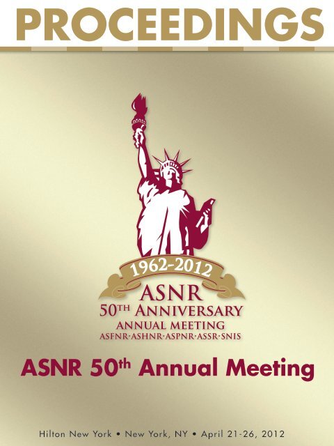
PROCEEDINGS<br />
<strong>ASNR</strong> 50 th <strong>Annual</strong> <strong>Meeting</strong><br />
Hilton New York • New York, NY • April 21-26, 2012
2<br />
Dear <strong>ASNR</strong> Members, Visitors, and Neuroscience Colleagues:<br />
I am happy to invite you to the American Society of Neuroradiology <strong>50th</strong> Anniversary <strong>Meeting</strong>. For this special<br />
event the Society is returning to its birthplace in New York City. The <strong>Meeting</strong> will be held April 21-26 at the<br />
Hilton New York, in the heart of Manhattan. President-Elect and Program Chair, Pamela Schaefer, MD, and the<br />
Program Committee have planned an exciting symposium and scientific program, along with special celebrations<br />
recognizing our milestone <strong>50th</strong> <strong>Annual</strong> <strong>Meeting</strong>.<br />
The Foundation of the <strong>ASNR</strong> Symposium 2012 “Acute Ischemic Stroke Imaging,” is at the heart of neuroradiology<br />
and will feature speakers with expertise across the range of adult and pediatric brain ischemia. We will continue<br />
to offer a Saturday morning session in collaboration with the International Society of Magnetic Resonance in<br />
Medicine (ISMRM). This mini symposium on “Advanced MR Imaging of Acute Ischemic Stroke” will feature<br />
new techniques and emerging new technologies that increasingly are applied to stroke.<br />
The 50 th <strong>Annual</strong> <strong>Meeting</strong> will provide informative updates on general neuroradiology and showcase subspecialty<br />
programming from the ASFNR, ASHNR, ASPNR, ASSR, and SNIS. As always, the heart of the meeting will be<br />
the invited lectures, original presentations, scientific posters, and educational exhibits. After a record number<br />
of abstract submissions, I can promise you more new and exciting material than ever before.<br />
There will be approximately 50 hours of continuing medical education credit available including size (6) Self-<br />
Assessment Module (SAM) sessions, Maintenance of Certification (MOC) interactive audience response<br />
technology case reviews, and new Expert: “How I Do It” sessions with practical clinical approaches to common<br />
imaging challenges. We will again feature valuable non-CME “How To Sessions” sponsored by our commercial<br />
partners, updating you on the capabilities and use of their hardware and software. Our Synaptic Junction this year<br />
will feature talks on practical issues in neuroradiology, including MR Safety for Patients with Implants, DTI for<br />
Dummies, Dose Extraction (for monitoring patient exposure), and a lively session on Communicating Results<br />
Directly to Patients. In response to rapid changes in the economic and health care environments, we will continue<br />
to feature programming on socioeconomic issues facing neuroradiology and practice quality improvement. We<br />
will expand our programming specific to fellows, residents, medical students and neuroradiologists in their<br />
first three years in practice.<br />
In celebration of our <strong>50th</strong> Anniversary we have planned some outstanding special events including reviews<br />
of the history of the field and the Society, where we are headed in the future, recognition of our founders and<br />
early members, a Keynote Address from one of our notable leaders in Neuroradiology, and an unforgettable<br />
<strong>50th</strong> Anniversary Reception at The Museum of Modern Art (MoMA).<br />
Among its most valuable functions, the <strong>Annual</strong> <strong>Meeting</strong> will be the ideal opportunity to stay connected with<br />
friends and colleagues and get the latest in brain, spine, and head and neck imaging. As befits a golden anniversary,<br />
this will be a truly unique event. I look forward to seeing you there!<br />
David B. Hackney, MD, FACR<br />
<strong>ASNR</strong> President<br />
David B. Hackney, MD, FACR<br />
<strong>ASNR</strong> President
Dear Friends:<br />
T HE C ITY OF N EW Y ORK<br />
O FFICE OF THE M AYOR<br />
N EW Y ORK, NY 10007<br />
April 21, 2012<br />
It is a great pleasure to welcome everyone to the American Society of Neuroradiology’s<br />
50 th <strong>Annual</strong> <strong>Meeting</strong> and The Foundation of the <strong>ASNR</strong> Symposium 2012.<br />
New York City is focused on helping all our residents enjoy the benefits of lifelong good<br />
health. Thanks to innovative public policies and our world-class medical institutions, we have<br />
made amazing gains over the past decade, and our residents are living longer than ever before as<br />
a result. That is why we are proud to applaud the <strong>ASNR</strong>’s thousands of neuroradiology<br />
professionals for their outstanding commitment to their patients. And as <strong>ASNR</strong> celebrates its 50 th<br />
Anniversary right here in New York where it was founded, we look forward to the latest research<br />
initiatives and advances in care making a lasting difference for people across the five boroughs<br />
and beyond.<br />
On behalf of all New Yorkers, I offer my best wishes for a productive meeting and<br />
symposium, a wonderful stay in our great City, and continued success in your vital mission.<br />
Sincerely,<br />
Michael R. Bloomberg<br />
Mayor<br />
3
<strong>ASNR</strong> ANNUAL MEETING<br />
HAS GONE MOBILE!<br />
Search for the '<strong>ASNR</strong>' guide under the "download guides" section of the application.<br />
4
Table of Contents<br />
II ..................................................................................................................................................... 2011 - 2012 <strong>ASNR</strong> Executive Committee<br />
II-III .............................................................................................................2011 - 2012 <strong>ASNR</strong> <strong>Annual</strong> <strong>Meeting</strong> Arrangements Committees<br />
IV ........................................................................................................................................................................................AJNR/<strong>ASNR</strong> Staff<br />
V-VI ..................................................................................................................................................................................General Information<br />
VII ...........................................................................................................................................Important Information for Young Professionals<br />
VIII ...........................................................................................................................................................................................Social Programs<br />
IX...............................................................................................................................................................Past <strong>Annual</strong> <strong>Meeting</strong>s of the <strong>ASNR</strong><br />
X ................................................................................................................................................................. <strong>ASNR</strong> Past Presidents & Founders<br />
XI.......................................................................................................................2012 <strong>ASNR</strong> Gold Medal Award Recipient & Past Recipients<br />
XII ......................................................................................................................2012 <strong>ASNR</strong> Honorary Member & Past Honorary Members<br />
XIII ................................... 2012 The Foundation of the <strong>ASNR</strong> Award for Outstanding Contributions in Research Award & Past Recipients<br />
XIII-XIV .................................................................................2012 <strong>ASNR</strong> Cornelius G. Dyke Memorial Award Recipient & Past Recipients<br />
XV ........................................................................................................................................... <strong>ASNR</strong> 2011 Outstanding Presentation Awards<br />
XV ............................................................................................................................................ 2011/2012 Specialty/Regional Society Awards<br />
XVI-XVII .................................................................................. The Foundation of the <strong>ASNR</strong> Basic Science Research Award Past Recipients<br />
XVIII .......................................................2012 The Foundation of the <strong>ASNR</strong> Scholar Award in Neuroradiology Research & Past Recipients<br />
XIX ..................................................................................... 2012 The Foundation of the <strong>ASNR</strong> Research Scientist Award & Past Recipients<br />
XIX .......................................................................2012 The Foundation of the <strong>ASNR</strong> Comparative Effectiveness Award & Past Recipients<br />
XIX .....................................................................................The Foundation of the <strong>ASNR</strong> Award in Cerebrovascular Disease Past Recipients<br />
XX ............................ Continuing Medical Education (CME), Accreditation Statement, Credit Designation Statement and Target Audience<br />
XXI .................................................................................................................................<strong>ASNR</strong> <strong>50th</strong> <strong>Annual</strong> <strong>Meeting</strong> Educational Objectives<br />
XXII.........................................................................................................................................................................Synaptic Junction Lectures<br />
XXIII ......................................................................................................................................................................................How-To Sessions<br />
XXIV .............................................................................................................................................................. Technical Exhibition Floor Plan<br />
XXV ......................................................................................................................................................................Technical Exhibition Roster<br />
XXVI - XXVII .......................................................................................................................... <strong>ASNR</strong> <strong>50th</strong> <strong>Annual</strong> <strong>Meeting</strong> Invited Speakers<br />
XXVIII - XXXI .....................................................................................................................................................Scientific Program Overview<br />
XXXII ..............................................................................................................................................................<strong>ASNR</strong> Future <strong>Annual</strong> <strong>Meeting</strong>s<br />
Page 1-97 ................................................................................................................................................................................Monday Sessions<br />
Page 98-183 ............................................................................................................................................................................ Tuesday Sessions<br />
Page 184-231 .....................................................................................................................................................................Wednesday Sessions<br />
Page 232-322 ........................................................................................................................................................................Thursday Sessions<br />
Page 322-391 ...................................................................................................................................... Electronic Excerpta Extraordinaire (EE)<br />
Page 391-466 ........................................................................................................................................................Scientific Posters (P) Printed<br />
Page 466-660 ..................................................................................................................................................Electronic Scientific Posters (eP)<br />
Page 660-716 ................................................................................................................................................Education Exhibits (EdE) Printed<br />
Page 716-796 ..........................................................................................................................................Electronic Education Exhibits (eEdE)<br />
Page 797-824 ......................................................................................................................................................Index of Program Participants<br />
5
2011-2012 <strong>ASNR</strong> Executive Committee<br />
David B. Hackney, MD, FACR<br />
President<br />
Pamela W. Schaefer, MD<br />
President-Elect/Program Chair<br />
Mauricio Castillo, MD, FACR<br />
Vice President/AJNR Editor<br />
Laurie A. Loevner, MD<br />
Treasurer<br />
David J. Seidenwurm, MD<br />
Secretary<br />
Pratik Mukherjee, MD, PhD<br />
Research Committee Co-Chair<br />
Pina C. Sanelli, MD, MPH<br />
Research Committee Co-Chair &<br />
Evidence Based Medicine Chair<br />
Joseph A. Maldjian, MD<br />
Functional Representative<br />
Suresh K. Mukherji, MD, FACR<br />
Fellowship Director Chair & Head<br />
and Neck Representative<br />
Joshua A. Hirsch, MD, FACR<br />
Neurointerventional Surgery<br />
Representative<br />
Caroline D. Robson, MB, ChB<br />
Pediatric Representative<br />
Walter S. Bartynski, MD<br />
Spine Representative<br />
Carolyn Cidis Meltzer, MD, FACR<br />
1st Past President & ACR Neuro<br />
Committee Liaison<br />
John R. Hesselink, MD, FACR<br />
2nd Past President<br />
Robert D. Zimmerman, MD, FACR<br />
3rd Past President & 2nd ABR Trustee<br />
Robert M. Barr, MD<br />
Member-at-Large &<br />
Clinical Practice Chair<br />
& ACR Councilor<br />
Barton F. Branstetter IV, MD<br />
Education Committee Chair<br />
Vincent P. Mathews, MD<br />
Rules Committee Chair<br />
& ABR 1st Trustee<br />
Tina Young Poussaint, MD<br />
Publication Committee Chair<br />
Clifford J. Belden, MD<br />
Membership Committee Chair<br />
Steven Falcone, MD<br />
Delegate to the AMA<br />
Jacqueline A. Bello, MD<br />
AMA Alternate, ACR Alternate<br />
Delegate and ACR Neuro Committee<br />
Representative<br />
James B. Gantenberg, FACHE<br />
<strong>ASNR</strong> Executive Director/CEO<br />
Commercial Relations Chair<br />
Peter B. Barker, D.Phil.<br />
Research Scientist Chair<br />
& Physicist Advisor<br />
Michael Tyszka, PhD<br />
Physicist Advisor<br />
Nafi Aygun, MD<br />
Physicist Advisor<br />
Howard A. Rowley, MD<br />
The Foundation of the <strong>ASNR</strong> Chair<br />
2011-2012 <strong>ASNR</strong> <strong>Meeting</strong> Arrangements Committees<br />
Scientific Program Committee<br />
Pamela W. Schaefer, MD<br />
President-Elect/Program Chair<br />
Konstantinos Arfanakis, PhD<br />
Daniel P. Barboriak, MD<br />
Peter B. Barker, D.Phil.<br />
Robert M. Barr, MD<br />
Walter S. Bartynski, MD<br />
Barton F. Branstetter IV, MD<br />
Mauricio Castillo, MD, FACR<br />
Colin P. Derdeyn, MD<br />
John L. Go, MD<br />
David B. Hackney, MD, FACR<br />
Ellen G. Hoeffner, MD<br />
Nickoleta L. Hoefling, MD<br />
Jerry G. Jarvik, MD<br />
Walter Kucharczyk, MD, FRCP(C)<br />
Christopher J. Moran, MD<br />
Jeffrey R. Petrella, MD<br />
C. Douglas Phillips, MD, FACR<br />
Whitney B. Pope, MD, PhD<br />
James M. Provenzale, MD<br />
Timothy P. L. Roberts, PhD<br />
II<br />
Caroline D. Robson, MB, ChB<br />
Pina C. Sanelli, MD, MPH<br />
Erin Simon Schwartz, MD<br />
David J. Seidenwurm, MD<br />
Dikoma C. Shungu, PhD<br />
A. Gregory Sorensen, MD<br />
Max Wintermark, MD<br />
Technical Exhibits<br />
Committee<br />
A. Orlando Ortiz, MD, MBA, FACR<br />
Chair<br />
Alan L. Willams, MD, FACR, MBA<br />
Consultant<br />
Todd Abruzzo, MD<br />
William G. Bradley, MD, PhD, FACR<br />
Joseph P. Comunale, Jr., MD<br />
Thomas S. Dina, MD<br />
Steven Falcone, MD<br />
John L. Go, MD<br />
Meng Law, MD<br />
Gregg H. Zoarski, MD
2011-2012 <strong>ASNR</strong> <strong>Meeting</strong> Arrangements Committees<br />
Audio Visual<br />
Committee<br />
Edward A. Michals, MD<br />
Chair<br />
Richard M. Berger, MD<br />
Andrew G. Bleicher, MD<br />
James Y. Chen, MD<br />
Steven W. Hetts, MD<br />
Jenny K. Hoang, MBBS<br />
Lucien M. Levy, MD, PhD<br />
Mark E. Mullins, MD, PhD<br />
Hemant A. Parmar, MD<br />
Stuart R. Pomerantz, MD<br />
Erin Simon Schwartz, MD<br />
Frank C. Tong, MD<br />
Achala S. Vagal, MD<br />
Richard H. Wiggins, III, MD, CIIP<br />
Max Wintermark, MD<br />
Education Exhibits<br />
Committee<br />
(Formerly Scientific<br />
Exhibits Committee)<br />
Dheeraj Gandhi, MD<br />
Chair<br />
Stephen Gebarski, MD<br />
Consultant<br />
Ellen G. Hoeffner, MD<br />
Consultant<br />
Ashley H. Aiken, MD<br />
Sameer A. Ansari, MD, PhD<br />
Nafi Aygun, MD<br />
Kathleen A. Barry, MD<br />
Mary E. Cunnane, MD<br />
Lawrence E. Ginsberg, MD<br />
Ajay Gupta, MD<br />
Blaine L. Hart, MD<br />
Daniel P. Hsu, MD<br />
Peter A. Janick, MD<br />
Carl E. Johnson, MD<br />
Michele H. Johnson, MD<br />
Bernadette L. Koch, MD<br />
Susan Palasis, MD<br />
Gregory W. Petermann, MD<br />
Jay J. Pillai, MD<br />
Jordan M. Prager, MD<br />
Stein Erik Rafto, MD<br />
Otto Rapalino, MD<br />
Amit M. Saindane, MD<br />
Haris Sair, MD<br />
Gaurang V. Shah, MD<br />
James G. Smirniotopoulos, MD<br />
Ashok Srinivasan, MD<br />
Bruce A. Wasserman, MD<br />
Christopher P. Wood, MD<br />
Geoffrey Young, MD<br />
Robert J. Young, MD<br />
Education Committee<br />
Barton F. Branstetter IV, MD<br />
Chair<br />
Ashley H. Aiken, MD<br />
Nafi Aygun, MD<br />
Peter B. Barker, D.Phil.<br />
Richard M. Berger, MD<br />
Eric C. Bourekas, MD<br />
Jerrold Boxerman, MD<br />
Dale A. Charletta, MD<br />
Rebecca S. Cornelius, MD<br />
H. Christian Davidson, MD<br />
Richard K. Downs, Sr., MD<br />
Edward J. Escott, MD<br />
Nikdokht Farid, MD<br />
Adam E. Flanders, MD<br />
Melanie B. Fukui, MD<br />
John L. Go, MD<br />
David B. Hackney, MD, FACR<br />
Joshua A. Hirsch, MD, FACR, FSIR<br />
Jenny K. Hoang, MBBS<br />
Steven G. Imbesi, MD<br />
Andrew Ku, MD<br />
Meng Law, MD<br />
Laurie A. Loevner, MD<br />
Joseph A. Maldjian, MD<br />
David S. Martin, MD<br />
Carolyn Cidis Meltzer, MD, FACR<br />
Gary M. Miller, MD<br />
Gul Moonis, MB, BS<br />
Toshio Moritani, MD, PhD<br />
Suresh K. Mukherji, MD, FACR<br />
Mark E. Mullins, MD, PhD<br />
Gerard J. Muro, MD<br />
Gregory W. Petermann, MD<br />
C. Douglas Phillips, MD, FACR<br />
Tina Young Poussaint, MD<br />
Edward P. Quigley, III, MD, PhD<br />
Caroline D. Robson, MB,ChB<br />
Karen L. Salzman, MD<br />
Pina C. Sanelli, MD, MPH<br />
Pamela W. Schaefer, MD<br />
David J. Seidenwurm, MD, FACR<br />
James G. Smirniotopoulos, MD<br />
Jay Starkey, MD<br />
Apostolos John Tsiouris, MD<br />
Achala S. Vagal, MD<br />
Daniel B. Vigneron, PhD<br />
Richard H. Wiggins, III, MD, CIIP<br />
Christopher P. Wood, MD<br />
Robert D. Zimmerman, MD, FACR<br />
III
AJNR Staff<br />
American Journal of Neuroradiology<br />
Headquarters Office<br />
2210 Midwest Road, Suite 205<br />
Oak Brook, IL 60523-8205<br />
Telephone: 630-574-1487<br />
Fax: 630-786-6251<br />
Internet: http://www.ajnr.org<br />
Karen Halm<br />
Managing Editor<br />
Ext. 238, khalm@asnr.org<br />
Jason Gantenberg<br />
Editorial Assistant<br />
Ext. 240, jason@asnr.org<br />
Mary Harder<br />
Editorial Assistant<br />
Ext. 239, mharder@asnr.org<br />
Additional<br />
Email Addresses<br />
Clinical Practice<br />
cpc@asnr.org<br />
Communications<br />
communications@asnr.org<br />
Executive<br />
executive@asnr.org<br />
Finance<br />
finance@asnr.org<br />
Foundation<br />
foundation@asnr.org<br />
<strong>Meeting</strong>s<br />
meetings@asnr.org<br />
Membership<br />
membership@asnr.org<br />
IV<br />
<strong>ASNR</strong> Staff<br />
American Society of Neuroradiology<br />
Headquarters Office<br />
2210 Midwest Road, Suite 207<br />
Oak Brook, IL 60523-8205<br />
Telephone: 630-574-0220<br />
Fax: 630-574-0661/630-574-1740<br />
Internet: http://www.asnr.org<br />
James B. Gantenberg, FACHE<br />
Executive Director/CEO<br />
Ext. 224, jgantenberg@asnr.org<br />
Arthur An<br />
Educational Technologist<br />
Ext. 241, aan@asnr.org<br />
Angelo Artemakis<br />
Director of Communications and Media Management<br />
Ext. 227, aartemakis@asnr.org<br />
Kenneth Cammarata<br />
Director of Specialty Societies and Member Services<br />
Ext. 226, kcammarata@asnr.org<br />
Tina Cheng, CPA<br />
Director of Finance and Information Systems<br />
Ext. 225, tcheng@asnr.org<br />
Michelle Davies<br />
<strong>Meeting</strong>s & Logistics Assistant<br />
Ext. 232, mdavies@asnr.org<br />
Pat Galle-Bingham<br />
Staff Accountant<br />
Ext. 222, pat@asnr.org<br />
Valerie Geisendorfer, CMP<br />
Senior Manager of Scientific <strong>Meeting</strong>s<br />
Ext. 228, vgeisendorfer@asnr.org<br />
Bonnie Mack<br />
Membership/Society Coordinator<br />
Ext. 234, bmack@asnr.org<br />
Karen Mansfield<br />
Office Manager/Executive Assistant<br />
Ext. 221, kmansfield@asnr.org<br />
Kelsey Pliner<br />
<strong>Meeting</strong>s Registrar<br />
Ext. 231, kpliner@asnr.org<br />
Lora Di Padova-Tannehill, CMP<br />
Director of Scientific <strong>Meeting</strong>s<br />
Ext. 229, ltannehill@asnr.org<br />
Stephanie Walsh, MBA<br />
Director of Development<br />
Ext. 235, swalsh@asnr.org
General Information (as of 3/21/12)<br />
<strong>Meeting</strong> Location<br />
Hilton New York<br />
1335 Avenue of the Americas<br />
New York, NY 10019<br />
(212) 315-1374<br />
<strong>Meeting</strong> Registration<br />
Registration is located on the 2nd Floor Promenade of the Hilton New York.<br />
The registration desk will be open during the following hours:<br />
Friday, April 20 ..........................................................3:00 pm - 8:00 pm<br />
Saturday, April 21 ....................................................... 6:30 am - 6:00 pm<br />
Sunday, April 22 ......................................................... 6:30 am - 6:00 pm<br />
Monday, April 23 ....................................................... 6:30 am - 6:00 pm<br />
Tuesday, April 24 ........................................................ 6:30 am - 6:00 pm<br />
Wednesday, April 25................................................... 6:30 am - 6:00 pm<br />
Thursday, April 26 ..................................................... 6:30 am - 6:00 pm<br />
Speaker Ready Room Location & Hours<br />
Hilton New York - Bryant (2nd Floor)<br />
Friday, April 20 ..........................................................3:00 pm - 8:00 pm<br />
Saturday, April 21 through Thursday, April 26 .......... 6:00 am - 6:00 pm<br />
Name Badges<br />
Please wear name badges at all times while you are attending the scientific<br />
sessions, social programs, and technical exhibits. Badge colors are identified<br />
as follows:<br />
<strong>ASNR</strong> Members, <strong>ASNR</strong> Members-In-Training & <strong>ASNR</strong> Subspecialty<br />
Societies (ASFNR, ASHNR, ASPNR, ASSR & SNIS) ................... Blue<br />
Non-Member .................................................................................Green<br />
Other Professional ......................................................................... Yellow<br />
Guest ..............................................................................................Peach<br />
Exhibitor ..........................................................................................Gold<br />
Staff............................................................................................... Purple<br />
Committee/Specialty/Regional<br />
Society <strong>Meeting</strong>s<br />
Please refer to the Daily Postings on the <strong>Meeting</strong>s & Announcements Board<br />
located on the 2 nd Floor Promenade at the Hilton New York.<br />
<strong>Meeting</strong>s & Announcements Board<br />
The <strong>Meeting</strong>s & Announcements Board is located on the 2 nd Floor Promenade<br />
at the Hilton New York. Please refer to the Daily Postings on the <strong>Meeting</strong>s &<br />
Announcements Board for information on committee meetings.<br />
CME Pavilion/Message Center/<br />
Alternate SAM Response Area<br />
Located in Gibson (2nd Floor) the CME Pavilion computer terminals will be<br />
available to registered attendees that can be used to evaluate attended sessions,<br />
print CME certificates and to check E-mail.<br />
Hilton New York - Gibson<br />
Saturday, April 21 ....................................................... 7:00 am - 9:00 pm<br />
Sunday, April 22 through Thursday, April 26 ............. 6:30 am - 9:00 pm<br />
Coat Check<br />
2nd Floor Promenade at the Hilton New York<br />
Hours of Operation:<br />
Saturday, April 21 ......................................................... 6:30am - 6:00pm<br />
Sunday, April 22 ........................................................... 6:00am - 6:00pm<br />
Monday, April 23 and Tuesday, April 24 ....................... 6:00am - 7:00pm<br />
Wednesday, April 25..................................................... 6:00am - 8:00pm<br />
Thursday, April 26 ....................................................... 6:00am - 7:00pm<br />
Concierge Desk<br />
Lobby Level of the Hilton New York<br />
Friday, April 20 through Friday, April 27 .................... 7:00am - 11:59pm<br />
Local Hospital<br />
Roosevelt Hospital - St. Luke’s • (212) 523-4000<br />
1000 Tenth Avenue<br />
New York, NY 10019<br />
Emergency Service Procedure<br />
The Hilton New York is fully prepared to handle different types of situations<br />
to assist our guests. The following is information on The Hilton New York’s<br />
emergency procedures:<br />
Do not call 9-1-1 from your cellular phone. Calling 9-1-1 delays the<br />
response to the emergency by the first responders.<br />
• Locate the nearest Hilton New York HOTEL PHONE and dial our Security<br />
Control at extension 5747 or “66” for a life emergency.<br />
• If you are calling from a cellular phone, the number is 212-261-5747 or<br />
the hotel general number at 212-586-7000.<br />
• Notify the Security Control of the location of the person requiring medical<br />
attention, along with a description of the person and their injury. The<br />
Security Control will contact your contracted medical service provider.<br />
• Return to the individual.<br />
• The hotel has an emergency response team 24 hours a day. In the event<br />
of an emergency, calling the emergency number 66 will initiate the<br />
appropriate response.<br />
• Paramedics, Fire Department, and the Police Department are all located<br />
within minutes from the hotel.<br />
• The Hilton New York’s Security Department, as well as a small number of<br />
other employees, are trained in CPR and First Aid.<br />
• Emergency evacuation routes and procedures are located on the inside of<br />
all guest room doors.<br />
Remote Access Features<br />
Continues in 2012!<br />
A link will be provided so that you can<br />
evaluate for CME credits from your personal<br />
devices (phone/laptop/PDA) at any time.<br />
In addition you will be able to participate in<br />
SAM sessions remotely through Sunday, May 6.<br />
V
General Information (continued) (as of 3/21/12)<br />
Food Service<br />
Monday through Thursday: <strong>ASNR</strong> will provide Continental Breakfast,<br />
Morning & Afternoon Beverage Breaks. Lunch on own.<br />
Please refer to the schedule below.<br />
Continental Breakfast<br />
Monday, April 23 through Thursday, April 26 ..........3 rd Floor Promenade<br />
Continuous Coffee Service<br />
Tuesday, April 24 through Thursday, April 26 ................. Americas Hall I<br />
Morning Beverage Breaks<br />
Saturday, April 21 through Monday, April 23 ...........3 rd Floor Promenade<br />
Tuesday, April 24 through Thursday, April 26 ................. Americas Hall I<br />
Afternoon Beverage Breaks<br />
Saturday, April 21 through Monday, April 23 ...........3 rd Floor Promenade<br />
Tuesday, April 24 through Thursday, April 26 ................. Americas Hall I<br />
<strong>ASNR</strong> Publications Booth<br />
3rd Floor Promenade<br />
All attendees are invited to stop by the booth any time to tour the AJNR<br />
and Neurographics Web sites and AJNR Blog and app.<br />
Meet AJNR’s Editor-in-Chief<br />
Dr. Mauricio Castillo, AJNR’s Editor-in-Chief, will be present at the Journal’s<br />
booth Monday - Thursday from 11:00 am until noon to answer questions<br />
regarding the Journal’s online features, listen to suggestions, talk about<br />
projects with prospective authors, and advise fellows regarding their future<br />
contributions to AJNR.<br />
Neurographics<br />
Neurographics is <strong>ASNR</strong>’s quarterly online educational journal, featuring peerreviewed<br />
scientific exhibits and select case reports. Neurographics is available<br />
free online www.neurographics.org. Neurographics Editor Dr. Barton<br />
Branstetter IV will be available in the <strong>ASNR</strong> Publications booth Monday<br />
through Thursday from 11:00 am to noon to answer questions.<br />
Booth Schedule<br />
Hilton New York - 3rd Floor Promenade<br />
Saturday, April 21 through Sunday, April 22 .............. 7:30 am - 5:00 pm<br />
Monday, April 23 through Thursday, April 26 ............ 8:30 am - 5:00 pm<br />
VI<br />
How-To Sessions<br />
Grand Ballroom Suite (3rd Floor)<br />
Focus/Scientific Paper Sessions<br />
Grand Ballroom Suite, Trianon Ballroom, Beekman/Sutton North,<br />
Sutton Center/South, Murray Hill<br />
Synaptic Junction Lectures<br />
Murray Hill, Beekman/Sutton North<br />
EXHIBITS<br />
Education Exhibits & Electronic<br />
Education Exhibits<br />
Rhinelander (2nd Floor)<br />
Scientific Posters, Electronic<br />
Scientific Posters and Electronic<br />
Excerpta Extraordinaire<br />
Americas Hall II<br />
Technical Exhibits<br />
Americas Hall I<br />
MISCELLANEOUS<br />
American Board of Radiology (ABR)<br />
Information Desk<br />
2 nd Floor Promenade<br />
Young Professionals Lounge (Fellows,<br />
Residents, Medical Students and<br />
Neuroradiologists in their first three<br />
years in practice)<br />
Regent (2nd Floor)<br />
Headquarters Office<br />
Nassau West (2nd Floor)<br />
VIP/The Foundation of the <strong>ASNR</strong> Lounge<br />
Hudson (4th Floor)
Important Information for Young Professionals<br />
Young Professionals Lounge - Regent (2 nd Floor)<br />
( Fellows, Residents, Medical Students and Neuroradiologists in their first three<br />
years in practice)<br />
Visit the Fellows, Residents & Medical Students Lounge in the Regent (2 nd Floor) open from 6:30am – 6:00pm daily from Saturday, April 21 -<br />
Thursday, April 26. This will provide a “home base” for Young Professionals to network and share knowledge and experiences throughout the meeting.<br />
Young Professionals Luncheon<br />
Tuesday, April 24 • 12:00pm - 1:00pm<br />
Gramercy West (2 nd Floor)<br />
Fellows, Residents, Medical Students and Neuroradiologists in their first three years in practice – join <strong>ASNR</strong> Leaders for Lunch, at 12:00pm on<br />
Tuesday, April 24, 2012 in the Gramercy West (2 nd Floor).<br />
Entering And Thriving In Private Practice<br />
8:30am - 10:00am • Murray Hill<br />
Welcome to the Real World<br />
John E. Jordan, MD<br />
Understanding the Food Chain<br />
Robert M. Barr, MD<br />
The Corporate Environment<br />
Stephen T. Sweriduk, MD<br />
The HMO Environment<br />
D. Christian Sonne, MD<br />
Making the Transition<br />
David J. Seidenwurm, MD, FACR<br />
Questions and Answers<br />
Young Professionals Sessions<br />
Fellows, Residents, Medical Students and Neuroradiologists in their first three years in practice, the <strong>ASNR</strong> has designed specific program<br />
topics for learning during the <strong>ASNR</strong> <strong>Annual</strong> <strong>Meeting</strong>. You won’t want to miss the following session presentations:<br />
Tuesday, April 24<br />
Academic Session<br />
1:00pm - 2:30pm • Murray Hill<br />
Getting Your First Job<br />
Carolyn C. Meltzer, MD, FACR<br />
Building a Research Program<br />
Soonmee Cha, MD<br />
The Different Component of Academic Radiology<br />
Suresh K. Mukherji, MD, FACR<br />
Getting Promoted: The Different<br />
Promotion Tracks<br />
David M. Yousem, MD, MBA<br />
Discussion<br />
VII
Social Programs<br />
“<strong>ASNR</strong> Welcome Reception”<br />
Monday, April 23<br />
7:00 pm - 10:00 pm<br />
Off-site Location:<br />
The Museum of Modern Art (MoMA)<br />
Join us at The Museum of Modern Art to celebrate <strong>ASNR</strong>’s <strong>50th</strong> Anniversary.<br />
MoMA is often identified as the most influential museum of modern art in the<br />
world. Art enthusiasts and novices alike are often awestruck by the masterpieces<br />
before them, including Monet’s Water Lilies, Picasso’s Les Demoiselles d’<br />
Avignon and van Gogh’s Starry Night. The museum’s collection offers an<br />
unparalleled overview of modern and contemporary art, including works of<br />
architecture and design, drawings, painting, sculpture, photography, prints,<br />
illustrated books and artist’s books, film and electronic media. In addition to<br />
the artwork, one of the main draws of MoMA is the building itself. The main<br />
entrance is a 110 foot atrium and inside a maze of glass walkways permits art<br />
viewing from many angles. Also of interest is the Abby Aldrich Rockefeller<br />
Sculpture Garden. A glass wall lets visitors look directly into the surrounding<br />
galleries from the garden, where there’s also a reflecting pool and trees. You don’t<br />
want to miss this special experience. We look forward to welcoming you to The<br />
Museum of Modern Art.<br />
• Includes access to the 5th floor gallery, sculpture garden (weather permitting),<br />
dinner buffet and three (3) hour open bar.<br />
• FREE for those who register for the meeting.<br />
• Guest Ticket is $75.00.<br />
“Closing Reception with<br />
Technical Exhibitors”<br />
Wednesday, April 25<br />
6:30 pm - 7:30 pm<br />
Hilton New York Hotel in Americas Hall I<br />
We look forward to closing the social events with a pre-dinner reception of<br />
New York’s culinary favorites. The reception, located in Americas Hall I offers<br />
attendees the perfect opportunity to see this year’s Technical Exhibition, the<br />
<strong>ASNR</strong>’s annual showcase for the newest products and services for the field of<br />
Neuroradiology. Enjoy complimentary pre-dinner hors d’oeuvres and beverages<br />
while you network with exhibitor representatives and fellow colleagues. The<br />
<strong>ASNR</strong> Technical Exhibition is the place where advanced technology, diagnostic<br />
and interventional Neuroradiological excellence come together.<br />
• Includes pre dinner buffet and one (1) hour open bar.<br />
• FREE for those who register for the meeting.<br />
• Guest Ticket is $40.00.<br />
VIII<br />
2012 Education Exhibits Tour<br />
Rhinelander (2 nd Floor)<br />
Monday, April 23 ......................................12:00pm - 1:00pm<br />
Wednesday, April 25..................................12:00pm - 1:00pm<br />
NEW in 2012! You will receive CME credits for participating<br />
in the Education Exhibits Tour!<br />
Review Education Exhibits, visual presentation/educational displays designed<br />
to further the understanding of Neuroradiology by featuring cutting-edge<br />
material or by offering an instructional review of a particular topic. The session<br />
will allow for review of visually-oriented educational displays that demonstrate<br />
novel and innovative applications for computers in Neuroradiology clinical<br />
practice, training and research. Share this educational opportunity with your<br />
colleagues and primary Exhibit presenters and exchange in a lively Questions<br />
& Answers Session.<br />
Another unique feature not to be missed at the <strong>ASNR</strong>…See you on<br />
the tour!<br />
“Case of the Day”<br />
Monday, April 23 through<br />
Wednesday, April 25<br />
Rhinelander (2 nd Floor)<br />
New Program Feature at the <strong>ASNR</strong> <strong>50th</strong> <strong>Annual</strong> <strong>Meeting</strong>!<br />
A new addition to the <strong>ASNR</strong> program for the <strong>ASNR</strong> <strong>50th</strong> <strong>Annual</strong> <strong>Meeting</strong><br />
is the Case of the Day (COD) program. Led by Drs. Mark Mullins and Char<br />
Branstetter, COD is scheduled from Monday, April 23, through Wednesday,<br />
April 25.<br />
Cases will be presented in Rhinelander at the Hilton New York and will be<br />
available in printed poster format as well as on a video loop at a nearby monitor.<br />
There will be 6 categories: brain, functional, head & neck, pediatric, spine and<br />
vascular/interventional, each with 1 case per day. Answers will be presented on<br />
the day following their respective questions.<br />
An online system will be available for submission of preferred diagnoses. Be<br />
sure to participate in this new interactive programming feature with a limited<br />
number of prizes awarded to those participants with highest daily correct<br />
responses.<br />
No CME credit is available for this activity.
Past <strong>Annual</strong> <strong>Meeting</strong>s<br />
Organizational <strong>Meeting</strong><br />
May 19, 1962 - New York<br />
Keene’s English Chophouse<br />
Second Business <strong>Meeting</strong><br />
October 5, 1962 - Washington, DC<br />
Shoreham Hotel<br />
First <strong>Annual</strong> <strong>Meeting</strong><br />
October 7, 1963 - Montreal<br />
Queen Elizabeth Hotel<br />
Second <strong>Annual</strong> <strong>Meeting</strong><br />
September 23, 1964 - New York<br />
Waldorf Astoria<br />
Third <strong>Annual</strong> <strong>Meeting</strong><br />
June 11, 1965 - Atlantic City<br />
Dennis Hotel<br />
Fourth <strong>Annual</strong> <strong>Meeting</strong><br />
June 15-16, 1966 - Washington, DC<br />
Sheraton-Park Hotel<br />
Fifth <strong>Annual</strong> <strong>Meeting</strong><br />
May 15, 1967 - New York<br />
Columbia University<br />
Sixth <strong>Annual</strong> <strong>Meeting</strong><br />
September 27-28, 1968 - New Orleans<br />
Jung Hotel<br />
Seventh <strong>Annual</strong> <strong>Meeting</strong><br />
May 13-19, 1969 - Cleveland<br />
Joint <strong>Meeting</strong> with American<br />
Association of Neurological Surgeons<br />
Sheraton-Cleveland Hotel<br />
Eighth <strong>Annual</strong> <strong>Meeting</strong><br />
February 12-13, 1970 - Washington, DC<br />
Washington Hilton<br />
Ninth <strong>Annual</strong> <strong>Meeting</strong><br />
May 27-29, 1971 - San Francisco<br />
Fairmont Hotel<br />
Tenth <strong>Annual</strong> <strong>Meeting</strong><br />
February 21-24, 1972 - Mexico City<br />
Maria-lsabel Sheraton<br />
Eleventh <strong>Annual</strong> <strong>Meeting</strong><br />
May 26-28, 1973 - Boston<br />
Statler Hilton<br />
Twelfth <strong>Annual</strong> <strong>Meeting</strong><br />
March 14, 1974 - Punta del Este, Uruguay<br />
(In conjunction with X Symposium<br />
Neuroradiologicum)<br />
Punta del Este, Uruguay<br />
Thirteenth <strong>Annual</strong> <strong>Meeting</strong><br />
June 3-7, 1975 - Vancouver<br />
Bayshore Inn<br />
Fourteenth <strong>Annual</strong> <strong>Meeting</strong><br />
May 18-22, 1976 - Atlanta<br />
Peachtree Plaza<br />
Fifteenth <strong>Annual</strong> <strong>Meeting</strong><br />
March 27-31, 1977 - Bermuda<br />
Hamilton Princess Hotel<br />
Sixteenth <strong>Annual</strong> <strong>Meeting</strong><br />
February 26-March 2, 1978 - New Orleans<br />
Hyatt Regency<br />
Seventeenth <strong>Annual</strong> <strong>Meeting</strong><br />
May 20-24, 1979 - Toronto<br />
Hotel Toronto<br />
Eighteenth <strong>Annual</strong> <strong>Meeting</strong><br />
March 16-21, 1980 - Los Angeles<br />
Century Plaza<br />
Nineteenth <strong>Annual</strong> <strong>Meeting</strong><br />
May 5-9, 1981 - Chicago<br />
Marriott Hotel<br />
Twentieth <strong>Annual</strong> <strong>Meeting</strong><br />
October 10-16, 1982 - Washington, DC<br />
(In conjunction with XII Symposium<br />
Neuroradiologicum)<br />
Washington Hilton<br />
Twenty-First <strong>Annual</strong> <strong>Meeting</strong><br />
June 5-9, 1983 - San Francisco<br />
St. Francis Hotel<br />
Twenty-Second <strong>Annual</strong> <strong>Meeting</strong><br />
June 2-7, 1984 - Boston<br />
Westin Copley Place Hotel<br />
Twenty-Third <strong>Annual</strong> <strong>Meeting</strong><br />
February 18-23, 1985 - New Orleans<br />
Marriott Hotel<br />
Twenty-Fourth <strong>Annual</strong> <strong>Meeting</strong><br />
January 19-23, 1986 - San Diego<br />
Sheraton Harbor Island<br />
Hotel<br />
Twenty-Fifth <strong>Annual</strong> <strong>Meeting</strong><br />
(Silver Anniversary)<br />
May 10-15, 1987 - New York<br />
Hilton New York<br />
Twenty Sixth <strong>Annual</strong> <strong>Meeting</strong><br />
May 15-20, 1988 - Chicago<br />
Chicago Hilton & Towers<br />
Twenty-Seventh <strong>Annual</strong> <strong>Meeting</strong><br />
March 19-24, 1989 - Orlando<br />
The Peabody Orlando<br />
Twenty-Eighth <strong>Annual</strong> <strong>Meeting</strong><br />
March 19-23, 1990 - Los Angeles<br />
Century Plaza Hotel & Tower<br />
Twenty-Ninth <strong>Annual</strong> <strong>Meeting</strong><br />
June 9-14, 1991 - Washington, DC<br />
The Washington Hilton and<br />
Towers<br />
Thirtieth <strong>Annual</strong> <strong>Meeting</strong><br />
May 31-June 5, 1992 - St. Louis<br />
Adam’s Mark<br />
Thirty-First <strong>Annual</strong> <strong>Meeting</strong><br />
May 17-20, 1993 - Vancouver<br />
Vancouver Trade and<br />
Convention Center<br />
Thirty-Second <strong>Annual</strong> <strong>Meeting</strong><br />
May 3-7, 1994 - Nashville<br />
Opryland Hotel and<br />
Conference Center<br />
Thirty-Third <strong>Annual</strong> <strong>Meeting</strong><br />
May 23-27, 1995 - Chicago<br />
Sheraton Chicago Hotel and<br />
Towers<br />
Thirty-Fourth <strong>Annual</strong> <strong>Meeting</strong><br />
June 23-27, 1996 - Seattle<br />
Washington State<br />
Convention<br />
and Trade Center<br />
Thirty-Fifth <strong>Annual</strong> <strong>Meeting</strong><br />
May 18-22, 1997 - Toronto<br />
Metro Toronto Convention<br />
Center<br />
Thirty-Sixth <strong>Annual</strong> <strong>Meeting</strong><br />
May 17-21, 1998 - Philadelphia<br />
(In conjunction with XVI Symposium<br />
Neuroradiologicum)<br />
Pennsylvania Convention<br />
Center<br />
Thirty-Seventh <strong>Annual</strong> <strong>Meeting</strong><br />
May 23-28, 1999 - San Diego<br />
San Diego Convention<br />
Center<br />
Thirty-Eighth <strong>Annual</strong> <strong>Meeting</strong><br />
April 4-8, 2000 - Atlanta<br />
Hyatt Regency Atlanta<br />
Thirty-Ninth <strong>Annual</strong> <strong>Meeting</strong><br />
April 23-27, 2001 - Boston<br />
Hynes Convention Center<br />
Fortieth <strong>Annual</strong> <strong>Meeting</strong><br />
May 13-17, 2002 - Vancouver<br />
Vancouver Convention<br />
& Exhibition Center<br />
Forty-First <strong>Annual</strong> <strong>Meeting</strong><br />
April 28 - May 2, 2003 - Washington, DC<br />
Marriott Wardman Park<br />
Hotel<br />
Forty-Second <strong>Annual</strong> <strong>Meeting</strong><br />
June 7-11, 2004 - Seattle<br />
Washington State<br />
Convention and Trade Center<br />
Forty-Third <strong>Annual</strong> <strong>Meeting</strong><br />
May 21-27, 2005 - Toronto<br />
Metro Toronto Convention<br />
Center<br />
Forty-Fourth <strong>Annual</strong> <strong>Meeting</strong><br />
April 29-May 5, 2006 - San Diego<br />
San Diego Convention<br />
Center<br />
Forty-Fifth <strong>Annual</strong> <strong>Meeting</strong><br />
June 9-14, 2007 - Chicago<br />
Hyatt Regency Chicago<br />
Forty-Sixth <strong>Annual</strong> <strong>Meeting</strong><br />
May 30-June 5, 2008 - New Orleans<br />
Ernest N. Morial Convention<br />
Center<br />
Forty-Seventh <strong>Annual</strong> <strong>Meeting</strong><br />
May 16-21, 2009 - Vancouver<br />
Vancouver Convention and<br />
Exhibition Centre<br />
Forty-Eigth <strong>Annual</strong> <strong>Meeting</strong><br />
May 15-20, 2010 - Boston<br />
Hynes Convention Center<br />
Forty-Ninth <strong>Annual</strong> <strong>Meeting</strong><br />
June 4-9, 2011 - Seattle<br />
Washington State<br />
Convention Center<br />
IX
<strong>ASNR</strong> Past Presidents and Founders<br />
1962-64<br />
Juan M. Taveras, MD*<br />
1964-65<br />
Mannie M. Schechter, MD*<br />
1965-66<br />
Donald L. McRae, MD*<br />
1966-67<br />
Ernest H. Wood, MD*<br />
1967-68<br />
Harold O. Peterson, MD*<br />
1968-69<br />
Colin B. Holman, MD*<br />
1969-70<br />
Giovanni Di Chiro, MD*<br />
1970-71<br />
D. Gordon Potts, MD<br />
1971-72<br />
Norman E. Chase, MD<br />
1972-73<br />
Fred J. Hodges, III, MD*<br />
1973-74<br />
T. Hans Newton, MD*<br />
1974-75<br />
Hillier L. Baker, Jr., MD*<br />
1975-76<br />
Irvin I. Kricheff, MD<br />
1976-77<br />
Norman E. Leeds, MD<br />
1977-78<br />
Sadek K. Hilal, MD*<br />
1978-79<br />
Stephen A. Kieffer, MD<br />
1979-80<br />
David O. Davis, MD<br />
Founding Members<br />
Norman E. Chase, MD<br />
Giovanni Di Chiro, MD*<br />
William N. Hanafee, MD<br />
Fred J. Hodges, III, MD*<br />
Colin B. Holman, MD*<br />
X<br />
1980-81<br />
George Wortzman, MD<br />
1981-82<br />
Gabriel H. Wilson, MD*<br />
1982-83<br />
Arthur E. Rosenbaum, MD<br />
1983-84<br />
O. Wayne Houser, MD<br />
1984-85<br />
Samuel M. Wolpert, MD<br />
1985-86<br />
R. Thomas Bergeron, MD<br />
1986-87<br />
Derek C. Harwood-Nash, MD*<br />
Norman E. Leeds, MD<br />
Eugene V. Leslie, MD*<br />
Donald L. McRae, MD*<br />
Thomas H. Newton, MD*<br />
Harold O. Peterson, MD*<br />
1987-88<br />
Michael S. Huckman, MD<br />
1988-89<br />
Anne G. Osborn, MD<br />
1989-90<br />
Joseph F. Sackett, MD<br />
1990-91<br />
Anton N. Hasso, MD, FACR<br />
1991-92<br />
R. Nick Bryan, MD, PhD, FACR<br />
1992-93<br />
David Norman, MD<br />
1993-94<br />
Glenn S. Forbes, MD<br />
1994-95<br />
Robert M. Quencer, MD<br />
1995-96<br />
Robert R. Lukin, MD<br />
1996-97<br />
Burton P. Drayer, MD<br />
1997-98<br />
Richard E. Latchaw, MD<br />
1998-99<br />
A. James Barkovich, MD<br />
D. Gordon Potts, MD<br />
1999-00<br />
Eric J. Russell, MD, FACR<br />
2000-01<br />
William S. Ball, Jr., MD<br />
2001-02<br />
William P. Dillon, MD<br />
2002-03<br />
Patrick A. Turski, MD<br />
2003-04<br />
Charles M. Strother, MD<br />
2004-05<br />
Victor M. Haughton, MD<br />
2005-06<br />
Patricia A. Hudgins, MD<br />
2006-07<br />
Robert I. Grossman, MD<br />
2007-08<br />
David M. Yousem, MD, MBA<br />
2008-09<br />
Robert D. Zimmerman, MD, FACR<br />
2009-10<br />
John R. Hesselink, MD, FACR<br />
2010-11<br />
Carolyn C. Meltzer, MD, FACR<br />
Mannie M. Schechter, MD*<br />
Juan M. Taveras, MD*<br />
Ernest H. Wood, MD*<br />
*deceased
Awards and Honors<br />
2012 <strong>ASNR</strong> Gold Medal Award<br />
2012 Gold Medal Recipient<br />
A. James Barkovich, MD<br />
University of California, San Francisco<br />
The Gold Medal fosters the highest standards of the American Society of Neuroradiology, based on exceptional quality, service,<br />
and excellence, and not necessarily on fame. It emphasizes both professional and personal attributes... individuals who are superb<br />
neuroradiologists, clinicians, or scientists, and truly outstanding. The recipients are individuals who have extended themselves beyond<br />
furthering their own careers through contributions at all levels of professional strata, with an accent on consistency and duration of<br />
these outstanding contributions.<br />
Anthony James Barkovich, M.D. is Professor of Radiology and Biomedical<br />
Imaging, Neurology, Pediatrics, and Neurosurgery and Chief of Pediatric<br />
Neuroradiology at the University of California, San Francisco. His undergraduate<br />
education was at the University of California at Davis, he pursued graduate<br />
studies in Chemistry at the University of California at Berkeley, and he<br />
received his medical education at the George Washington University. He<br />
completed his internship and Radiology residency at the Letterman Army<br />
Medical Center in San Francisco, CA and then a neuroradiology fellowship<br />
at the Walter Reed Army Medical Center in Washington, D.C. During his<br />
fellowship, he spent time at Johns Hopkins Hospital, Children’s National<br />
Medical Center in Washington, D.C., and the Hospital for Sick Children in<br />
Toronto. After serving 3½ years in the U.S. Army as the neuroradiologist at<br />
Letterman Army Medical Center in San Francisco, Dr. Barkovich joined the<br />
faculty of the University of California, San Francisco in 1989, where he has<br />
remained for more than 22 years.<br />
Dr. Barkovich is fascinated by normal development of the central nervous<br />
system and the results of perturbations of that process. He recognized early<br />
that MRI was a potentially groundbreaking tool for the study of normal and<br />
abnormal brain development, so he focused upon optimizing techniques for the<br />
evaluation of small brains. This pursuit involved conceptualizing and aiding<br />
in designing and building MRI compatible incubators and high resolution<br />
Awards and Honors<br />
<strong>ASNR</strong> Gold Medal Award Past Recipients<br />
1995<br />
Juan M. Taveras, MD*<br />
T. Hans Newton, MD*<br />
1996<br />
Sadek K. Hilal, MD*<br />
Giovanni Di Chiro, MD*<br />
1997<br />
Derek C. Harwood-Nash, MB, ChB.,<br />
DSc, FRCPC, FACR, RCRAD(SA)*<br />
1998<br />
Irvin I. Kricheff, MD<br />
D. Gordon Potts, MD<br />
1999<br />
Grant B. Hieshima, MD<br />
Michael S. Huckman, MD<br />
2000<br />
Hillier L. “Bud” Baker, Jr., MD*<br />
2001<br />
O. Wayne Houser, MD<br />
J. Arliss Pollock, MD*<br />
2002<br />
R. Thomas Bergeron, MD<br />
David O. Davis, MD<br />
imaging coils, and adapting imaging sequences to maximize the information<br />
gained from these studies. Combining embryology, genetics, molecular<br />
biology and basic physical and biological science, he collaborated with fellow<br />
researchers to better understand and classify steps in brain development and<br />
determine at what steps normal development became perturbed. This work<br />
led to a series of grants funded by the NIH, which have supported much of the<br />
work since 1993. This work gained him recognition in many fields including<br />
metabolic disease, medical genetics and epilepsy. His ideas in these areas were<br />
published in many peer-reviewed papers and a series of books that he singleauthored,<br />
co-authored, or co-edited.<br />
Dr. Barkovich has served the <strong>ASNR</strong> and other radiology organizations for<br />
many years, serving as Secretary, Vice-President, and President-Elect of the<br />
<strong>ASNR</strong> before ascending to the Presidency in 1998. He was the Chairman of<br />
The Foundation of the <strong>ASNR</strong> (formerly the Neuroradiology Education and<br />
Research Foundation) from 2001-2007. He served as Secretary-General of<br />
the World Federation of Neuroradiological Societies from 2002-2006. Dr.<br />
Barkovich’s contributions have been recognized by multiple awards, including<br />
the Goldenson Technology Award from the United Cerebral Palsy R&E<br />
Foundation, the Peter Emil Becker Award from the German-Swiss-Austrian<br />
Society of Child Neurology for Outstanding Contributions to Pediatric<br />
Neurology and Neuroradiology, honorary membership in the European<br />
Society of Neuroradiology, and the initial Gold Medal of the American Society<br />
of Pediatric Neuroradiology.<br />
2003<br />
Norman E. Leeds, MD, FACR<br />
Anne G. Osborn, MD, FACR<br />
2004<br />
Ralph Heinz, MD, FACR<br />
Stephen A. Kieffer, MD, FACR<br />
2005<br />
Samuel M. Wolpert, MD<br />
2006<br />
R. Nick Bryan, MD, PhD, FACR<br />
Charles W. Kerber, MD<br />
2007<br />
Robert M. Quencer, MD<br />
2008<br />
Robert R. Lukin, MD<br />
2009<br />
Glenn S. Forbes, MD, FACR<br />
2010<br />
Anton N. Hasso, MD. FACR<br />
2011<br />
Burton P. Drayer, MD, FACR, FAAN<br />
*deceased<br />
XI
Awards and Honors<br />
2012 <strong>ASNR</strong> Honorary Member<br />
2012 <strong>ASNR</strong> Honorary Member Recipient:<br />
Francis O. Brunelle, MD<br />
Hospital Necker-Enfants Malades, Paris, France<br />
Professor Francis Brunelle graduated from Cochin Medical School in Paris<br />
in 1972. He served as a volunteer in Africa (Togo) as a military surgeon<br />
and, during his stay, he created the first nursing school in Togo. During<br />
his residency, he trained as a paediatrician, neonatologist, and finally as a<br />
radiologist. Prof. Brunelle received a master’s degree in biochemistry during<br />
is residency. Afterwards, he spent a year as a visiting fellow in Alex Margulis’<br />
department, where he trained with Harry Genant and Roy Filly, as well as with<br />
Hans Newton and David Norman in neuroradiology.<br />
Back in France he became an associate professor of radiology in Dr. Chaumont’s<br />
department in Bicetre Hospital. The presence of Professor Daniel Alagille, a<br />
world specialist of liver diseases in children, led him to develop diagnostic and<br />
interventional radiology in liver diseases in children. Most of the abdominal<br />
interventional techniques were developed during this time.<br />
During this period, in 1981, he spent four months in Derek Harwood-Nash’s<br />
department, where he trained in paediatric neuroradiology, writing a paper<br />
entitled, “Brain vascular malformations in children.”<br />
In 1985, he became full professor of radiology at Necker Enfants-Malades<br />
Hospital, which was the turning point of his career. The presence at Necker<br />
of Professor Hirsch, a pioneer of paediatric neurosurgery, and Doctor Jean<br />
Aicardi, one of the leading people in paediatric neurology, led him to develop<br />
Past <strong>ASNR</strong> Honorary Members<br />
Torsten Almen, MD<br />
Mitchel S. Berger, MD<br />
James W. Bull, MD*<br />
Graeme M. Bydder, MD, ChB<br />
M. Paul Capp, MD<br />
Sten Cronqvist, MD*<br />
B. G. Ziedses des Plantes, MD*<br />
George du Boulay, MD*<br />
Richard R. Ernst, MD<br />
Torgny V. B. Greitz, MD<br />
XII<br />
The Gold Medal fosters the highest standards of the American Society of Neuroradiology, based on exceptional quality, service,<br />
and excellence, and not necessarily on fame. It emphasizes both professional and personal attributes... individuals who are superb<br />
neuroradiologists, clinicians, or scientists, and truly outstanding. The recipients are individuals who have extended themselves beyond<br />
furthering their own careers through contributions at all levels of professional strata, with an accent on consistency and duration of<br />
these outstanding contributions.<br />
Godfrey N. Hounsfield, PhD*<br />
Yun Peng Huang, MD<br />
Ian Isherwood, MD<br />
Pierre Lasjaunias, MD, PhD<br />
Paul C. Lauterbur, PhD*<br />
Dennis LeBihan, MD, PhD<br />
Marco Leonardi, MD<br />
Erik Lindgren, MD*<br />
Claude H. Manelfe, MD<br />
Professor Luc Picard<br />
the diagnostic and interventional neuroradiology techniques in children.<br />
He created the first paediatric neuroradiology course in France, which<br />
rapidly became the European Course of Paediatric Neuroradiology, held<br />
in conjunction with the European Society of Paediatric Radiology and the<br />
European Society of Neuroradiology. In addition, he also created the first<br />
European course of Paediatric Radiology in 1992.<br />
The presence of a very active department of genetics allowed the development<br />
of research in neurogenetics diseases in children. The creation of a research<br />
unit made possible the development of research in this field, mainly in autism.<br />
The department was a pioneer in foetal imaging with MRI, the first foetal<br />
MRI being performed in 1987.<br />
Prof. Brunelle wrote 322 peer-reviewed papers and 3 books, among them<br />
Imaging Children (with Carty, Stringer and Kao). He is a former President<br />
of the Medical Board in Necker; Editor of Paediatric Radiology; Secretary of<br />
the French College of Radiologists (CERF); and Counsellor of the Minister<br />
of Health (2004-2007). He served as President (co-president Janet Strife) of<br />
the International Paediatric Radiology (IPR) meeting in Paris in 2001, and<br />
Director and Chairman of the Imaging Department at Necker. He is one of<br />
the founders of the renovation plan for Necker Hospital, whose new building<br />
is to be opened in July 2012. Prof. Brunelle is one of the three authors of the<br />
redaction of the medical strategy of the Necker Hospital. He is also one of the<br />
two founders of a university diploma, “Management for Doctors.”<br />
Prof. Brunelle and his wife Anne, an architect, are the parents of six children.<br />
Michel Ter Pogossian, MD*<br />
Joseph Ransohoff, MD*<br />
Jesus Rodriguez-Carbajal, MD<br />
Lee F. Rogers, MD<br />
Prof. Lucy Balian Rorke<br />
Michael Radford Sage, MD,<br />
FRANZCR, FRCR, FRCPC<br />
(Lon), FRCPC (Ed), FHKCR (Hon)<br />
Georges Salamon, MD<br />
George Schuyler<br />
S. I. Seldinger, MD<br />
Fjodor Serbinenko, MD<br />
Mutsumasa Takahashi, MD<br />
E. Turgut Tali, MD<br />
Galdino E. Valvassori, MD<br />
Marjo S. van der Knaap, MD<br />
Prof. Jacqueline Vignaud<br />
M. Gazi Yasargil, MD<br />
Ian R. Young, BSc, PhD<br />
*deceased
Awards and Honors<br />
The Foundation of the <strong>ASNR</strong> Award for<br />
Outstanding Contributions in Research<br />
This award, created in 2004 in recognition of consistent excellence and lifelong accomplishment in basic or clinical neuroscience research, is given to an <strong>ASNR</strong><br />
senior member over the age of 50 recognized in the neuroradiology field for distinguished long term achievement in basic or clinical research.<br />
The recipient of the 2012 award is:<br />
Robert A. Zimmerman, MD<br />
Children’s Hospital of Philadelphia, Pennsylvania<br />
Dr. Robert A. Zimmerman graduated from Temple University in Philadelphia,<br />
Pennsylvania, and from Georgetown University School of Medicine in<br />
Washington, D.C., Summa Cum Laude. He completed a Residency in<br />
Radiology and a Fellowship in Special Procedures at the Hospital of the<br />
University of Pennsylvania in Philadelphia. In 1969, Dr. Zimmerman left for<br />
Europe, where he served three years as a radiologist in the U.S. Army. After<br />
his tour of duty, he returned to the Hospital of the University of Pennsylvania<br />
and advanced through the ranks from Assistant to Associate to Full Professor<br />
of Radiology and Radiology in Neurosurgery at the University of Pennsylvania<br />
School of Medicine. In 1989, Dr. Zimmerman moved his full-time practice<br />
from the Hospital of the University of Pennsylvania to the Children’s Hospital<br />
of Philadelphia, where he became the Division Chief and the Director of the<br />
Neuroradiology Fellowship Program. Dr. Zimmerman also served as Vice-<br />
Chief of Radiology at The Children’s Hospital from 2000-2004, and in 2007,<br />
Dr. Zimmerman received The Children’s Hospital of Philadelphia Endowed<br />
Chair in Pediatric Neuroradiology.<br />
Dr. Zimmerman was the Co-Principal Investigator on the first NIH grant<br />
for the CT evaluation of cerebral trauma. He authored many papers that<br />
described, for the first time, CT findings in non-accidental trauma of children<br />
and infants, cerebral brain swelling in trauma, shearing injuries, active bleeding<br />
in epidural hematoma and evolution of hematomas on CT. Dr. Zimmerman<br />
also used the first clinical spiral CT. He worked with GE on the development<br />
of the first high field (1.5T) MRI and published the first high field images<br />
done clinically and the first MRS performed in a clinical MRI. He described,<br />
2012 <strong>ASNR</strong> Cornelius G. Dyke Memorial Award<br />
The foundation of the asnr<br />
often for the first time, characteristics of pediatric brain tumors and showed<br />
the correlation of pathology with imaging findings.<br />
Dr. Zimmerman has authored over 400 manuscripts in peer-reviewed journals<br />
and has contributed over 80 chapters to medical texts and reviews, as well<br />
as co-editing two Neuroradiology textbooks. For his work, Dr. Zimmerman<br />
has received the Gold Medal Award from the American Society of Pediatric<br />
Neuroradiology (2002), and has been awarded Honorary Membership in the<br />
European Society of Neuroradiology (2001). He served as the USA Editorin-Chief<br />
of Neuroradiology from 1989 through 2005. His current research<br />
interests include the use of diffusion imaging, and proton spectroscopy<br />
and ASL in brain tumors. His interests also include fetal MR imaging,<br />
neuroimaging of pediatric stroke, imaging of pediatric trauma and hypoxic/<br />
ischemic brain injury in neonates.<br />
With his wife, Dianne, Dr. Zimmerman enjoys traveling, gardening, and<br />
spending time with his two grandchildren.<br />
Past Award Recipients:<br />
2011 Thomas P. Naidich, MD<br />
2010 Victor M. Haughton, MD<br />
2009 R. Gilberto Gonzalez, MD, PhD<br />
2008 A. James Barkovich, MD<br />
2007 Clifford R. Jack, Jr., MD<br />
2006 No Award Given<br />
2005 Dixon M. Moody, MD<br />
2004 Robert I. Grossman, MD<br />
This award was established to honor Cornelius G. Dyke, one of the pioneers in neuroradiology, and is given to a trainee or junior faculty member in<br />
neuroradiology for excellence as demonstrated in a paper, which represents original, unpublished research in some aspect of neuroradiology.<br />
The recipient of the 2012 award is:<br />
Rahul S. Desikan, MD, PhD<br />
University of California, San Diego<br />
“Phospho-tau not APOE ε4 Modulates Amyloid-β Associated Neurodegeneration<br />
Incognitively Normal Older Individuals”<br />
Dr. Rahul S. Desikan is a first year research-track radiology resident at<br />
the University of California, San Diego (UCSD), and a research fellow<br />
in the laboratory of Dr. Anders Dale, also at UCSD. He completed<br />
his undergraduate studies in Classics, Biology and Neuroscience<br />
at Boston University in 1999, and his MD and PhD degrees also<br />
at Boston University in 2009. During his third and fourth years of<br />
medical school, Rahul simultaneously served as a postdoctoral fellow<br />
in the laboratory of Dr. Bruce Fischl at the Athinoula A. Martinos<br />
Center at Massachusetts General Hospital. His graduate research<br />
Presentation Time: Monday, April 23, 3:48pm<br />
Location: Beekman/Sutton North<br />
involved developing an automated, structural MRI-based technique<br />
for automatically parcellating the human cerebral cortex into gyralbased<br />
regions, and applying this methodology for the early detection<br />
of Alzheimer’s disease. His current research involves elucidating the<br />
genetic, cellular and neuroimaging characteristics of Alzheimer’s<br />
disease. He is the recipient of a Junior Investigator Award from the<br />
American Foundation for Aging Research-General Electric Healthcare,<br />
and a Young Scholar’s Award from the Alzheimer’s Association San<br />
Diego-Imperial Chapter.<br />
XIII
Awards and Honors<br />
<strong>ASNR</strong> Cornelius G. Dyke Memorial Award Past Recipients<br />
1972<br />
George M. McCord, MD<br />
“ The Venous Drainage to The Inferior Sagittal<br />
Sinus”<br />
1973<br />
Barton Lane, MD<br />
“ Cerebrospinal Fluid Pulsations at<br />
Myelography: A Video-Densitometric Study”<br />
1974<br />
Jacques Theron, MD<br />
“ Anatomical-Radiological Correlates of the<br />
Anterior Choroidal Artery”<br />
1975<br />
Thomas P. Naidich, MD<br />
“ The Normal Anterior Inferior Cerebellar<br />
Artery”<br />
1976 - No Award<br />
1977<br />
Burton P. Drayer, MD<br />
“ The Capacity for CT Diagnosis of Cerebral<br />
Infarction. An Experimental Study in the<br />
Non-Human Primate”<br />
1978<br />
Joseph A. Horton, MD<br />
“ The Grain in the Stone: A Computer<br />
Search for Hidden CT Patterns”<br />
1979<br />
Dieter R. Enzmann, MD<br />
“ Experimental Brain Abscess Evolution Studied<br />
with the CT Scan and Neuropathological<br />
Correlation”<br />
1980 - No Award<br />
1981<br />
A. Ronald Cowley, MD<br />
“ The Influence of Fiber Tracts on the CT<br />
Appearance of Cerebral Edema: An<br />
Anatomical Pathological Correlation”<br />
1982<br />
B. Ludwig, MD<br />
“ Postmortem CT and Autopsy in Perinatal<br />
Intracranial Hemorrhage”<br />
1983 - No Award<br />
1984<br />
Val M. Runge, MD<br />
“ Contrast Enhanced Magnetic Resonance<br />
Evaluation of a Brain Abscess Model”<br />
1985 - No Award<br />
1986<br />
Jeremy B. Rubin, MD<br />
“ Part 1 Imaging Spinal CSF Pulsation by<br />
2DFT Magnetic Resonance: Significance<br />
During Clinical Imaging”<br />
“ Part 2 Harmonic Modulation of Proton MR<br />
Precessional Phase by Pulsatile Motion<br />
Origin of Spinal CSF Flow Phenomenon”<br />
XIV<br />
1987 - No Award<br />
1988<br />
Vincent P. Mathews, MD<br />
“ Gadolinium Enhanced MR Imaging of<br />
Experimental Bacterial Meningitis:<br />
Evaluation and Comparison of CT”<br />
1989<br />
Allen D. Elster, MD<br />
“ Europium-DTPA: Development and Testing<br />
of a Gadolinium Analogue Traceable by<br />
Fluorescence Microscopy”<br />
1990<br />
Marvin D. Nelson, Jr., MD<br />
“ The Search for Human Telencephalic<br />
Ventriculofungal Arteries”<br />
1991<br />
Udo P. Schmiedl, MD<br />
“ Quantitation of Pathological Blood-Brain<br />
Barrier Permeability in an Astrocytic Glioma<br />
Using Contrast Enhanced MR”<br />
1992<br />
R. Gilberto Gonzalez, MD<br />
“ Quantitative In Vivo Human Brain Lithium<br />
Magnetic Resonance Spectroscopy”<br />
Frank J. Lexa, VII, MD<br />
“ Wallerian Degeneration in the Feline Visual<br />
System: Characterization by Magnetization<br />
Transfer Rate with Histopathologic<br />
Correlation”<br />
1993<br />
Marc Jouandet, MD<br />
“ Mapping the Human Cerebral Cortex with<br />
Brainprints”<br />
1994<br />
A. Gregory Sorensen, MD<br />
“ Functional Magnetic Resonance Imaging of<br />
Brain Activity and Perfusion in Patients with<br />
Chronic Cortical Stroke A”<br />
1995<br />
John L. Ulmer, MD<br />
“ Magnetization Transfer or Spin-Lock? An<br />
Investigation of Off-Resonance Saturation Pulse<br />
Imaging Using Varying Frequency Offsets”<br />
1996<br />
John C. Strainer, MD<br />
“ fMRI of Primary Auditory Cortex: An<br />
Analysis of Pure Tone Activation and Tone<br />
Discrimination”<br />
1997<br />
Stephen G. Imbesi, MD<br />
“ Why Do Ulcerated Atherosclerotic<br />
Caroid Artery Plaques Embolize?<br />
A Flow Dynamics Study”<br />
David F. Kallmes, MD<br />
“ Guglielmi Detachable Coil Embolization<br />
for Unruptured Aneurysms in Neurosurgical<br />
Candidates: A Cost Effectiveness Exploration”<br />
1998 - No Award<br />
1999<br />
Aquilla S. Turk, DO<br />
“ Definition of Aneurysm Ostium (Neck)<br />
and Morphology Using Intravascular<br />
Ultrasound: An Experimental Study in<br />
Canines”<br />
2000<br />
William F. Marx, MD<br />
“ Endovascular Treatment of Experimental<br />
Aneurysms Using Biologically Modified<br />
Embolic Devices: Coil-Mediated Intra-<br />
Aneurysmal Delivery of Fibroblast Tissue<br />
Allografts”<br />
2001 - No Award<br />
2002<br />
Mehmet Kocak, MD<br />
“ Functional MR Imaging of the Motor<br />
Homunculus: Towards Optimizing<br />
Paradigms for Clinical Scenarios”<br />
2003 - No Award<br />
2004<br />
Eric D. Schwartz, MD<br />
“ Apparent Diffusion Coefficients Within<br />
Spinal Cord Transplants and Surrounding<br />
White Matter Correlate With Degree of<br />
Axonal Dieback Following Injury”<br />
2005 - No Award<br />
2006<br />
Ashok Panigrahy, MD<br />
“ Quantitative Short Echo Time 1H Magnetic<br />
Resonance Spectroscopy of Untreated<br />
Pediatric Brain Tumors: Pre-operative<br />
Diagnosis and Characterization”<br />
2007<br />
Yulin Ge, MD<br />
“ Quantitative Assessment of Iron<br />
Accumulations in the Deep Gray Matter<br />
of Multiple Sclerosis by Magnetic Field<br />
Correlation Imaging”<br />
2008 - No Award<br />
2009<br />
Josser E Delgado Almandoz, MD<br />
“ Diagnostic Accuracy and Yield of<br />
Multi-Detector CT Angiography<br />
in the Evaluation of Spontaneous<br />
Intraparenchymal Cerebral Hemorrhage”<br />
2010<br />
Elysa Widjaja, MD<br />
“ Alteration of Human Fetal Subplate Layer<br />
and Intermediate Zone During Normal<br />
Development on MR and Diffusion<br />
Tensor Imaging”<br />
2011 - No Award
Awards and Honors<br />
<strong>ASNR</strong> 2011 Outstanding Presentation Awards<br />
<strong>ASNR</strong> is pleased to announce the winners of the Outstanding Presentation<br />
Awards. The overall quality of the paper and poster presentations at the Seattle<br />
meeting was excellent, and the winners should be commended for their<br />
outstanding work. A $1,000 award will be given to the primary author of each<br />
winning presentation.<br />
Seven awards were presented in Six categories: in Adult Brain/General<br />
Neuroradiology (two awards, one awarded by the <strong>ASNR</strong>, and another awarded<br />
by The Foundation of the <strong>ASNR</strong>), Functional Neuroradiology, Head and<br />
General Neuroradiology<br />
(on behalf of the <strong>ASNR</strong>)<br />
PAPER 217: Comparison of Arterial Spin Labeling and Bolus Perfusion-<br />
Weighted Imaging for Detecting Mismatch in Acute Stroke<br />
Elmogy, I.S. 1 • Zaharchuk, G. 2 • Fischbein, N.J. 2 • Albers, G.W. 2<br />
( 1Mansoura Advanced Radiology Center, Mansoura, EGYPT, 2Stanford University, Stanford, CA)<br />
General Neuroradiology<br />
(on behalf of The Foundation of the <strong>ASNR</strong>)<br />
PAPER 248: Diffusion Kurtosis Imaging of Normal Pressure<br />
Hydrocephalus<br />
Pawar, R.V. 1 • Kong, S.E. 2 • Jenson, J.H. 3 • Golomb, J.B. 1 • Babb, J.S. 1 •<br />
George, A.E. 1<br />
( 1New York University Langone Medical Center, New York, NY, 2New York<br />
Medical College, Valhalla, NY, 3New York University School of Medicine, New<br />
York, NY)<br />
Functional Neuroradiology<br />
PAPER 87: Resting Brain for Neurosurgical Planning<br />
Stufflebeam, S.M. 1 • Liu, H. 1 • Buckner, R. 1 • Tanaka, N. 1 • Grant, P. 2<br />
( 1Massachusetts General Hospital Martinos Center, Charlestown, MA,<br />
2Children’s Hospital Boston, Charlestown, MA)<br />
2011/2012 Specialty/Regional Society Awards<br />
Neck, Interventional (The Michael Brothers Memorial Award), Pediatrics<br />
(The Derek Harwood-Nash Award), and Spine. Both awards in Adult Brain/<br />
General Neuroradiology were selected by a panel of <strong>ASNR</strong> members under<br />
the direction of the Education Committee. The five subspecialty awards<br />
were judged by panels selected from each of the specialty societies, including<br />
ASFNR, ASHNR, ASPNR, ASSR, and SNIS. We would like to thank all<br />
of the judges for their tremendous effort in evaluating the presentations.<br />
Congratulations to the winners!<br />
Head and Neck Radiology<br />
Paper 429: Peripheral Nerve Tractography in Brachial Plexus Lesions<br />
Kasprian, G.J. • Breitenscher, J. • Mallouhi, A. • Czerny, C. • Prayer, D.<br />
(University of Vienna, Vienna, AUSTRIA)<br />
Interventional Neuroradiology<br />
(The Michael Brothers Memorial Award)<br />
Paper 117: Intra-Arterial Chemotherapy (Chemosurgery) in the<br />
Treatment of Retinoblastoma: 4-Year Experience<br />
Gobin, Y1 • Abramson, D. 2 • Marr, B. 2 • Brodie, S. 3 • Dunkel, I. 2<br />
( 1Weill Cornell Medical College, New York, NY, 2Memorial Sloan-Kettering<br />
Cancer Center, New York, NY, 3Mount Sinai College of Medicine, New York,<br />
NY)<br />
Pediatric Neuroradiology<br />
(The Derek Harwood-Nash Award)<br />
PAPER 225: Application of Diffusion Tensor Imaging as a Surrogate for<br />
Neurologic Deficit of Spinal Cord Injury<br />
Schopf, V. • Kasprian, G.• Mitter, C.iBrugger, P.C. • Prayer, D.<br />
(Medical University of Vienna, Vienna, AUSTRIA)<br />
Spine<br />
PAPER 33: Diffusion Tensor Imaging and Tractography of Traumatic<br />
Brachial Plexus Palsies: Preliminary Experience<br />
Zohrabian, V. 1 • Zussman, B. 1 • Frangos, A. 1 • iMatthews, J. 1 • Lackey, J. 1<br />
• Dresner, M.A2 • Lai, S. 1 • i Marino, R. 1 • iGorniak, R. 1 • Flanders, A.E. 1<br />
( 1Thomas Jefferson University Hospital, Philadelphia, PA, 2Philips Medical<br />
Systems, Cleveland, OH)<br />
The American Society of Neuroradiology is pleased to announce the recipients of the 2011/2012 Special/Regional Society Awards. These individuals were<br />
selected by the respective societies as having the best presentation at each society’s most recent <strong>Annual</strong> <strong>Meeting</strong>.<br />
American Society of Spine Radiology (ASSR)<br />
(Mentor Award)<br />
“Improved Outcomes with Percutaneous Image-Guided Lumbar<br />
Synovial Cyst Treatment: Novel Approaches and Techniques”<br />
Leena Tekchandani, MD<br />
Winthrop-University Hospital, Mineola, NY<br />
Eastern Neuroradiological Society (ENRS)<br />
(The Norman E. Leeds Award)<br />
“ Dynamic Susceptibility Contrast MRI Perfusion May Not Predict<br />
Progressive Disease in Patients with Recurrent Glioblastoma<br />
Receiving Bevacizumab.”<br />
Andrew D. Schweitzer, MD<br />
Memorial Sloan-Kettering Cancer Center, New York<br />
Southeastern Neuroradiological Society (SENRS)<br />
“Morphologic Approach to White Matter Disease”<br />
Jeff L. Creasy, MD<br />
Vanderbilt University Medical Center, Nashville<br />
Western Neuroradiological Society (WNRS)<br />
(The Gabriel H. Wilson Award)<br />
“Adult Cystic Fibrosis Sinonasal CT Findings”<br />
Sarah Elizabeth Stilwill, MD<br />
University of Utah, Salt Lake City<br />
XV
Awards and Honors<br />
The Foundation of the <strong>ASNR</strong> Basic Science Research Award Past Recipients<br />
1986-87<br />
Jeremy B. Rubin, MD<br />
Stanford University Medical Center<br />
“ New Methods Using MRI to Assess Ventricular<br />
Shunt Function and Measure Intravenous<br />
Pressure Non-invasively in Patients with<br />
Ventricular Shunt Catheters”<br />
1987-88<br />
No Award<br />
1988-89<br />
Apichai Jarenwattananon, MD<br />
University of Wisconsin Medical<br />
Center<br />
“ In-Vivo Sodium MRI (Na-MRI) in Canine<br />
Model of Status Epilepticus”<br />
Warren A. Stringer, MD<br />
Loma Linda University Medical<br />
Center<br />
“ Evaluation of the Relationships Between<br />
Cerebral Perfusion, Ventilation, and<br />
Intracranial Pressure by Xenon-enhanced<br />
Computed Tomography in Children with<br />
Cerebral Edema”<br />
1989-90<br />
Todd Lempert, MD<br />
University of California at San<br />
Francisco<br />
“ Evaluation of the Healing Response<br />
to Thrombogenic Coil Occlusion of<br />
Experimental Aneurysms”<br />
1990-91<br />
Lori L. Baker, MD<br />
Stanford University Medical Center<br />
“ Evaluation of MR Diffusion Imaging Versus<br />
Magnetic Susceptibility Enhanced Mapping<br />
of Perfusion Pool in Regional Cerebral<br />
Ischemia”<br />
Lee H. Monsein, MD<br />
The Johns Hopkins University School<br />
of Medicine<br />
“ Primate Model of Reversible Regional<br />
Cerebral Ischemia”<br />
XVI<br />
1991-92<br />
Steven N. Breiter, MD<br />
The Johns Hopkins Hospital<br />
“ Proton MRS in the Determination of Lactic<br />
Acid Concentration in Seizures, Both<br />
Human and Animal”<br />
Frank J. Lexa, VII, MD<br />
University of Pennsylvania<br />
“ MRI Demonstration of Axonal Transport in<br />
the Mammalian CNS”<br />
1992-93<br />
Michael A. Kraut, MD, PhD<br />
The Johns Hopkins Hospital<br />
“ Lactate Production and Metabolism in<br />
Cerebral Activation”<br />
Brian W. Chong, MD<br />
University of California at San Diego<br />
“ A Search for Hidden MRI Flow Patterns in<br />
Human Cranial Vessels”<br />
1993-94<br />
Thomas E. Conturo, MD, PhD<br />
The Johns Hopkins Hospital and<br />
Johns Hopkins University<br />
“ Mechanisms of the Phase Enhancement<br />
Effects of Bolus-Injected Paramagnetic<br />
Contrast Agents and Applications in<br />
Quantitative Cerebral Blood Volume and<br />
Flow Imaging”<br />
John P. Karis, MD<br />
Barrow Neurological Institute<br />
“ Epilepsy Localization: Advanced High<br />
Resolution MRI-PET FDG Correlation”<br />
1994-95<br />
Jerry Burke, MD<br />
Bowman Gray School of Medicine<br />
“ Serial Positron Emission Tomography and<br />
Functional MR Imaging of Stroke”<br />
Robert Fulbright, MD<br />
Yale University School of Medicine<br />
“ Functional MR Imaging of the Spine”<br />
1995-96<br />
Norman J. Beauchamp, MD<br />
The Johns Hopkins Hospital<br />
“ The Natural History of ‘Areas of Risk of<br />
Infarction’ as Defined by Perfusion MRI and<br />
MR Spectroscopy”<br />
Anthony Masaryk, MD<br />
University of Wisconsin-Madison<br />
“ Analysis of Aneurysm Hemodynamics<br />
Using MRI/MRA Morphology and<br />
Flow Measurements Correlated with<br />
Hemodynamic Numerical Analysis and<br />
Simulation”<br />
1996-97<br />
Joseph T. Lurito, MD, PhD<br />
The Johns Hopkins Hospital<br />
“ Functional MRI and Electrophysiologic<br />
Correlates of Sub-modality Specific<br />
Somatosensory Activation”<br />
Jeffrey L. Sunshine, MD<br />
University Hospitals of Cleveland<br />
“ Early Identification of Ischemic Penumbra<br />
by Diffusion and Perfusion MR in Acute<br />
Stroke”<br />
1997-98<br />
Huy M. Do, MD<br />
University of Virginia Health<br />
Sciences Center<br />
“ The Neuroprotective Effect of Intraarterial<br />
Nerve Growth Factor (HGF) in a Rabbit<br />
Embolic Stroke Model”<br />
1998-99<br />
William F. Marx, MD<br />
University of Virginia<br />
“ Endovascular Treatment of Experimental<br />
Aneurysms Using Biologically Modified<br />
Embolic Coils: Promotion of Permanent<br />
Occlusion via Intra-aneurysmal Fibroblast<br />
Delivery”<br />
The foundation of the asnr
Awards and Honors<br />
The Foundation of The <strong>ASNR</strong> Basic Science Research Award Past Recipients (continued)<br />
1999-00<br />
Kevin R. Moore, MD<br />
University of Utah Center for<br />
Advanced Medical Technology<br />
“ Meg-Constrained High-Resolution Surface-<br />
Coil MR Imaging and MR Spectroscopy for<br />
Evaluating Medically Refractory Epilepsy”<br />
John G. Short, MD<br />
University of Virginia<br />
“ Induction of Spinal Interbody Fusion Using<br />
Gene Therapy Tissue<br />
Engineering Techniques”<br />
2000-01<br />
John Port, MD, PhD<br />
The Johns Hopkins Medical<br />
Institution<br />
“ Imaging Selective Attention Mechanisms”<br />
Eric Schwartz, MD<br />
Hospital of the University of<br />
Pennsylvania<br />
“ Diffusion-based MR Imaging in a Rat Spinal<br />
Cord Following Injury and Transplantation”<br />
2001-02<br />
Pratik Mukherjee, MD, PhD<br />
Mallinckrodt Institute of Radiology,<br />
Washington University School<br />
of Medicine<br />
“ Comparison of Magnetic Resonance<br />
Imaging and Positron Emission Tomography<br />
in the Study of Cerebral Hemodynamics”<br />
2002-03<br />
John G. Dalle, DO<br />
University of Utah School of Medicine<br />
“ Polymer-Chelate Conjugates for Diagnostic<br />
Cancer Imaging”<br />
Christopher Lascola, MD, PhD<br />
Duke University Medical Center<br />
“ Magnetic Resonance Imaging of Spreading<br />
Depression-Induced Reactive Gliosis in Mice”<br />
2003-04<br />
Dheeraj Gandhi, MD<br />
University of Michigan Health<br />
System<br />
“ Can the Choline/Creatine Ratio Predict<br />
Early Treatment Response of Head and<br />
Neck Squamous Cell Carcinma Treated<br />
with Radiation Therapy in an Animal<br />
Model: A Prospective Study”<br />
Susan M. Kealey, MD<br />
Duke University Medical Center<br />
“ Correlation of MR Permeability<br />
Measurements with Histologic Markers of<br />
Angiogenesis in Rodent High-Grade Brain<br />
Tumors Before and After Treatment with<br />
Antiangiogenesis Agent PTK 787”<br />
2004-05<br />
Tuong Huu Le, MD, PhD<br />
University of California, San<br />
Francisco<br />
“ Structural and Functional Correlates of<br />
Axonal Shearing in Traumatic Brain Injury:<br />
A Combined DTI, fMRI and MSI Study”<br />
Whitney B. Pope, MD, PhD<br />
David Geffen School of Medicine at<br />
University of California, Los Angeles<br />
“ Identification of Unstable Atherosclerotic<br />
Plaque at the Carotid Bifurcation Using<br />
High-Resolution CT-PET Imaging: Correlation<br />
to Histopathology and Patient Symptoms”<br />
2005-06<br />
Srinivasan Mukundan, Jr., PhD, MD<br />
Duke University Medical Center<br />
“ Toward the Development of a Nanoscale,<br />
Target-Specific Liposomal Platform<br />
Technology for Computed Tomography<br />
Based Molecular Imaging”<br />
Max Wintermark, MD<br />
University of California, San<br />
Francisco<br />
“ Morphometric and Functional<br />
Characterization of Atherosclerotic<br />
Carotid Disease by Multidetector-Row CT-<br />
Angiography: A Comparative Study with<br />
Ex Vivo Histology and Imaging”<br />
2006-07<br />
Michelle S. Bradbury, MD, PhD<br />
Memorial Sloan Kettering Cancer<br />
Center<br />
“ In Vivo Monitoring of Human Embroyonic<br />
Stem Cell-derived Grafts in the Central<br />
Nervous System of Living Animals Using<br />
Optical Imaging Technologies”<br />
Edwin Wang, MD<br />
New York University School of<br />
Medicine<br />
“ In Vivo Imaging of the Epidermal Growth<br />
Factor Receptor with a Bimodal MRI/<br />
Optical Contrast Agent”<br />
2007-08<br />
Myria Petrou, MA, MBChB<br />
University of Michigan, Ann Arbor, MI<br />
“ Investigation of Axonal Loss, Demyelination<br />
and Response to Treatment in a Mouse<br />
Model of Multiple Sclerosis Using Diffusion<br />
Tensor Imaging”<br />
Elysa Widjaja, MD<br />
Hospital for Sick Children, Toronto,<br />
Canada<br />
“ Determining the Extent of Diffusion Tensor<br />
Abnormalities in Focal Cortical Dysplasia”<br />
2008-09<br />
Bradley Foerster, MD<br />
Johns Hopkins Hospital, Baltimore,<br />
Maryland<br />
“ MR Molecular Imaging of Glutamate<br />
Carboxypeptidase II (GCPII) Modulation in<br />
an Experimental Model of Stroke”<br />
Noriko Salamon, MD<br />
David Geffen School of Medicine at<br />
the University of California at Los<br />
Angeles, Los Angeles, California<br />
“ Diffusion Tensor Imaging in the<br />
Identification of the Extra-Hippocampal<br />
Abnormality in the Patients with Mesial<br />
Temporal Lobe Epilepsy with Hippocampal<br />
Sclerosis”<br />
2009-10<br />
Christopher T. Whitlow, MD, PhD<br />
Wake Forrest University School of<br />
Medicine, Chapel Hill, NC<br />
“ Evolving Patterns of Functional Connectivity<br />
in the Developing Brain: An Arterial Spin<br />
Labeling Perfusion MRI and Computational<br />
Network Analytical Investigation”<br />
2010-11<br />
Stephen E. Jones, MD, PhD<br />
Cleveland Clinic<br />
“ Validation of Probalistic DTI Algorhythms<br />
Using Direct Stimluation Data from<br />
Stereotactic EEG Electrodes in Humans”<br />
2011-12<br />
Rivka R. Colen, MD<br />
Brigham and Women’s Hospital,<br />
Boston, MA<br />
“ Targeted Drug Delivery of Bortexomib<br />
using ‘Microbubble’ Enhanced MR-guided<br />
Focused Ultrasound (MRgFUS): A Glioma<br />
Rat Model of Concept Study”<br />
XVII
Awards and Honors<br />
2012 The Foundation of the <strong>ASNR</strong> Scholar<br />
Award in Neuroradiology Research<br />
Since 1995, The Foundation of the <strong>ASNR</strong> has been in the process of raising<br />
funds to support neuroradiology research. This is one of the most important<br />
goals of the The Foundation of the <strong>ASNR</strong>, and of the <strong>ASNR</strong> as the premier<br />
organization for neuroradiology. This award was created for young investigators<br />
in the early stages of their careers, to enhance their competency in areas<br />
important to the future of neuroradiology, including health services research,<br />
physiological imaging and interventional neuroradiology. It also affords the<br />
Foundation the opportunity to begin to develop leadership in these areas.<br />
1999<br />
L. Santiago Medina, MD, MPH<br />
Children’s Hospital Medical Center,<br />
Cincinnati, OH<br />
“ The Role and Cost-Effectiveness of Imaging<br />
in Newborns with Suspected Occult Spinal<br />
Dysraphism”<br />
2000<br />
Melanie B. Fukui, MD<br />
University of Pittsburgh Medical<br />
Center, Pittsburgh, PA<br />
“ Carotid Stenosis Evaluation: Cost-<br />
Effectiveness of Computed Tomographic<br />
Angiography vs. Magnetic Resonance<br />
Angiography”<br />
2001<br />
Soonmee Cha, MD<br />
New York University Medical Center,<br />
New York, NY<br />
“ Dynamic Contrast Enhanced T2*-weighted<br />
MRI and Histopathological Assessment of<br />
Experimental Glioma”<br />
2002<br />
James D. Eastwood, MD<br />
Duke University Medical Center,<br />
Durham, NC<br />
“ CT Perfusion Imaging in Subarachnoid<br />
Hemorrhage Related Vasospasm”<br />
2003<br />
Steven G. Imbesi, MD<br />
University of California, San Diego<br />
Medical Center, San Diego, CA<br />
“ Alteration of Intracranial Aneurysm Flow<br />
Dynamics: Development and Evaluation of<br />
Potential Neurointerventional Endovascular<br />
Treatment Regimens of Wide Necked<br />
Aneurysms”<br />
2004<br />
Pratik Mukherjee, MD, PhD<br />
University of California San<br />
Francisco, San Francisco, CA<br />
“ Diffusion Tensor MR Imaging and<br />
Quantitative Tractography of Brain<br />
Development in Premature Newborns”<br />
2005<br />
Donna R. Roberts, MD<br />
University of California, San<br />
Francisco, CA<br />
“ The Assessment of Image-guided<br />
Transcranial Magnetic Stimulation as an<br />
Adjuvant to Extradural Cortical Stimulation<br />
for the Treatment of Chronic Facial Pain”<br />
Steven G. Imbesi, MD<br />
University of California Medical<br />
Center, San Diego, CA<br />
“ Alteration of Intracranial Aneurysm Flow<br />
Dynamics: Development and Evaluation of<br />
Potential Neurointerventional Endovascular<br />
Treatment Regimens for Wide Necked<br />
Aneurysms”<br />
2006<br />
Erin Simon Schwartz, MD<br />
Children’s Hospital of Philadelphia,<br />
Philadelphia, PA<br />
“ A Comprehensive MR Assessment of Fetal<br />
Physiologic Well-Being”<br />
2007<br />
Pina C. Sanelli, MD<br />
Weill Medical College of Cornell<br />
University, New York, NY<br />
“ Assessing the Value of CT Perfusion in<br />
Improving Clinical Outcomes in Aneurysmal<br />
Subarachnoid Hemorrhage Using a<br />
Decision Analytic Model”<br />
The recipient of the 2012 award is:<br />
Seena Dehkharghani, MD<br />
Emory University, Atlanta, GA<br />
“ Investigating the Utility of MR Thermometry and Perfusion in the<br />
Evaluation of Cerebrovascular Ischemia: Applications to the<br />
Ischemic Penumbra Model of Neurovascular Injury”<br />
The Foundation of the <strong>ASNR</strong> Scholar Award in Neuroradiology Research Past Recipients<br />
XVIII<br />
The foundation of the asnr<br />
2008<br />
Tammie L. S. Benzinger, MD, PhD<br />
Washington University School of<br />
Medicine, St. Louis, MO<br />
“ Childhood Leukodystrophy with<br />
Neuromuscular Disability: Translational<br />
Use of MRI Directional Diffusivity as a Tool<br />
for Prognosis and Response to Therapy”<br />
Vivek Prabhakaran, MD, PhD<br />
Johns Hopkins Hospital, Baltimore, MD<br />
“ Characterizing the Neural Substrates<br />
of Stroke Recovery Utilizing fMRI and<br />
Perfusion Imaging”<br />
2009<br />
Greg Zaharchuk, M.D., PhD<br />
Stanford University Medical Center,<br />
Stanford, CA<br />
“ Optimizing Arterial Spin Label MRI for<br />
the Visualization of Collateral Flow in<br />
Moyamoya Disease”<br />
2010<br />
Steven W. Hetts, MD<br />
University of California, San Francisco<br />
“ Endovascular Catheter for Magnetic<br />
Navigation under MRI Guidance:<br />
Evaluation of Healing In Vivo at 1.5T”<br />
Danel Mandell, MD<br />
Hospital for Sick Children<br />
“ Evolution of Individual White Matter Lesions<br />
in Leukaoraiosis: A Serial MRI Study”<br />
2011<br />
Michael J. Paldino, MD<br />
Children’s Hospital Boston<br />
“ Abnormal Structural and Functional<br />
Connectivity in Pediatric Patients with Focal<br />
Cortical Dysplasia”
Awards and Honors<br />
2012 The Foundation of The <strong>ASNR</strong> Research Scientist Award<br />
The foundation of the asnr<br />
The Foundation of the <strong>ASNR</strong> and Partner Organizations have established the<br />
Research Scientist Award to provide research opportunities for PhD investigators<br />
to advance the science of Neuroradiology. Scientific research is the foundation<br />
upon which the clinical practice of Neuroradiology is built. Research scientists<br />
in partnership with radiologists have traditionally led the development of new<br />
imaging technologies and image-guided procedures. The directive of these<br />
awards is to assist young scientists in their transition to becoming independent<br />
investigators in neuroradiology research, as well as to help established<br />
investigators initiate new projects with relevance to neuroradiology.<br />
2011<br />
Jaime F Mata, PhD<br />
University of Virginia, Charlottesville, VA<br />
“Non-Invasive Targeted Delivery and In-vivo Evaluation of Brain<br />
Therapy Using MR Guided Focused Ultrasound”<br />
The Foundation of the <strong>ASNR</strong> has established the Comparative Effectiveness<br />
Research Award to support investigators in the generation and synthesis of<br />
evidence that compares the benefits and harms of diagnostic imaging methods<br />
or interventions to prevent, diagnose, treat, and monitor a clinical condition<br />
or to improve the delivery of care. Comparative effectiveness research (CER)<br />
is valuable to clinicians, patients, purchasers, and policy makers in making<br />
informed decision that will improve health care at both the individual and<br />
population levels. Neuroradiologists have an important role in the development<br />
of new imaging technologies and image-guided procedures, as well as in the<br />
assessment of these techniques in the clinical care of patients.<br />
2011<br />
Ajay Gupta, MD<br />
New York Presbyterian Hospital<br />
“Comparison of CT Perfusion and Digital Subtraction Angiography in<br />
the Evaluation of Delayed Cerebral Ischemia Using Bayesian Analysis”<br />
The Foundation of the <strong>ASNR</strong> Award in Cerebrovascular Disease<br />
Research Award Past Recipients<br />
2003-04<br />
Joshua Shimony, MD, PhD<br />
Washington University School of Medicine, St. Louis, MO<br />
“Improved MR Methods for Evaluation of Brain Perfusion in Patients<br />
with Carotid Stenosis”<br />
2004-05<br />
Timothy J. Kaufmann, MD<br />
Mayo Clinic and Foundation, Rochester, MN<br />
“A Prosective Clinical Trial of 3.0T MR Angiography in the Followup<br />
of Intracranial Aneurysms Treated with Endovascular Coils”<br />
2005-06<br />
Donna Hoghooghi, MD<br />
University of California, San Francisco, CA<br />
“Extent and Effectiveness of Embolization and Determination of<br />
Vascular Supply in Meningiomas Using a Combined Interventional<br />
X-ray/MR Fluoroscopy Suite”<br />
The recipient of the 2012 award is:<br />
Jeffrey I. Berman, PhD<br />
Children’s Hospital of Philadelphia, Philadelphia, PA<br />
“ Diffusion MR Assessment of the Auditory Radiation in Autism”<br />
2012 The Foundation of the <strong>ASNR</strong> Comparative Effectiveness Research Award<br />
The recipient of the 2012 award is:<br />
Ari M. Blitz, MD<br />
Johns Hopkins Hospital<br />
“Comparative Effectiveness of High Resolution 3D vs. Standard 2D<br />
Protocol Pituitary MRI for Cushing’s Disease”<br />
2006-07<br />
Albert Yoo, MD<br />
Massachusetts General Hospital, Boston, MA<br />
“Predicting Clinical Outcomes of Intra-arterial Thrombolysis in Acute<br />
Stroke Using Diffusion/Perfusion MRI”<br />
2007-08<br />
Todd Abruzzo, MD<br />
University of Cincinnati Medical Center, Cincinnati, OH<br />
“Modeling Modes of Intracranial Aneurysm Recurrence After Coil<br />
Embolization: Mechanisms of Coil Compaction and Aneurysm Re-Growth”<br />
2008-09<br />
Humberto Morales, MD<br />
University of Cincinnati, Cincinnati, OH<br />
“Safety of Iodinated Contrast Material in a Middle Cerebral Artery<br />
Occlusion/Reperfusion Model”<br />
XIX
Continuing Medical Education (CME)<br />
Take Your Official Continuing Medical Education (CME) Certificate Home With You!<br />
An enhancement of the Continuing Medical Education online evaluation<br />
system allows for attendees to print out their official CME certificate for the<br />
number of hours claimed during The Foundation of the <strong>ASNR</strong> Symposium<br />
2012 and <strong>ASNR</strong> <strong>50th</strong> <strong>Annual</strong> <strong>Meeting</strong> and take it with them when they leave.<br />
Go to any terminal in the CME Pavilion and follow the simple directions for<br />
printing out an official The Foundation of the <strong>ASNR</strong> Symposium 2012 and<br />
<strong>ASNR</strong> <strong>50th</strong> <strong>Annual</strong> <strong>Meeting</strong> CME Certificate.<br />
Following the meeting, the <strong>ASNR</strong> 2012 CME certificate site will be<br />
available online by mid-July for 90 days for attendees to print out their<br />
CME certificates.<br />
Please Note: Due to the availability of CME certificates online, certificates<br />
will not be mailed to attendees.<br />
Letter of Attendance<br />
If you wish to obtain a Letter of Attendance, please request one at the Registration<br />
Desk located on the 2 nd Floor Promenade of the Hilton New York.<br />
Continuing this year!<br />
• Evaluations available online with remote access 24/7 through<br />
the meeting!<br />
• Streamlined format!<br />
• Open throughout the meeting until Sunday, May 6, 2012<br />
Accreditation Statement<br />
CME Pavilion - Gibson (2 nd Floor)<br />
To access the CME evaluation program, scan the barcode on the front of your<br />
badge at the reader at one of the terminals and follow the simple directions<br />
for selecting and evaluating the sessions you have attended. The CME credit<br />
hours awarded to a session will automatically be recorded in your record when<br />
the evaluation for a session is completed. Evaluations can be completed at the<br />
end of a session, during breaks, at the end of the day or the end of the week, as<br />
well as on your personal device. You will be able to view a record of the sessions<br />
you have evaluated and the number of CME credit hours earned throughout<br />
the program. It will also be possible to print your certificate and transcript to<br />
take home with you.<br />
Please Note: To receive CME credit for sessions attended at The Foundation of the<br />
<strong>ASNR</strong> Symposium 2012 and <strong>ASNR</strong> <strong>50th</strong> <strong>Annual</strong> <strong>Meeting</strong>, all evaluations must<br />
be entered by Sunday, May 6, 2012.<br />
Scientific Program and <strong>Meeting</strong> Evaluation<br />
The 2012 Continuing Medical Education (CME) Pavilion allows online<br />
recording of CME credits via the Internet. The improvements have created a<br />
faster and more user-friendly system for evaluating sessions and speakers and<br />
recording CME hours electronically.<br />
The CME Pavilion is easily accessible in the Gibson at the Hilton New York.<br />
Please complete the evaluations for each session to assist in planning future<br />
meetings and to help us maintain accreditation of future programs.<br />
The American Society of Neuroradiology is accredited by the Accreditation Council for Continuing Medical Education (ACCME) to<br />
provide continuing medical education for physicians. The American Society of Neuroradiology takes responsibility for the content,<br />
quality, and scientific integrity of the CME activity.<br />
Credit Designation Statement<br />
The American Society of Neuroradiology designates this live activity for a maximum of 33.75 AMA PRA Category 1 Credit(s).<br />
Physicians should claim only credit commensurate with the extent of their participation in the activity.<br />
Target Audience<br />
The <strong>50th</strong> <strong>Annual</strong> <strong>Meeting</strong> is the premier Neuroradiology meeting in the world. It is designed for general radiologists who read<br />
Neuroradiology studies, dedicated neuroradiologists who strive to better integrate advanced imaging and image-guided diagnostic<br />
and therapeutic strategies into daily practice, and scientists who seek better understanding of the current state of these technologies<br />
and how they are applied to evaluate the brain, spine, and head and neck. All participants will benefit from exchange of ideas<br />
with colleagues from around the world. Programming will include invited talks and original presentations of new results in imaging<br />
methods and techniques such as MR diffusion and perfusion weighted imaging, susceptibility weighted imaging, magnetic resonance<br />
spectroscopy, functional magnetic resonance imaging, MR angiography, CT perfusion, CT angiography, CT iterative reconstruction<br />
techniques, dual energy CT, catheter-based angiography and associated therapies, image-guided spine interventions, molecular<br />
imaging and positronemission tomography. Sessions dealing with pediatric neuroradiology, head and neck imaging, diagnostic<br />
imaging of the spine and spinal therapeutic interventions, vascular imaging and interventions, functional and molecular imaging, and<br />
adult neuroradiology will be offered. There will also be sessions on multiple relevant socioeconomic and practice management issues.<br />
XX
<strong>ASNR</strong> <strong>50th</strong> Overall <strong>Annual</strong> <strong>Meeting</strong> Educational Objectives<br />
After attending the <strong>50th</strong> <strong>Annual</strong> <strong>Meeting</strong>, you<br />
will be able to:<br />
• Analyze the clinical spectrum and imaging finding associated<br />
with cerebellar disorders, craniofacial malformations, epilepsy and<br />
prematurity in children<br />
• Review in Maintenance of Certification (MOC) and Self-Assessment<br />
Module (SAM) programming with sessions highlighting Pediatric<br />
Neuroradiology, Head and Neck Imaging, Cerebrovascular Diseases,<br />
and Spine Imaging and Intervention.<br />
• Identify major health policy and socioeconomic factors likely to<br />
affect the future of Neuroradiology, including changes in payment<br />
systems, evolution of health care reform, comparative effectiveness<br />
research, and evidence based medicine.<br />
• Analyze the indications for, imaging findings, and outcomes related<br />
to diagnosis and endovascular therapy of venous disease, dural<br />
arteriovenous fistulae, carotid disease and aneurysms.<br />
• Describe imaging findings associated with sinonasal, skull base and<br />
temporal bone pathology as well as head and neck cancer.<br />
• Identify abnormalities of and perform interventional procedures on<br />
the spine, disk, pelvis and sacro-iliac joint.<br />
• Apply conventional and advanced imaging methods to the diagnosis<br />
and treatment planning of psychiatric diseases, dementias, acute<br />
stroke and other vascular diseases, traumatic brain injury, white<br />
matter diseases, infections and intracranial neoplasms.<br />
• Define and analyze new developments in molecular imaging, positron<br />
emission tomography imaging, functional MRI, diffusion imaging,<br />
brain perfusion imaging, and MR spectroscopy.<br />
• Analyze new developments in computed tomography including<br />
iterative reconstruction, dual energy CT, and 4D CT.<br />
• Evaluate leadership and management approaches to optimizing<br />
performance of radiology practice.<br />
XXI
Synaptic Junction Programming (as of 3/21/12)<br />
The <strong>ASNR</strong> Synaptic Junction provides the opportunity for connecting your practice to the future through offerings focused on enhancing<br />
workflow, efficiency, standardization of interpretation and reporting, and improvements in revenue cycle, particularly those that involve software,<br />
hardware and informatics-based solutions.<br />
8:30am - 10:00am • Murray Hill<br />
MR Safety for Patients with Implants<br />
Emanuel Kanal, MD<br />
Alex Mamourian, MD<br />
Harold I. Litt, MD, PhD<br />
Tuesday, April 24<br />
8:30am - 10:00am • Beekman/Sutton North<br />
DTI For Dummies<br />
Diffusion Tenor Imaging/Physics<br />
Michael L. Lipton, MD, PhD<br />
Current Applications<br />
Vikas Agarwal, MD<br />
Current Research and Future Innovations<br />
Andrew G. Bleicher, MD<br />
4:45pm - 6:15pm • Murray Hill<br />
Point/Counterpoint: Communicating Results Directly<br />
to Patients<br />
Annette J. Johnson, MD, MS<br />
Barton F. Branstetter IV, MD<br />
1:00pm - 2:30pm • Murray Hill<br />
Dose Extraction<br />
Tessa Cook, MD<br />
XXII<br />
Monday, April 23<br />
Wednesday, April 25<br />
Lecture Programming<br />
Only in 2012<br />
2011-2012 Education<br />
Subcommittee:<br />
Synaptic Junction<br />
John L. Go, MD<br />
Chair<br />
Barton F. Branstetter IV, MD<br />
Falgun Chokshi, MD<br />
Nikdokht Farid, MD<br />
Adam E. Flanders, MD<br />
Andrew Ku, MD<br />
Laurie A. Loevner, MD<br />
Gul Moonis, MB, BS<br />
Mark E. Mullins, MD<br />
C. Douglas Phillips, MD, FACR<br />
Edward P. Quigley, III, MD, PhD<br />
Pina C. Sanelli, MD, MPH
How-To Sessions (As of 3/21/12)<br />
The leadership of the <strong>ASNR</strong><br />
is pleased to present more<br />
instructional How-To Sessions.<br />
These one hour sessions,<br />
presented by major corporate<br />
contributors, deal with advances<br />
in imaging and procedures, as<br />
well as principles in Neuroradiology and image information management.<br />
The How-To Sessions are scheduled throughout the week. The How-<br />
To Sessions offer a unique opportunity for neuroradiologists to discuss<br />
techniques, procedures and products with their colleagues as well as with<br />
technical specialists from the imaging industry. The sessions may include<br />
a brief didactic presentation and demonstration, all with a strong practical<br />
emphasis. A significant portion of each session is devoted to questions and<br />
answers. As problems and solutions relating to the imaging techniques will<br />
be addressed with previous years sessions addressing clinical CT Imaging.<br />
Practical Neuro CTA and advances in MR Imaging. Please note: Due to<br />
the direct financial support from companies to conduct these sessions,<br />
CME credit hours will not be granted for any How-To Sessions.<br />
How-To Session Programming in the Grand<br />
Ballroom Suite.<br />
Sunday, April 22 .......................................................................................... 6:50am - 7:50am<br />
Monday, April 23 ......................................................................................... 6:20am - 7:20am<br />
Tuesday, April 24 ........................................................................................ 6:20am - 7:20am<br />
Tuesday, April 24 ......................................................................................... 6:30pm - 7:30pm<br />
Wednesday, April 25 .................................................................................... 6:20am - 7:20am<br />
Thursday, April 26 ....................................................................................... 6:20am - 7:20am<br />
Please Note: Due to the direct financial support from companies to conduct these sessions, CME credit hours will not be granted for any How-To Sessions<br />
XXIII
Technical Exhibition Hall - Americas Hall I (as of 3/21/12)<br />
Exhibit Hours<br />
Tuesday, April 24 through Thursday, April 26 ................. 10:00am – 4:00pm<br />
ENTRANCE<br />
XXIV<br />
NordicNeuroLab<br />
DFINE, Inc.<br />
Anatom-e Neuro<br />
Information<br />
Systems, Ltd.<br />
Covidien<br />
Bayer Medical<br />
Info<br />
Invivo<br />
Corporation<br />
Elsevier, Inc.<br />
Springer<br />
Stryker<br />
Interventional<br />
Spine<br />
Bracco<br />
Diagnostics, Inc.<br />
Carestream<br />
20'<br />
227<br />
225<br />
223<br />
221<br />
219<br />
217<br />
215<br />
213<br />
209<br />
207<br />
205<br />
203<br />
201<br />
300<br />
8'<br />
220<br />
20'<br />
Workshop<br />
20'<br />
Toshiba America<br />
Medical Systems, Inc.<br />
200<br />
100<br />
20'<br />
20'<br />
Philips Healthcare 20'<br />
20'<br />
Vital<br />
8'<br />
106<br />
304 306<br />
402<br />
400<br />
323<br />
321<br />
20'<br />
Hitachi Medical Systems<br />
America, Inc.<br />
109<br />
110<br />
20'<br />
308<br />
20'<br />
Synthes Spine Benvenue Medical<br />
20'<br />
NeuroTherm, Inc.<br />
Radiological Society of North<br />
America (RSNA)<br />
Lippincott Williams<br />
and Wilkins<br />
Thieme Medical<br />
Publishers<br />
MicroVention<br />
Olea Medical<br />
Siemens Medical Solutions<br />
USA, Inc.<br />
GE Healthcare 30'<br />
320<br />
20'<br />
318<br />
319<br />
317<br />
116<br />
Bayer HealthCare<br />
FE<br />
Amirsys, Inc.<br />
Laptop & Computer Charging Station<br />
Guterbet, LLC<br />
MR Instruments<br />
iCad, Inc.<br />
Medtronic
Technical Exhibits Roster (as of 3/21/12)<br />
Hilton New York - America Hall I<br />
Tuesday, April 24 ......................................................................................................................................................................... 10:00 am – 4:00 pm<br />
Wednesday, April 25.................................................................................................................................................................... 10:00 am – 4:00 pm<br />
Thursday, April 26 ...................................................................................................................................................................... 10:00 am – 4:00 pm<br />
Amirsys, Inc .................................................................. Booth #321<br />
2180 South 1300 East, Suite 570<br />
Salt Lake City, UT 84106<br />
Anatom-e Neuro Information Systems, Ltd. .................. Booth #221<br />
2047 University Boulevard<br />
Houston, TX 77030<br />
Bayer HealthCare .......................................................... Booth #308<br />
6 West Belt<br />
Wayne, NJ 07470<br />
Bayer Medical Info ........................................................ Booth #215<br />
6 West Belt<br />
Wayne, NJ 07470<br />
Benvenue Medical ........................................................ Booth #323<br />
3052 Bunker Hill Lane, Suite 120<br />
Santa Clara, CA 95054<br />
Bracco Diagnostics Inc. ................................................. Booth #201<br />
107 College Road East<br />
Princeton, NJ 08540<br />
Carestream .................................................................... Booth #227<br />
150 Verona Street<br />
Rochester, NY 14608<br />
Covidien ....................................................................... Booth #217<br />
9775 Toledo Way<br />
Irvine, CA 92618<br />
DFINE, Inc. ................................................................. Booth #223<br />
3047 Orchard Parkway<br />
San Jose, CA 95134<br />
Elsevier Inc. ................................................................... Booth #209<br />
1600 John F. Kennedy Boulevard, Suite 1800<br />
Philadelphia, PA 19103<br />
GE Healthcare............................................................... Booth #320<br />
9900 West Innovation Drive<br />
Wauwatosa, WI 53226<br />
Guerbet LLC ................................................................. Booth #116<br />
1185 West 2nd Street<br />
Bloomington, IN 47403<br />
Hitachi Medical Systems America, Inc. ......................... Booth #109<br />
1959 Summit Commerce Park<br />
Twinsburg, OH 44087<br />
iCad, Inc. ...................................................................... Booth #319<br />
98 Spit Brook Road, Suite 100<br />
Nashua, NH 03062<br />
Invivo Corporation ....................................................... Booth #213<br />
N27 W 23676 Paul Road<br />
Pewaukee, WI 53072<br />
Lippincott Williams and Wilkins .................................. Booth #100<br />
New York, NY<br />
Medtronic ..................................................................... Booth #317<br />
2600 Sofamor Danek Drive<br />
Memphis, TN 38132<br />
MicroVention ................................................................ Booth #106<br />
1311 Valencia Avenue<br />
Tustin, CA 92780<br />
MR Instruments ............................................................ Booth #318<br />
5610 Rowland Road, Suite 145<br />
Minneapolis, MN 55343<br />
NeuroTherm, Inc. ......................................................... Booth #400<br />
30 Upton Drive, Suite 2<br />
Wilmington, MA 01887<br />
NordicNeuroLab ........................................................... Booth #225<br />
234 West Florida Street, Suite 210<br />
Milwaukee, WI 53204<br />
Olea Medical ................................................................. Booth #306<br />
533 A Putnam Avenue<br />
Cambridge, MA 02139<br />
Philips Healthcare ......................................................... Booth #200<br />
22100 Bothell Everett Highway<br />
Bothell, WA 98021<br />
Radiological Society of North America (RSNA) ............ Booth #219<br />
820 Jorie Boulevard<br />
Oak Brook, IL 60523<br />
Siemens Medical Solutions USA, Inc. ............................ Booth #110<br />
51 Valley Stream Parkway<br />
Malvern, PA 19355<br />
Spinger .......................................................................... Booth #205<br />
233 Spring Street<br />
New York, NY 10013<br />
Stryker Interventional Spine .......................................... Booth #203<br />
4100 East Milham Avenue<br />
Kalamazoo, MI 49001<br />
Synthes Spine ................................................................ Booth #402<br />
1302 Wrights Lane East<br />
West Chester, PA 19380<br />
Thieme Medical Publishers ........................................... Booth #207<br />
333 Seventh Avenue<br />
New York, NY 10001<br />
Toshiba America Medical Systems, Inc .......................... Booth #220<br />
2441 Michelle Drive<br />
Tustin, CA 92780<br />
Vital ............................................................................. Booth #304<br />
5850 Opus Parkway, Suite 300<br />
Minnetonka, MN 55343<br />
XXV
<strong>ASNR</strong> <strong>50th</strong> <strong>Annual</strong> <strong>Meeting</strong> Invited Speakers (as of 3/21/12)<br />
Vikas Agarwal, MD<br />
University of Pittsburgh<br />
Medical Center<br />
Frederica Agosta, MD<br />
San Raffaele Scientific Institute<br />
and Vita-Salute University, Italy<br />
Andres Arbelaez, MD<br />
Corbic Cardioneurovascular /<br />
Hospital Pablo Tobon Uribe,<br />
Columbia<br />
Krystal Archer-Aarroyo, MD<br />
University of Maryland School of<br />
Medicine<br />
Roland Bammer, PhD<br />
Stanford University<br />
Peter B. Barker, D.Phil.<br />
Johns Hopkins University School<br />
of Medicine<br />
Frederik Barkhof, MD<br />
Vrije Universiteit Medical Centre,<br />
Netherlands<br />
A. James Barkovich, MD<br />
University of California,<br />
San Francisco Medical Center<br />
Robert M. Barr, MD<br />
Mecklenburg Radiology<br />
Associates, P.A.<br />
Walter S. Bartynski, MD<br />
Presbyterian University Hospital<br />
Tracy T. Batchelor, MD, MPH<br />
Massachussets General Hospital/<br />
Harvard Medical School<br />
Jacqueline A. Bello, MD<br />
Montefiore Medical Center<br />
Alex Berenstein, MD<br />
Roosevelt Hospital<br />
Rafeeque A. Bhadelia, MD<br />
Beth Israel Deaconess<br />
Medical Center<br />
Erin D. Bigler, PhD<br />
Brigham Young University<br />
Susan I. Blaser, MD<br />
Hospital for Sick Children,<br />
Canada<br />
Andrew G. Bleicher, MD<br />
University of Pittsburgh<br />
Medical Center<br />
Timothy N. Booth, MD<br />
Children’s Medical Center of<br />
Dallas / University of Texas at<br />
Southwestern<br />
Craig A. Branch, PhD<br />
Albert Einstein College<br />
of Medicine<br />
Barton F. Branstetter IV, MD<br />
University of Pittsburgh<br />
Medical Center<br />
XXVI<br />
Allan L. Brook, MD<br />
Montefiore Medical Center<br />
Francis Brunelle, MD<br />
Hopital Necker-Enfants Malades,<br />
France<br />
R. Nick Bryan, MD, PhD<br />
University of Pennsylvania<br />
Health System<br />
Soonmee Cha, MD<br />
University of California,<br />
San Francisco Medical Center<br />
Tessa Cook, MD<br />
University of Pennsylvania<br />
DeWitte T. Cross III, MD<br />
Washington University<br />
School of Medical<br />
Mary E. Cunnane, MD<br />
Massachusetts Eye and<br />
Ear Infirmary<br />
Anders M. Dale, PhD<br />
University of California,<br />
San Diego<br />
Colin P. Derdeyn, MD<br />
Mallinckrodt Institute<br />
William D. Donovan, MD, MPH, FACR<br />
The William W. Backus Hospital<br />
Howard L. Dorne, MD<br />
Vascular and Interventional<br />
Specialists of Orange County<br />
Paul Dreyfuss, MD<br />
University of Washington<br />
Clifford J. Eskey, MD, PhD<br />
Dartmouth-Hitchcock<br />
Medical Center<br />
Zahi A. Fayad, PhD, FAHA, FACC<br />
Mount Sinai School of Medicine<br />
David J. Fiorella, MD, PhD<br />
Stony Brook University<br />
Medical Center<br />
Donald F. Frei, Jr., MD<br />
Radiology Imaging Associates<br />
William C. Gaetz, PHD<br />
University of Pennsylvania<br />
School of Medicine<br />
Emerson L. Gasparetto, MD, PhD<br />
University Federal of Rio De<br />
Janeiro and DASA, Brazil<br />
Lawrence E. Ginsberg, MD<br />
MD Anderson Cancer Center<br />
Christine M. Glastonbury, MBBS<br />
University of California,<br />
San Francisco<br />
R. Gilberto Gonzalez, MD, PhD<br />
Massachusetts General Hospital<br />
P. Ellen Grant, MD<br />
Children’s Hospital Boston<br />
Robert I. Grossman, MD<br />
NYU Langone Medical Center<br />
Rajiv Gupta, MD, PhD<br />
Massachusetts General Hospital<br />
David B. Hackney, MD, FACR<br />
Beth Israel Deaconess<br />
Medical Center<br />
Danial K. Hallam, MD, MSc<br />
University of Washington,<br />
Harborview Medical Center<br />
H. Ric Harnsberger, MD<br />
University of Utah School<br />
of Medicine<br />
Donald V. Heck, MD<br />
Forsyth Radiology Associates<br />
Jeroen Hendrikse, MD, PhD<br />
University Medical Center-<br />
Utrecht, Netherlands<br />
Alan S. Hilibrand, MD<br />
Jefferson Medical College /<br />
The Rothman Institute<br />
Andrei I. Holodny, MD<br />
Memorial Sloan-Kettering<br />
Cancer Center / Weill Medical<br />
College of Cornell University<br />
Michael S. Huckman, MD<br />
Rush University Medical Center<br />
Patricia A. Hudgins, MD<br />
Emory University School<br />
of Medicine<br />
Jeffrey G. Jarvik, MD<br />
University of Washington<br />
Mahesh V. Jayaraman, MD<br />
Warren Alpert School of<br />
Medicine at Brown University /<br />
Rhode Island Hospital<br />
Annette J. Johnson, MD, MS<br />
Wake Forest University School<br />
of Medicine<br />
Crystal A. Jones<br />
University of Michigan<br />
Health System<br />
John E. Jordan, MD<br />
Advanced Imaging of South Bay<br />
Emanuel Kanal, MD<br />
University of Pittsburgh<br />
Medical Center<br />
Marc D. Keshishian, MD<br />
Blue Care Network<br />
Elizabeth A. Krupinski, PhD<br />
The University of Arizona<br />
David A. Kumpe, MD, FACR<br />
University of Colorado Hospital<br />
John I. Lane, MD<br />
Mayo Clinic Rochester, MN<br />
Meng Law, MD<br />
Keck Medical Center of USC<br />
Frank J. Lexa, VII, MD<br />
The Wharton School of The<br />
University of Pennsylvania<br />
Catherine Limperopoulos, PhD<br />
Children’s National Medical<br />
Center / George Washington<br />
University School of Medicine<br />
and Health Science<br />
Michael L. Lipton, MD, PhD<br />
Albert Einstein College<br />
of Medicine<br />
Andrew W. Litt, MD<br />
Dell Healthcare and Life Sciences<br />
Harold I. Litt, MD, PhD<br />
Perelman School of Medicine at<br />
The University of Pennsylvania<br />
Laurie A. Loevner, MD<br />
University of Pennsylvania<br />
Medical Center<br />
Jeffrey C. Lotz, PhD<br />
University of California,<br />
San Francisco<br />
Hanzhang Lu, PhD<br />
University of Texas Southwestern<br />
Medical Center<br />
Joseph A. Maldjian, MD<br />
Wake Forest University School<br />
of Medicine<br />
Alex Mamourian, MD<br />
University of Pennsylvania<br />
Graeme F. Mason, PhD<br />
Yale School of Medicine<br />
Chester A. Mathis, PhD<br />
University of Pittsburgh School<br />
of Medicine<br />
Sean Meagher, MD<br />
Central Illinois Radiological<br />
Associates, Ltd.<br />
Carolyn C. Meltzer, MD, FACR<br />
Emory University School<br />
of Medicine<br />
Michelle A. Michel, MD<br />
Medical College of Wisconsin /<br />
Froedtert Hospital<br />
David J. Mikulis, MD<br />
Toronto Western Hospital,<br />
Canada<br />
Michael T. Modic, MD<br />
Cleveland Clinic Foundation<br />
Andrew J. Molyneux, MD<br />
Neurovascular Research<br />
Nuffield Surgery<br />
Gul Moonis, MD, BS<br />
Beth Israel Deaconess<br />
Medical Center<br />
Christopher J. Moran, MD<br />
Washington University School<br />
of Medicine
<strong>ASNR</strong> <strong>50th</strong> <strong>Annual</strong> <strong>Meeting</strong> Invited Speakers (as of 3/21/12)<br />
Pratik Mukherjee, MD, PhD<br />
University of California,<br />
San Francisco<br />
Suresh K. Mukherji, MD, FACR<br />
University of Michigan<br />
Health System<br />
Srini Mukundan, Jr., MD, PhD<br />
Brigham and Women’s Hospital<br />
Cameron G. McDougall, MD<br />
Barrow Neurological Associates<br />
Geraldine McGinty, MD, MBA<br />
NRAD Medical Services<br />
Thomas P. Naidich, MD<br />
Mount Sinai Medical Center<br />
Edward A. Neuwelt, MD<br />
Oregon Health & Science<br />
University<br />
Darren B. Orbach, MD, PhD<br />
Children’s Hospital Boston<br />
Anne G. Osborn, MD<br />
University of Utah Medical<br />
Center<br />
Enrique Palacios, MD, FACR<br />
Tulane University<br />
Susan Palasis, MD<br />
Children’s Healthcare of Atlanta<br />
Michael J. Paldino, MD<br />
Children’s Hospital Boston<br />
Jeffrey R. Petrella, MD<br />
Duke University Medical Center<br />
C. Douglas Phillips, MD, FACR<br />
Weill Medical College of<br />
Cornell University / New York<br />
Presbyterian Hospital<br />
Stuart R. Pomerantz, MD<br />
Massachusetts General Hospital<br />
Whitney B. Pope, MD, PhD<br />
David Geffen School of<br />
Medicine at University of<br />
California, Los Angeles<br />
John D. Port, MD, PhD<br />
Mayo Clinic, Rochester MN<br />
Judith M. Donovan Post, MD, FACR<br />
Miller School of Medicine at The<br />
University of Miami<br />
James M. Provenzale, MD, FACR<br />
Duke Medical Center<br />
Robert M. Quencer, MD<br />
Miller School of Medicine at The<br />
University of Miami<br />
Perry F. Renshaw, MD, PhD, MBA<br />
The University of Utah<br />
Timothy P.L. Roberts, PhD<br />
The Children’s Hospital of<br />
Philadelphia<br />
Richard L. Robertson, MD<br />
Children’s Hospital Boston<br />
Caroline D. Robson, MB, ChB<br />
Children’s Hospital Boston<br />
Howard E. Rockette, PhD<br />
University of Pittsburgh<br />
Gary F. Rogers, MD<br />
Children’s National Medical<br />
Center<br />
Bruce R. Rosen, MD, PhD<br />
Boston Massachusetts<br />
General Hospital<br />
Jeffrey S. Ross, MD<br />
Barrow Neurological Institute,<br />
St. Joseph’s Hospital<br />
Andrea Rossi, MD<br />
G. Gaslini Children’s Hospital,<br />
Italy<br />
Alex Rovira, MD<br />
Hospital Vall d’Hebron<br />
University, Spain<br />
Laura M. Rowland, PhD<br />
University of Maryland School of<br />
Medicine/Maryland Psychiatric<br />
Research Center<br />
Howard A. Rowley, MD<br />
University of Wisconsin<br />
Zoran Rumboldt, MD, PhD<br />
Medical University of<br />
South Carolina<br />
Haris Sair, MD<br />
Johns Hopkins Medicine<br />
Stephen Salloway, MD, MS<br />
The Warren Alpert Medical<br />
School of Brown University<br />
Pina C. Sanelli, MD, MPH<br />
New York Presbyterian Hospital/<br />
Cornell University<br />
Pamela W. Schaefer, MD<br />
Massachusetts General Hospital<br />
Ilona M. Schmalfuss, MD<br />
NF/SG Veterans Administration/<br />
University of Florida<br />
David J. Seidenwurm, MD, FACR<br />
Radiological Associates<br />
Aseem Sharma, MD<br />
Washington University School<br />
of Medicine/ Mallinckrodt<br />
Institute of Radiology<br />
Rahul Sharma, MD, MBA, FACEP<br />
Weill Cornell Medical College<br />
Deborah R. Shatzkes, MD<br />
Lenox Hill Hospital<br />
Martha E. Shenton, PhD<br />
Harvard Medical School /<br />
Brigham and Women’s Hospital<br />
Hyunsuk Shim, PhD<br />
Emory University<br />
Daniel K. Sodickson, MD, PhD<br />
NYU Langone Medical Center<br />
D. Christian Sonne, MD<br />
Kaiser Permanente Oakland<br />
Medical Center<br />
Michael G. Stewart, MD, MPH<br />
Weill Medical College of<br />
Cornell University<br />
Jeffrey A. Stone, MD<br />
Mayo Clinic Florida<br />
Pia Maly Sundgren, MD, PhD<br />
Institution of Clinical Sciences<br />
Lund University, Sweden<br />
Joel D. Swartz, MD<br />
Germantown Imaging Associates<br />
Rick Swartz, MD, PhD<br />
Sunnybrook Health Sciences<br />
Centre, Toronto<br />
Stephen T. Sweriduk, MD<br />
Shields Healthcare Group<br />
E. Turgut Tali, MD<br />
Gazi University School of<br />
Medicine, Turkey<br />
Majda M. Thurnher, MD<br />
Medical University of Vienna,<br />
Austria<br />
Frank C. Tong, MD<br />
Emory University School of<br />
Medicine<br />
Johan W. Van Goethem, MD, PhD<br />
University Hospital of Antwerp,<br />
Belgium<br />
Gilbert Vezina, MD<br />
Children’s National Medical<br />
Center<br />
Fernando Vinuela, MD<br />
UCLA Medical Center<br />
Katie D. Vo, MD<br />
Mallinckrodt Institute of<br />
Radiology / Washington<br />
University Medical Center<br />
Ajay K. Wakhloo, MD, PhD<br />
University of Massachusetts<br />
Bruce A. Wasserman, MD<br />
Johns Hopkins University<br />
Alyssa T. Watanabe, MD<br />
Voxel Imaging, Inc. /<br />
University of Southern<br />
California School of Medicine<br />
Patrick Y. Wen, MD<br />
Dana-Farber / Brigham &<br />
Women’s Cancer Center<br />
Christopher T. Whitlow, MD, PhD<br />
Wake Forest University School<br />
of Medicine<br />
Richard H. Wiggins, III, MD, CIIP<br />
University of Utah,<br />
Health Sciences Center<br />
Daniel W. Williams, III, MD<br />
Wake Forest University School<br />
of Medicine<br />
Dean F. Wong, MD, PhD<br />
Johns Hopkins Medical Institutions<br />
Wade H.M. Wong, DO<br />
University of California<br />
San Diego Medical Center<br />
David M. Yousem, MD, MBA<br />
Johns Hopkins Medical Institution<br />
Gregory G. Zaharchuk, MD, PhD<br />
Lucille Packard Children’s<br />
Hospital at Stanford<br />
Gregory J. Zipfel, MD<br />
Washington University in St.<br />
Louis, School of Medicine<br />
XXVII
<strong>ASNR</strong> <strong>50th</strong> <strong>Annual</strong> <strong>Meeting</strong><br />
Scientific Program Overview (as of 3/21/12)<br />
Monday through Thursday: <strong>ASNR</strong> will provide Breakfast, Morning & Afternoon Beverage Breaks. Lunch on own.<br />
Tuesday through Thursday: <strong>ASNR</strong> will provide Continuous Coffee Service in the Technical Exhibit Hall Americas Hall I<br />
Monday April 23, 2012<br />
6:00am - 7:55am Continental Breakfast<br />
6:20am - 7:20am How-To Session - Bracco Diagnostics Inc.<br />
7:25am - 7:30am (1) Opening Remarks<br />
p. 1<br />
7:30am - 8:30am (2) Maintenance of Certification (MOC) - Review<br />
Session (AR) Pediatrics<br />
p. 1<br />
8:30am - 10:00am (3) Collaboration with Society For Neuro<br />
Oncology (SNO): Assessment of Tumor<br />
Recurrence In The Modern Era<br />
p. 1<br />
8:30am - 10:00am (4) ASPNR Programming: Fetal And Neonatal<br />
Imaging<br />
p. 2<br />
8:30am - 10:00am (5) Synaptic Junction Programming: MR Safety<br />
For Patients With Implants<br />
p. 3<br />
8:30am - 10:00am (6) Advanced CT Imaging<br />
p. 4<br />
8:30am - 10:00am (7) Cutting Edge Research Support By The<br />
Foundation of The <strong>ASNR</strong><br />
p. 4<br />
10:00am - 10:30am Morning Beverage Break<br />
10:30am - 12:00pm (8) Parallel Scientific Paper Sessions<br />
Session 8a Adult Brain - Cerebrovascular I<br />
p. 5<br />
Session 8b Adult Brain - New Techniques/Post<br />
Processing – I<br />
p. 13<br />
Session 8c Pediatrics: New Techniques<br />
p. 21<br />
Session 8d Adult Brain: Trauma<br />
p. 28<br />
Session 8e Anatomy & Applications of Advanced<br />
Imaging<br />
p. 36<br />
12:00pm - 1:00pm Lunch On Own<br />
12:00pm - 1:00pm (9) Education Exhibits Tour<br />
1:00pm- 2:30pm (10) ASFNR Programming Implementing<br />
Functional Imaging In The Clinical Setting<br />
(SAM # 1) (AR)<br />
p. 43<br />
XXVIII<br />
*AR = Audience Response Session<br />
1:00pm - 2:30pm (11) ASPNR Programming: Advanced Pediatric<br />
Neuroimaging Techniques<br />
p. 45<br />
1:00pm - 2:30pm (12) Socioeconomic Programming: Healthcare/<br />
Payment Methodology/Health Services<br />
p. 46<br />
1:00pm - 2:30pm (13) General Programming: CNS Infections<br />
p. 46<br />
1:00pm - 2:30pm (14) An Expert: How I Do It<br />
p. 48<br />
2:30pm - 3:00pm Afternoon Beverage Break<br />
3:00pm - 4:30pm (15) Parallel Scientific Paper Sessions<br />
Session 15a Adult Brain: Neoplasms I<br />
p. 49<br />
Session 15b Adult Brain: New Techniques/Post<br />
Processing - II<br />
p. 57<br />
Session 15c Adult Brain: Degenerative/<br />
Demyelinating/Metabolic Diseases<br />
p. 65<br />
Session 15d Pediatrics: Developmental/Congenital<br />
Malformations<br />
p. 72<br />
Session 15e Intracranial Manifestation and Rare<br />
Diseases/Miscellaneous<br />
p. 79<br />
4:45pm - 6:15pm (16) ASPNR Programming: Pediatric Head and<br />
Neck (SAM #2) (AR)<br />
p. 86<br />
4:45pm - 6:15pm (17) ASFNR Programming: Advanced Imaging In<br />
Psychiatric Disease: Opportunities And Trends<br />
p. 87<br />
4:45pm - 6:15pm (18) Advanced Imaging Seminar: Molecular<br />
Imaging Update<br />
p. 88<br />
4:45pm - 6:15pm (19) Appropriate Utilization of Cervical Spine<br />
Imaging in Trauma<br />
p. 89<br />
4:45pm - 6:15pm (20) Parallel Session: Pediatric: Miscellaneous<br />
P. 90<br />
7:00pm - 10:00pm Welcome Reception At The Museum of Modern<br />
Art (MoMA) (Off Site)<br />
** Session Programming qualified by the American Board of Radiology (ABR) in meeting the criteria for self-assessment toward the purpose of fulfilling requirements<br />
in the ABR Maintenance of Certification Program. 3/12. To obtain current SAM credit information, visit www.asnr.org
Scientific Program Overview (as of 3/21/12)<br />
Tuesday April 24, 2012<br />
6:00am - 7:55am Continental Breakfast<br />
6:20am - 7:20am How - To Session - Philips Healthcare<br />
7:30am - 8:30am (21) Maintenance of Certification (MOC) -<br />
Review Session (AR) Head And Neck<br />
p. 98<br />
8:30am - 10:00am (22) General Programming: Collaboration with<br />
Society The SILAN Session<br />
In Honor of Dr. Juan Taveras<br />
p. 98<br />
8:30am - 10:00am (23) General Programming: Alzheimer’s<br />
Dementia<br />
p. 100<br />
8:30am - 10:00am (24) Synaptic Junction Programming: DTI For<br />
Dummies<br />
p. 102<br />
8:30am - 10:00am (25) An Expert: How I Do It<br />
p. 102<br />
8:30am - 10:00am (26) Young Professional Programming: Entering<br />
And Thriving In Community Practice<br />
p. 104<br />
10:00am - 10:30am Morning Beverage Break<br />
10:30am - 12:00pm (27) Parallel Scientific Paper Sessions<br />
*AR = Audience Response Session<br />
Session 27a Adult Brain: Neoplasms II<br />
p. 105<br />
Session 27b Functional Imaging I<br />
p. 114<br />
Session 27c Adult Brain: Vascular, Intracranial<br />
p. 122<br />
Session 27d Head & Neck: Anatomy, Face, Orbit<br />
and Congenital<br />
p. 129<br />
Session 27e Pediatric: Functional Imaging &<br />
Neonatal Imaging<br />
p. 135<br />
12:00pm - 1:00pm Lunch On Own<br />
12:00pm - 1:00pm Young Professionals Luncheon<br />
12:00pm - 1:00pm (28) American Society of Pediatric Neuroradiology<br />
(ASPNR) Business <strong>Meeting</strong> (for members only)<br />
(with lunch provided)<br />
1:00pm - 2:30pm (29) ASPNR Programming: 2012 ASPNR<br />
Interesting Case Session<br />
p. 142<br />
1:00pm - 2:30pm (30) ASFNR Programming: Emerging Techniques<br />
In Functional Neuroradiology<br />
p. 143<br />
1:00pm - 2:30pm (31) ASHNR Programming: Skull Base<br />
p. 144<br />
1:00pm - 2:30pm (32) General Programming: Vessel Wall Imaging<br />
p. 145<br />
1:00pm - 2:30pm (33) Young Professionals Programming: Academic<br />
Session<br />
p. 146<br />
2:30pm - 3:00pm Afternoon Beverage Break<br />
3:00pm - 4:30pm (34) Parallel Scientific Paper Sessions<br />
Session 34a Adult Brain: Neoplasms III<br />
p. 147<br />
Session 34b Pediatric: Other<br />
p. 154<br />
Session 34c Interventional: Arteriovenous<br />
Malformations, Fistulae & Other Vascular Lesions<br />
p. 161<br />
Session 34d Head & Neck: Pharynx, Larynx, Soft<br />
Tissue Neck, Sinonasal and Temporal Bone<br />
p. 169<br />
3:00pm - 4:30pm (35) Grant Writing Seminar<br />
p. 177<br />
4:45pm - 6:15pm (36) ASHNR Programming: Sinus / Orbit<br />
p. 177<br />
4:45pm - 6:15pm (37) General Programming: Aneurysms<br />
p. 179<br />
4:45pm - 6:15pm (38) What Is Quality? …Perspectives From<br />
Patient, The Payers And (Least Importantly) Us?<br />
(SAM #3) (AR)<br />
p. 179<br />
4:45pm - 6:15pm (39) Synaptic Junction Programming: Point/<br />
Counterpoint: Communicating Results Directly<br />
To Patients<br />
p. 182<br />
4:45pm - 6:15pm (40) Advanced Imaging Seminar:<br />
Neurotransmitters And Imaging<br />
p. 182<br />
6:30pm - 7:30pm How-To Session<br />
-Janssen Alzheimer Immunotherapy/Pfizer Inc.<br />
** Session Programming qualified by the American Board of Radiology (ABR) in meeting the criteria for self-assessment toward the purpose of fulfilling requirements<br />
in the ABR Maintenance of Certification Program. 3/12. To obtain current SAM credit information, visit www.asnr.org<br />
XXIX
Scientific Program Overview (as of 3/21/12)<br />
Wednesday April 25, 2012<br />
6:00am - 7:55am Continental Breakfast<br />
6:20am - 7:20am How - To Session –Toshiba Medical Systems<br />
America, Inc.<br />
7:30am - 8:30am (41) Maintenance of Certification (MOC) -<br />
Review Session (AR) Cerebrovascular<br />
p. 814<br />
8:30am - 10:00am (42) General Session: Luminary Session:<br />
Past, Present And Future<br />
p. 184<br />
10:00am - 10:30am Morning Beverage Break<br />
10:30am - 11:00am (43) Keynote Speaker<br />
p. 185<br />
11:00am - 11:15am (44) <strong>ASNR</strong> Presidential Address<br />
p. 186<br />
11:15am - 11:30am (45) <strong>ASNR</strong> Award Announcements<br />
-Gold Medal Award<br />
-Honorary Member Award<br />
- 2012 The Foundation of the <strong>ASNR</strong> Outstanding<br />
Contribution in Research Award<br />
- 2012 The Foundation of the <strong>ASNR</strong> Scholar<br />
Award in Neuroradiology Research<br />
- 2012 The Foundation of the <strong>ASNR</strong> Research<br />
Scientist Award<br />
- 2012 The Foundation of the <strong>ASNR</strong> Comparative<br />
Effectiveness Research Award<br />
-2012 Women in Neuro Award<br />
-Cornelius G. Dyke Memorial Award<br />
-2011 Trainee Award<br />
-2011 Outstanding Presentation Awards<br />
11:30am - 12:00pm (46) American Society Of Neuroradiology (<strong>ASNR</strong>)<br />
<strong>Annual</strong> Business <strong>Meeting</strong> (Members Only)<br />
12:00pm - 1:00pm (47) Education Exhibits Tour<br />
12:00pm - 1:00pm Lunch On Own<br />
1:00pm - 2:30pm (48) ASHNR Programming: Head And Neck<br />
Cancer (SAM # 4) (AR)<br />
p. 186<br />
1:00pm - 2:30pm (49) SNIS Programming: Venous Disease<br />
p. 187<br />
1:00pm - 2:30pm (50) ASSR Programming: Advanced Imaging of<br />
The Spine And Disk<br />
p. 188<br />
1:00pm - 2:30pm (51) Synaptic Junction Programming: Dose<br />
Extraction<br />
p. 189<br />
1:00pm - 2:30pm (52) An Expert: How I Do It<br />
p. 190<br />
XXX<br />
*AR = Audience Response Session<br />
2:30pm - 3:00pm Afternoon Beverage Break<br />
3:00pm - 4:30pm (53) Parallel Scientific Paper Sessions<br />
Session 53a Adult Brain: Vascular, Extracranial &<br />
Intracranial<br />
p. 190<br />
Session 53b Spine: Other<br />
p. 197<br />
Session 53c Interventional: Aneurysms I<br />
p. 206<br />
Session 53d Head & Neck: Other, New<br />
Techniques & Temporal Bone<br />
p. 212<br />
Session 53e Socioeconomics<br />
p. 219<br />
4:45pm - 6:15pm (54) ASSR Programming: Epidemiology Basics For<br />
Spine Imaging And Interventions<br />
p. 226<br />
4:45pm - 6:15pm (55) Socioeconomics Programming: The Nuts &<br />
Bolts of Health Care Payment And Reform: What<br />
You Need To Know!<br />
p. 227<br />
4:45pm - 6:15pm (56) SNIS Programming: Dural Arteriovenous<br />
Fistulae<br />
p. 229<br />
4:45pm - 6:15pm (57) ASHNR Programming: Temporal Bone<br />
p. 229<br />
4:45pm - 6:15pm (58) Advanced Imaging Seminar: Faster And Faster<br />
p. 231<br />
6:30pm - 7:30pm Closing Reception with Technical<br />
Exhibitors<br />
Americas Hall I, Hilton New York<br />
** Session Programming qualified by the American Board of Radiology (ABR) in meeting the criteria for self-assessment toward the purpose of fulfilling requirements<br />
in the ABR Maintenance of Certification Program. 3/12. To obtain current SAM credit information, visit www.asnr.org
Scientific Program Overview (as of 3/21/12)<br />
Thursday April 26, 2012<br />
6:00am - 7:55am Continental Breakfast<br />
6:20am - 7:20am How - To Session<br />
- Siemens Medical Solutions USA, Inc.<br />
7:30am - 8:30am (59) Maintenance of Certification (MOC) -<br />
Review Session (AR) Spine<br />
p. 232<br />
8:30am - 10:00am (60) ASSR Programming: The Sacroiliac Joint and<br />
Pelvis: Imaging and Interventions (SAM #5) (AR)<br />
p. 232<br />
8:30am - 10:00am (61) Collaboration With European Society Of<br />
Neuroradiology (ESNR): White Matter Diseases<br />
p. 234<br />
8:30am - 10:00am (62) Socioeconomic Programming: Current Issues<br />
In Stroke Imaging And Treatment: What Is The<br />
Evidence?<br />
p. 236<br />
8:30am - 10:00am (63) Parallel Session: Interventional and Spine:<br />
Miscellaneous<br />
p. 237<br />
10:00am - 10:30am Morning Beverage Break<br />
10:30am - 12:00pm (65) Parallel Scientific Paper Sessions<br />
Session 65a Adult Brain: Degenerative/<br />
Demyelinating/Metabolic Diseases II<br />
p. 242<br />
Session 65b Inteventional: Aneurysms II<br />
p. 249<br />
Session 65c Spine Intervention<br />
p. 256<br />
Session 65d Epilepsy & Other<br />
p. 263<br />
Session 65e Neoplasms - Spine and Brain<br />
p. 267<br />
12:00pm - 1:00pm Lunch On Own<br />
1:00pm - 2:30pm (66) Parallel Scientific Paper Sessions<br />
*AR = Audience Response Session<br />
Session 66A Adult Brain: Cerebrovascular II<br />
p. 276<br />
Session 66b Interventional: Thrombolysis/Stroke<br />
p. 285<br />
Session 66c Interventional: New Devices &<br />
Techniques<br />
p. 293<br />
Session 66d Spine: New Techniques &<br />
Degenerative<br />
p. 299<br />
Session 66e Adult Brain: Functional Imaging II<br />
p. 306<br />
2:30pm - 3:00pm Afternoon Beverage Break<br />
3:00pm - 4:30pm (67) SNIS Programming: Carotid Disease<br />
(SAM #6) (AR)<br />
p. 313<br />
3:00pm - 4:30pm (68) General Programming: Mild Traumatic<br />
Brain Injury<br />
p. 314<br />
3:00pm - 4:30pm (69) ASSR Programming: Spine Potpourri<br />
p. 315<br />
3:00pm - 4:30pm (70) Parallel Session: Interventional and Spine:<br />
Miscellaneous<br />
p. 316<br />
4:30pm - 4:45pm (72) Closing Remarks<br />
p. 322<br />
** Session Programming qualified by the American Board of Radiology (ABR) in meeting the criteria for self-assessment toward the purpose of fulfilling requirements<br />
in the ABR Maintenance of Certification Program. 3/12. To obtain current SAM credit information, visit www.asnr.org<br />
XXXI
American Society of Neuroradiology (<strong>ASNR</strong>)<br />
Future <strong>Annual</strong> <strong>Meeting</strong>s<br />
XXXII<br />
51 st <strong>Annual</strong> <strong>Meeting</strong> | May 18 - 23<br />
San Diego Convention Center, San Diego, California<br />
SAN DIEGO 2013<br />
52 nd <strong>Annual</strong> <strong>Meeting</strong> | May 17 - 22<br />
Palais des Congrés de Montreal, Montreal, Quebec, Canada<br />
MONTREAL 2014<br />
Future <strong>Annual</strong> <strong>Meeting</strong>s 2015-2019 to be confirmed soon!
Platinum Level<br />
• An educational grant in support of The Foundation of the <strong>ASNR</strong> Symposium 2012: Acute Ischemic Stroke Imaging<br />
• <strong>Annual</strong> educational grant in support of the Maintenance of Certification (MOC) and Self Assessment Modules<br />
(SAM)* programming live and as an internet activity on the <strong>ASNR</strong> website<br />
• Performance and Quality Improvement (PQI) Initiative<br />
Gold Level<br />
Silver Level<br />
Bronze Level<br />
* Session Programming qualified by the American Board of Radiology (ABR) in meeting the criteria for self-assessment toward the purpose of fulfilling requirements in the ABR<br />
Maintenance of Certification Program. 3/12. To obtain current SAM credit information, visit www.asnr.org<br />
PROGRAM CONTRIBUTORS
_______________________________________________________________ 1 _____________________________________________________________________<br />
Monday Morning<br />
7:25 AM - 7:30 AM<br />
Grand Ballroom Suite<br />
(1) Opening Remarks<br />
O-1 7:25 AM - 7:30 AM<br />
Opening Remarks<br />
David B. Hackney, MD, FACR<br />
Radiology, Beth Israel Deaconess<br />
Boston, MA, USA<br />
Monday Morning<br />
7:30 AM - 8:30 AM<br />
Grand Ballroom Suite<br />
(2) MAINTENANCE OF<br />
CERTIFICATION (MOC) - REVIEW<br />
SESSION (AR) PEDIATRICS<br />
O-2 7:30 AM - 8:00 AM<br />
Pediatrics<br />
Gilbert Vezina, MD<br />
Children's Nat'l Med Ctr.<br />
Washington, DC, USA<br />
Unknown cases with pertinent multiple choice<br />
questions will be presented. Illustrative cases of<br />
congenital and acquired disorders of the pediatric brain<br />
will be used; the difficulty level will be that expected<br />
within the fund of knowledge possessed by<br />
neuroradiologists.<br />
O-3 8:00 AM - 8:30 AM<br />
Pediatrics<br />
Timothy N. Booth, MD<br />
Radiology, Children's Medical Center of Dallas<br />
Dallas, TX, USA<br />
Unknown cases with pertinent multiple choice<br />
questions will be presented with related cases<br />
presented. Illustrative cases of congenital and acquired<br />
disorders of the pediatric head and neck will be used;<br />
the difficulty level will be that expected within the fund<br />
of knowledge possessed by neuroradiologists.<br />
Monday Morning<br />
8:30 AM - 10:00 AM<br />
Grand Ballroom Suite<br />
(3) COLLABORATION WITH<br />
SOCIETY FOR NEURO ONCOLOGY<br />
(SNO): ASSESSMENT OF TUMOR<br />
RECURRENCE IN THE MODERN<br />
ERA<br />
O-4 8:30 AM - 8:50 AM<br />
Advanced Neuroimaging of Response and of<br />
Progression Following Anti-Angiogenic Therapy<br />
Pia Maly Sundgren, MD, PhD<br />
Center for Medical Imaging Physiology, Lund University<br />
Lund, Sweden<br />
Glioblastoma is the most common primary brain tumor<br />
and is a highly angiogenic tumor. Primarily the<br />
angiogenesis is mediated by vascular endothelial<br />
growth factor (VGEF) with blood vessels that<br />
distinctively differs from normal vessel but other<br />
proantigenitc factors play a role as well. Due to the<br />
ineffective therapy the prognosis of gliomblastoma is<br />
poor and has driven the research to find new<br />
therapeutic agencies. An example is of this Bevacizumab<br />
(Avastin), a humanized monoclonal antibody against<br />
VEGF. However, despite advanced neuroimaging<br />
techniques it remains difficult to predict tumor<br />
response to anti-angiogenic therapy in individual<br />
patients. For example there is diminished contrast<br />
enhancement of the tumor and reduced surrounding<br />
edema most likely due to inhibition in vessel<br />
permeability early in the treatment with bevacizumab.<br />
This finding is named ”pseudoresponse” since the<br />
decreased enhancement can be secondary to an<br />
antipermeability effect rather than the result of<br />
reduction in tumor size. The opposite is seen in the<br />
treatment with termozolamide and radiation where an<br />
increase in area of enhancement is seen that resolves<br />
over time - so called pseudoprogression. Both these<br />
conditions are well demonstrated on conventional MR<br />
imaging as well as on advanced MRI like diffusion and<br />
perfusion imaging. However they still may cause<br />
confusion and wrongly results in change of therapy as<br />
well as in the interpretation of results of Phase I and<br />
Phase II clinical trials. To partially address this issue the<br />
MacDonald criteria have recently been revised. The<br />
present lecture will focus present knowledge and<br />
research on the use of advanced imaging to support<br />
response or progression during ongoing therapy.
_______________________________________________________________ 2 _____________________________________________________________________<br />
O-5 8:50 AM - 9:10 AM<br />
Decision Making Following Anti-Angiogenic<br />
Therapy: The Neuro-Oncologist's Perspective<br />
Tracy T. Batchelor, MD, MPH<br />
CERN Foundation<br />
Dayton, OH, USA<br />
O-6 9:10 AM - 9:30 AM<br />
Advanced Imaging and Non-FDG PET for<br />
Differentiating True From Pseudo-Progression<br />
Whitney B. Pope, MD, PhD<br />
Radiology, UCLA<br />
Los Angeles, CA, USA<br />
The role of advanced MRI and non-FDG PET in the<br />
evaluation of brain tumor patients, specifically the<br />
ability to differentiate true- from pseudoprogression<br />
will be presented.<br />
O-7 9:30 AM - 9:50 AM<br />
Review of RANO<br />
Patrick Y. Wen, MD<br />
Dana-Farber/Brigham and Women's Cancer Center<br />
Boston, MA, USA<br />
Dr. Patrick Wen graduated from St. Bartholomew’s<br />
Hospital Medical College, University of London. He<br />
completed his internal medicine training at University<br />
of London affiliated hospitals. His neurology training<br />
was completed at the National Hospitals for Nervous<br />
Diseases at Queen Square, London, and in the Harvard-<br />
Longwood Neurology Training Program. Dr. Wen is<br />
currently a Professor of Neurology at Harvard Medical<br />
School, the Director of the Center for Neuro-Oncology at<br />
Dana-Farber/Brigham and Women’s Cancer Center and<br />
Director of the Division of Neuro-Oncology in the<br />
Department of Neurology at Brigham and Women’s<br />
Hospital. He was Vice President of the Society For<br />
Neuro-Oncology. He is Chair-Elect of the Neuro-<br />
Oncology Section of the American Academy of<br />
Neurology, Vice Chair of the Neuro-Oncology Committee<br />
in the Alliance for Clinical Trials in Oncology (ACTION)<br />
Cooperative Group, and chairs the New Agents<br />
Committee of the Adult Brain Tumor Consortium. His<br />
research is focused on novel treatments for brain<br />
tumors, including targeted molecular therapies and<br />
inhibitors of angiogenesis. In addition, he is interested<br />
in response assessment and optimal trial designs in<br />
neuro-oncology and is a leader of the Response<br />
Assessment in Neuro-oncology Working Group that<br />
recently proposed new response criteria for gliomas.<br />
Monday Morning<br />
8:30 AM - 10:00 AM<br />
Trianon Ballroom<br />
(4) ASPNR PROGRAMMING: FETAL<br />
AND NEONATAL IMAGING<br />
O-8 8:30 AM - 8:55 AM<br />
Advanced Fetal and Neonatal Imaging Techniques<br />
P. Ellen Grant, MD<br />
Radiology, MGH<br />
Boston, MA, USA<br />
O-9 8:55 AM - 9:20 AM<br />
New Insights into Neurocognitive Deficits of<br />
Prematurity<br />
A. James Barkovich, MD<br />
UCSF Medical Center<br />
San Francisco, CA, USA<br />
Presentation Summary: Imaging Findings and Causes of<br />
Disability in Premature Infants<br />
Currently, 12% of live births in the U.S. are premature;<br />
although most research has focused upon the subset<br />
with very low birthweight (slightly more than 10% of<br />
prematures), all prematurely born neonates are at<br />
increased risk for neurodevelopmental disorders. This<br />
presentation will focus on disorders of brain<br />
development in the prematurely born neonate and the<br />
state of maturation of the brain at various times of<br />
development. Late migrating neurons, connections with<br />
the cerebral subplate and the developing cerebral<br />
cortex, and cerebellar neuronal migration and<br />
connections all are ongoing in neonates who are born<br />
prematurely. A better understanding of the status of<br />
development helps to understand the significant<br />
incidence of neurodevelopmental delay and behavioral<br />
disorders in these children.<br />
O-10 9:20 AM - 9:45 AM<br />
Fetal and Neonatal Cerebellar Abnormalities<br />
Catherine Limperopoulos, PhD<br />
George Washington Univ. Schl of Medicine & Health<br />
Sciences<br />
Washington, DC, USA
_______________________________________________________________ 3 _____________________________________________________________________<br />
Monday Morning<br />
8:30 AM - 10:00 AM<br />
Murray Hill<br />
(5) SYNAPTIC JUNCTION<br />
PROGRAMMING: MR SAFETY FOR<br />
PATIENTS WITH IMPLANTS<br />
O-11 8:30 AM - 8:55 AM<br />
Emanuel Kanal, MD<br />
Univ of Pittsburgh Med Ctr<br />
Pittsburgh, PA, USA<br />
Presentation Summary<br />
The MR industry has come a long way since the initial<br />
introduction of MRI into the clinical arena, when many<br />
implants, and especially electromagnetically active<br />
implants, were considered absolute contraindications.<br />
This presentation will discuss the potential safety<br />
concerns of implants in the MR imaging environment.<br />
The static magnetic fields, the gradient magnetic fields,<br />
and the radiofrequency time varying magnetic fields<br />
associated with the MR imaging process each has the<br />
potential to interact with implants in various and<br />
unique mechanisms that introduce their own potential<br />
safety concerns. The rather unique role that the static<br />
magnetic field strength can play in determining the<br />
safety of MR scanning of certain implants or devices will<br />
also be reviewed. Specific devices that have been shown<br />
to be safe in MR environments will be highlighted, and<br />
specific solutions will be discussed that have enabled us<br />
to safely and diagnostically expose patients with<br />
numerous implanted devices to the potential diagnostic<br />
benefits of the MR imaging process. Among these will be<br />
the relatively recently FDA-approved first MR<br />
conditional labeling of a cardiac pacemaker system. We<br />
will also entertain a brief glimpse into the direction that<br />
our industry is taking towards developing even more<br />
ambitious MR conditionally labelled devices. Finally, we<br />
will introduce possible approaches that the MR industry<br />
is considering to accommodate the ever increasing<br />
number of patients in whom device have been<br />
implanted, including Fixed Parameter Mode scanning<br />
protocols, and explain how they work and the rationale<br />
behind the push for their adoption into our MR imaging<br />
industry.<br />
O-12 8:55 AM - 9:20 AM<br />
Alex Mamourian, MD<br />
Radiology, University of Pennsylvania<br />
Philadelphia, PA, USA<br />
Presentation Summary<br />
In this presentation I will provide a reasonable<br />
approach to imaging patients with vagal nerve<br />
stimulators and programmable shunts using MR.<br />
O-13 9:20 AM - 9:45 AM<br />
Harold I. Litt, MD, PhD<br />
Radiology, University of Pennsylvania<br />
Philadelphia, PA, USA<br />
Harold Litt MD-PhD is Associate Professor of Radiology<br />
and Medicine at the Perelman School of Medicine of the<br />
University of Pennsylvania and Chief of the<br />
Cardiovascular Imaging Section of the Department of<br />
Radiology. After obtaining AB and AM degrees in physics<br />
at Harvard University, and an MD and PhD in biophysics<br />
from SUNY at Buffalo, he undertook an internship at<br />
Mount Auburn Hospital-Harvard Medical School and<br />
residency in diagnostic radiology at the University of<br />
Pennsylvania School of Medicine. Following fellowship<br />
training in cardiothoracic radiology at the University of<br />
California at San Francisco, he returned to Penn. Dr. Litt's<br />
research concerns novel methods and applications of<br />
Computed Tomography and Magnetic Resonance<br />
Imaging in cardiovascular disease, including methods for<br />
visualization of large clinical imaging datasets, imaging<br />
of patients with implanted cardiac devices, and<br />
quantitative analysis of cardiac motion. He is principal<br />
investigator of a large, multicenter trial sponsored by the<br />
Commonwealth of Pennsylvania Tobacco Settlement<br />
Fund comparing CT with usual care for evaluation of<br />
patients presenting to emergency departments with chest<br />
pain.<br />
Presentation Summary<br />
Recent experience is slowly overturning the long held<br />
belief that an implanted pacemaker or defibrillator is an<br />
absolute contraindication to MRI. Imaging of device<br />
patients may have an acceptable risk profile in certain<br />
circumstances. While the recent emergence of "MRIcompatible"<br />
devices addresses some safety issues,<br />
others remain and legacy devices will persist for many<br />
years. A program for imaging of device patients should<br />
include appropriate clinical and safety checks, and<br />
requires close collaboration between radiologists and<br />
electrophysiologists. Neuroradiologists have an<br />
important role to play in such a program, given the<br />
importance of brain, head and neck, and spine MRI to<br />
patient care.<br />
There are many potential interactions between MRI,<br />
implanted devices, and patients' electrophysiology.<br />
Imaging of such patients requires choosing appropriate<br />
MRI hardware (field strength, gradients, coils) and<br />
software (pulse sequences), as well as device settings,<br />
to obtain diagnostic studies with minimum risk to a<br />
particular patient.<br />
Implanted cardiac devices may also be affected by CT<br />
when automated tube current adjustment techniques<br />
are used, although these effects are limited to the short<br />
time period when the generator is exposed to the timevarying<br />
x-rays.
_______________________________________________________________ 4 _____________________________________________________________________<br />
Monday Morning<br />
8:30 AM - 10:00 AM<br />
Beekman/Sutton North<br />
(6) ADVANCED CT IMAGING<br />
O-14 8:30 AM - 9:00 AM<br />
Dual Energy CT Fundamental Principles and Neuro<br />
Rajiv Gupta, MD, PhD<br />
Neuroradiology, Mass Gen Hospital<br />
Boston, MA, USA<br />
Presentation Summary<br />
This presentation will provide a hands-on introduction<br />
to dual energy CT (DECT). Fundamental principles of<br />
this new modality and its technical implementations<br />
will be described. The main capabilities and limitations<br />
of DECT will be summarized and illustrated with the<br />
help of clinical examples.<br />
O-15 9:00 AM - 9:30 AM<br />
Interactive Reconstruction<br />
Stuart R. Pomerantz, MD<br />
Neuroradiology, Massachusetts General Hospital<br />
Boston, MA, USA<br />
O-16 9:30 AM - 10:00 AM<br />
Wide Coverage CT<br />
Srini Mukundan, Jr, MD, PhD<br />
Radiology, Brigham & Women's Hospital<br />
Boston, MA, USA<br />
Presentation Summary<br />
Over the past decade, the development of highperformance<br />
x-ray tubes, spiral scan acquisition and<br />
multi-row detectors has led to the use of computed<br />
tomography (CT) as a means for generating singlephase<br />
volumetric angiograms of the neurovascular<br />
system. More recently, additional advances including<br />
the development of extreme wide-area detectors, rapid<br />
translating table technology and dual-energy<br />
approaches have further enabled the transformation of<br />
neurological computed tomography angiography (CTA)<br />
into a four-dimensional technology capable of<br />
demonstrating the transit of x-ray contrast media<br />
through the cerebrovascular system as a function of<br />
time. The value of these techniques is enhanced by the<br />
fact that rapid acquisition approaches facilitates robust<br />
bone subtraction angiography. When combined with<br />
volumetric computer reconstructions, dynamic<br />
maximum intensity projection images are created that<br />
appear similar to traditional catheter-based<br />
neuroangiography. Moreover, 4D CTA studies are<br />
typically performed at lower kilovoltage than<br />
traditional CTA which has lead to relatively modest<br />
radiation dose for these newer studies. Finally, if<br />
obtained at an appropriate sampling rate, the data may<br />
be used not only to generate angiographic images, but<br />
may be reprocessed with perfusion algortihms to<br />
generate whole brain perfusion maps -- all obtained<br />
with a single injection of contrast media and a single<br />
acquisition. The seminar will include discussions of<br />
wide area detectors, helical shuttle scanning,<br />
angiography, perfusion and dose.<br />
Monday Morning<br />
8:30 AM - 10:00 AM<br />
Sutton Center/South<br />
(7) CUTTING EDGE RESEARCH<br />
SUPPORT BY THE FOUNDATION OF<br />
THE <strong>ASNR</strong><br />
O-17 8:30 AM - 8:45 AM<br />
Foundation of the <strong>ASNR</strong>: Where Do Your<br />
Contributions Go?<br />
Howard A. Rowley, MD<br />
Radiology Dept., Univ. Of Wisconsin<br />
Madison, WI, USA<br />
Presentation Summary<br />
The Foundation of the <strong>ASNR</strong> is dedicated to research<br />
and education in Neuroradiology. With your help, we<br />
fund multiple annual seed grants to young investigators<br />
who are developing tools for practice tomorrow. Our<br />
sponsorship touches every facet of Neuroradiology<br />
education, helps our fellows thrive, and keeps all of us<br />
refreshed in best practices. This is your specialty, and<br />
your chance to help give back. Please consider a<br />
donation to the Foundation in your charitable giving<br />
plans. Thank you for investing in Neuroradiology!<br />
O-18 8:45 AM - 9:05 AM<br />
2009 Scholar's Award: Optimizing Arterial Spin<br />
Label MRI for the Visualization of Collateral Flow in<br />
Moyamoya Disease<br />
Greg G. Zaharchuk, MD, PhD<br />
Department of Radiology, Lucille Packard Children's<br />
Hospital at Stanford<br />
Stanford, CA, USA<br />
Presentation Summary<br />
This talk focuses on the research project that originated<br />
from a 2009 <strong>ASNR</strong> Foundation Scholar Award Grant,<br />
which focused on using arterial spin label (ASL) to<br />
assess collateral flow patterns in Moyamoya disease<br />
patients. Collateral pathways are known to support the<br />
cerebral circulation during acute and chronic stroke.
_______________________________________________________________ 5 _____________________________________________________________________<br />
The gold-standard for assessing collaterals is digital<br />
subtraction angiography (DSA), but it is a nontomographic<br />
technique that requires subjective grading.<br />
ASL is a noninvasive method of measuring cerebral<br />
blood flow (CBF) that applies a magnetic label to<br />
endogenous water protons of the blood, which then<br />
distribute into the brain in proportion to local CBF.<br />
Because the label decays with the blood T1, delayed<br />
flow is highlighted. Moyamoya disease patients have<br />
narrowing of the large cerebral arteries and often<br />
demonstrate exuberant collateral networks. We<br />
examined newly diagnosed Moyamoya patients with<br />
multi-time-delay ASL and velocity-selective ASL<br />
(VSASL), which is insensitive to arterial delay, and<br />
compared them to gold-standard DSA and stable xeCT<br />
perfusion measurements. ASL was found to provide a<br />
good tomographic map of presence and intensity of<br />
collateral CBF. During the grant period, several new<br />
avenues were explored, including methods to extract<br />
quantitative CBF, to improve VSASL, and to assess<br />
cerebral oxygenation.<br />
O-19 9:05 AM - 9:25 AM<br />
2009 Cerebrovascular Disease Research Award:<br />
Comparison and Statistical Validation of Cerebral<br />
Flow Results Obtained Using Phase Contrast MRA<br />
and Computational Flow Dynamics in an In Vitro<br />
Cerebral Aneurysm Model<br />
Frank C. Tong, MD<br />
Department of Radiology. Emory University School of<br />
Medicine<br />
Atlanta, GA, USA<br />
Presentation Summary<br />
This presentation will provide a project summary of the<br />
funded project - Comparison and Statistical Validation<br />
of Cerebral Flow Results Obtained Using Phase Contrast<br />
MRA and Computational Flow Dynamics in an In Vitro<br />
Cerebral Aneurysm Model. The experimental methods,<br />
challenges, and results will be presented as well as the<br />
future extensions of this project.<br />
O-019a 9:25 AM - 9:45 AM<br />
2008 Basic Science Research Award: Evolving<br />
Patterns of Functional Connectivity in the<br />
Developing Brain: An Arterial Spin Labeling<br />
Perfusion MRI and Computational Network<br />
Analytical Investigation<br />
Christopher T. Whitlow, MD, PhD<br />
Radiology Dept, Wake Forest School of Medicine<br />
Winston-Salem, NC, USA<br />
Presentation Summary<br />
Neurodevelopmental outcomes related to preterm birth<br />
have remained poor despite a marked reduction in<br />
mortality associated with premature birth. While only<br />
10-15% of preterm infants will develop cerebral palsy,<br />
more than 60% will develop non-motor cognitive<br />
impairments, frequently in the absence of MRI and ultrasounddetectable<br />
brain lesions. These impairments result in<br />
significant morbidity and high costs to society. Clinical<br />
MRI has been largely unsuccessful in accurately<br />
identifying non-motor related neurodevelopmental<br />
brain abnormalities in preterm neonates, particularly at<br />
the individual patient level. As such, abnormalities are<br />
typically diagnosed clinically at later stages of<br />
development when targeted intervention may be less<br />
effective. Early diagnosis of impairments is essential for<br />
facilitating targeted treatment during critical periods of<br />
brain development to reduce the frequency and severity<br />
of disability. The purpose of this presentation is to<br />
discuss network connectivity determinants of normal<br />
brain development based upon work funded by the<br />
Foundation of the <strong>ASNR</strong>, and to introduce a new<br />
subsequently funded project to study<br />
neurodevelopmental outcomes of preterm birth that<br />
arose directly from Foundation support. The aims of<br />
this newly funded project are to 1) examine structural<br />
brain network connectivity determinants of poor<br />
developmental outcomes in preterm neonates, and 2)<br />
apply a machine learning framework for potential use<br />
as a diagnostic tool to predict future cognitive<br />
impairments on the basis of structural MRI data<br />
collected at term-equivalent age. This presentation<br />
highlights the importance of early research funding for<br />
individual career development, and how research<br />
support granted by the Foundation of the <strong>ASNR</strong> leads to<br />
subsequent funding success from other sources.<br />
Monday Morning<br />
10:30 AM - 12:00 PM<br />
Grand Ballroom Suite<br />
(8a) Adult Brain - Cerebrovascular I<br />
O-20 10:30 AM - 10:38 AM<br />
Can a Malignant CT Perfusion Profile Be Identified<br />
Using Noncontrast CT Hypodensity Alone? A Study<br />
in Acute Ischemic Stroke Patients Imaged within 4.5<br />
Hours of Symptom Onset<br />
Zaharchuk, G.·Inoue, M.·Mlynash, M.·Straka,<br />
M.·Lansberg, M.·Bammer, R.·Albers, G. W.<br />
Stanford University<br />
Stanford, CA<br />
PURPOSE<br />
The “malignant profile” is an MRI pattern based on<br />
diffusion and perfusion imaging that is associated with<br />
poor outcomes following reperfusion in the 3-6 hour<br />
time window. The aim of this study was to estimate the<br />
incidence and prognostic implications of the malignant<br />
profile, as identified by CT perfusion (CTP), in patients<br />
treated with tPA 1/3 rd of the MCA<br />
territory.
_______________________________________________________________ 6 _____________________________________________________________________<br />
MATERIALS & METHODS<br />
This retrospective study included all patients treated<br />
with tPA at our institution from May 2009 to May 2011<br />
who had CTP performed prior to tPA therapy and<br />
within 4.5 hours of symptom onset. Perfusion data were<br />
acquired on 16- and 64-row CT scanners; two separate<br />
scans were done yielding either 4 or 8 cm coverage<br />
depending on the scanner. The incidence of the<br />
malignant profile, based on the previously published<br />
definition of a Tmax >8 sec perfusion lesion >85 mL was<br />
assessed using an automated software program<br />
(RAPID). This program also estimates the infarct core<br />
based on cerebral blood flow and cerebral blood volume<br />
thresholds 1/3 rd of the MCA territory.<br />
RESULTS<br />
One hundred twenty-one patients were treated during<br />
the prespecified time period and 50 of these patients<br />
had a diagnostic quality CTP study performed. Mean age<br />
was 75 ± 14 years, 54% were female, and median (IQR)<br />
NIHSS was 12 (5-18). Mean time to CT was 106 ± 52<br />
min (range 42-246 min). Six patients (12%) met the<br />
prespecified CTP criteria for the malignant profile. The<br />
poor outcome rate in these patients was 100% (6/6<br />
patients, all died in the hospital) vs 11% (5/44) in the<br />
nonmalignant patients (p1/3 rd the MCA territory was seen in 4/6<br />
(67%) of CTP malignant patients and 2/44 (5%) of CTP<br />
nonmalignant patients. Based on this, the sensitivity,<br />
specificity, PPV, and NPV of nCT for predicting a<br />
malignant CTP profile was 0.67 (95% CI 0.24-0.94), 0.95<br />
(0.83-0.99), 0.67 (0.24-0.94), and 0.95 (0.83-0.99),<br />
respectively.<br />
CONCLUSION<br />
The CTP malignant profile is present in approximately<br />
10-15% of tPA eligible patients imaged within 4.5 hours<br />
of symptoms onset. The clinical outcome of these<br />
patients is poor despite tPA therapy, consistent with<br />
data from patients treated with iv tPA in the 3-6 hour<br />
window. In this small cohort, lack of nCT hypodensity<br />
>1/3 rd of the MCA territory was a specific predictor of a<br />
negative malignant CTP profile, but sensitivity appears<br />
somewhat lower. CT perfusion may therefore identify a<br />
subset of patients that do not have large nCT<br />
hypodensity in the tPA time window who will do poorly.<br />
More study in larger cohorts would help clarify this.<br />
KEY WORDS: CT perfusion, stroke, mismatch<br />
O-21 10:38 AM - 10:46 AM<br />
Automated Regional and Tissue-Specific<br />
Assessment of CT Perfusion Parameters in Acute<br />
Ischemic Stroke<br />
Kemmling, A.1, 2·Kamalian, S. 1·Krumm, R. 2·Payabvash,<br />
S. 1·Souza, L. 1·Lev, M. H. 1<br />
1Massachusetts General Hospital, Boston, MA,<br />
2University of Munster, Munster, GERMANY<br />
PURPOSE<br />
Dynamic CT-perfusion (CTP) frequently is used in acute<br />
stroke imaging; however, there is need for further<br />
standardization of this technique for reliable<br />
quantification of tissue at risk. Perfusion thresholds to<br />
identify salvageable tissue have been established, but<br />
there is evidence for significantly different regional<br />
ischemic vulnerability. The purpose of this study was to<br />
present a robust algorithm for fully automated<br />
assessment of regional tissue-specific CT perfusion<br />
(CTP) parameters in acute ischemic stroke.<br />
MATERIALS & METHODS<br />
Acute stroke imaging (native CT and dynamic CTP) was<br />
performed in 83 consecutive acute middle cerebral<br />
arterial strokes on a 128-slice CT scanner (Somatom<br />
Definition AS+, Siemens Medical Solutions). Regional<br />
CTP assessment was performed by an automated<br />
routine (Figure 1): (1) Robust nonlinear registration of<br />
CTP maps to MNI-152 space regardless of brain<br />
coverage was accomplished by registration of native CT<br />
to the anatomical CT perfusion image followed by<br />
inversion of the transformation matrix (CTPtoCT). The<br />
matrix CTPtoCT and nonlinear warpfield CTtoMNI were<br />
concatenated (CTPtoMNI). The warpfield CTPtoMNI<br />
was applied to perfusion maps. (2) Probabilistic atlases<br />
(Harvard-Oxford structural atlas) and highly refined<br />
probabilistic white and gray maps (in-house<br />
development) were used to calculate regional brain<br />
perfusion parameter. (3) Error correction excluded<br />
nonsignificant voxels within cerebrospinal fluid and<br />
vessels. (4) Automated relative and absolute CT<br />
perfusion values were calculated and compared to<br />
selected manually traced perfusion values for reference.<br />
RESULTS<br />
Registration between perfusion maps and standard<br />
space was robust and reliable. Automated and manually<br />
obtained brain perfusion values were not significantly<br />
different; the average difference was less than 4.3% (SD<br />
3.1%; p < 0.01). Figure 2 shows regional and tissuespecific<br />
assessment of perfusion parameters.
_______________________________________________________________ 7 _____________________________________________________________________<br />
CONCLUSION<br />
The presented algorithm is robust and reliable and may<br />
be used to address the need for automated user<br />
independent assessment of regional perfusion<br />
parameters in acute stroke.<br />
KEY WORDS: Stroke, CT perfusion, software<br />
O-22 10:46 AM - 10:54 AM<br />
Pretreatment CT Angiography Source Image<br />
ASPECTS’ Impact on Outcome in Endovascularly<br />
Treated Stroke Patients: Preliminary Results from<br />
the Penumbra START Trial<br />
Frei, D. F. 1·Yoo, A. J. 2·Heck, D. V. 3·Hellinger, F.<br />
R. 4·McCollom, V. E. 5·Fiorella, D. J. 6·Turk, A. S. 7·Malisch, T.<br />
W. 8·Zaidat, O. O. 9·Alexander, M. J. 10·Devlin, T. G. 11·Levy,<br />
E. I. 12·Shah, Q. A. 13·Hui, F. K. 14·Goyal, M. 15·Ghodke, B.<br />
V. 16·Shaibani, A. 17·Harrigan, M. R. 18·Jovin, T.<br />
G. 19·Madison, M. T. 20·Chaudhry, Z. A. 2·Gonzalez, R.<br />
G. 2·Barraza, L. 21·Sit, S. P. 21·Bose, A. 21·for the START<br />
Investigators<br />
1Swedish Medical Center, Englewood, CO,<br />
2Massachusetts General Hospital, Boston, MA, 3Forsyth<br />
Medical Center, Winston Salem, NC, 4Florida Hospital,<br />
Orlando, FL, 5Mercy Health Center, Oklahoma City, OK,<br />
6Stony Brook Medical Center, Stony Brook, NY, 7Medical<br />
University of South Carolina, Charleston, SC, 8Alexian<br />
Brothers Medical Center, Elk Grove Village, IL, 9Medical<br />
College of Wisconsin, Milwaukee, WI, 10Cedars-Sinai<br />
Medical Center, Los Angeles, CA, 11Chattanooga Center<br />
for Neurologic Research, Chattanooga, TN, 12Kaleida<br />
Health, Buffalo, NY, 13Abington Memorial Hospital,<br />
Abington, PA, 14Cleveland Clinic, Cleveland, OH,<br />
15Alberta Health Services, Calgary, AB, CANADA,<br />
16Harborview Medical Center, Seattle, WA, 17Northwest<br />
Community Hospital, Arlington Heights, IL, 18University<br />
of Alabama at Birmingham, Birmingham, AL,<br />
19University of Pittsburgh Medical Center, Pittsburgh,<br />
PA, 20St. Paul Radiology, St. Paul, MN, 21Penumbra, Inc.,<br />
Alameda, CA<br />
PURPOSE<br />
CT remains the most widely used modality for stroke<br />
evaluation, and ischemic change on noncontrast CT<br />
(NCCT) using ASPECTS has been demonstrated to<br />
predict clinical outcome to endovascular therapy (EVT).<br />
However, there is no standard imaging approach to<br />
select patients for EVT and data is lacking on the<br />
influence of pretreatment ASPECTS (pre-ASPECTS)<br />
from CTA source image (CTA SI) on outcomes following<br />
EVT.<br />
MATERIALS & METHODS<br />
START was a prospective multicenter study to evaluate<br />
the impact of pretreatment core infarct size in patients<br />
undergoing EVT with the Penumbra System. The<br />
imaging approach included NCCT, CTA SI, CT perfusion<br />
or MRI diffusion imaging. This study focused on the CTA<br />
SI results. ASPECTS was graded in a blinded fashion and<br />
analyzed according to a priori classification (0-4, 5-7, 8-<br />
10) and the entire scale. Clinical outcomes were<br />
dichotomized as 90-day modified Rankin Scale scores<br />
(mRS) of 0-2 (good) vs 3-6. Univariate and multivariate<br />
analyzes were performed to determine predictors of<br />
outcome.<br />
RESULTS<br />
Of the 145 patients enrolled, 56 met study criteria for<br />
this preliminary analysis. Mean age was 65.5 ± 14.2<br />
years; median NIHSS was 19.5 (14-24). There were 30<br />
(53.6%) females, and target vessel occlusions were in<br />
the ICA (n = 14), MCA M1 and M2 segments (n = 41).<br />
The median pre-ASPECTS on CTA SI was 6 (4.5-7).<br />
There were 14 (25%) patients with scores of 0-4, 34<br />
(60.7%) with 5-7, 8 (14.3%) with 8-10. The rate of TIMI<br />
2-3 revascularization was 87.3% (48/55). The median<br />
time from groin puncture to end of aspiration was 73.5<br />
(40-108) minutes. Twenty-seven (48.2%) patients<br />
achieved a good 90-day outcome. Fifteen (26.8%) died.<br />
Only one (1.8%) patient had symptomatic hemorrhage.<br />
Higher pre-ASPECTS on CTA SI was associated with<br />
good outcomes (mRS 0-2) [median 6 (IQR 5-7) vs 5 (IQR<br />
3-7), p
_______________________________________________________________ 8 _____________________________________________________________________<br />
CONCLUSION<br />
These preliminary results show that higher<br />
pretreatment ASPECTS on CTA source images is<br />
associated with better outcomes following endovascular<br />
therapy. Comparative studies versus NCCT ASPECTS are<br />
necessary to determine their relative accuracy for<br />
outcome prediction.<br />
KEY WORDS: Stroke, penumbra, ASPECTS<br />
O-23 10:54 AM - 11:02 AM<br />
Poor Collateral Status on Timing-Invariant CT<br />
Angiography Is a Strong Predictor of Poor Clinical<br />
Outcome of Acute Large Vessel Occlusion Stroke<br />
Patients<br />
Smit, E. J. 1·Vonken, E. J. 1·van Seeters, T. 1·Dankbaar, J.<br />
W. 1·Van Der Schaaf, I. C. 1·Van Ginneken, B. 2·Velthuis,<br />
B. 1·Prokop, M. 2<br />
1UMC Utrecht, Utrecht, NETHERLANDS, 2Radboud<br />
University Nijmegen Medical Centre, Nijmegen,<br />
NETHERLANDS<br />
PURPOSE<br />
To assess whether timing-invariant CTA (TI CTA),<br />
which is insensitive to delayed contrast arrival, displays<br />
collateral arteries that are not visible on standard CTA,<br />
and compare their predictive values on clinical<br />
outcome.<br />
MATERIALS & METHODS<br />
We selected 40 consecutive ischemic stroke patients<br />
with unilateral ICA and/or MCA occlusion from our<br />
clinical database. Standard CTA and CT perfusion (CTP)<br />
data were acquired at admission on a 128 detector row<br />
scanner. The CTP source images were used to<br />
reconstruct TI CTA. Clinical outcome data (modified<br />
Rankin Scale, mRS) after three months were available.<br />
Good clinical outcome was defined as mRS ≤2 and poor<br />
clinical outcome as mRS >2. Four experienced observers<br />
individually assessed collateral status on CTA and TI<br />
CTA in a blinded and randomized manner. Collateral<br />
status was rated good if ≥50% of collaterals were<br />
present and poor if
_______________________________________________________________ 9 _____________________________________________________________________<br />
set into one CT volume. Intravascular thrombus extent<br />
was assessed with a previously described,<br />
semiquantitative clot burden score (CBS, 0-10; where 0<br />
= complete infraclinoid ICA to distal MCA occlusion and<br />
10 = patent vasculature). In patients with sufficient<br />
collateral flow, the length of the filling defect was<br />
measured on axial source images and coronal maximum<br />
intensity projections.<br />
RESULTS<br />
Estimates of intravascular thrombus extent obtained<br />
from 4D CTA tMIP (median CBS: 8, interquartile range:<br />
6-8) were significantly lower than those obtained from<br />
single-phase CTA (median CBS: 6, interquartile range:<br />
4-7; p < 0.01). Due to insufficient collateral flow, raters<br />
were unable to define the distal thrombus end in eight<br />
patients on CTA (18.6%) vs two patients on 4D CTA<br />
(4.7%). Mean length of the filling defect was<br />
significantly lower for 4D CTA (mean: 14.7 mm, SD: 8,8<br />
mm) than for single-phase CTA (mean: 18.0 mm, SD: 8,0<br />
mm; p = 0.01).<br />
CONCLUSION<br />
In acute stroke patients undergoing multimodal CT,<br />
analysis of 4D CTA (Fig. 1, B) offers a more reliable and<br />
narrow estimation of intravascular thrombus extent<br />
than conventional arterial-phase CTA alone (Fig. 1, A).<br />
As there is no current gold standard for assessing clot<br />
burden and thrombus length, further research is<br />
necessary to validate our findings and assess the use of<br />
4D CTA in predicting outcomes and success of different<br />
recanalization strategies.<br />
KEY WORDS: Stroke, CT perfusion, CT angiography<br />
O-25 11:10 AM - 11:18 AM<br />
Gray and White Matter CT Perfusion Thresholding<br />
for Outcome Prediction in 114 Patients with Acute<br />
Ischemic Stroke<br />
Eilaghi, A. 1,2·Lee, T. Y. 1·Jakubovic, R. 2·Swartz, R.<br />
H. 2·Djalilvand, A. 2·Aviv, R. I. 2<br />
1Robarts Research Institute, UWO, London, ON,<br />
CANADA, 2Sunnybrook Health Sciences Centre, Toronto,<br />
ON, CANADA<br />
PURPOSE<br />
CT perfusion (CTP) isbeing evaluated increasingly to<br />
predict tissue fate in ischemic stroke although few large<br />
series report discrete thresholds for gray (GM) and<br />
white matter (WM). The purpose of this study is to<br />
define and evaluate the performance of baseline CTPderived<br />
GM/WM thresholds for distinction of infarct<br />
versus noninfarct in a large cohort utilizing an<br />
automated GM/WM tissue probability map.<br />
MATERIALS & METHODS<br />
Baseline noncontrast CT (NCCT) and CTP (CTP5, GE<br />
Healthcare, Waukesha, WI) imaging of 114 stroke<br />
patients within 4.5 hours of anterior circulation stroke<br />
onset were obtained. Recanalization was determined by<br />
CTA at 24 hours. Traced baseline CBV and 5-7 day<br />
FLAIR MRI ROIs defined the core and final infarct<br />
respectively. Baseline CTP TMAX ROI defined tissue at<br />
risk. Gray matter/WM tissue binary masks/ROIs were<br />
generated automatically by thresholding SPM8-derived<br />
probability maps and manually checked by an<br />
experienced neuroradiologist. Mirror ROIs were<br />
obtained from the contralateral GM and WM regions.<br />
Average CTP, FLAIR images, GM/WM tissue probability<br />
maps and all ROIs were coregistered with baseline<br />
NCCT using SPM8. Performance for tissue-specific<br />
thresholds to predict infarct was determined for GM<br />
and WM using a receiver operator characteristic curve<br />
(ROC) technique.<br />
RESULTS<br />
The mean ± SD age of 114 patients was 69 ± 14 years;<br />
56 male (49%). Intravenous rtPA was administered in<br />
89 patients (78%) and recanalization occurred in 59<br />
patients (49%). Infarct was right hemispheric in 55<br />
patients (48%). Gray matter CBF and CBV were<br />
significantly different for all regions compared to WM<br />
(p
_______________________________________________________________ 10 _____________________________________________________________________<br />
best discriminator between infarct and noninfarct is<br />
relative CBF.<br />
KEY WORDS: CT perfusion, infarct prediction, gray and<br />
white matter<br />
O-26 11:18 AM - 11:26 AM<br />
Extended Brain Perfusion CT in the Detection of<br />
Crossed Cerebellar Diaschisis in Acute Ischemic<br />
Stroke<br />
Das, T. 1·Thomas, O. M. 1,2·Antoun, N. M. 1·Scoffings, D. J. 1<br />
1Cambridge University Hospitals NHS Trust, Cambridge,<br />
UNITED KINGDOM, 2University of Cambridge,<br />
Cambridge, UNITED KINGDOM<br />
PURPOSE<br />
Crossed cerebellar diaschisis (CCD) following<br />
supratentorial stroke is characterized by reduced blood<br />
perfusion in the contralateral cerebellar hemisphere,<br />
thought to be consequent to disruption of afferent<br />
inputs in cerebropontocerebellar pathways. Although<br />
reported in the context of PET, SPECT and MR brain<br />
perfusion studies, the detection rate during CT<br />
perfusion studies in the hyperacute or acute setting is<br />
not known.<br />
MATERIALS & METHODS<br />
Extended brain (100 mm coverage) perfusion CT<br />
studies of 67 out of 127 consecutive patients that<br />
presented with stroke-like symptoms during a 9-month<br />
period were analyzed retrospectively. Patients with<br />
non-MCA territory deficits or confirmed vertebrobasilar<br />
disease were excluded, as were studies that lacked<br />
coverage of the cerebellum or were otherwise of<br />
insufficient quality. Ten cases with no evidence of<br />
perfusion deficit and confirmation of an alternative<br />
diagnosis such as hemiplegic migraine were used as a<br />
control cohort. Cerebral blood flow (CBF), volume<br />
(CBV), mean transit time (MTT), time to peak (TTP) and<br />
time to drain (TTD) maps were calculated for each<br />
patient. In 57 cases with MCA territory ischemia or<br />
infarction, ROIs placed in the cerebellar hemispheres<br />
ipsilateral and contralateral to the affected MCA<br />
territory were used to measure CBF. A cerebellar<br />
asymmetry index (AI) was calculated for each case as<br />
[(Ipsilateral CBF - Contralateral CBF)/Mean CBF]. A<br />
threshold for hypoperfusion was defined as a CBF AI 2<br />
standard deviations (2 SD) greater than the Mean CBF<br />
AI of the control cohort. Cases with an AI above this<br />
threshold were grouped as those demonstrating CCD.<br />
Asymmetry indexes for CBV, TTP, TTD and MTT were<br />
calculated in a similar manner. The Alberta Stroke<br />
Program Early CT Score (ASPECTS) was used to indicate<br />
the volume of affected tissue in the MCA territory on<br />
perfusion maps.<br />
RESULTS<br />
The mean CBF AI in the control cohort was -0.3% (SD<br />
6.2%). Thus, a threshold of 12.1% (2 SD above the<br />
mean) was used to define cerebellar hypoperfusion.<br />
Thirty-seven per cent (21/57) of cases demonstrated<br />
cerebellar hypoperfusion in the hemisphere<br />
contralateral to the affected MCA territory, consistent<br />
with CCD. Of these, 86% (18/21) also demonstrated an<br />
increase in TTP, TTD or MTT. Thirty-eight per cent<br />
(8/21) had reduced CBV but no evidence of cerebellar<br />
infarction of subsequent imaging. There was no<br />
significant difference in volume (as defined by<br />
ASPECTS) of ischemic tissue, infarcted tissue or<br />
ischemic penumbra between cases with or without CCD.<br />
Interestingly, two patients with lacunar infarcts<br />
confirmed by subsequent diffusion-weighted MRI<br />
demonstrated clear CCD.<br />
CONCLUSION<br />
Extended brain perfusion CT can demonstrate the<br />
presence of crossed cerebellar diaschisis in the<br />
hyperacute or acute setting. The presence of CCD is<br />
unrelated to supratentorial ischemic volume and can<br />
occur in the context of lacunar MCA territory infarction.<br />
In such cases, cerebellar hypoperfusion due to crossed<br />
cerebellar diaschisis may be indistinguishable from<br />
cerebellar ischemia.<br />
KEY WORDS: Crossed cerebellar diaschisis, CT perfusion<br />
O-27 11:26 AM - 11:34 AM<br />
Perfusion Imaging Predicts Outcome in Transient<br />
Ischemic Attack and Minor Stroke: A Prospective<br />
Derivation-Validation Study<br />
Asdaghi, N. 1·Coulter, J. I. 2·Modi, J. 2·Qazi, A. 2·Goyal,<br />
M. 2·Butcher, K. S. 3·Demchuk, A. M. 2·Hill, M. D. 2·Coutts, S.<br />
B. 2<br />
1University of British Columbia, Vancouver, BC,<br />
CANADA, 2Calgary Stroke Program, Calgary, AB,<br />
CANADA, 3University of Alberta, Edmonton, AB,<br />
CANADA<br />
PURPOSE<br />
Patients presenting with transient or minor ischemic<br />
symptoms (TIA/MIS) are at risk for early deterioration.<br />
Identification of those at highest risk for progression<br />
may justify more aggressive acute reperfusion<br />
treatments. We tested the hypothesis that baseline<br />
perfusion (PWI) - diffusion (DWI) mismatch predicts<br />
clinical deterioration and infarct growth on follow-up<br />
imaging in this population.<br />
MATERIALS & METHODS<br />
Patients with TIA/MIS (NIH Stroke Scale ≤ 3) were<br />
enrolled prospectively and imaged within 24 hours of<br />
symptom onset as part of two sequential prospective<br />
imaging studies. All patients had clinical follow up.<br />
Baseline DWI and PWI (tmax+4s delay) and follow-up<br />
FLAIR infarct volumes [day 30 (derivation), day 90<br />
(validation) cohort] were measured. Mismatch volumes<br />
were calculated as (Tmax+4s delay) - DWI lesion<br />
volume. Primary outcome was infarct growth on FLAIR<br />
imaging which was defined a priori as growth of at least<br />
2.0 ml. Secondary outcome was clinical progression.<br />
RESULTS<br />
One hundred thirty-seven patients were included in the<br />
derivation and 281 patients in the validation cohorts.
_______________________________________________________________ 11 _____________________________________________________________________<br />
The rates of DWI (54% vs 56%, p = 0.67) and PWI<br />
lesions (42% vs 34.5%, p = 0.16) at baseline were<br />
similar between the cohorts. The median time between<br />
symptom onset and baseline imaging was significantly<br />
shorter in the derivation (9.2 h, IQR=9.4) relative to the<br />
validation sets (15.1h, IQR=12.5 p
_______________________________________________________________ 12 _____________________________________________________________________<br />
O-29 11:42 AM - 11:50 AM<br />
Diffusion-Weighted Reversal Is Associated with<br />
Small Infarct Volume and Early Reperfusion in<br />
Patients with Transient Ischemic Attack and Minor<br />
Stroke<br />
Asdaghi, N. 1·Coulter, J. I. 2·Modi, J. 2·Qazi, A. 2·Goyal,<br />
M. 2·Butcher, K. S. 3·Demchuk, A. M. 2·Hill, M. D. 2·Coutts, S.<br />
B. 2<br />
1University of British Columbia, Vancouver, BC,<br />
CANADA, 2Calgary Stroke Program, Calgary, AB,<br />
CANADA, 3University of Alberta, Edmonton, AB,<br />
CANADA<br />
PURPOSE<br />
One third of patients with TIA and minor ischemic<br />
stroke (MIS) have evidence of ischemic penumbra,<br />
defined as hypoperfused regions that have not been<br />
damaged irreversibly. Diffusion-weighted imaging<br />
(DWI) lesions are thought to represent irreversibly<br />
damaged tissue. Diffusion-weighted imaging reversal<br />
therefore has implications in accurate estimation of<br />
penumbra. We aimed to determine the rate of DWI<br />
reversal in this population.<br />
MATERIALS & METHODS<br />
Patients with TIA/MIS (NIH Stroke Scale ≤ 3) were<br />
enrolled prospectively and imaged within 24 hours of<br />
symptom onset as part of two prospective imaging<br />
cohorts. Patients were included if their baseline<br />
modified Rankin scale (mRS) score was ≤1. All patients<br />
were followed clinically for 3 months and had a repeat<br />
MRI either at day 30 or 90. Baseline diffusion and<br />
perfusion lesions and follow-up FLAIR final infarct<br />
volumes were measured.<br />
RESULTS<br />
Four hundred eighteen patients were included; 55.5%<br />
had DWI lesions and 37% had PWI (Tmax + 2s delay)<br />
deficits at baseline. A total of 337 (81%) patients had<br />
follow-up imaging. Diffusion-weighted imaging reversal<br />
occurred in 22/192 (11.5%) of patients who had a<br />
diffusion lesion at baseline. The median time from<br />
symptom onset to follow-up imaging was not<br />
significantly different between those with or without<br />
DWI reversal (78.6 days, IQR = 33.3 vs 79.7 days, IQR =<br />
59.4, p = 0.65). The median DWI lesion volume was<br />
significantly smaller in those with reversal (0.27 ml, IQR<br />
= 0.75 ml) compared to those who did not reverse (1.45<br />
ml, IQR = 3.8 ml, p
_______________________________________________________________ 13 _____________________________________________________________________<br />
RESULTS<br />
None of the DTI metrics demonstrated a significant<br />
correlation with FMMS score. The DKI metric of axial<br />
kurtosis (KA), however, demonstrated a significant<br />
negative correlation (r = -0.89 and p = 0.0005) with<br />
FMMS score consistent with compromised tract<br />
integrity (Figure 1). Other diffusion metrics, including<br />
FA (Figure 1), did not demonstrate significant<br />
correlations. One explanation for these results is that<br />
diffusional non-Gaussianity measured with DKI allows<br />
for a more general and complete characterization of<br />
complex microarchitecture compared with DTI, which is<br />
limited by an assumption of Gaussian diffusion that fails<br />
to capture the full diffusional dynamics of complex<br />
media.<br />
CONCLUSION<br />
The current study demonstrates for the first time that<br />
the measurement of structural integrity of corticospinal<br />
tracts using diffusional kurtosis metrics significantly<br />
correlates with stroke patient’s motor impairment in<br />
the acute phase.<br />
KEY WORDS: Stroke, diffusion MR imaging, motor<br />
impairment<br />
Monday Morning<br />
10:30 AM - 12:00 PM<br />
Trianon Ballroom<br />
(8b) Adult Brain: New<br />
Techniques/Post Processing - I<br />
O-31 10:30 AM - 10:38 AM<br />
Selection of an Arterial Input Function Distal to<br />
Vasospasm Does Not Affect CT Perfusion Data in<br />
Aneurysmal Subarachnoid Hemorrhage<br />
Shin, B.·Anumula, N.·Ferrone, A.·Masi, P.·Campbell,<br />
R.·Spandorfer, R.·Rua, S.·Sanelli, P. C.<br />
Weill Cornell Medical College<br />
New York, NY<br />
PURPOSE<br />
The postprocessing of CT perfusion (CTP) maps relies<br />
on the appropriate selection of an arterial input<br />
function (AIF). The purpose of this study is to determine<br />
whether selecting an AIF distal to vasospasm affects the<br />
quantitative cerebral blood flow (CBF), cerebral blood<br />
volume (CBV), and mean transit time (MTT) values in<br />
aneurysmal subarachnoid hemorrhage.<br />
MATERIALS & METHODS<br />
A retrospective analysis of 20 consecutive CTP exams<br />
was performed in 17 patients with aneurysmal<br />
subarachnoid hemorrhage. CT perfusion exams were<br />
included if at least one first order cerebral arterial<br />
segment (A1 or M1) had vasospasm in order to select<br />
the AIF distal to both vasospasm and nonvasospasm<br />
vessels within the same patient. Vasospasm was<br />
determined by the CT angiogram performed during the<br />
same acquisition time as CTP. Each CTP was<br />
postprocessed several times using standardized<br />
methods, only varying the selection of the AIF distal to<br />
both vasospasm and nonvasospasm arteries. Region-ofinterest<br />
(ROI) analysis of the cortex was performed in a<br />
standardized fashion to acquire quantitative CTP values.<br />
Quantitative data for CBF, CBV and MTT were compared<br />
for AIF selection distal to vasospasm and nonvasospasm<br />
arteries. Paired-samples t-test was performed for<br />
statistical analysis; threshold for significance was<br />
defined as p
_______________________________________________________________ 14 _____________________________________________________________________<br />
Table 1. Quantitative CTP values for AIF selection<br />
Nonvasospa<br />
sm AIF<br />
R ACA<br />
(Nonvasos<br />
pasm AIF)<br />
R ACA<br />
(Vasospas<br />
m AIF)<br />
R MCA<br />
(Nonvasos<br />
pasm AIF)<br />
R MCA<br />
(Vasospas<br />
m AIF)<br />
R PCA<br />
(Nonvasos<br />
pasm AIF)<br />
R PCA<br />
(Vasospas<br />
m AIF)<br />
L ACA<br />
(Nonvasos<br />
pasm AIF)<br />
L ACA<br />
(Vasospas<br />
m AIF)<br />
L MCA<br />
(Nonvasos<br />
pasm AIF)<br />
L MCA<br />
(Vasospas<br />
m AIF)<br />
L PCA<br />
(Nonvasos<br />
pasm AIF)<br />
L PCA<br />
(Vasospas<br />
m AIF)<br />
Cerebral Blood Flow<br />
(mL/100gm/min)<br />
Mean<br />
Std.<br />
Deviatio<br />
n<br />
53.06 15.01<br />
Cerebral Blood Flow<br />
(mL/100gm/min)<br />
Mea<br />
n<br />
40.0<br />
1<br />
39.6<br />
3<br />
58.7<br />
5<br />
59.3<br />
7<br />
52.1<br />
6<br />
52.2<br />
7<br />
37.4<br />
3<br />
37.3<br />
9<br />
57.0<br />
3<br />
57.2<br />
9<br />
42.5<br />
3<br />
42.6<br />
5<br />
Std.<br />
Deviati<br />
on<br />
11.88<br />
pvalue<br />
0.808<br />
84<br />
pvalu<br />
e<br />
0.63<br />
9<br />
Cerebral Blood<br />
Volume<br />
(mL/100gm)<br />
Mea<br />
n<br />
Std.<br />
Deviatio<br />
n<br />
2.59 0.69<br />
Cerebral Blood<br />
Volume<br />
(mL/100gm)<br />
Mea<br />
n<br />
Std.<br />
Deviati<br />
on<br />
2.18 0.59<br />
pvalue<br />
0.688<br />
16<br />
pvalu<br />
e<br />
0.11<br />
2<br />
Mean Transit Time<br />
(seconds)<br />
Mea<br />
n<br />
Std.<br />
Deviatio<br />
n<br />
4.84 0.91<br />
Mean Transit Time<br />
(seconds)<br />
Mea<br />
n<br />
Std.<br />
Deviati<br />
on<br />
9.69 2.17 0.59 5.00 1.92<br />
19.93<br />
0.789<br />
91<br />
2.78 0.67<br />
0.504<br />
61<br />
pval<br />
ue<br />
0.5<br />
56<br />
p-value<br />
5.10 1.59 0.62169<br />
4.80 1.23 0.59343<br />
17.14 2.77 0.67 4.70 1.377<br />
25.93<br />
0.964<br />
00<br />
2.92 1.21<br />
0.351<br />
38<br />
23.68 2.90 1.16 5.36 1.16<br />
11.44<br />
0.976<br />
97<br />
2.03 0.61<br />
0.749<br />
27<br />
11.22 2.03 0.60 4.89 1.48<br />
17.53<br />
0.899<br />
83<br />
2.59 0.69<br />
0.428<br />
41<br />
14.71 2.58 0.68 4.42 1.08<br />
15.37<br />
0.942<br />
94<br />
2.34 0.74<br />
0.451<br />
38<br />
14.53 2.33 0.70 5.25 1.14<br />
5.48 1.17 0.56820<br />
4.98 1.18 0.61295<br />
4.51 0.98 0.58347<br />
5.34 1.02 0.66126<br />
CONCLUSION<br />
Placement of an AIF distal to vasospasm does not<br />
significantly affect the quantitative CTP data in<br />
aneurysmal subarachnoid hemorrhage. This<br />
information is helpful in evaluating perfusion<br />
abnormalities in patients with vasospasm involving<br />
multiple vessels.<br />
O-32 10:38 AM - 10:46 AM<br />
Evaluation of the Impact of Major Dose Reduction<br />
for Quantitative Brain Perfusion Studies Using a<br />
Low-Dose Simulation Algorithm and Iterative<br />
Reconstruction<br />
Fardanesh, M.·Siegel, E.·Kouo, T.·Sterbis, K.<br />
University of Maryland School of Medicine<br />
Baltimore, MD<br />
PURPOSE<br />
Using a structured approach, we investigated the<br />
minimum radiation dose required for quantitative brain<br />
perfusion, while maintaining diagnostic accuracy. We<br />
hypothesized that the regional nature of the flow<br />
analysis may be more tolerant than other types of<br />
imaging studies to radiation dose reduction.<br />
MATERIALS & METHODS<br />
CT brain perfusion studies were acquired on 10 patients<br />
(8F, 2M; avg. age = 44.5 years, 18-70) using our routine<br />
institution protocol (80kVp, 125mAs) on a 256-slice CT<br />
scanner. The projection data were processed using a<br />
specially designed low-dose simulation algorithm, to<br />
generate datasets at 30%, 50%, and 70% dose<br />
reduction (relative original acquisition) levels with<br />
filtered back projection (FBP) reconstruction.<br />
Additionally, four cases with 70% simulated dose<br />
reduction were reconstructed using an iterative<br />
reconstruction technique (iDose 4, Philips Healthcare).<br />
Quantitative perfusion parameters (MTT, CBF, CBV, and<br />
TTP) were measured for regions-of-interest in the ACA,<br />
MCA and PCA territories at the different dose levels<br />
(original and simulated). The qualitative evaluation of<br />
the low-dose simulated perfusion maps relative to the<br />
original routine dose was performed by two<br />
radiologists blinded to the dose-reduction level and<br />
reconstruction technique. A five-point scale was used to<br />
rate diagnostic confidence (0-25-50-75-100%). Data<br />
were analyzed using the Wilcoxon signed-rank test<br />
(reader scores) and Bland Altman analysis (quantitative<br />
measurements).<br />
RESULTS<br />
Fifty percent dose reduction (relative to 80kVp,<br />
125mAs) was achievable with no significant loss in<br />
quantitative perfusion parameters, when using FBP.<br />
The maps demonstrated different levels of dose<br />
reduction acceptability (Map, Max-Dose-Reduction, pvalue;<br />
MTT, 50%, 0.181; CBF, 50%, 0.1; Summary Map,<br />
70%, 0.371). A 70% dose reduction (relative to 80kVp,<br />
125mAs) was achievable across all perfusion maps with<br />
the application of the iterative reconstruction<br />
technique. The diagnostic confidence scores were<br />
similar between the low-dose iDose 4 reconstructions<br />
(map, mean score: TTP, 3.8; CBF, 3.6; CBV, 3.8; MTT 3.5)<br />
and the routine-dose FBP reconstructions (mean score<br />
4). These were both substantially higher than the lowdose<br />
FBP reconstructions (map, mean score: TTP, 1.5;<br />
CBF, 2.8; CBV, 2.4; MTT 2.1).<br />
CONCLUSION<br />
Our study using low-dose simulations of clinical data<br />
suggests that the accuracy of CT brain perfusion<br />
assessment in evaluation of patients with acute stroke<br />
seems to be less dependent on radiation exposure than<br />
conventional anatomical brain imaging and that<br />
application of iterative reconstruction can allow further<br />
significant dose reductions of as much as 70% for CT<br />
brain perfusion studies without compromising<br />
diagnostic accuracy or confidence in evaluation of<br />
patients with suspected acute stroke. We are not aware<br />
of any prior studies that have determined the impact of<br />
iterative reconstruction in a quantitative fashion using<br />
automated image analysis and simulated exposure<br />
reduction.
_______________________________________________________________ 15 _____________________________________________________________________<br />
KEY WORDS: Brain perfusion CT, low dose, iterative<br />
reconstruction<br />
O-33 10:46 AM - 10:54 AM<br />
Comparison of Arterial Spin Labeling and Dynamic<br />
Susceptibility Contrast-Enhanced MR Perfusion in<br />
Differentiating Tumor from Radiation Necrosis<br />
Colantoni, W.·Bert, R. J.·Borges, M. T.<br />
University of Colorado Anschutz Medical Campus<br />
Aurora, CO<br />
PURPOSE<br />
MR contrast-enhanced susceptibility perfusion (CESP)<br />
has demonstrated its utility in differentiating central<br />
nervous system recurrent tumor from treatmentrelated<br />
changes. Arterial spin labeling (ASL) perfusion<br />
recently has been FDA approved for three MRI<br />
manufacturers. Preliminary studies and vendorsupplied<br />
literature suggest that ASL may be equal to<br />
CESP in posttreatment tumor evaluation. We have<br />
tested this hypothesis with a pseudo prospective<br />
reevaluation of tumors where ASL and/or CESP were<br />
obtained. Some cases now have a confirmed diagnosis.<br />
MATERIALS & METHODS<br />
IRB exemption was obtained to collect and reevaluate<br />
tumor studies in our PACS database. Thirty-one<br />
patients, 36 total studies were selected from the PACS<br />
(McKesson) database where ASL (Philips, GE), and/or<br />
CESP (Philips, GE, Siemens) had been performed. Cases<br />
were stored in the teaching files section of the PACS<br />
(McKesson) and reevaluated by two of three trained<br />
neuroradiologists (each reading 2/3 of cases). Only<br />
primary tumors previously subjected to chemoradiation<br />
were evaluated. Most were glial tumors with two<br />
anaplastic menigiomas and one lymphoid tumor.<br />
Categorization used a 3 point scale: 1 = increased<br />
perfusion/persistent tumor, 2 = equivocal<br />
perfusion/indefinite, 3 = decreased perfusion/<br />
treatment necrosis. After independent scoring, data<br />
were transferred to a single spreadsheet. Two<br />
observers compared the prospective diagnosis to<br />
confirmed diagnoses by pathology or follow-up imaging<br />
demonstrating clear growth or necrotic<br />
encephalomalacia. Observer concordance, technique<br />
concordance and accuracy were scored.<br />
RESULTS<br />
Reader concordance, technique concordance and reader<br />
accuracy are shown in Table 1. The figure below<br />
demonstrates a typical case, where DSCP and ASL both<br />
predicted recurrent tumor later confirmed by biopsy<br />
and tumor growth. There were no completely<br />
discordant reads between readers or between CESP and<br />
ASL. Partial concordances, defined as one reader rating<br />
of intermediate and another rating clearly tumor or<br />
radiation necrosis, were few. Both ASL and CESP were<br />
generally accurate, in those cases with current available<br />
definitive diagnoses. The only clear error occurred in a<br />
case where only CESP was used and tumor recurrence<br />
was missed.<br />
Table 1<br />
Reader Concordance Complete Partial Discordant<br />
ASL 30/33 33/33 0/33<br />
CESP 10/12 12/12 0/12<br />
Concordance<br />
ASL vs. CSP<br />
9/11 11/11 0/11<br />
Accuracy ASL CSP EQ<br />
Reader 1 11/11 5/6 1 IND<br />
Reader 2 12/12 3/4 2 IND<br />
Reader 3 11/11 3/3<br />
CONCLUSION<br />
Arterial spin labeling perfusion has promise as a<br />
reliable alternative to CESP in differentiating<br />
recurrent/persistent neoplasm from radiation necrosis<br />
and accurately supports low-grade tumor diagnosis. A<br />
controlled prospective study is warranted.<br />
KEY WORDS: ASL, perfusion, necrosis<br />
O-34 10:54 AM - 11:02 AM<br />
Non-Gaussian Diffusion Imaging of Normal Pressure<br />
Hydrocephalus and the Effects of Microvascular<br />
Disease<br />
Eubig, J. A.·Johnson, G.·Babb, J. S.·Golomb, J. B.·George, A.<br />
E.<br />
New York University Medical Center<br />
New York, NY<br />
PURPOSE<br />
Normal pressure hydrocephalus (NPH) is a debilitating<br />
but treatable disorder that is characterized by<br />
progressive gait apraxia, dementia and urinary<br />
incontinence. Diffusional kurtosis imaging (DKI), an<br />
extension of diffusion tensor imaging (DTI) which is<br />
based on the nongaussian distribution of proton<br />
diffusion, is believed to reflect neural tissue<br />
microstructural complexity. We recently have<br />
demonstrated in a pilot study that DKI is significantly<br />
decreased in periventricular parenchyma of patients<br />
with NPH (<strong>ASNR</strong> Award Paper 2011). However, the<br />
relative contribution of chronic microvascular disease<br />
(CMD), a commonly seen comorbidity in patients with<br />
NPH, to alterations in DKI and DTI has not been studied.<br />
We expanded our analysis to a larger group of patients<br />
to include DKI and standard DTI metrics including<br />
fractional anisotropy (FA). Correlations were
_______________________________________________________________ 16 _____________________________________________________________________<br />
investigated between severity of hydrocephalus,<br />
severity of periventricular (CMD) and kurtosis,<br />
diffusion, and FA measures.<br />
MATERIALS & METHODS<br />
Retrospective study of NPH patients (n = 36) compared<br />
to age-matched normal controls (n = 34) with standard<br />
diffusion tensor imaging (DTI) and DKI. All patients<br />
were scanned at 1.5 T field strength (Siemens Avanto).<br />
Regions of interest included the bilateral frontal and<br />
parietal periventricular white matter, genu and<br />
splenium of the corpus callosum, and bilateral posterior<br />
limbs of the internal capsules. The severity of<br />
hydrocephalus and CMD were graded on a 4-point scale.<br />
T-test and Spearman correlation test were utilized for<br />
analysis.<br />
RESULTS<br />
Normal pressure hydrocephalus patients relative to<br />
normals demonstrated statistically significant decrease<br />
in kurtosismean, kurtosisradial, kurtosisaxial, diffusionmean,<br />
diffusionradial, diffusionaxial, and FA in all regions of<br />
interest except the genu and splenium of the corpus<br />
callosum. Decreases in all kurtosis metrics ranged from<br />
10-25% with the greatest changes seen in kurtosisradial<br />
(p
_______________________________________________________________ 17 _____________________________________________________________________<br />
artery were depicted equally good both before and after<br />
correction. A restenosis within a small Wingspan stent<br />
was shown sufficiently before and after application of<br />
the MARA, demonstrating that the algorithm did not<br />
reduce contrast or spatial resolution of the ivACT.<br />
CONCLUSION<br />
Use of the MARA in our study significantly reduced<br />
artifacts around metallic implants on ivACT images and<br />
allowed for delineation of the surrounding structures<br />
(residual aneurysm, parent vessel, parenchyma). A<br />
degradation of image quality on other parts of the<br />
datasets could not be demonstrated after correction. An<br />
implementation of this algorithm in a commercially<br />
available angiography workstation could reverse or at<br />
least significantly reduce the known limitations of<br />
ivACT in the follow up of multiple clips, extensive coils,<br />
etc.<br />
KEY WORDS: Metal artifact reduction, flat-panel<br />
angiographic CT, noninvasive follow-up methods<br />
O-36 11:10 AM - 11:18 AM<br />
New Structure-Preserving Denoising Technique<br />
Produces CT Head Images Comparable to Adaptive<br />
Statistical Iterative Reconstruction at Identical<br />
Reduced Radiation Dose<br />
Stein, E. G. 1·Golan, S. S. 2·Ng, S. 3·Tanenbaum, L. N. 2<br />
1Maimonides Medical Center, Brooklyn, NY, 2Mount<br />
Sinai Medical Center, New York, NY, 3Mount Sinai<br />
Medical Center, Brooklyn, NY<br />
PURPOSE<br />
Low-radiation doses utilized for CT have high levels of<br />
noise when reconstructed with filtered back projection<br />
(FBP). Iterative reconstruction (ASIR) is a new<br />
technique that reduces image noise. Another method of<br />
noise reduction is structure-preserving denoising (SPD)<br />
which can be applied to any CT image of any generation<br />
or manufacturer. We evaluated quantitative and<br />
qualitative measures of dose-reduced CT of the head<br />
reconstructed with either FBP or ASIR, and FBP images<br />
processed with SPD (FBP-SPD).<br />
MATERIALS & METHODS<br />
Nineteen head CT examinations performed using a<br />
reduced radiation dose were processed using both FBP<br />
and ASIR. A novel algorithm was applied to the FBP data<br />
set (FBP-SPD). Signal-to-noise (SNR) and contrast-tonoise<br />
ratios (CNR) for the datasets were calculated.<br />
Qualitative assessment of each study for overall quality<br />
and gray-white distinction was performed on a 4-point<br />
scale (1-best, 4-worst) by two radiologists blinded to<br />
the reconstruction method.<br />
RESULTS<br />
Compared to dose reduced FBP, SNR increased 31%<br />
and CNR increased 29% with the FBP-SPD images. The<br />
application ASIR increased SNR 15% and CNR 21%. The<br />
neuroradiologists scored gray-white distinction at 2.5 ±<br />
0.6 for FBP, 1.8 ± 0.4 for FBP-SPD, and 1.6 ± 0.5 for<br />
ASIR. Overall quality was scored at 2.4 ± 0.5 for FBP, 1.5<br />
± 0.5 for FBP-SPD and 1.6 ± 0.5 for ASIR. Gray-white<br />
distinction and overall quality for the ASIR and FBP-SPD<br />
images were statistically significantly better than for<br />
the FBP images (p = 0.0001) but there was no<br />
significant difference between ASIR and SPD images.<br />
CONCLUSION<br />
Structure-preserving denoising is manufacturer<br />
agnostic technique that produces images markedly<br />
better than those reconstructed with FBP and<br />
comparable in quality and gray-white distinction to<br />
those produced using ASIR.<br />
KEY WORDS: Postprocessing, radiation dose, CT<br />
O-37 11:18 AM - 11:26 AM<br />
Cerebrovascular and Functional MR Imaging with<br />
the Blood Pool Agent Ferumoxytol: Initial<br />
Experience in Normal Subjects<br />
Zaharchuk, G.·Christen, T.·Qiu, D.·Ni,<br />
W.·Schmiedeskamp, H.·Bammer, R.·Moseley, M. E.<br />
Stanford University<br />
Stanford, CA<br />
PURPOSE<br />
Long half-life or “blood pool” contrast agents potentially<br />
could improve imaging contrast in a wide variety of<br />
diseases. In particular, ferumoxytol (Feraheme, AMAG<br />
Pharmaceuticals Inc., Cambridge, MA) is an ultrasmall<br />
paramagnetic iron oxide particle compound recently<br />
approved for human use as a treatment for irondeficiency<br />
anemia, but which has favorable contrast<br />
agent features, including long intravascular half-life<br />
(approximately 15 hours) and strong T2* shortening<br />
effect. Herein, we report on our initial experience with<br />
ferumoxytol in humans for cerebrovascular and<br />
functional MRI (fMRI).<br />
MATERIALS & METHODS<br />
In this IRB-approved study, seven normal subjects<br />
received ferumoxytol at multiple doses up to a<br />
maximum of approximately 7 mg Fe/kg. Highresolution<br />
(0.7 mm 3 voxel size) T2 and T2* mapping<br />
using multiecho SE and GE sequences was performed<br />
before and after contrast administration. These images<br />
were used to calculate molar relaxivity and to create<br />
high-resolution CBV maps. During contrast injection (4
_______________________________________________________________ 18 _____________________________________________________________________<br />
ml injected at 1 ml/s), a lower resolution spin and<br />
gradient echo (SAGE)-EPI sequence (TR 1800, 5 echoes<br />
between 17 and 97 ms, 41 mm 3 voxel size) was used to<br />
track the bolus and create maps of CBV, CBF, and Tmax<br />
using automated software (RAPID). An fMRI brain<br />
activation paradigm (unilateral finger tapping 48 s<br />
on/48 s off, 6 epochs) was performed pre and<br />
postcontrast using a standard GE EPI pulse sequence<br />
(TR/TE 2000/35 ms). Additional imaging was<br />
performed in two subjects 6-24 hours following the<br />
initial dose.<br />
RESULTS<br />
A marked reduction in T2* was identified following<br />
ferumoxytol administration, which was about 1.5x<br />
greater in gray matter compared with white matter.<br />
Quantitative CBV maps in gray and white matter were<br />
5.4 ± 0.5% and 3.1 ± 0.3%, consistent with prior<br />
literature values. Signal-to-noise ratio was adequate to<br />
create high-resolution CBV maps with voxel sizes of
_______________________________________________________________ 19 _____________________________________________________________________<br />
CONCLUSION<br />
Previous studies have demonstrated dose reductions<br />
with acceptable image quality for noncontrast CT head<br />
studies. Our results demonstrate that new advanced CT<br />
reconstruction algorithms, such as ASIR, can be used to<br />
reduce patient radiation by 30-40% in most<br />
neuroradiology CT protocols without reductions in<br />
diagnostic accuracy. Alternatively, ASIR can be used to<br />
improve image quality without dose penalty - for<br />
example, we now use thinner slices with ASIR in our<br />
low-dose noncontrast pediatric head without an<br />
increase in radiation dose (161 ± 41 mGy-cm). Newer<br />
model-based iterative reconstruction algorithms may<br />
lower CT radiation to patients even further.<br />
KEY WORDS: Radiation, dose, ASIR<br />
O-39 11:34 AM - 11:42 AM<br />
High Quality Low Radiation Dose Unenhanced Head<br />
Computed Tomography Using a Fourth Generation<br />
Iterative Reconstruction Technique: Cadaver and<br />
Clinical Studies<br />
Lahiji, K.·Daly, B.·Fardanesh, R. M.·Kouo, T. C.·Mehta,<br />
D.·Morales, R.·Steven, A.·Yang, Z.<br />
University of Maryland<br />
Baltimore, MD<br />
PURPOSE<br />
The recent introduction of iterative reconstruction (IR)<br />
techniques for computed tomography (CT) allows for<br />
potential reduction of radiation dose without loss of<br />
diagnostic quality compared with conventional filtered<br />
back projection (FBP) techniques at standard dose. We<br />
investigated the use of a new fourth generation IR (IR4)<br />
technique for radiation dose reduction in cadaveric and<br />
clinical head CT studies.<br />
MATERIALS & METHODS<br />
Unenhanced head CT scans of two human cadavers<br />
were scanned at standard and reduced dose levels<br />
(19%-72% dose reduction) and image sets were<br />
reconstructed using both FBP and 4th-generation IR<br />
(IR4, iDose4, Philips Healthcare) to determine optimal<br />
techniques. Subsequently unenhanced head CT scans of<br />
40 patients were performed at 33-43% reduced<br />
radiation level (120 kVp, 200 effective mAs) and image<br />
sets were generated using the same IR4 and FBP<br />
reconstruction techniques. IR4 was tested at different<br />
strength applications determined by prior cadaveric<br />
scan findings: Level 2 (30%), Level 3 (40%), and Level 4<br />
(50%), corresponding to appropriate compensation for<br />
increased image noise with incrementally greater dose<br />
reduction. All reconstructions were subjected to<br />
qualitative and quantitative analysis, using a recent<br />
standard dose (120kV, 300-350 effective mAs) head CT<br />
reconstructed with FBP in the same patient as a<br />
reference. Overall image quality, gray-white matter<br />
differentiation, posterior fossa and subarachnoid<br />
conspicuity, and artifacts were blindly scored by three<br />
neuroradiologists. Quantum noise was measured with<br />
standard regions of interest at the cerebral cortex,<br />
caudate head, and cerebellar hemisphere levels<br />
bilaterally.<br />
RESULTS<br />
Mean qualitative reader scores for gray-white matter<br />
differentiation, posterior fossa and subarachnoid<br />
conspicuity, and artifact presence showed no significant<br />
difference between low dose IR4 L2 and L3 datasets and<br />
standard-dose FBP datasets. No significant difference<br />
was seen for overall image quality in two of three<br />
readers. IR4 L2 and L3 scored significantly better than<br />
low dose FBP datasets for all criteria (P
_______________________________________________________________ 20 _____________________________________________________________________<br />
O-40 11:42 AM - 11:50 AM<br />
Graph Theoretical Analysis Applied to Structural<br />
MR Imaging Data Reveals Disease-Specific Gray<br />
Matter Changes in Patients with Diabetes Mellitus<br />
Moody, E. A. 1,2·Whitlow, C. T. 1,3·Bowden, D.<br />
W. 1·Maldjian, J. A. 1<br />
1Wake Forest School of Medicine, Winston-Salem, NC,<br />
2Virginia Tech - Wake Forest University Center for<br />
Injury Biomechanics, Winston-Salem, NC, 3Translational<br />
Science Institute, Winston-Salem, NC<br />
PURPOSE<br />
Diabetes mellitus (DM) is known to affect the brain,<br />
with development of progressive gray matter atrophy,<br />
white matter lesions, and cognitive impairments. Graph<br />
theoretic structural network methods have not been<br />
used yet to characterize DM-related changes in the<br />
brain that may reflect important disease-specific endorgan<br />
determinants of cognitive impairment. In this<br />
study, we applied graph theory network methods to<br />
structural MRI data hypothesizing that DM would be<br />
associated with a disease-specific pattern of gray matter<br />
volume change compared to nondiabetic controls.<br />
MATERIALS & METHODS<br />
An institutional ethics committee approved this study.<br />
Two hundred diabetic and 92 nondiabetic control<br />
subjects were scanned in a 1.5 T GE scanner using an 8channel<br />
head coil (GE Medical Systems, Milwaukee, WI,<br />
USA) for collection of structural anatomical MRI data.<br />
All data were motion-corrected, segmented and<br />
normalized to a standard template using the VBM8<br />
toolbox within SPM8. Modulated gray matter maps from<br />
all subjects in each group were concatenated into a 4D<br />
image data file and used to construct a group-specific<br />
voxel-based 159,844x159,844 correlation matrix for<br />
graph theoretic analysis. Group-specific binarized<br />
adjacency matrices for diabetics and controls were<br />
constructed at a cost of 0.1 from which diabetic and<br />
nondiabetic degree maps were constructed (Figure 1).<br />
T-tests of degree between groups were performed for<br />
all regions.<br />
RESULTS<br />
Figure 1 demonstrates group structural network degree<br />
maps for diabetics (bottom row) and nondiabetic<br />
controls (top row). High (white) and low (purple)<br />
degree voxels correspond to relatively large or small<br />
number of associations with other voxels, respectively,<br />
based upon similarity in voxel gray-matter volume<br />
changes across subjects compared to all other brain<br />
voxels. These group-specific degree maps demonstrate<br />
striking right to left similarity, and that high and low<br />
degree voxels cluster in regions reflecting known<br />
anatomical structures. Regions containing highest<br />
degree voxels for DM-specific and nondiabetic degree<br />
maps include the amygdala, hippocampus, insular<br />
cortex, anterior cingulate cortex, and basal ganglia. Ttests<br />
corrected for multiple comparisons demonstrated<br />
significant differences in voxel degree for diabetics<br />
compared to controls, with increases in degree for<br />
putamen, temporal pole, and frontal superior regions,<br />
along with decreases in degree for occipital and inferior<br />
frontal regions (p
_______________________________________________________________ 21 _____________________________________________________________________<br />
the MNS (multinuclear spectroscopy) hardware<br />
package. 13C MRSI studies were carried out as<br />
previously published.<br />
RESULTS<br />
For HP [1- 13C] DHA studies, in voxels corresponding to<br />
brain, the average VitC/ [VitC + DHA] was 0.51 ± 0.1,<br />
significantly higher than that seen in surrounding<br />
tissues. A corresponding image is seen in Figure 1,<br />
demonstrating localization of the [1- 13C] VitC<br />
metabolite within the brain. In contrast, following<br />
injection of HP [1- 13C] VitC, no observable metabolite<br />
was seen. In these studies, the magnitude of [1- 13C] VitC<br />
resonances was higher in surrounding tissues than in<br />
the brain.<br />
CONCLUSION<br />
The steady-state concentration of Vitamin C in the brain<br />
is remarkably high, estimated to be 10 mM in neurons,<br />
while glia harbor high concentrations of GSH. On<br />
injection of HP [1- 13C] DHA into a normal rat, significant<br />
reduction to [1- 13C] Vitamin C was observed within the<br />
brain, with essentially no background conversion<br />
observed in surrounding tissues. Given the limited<br />
spatial resolution of this study, we were not able to<br />
determine whether this reduction took place primarily<br />
in gray matter (dominated by neurons) or white matter<br />
(predominantly glia). Rapid reduction of HP [1- 13C]<br />
DHA within the brain suggests a role for this probe in<br />
functional imaging (GLUT transporter density),<br />
determining local glutamate concentrations, as well as<br />
predicting vulnerability to reactive oxygen species<br />
(ROS).<br />
KEY WORDS: Hyperpolarized, ascorbate, brain<br />
Monday Morning<br />
10:30 AM - 12:00 PM<br />
Beekman/Sutton North<br />
(8c) Pediatrics: New Techniques<br />
O-42 10:30 AM - 10:38 AM<br />
Model-Based Iterative Reconstruction for Pediatric<br />
Head CT Allows Greater Reductions in Radiation<br />
Dose than Adaptive Statistical Iterative<br />
Reconstruction<br />
Mirza, N.·Delman, B.·Pawha, P.·Tanenbaum, L. N.<br />
Mount Sinai School of Medicine<br />
New York, NY<br />
PURPOSE<br />
The number of CT scans performed on children has<br />
increased substantially in the past 2 decades and<br />
approximately 11% of scans are performed on the<br />
pediatric population. The effective child’s CT radiation<br />
dose is 2 (newborn) to 1.6 (5 years old) more than<br />
adults. Children undergoing CT examinations are more<br />
likely to have multiple scans. Growing tissues/organs<br />
are more sensitive to radiation effects and children have<br />
a longer latent time for potential radiation related<br />
cancers to develop. Adaptive statistical iterative<br />
reconstruction (ASIR) techniques provide standard<br />
image quality at substantially lower radiation dose than<br />
traditional filtered back projections (FBP) allow.<br />
Adaptive statistical iterative reconstruction leaves<br />
substantial information about the CT imaging chain<br />
untapped to permit rapid reconstruction times at the<br />
expense of quantum and system noise remaining in the<br />
image. Mode-based iterative reconstruction techniques<br />
(MBIR VEO) perform a more complex reconstruction,<br />
modeling system optics and statistics along with 3D<br />
three point beam analysis. While reconstruction times<br />
are longer, with fewer assumptions image noise is<br />
reduced drastically and dose reductions of up to 75%<br />
over FBP can be accomplished.<br />
MATERIALS & METHODS<br />
Mode-based iterative reconstruction techniques images<br />
were obtained at a 30% lower dose (higher noise index)<br />
than currently in use for ASIR studies at our institution.<br />
Adaptive statistical iterative reconstruction and FBP<br />
images were reconstructed retrospectively for analysis<br />
from the dose reduced raw data. A quantitative (noise)<br />
and qualitative comparison (gray-white differentiation,<br />
overall quality) of the clinical MBIR VEO images was<br />
performed against the ASIR and FBP studies as well as<br />
against routine clinical expectations for pediatric head<br />
CT imaging. All examinations were performed on a<br />
clinical GE HD 750 scanner.<br />
RESULTS<br />
Mode-based iterative reconstruction techniques<br />
produced images that were lower in noise than those
_______________________________________________________________ 22 _____________________________________________________________________<br />
with ASIR and both were lower in noise than those with<br />
FBP. The MBIR VEO images were of standard quality,<br />
superior to those with ASIR and FBP and better than<br />
expected for dose conscious pediatric head CT in<br />
routine practice.<br />
CONCLUSION<br />
Mode-based iterative reconstruction techniques<br />
provide high image quality at radiation doses lower<br />
than can be provided with ASIR or FBP techniques and<br />
should become the standard for routine pediatric head<br />
imaging when widely available.<br />
KEY WORDS: Radiation dose, model-based iterative<br />
reconstruction, CT<br />
O-43 10:38 AM - 10:46 AM<br />
"FAST" MR Imaging in the Evaluation of Pediatric<br />
Hydrocephalus<br />
Eckel, L. J.·Diehn, F.·Hunt, C.·Schwartz, K.·Keating,<br />
G.·Wetjen, N.·Welker, K.<br />
Mayo Clinic<br />
Rochester, MN<br />
PURPOSE<br />
Determine if MRI can be used successfully instead of CT<br />
for the routine evaluation of ventricular size in pediatric<br />
patients.<br />
MATERIALS & METHODS<br />
Over the course of more than 2 years, all pediatric<br />
patients needing an imaging evaluation of ventricular<br />
size were scheduled for MRI. In the past, these<br />
evaluations have been done by CT as it was quicker and<br />
provided the needed information. Utilizing a "fast" MRI<br />
sequence, such as FIESTA or T2 HASTE, two planes of<br />
imaging can be performed in less than 2 minutes.<br />
Utilizing MRI eliminates the radiation exposure from<br />
CT, an important consideration in this patient<br />
population group, which is young and often in need of<br />
multiple imaging evaluations.<br />
RESULTS<br />
During the time of our study, approximately 350 MRIs<br />
were performed in patients ranging in age from 0 to 14<br />
years old. In every case, "fast" MRI was able to answer<br />
the clinical question. No CT exams were needed,<br />
eliminating radiation in these patients. In addition, no<br />
diagnostic MRI was needed, eliminating the need for<br />
pediatric sedation.<br />
CONCLUSION<br />
In the pediatric population, when evaluating ventricular<br />
size, "fast" MRI should be the first test of choice, as it is<br />
effective and reduces radiation exposure associated<br />
with CT.<br />
KEY WORDS: Pediatric, hydrocephalus, MR imaging<br />
O-44 10:46 AM - 10:54 AM<br />
Clinical Applications for Fast Brain MR Imaging in<br />
Children<br />
Aw, J.·Schoeneman, S. E.·Boylan, E. E.·Burrowes, D.<br />
Children's Memorial Hospital<br />
Chicago, IL<br />
PURPOSE<br />
Fast brain MRI imaging has been introduced recently<br />
for assessing brain ventricles in patients with<br />
ventriculo-peritoneal shunts (VPS). However, there is<br />
minimal literature on how this method could be<br />
beneficial for other indications. Compared to CT, this<br />
method reduces lifetime cumulative radiation dose and<br />
has multiplanar capabilities. It is much faster than full<br />
brain MRI, reducing exam costs and potentially<br />
reducing the need for sedation. However, fast brain MRI<br />
has lower resolution than a standard brain MRI and<br />
cannot detect hemorrhage. The goal of our study is to<br />
assess retrospectively additional clinical applications<br />
for fast brain MRI.<br />
MATERIALS & METHODS<br />
Patients who had undergone fast brain MRI were found<br />
in XenoBase, a database which references deidentified<br />
electronic medical records at our institution. XenoBase<br />
then was used to identify subgroups by diagnosis. One<br />
of the most common diagnoses was arachnoid cyst.<br />
After IRB approval, patients with fast brain MRI and<br />
arachnoid cyst were identified so that their fast brain<br />
MRI could be compared to any full brain MRI and to<br />
clinical outcomes. Fast brain MRI findings were<br />
excluded if there was not a full brain MRI within 1 year.<br />
Agreement with full brain MRI, and documented effect<br />
on clinical management were coded for statistical<br />
review.<br />
RESULTS<br />
Between 2008 and 2011, there were 14 arachnoid cyst<br />
patients with 16 fast brain MRI exams. In 12 members<br />
of this group, fast brain MRI was used alone or in<br />
comparison with a full brain MRI in the decision to<br />
follow or discharge the patient. Two exams failed to<br />
report a pituitary cyst and mass effect which were<br />
reported on prior exams. Six patients had no<br />
complications in the year following their fast brain MRI.<br />
The most common complication was headache (four<br />
cases).<br />
CONCLUSION<br />
Fast brain MRI reduces radiation exposure, time, cost,<br />
and the need for sedation in VPS patients. These<br />
benefits may be extended cautiously to other patient<br />
groups also requiring routine imaging, such as those<br />
with arachnoid cysts.<br />
KEY WORDS: Brain ventricles, arachnoid cyst, fast brain<br />
MR imagng
_______________________________________________________________ 23 _____________________________________________________________________<br />
O-45 10:54 AM - 11:02 AM<br />
Evaluating Pediatric Neuropathologies Using<br />
Multiple TE-Weighted Susceptibility Images Using<br />
Multi-Shot EPI Sequence<br />
Soman, S.·Qiu, D.·Barnes, P. D.·Moseley, M.·Yeom, K.<br />
Stanford University<br />
Stanford, CA<br />
PURPOSE<br />
Susceptibility-weighted imaging (SWI) has been proven<br />
clinically useful in a variety of clinical scenarios.<br />
However, for any susceptibility-weighted imaging, the<br />
length of the TE will determine the extent of<br />
susceptibility effects depicted. Sequences such as<br />
susceptibility-weighted imaging regularly employ a 3D<br />
gradient sequence at a single TE value to obtain<br />
isotropic susceptibility-weighted images. The multishot<br />
EPI sequence (3D MSME EPI) is an MRI pulse that<br />
allows for the acquisitions of images for multiple TE per<br />
each TR, and can effectively acquire images for multiple<br />
TEs in the same time as a single 3D GRE sequence. We<br />
sought to evaluate the utility of multiple TE images for<br />
evaluating pediatric pathologies compared to each<br />
other as well as against our standard 2D GRE sequence.<br />
MATERIALS & METHODS<br />
Fifty pediatric patients imaged as part of our clinical<br />
practice had multi-shot EPI sequences and 2D GRE<br />
sequences performed under an IRB approved protocol.<br />
TE = 15 (short), 25 (medium) and 60 (long) were used.<br />
Imaging of patients ranging from 1 to 21 years in age<br />
demonstrated various stages of tumors, hemorrhage,<br />
postoperative changes, or no abnormalities. A boardcertified<br />
pediatric neuroradiologist and a second year<br />
neuroradiology fellow reviewed all images.<br />
RESULTS<br />
Numerous instances were found where long 3D TE<br />
images better depicted pathologies (such as blood<br />
products, postoperative changes, or siderosis, vessel<br />
prominence) than short or medium 3D TE images or 2D<br />
GRE images. In one case, a patient with elevated ferritin<br />
levels suspected of having hemophagocytic<br />
lymphohistiocytosis demonstrated siderosis on long TE<br />
images, while all other sequences appeared to be<br />
negative. Numerous cases also demonstrated that the<br />
extent of artifact (motion, braces, hardware) worsened<br />
with increasing TE.<br />
CONCLUSION<br />
Multi-shot EPI images can provide images that<br />
demonstrate susceptibility findings better seen on long<br />
TE 3D GRE images, can depict findings on short TE<br />
images that may be obscured by artifacts on longer TE<br />
images, and overall, often can show findings not visible<br />
on 2D GRE images, all in the same time as standard 3D<br />
GRE sequences (such as SWI). Additionally, this method<br />
produces multiple TE images that may be useful for<br />
advanced applications such as calculating T2* values<br />
(which may be useful in characterizing<br />
neuropathologies), producing phase images or<br />
performing quantitative susceptibility mapping.<br />
KEY WORDS: Susceptibility-weighted imaging, multiple<br />
TE imaging, pediatric neuroimaging<br />
O-46 11:02 AM - 11:10 AM<br />
Identification and Quantification of Spatial<br />
Distortion and Other Imaging Artifacts in a 3.0 T<br />
Intraoperative MR Imaging Scanner<br />
Choudhri, A. F.·Chin, E. M.·Whitehead, M. T.·Klimo,<br />
P.·Boop, F. A.<br />
University of Tennessee Health Science Center<br />
Memphis, TN<br />
PURPOSE<br />
Intraoperative MRI (iMRI) is a new technology which<br />
allows acquisition of images during a surgical<br />
procedure, typically brain tumor resection, to assess for<br />
the presence of residual tumor. With 3.0 T iMRI<br />
equipment, susceptibility artifacts and field<br />
inhomogeneity due to surgical equipment and<br />
nonstandard patient positioning has reduced image<br />
quality and resolution. We performed a review of iMRI<br />
images to qualitatively and quantitatively characterize<br />
the presence and severity of spatial distortion.<br />
MATERIALS & METHODS<br />
We analyzed MRI scans from 50 consecutive pediatric<br />
patients undergoing surgery for primary brain tumors<br />
who were scanned with a 3.0 T iMRI (Siemens AG,<br />
Munich, Germany, and IMRIS, Winnipeg, CA) to quantify<br />
spatial distortion. DICOM datasets were evaluated in the<br />
MATLAB computing environment, and distortion was<br />
calculated by comparing to preoperative volumetric<br />
datasets performed for stereotactic navigation that<br />
were performed in a routine diagnostic MRI scanner.<br />
Images were analyzed both without and with vendorprovided<br />
distortion correction algorithms. Factors<br />
including head position, presence and location of<br />
external fixation frame, size of the resection cavity and<br />
quantity of postoperative/intraoperative<br />
pneumocephalus were evaluated.<br />
RESULTS<br />
Severity of spatial distortion most strongly correlated<br />
with distance from the isocenter of the magnetic field.<br />
In patients who were repositioned for further scanning<br />
without distortion, the repeat scans near the isocenter<br />
of the magnetic field had less distortion than was seen<br />
by application of vendor-provided distortion correction<br />
algorithms to the initial scans. Image A shows a sagittal<br />
intraoperative scan performed approximately 10 cm<br />
above isocenter of the magnetic field, with an elongated<br />
appearance of the anterior structures. A distortion map<br />
is shown demonstrating the magnitude and direction of<br />
distortion (Image B), as compared to preoperative<br />
structural imaging. In this patient, there was<br />
approximately 5 mm spatial shift of an 11 mm lesion<br />
that was being targeted for biopsy.
_______________________________________________________________ 24 _____________________________________________________________________<br />
CONCLUSION<br />
We have performed a systematic study of sources of<br />
distortion in the high-field intraoperative MRI setting.<br />
Intraoperative MRI spatial distortion has the potential<br />
to negatively impact surgical outcomes if these images<br />
are used for surgical navigation, and neuroradiologists<br />
and neurosurgeons must be aware of the possibility and<br />
attempt to decrease distortion by positioning the<br />
patient as close to isocenter as possible and minimize<br />
pneumocephalus at the time of scanning. This<br />
information can help predict the presence of and<br />
severity of image distortion, which may be able to<br />
improve the effectiveness of iMRI guidance for brain<br />
tumor resection.<br />
KEY WORDS: Intraoperative MR imaging, artifact, tumor<br />
O-47 11:10 AM - 11:18 AM<br />
Arterial Spin Labeling Perfusion MR after Hypoxic-<br />
Ischemic Injury to the Brain in Children<br />
Vossough, A.·Ziai, P.<br />
Children's Hospital of Philadelphia<br />
Philadelphia, PA<br />
PURPOSE<br />
To use arterial spin labeling (ASL) perfusion MRI for<br />
assessment of perfusion changes encountered after<br />
hypoxic-ischemic injury to the brain in children.<br />
MATERIALS & METHODS<br />
We performed a retrospective study of ASL perfusion<br />
brain MRIs of children with acute hypoxic-ischemic<br />
injury to the brain who had undergone quantitative ASL<br />
imaging of the brain. All patients were studied with a<br />
pulsed ASL technique in addition to conventional and<br />
diffusion-weighted imaging. Many also had<br />
susceptibility-weighted imaging (SWI) MR venography<br />
performed as an indirect qualitative assessment of<br />
deoxhemoglobin in the venous vasculature. Quantitative<br />
cerebral blood flow measurements of the cortical and<br />
deep gray matter were performed using regions of<br />
interest.<br />
RESULTS<br />
Results of quantitative arterial spin labeling perfusion<br />
was available in 21 children with hypoxic-ischemic<br />
injury, as evidenced by diffusion and conventional MRI<br />
sequences. The patients demonstrated significantly<br />
increased gray matter cerebral blood flow<br />
measurements on arterial perfusion imaging after<br />
hypoxic-ischemic injury. In 18 patients the perfusion<br />
changes were more localized to the area of apparent<br />
ischemic injury, whereas in three, there was more<br />
generalized hyperperfusion in the brain. Mean gray<br />
matter cerebral blood flow was measured at 162+/-38<br />
mL/100g/minute in the areas of hyperperfusion. In<br />
patients that did have SWI MR venography, it generally<br />
demonstrated relative disappearance of the venous<br />
BOLD signal, presumably due to decreased<br />
deoxyhemoglobin in venous blood secondary to<br />
hyperperfusion.<br />
CONCLUSION<br />
Marked local or global hyperperfusion can be seen in<br />
children with hypoxic-ischemic injury on arterial spin<br />
labeling perfusion MRI. Arterial spin labeling perfusion<br />
MRI can play an important role in detection of perfusion<br />
alterations in the brain in children with hypoxicischemic<br />
injury. It may have potential to serve as a<br />
marker in the assessment of secondary brain damage<br />
after hypoxic-ischemic injury and in the evaluation of<br />
neuroprotective treatment strategies for these patients.<br />
KEY WORDS: Arterial spin labeling, hypoxic ischemic<br />
injury, children<br />
O-48 11:18 AM - 11:26 AM<br />
Accuracy of Time-Resolved Dynamic Contrast-<br />
Enhanced MR Angiography in Classifying Soft Tissue<br />
Vascular Anomalies in the Head and Neck in<br />
Children<br />
Tekes, A.·Esenkaya, A.·Weiss, C.·Ali, S.·Mitchell,<br />
S.·Huisman, T. A.<br />
Johns Hopkins Hospital<br />
Baltimore, MD<br />
PURPOSE<br />
Soft tissue vascular anomalies (VA) commonly present<br />
in the head and neck in children. Accurate classification<br />
is crucial since treatment and morbidity changes<br />
significantly between vascular tumors and vascular<br />
malformations. Further classification of vascular<br />
malformations is critical as the treatment method and<br />
agents are different for slow and fast flow vascular<br />
malformations. Conventional MRI with contrast is<br />
performed routinely for diagnosis and follow up of VA.<br />
Our objective is to evaluate accuracy of time-resolved<br />
dynamic contrast-enhanced MRA (TWIST) against<br />
conventional MRI with contrast in classification of soft<br />
tissue vascular anomalies in the head and neck in<br />
children.<br />
MATERIALS & METHODS<br />
Children with suspected diagnosis of VA in the head and<br />
neck were referred for MRI from 2008 to 2011. Twentyeight<br />
children from 0-17 years of age were enrolled.<br />
Time-resolved dynamic contrast-enhanced MRA and<br />
conventional MRI was performed (Triplanar T2weighted<br />
imaging with fat saturation, precontrast axial<br />
T1-weighted imaging, and postcontrast triplanar fatsuppressed<br />
T1-weighted imaging). Time-resolved<br />
dynamic contrast-enhanced MRA was performed in<br />
coronal plane using blood-pool MR contrast agent<br />
(Ablavar-Lantheus) to enhance image quality and<br />
spatial resolution of MRA. Two pediatric
_______________________________________________________________ 25 _____________________________________________________________________<br />
neuroradiologists evaluated all patients in two different<br />
sessions, 15 days apart: one session conventional MRI<br />
with contrast was evaluated, in the second session<br />
TWIST was evaluated. Clinical evaluation and/or<br />
percutaneous venogram/lymphogram data was the<br />
gold standard.<br />
RESULTS<br />
Our patients had diagnosis of infantile hemangioma (n =<br />
4), venous malformation (n = 12), and lymphatic<br />
malformation (n = 12). Time-resolved dynamic<br />
contrast-enhanced MRA alone could accurately classify<br />
26/28, conventional MRI with contrast could accurately<br />
classify 22/28. Conventional MRI with contrast<br />
combined with TWIST could accurately classify all<br />
cases.<br />
CONCLUSION<br />
Time-resolved dynamic contrast-enhanced MRA is<br />
superior to conventional MRI with contrast in<br />
classification of VA in the head and neck in children.<br />
Time-resolved dynamic contrast-enhanced MRA offers<br />
high temporal resolution in the order of seconds, and<br />
provides functional data about the dynamics of contrast<br />
enhancement comprising the arterial, venous and<br />
delayed venous phases, which reflects the nature of the<br />
vascular anomaly. MR imaging of soft tissue VA in the<br />
head and neck would be incomplete without TWIST.<br />
Use of Ablavar significantly reduces dose (1/3 of other<br />
gadolinium-based contrast agents), and does not<br />
require trigger imaging. Conventional MRI provides<br />
important information regarding the anatomical extent,<br />
size, and relation to critical anatomical structures, thus<br />
when combined with TWIST, MRI provides the best<br />
information without use of radiation in children.<br />
KEY WORDS: Vascular anomalies, dynamic evaluation of<br />
vascular anomalies, TWIST, children<br />
O-49 11:26 AM - 11:34 AM<br />
MR Imaging Depicts Retinal Hemorrhages in<br />
Nonaccidental Trauma<br />
Zuccoli, G.·Panigrahy, A.·Halidpur, A.·Squires, J.·Wolford,<br />
J.·Berger, R.·Berger, R.<br />
Children's Hospital of Pittsburgh<br />
Pittsburgh, PA<br />
PURPOSE<br />
Abusive head trauma (AHT) is the leading cause of<br />
mortality from physical abuse in pediatric patients, and<br />
the leading cause of severe TBI in infants. Children who<br />
are victims of AHT often have nonspecific symptoms<br />
that overlap with relatively innocuous or self-limiting<br />
diagnoses, especially in cases without obvious<br />
superficial bruising. However, these patients usually<br />
demonstrate retinal hemorrhages (RH) which are<br />
highly specific for AHT. Dilated ophthalmologic exam by<br />
a trained clinician, the gold standard test for detection<br />
of RH, is not always available in the acute care setting.<br />
Purpose of this study is to evaluate for RH using MRI T2<br />
Star weighted angiography (SWAN).<br />
MATERIALS & METHODS<br />
Our study group included 14 pediatric patients with<br />
nonaccidental trauma who were selected<br />
retrospectively from the patients seen at our institution<br />
in 2011. In our study group, the average age was 10.6<br />
months (range 2 to 24 months). The images were<br />
evaluated retrospectively by an experienced fellowshiptrained<br />
neuroradiologist. Retinal hemorrhages were<br />
defined as foci of low signal intensity along the retina on<br />
SWAN. Lesions were classified into “point” like and<br />
“band” like based on their morphology. Two SWAN<br />
protocols were used: low resolution (3-5 mm thickness)<br />
and high resolution (1-2 mm thickness). Retinal<br />
hemorrhages were classified into bilateral, unilateral,<br />
intraretinal, preretinal, subretinal and intraretinal.<br />
RESULTS<br />
Clinical Findings - All the patients had bilateral RH at<br />
eye exam. Extension of this hemorrhage into the<br />
preretinal (subhyaloid) space was identified in eight<br />
patients (57%), and subretinal hemorrhage was present<br />
in three patients (21.4%). In one of the patients,<br />
hemorrhage was found in all three compartments.<br />
Imaging Findings - MR SWAN images revealed<br />
abnormally hypointense periretinal signal consistent<br />
with blood products in the orbits of 11 out of the 14<br />
patients confirmed to have retinal hemorrhages on<br />
dilated eye exam (78.5%). Two patients whose 5 mm<br />
SWAN images were initially negative showed findings<br />
positive for retinal hemorrhage using thinner slices of 1<br />
mm. Of the patients whose SWAN images were assessed<br />
at thin 1 mm slices, only one patient conclusively<br />
demonstrated no discernable features of retinal<br />
hemorrhage. This patient showed only intraretinal<br />
hemorrhage at eye exam. Imaging features of RH could<br />
further be divided into "point-like" and "band-like"<br />
findings. In nine patients, the retinal hemorrhage was<br />
point-like (81.8%), while four patients demonstrated<br />
the band-like retinal hemorrhage (36.6%). Each of these<br />
patients had other signs of head trauma on multiple MR<br />
sequences such as subdural hemorrhages, edema and<br />
calvarial fracture.<br />
CONCLUSION<br />
Dilated ophthalmologic exam by a trained clinician, the<br />
gold standard test for detection of RH, is not always<br />
available in the acute care setting. Our study suggests<br />
that high-resolution MRI SWAN imaging is useful in<br />
screening such patients for RH, especially in situations<br />
where nonaccidental trauma is suspected.<br />
KEY WORDS: Nonaccidental trama, retinal<br />
hemorrhages, eye<br />
O-50 11:34 AM - 11:42 AM<br />
Transfontanellar Contrast-Enhanced Ultrasound of<br />
Brain Injury in New Borns: Preliminary RESULTS<br />
Kastler, A. 1·Manzoni, P. 2·Chapuy, S. 2·Aubry, S. 2·Cattin,<br />
F. 2·Biondi, A. 2·Kastler, B. 2<br />
1University Hospital, Clermont Ferrand, FRANCE,<br />
2University Hospital, Besancon, FRANCE
_______________________________________________________________ 26 _____________________________________________________________________<br />
PURPOSE<br />
MR imaging is to date the best imaging modality in the<br />
early depiction of multiple types of neuropathologic<br />
lesions in newborns. Indeed, it offers excellent softtissue<br />
contrast and good spatial resolution and it is a<br />
noninvasive and nonionizing technique. Literature<br />
comparing the usefulness of US and MR examinations of<br />
the neonatal brain suggest that US is not as effective a<br />
modality as MR. However, in emergency settings, US<br />
remains better than MR regarding access and<br />
availability as it is a real-time bed side imaging<br />
modality. In order to improve US sensitivity and due to<br />
delayed MRI access, transfontanellar contrast-enhanced<br />
ultrasound (TCEUS) was performed as first line<br />
examination and compared to MRI in brain injury<br />
diagnosis in neonates.<br />
MATERIALS & METHODS<br />
Five new borns were assessed during the neonatal<br />
period with TCEUS and brain MRI. Accuracy of the<br />
imaging findings were analyzed and compared. Three of<br />
these neonates presented with hypoxic-ischemic<br />
encephalopathy (HIE) symptoms, one suffered from<br />
severe epileptic seizures and another presented with<br />
postesophageal atresia repair neurologic impairment.<br />
All TCEU were realized under close neonatal intensive<br />
care supervision. Two separate injections of contrast<br />
material were systematically realized, subsequent<br />
arterial, venous and delayed phase US imaging were<br />
analyzed. The following MRI protocol was used: axial<br />
DWI,T1,T2, T2 and 3D TOF AngioMR.<br />
RESULTS<br />
In all five patients, a good correlation between TCEU<br />
and MRI was found. In neonates with HIE (three cases),<br />
TCEUS and DWI sequences showed concordant<br />
impaired territories : hypovascularization on TCEUS<br />
versus hyperintensity on MRI (Figure 1). In the patient<br />
with postsurgical neurologic impairment, both MRI and<br />
TCEU showed intraventricular hemorrhage associated<br />
with subdural hematoma, but no ischemic lesions were<br />
seen. Finally, no abnormalities were detected in the<br />
patient with severe epileptic impairment on both<br />
imaging modalities.<br />
CONCLUSION<br />
Transfontanellar contrast-enhanced ultrasound appears<br />
as a potential alternative and accessible imaging<br />
modality in the assessment of emergency neonatal<br />
brain injury. These promising preliminary results have<br />
to be confirmed by a larger prospective series of<br />
patients.<br />
KEY WORDS: Transcranial ultrasound, contrast material<br />
O-51 11:42 AM - 11:50 AM<br />
3D Reconstruction of Fetal Brain MR Imaging<br />
Kuklisova-Murgasova, M. 1·Quaghebeur, G. 2·Schnabel, J.<br />
A. 2<br />
1Institute of Biomedical Engineering, Oxford, UNITED<br />
KINGDOM, 2University of Oxford, Oxford, UNITED<br />
KINGDOM<br />
PURPOSE<br />
Fetal brain MRI has been used in clinical practice since<br />
fast single-shot techniques for acquisition of T2w 2D<br />
slices were developed to freeze fetal motion in time.<br />
Stacks of thick slices are acquired in three orthogonal<br />
directions. Slice thickness is usually 3 to 4 mm to obtain<br />
good contrast-to-noise ratio. Such images provide<br />
excellent delineation of fetal brain anatomy and are<br />
suitable for qualitative assessment of brain<br />
abnormalities. However, compromised fetal brain<br />
development (e.g., during IUGR) often is demonstrated<br />
by more subtle changes, which only can be assessed by<br />
quantitative analysis. Studies on preterm infants<br />
suggest that quantitative biomarkers, such as reduced<br />
cortical volume and sulcation index characterize IUGR.<br />
More advanced image analysis, currently used for<br />
research into brain development of preterm neonates,<br />
requires volumetric brain MR images. Due to their long<br />
acquisition times fetal motion presents a major<br />
challenge as 3D MR scanning sequences are not<br />
applicable in presence of unpredictable and fast fetal<br />
motion. We propose a method for the reconstruction of<br />
volumetric fetal MRI from 2D slices, comprising superresolution<br />
reconstruction of the volume combined with<br />
intensity matching of acquired 2D slices and automatic<br />
exclusion of motion corrupted and misaligned slices.<br />
MATERIALS & METHODS<br />
The motion between slices is corrected using automatic<br />
registration method. Initially all stacks are aligned using<br />
volumetric registration. Then a 3D volume is<br />
reconstructed using model of acquisition of a thick slice.<br />
The alignment then is iteratively refined by registering<br />
slices to the latest reconstructed volume. During<br />
reconstruction, inconsistencies in scaling of the slices<br />
and difference in the bias fields are estimated<br />
automatically and corrected. Slices corrupted by sudden<br />
fetal motion are detected automatically and excluded<br />
during reconstruction. The reconstruction method was<br />
applied to simulated data, as well as clinically acquired<br />
fetal MRI scans.<br />
RESULTS<br />
We successfully reconstructed 10 clinically acquired<br />
fetal brain MR images with 3 and 4 mm thick slices. Our<br />
results show that the proposed method produces good<br />
quality reconstruction results for clinical data<br />
consisting of a small number of thick slices. The<br />
reconstructed volumes will be used to perform<br />
segmentation of brain structures to calculate volumes<br />
and assess cortical folding. Such measurement can<br />
enhance neurologic assessment of fetal brain<br />
development.
_______________________________________________________________ 27 _____________________________________________________________________<br />
CONCLUSION<br />
This method addresses the need for a comprehensive<br />
reconstruction model of 3D fetal MRI.<br />
KEY WORDS: Fetal MR imaging, 3D reconstruction<br />
O-52 11:50 AM - 11:58 AM<br />
Non-Gaussian Diffusion-Weighted Imaging of the<br />
Developing Brain<br />
Paydar, A.·Adisetiyo, V.·Fieremans, E.·Sheth,<br />
H.·Nwankwo, J.·Jensen, J.·Milla, S.<br />
New York University School of Medicine<br />
New York, NY<br />
PURPOSE<br />
Significant brain development occurs in early childhood<br />
with growth and maturation of the brain seen at both<br />
macro- and microstructural levels. In vivo<br />
characterization and accurate diagnosis of<br />
microstructural abnormality currently remains<br />
challenging. Magnetic resonance imaging (MRI) has<br />
become the gold standard for high-resolution brain<br />
imaging during the course of development. Particularly,<br />
the advent of diffusion-weighted imaging (DWI) has<br />
improved detection of microstructural changes during<br />
brain maturation. However, standard DWI methods,<br />
such as the widely used diffusion tensor imaging (DTI)<br />
technique, are based on a Gaussian model of water<br />
diffusion that may limit sensitivity to microstructural<br />
tissue properties. Recently, diffusion-weighted<br />
techniques have been developed that utilize non-<br />
Gaussian modeling in order to better detect<br />
microstructural complexity in both white matter (WM)<br />
and gray matter (GM). One of these non-Gaussian<br />
diffusion techniques, diffusional kurtosis imaging (DKI),<br />
has demonstrated an increased sensitivity to changes in<br />
tissue microstructure as compared to Gaussian DTI in a<br />
rodent brain maturation study. Our purpose is to utilize<br />
DKI, a non-Gaussian diffusion model, to quantify<br />
microstructural changes that occur during brain<br />
maturation in both WM and GM and to compare this<br />
data with DTI metrics. We hypothesize that due to the<br />
improved ability to detect microstructural changes,<br />
non-Gaussian DWI will be able to detect alterations in<br />
myelination and brain development, as reflected by<br />
kurtosis metrics.<br />
MATERIALS & METHODS<br />
We have performed a retrospective review of 50<br />
pediatric patients who underwent non-Gaussian DWI<br />
(DKI) from 2009-2011 on a 1.5 T MRI. These subjects<br />
ranged from 1 day to 754 days in age. Diffusional<br />
kurtosis imaging data was processed using the MatLab<br />
Diffusional Kurtosis Estimator software and the<br />
diffusion and kurtosis tensors were calculated to<br />
produce parametric maps for DTI diffusion metrics<br />
[mean diffusivity (MD) and fractional anisotropy (FA)],<br />
as well as a non-Gaussian metric called the mean<br />
kurtosis (MK). Subsequently, data were collected and<br />
analyzed from regions of interest (ROI) within multiple<br />
WM and GM structures, including the corpus callosum,<br />
basal ganglia, thalamus, and multiple subcortical WM<br />
areas. Diffusion metrics were analyzed with respect to<br />
age.<br />
RESULTS<br />
Gaussian-based DTI demonstrates expected increased<br />
anisotropy as measured by FA within the WM overtime,<br />
as myelination gradually takes place during early<br />
childhood [e.g., FA genu change from birth to 2 years of<br />
+82% (R2 = 0.63); FA splenium +70% (R2 = 0.66)].<br />
Diffusion tensor imaging data show changes within the<br />
GM [e.g., FA thalamus change of +63% (R2 = 0.33); FA<br />
putamen, change of -7% (R2 = 0.024)]. Non-Gaussian<br />
DKI also demonstrates a consistent increase in kurtosis<br />
metrics within WM [e.g., MK genu, change of +116% (R2<br />
= 0.84); MK splenium, change of +160% (R2 = 0.82)] as<br />
well as in GM [e.g., MK thalamus, change of +138% (R2<br />
= 0.76); MK putamen, change of +78% (R2 = 0.66)]<br />
during brain maturation.<br />
CONCLUSION<br />
Diffusional kurtosis imaging metrics demonstrate<br />
significant change consistent with known patterns of<br />
brain maturation. This study suggests that DKI can<br />
detect progressive changes in microstructural<br />
complexity in both white matter and gray matter during<br />
human brain development.<br />
KEY WORDS: Diffusional kurtosis imaging, DKI, brain<br />
development
_______________________________________________________________ 28 _____________________________________________________________________<br />
Monday Morning<br />
10:30 AM - 12:00 PM<br />
Sutton Center/South<br />
(8d) Adult Brain: Trauma<br />
O-53 10:30 AM - 10:38 AM<br />
Acute Diffusion Tensor Imaging Predicts Functional<br />
Outcomes at One Year Post Mild Traumatic Brain<br />
Injury<br />
Rosenbaum, S. B. 1·Gardin, T. M. 2·Kim, N. 1·Zimmerman,<br />
M. E. 1·Lipton, R. B. 1·Lipton, M. L. 1<br />
1Albert Einstein College of Medicine, Bronx, NY,<br />
2Montefiore Medical Center, Bronx, NY<br />
PURPOSE<br />
Although most mTBI patients recover, approximately<br />
30% have persistent symptoms (Alexander 1995). Our<br />
purpose was to determine if voxelwise assessment of<br />
whole-brain FA at the time of mTBI can predict the<br />
persistence of postconcussive symptoms (PCS) and<br />
health-related quality of life (HRQoL) at 1 year post<br />
injury.<br />
MATERIALS & METHODS<br />
This study was conducted with IRB approval and<br />
written, informed consent, and complied with HIPAA<br />
guidelines. Sixteen mTBI patients were recruited from a<br />
large emergency center. Diffusion tensor imaging (3.0 T,<br />
b = 1000, 32 directions) was performed within 2 weeks<br />
of injury, and tests of PCS [Post Concussion Symptoms<br />
Questionnaire (RPQ)] and HRQoL [Sickness Impact<br />
Profile (SIP)] were administered at 1 year post injury.<br />
Voxelwise assessment [p(individual voxel)
_______________________________________________________________ 29 _____________________________________________________________________<br />
interaction effect of APOE4 and volume of abnormally<br />
low FA for prediction of memory and verbal fluency at 6<br />
months and 1 year post injury.<br />
RESULTS<br />
Six of 16 subjects carried at least one E4 allele. Initial ttests<br />
revealed no significant difference between E4+<br />
and E4- patients in low FA volume or cognitive<br />
performance. Hierarchical regressions, however,<br />
demonstrated significant interactions between E4<br />
status and low FA volume for prediction of worse<br />
cognitive outcomes. E4+ interacted significantly with<br />
low FA volume to predict worse memory performance 6<br />
months post injury (F =5.516, p < .04), and worse verbal<br />
fluency 1 year following injury (F = 9.980, p < .01).<br />
Thus, volume of abnormally low FA at time of injury<br />
interacted with APOE genotype to predict cognitive<br />
performance at sub-acute (6 months) and chronic (1<br />
year) time points.<br />
CONCLUSION<br />
Although neither genotype nor DTI alone determined<br />
long-term prognosis, combined assessment of both<br />
factors at time of injury predicted cognitive<br />
performance at 6 months and 1 year. These preliminary<br />
results indicate that larger studies are warranted to<br />
confirm and refine the use of multimodal personalized<br />
assessments such as this for the assessment of mTBI<br />
outcome. Confirmation of effectiveness could guide<br />
management of mTBI patients and provide a much<br />
needed biomarker for the conduct of clinical treatment<br />
trials.<br />
KEY WORDS: Mild traumatic brain injury, diffusion<br />
tensor imaging, apolipoprotein E<br />
O-55 10:46 AM - 10:54 AM<br />
Could Heading Be Bad for Your Brain? Soccer<br />
Players Who Head Excessively Demonstrate<br />
Diffusion Tensor Imaging Abnormalities Similar to<br />
Traumatic Brain Injury, Which Are Associated with<br />
Cognitive Impairment<br />
Kim, N. 1·Zimmerman, M. 1·Lipton, R. B. 1·Stewart,<br />
W. 2·Gulko, E. 1·Branch, C. A. 1·Lipton, M. L. 1<br />
1Albert Einstein College of Medicine of Yeshiva<br />
University, Bronx, NY, 2Geisinger Health System,<br />
Danville, PA<br />
PURPOSE<br />
Soccer heading may represent a form of repetitive mild<br />
trauma and be associated with long-term cognitive<br />
impairment. Because abnormally low white matter<br />
(WM) fractional fnisotropy (FA) has been associated<br />
with cognitive impairment in patients with traumatic<br />
brain injury (TBI), we hypothesized that a threshold<br />
level (β) exists for heading exposure above which<br />
evidence of white matter injury and cognitive<br />
impairment would be detectable.<br />
MATERIALS & METHODS<br />
The study had IRB approval, included written informed<br />
consent, and complied with HIPAA. Thrity-two amateur<br />
soccer players were recruited. Heading exposure was<br />
assessed with a standardized questionnaire and<br />
diffusion tensor imaging (DTI) (3.0 T; 32 directions; b<br />
=1000) was performed. Regions of interest (ROI) where<br />
FA was associated with heading exposure were<br />
determined using a voxelwise t-test comparing FA<br />
between subjects with the most heading exposure<br />
(upper quartile; > 1,320 heads/year) and those with<br />
lesser exposure (lower 3 quartiles; < 1,320 heads/year).<br />
Cognitive performance was assessed using a<br />
standardized battery (CogState; Collie et al., 2003;<br />
Maruff et al., 2009). Based on our threshold hypothesis<br />
we assumed that an S or inverse S shape related FA<br />
measures and cognitive performance against heading<br />
exposures. Such nonlinear shapes (S, inverse S) were<br />
modeled based on inverse logit (IL) functions with three<br />
parameters: (1) baseline (α1) (2) association direction<br />
and scale factor (α2) (3) departure position (β) in<br />
heading exposure scales from baseline. These<br />
parameters were estimated utilizing a simulated<br />
annealing algorithm (Kirkpatrick et al., 1983) and<br />
gradient-based nonlinear regression algorithm (Bates<br />
and Watts, 1988) for each ROI and cognitive domain.<br />
RESULTS<br />
Five ROIs were identified in temporooccipital WM and<br />
one in frontal WM where greater heading was<br />
associated with low FA. We found significant effects of β<br />
for all ROIs, and two cognitive domains (visual motor<br />
function, and verbal learning and memory). Estimates of<br />
β were ranged between 1080 and 1440 heads per year.<br />
Both cognitive domains showed significant correlations<br />
with FA measures (two ROIs in temporooccipital WM<br />
and one in frontal WM), mediated by heading<br />
exposures.<br />
CONCLUSION<br />
Greater heading frequency is associated with low FA.<br />
Exceeding a threshold for heading frequency (1000-<br />
1500) may result in brain abnormalities similar to those<br />
seen in TBI, which confer cognitive impairment. If<br />
confirmed, these findings suggest that brain<br />
abnormalities related to heading are of clinical<br />
importance. Defining the appropriate exposure<br />
thresholds could facilitate public health interventions to<br />
limit heading exposure and its adverse consequences.<br />
KEY WORDS: Soccer, DTI, cognition<br />
O-56 10:54 AM - 11:02 AM<br />
Mild Traumatic Brain Injury Causes Reduction in<br />
Hippocampal Volumes over Time that Correlates<br />
with Patient-Reported Cognitive Impairment but<br />
Does Not Correlate with Verbal Memory Function<br />
Venugopal, S. 1·Sampat, M. 1·LaHue, S. 1·Cooper,<br />
S. 1·Heffernan, A. 1·Yue, J. 1·Lee, H. 1·Ghajar, J. 2·Manley,<br />
G. 1·Mukherjee, P. 1<br />
1University of California San Francisco, San Francisco,<br />
CA, 2Brain Trauma Foundation, New York, NY
_______________________________________________________________ 30 _____________________________________________________________________<br />
PURPOSE<br />
Mild traumatic brain injury (mTBI) is a growing<br />
epidemic often leading to significant long-term<br />
cognitive impairments, especially memory deficits. We<br />
explored volumetric changes in hippocampus and<br />
amygdala longitudinally after mTBI and correlated<br />
these results with verbal memory function as well as<br />
patient-reported cognitive impairments.<br />
MATERIALS & METHODS<br />
Sixty-two adult patients, aged 17-57 years, with mTBI<br />
(GCS 13-15), loss of consciousness and posttraumatic<br />
amnesia, were enrolled prospectively and compared to<br />
42 matched controls. The study protocol was approved<br />
by our institutional Committee on Human Research. Of<br />
the 62 patients, 33 had follow-up imaging at 1 year.<br />
Neuropsychological tests, including California Verbal<br />
Learning Test 2nd edition (CVLT-II), Center for<br />
Epidemiological Studies-Depression scale (CES-D),<br />
Cognitive Failure and PTSD questionnaires were<br />
administered to mTBI patients at both time points and<br />
to the controls once (except PTSD). Volumetric<br />
segmentation was performed with FreeSurfer<br />
(http://surfer.nmr.mgh.harvard.edu) on T1-FSPGR<br />
images acquired at 1-mm voxel resolution on a 3 T<br />
scanner. The segmentation was reviewed for accuracy<br />
by a board-certified neuroradiologist and data were<br />
analyzed after correction for intracranial volume (ICV).<br />
Pearson’s correlation analysis was performed to<br />
evaluate association between hippocampal volumes and<br />
memory functions, both researcher-administered and<br />
patient reported.<br />
RESULTS<br />
Statistically significant atrophy versus controls was<br />
found in both left and right hippocampi at 1 month and<br />
at 1 year post mTBI (p
_______________________________________________________________ 31 _____________________________________________________________________<br />
CONCLUSION<br />
Resting-state fMRI of a relatively large cohort of mTBI<br />
patients reveals reduced functional connectivity of<br />
multiple networks, most prominently the default mode<br />
network and hippocampus; however, connectivity is<br />
increased in a motor-SMA network that includes the<br />
dACC. Further investigations are underway on<br />
correlation of these findings with neurocognitive<br />
performance. These initial results demonstrate that<br />
resting-state fMRI has promise as an imaging biomarker<br />
for clinical outcome after mTBI.<br />
KEY WORDS: Traumatic brain injury, concussion,<br />
resting-state functional MR imaging<br />
O-58 11:10 AM - 11:18 AM<br />
Characterization of Thalamocortical Association<br />
Using Resting-State Functional MR Imaging in Mild<br />
Traumatic Brain Injury<br />
Zhou, Y.·Lui, Y. W.·Reaume, J.·Grossman, R. I.·Ge, Y.<br />
New York University Langone Medical Center<br />
New York, NY<br />
PURPOSE<br />
Previously using resting-state (rs) fMRI, we<br />
documented increased thalamocortical connectivity in<br />
patients with mild traumatic brain injury (mTBI). In<br />
order to further characterize thalamic injury in mTBI, in<br />
this study, we examined the amplitude of low-frequency<br />
fluctuations (ALFF) which is believed to reflect<br />
spontaneous brain activity intensity and temporal<br />
thalamocortical coherence between mTBI patients and<br />
controls.<br />
MATERIALS & METHODS<br />
Thirty-three patients with clinically-defined mTBI were<br />
recruited in this study with a mean interval between<br />
MRI and trauma of 22 days (3~53 days). For<br />
comparison, 33 age-matched healthy controls also were<br />
recruited. All patients have various posttraumatic<br />
symptoms. MR imaging was performed at Siemens 3 T<br />
MR with a standard rs fMRI sequence (TR/TE = 2<br />
sec/30 msec, flip angle = 75 o, resolution = 1.72x1.72x6<br />
mm 3, 153 time points). In addition, conventional T2weighted<br />
imaging and a 3D high-resolution T1-MPRAGE<br />
also were obtained. All rs fMRI datasets were realigned<br />
and coregistered to the 3D T1 image and then<br />
normalized to the MNI template. The temporal<br />
coherence was computed based on the averaged time<br />
courses of the voxels between left and right thalamus<br />
(thalamo-thalamo) as well as between thalamus and<br />
each of 44 cortical Brodmann Areas (BAs) (thalamocortical).<br />
The fractional ALFF (fALFF) was computed<br />
using the REST software (www.restfmri.net).<br />
RESULTS<br />
Consistent with previous data, we also found<br />
significantly increased thalamocortical functional<br />
connectivity in patients (P
_______________________________________________________________ 32 _____________________________________________________________________<br />
PURPOSE<br />
Traumatic brain injury (TBI) is a major public health<br />
problem and is particularly significant in high-risk<br />
activities, such as certain contact sports and military<br />
action. The mechanisms of TBI vary depending upon the<br />
type and severity of trauma, and may include both<br />
primary injury that is a direct result of the impact as<br />
well as secondary injuries that may be related to<br />
intracranial hypertension, ischemia, cell death, and<br />
damage to the blood-brain barrier (BBB). In particular,<br />
loss of BBB integrity is associated with increased edema<br />
and subsequent brain injury, including seizure disorder.<br />
Consequently, we have investigated several<br />
pharmacologic agents in search of a method to decrease<br />
secondary injury, including several polymers that may<br />
function as putative membrane resealing agents that<br />
may help to restore BBB integrity.<br />
MATERIALS & METHODS<br />
We employed a model of TBI using a controlled cortical<br />
impact with adult mice. The sequelae of TBI were<br />
assessed with multiple animals and compared with<br />
controls. Multiple assays were performed, including<br />
MRI with T2 and DWI sequences using a small bore 9.4<br />
T scanner, in vivo propidium iodide staining, ex vivo<br />
measurement of wet-dry brain weight, and BBB leakage<br />
assay using Evans blue extravasation. Motor and<br />
cognitive functions were tested using wire grip and<br />
Morris water maze tests, respectively. Controls included<br />
sham TBI and animals treated with control vehicle<br />
(PBS) in alternative to a pharmacologic agent.<br />
RESULTS<br />
One of the pharmacologic agents tested, a<br />
vinylpyrrolidone-vinyl acetate copolymer, was found to<br />
significantly reduce brain edema and subsequent lesion<br />
size. Lesion volume was assessed by MRI, using both T2<br />
and ADC signal abnormality. Volumes derived from the<br />
T2 signal abnormality demonstrated a significant<br />
difference in lesion size, with the lesions in the treated<br />
animals approximately 38% (p
_______________________________________________________________ 33 _____________________________________________________________________<br />
presence of T2 hyperintensities in the white matter<br />
(60.2%). Microhemorrhage was noted in 6.2% and<br />
encephalomalacia was noted in 8.0%. Postcontrast T2<br />
enhancement was seen in 18.6%, with surface veins<br />
being the most likely source. Other findings include:<br />
Cavum septum pellucidum (21.2%), dilated<br />
perivascular spaces (27.4%), pituitary abnormalities<br />
(8.0%), and mastoid fluid (16.8%).<br />
CONCLUSION<br />
Nearly all of the subjects referred for imaging at the<br />
NICOE had at least one blast exposure.<br />
Microhemorrhage was seen in a surprisingly small<br />
percentage of subjects. Of the patients with positive<br />
readings, 37.3% had prior MRIs without TBI findings.<br />
Structural imaging using 1 mm isotropic voxels may be<br />
more sensitive than routine imaging for the<br />
identification of intracranial abnormalities, which may<br />
be associated with brain injury. The presence of T2<br />
hyperintensities within the white matter was the most<br />
common finding among the subjects. While these<br />
lesions are nonspecific, their presence in characteristic<br />
locations in this young age group could be<br />
representative of traumatic brain injury.<br />
KEY WORDS: Traumatic brain injury<br />
O-61 11:34 AM - 11:42 AM<br />
Traumatic Extravasation on Arterial Imaging<br />
Predicts Hematoma Expansion in Traumatic Head<br />
Injuries<br />
Letourneau-Guillon, L.·Huynh, T.·Jakobovic, R.·Milwid,<br />
R.·Aviv, R. I.<br />
Sunnybrook Health Sciences Centre<br />
Toronto, ON, CANADA<br />
PURPOSE<br />
Several studies have suggested that intrahematoma<br />
contrast extravasation on computed tomography<br />
angiography (CTA) predicts hematoma expansion, poor<br />
outcome, and mortality in spontaneous intracerebral<br />
hemorrhages (ICH). The purpose of this study was to<br />
determine the predictive value of extravasation on CTA<br />
for hematoma expansion, need for surgical drainage and<br />
in-hospital mortality in traumatic head injuries.<br />
MATERIALS & METHODS<br />
We retrospectively reviewed trauma patients<br />
presenting with at least one intracranial hematoma > 1<br />
ml studied with CTA within 24 hours of admission.<br />
Intracerebral, subdural (SDH) and epidural (EDH)<br />
hematomas were included. Clinical, paraclinical and<br />
imaging data were reviewed. Two readers blinded to<br />
outcomes evaluated the CTA source images for the<br />
presence of extravasation within the hematomas.<br />
Volumetric assessment of the hematomas on both the<br />
initial and follow-up imaging was performed using a<br />
semiautomated software. Hematoma expansion was<br />
defined as growth of > 6ml or > 33%. Univariate<br />
analysis was performed with Fisher exact test and<br />
Wilcoxon rank sum test.<br />
RESULTS<br />
Sixty patients admitted between July 2007 and August<br />
2011 met the inclusion criteria. A total of 138<br />
hemorrhagic lesions [83 (60%) intracerebral<br />
hematomas, 44 (32%) subdural hematomas, 11 (8%)<br />
epidural hematomas] were evaluated. Extravasation on<br />
CTA was present in 41 (30%) hematomas. The<br />
interobserver agreement for the presence of at least one<br />
focus of contrast extravasation was high (k = 0.89; 95%<br />
CI 0.79 - 0.97). Ninety-eight (71%) lesions had imaging<br />
follow up. Lack of follow up was due to interval surgical<br />
drainage [n = 14 (10%)] or mortality prior to follow up<br />
[n = 26 (19%)]. Presence of contrast extravasation in<br />
intra and extra axial hemorrhage was associated with<br />
larger median final hematoma size (11.2 ml vs 2.2 ml,<br />
p
_______________________________________________________________ 34 _____________________________________________________________________<br />
the Canadian CT Head Rule (CCHR) that was published<br />
11 years ago. However, various reports in the current<br />
literature have challenged their practical value. The<br />
purpose of our study is to evaluate the compliance and<br />
effectiveness of using the CCHR in a busy emergency<br />
department in Saudi Arabia’s main trauma center.<br />
MATERIALS & METHODS<br />
A cross-sectional retrospective study of 386 patients<br />
who presented to the emergency department from July<br />
2010 to June 2011 with a mild head injury, was<br />
conducted following the institutional review board<br />
(IRB) approval. The patients were selected from the<br />
emergency department registry out of 1,200 patients<br />
with head trauma of the same period by systematic<br />
randomization. The CCHR was reapplied on the<br />
patients’ charts. A survey was generated to evaluate<br />
emergency physicians’ level of education and<br />
application of the CCHR and to evaluate their ability to<br />
determine the necessity of CT scans by giving scenarios<br />
of patients presenting to the emergency department<br />
with mild head injury. Two experienced<br />
neuroradiologists reviewed the CT scans of these<br />
patients to evaluate the significance of the injury<br />
according to the CCHR. The main outcome measure was<br />
the need for neurosurgical intervention and clinically<br />
important head injury on CT<br />
RESULTS<br />
The level of education regarding the CCHR was found to<br />
be optimal among emergency physicians using casebased<br />
scenario survey. However, 77.98% of the cranial<br />
CT scans conducted were unjustified suggesting<br />
noncompliance with CCHR by 36.54%. Patients who had<br />
abnormal cranial CT scans findings constituted 2.65%<br />
of the unjustified CT scans and 9.4% of the justified CT<br />
scans. Abnormalities that were found in the eight<br />
unjustified CT scans were not significant and no follow<br />
up scans were performed. Whereas abnormalities found<br />
on justified CT scans included a patient with<br />
subarachnoid hemorrhage and skull fracture that had a<br />
stable course on follow-up CT and a patient with<br />
extradural hemorrhage, subdural hemorrhage, and skull<br />
fracture which improved on follow-up CT.<br />
CONCLUSION<br />
The CCHR has a poor compliance potential in busy<br />
emergency departments of trauma centers and may not<br />
reduce the incidence of unjustified cranial CT scans.<br />
Even when the guidelines are applied, they may not<br />
prevent any unjustified CT scans.<br />
KEY WORDS: CCHR, mild head injury, CT<br />
O-63 11:50 AM - 11:58 AM<br />
Combining Clinical Neuroimaging and Real-World<br />
Mechanical Impact Data to Investigate Motor<br />
Vehicle Crash-Related Subdural Hematoma<br />
Urban, J. 1,2·Whitlow, C. T. 1,3·Maldjian, J. A. 1·Powers, A.<br />
K. 1·Stitzel, J. D. 1,2<br />
1Wake Forest University School of Medicine, Winston-<br />
Salem, NC, 2Virginia Tech - Wake Forest University<br />
Center for Injury Biomechanics, Winston-Salem, NC,<br />
3Translational Science Institute, Winston-Salem, NC<br />
PURPOSE<br />
Approximately 1.7 million people sustain a traumatic<br />
brain injury (TBI) each year, with motor vehicle crash<br />
(MVC) representing the leading cause for<br />
hospitalization. Subdural hematoma (SDH) is a common<br />
and serious consequence of MVC-related TBI that is<br />
associated with high mortality and morbidity rates.<br />
Little is known, however, about the relationship<br />
between specific crash parameters and resulting<br />
intracranial trauma, particularly as a function of age. In<br />
this study, SDH volume and midline-shift were analyzed<br />
in order to better understand occupant injury, with the<br />
hypothesis that these traumatic neuroimaging findings<br />
would correlate with specific crash parameters and age.<br />
MATERIALS & METHODS<br />
Head computed tomography (CT) demonstrating SDH<br />
(n =57) were selected from the Crash Injury Research<br />
Engineering Network (CIREN) database.<br />
Semiautomated methods were used to quantify<br />
intracranial volume. Acute SDHs were segmented<br />
manually across serial axial CT images, providing total<br />
SDH injury volume that was expressed as a percentage<br />
of total intracranial volume for the purposes of<br />
normalization (Figure 1). Midline-shift was measured at<br />
the level of the Foramen of Monroe from the septum<br />
pellucidum to the midsagittal anterior-posterior line.<br />
The relationship between SDH volume, crash<br />
parameters and occupant age then were investigated<br />
using the Pearson product-moment correlation<br />
coefficient.
_______________________________________________________________ 35 _____________________________________________________________________<br />
RESULTS<br />
As expected, midline shift was positively correlated<br />
with percent subdural volume (r2 = 0.3141, p>0.001).<br />
Of all subdural hematoma patients who were drivers in<br />
near-side crashes, injury severity score was positively<br />
correlated with maximum crush of the vehicle (r2 =<br />
0.3771, p = 0.0256). For all patients with midline shift ><br />
0 who were drivers in frontal crashes, there was a trend<br />
for increasing midline-shift with age (r2 = 0.6008, p =<br />
0.0702). There was also a trend for positive correlation<br />
between percent subdural hematoma volume and<br />
deltaV (change in velocity of the vehicle during the<br />
crash) for frontal crashes (r2 = 0.1912, p = 0.0792).<br />
CONCLUSION<br />
This study is the first volumetric analysis of real-world<br />
brain injuries using clinical CT and known mechanical<br />
impact data. Results demonstrate an increase in<br />
subdural volume by crash type that is correlated with<br />
age. That older patients have a greater volume of SDH<br />
suggests the possibility of traumatic rupture of bridging<br />
veins in the setting of age-related atrophy as a potential<br />
etiology for this finding. These data highlight the utility<br />
of combining clinical neuroimaging with demographics<br />
and mechanical crash data for investigating<br />
mechanisms of TBI. Such data may be used in the future<br />
to design safer cars and to inform diagnosis and<br />
treatment algorithms for MVC-related head trauma.<br />
KEY WORDS: Subdural hematoma, motor vehicle crash,<br />
aging<br />
O-063a 11:58 AM - 12:06 PM<br />
Differentiating Pseudo-Progression from True<br />
Tumor Progression in High-Grade Gliomas Using<br />
Morphologic Imaging Features and Radiation<br />
Dosimetric Analysis of Morphologic Features<br />
Agarwal, A. 1·Kumar, S. 1·Narang, J. 1·Schultz, L. 1·Flanders,<br />
A. 2·Mikkelsen, T. 1·Jain, R. 1<br />
1Henry Ford Health System, Detroit, MI, 2Thomas<br />
Jefferson University Hospital, Philadelphia, PA<br />
PURPOSE<br />
Pseudo progression (PsP) is a term used to describe<br />
paradoxical increase of contrast enhancement within 12<br />
weeks of chemoradiation therapy in gliomas<br />
attributable to treatment effects rather than true tumor<br />
progression (TTP). The purpose of this study was to<br />
evaluate the utility of morphologic imaging features and<br />
dosimetric analysis of MRI changes in differentiating<br />
PsP from TTP.<br />
MATERIALS & METHODS<br />
Serial MRI examinations of 163 patients treated for a<br />
high-grade glioma at the Henry Ford Hospital between<br />
December 2008 and March 2011 were reviewed. Fortysix<br />
patients (30%; mean age 56.7 years, 30 females and<br />
16 males) showed a recurrent or progressive-enhancing<br />
lesion (RPEL) within 12 weeks of radiotherapy. We<br />
used an in-house modified scoring system based on 17<br />
different morphologic features (modified VASARI<br />
features) to assess the MRI studies. The size of the<br />
enhancing component of the lesion was compared with<br />
the baseline study and the percentage increase in size<br />
was determined.The MRI changes were defined as<br />
recurrent volume (Vrec) and registered with<br />
pretreatment CT dataset and the actual dose received<br />
by the Vrec during treatment was calculated using dosevolume<br />
histograms. The Vrec pattern was divided into:<br />
1) "central,” in which more than 95% of the Vrec was in<br />
the original high dose field (D95 or 95% isodose line);<br />
2) “in-field,” in which 80% to 95% of Vrec was inside the<br />
D95; 3) “marginal,” in which 20 and 80% of Vrec was<br />
inside the D95; 4) “outside,” in which less than 20% of<br />
Vrec was inside the D95. Comparisons among the two<br />
groups were done using Fisher’s exact test for the<br />
categorical features and Kruskal-Wallis for the ordinal<br />
features.<br />
RESULTS<br />
Out of the 17 different morphologic MRI features<br />
assessed to differentiate between PsP and TTP, the size<br />
of the T2-FLAIR signal abnormality was larger in the<br />
TTP group in comparison to PsP group (p = 0.002). The<br />
percentage increase in size of the enhancing component<br />
from the base line study was also larger in the TTP<br />
group in comparion to the PsP group (p = 0.011). The<br />
margins of the RPEL were poorly defined in PsP group<br />
(100%) in comparison to the TTP group (72%) (p =<br />
0.088). The enhancing lesion was seen to cross the<br />
midline in 28% of patients in the TTP group in<br />
comparison to none in the PsP group (p = 0.088). The<br />
other morphologic MR features evaluated had no<br />
predictive value in differentiating between the PsP and<br />
TTP groups. Analysis of radiation dose distribution<br />
showed that MRI changes were centrally located in PsP<br />
and TTP groups with no significant difference.<br />
CONCLUSION<br />
Morphologic imaging features have a very limited role<br />
in differentiating between PsP and TTP. Larger size of<br />
the recurrent enhancing lesion is a useful MRI feature<br />
favoring TTP rather than PsP. Lesions with poorly<br />
defined margins and crossing midline are more likely to<br />
represent TTP. There was no correlation between the<br />
pattern of MRI changes and radiation dose distribution<br />
between PsP and TTP. Hence, functional MR imaging<br />
may provide additional physiologic information needed<br />
in differentiating between PsP and TTP.<br />
KEY WORDS: Glioma, pseudoprogression, tumor<br />
progression
_______________________________________________________________ 36 _____________________________________________________________________<br />
Monday Morning<br />
10:30 AM - 12:00 PM<br />
Murray Hill<br />
(8e) Anatomy & Applications of<br />
Advanced Imaging<br />
O-64 10:30 AM - 10:38 AM<br />
Asymmetry of the Hippocampus at 3 T: From the<br />
Dallas Heart Study<br />
Lucarelli, R. T.·King, K. S.<br />
University of Texas Southwestern Medical School<br />
Dallas, TX<br />
PURPOSE<br />
A basic detection algorithm in neuroradiology is the use<br />
of asymmetry to identify abnormalities. This principle is<br />
essential to the lateralization of the seizure focus in<br />
intractable temporal lobe epilepsy. The imaging<br />
markers of mesial temporal sclerosis consist of<br />
hippocampal volume asymmetry, T2 prolongation, and<br />
loss of internal architecture. Asymmetry of hippocampal<br />
volumes also has been suggested to be more accurate<br />
than total volume in predicting cognitive impairment.<br />
Initial investigators asserted that even minor<br />
asymmetry in the hippocampus is highly specific for<br />
disease. It was soon recognized, however, that to<br />
understand the specificity and sensitivity of asymmetry<br />
for detection of disease it was necessary to establish the<br />
range of values in the general population. Previous<br />
meta-analysis of multiple smaller studies demonstrated<br />
right left asymmetry of the hippocampus in the general<br />
population, but was based primarily on studies using<br />
user-guided segmentation. Recently, it has been shown<br />
that there is a bias in these user-directed segmentations<br />
of right versus left structures. New programs, such as<br />
FSL and Freesurfer, have allowed automatic<br />
segmentation of the hippocampus and other deep brain<br />
nuclei. We report here an evaluation of a large<br />
community-based cohort, with right left hippocampal<br />
asymmetry data along with an evaluation of how this<br />
varies across the adult lifespan.<br />
MATERIALS & METHODS<br />
Whole-brain 3D-MPRAGE MR images were obtained at<br />
3 T on 1545 Dallas Heart Study participants including<br />
878 females and 667 males aged 19-85 years.<br />
Quantification of MR images was achieved with FMIRB<br />
software library (FSL) and hippocampal volume data<br />
were collected using FSLstats routine. Relative<br />
hippocampal difference was calculated using total<br />
hippocampal volume in each subject. Statistical<br />
significance was determined with two-tailed t-tests<br />
performed with SAS version 9.2.<br />
RESULTS<br />
Right relative hippocampal volume is greater than left<br />
hippocampal volume (mean 4.87%, p = 0.001) in the<br />
Dallas Heart Study population. Men have slightly more<br />
asymmetry (5.38%, p = 0.001) than females (4.53%, p =<br />
0.001). There is significant variability across the<br />
population in the degree of asymmetry, with most<br />
participants having less than 10% asymmetry,<br />
regardless of age. However there is an increase in the<br />
relative difference of right vs left hippocampal. volumes<br />
associated with increasing age (Table 1).<br />
Table 1 - Absolute Percent Difference Between Right<br />
and Left Hippocampi<br />
Age 1st Quartile Median 3rd Quartile 90%ile<br />
20-29 1.81% 2.65% 5.46% 7.93%<br />
30-39 1.48% 3.15% 5.36% 8.90%<br />
40-49 1.70% 3.78% 6.56% 12.43%<br />
50-59 1.57% 3.61% 6.75% 11.54%<br />
60-69 1.56% 3.76% 6.80% 10.69%<br />
70-79 3.06% 4.42% 7.24% 11.45%<br />
CONCLUSION<br />
Here we provide normal ranges of hippocampal<br />
asymmetry in a large community-based cohort with a<br />
wide range of ages and with diverse ethnic<br />
representation. We further demonstrate that these<br />
values vary with age and sex. These data will be useful<br />
in the neuroanatomical analysis of the hippocampus as<br />
it relates to a wide range of pathology.<br />
KEY WORDS: Asymmetry, hippocampus, normal<br />
O-65 10:38 AM - 10:46 AM<br />
Hippocampal Asymmetry in Patients with and<br />
without History of Seizure<br />
Wycliffe, N. D.·Volokhina, Y.·Oyoyo, U.·Kido, D.<br />
Loma Linda University Medical Center<br />
Loma Linda, CA<br />
PURPOSE<br />
The diagnosis of subtle hippocampal sclerosis is difficult<br />
when there is normal asymmetry of the hippocampus.<br />
The purpose of this retrospective study was to see if the<br />
hippocampal asymmetry could be correlated to EEG<br />
abnormality in patients with history of seizure and to<br />
determine the incidence of asymmetry in patients with<br />
and without history of seizure.<br />
MATERIALS & METHODS<br />
This retrospective study is IRB and HIPPA compliant.<br />
High-resolution T2-weighted coronal images of the<br />
temporal lobes obtained for evaluation of seizure in 55<br />
patients in 3 T Siemens Trio Tim MRI scanners were<br />
reviewed retrospectively . Presence or absences of EEG<br />
abnormality in the temporal lobes was noted in patients<br />
with seizure from chart review. MR imaging of a second<br />
group of 100 patients without history of seizure also<br />
was reviewed. For these patients 1 mm thick coronal<br />
images were reformatted in standard coronal viewing
_______________________________________________________________ 37 _____________________________________________________________________<br />
plane for hippocampus from T2-weighted 3D acquired<br />
source images obtained at 3 T Siemens Trio Tim MRI<br />
scanners, using the Aquarius NET (AqNET) software.<br />
The hippocampi of the patients from both groups were<br />
examined for the size, shape and any abnormal T2<br />
lengthening. For data acquisition, the hippocampus size<br />
were designated in a scale of 5, 1 being flat, 3 being oval<br />
and 5 being round, 2 being intermediate between 1 and<br />
3 and 4 being intermediate between 3 and 5.<br />
RESULTS<br />
Out of 55 cases of patients with history of seizure, five<br />
had abnormal EEG in the temporal lobes. Out of these<br />
five patients, three had symmetrical hippocampi and<br />
two had asymmetry of the hippocampus. One patient<br />
with symmetric hippocampus had T2 lengthening in<br />
both hippocampi consistent with hippocampal sclerosis.<br />
Of the remaining 50 patients in the seizure group with<br />
negative EEG, 22(44%) had symmetrical hippocampi<br />
and 28 (56%) had asymmetric hippocampi. The 100<br />
nonseizure group of patients was divided equally with<br />
50 patients (50%) showing symmetric hippocampi and<br />
50 (50%) showing asymmetric hippocampi. The shape<br />
and area of the hippocampi were independent of the<br />
age and sex of patients in both groups. Majority of the<br />
symmetric hippocampi were of shape 2, intermediate<br />
between flat and oval, in both seizure (24%) and<br />
nonseizure (32%) groups. Symmetric oval shape was<br />
seen in 10% in the seizure patients and 12% in<br />
nonseizure patients. Among the patients with<br />
asymmetrical hippocampi, there was more frequent<br />
oval and round shape noted in the left side compared to<br />
the right side. The distribution patterns of asymmetry<br />
between the right and the left hippocampi did not<br />
correlate with history of seizure.<br />
CONCLUSION<br />
Asymmetry of the hippocampus between the right and<br />
the left side is a common finding in both seizure and<br />
nonseizure patients and does not in itself represent<br />
hippocampal sclerosis.<br />
KEY WORDS: Hippocampus, asymmetry, non-seizure<br />
O-66 10:46 AM - 10:54 AM<br />
Semi-Automated Landmark Identification Method<br />
Used to Quantify Age-Related Shape Change of Brain<br />
Structures<br />
Urban, J. E. 1·Maldjian, J. A. 2·Whitlow, C. T. 2,3·Stitzel, J. D. 1<br />
1Virginia Tech - Wake Forest University Center for<br />
Injury Biomechanics, Winston-Salem, NC, 2Wake Forest<br />
University School of Medicine, Winston-Salem, NC,<br />
3Translational Science Institute, Winston-Salem, NC<br />
PURPOSE<br />
Various structures within the brain are associated with<br />
aging. The ventricles are known to enlarge with age as<br />
the cerebrospinal fluid (CSF) increases. The<br />
hippocampus volume loss has been correlated closely<br />
with the development of Alzheimer disease. The<br />
objective of this study is to describe the size and shape<br />
changes of the adult ventricles and hippocampus using<br />
a semiautomated method for acquiring landmark data<br />
followed by a geometric morphometric analysis<br />
technique. The methodology developed is a multiplestep<br />
processing of magnetic resonance images (MRI) in<br />
a brain atlas.<br />
MATERIALS & METHODS<br />
The MRI data were collected for patients ranging from<br />
ages 20 to 96 years old and grouped by decade to create<br />
group-specific templates. Each template was<br />
normalized to an atlas in Montreal Neurological<br />
Institute (MNI) space using a high-dimensional<br />
nonlinear transformation procedure. Preexisting label<br />
maps in MNI space and a manually segmented label<br />
map of the lateral ventricles were applied to segment<br />
structures of the brain and establish homologous<br />
landmarks surrounding the axial surface of the<br />
structures. The homologous landmarks were<br />
transformed back to patient space for output of each<br />
landmark as point cloud data. A Generalized Procrustes<br />
Analysis (GPA) was completed for the homologous<br />
landmarks to determine the size and shape differences<br />
of the ventricles and hippocampus. The coordinate<br />
locations were regressed onto age, which generated a<br />
model predicting the landmark location for each age.<br />
Permutation testing of 1x10 5 random trials was used to<br />
assess the model significance.<br />
RESULTS<br />
Complete landmark datasets were collected for all<br />
subjects. Significant differences in anterior/posterior<br />
(AP), medial/lateral (ML), and cranial/caudal (CC)<br />
directions were observed with age for the hippocampus<br />
(P
_______________________________________________________________ 38 _____________________________________________________________________<br />
This novel method of semiautomated landmark<br />
identification will allow for the creation of mapping<br />
functions to describe age-associated size and shape<br />
changes of the brain with regard to structure. This work<br />
may lead to a better understanding of how brain<br />
morphology is correlated to biomechanics and<br />
functional outcomes of traumatic brain injury.<br />
KEY WORDS: Aging, image registration, morphometric<br />
analysis<br />
O-67 10:54 AM - 11:02 AM<br />
Imaging Paleocortical Lamination Patterns of the<br />
Adult Human Olfactory Bulb and Tract: Comparison<br />
of Standard- and High-Resolution 3 T MR Imaging<br />
and MR Microscopy at 9.4 T<br />
Burmeister, H. P. 1·Bitter, T. 1·Heiler, P. M. 2·Irintchev,<br />
A. 1·Froeber, R. 1·Dietzel, M. 1·Baltzer, P. A.<br />
T. 1·Reichenbach, J. R. 1·Schad, L. R. 2·Gudziol,<br />
H. 1·Guntinas-Lichius, O. 1·Kaiser, W. A. 1<br />
1University Hospital - Friedrich-Schiller-University Jena,<br />
Jena, GERMANY, 2Heidelberg University, Mannheim,<br />
GERMANY<br />
PURPOSE<br />
Neurologic and smelling disorders (e.g., Alzheimer<br />
disease, sinonasal disease) negatively affect the<br />
microstructural integrity of the olfactory bulb's (OB)<br />
paleocortical layers. Recovery processes depend on<br />
active restoration of this microstructural integrity<br />
enabled by neuroneogenesis in the OB. The aim of this<br />
study was to evaluate lamination patterns of the OB and<br />
adjacent tract (OT) using high resolution MRI at 3 T as<br />
well as MR microscopy at 9.4 T in comparison with<br />
histologic sections.<br />
MATERIALS & METHODS<br />
Twenty-four human OBs were imaged in vitro using<br />
standard (2 mm slice thickness) and high-resolution<br />
(0.2 mm slice thickness) T1-weighted and T2-weighted<br />
MR imaging at 3 T. Based on signal intensity differences,<br />
the number of OB layers and the OB lamination patterns<br />
were assessed by two observers in consensus. Results<br />
were compared using Wilcoxon test. Signal intensity<br />
profiles were compared to reference Nissl stained<br />
histologic sections and imaging results of MR<br />
microscopy. OT lamination patterns were assessed and<br />
different configurations of cross-sectional areas were<br />
compared to macroscopic results and OB/OT<br />
lamination patterns.<br />
RESULTS<br />
Standard resolution at 3 T identified three layers in<br />
8.3%, two layers in 83.3%, and one layer in 8.3%. Highresolution<br />
at 3 T (4 layers in 91.7%, 3 layers in 8.3%)<br />
significantly performed better (P
_______________________________________________________________ 39 _____________________________________________________________________<br />
histologies and patient ages. The DAT (Anatom-e<br />
Workstation, Houston, TX) is a 3D reference template of<br />
a normal volunteer’s brain with 1,185 segmented<br />
labelled structures which undergo linear fitting to the<br />
global supratentorial cortical contours of the patients’<br />
MRI. The fMRI data were gathered using a one or two<br />
hand motor task with thresholding via an AFNI<br />
workstation (NIH Bethesda, MD) to the optimal balance<br />
of signal to noise. To facilitate visual analysis, the fMRI<br />
of the hand examinations were reformatted into a<br />
volume-rendered surface view superimposed on<br />
precontrast 3D T1 MR images containing the hand<br />
motor fibers of the DAT. Five reviewers assessed the<br />
data for concordance between DAT, fMRI and when<br />
necessary anatomical landmarks.<br />
RESULTS<br />
Available fMRI data were diagnostically usable in 92%<br />
(33 of 36 analyzed hemispheres), with DAT anatomical<br />
accuracy in the remaining cases. The DAT prediction<br />
and fMRI findings were concordant in all nine normal<br />
hemispheres and in 20 of 24 (83%) glioma-bearing<br />
hemispheres. The inter-rater reliability was very good.<br />
The activity from fMRI tended to be located near the<br />
central sulcus while the DAT localized to the anatomical<br />
position of the MCH (i.e., the hand knob). The four<br />
discordant cases resulted from substantial mass effect<br />
by large frontal tumors. In these cases the DAT localized<br />
the fibers to the precentral sulcus with ½ to 1 gyrus of<br />
posterior shift (6-10 mm). Unlike tumor histology,<br />
gender or age, the location of the tumor was significant<br />
in predicting the mismatch between DAT and fMRI (P =<br />
0.02). A different pattern of mass effect was noted in the<br />
three patients who had previous operation, compared<br />
to the one surgically naïve patient.<br />
CONCLUSION<br />
This study validated DAT’s anatomical atlas and<br />
alignment process for the expected position of the MCH.<br />
KEY WORDS: Brain atlas, functional MR imaging, glial<br />
tumor<br />
O-69 11:10 AM - 11:18 AM<br />
Parameters from Arterial Spin Labeling and DSC MR<br />
Imaging Identify Watershed Regions in the Human<br />
Brain<br />
Thompson, G.·Jackson, A.<br />
University of Manchester<br />
Manchester, UNITED KINGDOM<br />
PURPOSE<br />
Knowledge of arterial watershed regions in the brain is<br />
used to identify tissue which may be at risk during<br />
times of cerebral perfusion autoregulatory failure, or<br />
metabolic stress in the brain parenchyma. This applies<br />
to acute insults during systemic hypoperfusion and<br />
stroke, and longer-term insults through chronic<br />
metabolic or autoregulatory change implicated in<br />
diseases such as Alzheimer. While many hard copy<br />
atlases exist depicting watershed regions in the brain,<br />
and these areas are well known empirically among<br />
neuroradiologiats, few if any atlases are available in<br />
commonly used template space for groupwise analysis<br />
of disease with respect to watershed zones. Vessel<br />
selective arterial spin labeling (ASL) can identify<br />
regional perfusion but it can be difficult to implement.<br />
We wished to identify methods to interrogate<br />
watershed regions from the timing parameters derived<br />
form perfusion-weighted imaging (PWI) that could be<br />
used individually, and in a template or atlas format in<br />
MNI space.<br />
MATERIALS & METHODS<br />
Fourteen young, healthy volunteers [5 M:9 F, mean 29.2<br />
(29-48) years] underwent ASL and DSC MRI imaging at<br />
3 T on two separate occasions. Phase contrast<br />
angiography was used to ensure all participants had a<br />
nonvariant circle of Willis, and normal flow<br />
characteristics in the internal carotid and<br />
vertebrobasilar arteries. Arrival time (tA) was derived<br />
from multiinversion ASL, and mean transit time (MTT),<br />
arrival time (T0) and time to peak (TTP) were derived<br />
from DSC using gamma-variate fitting. These were<br />
constructed into templates in MNI space using FSL and<br />
skeletonized to identify border zones of delayed or<br />
prolonged timing parameters.<br />
RESULTS<br />
The timing parameters from perfusion-weighted MR<br />
provide a probabilistic identification of major arterial<br />
territory watershed zones, in addition to perforating<br />
watershed zones in subcortical tissues. When<br />
constructed into a template in MNI space, there is good<br />
subjective correlation between the watershed zones<br />
identified here, and current hard copy atlases and MCA<br />
territory from a recent nuclear medicine study. The<br />
figure demonstrates the anterior, posterior, middle,<br />
thalamic and lenticulostriate territories and respective<br />
borderzones in MNI space as defined by these methods.<br />
CONCLUSION<br />
We have demonstrated the use of PWI timing<br />
parameters for the identification of watershed regions<br />
in the brain, and used them to create a template in MNI<br />
space. While this currently is limited to those with<br />
normal cerebrovascular anatomy and flow, it may help<br />
in the analysis of imaging data in which subjective<br />
anatomical delineations of watershed regions currently<br />
are employed. An atlas also is definable.<br />
KEY WORDS: ASL, DSC, watershed
_______________________________________________________________ 40 _____________________________________________________________________<br />
O-70 11:18 AM - 11:26 AM<br />
Quantitative Magnetization Transfer and Diffusion<br />
Tensor Imaging Provide Complementary White<br />
Matter Information<br />
Thompson, G. 1·Zhao, S. 1·Miller, P. 2·Parker, G. J.<br />
M. 1·Jackson, A. 1<br />
1University of Manchester, Manchester, UNITED<br />
KINGDOM, 2University of Oxford, Oxford, UNITED<br />
KINGDOM<br />
PURPOSE<br />
Diffusion tensor imaging (DTI) and magnetization<br />
transfer (MT) imaging have been used to study white<br />
matter (WM) in health and disease. Diffusion tensor<br />
imaging-derived fractional anisotropy (FA) correlates<br />
with fiber orientation, density, and caliber. Voxels with<br />
coherent fiber directions and greater density exhibit<br />
higher FA. The bound fraction (f) from quantitative MT<br />
imaging (qMT) relates to the macromolecular content of<br />
the voxel. In the brain, this has been shown to relate to<br />
myelination, although other macromolecules make this<br />
relationship more complex. The relationship between<br />
these parameters may provide further insights into WM<br />
in development and disease. We aimed to compare qMT<br />
f and FA values to assess the relationship between these<br />
two distinct WM parameters. A secondary objective was<br />
to determine whether whole brain qMT f could be<br />
derived without the need for B1 field correction, even at<br />
higher field strengths.<br />
MATERIALS & METHODS<br />
Whole brain qMT f and FA maps were generated for 26<br />
healthy volunteers at 3 T. High-resolution (1x1x1mm)<br />
T1-weighted images were acquired and nonlinearly<br />
registered to MNI152 space. The resulting warps were<br />
applied to the f and FA maps to produce respective<br />
templates in MNI space. The JHU white matter atlas was<br />
used to extract the values of FA and f in six midline and<br />
21 paired WM structures. Crossing fibers were<br />
identified using BedpostX.<br />
RESULTS<br />
In 35/48 of tracts examined, FA was correlated<br />
significantly directly to f (blue). In the corona radiata,<br />
however (6/48), voxels were found with high f but low<br />
FA (green). No significant relationships were observed<br />
for gray matter. The subcortical white matter exhibited<br />
low FA but intermediate f. Clustering of regions which<br />
did not exhibit direct FA/f correlation revealed a<br />
pattern consistent with crossing fibers. It was<br />
confirmed that for f, even at 3 T, there is no need for B1<br />
correction.<br />
CONCLUSION<br />
Within major white matter tracts, qMT f is directly<br />
correlated with FA (blue, red), except in areas where<br />
lower FA is likely related to fiber crossing and<br />
directional heterogeneity within tracts (green). This<br />
pattern also is seen in the subcortical region, where Ufibers<br />
exhibit myelination and intermediate f, but<br />
intravoxel directional change and low FA. The<br />
combination of f and FA has potential to characterize<br />
WM structure, and may provide further complementary<br />
information to assess and quantify the structural<br />
changes occurring during development and disease.<br />
Finally, f can be measured at 3 T without the need for<br />
B1 homogeneity correction.<br />
KEY WORDS: qMT, Diffusion tensor imaging, white<br />
matter<br />
O-71 11:26 AM - 11:34 AM<br />
Structural Changes in the Central Nervous System of<br />
North Sea Divers<br />
Moen, G.·Grüner, R.<br />
Haukeland University Hospital<br />
N- 5021 Bergen, NORWAY<br />
PURPOSE<br />
Diving is associated with a risk of cerebral<br />
decompression illness, and the prevalence of neurologic<br />
symptoms is higher in divers compared with control<br />
groups. Microvascular dysfunction due to gas<br />
mircoembolism and exposure to hyperoxia are possible<br />
mechanisms which may result in the microstructural<br />
and microvascular deficits found in these divers. Early<br />
neuropathologic studies in few subjects have shown<br />
evidence of injury and focal degeneration in gray matter<br />
and white matter. The purpose of this study was thus to<br />
use conventional MRI to investigate if the following<br />
parameters would be more prevalent among<br />
symptomatic divers; volumetric changes of common<br />
brain structures, abundance of hyperintense lesions,<br />
abundance of focal hemorrhages.<br />
MATERIALS & METHODS<br />
Divers (N = 91, 51 ± 7 years) with an average<br />
experience of 15 years (range 3 - 38 years) of<br />
professional diving were compared to an age, height<br />
and body weight-matched control group (N = 45, 53 ± 7<br />
years). On average, the divers performed work on
_______________________________________________________________ 41 _____________________________________________________________________<br />
maximal diving depths of 176 meters (range 85 - 500<br />
meters). Extensive conventional imaging of head,<br />
medulla and columna, including diffusion and perfusion<br />
imaging, was performed on a 1.5 T MRI system.<br />
Structural images were assessed by two experienced<br />
neuroradiologists. Additionally, two MPRAGE (3D T1,<br />
TE/TI/TR = 4/20/9.7, 1 mm3) per participant were<br />
averaged and automatically segmented using the<br />
Freesurfer software. Significance was tested using twotailed<br />
student t-tests (p
_______________________________________________________________ 42 _____________________________________________________________________<br />
confluence type torcular and OS, the coexistence of<br />
fenestrated straight sinus and OS, the fenestrated<br />
straight sinus existence in female gender were<br />
statistically significant.<br />
CONCLUSION<br />
Considering the possible complications, it is important<br />
to evaluate DVS and to determine their variations with<br />
appropriate imaging techniques for the assessment of<br />
radical neck dissections, cerebrovascular diseases, brain<br />
tumors, and making posterior cranial fossa or occipital<br />
surgical decisions. Flow gap is mistaken as the pseudo<br />
TS agenesis in 2D TOF MRV. Therefore, 2D TOF MRV<br />
examination is not sufficient by itself for the assessment<br />
of the DVS and it has to be correlated with other<br />
techniques. As the DVS demonstrate different filling in<br />
terms of the injected artery and the direction of the<br />
injection during the intraarterial DSA, the assessment<br />
should be performed with bilateral selective ICA and VA<br />
injections with all the projections. Dural venous sinuses<br />
were visualized satisfactorily by contrast-enhanced 3D<br />
SPGR MRI. Torcular herophili were classified more<br />
accurately with MIP images from different angles. Dural<br />
venous sinus are varied considerably, and the literature<br />
lacks of an exact classification of the variations detected<br />
in this study. Hence we reclassified the DVS on the basis<br />
of imaging findings and presented this work for clinical<br />
purposes.<br />
KEY WORDS: Dural venous sinuses, radiologic imaging<br />
methods, torcular herophili<br />
O-74 11:50 AM - 11:58 AM<br />
Effect of Breathing Pattern on Hemodynamics<br />
during Spinal Angiography<br />
Klostranec, J. M. 1·Gailloud, P. 2<br />
1University of Toronto, Toronto, ON, CANADA, 2Johns<br />
Hopkins University, Baltimore, MD<br />
PURPOSE<br />
Spinal digital subtraction angiography (SpDSA) is the<br />
gold standard imaging technique for the investigation of<br />
spinal cord vascular disorders, and plays an important<br />
role in the planning of their endovascular or<br />
microsurgical management. Spinal DSA is performed<br />
most commonly under conscious sedation (CS), and<br />
endovascular treatment under general anesthesia (GA).<br />
In either case, changes in intrathoracic pressure (ITP)<br />
may occur with variations in breathing patterns, and<br />
have an impact on intrathoracic blood vessel patency<br />
and flow patterns. We report five observations in which<br />
a change in breathing pattern modified the content and<br />
significance of SpDSA images.<br />
MATERIALS & METHODS<br />
Case 1 - 60-year-old woman investigated for a suspicion<br />
of vascular malformation, negative angiogram. Case 2 -<br />
54-year-old woman undergoing endovascular<br />
treatment of a spinal epidural arteriovenous fistula<br />
(SEAVF). Case 3 - 45-year-old woman undergoing<br />
endovascular treatment of a spinal arteriovenous fistula<br />
(SDAVF). Case 4 - 26-year-old man investigated for a<br />
suspicion of vascular malformation, diagnosed with a<br />
SEAVF. Case 5 - 59-year-old woman undergoing<br />
endovascular treatment of a spinal epidural<br />
arteriovenous fistula (SEAVF). Breathing patterns were<br />
controlled either by ventilator settings in patients under<br />
GA (2, 3, 5), or by the patients themselves at the request<br />
of the angiographer when under CS (1, 4).<br />
RESULTS<br />
Case 1 illustrates the wide alterations in collateral<br />
branches documented with or without breath hold<br />
(Figure), showing that detection of potentially<br />
dangerous collateral pathways may depend on the<br />
breathing pattern. Cases 2 and 3 show two instances in<br />
which small anterior or posterior spinal arteries<br />
associated with vascular malformations were not<br />
detected during initial SpDSA performed under CS, but<br />
were visible at the time of treatment under GA with<br />
breath hold, leading in one case to a modification of the<br />
endovascular approach, in the other to a change from<br />
endovascular to surgical management. Cases 4 and 5<br />
document the modification of the venous drainage of<br />
SEAVFs with change in breathing patterns; during<br />
breath hold, the drainage was towards intrathoracic<br />
paravertebral veins only, while during a Valsalva<br />
maneuver, it was mostly or only redirected towards the<br />
epidural plexus. Such changes may influence the<br />
angiographic conspicuity of small vascular lesions.<br />
CONCLUSION<br />
We present five cases in which ITP changes resulting<br />
from alterations in breathing pattern modified the<br />
content and significance of SpDSA images. Repeating<br />
injections under various breathing conditions may<br />
increase the collected angiographic information, in<br />
particular when characterizing spinal vascular<br />
malformations, or when facing suspicious angiographic<br />
findings.<br />
KEY WORDS: Spinal angiography, intrathoracic<br />
pressure, hemodynamics
_______________________________________________________________ 43 _____________________________________________________________________<br />
O-75 11:58 AM - 12:06 PM<br />
Common Peroneal Neuropathy: How Much 3 T MR<br />
Neurography Adds?<br />
Chalian, M.·Thawait, G. K.·Thawait, S. K.·Del Grande,<br />
F.·Chhabra, A.<br />
The Johns Hopkins Hospital<br />
Baltimore, MD<br />
PURPOSE<br />
To examine diagnostic accuracy of semiquantitative and<br />
qualitative magnetic resonance neurography (MRN)<br />
criteria in common peroneal nerve (CPN) neuropathy.<br />
MATERIALS & METHODS<br />
Institutional review board approval was obtained with<br />
waiver of informed consent for this HIPPA compliant<br />
retrospective study. A review of 28 knees in 28 subjects<br />
(12 men, 16 women; age range 13 to 84 years, mean age<br />
42 ± 20 years) who had undergone MR neurography of<br />
the knee was performed. Thirteen patients who had a<br />
final diagnosis of CPN were classified as cases and 15<br />
patients who lacked a final diagnosis of CPN neuropathy<br />
were classified as controls. Morphologic characteristics<br />
of the CPN including nerve T2 signal intensity, nerve<br />
size, nerve course, fascicles morphology, regional<br />
muscle edema and fatty infiltration, and an overall<br />
assessment of the CPN as being normal or abnormal<br />
were evaluated by two independent radiologists<br />
blinded to the clinical history. Overall sensitivity,<br />
specificity, and accuracy compared against our<br />
reference standards were expressed as percentages.<br />
Interobserver agreements were assessed using linear<br />
weighted kappa statistics.<br />
RESULTS<br />
Common peroneal neve T2 signal abnormality had the<br />
highest sensitivity (77%) in identifying CPN<br />
neuropathy. Except for T2 signal abnormality, overall<br />
specificity for the nerve morphologic parameters and<br />
muscle denervation change assessed was fairly high,<br />
ranging from 73% to 100%. The consensus accuracy<br />
ranged from 68 to 79% for the morphologic<br />
characteristics assessed. The interobserver<br />
reproducibility was very good (k = 0.90 to 0.91) for<br />
assessment of regional muscle denervation changes and<br />
moderate (k = 0.46 to 0.59) for morphologic CPN<br />
characteristics.<br />
CONCLUSION<br />
MR neurography is a useful modality in supplementing<br />
the diagnosis of common peroneal neuropathy. Utilizing<br />
a predefined classification criteria helps standardize the<br />
morphologic criteria of CPN neuropathy diagnosis.<br />
KEY WORDS: Common peroneal nerve, neuropathy, MR<br />
neurography<br />
Monday Afternoon<br />
1:00 PM - 2:30 PM<br />
Grand Ballroom Suite<br />
(10) ASFNR PROGRAMMING:<br />
IMPLEMENTING FUNCTIONAL<br />
IMAGING IN THE CLINICAL<br />
SETTING (SAM # 1) (AR)<br />
*Qualified by the American Board of Radiology (ABR)<br />
in meeting the criteria for Self-Assessment towards<br />
the purpose of fulfilling requirements in the ABR<br />
Maintenance of Certification program data 3/12.<br />
O-76 1:00 PM - 1:25 PM<br />
fMRI<br />
Jeffrey R. Petrella, MD<br />
Radiology, Duke University Medical Center<br />
Durham, NC, USA<br />
PRESENTATION SUMMARY<br />
Functional MRI (fMRI) has been on the medical imaging<br />
horizon for more than twenty years, and many<br />
commercial MRI scanners sold today are capable of<br />
implementing this fMRI; however, this technique is<br />
currently underutilized in clinical care. To date, the<br />
most important clinical application of fMRI is as an aid<br />
to identify functionally eloquent cortex for presurgical<br />
planning in patients with potentially resectable brain<br />
lesions, such as tumors and epileptogenic foci.<br />
There are multiple barriers to adoption of fMRI in<br />
everyday clinical practice. Because fMRI requires a<br />
patient’s active engagement in a task, it constitutes a<br />
fundamental departure from the traditional approach to<br />
clinical MRI scanning, in which technologists execute an<br />
appropriate series of pulse sequences and patients are<br />
instructed only to remain still.fMRI is most effective in a<br />
cooperative patient with adequate vision or hearing,<br />
capable of following directions, sometimes a significant<br />
challenge in critically ill, elderly or pediatric patients.<br />
There is a significant learning curve involving both<br />
technical and interpretive aspects of fMRI. In addition to<br />
an MRI scanner with sufficient, field strength, gradient<br />
strength and speed, fMRI requires additional hardware<br />
and software for stimulus presentation and imaging<br />
analysis to make the scanner truly “fMRIready”.Significant<br />
attention to quality control,<br />
particularly monitoring of patient motion and task<br />
engagement are needed to obtain adequate confidence<br />
in information for presurgical planning. The<br />
interpreting radiologist must gain considerable<br />
experience with a particular paradigm or set of<br />
paradigms. Apriori knowledge of putative eloquent<br />
activation sites can greatly aid in the interpretation of<br />
noisy scans, or those with weak activation. Knowledge
_______________________________________________________________ 44 _____________________________________________________________________<br />
of gyral anatomy in three planes is important for<br />
accurately describing the location and extent of<br />
clinically relevant activation foci.<br />
Incorporation of fMRI as a standard part of<br />
neuroradiology fellowship curriculum, as well as minifellowships<br />
consisting ofcase reviewsand the<br />
opportunity to observe patient training and<br />
scanning,are methods to gain sufficient experience to<br />
begin implementation.A number of 3 rd party vendors<br />
now offer “turn-key” solutions, involving hardware<br />
software as well as intensive one-on-one training to<br />
train technologists in performing fMRI exams. Several<br />
organizations, including the American Society of<br />
Functional Neuroradiology and the fMRI subcommittee<br />
of the Quantitative Imaging Biomarkers Alliance, are<br />
working with academia and industry to create<br />
standards for fMRI acquisition, analysis and<br />
interpretation. Such efforts should greatly<br />
facilitatewidespread adoption of fMRI into clinical<br />
practice.<br />
O-77 1:25 PM - 1:50 PM<br />
ASL<br />
Joseph A. Maldjian, MD<br />
Wake Forest University School of Medicine<br />
Winston-Salem, NC, USA<br />
PRESENTATION SUMMARY<br />
ASL imaging has been in development for over a decade<br />
[1-3]. During this time, it has remained predominantly a<br />
research tool. With the recent availability of the<br />
technique from a variety of MRI vendors, ASL is now<br />
entering more broadly into the clinical realm. Unlike<br />
conventional bolus gadolinium techniques, ASL does not<br />
require intravenous contrast, is quantitative, and is<br />
readily repeatable. ASL applications are evolving as<br />
clinical experience is gained with this technique. At<br />
Wake Forest University School of Medicine, we have<br />
implemented an automated processing pipeline capable<br />
of handling a substantial clinical volume of perfusion<br />
acquisitions, allowing us to perform over 15,000 clinical<br />
ASL examinations in the past several years. These<br />
studies have revealed many pathologic and physiologic<br />
processes readily identified with quantitative perfusion<br />
imaging [4].<br />
Artifacts. There are several frequently encountered<br />
artifacts in the clinical population with ASL.<br />
Susceptibility artifacts are present as is typical for any<br />
fast imaging based technique. ASL relies on a<br />
subtraction of imaging pairs to obtain the perfusion<br />
signal, also making it highly susceptible to slight motion<br />
artifacts, or transient gradient hardware instability.<br />
These artifacts can manifest in a variety of ways, most<br />
typically as rings of high signal, low signal, or signal in<br />
CSF spaces and can make the resulting images<br />
uninterpretable. Circulating gadolinium-based contrast<br />
agents significantly shorten T1 in all tissues in both the<br />
control and label conditions. The effect of this T1<br />
shortening is to minimize the measurable differences<br />
between the spin tag and control conditions producing<br />
maps with almost no usable signal.<br />
Normal Perfusion Patterns. ASL perfusion patterns<br />
demonstrate age-related changes in perfusion signal.<br />
Pediatric patients in the 5-15 year old range<br />
demonstrate high perfusion values. Adults demonstrate<br />
a gradual age-related decline in brain perfusion.<br />
This talk will highlight the clinical experience with ASL<br />
including artifacts, normal perfusion patterns, and<br />
hyper and hypo-perfusion patterns in pathology.<br />
O-78 1:50 PM - 2:15 PM<br />
Spectroscopy<br />
Peter B. Barker, D.Phil, PhD<br />
Radiology and Radiological Science<br />
Johns Hopkins University School of Medicine<br />
Baltimore, MD, USA<br />
PRESENTATION SUMMARY<br />
MR spectroscopy (MRS) offers a non-invasive way of<br />
measuring selected metabolites within the brain. Some<br />
of these compounds (NAA, glutamate) may represent<br />
neuronal integrity, while others (choline, myo-inositol)<br />
are thought to be more abundant in glial cells. Lactate<br />
may also be detected under certain pathological<br />
conditions, such as acute hypoxic or ischemic injury.<br />
The number of compounds that can be detected changes<br />
as a function of magnetic field strength and acquisition<br />
conditions used (in particular, the echo time (TE)).<br />
MRS may be helpful in the diagnosis of a number of<br />
different of neurological conditions. Most commonly it<br />
is used in the evaluation of brain tumors; pre-surgical<br />
MRS may help distinguish neoplastic from nonneoplastic<br />
or benign lesions, help establish tumor grade,<br />
and provide prognostic information. During and after<br />
treatment, MRS may be useful in evaluating therapeutic<br />
response and tumor regrowth. MRS may also be helpful<br />
in the diagnosis of hypoxic-ischemic injury, and<br />
characterizing various metabolic and demyelinating<br />
diseases.<br />
This presentation will discuss optimal procedures for<br />
performing and interpreting MRS and MRSI, and will<br />
also describe normal regional and age-related<br />
variations. Characteristic examples of MRS in brain<br />
tumors, neurometabolic diseases and other neurological<br />
disorders will be presented.
_______________________________________________________________ 45 _____________________________________________________________________<br />
Monday Afternoon<br />
1:00 PM - 2:30 PM<br />
Trianon Ballroom<br />
(11) ASPNR PROGRAMMING:<br />
ADVANCED PEDIATRIC<br />
NEUROIMAGING TECHNIQUES<br />
O-79 1:00 PM - 1:25 PM<br />
Novel MR Techniques (DWI/DTI/fast SWI, etc)<br />
Roland Bammer , PhD<br />
Standford University<br />
Stanford, CA, USA<br />
O-80 1:25 PM - 1:50 PM<br />
Advanced MR Imaging in Patients with Focal<br />
Epilepsy: Focus Detection and Abnormal<br />
Connectivity<br />
Michael J. Paldino, MD<br />
Children's Hospital Boston<br />
Boston, MA, USA<br />
PRESENTATION SUMMARY<br />
Most higher-order functions of the human brain emerge<br />
from parallel processing within sub-specialized, but<br />
distributed, functional systems. A complex neural<br />
network, formed by some 10 10 neurons, forms the<br />
structural substrate for efficient interaction between<br />
local and distributed areas of the cerebrum. To date, the<br />
impact of disordered developmental and electrical<br />
processes on this complex circuitry remains largely<br />
unknown.<br />
Epilepsy is a common neurological condition defined by<br />
recurrent, unprovoked seizures that affects one percent<br />
of the population, including one in 200 children. Unlike<br />
in adults, developmental lesions predominate as the<br />
source of seizures in children; in particular, focal<br />
cortical dysplasia (FCD) is the most common anatomic<br />
substrate for intractable epilepsy in children. Although<br />
the workup has traditionally centered on detecting<br />
localized dysplastic abnormalities, there is evidence<br />
that malformations of cortical development (MCD) are<br />
associated with extensive alterations in tissue structure.<br />
Cortical structure outside of the region of MCD is<br />
normal, at least in the sense that it does not<br />
demonstrate the same histopathologic abnormalities<br />
that characterize dysplastic cortex. That patients with<br />
MCDs experience a wide range of functional deficits<br />
which often cannot be explained on the basis of the<br />
location of the structural abnormality therefore<br />
suggests abnormal cortico-cortical interaction.<br />
Regardless of whether such abnormal connectivity is<br />
established by genetic/developmental processes or by<br />
activity-dependent reorganization, such aberrancy has<br />
obvious potential implications for neuro-cognitive<br />
development. Equally important, however, is the<br />
potential for developmental processes and seizure<br />
activity to establish and/or reinforce epileptogenic<br />
networks, which could predispose a patient to recurrent<br />
seizures after surgical management.<br />
The goal of this lecture is to examine the potential of<br />
multi-modal magnetic resonance imaging: 1. to detect<br />
anatomic substrates of focal seizures and 2. to<br />
characterize network connectivity in pediatric patients<br />
with partial epilepsy.<br />
O-81 1:50 PM - 2:15 PM<br />
Imaging Epileptic Discharges with Encephalgraphic<br />
MR<br />
Darren B. Orbach, MD, PhD<br />
Children's Hospital Boston / Harvard Medical School<br />
Boston, MA, USA<br />
PRESENTATION SUMMARY<br />
Our group has developed a new approach to functional<br />
neuroimaging, which we call encephalographic MRI<br />
(eMRI). The technique combines simultaneous EEG and<br />
fast gradient-echo echo-planar imaging in patients with<br />
large amplitude, stereotyped electrical brain discharges.<br />
Our goal is to use MR to visualize fast phenomena which<br />
are more directly related to neuronal activity than are<br />
the metabolic/blood flow changes typically imaged with<br />
techniques such as BOLD and PET. We have<br />
demonstrated fast changes in both MR magnitude and<br />
phase images, concurrent with interictal epileptic<br />
spikes seen on simultaneously acquired EEG. Possible<br />
mechanisms underlying these fast changes, as well as<br />
potential applications, particularly in pediatric epilepsy,<br />
will be discussed.
_______________________________________________________________ 46 _____________________________________________________________________<br />
Monday Afternoon<br />
1:00 PM - 2:30 PM<br />
Beekman/Sutton North<br />
(12) SOCIOECONOMIC<br />
PROGRAMMING:<br />
HEALTHCARE/PAYMENT<br />
METHODOLOGY/HEALTH<br />
SERVICES<br />
O-82 1:00 PM - 1:30 PM<br />
Healthcare/Payment Methodology/Health Services<br />
Andrew W. Litt, MD<br />
Dell Healthcare and Life Sciences<br />
Plano, TX, USA<br />
PRESENTATION SUMMARY<br />
Healthcare payment is undergoing one of the most<br />
significant changes since the creation of Medicare and<br />
Medicaid in the 1960’s. Both the public (government)<br />
and private (insurance) payors of health care are no<br />
longer willing to pay providers purely for the volume of<br />
care they deliver. Whether it’s Accountable Care<br />
Organizations, episode of care payments, pay-forperformance<br />
models or other new forms of healthcare<br />
payment there is a clear shift of risk to the providers .<br />
These models will require physicians, hospitals and<br />
others to assume some or all of the financial risk as well<br />
as the outcome/quality risk. This talk will review the<br />
new payment models and discuss the key requirements<br />
towards managing those risk elements. In particular, it<br />
will focus on the use of information and information<br />
technology to manage patients as individuals and as<br />
populations. The role of data management and analytics<br />
will be explored as a key component of improving care<br />
while better managing cost. Finally, the use of new data<br />
storage and aggregation tools such as cloud computing<br />
will be presented as a means of developing a common,<br />
shared data platform that will allow for patients and<br />
their physicians to improve health.<br />
O-83 1:30 PM - 2:00 PM<br />
Healthcare/Payment Methodology/Health Services<br />
Frank James Lexa, VII, MD<br />
Wharton School, Univ. of Pennsylvania, Global<br />
Consulting Practicum<br />
Philadelphia, PA, USA<br />
PRESENTATION SUMMARY<br />
The methods in the USA for paying for radiologist's<br />
services are likely to undergo a radical transformation<br />
during the next several years. This session will briefly<br />
review both historical and current mechanisms for<br />
valuation of radiologist's efforts and then move towards<br />
projections of how radiologists could (or should) be<br />
paid for their work in the future in entities including:<br />
accountable care organizations, capitated and bundled<br />
payment schemes, medical homes, and beyond.<br />
O-84 2:00 PM - 2:30 PM<br />
Healthcare/Payment Methodology/Health Services<br />
Marc D. Keshishian, MD<br />
Blue Care Network<br />
Southfield, MI, USA<br />
PRESENTATION SUMMARY<br />
Marc Keshishian is a graduate of Wayne State University<br />
School of Medicine and did his internal medicine<br />
residency at Detroit General/Receiving Hospital. He<br />
practiced medicine for 20 years. He has worked for Blue<br />
Cross Blue Shield of Michigan/Blue Care Network for<br />
the last ten years. He is the Senior Vice President and<br />
Chief Medical Officer of Blue Care Network where he is<br />
in charge of quality, utilization, credentialing, medical<br />
policy and pharmacy operations.<br />
Present information on the positive relationship<br />
between an insurance company, Blue Cross Blue Shield<br />
of Michigan, and the specialists in the state.<br />
Monday Afternoon<br />
1:00 PM - 2:30 PM<br />
Sutton Center/South<br />
(13) GENERAL PROGRAMMING:<br />
CNS INFECTIONS<br />
O-85 1:00 PM - 1:20 PM<br />
CNS Fungal Infections Masquerading as Bacterial<br />
and Mycobacterial Infections<br />
M. Judith Donovan Post, MD<br />
Univ. of Miami, Miller School of Medicine<br />
Miami, FL, USA<br />
PRESENTATION SUMMARY<br />
Fungal brain abscesses, such as those due to<br />
cryptococcal infection, aspergillosis (invasive and noninvasive<br />
allergic aspergillosis), mucormycosis,<br />
candidiasis, and dermatiaceous (darkly pigmented )<br />
fungal infection may be difficult to differentiate from<br />
bacterial and mycobacterial infections of the brain. This<br />
is due to the overlap in imaging findings on<br />
conventional MR. While certain imaging features such<br />
as signal heterogeneity on T2WI and diffusion imaging<br />
and multiplicity of lesions may be clues to the presence<br />
of fungal brain abscesses, as suggested by Mueller-Mang<br />
et al., they are not pathognomonic. More advanced<br />
imaging techniques such as proton MR spectroscopy<br />
and DTI with FA measurements may be needed to<br />
narrow the differential diagnosis.The different
_______________________________________________________________ 47 _____________________________________________________________________<br />
biochemical environemnts of fungal brain abscesses as<br />
opposed to bacterial and mycobacterial abscesses may<br />
allow for a diagnostic differentiation. Knowledge of the<br />
immune status of the patient, the clinical presentation<br />
and laboratory data will also provide diagnostic insight.<br />
O-86 1:20 PM - 1:40 PM<br />
CNS Lesions in AIDS: The HAART Era (to Include<br />
IRIS)<br />
Emerson L. Gasparetto, MD, PhD<br />
Radiology, University Federal of Rio De Janeiro<br />
Rio De Janeiro, Brazil<br />
PRESENTATION SUMMARY<br />
This presentation aims to present the most common<br />
brain imaging findings in AIDS patients with CNS<br />
lesions in the HAART era. The importance of the HIVrelated<br />
cognitive deficits and the most frequent imaging<br />
abnormalities is going to be emphasized. Also, all the<br />
imaging findings in patients with CNS-IRIS are going to<br />
be stressed. In addition, although uncommon nowadays,<br />
the imaging findings of toxoplasmosis, PML,<br />
cryptococcosis and tuberculosis are going to be<br />
reviewed. Finally, the future potential of advanced MRI<br />
techniques and post-processing techniques for the<br />
better understanding of the CNS damage in patients<br />
with HIV infection are going to be discussed.<br />
O-87 1:40 PM - 2:00 PM<br />
Viruses: Difficulties in Diagnosing Viruses By<br />
Imaging<br />
Zoran Rumboldt, MD, PhD<br />
Medical University of South Carolina<br />
Charleston, SC, USA<br />
PRESENTATION SUMMARY<br />
Neuroimaging constitutes an important component in<br />
the diagnostic process of viral CNS infections, along<br />
with the history, physical examination and<br />
cerebrospinal fluid analysis. Over 100 different invading<br />
viruses, the most common of which are herpes simplex,<br />
herpes zoster, arboviruses and enteroviruses, have<br />
been identified as causative agents of encephalitis. The<br />
etiology of encephalitis remains unknown in many<br />
cases of this complex syndrome, since probable<br />
etiologic agent is established in only 16-69% of cases.<br />
Viruses gain access through hematogenous<br />
dissemination or, less frequently, along the peripheral<br />
nerves, and many have a predilection for the gray<br />
matter, whether it is cortical, deep, or intramedullary.<br />
Some viruses, on the contrary, such as HIV and CJ virus,<br />
prefer the white matter. Acute viral encephalitis<br />
produces parenchymal infiltration with inflammatory<br />
cells, leading to neuronal injury and at times to<br />
extensive necrosis. The imaging findings reflect these<br />
pathologic changes, generally visualized as areas of low<br />
attenuation on CT, as well as of low T1 and high T2<br />
signal intensity on MRI. Diffusion imaging is superior to<br />
conventional MR imaging for early detection of<br />
encephalitis, revealing bright DWI signal and reduced<br />
diffusion on ADC maps.<br />
Radiological presentations of encephalitides are<br />
pleomorphic, but a fairly common finding is bilateral T2<br />
hyperintensity in the thalami and/or basal ganglia, and<br />
viral infection should be taken into consideration<br />
whenever such bilateral deep gray matter lesions are<br />
found in an appropriate clinical setting. A number of<br />
viral infectious agents characteristically lead to specific<br />
localization and/or imaging appearance of CNS lesions.<br />
However, these viruses may also present with<br />
nonspecific findings, while other infections and noninfectious<br />
diseases may simulate their characteristic<br />
presentation. For instance, a transient lesion of high T2<br />
signal and decreased diffusion in the corpus callosum<br />
splenium has been documented with influenza,<br />
adenovirus and rotavirus encephalitides, but also with<br />
E. coli infection, antiepileptic medication withdrawal,<br />
hypoglycemia, osmotic myelinolysis and other disease<br />
processes.<br />
O-88 2:00 PM - 2:20 PM<br />
Spinal Infections: An Update<br />
E. Turgut Tali, MD<br />
Radiology/Neuroradiology<br />
Gazi University School of Medicine<br />
Besevler Ankara, Turkey<br />
PRESENTATION SUMMARY<br />
Spinal infection is a significant cause of morbidity and<br />
mortality. This entity is notoriously difficult to<br />
differentiate clinically from degenerative processes,<br />
noninfective inflammatory disorders, and spinal<br />
neoplasm.<br />
Recent epidemiologic studies point to an increasing<br />
prevalence of spinal epidural infection. MRI<br />
demonstrates soft tissue mass within the epidural space<br />
encroaching upon the theca or spinal nerves. MRI signal<br />
depends on the contents of the lesion. Frequently, long<br />
segment iso-hyperintense epidural mass lesion with<br />
hypointense thickened, displaced dura on T1- and T2WI<br />
is observed. Phlegmon has no liquid component or pus<br />
with almost uniform enhancement. Abscesses have a<br />
liquid component with rim enhancement.<br />
Infective spondylitis accounts for approximately 2-7%<br />
of all cases of osteomyelitis. T1WI is useful to<br />
differentiate infected area. Low signal areas and<br />
interruption of the cortical continuity, destruction of the<br />
cortical margins are typical on T1WI whereas high<br />
signal of affected areas of the vertebral body and disc is<br />
typical on T2WI. Fat suppressed T2WI shows better<br />
conspicuity of the infected areas. Contrast enhancement<br />
is the earliest sign and pathognomonic in the acute<br />
inflammatory episode and even subtle infection then<br />
persists to a varying degree for several weeks or<br />
months. It is also helpful to differentiate the disc, the<br />
body and the phlegmon, which enhances<br />
homogeneously/inhomogenously from abscess and<br />
necrosis, which appear as peripherally enhancing mass<br />
with hypointense liquefactive center. Fat suppressed<br />
T1WI can be helpful to obtain the better contrast<br />
between the tissues.<br />
Pyogenic leptomeningitis is the most common bacterial<br />
infection of the spinal axis. On MRI, the precontrast
_______________________________________________________________ 48 _____________________________________________________________________<br />
T1WI may be either normal or may reveal nonspecific<br />
abnormalities. T2WI are of limited use since the signal<br />
intensity of CSF may obscure the meningeal structures.<br />
Postcontrast T1WI may show inflamed dura or nerve<br />
sheath with possible involvement of the spinal cord.<br />
Three patterns of enhancement are defined: linear,<br />
nodular and diffuse; among which the linear<br />
enhancement is the most common pattern.<br />
Infection of the spinal cord is relatively uncommon and<br />
may be caused by bacteria, virus, fungi and parasites.<br />
MRI findings of bacterial myelitis may differ in a wide<br />
spectrum from mild edema, swelling with mild or no<br />
contrast enhancement to prominent edema and abscess<br />
formation with diffuse, patchy or ring enhancement<br />
consistent with the stage of the infection. Viruses are<br />
the most common agents to infect the spinal cord. MR<br />
features of viral myelitis are nonspecific and<br />
indistinguishable from other forms of myelitis.<br />
Monday Afternoon<br />
1:00 PM - 2:30 PM<br />
Murray Hill<br />
(14) AN EXPERT: HOW I DO IT<br />
O-89 1:00 PM - 1:30 PM<br />
Thyroid and Parathyroid Imaging<br />
Laurie A. Loevner, MD<br />
Univ of Penn Medical Center<br />
Philadelphia, PA, USA<br />
O-90 1:30 PM - 2:00 PM<br />
Evaluation of Suspected Dural Sinus Thrombosis:<br />
Principles and Pitfalls<br />
James M. Provenzale, MD<br />
Radiology, Duke University Medical Center and Emory<br />
University School of Medicine<br />
Durham, NC, USA<br />
PRESENTATION SUMMARY<br />
Dural sinus thrombosis (DST) is a potentially lifethreatening<br />
entity that is often difficult to diagnose, for<br />
a variety of reasons. This presentation will<br />
systematically review the challenges inherent in<br />
imaging diagnosis of DST by using a classification<br />
system for categorizing errors; although this<br />
classification system can be applied to a wide variety of<br />
radiological diagnoses, in this presentation, solely DST<br />
will be considered.<br />
Type 1 Error- Failure to detect finding (“miss”)<br />
One of the most common causes of failure to diagnose<br />
DST on MR images is failure to detect absence of a<br />
normal flow void within a dural sinus. Another common<br />
form of error is due to lack of conspicuity of the<br />
abnormality, i.e., low level of contrast between the<br />
finding and adjacent normal tissue. For instance, the<br />
density of an abnormal structure seen on CT may not<br />
sufficiently differ from that of surrounding tissue.<br />
Type 2 Error- Wrongly interpreting a finding as<br />
abnormal (“over-call”)<br />
On unenhanced CT, high density within a dural sinus<br />
due to causes other than thrombosis can produce a<br />
false-positive finding (or “over-call”). A primary<br />
example is increased density due to a high hematocrit,<br />
commonly seen in young children and in certain disease<br />
states (e.g., polycythemia vera).<br />
Potential causes of error on MR imaging include<br />
asymmetry of size of the transverse sinuses, and<br />
misinterpretation of the bright signal from flow-related<br />
enhancement or in-plane flow as thrombus.<br />
Type 3 Error- Recognizing abnormality but dismiss as<br />
normal or artifact (“under-call”)<br />
This type of error is due to failure to correctly interpret<br />
a finding as being abnormal. As an example, DST can<br />
appear dark on T2-weighted images and thus simulate a<br />
flow void. Therefore, particular attention must be given<br />
to the appearance of the dural sinus on other pulse<br />
sequences. Over-reliance on the presence of contrastenhancement<br />
of a dural sinus as an indication of<br />
patency is another cause of a false-negative error. It is<br />
true that, in the vast majority of cases, contrast<br />
enhancement of a sinus is due to flow of contrast<br />
material within the sinus and an indication of patency.<br />
However, on occasion a thrombosed dural sinus can<br />
contrast-enhance.<br />
Type 4 Error - Recognizing abnormality but assign<br />
incorrect etiology<br />
In a type 4 error, the mistake is not in assuming that an<br />
abnormal finding is normal but in failing to correctly<br />
explain the abnormality, by assigning an incorrect cause<br />
to the abnormal finding. A primary example of this type<br />
of error is seen when venous infarction occurs<br />
secondary to DST. Failure to recognize the infarct as<br />
venous in origin (as opposed to the much more common<br />
arterial origin) is the origin of the problem. Such<br />
infarcts are often hemorrhagic and subcortical in<br />
location, an unusual appearance for arterial infarcts.<br />
Type 5 Error - Failure to recognize limitations of<br />
imaging technique/recommend next imaging step<br />
An example of failure to recognize limitations of a pulse<br />
sequence can be seen in the use of saturation pulses in<br />
MR venography. An arterial saturation pulse (which is<br />
placed caudad to the imaging slice in two-dimensional<br />
MR angiography) is frequently used to diminish the<br />
signal generated by flowing blood in arteries. However,<br />
this same saturation pulse can obliterate signal from<br />
flowing blood in the superior sagittal sinus, producing a<br />
false-positive study.
_______________________________________________________________ 49 _____________________________________________________________________<br />
O-91 2:00 PM - 2:30 PM<br />
Imaging The Spinal Cord<br />
Gordon K. Sze, MD<br />
Yale University<br />
New Haven, CT, USA<br />
PRESENTATION SUMMARY<br />
On many levels, imaging of the spinal cord remains<br />
challenging, from clinical implementation to diagnosis<br />
to technical advances. First, clinically, MR scans of the<br />
spinal cord are often requested emergently. We have<br />
utilized a clinical algorithm to categorize requests into<br />
emergent (less than 2 hours), urgent (less than 6<br />
hours), and routine (within 24 hours), thereby refining<br />
those clinical presentations which warrant mobilization<br />
of manpower and scanner time.<br />
Second, diagnosis of certain spinal cord lesions remains<br />
perplexing. While most cases are routine, every center<br />
encounters cases in which the diagnosis is not readily<br />
apparent. For example, the abnormal spinal cord which<br />
exhibits hyperintensity on T2-weighted images with<br />
equivocal or patchy enhancement can be a diagnostic<br />
dilemma. This talk will discuss an organized pattern of<br />
analysis of these lesions, in the context of unusual<br />
etiologies.<br />
Third, due to the small size of the spinal cord and its<br />
surrounding structures, technical advances remain<br />
difficult to implement in routine spinal cord imaging.<br />
This talk will conclude with a discussion of two<br />
technical aspects, demonstration of normal vascular<br />
anatomy and the characterization of vascular lesions of<br />
the spinal cord and their diagnosis with MR imaging,<br />
MR angiography and CT angiography, and the use of<br />
diffusion weighted imaging and diffusion tensor<br />
imaging in the analysis of spinal cord anatomy and<br />
potential spinal cord lesions.<br />
Monday Afternoon<br />
3:00 PM - 4:30 PM<br />
Grand Ballroom Suite<br />
(15a) Adult Brain: Neoplasms I<br />
O-97 3:00 PM - 3:08 PM<br />
Probabilistic Radiographic Atlas of Glioblastoma<br />
Phenotypes<br />
Ellingson, B. M.·Cloughesy, T. F.·Zaw, T.·Lai,<br />
A.·Nghiemphu, P. L.·Pope, W. B.<br />
David Geffen School of Medicine, University of California<br />
Los Angeles<br />
Los Angeles, CA<br />
PURPOSE<br />
Tumor location is known to be a significant prognostic<br />
factor in glioblastoma, likely due to genetic profile of<br />
tumor precursor cells and the state in the development<br />
cycle that these cells transform. The purpose of the<br />
current study was to construct and analyze probabilistic<br />
atlases examining preoperative tumor locations on MR<br />
imagines and corresponding biologicl and<br />
interventional phenotypes in order to provide insight in<br />
the niche locations of glioblastoma “cells of origin.”<br />
MATERIALS & METHODS<br />
A total of 385 glioblastoma patients were enrolled in<br />
this HIPAA compliant retrospective study. Preoperative<br />
MR images in all patients were aligned to the MNI152<br />
brain atlas. Contrast-enhancing regions on T1-weighted<br />
images (including regions of necrosis) and T2/FLAIR<br />
hyperintense regions were contoured on all images for<br />
all patients. Patients were stratified by age, MGMT<br />
promoter methylation status, IDH1 mutation status, and<br />
gene expression profile phenotypes. The frequency of<br />
tumor occurrence was calculated by counting the total<br />
number of times a contoured tumor occurred within an<br />
image voxel on the atlas divided by the total number of<br />
patients within that phenotype. A voxel-wise Fisher’s<br />
exact test with a cluster-based correction using random<br />
permutations was used to find locations specific to<br />
different. A p-value of < 0.05 and a cluster size of more<br />
than 12 cc was required for statistical significance.<br />
RESULTS<br />
Probabilistic atlases demonstrate a high frequency of<br />
tumor occurrence in the periventricular white matter<br />
regions adjacent to the subventricular zone, which is<br />
known to harbor neural stem cells in the adult brain.<br />
Younger patients were more likely to have frontal lobe<br />
involvement at first presentation. MGMT promoter<br />
methylated tumors occur in a significantly higher<br />
frequency within the left temporal lobe. Patients with<br />
tumors in this region had a significantly longer survival<br />
independent of MGMT promoter methylation status.<br />
Patients with IDH1 mutated tumors were significantly<br />
more likely to have tumors in the frontal lobe. Similarly,<br />
the proneural genotype was localized more to the<br />
frontal lobe, whereas the mesenchymal subtype was<br />
more likely to be medial, in the right hemisphere, and<br />
posterior.
_______________________________________________________________ 50 _____________________________________________________________________<br />
CONCLUSION<br />
Our data clearly demonstrates the inherent power of<br />
large-scale probabilistic radiographic atlases in<br />
identifying regions with the highest tumor incidence,<br />
which may be useful in identifying niche locations for<br />
glioma “cells of origin.”<br />
KEY WORDS: Glioblastoma, atlas, genomic phenotype<br />
O-98 3:08 PM - 3:16 PM<br />
Imaging Genomic Mapping in Glioblastoma<br />
Multiforme: A Novel Quantitative Merge between<br />
Imaging and Genomics. The Creation of a Signature<br />
for Tumor Necrosis Using Image Genomic Analysis<br />
in 12, 764 genes and 555 microRNAs<br />
Colen, R. R. 1·Mahajan, B. 1·Bruyere, J. 1·Jolesz, F.<br />
A. 1·Sathyan, P. 2·Zinn, P. O. 2<br />
1Brigham and Women's Hospital, Boston, MA, 2M.D.<br />
Anderson Cancer Center, Houston, TX<br />
PURPOSE<br />
To create a imaging genomic map, linking MR imaging<br />
traits with gene- and miRNA expression profiles, in<br />
patients with glioblastoma multiforme (GBM) to<br />
determine genomic correlates of necrosis to identify<br />
relevant MRI biomarkers and to find new genomic<br />
targets for GBM treatment. The introduction of gene<br />
microarrays, a method allowing for analysis of<br />
thousands of genes, has resulted in a greater insight into<br />
gliomagenesis, treatment response, and patient<br />
prognosis. However, specialized and invasive tissue<br />
sampling remains a limitation and bottleneck for its<br />
widespread use in the standard clinical realm. Here, we<br />
present the first study examining in a quantitative way<br />
imaging genomic in GBM to determine novel and<br />
targetable molecular necrosis correlates in GBM.<br />
MATERIALS & METHODS<br />
We retrospectively identified 78 treatment naïve GBM<br />
patients from The Cancer Genome Atlas (TCGA) who<br />
had genetic expression profiles and pretreatment MR<br />
imaging. Image analysis was done on slicer 3.6<br />
(slicer.org) and reviewed blindly in consensus by two<br />
neuroradiologists. The postcontrast T1-weighted<br />
images was used to quantitate necrosis volumes.<br />
Biostatistics analysis was performed for gene and<br />
miRNA sets whereas the median necrosis volume was<br />
taken as the cutoff to define high and low groups. These<br />
groups then were analyzed by Comparative Marker<br />
Selection (Broad Inst.). A total of 12,764 genes and 555<br />
microRNAs were analyzed. Among the whole gene set<br />
the most upregulated mRNAs/miRNAs (N = 100), were<br />
analyzed with ingenuity pathway analysis (IPA).<br />
RESULTS<br />
Ingenuity pathway analysis identified molecular<br />
networks, as well as canonical and functional pathways<br />
highly associated with cancer, cell death, cell cycle, and<br />
apoptosis in those patients with high necrosis.
_______________________________________________________________ 51 _____________________________________________________________________<br />
CONCLUSION<br />
The necrosis radiophenotype identified genes and<br />
miRNAs and corresponding molecular networks that<br />
were highly associated with cell death and apoptosis. By<br />
these means we were able to identify possible key genes<br />
and miRNAs involved in cell cycle regulation and cell<br />
death. The uncovered genes and miRNAs represent new<br />
insight into necrosis as seen on MRI and an underlying<br />
possible molecular mechanisms of GBM aggressiveness<br />
and novel therapeutic approaches. Necrosis imaging<br />
biomarkers reflecting a specific molecular cancer<br />
composition will augment the predictive power for<br />
tumor aggressiveness and therapy response and<br />
identify novel targets GBM therapy.<br />
KEY WORDS: Glioblastoma, imaging genomics, necrosis<br />
O-99 3:16 PM - 3:24 PM<br />
MR Imaging to Predict Surgical and Radiation<br />
Dosimetry Borders: A Methodology Feasibility<br />
Study<br />
Colen, R. R. 1·Kovacs, A. 1,2·Mahajan, B. 1·Zinn, P.<br />
O. 3·Schwarm, F. P. 1·Jolesz, F. 1<br />
1Brigham and Women's Hospital, Boston, MA,<br />
2University of Kaposvar, Kaposvar, HUNGARY, 3M.D.<br />
Anderson Cancer Center, Houston, TX<br />
PURPOSE<br />
To develop a robust methodology using MRI for<br />
predicting regions of relapse and thus help define the<br />
surgical resection and radiation dosimetry borders of<br />
the tumor beyond the area of enhancement and that<br />
which are used on conventional surgical and radiation<br />
treatment planning.<br />
MATERIALS & METHODS<br />
We retrospectively identified 60 consecutive<br />
glioblastoma multiforme (GBM) patients (2005-2010)<br />
who had (presurgical, postsurgical, preradiation, and<br />
relapse) MRI studies and undergone radiation<br />
treatment (RT). The 3D Slicer software 3.6 (slicer.org)<br />
was used for all purposes of image analysis,<br />
manipulation and segmentation. All images were<br />
reviewed in consensus. Segmentation was carried out in<br />
a simple hierarchical model of anatomy, proceeding<br />
from peripheral to central. Three distinct structures<br />
were segmented (edema/invasion, enhancing tumor,<br />
and necrosis) on the presurgical scan. Five structures<br />
were segmented on the postoperative scans<br />
(edema/invasion, enhancing tumor, and necrosis and<br />
then also hemorrhage and surgical cavity).<br />
Subsequently, the models of edema, tumor and necrosis<br />
were generated and the quantitative volumes of the<br />
same were automatically calculated. MR perfusion<br />
(MRP) scans (N = 20) were registered to segmented<br />
images and quantitative perfusion parameters obtained.<br />
RESULTS<br />
Accurate registration (within 2 mm) occurred among<br />
the images and the radiation planning CT images. In<br />
those scans in which complex rotation modifications<br />
and registrations were required, registration was<br />
performed using more sophisticated techniques such as<br />
transformations on 3D slicer and registration was<br />
deemed adequate when error was 2 mm or less.<br />
Thirteen of 20 patients demonstrated increases in<br />
perfusion (reflective of tumor infiltration) beyond the<br />
border of enhancement and corresponding to regions of<br />
subsequent recurrence, suggesting that surgical<br />
resection and radiation planning to include those<br />
abnormal regions seen on advance imaging techniques<br />
(not on conventional MRI) might result in better tumor<br />
treatment and control of recurrence.<br />
CONCLUSION<br />
We present a robust image analysis<br />
methodology/platform for accurate registration of<br />
conventional and advanced MRI sequences. MR<br />
perfusion abnormalities seen outside the area of<br />
enhancement predict regions of subsequent recurrence<br />
and relapse and thus registering and including these<br />
regions in the surgical resection and RT dosimetry<br />
planning system can be expected to decrease tumor<br />
recurrence and increase survival.<br />
KEY WORDS: GBM, MR imaging, radiotherapy<br />
O-100 3:24 PM - 3:32 PM<br />
Super Resolution Track Density Imaging of White<br />
Matter Signal Abnormality in Suspected Recurrent<br />
High Grade Glioma<br />
Cohen, B. A.·Barajas, R. F.·Yu, J. J.·von Morze, C.·Hess, C.<br />
P.·Cha, S.<br />
University of California San Francisco School of<br />
Medicine<br />
San Francisco, CA<br />
PURPOSE<br />
White matter abnormality (WMA) in high grade glioma<br />
(HGG) patients following surgery and<br />
radiochemotherapy poses a diagnostic challenge due to<br />
heterogeneous underlying pathobiology of infiltrative<br />
tumor, treatment effects, and edema. Track density<br />
imaging (TDI) is a novel diffusion reconstruction<br />
method that achieves a unique image contrast at<br />
submillimeter resolution by using high angular<br />
resolution imaging (HARDI) and whole-brain fiber<br />
tractography. The purpose of our study was to assess<br />
WMA in recurrent HGG. We hypothesize that track<br />
density is decreased in both WMA and normalappearing<br />
white matter (NAWM) in the diseased<br />
hemisphere compared to the contralateral side, and<br />
postulate that numerical values of track density are<br />
related inversely to the degree of tumor infiltration<br />
along white matter tracks.<br />
MATERIALS & METHODS<br />
Nineteen patients with suspected recurrent HGG were<br />
enrolled. Of the 19, florid tumor recurrence was noted<br />
in 11 and >50% treatment effect in eight patients. Track<br />
density imaging maps were constructed from HARDI<br />
data at 250 micron isotropic spatial resolution.<br />
Nontargeted regions of interest (ROI) then were drawn<br />
manually on the WMA and NAWM. For each ROI, a
_______________________________________________________________ 52 _____________________________________________________________________<br />
corresponding contralateral ROI was drawn. Across the<br />
entire cohort, track densities were compared to their<br />
contralateral values for both WMA and adjacent NAWM.<br />
Track densities, normalized to their homologous<br />
contralateral counterparts, then were compared<br />
between the recurrent tumor and treatment effect<br />
groups. For patients that subsequently recurred<br />
following resection (n = 12), targeted ROIs were placed<br />
on sites of future recurrence. Finally, normalized track<br />
density was compared between the targeted sites and<br />
nontargeted WMA. A two-tailed Student's t test was<br />
used for statistical analysis.<br />
RESULTS<br />
Normalized track densities for the surrounding WMA<br />
and adjacent NAWM were 0.759 ± 0.621 and 0.987 ±<br />
0.277 (mean ± SD), respectively, for recurrent tumor<br />
patients; 0.901 ± 0.668 and 0.923 ± 0.139 in those with<br />
predominantly treatment effect; and 0.820 ± 0.637 and<br />
0.960 ± 0.226 across all subjects. For all 19, track<br />
density was decreased in the surrounding WMA (p =<br />
0.0003) but indistinguishable for NAWM (p = 0.602)<br />
compared with the contralateral side. The track<br />
densities of WMA and adjacent NAWM were similar in<br />
the two groups (p > 0.514). However, normalized track<br />
density in the targeted sites of future recurrence (0.444<br />
± 0.196) was significantly smaller than the nontargeted<br />
WMA (p = 0.003).<br />
CONCLUSION<br />
Track density imagingI provides a novel tool for white<br />
matter evaluation in patients with recurrent HGG. The<br />
technique helps to distinguish between recurrent<br />
infiltrating tumor and treatment effects, and ultimately<br />
may be useful for predicting the site(s) of future<br />
recurrent disease.<br />
KEY WORDS: Track density imaging, super resolution,<br />
high grade glioma<br />
O-101 3:32 PM - 3:40 PM<br />
Suppression of Early Treatment-Related Effects on<br />
MR Imaging (Pseudoprogression) in Patients with<br />
Newly Diagnosed Glioblastoma Receiving a VEGF<br />
Signaling Inhibitor Combined with Temozolomide-<br />
Based Chemoradiation<br />
Pinho, M. C. 1,2·Jennings, D. L. 1·Polaskova, P. 1·Batchelor,<br />
T. 3·Sorensen, A. G. 4·Gerstner, E. R. 3<br />
1Athinoula A. Martinos Center for Biomedical Imaging,<br />
Massachusetts General Hospital and Harvard Medical<br />
School, Boston, MA, 2Hospital Israelita Albert Einstein,<br />
Sao Paulo, BRAZIL, 3Pappas Center for Neuro-Oncology,<br />
Massachusetts General Hospital and Harvard Medical<br />
School, Boston, MA, 4Siemens Healthcare USA, Malvern,<br />
PA<br />
PURPOSE<br />
To evaluate the impact of anti-VEGF therapy on the<br />
incidence of early treatment-related effects, or<br />
pseudoprogression (PP), detected by MRI in patients<br />
with newly diagnosed glioblastoma treated with<br />
cediranib, a VEGF signaling tyrosine-kinase inhibitor.<br />
MATERIALS & METHODS<br />
Patients with newly diagnosed glioblastoma enrolled in<br />
an IRB-approved clinical trial were evaluated<br />
longitudinally by serial MRIs. Treatment for all included<br />
standard temozolomide-based chemoradiation plus<br />
cediranib during initial chemoradiation and on<br />
maintenance. Serial 3 T MRI scans were performed<br />
prior to chemoradiation, weekly during the initial 6<br />
weeks of treatment and every 4 weeks after<br />
chemoradiation. T1-weighted (pre/postcontrast) and<br />
FLAIR images were assessed for response using<br />
bidimensional and perimeter-based volumetric<br />
methods. Disease progression was defined according to<br />
updated RANO (Response Assessment in Neurooncology)<br />
criteria. Patients who presented with<br />
increasing/new contrast-enhanced lesions during the<br />
first 12 weeks after chemoradiation were considered to<br />
have apparent early progression (AEP) and kept on<br />
treatment. Lesions that subsequently stabilized or<br />
regressed on MRI follow up were classified as PP, while<br />
those that maintained growth despite treatment were<br />
classified as ”true” early tumor progression (ETP).<br />
RESULTS<br />
The initial group consisted of 40 patients (age = 22-74<br />
years, mean age = 56 years; 27 males, 13 females). Mean<br />
volume of residual tumors at baseline was 29.39 cc (±<br />
23.40) on T1-weighted postcontrast and 68.18 cc (±<br />
62.98) on FLAIR. Currently, 21 patients are still alive<br />
and 19 have deceased, with a median follow up of 240<br />
days for the whole group: 162 for the patients who died<br />
and 314 for the patients still living. Ten patients left the<br />
study due to treatment-related toxicity within 18 weeks<br />
of starting therapy and were excluded from the analysis.<br />
Considering the remaining 30 patients; 17 (54%)<br />
exhibited partial response and 11 (31%) were classified<br />
as stable disease. Only 2 (6%) patients presented with<br />
AEP, both of which were confirmed as ETP (one had<br />
biopsy confirmation and the other an early recurrence<br />
on the contralateral hemisphere). There were no cases<br />
of AEP that would fulfill the criteria for PP.<br />
CONCLUSION<br />
In patients with newly diagnosed glioblastoma treated<br />
with temozolomide and radiation, the addition of a<br />
VEGF signaling inhibitor suppressed the development of<br />
early treatment-related effects (PP), which has been<br />
described in 26-58% of patients after standard<br />
chemoradiation alone at early postradiotherapy
_______________________________________________________________ 53 _____________________________________________________________________<br />
imaging. This suppressive phenomenon likely is related<br />
to the powerful antipermeability effect of this class of<br />
agents, which modifies significantly the imaging<br />
appearance of these tumors. Although mechanistically<br />
expected, to our knowledge the preventive effect of<br />
anti-VEGF agents on the development of PP has not<br />
been described systematically, and has important<br />
implications for patient care. Suppressing the<br />
inflammatory process, edema and mass effect that<br />
accompanies chemoradiation may improve the<br />
tolerability of treatment and have a steroid-sparing<br />
effect. In patients with poor performance status or<br />
large, inoperable tumors, the addition of anti-VEGF<br />
agents is an interesting strategy to make<br />
chemoradiation feasible. Suppression of PP has the<br />
additional potential benefit of reducing the diagnostic<br />
dilemma surrounding increased contrast enhancement<br />
in early posttreatment evaluations and avoiding<br />
unnecessary invasive diagnostic interventions to<br />
evaluate these MRI changes.<br />
KEY WORDS: Glioblastoma, pseudoprogression, VEGF<br />
O-102 3:40 PM - 3:48 PM<br />
Quantitative Histogram Analysis of Diffusion and<br />
Diffusion Tensor Imaging of Glioblastomas for the<br />
Prediction of Clinical Outcome: A TCGA Glioma<br />
Phenotype Research Group Project<br />
Desai, H. 1·Hwang, S. N. 1·Holder, C. A. 1·Flanders,<br />
A. 2·Jilwan-Nicolas, M. 3·Kirby, J. 4·Saltz, J. 5·Gutman, D.<br />
A. 5·Raghavan, P. 3·Buetow, K. H. 6·TCGA Glioma<br />
Phenotype Research Group<br />
1Emory University School of Medicine, Atlanta, GA,<br />
2Thomas Jefferson University Hospital, Philadelphia, PA,<br />
3University of Virginia, Charlotteville, VA, 4SAIC-<br />
Frederick, Inc., Frederick, VA, 5Emory University,<br />
Atlanta, GA, 6National Cancer Institute, Bethesda, MD<br />
PURPOSE<br />
Investigate quantitative parameters derived by<br />
histogram analysis of diffusion and diffusion tensor<br />
imaging of glioblastomas as markers for clinical<br />
outcome.<br />
MATERIALS & METHODS<br />
As part of The Cancer Genome Atlas (TCGA) MRI<br />
characterization project of the National Cancer Institute,<br />
the multi-institutional TCGA Glioma Phenotype<br />
Research Group has been investigating MRI, including<br />
diffusion imaging and diffusion tensor imaging, as a<br />
means of predicting clinical outcomes for glioblastoma<br />
patients. For the current work, quantitative histogram<br />
analysis was performed on apparent diffusion<br />
coefficient (ADC) and fractional anisotropy (FA) maps<br />
for correlation with clinical outcomes. Small,<br />
noninclusive regions of interest (ROI) were selected<br />
manually within cerebrospinal fluid (CSF), normalappearing<br />
corpus callosum (usually splenium but<br />
placed in the genu if tumor involved the splenium), and<br />
normal-appearing white matter (centrum semiovale) to<br />
obtain characteristic values for these sites. Voxel ADC<br />
and FA values were normalized by the characteristic<br />
values measured in the ROIs. The volume of tumor was<br />
defined on the basis of the b = 0 diffusion image by<br />
application of an Otsu thresholding method using the<br />
NIH ImageJ software. Mean, median, standard deviation,<br />
skew, and kurtosis of the normalized ADC and FA<br />
histograms were computed from voxels within tumor.<br />
Tumor volume also was computed. The diffusion<br />
histogram analysis has been performed on 39 of the<br />
TCGA glioblastomas at the current time. Eighteen of<br />
these data sets also included DTI. Clinical data including<br />
time to tumor progression and time to death are<br />
publicly available and were correlated with diffusion<br />
parameters. Linear correlation was performed between<br />
the diffusion and clinical parameters. As part of prior<br />
analysis, at least three neuroradiologists (from a panel<br />
of six) independently reviewed each MRI of 75<br />
glioblastoma patients in the TCGA database. All images<br />
were evaluated according to 26 imaging features<br />
(VASARI feature set, https://wiki.nci.nih.gov/<br />
display/CIP/VASARI). Multireader assessment of each<br />
tumor was reduced to a single score for each feature.<br />
Correlations also were performed to assess agreement<br />
with the neuroradiologist interpretation.<br />
RESULTS<br />
Tumor volume obtained from the b = 0 images strongly<br />
correlated with area measurements performed by<br />
neuroradiologists computed as the product of<br />
orthogonal tumor dimensions (p
_______________________________________________________________ 54 _____________________________________________________________________<br />
can observe “dark spots” (DS) in glioblastomas(GB) like<br />
intratumoral susceptibility signal intensity (ITSS) on<br />
SWI. Intratumoral susceptibility signal intensity on GB<br />
already is reported, but its distribution in GB is not<br />
discussed fully in the literature. Because GB is a<br />
heterogeneous tumor with solid component of variable<br />
grade and cystic/necrotic component, it is important to<br />
know what DS on PRESTO in GB reflect. The aim of this<br />
study is to evaluate the distribution of DS on PRESTO in<br />
GB and clarify its relationship between contrast<br />
enhancement (CE), signals of diffusion-weighted image<br />
(DWI), and pathologic findings.<br />
MATERIALS & METHODS<br />
We retrospectively reviewed the cases with GB<br />
experienced in our institution from 2007-2010.<br />
Eighteen pathologically proven glioblastomas in 17<br />
patients were involved in this study. T2-weighted image<br />
(T2WI), CE-T1-weighted image (CET1WI), DWI (b =<br />
1000) and PRESTO on 3 T MRI was obtained in all cases.<br />
Intratumoral component was classified as solid with CE,<br />
solid without CE, necrosis/cystic component,<br />
peritumoral edema, according to the findings of T2WI<br />
and CET1WI. Dark spots on PRESTO was graded from 0<br />
to 2 :(0) absent, (I) a few, (II) numerous to throughout<br />
the tumor. Dark spots in each component was<br />
compared to findings of CET1WI, DWI, and pathologic<br />
findings.<br />
RESULTS<br />
Solid component in GB was in 14/18 and DS was graded<br />
II in all 14, which also showed high signal intensity<br />
(HSI) on DWI (14/18). Five had non-CE solid<br />
component and DS was graded II in one and O in four.<br />
Among them, two of five showed HSI on DWI. Eight had<br />
necrotic or cystic component within solid component,<br />
and DS was graded in II in one, I in one and O in six. All<br />
showed low signal intensity (LSI) on DWI. Four were<br />
mainly cystic mass with thin cyst walls, which were<br />
with CE. Cyst walls were in grade II (two) and grade I<br />
(two) on PRESTO, and all showed HIS on DWI. Cystic<br />
component of cystic GB were grade II in two and grade<br />
O in two. All showed LSI on DWI. Peritumoral edema<br />
was in 11/18 and there were no DS in all cases, which<br />
showed LSI on DWI. Pathologic specimens obtained<br />
from solid component showed abundant tumor vessels<br />
in the solid part of the tumor in 14/17, with<br />
hemorrhage in four and without hemorrhage in ten. In<br />
cystic/necrotic components (12), only three of 12<br />
showed intracystic hemorrhage.<br />
CONCLUSION<br />
Dark spots on PRESTO in GB mainly distribute in<br />
contrast-enhanced solid component with high signal on<br />
DWI, which suggested to be the highly malignant<br />
component of tumors. Compared to the pathologic<br />
findings, DS on PRESTO in solid component of GB may<br />
reflect abundant tumor vessels rather than intratumoral<br />
microhemorrhage.<br />
KEY WORDS: Principle of echo-shifting with a train of<br />
observa, glioblastoma, pathology<br />
O-104 3:56 PM - 4:04 PM<br />
Arterial Spin Labeling Perfusion MR Imaging<br />
Differentiates Tumefactive Demyelinating Lesions<br />
from Glioblastoma Multiforme<br />
Soun, J. E. 1·Pollock, J. M. 2·Maldjian, J. A. 1·Whitlow, C. T. 1<br />
1Wake Forest University School of Medicine, Winston-<br />
Salem, NC, 2Oregon Health & Science University,<br />
Portland, OR<br />
PURPOSE<br />
Tumefactive demyelinating lesions (TDL) and<br />
glioblastoma multiforme (GBM) can both present on<br />
MRI as rim-enhancing lesions, posing a diagnostic<br />
challenge. Glioblastoma multiformes are known to be<br />
exuberantly angiogenic, demonstrating greater<br />
intratumoral blood volume compared to TDL on<br />
gadolinium-based perfusion MRI. No studies to date,<br />
however, have extended these findings to pulsed<br />
arterial spin labeling (PASL) perfusion MRI. In the<br />
present study, we measured maximum intratumoral<br />
blood flow in GBM and TDL on PASL images,<br />
hypothesizing that GBM would demonstrate higher<br />
maximum intratumoral blood flow compared to TDL.<br />
MATERIALS & METHODS<br />
In this IRB-approved study, clinical MRI demonstrating<br />
TDL (n = 12; eight subjects) or GBM (n = 18; 14<br />
subjects) that included PASL were identified<br />
retrospectively. Maximum intratumoral blood flow<br />
(mL/100 g/minute) was measured from a region of<br />
interest (ROI) manually drawn around each lesion on<br />
PASL images and confirmed on coregistered contrastenhanced<br />
T1-weighted sequence using Aquarius<br />
iNtuition (TeraRecon, San Mateo, California). One-way<br />
between groups analysis of covariance (ANCOVA) was<br />
conducted, with tumor type (GBM, TDL) as the<br />
independent variable and maximum intratumoral blood<br />
flow as the dependent variable. Tumor volume and<br />
global whole-brain cerebral blood flow (CBF) were used<br />
as covariates. An additional ANCOVA was conducted<br />
with the same independent variable, but using the ratio<br />
of maximum intratumoral blood flow and global wholebrain<br />
CBF as the dependent variable to normalize ROI<br />
values by the magnitude of background whole-brain<br />
perfusion. Tumor volume was used as a covariate for<br />
this analysis.<br />
RESULTS<br />
ANCOVA controlling for differences in tumor volume<br />
and global brain CBF revealed a statistically significant<br />
difference in maximum intratumoral blood flow<br />
between tumor types, which was 68.2% higher in GBM<br />
(mean ± SD; 140.3 ± 62.3) compared to TDL (mean ±<br />
SD; 83.4 ± 24.0) [F(1,26) = 14.817, p = .001] (Figure 1).<br />
ANCOVA controlling for tumor volume and using tumor<br />
ROI values normalized by background whole-brain CBF<br />
demonstrated a similar statistically significant<br />
difference, with higher tumor blood flow ratio for GBM<br />
(mean ± SD; 3.3 ± 1.6) compared to TDL (mean ± SD; 1.5<br />
± 0.4) [F(1,27) = 11.733, p = .002].
_______________________________________________________________ 55 _____________________________________________________________________<br />
CONCLUSION<br />
Concordant with our hypothesis, maximum<br />
intratumoral blood flow measured with PASL was<br />
greater in GBM compared to TDL. This finding likely<br />
reflects histopathologic characteristics of GBMs, which<br />
are known to be exuberantly angiogenic. Measuring<br />
maximum intratumoral blood flow on PASL images at<br />
the clinical workstation using standard ROI tools<br />
represents a simple, noninvasive method for evaluating<br />
brain tumors, providing an additional approach for<br />
distinguishing GBM from TDL.<br />
KEY WORDS: Arterial spin labeling, glioblastoma<br />
multiforme, tumefactive demyelination<br />
O-105 4:04 PM - 4:12 PM<br />
Advanced 3.0 T MR Sequences in Assessing for<br />
Malignant Transformation of Low-Grade Gliomas<br />
Craven, I. J. 1·Romanowski, C. A. 1·Jellinek, D.<br />
A. 1·McKevitt, F. 1·Larsen, J. 1·Metherall, P. 1·Currie,<br />
S. 2·Griffiths, P. D. 2·Wilkinson, I. D. 2·Hoggard, N. 2<br />
1Royal Hallamshire Hospital, Sheffield, UNITED<br />
KINGDOM, 2University of Sheffield, Sheffield, UNITED<br />
KINGDOM<br />
PURPOSE<br />
All patients with apparent low-grade gliomas (LGG) in<br />
our institution undergo a period of MR surveillance to<br />
try and identify the subpopulation who subsequently<br />
may undergo transformation into higher grade tumors<br />
and would thus benefit from adjuvant treatment.<br />
Imaging is performed at 3 monthly intervals until the<br />
tumor is characterized; if the tumor is felt to be benign<br />
the interval is increased to 6 months.<br />
MATERIALS & METHODS<br />
All patients undergoing surveillance are imaged using<br />
3.0 T MR protocol including volumetric FLAIR, MR<br />
spectroscopy (MRS), MR perfusion and MR diffusionweighted<br />
imaging. Semiautomated postprocessing<br />
software (MIM) utilizing thresholding and region<br />
growing techniques was used to calculate tumor<br />
volumes. In patients with sequential studies the growth<br />
rate of the tumor was calculated and spatial<br />
coregistration was used to assess apparent areas of<br />
tumor growth. We performed retrospective analysis of<br />
all patients on our LGG surveillance program with<br />
respect to the following: • Tumor volumes. • MR<br />
spectroscopy. • MR perfusion. • MR diffusion. We<br />
compared a subgroup of patients who had histologic<br />
proof of transformation with the total population<br />
undergoing surveillance.<br />
RESULTS<br />
A total of 265 examinations on 121 patients were<br />
reviewed. Seventy-eight of 121 had more than one<br />
examination. Twenty of 78 patients underwent biopsy<br />
or resection based on clinical and/or radiologic<br />
suggestion of progression and 18/20 had confirmed<br />
grade 3 or 4 glioma. When compared to the<br />
nontransformers, the transforming group had a higher<br />
mean volume at presentation (53 ml vs 27 ml) and an<br />
increased equivalent annual growth rate (56.7% vs<br />
27.6%). In those with suspected progression, MRS<br />
demonstrated a mean Cho/Cr peak ratio of 1.89 and<br />
mean rCBV ratio was 4.06. Of the 20 patients where<br />
transformation had been predicted radiologically,<br />
13/20 either showed no abnormal contrast<br />
enhancement or no change in enhancement pattern<br />
from the previous study. Perfusion and diffusion data<br />
from the coregistered longitudinal studies also will be<br />
available.<br />
CONCLUSION<br />
Advanced MR techniques can be used to help predict<br />
tumors that transform and often before more<br />
conventional markers of progression (e.g., contrast<br />
enhancement). The semiautomated volumetric analysis<br />
can be used in a clinical setting and provides knowledge<br />
of the growth pattern which can help guide clinical<br />
management. Spatial coregistration of longitudinal<br />
studies allows subanalysis of the more active elements<br />
of the tumor and can help target biopsy.<br />
KEY WORDS: Low-grade glioma, volumetric analysis,<br />
MR perfusion<br />
O-106 4:12 PM - 4:20 PM<br />
Elevated Myoinositol in Diffuse Astrocytomas: A<br />
Marker of Anaplastic Phenotype, and Not<br />
Attributable to Microglial Infiltration<br />
Updahyay, N.·Roncaroli, F.·Waldman, A. D.<br />
Imperial College<br />
London, UNITED KINGDOM<br />
PURPOSE<br />
The relationship between MRS-visible metabolite levels<br />
in vivo and glioma grade has been investigated<br />
extensively, although the relevance of myoinositol<br />
(Myo) to grade and malignant transformation is less
_______________________________________________________________ 56 _____________________________________________________________________<br />
well established. Moreover, the moieties and cell types<br />
that contribute to the detectable Myo pool have not<br />
been elucidated. Microglial infiltration has been<br />
associated with Myo elevation in inflammatory and<br />
neurodegenerative diseases, and has been implicated as<br />
having a role in neoangiogenesis and the malignant<br />
transformation of gliomas. The aim of this study was to<br />
examine the relationship between Myo, Cho and relative<br />
cerebral blood flow (rCBV), in grade II and III<br />
astrocytomas, and compare the imaging biomarkers<br />
with enzymatic markers of Myo synthesis and<br />
microglial cell type in tissue specimens from these<br />
gliomas.<br />
MATERIALS & METHODS<br />
Twenty-eight patients with histologically confirmed<br />
diffuse astrocytomas (14 diffuse low grade, WHO grade<br />
II; 14 anaplastic, WHO grade III) were included (13<br />
stereotactic biopsies, 15 surgical specimens). All had<br />
multivoxel spectroscopic acquisition as part of a<br />
multimodal physiologic MRI protocol performed at<br />
either 1.5 T or 3 T (TE = 30ms, TR = 1500ms, 8x8 grid;<br />
Siemens Avanto/Verio, Erlangen, De). Metabolite ratios<br />
were estimated using proprietary software, following<br />
visual inspection of spectra for quality and satisfactory<br />
resonance peak fitting in the frequency domain.<br />
Relative cerebral blood volume (rCBV) was measured<br />
semiquantitatively using gradient echo dynamic<br />
susceptibility (DSC) MRI in 20 patients. Tissue<br />
immunostains with rabbit polyclonal antibodies<br />
directed against human inositol monophosphatase<br />
(IMPA1; Sigma - dilution 1:400; 25 cases) and Iba1<br />
(Wako - dilution 1:750; 21 cases), were assessed<br />
semiquantitatively. The Kruskal-Wallis test was used to<br />
compare metabolite ratios and Wilcoxon rank-sum test<br />
was used to compare metabolite ratios and Iba-1 and<br />
IMPA1 levels between the groups. Spearman’s rank<br />
correlation coefficient was used to compare Myo/Cr<br />
with Cho/Cr, and Myo/Cr with Iba1. Fisher’s exact test<br />
was used to compare the proportion of voxels with the<br />
highest Myo/Cr co-localized with highest Cho/Cr and<br />
rCBV.<br />
RESULTS<br />
Myoinositol/Cr was significantly lower in grade II<br />
tumors compared with grade III tumors (p two-fold) compared to<br />
controls (p
_______________________________________________________________ 57 _____________________________________________________________________<br />
CONCLUSION<br />
Elevated serum lactate dehydrogenase (LDH) is a<br />
hallmark of lymphoma, and is correlated with disease<br />
relapse/recurrence in several lymphoma subtypes. As<br />
for other cancers, enhanced expression of LDH (with a<br />
concomitant rise in lactate) reflects a primitive<br />
phenotype shifted away from oxidative phosphorylation<br />
and towards flux through glycolysis, consistent with the<br />
Warburg hypothesis. This phenotype not only reflects<br />
the elevated energy demands of tumor, but also relates<br />
to tumor progression, with acidification of the tumor<br />
microenvironment promoting local invasion. Our<br />
hypothesis that changes in CSF lacate seen in PCNSL<br />
patients relate directly to tumor adaptation and disease<br />
aggressiveness. GC-MS studies on the CSF of lymphoma<br />
patients demonstrated significant elevations in lactate.<br />
Studies in a murine model using HP 13C spectroscopy<br />
suggest that metabolic imaging will provide new<br />
diagnostic and prognostic biomarkers for CNS<br />
lymphoma.<br />
KEY WORDS: Hyperpolarized, lymphoma, pyruvate<br />
Monday Afternoon<br />
3:00 PM - 4:30 PM<br />
Trianon Ballroom<br />
(15b) Adult Brain: New<br />
Techniques/Post Processing - II<br />
O-108 3:00 PM - 3:08 PM<br />
Noncontrast MR Angiography Using Pseudo-<br />
Continuous Arterial Spin Labeling and 3D Radial<br />
Sampling: Initial Clinical Experience<br />
Rusinak, D.·Wu, H.·Johnson, K.·Wu, Y.·Turski, P.<br />
University of Wisconsin<br />
Madison, WI<br />
PURPOSE<br />
To introduce a novel time resolved, noncontrast MRA<br />
technique that provides high-resolution whole brain MR<br />
angiograms with 200 ms temporal resolution.<br />
MATERIALS & METHODS<br />
PCASL-VIPR uses flow-driven adiabatic inversion to<br />
efficiently label spins passing through an axial slice just<br />
below the skull base. Varying the duration of the<br />
labeling pulse controls the length of the bolus allowing<br />
visualization of filling dynamics. PCASL-VIPR utilizes a<br />
highly undersampled 3D radial SPGR readout to allow<br />
for high-resolution whole-brain PCASL angiograms in<br />
clinically feasible scan times. We have collected timeaveraged<br />
acquisition with a single 3s tag and timeresolved<br />
images following labeling durations of 0, 200,<br />
400, 600, 800, 1000, 1500 and 3000 msec (total scan<br />
time 6 min). To assess image quality two investigators<br />
were presented with source images and MIPs from 3D<br />
TOF and time-averaged PCASL-VIPR exams on a PACS<br />
workstation. Vessel conspicuity and saturation artifacts<br />
were evaluated on a four-point scale. Eight vessel<br />
segments were evaluated in 10 subjects and pathologic<br />
conditions (AVM, aneurysm, stenosis) were evaluated<br />
for each of the five patients. A Wilcoxon signed rank<br />
method was used to test for a significant (p < 0.05).<br />
Time resolved images were evaluated for filling pattern<br />
visualization and time of arrival measurements.<br />
RESULTS<br />
The figure shows 200, 600, 1000, 1200, and 3000 msec<br />
images from a patient with an AVM and the time of<br />
arrival map generated using the complete dataset. Our<br />
image quality evaluation in volunteer subjects<br />
demonstrated that the PCASL-VIPR provides diagnostic<br />
quality images equivalent to 3D TOF with less<br />
saturation artifacts (Wilcoxon signed rank method<br />
p
_______________________________________________________________ 58 _____________________________________________________________________<br />
O-109 3:08 PM - 3:16 PM<br />
Combined Cerebral Perfusion Imaging, Contrast-<br />
Enhanced MR Angiography, and Phase-Contrast<br />
Flow Analysis of the Cerebrospinal Veins Using a<br />
Single Dose of Blood-Pool Contrast: Test-Retest<br />
Reproducibility<br />
Huston, J. M.·Vigen, K.·Field, A.<br />
University of Wisconsin<br />
Madison, WI<br />
PURPOSE<br />
Altered cerebral white matter perfusion is a component<br />
of the current hypothesis linking chronic cerebrospinal<br />
venous insufficiency (CCSVI) to the pathogenesis of<br />
multiple sclerosis. Dynamic susceptibility contrast<br />
(DSC) MRI is the most common technique for imaging<br />
cerebral perfusion. Using conventional gadolinium<br />
contrast agents, multiple injections would be needed for<br />
first-pass perfusion imaging, contrast-enhanced<br />
magnetic resonance angiography (CE MRA) and phasecontrast<br />
analysis of venous flow. Use of a blood-pool<br />
gadolinium contrast agent, such as gadofosveset<br />
trisodium, allows perfusion imaging and CE MRA with a<br />
single injection. The purpose of this study is to evaluate<br />
the feasibility of a comprehensive CCSVI imaging<br />
protocol comprising perfusion, CE MRA, and venous<br />
flow quantitation from a single injection of blood-pool<br />
contrast, and to assess the test-retest reproducibility of<br />
cerebral perfusion parameters derived from this<br />
protocol.<br />
MATERIALS & METHODS<br />
A single dose of gadofosveset trisodium (Ablavar,<br />
Lantheus Medical Imaging, 0.03 mmol/kg at 3 mL/s)<br />
was used to obtain first-pass DSC perfusion, contrastenhanced<br />
MRA, and time-resolved PC-VIPR MRV<br />
imaging in six healthy volunteers on a clinical 3 T<br />
system. Cerebral blood flow (CBF), cerebral blood<br />
volume (CBV), and mean transit time (MTT) maps were<br />
coregistered to axial T1-weighted MRI and symmetric<br />
regions of interest (ROI) were placed over bilateral<br />
centrum semiovale (fixed ellipse, area 265 mm2) to<br />
obtain mean white matter CBF, CBV, and MTT for each<br />
exam. Volunteers were repeatedly imaged on two<br />
separate days to assess reproducibility of hemodynamic<br />
parameters using Bland-Altman analysis.<br />
RESULTS<br />
T2* signal change with single-dose Ablavar was seen<br />
less than typical for more conventional gadolinium<br />
contrast agents as previously reported in pigs but<br />
remained sufficient to generate comparable parametric<br />
maps. Cerebral blood flow measurements ranged<br />
between 9.14-21.71 mL/100g/min on day 1 and 9.31-<br />
22.48 mL/100g/min on day 2 (limits of agreement, 2.76<br />
+/- 10.71) . Cerebral blood volume measurements<br />
ranged between 0.66-2.08 mL/100g on day 1 and 0.90-<br />
1.95 mL/100g on day 2 (limits of agreement, 0.29 +/-<br />
1.15). Mean transit time measurements ranged between<br />
5.44-7.80 s on day 1 and 5.12-8.61 s on day 2 (limits of<br />
agreement, 0.08 +/- 1.05).<br />
CONCLUSION<br />
Dynamic susceptibility contrast MRI with single-dose<br />
blood-pool contrast is feasible, reproducible, maintains<br />
a cost profile similar to more conventional gadoliniumbased<br />
agents, and can be combined with CE MRA and<br />
quantitative flow MRV without the need for multiple<br />
injections.<br />
KEY WORDS: Blood pool contrast agent, cerebral<br />
perfusion, PC-VIPR<br />
O-110 3:16 PM - 3:24 PM<br />
Visualization of Extracranial-Intracranial Bypass by<br />
Noncontrast Time-Resolved MR Angiography Using<br />
Arterial Spin Labeling<br />
Tsuchiya, K.·Gomyo, M.·Ohara, A.·Imai, M.·Tateishi,<br />
H.·Nitatori, T.<br />
Kyorin University<br />
Tokyo, JAPAN<br />
PURPOSE<br />
Our purpose was to assess the visualization of the<br />
extracranial-intracranial (EC-IC) bypass by using<br />
noncontrast time-resolved MR angiography that has<br />
been developed based on the arterial spin labeling<br />
(ASL) technique.<br />
MATERIALS & METHODS<br />
The patient group comprised a total of 26 patients (age<br />
range, 21-80 years; mean age, 59 years; 13 men and 13<br />
women). Their disease included stenosis or occlusion of<br />
the internal carotid artery (14 patients) or the middle<br />
cerebral artery (MCA, fivw patients), moyamoya disease<br />
(five patients), and dissection in the vertebrobasilar<br />
system (two patients). Procedures of EC-IC bypass<br />
included a superficial temporal artery (STA) MCA<br />
bypass in 19 patients and a high-flow bypass by using a<br />
graft taken from the radial artery or other vessels in<br />
seven patients. Noncontrast time-resolved MR<br />
angiography was performed on a 1.5 T system using a<br />
3D steady-state coherent gradient-echo sequence with<br />
parameters as follows: TR/TE/FA, 5.5 ms/2.5 ms/120<br />
deg; FOV, 33x33 cm; imaging matrix, 256 x 256; slab<br />
thickness, 58 mm; and section thickness, 5 mm<br />
interpolated to 2.5 mm. In synchronization with the<br />
cardiac cycle by using the digital pulse wave, we<br />
performed 5-7 scans changing an inversion time by 200<br />
ms in the coronal plane centered at the bypass site.<br />
Obtained images were compared visually with 3D timeof-flight<br />
(TOF) MR angiograms in all patients as well as<br />
digital subtraction angiograms in six patients regarding<br />
the visualization of the anastomosis and distal arterial<br />
branches. The assessment was done using a three-point<br />
grading system by consensus of two neuroradiologists.<br />
RESULTS<br />
Noncontrast time-resolved MR angiography better<br />
depicted anastomosis and distal arterial branches than<br />
3D TOF MR angiography in 12 patients (46%), while the<br />
two techniques were equivalent in 14 patients (54%).<br />
Noncontrast time-resolved MR angiography
_______________________________________________________________ 59 _____________________________________________________________________<br />
comparably demonstrated the bypass with digital<br />
subtraction angiography in the six patients.<br />
CONCLUSION<br />
Noncontrast time-resolved MR angiography is at least<br />
equivalent to 3D TOF MR angiography and conventional<br />
digital subtraction angiography to evaluate the<br />
postoperative status of EC-IC bypass.<br />
KEY WORDS: Arterial spin labeling, MR angiography,<br />
bypass<br />
O-111 3:24 PM - 3:32 PM<br />
Cerebral Perfusion Postprocessing Using Vascular<br />
Territory-Based Region of Interest Analysis<br />
Vakil, P.·Qian, R.·Habib, A.·Vranic, J.·Shaibani, A.·Ansari,<br />
S. A.·Carroll, T.·Hurley, M. C.<br />
Northwestern University Feinberg School of Medicine<br />
Chicago, IL<br />
PURPOSE<br />
Sampling cerebral perfusion data involves the<br />
placement of discrete regions of interest (ROIs) on axial<br />
perfusion images. Regions of interest often are created<br />
by a radiologist in a simple clock face distribution which<br />
lacks vascular pathophysiologic coherence. This can<br />
result in a tailored but subjective assessment that<br />
cannot be compared easily serially or cross-sectionally.<br />
We illustrate an automated system of cerebral perfusion<br />
analysis using ROIs that conform to standard<br />
cerebrovascular anatomical landmarks.<br />
MATERIALS & METHODS<br />
Vascular territories were drawn by an experienced<br />
neuroradiologist on a normalized canonical T1 image<br />
using the MRIcron software. The following territories<br />
were considered: basilar; superior cerebellar, combined<br />
anterior inferior cerebellar/posterior inferior<br />
cerebellar; superior, inferior, and M1 perforator regions<br />
of the middle cerebral artery; posterior cerebral; and<br />
anterior cerebral arteries. These vascular territories<br />
were registered automatically to each subject’s cerebral<br />
MR perfusion (MRP) maps using the statistical<br />
parametric mapping software (SPM) package. MR<br />
perfusion parameters (cerebral blood flow and volume)<br />
were calculated in each vascular territory using<br />
MATLAB. The validity of this technique is demonstrated<br />
in 10 patients with neurovascular disease (six patients<br />
with acute ischemic stroke and four patients with<br />
chronic occlusive disease) who received DSC MRI<br />
perfusion and anatomical T1-weighted imaging as part<br />
their neurovascular assessment. Automated vascular<br />
territory registration was assessed independently by<br />
two neuroradiologists to determine conformance to<br />
anatomical landmarks. A grade on a scale of 0-2 was<br />
assigned (0-poor, 2-excellent).<br />
RESULTS<br />
Vascular territory registration was performed with near<br />
perfect accuracy (average score of 1.9/2) for each<br />
territory. Figure 1a displays a canonical T1 image with<br />
overlaid vascular territories drawn as color coded ROIs.<br />
Figure 1b demonstrates the registered PCA territory of<br />
a single subject presenting with an acute embolic stroke<br />
in the right PCA This can be visualized as reduced CBF<br />
in the right PCA vascular distribution in the MR CBF<br />
perfusion image (Figure 1c). This also is demonstrated<br />
in the vascular territory bar graph analysis which shows<br />
the qCBF values in gray matter of each vascular<br />
distribution (Figure 1d). In this figure, the blue line<br />
shows mean qCBF in healthy gray matter (55<br />
ml/100g/min). The dotted blue lines demonstrate one<br />
standard deviation above and below the healthy mean.<br />
CONCLUSION<br />
We find that our automated ROI analysis may provide<br />
new insights into source of ischemia and an objective<br />
and reproducible means for evaluating vascular<br />
territory perfusion in patients with neurovascular<br />
disease.<br />
KEY WORDS: Region of interest, vascular territory<br />
O-112 3:32 PM - 3:40 PM<br />
Evaluation of Cerebrospinal Venous Anatomy and<br />
Hemodynamics Using Isotropic-Voxel, Time-<br />
Resolved Phase Contrast Vastly Undersampled<br />
Radial Projection Imaging<br />
Huston, J. M.·Schrauben, E.·Johnson, K.·Wieben, O.·Field,<br />
A.<br />
University of Wisconsin<br />
Madison, WI<br />
PURPOSE<br />
Abnormal venous hemodynamics is a key concept in the<br />
current hypothesis of chronic cerebrospinal venous<br />
insufficiency (CCSVI) as a potentially causative factor in<br />
multiple sclerosis. Ultrasonography was the primary<br />
method used by Zamboni et al but several authors have<br />
since raised concerns regarding operator dependence<br />
and interobserver reproducibility of ultrasonography.<br />
PC VIPR is a novel magnetic resonance imaging (MRI)<br />
technique that may provide more objective,
_______________________________________________________________ 60 _____________________________________________________________________<br />
reproducible imaging including both high-resolution 3D<br />
vascular anatomical images and hemodynamic<br />
measurements in 3D. The purpose of this study is to<br />
evaluate feasibility and reproducibility of PC VIPR<br />
imaging of the cerebrospinal venous system.<br />
MATERIALS & METHODS<br />
PC VIPR imaging of five healthy volunteers was<br />
performed on a clinical 3 T system. A single dose of<br />
gadofosveset trisodium (Ablavar, Lantheus Medical<br />
Imaging, 0.03-0.05 mmol/kg at 3 mL/s) performed<br />
“triple duty” as a contrast agent for first-pass perfusion,<br />
contrast-enhanced MRA, and PC VIPR. Cerebral, internal<br />
jugular (IJV) and azygous (AV) veins were imaged in<br />
three stages with neurovascular and cardiac phasedarray<br />
coils. Maximum intensity projection imaging was<br />
performed with corresponding flow visualization in the<br />
form of vector fields. Peak and mean flow were<br />
measured from the dominant internal jugular vein at<br />
the levels of the jugular bulb and subclavian junction,<br />
and from the azygous vein. Volunteers were imaged<br />
repeatedly on two separate days for Bland-Altman<br />
analysis of test-retest reproducibility.<br />
RESULTS<br />
Cerebral veins, IJV, and AV were well depicted in 2D and<br />
3D. At the level of jugular bulb, peak velocities within<br />
the dominant IJV ranged between 4.92-7.69 mL/s on<br />
day 1 and 3.40-9.30 mL/s on day 2 (limits of agreement,<br />
0.78 +/- 4.68 mL/s). The mean velocities ranged<br />
between 3.25-4.95 mL/s on day 1 and 1.56-5.94 mL/s<br />
on day 2 (limits of agreement, 0.69 +/- 2.25 mL/s). At<br />
the level of the junction of the subclavian vein, peak<br />
velocities within the internal jugular vein ranged<br />
between 3.84-15.00 mL/s on day 1 and 2.32-19.96<br />
mL/s on day 2 (limits of agreement, -2.80 +/- 12.68<br />
mL/s). The mean velocities ranged between 1.11-7.95<br />
mL/s on day 1 and 0.09-9.05 mL/s on day 2 (limits of<br />
agreement, -0.34 +/- 7.14 mL/s). At the level of the<br />
azygous veins, the peak velocities ranged between 5.45-<br />
9.11 mL/s on day 1 and 6.13-10.12 mL/s on day 2<br />
(limits of agreement, -1.10 +/- 4.94 mL/s). The mean<br />
velocities ranged between 1.54-5.19 mL/s on day 1 and<br />
1.02-4.63 mL/s on day 2 (limits of agreement, -0.36 +/-<br />
1.79 mL/s).<br />
CONCLUSION<br />
PC VIPR is a feasible noninvasive technique that<br />
provides detailed evaluation of the cerebrospinal<br />
venous anatomy with flow quantitation in 3D and<br />
should provide more objective, reproducible imaging of<br />
CCSVI than operator-dependent ultrasonography. The<br />
addition of retrospective cardiac-respiratory double<br />
gating should further improve reliability of quantitative<br />
flow measurements.<br />
KEY WORDS: Cerebrospinal venous anatomy,<br />
cerebrospinal venous hemodynamics, PC-VIPR<br />
O-113 3:40 PM - 3:48 PM<br />
Comparison between T1 TSE, T1 MPRAGE and<br />
Subtraction Postprocessing at 1.5 T and Comparison<br />
between T1 FLAIR, T1 MPRAGE and Subtraction<br />
Postprocessing at 3 T<br />
Downs, R. K.·Bashir, M.·Heidenreich, J.<br />
University of Louisville<br />
Louisville, KY<br />
PURPOSE<br />
Using contrast to background ratios to objectively<br />
compare the visibility of enhancing brain tumors<br />
(mostly metastases) of different sizes using 5 mm slice<br />
thickness T1 TSE and 1.5 mm slice thickness T1<br />
MPRAGE for 1.5 T and compare 4 mm slice thickness T1<br />
FLAIR with 1.5 mm slice thickness T1 MPRAGE at 3 T to<br />
determine which would be better for detection of small<br />
lesions. Each series also was subtracted digitally from<br />
its precontrast series and the contrast to background of<br />
lesions in the subtracted series was added to the<br />
comparison.<br />
MATERIALS & METHODS<br />
Approved IRB. Enrolled 13 patients with a total of 38<br />
lesions at 1.5 T and 14 patients with a total of 55 lesions<br />
at 3 T. Used 1.5 T Siemens Espree and 3 T Siemens<br />
Verio. At 1.5 T precontrast and postcontrast T1 TSE<br />
(parameters TE 17, TR 400, FA 90, matrix 186 x256,<br />
slice thickness 5 mm, gap 1.25 mm, FOV 200 x 220) and<br />
precontrast and postcontrast T1 MPRAGE (parameters<br />
TE 4.07, TR 894, FA 15, matrix 512 x 512, slice<br />
thickness 1.5 mm, gap 0 mm, FOV 350 x 350. At 3 T<br />
precontrast and postcontrast T1 FLAIR (parameters TE<br />
12, TR 2610, TI 928, FA 120, matrix 202 x 320, slice<br />
thickness 4 mm, gap 1.2 mm, FOV 200 x 220) and<br />
precontrast and postcontrast T1 MPRAGE (parameters<br />
TE 2.89, TR 1190, FA 9, matrix 512 x 512, slice<br />
thickness 1.5 mm, gap 0 mm, FOV 350 x 350). Digital<br />
subtraction between each pre and postcontrast series<br />
type was performed. Contrast to background (C/B)<br />
signal intensity was measured using ROI over<br />
enhancement (centrally if solid, along the rim if ring<br />
enhancing) (Ce), ROI over contralateral normal brain of<br />
same type (Cb), and ROIs over nearest air (Ca) then<br />
calculating: C/B = (Ce-Ca)/(Cb-Ca). For subtraction<br />
imaging Cb-Ca was occasionally zero or less, in these<br />
instances was defined as ‘1’ to prevent inadvertent<br />
amplification. The data was graphed as C/B versus<br />
lesion size for each series and compared.<br />
RESULTS<br />
Subtraction provided the clear advantage in contrast to<br />
background with values ranging from 10x to 100x that<br />
of comparable sized nonsubtracted values. Thin section<br />
T1 MPRAGE provided increased contrast to background<br />
values compared to T1 TSE at 1.5 T but was inferior to<br />
T1 FLAIR at 3 T which increased with lesion size but for<br />
lesions less than 10 mm was not significant. One 2 mm<br />
lesion seen on 1.5 T MPRAGE was not seen on T1 TSE<br />
and in another patient one 3 mm lesion seen on 1.5 T T1<br />
TSE was not seen on T1 MPRAGE.
_______________________________________________________________ 61 _____________________________________________________________________<br />
CONCLUSION<br />
Subtraction provides a clear advantage in improved<br />
contrast to background for all lesion sizes compared to<br />
nonsubtracted T1 TSE, T1 FLAIR and MPRAGE at 1.5 T<br />
and 3 T. T1 MPRAGE provided mildly improved contrast<br />
to background compared to T1 TSE at 1.5 T but was<br />
mildly inferior to T1 FLAIR at 3 T, the difference<br />
progressed with lesion size. However, for small lesions<br />
(< 10 mm) which are what radiologists are most<br />
concerned about detecting there was no significant<br />
difference.<br />
KEY WORDS: MR imaging, subtraction, postprocessing<br />
O-114 3:48 PM - 3:56 PM<br />
Improved Subthalamic Nucleus Visualization Using<br />
Quantitative Susceptibility Mapping<br />
Eskreis-Winkler, S. 1·Liu, T. 2·Kaplitt, M. 1·Tsiouris, A.<br />
J. 1·Wang, Y. 1<br />
1Weill Medical College of Cornell University, New York,<br />
NY, 2MedImageMetric LLC, New York, NY<br />
PURPOSE<br />
Neurosurgeons performing deep brain stimulation<br />
procedures on Parkinson’s disease patients often must<br />
pinpoint the precise location of the subthalamic nucleus<br />
(STN). However, the STN can be difficult to visualize on<br />
conventional MR images, often T2-weighted (T2W)<br />
images. Since the STN is a site of iron deposition in the<br />
brain, it appears hyperintense on quantitative<br />
susceptibility mapping (QSM), a recently developed MRI<br />
technique that is sensitive to the presence of iron. The<br />
purpose of this study is to test the hypothesis that QSM<br />
is better than T2W for delineating the STN.<br />
MATERIALS & METHODS<br />
Five healthy volunteers were imaged using a T2W fast<br />
spin-echo sequence and a multiecho gradient-echo<br />
sequence with identical resolution (0.75×0.75×2mm 3,<br />
40 coronal slices) and similar scan time (under 5 min)<br />
on a 3 T MRI system. Quantitative susceptibility<br />
mapping was reconstructed from the gradient echo<br />
sequence. The contrast-to-noise ratios (CNRs) of the<br />
STNs in the T2W and QSM images for each subject were<br />
measured using a 1D signal intensity plot through the<br />
STN. The peak STN voxel value minus the minimum<br />
voxel value of the neighboring background was taken as<br />
the contrast. Noise was measured from a region of<br />
interest inside the lateral ventricle by calculating the<br />
standard deviation of the signal intensity. A paired t-test<br />
was performed on the STN CNRs to assess the<br />
difference between T2W and QSM.<br />
RESULTS<br />
The CNRs of the STN were on average 3.89 on T2weighted<br />
images and 51.18 on QSMs, a 13 fold<br />
improvement (p = 0.010). An example case is illustrated<br />
in Fig.1 (arrow points to right STN).<br />
CONCLUSION<br />
Quantitative susceptibility mapping provides better<br />
CNR for depicting the STN than conventional T2W<br />
imaging, and may be helpful to neurosurgeons planning<br />
deep brain stimulation procedures on Parkinson’s<br />
disease patients.<br />
KEY WORDS: QSM, STN, Subthalamic nucleus<br />
O-115 3:56 PM - 4:04 PM<br />
Pitfalls of 3D FLAIR Brain Imaging: A Prospective<br />
Comparison with 2D FLAIR<br />
Kakeda, S. 1·Korogi, Y. 1·Hiai, Y. 2·Ohnari, N. 1·Sato,<br />
T. 1·Hirai, T. 3<br />
1University of Occupational and Environmental Health<br />
School of Medicine, Kitakyushu-shi, JAPAN, 2Kumamoto<br />
University, Kumamoto-shi, JAPAN, 3Kumamoto<br />
University School of Medicine, Kumamoto-shi, JAPAN<br />
PURPOSE<br />
Several authors have reported advantages of 3D fluidattenuated<br />
inversion-recovery (FLAIR) over 2D FLAIR<br />
especially in the detection of MS lesions. To our<br />
knowledge, however, no previous studies have<br />
systematically compared 2D and 3D FLAIR. The purpose<br />
of our present study was to compare prospectively the<br />
image contrast of various brain lesions on 2D and 3D<br />
FLAIR images and to highlight the pitfalls of 3D FLAIR.<br />
MATERIALS & METHODS<br />
In this prospective study, 130 patients suspected of<br />
having brain lesions on the basis of clinical history or<br />
neurologic findings underwent MR imaging with 2D and<br />
3D FLAIR, and a total of 94 different lesions were<br />
evaluated. The conspicuity and detection of the various<br />
lesions were assessed qualitatively, and the contrast<br />
ratio between the gray or white matter and lesions was<br />
calculated as a quantitative assessment. We also<br />
performed a phantom study to investigate the effects of<br />
different flow velocities on 2D and 3D FLAIR.<br />
RESULTS<br />
With regard to the conspicuity and detection, 3D FLAIR<br />
was equal or superior to 2D FLAIR for multiple<br />
sclerosis, ischemic lesions or infarction, brain tumors,<br />
and chronic trauma. For these lesions, the mean<br />
contrast ratios were higher on 3D FLAIR than on 2D<br />
FLAIR images. In terms of the conspicuity of the<br />
extraaxial tumors (meningioma and schwannoma) and<br />
the subarachnoid hemorrhage, 3D FLAIR were also<br />
superior to 2D FLAIR due to the uniform suppression of<br />
the cerebral spinal fluid (CSF) signal eliminating CSF<br />
pulsation artifacts. However, 3D FLAIR was sometimes
_______________________________________________________________ 62 _____________________________________________________________________<br />
inferior to 2D FLAIR in terms of lesion conspicuity in<br />
the patients with hippocampal sclerosis and<br />
leptomeningeal metastasis. Moreover, the ivy sign in<br />
patients with moyamoya disease frequently was<br />
obscured on 3D FLAIR compared with 2D FLAIR (Fig.,<br />
arrows). The phantom study demonstrated that the<br />
signal intensity ratio on 3D FLAIR decreased more<br />
rapidly with increasing velocity than that on 2D FLAIR,<br />
which might result in a difference in the depiction of the<br />
ivy sign between both sequences.<br />
CONCLUSION<br />
Although 3D FLAIR may replace 2D FLAIR images for<br />
most patients, the radiologists should keep in mind that<br />
3D has some pitfalls.<br />
KEY WORDS: 3D FLAIR, ivy sign, pitfall<br />
O-116 4:04 PM - 4:12 PM<br />
Cortical Volume Measurements in Freesurfer 4.5<br />
and 5.0<br />
Tanabe, J. L.·Krmpotich, T.·York, P.·Rojas, D.·Miller, D.<br />
University of Colorado Denver<br />
Denver, CO<br />
PURPOSE<br />
Quantitative neuroimaging tools are useful for studying<br />
neurologic and psychiatric disorders but their<br />
application remains largely limited to research.<br />
Freesurfer (FS) is one tool that measures regional<br />
cortical volume and thickness based on 3 D surface<br />
modeling of cortical and sulcal topology. Editing and<br />
long computation times are major impediments to<br />
translating FS to the clinic. Updates to FS (v5.0)<br />
optimize algorithms and shorten computation time, but<br />
validations across versions have not been performed.<br />
The goal of this study was to evaluate the consistency of<br />
cortical volume measurements obtained from fully<br />
automated FS v5.0 to edited FS v4.5.<br />
MATERIALS & METHODS<br />
Fifty-four adults (35.6 ± 8.1 years) were scanned on a 3<br />
T MR system using a quadrature head coil. T1-weighted<br />
SPGR-IR images were acquired (TR 45, TE 20, FA 45°,<br />
256 2, .9 x .9 mm 2 in-plane, 1.7 mm thick). Images were<br />
processed with FS v4.5. A trained researcher corrected<br />
inaccuracies in white/gray matter boundary and pial<br />
surface. The same dataset then was analyzed with FS<br />
v5.0. Editing was not conducted on this dataset because<br />
we wanted to assess the performance of fast and fully<br />
automated processing to a “gold standard” (i.e., edited<br />
and verified) dataset. Cortical volumes in 34 gyralbased<br />
regions (Desikan’s atlas) were obtained for each<br />
hemisphere. Statistical analyzes: Volumes were<br />
compared with 2-tailed, paired t-test, p
_______________________________________________________________ 63 _____________________________________________________________________<br />
O-117 4:12 PM - 4:20 PM<br />
Evaluation of Metal Artifact Reduction Techniques<br />
Using 2D View Angle Tilting and Slice-Encoding for<br />
Metal Artifact and 3D Multi-Slab Acquisition with<br />
View Angle Tilting Gradient, Based on SPACE<br />
(MSVAT-SPACE) Techniques in Brain Imaging at 1.5<br />
and 3 T<br />
Trelles de Belaunde, M. 1·Goerner, F. 1·Ai, T. 2·Padua,<br />
A. 3·Nittka, M. 4·Runge, V. 1<br />
1University of Texas Medical Branch at Galveston,<br />
Galveston, TX, 2Tongji Hospital, Huazhong University of<br />
Science and Technology, Wuhan, CHINA, 3Siemens<br />
Healthcare, Houston, TX, 4Siemens Healthcare,<br />
Erlangen, GERMANY<br />
PURPOSE<br />
To evaluate 2D standard FSE sequences (STANDARD),<br />
sequences with optimized BW for metallic artifact<br />
suppression (OPTIMIZED) and the work in progress<br />
sequences View Angle Tilting (VAT) and Slice-Encoding<br />
for Metal Artifact with View Angle Tilting (SEMAC-VAT)<br />
and the 3D sequence Multi-Slab Acquisition with VAT<br />
based on the SPACE Sequence (MSVAT-SPACE) in brain<br />
imaging for the reduction of metallic artifacts at 1.5 and<br />
3 T.<br />
MATERIALS & METHODS<br />
Agarose phantoms with two different aneurysm clips<br />
and an agarose phantom built to simulate a head with<br />
braces were imaged at 1.5 T (Espree, Siemens<br />
Healthcare) and 3 T (Verio, Siemens Healthcare)<br />
evaluating 2D FSE T1, T1 FLAIR (at 3 T), T2 and T2<br />
FLAIR sequences using STANDARD, OPTIMIZED, VAT<br />
and SEMAC-VAT techniques and 3D MSVAT-SPACE,<br />
SPACE, MP-RAGE and FLASH sequences. A healthy<br />
volunteer with braces was imaged at 1.5 T. Artifact size<br />
was calculated for the aneurysm clips and SNR was<br />
obtained for all sequences. These values were reported<br />
as a percentage of the value on the STANDARD<br />
sequence to better quantitate the effect of BW<br />
optimization, VAT and SEMAC-VAT techniques. A<br />
blinded read was performed on images from the metal<br />
braces phantom and healthy volunteer to measure<br />
geometric distortion, blurring, size of artifact and<br />
overall image quality by an experienced<br />
neuroradiologist.<br />
RESULTS<br />
Overall aneurysm clip artifact size was reduced by<br />
32.9%, 51.3% and 53.7% for OPTIMIZED, VAT and<br />
SEMAC-VAT. SNR was reduced by 34.4%, 49.7% and<br />
44.4% respectively. At 1.5 T, VAT and SEMAC-VAT<br />
provide an additional 15.2% and 17.9% reduction in<br />
artifact size to bandwidth optimization. The results<br />
were more marked at 3 T with 21.6% and 23.8%<br />
additional reduction in artifact size. 3D imaging showed<br />
an overall artifact reduction of 38.5% for MS-VAT<br />
SPACE when compared to standard SPACE with MP-<br />
RAGE and FLASH showing even larger artifact sizes<br />
(approximately 376% and 665% larger than standard<br />
SPACE, respectively). The blinded read consistently<br />
ranked artifact size and geometric distortion from<br />
largest to smallest STANDARD, OPTIMIZED and VAT =<br />
SEMAC-VAT. Blurring was greatest in the VAT and<br />
SEMAC-VAT sequences followed by the OPTIMIZED and<br />
STANDARD sequences.<br />
CONCLUSION<br />
MSVAT-SPACE, VAT and SEMAC-VAT sequences provide<br />
additional improvement for metallic artifacts on MRI by<br />
reducing artifact size and field distortion over that<br />
achievable with bandwidth optimization. The additional<br />
improvement was more evident at 3 T. Potential<br />
limitations include decreased SNR, blurring,<br />
incremental imaging time and increased SAR deposition<br />
especially at 3 T and with SEMAC technique.<br />
KEY WORDS: Metal artifact, VAT, SEMAC<br />
O-118 4:20 PM - 4:28 PM<br />
Effects of Increased Noise on Image Quality and<br />
Quantitative Interpretation in Low-Dose CT<br />
Perfusion<br />
Shih, J.·Juluru, K.·Liu, Y.·Herrmann, C.·Hoelscher, A.·Raj,<br />
A.·Comunale, J.·Delaney, H.·Greenberg, E.·Sanelli, P.<br />
Weill Cornell Medical College/New York Presbyterian<br />
Hospital<br />
New York, NY<br />
PURPOSE<br />
To determine the effects of increased noise on image<br />
quality and quantitative interpretation in low-dose CT<br />
perfusion (CTP).<br />
MATERIALS & METHODS<br />
Retrospective analysis of 20 consecutive CTP in<br />
subarachnoid hemorrhage patients was performed. The<br />
acquired CTP was used as the reference dataset scanned<br />
with standard protocol (190mA,80kVp,5.0mm slices).<br />
Varying amounts of spectral noise were applied to the<br />
reference to generate noise-simulated datasets<br />
estimating different tube current levels (188, 177, 167,<br />
127, 44mA). Standardized methods were used to<br />
postprocess datasets into cerebral blood flow (CBF),
_______________________________________________________________ 64 _____________________________________________________________________<br />
cerebral blood volume (CBV), and mean transit time<br />
(MTT) maps. Qualitative analysis: Six blinded<br />
radiologists determined quality scores on a Likert scale<br />
(grade 1 as highest quality compared to reference<br />
image) using a software program that randomly<br />
displayed a noise-simulated CTP map with its reference<br />
for comparison. Spearman rank correlation coefficients<br />
determined the relationship between quality grade and<br />
noise level. Interobserver agreement was calculated by<br />
prevalence-adjusted bias-adjusted kappa<br />
values(PABAK). Quantitative analysis: Using<br />
standardized methods, 26 identical region-of-interest<br />
(ROI) placement sampling the cortex on each slice was<br />
performed. Mean and standard error of CBF, CBV and<br />
MTT were calculated. Paired two-tailed t-test<br />
determined statistical significance.<br />
RESULTS<br />
A total of 1200 maps were generated; 400 CBF, 400 CBV<br />
and 400 MTT. There were 104 ROIs per subject per<br />
noise level for each map. Qualitative analysis: There<br />
were 7200 quality grades collected from six<br />
radiologists. The distribution of quality grades for each<br />
noise-simulated level for CBF, CBV and MTT is<br />
presented in Figure 1. The correlation coefficients for<br />
CBF (-0.34,p
_______________________________________________________________ 65 _____________________________________________________________________<br />
Monday Afternoon<br />
3:00 PM - 4:30 PM<br />
Beekman/Sutton North<br />
(15c) Adult Brain:<br />
Degenerative/Demyelinating/Meta<br />
bolic Diseases<br />
O-119 3:00 PM - 3:08 PM<br />
Scientific Paper (O)<br />
Smith, E. A.<br />
University of Michigan<br />
Ann Arbor, MI<br />
O-120 3:08 PM - 3:16 PM<br />
White Matter Abnormalities in Late Life Depression:<br />
Relationship to Age and Treatment Response<br />
Mettenburg, J. M.·Benzinger, T. L. S.·Shimony, J.<br />
S.·Snyder, A. Z.·Sheline, Y. I.<br />
Washington University School of Medicine<br />
St. Louis, MO<br />
PURPOSE<br />
Traditional T2-weighted MR imaging results are<br />
nonspecific for the extent of underlying white matter<br />
structural abnormalities present in late life depression.<br />
Diffusion tensor imaging provides a unique opportunity<br />
to investigate the extent and nature of structural injury,<br />
but has been limited by examining only a subset of<br />
regions of interest and by confounds common to the<br />
study of an elderly population, including comorbid<br />
vascular pathology. Furthermore, comprehensive<br />
correlation of diffusion tensor imaging (DTI)<br />
measurements, including axial and radial diffusivity<br />
measurements, has not been demonstrated in the late<br />
life depression population.<br />
MATERIALS & METHODS<br />
Fifty-one depressed and 16 nondepressed, age- and<br />
cerebrovascular risk factor-matched elderly subjects<br />
underwent traditional anatomical T1- and T2-weight<br />
imaging, as well as DTI. The DTI data were skeletonized<br />
using tract-based spatial statistics (TBSS), and both<br />
regional and global analyzes were performed.<br />
Relationships of the DTI findings with age and<br />
treatment response are explored and discussed.<br />
RESULTS<br />
Widespread structural abnormalities within white<br />
matter were detected, most prominent in uncinate and<br />
cingulate white matter, accounting for age, gender and<br />
education and matched for global T2 white matter<br />
hyperintensity volume. Regional differences generally<br />
were characterized by an increase in radial diffusivity.<br />
Age-related changes particularly in the cingulate bundle<br />
were more advanced in individuals with late life<br />
depression relative to controls. Individuals in remission<br />
after 12 weeks demonstrate less extensive white matter<br />
abnormalities than individuals with persistent<br />
depressive symptoms, when compared to controls with<br />
TBSS.<br />
CONCLUSION<br />
White matter integrity is compromised in late life<br />
depression, largely manifested by increased radial<br />
diffusivity in specific regions, suggesting underlying<br />
myelin injury. A possible mechanism for underlying<br />
myelin injury is chronic white matter ischemia related<br />
to intrinsic cerebrovascular disease. In some regions<br />
such as the cingulate bundle, the white matter injury<br />
related to late-life depression appears to be<br />
independent of and compounded by age-related<br />
changes. White matter integrity may be necessary for<br />
successful treatment of late life depression, suggesting a<br />
need for intact functional networks.<br />
KEY WORDS: Diffusion tensor imaging, late life<br />
depression, radial diffusivity<br />
O-121 3:16 PM - 3:24 PM<br />
Interseptal Distance: A New Marker for Alzheimer<br />
Disease and Mild Cognitive Impairment<br />
Gan, C. L. 1·O'Sullivan, M. 1,2·Halpin, S. 1<br />
1University Hospital of Wales, Cardiff, UNITED<br />
KINGDOM, 2Cardiff University, Cardiff, UNITED<br />
KINGDOM<br />
PURPOSE<br />
Whilst the majority of the neuroimaging research in<br />
Alzheimer disease has been centered around mesial<br />
temporal lobe atrophy a few studies have shown<br />
subcallosal septal area atrophy in relation to Alzheimer<br />
disease. The subcallosal area and paraterminal gyrus,<br />
collectively known as the septal area, are part of the<br />
limbic system. The septal nuclei are subcortical gray<br />
matter nuclei deep to the septal area and are readily<br />
apparent on routine CT scans of the brain. Based on<br />
clinical observations, this paper tests a hypothesis that<br />
the distance between right and left septal nuclei is a<br />
surrogate measure for atrophy of these nuclei, and<br />
therefore potentially an easily quantifiable imaging<br />
marker for neurodegeneration.<br />
MATERIALS & METHODS<br />
(i) To observe the variation of the interseptal distance<br />
(ISD) within a population, ISD in 250 consecutive<br />
patients who had CT brain at University Hospital of<br />
Wales, Cardiff, U.K., excluding patients with intracranial<br />
mass lesions, large infarcts and history of neurosurgery,<br />
were measured independently by an experienced<br />
neuroradiologist and a second-year radiology trainee.<br />
Any history of Alzheimer disease, cognitive impairment<br />
or other chronic memory problems was noted. The<br />
measurements then were compared between the two<br />
assessors for reproducibility analysis. An optimum cutoff<br />
value was calculated between normal and abnormal<br />
values. (ii) To test our proposed ISD cut-off value in
_______________________________________________________________ 66 _____________________________________________________________________<br />
Alzheimer disease patients, we measured the ISD from<br />
CT brain images in the first 20 patients from the<br />
memory clinic database who had established Alzheimer<br />
disease. (iii) To assess the value of applying the ISD cutoff<br />
value in mild cognitive impairment, ISD then was<br />
measured from MRI images of 66 datasets taken from a<br />
research study investigating structural correlates of<br />
ageing and mild cognitive impairment, including 21<br />
patients with mild cognitive impairment and 45 agematched<br />
healthy controls. The radiologists were<br />
blinded to clinical details.<br />
RESULTS<br />
(i) Interseptal distance measurement was achieved in<br />
all 250 patients, taking only a few seconds each. Mean<br />
ISD for control patients was 2.3 mm; for those 28<br />
patients with chronic memory problems was 5.9 mm (p<br />
= 0.001), with an inter-rater coefficient of variation of<br />
10.5% and an intraclass correlation coefficient of 0.96.<br />
The optimum cut-off between normal and abnormal ISD<br />
was assessed as 4 mm with sensitivity of 85.7% and<br />
specificity of 85.8%. (ii & iii) Mean ISDs for the different<br />
patient groups were: Alzheimer disease patients: 6.1<br />
mm; mild cognitive impairment patients: 3.84 mm and<br />
healthy controls: 2.18 mm (p = 0.001). All Alzheimer<br />
disease patients invariably had an ISD of more than 4<br />
mm. Using a cut-off value of 4 mm, we correctly<br />
identified 10 mild cognitive impairment patients<br />
(47.6%) and 38 healthy controls (84.4%).<br />
CONCLUSION<br />
To our knowledge, this is the first study that has<br />
demonstrated a highly accurate, simple, quick, reliable<br />
and reproducible quantitative measurement for septal<br />
area atrophy as a marker for neurodegenerative states.<br />
The results confirmed our hypothesis that Alzheimer<br />
disease and mild cognitive impairment patients have<br />
significantly wider ISD. Its use simplifies the imaging<br />
correlation of Alzheimer disease and mild cognitive<br />
impairment.<br />
KEY WORDS: Interseptal distance, Alzheimer disease,<br />
mild cognitive impairment<br />
O-122 3:24 PM - 3:32 PM<br />
Gray Matter Volume Loss in Patients with Alzheimer<br />
Disease: Is There a Gender Difference?<br />
Weininger, M. 1·Vavro, H. 1,2·Patrick, K. 1·Parker, R.<br />
O. 1·Rumboldt, Z. 1·Spampinato, M. V. 1<br />
1Medical University of South Carolina, Charleston, SC,<br />
2University Hospital Dubrava, Zagreb, CROATIA<br />
PURPOSE<br />
Our purpose was to assess if there are gender specific<br />
differences in gray matter volume loss patterns in<br />
patients with newly diagnosed Alzheimer disease (AD)<br />
using voxel-based morphometry, a whole-brain analysis<br />
technique.<br />
MATERIALS & METHODS<br />
Our study population included 83 patients (52 men, 31<br />
women; mean age 77±7 years) from the Alzheimer’s<br />
Disease Neuroimaging Initiative (ADNI), who during<br />
study participation experienced progression from<br />
amnestic mild cognitive impairment (aMCI) to AD.<br />
Imaging and clinical evaluation of aMCI patients were<br />
obtained every 6 months as part of the ADNI protocol.<br />
Brain MRIs obtained 1 year before conversion from<br />
aMCI to AD, at the time of AD diagnosis, and after 12<br />
months were available. Voxel-based morphometry with<br />
SPM5 was used to process MPRAGE images. The<br />
General Linear Model Repeated Measures was<br />
performed to evaluate for differences over time in<br />
disease severity, measured using the Clinical Dementia<br />
Rating (CDR) scale. Comparison between longitudinal<br />
imaging data was conducted using two-sample and<br />
paired t-tests. Results were considered significant when<br />
the p-value < 0.05 after familywise error rate correction<br />
(FWER) for multiple comparisons.<br />
RESULTS<br />
There was a significant progressive cognitive decline in<br />
both males and females (p < 0.001) without significant<br />
interaction between CDR and gender. Baseline group<br />
comparison showed significantly greater atrophy in the<br />
posterior cingulate gyrus in females than males.<br />
Longitudinal analyzes revealed gender-specific<br />
differences in gray matter volume loss during the 12<br />
months before AD diagnosis (women: bilateral uncus;<br />
men: right inferior frontal lobe; left inferior frontal lobe;<br />
right uncus;) and during the 12 months after AD<br />
diagnosis (women: left hippocampus; left superior and<br />
middle temporal gyrus; right superior and inferior<br />
temporal gyrus; right gyrus rectus; right superior<br />
parietal lobule; men: bilateral hippocampus and right<br />
parahippocampal gyrus; left insula; left superior frontal<br />
gyrus; left inferior parietal lobule; left precuneus, left<br />
caudate).<br />
CONCLUSION<br />
Our data indicate that the extent and distribution of<br />
regional gray matter volume loss in patients with AD is<br />
strongly influenced by the gender, with men<br />
experiencing a faster rate of gray matter volume loss<br />
compared to women at the same disease stage. This<br />
finding has potentially important implications for<br />
therapeutic approaches in AD and should be taken into<br />
consideration in clinical trials.
_______________________________________________________________ 67 _____________________________________________________________________<br />
KEY WORDS: Alzheimer disease, gray matter volume<br />
loss, voxel-based morphometry<br />
O-123 3:32 PM - 3:40 PM<br />
1H MR Spectroscopy Quantification and Partial<br />
Volume Effect Correction in Alzheimer Disease<br />
Mato Abad, V. 1,2·Hernández-Tamames, J. 3,2·Álvarez-<br />
Linera, J. M. 4,2·Pereira Loureiro, J. 1·García-Álvarez,<br />
R. 5·Ramos, A. 6,2·Frank, A. 7<br />
1Centro IMEDIR. Universidad de A Coruña, A Coruña,<br />
SPAIN, 2Laboratorio Neuroimagen Fundación CIEN-<br />
Fundación Reina Sofía, Madrid, SPAIN, 3Universidad Rey<br />
Juan Carlos, Madrid, SPAIN, 4Hospital Ruber<br />
Internacional, Madrid, SPAIN, 5Research and<br />
Collaborations, General Electric Healthcare, Buc,<br />
FRANCE, 6Hospital 12 de Octubre, Madrid, SPAIN,<br />
7Hospital Universitario La Paz, Madrid, SPAIN<br />
PURPOSE<br />
The aim of this study is to assess the convenience of<br />
applying methods of partial volume correction to<br />
reduce intersubject variability in 1H magnetic resonance<br />
spectroscopy ( 1H MRS) studies. In particular, in studies<br />
of normal aging and neurodegenerative diseases, as<br />
Alzheimer disease.<br />
MATERIALS & METHODS<br />
An automatic process has been developed with<br />
MATLAB software for: (1) Perform brain tissue<br />
segmentation from high-resolution 3D anatomical<br />
volume with SPM8 software; (2) Localize 1H MRS voxel<br />
over the high-resolution 3D anatomical volume; (3)<br />
Calculate the partial volume of GM, WM and CBF within<br />
the voxel; (4) Quantify spectra using own in-house and<br />
LCModel software; (5) Finally, correct LCModel<br />
absolute quantifications by partial volume<br />
measurements. This process has been validated over<br />
135 subjects belonging to DEMCAM project. These<br />
subjects (range 57-99 years) are classified into four<br />
groups as follows: 38 healthy participants, 20 amnesic<br />
mild cognitive impairment patients, 22 multidomain<br />
mild cognitive impairment patients (mMCI) and 55<br />
Alzheimer disease patients (AD). 1H MRS exam (single<br />
voxel PRESS: TR/TE=1500/35 ms) was performed from<br />
voxels placed in posterior cingulate and both<br />
hippocampus for each subject. Also, MRI 3D brain<br />
volume (SPGR TR/TE = 7/3 ms) was acquired for each<br />
subject. All MR data were acquired on a clinical 3.0T GE<br />
Signa HDx scanner.<br />
RESULTS<br />
Figure 1a shows mean values of mI and NAA metabolite<br />
concentrations along different groups. Figure 1b and<br />
Figure 1c show the variance values of mI and NAA<br />
respectively in each group for the absolute<br />
concentration and corrected by partial volume effect<br />
measures. It is very significant how increases the mI<br />
and decreases the NAA with the progression of the<br />
disease, varying the ratios behavior in each group, and<br />
the high reduction of the variability in each group after<br />
doing correction. Figure 1d shows how varying the<br />
voxel distribution of cerebrospinal fluid (CSF), gray<br />
matter (GM) and white matter (WM) with respect to the<br />
total volume (TV) of the voxel for each group.<br />
CONCLUSION<br />
We found that quantification of metabolites after<br />
correction of the partial volume effect provides more<br />
accurate quantitative measurements, reduces the<br />
intersubject variability and improves the statistical<br />
results when performing group comparisons in studies<br />
of normal aging and neurodegenerative diseases. The<br />
automatic process developed facilitates the use of these<br />
techniques in statistical studies, improves the results of<br />
the 1H MRS studies and reduces the sample size N,<br />
which suggests a potential for important research cost<br />
savings.<br />
KEY WORDS: 1H MR spectroscopy, Alzheimer disease,<br />
quantification<br />
O-124 3:40 PM - 3:48 PM<br />
Naming Impairment and Amnestic Mild Cognitive<br />
Impairment: A Cross-Sectional Voxel-Based<br />
Morphometry Study<br />
Spampinato, M. 1·Vavro, H. 2·Patrick, K. 1·Parker,<br />
R. 1·Mintzer, J. 1·Rumboldt, Z. 1<br />
1Medical University of South Carolina, Charleston, SC,<br />
2University Hospital Dubrava, Zagreb, CROATIA<br />
PURPOSE<br />
Our goal was to evaluate gray matter volume loss<br />
patterns in patients with amnestic mild cognitive<br />
impairment (aMCI) with and without naming<br />
impairment using voxel-based morphometry (VBM).<br />
Materials & Methods<br />
One hundred and one aMCI patients (73 males, median<br />
age 75 years) from the Alzheimer’s Disease<br />
Neuroimaging Initiative (ADNI) with stable cognitive<br />
status over the course of at least 3 years were included<br />
in the study. The 30-item Boston Naming Test (BNT), a<br />
measure of word retrieval performance, was<br />
administered to all subjects at the time of recruitment<br />
in the study (median age 27 years, range 4-30 years).<br />
The sample was divided in two groups: 39 patients with<br />
BNT score below the median (low BNT), and 62 patients<br />
with BNT score equal or above the median (high BNT).
_______________________________________________________________ 68 _____________________________________________________________________<br />
Clinical Dementia Rating (CDR) scale and CDR Sum of<br />
Boxes were obtained at baseline, and then after 12 and<br />
24 months to assess disease severity. Pearson χ2 test,<br />
one-way Anova, and General Linear Model Repeated<br />
Measures were used to compare demographic<br />
characteristics and disease severity between groups.<br />
Brain MRIs, obtained at the time of initial clinical<br />
evaluation and word retrieval performance assessment,<br />
were available for all subjects. Voxel-based<br />
morphometry with SPM5 was used to process MPRAGE<br />
images. Group comparison was conducted using a twosample<br />
t-test. The results were considered significant if<br />
the p-value was less than 0.05 after familywise error<br />
rate correction (FWER) for multiple comparisons.<br />
RESULTS<br />
There were no significant differences in gender<br />
distribution, years of education or disease severity at<br />
baseline, 12- and 24-month follow up between high<br />
BNT and low BNT aMCI subjects. There was a<br />
statistically significant difference in age between low<br />
and high BNT patients (low BNT median age [SD] = 76.8<br />
[6.9]; high BNT median age [SD] = 73.6 [7.6], p = 0.038);<br />
therefore age was included as a covariate in the VBM<br />
analysis. There was significantly lower GM volume in<br />
the low BNT than high BNT group in the right and left<br />
superior temporal gyri (respectively p < 0.001 and p =<br />
0.001) and left insular cortex (p = 0.001). There was no<br />
significant GM atrophy in the high BNT compared with<br />
the low BNT group.<br />
CONCLUSION<br />
The spatial distribution and severity of GM atrophy<br />
differ in aMCI subjects with and without word retrieval<br />
impairment. MR imaging can improve our<br />
understanding of the complex relationship between GM<br />
volume loss, language impairment, and cognitive<br />
decline in patients with aMCI.<br />
KEY WORDS: MR imaging, naming impairment, mild<br />
cognitive impairment<br />
O-125 3:48 PM - 3:56 PM<br />
Phospho-Tau not APOE ε4 Modulates Amyloid-β<br />
Associated Volume Loss in Cognitively Normal Older<br />
Individuals<br />
Desikan, R. S. 1·McEvoy, L. 1·Thompson, W. 1·Holland,<br />
D. 1·Aisen, P. 1·Brewer, J. 1·Sperling, R. 2·Dale, A. 1<br />
1University of California San Diego, San Diego, CA,<br />
2Harvard Medical School, Boston, MA<br />
PURPOSE<br />
The relationship between neurodegeneration, the e4<br />
allele of apolipoprotein E (APOE ε4), and the two<br />
hallmark proteins of Alzheimer disease (AD), amyloid-β<br />
(Aβ) and tau, among cognitively normal older<br />
individuals is not well understood.<br />
MATERIALS & METHODS<br />
We examined 107 clinically normal older participants<br />
(total of 417 longitudinal MRI scans). Using linear<br />
mixed effects models we investigated the interaction<br />
between CSF Aβ1-42 and both CSF p-tau181p (phosphotau)<br />
and APOE ε4 genotype on entorhinal cortex<br />
atrophy rate. We also examined the relationship<br />
between APOE ε4 genotype, CSF Aβ1-42 and CSF ptau181p<br />
on atrophy rate of other AD-vulnerable<br />
neuroanatomic regions.<br />
RESULTS<br />
We found a significant interaction between CSF ptau181p<br />
and CSF Aβ1-42 on entorhinal cortex atrophy rate.<br />
In the presence of CSF p-tau181p, Aβ-associated<br />
entorhinal cortex atrophy was increased; in the absence<br />
of CSF p-tau181p, the effect of CSF Aβ1-42 on longitudinal<br />
entorhinal volume loss was not significant. In<br />
comparison, we found no interaction between APOE ε4<br />
genotype and CSF Aβ1-42 on entorhinal atrophy rate. We<br />
found similar results in other AD-vulnerable regions.
_______________________________________________________________ 69 _____________________________________________________________________<br />
CONCLUSION<br />
In cognitively normal older individuals, phospho-tau<br />
modulates Aβ-associated degeneration. In contrast, the<br />
effect of APOE ε4 genotype on Aβ-associated volume<br />
loss is not significantly different from zero. Primary and<br />
secondary Ab prevention trials in cognitively normal<br />
older individuals should consider carefully this<br />
relationship between AD biomarkers.<br />
KEY WORDS: Alzheimer disease, Tau, amyloid<br />
O-126 3:56 PM - 4:04 PM<br />
Microstructural Change of the Brain in Chronic<br />
Obstructive Pulmonary Disease: A Voxel-Based<br />
Investigation by MR Imaging<br />
Ryu, C. 1·Jahng, G. 1·Rhee, H. 1·Choi, C. 1·Kim, S. 1·Kim, M. 2<br />
1Kyung Hee University Hospital, Seoul, KOREA,<br />
REPUBLIC OF, 2Kyung Hee University, Seoul, KOREA,<br />
REPUBLIC OF<br />
PURPOSE<br />
A cognitive deficit is a common problem in patients<br />
with chronic obstructive pulmonary disease (COPD).<br />
The aim of this study was to evaluate prospectively if<br />
MRI can demonstrate the microstructural volume loss<br />
and the diffusion anisotropic change in subjects with<br />
COPD, compared with cognitively normal (CN) elderly<br />
subjects.<br />
MATERIALS & METHODS<br />
Institutional review board approval and informed<br />
consent were obtained. Six subjects with severe COPD<br />
(6 men, mean age, 69.8 years), 13 with moderate COPD<br />
(12 men, 1 women; mean age, 65.3 years), and 12 CN<br />
subjects (12 men, mean age, 63.9 years) underwent<br />
isotropic volumetric T1-weighted imaging and diffusion<br />
tensor imaging (DTI) with 32 directions and b-values of<br />
0 and 800 sec/mm2. Voxel-based statistical analyzes<br />
among groups were performed on brain volumes,<br />
fractional anisotropy (FA) and trace. Cognitive function<br />
tests (CFT) were performed in all subjects, and the CFT<br />
scores were compared among three groups.<br />
RESULTS<br />
There wasn’t any significant regional difference of gray<br />
matter in both severe and moderate COPD groups<br />
compared with the normal control group. Comparing<br />
between severe COPD and CN or between severe and<br />
moderate COPD, FA reduced extensively in both the<br />
cerebral cortex, frontoparietal periventricular white<br />
matter and the corticospinal tract. The trace value of the<br />
moderate COPD group was significantly higher in<br />
bilateral occipital cortices, left hippocampus and left<br />
superior frontal cortex than that of the CN group. The<br />
regions with increased trace in COPD groups were<br />
matched with the region with decreased FA in COPD<br />
compared with CN. The severe COPD group showed<br />
significantly lower scores in the language-related<br />
function, visuospatial function and frontal executive<br />
function than those of the CN and moderate COPD<br />
groups (Kruskal-Wallis test
_______________________________________________________________ 70 _____________________________________________________________________<br />
respect to gray and white matter (GM, WM) metabolism<br />
and follow its progression over 3 years.<br />
MATERIALS & METHODS<br />
Eighteen recently diagnosed, mildly disabled (baseline<br />
time from diagnosis: 32 months, mean Expanded<br />
Disability Status Scale score: 1.4) RR MS patients were<br />
scanned with MRI and 3D 1H-MRSI at 3 T semiannually<br />
for 3 years. Ten healthy gender- and age-matched<br />
controls were followed annually. Average absolute<br />
amounts of N-acetylaspartate (NAA), choline (Cho),<br />
creatine (Cr) and myo-inositol (mI) were obtained for<br />
all the GM and WM in the 360 cm3 volume-of-interest<br />
(VOI). The VOI's cerebro-spinal-fluid (CSF), GM and WM<br />
fractions (CSFf, GMf, WMf) were obtained by<br />
segmentation of T1-weighted images and lesion<br />
volumes were calculated from T2-weighted MRI.<br />
RESULTS<br />
Metabolism: Only two patients were not on<br />
medications. At baseline, the average concentrations of<br />
WM Cr and Cho in patients, 5.1 ± 0.5 and 1.5 ± 0.1 mM<br />
were higher than controls', 4.7 ± 0.4 and 1.3 ± 0.2 mM.<br />
Lower WM NAA and higher WM mI were observed in<br />
almost all subsequent timepoints. The magnitude of the<br />
difference in any of these WM concentrations between<br />
baseline and 3 years was less than 3%. Boxplots of the<br />
distributions of metabolic concentrations of patients<br />
and controls at each time point (7 for patients, 5 for<br />
controls), is shown in Fig. 1. Results, volumetry: The<br />
patients' CSFf was normal at baseline, but higher than<br />
controls' thereafter. Finally, there was a significant<br />
increase in the rate of change of patients' average lesion<br />
volumes.<br />
CONCLUSION<br />
This study examined the presence and evolution of<br />
diffuse abnormalities in early RR MS. Metabolic changes<br />
in patients compared to controls were found only in<br />
WM (Fig. 1). In the context of increasing atrophy, these<br />
diffuse neuronal and glial abnormalities may include<br />
axonal dysfunction/loss, astrogliosis, inflammation and<br />
de-/re-myelination. These are present in the normalappearing<br />
WM of patients between their 3rd and 6th<br />
year after diagnosis, but do not significantly change in<br />
magnitude over this time period. It is not known<br />
whether this represents the natural course of MS or the<br />
effect of immuno-modulatory medication.<br />
KEY WORDS: Multiple sclerosis, proton MR<br />
spectroscopy, serial study<br />
O-128 4:12 PM - 4:20 PM<br />
MR Imaging in Clinically Isolated Syndromes and<br />
Relapsing Multiple Sclerosis after Single-Dose and
_______________________________________________________________ 71 _____________________________________________________________________<br />
Cumulative Double-Dose Gadobutrol at 3.0 T:<br />
Interim Analysis<br />
Rovira, A.·Auger, C.·Huerga, E.·Corral, J. F.·Mitjana,<br />
R.·Sastre-Garriga, J.·Montalban, X.<br />
Hospital Vall d'Hebron<br />
Barcelona, SPAIN<br />
PURPOSE<br />
Several strategies have been proposed to increase the<br />
sensitivity of enhanced MRI for detecting disease<br />
activity in multiple sclerosis (MS), such as the use of<br />
contrast agents that achieve high T1 shortening, and a<br />
minimum 10-minute delay between gadolinium<br />
injection and scanning. There is now a large body of<br />
evidence indicating that a combination of these two<br />
strategies can lead to a significant increase in sensitivity<br />
compared to the standard technique. The aim of this<br />
study is to prospectively compare the gadoliniumenhancing<br />
lesion detection rate with a single and a<br />
cumulative dose of gadobutrol at 3.0 T with different<br />
delays after contrast injection in patients with a<br />
clinically isolated syndrome (CIS) and relapsing MS. The<br />
hypothesis is that the combined advantages of<br />
increasing delay and cumulative contrast dose will<br />
significantly increase the detection rate of active<br />
lesions.<br />
MATERIALS & METHODS<br />
Fifty-one patients with a CIS or relapsing MS [38<br />
women (74%), mean age 34 years], who showed focal<br />
white matter lesions of the type seen in MS on previous<br />
brain MRI were included in this study. Sixteen patients<br />
were under treatment with immunomodulatory drugs<br />
(all but two with relapsing MS) at the time of the MRI<br />
examination. Patients underwent 3.0 T brain MRI<br />
consisting of transverse proton-density, T2-weighted<br />
and unenhanced T1-weighted sequences, and four sets<br />
of contrast-enhanced T1-weighted sequences<br />
performed 5 and 15 minutes after a single (0.1<br />
mmol/kg) and a cumulative (0.1 + 0.1 mmol/kg) dose of<br />
gadobutrol. Each of the four sets of contrast-enhanced<br />
T1-weighted scans was evaluated for the presence and<br />
number of gadolinium-enhancing lesions in a random<br />
fashion and using objective image interpretation<br />
criteria by one of four experienced neuroradiologists<br />
blinded to the time point at which the enhanced<br />
sequences were obtained and using the unenhanced<br />
sequences as references. An independent reader not<br />
blinded to the timing of sequence acquisition identified<br />
the total number of enhancing lesions, and this number<br />
was used as the standard of reference. The data<br />
obtained were fitted to a negative binomial generalized<br />
linear model, with the two doses (single and<br />
cumulative) and the two time points at each dose as a<br />
repetitive measure.<br />
RESULTS<br />
Thirty patients (59%) had at least one gadoliniumenhancing<br />
lesion in the standard of reference. All active<br />
patients were identified in the 15 minutes-delay<br />
cumulative dose scan, whereas one was missed in the<br />
short-delay cumulative scan and two were missed in<br />
each of the single-dose scans. Relapsing MS patients had<br />
a significantly higher number of active lesions<br />
compared to CIS patients only at the two single-dose<br />
time points. A significant increase in the mean number<br />
of active lesions was observed with increasing delay<br />
and dose: mean (median) number of lesions was 3.6 (1),<br />
4.1 (1), 4.9 (2), and 5.4 (2) for each of the four time<br />
points<br />
CONCLUSION<br />
A cumulative gadobutrol dose associated with a longer<br />
delay time after contrast injection significantly<br />
increases detection of gadolinium-enhancing lesions in<br />
patients with CIS or relapsing MS. The cumulative dose<br />
increased identification of active patients by 3% to 7%.<br />
KEY WORDS: Multiple sclerosis, gadolinium, contrast<br />
media<br />
O-129 4:20 PM - 4:28 PM<br />
Differentiating Brain Lesions of Neuromyelitis<br />
Optica and Multiple Sclerosis Using Ultra-High-Field<br />
(7 T) MR Imaging<br />
Ge, Y.·Kister, I.·Zhou, Y.·Herbert, J.·Jaggi, H.·Grossman, R.<br />
I.<br />
New York University Langone Medical Center<br />
New York City, NY<br />
PURPOSE<br />
With increased clinical availability of MRI, brain lesions<br />
were found to be relatively common in patients with<br />
neuromyelitis optica (NMO); however, it is difficult to<br />
differentiate brain lesions of this entity from<br />
conventional multiple sclerosis (MS) based on the<br />
standard MR imaging. This study was to determine<br />
whether ultra-high-field (7 T) MR plays a role in<br />
demarcating the boundary of NMO and MS.<br />
MATERIALS & METHODS<br />
Six patients with NMO IgG-seropositive (all females, age<br />
of 45.6 ± 6.5 years) and six patients with relapsing<br />
remitting (RR) MS patients (5 females, 1 male, age of<br />
47.5 ± 12.5 years) were recruited for this study. The<br />
imaging data were acquired at a 7 T whole-body human<br />
MR using a 24-element head array coil. NMO and MS<br />
patients were scanned using identical imaging protocol<br />
including a high-resolution 2D gradient echo sequence<br />
optimized to best visualize venous structures<br />
(TR/TE/flip angle = 500 ms/25 ms/35°, voxel size =<br />
0.23x0.23x2 mm 3), which is highly sensitive to venous<br />
vasculature and used to visually examine the presence<br />
of brain lesions and central venule within lesions. In<br />
addition, a multi-echo susceptibility weighted imaging<br />
(ME-SWI) was performed to generate T2* mapping and<br />
MR venography with the following parameters: TR = 36<br />
ms, TEs: six in-phase echoes from 6.15 to 31.62ms and<br />
echo spacing of 5.1ms; flip angle (FA) = 20°; and voxel<br />
size = 0.5×0.5×1mm3.<br />
RESULTS<br />
In six NMO patients, we found a total of 24<br />
supratentorial white matter lesions, all
_______________________________________________________________ 72 _____________________________________________________________________<br />
contrast, in six patients with MS, we found 156 small<br />
lesions (
_______________________________________________________________ 73 _____________________________________________________________________<br />
RESULTS<br />
With respect to CSF flow within the cerebral aqueduct,<br />
there was no statistical difference between the three<br />
neck positions in the normal volunteer group.<br />
Preoperative studies in the Chiari I group demonstrated<br />
a statistically significant difference (P = 0.03 paired<br />
student t-test) in the maximum cephalic velocity of CSF<br />
flow between the neck neutral (4.3 cm/s) and flexed<br />
(3.7 cm/s) positions. This difference was not present in<br />
the postoperative Chiari group between the neck<br />
neutral (2.9 cm/s) and flexed (2.9 cm/s) P = 0.97<br />
positions. At the CVJ, the anterior compartment<br />
(anterior to the dentate ligament) maximum cephalic<br />
CSF velocity measurements did not demonstrate a<br />
significant statistical difference between the three neck<br />
positions for the normal volunteer group. However, the<br />
Chiari I patients showed a significant difference (P =<br />
0.04) between the neutral (3.0 cm/ s) and flexed (4.0<br />
cm/s) positions postoperatively though preoperatively<br />
there was no difference (4.1 vs 4.1 cm/s).<br />
CONCLUSION<br />
Neck position affects CSF flow in symptomatic Chiari I<br />
patients. Flexion of the neck may cause cranial<br />
displacement of the tonsils, relieving the “ball-valve”<br />
effect of the low cerebellar tonsils which is present or<br />
accentuated with neutral/extended neck positioning.<br />
Cerebrospinal fluid flow in the cerebral aqueduct may<br />
be a sensitive indicator for predicting improvement in<br />
CSF flow postoperatively and may help to predict which<br />
patient may benefit from surgery.<br />
KEY WORDS: Chiari I malformation, phase-contrast MR<br />
imaging, cerebrospinal fluid<br />
O-132 3:08 PM - 3:16 PM<br />
Altered Sulcation in Fetuses with Chiari II<br />
Malformations<br />
Glenn, O. A. 1·Menon, P. 1·Lee, H. 1·Gupta, N. 1·Farmer,<br />
D. 2·Xu, D. 1<br />
1University of California San Francisco, San Francisco,<br />
CA, 2University of California Davis, San Francisco, CA<br />
PURPOSE<br />
To describe the appearance of primary sulcal formation<br />
in fetuses with Chiari II malformations.<br />
MATERIALS & METHODS<br />
We reviewed brain MRI of fetuses aged 23-26<br />
gestational weeks with Chiari II malformations during a<br />
7-year period. We evaluated the sulcation pattern,<br />
subarachnoid space effacement (mild if CSF was seen<br />
anterior to the cerebellum and above the level of the<br />
lateral ventricles; moderate if there was no CSF anterior<br />
to the cerebellum and CSF was seen above the level of<br />
the lateral ventricles; severe if there was no CSF<br />
infratentorially and superior to the lateral ventricles),<br />
and lateral ventricular size. Postnatal MRIs were<br />
reviewed when available. This study was approved by<br />
our IRB.<br />
RESULTS<br />
Forty-five fetal MRIs were reviewed. Mean GA at time of<br />
MRI was 24.3 weeks. Five fetuses had mild CSF<br />
effacement, 17 had moderate CSF effacement, 25 had<br />
severe CSF effacement. Eighteen fetuses had mild<br />
ventriculomegaly (15mm). Sylvian fissure was present<br />
in all cases and had an angled appearance. The<br />
parietooccipital sulcus was visible in 19 fetuses, and<br />
was angled horizontally. Calcarine sulcus was visible in<br />
two fetuses. Central sulcus was visible in eight cases.<br />
Eleven fetuses had anomalous sulcation pattern<br />
characterized by a visible superior temporal sulcus<br />
without visible central sulcus (mean GA 24.6 weeks;<br />
range 23.3 to 25.3 weeks). Seven fetuses had a visible<br />
superior frontal sulcus without visible central sulcus<br />
(mean GA 24.6 weeks; range 23.9 to 25.3 weeks). There<br />
was no correlation of anomalous sulcation pattern with<br />
ventriculomegaly or CSF effacement. In eight of the 13<br />
fetuses with anomalous sulcation patterns, postnatal<br />
MRIs were performed and showed no evidence of<br />
polymicrogyria.<br />
CONCLUSION<br />
In 29% of fetuses with Chiari II malformation, we<br />
observed an unusual pattern of sulcation characterized<br />
by early formation of normally late appearing sulci, and<br />
absence of normally early appearing sulci. In addition,<br />
the Sylvian fissure was abnormally angled. This altered<br />
formation of cerebral sulci has not been described<br />
previously in fetuses with Chari II malformations and<br />
does not seem to represent polymicrogyria. Altered<br />
sulcation has been observed in neonatal sheep with<br />
untreated myelomeningoceles, but not in those with<br />
prenatal repair of myelomeningoceles suggesting that<br />
altered CSF dynamics may influence sulcal formation. In<br />
addition, children with Chiari II malformations are<br />
reported to have increased cortical complexity in<br />
temporal, parietal, and frontal regions. The<br />
identification of altered sulcation in some fetuses with<br />
Chiari II malformations hopefully will provide<br />
additional insight into the complexities underlying<br />
sulcal formation and conditions of abnormal sulcation.<br />
Importantly, these fetuses with unusual sulcation<br />
patterns detected by fetal MRI should not be mistakenly<br />
diagnosed with polymicrogyria.<br />
KEY WORDS: Chiari II, fetal MR imaging, sulcation<br />
O-133 3:16 PM - 3:24 PM<br />
Brain Apparent Diffusion Coefficient in Fetuses with<br />
Chiari Malformation Type II<br />
Righini, A. 1·Doneda, C. 1·Parazzini, C. 1·Re, T. J. 2·Plebani,<br />
M. 2·Milani, S. 2·Triulzi, F. 1<br />
1ICP, Milan, ITALY, 2University of Milan, Milan, ITALY<br />
PURPOSE<br />
To determine apparent diffusion coefficient (ADC)<br />
values in select brain regions of fetuses affected by<br />
Chiari malformation type II (CM II) and to compare<br />
them with those of normal fetuses.
_______________________________________________________________ 74 _____________________________________________________________________<br />
MATERIALS & METHODS<br />
Apparent diffusion coefficient values were measured in<br />
utero, by using an echo-planar three axes diffusion<br />
sequence (b-factor = 0 - 600 s/mm 2), in frontal and<br />
occipital developing white matter (FWM and OWM),<br />
basal ganglia (BG) and cerebrospinal fluid (CSF) of 10<br />
fetuses affected by CM II. Gestational age (GA) ranged<br />
from 19 to 35 weeks (mean 23.3). Results were<br />
compared with those determined in a population of 10<br />
GA matched normal fetuses. The brain parenchyma<br />
compression index (CI), defined as the total area within<br />
the skull divided by the ventricular area (both areas<br />
measured on a single axial slice just above the thalami),<br />
also was calculated.<br />
RESULTS<br />
In CM II fetuses, mean ADC value in FWM was 2.01 [sd]<br />
0.28 μm 2/ms, in OWM was 1.96 [sd] 0.33 μm 2/ms, in BG<br />
was 1.36 [sd] 0.13 μm 2/ms, and in CSF was 3.03 [sd]<br />
0.25 μm 2/ms. In controls, the mean ADC value in FWM<br />
was 1.75 [sd] 0.11 μm 2/ms, in OWM was 1.71 [sd] 0.15<br />
μm 2/ms, in BG was 1.31 [sd] 0.17 μm 2/ms, and in CSF<br />
was 3.01 [sd] 0.23 μm 2/ms. A significant difference<br />
between mean ADC values of the two groups was found<br />
respectively in FWM (p = 0.02) and OWM (p = 0.04)<br />
(Figure 1). ADC values in FWM and OWM tended to<br />
correlate with CI without reaching a statistical<br />
significance.<br />
Figure 1. Graph showing ADC values in CM II and<br />
control groups.<br />
CONCLUSION<br />
In CM II affected fetuses higher ADC values in the<br />
developing white matter could possibly be explained by<br />
interstitial water increase due to venous congestion,<br />
ventricular dilatation and cranial stenosis. Our data<br />
raise the question as to whether the ADC anomalies<br />
discovered in CM II affected fetuses may influence<br />
normal tissue development and merit further<br />
investigation.<br />
KEY WORDS: Chiari malformation, ADC, fetal MR<br />
imaging<br />
O-134 3:24 PM - 3:32 PM<br />
Diffusion-Weighted Imaging of the Cerebellum in<br />
the Fetus with Chiari 2 Malformation<br />
Philpott, C. 1·Shannon, P. 2·Chitayat, D. 2·Ryan, G. 2·Blaser,<br />
S. 1<br />
1Hospital for Sick Children, Toronto, ON, CANADA, 2Mt.<br />
Sinai Hospital, Toronto, ON, CANADA<br />
PURPOSE<br />
Diffusion-weighted imaging, specifically apparent<br />
diffusion coefficient mapping (ADC maps) of the brain,<br />
is used to assess the integrity of neural tissue on a<br />
molecular level. In the developing brain, these values<br />
additionally reflect cellular maturation, tissue<br />
organization and reduction of water content. Apparent<br />
diffusin coefficient values in the pons and cerebellum<br />
have been shown to decrease from 23 gestational weeks<br />
(GW), thought to reflect brain maturation. Magnetic<br />
resonance imaging (MRI) of the fetus is employed in<br />
cases of suspected Chiari 2 malformations where<br />
further evaluation of the posterior fossa is required. Our<br />
aim was to evaluate whether there were any<br />
abnormalities of diffusion in the cerebellum associated<br />
with the abnormal cerebellar configuration in fetuses<br />
with Chiari 2 malformation.<br />
MATERIALS & METHODS<br />
Fetuses with diagnoses of Chiari 2 malformations were<br />
identified using a text word search program (ISYSTM).<br />
All diffusion imaging had been performed on a 1.5 T<br />
Siemens AvantoTM system, with measurements from<br />
the calculated ADC map acquired from regions of<br />
interest placed in each cerebellar hemisphere and the<br />
pons. Values in groups of patients with Chiari 2<br />
malformations were compared with those from fetuses<br />
with structurally normal brains, using statistical<br />
analysis accounting for the dependent variable of GW.<br />
RESULTS<br />
There were seven fetuses with Chiari 2 malformation<br />
and 15 normal fetuses, ranging from 20 to 29 GW. The<br />
ADC values of the cerebellum with fetuses with Chiari 2<br />
malformation were on average, 35% higher (1.82 (±<br />
0.1) mm2/s), than ADC values in the normal fetuses<br />
(1.37 ± 0.07), statistically significant (p
_______________________________________________________________ 75 _____________________________________________________________________<br />
O-135 3:32 PM - 3:40 PM<br />
Atypical Hemispheric Asymmetry in Callosal<br />
Agenesis<br />
Al-Mukhtar, A.<br />
Medical University of Vienna<br />
Vienna, AUSTRIA<br />
PURPOSE<br />
To proof or rule out the hypothesis, that different types<br />
of comissural agenesis are associated with atypical<br />
developmental patterns of hemispheric asymmetry in<br />
the fetal brain.<br />
MATERIALS & METHODS<br />
In a retrospective analysis of 37 prenatal MR studies<br />
(1.5, orthogonal T2-weighted sequences in 3 section<br />
planes) in 37 fetuses (mean GW:27), with callosal a-<br />
(32/37) and hypogenesis (5/37), three raters blinded<br />
to the laterality of the systematically presented coronal<br />
T2-weighted fetal brain images, identified the right<br />
hemispheric (higher and deeper Sylvian fissue, earlier<br />
appearance of the superior temporal gyrus - STG, more<br />
flattened neocortical surface) or left hemispheric<br />
(shallower Sylvian fissure, more rounded lateral<br />
neocortex, later STG) surface pattern. The frequency of<br />
the different patterns [inverted or "atypically“<br />
asymmetric (Figure B), symmetric, "normal“ leftward<br />
asymmetry (Figure A)] were compared to the data of a<br />
normal cohort.<br />
RESULTS<br />
The raters provided consistent results in 26/37 (70%)<br />
cases. The atypical asymmetry pattern was present in<br />
19% (7/37) of cases with isolated callosal agenesis,<br />
whereas 3%(1/37) of cases with associated<br />
malformation showed this pattern (vs 3.5% in normal<br />
fetuses). The symmetric pattern was found in 8/37<br />
(21%) and the typical pattern in 21/37 (57% vs 94.2%<br />
in normal fetuses).<br />
CONCLUSION<br />
Compared to normal fetuses, cases with callosal a- and<br />
hypogenesis are associated with an up to six times<br />
higher frequency of an atypical structural hemispheric<br />
lateralization pattern, as detected by fetal MRI. This<br />
emphasizes the widespread impact of laterality genes,<br />
which are frequently expressed abnormally in acallosal<br />
brains and potentially providing a new valuable<br />
functional prognostic marker in this condition.<br />
KEY WORDS: Agenesis
_______________________________________________________________ 76 _____________________________________________________________________<br />
O-136 3:40 PM - 3:48 PM<br />
Evaluation of the Normal Fetal Cerebellar Vermis on<br />
Fetal MR Imaging<br />
Jasinski, T. H. 1·Tanner, C. E. 2·Wolfbreg, A. 2·Madan, N. 2<br />
1Tufts University School of Medicine, Boston, MA, 2Tufts<br />
Medical Center, Boston, MA<br />
PURPOSE<br />
Dandy-Walker malformation continuum (DWM) is a<br />
rare congenital malformation characterized by<br />
cerebellar vermian hypoplasia, cystic dilatation of the<br />
fourth ventricle, and enlargement of the posterior fossa.<br />
The spectrum of malformation, however, is variable.<br />
While the classic malformations are clearly identifiable<br />
on routine prenatal imaging, subtle variants with mild<br />
inferior vermian hypoplasia and widening of the<br />
foramen of Magendie (FM) are oftentimes difficult to<br />
distinguish from normal. Additionally, there is no<br />
normative data that exists for the size of the foramen of<br />
Magendie. Inability to make a definitive diagnosis<br />
increases patient’s anxiety and hinders the patient’s<br />
ability to make informed therapeutic decisions<br />
regarding the pregnancy. The purpose of this study was<br />
to establish retrospectively a range of normative values<br />
for the foramen of Magendie and the fetal cerebellar<br />
vermis.<br />
MATERIALS & METHODS<br />
All fetal MRI studies from 2004-2011 were reviewed<br />
retrospectively and all studies in pregnancies without<br />
neurologic abnormalities (determined by clinical and<br />
imaging follow up) were included in this study. In total,<br />
88 patients were identified as part of this normal<br />
cohorot. The cerebellar vermian area and the sagittal<br />
diameter of the foramen of Magendie were measured in<br />
all patients. Pearson product-moment correlation<br />
coefficients were determined with regard to each<br />
measurement and the fetal gestational age. The upper<br />
limits of normal for the FM then was determined, and<br />
applied retrospectively to the 110 patients not included<br />
in the normal cohort, including those subsequently<br />
diagnosed with DWM.<br />
RESULTS<br />
The cerebellar vermian area demonstrated linear<br />
growth during the pregnancy (r = 0.917, p
_______________________________________________________________ 77 _____________________________________________________________________<br />
Table 1<br />
Anterior<br />
Periventricular Nodular<br />
Heterotopia<br />
n =<br />
21%<br />
Posterior n =<br />
32%<br />
n % n %<br />
Cortical anomalies 5 23.8 20 56.3<br />
Abnormal hippocampi 4 19 8 25<br />
Abnormal basal ganglia 3 14.3 1 3.1<br />
White matter volume<br />
reduction<br />
10 47.6 26 81.3<br />
Delayed myelination 1 20 4 80<br />
Corpus callosum agenesis 1 4.8 5 15.6<br />
Corpus callosum<br />
hypogenesis<br />
Absence of hippocampal<br />
commissure<br />
3 14.3 8 25<br />
2 9.5 6 18.8<br />
Vermis hypoplasia 5 23.8 18 56.3<br />
Cerebellar hypoplasia 2 9.5 9 28.1<br />
Abnormal brainstem 4 19 15 46.9<br />
Abnormal pituitary gland 3 14.3 2 6.3<br />
CONCLUSION<br />
Different malformation complexes are associated with<br />
PNH depending on their location. Posterior PNH are<br />
associated with more severe and more frequent<br />
malformations of the cerebral cortex, white matter, and<br />
posterior fossa elements.<br />
KEY WORDS: Heterotopia, malformations of cortical<br />
developmental, MR imaging<br />
O-138 3:56 PM - 3:04 PM<br />
Temporal Lobe Features in Children with<br />
Hypochondroplasia/FGFR3 Gene Mutation<br />
Philpott, C.·Widjaja, E.·Raybaud, C.·Branson, H.·Kannu,<br />
P.·Blaser, S.<br />
Hospital for Sick Children<br />
Toronto, ON, CANADA<br />
PURPOSE<br />
Thanatophoric dysplasia and hypochondroplasia are<br />
both caused by fibroblast growth factor receptor 3<br />
(FGFR3) gene mutations. Temporal lobe dysplasia has<br />
been well described on pathology and MRI in the fetus<br />
and infant with thanatophoric dysplasia, a lethal form of<br />
chondrodysplasia. These findings include hippocampal/<br />
parahippocampal dysplasia, polymicrogyria, subarachnoid<br />
neuroglial heterotopia of the temporal cortex<br />
and aberrant sulcation of the temporal lobe. Anecdotal<br />
cases of temporal lobe dysplasia in hypochondroplasia<br />
also have been described in the literature. We reviewed<br />
our cases of hypochondroplasia to ascertain the<br />
frequency of the findings.<br />
MATERIALS & METHODS<br />
Brain imaging studies of eight children with<br />
hypochondroplasia were identified via a radiology text<br />
word search program (ISYSTM). The temporal lobes<br />
were assessed on CT and MRI for size and configuration<br />
of the temporal horn and aberrant sulcation of the<br />
inferior surface of the temporal lobe.<br />
RESULTS<br />
Axial imaging (CT and MRI) demonstrated a triangularshaped<br />
temporal horn in all. Inferior temporal lobe<br />
sulcation in the normal temporal lobe parallels the long<br />
axis of the temporal lobe. Axial CT and MRI in our<br />
cohort of hypochondroplasia patients revealed a deep<br />
transverse fissure of the inferior temporal lobe surface,<br />
in addition to medially arrayed inferior temporal lobe<br />
sulcation, with the medial aspect of the gyri "pointing"<br />
towards the circum-mesencephalic cistern. Coronal<br />
imaging revealed redundant folding of the inferior<br />
surface of the temporal lobe, while sagittal views sliced<br />
through the deep transverse fissure. Extent to the calcar<br />
avis was less common. Only a few of our patients<br />
suffered from seizures, apnea or symptoms directly<br />
referable to temporal lobe dysplasia.<br />
CONCLUSION<br />
We confirmed frequent inferomedial temporal lobe<br />
abnormalities, specifically hippocampal dysplasia and<br />
aberrant temporal lobe sulcation in our cohort of<br />
patients with hypochondroplasia. This may be<br />
associated with seizures or apnea or may be<br />
asymptomatic. Murine models with mutant FGF3<br />
display increased neuroprogenitor proliferation,<br />
cortical thickness and surface area in the<br />
occipitotemporal cortex. This is thought to result in<br />
excessive convolution and likely explains the imaging<br />
findings in this patient cohort.<br />
KEY WORDS: Medial temporal lobe, FGF3,<br />
hypochondroplasia<br />
O-139 3:04 PM - 3:12 PM<br />
Macrocephaly-Capillary Malformation Syndrome:<br />
Radiologic and Clinical Review<br />
Sanchez-Montanez, A. 1·Parra, P. 2·Boronat, S. 3·Delgado,<br />
I. 3·Cano, P. 3·Vazquez, E. 3<br />
1IDI Hospital Universitari Vall d'Hebron, Barcelona,<br />
SPAIN, 2Complejo Hospitalario de Cáceres, Barcelona,<br />
SPAIN, 3ICS Hospital Universitari Vall d'Hebron,<br />
Barcelona, SPAIN<br />
PURPOSE<br />
Macrocephaly-capillary malformation (M-CM) is a<br />
syndrome of unknown etiology whose main clinical<br />
symptoms are macrocephaly and capillary<br />
malformations, which frequently occur in the philtrum,
_______________________________________________________________ 78 _____________________________________________________________________<br />
upper lip and nose, also found in limbs and trunk. Other<br />
features include neonatal hypotonia, developmental<br />
delay, hydrocephalus, overgrowth, polydactyly, body<br />
asymmetry and connective tissue disorders. The most<br />
valuable neuroimaging findings include white matter<br />
lesions, cerebral asymmetry, ventriculomegaly,<br />
cerebellar tonsillar herniation, cortical dysplasia and<br />
polymicrogyria. The pathogenesis of hydrocephalus and<br />
tonsillar herniation is attributed to a combination of<br />
several factors, being the initial event a cerebellar rapid<br />
growth difficult and distorting the dynamics of<br />
cerebrospinal fluid. To this date approximately 130<br />
cases have been reported. The aim of this paper is to<br />
review the radiologic findings of five patients with M-<br />
CM recently studied in our institution, conducting a<br />
review of the literature.<br />
MATERIALS & METHODS<br />
We reviewed the medical records of five patients with<br />
M-CM from 2005 to the current date in our institution.<br />
We describe their clinical and radiologic findings,<br />
particularly MRI, compared with those described in the<br />
literature.<br />
RESULTS<br />
The most common radiologic findings in the five<br />
patients diagnosed with M-CM were tonsillar descent<br />
(5/5), ventriculomegaly (5/5), white matter lesions<br />
(4/5), dilated Virchow-Robin spaces (3/5), and<br />
abnormalities of venous sinuses and/or intracranial<br />
veins (3/5), findings consistent with those previously<br />
described. The most frequent clinical manifestations in<br />
our series were macrocephaly (5/5), developmental<br />
delay (4/5), overgrowth (5/5) and cutaneous<br />
manifestations such as midline facial nevus flammeus<br />
(5/ 5).<br />
CONCLUSION<br />
The recognition of typical imaging findings of this entity<br />
is very important, especially in order to achieve an early<br />
management of ventriculomegaly.<br />
KEY WORDS: Macrocephaly, hydrocephalus, capillary<br />
malformation<br />
O-140 3:12 PM - 3:20 PM<br />
Bilateral Cavitation of Ganglionic Eminence: A Fetal<br />
MR Imaging Sign of Halted Brain Development<br />
Righini, A. 1·Parazzini, C. 1·Doneda, C. 1·Re, T. J. 2·Triulzi, F. 1<br />
1ICP, Milan, ITALY, 2University of Milan, Milan, ITALY<br />
PURPOSE<br />
The ganglionic eminence (GE) is pivotal for basal<br />
ganglia and cortical (i.e., interneurons) development, as<br />
extensively demonstrated in rat models. Data on human<br />
GE anomalies are very scarce. We report five<br />
lissencephalic fetuses, all showing similar bilateral GE<br />
region cavitations.<br />
MATERIALS & METHODS<br />
From 10 years prenatal MR imaging database, five cases<br />
(22, 29, 23, 22, 25 weeks gestation respectively, four<br />
females, one male) with reported “cavitations or cysts<br />
in GE basal ganglia region” were collected. Two cases<br />
had familial recurrence.<br />
RESULTS<br />
At prenatal MR imaging, all cases displayed moderate<br />
microencephaly, corpus callosum agenesis (with<br />
rudimental hippocampal commissure in four), defective<br />
opercularization, and bilateral symmetric cavitations in<br />
GE region (Figure 1). One case had bilateral frontal band<br />
heterotopias. Pathology (two cases), MR-autopsy (one<br />
case), and neonatal MR imaging (one case) confirmed<br />
lissencephaly and showed GE anomaly to be of<br />
malformative rather than necrotic-clastic origin. One<br />
pregnancy was terminated without available pathology.<br />
Genotyping, aimed at determining the specific types of<br />
lyssencephalies involved, is under investigation for<br />
cases with available material.<br />
Figure 1. Four coronal and one axial T2-weighted<br />
sections showing each of the five reported cases with<br />
GE cavitations (black arrows).<br />
CONCLUSION<br />
The reported cases suggest how, in humans, defective<br />
GE region may play a key role in the development of<br />
complex brain malformations such as some<br />
lissencephalies.<br />
KEY WORDS: Fetal MR imaging, ganglionic eminence,<br />
brain development<br />
O-142 3:20 PM - 3:28 PM<br />
Delineation and Diagnostic Criteria of Oral-Facial-<br />
Digital Syndrome Type VI<br />
Poretti, A. 1·Vitiello, G. 2·Hennekam, R. C. M. 3·Huisman, T.<br />
A. G. 1·Valente, E. 2·Boltshauser, E. 4<br />
1The Johns Hopkins Medical School, Baltimore, MD,<br />
2IRCCS Casa Sollievo della Sofferenza Institute, San<br />
Giovanni Rotondo, ITALY, 3Academic Medical Centre,<br />
Amsterdam, NETHERLANDS, 4University Children’s<br />
Hospital, Zurich, SWITZERLAND<br />
PURPOSE<br />
Oral-facial-digital syndrome type VI (OFD VI)<br />
represents a rare subtype of Joubert syndrome and<br />
related disorders (JSRD). Originally, polydactyly, enoral<br />
findings, intellectual disability, and vermian agenesis at<br />
post-mortem examination characterized the syndrome.<br />
Subsequently, MRI showed the molar tooth sign (MTS)<br />
in patients with OFD VI, prompting the inclusion of OFD<br />
VI in JSRD. We aimed to: 1) evaluate the spectrum of<br />
neuroimaging findings, 2) characterize the neurologic<br />
and dysmorphic features, the involvement of other<br />
organs, and the neurodevelopmental outcome, and 3)<br />
suggest diagnostic criteria for OFD VI.
_______________________________________________________________ 79 _____________________________________________________________________<br />
MATERIALS & METHODS<br />
The inclusion criteria were 1) MTS and one enoral<br />
finding [tongue hamartoma(s), or multiple frenula, or<br />
cleft lip/palate] and polydactyly (pre-, meso-, or<br />
postaxial) or 2) MTS and more than one typical enoral<br />
finding. All images were evaluated retrospectively for<br />
qualitative assessment of infra-/supratentorial<br />
structural abnormalities. Information about neurologic<br />
and dysmorphic features, involvement of other organs,<br />
and neurodevelopmental/neurocognitive outcomes<br />
were collected by review of the clinical history and<br />
follow-up examination.<br />
RESULTS<br />
Sixteen patients could be included in the study (11<br />
males). At the last follow up at the median age of 10.5<br />
years (2.5 months - 28.4 years), 14 patients were alive.<br />
The neuroimaging pattern included severe vermian<br />
hypoplasia (67%), hypoplastic (40%) and dysplastic<br />
(53%) cerebellar hemispheres, marked enlargement of<br />
the posterior fossa (67%), increased retrocerebellar<br />
collection of cerebrospinal fluid (47%), abnormal<br />
brainstem (73%), and supratentorial abnormalities<br />
such as ventriculomegaly (40%), callosal dysgenesis<br />
(27%), hypothalamic hamartomas (27%), and<br />
polymicrogyria (20%). We identified two new JSRD<br />
neuroimaging findings: ascending superior cerebellar<br />
peduncles (53%) and fused thalami (27%). The<br />
neurologic features included generalized muscular<br />
hypotonia (75%), truncal ataxia (present in all eight<br />
patients that could be evaluated), limb ataxia (present<br />
in four of five patients that could be evaluated), ocular<br />
motor apraxia (63%), strabismus (56%), nystagmus<br />
(19%), epileptic seizures (19%), and severe scoliosis<br />
(19%). All patients had motor developmental<br />
abnormalities. Developmental/cognitive outcome was<br />
abnormal in 93% of the patients (profound in 60%).<br />
The dysmorphic features included tongue hamartomas<br />
(81%), cleft lip/palate (31%), additional frenula (25%),<br />
upper lip notch (19%), postaxial (69%), preaxial (44%),<br />
and mesoaxial polydactyly (13%). Involvement of other<br />
organs was present in 11 patients and included<br />
particularly colobomas (31%).<br />
CONCLUSION<br />
The OFD VI neuroimaging pattern was found to be more<br />
severe than in other JSRD subgroups. Tongue<br />
hamartomas, additional frenula, upper lip notch, and<br />
mesoaxial polydactyly are specific findings in OFD VI,<br />
while cleft lip/palate and other types of polydactyly are<br />
not specific. Involvement of other organs may include<br />
particularly colobomas. The majority of the patients<br />
have absent motor development and profound cognitive<br />
impairment. Compared with other JSRD subgroups, the<br />
neurologic findings and impairment of motor<br />
development and cognitive functions in OFD VI are<br />
significantly worse, suggesting a correlation with the<br />
more severe neuroimaging findings. Based on the<br />
literature and this study we suggest the following<br />
diagnostic criteria for OFD VI: MTS and 1) tongue<br />
hamartoma(s) and/or additional frenula and/or upper<br />
lip notch; and/or 2) mesoaxial polydactyly of one or<br />
more hands or feet; and/or 3) hypothalamic<br />
hamartoma.<br />
KEY WORDS: Oral-Facial-Digital syndrome type VI,<br />
Joubert Syndrome and related disorders, cerebellar<br />
malformation<br />
Monday Afternoon<br />
3:00 PM - 4:30 PM<br />
Murray Hill<br />
(15e) Intracranial Manifestations of<br />
Rare Diseases/Miscellaneous<br />
O-143 3:00 PM - 3:08 PM<br />
Naming Impairment and Gray Matter Loss in Early<br />
Alzheimer Disease<br />
Vavro, H. 1·Parker, R. 2·Patrick, K. 2·Rumboldt, Z. 2·Mintzer,<br />
J. 2·Spampinato, M. 2<br />
1University Hospital Dubrava, Zagreb, CROATIA,<br />
2Medical University of South Carolina, Charleston, SC<br />
PURPOSE<br />
Our goal was to evaluate gray matter (GM) volume loss<br />
patterns in patients with newly diagnosed Alzheimer<br />
disease (AD) with and without naming impairment<br />
using voxel-based morphometry (VBM).<br />
Materials & Methods<br />
We included in the study 76 subjects (32 women, 44<br />
men; mean age 76 ±7 years) participating in the<br />
Alzheimer’s Disease Neuroimaging Initiative (ADNI)<br />
with progression from amnestic mild cognitive<br />
impairment (aMCI) to AD during the course of the<br />
study. The 30-item Boston Naming Test (BNT), a<br />
measure of word retrieval performance, was<br />
administered to all subjects (median 25, range 7-30<br />
years). Patients were divided in two groups, 41 subjects<br />
with BNT score equal or above the median (high BNT)<br />
and 35 subjects with BNT score below the median (low<br />
BNT). The Clinical Dementia Rating scale was used to<br />
assess disease severity. Pearson χ2 test, one-way Anova,<br />
and General Linear Model Repeated Measures was used<br />
to compare demographic characteristics and disease<br />
severity between groups. Brain MRIs obtained at the<br />
time of the diagnosis of AD and 12 months later were<br />
available for each patient. VBM with SPM5 was used to<br />
process MPRAGE images. Comparison between<br />
longitudinal imaging data was conducted using paired ttests<br />
and results were considered significant when the<br />
p-value was less than 0.05 with familywise error rate<br />
correction (FWER) for multiple comparisons.<br />
RESULTS<br />
There were no significant differences in demographic<br />
characteristics and disease severity between the two<br />
groups. Longitudinal analyzes of the imaging data<br />
revealed differences in the distribution and severity of<br />
GM volume loss between groups, with more widespread<br />
atrophy in the low BNT group. In low BNT subjects
_______________________________________________________________ 80 _____________________________________________________________________<br />
there was greater involvement of the left cerebral<br />
hemisphere, including the temporal lobe, frontal lobe,<br />
insula, and parietal lobe; on the right in the frontal lobe<br />
and parietal lobe. In the high BNT group GM volume<br />
loss was bilateral with overall greater involvement of<br />
the right cerebral hemisphere, including bilateral<br />
temporal and frontal lobes, left caudate nucleus and<br />
right insula. In both groups bilateral hippocampal<br />
volume loss was observed.<br />
CONCLUSION<br />
The extent and distribution of GM volume loss in<br />
patients with early AD at the same disease stage<br />
significantly differs in the presence and absence of word<br />
retrieval impairment. These findings have potential<br />
implications for therapeutic approaches and prognostic<br />
considerations in AD.<br />
KEY WORDS: Alzheimer disease, MR imaging, naming<br />
impairment<br />
O-144 3:08 PM - 3:16 PM<br />
Conventional Imaging Findings and Complications<br />
of Hereditary Hemorrhagic Telangiectasia in a<br />
Large Serial Cohort<br />
McKenzie, G. A.·Hunt, C. H.·Thielen, K. R.·Kallmes, D.<br />
F.·Cloft, H.·Wood, C. P.<br />
Mayo Clinic<br />
Rochester, MN<br />
PURPOSE<br />
Hereditary hemorrhagic telangiectasia (HHT) is an<br />
autosomal dominant disorder with widespread<br />
multiorgan angiodysplastic lesions. Central nervous<br />
system (CNS) complications can be from vascular<br />
abnormalities arising within the CNS or secondary to<br />
vascular abnormalities outside of the CNS. A large<br />
review was performed of a cohort of patients with HHT<br />
to better describe not only incidence and type of<br />
intrinsic CNS vascular abnormalities, but also the<br />
frequency and nature of the CNS complications. This<br />
included whether the complication was a result of an<br />
intrinsic CNS or extrinsic vascular abnormality.<br />
MATERIALS & METHODS<br />
From our institutional database of patients with the<br />
confirmed diagnosis of HHT, a retrospective review of<br />
all patients from 7/2003 to 3/2011 was performed. All<br />
prior cross-sectional imaging studies (CT, MRI) were<br />
reviewed for common vascular abnormalities<br />
associated with HHT, including pulmonary and cerebral<br />
arteriovenous malformations (AVM), as well as for<br />
presence of right-to-left shunt on echocardiography and<br />
nuclear medicine perfusion studies. In addition,<br />
presumed complications of HHT also were documented<br />
from an imaging perspective, including stroke, cerebral<br />
abscess, and intracranial hemorrhage.<br />
RESULTS<br />
A total of 366 patients were identified with a diagnosis<br />
of HTT (229 male, 137 female) with an average age of<br />
45.6 years at the date of their initial evaluation at our<br />
institution (SD +/- 18.0 years). The majority of these<br />
patients (228, 62.3%) had the classic history of<br />
significant bouts of epistaxis. A total of 155 patients<br />
(42.3%) had pulmonary AVMs with nuclear medicine<br />
pulmonary shunt indexes ranging from 1-66% (mean,<br />
7.9%, SD 10.3%). Extracardiac shunting was also noted<br />
on 177 out of 366 patients (48.4%) with contrast<br />
echocardiography. Sixty-four patients (17.5%) had a<br />
CNS vascular anomaly with the most commonly<br />
identified abnormality being an arteriovenous<br />
malformation (11.7%). Major complications of HHT<br />
were not infrequent with 10.6% (21 stroke, 8 brain<br />
abscess, 10 intracranial hemorrhages).<br />
CONCLUSION<br />
Central nervous system vascular abnormalities are<br />
relatively common on cross-sectional imaging in this<br />
series of patients with HHT with the most common<br />
being an AVM. Hereditary hemorrhagic telangiectasia is<br />
not a benign disorder with frequent major<br />
complications related to intractable epistaxis, right-toleft<br />
extracardiac shunting, and intracranial hemorrhage.<br />
Careful follow up and long-term surveillance is needed<br />
in these patients to monitor for potential complications.<br />
KEY WORDS: HHT, hereditary hemmorhagic<br />
telengiectasia, complication rate<br />
O-145 3:16 PM - 3:24 PM<br />
SMART Syndrome Is Not Always Benign: A Case<br />
Series<br />
Black, D. F.·Lindell, E. P.·Morris, J. M.·Krecke, K.<br />
N.·Worrell, G. A.·Bartleson, J. D.·Lachance, D. H.<br />
Mayo Clinic<br />
Rochester, MN<br />
PURPOSE<br />
The syndrome of stroke-like migraine attacks after<br />
radiation therapy (SMART) was described in 2003 but<br />
little is known about the cause, natural history, or longterm<br />
imaging findings. We report on ten SMART<br />
patients and review pertinent clinical, imaging, and<br />
pathologic findings in order to better understand this<br />
condition that was previously thought to be benign.<br />
MATERIALS & METHODS<br />
Following institutional review board approval, cerebral<br />
magnetic resonance imaging findings of 10 patients
_______________________________________________________________ 81 _____________________________________________________________________<br />
seen by the authors and diagnosed with SMART<br />
syndrome at our institution between 3/1/2006 to<br />
8/1/2010 were reviewed retrospectively. MR imaging<br />
results were correlated with clinical and pathologic data<br />
in a case series format.<br />
RESULTS<br />
All ten subjects had remote cerebral irradiation for<br />
treatment of neoplasia and developed subsequent<br />
episodes of neurologic signs and symptoms. These<br />
attacks were accompanied by characteristic, unilateral,<br />
posterior gyral gadolinium enhancement often<br />
associated with gyral thickening and mild T2<br />
hyperintensity best seen on FLAIR images. Five of the<br />
10 patients had mildly restricted diffusion in these<br />
regions. Three subjects developed permanent cortical<br />
laminar necrosis following a SMART attack. Patient age<br />
at diagnosis ranged from 37-60 years (mean 49). The<br />
interval between radiation therapy and onset of SMART<br />
ranged from 6-32 years (mean 19). SMART attacks were<br />
associated with headaches in 70% and seizures in 60%<br />
of the cases. Unlike prior reports, half of the patients<br />
developed permanent neurologic deficits beyond their<br />
clinical baseline. Most commonly these included<br />
sensorimotor deficits, ataxia and cognitive impairment.<br />
None of the patients had tumor progression or<br />
recurrence as an explanation for their symptoms or<br />
findings. Patients who recovered returned to baseline<br />
within 2 months on average. Patients were treated<br />
symptomatically with a variety of medical therapies.<br />
Four of the five patients with permanent sequelae<br />
underwent brain biopsy which only demonstrated<br />
gliosis.<br />
CONCLUSION<br />
Contrary to prior reports, SMART syndrome is not<br />
always benign. Some patients suffer persistent<br />
worsening from their baseline neurologic status.<br />
Typical imaging findings include thick, unilateral, gyral<br />
enhancement in the posterior cerebrum, which often<br />
correlates with the site of seizure activity, suggesting<br />
that seizures play a significant role in SMART syndrome.<br />
The anatomical localization of patients’ deficits,<br />
seizures, and MRI findings were always within the<br />
previous radiation field. SMART is a delayed<br />
complication of cerebral irradiation but, in the four<br />
biopsied patients, results did not reveal a more specific<br />
pathophysiologic etiology. Various therapeutic options<br />
were employed but no definitely beneficial treatment is<br />
yet known.<br />
KEY WORDS: Whole brain radiation, SMART syndrome,<br />
seizure<br />
O-146 3:24 PM - 3:32 PM<br />
Frontal and Insular Cortical Thickness in Stimulant<br />
Dependent Individuals<br />
Tanabe, J. L.·York, P.·Krmpotich, T.·Miller, D.·Dalwani,<br />
M.·Sakai, J.·Mikulich-Gilbert, S.·Thompson, L.·Rojas,<br />
D.·Crowley, T.<br />
University of Colorado<br />
Denver, CO<br />
PURPOSE<br />
Structural changes in prefrontal cortex are reported<br />
frequently in drug-dependent subjects. Recent evidence<br />
suggests that insula, a component of the limbic system,<br />
is involved in addictive behavior. Few studies have<br />
focused on insula’s role in substance abuse. We<br />
investigated whether there were differences in cortical<br />
thickness in substance-dependent individuals (SDI)<br />
compared to controls. We hypothesized that compared<br />
to controls, SDI would have decreased prefrontal and<br />
insular cortical thickness.<br />
MATERIALS & METHODS<br />
Twenty-eight SDI (11M/17F, 36 ± 9 years) were<br />
compared to 28 controls of similar gender and age<br />
(15M/13F, 35 ± 7 years). Substance-dependent<br />
individuals were dependent on methamphetamine<br />
and/or cocaine (DSM-IV criteria). 3D T1-weighted MRI<br />
of the brain was acquired on a 3 T scanner. Cortical<br />
thickness was calculated with FreeSurfer(v4.5)<br />
(http://www.surfer.nmr.mgh.harvard.edu). Topological<br />
inaccuracies were corrected by an investigator blinded<br />
to group membership. Region of interest (ROI) and<br />
whole brain analyzes were conducted. Three ROIS were<br />
selected a priori using an automated gyral-based<br />
parcellation method in FS: orbitofrontal cortex, inferior<br />
frontal cortex, and insula. Cortical thickness was<br />
compared across group using a 2-tailed ANCOVA,<br />
adjusted for age and total intracranial volume. Cortical<br />
thickness was correlated with duration of stimulant<br />
dependence. Whole brain: data were smoothed (10 mm<br />
FWHM) and tested for group differences, after adjusting<br />
for age and intracranial volume. To verify ROI results,<br />
statistical threshold was set at p
_______________________________________________________________ 82 _____________________________________________________________________<br />
CONCLUSION<br />
Thinning of the left insula in stimulant-dependent<br />
individuals is consistent with previous surface-based<br />
morphometric studies in patients dependent on<br />
cocaine, alcohol, and marijuana. Causality cannot be<br />
determined, but a negative correlation between<br />
duration of dependence and left insular thickness may<br />
suggest that chronic drug exposure could result in<br />
cortical thinning. These findings are consistent with a<br />
role for insula in the neurobiology of substance<br />
dependence.<br />
KEY WORDS: Cortical thickness, insula, substance<br />
dependence<br />
O-147 3:32 PM - 3:40 PM<br />
Imaging of Dizziness in the ED: What Is the<br />
Diagnostic Yield and Are Clinical Factors Helpful in<br />
Predicting Imaging Outcome?<br />
Buckle, C.·Lawhn- Heath, C.·Straus, C.<br />
University of Chicago Hospitals<br />
Chicago, IL<br />
PURPOSE<br />
The purpose of this study was to determine the<br />
diagnostic yield of dizziness imaging in the ED and to<br />
identify clinical factors that might predict positive<br />
imaging findings in a large sample of patients<br />
presenting with acute dizziness to the emergency room.<br />
MATERIALS & METHODS<br />
Four hundred fifty-six consecutive head CTs performed<br />
on ER patients for the primary or secondary indication<br />
of vertigo and/or dizziness were reviewed<br />
retrospectively in this IRB-approved study. Clinical<br />
factors from the ED admission was obtained in 313, of<br />
these patients including age, gender, history (onset,<br />
duration, quality of dizziness), associated symptoms,<br />
past medical history, vascular risk factors and<br />
indication for the exam (r/o mass, bleed etc.). Head CT<br />
findings were classified into acute, subacute and chronic<br />
and were compared with follow-up imaging (repeat CT,<br />
MRI) where available. Multiple clinical factors coded in<br />
either an ordinal or binary fashion. Ordinal factors were<br />
entered into a logistical regression model. Pairwise<br />
comparisons of the final coding and the binary variables<br />
were performed using a 2x2 contingency table analysis<br />
of independence via Fisher’s Exact Test and Pearson’s<br />
Chi-Square Test. Subgroup analysis between the<br />
presence of neurologic disease and vascular risk factors<br />
with the presence of a positive acute imaging finding<br />
was performed using Pearson’s Chi-Square test and<br />
Fisher’s Exact Test. All tests were performed at the α =<br />
0.05 significance level.<br />
RESULTS<br />
The incidence of acute findings on CT was 1.9%. The<br />
incidence of any significant finding was 4.7%. Follow-up<br />
imaging, predominantly MRI was obtained 32% of the<br />
time, and changed the diagnosis 8% of the time. In<br />
particular, MRI detected several posterior fossa strokes<br />
not seen on CT. Of the clinical factors, only history of a<br />
mass (benign or malignant) and the type of dizziness<br />
(vertigo vs nonvertigo) were significant predictors for<br />
the presence of positive CT head findings. The presence<br />
of vascular risk factors, associated symptoms, and the<br />
known neurologic disease were not predictive of a acute<br />
cause for dizziness on either CT or MRI.<br />
CONCLUSION<br />
This study shows that the diagnostic yield of head CT<br />
for emergent findings in a large sample of patients<br />
presenting with acute dizziness is extremely low.<br />
However, MRI in appropriately selected patients<br />
changes the diagnosis a significant amount of the time.<br />
Furthermore, we show that the clinical evaluation of<br />
dizziness in the ER is challenging with few clinical<br />
factors (only a history of brain mass and the type of<br />
vertigo) predictive of imaging outcome. Surprisingly,<br />
vascular risk factors were not associated significantly<br />
with the presence of acute vertigo possibly because they<br />
are so common in the population at large. A small<br />
number of true positive findings and the retrospective<br />
design of our study limits generalizability of the data;<br />
however, the findings suggest that the evaluation of<br />
dizziness is a challenging problem faced by ER<br />
physicians and that more imaging with MRI (as opposed<br />
to CT) may be warranted, given the lack of reliable<br />
clinical factors to distinguish emergent from<br />
nonemergent causes of dizziness.<br />
KEY WORDS: Dizziness, ER, vertigo<br />
O-148 3:40 PM - 3:48 PM<br />
Age-Related Degradation of Limbic System: A<br />
Quantitative Analysis with Diffusion Tensor<br />
Imaging<br />
Gunbey, H. P. 1·Ercan, K. 2·Zan, E. 3·Findikoglu, A.<br />
S. 4·Karaoglanoglu, M. 2·Arslan, H. 2<br />
1Cankırı Government Hospital, Cankiri, TURKEY,<br />
2Ataturk Education and Research Hospital, Ankara,<br />
TURKEY, 3Mardin Kadin Dogum ve Cocuk Hastaliklari<br />
Hastanesi, Mardin, TURKEY, 4Erzincan Government<br />
Hospital, Erzincan, TURKEY<br />
PURPOSE<br />
The limbic system; a network of gray matter<br />
interconnected by white matter fibers is primarily<br />
responsible for emotions and memories. It is known to<br />
undergo degradation with aging as an important part of<br />
cerebral white matter. Although the gray matter<br />
components and the connecting fibers of this system<br />
have been studied separately, the integrity of the whole<br />
components including the hippocampus, amygdala,
_______________________________________________________________ 83 _____________________________________________________________________<br />
parahippocampal gyrus, cingulum and fornix have not<br />
been yet analyzed with diffusion tensor imaging (DTI).<br />
We investigated the age-related changes of quantitative<br />
diffusivity parameters and fiber characteristics of limbic<br />
system in healthy volunteers.<br />
MATERIALS & METHODS<br />
Thirty-one healthy subjects aged 25-70 years were<br />
examined at 1.5 using standart DTI sequence.<br />
Quantitative fiber tracking was performed for 3D<br />
segmentation of the white matter tracts of fornix,<br />
cingulum and the parahippocampal gyrus to determine<br />
fractional anisotropy (FA) and mean diffusivity (ADC)<br />
measurements. The FA and ADC values of bilateral<br />
hippocampus, amygdala and parahippocampal gyrus<br />
also were obtained as the gray matter components.<br />
RESULTS<br />
Moderately significant negative correlations were found<br />
between the FA values of left hippocampus and right<br />
parahippocampal gyrus; and age (r = -0.486, p = 0.006).<br />
Also negative weak correlations were observed<br />
between the FA values of the left parahippocampal<br />
gyrus and fornix; and age (p
_______________________________________________________________ 84 _____________________________________________________________________<br />
O-150 3:56 PM - 4:04 PM<br />
Should We Be "Nervous" about Celiac Disease?<br />
Currie, S. 1·Hadjivassiliou, M. 2·Clark, M. R. J. 1·Craven, I.<br />
J. 1·Sanders, D. S. 2·Wilkinson, I. D. 1·Griffiths, P.<br />
D. 1·Hoggard, N. 1<br />
1Academic Unit of Radiology, Sheffield, UNITED<br />
KINGDOM, 2Royal Hallamshire Hospital, Sheffield,<br />
UNITED KINGDOM<br />
PURPOSE<br />
To establish the neurologic magnetic resonance (MR)<br />
imaging features of patients with celiac disease referred<br />
to the neurology clinic, Sheffield, UK.<br />
MATERIALS & METHODS<br />
Retrospective examination of MR images obtained from<br />
a consecutive cohort of patients (n = 36) with biopsy<br />
proven celiac disease referred for a neurologic opinion.<br />
Patients were divided into subgroups based on their<br />
primary neurologic complaint. All patients underwent 3<br />
T MR imaging comprising axial T2-weighted, sagittal<br />
T1-weighted volume and single voxel point-resolved<br />
proton spectroscopy sequence (PRESS) of the cerebellar<br />
hemisphere and vermis (TE 144; TR 2000). Cerebellar<br />
volume, normalized to total intracranial volume and<br />
voxe- based morphometry (VBM) assessing differential<br />
gray matter volume loss was calculated using FSL<br />
(FMRIB Software Library). Cerebral white matter<br />
abnormalities (WMAs) were characterized using a<br />
previously published method, namely assessing size,<br />
number and distribution. MR spectroscopy, normalized<br />
cerebellar volume, cerebellar gray matter volume loss<br />
and WMAs were compared with age- and sex-matched<br />
controls.<br />
RESULTS<br />
Patient mean age = 46.4 ± 14.9 years (range 19 to 73).<br />
Primary neurologic complaint comprised balance<br />
disturbance (n = 23; 63.9%), headache (6; 16.7%) and<br />
peripheral sensory loss (4; 11.1%). Compared with<br />
controls, cerebellar volume was reduced significantly in<br />
the patient group (6.83 ± 0.76% vs 7.67 ± 0.96% of total<br />
intracranial volume, p = 0.002). This significant<br />
difference was only apparent in the balance disturbance<br />
subgroup (6.76 ± 0.83% vs 7.59 ± 1.06%, p = 0.009).<br />
VBM revealed statistically significant reduced gray<br />
matter primarily affecting the superior aspect of the<br />
cerebellum in celiac patients, compared to controls<br />
(uncorrected p < 0.01). Compared with controls mean<br />
NAA/Cr ratio was lower in the patient group but this<br />
failed to reach statistical significance. Fourteen (39.9%)<br />
patients demonstrated cerebral white matter<br />
abnormalities (WMAs) unexpected for the patient’s age<br />
with the highest incidence occurring in the headache<br />
subgroup (66.6% compared with 53% and 25% in the<br />
balance disturbance and peripheral sensory loss<br />
subgroups respectively). The headache subgroup<br />
averaged more than twice the number of WMAs per MR<br />
compared to the balance disturbance subgroup and six<br />
times more than peripheral sensory subgroup (mean =<br />
19, 9 and 3 respectively).<br />
CONCLUSION<br />
Celiac patients referred to neurology demonstrate high<br />
rates of significant cerebellar abnormality on MR<br />
imaging. This suggests that gastroenterologists should<br />
have a low threshold for referral for celiac patients and<br />
has implications for health resources if screening for<br />
celiac disease commences.<br />
KEY WORDS: Cerebellum, celiac, MR imaging<br />
O-151 4:04 PM - 4:12 PM<br />
Imaging Features of Intracranial Textiloma: A Meta-<br />
Analysis and Three New Cases<br />
Warren, A. L. 1·Kleinschmidt-DeMasters, B. K. 2·Salzman,<br />
K. L. 1·Chin, B. M. 1·Krisht, K. 1·Couldwell, W. T. 1·Osborn, A.<br />
G. 1<br />
1University of Utah School of Medicine, Salt Lake City,<br />
UT, 2University of Colorado School of Medicine, Aurora,<br />
CO<br />
PURPOSE<br />
Textiloma, a foreign body reaction to retained surgical<br />
elements, can be mistaken for abscess or recurrent<br />
tumor on imaging. Although this phenomenon has been<br />
well described outside the central nervous system, only<br />
small series or case reports of intracranial textilomas<br />
exist. This paper reviews all published cases of<br />
intracranial textiloma with a meta-analysis of imaging<br />
features. We also present three new cases of<br />
intracranial textiloma.<br />
MATERIALS & METHODS<br />
An exhaustive literature search identified 113 cases of<br />
iatrogenic intracranial foreign body reaction published<br />
since 1972. Cases included 46 foreign body reactions to<br />
hemostatic aids following tumor resection; 39 reactions<br />
to materials wrapped around intracranial aneurysms;<br />
12 cases following microvascular decompression with<br />
Teflon, sponge, or other material; and 16 miscellaneous<br />
cases. Seventy-five cases had foreign body reaction<br />
confirmed on histopathology. Imaging illustrations for<br />
pathologically confirmed cases were reviewed to<br />
characterize CT and MR imaging features.<br />
RESULTS<br />
Of 75 pathology-proven cases, 62 cases had at least one<br />
CT or MRI illustration. Forty cases had CT imaging (16<br />
NECTs, 35 CECTs, 11 both). Thirty-four of 35 CECTs<br />
showed textiloma enhancement, with 15/35<br />
demonstrating ring, 11/35 homogeneously solid, and<br />
8/35 heterogeneously solid patterns. Thirty-five cases<br />
had MR imaging and12 had precontrast T1-weighted<br />
imaging, 30 T1C+, 10 T2-weighted imaging, and 5<br />
FLAIR. Precontrast T1-weighted was predominantly
_______________________________________________________________ 85 _____________________________________________________________________<br />
hypointense (9/12). Two of 12 were isointense and<br />
1/12 was hyperintense. T2 features varied. Six of 10<br />
were hyperintense while 4/10 were hypointense. MR<br />
enhancement patterns included heterogeneously solid<br />
(15/30), ring (12/30), and homogeneously solid (3/30)<br />
patterns. Of two cases with diffusion-weighted imaging<br />
(DWI), one case demonstrated restricted diffusion. All<br />
three of our cases had T1C+ scans. All enhanced<br />
strongly. Two of three had solid homogeneous<br />
enhancement while one demonstrated ring<br />
enhancement. All three cases were hypointense on T1weighted<br />
imaging and partially or completely<br />
hypointense on T2-weighted imaging. Two had DWI<br />
scans; both showed mild restriction. Three of three<br />
showed hypointensity on T2* (GRE or SWI).<br />
CONCLUSION<br />
Intracranial textiloma should be considered in the<br />
differential diagnosis of an enhancing mass lesion on CT<br />
or MRI at the site of previous neurosurgical resection.<br />
Intracranial textilomas virtually always enhance. When<br />
hypointense signal on T2-weighted MRI or blooming on<br />
T2* is present, these features may help distinguish<br />
textiloma from abscess or recurrent tumor.<br />
KEY WORDS: Foreign-body granuloma, neoplasm<br />
recurrence, diagnosis, hemostasis, surgical<br />
instrumentation<br />
O-152 4:12 PM - 4:20 PM<br />
Monitoring the Effects of Cumulative Radiation<br />
Exposure in Aneurysmal Subarachnoid Hemorrhage<br />
Rosenbaum, D. G. 1·Minkowitz, S. 2·Loftus, M. L. 1·Shih,<br />
J. 1·Sanelli, P. C. 1<br />
1NewYork-Presbyterian Hospital, New York, NY, 2Weill<br />
Cornell Medical College, New York, NY<br />
PURPOSE<br />
To monitor the cumulative radiation exposure due to CT<br />
imaging and the frequency of deterministic effects in<br />
aneurysmal subarachnoid hemorrhage (SAH).<br />
MATERIALS & METHODS<br />
We performed a retrospective study of consecutive<br />
aneurysmal SAH patients. Data collection included<br />
number and type of CT examinations of the head<br />
performed during hospitalization, including<br />
unenhanced CT, CT angiography (CTA), and CT<br />
perfusion (CTP). CT perfusion examinations were<br />
performed using 80 kVp and mA less than 200. The<br />
radiation exposure for each CT was recorded as doselength<br />
product (DLP), an indicator of the energy<br />
imparted to organs and a surrogate for overall radiation<br />
burden. Chart reviews of both hospitalization and<br />
subsequent outpatient visits were undertaken for the<br />
following deterministic effects that may occur from<br />
ionizing radiation: delayed wound healing not<br />
attributable to other causes, skin erythema/burn,<br />
temporary or permanent epilation, and cataract<br />
formation. Length of outpatient follow up was<br />
documented as days from the last inpatient CT. Overall<br />
mean DLP and standard deviation (SD) were calculated<br />
for the study population and for each type of CT<br />
examination. Overall mean number of CT examinations<br />
was calculated for the study population and for each<br />
type of CT examination.<br />
RESULTS<br />
A total of 127 patients (mean age 52.7 years, SD12.5)<br />
admitted for aneursymal SAH were included in this<br />
study. Mean cumulative DLP for the study population<br />
was 11,421 mGy-cm (SD5,024), with unenhanced CT,<br />
CTA, and CTP accounting for 33%, 28%, and 38% of the<br />
radiation burden, respectively. Patients underwent an<br />
average of 6.5 (SD3.6) CT examinations during<br />
hospitalization, consisting of 3.6 (SD3.6) unenhanced<br />
CT, 1.03 (SD0.82) CTA, and 1.83 (SD1.01) CTP. The<br />
mean length of outpatient follow up was 352 days<br />
(SD476). Adverse biologic events were found in 5.5%<br />
(7/127), with 3% (4/127) cataract formation and 2%<br />
(3/127) delayed wound healing. No cases of skin<br />
erythema/burn or epilation were noted. In two cases of<br />
cataract formation, the diagnosis was made within 4<br />
months of the patient’s last inpatient CT, possibly<br />
occurring too early to be attributed to radiation<br />
exposure. In the remaining two cases, cataract<br />
formation occurred within 3-5 years of the patient’s last<br />
inpatient CT. In the cases of delayed wound healing, two<br />
cases involved persistent drainage not attributable to<br />
other causes (such as infection) and the third case<br />
involved frank dehiscence necessitating suture<br />
placement. Mean cumulative DLP for these seven<br />
patients who developed adverse biologic events was<br />
13,446 mGy-cm (SD5,725) and mean number of CT<br />
examinations was 6.0 (SD2.7).<br />
CONCLUSION<br />
The frequency of adverse biologic events that have been<br />
described as deterministic effects occurring from<br />
radiation exposure was calculated as 5.5%, with<br />
cataract formation occurring in 3% and delayed wound<br />
healing in 2% of the study population. However, there is<br />
a reported 3-13% incidence of cataract formation in<br />
those over 40 years old and a 5-11% incidence of<br />
delayed wound healing for craniotomies in the general<br />
population. Although causation of these biologic events
_______________________________________________________________ 86 _____________________________________________________________________<br />
cannot be determined, these findings may provide<br />
further support that there is a low incidence of<br />
deterministic effects from cumulative radiation<br />
exposure in aneurysmal SAH.<br />
O-153 4:20 PM - 4:28 PM<br />
Radiation Awareness in Patients Having a Head CT<br />
Scan: A Survey<br />
Adas, R. A. A.·Chakraborty, S.<br />
The Ottawa Hospital/University<br />
Ottawa, ON, CANADA<br />
PURPOSE<br />
In light of ongoing media madness regarding increasing<br />
radiation exposure and possible ill-effects due to ever<br />
increasing number of CT scans, we conducted a survey<br />
to determine the level of "radiation' awareness among<br />
patients being referred for a CT scan of the head.<br />
MATERIALS & METHODS<br />
The survey was conducted over a 6-month period<br />
following REB approval. Patients scheduled for a CT<br />
head scan received an information sheet that explained<br />
the purpose of the survey. The participants received a<br />
three-page questionnaire with 19 multiple choice<br />
questions about the indication, referring physicians and<br />
explanation about the benefit and risk of the scan that<br />
they were given. Questions also were directed to assess<br />
their awareness about radiation exposure, risk, organ<br />
dose. Finally they were asked about who they think<br />
should provide this information. After completing the<br />
survey the patients received a summarized information<br />
sheet explaining radiation exposure related to their<br />
scan and answers to some of the survey questions.<br />
RESULTS<br />
We received the feedback of 60 patients, age range (21-<br />
87 years) mean age of 62. One third of the patient was<br />
not aware about being exposed to radiation and it's<br />
relation to cancer. Twenty per cent of patients had no<br />
information why they were doing their scan. And more<br />
than 60% of patients had no information about their<br />
scan's dose, risks, and benefits. However most patients<br />
believe the radiologists would be the best person to<br />
explain this information to patients.<br />
CONCLUSION<br />
The increasing use of CT has led to several concerns<br />
regarding safety, dose, and radiation-induced cancers.<br />
Though this is a well discussed issue in imaging<br />
departments, the level of awareness in patients and<br />
referring physicians are unknown. The results of our<br />
survey showed that 60% of patients had no information<br />
about their scans prior to their booked appointments.<br />
We hope that by continuing this survey, we provide<br />
patients with brief proper information about radiation<br />
exposure and stimulate their curiosity. Also with this<br />
result we hope to improve dissemination of knowledge<br />
about radiation exposure to the general public,<br />
including prospective patients as well as the referring<br />
physicians.<br />
KEY WORDS: Radiation awareness, CT head, survey<br />
Monday Afternoon<br />
4:45 PM - 6:15 PM<br />
Grand Ballroom Suite<br />
(16) ASPNR PROGRAMMING:<br />
PEDIATRIC HEAD AND NECK (SAM<br />
#2) (AR)<br />
*Qualified by the American Board of Radiology (ABR)<br />
in meeting the criteria for Self-Assessment towards<br />
the purpose of fulfilling requirements in the ABR<br />
Maintenance of Certification program data 3/12.<br />
O-154 4:45 PM - 5:10 PM<br />
Craniofacial Malformations: Radiological<br />
Perspective<br />
Susan I. Blaser, MD<br />
Diagnostic Radiology. Hosp. for Sick Children<br />
Toronto, ON, Canada<br />
O-155 5:10 PM - 5:35 PM<br />
Craniofacial Malformations: Clinical Perspective<br />
Gary F. Rogers, MD<br />
Radiology, Children's National Medical Center<br />
Washington, DC, USA<br />
Gary F. Rogers, M.D., J.D., M.B.A., M.P.H., is the Chief of the<br />
Division of Plastic and Reconstructive Surgery at<br />
Children's National Medical Center in Washington, D.C.,<br />
and an Associate Professor of Surgery and Pediatrics at<br />
George Washington School of Medicine. Dr. Rogers is a<br />
graduate of University of California, Los Angeles (B.S.,<br />
1987), Tulane University School of Medicine (M.D., 1991),<br />
Tulane University A.B. Freeman School of Business<br />
(M.B.A., 1991), Tulane University School of Public Health<br />
and Tropical Medicine (M.P.H., 1991), and the University<br />
of North Carolina, Chapel Hill School of Law (J.D., 1997).<br />
He completed residencies in orthopedic surgery<br />
(University of North Carolina, Chapel Hill 1996) and<br />
plastic surgery (University of Tennessee College of<br />
Medicine, Chattanooga, 1999), followed by fellowship<br />
training in pediatric plastic surgery (Children's Hospital<br />
Boston, 2001) and hand/microsurgery (Massachusetts<br />
General Hospital, 2002). He is certified by the American<br />
Board of Plastic Surgery, the American Board of<br />
Orthopedic Surgery, and holds a subspecialty certificate<br />
in surgery of the hand. He is an Associate Editor for the<br />
Journal of Craniofacial Surgery and has published over 80<br />
peer-reviewed publications.<br />
OBJECTIVES:<br />
1.Understand the pathogenesis of craniosynostosis<br />
2.Recognize the associated clinical features of various<br />
forms of craniosynostosis.
_______________________________________________________________ 87 _____________________________________________________________________<br />
3.Understand indications for treatment and surgical<br />
options.<br />
PRESENTATION SUMMARY:<br />
Craniosynostosis is the premature fusion of one or more<br />
cranial sutures. This pathologic process occurs in 1 in<br />
2,000 to 2,500 live births and can occur in association<br />
with no less than 130 different syndromes. Any cranial<br />
suture can be affected, but fusion is most common in the<br />
sagittal suture (40-55%), followed by the coronal (20-<br />
25%), metopic (5-15%), and lambdoid (1%) sutures.<br />
Craniosynostosis results in characteristic changes to<br />
cranial shape that often suggest which suture(s) is<br />
involved (Virchow’s Law). Some rare forms of<br />
craniosynsotosis have unusual cranial shapes and<br />
require high resolution CT to make the diagnosis.<br />
Intracranial hypertension has been documented in 10-<br />
15% of patients with single suture synostosis, and as<br />
many as 75% of patients with multiple sutures involved.<br />
Affected patients, especially those with an associated<br />
syndrome, may also have other physical findings such<br />
as midface hypoplasia, deafness, visual disturbances<br />
and blindness, speech impairments, learning and<br />
cognitive disabilities, nasopharyngeal airway<br />
obstruction, swallowing dysfunction, heart and lung<br />
abnormalities, and extremity anomalies.<br />
The diagnosis, management, and treatment of<br />
craniosynostosis requires coordinated care. This is best<br />
accomplished by interdisciplinary team comprised of<br />
professionals from the following disciplines:<br />
anesthesiology, craniofacial surgery, genetics, hand<br />
surgery, intensive care, neurosurgery, nursing,<br />
ophthalmology, orthodontics, pediatrics, pediatric<br />
dentistry, prosthodontics, psychology, radiology, social<br />
work, and speech/language pathology. Treatment<br />
options include suturectomy, spring-mediated<br />
distraction, and open cranial remodeling procedures.<br />
The pathogenesis of craniosynostosis is complex and, in<br />
most cases, is not fully understood. Contemporary<br />
research has demonstrated that the subjacent dura<br />
mater affects cranial suture patency by temporally and<br />
spatially supplying growth factors (e.g., FGF-2) and<br />
cellular elements (e.g., osteoblastic cells) to the<br />
overlying osteogenic fronts and suture mesenchyme.<br />
Mutations in TWIST, MSX, EFNB1, and FGFR1-3 genes<br />
have been associated with the development of some<br />
types of craniosynostosis, but most forms of have no<br />
identifiable genetic cause.<br />
O-156 5:35 PM - 6:10 PM<br />
Syndromic Hearing Loss<br />
Caroline D. Robson, MB, ChB<br />
Radiology, Children's Hospital<br />
Boston, MA, USA<br />
Monday Afternoon<br />
4:45 PM - 6:15 PM<br />
Trianon Ballroom<br />
(17) ASFNR PROGRAMMING:<br />
ADVANCED IMAGING IN<br />
PSYCHIATRIC DISEASE:<br />
OPPORTUNITIES AND TRENDS<br />
O-157 4:45 PM - 5:10 PM<br />
Advanced Imaging of Affective Disorders and<br />
Schizophrenia<br />
Perry F. Renshaw, MD, PhD, MBA<br />
The University of Utah<br />
Salt Lake City, UT, USA<br />
Dr. Renshaw received his M.D. and Ph.D. (biophysics)<br />
degrees from the University of Pennsylvania before<br />
completing his residency in adult psychiatry at the<br />
Massachusetts General Hospital. In 1992, he accepted a<br />
position at the the McLean Hospital Brain Imaging<br />
Center, where his NIH-funded worked focused on the use<br />
of multinuclear MRS to study individuals with substance<br />
abuse and mood disorders. In 2008, Dr. Renshaw was<br />
recruited to the University of Utah as a USTAR<br />
Investigator and Professor of Psychiatry. He is an author<br />
of over 300 peer reviewed articles and has received<br />
numerous awards and honors for his research.<br />
PRESENTATION SUMMARY<br />
Recently, three independent reports have suggested a<br />
strong association between regional suicide rates in the<br />
United States and altitude (Kim, 2001; Brenner, 2011;<br />
Betz, 2011). This association may be due to a poorly<br />
understood increase in rates of depression with<br />
elevation (DelMastro, 2011; Betz, 2011; Gamboa, 2011).<br />
We have evaluated whether there is a difference in<br />
brain chemistry with altitude by conducting proton<br />
(1H) magnetic resonance spectroscopy (MRS) studies of<br />
healthy volunteers in Salt Lake City (UT) and in Belmont<br />
(MA), which are 4,700 and 44 feet above sea level,<br />
respectively. 2D J-resolved 1H MRS was used to acquire<br />
MRS data from the anterior cingulate cortex (ACC) and<br />
the parietal-occipital cortex (POC) in ten age- and sexmatched<br />
healthy volunteers at each site. The UT cohort<br />
showed significantly decreased ACC glutamate (p = .01),<br />
total creatine (p < .001), and lactate (p = .05).<br />
Significant differences were not observed for POC<br />
metabolites between the UT and MA cohorts. The<br />
present findings are of interest in light of commonly<br />
reported reductions in cortical glutamate/glutamine<br />
levels in major depression (Yuksel, 2010). Similarly,<br />
reductions in the proton MRS creatine resonance would<br />
be consistent with lower levels of phosphocreatine,<br />
which has been suggested as a marker for poor<br />
antidepressant treatment response (Iosifescu, 2008).<br />
This is the first report of changes in brain chemistry
_______________________________________________________________ 88 _____________________________________________________________________<br />
with altitude and the findings suggest that novel<br />
therapeutic strategies may be necessary for treating<br />
depression at higher elevations.<br />
O-158 5:10 PM - 5:35 PM<br />
Advanced Imaging for the Evaluation of<br />
Schizophrenia and Related Illnesses<br />
Martha Shenton, PhD<br />
Psychiatry & Radiology, Brigham and Women's Hospital<br />
Boston, MA, USA<br />
O-159 5:35 PM - 6:00 PM<br />
MR Techniques for Imaging in Psychiatric Disease:<br />
Current Status and Future Directions<br />
John D. Port, MD, PhD<br />
Mayo Clinic<br />
Rochester, MN, USA<br />
PRESENTATION SUMMARY<br />
This presentation will review the major MR imaging<br />
techniques currently used to study psychiatric illness.<br />
MR volumetry, voxel-based morphometry, diffusion<br />
tensor imaging, functional MRI and MR spectroscopy<br />
will be discussed, including a summary of the technique,<br />
a brief review of the literature, and a discussion of the<br />
potential value and limitations of each technique.<br />
Finally, future MR tool development for psychiatric<br />
illness will be discussed in the context of what will be<br />
available in the next few years.<br />
Monday Afternoon<br />
4:45 PM - 6:15 PM<br />
Beekman/Sutton North<br />
(18) ADVANCED IMAGING<br />
SEMINAR: MOLECULAR<br />
NEURORADIOLOGY<br />
O-160 4:45 PM - 5:15 PM<br />
New Tracers for Tumor Imaging<br />
Hyunsuk Shim, PhD<br />
Radiology, Emory University<br />
Alanta, GA, USA<br />
POSITION TITLE: Tenured Associate Professor of<br />
Radiology<br />
EDUCATION/TRAINING<br />
Yonsei University at Seoul, Korea B.S. Physics<br />
Univ of Illinois at Urbana-Champaign Ph.D. Biophysics<br />
Johns Hopkins School of Medicine Post-doc Radiology<br />
Johns Hopkins School of Medicine Post-doc Molecular<br />
Oncology<br />
Research Interest: My laboratory has a diverse range of<br />
expertise in molecular imaging, cancer biology, and<br />
pharmacology. We have been studying the involvement of<br />
chemokine, stromal cell derived factor -1 (SDF-1) and its<br />
receptor CXCR4 in cancer invasion/metastasis. We are<br />
studying cancer invasion and metastasis, especially<br />
recruitment and homing of inflammatory and tumor cells<br />
at the distant organ sites. We have successfully led an<br />
interdisciplinary research team to discover a series of<br />
novel, potent anti-CXCR4 molecules, one of which is under<br />
clinical evaluation. We are also developing methods to<br />
detect metastatic tumor and tumor metabolism by using<br />
PET.<br />
Honors<br />
American Association for Cancer Research AFLAC Young<br />
Investigator Award 1997<br />
Leukemia Society of America, Special Fellow Award 1998<br />
Georgia Cancer Coalition, Distinguished Cancer Clinicians<br />
and Scientists 2002<br />
The Phi Beta Kappa Society Excellent Teaching<br />
Recognition 2009<br />
Radiology Department, Emory University, Outstanding<br />
Scientific Contribution Award 2011<br />
Number of published articles: 47 peer reviewed articles,<br />
50 International meeting abstracts.<br />
PRESENTATION SUMMARY<br />
Molecular imaging is one of the fastest growing areas of<br />
medical imaging. Positron emission tomography (PET)<br />
and single photon emission tomography (SPECT) have<br />
been widely used in the clinical management of patients<br />
with cancer. Nuclear imaging provides biological<br />
information at the cellular, subcellular, and molecular<br />
level in living subjects with non-invasive procedures. In<br />
particular, PET imaging takes advantage of traditional<br />
diagnostic imaging techniques and introduces positronemitting<br />
probes to determine the expression of<br />
indicative molecular targets at different stages of<br />
cancer. 18F-fluorodeoxyglucose ( 18F-FDG), the only FDA<br />
approved oncological PET tracer, has been widely<br />
utilized in cancer diagnosis, staging, restaging, and even<br />
monitoring response to therapy; however, 18F-FDG is<br />
not a tumor-specific PET tracer. Over the last decade,<br />
many promising tumor-specific PET tracers have been<br />
developed and evaluated in preclinical and clinical<br />
studies. I will present an overview of the current non-<br />
18F-FDG PET tracers in oncology that have been<br />
developed based on tumor characteristics such as<br />
increased metabolism, hyperproliferation, angiogenesis,<br />
hypoxia, apoptosis, and tumor specific antigens and<br />
surface receptors. The corresponding SPECT tracers are<br />
discussed as well.<br />
O-161 5:15 PM - 5:45 PM<br />
Imaging of Tumor Response - Pseudo or Real?<br />
Meng Law, MD<br />
USC Medical Center<br />
Los Angeles, CA, USA<br />
EDUCATIONAL OBJECTIVES
_______________________________________________________________ 89 _____________________________________________________________________<br />
1. Understand the entities of pseudo-phenomena in the<br />
post therapeutic brain in the setting of novel therapies<br />
2. Understand how to recognize pseudoprogression<br />
following treatment with temozolomide and radiation<br />
which is currently the standard of care for malignant<br />
glioma<br />
3. Understand how to recognize pseudoresponse<br />
following treatment with novel anti-angiogenic<br />
therapies such as bevacizumab<br />
4. Recognize some of the challenges and limitations<br />
with conventional MR imaging in the characterization of<br />
pseudophenomena<br />
5. Appreciate how advanced MR techniques such as<br />
perfusion, permeability, diffusion and MR spectroscopy<br />
could be useful in characterization of<br />
pseudophenomena<br />
PRESENTATION SUMMARY (Abstract from Reference 1 Wen<br />
P et al):<br />
Currently, the most widely used criteria for assessing<br />
response to therapy in high-grade gliomas are based on<br />
two-dimensional tumor measurements on magnetic<br />
resonance imaging (MRI), in conjunction with clinical<br />
assessment and corticosteroid dose (the Macdonald<br />
Criteria). It is increasingly apparent that there are<br />
significant limitations to these criteria, which only<br />
address the contrast-enhancing component of the<br />
tumor. For example, chemoradiotherapy for newly<br />
diagnosed glioblastomas results in transient increase in<br />
tumor enhancement (pseudoprogression) in 20% to<br />
30% of patients, which is difficult to differentiate from<br />
true tumor progression.<br />
Antiangiogenic agents produce high radiographic<br />
response rates, as defined by a rapid decrease in<br />
contrast enhancement on CT/MRI that occurs within<br />
days of initiation of treatment and that is partly a result<br />
of reduced vascular permeability to contrast agents<br />
rather than a true antitumor effect. In addition, a subset<br />
of patients treated with antiangiogenic agents develop<br />
tumor recurrence characterized by an increase in the<br />
nonenhancing component depicted on T2weighted/fluid-attenuated<br />
inversion recovery<br />
sequences. The recognition that contrast enhancement<br />
is nonspecific and may not always be a true surrogate of<br />
tumor response and the need to account for the<br />
nonenhancing component of the tumor mandate that<br />
new criteria be developed and validated to permit<br />
accurate assessment of the efficacy of novel therapies.<br />
Novel imaging techniques such as perfusion,<br />
permeability, diffusion and spectroscopic imaging<br />
technique are required to help characterize and<br />
understand these phenomena.<br />
O-162 5:15 PM - 5:45 PM<br />
PET/MR: Neuro Applications<br />
Zahi A. Fayad , PhD, FAHA, FACC<br />
Mount Sinai School of Medicine<br />
New York, NY, USA<br />
PRESENTATION SUMMARY<br />
Atherosclerosis-related diseases are projected to cost<br />
more than $500 billion in the United States in 2010.<br />
The majority of life threatening consequences of<br />
atherosclerosis, including myocardial infarction (MI)<br />
and stroke, result from acute thrombus formation on<br />
the surface of a plaque. The newly formed thrombus<br />
may completely occlude the lumen in situ, or embolize<br />
to occlude a distal narrower lumen, and in so doing<br />
cause infarction to the territory supplied by the artery.<br />
Most clinical investigations for atherosclerosis provide<br />
a readout on the degree of stenosis caused by the<br />
plaque. However, it is now well recognized that the<br />
plaque that is at risk of thrombus formation is not<br />
necessarily the plaque that impinges most on the<br />
lumen of the vessel. Numerous imaging techniques<br />
are currently being developed to provide information<br />
on both the composition and function of the plaques.<br />
Plaque rupture accounts for the majority of acute<br />
thrombus formation and is defined as a deep injury to<br />
the fibrous cap with consequent exposure of the<br />
underlying lipid-rich core to the arterial blood. The<br />
prerupture lesion that most closely resembles a<br />
ruptured plaque is the thin-cap fibroatheroma (TCFA),<br />
characterized by a large necrotic core underneath a<br />
thin, fibrous cap. TCFAs contain an abundance of<br />
macrophages and show signs of neovascularization<br />
with intraplaque hemorrhage. All of these features can<br />
now be probed by novel imaging techniques. Some of<br />
these techniques, such as ultrasound imaging of<br />
carotid intima-media thickness and intravascular<br />
ultrasound of the coronary arteries, are already in<br />
clinical use. Others, such as magnetic resonance<br />
imaging (MRI) and positron emission tomography<br />
(PET), have found application in research in the<br />
analysis of the compositional features of carotid artery<br />
plaque, and have potential clinical utility. In this<br />
article, we review the various imaging modalities used<br />
for the evaluation and quantification of<br />
atherosclerosis. We also review new methods based<br />
on nanomedicine for novel treatment of<br />
atherosclerosis.<br />
Monday Afternoon<br />
4:45 PM - 6:15 PM<br />
Sutton Center/South<br />
(19) APPROPRIATE UTILIZATION<br />
OF CERVICAL SPINE IMAGING IN<br />
TRAUMA<br />
O-163 4:45 PM - 5:05 PM<br />
Are We Currently Practicing Accounding to the ACR<br />
Appropriateness Criteria?<br />
Pina C. Sanelli, MD, MPH<br />
Radiology, Weill Medical College of Cornell University<br />
New York, NY, USA
_______________________________________________________________ 90 _____________________________________________________________________<br />
O-164 5:05 PM - 5:30 PM<br />
What Are the Effects of Appropriate Utilization in<br />
Radiology Practice?<br />
Krystal Archer-Arroyo, MD<br />
University of Maryland School of Medicine<br />
Baltimore, MD, USA<br />
O-165 5:30 PM - 5:55 PM<br />
What Are the Barriers to Implementing Appropriate<br />
Utilization Tools in Clinical Practice?<br />
Rahul Sharma, MD, MBA, FACEP<br />
Weill Cornell Medical College<br />
New York, NY, USA<br />
Monday Afternoon<br />
4:45 PM - 6:15 PM<br />
Murray Hill<br />
(20) Pediatrics: Miscellaneous<br />
O-548 4:45 PM - 4:53 PM<br />
Radiation Injury in Young Children Treated with<br />
Proton Beam Radiation Therapy for Brain Tumors:<br />
MR Imaging Features with Clinical Correlations<br />
Sabin, N. D.·Merchant, T. M.·Harreld, J. H.·Patay,<br />
Z.·Wright, K. D.·Gray, J.·Gajjar, A.<br />
St. Jude Children's Research Hospital<br />
Memphis, TN<br />
PURPOSE<br />
Proton beam radiation therapy (PBRT) promises high<br />
dose conformity to the target volume while reducing<br />
radiation dose to normal tissues. Potential adverse<br />
effects, however, are poorly understood and sparsely<br />
documented in the literature, especially in the pediatric<br />
population. We report clinical and imaging findings of<br />
radiation-induced injury in eight children less than 3<br />
years of age treated with PBRT for primary brain<br />
tumors.<br />
MATERIALS & METHODS<br />
Clinical and imaging data were collected for eight<br />
children (age 7 months to almost 3 years) with brain<br />
tumors who received 54 cobalt gray equivalent PBRT<br />
following chemotherapy and developed subsequent<br />
MRI evidence of radiation injury of brain parenchyma.<br />
Six patients had posterior fossa tumors; two had<br />
supratentorial neoplasms. Following PBRT, oral<br />
chemotherapy was continued for all but one patient. All<br />
patients had MRI of the brain just after completion of<br />
PBRT and at approximately 3-month intervals. Contours<br />
of areas of suspected radiation injury were drawn on<br />
coregistered MRI images and PBRT planning isodose<br />
curves for each child and dose-volume histograms<br />
(DVH) were calculated for the abnormal areas.<br />
RESULTS<br />
On MRI obtained approximately 3 to 6 months after<br />
therapy, seven of eight patients had abnormal signal<br />
and enhancement consistent with necrosis in the<br />
posterior fossa, primarily in the brainstem. One had<br />
abnormal signal in the thalamus. Imaging findings<br />
resolved over 1 to 3 months in six of eight patients and<br />
were worse 2 months later in one patient. One patient<br />
expired before follow-up MRI. Two patients<br />
demonstrated potentially related clinical signs and<br />
symptoms; the remaining patients were asymptomatic.<br />
Coregistration of MRI images with isodose curves and<br />
associated DVH data reveal that most of the areas of<br />
abnormality are located along the deep margins of<br />
treatment regions receiving the highest radiation dose<br />
levels.<br />
0CONCLUSION<br />
Radiation injury of brain may occur in young children<br />
treated with PBRT for primary central nervous system<br />
tumors and may be asymptomatic. In this series, injury<br />
was most commonly at the deep margins of the highdose<br />
treatment region, possibly related to areas of<br />
highest energy deposition. These changes appeared<br />
unusually early after PBRT and improved relatively<br />
quickly in most patients. Possible etiologies for the<br />
unanticipated susceptibility of normal-appearing brain<br />
to PBRT in these very young children include age,<br />
focally intense deposition of energy related to the<br />
spread out Bragg peak and sensitization of brain tissue<br />
by chemotherapeutic agents. The brainstem may be<br />
particularly susceptible to the effects of PBRT.<br />
KEY WORDS: Neoplasms, radiation injury, proton beam<br />
radiation therapy<br />
O-173 4:53 PM - 5:01 PM<br />
Comparison of Spin Echo and Gradient Echo T1-<br />
Weighted and Spin Echo T2-Weighted Images at 3 T<br />
for Term Neonatal Myelination Evaluation<br />
McKinney, A.·Hanson, T. J.·Sarikaya, B.·Truwit, C. L.<br />
University of Minnesota & Hennepin County Medical<br />
Centers<br />
Minneapolis, MN<br />
PURPOSE<br />
MR imaging often is utilized to assess myelination and<br />
to evaluate for hypoxic-ischemic insult (HII) in term<br />
infants. There is little data comparing myelination<br />
scoring between gradient echo T1-weighted imaging<br />
(GET1WI), spin echo T1-weighted imaging (SET1WI),<br />
and spin echo T2-weighted imaging (SET2WI) at 3 T. A<br />
prior study of term equivalent premature patients<br />
described that GET1WI leads SET1WI at 3 T, while<br />
SET2WI depicted the highest number of myelinated<br />
structures. Our purpose was to compare the extent of<br />
myelination on these sequences in developmentally<br />
normal term infants.
_______________________________________________________________ 91 _____________________________________________________________________<br />
MATERIALS & METHODS<br />
IRB approval was obtained. Axial GET1WI, SET1WI, and<br />
SET2WI sequences are standard in our noncontrast 3 T<br />
MRI evaluation for HII. We retrospectively reviewed<br />
term neonates scanned at 6 months age were included. Two pediatric<br />
neuroradiologists independently reviewed the three<br />
sequences in 19 regions, based on previous studies.<br />
RESULTS<br />
MR images were scored as +, -, or +/- for myelination,<br />
myelination being bright signal on GET1WI and<br />
SET1WI, and dark on SET2WI. Myelination was present<br />
in 60.5-75.3% of structures on GET1WI, 25.8-59.5% on<br />
SET1WI, and 58.9-65.3% on SET2WI. Interobserver<br />
kappa was "moderate" for GET1WI (k = 0.435,<br />
p
_______________________________________________________________ 92 _____________________________________________________________________<br />
CONCLUSION<br />
Arterial spin labeling can reliably assess perfusion in<br />
perinatal arterial ischemic stroke. It may be used to<br />
assess tissue viability, to evaluate the effect of luxury<br />
perfusion and collateral arteries in the evolution of the<br />
penumbra and to monitor the effect of neuroprotective<br />
treatment after PAIS.<br />
KEY WORDS: Arterial spin labeling, neonate, stroke<br />
O-175 5:09 PM - 5:17 PM<br />
Insensitivity of Diffusion Tensor Imaging to<br />
Neonatal White Matter Injury<br />
Nagasunder, A. C. 1·Rollins, N. K. 1·Chalak, L. 2·Seo,<br />
Y. 1·Morriss, M. C. 1·Wang, Z. 1<br />
1Childrens Medical Center, Dallas, TX, 2University Texas<br />
Southwestern Medical Center, Dallas, TX<br />
PURPOSE<br />
An abnormal conventional MR imaging (cMRI) in the<br />
perinatal period has high predictive value for<br />
neurodevelopmental impairment following neonatal<br />
hypoxic ischemic encephalopathy (HIE) although a<br />
normal MR does not insure a normal clinical outcome.<br />
Diffusion tensor imaging (DTI) may increase the<br />
prognostic value of MRI in this setting. The purpose of<br />
this study therefore was to compare diffusion tensor<br />
metrics of neonates at risk for ischemic brain injury to<br />
those neonatal “controls”.<br />
MATERIALS & METHODS<br />
This study was IRB approved retrospectively. Subjects<br />
were selected from a database of neonates referred for<br />
MR imaging over the past 2 years for possible<br />
congenital brain malformation, apnea, or neonatal<br />
seizure in whom cMRI and neurologic examination<br />
were normal. Diffusion tensor imaging from this<br />
“control” group [n = 40; mean postconception age (PCA)<br />
41.7 ± 5.1 weeks] was compared to 28 patients at risk<br />
for brain injury due to HIE (mean PCA, 38.4 ± 7.6 weeks;<br />
imaged 5-10 days of age); 20 were s/p therapeutic<br />
hypothermia, and eight patients s/p ECMO (mean PCA<br />
40.3 ± 3.7 weeks; imaged 14-28 days of age) for<br />
neonatal respiratory failure not due to congenital<br />
diaphragmatic hernia or heart disease. MR imaging<br />
done at 3 T included T1, T2, and DWI; posterior limbs of<br />
internal capsules (PLIC), callosal genu (CG) and<br />
splenium (CS) were assessed as normal or abnormal on<br />
cMR. Diffusion tensor imaging parameters were SS-EPI,<br />
b = 700, 30 directions, TE = 84 ms, 2mm 3 voxel, 128 2<br />
matrix size, SENSE = 2-2.5, 3 repetitions. After<br />
correction of eddy current distortion and motion<br />
artifact, tensor data were postprocessed using FSL<br />
(FMRIB, Oxford, UK) and Tract-Based Spatial Statistics<br />
modified for use in neonates (Ball). Tensor metrics<br />
were analyzed using custom software written in IDL<br />
(IDL Research Systems Inc. Boulder, CO). The posterior<br />
limbs of internal capsules (PLIC), callosal genu (CG) and<br />
splenium (CS) were interrogated for fractional<br />
anisotropy (FA), and axial (AD) and radial diffusivity<br />
(RD). Ninety-five per cent confidence intervals for FA,<br />
AD, and RD were constructed from “control” data using<br />
linear regression analysis; FA, AD and RD for at-risk<br />
patients were considered abnormal if outside the<br />
confidence intervals. Findings on cMRI were compared<br />
to DTI.<br />
RESULTS<br />
Conventional MR imaging was abnormal in the PLIC, CS,<br />
and/or CC in five subjects s/p cooling HIE but in 0/8<br />
ECMO patients. Of the HIE patients, FA values were<br />
outside the 95% confidence intervals in the PLIC in five<br />
subjects, in the CS in five, and the CG in four. With the<br />
use of FA+AD, there were six, six, and nine subjects<br />
outside the confidence intervals for the PLIC, CS, and<br />
CG, respectively. The use of FA+AD+RD increased<br />
number of the outliers to six, seven, and ten for the<br />
PLIC, CS, and CG, respectively. Diffuse atrophy and<br />
abnormal deep white matter was seen in seven of eight<br />
ECMO survivors; DTI in the PLIC, CS, and CC were<br />
consistently normal.<br />
CONCLUSION<br />
Diffusion tensor imaging is not sufficiently sensitive to<br />
detection of white matter injury in either patients at<br />
risk for or with HIE nor for survivors of neonatal ECMO<br />
in whom cMRI suggests white matter injury.<br />
KEY WORDS: Neonatal HIE, diffusion tensor imaging<br />
O-176 5:17 PM - 5:25 PM<br />
Absolute Values and Interhemispheric Ratios of<br />
Cerebral Blood Flow Assessed by Arterial Spin<br />
Labeling and Transcranial Doppler<br />
Ultrasonography in Children with Sickle Cell<br />
Disease<br />
Behpour, A. M. 1,2·Yu, J. 2·Leung, J. 2·Mikulis, D. 1,3·Kassner,<br />
A. 1,2<br />
1University of Toronto, Toronto, ON, CANADA, 2Hospital<br />
for Sick Children, Toronto, ON, CANADA, 3Toronto<br />
Western Hospital, Toronto, ON, CANADA<br />
PURPOSE<br />
Mechanisms responsible for cerebral ischemic events in<br />
sickle cell disease (SCD) are very complex and seem to<br />
be related to abnormal blood flow. Blood flow<br />
abnormalities can be due to narrowing or occlusion of<br />
cerebral vessels as well as hyperemia associated with<br />
chronic anemia. Advanced neuroimaging techniques<br />
such as arterial spin labeling (ASL) might be able to<br />
distinguish these mechanisms. The purpose of this<br />
study was to compare absolute values and<br />
interhemispheric ratios of blood flow obtained in the<br />
middle cerebral artery (MCA) territories with ASL
_______________________________________________________________ 93 _____________________________________________________________________<br />
versus transcranial Doppler ultrasonography (TCD) in<br />
children with SCD.<br />
MATERIALS & METHODS<br />
Nine neurologically normal children with SCD (10 - 18<br />
years old) along with five age-matched healthy controls<br />
underwent ASL and TCD. Arterial spin labeling data<br />
were acquired on a 3.0 T MRI system (Magnetom<br />
TrimTrio, Siemens Medical Solutions, Erlangen,<br />
Germany) using a PICORE Q2T sequence. Cerebral<br />
blood flow (CBF) was calculated in each subject for each<br />
MCA teritory thresholded for gray matter using the ASL<br />
data processing toolbox (ASLtbx) provided by Wang et<br />
al. Blood flow velocities of the right and left MCAs were<br />
acquired with a TCD system (iU-22 xMatrix; Philips<br />
Electronics, Best, the Netherlands) equipped with an S5-<br />
1 transducer. The highest time average peak velocity<br />
(TAPV) between three consecutive measurements was<br />
obtained in each MCA The mean CBF and TAPV were<br />
compared between patients and controls using a Mann-<br />
Whitney U-test. In addition, a left to right<br />
interhemispheric ratio was calculated for CBF and TAPV<br />
values. The correlation of ASL and TCD ratios was<br />
examined by a Spearman correlation coefficient (rs).<br />
Absolute values and ratios were defined as abnormal if<br />
they exceed two standard deviations of the mean of the<br />
control group.<br />
RESULTS<br />
The average CBF and TAPV were 64.36 ± 3.61<br />
ml/min/100 g (p = 0.003) and 97.06 ± 5.1 cm/s (p =<br />
0.06) in patients, 40.47 ± 1.15 ml/min/100 g and 77.98<br />
± 3.25 cm/s in controls, respectively. Individual CBF<br />
values in both MCA territories of all patients were<br />
higher than controls. However, abnormal TAPV values<br />
were identified only in five patients. The CBF and TAPV<br />
ratios varied between 0.83-1.2 and 0.73-1.01 in<br />
patients, 0.98-1.08 and 0.86-1.08 in controls,<br />
respectively. Cerebral blood flow ratios were<br />
significantly correlated with TAPV ratios in patients (rs<br />
= 0.71, p = 0.047). Abnormal CBF ratios were observed<br />
in three patients, whereas only one of these three<br />
patients had an abnormal TAPV ratio.<br />
CONCLUSION<br />
Elevated CBF in SCD patients most likely is due to<br />
anemia with reduced oxygen delivery and a<br />
compensatory vasodilatory response in arterioles.<br />
Elevated TAPV also can be due to this effect but also<br />
could be due to MCA stenoses without anemia. Arterial<br />
spin labeling can be confounded by stenoses that result<br />
in increased transit times where flow underestimation<br />
is seen due to delay in the transit of magnetically<br />
labeled blood water protons. Application of either<br />
method should take into consideration these possible<br />
confounds through angiographic assessment of the<br />
large arterial supply vessels when applying absolute<br />
values or interhemispheric ratios for managing patients.<br />
KEY WORDS: Neuroimaging, cerebral blood flow, sickle<br />
cell disease<br />
O-177 5:25 PM - 5:33 PM<br />
Intracranial Arterial Calcifications in a Pediatric<br />
Population: Prevalence and Possible Relationship to<br />
Childhood Disease<br />
Harri, P. A. 1·Desai, N. 1·Chen, Z. 2·Mullins, M. E. 1<br />
1Emory University School of Medicine, Atlanta, GA,<br />
2Rollins School of Public Health, Atlanta, GA<br />
PURPOSE<br />
The objective of this study was to retrospectively<br />
examine the prevalence of intracranial anterior and<br />
posterior circulation arterial calcification in a pediatric<br />
population as identified on routine, noncontrast head<br />
CT scans and to determine if correlations between<br />
specific underlying medical conditions and such<br />
calcifications exist.<br />
MATERIALS & METHODS<br />
This retrospective study was approved by our IRB.<br />
Consecutive noncontrast head CT scans on both male<br />
and female children ages 0-20 years between August 1,<br />
2010 - October 31, 2010 at a large tertiary referral<br />
children’s hospital were collected using the institutional<br />
picture archiving and communications system (PACS).<br />
Two fellowship-trained neuroradiologists<br />
independently interpreted these scans to determine the<br />
presence or absence of intracranial arterial calcification,<br />
and its location. The electronic medical records of all<br />
patients were reviewed for clinical history prompting<br />
the examination, demographics and medical history,<br />
especially disease processes that may predispose to<br />
premature arterial calcifications such as calcium<br />
metabolism disorders, diabetes mellitus, renal disease,<br />
and hyperlipidemia. Consensus conference was used to<br />
resolve disagreements. Chi-square and Fisher exact<br />
tests were used as appropriate to examine the potential<br />
significance of calcification incidence among<br />
aforementioned diseases. Statistical significance was<br />
defined as p-value < .05.<br />
RESULTS<br />
A total of 524 scans were included in the study. Arterial<br />
calcifications were observed in a total of 28/524<br />
patients (5.3%) with calcifications isolated to the<br />
anterior circulation in 26/524 (5.0%), isolated to the<br />
posterior circulation in 1/524 (0.2%) and involving<br />
both systems in 1/524 (0.2%). There were no<br />
statistically significant differences in incidence of such<br />
calcifications between genders, but significantly more<br />
calcifications were found in children aged 6-12 years<br />
than any other age group. No significant associations<br />
were found with calcium metabolism disorder, diabetes<br />
mellitus, renal disease and hyperlipidemia and presence<br />
of calcification.<br />
CONCLUSION<br />
A previous study found intracranial carotid artery<br />
calcifications in 25% of pediatric patients undergoing a<br />
CT scan of the temporal bone. This study found<br />
intracranial arterial calcification to be a much rarer<br />
finding in the pediatric population. When present, such<br />
calcifications occur predominantly in the anterior
_______________________________________________________________ 94 _____________________________________________________________________<br />
circulation and correlate poorly in our study sample<br />
with major diseases and risk factors that we expect to<br />
predispose adults to intracranial atherosclerotic<br />
disease. While the exact mechanism of such<br />
calcifications in children is unclear, further longitudinal<br />
studies are indicated to evaluate for long-term risk for<br />
atherosclerotic complication in these individuals or<br />
conversely to truly establish the benignity of this<br />
finding.<br />
KEY WORDS: Intracranial arterial calcifications,<br />
prevalence<br />
O-178 5:33 PM - 5:41 PM<br />
Trend in the Use of Head and Cervical CT, and Brain<br />
and Spine MR Imaging for Children with Traumatic<br />
Brain Injuries in a Level I Trauma Center<br />
Roudsari, B.·Jarvik, J. G.<br />
University of Washington<br />
Seattle, WA<br />
PURPOSE<br />
To evaluate the recent trend in utilization of head and<br />
cervical CT and brain and spine MRI in pediatric trauma<br />
patients admitted from 2005-2009 to Harborview<br />
Medical Center, the only level I trauma center in the<br />
northwest of the United States.<br />
MATERIALS & METHODS<br />
We linked the HMC trauma registry to HMC billing<br />
department data and extracted the following variables:<br />
age, gender, race/ethnicity, insurance status,<br />
mechanism of injury, injury severity score, length of<br />
hospitalization, ICU admission status, final disposition,<br />
year of admission, and type and frequency of performed<br />
CTs. We classified age into 0-14 and 15-18 years. We<br />
plotted the rate of head CT and brain and cervical spine<br />
MRI against the year of admission. We used negative<br />
binomial regression to evaluate the 5-year trend in use<br />
of CT and MRI for these patients, after adjustment for<br />
age, gender, mechanism and severity of injury, and<br />
hospital disposition (dead vs alive). We present the<br />
results as incidence rate ratio (IRR) with the 95%<br />
confidence intervals.<br />
RESULTS<br />
There were 4,922 trauma patients 18 or younger<br />
admitted over the study period. For children 14 and<br />
younger, between 2005 and 2009, the use of head and<br />
cervical spine CT decreased 80% and 53%, respectively.<br />
The corresponding drop in the use of brain and spine<br />
MRI were 165% and 357%, respectively. For children<br />
15-18 years, head CT dropped 15% between 2005 and<br />
2006 and remained relatively unchanged between 2006<br />
and 2009. The change in cervical spine CT, brain and<br />
spine MRI was comparable to children < = 14 years.<br />
Multivariable analyzes demonstrated that after<br />
adjustment for previously mentioned covariates, among<br />
children ≤14 years, the use of head CT significantly<br />
dropped in 2009 compared to 2005 (IRR:0.71, 95% CI:<br />
0.59-0.87). No significant change in the use of cervical<br />
spine CT, brain MRI or spine MRI was observed. Among<br />
children 15-18 years, we did not observe any significant<br />
change in the use of head CT, brain MRI or spine MRI.<br />
However, cervical spine CT was less utilized in 2009<br />
compared to 2005 (IRR: 0.69, 95% CI: 0.50-0.94).<br />
Table 1: Unadjusted annual utilization rate (per 1000<br />
patient) of head and cervical spine CT and brain and<br />
spine MRI, Harborview Medical Center, 2005-2009<br />
Age Year Head CT C-Spine CT MRI brain MRI spine<br />
0-14 2005 859 348 45 32<br />
2006 699 329 27 26<br />
2007 608 326 18 38<br />
2008 610 282 25 31<br />
2009 478 227 17 7<br />
15-18 2005 1193 456 46 64<br />
2006 1033 399 44 72<br />
2007 1008 472 53 46<br />
2008 993 345 36 53<br />
2009 1059 287 64 43<br />
CONCLUSION<br />
In contrast to published literature, we observed a<br />
declining trend in the use of CT for children ≤14 years in<br />
our trauma center without an increase in MRI use.<br />
However, the use of head CT for teenagers 15-18 has<br />
not changed during the most recent years. We observed<br />
a similar pattern for adults in our trauma center,<br />
indicating that teenagers are treated similarly to adults.<br />
KEY WORDS: Trend analysis, CT and MR imaging,<br />
pediatric head and spine trauma<br />
O-179 5:41 PM - 5:49 PM<br />
Motion-Corrected Diffusion Tensor Imaging for<br />
Robust Pediatric Neuroimaging<br />
Holdsworth, S. J. 1·Yeom, K. 2·Skare, S. 3·Barnes,<br />
P. 2·Bammer, R. 1,2<br />
1Center for Quantitative Neuroimaging, Stanford, CA,<br />
2Pediatric Radiology Section, Stanford, CA, 3Karolinska<br />
Institute, Stockholm, SWEDEN.<br />
PURPOSE<br />
A common problem with pediatric MR imaging is<br />
motion. Diffusion-weighted imaging (DWI) is highly<br />
sensitive to the effects of motion. The resulting artifacts<br />
can manifest in several ways, and include blurring<br />
(motion between volumes), slice "drop-outs" (motion<br />
during the diffusion encoding), and aliasing (motion<br />
during the parallel imaging/ghost calibration scan). In<br />
order to improve image quality in pediatric DWI and<br />
avoid repeat scans and prolonged scan time, we have<br />
implemented our GRAPPA-accelerated diffusion tensor<br />
(DT) EPI sequence on over 1,600 pediatric patients, and<br />
have developed tailored reconstruction software to<br />
correct for large motion and reduce the need for general<br />
anesthesia (GA).<br />
MATERIALS & METHODS<br />
GRAPPA-accelerated EPI DTI data were collected on<br />
100 patients at 1.5 T and 1,500 patients at 3 T between
_______________________________________________________________ 95 _____________________________________________________________________<br />
the ages of 1 month and 18 years old. With these data,<br />
we investigated the use of a method to perform 3D<br />
rigid-body realignment with importance weighting and<br />
a new data rejection criterion for large motion.<br />
RESULTS<br />
The figure shows a term infact s.p. cardiac arrest. Small<br />
blood products were inconspicuous on uncorrected<br />
DWI (a) but seen on motion-corrected DWI (b). Blood<br />
products along the right medial occipital region were<br />
equivocal without motion correction and similarly the<br />
signal within frontal interhemispheric region. The latter<br />
was a “pseudo lesion” caused by motion. Using the<br />
maximum motion detected from the 3D realignment<br />
procedure on the 1,600 DTI exams, 51% of patients did<br />
not move (either through cooperation or general<br />
anesthesia, denoted here by
_______________________________________________________________ 96 _____________________________________________________________________<br />
CONCLUSION<br />
Motion correction is an important consideration in<br />
pediatric SC DTI. In controls and subjects with SCI,<br />
analysis showed RdSLS to be an effective and reliable<br />
correction method. Development of SC specific<br />
correction methods would be an important step moving<br />
forward in SC DTI.<br />
KEY WORDS: Pediatric, diffusion tensor imaging, spinal<br />
cord<br />
O-181 5:57 PM - 6:05 PM<br />
Diffusion Tensor Imaging of the Normal Pediatric<br />
Spinal Cord Using an Inner-Field-of-View EPI<br />
Sequence<br />
Barakat, N. 1·Mohamed, F. 1·Hunter, L. 2·Shah, P. 1·Faro,<br />
S. 1·Samdani, A. 2·Finsterbusch, J. 3·Betz, R. 2·Gaughan,<br />
J. 1·Mulcahey, M. 2<br />
1Temple University, Philadelphia, PA, 2Shriners Hospital<br />
for Children, Philadelphia, PA, 3University Medical<br />
Center Hamburg-Eppendorf, Hamburg, GERMANY<br />
PURPOSE<br />
The purpose of this study was to (a) evaluate the<br />
accuracy of cervical spinal cord DTI in children using a<br />
newly developed inner-field-of-view (iFoV) sequence<br />
with spatially selective 2D RF excitations and (b)<br />
examine reproducibility of the DTI measures.<br />
MATERIALS & METHODS<br />
Twenty-five subjects (mean age = 13.28 years) without<br />
evidence of spinal cord (SC) pathology were recruited.<br />
Subjects and their parents provided written informed<br />
assent and consent of the IRB-approved protocol. The<br />
iFoV sequence was implemented on a 3.0 T Siemens<br />
Verio MR scanner. High-resolution axial DTI images<br />
were acquired to cover the cervical SC (C1-C7).<br />
Diffusion tensor imaging parameters included: 20<br />
directions, b = 1000s/mm2, voxel size = 1.2x1.2x3 mm 3,<br />
axial slices = 35-45, TR = 6100-8000 ms, TE = 115ms,<br />
averages = 3 and TA = 7 min. Conventional T1- and T2-<br />
weighted scans also were obtained. Anesthesia, cardiac<br />
and/or respiratory gating were not used. To test for<br />
reproducibility of the DTI measures, all subjects<br />
returned within 2 to 9 hours for a second scan. Motion<br />
correction of the DTI images was performed. Next,<br />
tensor estimation was done using MedINRIA. Diffusion<br />
tensor imaging indices were extracted from ROIs drawn<br />
at every axial slice location along the SC for both scans:<br />
fractional anisotropy (FA), axial diffusivity (AD), radial<br />
diffusivity (RD), mean diffusivity (MD), relative<br />
anisotropy (RA) and volume ratio (VR), planar index<br />
(Cp), linear index (Cl) and spherical index (Cs). The<br />
ROIs were carefully drawn so that there was a<br />
consistent sparring of the outer margin of the cervical<br />
cord that represented approximately one voxel width to<br />
minimize volume averaging with the CSF. Diffusion<br />
tensor imaging indices were reported at each disk level<br />
of the cervical SC as well as the middle portion of the<br />
cervical vertebral body. Statistical analysis was<br />
performed using intra-class-correlation (ICC) to test the<br />
reliability of the DTI datasets.<br />
RESULTS<br />
Imaging with the iFoV sequence resulted in reliable DTI<br />
data with reduced eddy current distortions and good<br />
delineation of gray white matter structures (Figure1).<br />
Subjects showed mean ± standard deviation FA = 0.50 ±<br />
0.11, AD = 0.97 ± 0.20x10 -3mm 2/s, RD = 0.41 ± 0.13x10 -<br />
3mm 2/s, MD = 0.59 ± 0.15x10 -3mm 2/s, RA = 0.35 ± 0.08,<br />
VR = 0.03 ± 0.00, Cp = 0.13 ± 0.07, Cl = 0.29 ± 0.09 and<br />
Cs = 0.58 ± 0.11. Between C1 and C7 levels, there was a<br />
progressive decrease in FA (15%), AD (16%), MD (9%),<br />
RA (23%) and Cl (27%) and an increase in RD (1%), VR<br />
(11%), Cp (36%) and Cs (7%). Reliability test showed<br />
moderate to strong agreement between the two scans<br />
in all the subjects for all DTI parameters<br />
(0.72
_______________________________________________________________ 97 _____________________________________________________________________<br />
1University of Utah, Salt Lake City, UT, 2Primary<br />
Children's Medical Center, Salt Lake City, UT<br />
PURPOSE<br />
Complex Chiari Malformation, a newly defined term in<br />
the neurosurgical literature, includes Chari 1.5 features<br />
as well as other anomalies of the craniovertebral<br />
junction. The purpose of this study was to characterize<br />
the relative frequency, clinical presentation, and<br />
radiologic spectrum of this entity at a large, pediatric<br />
tertiary care referral center.<br />
MATERIALS & METHODS<br />
A retrospective search of imaging databases using the<br />
keyword “Chiari 1” was performed for the time period<br />
2006 to 2011. All studies with patients < 18 years of age<br />
were reviewed for features of Complex Chiari<br />
Malformation. Clinical data and imaging features were<br />
abstracted. Studies were excluded if pre or<br />
postoperative imaging or clinical notes were not<br />
available.<br />
RESULTS<br />
Twelve (13%) of the 92 patients characterized with<br />
Chiari 1 malformation had Complex Chiari<br />
Malformation features, including inferior herniation of<br />
the obex and cerebellar tonsils through the foramen<br />
magnum (Chiari 1.5 malformation), ventral brainstem<br />
compression due to odontoid retroflexion, medullary<br />
“kinking,” basilar invagination, and/or other skull base<br />
abnormalities. The patients presented with signs and<br />
symptoms similar to those of Chiari 1 malformation<br />
patients (e.g., headache, weakness) as well as with<br />
similar incidences of scoliosis and syringohydromyelia.<br />
The Complex Chiari Malformation population more<br />
often required occipitocervical fusion and transoral<br />
odontoid resection for persistent symptoms and/or<br />
unresolved syringohydromyelia despite suboccipital<br />
craniectomy, C1 laminectomy, and duroplasty.<br />
CONCLUSION<br />
Knowledge of Complex Chiari Malformation and its<br />
distinctive clinical and radiologic features facilitates<br />
identification of patients most likely to present with<br />
severe symptoms and require extensive or repeated<br />
surgical procedures. Ongoing research suggests<br />
potential differences in imaging surveillance, operative<br />
strategies to treat brainstem compression and<br />
occipitocervical instability, and outcomes compared to<br />
those of routine Chiari 1 malformation.<br />
KEY WORDS: Complex Chiari, Chiari 1.5
_______________________________________________________________ 98 _____________________________________________________________________<br />
Tuesday Morning<br />
7:30 AM - 8:30 AM<br />
Grand Ballroom Suite<br />
(21) MAINTENANCE OF<br />
CERTIFICATION (MOC) - REVIEW<br />
SESSION (AR) HEAD AND NECK<br />
O-183 7:30 AM - 8:00 AM<br />
Head & Neck<br />
Gul Moonis, MD, BS<br />
Radiology, Beth Israel Deaconess Medical Center<br />
Boston, MA, USA<br />
PRESENTATION SUMMARY<br />
Discuss representative cases of Temporal bone/skull<br />
base.<br />
2. Apply differential diagnoses in clinical practice.<br />
3. Identify key imaging features of various lesions in the<br />
head and neck.<br />
4. Identify and list the top radiographic differential<br />
considerations for each diagnosis.<br />
O-184 8:00 AM - 8:30 AM<br />
Head & Neck<br />
Mary E. Cunnane, MD<br />
Radiology, Massachusetts Eye and Ear Infirmary<br />
Boston, MA, USA<br />
Tuesday Morning<br />
8:30 AM - 10:00 AM<br />
Grand Ballroom Suite<br />
(22) GENERAL PROGRAMMING:<br />
COLLABORATION WITH SOCIETY<br />
FOR SILAN<br />
O-185 8:30 AM - 8:40 AM<br />
Short Presentation about Dr. Juan Taveras<br />
R Gilberto Gonzalez, MD, PhD<br />
Neuroradiology, Massachusetts General Hospital<br />
Boston, MA, USA<br />
PRESENTATION SUMMARY<br />
This will be an introducton into the life of Dr. Juan<br />
Taveras<br />
O-186 8:40 AM - 9:00 AM<br />
Arteriovenous Malformations<br />
Fernando Vinuela, MD<br />
Interventional Neuroradiology, UCLA Medical Center<br />
Los Angeles, CA, USA<br />
PRESENTATION SUMMARY<br />
Brain avms are the most frequent vascular<br />
malformation and those which cause most morbidity<br />
and mortality.The goals of endovascular management of<br />
cranial avms includes cure of the disease while<br />
maintaining normal structure and function, promoting<br />
quality of life and helping patients to cope with his/her<br />
condition.Multidisciplinary treatment is the best<br />
therapeutic approach in large and complex avms. It<br />
includes surgery, radiosurgery and embolization.The<br />
pre-diagnostic objectives involves avm localization,<br />
angioarchitecture and brain functional evaluation. The<br />
endovascular procedures must be tailored to achieve<br />
attainable and realistic objectives in order to decrease<br />
iatrogenia. The objectives of embolization includes:<br />
complete avm occlusion,embolization of risk targets and<br />
preparation for estereotactic radiosurgery or surgical<br />
removal. A temporal evolution of neuroendovascular<br />
techniques and examples of technical and clinical<br />
failures will be displayed in this presentation.<br />
O-187 9:00 AM - 9:20 AM<br />
Neurocysticercosis and Other Tropical Infectious<br />
Diseases<br />
Enrique Palacios, MD, FACR<br />
Radiology, Tulane University<br />
New Orleans, LA, USA<br />
PRESENTATION SUMMARY<br />
In the present era of worldwide rapid travel,<br />
immigration to and from developing countries and<br />
international commerce, the term “tropical diseases”<br />
could be considered outdated or even obsolete. In<br />
reviewing the current literature it is evident that<br />
parasitic and infectious diseases seen in the so-called<br />
endemic areas of the world are now encountered in<br />
non-endemic areas, including the United States. In<br />
addition, there has been a rise in immune suppression,<br />
particularly HIV-AIDS and that has caused an increase<br />
in opportunistic and tropical diseases, which often leads<br />
to poor treatment response.<br />
Remarks will be concentrated on the most common<br />
manifestations of tropical diseases of the Central<br />
Nervous System that were found previously only in<br />
endemic areas and that are now seen worldwide,<br />
including the United States. Special attention with a<br />
brief epidemiological description will be paid to<br />
neurocysticercosis, protozoal, viral infections and other<br />
less common parasitic disorders that now appear<br />
sporadically or in the form of outbreaks in non-endemic<br />
areas. The recent outbreaks in New York, Louisiana and<br />
California of West Nile encephalitis, not previously seen<br />
in the United States, brings to light the association of<br />
some of these infections of the brain.
_______________________________________________________________ 99 _____________________________________________________________________<br />
REFERENCE:<br />
1. Centers for Disease Control and Prevention, Center<br />
for Global Health, Division of Parasitic Diseases and<br />
Malaria 1600 Clifton Road, N.E., Mailstop F-22 + Atlanta,<br />
Georgia 30333, USA, http://www.cdc.gov/<br />
globalhealth/ntd/ +<br />
2. Infectious Diseases in Immigrants from the<br />
perspective of a Tropical Medicine Referral unit. Am J<br />
Trop Med Hgy 69(1), 2003, 115-121.<br />
3. Worldwide travel triggers spread of parasitic CNS<br />
infections. Diagnostic Imaging Europe. Vol. No February<br />
1, 2009.<br />
4. Neurocysticercosis: Radiologic-Pathologic Correlation<br />
Neurocysticercosis RadioGraphics 2010; 30:1705-1719.<br />
On-line 10.1148/rg. 306105522.Content Code NR<br />
5. Palmisano CT, Taylor V, Caillouet K, Byrd B, Wesson<br />
DM.: Impact of West Nile virus outbreak upon St.<br />
Tammany Parish Mosquito Abatement District. J Am<br />
Mosq Control Assoc. 2005 Mar; 21(1):33-8.<br />
Questions<br />
1. Should the term tropical diseases be considered<br />
obsolete, since they now appear in non-endemic non<br />
tropical area?<br />
2. Should neurocysticercosis considered to be endemic<br />
in some areas of the United States?<br />
3. Is the increase in immunosuppression a risk for<br />
increase in the prevalence in the so called tropical<br />
diseases?<br />
O-188 9:20 AM - 9:40 AM<br />
Multiple Sclerosis<br />
Alex Rovira, MD<br />
Department of Radiology, Hospital Universitari Vall<br />
d'Herbron<br />
Barcelona, Spain<br />
PRESENTATION SUMMARY<br />
Conventional MR imaging techniques, such as T2weighted<br />
and gadolinium-enhanced T1-weighted<br />
sequences are highly sensitive in detecting multiple<br />
sclerosis (MS) plaques and provide a quantitative<br />
assessment of inflammatory activity and lesion load.<br />
However, the changes identified by MR imaging in<br />
patients with MS are not completely disease-specific.<br />
Other disorders can cause white matter lesions with<br />
imaging characteristics similar to those seen in MS.<br />
Nevertheless, the MR imaging pattern of brain MS is<br />
usually relatively specific when age, clinical<br />
information, and the full range of MR imaging<br />
abnormalities are taken into consideration. MS lesions<br />
tend to affect specific regions of the brain hemispheres,<br />
including the periventricular white matter, the callososeptal<br />
interface along the inferior surface of the corpus<br />
callosum, and the juxtacortical white matter (“U”<br />
fibers). Posterior fossa lesions preferentially involve the<br />
floor of the fourth ventricle, the middle cerebellar<br />
peduncles, and the brainstem. Most brainstem lesions<br />
are contiguous with the cisternal or ventricular<br />
cerebrospinal fluid spaces, and range from large<br />
confluent patches to solitary, well-delineated<br />
paramedian lesions or discrete "linings" of the<br />
cerebrospinal fluid border zones. These areas are a key<br />
feature that helps to identify MS plaques and<br />
differentiate them from focal areas of ischemic<br />
demyelination and infarction, which preferentially<br />
involve the central pontine white matter. MS lesions of<br />
the spinal cord resemble those in the brain. On sagittal<br />
scans, the lesions characteristically have a cigar shape<br />
and rarely exceed two vertebral segments in length. On<br />
cross-section they typically occupy the lateral and<br />
posterior white-matter columns, extend to involve the<br />
central grey matter, and rarely occupy more than one<br />
half the cross-sectional area of the cord. The prevalence<br />
of cord abnormalities is as high as 92% in established<br />
MS. MRI identification of subclinical spinal cord lesions<br />
in patients with non-specific brain findings, particularly<br />
those >50 years old, can increase the diagnostic<br />
certainty because these lesions are relatively frequent<br />
in MS, but rare in other white matter diseases.<br />
Longitudinal and cross-sectional MR studies have<br />
shown that formation of new MS plaques is often<br />
associated with contrast enhancement. Incomplete ring<br />
enhancement on T1-weighted gadolinium-enhanced<br />
images, with the open border facing the gray matter of<br />
the cortex or basal ganglia is a common finding in active<br />
MS plaques and is a helpful feature for distinguishing<br />
between inflammatory-demyelinating lesions and other<br />
focal lesions such as tumors or abscesses.<br />
O-189 9:40 AM - 10:00 AM<br />
Temporal Lobe Epilepsy, The Role of Neuroimaging<br />
Andres Arbelaez, MD<br />
Neuroradiology, Corbic Cardio-neurovascular Institute -<br />
HPTU<br />
Medellin, Colombia<br />
PRESENTATION SUMMARY<br />
Focal epilepsy that is uncontrolled with antiepileptic<br />
drugs is potentially treatable with surgery. Such<br />
intervention is a consideration for 3% of patients with<br />
epilepsy. Temporal lobe epilepsy (TLE) encompasses a<br />
variety of disorders that have as a common feature<br />
seizures arising in a temporal lobe. Underlying<br />
pathologies include tumors, vascular malformations,<br />
cortical dysplasia, trauma, and hippocampal sclerosis.<br />
Each lesion has a unique pathophysiology and the type<br />
of seizure generated by them may vary according to its<br />
location.<br />
In TLE patients resistant to current antiepileptic,<br />
surgery is the standard treatment. Best results are<br />
obtained when the abnormality that gives rise to<br />
seizures is entirely removed and the eloquent cortical<br />
regions and connecting white matter tracts are<br />
preserved. The principal role of MRI is to reveal lesions<br />
that might cause epilepsy. Identification of a resectable<br />
underlying structural lesion improves the chances of<br />
seizure freedom.<br />
Structural imaging is essential in localization and<br />
lateralization of the seizure focus. MRI can reveal<br />
previously covert epileptic lesions, with quantitative<br />
and voxel-based analyses increasing its diagnostic yield.<br />
Despite improved techniques in neuroimaging that<br />
facilitate identification of surgical candidates, the<br />
success of epilepsy surgery has no changed
_______________________________________________________________ 100 ____________________________________________________________________<br />
substantially. Recent observations from histopathology,<br />
neuroimaging, and neuroscience laboratory suggest<br />
that the syndrome is not always a uniform entity.<br />
Despite clinical similarities, not all patients become<br />
seizure free. If structural MRI does not indicate a<br />
structural cause of epilepsy or if data indicating a lesion<br />
are discordant with clinical and EEG data, functional<br />
imaging with FDG PET, and SPECT can identify the<br />
epileptic focus and guide intracranial electrode<br />
placement. New MRI techniques can help to delineate<br />
extent of mesial temporal lobe pathology and identify<br />
extra hipppocampal neocortical changes, allowing<br />
individualized treatment planning. The purpose of this<br />
presentation is to show the selection process for<br />
surgery in some patients with TLE and the planning of<br />
the approach according to the multimodality findings.<br />
Presurgical Predictors of Seizure Control for mesial<br />
TLE:<br />
Positive: Circumscribed hippocampal sclerosis or<br />
tumors, greater volume resected, atrophy ipsilateral to<br />
EEG abnormality, only hippocampal atrophy on MRI,<br />
concordant memory asymmetry on WADA(fMRI).<br />
Controversial: Duration of epilepsy history. Negative:<br />
No histopathology in surgical specimen, lesion outside<br />
of resection, bilateral atrophy on MRI, history of<br />
secondary generalization, incomplete mesial resection,<br />
and wider FDG PET abnormality,<br />
Reliable integration of structural and functional data<br />
into surgical image-guidance systems promises safer<br />
neurosurgery for epilepsy in the future.<br />
Tuesday Morning<br />
8:30 AM - 10:00 AM<br />
Trianon Ballroom<br />
(23) GENERAL PROGRAMMING:<br />
ALZHEIMER'S DEMENTIA<br />
O-190 8:30 AM - 8:50 AM<br />
Preclinical Alzheimer's Dementia: A New Frontier in<br />
Treatment Trials<br />
Stephen Salloway, MD, MS<br />
Neurology,Warren Alpert Medical School of Brown<br />
University<br />
Privudence, RI, USA<br />
PRESENTATION SUMMARY<br />
There is an urgent need to develop treatments that<br />
delay the onset and slow the progression of Alzheimer’s<br />
disease. Advances in imaging and fluid biomarkers have<br />
led to the development of new research criteria that<br />
view Alzheimer’s disease (AD) as a multi-year illness<br />
with a long prodromal preclinical phase, a mildly<br />
symptomatic mild cognitive impairment (MCI) phase,<br />
and a more disabling dementia phase. There is general<br />
agreement among dementia experts that disease-<br />
modifying and preventative treatments will have their<br />
greatest impact in reducing the severity of AD when<br />
initiated early in the course of the disease. This makes<br />
the preclinical phase an important target for secondary<br />
prevention trials. Recent data from the Alzheimer’s<br />
Disease Neuroimaging Initiative (ADNI), the Dominantly<br />
Inherited Alzheimer’s Network (DIAN), the Australian<br />
Imaging and Biomarker Lifestyle (AIBL) study and other<br />
biomarker studies have demonstrated changes in<br />
biomarkers more than 10 years before the onset of<br />
symptoms. The first changes are seen in amyloid<br />
markers with increases on amyloid PET and decreases<br />
in CSF amyloid beta 42. These are followed by increases<br />
in CSF tau, decreased metabolism on FDG PET, and<br />
hippocampal and cortical atrophy and ventricular<br />
enlargement on MRI. These changes occur earlier in a<br />
dose dependent manner in Apolipoprotein epsilon E4<br />
carriers. Molecular imaging and MRI scanning, CSF<br />
testing of amyloid and tau, and genetic testing<br />
(Apolipoprotein E epsilon 4 allele and mutations in<br />
presenilin 1 and 2 and amyloid precursor protein<br />
genes) can be used to identify individuals at risk for AD,<br />
to monitor the progression of the disease, and to assess<br />
treatment outcomes. The new research diagnostic<br />
criteria for preclinical AD are being used as a basis for<br />
planning secondary prevention trials with amyloidmodifying<br />
agents which include imaging biomarkers<br />
and subtle changes on cognitive tests as primary<br />
outcome measures. This presentation will review the<br />
new research criteria and some of the challenges<br />
inherent in selecting biomarker outcomes for<br />
preclinical AD trials.<br />
REFERENCES:<br />
1. Rumboldt Z. Imaging of topographic viral CNS<br />
infections. Neuroimaging Clin N Am 2008; 18: 85-92.<br />
2. Rumboldt Z, Thurnher MM, Gupta RK. Central<br />
nervous system infections. Semin Roentgenol 2007; 42:<br />
62-91.<br />
3. Fink KR, Thapa MM, Ishak GE, Pruthi S. Neuroimaging<br />
of pediatric central nervous system cytomegalovirus<br />
infection. Radiographics 2010; 30: 1779-96.<br />
4. Handique SK, Das RR, Saharia B, Das P, Buragohain R,<br />
Saikia P. Coinfection of Japanese encephalitis with<br />
neurocysticercosis: an imaging study. AJNR 2008; 29:<br />
170-5.<br />
5. Shen WC, Chiu HH, Chow KC, et al. MR imaging<br />
findings of enteroviral encephaloymelitis: an outbreak<br />
in Taiwan. AJNR 1999; 20: 1889-95.<br />
6. Thurnher MM, Post MJ, Jinkins JR. MRI of infections<br />
and neoplasms of the spine and spinal cord in 55<br />
patients with AIDS. Neuroradiology 2000; 42: 551-63.<br />
7. Provenzale JM, van Landingham KE, White LE. Clinical<br />
and imaging findings suggesting human herpesvirus 6<br />
encephalitis. Pediatr Neurol 2010; 42: 32-9.<br />
8. Lim CC, Lee KE, Lee WL, et al. Nipah virus<br />
encephalitis: serial MR study of an emerging disease.<br />
Radiology 2002; 222: 219-26.
_______________________________________________________________ 101 ____________________________________________________________________<br />
O-191 8:50 AM - 9:10 AM<br />
Amyloid Imaging of Alzheimer's Dementia and<br />
Beyond<br />
Chester A.Mathis, PhD<br />
University of Pittsburgh School of Medicine<br />
Pittsburgh, PA, USA<br />
Chester A. Mathis, Ph.D., is Professor of Radiology and<br />
Pharmaceutical Sciences at the University of Pittsburgh,<br />
Director of the University of Pittsburgh Positron Emission<br />
Tomography (PET) Facility, University of Pittsburgh<br />
Distinguished Professor, and inaugural holder of the<br />
UPMC PET Research Endowed Chair. Dr. Mathis has a<br />
long standing interest in applying synthetic<br />
radiochemistry techniques to develop PET<br />
radiopharmaceuticals to study brain function in vivo.<br />
Over the past 30 years, he has focused primarily on the<br />
development of radiotracers to image neuroreceptor<br />
systems using PET imaging methodology. Approximately<br />
17 years ago, Dr. Mathis joined efforts with Dr. William E.<br />
Klunk of the Department of Psychiatry at the University of<br />
Pittsburgh to devise a PET radiotracer capable of<br />
imaging amyloid-beta plaques in living human brain. This<br />
work led to the development of Pittsburgh Compound-B<br />
(PiB) and other longer-lived analogues of PiB to noninvasively<br />
assess amyloid-beta plaque load in the brains<br />
of humans using PET imaging.<br />
PRESENTATION SUMMARY<br />
Imaging agents capable of assessing amyloid-beta (Aβ)<br />
content in vivo in the brains of Alzheimer’s disease<br />
(AD), mild cognitive impairment (MCI), and elderly<br />
normal control subjects will likely prove valuable as<br />
surrogate endpoints to assess the efficacy of antiamyloid<br />
therapeutics (Rinne 2010), as diagnostic agents<br />
to assess beta-amyloidosis of the brain in AD, MCI, and<br />
elderly control subjects who would likely benefit from<br />
anti-amyloid therapies, and to test the amyloid cascade<br />
hypothesis of AD (Mathis 2007). While 11C-labeled<br />
Pittsburgh Compound B (PiB) is currently the most<br />
documented Aβ imaging tracer in human studies<br />
throughout the world, the short half-life of 11C limits its<br />
clinical usefulness. At this time, three 18F-labeled Aβ<br />
tracers are undergoing Phase III clinical trials as Aβ PET<br />
imaging agents and show considerable promise. A brief<br />
overview of on-going studies using PiB and the 18Flabeled<br />
Aβ tracers in human imaging studies will be<br />
presented along with a discussion of the future<br />
usefulness of these agents in clinical practice.<br />
O-192 9:10 AM - 9:30 AM<br />
Quantitative Volumetic Imaging in AD Diagnosis and<br />
Treatment Trials<br />
Anders M. Dale, PhD<br />
University of California at San Diego<br />
La Jolla, CA, USA<br />
Presentation Summary<br />
Quantitative volumetric imaging shows great promise<br />
as biomarkers to evaluate treatment efficacy in clinical<br />
trials and for early diagnosis of Alzheimer’s Disease.<br />
Recent large scale, multi-site imaging studies, including<br />
the Alzheimer’s Disease Neuroimaging Initiative<br />
(ADNI), have demonstrated the importance of using<br />
optimized cross-vendor image acquisition protocols and<br />
analysis procedures. In this presentation we will<br />
highlight some of the main lessons learnt from such<br />
studies. We will further discuss the value of integrating<br />
volumetric imaging biomarkers with molecular and<br />
genetic information, and outline how these can be<br />
translated into clinical practice.<br />
O-193 9:30 AM - 9:50 AM<br />
Bridging the Gap: What Can Functional<br />
Neuroimaging Tell Us about the Pathophysiology in<br />
Alzheimer's Disease<br />
Jeffrey R. Petrella<br />
Radiology, Duke University Medical Center<br />
Durham, NC, USA<br />
PRESENTATION SUMMARY<br />
Aside from its use as a diagnostic tool in the central<br />
nervous system, one of the most exciting aspects of<br />
neuroimaging, particularly functional neuroimaging, is<br />
its ability to give us a window into the underlying<br />
pathology of diseases, such as Alzheimer’s disease (AD).<br />
A little over 100 years ago, dementia was considered<br />
not a disease, but a normal part of the human aging<br />
process. Then, in 1906, a german psychiatrist named<br />
Alois Alzheimer made a seminal discovery in a patient<br />
who developed dementia in her 50’s and died shortly<br />
thereafter. On autopsy, he described the plaques and<br />
tangles that we still consider to represent the<br />
underlying pathology of AD. Almost 70 years after<br />
Alzheimer’s original discovery, a link was suggested<br />
between Alzheimer’s discovery and the dementia of old<br />
age, suggesting the two conditions were pathologically<br />
identical [1]. Thereafter AD was no longer considered a<br />
rare condition of middle age, but the 4 th or 5 th most<br />
common cause of death in the U.S. A massive organized<br />
research effort on AD ensued, with subsequent studies<br />
suggesting how the pathology spread across the brain,<br />
and implicating the cholinergic system as a key part of<br />
the pathophysiology of AD, the first attempts at therapy<br />
aimed at enhancing this system as a means of improving<br />
cognitive performance [2].<br />
Today, the predominant theory on the pathophysiology<br />
of AD is the amyloid hypothesis, which posits amyloid<br />
deposition as an early sentinel event, followed by a<br />
downstream cascade of inflammation, neuronal<br />
damage/death, atrophy and cognitive dysfunction.<br />
Biomarkers, including those from neuroimaging, can<br />
help us better understand this cascade[3]. Several<br />
emerging findings based on neuroimaging, as well as<br />
failure of several recent multi-center anti-amyloid<br />
therapeutic trials, have thrown the amyloid hypothesis<br />
into question [4]. Clinical trials in subjects with<br />
preclinical AD, a new and controversial diagnostic<br />
category of AD, will be critical in this regard. Functional<br />
neuroimaging may help bridge the gap in our<br />
understanding of the pathology of AD and it’s<br />
downstream cognitive effects. The mechanism of how<br />
amyloid plaques and neurofibrillary tangles spread
_______________________________________________________________ 102 ____________________________________________________________________<br />
across the brain remains a mystery. Several recent<br />
studies using molecular imaging and functional MRI<br />
have given rise to theories on how this pathophysiology<br />
spreads and results in the phenotypic features of<br />
cognitive impairment and dementia [5-8]. As such,<br />
neuroimaging remains a powerful tool for<br />
understanding AD, and ultimately, in the search for a<br />
cure.<br />
Tuesday Morning<br />
8:30 AM - 10:00 AM<br />
Beekman/Sutton North<br />
(24) SYNAPTIC JUNCTION<br />
PROGRAMMING: DTI FOR<br />
DUMMIES<br />
O-194 8:30 AM - 9:00 AM<br />
Andrew G. Bleicher,MD<br />
Radiology, University of Pittsburgh Medical Center<br />
Pittsburgh, PA, USA<br />
PRESENTATION SUMMARY<br />
Unlike standard DWI, DTI estimates the directionality of<br />
water diffusion, with FA and DTT providing indirect<br />
measures of white matter integrity. More advanced<br />
methods hope to address weaknesses of DTI, most<br />
notably the fiber crossing problem. More direct<br />
evaluation of fiber tract orientation and integrity could<br />
transform white matter imaging and assessment from<br />
the level of basic science to clinical diagnosis and<br />
treatment (including surgical localization and<br />
rehabilitation). While almost all publications have<br />
evaluated between group differences between patient<br />
groups and controls, more advanced processing<br />
methods hope to provide clinically actionable<br />
information for individual patients. White matter tracts,<br />
brain connectivity, and damage from pathologic entities<br />
may be represented in the near future with greater<br />
accuracy and higher fidelity than currently available<br />
with DTI. Clinical examples will accompany a brief<br />
overview of methods.<br />
O-195 9:00 AM - 9:30 AM<br />
Vikas Agarwal, MD<br />
Department of Radiology, University of Pittsburgh<br />
Medical Center<br />
Pittsburgh, PA, USA<br />
PRESENTATION SUMMARY<br />
Diffusion tensor imaging (DTI) is an emerging tool in<br />
clinical neuroradiology. Diffusion of water molecules in<br />
the brain is not necessarily the same in all directions.<br />
The idea that the direction of diffusion is more<br />
organized in a specific direction is known as anisotropy.<br />
DTI is a more sophisticated form of diffusion based MRI<br />
which is obtained by applying diffusion gradients in at<br />
least 6 directions. It therefore allows for quantitative<br />
evaluation of the rate and direction of water motion<br />
within a voxel which in turn provides details on white<br />
matter tissue microstructure and organization which is<br />
beyond what is capable with conventional MRI<br />
sequences. With recent improvements in MR hardware,<br />
DTI has become a powerful non-invasive tool to<br />
evaluate the integrity and direction of fiber tracts in<br />
various pathological conditions. The most common<br />
clinical applications of DTI will be reviewed such as<br />
cerebral ischemia and wallerian degeneration, multiple<br />
sclerosis, traumatic brain injury and brain tumors.<br />
O-196 9:30 AM - 10:00 AM<br />
Michael L. Lipton, MD, PhD<br />
Radiology, Albert Einstein College of Medicine<br />
Bronx, NY, USA<br />
PRESENTATION SUMMARY<br />
This session is designed to be accessible to all<br />
neuroradiologists, but is especially appropriate for<br />
those without a high degree of familiarity with MR<br />
physics or DTI, including those who have not yet been<br />
exposed to DTI concepts at all. The explanations will not<br />
rely on mathematics. We will use a descriptive approach<br />
to review the basis of diffusion induced signal change<br />
and the nature of diffusion in tissue, with particular<br />
attention to the nature of diffusion anisotropy in white<br />
matter. The basic approach for directionally encoding<br />
diffusion images and extracting information about<br />
diffusion direction will be presented. The four principle<br />
diffusion metrics, fractional anisotropy (FA) and mean,<br />
axial and radial diffusivity will be explained and<br />
demonstrated. Microstructural and pathological<br />
correlates of various diffusion parameter scenarios will<br />
be covered, leaving the participant well-prepared for<br />
the second segment of this session, which covers clinical<br />
applications of DTI.<br />
Tuesday Morning<br />
8:30 AM - 10:00 AM<br />
Sutton Center/South<br />
(25) AN EXPERT: HOW I DO IT<br />
O-197 8:30 AM - 9:00 AM<br />
ASL in Neuropediatrics<br />
Francis Brunelle, MD<br />
Hopital Necker-Enfants Malades<br />
Paris, France<br />
INTRODUCTION<br />
In all fields of medicine understanding and analyze of<br />
the function of any organ relies on measurement of
_______________________________________________________________ 103 ____________________________________________________________________<br />
organ perfusion. Up to now most techniques are using<br />
radionuclide imaging such as scintigraphy or PET. In<br />
children PET is relatively difficult to use as the<br />
availability of PET scanner and tracers is limited. More<br />
over all those techniques are qualitative and do not<br />
allow precise quantitative measurements. In the brain<br />
presence of the Blood Brain Barrier limits even more<br />
the use of tracers as most of the molecules do not cross<br />
this sealed barrier.<br />
Several perfusion techniques were developed for study<br />
of the brain function: Xenon perfusion, contrast CT<br />
perfusion, O3 PET scan, and MRI perfusion with<br />
gadolinium.<br />
Recently Arterial Spin labelling, ASL, became available<br />
and represents a real change in our practice for<br />
research and clinical use. Indeed ASL is non invasive<br />
and allows a quantitative measurement of brain<br />
perfusion. Up to now ASL is limited to the head and<br />
neck region for technical reasons.<br />
Objectives:<br />
To present new applications of ASL in the diagnosis and<br />
follow-up of head and neck and brain lesions in<br />
children.<br />
Vascular malformations<br />
ASL in measuring the blood flow in vascular lesion<br />
allows differentiation between high flow and low flow<br />
lesions, venous, lymphatic and hyper vascular ones.<br />
Intracranial vascular malformations<br />
After treatment, surgery, embolisation or radiotherapy<br />
it is necessary to follow up those lesions. MRI alone<br />
even thanks to MRI angiography may be controversial.<br />
ASL in demonstrating the flow in the residual<br />
malformation became a useful diagnostic tool in our<br />
department.<br />
Moya Moya and vascular diseases<br />
ASL is now the standard examination for the study and<br />
follow-up of brain perfusion after surgery in Moya Moya<br />
disease and vascular diseases.<br />
ASL and epilepsy<br />
The standard examination to study the brain perfusion<br />
in epilepsy in children is PET or SPECT. In our practice<br />
ASL has replaced it. It shows hyper perfusion during<br />
seizures and hypo perfusion during the inter-ictal<br />
period.<br />
ASL and Autism<br />
Our team and others has demonstrated reduced<br />
perfusion with PET in the temporal region in children<br />
with autism. ASL has replaced PET and shows the same<br />
findings.<br />
Neurovascular coupling<br />
In several diseases abnormal regulation of the brain<br />
perfusion occurs after brain injury. Migraine, near miss<br />
syndrome, stroke-like diseases. ASL allows to a better<br />
understanding the alterations of the neurovascular<br />
coupling.<br />
In conclusion ASL offers a new tool in paediatric<br />
neuroradiology that will replace in many ways PET.<br />
Because of its non invasive nature, its possibility to<br />
quantify blood flow, it will also have a much larger use<br />
than PET.<br />
O-198 9:00 AM - 9:30 AM<br />
Challenges and Opportunities in Imaging the<br />
Complex Pediatric Patient<br />
Richard L. Robertson, MD<br />
Radiology, Children's Hospital<br />
Boston, MA, USA<br />
PRESENTATION SUMMARY<br />
Imaging of the complex pediatric patient can pose<br />
unique challenges that require attention to details<br />
specific to the child and to their disease state.<br />
Moyamoya is an example of a neurovascular condition<br />
that is illustrative of many issues faced in pediatric<br />
imaging.<br />
Patient size, relative risks of exposure to ionizing<br />
radiation are all important considerations in pediatric<br />
imaging. Children with neurovascular diseases may<br />
have varying degrees of developmental delay, comorbidities<br />
or hemodynamic sensitivities that must be<br />
accounted for imaging procedures, especially those that<br />
require sedation or anesthesia.<br />
Imaging techniques must be tailored to the<br />
pathophysiology of the child’s disease to avoid misrepresentation<br />
of a neurovascular abnormality. For<br />
example, the use of an inappropriately long echo time in<br />
MR angiography in the evaluation of a child with sickle<br />
cell disease may result in signal loss in the branch<br />
vessels of the circle of Willis due to turbulence from a<br />
combination of anemia and sickled cells. This signal loss<br />
may simulate Moyamoya vasculopathy. Since children<br />
with sickle cell disease are at risk for the development<br />
of steno-occlusive vascular changes and stroke, the<br />
appearance of the signal loss on MRA may lead to the<br />
recommendation for catheter angiography. If the signal<br />
loss is not recognized as being artifactual, the child may<br />
be exposed to the risks of angiography unnecessarily.<br />
Imaging of children with neurovascular disorders may<br />
stretch the limits of currently available techniques. An<br />
ideal MR exam in a child with Moyamoya would include<br />
an assessment of cerebral perfusion. Both contrast<br />
enhanced and arterial spin labeled MR techniques are<br />
limited in utility in children with Moyamoya due to the<br />
low velocity of blood flow and the variability in transit<br />
time for different regions of the brain due to the multivessel<br />
involvement in this condition.<br />
Procedural safety in children with neurovascular<br />
conditions requires a coordinated team approach. An<br />
appropriate pre-procedural assessment should identify<br />
relevant factors that increase the risk of the procedure.<br />
Intra-procedural safety is achieved through the<br />
combined efforts of a multi-disciplinary team. Postprocedural<br />
care is particularly important in preventing<br />
complications in children with neurovascular disorders.
_______________________________________________________________ 104 ____________________________________________________________________<br />
O-199 9:30 AM - 10:00 AM<br />
Pediatric Traumatic Brain Injury: What Advanced<br />
Neuroimaging Reveals<br />
Susan Palasis, MD<br />
Radiology, Children's Healthcare Of Atlanta<br />
Atlanta, GA, USA<br />
PRESENTATION SUMMARY<br />
Approximately half a million children sustain traumatic<br />
brain injury (TBI) in the USA each year. These are<br />
children of all different ages, sizes and at a particular<br />
state of development that effect how their brain<br />
responds to an injurious force. Anatomical and<br />
maturational characteristics of the pediatric brain make<br />
it more susceptible to traumatic injury than the adult’s.<br />
There are mechanisms of injury that are unique to the<br />
pediatric population such as non-accidental trauma<br />
(NAT). The forces generated during violent shaking of a<br />
young infant can yield patterns of closed head injury<br />
that can strongly suggest abuse. Another type of injury<br />
that is more often seen in the pediatric population is<br />
sports related “concussion”. The Center of Disease<br />
Control (CDC) and World Health Organization (WHO)<br />
have now replaced this term with “mild traumatic brain<br />
injury” (mTBI). Young children have the highest rates of<br />
mTBI, with sports and bicycle accidents accounting for<br />
the majority of cases in the 5-14 age group.<br />
Approximately 30% of patients with mTBI and<br />
transient post-traumatic amnesia have persistent<br />
symptoms, and a significant number of those have<br />
decreased functional outcome. These findings suggest<br />
that there is an anatomic or physiologic disruption of<br />
the brain that occurs in mTBI. The lack of imaging tools<br />
with sufficient thresholds of detection can lead to<br />
underdiagnosis.<br />
While CT remains the imaging modality of choice in the<br />
setting of acute TBI, MRI can reveal the presence or<br />
extent of brain injury that may not be readily apparent.<br />
Magnetic resonance spectroscopy (MRS), diffusion<br />
tensor imaging (DTI), and susceptibility weighted<br />
imaging (SWI) are some of the advanced MR techniques<br />
that can greatly enhance our ability to detect injury<br />
earlier and more accurately. This is helpful in<br />
subsequent clinical management and prognostication.<br />
Tuesday Morning<br />
8:30 AM - 10:00 AM<br />
Murray Hill<br />
(26) YOUNG PROFESSIONAL<br />
PROGRAMMING: ENTERING AND<br />
THRIVING IN COMMUNITY<br />
PRACTICE<br />
O-200 8:30 AM - 8:45 AM<br />
Welcome to the Real World<br />
John E. Jordan, MD, M.P.P.<br />
Advanced Imaging of South Bay, Inc.<br />
Torrance, CA, USA<br />
PRESENTATION SUMMARY<br />
The challenges, opportunities, and key determinants of<br />
a successful transition into radiology private practice<br />
will be addressed. These factors will be presented<br />
against the backdrop of complex environmental<br />
influences encountered inside and outside of the<br />
practice setting. Successful practitioners require a<br />
broad understanding of the dual role radiologists must<br />
play as clinicians and practice managers. Key elements<br />
of those roles include understanding the business side<br />
of radiology, as well as patient-centered excellence in<br />
clinical care and service delivery. Successful transitions<br />
require that one embrace those roles, and navigate the<br />
practice environment(s) with an eye toward excellence<br />
in patient care. Engaging in quality improvement,<br />
practice efficiency/ flexibility, enhancement of<br />
productivity, and aggressive practice development are<br />
important approaches. Uncovering and promoting<br />
added- value to payers, administrators, colleagues, and<br />
patients through marketing, and service excellence will<br />
always prove a winning strategy, that most can achieve<br />
with the appropriate focus and determination. Flexible<br />
practices with strong leadership, and with mechanisms<br />
to unleash creative solutions to environmental<br />
challenges will thrive, and those individuals adapting<br />
with these traits will be best equipped to transition and<br />
thrive in private practice.<br />
O-201 8:45 AM - 9:00 AM<br />
Understanding the Food Chain<br />
Robert M. Barr, MD<br />
Mecklenburg Radiology Associates, P.A.<br />
Charlotte, NC, USA<br />
PRESENTATION SUMMARY<br />
This session will be a brief review of the “alphabet<br />
soup” of coding, reimbursement, and coverage geared<br />
primarily for residents, fellows, and physicians entering<br />
private practice. What are the CPT and RUC, and why<br />
should Neuroradiologists care? What are RVUs and the
_______________________________________________________________ 105 ____________________________________________________________________<br />
RAW? What is CMS’s role, and what impact does it have<br />
on non-Medicare patients and payors? What about<br />
PPACA, PQRI, DRA, RAC, and LCDs? Quick definitions<br />
will be presented and key points will be highlighted, as<br />
well as simple steps that new physicians in practice can<br />
take to impact change, become involved, and potentially<br />
bring more revenue and better understanding of<br />
payment policy to their practices. Examples will be<br />
drawn from recent experience with general radiology<br />
and Neuroradiology procedures and imaging<br />
techniques.<br />
O-202 9:00 AM - 9:15 AM<br />
The Corporate Environment<br />
Stephen T. Sweriduk, MD<br />
Shields Healthcare Group<br />
Brockton, MA, USA<br />
PRESENTATION SUMMARY<br />
In the past, radiologists career choices were basically<br />
limited to two options, academic radiology and private<br />
practice. Today, radiologists also have the option to<br />
enter the corporate world. Corporate opportunities<br />
include working for private imaging companies,<br />
teleradiology companies, pharmaceutical and industry<br />
companies,as well as radiology benefits management<br />
companies. This talk will focus on the corporate<br />
environment, discussing various advantages and<br />
challenges associated with this career path.<br />
O-203 9:15 AM - 9:30 AM<br />
The HMO Environment<br />
D. Christian Sonne, MD<br />
Radiology, Kaiser Perma Oakland Medical Center<br />
Oakland, CA, USA<br />
PRESENTATION SUMMARY<br />
This presentation is part of a panel discussion about<br />
working in a community radiology practice primarily<br />
directed toward recent and impending graduates.<br />
Presentation will describe working in an HMO<br />
environment as experienced at Kaiser Permanente. Will<br />
compare and contrast to 5 year experience in large<br />
private practice. Will discuss pros and cons of working<br />
in a large institution as a salaried employee including<br />
benefits, work effort, and job/life satisfaction. Will<br />
discuss efficiency (and inefficiency) issues, utilization,<br />
incentives, hospital/physician alignment, interactions<br />
with clinical colleagues and impact of ongoing health<br />
care reforms.<br />
O-204 9:30 AM - 9:45 AM<br />
Making the Transition<br />
David J. Seidenwurm, MD, FACR<br />
Radiological Associates<br />
Sacramento, CA, USA<br />
PRESENTATION SUMMARY<br />
The transition between academic practice and<br />
community practice requires shifting one's focus, but<br />
not too much. Instead of optimizing the design of a<br />
research project for grant funding, or to advance the<br />
progress of basic science, we optimize the design of<br />
systems for the delivery of care to our community. The<br />
existence of a great technique does not help patients if<br />
the service cannot be delivered reliably by your<br />
partners when you are on vacation or assigned to a<br />
different site. Because you have recently been in<br />
academic practice or fellowship, you are expert in the<br />
scientific and technical aspects of neuroradiology, and<br />
can expand and improve the practice you join by<br />
bringing new techniques and concepts to your<br />
community. In order to do this successfully, you must be<br />
able to communicate to your new partners and bring<br />
them up to speed. No service is truly useful until three<br />
radiologists can provide it. Additionally, you must also<br />
develop relationships with referring clinicians that<br />
support your new contributions, while enhancing the<br />
existing practice and the incumbent radiologists. A few<br />
specific suggestions. Take on any assignment offered<br />
you by your new partners. Start with the boring, the<br />
difficult or the frustrating. Do the hardest and most<br />
complex cases. Do that well and you will be<br />
indispensable. Be a teacher. Teach the clinical and<br />
radiological community to inform them of new<br />
developments. Make complex concepts simple. Anyone<br />
can make simple concepts complex. Keep your reports<br />
concise and comprehensible. Try not to suggest too<br />
many further studies. Above all, answer the clinical<br />
question. And above all that, answer the phone!<br />
Tuesday Morning<br />
10:30 AM - 12:00 PM<br />
Grand Ballroom Suite<br />
(27a) Adult Brain: Neoplasms II<br />
O-207 10:30 AM - 10:38 AM<br />
Comparison of Dynamic Susceptibility Contrast MR<br />
Imaging Perfusion in Glioblastomas Treated with<br />
Bevacizumab vs Glioblastomas Treated without<br />
Bevacizumab: ENRS Award Winner<br />
Schweitzer, A.·Graber, J. J.·Young, R. J.·Vaisfeld, J.·Karimi,<br />
S.·Holodny, A. I.·Omuro, A. M.<br />
Memorial Sloan-Kettering Cancer Center<br />
New York, NY
_______________________________________________________________ 106 ____________________________________________________________________<br />
PURPOSE<br />
Bevacizumab is a monoclonal antibody targeting<br />
vascular endothelial growth factor A (VEGF-A)<br />
approved for treating recurrent malignant gliomas.<br />
Bevacizumab has direct effects on contrast<br />
enhancement and MRI determined vascular perfusion<br />
and permeability that does not necessarily indicate<br />
meaningful antitumoral activity. We hypothesize that<br />
MRI perfusion has limited utility in predicting<br />
progressive disease in patients with recurrent<br />
glioblastoma receiving bevacizumab. We compare<br />
perfusion changes in bevacizumab patients with<br />
perfusion changes in non-bevacizumab patients.<br />
MATERIALS & METHODS<br />
We retrospectively reviewed 34 patients with<br />
glioblastoma who underwent dynamic susceptibility<br />
contrast (DSC) MRI perfusion prior to progression: 18<br />
recurrent patients (median 54 years) treated with<br />
bevacizumab alone (n = 10) or bevacizumab in<br />
combination with other agents (n = 8); and 16 newly<br />
treated patients (median 61 years) who never received<br />
bevacizumab. Diagnosis of progressive disease was<br />
based on the Response Assessment in Neuro-Oncology<br />
(RANO) response criteria. Relative cerebral blood<br />
volume (rCBV), relative peak height (rPH) and<br />
percentage signal recovery (PSR) were measured by the<br />
tumoral regions-of-interest technique. Increases in<br />
rCBV ≥1.75, a commonly applied threshold, were<br />
measured. Statistical analysis was performed using<br />
Wilcoxon rank-sum tests and logistic regression.<br />
RESULTS<br />
No difference was found between the bevacizumab and<br />
the nonbevacizumab patients in median rCBV (1.76 vs<br />
2.08, p = .62), percent increase in rCBV (11.9 vs 17.7, p =<br />
.76), rPH (1.65 vs 1.66, p = .93), or PSR (.80 vs .89, p =<br />
.21). Increases in rCBV ≥1.75 threshold occurred in<br />
9/18 (50%) of bevacizumab patients and 10/16 (63%)<br />
of nonbevacizumab patients (p = .96) with odds ratio =<br />
.6. When applying the rCBV threshold to bevacizumab<br />
patients, differences were found in rCBV (1.18 if
_______________________________________________________________ 107 ____________________________________________________________________<br />
approved with informed consent obtained from each<br />
patient. MR Scanning: Dynamic SAGE-EPI scans<br />
acquired 4 GRE and 1 SE at every TR (1.8s) during the<br />
passage of a single-dose of gadolinium and allowed for<br />
computation of R2(t) and R2*(t) maps without T1contamination.<br />
Pharmacokinetic Modeling: A twocompartment<br />
model was implemented into the RAPID<br />
software package. The model assumed different<br />
transversal relaxivities for IPS and EES, unidirectional<br />
flow from IPS to EES, and [Gd]EES
_______________________________________________________________ 108 ____________________________________________________________________<br />
KEY WORDS: Diffusion kurtosis imaging, gliomas,<br />
grading<br />
O-210 10:54 AM - 11:02 AM<br />
Utility of Intraoperative MR Imaging in Improving<br />
Brain Tumor Resection: Single Center Initial<br />
Experience<br />
Ekram, T.·Jain, R.·Scarpace, L.·Rock, J.·Lee, I.·Omodon,<br />
M.·Rosenblum, M.·Kalkanis, S.<br />
Henry Ford Health System<br />
Detroit, MI<br />
PURPOSE<br />
Safe, maximal resection of brain tumors is one of the<br />
most important predictors of patient survival.<br />
Intraoperative magnetic resonance imaging (iMRI) has<br />
improved the outcome in brain tumor patients by<br />
increasing the amount of safe tumor resection. The<br />
purpose of this project is to determine the impact of<br />
intraoperative MRI utilization on brain tumor resection<br />
volumes and operative time.<br />
MATERIALS & METHODS<br />
Patients who underwent craniotomy for brain tumors<br />
with the assistance of iMRI between 02/11-07/11 were<br />
evaluated retrospectively for tumor volumes at three<br />
time points: prior to resection, intraoperatively<br />
immediately following the first resection, and after the<br />
second or final resection, to assess the impact of<br />
intraoperative imaging. Tumor volumes for the<br />
enhancing part of the tumor were assessed using<br />
postcontrast T1-weighted imaging. Resection volumes<br />
were divided into four categories according to the<br />
neurosurgical literature and outcomes data: gross total<br />
resection (GTR) as 100% resection, major resection<br />
(MajR) as 90-99%, subtotal resection (STR) as 50-89%,<br />
and partial resection (PR) as below 50%.<br />
RESULTS<br />
Twenty-eight patients underwent tumor resection using<br />
iMRI during the above time period. Seventeen patients<br />
(gliomas n = 16, metastases n = 1) who underwent<br />
surgery for an intraaxial tumor were included for this<br />
analysis. The first surgical resection prior to<br />
intraoperative imaging resulted in average tumor<br />
volume reduction of 13.6 ml (range of 0 - 57.7 ml; CI<br />
95% - 7.7 ml) and initial percentage reduction of 72%<br />
(0% - 100%; CI 95% - 16.8%) based on volumetric<br />
analysis of the enhancing component. In two cases,<br />
small enhancing nodules were missed on the first<br />
resection and iMRI identified those for subsequent<br />
resection. Fourteen patients (83%) underwent an<br />
additional surgical resection during the same procedure<br />
as intraoperative imaging identified residual enhancing<br />
tumor that was deemed safe for further resection.<br />
Second resection resulted in additional average tumor<br />
volume reduction of 2.6 ml (0 - 10.9 ml; CI 95% - 1.9 ml)<br />
and a percentage reduction of 25% (3% - 100%; CI 95%<br />
- 17%). After the first resection, two patients (11%) had<br />
GTR, four (24%) had MajR, eight (47%) had STR, and<br />
three (18%) had PR. With the iMRI-assisted second<br />
resection, overall resection grading for the 17 total<br />
patients improved, and the percentage of patients with<br />
GTR increased from 24% to 47%, with only 6% of<br />
patients ultimately having a PR. Overall, combining the<br />
clinically relevant levels of resection (GTR and MajR),<br />
the percentage improved from 35% to 76% with the<br />
use of iMRI. Average intraoperative MRI scan time was<br />
22 minutes. Postoperative clinical follow up<br />
demonstrated four (23%) patients with hemiplegia or<br />
aphasia with three demonstrating improvement or<br />
resolution at outpatient follow up. No significant<br />
postoperative complications were noted related to<br />
aggressive resection.<br />
CONCLUSION<br />
Intraoperative MRI improves safe resection of brain<br />
tumors in a majority of cases without significantly<br />
prolonging the operative time. A high percentage of<br />
patients benefited from intraoperative imaging. The<br />
real-time ability to identify residual enhancing tumor<br />
after initial resection improved the total tumor<br />
resection volume and percentage of patients with<br />
clinically significant tumor resection.<br />
O-211 11:02 AM - 11:10 AM<br />
Vasogenic Edema as a Marker of Treatment<br />
Response Demonstrated by MR Imaging in Newly<br />
Diagnosed Glioblastoma Receiving Anti-VEGF<br />
Therapy<br />
Fakhro, S. A. 1·Pinho, M. C. 2·Jennings, D. L. 2·Polaskova,<br />
P. 2·Emblem, K. 2·Sorensen, A. G. 3·Batchelor, T.<br />
T. 4·Gerstner, E. R. 4<br />
1Athinoula A. Martinos Center for Biomedical Imaging,<br />
Massachusetts General Hospital and Harvard Medical<br />
School, Manama, BAHRAIN, 2Athinoula A. Martinos<br />
Center for Biomedical Imaging, Massachusetts General<br />
Hospital and Harvard Medical School, Boston, MA,<br />
3Seimens Medical Solutions, Malvern, PA,<br />
4Massachusetts General Hospital Cancer Center, Boston,<br />
MA<br />
PURPOSE<br />
To evaluate if the presence of vasogenic edema might be<br />
a predictive marker of treatment response in newly<br />
diagnosed glioblastomas receiving a vascular<br />
endothelial growth factor (VEGF) signaling tyrosinekinase<br />
inhibitor (cediranib) as adjuvant therapy in<br />
addition to conventional temozolomide-based<br />
chemoradiation.<br />
MATERIALS & METHODS<br />
Thirty-seven patients with newly diagnosed<br />
glioblastomas were scanned at 3 T at two independent<br />
time points before adjuvant therapy (i.e., cediranib in<br />
combination with standard chemoradiation). Diffusio<br />
tensor imaging was performed at a TR = 7500 ms and<br />
TE = 84ms with b-values of 0 and 700s/mm 2 in 42<br />
directions. Nonenhancing tumor regions (NETRs) were<br />
obtained by subtracting areas of contrast enhancement<br />
and necrosis from manually outlined regions of interest<br />
containing the entire MRI tumor abnormality. Based on<br />
published imaging criteria, a neuroradiologist with<br />
experience on brain tumor imaging (blinded to
_______________________________________________________________ 109 ____________________________________________________________________<br />
posttreatment outcome and DTI data) classified the<br />
NETRs into predominantly nonenhancing tumor,<br />
predominantly vasogenic edema or mixed/unspecific<br />
cases. According to the radiologist’s assessment, six<br />
tumor NETRs were classified as predominantly<br />
vasogenic edema and 15 as predominantly<br />
nonenhancing tumor. Sixteen patients had mixed<br />
components or were unclassifiable due to small<br />
volumes of residual tumor. The calculated NETR<br />
volumes from days 1, 8, 43, and 50 were compared for<br />
each group, and the greatest percent volume reduction<br />
in the NETR compared to baseline was considered the<br />
best response for each patient. Median values of FA and<br />
ADC histograms of the NETR were compared between<br />
the neuroradiologist-classified groups.<br />
RESULTS<br />
The median volume percent change relative to baseline<br />
(best response) in the predominantly vasogenic NETR<br />
(VNETR) group was found to be significantly higher<br />
than that of the predominantly infiltrative NETR<br />
(INETR) group (-71.55% versus -32.52%; P
_______________________________________________________________ 110 ____________________________________________________________________<br />
global FC map was degraded by artifact and was<br />
uninterpretable. One glioblastoma had local and global<br />
FC values similar to white matter, resulting in poor<br />
visualization and delineation of the tumor. One<br />
glioblastoma had high local and low global FC values,<br />
with good delineation of the tumor and accurate spatial<br />
extent. The other five glioblastomas had predominantly<br />
low connectivity on both the local and global FC maps,<br />
but two of these tumors showed patchy area of<br />
increased connectivity, which spatially corresponded to<br />
low ADC values. Two of the seven glioblastomas also<br />
had abnormal peritumoral functional connectivity<br />
values.<br />
CONCLUSION<br />
Due to alterations in BOLD signal, most tumors easily<br />
were visualized on functional connectivity maps. Two<br />
glioblastomas and one meningioma demonstrated focal<br />
regions of increased local (short range) connectivity,<br />
that spatially correlated with decreased ADC values.<br />
Two of the glioblastomas had abnormal functional<br />
connectivity in the peritumoral white matter, possibly<br />
reflecting their invasiveness. Further quantitative<br />
analysis is required to determine whether different<br />
class of tumors are associated with different signatures<br />
on BOLD imaging.<br />
KEY WORDS: BOLD, functional connectivity, brain<br />
tumors<br />
O-213 11:18 AM - 11:26 AM<br />
Localization of Eloquent Cortex Using Resting-State<br />
Functional MR Imaging<br />
Shimony, J. S.·Lee, M.·Hacker, C.·Zhang, D.·Mitra,<br />
A.·Snyder, A.·Leuthardt, E.<br />
Washington University Medical School<br />
St. Louis, MO<br />
PURPOSE<br />
Currently, a challenge in the surgical treatment of brain<br />
tumors is to preserve eloquent areas of brain function<br />
while maximizing the extent of resection. The current<br />
gold standard for localization of eloquent cortex is<br />
direct electrocortical stimulation (DECS), during an<br />
awake craniotomy where the patient is able to<br />
participate in various cognitive tasks. The requirement<br />
for patient cooperation limits its use in a significant<br />
number of patients (e.g., children and medically tenuous<br />
patients) and thus prohibits an optimal resection for<br />
these individuals. Similarly task functional MR imaging<br />
(fMRI) requires patient cooperation. Resting-state<br />
functional connectivity MRI (fcMRI) has emerged as an<br />
alternative method for localization of brain networks<br />
that requires no active patient participation and can be<br />
done under sedation. The purpose of this study was to<br />
explore the use of fcMRI for localization of eloquent<br />
cortex in patients with brain tumors.<br />
MATERIALS & METHODS<br />
Fourteen subjects with a new diagnosis of brain tumor<br />
or metastasis were recruited prior to surgery. Twentyone<br />
normal controls were scanned identically as part of<br />
a different study. In addition to anatomical imaging,<br />
subjects also were scanned using BOLD sensitized<br />
resting state fMRI on a 3 T Siemens Allegra MR. For<br />
comparison, subjects also were scanned using a block<br />
design motor and language tasks, which were processed<br />
using standard techniques. Resting state analysis was<br />
performed using either a seed-based approach by<br />
placing seeds in the undistorted contralateral side of the<br />
brain. Alternatively, independent component analysis<br />
(ICA) was used to identify either the somatomotor or<br />
language networks.<br />
RESULTS<br />
The somatomotor cortex was identified consistently in<br />
all subjects using seed placement in the hand motor<br />
area in the undistorted side contralateral to the tumor<br />
using atlas coordinates. Identifying the language<br />
network required a more variable set of tools,<br />
depending on the tumor location. Placement of a seed<br />
region in Broca or Wernicke’s regions was not always<br />
possible when tumors cause severe distortion of the left<br />
hemisphere. ICA analysis provided a robust alternative<br />
method of identifying the language network in distorted<br />
brain. Following identification of components of the<br />
language networks in ICA, these were checked by<br />
placement of seed regions and by comparison with the<br />
task-based fMRI scans.<br />
CONCLUSION<br />
Resting-state fcMRI can provide useful information on<br />
the location of eloquent cortex for presurgical planning<br />
and can be performed in many patients that can’t<br />
benefit from traditional fMRI.<br />
KEY WORDS: Functional MR imaging, brain neoplasm<br />
O-214 11:26 AM - 11:34 AM<br />
Characterization of Brain Tumors Using<br />
Quanititative Susceptibility Mapping: A Pilot Study<br />
Horenstein, C. I.·Chow, D. S.·Jambawalikar, S.<br />
Columbia University Medical Center<br />
New York, NY<br />
PURPOSE<br />
Quantitative susceptibility mapping (QSM) combines<br />
the magnitude and phase information from an<br />
susceptibility-weighted imaging (SWI) sequence to<br />
estimate tissue susceptibility, independent of<br />
acquisition parameters. The susceptibility of water is<br />
defined as zero. Positive QSM values reflect<br />
paramagnetic tissue and negative values reflect<br />
diamagnetic tissue. We characterized the distribution of<br />
tumoral and peritumoral susceptibility within different<br />
classes of tumor, and tested the following hypotheses:<br />
1) intratumoral susceptibility increases with glioma<br />
grade, 2) glioblastomas have increased peritumoral<br />
susceptibility, compared to metastases, 3) intratumoral<br />
calcification on CT-scan spatially correlates with<br />
negative QSM values.
_______________________________________________________________ 111 ____________________________________________________________________<br />
MATERIALS & METHODS<br />
We retrospectively analyzed 19 brain tumors (4<br />
metastatic adenocarcinomas;1 metastatic renal cell<br />
carcinoma (RCC); 5 meningiomas; 1 hemangioblastoma;<br />
1 low-grade glioma(LGG); 3 intermediate-grade gliomas<br />
(IGG) and 4 glioblastomas (GBM)). Quantitative<br />
susceptibility maps were generated using MEDI, and<br />
coregistered to structural imaging. For intersubject<br />
comparisons, QSM values were transformed into zscores,<br />
based on a control ROI in the contralateral white<br />
matter. Tumoral and peritumoral ROIs were hand<br />
drawn in AFNI on the structural datasets. ROI histogram<br />
analysis was performed in matlab.<br />
RESULTS<br />
Displaying QSM maps in grayscale was better for<br />
visualizing normal anatomy. Displaying QSM maps as<br />
color overlays facilitated detection of intratumoral and<br />
peritumoral signal changes. Higher grade gliomas<br />
demonstrated a greater number of highly positive<br />
voxels, but also a greater number of highly negative<br />
voxels, resulting in small, variable shifts in the mean<br />
QSM values. Histogram analysis suggests that the<br />
standard deviation (std) of intratumoral QSM values<br />
(LGG:0.62; IGG:2.11; GBM:2.97) or range of<br />
intratumoral QSM values [LGG:(-2.2 to +3.1); IGG:(-21.4<br />
to +21.3); GBM:(-21.5 to 40.9)] better correlate to<br />
tumor grade than mean intratumoral QSM value.<br />
Histogram kurtosis (LGG:3.02; IGG:6.93; GBM:13.58)<br />
and skewness (LGG: 0.03; IGG: 6.93; GBM: 13.58) both<br />
increased with tumor grade. The mean and std of<br />
peritumoral QSM was similar between GBMs(-0.01+/-<br />
1.48) and metastases(-0.05+/-1.31); however GBMs had<br />
a wider range of peritumoral QSM values (GBM:(-15 to<br />
+18.7); met:(-7.6 to +9.7)]. The results are confounded<br />
by the inclusion of an RCC metastasis, which has higher<br />
tumoral and peritumoral QSM values than the<br />
adenocarcinomas, due to blood products. If only<br />
adenocarcinomas are considered, group differences<br />
between GBMs and metastases are accentuated. GBMs<br />
demonstrated higher peritumoral kurtosis (GBM:17.94;<br />
mets:6.04) and skewness (GBM:1.04; met:0.40). Due to<br />
small sample sizes, the above results were not<br />
statistically significant. Visual inspection revealed that<br />
calcifications on CT scan were correlated spatially with<br />
negative QSM values. Several tumors displayed highly<br />
negative QSM values without corresponding<br />
calcification on CT, an unexpected finding.<br />
CONCLUSION<br />
Quantitative susceptibility mapping is a novel MRI<br />
technique for characterization of brain tumors. Pilot<br />
data suggests that higher grade gliomas are associated<br />
with a wider range on intratumoral susceptibility than<br />
lower grade gliomas and that the GBMs are associated<br />
with a wider range of peritumoral susceptibility than<br />
metastases. Further evaluation is needed to determine<br />
whether these differences are significant between<br />
tumor classes. CT calcification had high spatial<br />
correlation with negative QSM voxels; however, several<br />
tumors displayed highly negative QSM values without<br />
corresponding calcification on CT, an unexpected<br />
finding. Further study is needed to define the sources of<br />
susceptibility as measured by QSM.<br />
KEY WORDS: Quantitative susceptibility mapping, brain<br />
tumor, Susceptibility-weighted imaging<br />
O-215 11:34 AM - 11:42 AM<br />
Noninvasive Differentiation of High-Grade and Low-<br />
Grade Glioma: A Pulsed Arterial Spin Labelling<br />
Study Using Relative Tumorous Blood Flow Values<br />
from Multiple Inversion Time Points<br />
Furtner, J.·Schoepf, V.·Kasprian, G.·Weber, M.·Widhalm,<br />
G.·Wolfsberger, S.·Preusser, M.·Hainfellner, J.·Prayer, D.<br />
Medical University Vienna<br />
Vienna, AUSTRIA<br />
PURPOSE<br />
Pulsed arterial spin labelling (pASL) is a noninvasive<br />
MRI perfusion method using the water in the arterial<br />
blood as endogenous contrast agent. The purpose of this<br />
study was to determine the most suitable pulsed<br />
arterial spin labelling inversion time point (TI) for the<br />
differentiation between high-grade and low-grade<br />
glioma, using a multi-TI-pASL approach which tracks<br />
the labelled bolus dynamically over time.<br />
MATERIALS & METHODS<br />
Thirty-three patients with gliomas, histologically<br />
classified as low-grade (n = 7) or high-grade gliomas (n<br />
= 26) according to the WHO brain tumor classification<br />
were included. A 3T MR scanner (Trio; Siemens Medical<br />
Solution, Erlangen, Germany) was used to perform pASL<br />
sequences at eight different inversion time points (370<br />
ms, 614 ms, 864 ms, 1114 ms, 1364 ms, 1684 ms, 1864<br />
ms, 2114 ms). The relative tumorous blood flow (rTBF)<br />
for all fixed inversion time points was calculated. A<br />
three-way mixed ANOVA was used to reveal potential<br />
differences in the rTBF between high-grade and lowgrade<br />
gliomas.<br />
RESULTS<br />
The difference of rTBF between high-grade and lowgrade<br />
gliomas obtained statistically significant results at<br />
370 ms (p = 0.003) and 640 ms (p = 0.012). No<br />
significant differences of signal intensity between highgrade<br />
and low-grade gliomas were obtained for all<br />
other inversion time points (p-values ranged from<br />
0.129 to 0.955).
_______________________________________________________________ 112 ____________________________________________________________________<br />
C<br />
ONCLUSION<br />
Our findings suggest that the most suitable inversion<br />
time point for differentiation of high-grade from lowgrade<br />
glioma using pulsed arterial spin labelling is 370<br />
ms. The application of the investigated pASL-inversion<br />
time point in this study will enable the noninvasive and<br />
fast characterization of such tumors in the future.<br />
KEY WORDS: Arterial spin labelling, brain tumor,<br />
relative cerebral blood flow<br />
O-216 11:42 AM - 11:50 AM<br />
Characterization of the Metabolic Changes at the<br />
Diffusion Tensor Defined Invasive Margins of<br />
Glioblastoma Using MR Spectroscopy<br />
Young, A. M. H.·Thomas, O. M.·Mosen, L. A.·Frary, A.<br />
J.·Lupson, V. C.·Price, S. J.<br />
Addenbrooke's Hospital<br />
Cambridge, UNITED KINGDOM<br />
PURPOSE<br />
Local white matter invasion is a key pathologicl<br />
hallmark of glioblastoma (GBM) and the major cause of<br />
treatment failure. Identification of the invasive tumor<br />
margin cannot be done on conventional MR imaging but<br />
is essential for appropriate treatment planning. Our<br />
work with diffusion tensor imaging (DTI) has shown it<br />
can accurately determine the invasive margin of<br />
gliomas. However, little is known about the metabolic<br />
activity of these regions. In this study, we have used<br />
magnetic resonance spectroscopy (MRS) to determine<br />
the metabolic profiles in the invasive region and<br />
compared this to normal-appearing white matter (both<br />
contralateral and adjacent, ipsilateral hemisphere white<br />
matter). In this way, we aim to characterize the changes<br />
in metabolic activity associated with tumor invasion.<br />
Materials & Methods<br />
The study group comprised 35 patients with newly<br />
diagnosed, subsequently histologically confirmed<br />
glioblastomas (24 males, 11 females; mean age 55,<br />
range 30 - 68 years). Imaging was performed at 3 T<br />
(Siemens TrioTim). Preoperatively, all patients<br />
underwent MR imaging which included DTI and<br />
multivoxel, short echo time (TE = 35ms) MRS sequences<br />
as well as routine clinical imaging (T2, FLAIR and pre<br />
and postcontrast T1 imaging) was used to plan the 2dimensional<br />
MRS grid position. The isotropic (p) and<br />
anisotropic (q) components of the diffusion tensor were<br />
calculated as previously described. Comparing these<br />
parameter maps, peritumoral invasion was identified by<br />
those regions that demonstrated an increase in the<br />
isotropic component (p) but that had normal<br />
anisotropic diffusion (q). These regions then were used<br />
to select appropriate MRS voxels of interest for analysis<br />
and compared to normal-appearing adjacent and<br />
contralateral white matter. MR spectroscopy was<br />
analyzed using LCModel. The statistical significance of<br />
the measured metabolites was determined using an<br />
ANOVA (Tukey-Kramer multiple comparisons test;<br />
Sigma Plot 5, Systat). The significance level was taken as<br />
a p value of less than or equal to 0.05.<br />
RESULTS<br />
Seventeen metabolites presented sufficient data to<br />
perform the statistical analysis. Of these, seven<br />
metabolites demonstrated significant concentration<br />
differences in the invasive region compared to normalappearing<br />
white matter. The invasive margin<br />
demonstrated a reduction in N-Acetylaspartate (NAA; p<br />
= 0.03) and Myo-inositol (Ins; p = 0.007), suggesting a<br />
reduction in neuron and astrocyte density respectively.<br />
Creatine (Cr) also was found to be significantly<br />
decreased in the invasive margin (p = 0.008).<br />
Interestingly, the phosphocreatine (PCr) appeared<br />
increased, but this did not reach statistical significance<br />
for the current sample size. Glutamate (Glu; p =0.008)<br />
and the glutamate/glutamine cycle (Glx; p = 0.008) also<br />
demonstrated significant reduction in the invasive<br />
margin compared to adjacent white matter suggesting<br />
disruption of the glial-matrix integrity. Comparisons<br />
between normal-appearing and contralateral white<br />
matter demonstrated no difference.<br />
CONCLUSION<br />
We have observed, for the first time to our knowledge, a<br />
number of metabolic changes within the invasive tumor<br />
margin as defined by DTI imaging. These suggest<br />
underlying alterations in neuron and immune cell<br />
density as well as shifts in signalling metabolites<br />
fundamental to neuronal function.<br />
KEY WORDS: Glioma, MR spectroscopy, invasive margin<br />
O-217 11:50 AM - 11:58 AM<br />
MR Hypoxia Index: Detecting Hypoxic Regions<br />
within Gliomas<br />
Toth, V. 1·Preibisch, C. 1·Hirsch, N. 1·den Hollander,<br />
J. 1·Foerschler, A. 1·Ringel, F. 1·Schlegel, J. 1·Kooijman,<br />
H. 2·Zimmer, C. 1<br />
1Klinikum rechts der Isar, Munich, GERMANY, 2Philips,<br />
Best, NETHERLANDS<br />
PURPOSE<br />
An intratumoral hypoxic microenvironment promotes<br />
the development of stem cells, which in turn play an<br />
important role in tumor genesis, progression and
_______________________________________________________________ 113 ____________________________________________________________________<br />
therapy resistance. In vivo identification of hypoxic<br />
regions in the brain is a target of new MRI techniques<br />
aiming at the noninvasive measurement of blood<br />
oxygenation. The detection of hypoxic niches in gliomas<br />
may predict the malignization of a low-grade tumor and<br />
hypoxic areas may serve as a hot spot for radiation<br />
therapy planning. In this work, a semiquantitative<br />
parameter, the Hypoxia Index is presented as a tool for<br />
a functional characterization of gliomas.<br />
MATERIALS & METHODS<br />
In a pilot study four healthy subjects (1 female, age 32.5<br />
± 15.2 years) and 20 patients (9 females, age 54 ± 24.6<br />
years) with brain tumors were examined (histological<br />
grades: °II in 2 cases, °III in 4 cases and °IV in 14 cases).<br />
In addition to our conventional protocol measurements<br />
of transverse relaxation times T2* and T2 as well as of<br />
DSC-CBV were performed on a 3 T Philips Achieva. R2’ =<br />
(1/T2*-1/T2) is sensitive for local susceptibility<br />
differences between blood and tissue and thus<br />
correlates with the blood oxygenation level. According<br />
to Yablonskiy and Haacke oxygen saturation Y = (1-q)<br />
and deoxygenation is related to q = R2’/(c·CBV) where c<br />
= 4/3·γ·π·Δχ·B0. The Hypoxia Index (HI) is calculated as<br />
a ratio of q/qmax with a threshold of qmax =<br />
median(q)+3*ς(q) and hypoxia maps are created, which<br />
show increased hypoxia values as hyperintensities.<br />
Evaluation was performed with custom programs in<br />
MATLAB and SPM8. In two patients histological<br />
correlation was performed on intraoperatively sampled<br />
biopsy probes.<br />
RESULTS<br />
R2' maps from healthy volunteers rendered<br />
homogenous maps with increased values in the basal<br />
ganglia due to iron deposition as well as in areas of<br />
macroscopic magnetic susceptibility gradients.<br />
Standard values for gray and white matter were<br />
collected. In nine patients we found hypoxic regions<br />
either in the contrast-enhancing solid tumor or in the<br />
non -enhancing infiltration zone. Hypoxia could be<br />
confirmed by HIF1α immunohistochemistry in biopsy<br />
probes from hypoxic areas. In other cases the<br />
hyperintense signal corresponded to tumor necrosis or<br />
intratumoral bleeding. In one patient a postoperative<br />
ischemic lesion appeared hypoxic on the maps.<br />
CONCLUSION<br />
The Hypoxia Index is a new parameter able to detect<br />
hypoxic areas within brain tumors in the clinical setting.<br />
A systematic correlation of the hypoxia maps with<br />
histology (HIF1α staining) and PET (18F-FMISO) is<br />
being performed at our center.<br />
KEY WORDS: Hypoxia, glioma, relaxometry<br />
O-218 11:58 AM - 12:06 PM<br />
Combined MR Imaging and Positron Emission<br />
Tomography Methionine Imaging for Assessing<br />
Pituitary Residual Secreting Adenomas in Surgically<br />
Treated Patients<br />
Pezzulo, M.·Goldmann, S.·Massager, N.·Devuyst,<br />
F.·DeWitte, O.·Balériaux, D.<br />
Universite Libre de Bruxelles<br />
Brussels, BELGIUM<br />
PURPOSE<br />
Postoperative evaluation of ptituitary adenomas is<br />
challenging and often extremely difficult. The combined<br />
use of positron emisison tomography (PET) methionine<br />
and MR imaging for identifying pituitary secreting<br />
residual adenomas in surgically treated patients as<br />
complementary imaging methods has become a routine<br />
and extremely useful procedure in our institution. The<br />
purpose of this presentation is to summarize the results<br />
of this experience.<br />
MATERIALS & METHODS<br />
Fifteen patients known for having pituitary adenomas<br />
surgically treated previously and with persisting<br />
endocrinopathy from hormonal hypersecretion,<br />
underwent MR imaging with high resolution T2 and T1<br />
sequences, dynamic contrast-enhanced T1 acquisitions<br />
as well as a 3D T1 sequence for coregistration with a<br />
subsequent PET methionine scan.<br />
RESULTS<br />
In 12 patients there was a good anatomical<br />
correspondence of the dynamic-enhancing MR features<br />
and the functional methionine hypercaptation<br />
delineating the residual adenoma; in one patient there<br />
was no methionine selective captation in the supposed<br />
residual GH secreting adenoma explained by the<br />
medical hormonal treatment; in one patient the MR<br />
failed to identify the residual adenoma within a largely<br />
surgically rearranged parenchyma while the PET clearly<br />
identifies the lesion; in one patient MR imaging<br />
suggested the presence of a residual adenoma while<br />
there was no hypercaptation at PET imaging: therefore,<br />
follow up rather than surgical treatment was chosen.<br />
CONCLUSION<br />
We recommend the combined use of morphologic and<br />
functional imaging of the sellar region in patients<br />
surgically treated for pituitary adenomas with<br />
persisting endocrinopathy. This approach allows better<br />
and more reliable identification of a residual<br />
microadenoma in order to plan further surgical or<br />
medical treatment.<br />
KEY WORDS: Pituitary adenoma, MR imaging, PET
_______________________________________________________________ 114 ____________________________________________________________________<br />
Tuesday Morning<br />
10:30 AM - 12:00 PM<br />
Trianon Ballroom<br />
(27b) Functional Imaging I<br />
O-219 10:30 AM - 10:38 AM<br />
Relationship between Language Lateralization and<br />
the Corpus Callosum in Brain Tumor Patients Using<br />
Functional MR Imaging and Diffusion Tensor<br />
Imaging<br />
Tantillo, G.·Holodny, A.·Lyo, J.·Brennan, N.·Peck, K. K.<br />
Memorial Sloan-Kettering Cancer Center<br />
New York, NY<br />
PURPOSE<br />
Previous research demonstrates a correlation between<br />
language lateralization and corpus callosal (CC)<br />
morphology in healthy subjects. We assessed the<br />
relationship between the CC microstructure and degree<br />
of language lateralization in patients with left<br />
hemispheric tumors to examine how brain pathology<br />
influences language lateralization. Functional MRI was<br />
used to measure neural activation, and diffusion tensor<br />
imaging (DTI) fractional anisotropy (FA), a measure of<br />
diffusion directionality, was used to measure the<br />
microstructure of the CC.<br />
MATERIALS & METHODS<br />
Twenty right-handed and one left-handed patient with<br />
left hemispheric brain tumor were studied. The tumors<br />
were centered in or directly adjacent to the expected<br />
location of Broca’s area. Functional MRI and DTI studies<br />
were performed on a 3 T GE scanner using EPI.<br />
Functional images were acquired with TR/TE =<br />
4000/40 ms; 128×128 matrix; 4.5 mm thickness. Image<br />
analysis was performed with AFNI software. Functional<br />
activity from language tasks (phonemic and semantic<br />
fluency) was generated using cross-correlation analysis.<br />
Regions of interest (ROI’s) were drawn using<br />
anatomical landmarks and the number of activated<br />
voxels were counted for Broca and Wernicke’s areas.<br />
The lateralization index was calculated using the<br />
formula LI = (L-R)/(L+R), and subjects were classified<br />
as right-dominant (RD), codominant (CD), or leftdominant<br />
(LD). For DTI analysis, color fractional<br />
anisotrophy (FA) maps were generated. Regions of<br />
interest of the anterior, posterior, and body of the<br />
corpus callosum were drawn. The FA in the ROIs were<br />
calculated and then averaged per region of the CC for<br />
statistical analysis.<br />
RESULTS<br />
Average FA were compared across the three Broca<br />
group, with the CD group having higher FA than either<br />
the LD or RD group. There was a statistically significant<br />
difference between CD and RD patients (p = 0.013). The<br />
difference between LD and CD Broca groups<br />
approached significance (p = 0.091), but did not reach<br />
it. The average FA also were compared across the three<br />
Wernicke groups, where there were no differences (p =<br />
0.3). Patients were further classified into a “lateralized”<br />
or “codominant” groups based on the LI. For Broca’s<br />
area, codominant groups had significantly higher FA<br />
than lateralized groups (p = 0.02 ).<br />
CONCLUSION<br />
As a group, patients who were LD or RD for language in<br />
Broca’s area ROI’s had lower FA in the anterior CC than<br />
those who were CD for language. When patients were<br />
sorted into lateralized vs codominant groups,<br />
codominant patients also had higher FA in the anterior<br />
CC than patients who displayed strong lateralization in<br />
either the left or right hemisphere. The greater FA in<br />
codominant patients may reflect a more directional<br />
microstructure for the CC in this region, suggesting a<br />
greater need for interhemispheric transfer of<br />
information. The presence of a brain tumor that<br />
encroaches upon functional cortical areas may trigger<br />
cortical reorganization as the brain adapts to tumor<br />
infiltration. This plasticity may be reflected in<br />
codominance as homologous language areas in the<br />
contralateral hemisphere may be recruited to assist in<br />
functional language tasks.<br />
KEY WORDS: Functional MR imaging, diffusion tensor<br />
imaging<br />
O-220 10:38 AM - 10:46 AM<br />
Relationship between White Matter Tracts and<br />
Outcomes in Patients with Intracranial Tumors as<br />
Demonstrated on Diffusion Tensor Imaging<br />
Gilloon, B. 1·Swan, B. 1·Huss, D. 1·Greenstein, M. 2·Voss,<br />
J. 2·Holdsworth, R. 1·Moritz, C. 1·Nair, V. 1·Field, A. 1·Kuo,<br />
J. 1·Meyerand, E. 1·Prabhakaran, V. 1<br />
1University of Wisconsin School of Medicine and Public<br />
Health, Madison, WI, 2University of Wisconsin, Madison,<br />
WI<br />
PURPOSE<br />
Language deficits commonly are observed in patients<br />
afflicted with brain tumors after undergoing a surgical<br />
resection. Yet, the factors underlying the cause of this<br />
morbidity is not well understood. Similarly, even when<br />
controlling for tumor grade and stage, patient survival<br />
or mortality varies considerably. Our objective is to<br />
elucidate the relationship between tumor proximity to<br />
various white matter tracts in the brain and morbidity<br />
and mortality in these patients.<br />
MATERIALS & METHODS<br />
Adult patients with brain tumors (n = 32) underwent<br />
preoperative diffusion tensor imaging (DTI) scans.<br />
These scans were analyzed and three major brain<br />
pathways identified. These are the right and left<br />
superior longitudinal fasciculi and the cingulum. Patient<br />
records were reviewed to determine postoperative<br />
language deficits as well as mortality information.
_______________________________________________________________ 115 ____________________________________________________________________<br />
RESULTS<br />
Preliminary analyzes of the patients showed that there<br />
was a significant linear relationship between the<br />
distance of the tumor to the right (p = 0.013) and left (p<br />
= 0.002) superior longitudinal fasciculi and language<br />
deficits, with greater distances resulting in lesser<br />
impairment. There was also a significant increase in<br />
postoperative life span related to increasing tumor<br />
distance from the cingulum (p = 0.043). analyzes of<br />
these patients are still underway and more patients are<br />
being added to the study as further DTIs are performed.<br />
CONCLUSION<br />
There existed a linear relationship between the distance<br />
of the tumor to either the right (p = 0.013) or left (p =<br />
0.002) superior longitudinal fasciculus and the presence<br />
of a postoperative language deficit. Patients who had<br />
tumors further away from their cingulum had longer<br />
postsurgical survival times (p = 0.043).<br />
KEY WORDS: Diffusion tensor imaging, brain tumor,<br />
functional MR imaging<br />
O-221 10:46 AM - 10:54 AM<br />
Probabilistic Fiber Tracking of the Language and<br />
Motor White Matter Networks of the Supplementary<br />
Motor Area in Patients with Brain Tumor<br />
Jenabi, M.·Peck, K. K.·Young, R. J.·Brennan, N.·Holodny,<br />
A. I.<br />
Memorial Sloan-Kettering Cancer Center<br />
New York, NY<br />
PURPOSE<br />
Accurate localization of the white matter pathways to<br />
and from the supplementary motor area (SMA)<br />
including SMA proper and pre-SMA is important for<br />
neurosurgical planning and to precisely determine if a<br />
lesion (tumor, infarct, etc.) correlates with a neurologic<br />
deficit. This study investigates the existence, location<br />
and relationship of pathways between the SMA and<br />
Broca’s area using fMRI and probabilistic diffusion<br />
tensor tractography in patients with brain tumors.<br />
MATERIALS & METHODS<br />
Twenty patients (10 males; mean age 43.8 years) with<br />
brain tumors (16 primary, 4 secondary) located in or<br />
adjacent to language pathways were studied. Functional<br />
MRI paradigms were performed on a 3 T scanner using<br />
gradient-echo echo-planar imaging (TR/TE = 4000/30<br />
ms, 128 x 128 matrix, 90 o flip angle, 4.5 mm slice<br />
thickness). We used block design motor (finger tapping)<br />
and language (phonemic fluency) paradigms. Diffusion<br />
tensor imaging was acquired using a single shot spinecho<br />
echo-planar imaging with 25 noncollinear<br />
directions. Functional activity during the language task<br />
and the motor task was analyzed using analysis of<br />
functional neuroimaging (AFNI). Peak activated voxels<br />
were selected to define the SMAlanguage, SMAmotor and<br />
Broca’s area seed regions for probabilistic fiber tracking<br />
performed with DTI&FiberTools (University Hospital,<br />
Freiburg, Germany) implemented in Matlab.<br />
Probabilistic tracts between SMAlanguage and Broca’s area<br />
as well as SMAmotor and Broca’s area were generated. To<br />
evaluate the degree of the fiber connectivity, the<br />
number of voxels (volume), mean and standard<br />
deviation of each tract of individual patients were<br />
compared by using paired Student t-tests.<br />
RESULTS<br />
Probabilistic tractography successfully showed the<br />
connectivity between SMAlanguage and Broca’s area and<br />
between SMAmotor and Broca’s area in both hemispheres<br />
via two, separate, unique paths for all subjects. The<br />
SMAlanguage path was significantly more prominent than<br />
the SMAmotor path in the degree of probabilistic<br />
connectivity (p
_______________________________________________________________ 116 ____________________________________________________________________<br />
functional connectivity structures across subjects, even<br />
if substantial reorganization has occurred, and<br />
anatomical correspondences are not reliable.<br />
MATERIALS & METHODS<br />
We conducted experiments on language fMRI data of 13<br />
subjects (6 control subjects, 7 tumor patients, for whom<br />
language areas were affected by the tumor). A<br />
functional geometry atlas was built from the control<br />
population. It captures the global connectivity structure<br />
that emerges during the cognitive task (antonym<br />
generation) while fMRI is acquired. For each subject we<br />
performed GLM analysis to detect activated areas.<br />
Subsequently a functional geometry map was built for<br />
the fMRI data of each tumor patient. Each map was<br />
matched to the atlas, and the activations in the control<br />
population were used to predict activation in the tumor<br />
patients. For comparison we conducted the same<br />
matching with an anatomical atlas (MNI). For this, we<br />
registered the anatomical T1 MRI data to a T1 MNI atlas<br />
template using FSL software. We compared the overlap<br />
between activated areas detected in the tumor patients,<br />
and the predicted activations. We used Dice score to<br />
quantify the overlap between predicted areas, and<br />
activated areas in the tumor patients.<br />
RESULTS<br />
The activation prediction based on the functional<br />
geometry atlas results in better overlap for all activated<br />
regions and activation p values smaller than 0.05. The<br />
average Dice score for functional matching is 0.1 to 0.2<br />
higher than for anatomical mapping.<br />
CONCLUSION<br />
Mapping of functional networks across subjects based<br />
on the functional connectivity structure is feasible. The<br />
accuracy of matching functionally corresponding<br />
regions is higher compared to registration based on<br />
anatomy. This is particularly relevant in cases where<br />
substantial reorganization has occurred, such as in<br />
tumor patients. Future application of the method is the<br />
study of reorganization processes, and a support for the<br />
presurgical mapping of functional areas in tumor<br />
patients.<br />
KEY WORDS: Functional geometry, functional networks<br />
O-223 11:02 AM - 11:10 AM<br />
Predicting Progression-Free Survival with Relative<br />
Cerebral Blood Volume in WHO Grade III Tumors<br />
Mangla, R.·Ekholm, S.<br />
University of Rochester<br />
Rochester, NY<br />
PURPOSE<br />
We have studied the correlation between preoperative<br />
relative cerebral blood volume (rCBV) and prognosis of<br />
WHO grade III tumors. The hypothesis was that grade<br />
III tumors with high rCBV are more likely to behave as<br />
glioblastoma multiforme and would have a poor<br />
prognosis, whereas those with low rCBV are more likely<br />
to have favorable outcome, like low-grade glioma.<br />
MATERIALS & METHODS<br />
We have studied 34 patients (mean age = 39.6 years, 21<br />
males and 13 females) who were diagnosed<br />
histopathologically as grade III anaplastic tumors and<br />
also had a preoperative MR perfusion scan . These<br />
include 20 cases of pure astrocytic tumor and 14 with<br />
oligodendroglial (OG) component. An increase in tumor<br />
size of 25 % on T1 contrast-enhanced images, or decline<br />
in the Karnofsky performance score were considered as<br />
progression.<br />
RESULTS<br />
For all grade III tumors (n = 34) mean rCBV was 2.56 ±<br />
1.38 with a progression-free survival (PFS) of 492 ± 397<br />
days. The mean rCBV of anaplastic astrocytomas (n =<br />
20) was 2.46 ± 1.27 with PFS 453 ± 361 days. In<br />
contrast the mean rCBV of OG group (n = 14) was 2.70 ±<br />
1.57 with a PFS of 946 ± 556 days. For grade III tumors<br />
as a group PFS was 1552.5 days (± 224.9) when rCBV<br />
was 2.06 (p = .001). For the anaplastic astrocytomas<br />
PFS was 851.6 ± 171.1 and 301.3 ± 74.4 days,<br />
respectively (p
_______________________________________________________________ 117 ____________________________________________________________________<br />
KEY WORDS: Glioma, cerebral blood volume, anaplastic<br />
astrocytoma<br />
O-224 11:10 AM - 11:18 AM<br />
Glioblastoma Multiforme Progression or<br />
Treatment-Related Pseudoprogression? A Direct<br />
Comparison of Common MR Imaging Diffusion and<br />
Perfusion Sequences at the Neuroradiology Clinical<br />
Workstation<br />
Whitlow, C. T.·Chan, M. D.·Wagner, J. S.·Sauer, R.<br />
N.·Lesser, G. J.·Bennett, M.·Tatter, S. B.·Ellis, T.<br />
L.·Paulsson, A. K.·Johnson, A. J.<br />
Wake Forest University School of Medicine<br />
Winston-Salem, NC<br />
PURPOSE<br />
In patients with glioblastoma multiforme (GBM) who<br />
are treated with concurrent radiation and<br />
temozolomide, differentiating between tumor<br />
progression and treatment-related pseudoprogression<br />
remains a challenge using MRI. The purpose of this pilot<br />
study was to conduct a direct comparison of dynamicsusceptibility<br />
contrast (DSC), pulsed arterial spin<br />
labeling (PASL) and diffusion-weighted imaging (DWI)<br />
for discriminating GBM progression from<br />
pseudoprogression, using a simple region of interest<br />
(ROI)-based analysis method amenable for use at the<br />
clinical workstation.<br />
MATERIALS & METHODS<br />
In this HIPAA-compliant, IRB-approved case-control<br />
study, patients with clinically proven GBM progression<br />
(n = 15) or pseudoprogression (n = 14) and MRI<br />
examinations including diffusion-weighted imaging<br />
(DWI) (recurrence n = 14; pseudoprogression n = 13),<br />
DSC (progression n = 11; pseudoprogression n = 10),<br />
and/or PASL (recurrence n = 9; pseudoprogression n =<br />
11) were identified retrospectively from a cancer center<br />
database. A circle ROI was placed manually to<br />
encompass the lesion on each sequence using Aquarius<br />
iNtuition (TeraRecon, San Mateo, CA), from which<br />
measures of maximum diffusion signal intensity (DSI),<br />
cerebral blood volume (CBV) and cerebral blood flow<br />
(CBF) were obtained, respectively. These data were<br />
normalized to maximum values obtained from a similar,<br />
contralateral, size-matched circle ROI, producing a<br />
dimensionless ratio of relative measures (rDSI, rCBV,<br />
rCBF). Serial t-tests were used to evaluate betweengroup<br />
demographic variables. Multivariate analysis of<br />
covariance (MANCOVA) controlling for between-group<br />
demographic differences then wasconducted, with<br />
outcome (progression, pseudoprogression) as the<br />
independent variable and rDSI, rCBV and rCBF as<br />
dependent variables. Missing data for dependent<br />
variables were replaced with variable-associated<br />
means.<br />
RESULTS<br />
Independent samples t-tests revealed no statistically<br />
significant difference between progression and<br />
pseudoprogression groups for age and total radiation<br />
dose. There was, however, a significant difference for<br />
the time-interval from therapy completion to reference<br />
MRI, with a longer interval for the progression group<br />
[t(27) = 2.104, p = .010]. One-way between-groups<br />
MANCOVA controlling for the significant demographic<br />
variable revealed a significant between-group<br />
difference on the combined rDSI, rDSC, and rPASL<br />
dependent variable [F(3, 24) = 8.902, p
_______________________________________________________________ 118 ____________________________________________________________________<br />
MATERIALS & METHODS<br />
A 3 T single voxel MR spectroscopy (PRESS, TR = 2000<br />
ms, TE = 144 ms) of the cerebellar hemisphere deep<br />
white matter and superior vermis and RS fMRI was<br />
performed on 10 healthy males (age 26 ± 4.8 years;<br />
range 20-33) before and after ingestion of equivalent<br />
31.56 g of ethanol (corresponding to 4 UK units: upper<br />
level of recommended daily limit, UK). All subjects were<br />
“social” drinkers with an average weekly alcohol<br />
consumption of 179 ± 83 g (range 36-316).<br />
MR imaging was performed prealcohol, immediately<br />
following alcohol and then at hourly intervals. End point<br />
was disappearance of alcohol peak at 1.3 parts per<br />
million on MR spectroscopy. Prior to each scan, all<br />
subjects had their percentage blood alcohol<br />
concentration (% BAC) measured using the<br />
breathalyzer AlcoSense Elite. To identify temporal<br />
differences in RSNs from pre to postalcohol states group<br />
comparison of RS fMRI data using multisubject<br />
independent component analysis (ICA) and dual<br />
regression (1) was performed with FSL (FMRIB<br />
Software Library). Temporal differences in area ratios<br />
of N-acetyl aspartate (NAA) and choline (Cho)<br />
referenced to creatine (Cr) were calculated using paired<br />
student t-test.<br />
RESULTS<br />
MR spectroscopic alcohol peaks were detected in all<br />
subjects immediately following alcohol and disappeared<br />
at 4 hours postingestion, meaning that subjects were<br />
examined at 6 time points: pre, immediate, 1 hour, 2<br />
hours, 3 hours and 4 hours postalcohol. Per cent of BAC<br />
peaked at 1 hour following alcohol ingestion then fell at<br />
subsequent intervals. Statistically significant temporal<br />
changes of RSNs are depicted in the Table and Figure. A<br />
significant increase in cerebellar NAA/Cr occurred from<br />
pre (0.96 ± 0.04) to 2 hours (0.99 ± 0.03) postalcohol<br />
consumption (CI 95%: 0.0457 to 0.0574; p = 0.026).<br />
Conversely, Cho/Cr significantly fell from immediately<br />
(0.80 ± 0.13) to 4 hours (0.75 ± 0.12) after alcohol<br />
intake (CI 95%: 0.004 to 0.099; p = 0.037).<br />
Resting state networks that significantly intensify at time<br />
periods following alcohol ingestion.<br />
Prealcoh<br />
ol<br />
1hr post 2hr post 3hr post<br />
Right<br />
Parietal-<br />
FrontalVis<br />
ual<br />
Right<br />
Right Parietal-<br />
FrontalLeft Parietal-<br />
FrontalVisualExtrastr<br />
iatal Visual<br />
Parietal-<br />
FrontalLeft<br />
Parietal-<br />
FrontalVis<br />
ual<br />
4hr<br />
post<br />
Left<br />
Parieta<br />
l-<br />
Fronta<br />
l<br />
CONCLUSION<br />
Single bolus alcohol ingestion significantly intensifies<br />
the functional connectivity of the visual and bilateral<br />
parietal-frontal networks and causes statistically<br />
significant alteration in cerebellar neurometabolites.<br />
KEY WORDS: Alcohol, resting-state networks, functional<br />
imaging<br />
O-226 11:26 AM - 11:34 AM<br />
Beyond the Tip of the Iceberg: Widespread pH<br />
Abnormalities Detected with 31P-MR Spectroscopy<br />
in the Normal-Appearing Parenchyma of Patients<br />
with Cortical Malformations and Epilepsy<br />
Andrade, C. S. 1·Otaduy, M. C. G. 1·Park, E. J. 1·Valente, K. D.<br />
R. 1·Tsunemi, M. H. 2·Leite, C. C. 1<br />
1Faculdade de Medicina da Universidade de Sao Paulo,<br />
Sao Paulo, BRAZIL, 2Universidade Estadual Paulista<br />
Julio de Mesquita Filho, Botucatu, BRAZIL<br />
PURPOSE<br />
The goal of this study was to evaluate the intracellular<br />
hydrogen-ion potential (pH) in the normal-appearing<br />
parenchyma (NAP) of patients with malformations of<br />
cortical development (MCD) and epilepsy.<br />
MATERIALS & METHODS<br />
Three-dimensional phosphorus magnetic resonance<br />
spectroscopy ( 31P-MRS) at 3.0 T was performed in 37<br />
MCD patients and in 31 matched-control subjects. The<br />
patients were assigned into three main MCD subgroups:<br />
hemimegalencephaly/cortical dysplasia (n = 10);<br />
heterotopia (n = 14); schizencephaly/polymicrogyria (n<br />
= 13). Voxels (12.5 cm 3) were selected in five<br />
homologous regions containing NAP: right putamen; left<br />
putamen; frontoparietal parasagittal cortex; right
_______________________________________________________________ 119 ____________________________________________________________________<br />
centrum semiovale; and left centrum semiovale. Robust<br />
methods of quantification were applied, and the<br />
intracellular pH was calculated with the chemical shifts<br />
of inorganic phosphate (Pi) relative to phosphocreatine<br />
(PCr).<br />
RESULTS<br />
In comparison to controls and assuming a p value <<br />
0.05, MCD patients presented acidosis in the<br />
frontoparietal parasagittal cortex, right centrum<br />
semiovale, and left centrum semiovale (Table 1).<br />
Dunnet and Dunn tests demonstrated that the<br />
differences in pH values remained statistically<br />
significant in all MCD subgroups (Figure 1).<br />
Table 1 - Comparisons of pH values between MCD patients and<br />
control subjects in five cerebral regions with normal appearing<br />
parenchyma<br />
Patients Controls<br />
Region Mean SD Mean SD p-value<br />
Right putamen 6.914 0.033 7.007 0.046<br />
Left putamen 7.011 0.040 6.995 0.043<br />
Frontoparietal<br />
parasagittal cortex<br />
Right centrum<br />
semiovale<br />
Left centrum<br />
semiovale<br />
6.985 0.022 7.087 0.048<br />
7.004 0.029 7.096 0.042<br />
6.995 0.030 7.088 0.045<br />
p =<br />
0.621<br />
p =<br />
0.170<br />
p <<br />
0.001*<br />
p <<br />
0.001<br />
p <<br />
0.001<br />
Note: *p-value obtained with Mann-Whitney test, other pvalues<br />
obtained with Student''s t test. p-values < 0.05 are<br />
printed in bold type. pH = hydrogen-ion potential, SD =<br />
standard deviation.<br />
CONCLUSION<br />
There is a large body of evidence pointing to<br />
neurometabolic disturbances as key factors in<br />
epileptogenesis. Intracellular pH is an important<br />
parameter of brain bionergetics and electrophysiologic<br />
status in neurons and glia. We recently have<br />
demonstrated pH disturbances in MCD lesions, but, to<br />
our knowledge, there are no 31P-MRS studies in the<br />
evaluation of the NAP in MCD patients heretofore. The<br />
present study demonstrates widespread acidosis in the<br />
NAP, and reinforces the idea that MCD visible lesions<br />
are only the tip of the iceberg. This issue may be of<br />
particular importance in patients who are candidates<br />
for lesion resection. The NAP could be a secondary<br />
target for medical interventions, and understanding the<br />
biochemical abnormalities that occur beyond the visible<br />
lesions may facilitate this approach.<br />
KEY WORDS: Phosphorus spectroscopy, epilepsy,<br />
malformations of cortical development
_______________________________________________________________ 120 ____________________________________________________________________<br />
O-227 11:34 AM - 11:42 AM<br />
Arterial Spin Labeling Perfusion at 3 T in Mild<br />
Cognitive Impairment Enriched for Vascular Risk<br />
Factors<br />
Aguilera, A.·Singh, M.·Law, M.·Hajjar, I.·Chui, H.<br />
University of Southern California<br />
Los Angeles, CA<br />
PURPOSE<br />
To compare cerebral blood flow in subjects with mild<br />
cognitive impairment to elderly normal controls using<br />
arterial spin labeling MR perfusion at 3 T, in a sample<br />
enriched for vascular risk factors.<br />
MATERIALS & METHODS<br />
Sixteen subjects enrolled in the National Institute on<br />
Aging funded Aging Brain program project (10 females<br />
and 6 males, median age 76.6 years, range 69-91 years)<br />
underwent arterial spin MR perfusion at 3 T. Based on<br />
the subject’s clinical dementia rating scale (CDR),<br />
Framingham cardiovascular risk profile (FCRP) and<br />
history of prior stroke or myocardial infarction, the<br />
subjects were stratified into three groups: 1. No<br />
cognitive impairment (n = 9), 2. Mild cognitive<br />
impairment with high vascular risk (n = 4) and 3. Mild<br />
cognitive impairment with low vascular risk (n = 3). We<br />
determined the cerebral perfusion in 38 regions of<br />
interest and compared the results between the three<br />
groups.<br />
RESULTS<br />
When comparing the normal aging subjects (group 1)<br />
and those with cognitive impairment (groups 2 and 3<br />
combined) there was a significant reduction in CBF in<br />
the parietal peri-trigonal white matter (p
_______________________________________________________________ 121 ____________________________________________________________________<br />
both perfusion and BAT during occurred carbogen<br />
administration (P
_______________________________________________________________ 122 ____________________________________________________________________<br />
Tuesday Morning<br />
10:30 AM - 12:00 PM<br />
Beekman/Sutton North<br />
(27c) Adult Brain: Vascular,<br />
Intracranial<br />
O-230 10:30 AM - 10:38 AM<br />
Ten-Year Experience of MDCT Angiography and the<br />
Detection of Brain Aneurysms: Is It Time to Go<br />
Straight to Aneurysm Repair or Conventional<br />
Angiogram?<br />
Heit, J. J.·Sabbag, D.·Gonzalez, R. G.·Schaefer, P.<br />
W.·Hirsch, J. A.·Romero, J. M.<br />
Massachusetts General Hospital<br />
Boston, MA<br />
PURPOSE<br />
Subarachnoid hemorrhage secondary to intracranial<br />
aneurysm (IA) rupture is a significant cause or<br />
morbidity and mortality. Two to three per cent of the<br />
population harbors an IA (ISUIA Trial, 1998; Rinkel et<br />
al., 1998), so the majority of IAs never rupture.<br />
Estimates of IA prevalence, distribution, and size are<br />
guided largely by studies that characterize IAs by<br />
conventional angiography and autopsy. CT angiography<br />
(CTA) is used increasingly for the evaluation of<br />
intracranial hemorrhage and other clinical concerns.<br />
There are few large studies describing IA characteristics<br />
in CTA patient populations, and existing studies have<br />
focused on patient populations undergoing CTA for<br />
specific indications, such as evaluation of carotid<br />
stenosis (Heman et al., 2009) or ischemic stroke (Oh et<br />
al., 2008). Here we describe the frequency, distribution,<br />
size, and incidence of IAs and IA rupture in a population<br />
undergoing CTA at a large referral center over a 10-year<br />
period.<br />
MATERIALS & METHODS<br />
Our study was approved by the hospital’s Institutional<br />
Review Board. We performed a review of all patients<br />
undergoing CTAs of the head from January 2000 to<br />
December 2010 at Massachusetts General Hospital with<br />
evidence of IA. Studies identifying an IA then were<br />
evaluated further for IA size, location, distribution, and<br />
the presence of associated intracranial hemorrhage.<br />
Patient records were reviewed for demographic<br />
information. Statistical analysis was performed using<br />
the SAS 9.1 software (SAS Institute Inc., Cary, NC).<br />
RESULTS<br />
Twenty-nine thousand three CTAs were identified, and<br />
IAs were identified in 2,933 patients (10%). Multiple<br />
IAs were identified in 544 patients (19%). IA<br />
distribution is summarized in the Figure. Intracranial<br />
aneurysm size was variable (1-35 mm). Large (>7 mm)<br />
IAs were most likely to occur at the anterior<br />
communicating artery or at branches of the internal<br />
carotid artery (ICA). Larger ( >7mm) IAs were<br />
associated more frequently with intracranial<br />
hemorrhage at presentation. Intracranial hemorrhage<br />
was present in 717 patients with an IA (24%).<br />
Aneurysm rupture was the cause of intracranial<br />
hemorrhage in 167 patients (6%). Incidental IAs were<br />
identified in 550 (19%) patients with intracranial<br />
hemorrhage. Seventy percent of IAs were identified in<br />
females and 30% were identified in males (p
_______________________________________________________________ 123 ____________________________________________________________________<br />
CTP following aneurysmal SAH-related delayed cerebral<br />
ischemia/vasospasm. We performed a receiver<br />
operating characteristic (ROC) analysis to find the most<br />
informative perfusion parameters, and to find the<br />
smallest possible combination of these parameters that<br />
was as informative as the data set as a whole.<br />
Materials & Methods<br />
We prospectively recruited 100 patients presenting<br />
with subarachnoid hemorrhage (SAH) over a 13-month<br />
period. Extended-coverage CTP (z-axis 100 mm) was<br />
performed on a Siemens Somatom Definition Flash on<br />
day 1, on day 3-10, and as clinically indicated. Of these<br />
patients, 21 were diagnosed with vasospasm jointly on<br />
clinical and radiologic grounds. A conventional<br />
deconvolution approach (syngo Volume Perfusion CT-<br />
Neuro) was used to derive the perfusion parameter<br />
maps [cerebral blood flow (CBF); cerebral blood volume<br />
(CBV); mean transit time (MTT); time to start (TTS);<br />
time to peak (TTP) and time to drain TTD)]. Volumetric<br />
regions of interest in areas of vasospasm and<br />
contralateral control regions were compared using an<br />
intra-subject ROC analysis, with area under curve (AUC)<br />
values calculated for each perfusion parameter. For<br />
each patient the maximum AUC value was calculated for<br />
every possible set of parameter maps, with set sizes<br />
ranging from 1 to 6 (all possible maps). The smallest set<br />
of maps with an AUC statistically indistinguishable from<br />
the maximum possible (i.e., an analysis of all six maps)<br />
was defined as the "minimum data set".<br />
RESULTS<br />
1) Individually, the TTD, TTP and TTS were the most<br />
informative parameters (median AUC: 0.852, 0.836 and<br />
0.833 respectively; not significantly different, Wilcoxon<br />
ranksum). The MTT, CBF, and CBV were significantly<br />
less informative (AUC values 0.712, 0.741 and 0.573<br />
respectively; all significantly lower than the TTD, TTP<br />
and TTS maps, P < 0.05, Wilcoxon ranksum). 2) The<br />
combination of the TTP and CBF parameter maps<br />
provided a minimum data set, (i.e., the AUC value<br />
(0.871) was statistically indistinguishable from an<br />
analysis of all six maps (0.890; Signtest, P = 1.000). This<br />
is the only two-membered combination of parameter<br />
maps for which this was the case. Eight threemembered<br />
combinations of parameter maps were also<br />
statistically indistinguishable from an analysis of all six<br />
maps.<br />
CONCLUSION<br />
This combination of results indicates a high degree of<br />
mathematical redundancy between the maps. Strikingly,<br />
a pair of parameter maps carries almost as much<br />
information as the full complement of six.<br />
KEY WORDS: Vasospasm, subarachnoid hemorrhage, CT<br />
perfusion<br />
O-232 10:46 AM - 10:54 AM<br />
Hematoma Expansion and Intraventricular<br />
Perforation Is Predicted by CT Angiography Spot<br />
Sign and Postcontrast Leakage for Acute<br />
Intracerebral Hemorrhage<br />
Tsukabe, A.·Watanabe, Y.·Tanaka, H.·Nishizawa,<br />
M.·Kunitomi, Y.·Yoshiya, K.·Tomiyama, N.<br />
Osaka University Graduate School of Medicine<br />
Suita City, Osaka Prefecture, JAPAN<br />
PURPOSE<br />
Morbidity and mortality in spontaneous intracerebral<br />
hematoma (ICH) are correlated with hematoma<br />
expansion and intraventricular hemorrhage. The<br />
presence of computed tomography angiography (CTA)<br />
spot sign and postcontrast CT (PC CT) leakage (PCL)<br />
have been reported to be correlated with hematoma<br />
expansion. But previous studies have not taken into<br />
account the presence of intraventricular hemorrhage<br />
for hematoma progression. The purpose of this study is<br />
to elucidate whether CTA spot sign and PCL can predict<br />
hematoma progression more accurately if newly<br />
occurred intraventricular hemorrhage is defined as<br />
hematoma progression in addition to hematoma<br />
volume.<br />
MATERIALS & METHODS<br />
All patients with primary ICH who underwent CTA and<br />
PC CT within 6 hours of symptom onset, as well as<br />
follow-up noncontrast CT (NC CT) before discharge or<br />
death between January 2009 and August 2011 were<br />
recruited. All CT examinations were performed with 64<br />
detector CT scanner. CT angiography studies were<br />
obtained using automatic triggering technique with 100<br />
cc contrast media (300 mgI/ml), injection rate of 4<br />
cc/sec. Postcontrast CT was obtained 120 seconds after<br />
contrast injection. One neuoradiologist reviewed<br />
admission and follow-up NC CT for measuring<br />
hematoma volume using ABC/2 method and<br />
determined the presence of intraventricular<br />
hemorrhage. He also reviewed CTA for spot sign and PC<br />
CT for PCL in another session. Predictive values of these<br />
two signs for hematoma progression were analyzed<br />
statistically respectively in two different definitions.<br />
Definition 1): Hematoma progression is defined as<br />
volume increase of hematoma size >20% or >6 mL.<br />
Definition 2): Hematoma progression is defined as<br />
hematoma volume increase as definition 1 or newly<br />
occurred intraventricular perforation regardless of<br />
volume change.<br />
RESULTS<br />
Twenty-six patients were recruited. The number of<br />
patients, whose hematoma size did not change, whose<br />
hematoma size increased, whose hematoma newly<br />
perforated into ventricle on follow-up NC CT was 19, 7,<br />
6, respectively. Table shows sensitivity, specificity,<br />
positive predictive value (PPV), negative predictive<br />
value (NPV), accuracy for hematoma progression of CTA<br />
spot sign and PCL in two different definitions. In<br />
definition 2, both signs have better sensitivity,<br />
specificity, PPV and accuracy. Postcontrast leakage was
_______________________________________________________________ 124 ____________________________________________________________________<br />
associated with higher likelihood for hematoma<br />
progression (in definition 1: p = 0.03, in definition 2: p <<br />
0.01, Fisher’s exact test).<br />
CT<br />
A<br />
spo<br />
t<br />
sig<br />
n<br />
PCL<br />
definition<br />
1<br />
definition<br />
2<br />
definition<br />
1<br />
definition<br />
2<br />
sensitivit<br />
y<br />
specificit<br />
y<br />
PPV<br />
NPV<br />
accurac<br />
y<br />
28.6% 73.7% 28.6% 73.7% 61.5%<br />
41.7% 85.7% 71.4% 63.2% 65.4%<br />
100% 47.4% 41.2% 100% 61.5%<br />
100% 64.3% 70.6% 100% 80.7%<br />
CONCLUSION<br />
CT angiography spot sign and PCL can predict<br />
hematoma progression more accurately if newly<br />
occurred intraventricular hemorrhage is defined as<br />
hematoma progression in addition to hematoma<br />
volume. Intraventricular hemorrhage is an important<br />
factor associated with hematoma progression.<br />
KEY WORDS: Intracerebral hemorrhage, CT<br />
angiography spot sign, stroke<br />
O-233 10:54 AM - 11:02 AM<br />
Utility of CT Angiography in Primary Angiitis of the<br />
Central Nervous System<br />
Johnson, J. M.·Franceschi, A. M.·Yoo, A. J.·Romero, J. M.<br />
Massachusetts General Hospital<br />
Boston, MA<br />
PURPOSE<br />
Primary angiitis (PA) of the central nervous system is a<br />
rare and poorly understood disease characterized by<br />
multifocal segmental inflammation of the small and<br />
medium vessels of the central nervous system (CNS),<br />
without signs of systemic or secondary vasculitis.<br />
Digital subtraction cerebral angiography (DSA) and<br />
brain biopsy typically are considered the “gold”<br />
standard diagnostic studies but are invasive procedures<br />
and in many reports have nonideal sensitivities of<br />
approximately 70%. Despite its noninvasive nature,<br />
ability for rapid acquisition and ready available, CT<br />
angiography (CTA) is traditionally not considered in the<br />
evaluation of suspected PACNS. We sought to evaluate<br />
the yield of CTA in a series of clinically definite cases of<br />
PACNS.<br />
MATERIALS & METHODS<br />
A retrospective database search was performed for<br />
cases between 2003 and 2011 with a clinical discharge<br />
diagnosis of CNS vasculitis and an evaluation with CT<br />
angiography of the head. Associated diagnostic imaging<br />
studies were also reviewed including MRI/MRA of the<br />
brain and DSA. Studies were reviewed for findings<br />
suggestive of vasculitis including multifocal narrowing<br />
in at least two vessels. Sensitivity analyzes were<br />
performed for CTA and DSA using the final clinical<br />
diagnosis as the “gold” standard.<br />
RESULTS<br />
Twenty-one patients were identified with clinically<br />
definite PACNS and a CTA evaluation. Mean age was<br />
43.3 ± 14.8 years (age range: 15-70), and 57.1% of<br />
patients were female. Of our patients, 15/21 (71.4%)<br />
had CTA findings typical for CNS vasculitis including<br />
multifocal narrowing in multiple vascular distributions.<br />
This compares to 11/14 (78.6%) patients who had<br />
findings typical for CNS vasculitis on DSA. Fourteen<br />
patients had both CTA and DSA. Of these 9/14 (64.3%)<br />
were positive for vasculitis on both studies. All patients<br />
(9/9) with CTA findings of CNS vasculitis also had<br />
suggestive findings on DSA.<br />
CONCLUSION<br />
In this small group of clinically definite cases of PACNS,<br />
all patients with CTA findings suggestive of vasculitis<br />
were also positive for DSA findings suggestive of<br />
vasculitis. The diagnostic yield of CTA is similar to our<br />
experience and the published experience with DSA.<br />
Considering the high positive predictive value of CTA<br />
for a positive DSA, the risk associated with DSA may not<br />
be justified.<br />
KEY WORDS: CT angiography, vasculitis, accuracy<br />
O-234 11:02 AM - 11:10 AM<br />
CT Perfusion Evaluation after Extracranial Bypass<br />
Surgery<br />
Vos, P. C.·Smit, E. J.·Riordan, A.·Dankbaar, J. W.<br />
University Medical Center Utrecht<br />
Utrecht, NETHERLANDS<br />
PURPOSE<br />
Extracranial bypass surgery is used to treat conditions<br />
like moyamoya disease, internal carotid artery<br />
occlusion or otherwise untreatable intracranial<br />
aneurysms. Postoperative evaluation of bypass patency<br />
on CT angiography (CTA) can be challenging due to<br />
small lumen size and artifacts from surgical clips. Small<br />
bypass lumen or bypass occlusion may result in<br />
hypoperfusion and thereby infarction. CT perfusion
_______________________________________________________________ 125 ____________________________________________________________________<br />
(CTP) measurements therefore may have additional<br />
value over CTA. Altered hemodynamics and thereby<br />
contrast arrival time, in the area fed by the bypass can<br />
influence perfusion measurement. Perfusion algorithms<br />
that are delay sensitive may display incorrectly lower<br />
underestimated perfusion values, thereby causing<br />
unnecessary concern. The purpose of this study is to<br />
compare different perfusion algorithms in patients after<br />
extracranial bypass surgery and look at the differences<br />
in postoperative perfusion values between patients<br />
with and without infarction on follow-up imaging.<br />
MATERIALS & METHODS<br />
We retrospectively identified all patients that were<br />
treated between May 2007 and May 2011 at our<br />
institution with extracranial bypass surgery for<br />
moyamoya disease, internal carotid artery disease or<br />
otherwise untreatable intracranial aneurysms.<br />
Inclusion criteria were: age>18, CTP and CTA imaging<br />
within 5 days after surgery, imaging follow up >6 days<br />
after postoperative imaging. Data were collected on<br />
gender, age and surgery indication. The postoperative<br />
CTA was evaluated for bypass patency. CT perfusion<br />
images were analyzed with a tracer delay insensitive<br />
(bSVD) and a delay sensitive (sSVD) algorithm using the<br />
perfusion mismatch analyzer developed by ASIST,<br />
Japan. Quantitative perfusion values were measured in<br />
regions of interest in the middle cerebral artery flow<br />
territory cortex of both hemispheres, and compared<br />
between the two algorithms using a paired t-test.<br />
Presence of a new infarct in the treated hemisphere was<br />
evaluated on follow-up imaging. Differences in CTP<br />
values from the two different algorithms were<br />
compared between patients with and without infarction<br />
on follow up using an independent samples t-test.<br />
RESULTS<br />
Sixteen patients matched our inclusion criteria (median<br />
age 54 years (30-78); six men vs 10 women; three<br />
moyamoya disease, 10 aneurysm, three internal carotid<br />
artery disease. Two patients showed bypass occlusion.<br />
Six patients had new infarcts in the treated hemisphere<br />
on imaging follow up. In the treated hemisphere CBF<br />
was significantly lower when measured with sSVD<br />
compared to bSVD (median: 45.4 vs 61.3, p
_______________________________________________________________ 126 ____________________________________________________________________<br />
O-236 11:18 AM - 11:26 AM<br />
Assessment of Cerebral Venous Thrombosis with<br />
Noncontrast Computed Tomography Using<br />
Corrected Hounsfield Unit Attenuation Values<br />
Besachio, D. A.·Quigley, E. P.·Shah, L. M.·Salzman, K. L.<br />
University of Utah<br />
Salt Lake City, UT<br />
PURPOSE<br />
Cerebral venous thrombosis is an uncommon, but<br />
potentially life-threatening condition with a variety of<br />
nonspecific clinical manifestations. Recent research has<br />
suggested that cerebral venous thrombosis may be<br />
predicted successfully through the use of Hounsfield<br />
unit (HU) attenuation values on noncontrast computed<br />
tomography (CT) scans while correcting for patient<br />
hematocrit values at the time of imaging through use of<br />
a hematocrit-Hounsfield ratio (H:H). The purpose of this<br />
study is to evaluate noncontrast CT examinations of the<br />
head in patients with known cerebral venous<br />
thrombosis as well as those without thrombosis and to<br />
determine if any reliable relationship exists between<br />
venous thrombosis, absolute noncontrast Hounsfield<br />
unit attenuation in the affected cerebral sinus or vein,<br />
and patient hematocrit.<br />
MATERIALS & METHODS<br />
Following institutional review board approval, a<br />
retrospective review of noncontrast head CTs<br />
performed on 18 patients with confirmed cerebral<br />
venous or dural sinus thrombosis and 18 patients<br />
without thrombosis were evaluated independently by<br />
three neuroradiologists. Maximum HU attenuation<br />
values in regions of known thrombosis were recorded<br />
with similarly placed measurements in normal control<br />
patients. Thirteen patients in each group had<br />
hematocrit levels drawn at the time of CT examination<br />
allowing for the calculation of an H:H.<br />
RESULTS<br />
The mean hematocrit between the two groups was not<br />
significantly different (39.0 and 39.4, respectively). The<br />
mean HU value for a thrombosed sinus was 69. The<br />
mean HU value for a nonthrombosed sinus was 51.<br />
There was some degree of overlap between the two<br />
groups. In cases of proven thrombosis, the mean HU<br />
values ranged from 57-83. Nonthrombosed mean HU<br />
values ranged from 32-63. Similarly, H:H demonstrated<br />
some overlap between cases of thrombosis and<br />
nonthrombosis. However, we found this overlap to be<br />
within a narrow range, with all thrombosis patients<br />
demonstrating an H:H of 1.4 or greater. Ninety-two per<br />
cent (12/13) of nonthrombosis cases were found to<br />
have an H:H of less than 1.5.<br />
CONCLUSION<br />
Our data suggests that high noncontrast HU values<br />
within a cerebral sinus or vein with elevated H:H may<br />
represent a simple, quantitative measure in<br />
determining the presence of cerebral venous<br />
thrombosis in noncontrast CT evaluation.<br />
KEY WORDS: Venous, thrombosis, Hounsfield<br />
O-237 11:26 AM - 11:34 AM<br />
T1 Gadolinium Enhancement of Intracranial<br />
Atherosclerotic Plaques Associated with<br />
Symptomatic Stenoses<br />
Vakil, P. 1·Habib, A. 2·Vranic, J. E. 2·Korutz, A. 2·Hurley, M.<br />
C. 2·Shaibani, A. 2·Carroll, T. J. 2·Ansari, S. A. 2<br />
1Northwestern University, Chicago, IL, 2Northwestern<br />
University Feinberg School of Medicine, Chicago, IL<br />
PURPOSE<br />
Inflammation in atherosclerotic plaques locally alters<br />
vascular endothelium permeability, allowing<br />
gadolinium (Gd) contrast uptake and visualization on<br />
T1-weighted MR imaging. Atherosclerotic plaque<br />
enhancement in the cervical carotid arteries has been<br />
correlated with a higher incidence of stroke. However,<br />
only a paucity of research has examined this<br />
relationship within the intracranial vasculature. We<br />
studied the relationship between intracranial plaque<br />
enhancement and acute symptomatic presentations of<br />
intracranial atherosclerotic disease.<br />
MATERIALS & METHODS<br />
Using our institution’s PACS and medical record<br />
databases, we retrospectively identified and analyzed<br />
11 patients with high-grade atherosclerotic plaques<br />
(>70% stenosis) within the proximal intracranial<br />
vasculature (supraclinoid ICA, A1-A2 ACA, M1-M2 MCA,<br />
vertebral-basilar, P1-P2 PCA) that met inclusion<br />
criteria: 1) MRI/MRA 3D time-of-flight (TOF) imaging at<br />
time of diagnosis or symptomatic presentation and 2)<br />
visualization of intracranial plaques in vessels<br />
perpendicular to the imaging plane to minimize partialvolume<br />
averaging. Plaques were classified as<br />
asymptomatic or symptomatic if they induced acute<br />
physiologic symptoms (as determined by neurologic<br />
exam on presentation) and/or demonstrated DWI<br />
positive infarcts in the vascular distribution of the<br />
stenosed vessel. Two neuroradiologists conducted<br />
independent and blinded evaluation of each intracranial<br />
plaque using axial 3D TOF and T1 spin-echo imaging<br />
(pre and postcontrast injection) acquired as part of the<br />
initial neurovascular MR imaging evaluation. Scoring<br />
was performed on a five point scale (1-5) and results<br />
were subgrouped based on the probability of plaque<br />
enhancement (1-2 unlikely to be enhancing, 3 equivocal,<br />
4-5 likely to be enhancing). Contralateral nonstenotic<br />
vessels served as internal controls.<br />
RESULTS<br />
In 11 patients, eight symptomatic and three<br />
asymptomatic intracranial plaques were subgrouped. In<br />
addition, seven intracranial plaques were scored as<br />
intermediate to highly enhancing (score of 4-5) and four<br />
plaques were scored as likely nonenhancing (score of 1-<br />
2). We found that six of seven (86%) enhancing plaques<br />
versus two of four (50%) nonenhancing plaques were<br />
symptomatic. We observed no associated vessel<br />
enhancement in the contralateral control symptomatic<br />
vessels. Figure 1a-b depicts pre and postcontrast T1 SE
_______________________________________________________________ 127 ____________________________________________________________________<br />
images of a typical plaque that was scored as highly<br />
enhancing (score 5) and symptomatic involving the A2<br />
ACA segment.<br />
CONCLUSION<br />
Our preliminary study demonstrates that T1 Gdenhancing<br />
plaques may be an indicator of progressing<br />
or symptomatic intracranial atherosclerotic disease. We<br />
report an association between intracranial plaque<br />
enhancement and incidence of acute symptoms from<br />
severe intracranial stenoses. Our findings suggest that<br />
acute inflammation of intracranial stenoses may<br />
exacerbate luminal narrowing and/or plaque instability<br />
leading to symptomatic presentations.<br />
KEY WORDS: Intracranial atherosclerosis, plaque<br />
imaging<br />
O-238 11:34 AM - 11:42 AM<br />
Association of MR Imaging Findings and Poor Visual<br />
Outcomes in Idiopathic Intracranial Hypertension<br />
Saindane, A. M.·Riggeal, B. D.·Bruce, B. B.·Newman, N.<br />
J.·Biousse, V.<br />
Emory University School of Medicine<br />
Atlanta, GA<br />
PURPOSE<br />
Idiopathic intracranial hypertension (IIH) is a condition<br />
characterized by elevated intracranial pressure (ICP)<br />
with normal cerebrospinal fluid (CSF) composition and<br />
no other identifiable cause. The clinical course of IIH is<br />
variable, resulting in irreversible vision loss in some<br />
patients, and a more benign course in others. Imaging is<br />
performed primarily to exclude an intracranial mass,<br />
hydrocephalus, or venous sinus thrombosis as a cause<br />
of elevated ICP. While imaging findings involving the<br />
orbits, sella turcica, and venous sinuses have been<br />
described in patients with IIH, their prognostic value for<br />
visual outcome has not been evaluated.<br />
MATERIALS & METHODS<br />
Fifty-seven patients with definite IIH were included in<br />
this IRB-approved study. All patients were evaluated by<br />
an experienced neuro-ophthalmologist and underwent<br />
funduscopic evaluation, visual field (VF) testing, lumbar<br />
puncture with opening pressure (OP) measurement,<br />
and MRI evaluation. Patients were divided into three<br />
groups: Group 1 - poor visual outcome with severe VF<br />
deficits (n = 9), Group 2 - good visual outcome with<br />
some VF deficits (n = 12), and Group 3 - good visual<br />
outcome with no VF deficits (n = 36). MR imaging<br />
studies were evaluated by an experienced<br />
neuroradiologist blinded to the patients’ visual<br />
outcome. Orbital findings of optic nerve (ON) head<br />
protrusion, ON head enhancement, posterior scleral<br />
flattening, increased peri-ON CSF, and ON tortuosity<br />
were evaluated, as were minimum optic canal diameter<br />
and presence or absence of optic canal CSF. Expansion<br />
of the sella turcica was graded, and the frequency of<br />
skull base cephaloceles was noted. MRVs were<br />
evaluated for transverse sinus (TS) stenosis and extent<br />
of calvarial venous channels. Gender, race, OP, BMI, and<br />
frequency of each MRI/MRV findings were evaluated as<br />
predictors of visual outcome. Patients also were divided<br />
into three groups based on OP (41) to<br />
determine differences in clinical and imaging findings<br />
associated with increasing OP.<br />
RESULTS<br />
No significant differences were found in mean BMI,<br />
patient age, race, or gender between the three visual<br />
outcome groups. The mean OP was significantly higher<br />
in group 1 than in group 3 (46 vs 35 cm water, p = 0.02)<br />
but not between other groups. Despite worse visual<br />
outcomes and sometimes fulminant vision loss, there<br />
were no significant differences in the frequency of<br />
orbital findings suggestive of papilledema and elevated<br />
ICP between group 1 and the other groups. No<br />
significant differences in sellar enlargement, frequency<br />
of skull base cephaloceles, or presence of TS stenosis<br />
were present between the three groups. Optic canal<br />
diameter was significantly smaller (3.7 mm vs 4.1 mm, p<br />
= 0.04) in patients with high OP (OP>41) than with low<br />
OP (OP
_______________________________________________________________ 128 ____________________________________________________________________<br />
observation or possibly calcium channel blockers,<br />
whereas CNS vasculitis is treated with steroids and<br />
immunosuppression. However, the diagnosis of RCVS<br />
currently is confirmed only in retrospect, when arterial<br />
narrowing resolves. Large artery CNS vasculitis is<br />
associated with arterial wall thickening and<br />
enhancement. Reversible cerebral vasoconstriction<br />
syndrome is a disorder of arterial tone regulation, and<br />
the limited histopathologic data available suggests an<br />
absence of arterial wall inflammation; therefore, we<br />
hypothesized that patients with RCVS would lack<br />
arterial wall enhancement<br />
MATERIALS & METHODS<br />
From a database of 114 patients studied with highresolution<br />
vessel wall MRI over a 4-year period, we<br />
identified all patients with multifocal segmental<br />
narrowing of large intracranial arteries and<br />
angiographic follow up, and performed a detailed chart<br />
review for each case. MR imaging was performed using<br />
a 3.0 T scanner with an 8-channel head coil. Vessel wall<br />
protocol included a time-of-flight MRA of the circle of<br />
Willis, T1-weighted black blood vessel wall sequence<br />
(single inversion recovery-prepared two-dimensional<br />
fast spin echo acquisition with field of view = 22x22 cm,<br />
acquired matrix =512x512; slice thickness = 2 or 3 mm;<br />
total slab thickness 2 to 3 cm, TR/TI/TE = 2263/860/13<br />
ms) before and after intravenous gadolinium. Images<br />
were obtained through the most severely narrowed<br />
arterial segments in each patient. Each vessel wall<br />
sequence was performed in both the short and long axis<br />
of the artery of interest. A neuroradiologist, blinded to<br />
vessel wall imaging and clinical data, graded the<br />
angiographic arterial narrowing as mild (80%) for each study. A<br />
different neuroradiologist, blinded to angiography and<br />
clinical data, categorized vessel wall thickness as<br />
“normal” (barely perceptible) versus “thickened,” and<br />
comparing the pre and postgadolinium images,<br />
classified arterial wall enhancement in narrowed<br />
segments as “absent/minimal” versus “present” for each<br />
study.<br />
RESULTS<br />
Seven patients satisfied the inclusion criteria. Median<br />
age was 45 years (range 19 - 69) and six of seven<br />
patients were women. Presenting symptoms were acute<br />
headache (n =2), neurologic deficit (n = 2), and<br />
headache with deficit (n = 3). Laboratory testing<br />
included a serum vasculitis workup in 7/7 patients and<br />
CSF analysis in 5/7 patients. Three patients lacked<br />
arterial wall enhancement, and these all had reversal of<br />
arterial narrowing within 3 months. Four patients<br />
demonstrated arterial wall enhancement, and these had<br />
persistent or progressive arterial narrowing at a<br />
median follow-up of 17 months (range 6-36), with final<br />
diagnoses of CNS vasculitis (3) and cocaine<br />
vasculopathy (1).<br />
CONCLUSION<br />
Preliminary results suggest that high-resolution<br />
contrast-enhanced vessel wall MRI may enable<br />
differentiation between reversible cerebral<br />
vasoconstriction and CNS vasculitis.<br />
KEY WORDS: MR imaging, vessel wall, vasculitis<br />
O-240 11:50 AM - 11:58 AM<br />
Intra-Voxel Incoherent Motion Revisited:<br />
Application to Quantitative Measurement of Brain<br />
Perfusion<br />
Federau, C. 1·Maeder, P. 1·O'Brien, K. 2·Browaeys,<br />
P. 1·Meuli, R. 1·Hagmann, P. 1<br />
1CHUV, Lausanne, SWITZERLAND, 2CIBM, Geneva,<br />
SWITZERLAND<br />
PURPOSE<br />
The MR-based methods in use in a clinical environment<br />
to measure brain perfusion are, in order of popularity,<br />
dynamic susceptibility contrast (DSC) MR, arterial spin<br />
labeling (ASL), and dynamic contrast-enhanced (DCE)<br />
MRI. The hypothesis, that perfusion could be measured<br />
with the Stejskal-Tanner diffusion sequence, was<br />
proposed 25 years ago by Le Bihan, under the name<br />
Intra-voxel Incoherent Motion (IVIM). Due to low<br />
signal-to-noise ratio, this idea could not be used<br />
clinically. The continued improvements over the last<br />
two decades of both the hardware and software<br />
technology, such as the use of stronger magnetic fields,<br />
multichannel receiver coils and better pulse sequences,<br />
enables the IVIM method for measuring perfusion to be<br />
reconsidered. Promising IVIM results were obtained<br />
recently in highly perfused organs such as the liver,<br />
kidney, and heart. The purpose of this study was to<br />
evaluate the feasibility of IVIM brain perfusion imaging<br />
in the clinical setting using current MRI technology.<br />
MATERIALS & METHODS<br />
Our experiments were performed on a 3 T scanner<br />
using a 32-mulitchannel receiver head coil. The Stejskal-<br />
Tanner diffusion pulse sequence was applied. Diffusionweighted<br />
images of 16 different b-values were acquired<br />
in three orthogonal directions. The signal obtained was<br />
fitted to a double exponential following the IVIM theory,<br />
and the perfusion fraction f and the pseudo-diffusion<br />
coefficient D* were extracted. TR/TE = 4000 ms/99 ms,<br />
4 averages, voxel size = 1.2 x 1.2 x 4 mm. We scanned 40<br />
patients with various cerebral perfusion-related<br />
pathologic conditions.<br />
RESULTS<br />
Signal decay curves were bi-exponential in the brain<br />
parenchyma of all patients, and consistent parametric<br />
maps of brain perfusion fraction f and pseudo-diffusion<br />
coefficient D* were obtained. In patients with a primary<br />
brain tumor (Fig 1) or a metastasis, hyperperfusion was<br />
measured. In patients with sub-acute stroke,<br />
hypoperfusion or reactive hyperperfusion was<br />
measured.<br />
Fig 1: Glioblastoma multiforme of right temporal lobe
_______________________________________________________________ 129 ____________________________________________________________________<br />
on T1 post-Gd image (A). Perfusion fraction f (B) and<br />
pseudo-diffusion coefficient D* (C) obtained by IVIM<br />
method show hyper-perfusion compatible with neoangiogenesis.<br />
CONCLUSION<br />
The IVIM method has the potential to become a key<br />
technique to measure brain perfusion in the clinical<br />
setting, since it is intrinsically quantitative, it is directly<br />
sensitive to blood flow within the capillary bed<br />
(independent of arterial transit and venous blood<br />
volume), it does not need iv-contrast injection, and it is<br />
based on a robust pulse sequence (Stejskal-Tanner).<br />
KEY WORDS: Perfusion, IVIM, quantitative<br />
Tuesday Morning<br />
10:30 AM - 12:00 PM<br />
Sutton Center/South<br />
(27d) Head & Neck: Anatomy, Face,<br />
Orbit and Congenital<br />
O-241 10:30 AM - 10:38 AM<br />
Prevalence of Incidental “Pseudostenosis” of the<br />
Internal Jugular Vein Near the Craniocervical<br />
Junction<br />
Diehn, F. E.·Schwartz, K. M.·Eckel, L. J.·Hunt, C. H.<br />
Mayo Clinic<br />
Rochester, MN<br />
PURPOSE<br />
Narrowing of the internal jugular vein (IJV) near the<br />
craniocervical junction is not uncommonly reported on<br />
cross-sectional neuroimaging examinations. Some<br />
recent literature ascribes a relationship between IJV<br />
narrowing and disease, namely “chronic cerebrospinal<br />
venous insufficiency” as a pathophysiologic basis for<br />
multiple sclerosis. Our goal was to retrospectively<br />
investigate the prevalence of narrowing of the IJV near<br />
the craniocervical junction in a large group of<br />
consecutive patients imaged with CT angiography<br />
(CTA).<br />
MATERIALS & METHODS<br />
IRB approval was obtained for this HIPAA compliant<br />
research protocol. Consecutive patients who had<br />
undergone neck CTA at our institution were included.<br />
Exclusion criteria were: history of head and neck<br />
neoplasm, neck surgery and/or radiation therapy, IJV<br />
stenting or resection, IJV thrombosis, inadequate<br />
opacification of the IJV or lack of available 0.75 mm<br />
axial source images. The appearance of the bilateral IJVs<br />
near the craniocervical junction on the remaining CTAs<br />
was reviewed. At the narrowest point of the IJV near the<br />
craniocervical junction, each IJV was graded<br />
subjectively according to degree of narrowing<br />
(occluded, nearly occluded, mild-moderate, noneminimal)<br />
and shape (absent, pinpoint, flattened,<br />
crescentic, ellipsoid), objectively measured (minimal<br />
diameter, diameter perpendicular to this, and area), and<br />
anatomically located (medial to, overlapping with, or<br />
lateral to the transverse foramen of C1).<br />
RESULTS<br />
From a group of 200 consecutive patients imaged with<br />
neck CTA, 18 were excluded for clinical criteria. Of the<br />
remaining 364 IJVs in 182 patients, 52 IJVs were<br />
excluded in 33 patients because of lack of adequate<br />
opacification (44 IJVs, 29 patients) or lack of available<br />
thin axial source images (8 IJVs, 4 patients). The final<br />
study group consisted of 312 IJVs (159 right and 163<br />
left) in 149 patients. The mean age was 64 years, with a<br />
slight male predominance (54%). The most common<br />
subjective degree of IJV narrowing was mild to<br />
moderate [117 IJVs (38%)]. The most common<br />
subjective IJV shape was flattened [111 IJVs (36%)].<br />
Subjective shape and degree of narrowing were highly<br />
correlated (p
_______________________________________________________________ 130 ____________________________________________________________________<br />
series to date, and to further characterize fracture<br />
subtype and location based on age.<br />
MATERIALS & METHODS<br />
Maxillofacial CT of patients with mandibular fractures<br />
over a 3-year period were reviewed retrospectively. All<br />
patients were scanned with 64 MDCT and axial 1.25<br />
mm, sagittal and coronal 1.5 mm images were reviewed<br />
with optional 3D reconstructions. In addition to age and<br />
specific trauma history, fracture subtype (simple,<br />
comminuted, displaced) and site (parasymphysis, body,<br />
angle, ramus, coronoid, subcondyle, condyle) were<br />
recorded for each traumatized mandible.<br />
RESULTS<br />
We identified 243 patients (200 males, 43 females, age<br />
range 8 months to 95 years), with 385 fractures.<br />
Unifocal fractures were seen in 120 patients (49% of<br />
the total number of patients). Of these 243 patients, 41<br />
pediatric/young adult patients (less than 21 years old)<br />
demonstrated 67 fractures and 202 adult patients<br />
(greater than 21 years old) demonstrated 318 fractures<br />
(see table). Among patients in both age groups, the<br />
mandibular angle was by far the most common site of<br />
fracture for unifocal injury. Among the younger age<br />
group, the body and parasymphysis were equivalently<br />
and most commonly affected regions of fracture in the<br />
setting multifocal injuries. Among the older age group,<br />
the body was by far the most commonly affected<br />
fracture site in the setting of multifocal injuries.<br />
Coronoid process fractures were less common but were<br />
only seen in the older age group, more frequently<br />
unifocally. Patients with mandibular body fractures<br />
tended to have additional injury affecting the<br />
contralateral mandibular angle or ramus.<br />
CONCLUSION<br />
Almost half of patients with mandibular fractures<br />
sustained unifocal injuries. Regardless of age, the<br />
mandibular angle was the most common site of unifocal<br />
injury. Among younger patients with multifocal injuries,<br />
the body and parasymphysis were affected most<br />
commonly, whereas among older patients, the body was<br />
the most common site of multifocal injury with<br />
additional fractures typically involving the contralateral<br />
angle or ramus.<br />
KEY WORDS: Mandible, fracture<br />
O-243 10:46 AM - 10:54 AM<br />
Pathologic Expansion of the Jaw: A Useful<br />
Parameter in the Differentiation between Benign<br />
and Malignant Lesions<br />
Gad, K. 1·Sciubba, J. 2·Antar, O. 1·Habba, M. 1·Hamed,<br />
M. 1·El-Nisr, M. 1·Yousem, D. 2<br />
1Suez Canal University, Ismailia, EGYPT, 2Johns Hopkins<br />
University, Baltimore, MD<br />
PURPOSE<br />
To describe the pattern of pathologic bone expansion<br />
among benign and malignant jaw lesions.<br />
MATERIALS & METHODS<br />
Ninety-eight patients of different age groups (mean age:<br />
39 + 18 years) with a clinically suspected jaw lesion<br />
were included in the study. Dental CT with panoramic<br />
and cross-sectional reconstruction was carried out on<br />
all patients.<br />
RESULTS<br />
Pathologic bone expansion occurred in 17% only of<br />
malignant lesions versus 44% of benign ones (P value=<br />
0.02). A jaw lesion with bone expansion had an Odds<br />
ratio of only 0.19 to be malignant (P value = 0.39, CI =<br />
95%). The direction of expansion (buccolingual versus<br />
mesiodistal) has shown an interesting relation to the<br />
type and location of jaw lesions.<br />
CONCLUSION<br />
Pathologic bone expansion was found to be an<br />
important imaging parameter that should be evaluated<br />
by dental CT when differentiating benign from<br />
malignant lesions of the jaw.<br />
KEY WORDS: Jaw, bone expansion<br />
O-244 10:54 AM - 11:02 AM<br />
Effects of Aging on Facial Fat Using Quantitative MR<br />
Imaging and Spatial Resolution Enhancement<br />
Technique<br />
Watanabe, M.·Jara, H.·Sekiya, K.·Liao, J. H.·Kussman,<br />
S.·Nadgir, R. N.·Sakai, O.<br />
Boston Medical Center-Boston University School of<br />
Medicine<br />
Boston, MA<br />
PURPOSE<br />
To study the effect of aging on facial fat using<br />
quantitative MRI (qMRI) analysis of proton density<br />
(PD), T1 and T2 values.<br />
MATERIALS & METHODS<br />
Following IRB approval and after obtaining informed<br />
consent, 36 subjects (19 males, 17 females; age range<br />
0.5-87 years, mean 32 years) underwent brain imaging<br />
which included the mixed-TSE pulse sequence using a<br />
1.5 T magnet. Subjects consisted of three volunteers and<br />
33 patients who were referred for various clinical<br />
reasons, including stroke, headache, seizure, epilepsy,
_______________________________________________________________ 131 ____________________________________________________________________<br />
developmental delay, and dementia. None of the<br />
subjects carried a history that might result in facial<br />
tissue asymmetry (i.e., trauma, facial weakness, and<br />
perineural tumor spread). Proton density values of<br />
cerebrospinal fluid were normalized to 1.0 for each<br />
subject. Using spatial resolution enhancement<br />
technique, each voxel (size: 0.94 x 0.94 x 3 mm 3) was<br />
subdivided into smaller voxels (size: 0.31 x 0.31 x 3<br />
mm 3). Then, T1, T2 and PD measurements were<br />
obtained within a rectangular voxel-based region-ofinterest<br />
(ROI) in buccal fat, subcutaneous cheek fat, and<br />
four eyelid fat regions in medial and lateral portions of<br />
upper and lower eyelids bilaterally.<br />
RESULTS<br />
The lateral lower eyelid region demonstrated<br />
characteristic age dependency; consistent decrease in<br />
T1 value with age was seen. The lateral upper eyelid<br />
and medial lower eyelid regions demonstrated slight T1<br />
increase with age while medial upper eyelids showed<br />
relatively stable T1 measurements without age<br />
dependency. T2 values of the lateral lower eyelid<br />
showed greater increase with age compared to other<br />
regions of eyelid fat. Proton density values of all four<br />
eyelid regions demonstrated similar gradual increase<br />
with age. Subcutaneous cheek fat demonstrated slight<br />
increase in PD and T1 values and slight decrease in T2<br />
values, while buccal fat demonstrated stable<br />
measurements in PD, T1 and T2 values without age<br />
dependency.<br />
CONCLUSION<br />
Our results indicate differing aging patterns of regional<br />
facial fat, with most dramatic changes over time at the<br />
lateral lower eyelid. The noninvasive assessment of<br />
facial fat may be useful for quantitative monitoring of<br />
facial aging and evaluation of effectiveness of targeted<br />
antiaging strategies.<br />
KEY WORDS: Facial fat, quantitative MR imaging, aging<br />
O-245 11:02 AM - 11:10 AM<br />
Effects of Aging on Facial Muscles Using Quantitative<br />
MR Imaging<br />
Watanabe, M.·Jara, H.·Sekiya, K.·Liao, J. H.·Kussman,<br />
S.·Nadgir, R. N.·Sakai, O.<br />
Boston Medical Center-Boston University School of<br />
Medicine<br />
Boston, MA<br />
PURPOSE<br />
To study the effect of aging on the facial muscles using<br />
quantitative MRI (qMRI) analysis of proton density<br />
(PD), T1 and T2 values.<br />
MATERIALS & METHODS<br />
Following IRB approval and after obtaining informed<br />
consent, 37 subjects (19 males, 18 females; age range<br />
0.5-87 years, mean 32 years) underwent brain imaging<br />
which included the mixed-TSE pulse sequence using a<br />
1.5 T magnet. Subjects consisted of three healthy<br />
volunteers and 34 patients who were referred for<br />
various clinical reasons, including stroke, headache,<br />
seizure, epilepsy, developmental delay, and dementia.<br />
None of the subjects carried a history that might result<br />
in facial muscle abnormality (i.e., trauma, facial<br />
weakness, and perineural tumor spread). After PD<br />
values of cerebrospinal fluid were normalized to 1.0 for<br />
each subject, T1, T2 and PD measurements were<br />
obtained within a rectangular voxel-based region-ofinterest<br />
(ROI) in five facial muscles, including masseter,<br />
buccinator, zygomaticus major, orbicularis oris and<br />
orbicularis oculi bilaterally.<br />
RESULTS<br />
The zygomaticus major muscle exhibited characteristic<br />
age-related changes. A rapid decrease in T2 values was<br />
seen during infancy through early childhood; this<br />
decrease tapered in adolescence. A slight increase then<br />
was seen, beginning in middle age and continuing<br />
through the later years. Proton density and T1 values of<br />
the zygomaticus major muscle increased with age.<br />
Orbicularis oculi muscles showed a slight increase in<br />
PD, T1, and T2 values with increase in age. Masseter,<br />
buccinators and orbicularis oris muscles demonstrated<br />
relatively stable measurements in PD, T1, and T2 values<br />
for all ages.<br />
CONCLUSION<br />
Our results indicate differing aging patterns of specific<br />
facial muscles, which could contribute to the<br />
morphologic changes in facial aging. The in vivo<br />
assessment of facial muscles would be useful for<br />
quantitative monitoring of facial aging, designing<br />
targeted antiaging strategies and evaluation of<br />
effectiveness of the antiaging strategies.<br />
KEY WORDS: Facial muscle, quantitative MR imaging,<br />
aging<br />
O-246 11:10 AM - 11:18 AM<br />
Parotid Gland Atrophy: A New Finding in Patients<br />
with Denervation of the Trigeminal Nerve<br />
Raz, E. 1·Galheigo, D. 1·Som, P. M. 2·D' Ambrosio,<br />
F. 3·Fatterpekar, G. 1
_______________________________________________________________ 132 ____________________________________________________________________<br />
1New York University, New York, NY, 2Mount Sinai<br />
Medical Center, New York, NY, 3Sapienza University,<br />
Rome, ITALY<br />
PURPOSE<br />
Trigeminal nerve (CN5) denervation is associated with<br />
atrophy of muscles innervated by the mandibular (V3)<br />
branch. Atrophy of the parotid gland accompanying<br />
such masticator muscle atrophy has not been described<br />
previously. The purpose of our study was to evaluate<br />
the association between parotid gland atrophy and CN5<br />
denervation.<br />
MATERIALS & METHODS<br />
A multi-instituitional retrospective study was carried<br />
out evaluating for cross-sectional (CT or MRI) imaging<br />
evidence of masticator muscle atrophy. Patients with<br />
myasthenia gravis, polymyositis, progressive systemic<br />
sclerosis, or rheumatoid arthritis which are systemic<br />
conditions known to cause masticator muscle atrophy<br />
were excluded from the study. In addition, patients with<br />
enhancement along the glossopharyngeal nerve and<br />
diffuse metastatic disease were excluded from the<br />
study. Based on these inclusion and exclusion criteria,<br />
26 such cases of masticator space atrophy were<br />
identified. These cases then were evaluated for the<br />
degree of fatty replacement of the masticator group of<br />
muscles, and the presence of ipsilateral parotid gland<br />
atrophy. Fatty replacement of the masticator group of<br />
muscles was graded as 0 = partial, and 1 = complete.<br />
Ipsilateral parotid gland atrophy was graded as 0 =<br />
absent, and 1 = present. A two-tailed Fisher’s exact test<br />
for categorical data was used to determine the<br />
correlation between the parotid gland atrophy in the<br />
side ipsilateral to the masticator space atrophy.<br />
RESULTS<br />
Partial fatty replacement of the masticator group of<br />
muscles was seen in five of the 26 patients (19%), and<br />
complete fatty replacement was seen in the remaining<br />
21 patients (81%). Ipsilateral parotid gland atrophy<br />
was seen in nine of the 26 patients (35%). More<br />
specifically, such parotid gland atrophy was seen only in<br />
patients with complete fatty replacement of the<br />
masticator group of muscles (9/21 = 43%). Statistical<br />
analysis using Fisher’s exact test demonstrated a p<br />
value < 0.0001, suggesting that this correlation between<br />
the parotid gland atrophy and CN5 denervation is<br />
significant. We presume that such atrophy of the<br />
parotid gland likely is influenced also by the chronicity<br />
of the event, and hence was not demonstrated in all our<br />
patients.<br />
CONCLUSION<br />
Ipsilateral parotid gland atrophy can sometimes be seen<br />
in the setting of trigeminal nerve denervation. Plausible<br />
hypothesis include: 1. Connections between the<br />
glossopharyngeal nerve and the auriculotemporal<br />
nerve, a branch of the mandibular division (V3) of the<br />
trigeminal nerve which provides parasympathetic<br />
innervation to the parotid gland. 2. Secondary disuse<br />
atrophy from atrophy of the masticator group of<br />
muscles.<br />
KEY WORDS: Trigeminal nerve, parotid gland,<br />
denervation<br />
O-247 11:18 AM - 11:26 AM<br />
Optimization of Contrast-Enhanced Multidetector<br />
CT Protocol for Parotid Tumor: What Does<br />
Contribute the Better Lesion Conspicuity of Parotid<br />
Tumor in Neck CT Examination?<br />
Lee, Y. 1·Kim, T. 1·Seo, H. 1·Suh, S. 2·Lee, N. 3·Kim, J. 3·Seol,<br />
H. 2<br />
1Ansan Hospital Korea University College of Medicine,<br />
Ansan, KOREA, REPUBLIC OF, 2Guro Hospital Korea<br />
University College of Medicine, Seoul, KOREA,<br />
REPUBLIC OF, 3Anam Hospital Korea University College<br />
of Medicine, Seoul, KOREA, REPUBLIC OF<br />
PURPOSE<br />
To determine the effect of various contrast media (CM)enhanced<br />
MDCT scanning parameters for better lesion<br />
conspicuity of parotid tumor.<br />
MATERIALS & METHODS<br />
We enrolled pathologically proven 237 parotid tumors<br />
(mean size: 26 ± 10 mm) which consisted of 27<br />
malignant tumors, 116 pleomorphic adenomas, 64<br />
Warthin’s tumors, 17 basal cell adenomas and 13<br />
others. Their preoperative 64-channel CT images were<br />
obtained by monophasic or biphasic scanning including
_______________________________________________________________ 133 ____________________________________________________________________<br />
following four acquisitions: without CM, and 40 s, 50 s<br />
and 70 s after intravenous injection. We recorded<br />
parotid-tumor difference (PTD) and contrast to noise<br />
ratio (CNR) by measuring Hounsfield values of parotid<br />
tumor, parenchyma and noise of ipsilateral masseter<br />
muscle. Their PTD and CNR were compared statistically<br />
to evaluate effects of scan delay, tumor size, iodine<br />
concentration of CM and pathologic types using<br />
multivariate analysis (p
_______________________________________________________________ 134 ____________________________________________________________________<br />
T2-TSE and FLAIR sequences. Volume of the cysts were<br />
measured manually on both 3D DRIVE and coronal T2-<br />
TSE sequences. Resistance index in the arteries of both<br />
eyes were compared using paired t-test or Wilcoxon<br />
signed rank test. Correlation between the size of the<br />
cysts on the one hand, and the indicators of glaucoma<br />
severity on the other hand (mean deviation and pattern<br />
standard deviation on visual field, thickness of the<br />
ganglionnar fiber layer on OCT) were assessed using<br />
Pearson or Spearman correlation coefficient.<br />
RESULTS<br />
All patients had an open-angle glaucoma. Cysts were<br />
always extra-optic and retrolaminar, mostly unilateral<br />
(11/12 patients), with a mean size of 23 mm3 ± 14.<br />
Cysts had a high signal on T2 and FLAIR sequences, and<br />
a variable signal in T1 and variable echogenicity,<br />
suggesting different proteinaceous contents. In 11/12<br />
patients, the cyst had a mass effect on the adjacent optic<br />
nerve. There was no associated perturbation of the<br />
arterial vascularization of the optic nerve on the side of<br />
the cyst. Cyst volumes were correlated conversely with<br />
the severity of glaucoma on the same eye (p
_______________________________________________________________ 135 ____________________________________________________________________<br />
O-251 11:50 AM - 11:58 AM<br />
Procedure Times for MR Imaging-Guided<br />
Sclerotherapy of Low-Flow Vascular Malformations<br />
in Head and Neck at 1.5 T<br />
Sandhu, G. S. 1·Derakhshan, J. J. 1·Nour, S. G. 2·Lewin, J.<br />
S. 3·Griswold, M. A. 1·Sunshine, J. L. 1·Hsu, D. P. 1<br />
1University Hospitals of Cleveland, Cleveland, OH,<br />
2Emory University, Atlanta, GA, 3Johns Hopskins<br />
University, Baltimore, MD<br />
PURPOSE<br />
MR imaging-guided sclerotherapy of low-flow vascular<br />
malformations was described initially at 0.2 T.<br />
Subsequently, the use of a 1.5 T system was reported,<br />
where the needle was placed into the lesion outside the<br />
magnet bore and the sclerosing agent then was injected<br />
under image guidance. Sclerotherapy now is performed<br />
at 1.5 T at our institution with the steps of needle<br />
placement as well as injection monitored by MRI. Here<br />
we study the times spent on various steps of<br />
sclerotherapy in all consecutive sessions conducted by a<br />
radiologist over a 2-year period.<br />
MATERIALS & METHODS<br />
A total of 29 sclerotherapy sessions were conducted on<br />
lesions located in the head/neck of 11 subjects (age 13<br />
days to 67 years) between August 2009 and July 2011.<br />
During each session, diagnostic images were first<br />
obtained to plan the needle trajectory. Subsequently,<br />
the skin entry site for the needle was localized by<br />
moving a syringe filled with water along the skin<br />
surface while monitoring by continuous imaging using a<br />
tri-plane True fast imaging with steady-state precession<br />
(True-FISP; TR/TE/α 5.2 ms/2.6 ms/60°, voxel size<br />
1.3×1.3×5 mm, averages 3) pulse sequence. Alignments<br />
of the three imaging planes of True-FISP were userspecified.<br />
Then an MRI-compatible needle was inserted<br />
into the lesion under image guidance using the same<br />
True-FISP. A sclerosing agent (ethanolamine/<br />
doxycycline) mixed with gadopentate dimeglumine<br />
(2µmol/1 cc) was administered while monitoring from<br />
images obtained using a tri-plane fast low-angle shot<br />
(FLASH; TR/TE/α 11.5 ms/5.3 ms/25°, voxel size<br />
1.3×1.3×5 mm, averages 3) pulse sequence with similar<br />
alignments of the three planes to the True-FISP. Finally,<br />
diagnostic imaging was repeated to verify success of the<br />
injection. The first 26 sessions were conducted using<br />
our first-generation True-FISP and FLASH (time/frame<br />
4 and 8 sec, respectively). The last three sessions were<br />
conducted using second-generation sequences with<br />
shorter frame times (250 ms-1 sec for True-FISP, 500<br />
ms-2 sec for FLASH) constructed by removing averaging<br />
and varying spatial resolutions. Times spent on entry<br />
site localization, needle placement, sclerosing agent<br />
injection, total needle and total scanner times were<br />
noted for each session.<br />
RESULTS<br />
The mean value ± standard deviation (min: sec) of times<br />
spent on entry site localization, needle placement and<br />
injection is 6:18 ± 8:25, 5:02 ± 7:38 and 3:21 ± 2:45,<br />
respectively. Corresponding values of total needle and<br />
scanner times are 10:36 ± 9:59 and 97:10 ± 23:48. Total<br />
needle time forms only 10.8% of total scanner time, the<br />
remaining fraction of which was spent on diagnostic<br />
imaging and operational planning. The mean value of<br />
total needle time in our study is ≈33% of the<br />
interventional time previously described at 0.2 T. In our<br />
initial experience, the use of second-generation<br />
sequences has further reduced total needle time.<br />
Complications are limited to one subject who developed<br />
skin breakdown over the lesion, which healed without<br />
any further intervention.<br />
CONCLUSION<br />
Flexibility in choosing alignments of three planes of<br />
interventional imaging sequences helps reduce the risk<br />
of injury to structures located in vicinity to the needle<br />
and in reducing procedure times. Sclerotherapy of lowflow<br />
malformations in the head/neck region is<br />
successfully performed at 1.5 T using tri-plane imaging<br />
for guidance with a reduced needle time of ≈10 min.<br />
KEY WORDS: MR imaging-guided sclerotherapy, lowflow<br />
vascular malformations, interventional MR<br />
imaging<br />
Tuesday Morning<br />
10:30 AM - 12:00 PM<br />
Murray Hill<br />
(27e) Pediatrics: Functional<br />
Imaging & Neonatal Imaging<br />
O-252 10:30 AM - 10:38 AM<br />
Pathologic Fetal Brain at "Rest"<br />
Schöpf, V.·Kasprian, G.·Brugger, P. C.·Prayer, D.<br />
Medical University Vienna<br />
Vienna, AUSTRIA<br />
PURPOSE<br />
Functional magnetic resonance imaging (fMRI) has<br />
allowed insights into the spatiotemporal distribution of<br />
human brain networks. According to the<br />
neurophysiologic property of the fetal brain to generate<br />
spontaneous activity, it has been shown already that<br />
resting-state network (RSN) patterns constitute in utero<br />
(Schöpf et al., 2011). In this study we investigated how<br />
detectable morphologic abnormalities of the fetal brain<br />
influence the emergence and constitutions of RSNs in<br />
utero.<br />
MATERIALS & METHODS<br />
This study included eight singleton fetuses between<br />
gestational weeks (GW) 20 - 36 from fetuses with brain<br />
abnormalities. One hundred and six patients were<br />
measured, 71 data sets had to be excluded due to<br />
movement related issues, 16 fetuses did not show any
_______________________________________________________________ 136 ____________________________________________________________________<br />
brain development abnormalities. Fetuses were<br />
diagnosed with an oligo hydramnion and complete<br />
kidney agenesis (fetus 1), a semilobar<br />
holoprocencephaly and hypertelorism (fetus 2),<br />
ventriculomegaly (fetus 3), Dandy Walker syndrome<br />
(fetus 4), an occipital tumor and clubfoot deformity<br />
(fetus 5), retrognathia, cardiomyopathy, bell-shaped<br />
thorax, and Noonan syndrome (fetus 6), and corpus<br />
callosum agenesis (fetus 7 and 8). MR imaging was<br />
performed on a 1.5 T unit (Philips Medical Systems,<br />
Best, The Netherlands) using a SENSE (sensitivity<br />
encoding) cardiac coil with five elements.<br />
Measurements were performed using single-shot<br />
gradient-recalled echo-planar imaging (EPI). Between<br />
10 and 15 axial slices (5 mm thickness) were acquired<br />
with a matrix size of 144 x 144, FOV of 250 x 250 mm<br />
and TE/TR of 50/1000 ms and a flip angle of 90°.<br />
Measurements included 50 to 300 image volumes with<br />
slices obtained perpendicular to the brainstem. Image<br />
preprocessing was performed with FSL (FMRIB's<br />
Software Library, www.fmrib.ox.ac.uk/fsl) including<br />
motion correction as implemented in MCFLIRT (FMRI<br />
motion correction tool) version 5.5 and brain extraction<br />
as implemented in BET (brain extraction tool) version<br />
2.1. The single-subject time series volumes then were<br />
analyzed using independent component analysis (ICA)<br />
as implemented in MELODIC (multivariate exploratory<br />
linear decomposition into independent components)<br />
version 3.10, a part of FSL, using FastICA (Beckmann et<br />
al., 2004).<br />
RESULTS<br />
Resting-state network patterns were obtained in all<br />
fetuses with brain malformations investigated. The<br />
extent of the network patterns differed depending on<br />
the brain pathology. Activity clusters were constituted<br />
as consistent and coherent configuration located in<br />
nonpathologic subplate areas. Among these RSNs a<br />
frontal network composition was detected consistently<br />
across all gestational ages and investigated fetuses<br />
especially showed varying spatial and temporal<br />
extension.<br />
CONCLUSION<br />
Information on connectivity and extend of functional<br />
resting-state networks in fetal pathologic brains can<br />
help to provide a predictive measure on the<br />
developmental outcome of the brain in the future.<br />
KEY WORDS: Functional MR imaging, resting-state<br />
networks<br />
O-253 10:38 AM - 10:46 AM<br />
Development of Functional Brain Network<br />
Interactions during Movie Watching<br />
Gao, W.·Gilmore, J.·Lin, W.<br />
University of North Carolina at Chapel Hill<br />
Chapel Hill, NC<br />
PURPOSE<br />
It has been suggested that the frontoparietal control<br />
network (FPC) will selectively augment/decrease the<br />
default network (DF) and dorsal attention network<br />
(DA), respectively, to cope with the cognitive demand<br />
elicited by movie watching in adults. In this study, we<br />
aimed to investigate the developmental processes of<br />
functional network interactions among the three<br />
networks during movie watching from children to<br />
adults.<br />
MATERIALS & METHODS<br />
Twenty-one 4-year-old (10 male, 48~53 months), 25 6year-old<br />
(11 male, 73~78 months) and 16 adult<br />
subjects (9 male, 25~33 years) were recruited to<br />
undergo fMRI scan during natural, continuous movie<br />
watching in a 3 T scanner. AT2*-weighted EPI sequence<br />
was used with TR = 2 sec, TE = 32 ms; 33 slices; and<br />
voxel size = 4 x 4 x 4 mm3. One hundred fifty volume<br />
data were acquired. Anatomical images also were<br />
acquired with TR = 1820 ms; TE = 4.38 ms; inversion<br />
time = 1100 ms; 144 slices; and voxel size = 1 x 1 x 1<br />
mm3. After preprocessing and warping all fMRI images<br />
into the MNI template space, the functional regions for<br />
each network were defined as spheres (8 mm radius)<br />
around previously published MIN coordinates (6, 9, and<br />
6 regions for FPC, DA, DF, respectively). Functional<br />
interactions between pairs of regions were calculated as<br />
the Pearson correlation between the corresponding<br />
average time series.<br />
Subsequently, both the within-network synchronization<br />
and the between-network interaction pattern were<br />
compared statistically across the three age groups to<br />
delineate their temporal evolution. Significance was<br />
defined as p
_______________________________________________________________ 137 ____________________________________________________________________<br />
integration process from 4 years olds to adults, which is<br />
consistent with the documented late emergence of<br />
related brain functions such as conflict resolution and<br />
self-control (6). More importantly, all between-network<br />
interactions show age-dependent improvements<br />
consistent with their matured architecture, suggesting<br />
that the maturation of FPC may play a critical role in the<br />
network-level interaction among the three networks.<br />
KEY WORDS: Brain development, functional networks,<br />
resting state<br />
O-254 10:46 AM - 10:54 AM<br />
Regional Involvement of Central Visual Pathways in<br />
Preterm Children with Periventricular<br />
Leukomalacia: Spatial Distribution Maps and “Along<br />
Tract” Probabilistic Tractography Analysis<br />
Ceschin, R. 1·Wisnowski, J. 2,3·Bluml, S. 2,3·Panigrahy, A. 1,2<br />
1Children's Hospital of Pittsburgh of University of<br />
Pittsburgh Medical College, Pittsburgh, PA, 2Children's<br />
Hospital of Los Angeles, Los Angeles, CA, 3University of<br />
Southern California, Los Angeles, CA<br />
PURPOSE<br />
To test the hypothesis that there is preferential regional<br />
damage to central visual pathways in preterm children<br />
with periventricular leukomalacia (PVL). We use novel<br />
postprocessing methodology to improve spatial<br />
visualization of probabilistic tractography data and<br />
enhance the ability to see “along” tract distribution.<br />
MATERIALS & METHODS<br />
Diffusion tensor images were acquired on a 1.5 T GE<br />
scanner in 25 directions on 16 preterm children with<br />
PVL (mean age 5.6 ± 4) and 75 controls (mean age 5.7 ±<br />
3.4). Probabilistic tractography and Tract-based spatial<br />
statistics (TBSS) were performed using Oxford’s FMRIB<br />
to delineate central visual pathways [optic radiation,<br />
posterior thalamic radiation, inferior frontal orbital<br />
fasiculus (IFOF), inferior frontal fasiculus (ILF) and<br />
splenium]. Fractiional anisotropy (FA) maps were<br />
coregistered into standard space using FSL’s nonlinear<br />
registration method (FNIRT), and the resulting warps<br />
were applied to the previously delineated tracts. Spatial<br />
distribution maps were generated by overlaying each<br />
cohort's averaged bilateral tracts onto standard space<br />
and thresholding to only include voxels containing at<br />
least 10% of subject tracts. Mean FA was extracted at<br />
each slice along the principal longitudinal direction of<br />
the tract, for each subject in coregistered space.<br />
RESULTS<br />
Significant group differences were noted using both the<br />
TBSS and averaged probabilistic tractography metrics<br />
measurements in both dorsal and ventral stream<br />
central visual pathways (p
_______________________________________________________________ 138 ____________________________________________________________________<br />
MATERIALS & METHODS<br />
A total of 96 term-equivalent neonates from a single<br />
institutional high-risk neonatal and cardiothoracic<br />
intensive care unit (ICU) population (42 CHD, 26<br />
preterm controls and 28 term control infants)<br />
participated in this IRB-approved, HIPPA compliant<br />
study. Enrolled patients underwent clinically indicated<br />
MR studies with the addition of diffusion tensor imaging<br />
(DTI). Diffusion tensor imaging metrics were measured<br />
using two independent quantitative analyzes: (1)<br />
manually-traced standardized ROI placement and (2)<br />
automated voxel-based tract-based spatial statistics<br />
(TBSS). Statistical comparison was performed using<br />
ANOVA with Bonferroni correction. Correlation with<br />
peri-operative clinical variables also was performed.<br />
RESULTS<br />
There was no difference in the postmenstrual age seen<br />
by MRI evaluation between the three neonatal groups at<br />
term-equivalent age. Widespread microstructural<br />
abnormalities were detected in multiple central visualrelated<br />
tracts (inferior frontal orbital fasiculus, the optic<br />
radiation and the splenium) and multiple nonvisual<br />
related tracts (genu, posterior limb internal capsule) in<br />
the CHD group when compared to the control term<br />
group. In contrast, there were only little microstructural<br />
differences in the CHD cases compared to the preterm<br />
controls in the selected central visual pathways<br />
including the splenium. In the CHD patients, there was a<br />
high correlation (r) between ADC/radial diffusivity of<br />
the thalamus and the preoperative pH level (r 2 = 0.6)<br />
and the microstructure of the thalamus was also<br />
modified by age and timing of surgery (p < 0.05). In<br />
addition, the thalamus was the only central visual<br />
pathway structure examined that demonstrated<br />
significant correlations between microstructural<br />
measurements and perioperative factors (p < 0.05).<br />
There were no differences in the microstructure of the<br />
central visual pathways between the preoperative MR<br />
scan and postop MR scans.<br />
CONCLUSION<br />
Neonates with CHD at term-equivalent age demonstrate<br />
diffuse microstructural abnormalities when compared<br />
to other high-risk term neonates without CHD. Marked<br />
similarity of microstructural changes with minimal<br />
differential involvement of central visual pathways<br />
between CHD neonates and preterm neonates without<br />
CHD suggest that there might be common mechanisms<br />
of aberrant development which may underlie some of<br />
the similar long-term cognitive visual deficits that are<br />
seen in these patients.<br />
KEY WORDS: Congenital heart disease<br />
O-256 11:02 AM - 11:10 AM<br />
MR Imaging and Ultrasonography of the Brain in the<br />
Extreme Preterm Neonate<br />
Barnes, P. D. 1·Bulas, D. 2·Slovis, T. 3·Wrage, L. 4·Cheng,<br />
H. 4·Hintz, S. 1·Higgins, R. 5·Finer, N. 6·Das, A. 4<br />
1Lucille Packard Children's Hospital at Stanford, Palo<br />
Alto, CA, 2Children's National Medical Center,<br />
Washington, DC, 3Children's Hospital of Michigan,<br />
Detroit, MI, 4RTI International, Research Triangle Park,<br />
NC, 5The Eunice Kennedy Shriver National Institute of<br />
Child Health and Human Development, Bethesda, MD,<br />
6University of San Diego, San Diego, CA<br />
PURPOSE<br />
To compare near-term brain MRI with early and late<br />
brain ultrasound (US) in a large cohort of extreme<br />
preterm newborns.<br />
MATERIALS & METHODS<br />
A prospective secondary study was done of near-term<br />
MRI (mean PMA 37.9 weeks) as compared with early<br />
(4-14 days postnatal age) and late (34-42 weeks PMA)<br />
US in 480 infants (mean 25.9 weeks EGA) as part of the<br />
multicenter, NICHD Neonatal Research Network NRN<br />
Surfactant Positive Airway Pressure and Oximetry Trial<br />
(SUPPORT). All had late US and MRI within 2 weeks of<br />
each other. Independent MRI and US central readers<br />
were masked to the clinical and other neuroimaging<br />
findings.<br />
RESULTS<br />
Three hundred six (89.5%) of 342 infants with normal<br />
late US had normal near term MRI or only mild white<br />
matter abnormalities (WMA); the remaining 36 (10.5%)<br />
had moderate-severe WMA. All 18 infants with late US<br />
findings of moderate-severe ventriculomegaly (VMG)<br />
had MRI findings of moderate-severe WMA, moderatesevere<br />
VMG, or cystic lesions. Seventy-six per cent of 46<br />
infants with grade 3 or 4 hemorrhage on early US had<br />
moderate-severe WMA on MRI. On MRI, cerebellar<br />
abnormalities were present in 79 (16.5%) and cerebral<br />
gray matter abnormalities were present in 6 (1%).<br />
Posterior fossa lesions were seen on US in 1.6%, but<br />
mastoid views were included in only about 50% of the<br />
centrally read US.<br />
CONCLUSION<br />
In the largest extreme preterm cohort to date with nearterm<br />
MRI and serial US, 19% had moderate to severe<br />
WMA on brain MRI, similar to previous reports.<br />
Cerebellar abnormalities were detected more<br />
frequently by MRI than by US. Neurodevelopmental<br />
outcomes at 18-22 months and school age will assess<br />
the relative and combined values of MRI and US as<br />
outcome predictors.
_______________________________________________________________ 139 ____________________________________________________________________<br />
KEY WORDS: Neonatal, prematurity, MR imaging<br />
O-257 11:10 AM - 11:18 AM<br />
MR Imaging in Therapeutic Hypothermia for Term<br />
Hypoxic-Ischemic Encephalopathy<br />
Shankaran, S. 1·Barnes, P. D. 2·Hintz, S. 2·Laptook,<br />
A. 3·Higgins, R. 4·McDonald, S. 5·Zaterka-Baxter, K. 5·Das,<br />
A. 5<br />
1Wayne State University School of Medicine, Detroit, MI,<br />
2Lucille Packard Children's Hospital at Stanford, Palo<br />
Alto, CA, 3Women's & Infants Hospital of Rhode Island<br />
Brown University, Providence, RI, 4The Eunice Kennedy<br />
Shriver National Institute of Child Health and Human<br />
Development, Bethesda, MD, 5RTI International,<br />
Research Triangle Park, NC<br />
PURPOSE<br />
To correlate brain MRI findings with<br />
neurodevelopmental outcomes in a retrospective study<br />
of term neonates enrolled in a NICHD Neonatal<br />
Research Network trial of whole body hypothermia.<br />
MATERIALS & METHODS<br />
Brain MRI findings were obtained by 44 weeks<br />
postmenstrual age in 136 infants with moderate-severe<br />
hypoxic-ischemic encephalopathy (HIE) who were<br />
randomized to cooling (33.5 degrees C for 72 hours).<br />
There were 73 in the hypothermia group and 63 in the<br />
control group. All MRIs were reviewed by a central<br />
reader masked to the clinical findings, groupings, and<br />
outcomes. The MRI findings were scored systemically<br />
according to pattern and extent of injury, including<br />
involvement of the cerebral hemispheres, basal ganglia,<br />
thalami, internal capsules, and other structures. Brain<br />
injury scores were correlated with death or disability at<br />
18 months postnatal age.<br />
RESULTS<br />
No MRI abnormalities were observed in 38 of 73 infants<br />
(52%) in the cooled group and in 22 of 63 infants (35%)<br />
in the control group (P = 0.08). Infants in the cooled<br />
group had fewer areas of injury (12%) as compared<br />
with the control group (22%, P = 0.02). Fifty-one infants<br />
were dead or disabled by 18 months. The brain injury<br />
score correlated with outcome of death or disability (P<br />
= 0.001) and disability among survivors (P = 0.0001).<br />
CONCLUSION<br />
Fewer areas of brain injury on MRI were observed<br />
following whole-body cooling. The MRI brain injury<br />
score is a marker of death or disability at 18 months<br />
following hypothermia for term HIE.<br />
KEY WORDS: Neonatal, hypoxia-ischemia, MR imaging<br />
O-258 11:18 AM - 11:26 AM<br />
Prognostic Utility of Combined Apparent Diffusion<br />
Coefficients and MR Spectroscopy in Infants with<br />
Hypoxic Ischemic Injury<br />
Johnson, J. M. 1·Ratai, E. 1·Chou, J. 1·Krishnamoorthy,<br />
K. 1·Caruso, P. 1·Grant, P. E. 2,1<br />
1Massachusetts General Hospital, Boston, MA,<br />
2Childrens Hospital Boston, Boston, MA<br />
PURPOSE<br />
Hypoxic-ischemic injury (HII) is an important cause of<br />
perinatal mortality and morbidity. Because the<br />
prognosis is uncertain, reliable prognostic indicators<br />
are needed. MR spectroscopy (MRS) and diffusionweighted<br />
imaging (DWI) have emerged as potential<br />
biomarkers for outcome. The purpose of this study was<br />
to evaluate combined use of ADC and MRS<br />
measurements retrospectively for prediction of<br />
outcome in neonates after HII.<br />
MATERIALS & METHODS<br />
A retrospective database search identified 17 neonates<br />
(mean gestational age 39.1 weeks) with HII. Patients<br />
were studied at 1.5 T within 5 days of life. Newborns<br />
with congenital malformations, inherited metabolic<br />
disorders, and evidence of intracranial infection were<br />
excluded. None underwent hypothermic treatment.<br />
Single voxel 1H-MRS (TR/TE = 1500/144 ms, NA = 128)<br />
was performed over a region of the right basal ganglia<br />
(BG). Absolute concentrations of choline (Cho), creatine<br />
(Cr), lactate (Lac) and N-acetylaspartate (NAA) were<br />
obtained. Apparent diffusion coefficient (ADC) regions<br />
of interest (ROI) were placed, matched to the SV MRS<br />
voxels. A receiver operating characteristic (ROC)<br />
analysis was conducted to determine the predictive<br />
value of the MRS and ADC measurements relative to<br />
survival. Patients were classified into favorable<br />
outcome (no, mild, and moderate developmental delay)<br />
and unfavorable outcome (severe developmental delay<br />
and death). Spearman Rank analysis was used to<br />
determine the relationship between ADC and<br />
spectroscopic markers.<br />
RESULTS<br />
Nine of 17 term neonates had unfavorable outcomes.<br />
Receiver operating characteristic analysis revealed an<br />
association between poor outcome and low ADC values<br />
in the basal ganglia. Low basal ganglia concentrations of<br />
Cho, Cr and NAA also were associated with poor<br />
outcome (Table 1). Spearman Rank analysis between<br />
ADC and spectroscopic markers revealed significant<br />
correlations between ADC and choline (p = 0.005, R =<br />
0.71) as well as ADC and NAA (p = 0.003, R = 0.73) No<br />
correlation with lactate was found.<br />
ROC analysis against favorable outcome<br />
Measurement AUC<br />
ADC 0.83<br />
Cho 0.73<br />
Cr 0.81<br />
NAA 0.81<br />
Lac 0.59<br />
CONCLUSION<br />
These results support prior studies that found that low<br />
ADC values and low concentrations of NAA and Cho in<br />
the BG correlate with poor outcome. Absolute<br />
concentrations yielded a better prognosis for the<br />
outcome compared to metabolic ratios. Lower Cr
_______________________________________________________________ 140 ____________________________________________________________________<br />
concentrations in the basal ganglia were associated<br />
with poor outcome consistent with decreased energy<br />
stores. Higher choline levels were predictive of better<br />
outcome and correlated with ADC values, possibly<br />
indicating sparing of glial cells.<br />
KEY WORDS: Hypoxic ischemic insult, apparent<br />
diffusion coefficient, MR spectroscopy<br />
O-259 11:26 AM - 11:34 AM<br />
Use of Novel MR Imaging Tractography-Derived<br />
Metrics to Detect Axonal Pathway Damage in<br />
Neonatal Encephalopathy<br />
Shen, P. Y.·Hess, C. P.·Von Morze, C.·Barkovich, A.<br />
University of California San Francisco Medical Center<br />
San Francisco, CA<br />
PURPOSE<br />
Cerebral and brainstem white matter tracts commonly<br />
are affected by hypoxic-ischemic injury. However, the<br />
small size of many pathways limits their precise<br />
anatomical localization in neonates. The goal of this<br />
work is to determine whether two recently proposed<br />
fiber tractography metrics, track density imaging (TDI)<br />
and average path-length (APL), can identify tractspecific<br />
differences between infants with mild to severe<br />
neurologic outcome after neonatal encephalopathy.<br />
MATERIALS & METHODS<br />
Patient selection and outcome measures: 23 preterm and<br />
term infants with neonatal encephalopathy were<br />
imaged at 3-4 days of life. Standardized neuromotor<br />
scores were obtained at 3 months of age. Data<br />
acquisition: Diffusion data were acquired on 1.5 T MRI<br />
scanner (General Electric Healthcare Waukesha, WI).<br />
Subjects were scanned utilizing a 30-direction DTI<br />
sequence with b = 700 s/mm 2 and voxel size of 2.5 x 2.5<br />
x 3 mm 3. Fiber-tracking: Fiber tractography was<br />
performed utilizing constrained spherical<br />
deconvolution using MRtrix (Brain Research Institute,<br />
Melbourne, Australia) and in-house software developed<br />
by our group. Tractrograph derived metrics: Track<br />
density maps were constructed by counting the fraction<br />
of the total number of tracts passing through each voxel.<br />
Average path-length maps were constructed by adding<br />
the length of all streamlines passing through each voxel<br />
and dividing that by track density. Image analysis:<br />
Regions of interest were drawn manually over the white<br />
matter tracts of the corpus callosum, anterior and<br />
posterior limbs of internal capsule, ventrolateral and<br />
pulvinar thalmus and cerebral peduncles by a single<br />
neuroradiologist. Calculated track density and average<br />
path-length were normalized to average intracranial<br />
volume to account for differences in brain size.<br />
RESULTS<br />
Tractography-derived maps, including TDI and APL,<br />
reconstructed from the comparatively lower-resolution<br />
diffusion data yield a dramatic improvement in both<br />
spatial resolution and white matter contrast. Neonatal<br />
fractional anisotropy, TDI and APL of the corpus<br />
callosum (FA p = 0.002, TDI p = 0.006, APL p = 0.08),<br />
posterior internal capsule (p = 0.002, 0.09, 0.8),<br />
ventrolateral thalamus (p = 0.03, 0.06, 0.07), pulvinar (p<br />
= 0.15, 0.03, 0.03) and cerebral peduncle (p = 0.008,<br />
0.007, 0.15) correlate inversely with severity of<br />
neurologic outcomes at 3 months of age.<br />
CONCLUSION<br />
High-resolution TDI and APL maps may help to<br />
prognosticate neurologic outcomes in encephalopathic<br />
neonates as has been shown with diffusivity and<br />
fractional anisotropy.<br />
Acknowledgement: Project is supported by the GERRAF<br />
Academic Fellowship and by NIH R01EB009756, NIH<br />
R01NS046432 and NIH P50NS035902.<br />
KEY WORDS: Hypoxic ischemic, track density imaging,<br />
average pathlength<br />
O-260 11:34 AM - 11:42 AM<br />
Predicting Outcomes in Hypoxic-Ischemic<br />
Encephalopathy: Tract-Based Analysis of<br />
Corticospinal Tract and Corpus Callosum<br />
de Macedo Rodrigues, K. 1·Fons Estupina, M. 1,2·McLean,<br />
A. 3·Khwaja, O. 1·Paldino, M. 1·Grant, P. 1<br />
1Children's Hospital Boston, Boston, MA, 2Sant Joan de<br />
Seu Hospital for Children, Barcelona, SPAIN, 3Brigham<br />
and Women's Hospital, Boston, MA, 4Children's Hospital<br />
Boston, Boston, MA<br />
PURPOSE<br />
Prior quantitative apparent diffusion coefficient (ADC)<br />
studies in hypoxic-ischemic encephalopathy (HIE) have<br />
been region of interest-based. We set out to determine<br />
if tract-based measures of CST and CC involvement<br />
showed selective CST involvement in HIE and if mean<br />
tract ADC decrease correlates with early motor<br />
outcomes.<br />
MATERIALS & METHODS<br />
Retrospectively included were neonates (≥36 weeks of<br />
gestation) with clinical evidence of HIE and brain MR<br />
imaging (MRI) including 30 direction diffusion tensor<br />
imaging (DTI) at 3 T within 7 days of birth. Neonates<br />
with normal brain MRI in the first week without HIE<br />
were included as controls. Tracts were reconstructed<br />
using Diffusion Toolkit with 35-degree angular<br />
threshold. CST, CC, splenium of the corpus callosum
_______________________________________________________________ 141 ____________________________________________________________________<br />
(SCC) and genu of the corpus callosum (GCC) were<br />
manually dissected in TrackVis. Motor outcome was<br />
assessed using the Gross Motor Function Classification<br />
System (GMFCS). The study was approved by the<br />
Committee on Clinical Investigation at Children's<br />
Hospital, Boston.<br />
RESULTS<br />
Inclusion criteria were met by 37 HIE and nine controls.<br />
Mean age at MRI was 2.9 ± 1.5 and 4.2 ± 0.9 days<br />
respectively. Mean age at last follow up was 12.7 ± 7.6<br />
months. HIE versus Control: Mean ADC in CST and CC<br />
was lower in HIE neonates but was significant only in<br />
CST. Mean FA in CST and CC was significantly lower in<br />
HIE patients. HIE Good vs Bad Outcome: Outcome in<br />
HIE patients was poor in 10 and good in 27. The poor<br />
outcome group had significantly lower mean ADC in<br />
CST, GCC and SCC but not in the entire CC. HIE<br />
Hypothermia vs No Hypothermia: Hypothermia was<br />
performed in 22. All hypothermia had significantly<br />
higher ADC in SCC. Mean FA was also higher in cooled in<br />
all tracts except in GCC but differences were not<br />
significant. HIE without and with neonatal seizure (NS):<br />
HIE with NS had significantly lower mean ADC in SCC<br />
and GCC as well as CST. In addition, HIE with NS had<br />
elevated mean FA in these same tracts but this was only<br />
significant in the RCST. HIE Hypothermia, with and<br />
without NS: Within the cooled group, those with NS had<br />
lower mean ADC in all the tracts but this was significant<br />
only in SCC and GCC but not in the entire CC. HIE with<br />
NS had higher mean FA in all selected tracts but this<br />
was only significant in the SCC.<br />
CONCLUSION<br />
Tract-based analysis demonstrated selective<br />
vulnerability of the CST in HIE compared to controls,<br />
with lower mean ADC correlating significantly with<br />
early neurologic outcome (neuromotor scores). Mean<br />
tract FA was also lower in HIE with poor motor<br />
outcome. Treatment with hypothermia was associated<br />
with a significant increase in mean ADC in SCC and<br />
significantly higher mean tract FA suggesting less<br />
metabolic compromise and preservation of white<br />
matter microstructure. Neonatal seizure were<br />
associated with lower ADC but higher FA in the SCC<br />
similar to other reports of transient seizure injury.<br />
KEY WORDS: Hypoxic-ischemic encephalopathy,<br />
diffusion tensor imaging, tractography<br />
O-261 11:42 AM - 11:50 AM<br />
Visualization of the Anisotropic Organization of the<br />
Transient Fetal Ganglionic Eminence Proliferative<br />
Zone Using In Utero Tractography<br />
Mitter, C. S.·Kasprian, G.·Brugger, P. C.·Hainfellner, J.<br />
A.·Prayer, D.<br />
Medical University of Vienna<br />
Vienna, AUSTRIA<br />
PURPOSE<br />
The fetal brain exhibits several transient structures and<br />
layers that are not found in the infant or adult brain and<br />
that play important roles in cerebral development.<br />
Located adjacent to the lateral ventricle, an enlarged<br />
portion of the ventricular/subventricular zone forms<br />
the ganglionic eminence that contains the proliferative<br />
zone for developing GABAergic neurons that form the<br />
interneurons of the cerebral cortex as well as the<br />
principal neurons of the basal ganglia. In order to<br />
identify the normal 3D imaging patterns of this<br />
structure, we used in utero diffusion tensor imaging<br />
(DTI)-based tractography to three-dimensionally<br />
display the normal organization of the ganglionic<br />
eminence proliferative zone based on its cell density<br />
related high anisotropy in living unsedated fetuses.<br />
MATERIALS & METHODS<br />
Preselected nonmotion degraded DTI examinations of<br />
13 living unsedated fetuses [21 - 27 gestational weeks<br />
(GW)] without gross cerebral malformations were<br />
included. Orthogonal axial diffusion tensor sequences<br />
(16 directions, reconstructed voxel size 0.94 mm x 0.94<br />
mm x 3 mm, b-values of 0s/mm 2 and 700s/mm 2) were<br />
performed using a 1.5 T superconducting MR unit. Color<br />
coded FA maps were coregistered geometrically with<br />
multiplanar T2-weighted MR sequences. The ganglionic<br />
eminence was defined anatomically using a multiple<br />
ROI approach and visualized using a FACT algorithm<br />
(Philips Extended MR WorkSpace 2.6.3.2).<br />
RESULTS<br />
Tractography of the ganglionic eminence was successful<br />
in 10/13 subjects in both hemispheres and in 3/13<br />
subjects in only the right hemisphere. According to its<br />
anisotropic diffusion properties multiple “pseudofiber”<br />
tracts were identified within the ganglionic eminence.<br />
Coregistered T2-weighted sequences confirmed the<br />
location to be within the strongly hypointense cell rich<br />
proliferative layers of the ganglionic eminence.<br />
“Pseudofiber” tracts showed a C-shaped course with an<br />
anterior-posterior orientation along the body of the<br />
lateral ventricle and a superior-inferior orientation in<br />
front of the trigone.<br />
Figure: Twenty-three GW-old fetus: Tractography of<br />
ganglionic eminence results in C-shaped “pseudofibers”<br />
along lateral ventricle, thus confirming strongly
_______________________________________________________________ 142 ____________________________________________________________________<br />
anisotropic diffusion properties of ganglionic eminence<br />
proliferative zone.<br />
CONCLUSION<br />
This in utero and in vivo study demonstrates the<br />
potential of DTI-based tractography to visualize the<br />
three-dimensional structure of the ganglionic eminence<br />
based on its anisotropic microstructure in unsedated<br />
fetuses as early as 21 GW. Anisotropic organization of<br />
the ganglionic eminence may be related to tangential<br />
migration of developing neurons in this region. Due to<br />
its important role in the formation of GABAergic<br />
neurons, pathologies of the ganglionic eminence have<br />
the potential to cause a range of neurodevelopmental<br />
disorders, including epilepsy.<br />
KEY WORDS: Fetal MR imaging, tractography, ganglionic<br />
eminence<br />
O-262 11:50 AM - 11:58 AM<br />
Assessment of Diffusion Images Acquired Using the<br />
American College of Radiology Phantom: A<br />
Comparison of Three Vendors<br />
Rollins, N. K. 1·Panigrahy, A. 2·Babcock, E. 3·Holshouser,<br />
B. 4·Huang, H. 3·Wang, Z. 1<br />
1Childrens Medical Center, Dallas, TX, 2Childrens<br />
Hospital of Pittsburgh, Pittsburgh, PA, 3University Texas<br />
Southwestern Medical Center, Dallas, TX, 4Loma Linda<br />
Childrens Hospital, Loma Linda, CA<br />
PURPOSE<br />
Technical differences in MR scanners may result in<br />
measurable differences in scalar metrics that could be<br />
attributed erroneously to biological differences in<br />
patients; an important issue in conducting a multisite<br />
study using diffusion imaging. The American College of<br />
Radiology (ACR) phantom is widely available. We used<br />
the ACR phantom to compare quality measures for<br />
diffusion tensor images (DTI) acquired on different MR<br />
vendors using standardized protocols.<br />
MATERIALS & METHODS<br />
Diffusion tensor imaging was acquired using the ACR<br />
phantom on 3 T scanners: Siemens [TrioTim Syngo B17<br />
(n = 2)], GE [HDXT 15.0 and HD16.0 (n = 2), and Philips<br />
Achieva R3.2-3.6 (n = 4)]. Participating sites followed<br />
mutually agreed upon vendor-specific DTI protocols<br />
used for neonates in clinical practice: SS-EPI; b = 700;<br />
~2 mm 3 voxel; 30-32 directions; TE ~84 msec; 1<br />
acquisition; parallel imaging; multichannel (8-32) head<br />
coil. Tensor data were analyzed using internally<br />
developed software in IDL. B0 and eddy current<br />
distortion were assessed by analyzing grid line<br />
distortion on ACR slice 5; deviations from linearity were<br />
measured in the phase encoding direction. ACR slice 7<br />
was used to assess SNR of the center and periphery of<br />
the b=0 image, and fractional anisotropy (FA) of the<br />
phantom, which should approach 0. Deviations from<br />
specified sequence parameters were recorded.<br />
RESULTS<br />
Both Siemens scanners had some image degradation<br />
due to vibration. Nonlinear gridline distortion was<br />
highest on the GE followed by Philips but was corrected<br />
with the subsequent use of second order shimming.<br />
Eddy current distortion was consistently smaller than<br />
acquisition voxel size after image registration. TE was<br />
the most variable parameter (74- 92 msec); acquisition<br />
bandwidth range was 1563-2928 Hz/pixel. The 2 mm<br />
slice thickness was problematic on clinical GE scanners<br />
resulting in nonisotropic voxels. Across vendors, SNR<br />
was consistently higher in the image periphery than<br />
center by a factor of 1.3-2.0. Signal to noise normalized<br />
by the voxel size was greatest at image center for GE<br />
and 14% lower on average on Siemens and Philips<br />
scanners. Fractional anisotropy deviated from 0 .07-<br />
2.4%. The highest FA values were seen on Philips<br />
scanners.<br />
CONCLUSION<br />
In the absence of an affordable widely available<br />
anisotropic phantom, the ACR phantom can be used to<br />
objectively assess image quality across vendors and<br />
sites; knowledge of which aids both the vendors and<br />
sites.<br />
KEY WORDS: Diffusion imaging, quality assurance,<br />
multisite comparison<br />
Tuesday Afternoon<br />
1:00 PM - 2:30 PM<br />
Grand Ballroom Suite<br />
(29) ASPNR PROGRAMMING: 2012<br />
ASPNR INTERESTING CASE<br />
SESSION (AR)<br />
O-263 1:00 PM - 1:30 PM<br />
A. James Barkovich, MD<br />
UCSF Medical Center<br />
San Francisco, CA, USA<br />
PRESENTATION SUMMARY<br />
I will look at and discuss cases that are presented to me.<br />
O-264 1:30 PM - 2:00 PM<br />
Andrea Rossi, MD<br />
Head, Neuroradiology, G. Gaslini Children's Hospital<br />
Genoa, Italy<br />
PRESENTATION SUMMARY<br />
A variety of interesting and informative spine cases will<br />
be shown and discussed with illustrations of the<br />
differential diagnosis and final diagnosis.<br />
O-265 2:00 PM - 2:30 PM<br />
Caroline D. Robson , MB, ChB
_______________________________________________________________ 143 ____________________________________________________________________<br />
Radiology, Children's Hospital<br />
Boston, MA, USA<br />
Presentation Summary<br />
Head and neck<br />
Tuesday Afternoon<br />
1:00 PM - 2:30 PM<br />
Beekman/Sutton North<br />
(30) ASFNR PROGRAMMING:<br />
EMERGING TECHNIQUES IN<br />
FUNCTIONAL NEURORADIOLOGY<br />
O-266 1:00 PM - 1:25 PM<br />
Florbetapir-PET Imaging for Beta Amyloid<br />
Evaluation<br />
Dean F. Wong, MD, PhD<br />
Johns Hopkins Medical Institutions<br />
Baltimore, MD, USA<br />
PRESENTATION SUMMARY<br />
Until the last decade, the measurement of brain βamyloid,<br />
an Alzheimer's disease marker, could only be<br />
performed post mortem. In 2003, the first PET imaging<br />
in a living human brain with 11C-PIB was performed.<br />
Since then, there has been an explosion of such studies<br />
globally, expanding our understanding of amyloid<br />
deposition in normal aging mild cognitive impairment<br />
(MCI) and dementia, such as Alzheimer’s disease (AD).<br />
11C-PIB PET has been a major research tool both crosssectionally<br />
and longitudinally. Baseline 11C-PIB has also<br />
been shown to predict longitudinal cognitive decline as<br />
well as correlate with clinical diagnoses and current<br />
cognitive status. Further, various methods of<br />
quantification of 11C-PIB have been attempted to help<br />
predict which patients with MCI are more likely to go on<br />
to develop AD.<br />
In 2010, 18F - AV45 (florbetapir F 18) was first<br />
demonstrated to show dramatic differences between<br />
patients with AD and matched healthy controls (HC). A<br />
major advantage of 18F is its 110-minute half life, versus<br />
the 20-minutes of 11C, which expands the availability of<br />
the PET imaging of brain amyloid geographically to<br />
centers that do not have onsite cyclotrons. In addition<br />
to florbetapir, a number of other 18F tracers, including<br />
AV-1 and [ 18F] flutemetamol, are being developed, at<br />
least two of which are being considered for future NDAs<br />
by the FDA for use as clinical radiopharmaceuticals in<br />
general nuclear medicine.<br />
Recently, the Alzheimer’s Disease Neuroimaging<br />
Initiative - 2(ADNI-2) adopted the use of florbetapir,<br />
based on similar studies done with MRI and FDG in<br />
ADNI-1, as one of the principal outcome measures for<br />
studying the natural course of Alzheimer's Disease.<br />
Such data will not only provide significant information<br />
on the characteristics and course of the disorder, but a<br />
guide by which drugs for the treatment of dementia<br />
might slow or change the progression of amyloid<br />
deposition.<br />
This presentation will emphasize the development of<br />
florbetapir studies and their quantification. The second<br />
portion will include how florbetapir has been expanded<br />
to numerous sites in North America and Europe and<br />
Asia and will give a general summary of the research to<br />
date. We will also document attempts at the validation<br />
of florbetapir for use in pathology; in January 2011, for<br />
example, amyloid deposition detected with PET in<br />
patients shortly before death was confirmed at autopsy.<br />
Finally, we will discuss potential future clinical and<br />
research usage of such tracers for other related<br />
disorders.<br />
O-267 1:25 PM - 1:50 PM<br />
Brain MR Imaging with Use of Iron Oxide<br />
Nanoparticle, Ferumoxytol, in Preclinical and<br />
Clinical Studies<br />
Edward A. Neuwelt, MD<br />
Oregon Health & Science University<br />
Portland, OR, USA<br />
PRESENTATION SUMMARY<br />
Brain MR Imaging with Use of Iron Oxide Nanoparticle,<br />
Ferumoxytol, in Preclinical and Clinical Studies<br />
The objectives of our ferumoxytol studies are to<br />
improve magnetic resonance imaging (MRI) approaches<br />
for characterizing tumor inflammation and vascular<br />
properties in animal brain tumor models and patients<br />
with central nervous system (CNS) lesions. Increased<br />
permeability of the blood-brain barrier (BBB) as well as<br />
inflammation both at the BBB and in brain tissue, are<br />
fundamental components of brain tumors and CNS<br />
injury from chemotherapy or radiation therapy.<br />
Conventional gadolinium-based contrast agents (GBCA)<br />
are low molecular weight extracellular chelates that<br />
rapidly and transiently detect nonspecific BBB<br />
permeability changes due to tumor or inflammation.<br />
Ferumoxytol iron oxide nanoparticle MR contrast agent,<br />
by contrast, is initially localized in the blood pool, and<br />
slowly leaks across the BBB to become intracellularly<br />
trapped in resident phagocytic cells. These<br />
nanoparticles will therefore function first as a blood<br />
pool agent for the accurate measurement of tumor<br />
relative cerebral blood volume (rCBV) with dynamic<br />
MRI at early time points (30 min), and then as cellular<br />
contrast agents for assessment of tumor inflammation<br />
(1-2 days). In animal models of primary and metastatic<br />
brain tumors, ferumoxytol dynamic MRI showed that<br />
the anti-angiogenic agent bevacizumab decreases tumor<br />
blood volume, equivalent to high dose steroid, while<br />
targeting vascular αV-integrins increased tumor blood<br />
volume. In clinical studies, we have optimized dynamic<br />
and anatomical MRI of brain tumors with ferumoxytol<br />
at different magnetic field strengths. Ferumoxytol<br />
appears to be a superior contrast agent, compared to<br />
GBCA, for assessing therapy-induced changes in tumor<br />
vasculature and can be used to clearly differentiate true<br />
tumor progression from pseudoprogression after
_______________________________________________________________ 144 ____________________________________________________________________<br />
chemoradiotherapy, and true tumor response from<br />
pseudoresponse after anti-angiogenic therapy. We are<br />
evaluating ferumoxytol for imaging inflammation,<br />
assessing nanoparticle uptake into tumor-associated<br />
macrophages. Our overall hypothesis is that MRI with<br />
ferumoxytol will be complimentary to GBCA, to improve<br />
diagnosis and provide better and earlier demonstration<br />
of tumor response to treatments. Ultimately, our goal is<br />
to validate dynamic MRI of vascular properties with<br />
ferumoxytol followed by anatomical imaging of<br />
inflammation, so that this becomes the new standard of<br />
care for both diagnosis and treatment response<br />
assessment in primary and metastatic brain tumors.<br />
O-268 1:50 PM - 2:10 PM<br />
VASO MR Imaging for Blood Volume Imaging<br />
Hanzhang Lu, PhD<br />
UT Southwestern Medical Center<br />
Dallas, TX, USA<br />
PRESENTATION SUMMARY<br />
Imaging of cerebral blood volume (CBV) can also be<br />
achieved by taking advantage of differences in intrinsic<br />
MR properties between blood and surrounding tissues.<br />
In this lecture, I will describe a technique called<br />
Vascular-Space-Occupancy (VASO) MRI (Lu et al., 2003),<br />
which utilizes T1 difference between tissue and blood<br />
to separate signals from these two compartments. This<br />
technique can operate in two different modes, one<br />
without exogenous contrast agent and the other with<br />
the injection of Gd-DTPA contrast agent. Both modes<br />
are introduced in this lecture. The first mode is<br />
completely based on intrinsic properties of the brain,<br />
thus has two advantages. First, relative changes in CBV<br />
can be measured without the need for exogenous<br />
contrast agent. Second, one can perform the<br />
measurement at a relatively high temporal resolution<br />
(e.g. 6 seconds) compared to the previous methods<br />
which requires considerable waiting to for the contrast<br />
agent to be washed out. The first mode can only give<br />
relative changes in CBV but not absolute CBV values at<br />
baseline. To obtain absolute CBV values at baseline, the<br />
second mode can be used which employs pre- and postcontrast<br />
scans, between which a dose of Gd-DTPA is<br />
injected (Lu et al., 2005; Lu et al., 2008). Compared to<br />
the other contrast-agent based methods, the VASO<br />
approach provides higher sensitivity, does not require<br />
the knowledge of arterial input function, and is<br />
relatively less sensitive to variations in contrast agent<br />
concentrations.<br />
Key Points<br />
VASO imaging is based on intrinsic T1 differences<br />
between blood and tissue<br />
The technique uses non-selective inversion recovery<br />
sequence and the inversion time is set to null the blood<br />
signal<br />
This method can be used to non-invasively detect CBV<br />
changes due to brain activation or physiologic challenge<br />
Absolute values of CBV can be measured by performing<br />
image subtraction between post- and pre-contrast scans<br />
when Gd-DTPA is used<br />
The post/pre signal difference has a straightforward<br />
relationship with CBV as the pre-contrast blood<br />
magnetization is 0 and the post-contrast blood signal is<br />
at equilibrium magnetization<br />
CBV quantification in VASO is therefore simpler<br />
compared to DSC and DCE methods, and does not<br />
require the knowledge of arterial input function<br />
O-269 2:10 PM - 2:30 PM<br />
Resting State fMRI for Mapping Brain Networks<br />
Haris Sair, MD<br />
Radiology/Neuroradiology, Johns Hopkins Medicine<br />
Baltimore, MD, USA<br />
PRESENTATION SUMMARY<br />
Resting state functional Magnetic Resonance Imaging<br />
(rs-fMRI) has been used for examination of the brain as<br />
an interconnected network of varied brain regions, each<br />
subserving a particular role in the context of integrated<br />
functioning. These networks represent anatomically<br />
distinct however temporally correlated regions of the<br />
brain, and disruptions both within and between these<br />
networks have been demonstrated in various clinical<br />
disorders. Rs-fMRI acquisition, processing, and analysis<br />
methods will be briefly introduced for mapping brain<br />
networks. The application of graph theory to brain<br />
mapping and functional connectivity will be reviewed.<br />
Common intrinsic brain networks that have been<br />
identified using rs-fMRI will be reviewed.<br />
Tuesday Afternoon<br />
1:00 PM - 2:30 PM<br />
Sutton Center/South<br />
(31) ASHNR PROGRAMMING:<br />
SKULL BASE<br />
O-270 1:00 PM - 1:25 PM<br />
Skull Base Anatomy/Developmental Lesions<br />
C. Douglas Phillips, MD, FACR<br />
Weill Med. College Of Cornell/NY Presby'n<br />
New York, NY, USA<br />
PRESENTATION SUMMARY<br />
The central skull base is a complex osteo-cartilaginous<br />
structure that interfaces with the sinonasal cavity, the<br />
orbits, the soft tissue of the supra hyoid neck, and the<br />
upper cervical spinal canal. Knowledge of the<br />
embryology and early development contribute to<br />
understanding the potential maldevelopmental<br />
conditions there, such as encephaloceles, and also the<br />
myriad malformations of the craniocervical junction.<br />
This introductory talk of the skull base will cover the<br />
normal and abnormal development of the skull base,<br />
and cover some more common malformations of the
_______________________________________________________________ 145 ____________________________________________________________________<br />
skull base. Complex anterior skull base lesions which<br />
may involve the sinonasal cavity will be discussed in<br />
more detail. Central skull base maldevelopmental<br />
lesions, including encephaloceles, will be discussed.<br />
Craniocervical junction pathology will be discussed<br />
briefly.<br />
O-271 1:25 PM - 1:50 PM<br />
Intrinsic Skull Base Pathology<br />
Richard H. Wiggins,III, MD, CIIP<br />
Radiology, University Of Utah Health Sciences Center<br />
Salt Lake City, UT, USA<br />
PRESENTATION SUMMARY<br />
Intrinsic Skull Base Pathology. The rare intrinsic lesions<br />
of the skull base are those arising from the bones of the<br />
skull base themselves. There are more common<br />
extrinsic lesions of the skull base, which invade the skull<br />
base from the surrounding structures. Both CT and MRI<br />
are complimentary for evaluation of lesions of the skull<br />
base. CT is best for osseous matrix evaluation, while<br />
MRI demonstrates improved soft tissue evaluation. Both<br />
intrinsic and extrinsic lesions of the skull base can have<br />
similar appearances, and their imaging appearances will<br />
be compared and described, including fibrous dysplasia,<br />
metastasis, meningioma, multiple myeloma, non-<br />
Hodgkin lymphoma, pituitary macroadenoma,<br />
arachnoid granulation, Langerhans histiocytosis,<br />
chordoma, Paget disease, and chondrosarcoma.<br />
O-272 1:50 PM - 2:10 PM<br />
Nasopharyngeal Neoplasms<br />
Christine M. Glastonbury, MBBS<br />
Radiology/Neuroradioloy, University Of California, San<br />
Francisco<br />
San Francisco, CA, USA<br />
PRESENTATION SUMMARY<br />
Imaging already plays a central role in the evaluation of<br />
nasopharyngeal disease as this region may be difficult<br />
to examine clinically. While nasopharyngeal carcinoma<br />
(NPC) is the most common primary malignancy of the<br />
nasopharynx it is an uncommon malignancy in much of<br />
the western world. The 2010 revised AJCC brought<br />
important changes to the TNM staging of NPC and<br />
additionally there have been important changes in the<br />
World Health Organization (WHO) terminology used for<br />
the histologic classification of nasopharyngeal<br />
carcinoma. This lecture will update you on these<br />
modifications and new terminologies while<br />
emphasizing the important nasopharyngeal anatomy<br />
landmarks, the imaging appearances and pitfalls of<br />
nasopharyngeal carcinoma and other common and rare<br />
nasopharyngeal neoplasms.<br />
Tuesday Afternoon<br />
1:00 PM - 2:30 PM<br />
Trianon Ballroom<br />
(32) GENERAL PROGRAMMING:<br />
INTRACRANIAL VESSEL WALL<br />
IMAGING<br />
O-273a 1:00 PM - 1:20 PM<br />
The Epidemiology and Clinical Implications of<br />
Intracranial Atherosclerotic Disease<br />
Rick Swartz, HBSc, MD, PhD, FRCPC<br />
Sunny Brook Health Sciences Centre<br />
Toronto, ON, Canada<br />
PRESENTATION SUMMARY<br />
Stroke is the leading cause of adult neurological<br />
disability in North America and the second leading<br />
cause of death worldwide. Intracranial atherosclerosis<br />
is the most common cause of stroke worldwide,<br />
accounting for nearly 50% of all strokes in Asia and<br />
over 10% in North America. This presentation will<br />
review the incidence and prevalence of intracranial<br />
atherosclerosis and its importance as a cause of stroke<br />
both nationally and globally. The clinical impact of<br />
intracranial atherosclerosis will be reviewed,<br />
highlighting recent changes in clinical management of<br />
symptomatic intracranial stenosis. The need for<br />
accurate diagnosis and improved prognostication will<br />
be discussed in the context of evolving treatment<br />
options for intracranial atherosclerosis.<br />
O-273 1:20 PM - 1:40 PM<br />
From Extracranial to Intracranial Disease: A New<br />
Frontier in Vascular Imaging<br />
Bruce A. Wasserman, MD<br />
Radiology, Johns Hopkins University<br />
Baltimore, MD, USA<br />
O-274 1:40 PM - 2:00 PM<br />
Vessel Wall MRI Imaging of Intracranial<br />
Vasculopathies<br />
David J. Mikulis, MD<br />
Toronto Western Hospital<br />
Toronto, ON, Canada<br />
PRESENTATION SUMMARY<br />
Ischemic stroke occurs as an ictal event where there is a<br />
sudden severe reduction in blood flow leading to<br />
permanent tissue injury. The temporal evolution of this<br />
injury can be categorized into acute, subacute, and<br />
chronic phases. The acute phase occurs within hours of<br />
symptom onset. The subacute phase occurs in the<br />
ensuing days, weeks, and months in which the injury
_______________________________________________________________ 146 ____________________________________________________________________<br />
and response to injury is completed. The chronic phase<br />
is the stable state of the tissue after these events have<br />
occurred. We have found evidence of an additional<br />
unrecognized phase that is associated with chronic<br />
blood flow insufficiency not associated with sudden<br />
onset of symptoms. This injury is occurring inter-ictally,<br />
and is a type of injury that has not been extensively<br />
studied and is not in the mindset of most clinicians and<br />
Neurologists. Evidence for this consists of thinning of<br />
the cerebral cortex and increased water diffusion in the<br />
adjacent white matter in patients who have severe<br />
vascular stenoses but with normal brain MRI studies.<br />
These patients have a very specific defect in flow<br />
physiology: they cannot increase blood flow in response<br />
to the increased metabolic demands of the tissue.<br />
Normally, blood flow increases 50-100% above resting<br />
values when a neural network becomes active. This<br />
increase fails to occur in these patients. Flow has<br />
therefore become “uncoupled.” Persistent ischemia<br />
does not occur because flow is adequate to match<br />
resting metabolic demand. The ischemia is episodic only<br />
occurring during neuronal activation. Chronic exposure<br />
of neural networks to inadequate flow during increased<br />
functional activity may lead to slow but progressive<br />
tissue injury. As expected the effect appears to be<br />
independent of the cause of the vascular narrowing as it<br />
has been observed in patients both atherosclerosis and<br />
moyamoya disease. The abnormal flow physiology<br />
occurs in the setting of severely stenotic or occluded<br />
cervical or circle of Willis vessels. From an<br />
autoregulation perspective, the impact of these<br />
narrowed vessels on vascular reserve can be severe<br />
with lack of vasodilatation during a flow stimulus.<br />
Under these conditions, the autoregulatory mechanism<br />
is incapable of lowering flow resistance any further<br />
(maximal arterial vasodilatation has been reached) and<br />
the increase in flow associated with increased neural<br />
activity does not occur. The purpose of this presentation<br />
therefore, is to shoe evidence of this phenomenon and<br />
the effect it has on patients in the absence of stroke-like<br />
events.<br />
O-275 2:00 PM - 2:20 PM<br />
Intracranial Vascular Imaging at 7T<br />
Jeroen Hendrikse, MD<br />
UMC Utrecht<br />
Utrecht, Netherlands<br />
PRESENTATION SUMMARY<br />
7 Tesla MRI gains sensitivity especially at high<br />
resolution with 2.3 fold increase in signal compared to 3<br />
Tesla MRI and 4.6 fold signal increase compared to 1.5<br />
Tesla MRI. With this signal to noise increase 7 Tesla MRI<br />
will make ultra high resolution 3D imaging possible<br />
with ~5 times shorter scan times for the same amount<br />
of signal compared to 3 Tesla. Three-dimensional (3D)<br />
MR imaging methods with a high isotropic resolution<br />
make it now possible to image the intracranial arteries<br />
beyond the intracranial segment of the carotid artery,<br />
basilar artery or M1 segment. Still, 7 Tesla MRI has also<br />
significant challenges that need to be dealt with such as<br />
an increase in susceptibility effects (these effects can<br />
also be exploited for the detection of blood breakdown<br />
products). Optimal intracranial vessel wall MRI<br />
sequences at 7 Tesla should combine an optimal blood<br />
and liquor suppression to show the vessel wall. A<br />
promising 7 Tesla MRI sequence is the 3D turbo spinecho<br />
(TSE) sequence with a 0.8x0.8x0.8mm isotropic<br />
resolution. In addition to detection of focal thickening<br />
and plaque of intracranial vessel walls 7 Tesla MRI has<br />
shown enhancement after contrast administration of<br />
small branches and signal loss due to thrombus. With<br />
the 3D TSE method 20% of the detected lesions showed<br />
enhancement.<br />
Tuesday Afternoon<br />
1:00 PM - 2:30 PM<br />
Murray Hill<br />
(33) YOUNG PROFESSIONAL<br />
PROGRAMMING: ACADEMIC<br />
SESSION<br />
O-276 1:00 PM - 1:15 PM<br />
Getting Your First Job<br />
Carolyn C. Meltzer, MD, FACR<br />
Radiology, Emory University School Of Medicine<br />
Atlanta, GA, USA<br />
O-277 1:15 PM - 1:30 PM<br />
Building a Research Program<br />
Soonmee Cha, MD<br />
Radiology. University of California, San Francisco<br />
Medical Center<br />
San Francisco, CA, USA<br />
PRESENTATION SUMMARY<br />
This presentation will focus on illustrating the basic<br />
steps to building a successful research program for<br />
junior faculty in academic neuroradiology. It will<br />
present unique challenges and pitfalls associated with<br />
developing a research program in neuroimaging.<br />
Scientific strategies, support system and key elements<br />
needed for developing the research team will be<br />
discussed. The importance of focusing on a specific<br />
disease topic or hypothesis-driven mechanistic<br />
approach rather than on a latest imaging technique or<br />
observational approach will be emphasized as the<br />
essential first step toward building a rewarding<br />
successful career in academic neuroradiology.
_______________________________________________________________ 147 ____________________________________________________________________<br />
O-278 1:30 PM - 1:45 PM<br />
The Different Components of Academic Radiology<br />
Suresh K. Mukherji, MD, FACR<br />
Radiology, University of Michigan Health System<br />
Ann Arbor, MI, USA<br />
This seesion will review the different components of<br />
acdemic career. In addtion to research, important<br />
componenets include teaching, administration and<br />
excellent clinical work.<br />
O-279 1:45 PM - 2:00 PM<br />
Getting Promoted: The Different Promotion Tracks<br />
David M. Yousem, MD, MBA<br />
Radiology Dept., Johns Hopkins Medical Institution<br />
Baltimore, MD, USA<br />
PRESENTATION SUMMARY<br />
This lecture will discuss the major promotion tracks for<br />
academic advancement focusing on Clinician, Educator,<br />
Research Scientist, Program Builder tracts. Emphasis on<br />
publication records vs. grants vs presenations vs society<br />
work vs leadership efforts and how they help will be<br />
highlighted. Tips for advancement that are immediately<br />
applicable will be provided.<br />
Tuesday Afternoon<br />
3:00 PM - 4:30 PM<br />
Grand Ballroom Suite<br />
(34a) Adult Brain Neoplasms III<br />
O-285 3:00 PM - 3:08 PM<br />
Survival Prediction in Glioblastoma: Role of Tumor<br />
Blood Volume versus Molecular Subclassification<br />
Using Genomic Mapping -- A TCGA Glioma<br />
Phenotype Research Group Project<br />
Jain, R. 1·Poisson, L. 1·Narang, J. 1·Gutman, D. 2·Flanders,<br />
A. 3·Daniel, B. 2·Jaffe, C. 4·Mikkelsen, T. 1·TCGA Glioma<br />
Phenotype Research Group<br />
1Henry Ford Health System, Detroit, MI, 2Emory<br />
University, Atlanta, GA, 3Thomas Jefferson University<br />
Hospital, Philadelphia, PA, 4Boston University, Boston,<br />
MA<br />
PURPOSE<br />
Molecular subclassification of glioblastoma (GBM) has<br />
led to a better understanding of tumor cell origin and<br />
biology; however, it has not shown any significant<br />
difference in survival in this subgroup of very<br />
aggressive neoplasms. The purpose of this study was to<br />
evaluate the role of tumor blood volume estimated<br />
using DSC T2* MR perfusion in survival prediction<br />
compared to the molecular subclasses of GBM.<br />
MATERIALS & METHODS<br />
Fifty-seven patients with treatment naïve GBM<br />
underwent DSC T2* MR perfusion studies at two<br />
different institutes. Of these, 50 patients had gene<br />
expression data available from TCGA. Relative cerebral<br />
blood volume (rCBV) maps were generated using<br />
NordicICE (Nordic Neuro Lab) software using leakage<br />
correction. rCBVmax, rCBVmean of the contrast enhancing<br />
part of the lesion (CEL) as well as rCBVNEL of the<br />
nonenhancing part of the lesion (NEL) were measured.<br />
All the patients were subclassified into classical,<br />
mesenchymal, neural and proneural based on Verhaak<br />
classification and also into mesenchymal, proneural and<br />
proliferative based on Phillips classification system. We<br />
correlated the perfusion parameters with the molecular<br />
subclasses as well as with patient survival. Cox<br />
regression was used to model the association of overall<br />
survival with perfusion parameters accounting for<br />
potential confounders. Additionally we included each of<br />
the Verhaak and Phillips classification groups as<br />
predictors. P-values were derived from Wald chi-square<br />
tests of the hazard ratio.<br />
RESULTS<br />
No statistically significant differences were noted for<br />
rCBVmax, rCBVmean of CEL as well as rCBVNEL between the<br />
four classes using Verhaak or three classes using<br />
Phillips classification system. However, increasing rCBV<br />
measures appear to relate to poor survival in GBM.<br />
rCBVmax (p = 0.0131) seems to be the most significant<br />
regardless of the potential confounders (MR scanner<br />
type, patient age) or molecular classification. rCBVNEL<br />
showed marginally significant increasing risk of death<br />
(p = 0.0555). Interestingly, the Verhaak classifier<br />
appears to have a confounding effect on the hazard ratio<br />
for rCBVmean (p = 0.0299) and potentially to a lesser<br />
degree for rCBVNEL (p = 0.0974); though not with<br />
rCBVmax . It is additionally interesting that the Phillips<br />
subclasses neither associate with patient survival nor<br />
affect the association of rCBV measures with survival.<br />
CONCLUSION<br />
In the future, molecular subclassification of gliomas<br />
potentially could lead to individualized therapy<br />
regimens targeting specific biologic pathways. However<br />
in the present study, the hemodynamic imaging<br />
biomarkers (i.e., rCBV measures) did not show any<br />
difference between different subclasses of<br />
glioblastomas using Verhaak or Phillips classification<br />
system based on genomic/molecular mapping. rCBV<br />
measures predicted patient overall survival better than<br />
the molecular subclasses, suggesting an important role<br />
non-invasive imaging biomarkers could play in patients<br />
prognosis and survival.<br />
KEY WORDS: Genomic mapping, tumor blood volume,<br />
glioblastoma
_______________________________________________________________ 148 ____________________________________________________________________<br />
O-286 3:08 PM - 3:16 PM<br />
Dynamic Susceptibility Contrast MR Imaging in<br />
Glioblastomas: Correlation of Whole-Tumor<br />
Histogram Analysis of the Normalized Cerebral<br />
Blood Volume and Vascular Permeability with<br />
Histopathologic and Molecular Markers of Tumor<br />
Aggressiveness<br />
Gasparotti, R. 1·Liserre, R. 2·Ferrara, M. 3·Grisanti,<br />
S. 2·Buglione di Monale, M. 1·Poliani, P. L. 1<br />
1University of Brescia, Brescia, ITALY, 2Spedali Civili di<br />
Brescia, Brescia, ITALY, 3Ospedale A. Manzoni, Lecco,<br />
ITALY<br />
PURPOSE<br />
To date, molecular subtypes of glioblastoma only can be<br />
identified by genetic analysis of surgical specimens. Aim<br />
of the study is to determine whether whole-tumor<br />
histogram analysis of the normalized cerebral blood<br />
volume (CBV) and vascular permeability can identify<br />
subtypes of glioblastomas (GBMs) with different<br />
histopathologic and molecular patterns of<br />
aggressiveness.<br />
MATERIALS & METHODS<br />
Dynamic susceptibility contrast (DSC) MR imaging<br />
(Avanto 1.5 T, Siemens, Erlangen, Germany) was<br />
obtained in a consecutive series of 30 patients (10<br />
females, 20 males; mean age 59 years; age range, 32-78<br />
years) with histologically confirmed GBMs, with a<br />
whole-brain T2*gradient-echo echo planar sequence<br />
during iv injection of a bolus of gadolinium-DTPA.<br />
Perfusion data were processed off-line with a dedicated<br />
software (NordicIce, NordicNeuroLab, Bergen, Norway)<br />
to create color-coded maps of rCBV and volume transfer<br />
coefficient (Ktrans). Regions of interest (ROIs) were<br />
manually drawn encompassing the whole contrastenhancing<br />
lesion and nonenhancing peritumoral area in<br />
normalized rCBV and Ktrans maps overlaid on axial<br />
postcontrast T1-weighted images. Data analysis was<br />
performed with the histogram analysis of normalized<br />
CBVs and Ktrans from the entire tumor volume. Brain<br />
MR examination also included DWI sequences with ADC<br />
map calculation. The following histopathologic markers<br />
were included into the statistical analysis: GBM cell<br />
proliferation (MIB), migration/invasion (Gemistocytes),<br />
angiogenesis (expression of the enhancer of Zeste 2,<br />
EZH2), epidermal growth factor receptor (EGFR), p53<br />
mutation.<br />
RESULTS<br />
For whole-tumor histogram analysis mean rCBV<br />
(p
_______________________________________________________________ 149 ____________________________________________________________________<br />
PFS, p
_______________________________________________________________ 150 ____________________________________________________________________<br />
has been performed on 39 of the TCGA glioblastomas at<br />
the time current time. Eighteen of these data sets also<br />
included DTI. Genomic features of the tumors were<br />
obtained from TCGA’s publicly available information<br />
(TCGA, Nature 455:1061, 2008), including mutation<br />
status (presence versus absence of a gene mutation) for<br />
the TP53, PTEN, EGFR, NF1, and IDH1 genes. On the<br />
basis of copy number data, subgroups were identified,<br />
including tumors with high-level EGFR gene<br />
amplification, high-level PDGFRA amplification,<br />
homozygous deletions of CDKN2A, and deletions of NF1.<br />
Student’s t-test was applied for statistical analysis.<br />
RESULTS<br />
Tumors with a TP53 mutation were found to have lower<br />
ADC normalized by CSF (p = 0.037, mean 34 versus 40).<br />
Tumors with high EGFR amplification demonstrated<br />
histograms with a narrowed distribution of FA<br />
normalized by the corpus callosum as demonstrated by<br />
decreased standard deviation (p = 0.027). Tumors with<br />
PDGRFA amplification demonstrated significant<br />
broadening of the ADC histogram normalized by CSF as<br />
demonstrated by increased standard deviation (p =<br />
0.03). Tumors with PDGRFA amplification also<br />
demonstrated decreased FA normalized by white<br />
matter (p = 0.037, mean 36 versus 48; p = 0.03, median<br />
32 versus 44). Tumors with CDKN2A homozygous<br />
deletion demonstrated a small but statistically<br />
significant increase in ADC normalized by the corpus<br />
callosum compared to tumors without the deletion (p =<br />
0.05, median 38.5 versus 37.0).<br />
CONCLUSION<br />
Quantitative histogram analysis of ADC and FA maps<br />
may provide a noninvasive means of predicting<br />
genomic features of tumors. This information may be<br />
helpful in predicting outcomes and tailoring treatment.<br />
KEY WORDS: Glioblastoma, diffusion, brain tumor<br />
O-290 3:40 PM - 3:48 PM<br />
Persistent Restricted Diffusion Abnormalities in<br />
Bevacizumab-Treated Glioma Patients: Relationship<br />
to Outcomes<br />
Mong, S.·Ellingson, B. M.·Nghiemphu, P. L.·Kim, H.<br />
J.·Mirsadraei, L.·Lai, A.·Yong, W. H.·Zaw, T. M.·Cloughesy,<br />
T. F.·Pope, W.<br />
David Geffen School of Medicine at the University of<br />
California Los Angeles<br />
Los Angeles, CA<br />
PURPOSE<br />
A fraction of patients with malignant glioma develop<br />
lesions with restricted diffusion during treatment with<br />
bevacizumab that persist for long periods of time.<br />
Etiologies proposed have included aggressive tumor<br />
versus atypical necrotic tumor. Our aim was to<br />
retrospectively characterize the evolution of these<br />
lesions and their relationship to patient outcome.<br />
MATERIALS & METHODS<br />
Twenty patients with malignant glioma treated with<br />
bevacizumab developed persistent diffusion<br />
abnormalities near the original tumor site for at least 2<br />
months. Change in mean ADC value and volume of the<br />
diffusion-restricted abnormality was computed for each<br />
time-point during follow up. Controls were matched by<br />
sex, age, bevacizumab treatment, grade and tumor<br />
diagnosis. An unpaired Student’s t-test was used to<br />
compare the mean ADC values across tumor grade. The<br />
relationships between the change in mean ADC over<br />
time and the change in volume of diffusion-restricted<br />
abnormality over time to survival outcomes, including<br />
time to progression (TTP), progression-free survival<br />
from the time of bevacizumab treatment (TTS) and<br />
overall survival from initial diagnosis (OS), were<br />
analyzed using Kaplan Meier analysis with log-rank test<br />
and Cox hazard models. Survival analysis was<br />
performed additionally to compare the outcomes from<br />
restricted diffusion cases with matched controls.<br />
RESULTS<br />
Of these patients with restricted diffusion lesions, 14<br />
(70%) had glioblastoma, and the rest were grade III<br />
gliomas. Of the patients with restricted diffusion lesions<br />
occurring after initiation of bevacizumab (75%), lesions<br />
occurred at a mean of 189 days after the start of<br />
treatment and persisted for a mean of 375 days from<br />
lesion onset. The average of mean ADC values for these<br />
lesions was not significantly different across tumor<br />
grades (unpaired t-test, p = 0.97). Positive change in<br />
mean ADC over time trended towards longer TTP when<br />
compared with stable or decreasing temporal changes<br />
in mean ADC (Log-rank; p = 0.09). Similarly, patients<br />
with increasing volume of restricted diffusion over time<br />
tended to trend with shorter TTS compared to patients<br />
demonstrating stable or decreasing volume, though this<br />
was not statistically significant (Log-rank, p = 0.08).<br />
Interestingly, patients with diffusion-restricted lesions<br />
had significantly greater TTP (Log-rank, p = 0.005), TTS<br />
(Log-rank, p = 0.01) and OS (Log-rank, p
_______________________________________________________________ 151 ____________________________________________________________________<br />
treatment effect, rather than development of aggressive,<br />
hypercellular tumor in a majority of cases.<br />
KEY WORDS: Glioblastoma, bevacizumab, restricted<br />
diffusion<br />
O-291 3:48 PM - 3:56 PM<br />
Probabilistic Functional Diffusion Maps in<br />
Glioblastoma<br />
Ellingson, B. M.·Cloughesy, T. F.·Lai, A.·Nghiemphu, P.<br />
L.·Pope, W. B.<br />
David Geffen School of Medicine, University of California<br />
Los Angeles<br />
Los Angeles, CA<br />
PURPOSE<br />
Functional diffusion mapping (fDM) uses the voxel-wise<br />
changes in apparent diffusion coefficient (ADC)<br />
measured in the same patient over time as a biomarker<br />
for cancer response to treatment. This technique has<br />
shown to be sensitive to multiple treatment paradigms;<br />
however, fDM quantification remains highly dependent<br />
on the quality of image coregistration between<br />
sequential ADC maps. The current study develops a<br />
probabilistic approach to fDM quantification, where<br />
finite translational and rotation perturbations are<br />
performed after initial registration of ADC maps.<br />
MATERIALS & METHODS<br />
A total of 143 histologically confirmed newly diagnosed<br />
glioblastoma patients with high quality diffusion MR<br />
images before and after initiation of radiochemotherapy<br />
(external beam radiotherapy combined with<br />
temozolomide) were included in the current study.<br />
Baseline scans were obtained 1 week prior to treatment<br />
and posttreatment scans were obtained 4-6 weeks after<br />
concurrent radiotherapy and chemotherapy. Apparent<br />
diffusion coefficient maps were coregistered. Then, a<br />
total of 1000 finite perturbations in translation (+/- 5<br />
mm) and rotation (+/- 15 deg) were applied, followed<br />
by repeated fDM quantification. The total probability of<br />
decreased ADC [p(ADC(-))] and increased ADC<br />
[p(ADC(+))] were quantified within pretreatment FLAIR<br />
hyperintense regions and used as biomarkers for<br />
treatment response.<br />
RESULTS<br />
Probabilistic fDMs illustrating the probability of<br />
obtaining fDM-classification per voxel during random,<br />
finite perturbations, showed regions of increasing and<br />
decreasing ADC with the tumor of newly diagnosed<br />
glioblastoma patients. Patients with probabilistic fDMclassified<br />
volume fraction of decreasing ADC in more<br />
than 20% of pretreatment FLAIR abnormal regions<br />
[%ADC(-) > 20%] had a significantly shorter PFS<br />
compared with patients having a lower volume fraction<br />
(Log-rank, P < 0.0001; HR = 3.3). Median PFS for<br />
patients with probabilistic fDM-classified %ADC(-) ><br />
20% was 239 days compared with 602 days for patients<br />
having %ADC(-) < 20%. Log-rank analysis was<br />
suggested patients with probabilistic fDM-classified<br />
volume fraction of increasing ADC in more than 20% of<br />
pretreatment FLAIR abnormal regions [%ADC(+) ><br />
20%] had a significantly longer PFS compared with<br />
patients having a lower volume fraction (Log-rank, P <<br />
0.0001; HR = 0.39).<br />
CONCLUSION<br />
Probabilistic fDMs provide a robust alternative to the<br />
use of traditional fDMs with respect to visualization and<br />
quantification. Probabilistic fDMs allow for a relatively<br />
registration-independent estimate of fDM parameters,<br />
resulting in increased clinical sensitivity.<br />
KEY WORDS: Functional diffusion maps, glioblastoma,<br />
biomarker<br />
O-292 3:56 PM - 4:04 PM<br />
Super-Resolution Track Density Imaging: Initial<br />
Clinical Experience and Biological Correlation<br />
within Glioblastoma Multiforme<br />
Barajas, R. F.·Hess, C.·Yu, J.·Phillips, J.·Von Morze, C.·<br />
Cha, S.<br />
University of California San Francisco<br />
San Francisco, CA<br />
PURPOSE<br />
Track-density imaging (TDI) allows for visualization of<br />
fine anatomical detail at submillimeter spatial<br />
resolution. The purpose of our study was to determine<br />
whether MR track density imaging (TDI) is influenced<br />
by and directly correlates with aggressive cellular<br />
expression patterns of treatment-naïve glioblastoma<br />
multiforme (GBM).<br />
MATERIALS & METHODS<br />
Eighteen patients with treatment-naïve GBM were<br />
recruited prospectively prior to surgical resection.<br />
Preoperative MRI on a 3 T MR scanner was performed<br />
with standard anatomical sequences in addition to high<br />
angular resolution diffusion imaging (HARDI; TR/TE =<br />
9s/94 ms, b = 2000 s/mm 2, 55 directions, slice<br />
thickness 2.2 mm) reconstructed at 250 micron<br />
resolution using probabilistic streamline tractography<br />
with constrained spherical deconvolution (model order<br />
= 8, 0.1 mm step size, 1 million seed points) allowing for<br />
the production of track density (TD) and fractional<br />
anisotropy (FA) maps. Forty-three tissue samples were<br />
collected utilizing stereotactic neurosurgical<br />
techniques. Histologic tissue analysis characterized<br />
aggressive features of GBM (tumor presence or absence,<br />
hypoxia, peri-axonal infiltration, and cellular<br />
proliferation) using a four tier scale. Track density and<br />
FA maps were coaligned with anatomical images<br />
allowing for measurements from each biopsy site and<br />
contralateral brain producing relative values (rTD and<br />
rFA). Logistic regression assessed for differential rTD
_______________________________________________________________ 152 ____________________________________________________________________<br />
and rFA expression between biopsy sites based on low<br />
and high aggressiveness patterns. Linear regression<br />
tested for significance between imaging variables and<br />
histologic parameters.<br />
RESULTS<br />
Mean rTD was not found to be significantly different<br />
between contrast-enhancing and nonenhancing regions.<br />
Mean rTD was more likely to be elevated within biopsy<br />
regions expressing aggressive cellular features (P<<br />
0.05). A strong positive correlation between relative<br />
track density and regional expression of tumor, vascular<br />
hyperplasia (Factor VIII), hypoxia (CA-9), per-axonal<br />
infiltration (SMI-31), and cellular mitosis (Ki-67) was<br />
observed irrespective of the presence or absence of<br />
contrast enhancement (P< 0.05). No significant<br />
differential expression or correlation was observed<br />
between rFA and histologic measures.<br />
CONCLUSION<br />
Track density imaging provides submillimeter spatial<br />
resolution of white matter integrity and can quantify<br />
cellular features of GBM aggressiveness suggesting TDI<br />
provides unique spatial resolution of white matter<br />
tracts that is not achieved by conventional FA maps.<br />
Correlation of rTD with cellular features of GBM<br />
aggression suggests TDI may be clinically useful in<br />
noninvasively identifying regions of glioma malignancy<br />
prior to surgical intervention.<br />
KEY WORDS: Track density imaging, glioblastoma,<br />
pathologic correlation<br />
O-293 4:04 PM - 4:12 PM<br />
"Diffusion-Contrast and Perfusion-Contrast<br />
Mismatching": Useful Findings in Differentiating<br />
Glioblastomas from Other Solitary Nonhemorrhagic<br />
Intraaxial Tumors<br />
Baek, H.·Kim, H.·Kim, S.·Lee, H.<br />
University of Ulsan College of Medicine, Asan Medical<br />
Center<br />
Seoul, KOREA, REPUBLIC OF<br />
PURPOSE<br />
To investigate the diagnostic utility of "diffusioncontrast<br />
(DC) and perfusion-contrast (PC)<br />
mismatching" in differentiating glioblastomas from<br />
other solitary nonhemorrhagic intraaxial tumors.<br />
MATERIALS & METHODS<br />
After approval from the institutional ethics committee<br />
was obtained, the MR imaging studies of 112<br />
consecutive patients with a solitary nonhemorrhagic<br />
enhancing tumor (glioblastoma = 59, metastasis = 39,<br />
lymphoma = 24), and without previous surgery or<br />
treatment, were reviewed. To determine the degree of<br />
DC or PC mismatching, we evaluated whether the area<br />
of contrast-enhancement (CE) corresponded with the<br />
area of decreased apparent diffusion coefficient (ADC)<br />
or increased normalized cerebral blood volume (nCBV).<br />
According to the degree of DC and PC mismatching, we<br />
were classified into four categories: matching (category<br />
1), positive mismatching (the area of CE is larger than<br />
that of decreased ADC or increased nCBV (category 2),<br />
negative mismatching (the area of CE is smaller than<br />
that of decreased ADC or increased nCBV (category 3),<br />
and discrepancy between the area of CE and that of<br />
decreased ADC or increased nCBV (category 4).<br />
Quantitative perfusion parameters obtained from the<br />
histogram of nCBV and T2* relaxivity curve were<br />
compared with categories of DC and PC mismatching.<br />
Statistical analysis consisted of a Chi-square test,<br />
receiver operating characteristic (ROC) curve, and oneway<br />
ANOVA.<br />
RESULTS<br />
There was a significant difference among the<br />
glioblastoma, metastasis, and lymphoma in the DC<br />
mismatching with excellent interobserver agreement<br />
(kappa = 0.87). Glioblastoma and metastasis had a<br />
significantly highest prevalence of negative<br />
mismatching (category 3) (37 of 59, 62.7%; P < .0001)<br />
and matching (category 1) (23 of 39, 59.0%; P < .0001),<br />
respectively. In PC mismatching, category 3 (negative<br />
mismatching) and category 1 (matching) were detected<br />
most frequently in glioblastoma (30 of 59, 50.8%) and<br />
metastasis (21 of 39, 53.8%), respectively. Among the<br />
DC mismatching categories, percent recovery of T2*<br />
relaxivity was significantly lowest in category 2 (mean =<br />
52.3, P = 0.001). The sensitivity, specificity and positive<br />
predictive value of DC mismatching for differentiating<br />
glioblastoma from other tumors were 62.7%, 87.3%<br />
and 82.2%, respectively.
_______________________________________________________________ 153 ____________________________________________________________________<br />
Categories of DC- and PC- contrast Mismatching according to<br />
the Pathology<br />
Diffusion-contrast<br />
Mismatching<br />
1. Matching<br />
2. Positive<br />
Mismatching<br />
3. Negative<br />
Mismatching<br />
Glioblastoma Metastasis Lymphoma<br />
20/59<br />
(33.9%)<br />
2/59 (3.3%)<br />
37/9<br />
(62.7%)<br />
4. Discrepency 0/59 (0%)<br />
Perfusion-contrast<br />
Mismatching<br />
1. Matching<br />
2.<br />
Positive Mismatching<br />
3. Negative<br />
Mismatching<br />
23/39<br />
(59.0%)<br />
8/39<br />
(20.5%)<br />
2/39<br />
(5.1%)<br />
6/39<br />
(15.4%)<br />
12/24<br />
(50.0%)<br />
12/24<br />
(50.0%)<br />
0/24 (0%)<br />
0/24 (0%)<br />
Glioblastoma Metastasis Lymphoma<br />
21/59<br />
(35.6%)<br />
8/59<br />
(13.6%)<br />
30/59<br />
(50.8%)<br />
21/39<br />
(53.8%)<br />
15/39<br />
(38.5%)<br />
3/39<br />
(7.7%)<br />
4/24<br />
(16.7%)<br />
20/24<br />
(83.3%)<br />
0/24 (0%)<br />
4. Discrepency 0/59 (0%) 0/39 (0%) 0/24 (0%)<br />
CONCLUSION<br />
Diffusion-contrast mismatching may be useful in<br />
differentiating glioblastomas from other<br />
nonhemorrhagic intraaxial tumors including metastasis<br />
or lymphoma when a negative mismatching is present.<br />
KEY WORDS: Nonhemorrhagic brain tumor, diffusionweighted<br />
image, perfusion MR image<br />
O-294 4:12 PM - 4:20 PM<br />
Glioblastoma Multiforme: Relationship to<br />
Subventricular Zone and Recurrence<br />
Kimura, M. C. G.·Lee, R. Y.·Miller, C. R.·Castillo, M.<br />
University of North Carolina<br />
Chapel Hill, NC<br />
PURPOSE<br />
To determine if the initial location of glioblastoma (GB)<br />
and its relationship to the subventricular zone (SVZ)<br />
determines the pattern of recurrence.<br />
MATERIALS & METHODS<br />
We analyzed the initial (prior to any treatment) and last<br />
follow-up MR studies in 48 patients with GB. On<br />
postcontrast images all nontreated GB were divided<br />
into three groups according to the relationship of their<br />
enhancing margins to the SVZ as follows: in continuity<br />
with the SVZ (group I), distant (subcortical, SC) (group<br />
II) and abutting both the SVZ and SC region (group III).<br />
Recurrences or continuous growth seen as enhancing<br />
areas on follow-up studies were characterized as “local”<br />
when they occurred within surgical bed, as “spread”<br />
when they occurred within 1 centimeter of the surgical<br />
bed, and as “distant” when they did not have any<br />
contact with surgical bed. Statistical analysis: The<br />
averages and p-values were calculated using Microsoft<br />
Excel 2007. A Fisher Exact Test was performed on the<br />
contingency table.<br />
RESULTS<br />
There were 48 patients with GB, 16 female and 32 male.<br />
The mean ages at presentation were for 36.8 +/- 20.4<br />
group I, 61.1 +/- 14.4 for group II, and 54.9 +/- 14.2 for<br />
group III. Forty-eight per cent of GBs were in contact<br />
with the SC and SVZ (Group III). Group II was the<br />
second most common (42%) and Group I the least<br />
frequent (10%). Overall, “Spread” and “local” patterns<br />
of recurrence/growth occurred essentially equally (45<br />
and 43% each, respectively) and “distant” in 12%. In<br />
Group I, 80% showed a “spread” pattern, 20% a “local”<br />
pattern, and none a “distant” pattern. In Group II, 48%<br />
showed a “spread” pattern, 33% a “local” pattern, and<br />
19% a “distant” one. In Group III, 56% showed a “local”<br />
pattern, 35% a “spread” pattern, and 9% a “distant” one.<br />
The type and location of recurrences and/or<br />
progressive growth had no specific relationship to the<br />
location of the initial tumors, with p = 0.275.<br />
Fisher Exact Test of GBs Recurrence<br />
Group I<br />
(SVZ)<br />
Group II<br />
(SC)<br />
Group III<br />
(both)<br />
Totals %<br />
Spread 4 10 9 23 0.45098<br />
Local 1 7 14 22 0.431373<br />
Distant 0 4 2 6 0.117647<br />
Totals 5 21 25 51<br />
Spread<br />
%<br />
80 47.62 36<br />
Local % 20 33.33 56<br />
Distant<br />
%<br />
0 19.05 8<br />
CONCLUSION<br />
The location of our GBs in relationship to the SVZ did<br />
not predict the pattern of tumor recurrence and/or<br />
extension. Our observation disagrees with the literature<br />
that suggests that types of recurrences can be predicted<br />
according to initial location of GBs. Most GBs<br />
recurred/grew in their surgical bed or slightly beyond<br />
it; distant recurrences and growth were the least<br />
common pattern.<br />
KEY WORDS: Glioblastoma recurrence, stem cell,<br />
subventricular<br />
O-295 4:20 PM - 4:28 PM<br />
Contrast-Enhanced 3D T1 FLAIR with Vascular<br />
Suppression to Improve Conspicuity of Enhancing<br />
Lesions<br />
Butman, J. A.<br />
The Clinical Center of the National Institutes of Health<br />
Bethesda, MD<br />
PURPOSE<br />
Contrast-enhanced 3D T1-FFE sequences demonstrate<br />
high signal in tissues due to blood-brain barrier<br />
breakdown and also in vessels due to intravascular<br />
contrast. The latter reduces conspicuity of small lesions
_______________________________________________________________ 154 ____________________________________________________________________<br />
and renders tumor segmentation difficult. The<br />
observation that vascular signal is nearly completely<br />
suppressed on 3D T2 FLAIR suggests that the dephasing<br />
of moving spins during the FLAIR echo train could be<br />
applied to eliminate vascular signal from T1-weighted<br />
sequences. Thus, we test the feasibility of suppressing<br />
vascular signal from high resolution 3D isotropic T1weighted<br />
contrast-enhanced images to improve<br />
conspicuity of enhancing lesions by using 3D T1 FLAIR<br />
technique.<br />
MATERIALS & METHODS<br />
3D T1-FLAIR was implemented on a 1.5 T Philips<br />
Achieva system. Contrast parameters: TR 1600 ms, TI<br />
750 ms, TE 32 ms, ETL 20, FA 90, variable FA, BW<br />
872Hz/px, 1 average. Geometric parameters: sagittal<br />
acquisition, 256 ×249, FOV 250 cm, sense × 3 in slice ×2<br />
in phase. Imaging time: 4 m 13 s. Contrast-enhanced 3D<br />
T1-FLAIR was performed on five patients with brain<br />
tumors and compared with contrast-enhanced 3D T1-<br />
FFE sequences. Contrast parameters were TR 6.8 ms, TE<br />
3.1 ms, BW 271 Hz/px. Geometric parameters were<br />
sagittal acquisition 256 × 253, FOV 240 cm, no sense<br />
acceleration. Imaging time: 5 m 26 s.<br />
RESULTS<br />
Complete suppression of vascular signal from the<br />
arterial circulation was achieved in each case on the 3D<br />
T1-FLAIR. Near complete suppression of venous signal<br />
was achieved from the venous sinuses as well as the<br />
deep cortical venous system. Typically only a few<br />
superficial cortical veins draining into the sagittal sinus<br />
exhibited enhanced signal. Most of the normal<br />
enhancing tissues (i.e., pituitary, choroid plexus, sinus<br />
mucosa) were well seen on 3D T1-FLAIR. The exception<br />
was the dura of the falx and tentorium which was not<br />
visualized at all. All enhancing tumors that were<br />
identified on the 3D T1-FFE also were identified readily<br />
on the 3D T1 FLAIR. For detection of small lesions using<br />
maximum intensity projection, the advantage of<br />
vascular suppression is readily apparent (Figure).<br />
CONCLUSION<br />
Suppression of vascular signal on contrast-enhanced<br />
T1-weighted imaging is achievable by using a 3D T1-<br />
FLAIR readout. Thus, high-resolution isotropic imaging<br />
of contrast-enhancing brain lesions not confounded by<br />
vascular enhancement is now achievable. Further<br />
sequence optimization to achieve greater image<br />
contrast, signal to noise, and to reduce image blurring is<br />
likely achievable and the goal of future work.<br />
KEY WORDS: FLAIR, metastases, vasculature<br />
Tuesday Afternoon<br />
3:00 PM - 4:30 PM<br />
Trianon Ballroom<br />
(34b) Pediatrics: Other<br />
O-296 3:00 PM - 3:08 PM<br />
Brain Structural Development during the First 18<br />
Months of Life: A Longitudinal MR Imaging Study<br />
Lin, W.·Zhu, H.·Shen, D.·Shi, F.·Linglong, K.·Wang, L.·Gao,<br />
W.·Smith, J. K.<br />
University of North Carolina at Chapel Hill<br />
Chapel Hill, NC<br />
PURPOSE<br />
To characterize normal overall and regional brain<br />
growth trajectories during the first 18 months of life<br />
with a longitudinal MR imaging study.<br />
MATERIALS & METHODS<br />
Thirteen normal and healthy pediatric subjects were<br />
recruited and imaged every 3 months starting from 26.3<br />
± 7.8 days till 368.8 ± 12 days, and again at 557.5 ± 24<br />
days of age [total 6 time points (tps)] using 3T MRI. T1<br />
(MP-RAGE) and T2-weighted images were obtained.<br />
Images from each subject at different tps were<br />
coregistered using an intensity-based 4D HAMMER and<br />
subsequently warped onto the MNI brain atlas. Tissue<br />
segmentation was performed to separate gray (GM) and<br />
white (WM) matter. Brain parcellation based on the<br />
MNI coordinates was accomplished and through which<br />
volumetric measurements of the primary motor,<br />
sensory, visual (Calcarine fissure), auditory (Heschl’s<br />
gyrus), and language (Broca’s and Wernicke’s areas)<br />
cortices were assessed.<br />
RESULTS<br />
The ICV increases 78.6% ± 15% in year one although<br />
the most dramatic growth occurs during the first 3<br />
months (34.1% ± 6%). White matter grows about 16%<br />
every 3 months between month 0-6, followed by a much<br />
slower pace (8%) between 6-12 months. However, the<br />
growth of WM reverses and exhibits a rapid growth<br />
(28%) between 12-18 months. A remarkable growth of<br />
GM is observed between 0-6 months (33.7% 0-3
_______________________________________________________________ 155 ____________________________________________________________________<br />
months and 37.7% 3-6 months), followed by a slight<br />
reduction to 27.2% 6-9, 22.7% 9-12, and 22.8% 12-18<br />
months. Together, our findings suggest that the first 6<br />
months of life are of critical importance with<br />
remarkable increase of GM and WM volumes whereas<br />
the second half of the first year is dominated by GM<br />
increase. In contrast, a marked increase in WM<br />
outpacing the GM is observed between 12-18 months,<br />
suggesting a potential shift of developmental focus to<br />
establish connections among different brain regions.<br />
The five functional areas exhibit distinctly different<br />
growth trajectories. In particular, the growth<br />
trajectories of GM within these five functional areas<br />
shed new light on the developmental processes. The<br />
visual, motor and sensory areas have a similar<br />
trajectory with the exception that the growth rate of the<br />
visual cortex decreases after month 12, suggesting<br />
active pruning in the visual cortex after year 1. In<br />
contrast, the trajectories of the auditory and language<br />
areas exhibit a faster pace than other functional areas<br />
after month 3 with the language area showing the<br />
fastest growth. More importantly, the increased growth<br />
rate of the auditory area appears short-lived where its<br />
trajectory returns to a similar level as that of<br />
motor/sensory after year 1 while the language area<br />
continues its rapid growth. Although the reduced<br />
growth rate in the auditory cortex is not as apparent as<br />
that in the visual cortex, it may suggest that pruning<br />
process also takes place after year 1 in the auditory<br />
cortex.<br />
CONCLUSION<br />
Our study represents the first longitudinal study with<br />
an extremely short time interval between scans and in<br />
very young pediatric subjects. Results not only provide<br />
unprecedented insights into normal brain development<br />
but offer invaluable normative data for the study of<br />
neurodevelopmental disorder.<br />
KEY WORDS: Early brain development, MR imaging<br />
O-297 3:08 PM - 3:16 PM<br />
Use of In Utero MR Imaging in Prenatal<br />
Investigation of Enlarged Dural Venous Sinuses with<br />
Thrombus<br />
Fanou, E. M. 1·Reeves, M. J. 2·Howe, D. 3·Joy, H. 4·Morris,<br />
S. 5·Russell, S. 6·Griffiths, P. 2<br />
1University Hospital of North Staffordshire, Stoke-on-<br />
Trent, UNITED KINGDOM, 2Academic Unit of Radiology,<br />
Sheffield, UNITED KINGDOM, 3Princess Anne Hospital,<br />
Southampton, UNITED KINGDOM, 4University Hospital<br />
of Southampton, Southampton, UNITED KINGDOM,<br />
5University Hospital of Wales, Cardiff, UNITED<br />
KINGDOM, 6St Mary's Hospital, Manchester, UNITED<br />
KINGDOM<br />
PURPOSE<br />
To describe and evaluate the diagnostic value of in<br />
utero MR (iuMR) imaging, in the prenatal diagnosis of<br />
dural venous sinus thrombosis (DVST) and its clinical<br />
significance.<br />
MATERIALS & METHODS<br />
We report a series of ten fetuses with DVST, imaged at<br />
four centers (Southampton, Manchester, Cardiff and<br />
Sheffield).The women were referred for iuMR imaging<br />
due to space-occupying lesions identified on antenatal<br />
ultrasound (all cases in the second trimester).<br />
RESULTS<br />
In eight cases the dilated dural sinus with thrombus was<br />
in the vicinity of the Torcular Herophili (straight sinus,<br />
posterior portion of the superior sagittal sinus or<br />
transverse sinus). In one case it was confined to the<br />
straight sinus and in one case thrombus was seen at the<br />
lateral aspect of the cranium. Follow-up imaging (either<br />
iuMR or ultrasound) was performed in seven cases and<br />
showed complete resolution in two cases, regression in<br />
two cases, stable appearances in two and progression in<br />
one case. Mild ventriculomegaly was present in two<br />
cases. Termination of the pregnancy was performed in<br />
four cases. Clinical follow up is ongoing with the other<br />
six cases.<br />
CONCLUSION<br />
To our knowledge this is one of the largest series of<br />
cases of prenatal DVST reported in the literature. In a<br />
previous report Merzoug et al (2008), described 13<br />
cases, all involving the vicinity of the torcula. Early<br />
clinical follow up suggests a good prognosis in the cases<br />
available for study so far.<br />
KEY WORDS: In utero MR imaging, dural venous sinus<br />
thrombosis, Torcular Herophili<br />
O-298 3:16 PM - 3:24 PM<br />
Childhood Posterior Reversible Encephalopathy<br />
Syndrome: Increased Leptomeningeal FLAIR Signal<br />
Agarwal, A.·Altinok, D.<br />
Children's Hospital of Michigan<br />
Detroit, MI<br />
Purpose<br />
Posterior reversible encephalopathy (PRES) is a clinicoradiologic<br />
syndrome characterized clinically by<br />
headache, seizures and altered sensorium and<br />
radiologic changes which are reversible.The purpose of<br />
this study was to describe the spectrum of MR imaging<br />
findings in childhood PRES and to have an insight into<br />
the pathophysiology.<br />
MATERIALS & METHODS<br />
MR imaging of 20 clinically diagnosed cases of PRES<br />
between July 2009 and June 2011 were reviewed. Out of<br />
the 20 patients, 12 (60%) were girls and 8 (40%) were<br />
boys with mean age of 9 years (range, 5-13 years). The<br />
final diagnosis of PRES was made based on the clinical<br />
presentation and the MR imaging features at the time of<br />
presentation which were reversible on the follow-up<br />
imaging. The medical records of the patients were<br />
reviewed to find the underlying medical disease.<br />
RESULTS<br />
The most common MR imaging finding was high T2-<br />
FLAIR signal in the cortex and subcortical white matter
_______________________________________________________________ 156 ____________________________________________________________________<br />
of both cerebral hemispheres, particularly in the<br />
parieto-occipital lobes (n = 16). The second most<br />
common MR imaging finding was increased<br />
leptomeningeal signal on FLAIR sequence ( n= 7). Four<br />
out of these seven patients had no other parenchymal<br />
findings. There was leptomeningeal enhancement in<br />
five out of the seven patients with increased<br />
leptomeningeal FLAIR signal. There were atypical<br />
imaging findings like involvement of the cerebellum (n<br />
= 5), brain stem (n = 3), basal ganglia (n = 4), corpus<br />
callosum (n = 1) and deep white matter (n = 1). Some of<br />
the other imaging findings were diffusion restriction (n<br />
= 3), parenchymal hemorrhage (n = 2) and focal<br />
parenchymal enhancement (n = 3). Eight out of the 20<br />
patients included in the study were on<br />
immunosuppressant therapy for organ transplant. Four<br />
patients had severe hypertension at the time of<br />
presentation. Some of the other underlying medical<br />
conditions were neurofibromatosis (n = 1),<br />
autoimmune disorders (n = 1), acute on chronic renal<br />
failure (n = 1) and Henoch Schonlein purpura (n = 1).<br />
CONCLUSION<br />
Childhood PRES commonly is seen in the setting of<br />
immunosuppressant therapy for organ transplant and<br />
severe hypertension. Childhood PRES also may develop<br />
in patients with systemic conditions such as<br />
neufibromatosis1, acute or chronic renal failure,<br />
autoimmune disease and after cancer chemotherapy.<br />
The most characteristic imaging pattern is cortical and<br />
subcortical vasogenic edema, particularly in the parietooccipital<br />
lobes. This pattern is also the most common<br />
pattern described in adults. Fairly high incidence of<br />
abnormal increased leptomeningeal FLAIR signal and<br />
leptomeningeal enhancement in our study makes us<br />
propose sepsis/systemic inflammation with meningeal<br />
involvement plays an important role in the<br />
pathophysiology of PRES.<br />
KEY WORDS: Childhood, PRES, encephalopathy<br />
O-299 3:24 PM - 3:32 PM<br />
HIMAL Is a Malformation that Predisposes to<br />
Prolonged Febrile Seizures: Baseline MR Imaging<br />
Data from the FEBSTAT Study<br />
Chan, S. 1·Bello, J. A. 2·Shinnar, S. 2·Hesdorffer, D.<br />
C. 1·Lewis, D. V. 3·MacFall, J. 3·Shinnar, R. C. 2·Gomes, W.<br />
A. 2·Nordli, D. 4·Pellock, J. 5·Frank, L. M. 6·Moshe, S. L. 2·Sun,<br />
S. 5·and the FEBSTAT Study Team<br />
1Columbia University, New York, NY, 2Montefiore<br />
Medical Center & Albert Einstein College of Medicine,<br />
Bronx, NY, 3Duke University, Durham, NC, 4Children’s<br />
Memorial Hospital, Northwestern University Feinberg<br />
School of Medicine, Chicago, IL, 5Virginia<br />
Commonwealth University, Richmond, VA, 6Eastern<br />
Virginia Medical School, Norfolk, VA<br />
PURPOSE<br />
HIMAL is a form of hippocampal malrotation with a<br />
normal corpus callosum, which is characterized by<br />
incomplete inversion of the hippocampus with a<br />
rounded shape and blurred internal architecture.<br />
HIMAL is seen much more often on magnetic resonance<br />
(MR) imaging examinations of people with epilepsy,<br />
than in people without epilepsy. It has been unclear<br />
whether HIMAL represents a pathologic finding or an<br />
anatomical variant.<br />
MATERIALS & METHODS<br />
FEBSTAT is a prospective multicenter study of febrile<br />
status epilepticus (FSE) (lasting 30 minutes or longer)<br />
designed to determine whether or not a prolonged<br />
febrile seizure causes acute hippocampal damage and, if<br />
so, whether or not the damage leads to mesial temporal<br />
sclerosis. As part of this research study, eligible subjects<br />
have a baseline MR imaging study within 72 hours of<br />
the episode of status epilepticus, and a follow-up MR<br />
imaging study at 1 year. All MR imaging exams undergo<br />
both quantitative analysis of hippocampal volume and<br />
T2 signal by two research scientists, and visual analysis<br />
by two experienced neuroradiologists, all of whom were<br />
blinded to the clinical details. The control group<br />
consisted of a cohort of children with a first simple<br />
febrile seizure who had previously undergone MR<br />
imaging using a similar study protocol.<br />
RESULTS<br />
Among the 190 children with FSE, HIMAL occurred in<br />
15 children (7.9%), of which 13 (86.7%) were on the<br />
left side and 2 were bilateral (13.3%). Increased T2<br />
signal in the right hippocampus occurred in one child<br />
who had bilateral HIMAL; no other increased T2 signal<br />
was seen in HIMAL. HIMAL was not associated with age<br />
at FSE, or with peak temperature, but it was associated<br />
with gender. Fourteen of the 15 HIMALs occurred in<br />
males and one in females (exact p = 0.000525). Among<br />
96 controls with a first simple febrile seizure (FS), 2<br />
(2.1%) had HIMAL: one on the left and one bilateral.<br />
Using logistic regression, the odds ratio for having<br />
HIMAL in cases with FSE compared with simple FS was<br />
4.01 (95% CI = 0.90-17.9).<br />
CONCLUSION<br />
These data indicate that HIMAL is a developmental<br />
malformation that predominantly affects the left<br />
hippocampus, and is found much more frequently<br />
among male children. The higher frequency of HIMAL in<br />
children with prolonged FS suggests that it is a<br />
pathologic finding. Long-term clinical follow up and<br />
subsequent MR imaging will be needed to ascertain<br />
whether or not there is a higher rate of subsequent<br />
temporal lobe epilepsy and mesial temporal sclerosis in<br />
these children.<br />
Acknowledgement: Supported by Grants NS 43209<br />
“Consequences of Prolonged Febrile Seizures in<br />
Childhood” from NINDS, and HD 36867 from NICHD.<br />
KEY WORDS: HIMAL, hippocampal malrotation, febrile<br />
seizures<br />
O-300 3:32 PM - 3:40 PM<br />
Spectrum of MR Imaging Abnormalities in Children<br />
with Epilepsy and SCN1A Gene Mutation<br />
Patay, Z. 1·Van Poppel, K. 2·Roberts, D. 3·Clarke,<br />
D. 2·McGregor, A. 2·Perkins, F. F. 2·Wheless, J. 2
_______________________________________________________________ 157 ____________________________________________________________________<br />
1St. Jude Children's Research Hospital, Memphis, TN,<br />
2LeBonheur Neuroscience Institute, Memphis, TN,<br />
3Medical University of South Carolina, Charleston, SC<br />
PURPOSE<br />
SCN1A gene mutations have been found in multiple<br />
epilepsy syndromes, ranging from benign entities to<br />
severe epileptic encephalopathies. At our institution, we<br />
diagnosed several children with SCN1A mutations and<br />
observed abnormalities involving mesial temporal (MT)<br />
structures on MRI. We therefore set out to<br />
retrospectively evaluate MRI findings in all of our<br />
patients with SCN1A mutations focusing on the<br />
incidence and spectrum of MT abnormalities.<br />
MATERIALS & METHODS<br />
We carried out a retrospective review of 20 patients<br />
(M/F: 15/5, age range: 10 months-14 years) with<br />
epilepsy and confirmed SCN1A gene abnormalities<br />
diagnosed between 2005 and 2010. All but four had<br />
onset of epilepsy within the first year of life (range: 3<br />
months-4 years). MR imaging was performed on 1.5 or<br />
3 T units. Imaging protocols were inconsistent but<br />
always included coronal thin-section T2-weighted<br />
imaging of the temporal lobes. Two neuroradiologists<br />
reviewed MRIs independently. Brain abnormalities<br />
were categorized as temporal or extratemporal.<br />
Temporal lobe abnormalities were divided into five<br />
groups: 1) Definite MT sclerosis (MTS) (hippocampal<br />
volume loss, T2/FLAIR hypersignal and internal<br />
architecture alteration), 2) Possible MTS (two of the<br />
above), 3) Other MT abnormalities (only one of the<br />
above or other abnormality, such as malrotation or<br />
dysmorphia), 4) Nonparenchymal temporal lobe<br />
changes (NPTLC) and 5) Normal.<br />
RESULTS<br />
Abnormalities were found in 19 patients. Definite MTS<br />
was found in six patients (30%), two of which were<br />
bilateral, possible MTS in four patients (20%), two of<br />
which were bilateral, and other MT abnormalities in<br />
three patients (15%). In the NPTLC group one patient<br />
had a right hippocampal fissure cyst and one patient<br />
had hippocampal inclusion cysts. Extratemporal<br />
abnormalities were found in six patients: diffuse brain<br />
atrophy (two patients), enlarged perivascular spaces<br />
within centrum semiovale (one patient), nonspecific<br />
globus pallidus lesion (one patient), nonspecific<br />
centrum semiovale T2 hyperintensities (one patient)<br />
and persistent cavum Vergae (one patient). Of the six<br />
patients with definite MTS, 4 had classic severe<br />
myoclonic epilepsy of infancy (SMEI) with prolonged<br />
febrile seizures (FS) and one had a history of simple FS.<br />
Of the 4 patients with possible MTS, 1 had SMEI and 2<br />
had simple FS. Among the 3 with other MT<br />
abnormalities, one had SMEI and one patient had<br />
multiple episodes of status epilepticus. In the seven<br />
patients with normal MRIs, two had SMEI, one had<br />
prolonged and one had a simple FS.<br />
CONCLUSION<br />
The incidence of MTS in pediatric epilepsy is unknown,<br />
but believed to be relatively uncommon. In our series of<br />
children with SCN1A gene mutation, 50% had findings<br />
of definite or possible MTS, many without history of<br />
prolonged FS. While the MRI appearance of MTS was<br />
not different from that observed in older patients, the<br />
most striking finding of our study was indeed the<br />
remarkably high percentage of MT abnormalities (in<br />
particular MTS) for the patients’ age. It is unclear<br />
whether these MRI findings are related to the genetic<br />
abnormality itself or are a consequence of the<br />
intractable seizures. We believe that testing for SCN1A<br />
mutation should be considered in young children with<br />
epilepsy and MRI evidence of MT abnormalities.<br />
KEY WORDS: Epilepsy, mesial temporal sclerosis,<br />
SCNA1 mutation<br />
O-301 3:40 PM - 3:48 PM<br />
Resting-State Functional Connectivity MR Imaging<br />
Identifies Epileptic Foci Localized with<br />
Magnetoencephalography<br />
Stufflebeam, S. M. 1·Liu, H. 1·Sepulcre, J. 1·Madsen, J.<br />
R. 2·Tanaka, N. 1·Grant, P. E. 2,1<br />
1Massachusetts General Hospital-Martinos Center,<br />
Charlestown, MA, 2Children's Hospital Boston, Boston,<br />
MA<br />
PURPOSE<br />
The best hope of cure in a medically refractory patient<br />
with epilepsy is the surgical removal of the<br />
epileptogenic cortex with preservation of the eloquent<br />
cortex. Despite use of invasive methods to find an<br />
epileptic focus, about one third of patients have<br />
recurrent seizures or suffer from significant motor or<br />
cognitive deficits. We previously have identified areas of<br />
abnormal connectivity using rsfcMRI and showed good<br />
correlation with invasive localizations in a pilot study.<br />
Here we identify areas of abnormal connectivity using<br />
rsfcMRI and compare localization to magnetoencephalography<br />
(MEG) in a larger number of patients.<br />
MATERIALS & METHODS<br />
We retrospectively studied 14 patients with epilepsy<br />
who were candidates for surgical resection. Structural<br />
MRI: T1 MPRAGE acquired on a 3T Tim-Trio scanner<br />
with a commercial 32-channel head coil (Siemens,<br />
Erlangen, Germany). Functional MRI: Standard MGH<br />
BOLD fMRI sequences were used for the resting-state<br />
and task fMRI scans. Subjects were asked to keep eyes<br />
open and fixate on a cross-hair during a BOLD EPI<br />
resting-state fMRI (3 mm isotropic voxels). MEG:<br />
Whole-head MEG was obtained using a Elekta-<br />
Neuromag device. Equivalent current dipole (ECD)<br />
source analysis was performed on the inter-ictal<br />
discharges using a single layer boundary element<br />
model. The ECDs were fused to the MPRAGE anatomical<br />
volume from each patient’s MRI. Epileptic discharges<br />
were localized by experienced<br />
magnetoencephalographers, and the lobar localizations<br />
of interictal discharges were determined. rsfcMRI<br />
analysis: We chose a neighborhood strategy to define<br />
local and remote limits. Specifically, the low-pass<br />
filtered fMRI signal time course of each voxel from the<br />
subject’s brain was correlated (Pearson correlation) to
_______________________________________________________________ 158 ____________________________________________________________________<br />
every other voxel time course, at the local (all voxels in<br />
a 3 mm radius beyond the original voxel) and remote<br />
level (voxels outside a 25 mm radius sphere). The<br />
degree of coupling was calculated by summing the<br />
number of voxels above a correlation threshold of 0.25<br />
both locally and remotely. In order to determine<br />
statistically significant differences between the patients<br />
and controls, we calculated the Z value at each voxel in<br />
the individual patients using our ad hoc normative<br />
control sample of subjects. The lobar localization of the<br />
abnormal local and remote rsfcMRI coupling was<br />
compared to that of the localization of the MEG<br />
observed interictal discharges.<br />
RESULTS<br />
Three subjects had excessive head motion and were<br />
excluded. Two subjects had no statistically abnormal<br />
coupling, although MEG identified foci in the frontal<br />
lobes. In the remaining nine subjects, the rsfcMRI was<br />
overlapped by the MEG results in eight cases.<br />
CONCLUSION<br />
Using a single 10-minute rs-fMRI acquisition, it might be<br />
possible to determine the epileptic foci. This will be<br />
further assessed with surgical outcomes.<br />
KEY WORDS: Presurgical mapping, seizure localization,<br />
magnetoencephalography<br />
O-302 3:48 PM - 3:56 PM<br />
Brain MR Imaging Abnormalities in Infantile GM2<br />
Gangliosidosis<br />
Caruso, P. A. 1·Loes, D. J. 2·Pan, J. J. 1·Eichler, F. S. 1<br />
1Massachusetts General Hospital, Boston, MA,<br />
2Suburban Radiologic Consultants, Ltd., Minneapolis,<br />
MN<br />
PURPOSE<br />
GM2 gangliosidoses are autosomal recessive disorders<br />
caused by deficiency of β-hexosaminidase. The<br />
deficiency results in excess accumulation of glycolipids<br />
in the lysosomes of neuronal cells and leads to<br />
neurodegeneration. The infantile variant is rapidly<br />
progressive and leads to death before 4 years of age.<br />
Changes in the brain can be detected by neuroimaging<br />
techniques. We set out to examine brain MRIs in the<br />
largest cohort of infantile GM2 patients to date.<br />
MATERIALS & METHODS<br />
Brain MRIs of infantile GM2 gangliosidoses patients<br />
were collected by the National Tay Sachs and Allied<br />
Diseases Association (NTSAD) and examined by two<br />
independent neuroradiologists. Changes were evaluated<br />
in both white matter and gray matter regions on<br />
conventional imaging (T1, T2, and FLAIR).<br />
RESULTS<br />
A total of 10 brain MRIs of patients with infantile GM2<br />
gangliosidoses was examined. The average age at time<br />
of MRI is 11.8 months (range 9-18 months). All (10/10)<br />
patients displayed hypomyelination on the MRI with<br />
relative preservation of myelin in the splenium and<br />
genu of the corpus callosum. Most patients had<br />
hyperintensities in the caudate (8/10) and putamen<br />
(9/10) on T2-weighted images, with a few displaying<br />
swelling of these structures as well (3/10). A minority<br />
of patients displayed subtle cerebellar white matter<br />
abnormalities as well (3/10). No atrophy and no<br />
ventricular enlargement was present.<br />
CONCLUSION<br />
We find hypomyelination and gray and white matter<br />
changes in specific structures on the brain MRIs of GM2<br />
patients. We propose a scoring system based on these<br />
abnormalities to evaluate the progression of GM2<br />
gangliosidoses. Prospective validation of this scoring<br />
system is needed in a separate cohort. We hope that the<br />
scoring system will be of utility in future clinical trials of<br />
this devastating disorder.<br />
KEY WORDS: GM2 gangliosidoses, Infantile<br />
O-303 3:56 PM - 4:04 PM<br />
Neuroradiologic Correlates of Clinical Disability and<br />
Progression in the X-Linked Leukodystrophy<br />
Pelizaeus-Merzbacher Disease<br />
Laukka, J. J. 1,2·Kamholz, J. 2<br />
1Michigan State Universtiy, Detroit, MI, 2Detroit Medical<br />
Center, Detroit, MI<br />
PURPOSE<br />
To determine whether quantitative measures of MRI<br />
data from patients with Pelizaeus-Merzbacher disease<br />
(PMD), an X-linked dysmyelinating disorder, caused by<br />
mutations affecting the proteolipid protein, correlates<br />
with clinical severity or progression. Consistent with<br />
this notion, brain MRI studies in patients with PMD<br />
have demonstrated patterns consistent with<br />
hypomyelination (Figure 1), although patients with<br />
PLP1 null mutations also have evidence of axonal<br />
degeneration. Development of reliable noninvasive<br />
quantitative biomarkers of disease activity would be<br />
useful not only for following the natural history of the<br />
disease, but also potential for evaluating future<br />
therapies.<br />
MATERIALS & METHODS<br />
In our current work we have analyzed the clinical<br />
phenotypes and MRI scans of 52 male patients with<br />
PMD and 9 female carriers for whom the PLP1 genotype<br />
had been determined. In addition, we developed a 32point<br />
functional disability scoring system for PMD, and<br />
validated it for inter-rater reliability. We quantified<br />
total brain volume, white matter volume, inter-caudate<br />
ratio, and corpus callosum area for our cohort of<br />
patients using conventional T1- and T2-weighted MRI
_______________________________________________________________ 159 ____________________________________________________________________<br />
sequences, and correlated these data to the patient’s<br />
functional disability score.<br />
RESULTS<br />
The results of this study demonstrate that decreased<br />
white matter volume is a common finding for all the<br />
PMD patients in this cohort, regardless of genotype. In<br />
addition, clinical severity is correlated with white<br />
matter volume, inter-caudate ratio and corpus callosum<br />
volume. Although there are multiple genetic and<br />
molecular mechanisms causing PMD in this cohort,<br />
these data imply that white matter atrophy is the major<br />
pathologic determinant of the clinical disability in most<br />
patients.<br />
CONCLUSION<br />
MR imaging analysis of patients with PMD will not only<br />
further our understanding of this inherited<br />
leukodystrophy, but also will provide important<br />
insights into molecular pathogenesis of other more<br />
common, acquired white matter diseases, such as<br />
multiple sclerosis (MS). For example, the role of<br />
demyelination in causing the clinical signs and<br />
symptoms in MS is complex, and long-term disability in<br />
this disease may be caused by axonal degeneration, not<br />
by demyelination and/or abnormalities of nerve<br />
conduction. Our studies of patients with PMD will help<br />
further elucidate the role of axonal degeneration in<br />
disease progression in a disease where the specific<br />
cause is known. These studies may also help to identify<br />
new cellular and molecular targets that will be relevant<br />
for the treatment of patients with both PMD and MS.<br />
KEY WORDS: Pelizaeus-Merzbacher disease, proteolipid<br />
protein, leukodystrophy<br />
O-304 4:04 PM - 4:12 PM<br />
Successful Treatment and Brain Creatine<br />
Restoration in Knockout Mice with Creatine<br />
Transporter Deficiency<br />
Cecil, K. M. 1·Lindquist, D. M. 1·Pyne-Geithman, G.<br />
J. 2·Clark, J. F. 2<br />
1Cincinnati Children's Hospital Medical Center,<br />
Cincinnati, OH, 2University of Cincinnati, Cincinnati, OH<br />
PURPOSE<br />
Creatine transporter deficiency (CTD) is an untreatable<br />
X-linked mental retardation syndrome with severe<br />
cognitive and speech impairment. Patients are<br />
identified by an absence of creatine in the brain on<br />
proton MR spectroscopy (MRS) and distinguished from<br />
two creatine synthesis deficiency syndromes with<br />
genetic testing. For CTD, defects of the transporter<br />
(gene-SLC6A8 on Xq28) prevent creatine from crossing<br />
the blood-brain barrier and entering brain cells. A brain<br />
specific CTD knockout mouse was developed replicating<br />
key features of the human disease and establishing an<br />
animal model for treatment of CTD. We report the<br />
successful treatment of the CTD knockout mouse and<br />
present confirmation by MRS.<br />
MATERIALS & METHODS<br />
Brain specific knockout and litter-mate control mice<br />
were assigned randomly and treated with on one of<br />
three supplements: AgentX (confidential), creatine or<br />
maltodextrine as placebo. Proton and phosphorus MRS<br />
data were collected on a 7 T MR system. Mice (N = 16)<br />
were studied with MRS after 9 weeks of<br />
supplementation. Single voxel proton data were<br />
acquired on a 144 uL voxel covering the cerebrum using<br />
a double spin-echo sequence. Phosphorus MRS data<br />
were acquired with an ISIS sequence from the same<br />
voxel. Metabolite quantification was performed with<br />
jMRUI and compared between groups and over time<br />
with statistical tests for significance (t-tests, ANOVA).<br />
RESULTS<br />
Creatine and phosphocreatine levels in the brain were<br />
significantly higher after 9 weeks supplementation of<br />
AgentX in knockout mice, compared to creatine and<br />
placebo supplemented knockout mice (Figure 1-<br />
Phosphorus MRS data).<br />
CONCLUSION<br />
Successful treatment was achieved in a brain specific<br />
knockout mouse model for the second largest known<br />
cause of X-linked mental retardation in humans, CTD.<br />
KEY WORDS: Creatine deficiency, phosphorus,<br />
spectroscopy<br />
O-305 4:12 PM - 4:20 PM<br />
Diffusion Tensor Imaging Findings in Autosomal<br />
Recessive Spastic Ataxia of Charlevoix-Saguenay<br />
Oguz, K. K. 1,2·Gocmen, R. 2·Turk, S. 3·Sanverdi,<br />
E. 2·Doerschner, K. 3<br />
1Bilkent University, National Magnetic Resonance<br />
Research Centre, Ankara, TURKEY, 2Hacettepe<br />
University, Ankara, TURKEY, 3Bilkent University,<br />
Ankara, TURKEY<br />
PURPOSE<br />
Autosomal recessive spastic ataxia of Charlevoix-<br />
Saguenay (ARSACS) is a rare neurodegenerative<br />
disorder characterized by early-onset cerebellar ataxia,<br />
spasticity, nystagmus, peripheral neuropathy,<br />
pyramidal tract signs, extremity deformities, and<br />
hypermyelination of the retinal nerve fibers. Magnetic
_______________________________________________________________ 160 ____________________________________________________________________<br />
resonance imaging (MRI) findings in ARSACS include<br />
early, progressive atrophy of the predominantly<br />
superior vermis, cerebellar hemispheres, and linear<br />
hypointensity in the pons on T2-weighted images.<br />
Bilateral paramedian linear T2 hypointensities in the<br />
pons might reflect abnormality of the corticospinal<br />
tract. A recent article suggested hypertrophy of the<br />
crossing pontine tract (CPT) as the principle<br />
abnormality compressing corticospinal tract (CST)s at<br />
basis of the pons. We aimed to determine diffusion<br />
tensor imaging (DTI) abnormalities mainly in the<br />
brainstem to further enhance our understanding of<br />
etiology in ARSACS.<br />
MATERIALS & METHODS<br />
Four patients (M/F: 3/1, mean age:18.75 years<br />
(range:3-51years) and 4 age- and sex matched healthy<br />
subjects were examined on 1.5 T MR scanner<br />
(Magnetom TIM, Siemens, Germany). A routine MR<br />
imaging and T2* GRE axial (TR/TE; 850/26 ms, FA:20 0)<br />
were obtained. Diffusion tensor imaging applied a<br />
single-shot echo-planar imaging sequence with a b<br />
factor of 1000s/mm 2, 64 independent directions, TR/TE<br />
=5814 /98 ms, FOV=230x230 mm, matrix = 128×128,<br />
voxel-size = 1.5X1.8X1.8 mm 3, 25 sections with 3 mm<br />
thickness without intersection gap. Fiber tractography<br />
of CSTs were generated using available software<br />
(syngoDTI, Siemens). Vermian and cerebellar atrophy,<br />
presence of linear hypointensities on T2-weighted and<br />
T2*GRE images were recorded from MRI. Diffusionsensitized<br />
image sets were analyzed using FSL<br />
(www.fmrib.ox.ac.uk/fsl). All scans were corrected for<br />
head motion and eddy currents using the affine<br />
registration (FDT, FMRIB's Diffusion Tool). b0 volumes<br />
of each subject were extracted and averaged. The main<br />
diffusion tensor was fitted in each voxel with FSL DTIFit<br />
and the FA and MD maps were calculated. Then ROIs<br />
were drawn on color-coded FA maps at CPTs, and along<br />
CSTs from brainstem up to precentral gyri using JHU<br />
white matter atlas. Extracted ROIs were verified using<br />
an FA template in the MNI space .To test for differences<br />
in diffusion indices between groups the Mann-Whitney<br />
U test was used and p values below 0.05 were accepted<br />
for significance.<br />
RESULTS<br />
All four patients had vermian and cerebellar atrophy,<br />
linear parallel T2 hypointensities with no apparent<br />
susceptibility signal on T2*GRE images. In the<br />
brainstem CSTs showed significantly lower FA (right:<br />
0.33 vs 0.56, left:0.35 vs 0.51; p:0.02) and higher MD<br />
(right: 0.72x10 -3 vs 0.64x10 -3, left: 0.76x10 -3 vs 0.66x10 -<br />
3 mm 2/s; p:0.04) in the patients without significant<br />
change in upper segments of the CSTs (p>0.05).<br />
Although CPTs had higher FA than healthy subjects<br />
(mean: 0.67 vs 0.53), this difference didn’t reach<br />
statistical significance (p>0.05).<br />
CONCLUSION<br />
In a limited number of ARSACS patients, diffusion<br />
abnormalities mainly localized in the brainstem where<br />
fiber tractography of CSTs showed weakening and<br />
decrease in thickness. Increased FA values of CPTs may<br />
arise from increases in myelination as observed in<br />
retinal nerve fibers and increased ADC with decreased<br />
FA in CST in the brainstem from distorted myelination<br />
and various microstructural changes.<br />
KEY WORDS: Autosomal recessive spastic ataxia of<br />
Charlevoix-S, diffusion tensor imaging<br />
O-306 4:20 PM - 4:28 PM<br />
Arterial Spine Labeling Perfusion MR Imaging in<br />
Children with Hemiplegic Migraine<br />
Ziai, P.·Vossough, A.·Zimmerman, R. A.<br />
Children's Hospital of Philadelphia<br />
Philadelphia, PA<br />
PURPOSE<br />
Hemiplegic migraine is a serious and potentially<br />
debilitating form of migraine. The purpose of this study<br />
was to assess arterial spin labeling (ASL) perfusion in<br />
the imaging of hemiplegic migraine in children.<br />
MATERIALS & METHODS<br />
We performed a retrospective study of ASL perfusion<br />
brain MRIs of children with a final clinical diagnosis of<br />
hemiplegic migraine that had quantitative ASL imaging<br />
and had negative diffusion imaging. All patients<br />
underwent pulsed arterial spin labeling (PASL).<br />
Quantitative assessment of cerebral blood flow (CBF) of<br />
the cortical regions of the hemispheres was performed.<br />
Two regions of interest were assessed and averaged on<br />
each hemisphere and compared to similar location and<br />
size regions in the contralateral hemisphere. Mean<br />
blood flow and ratio between the two hemispheres was<br />
calculated. An asymmetry index between the two<br />
hemispheres also was calculated and assessed in the<br />
context of clinical lateralization.<br />
RESULTS<br />
Results of quantitative arterial spin labeling perfusion<br />
was available in seven children with hemiplegic<br />
migraine. Arterial spin labeling perfusion MRI revealed<br />
hyperperfusion of the cerebral hemisphere<br />
contralateral to the side of the patients' symptoms and<br />
hemiparesis in all these cases. The cerebral blood flow<br />
measurement in the involved hemisphere was 169.4+/-<br />
29.0 mL/100g/min and in the normal hemisphere was<br />
75+/-17.3 mL/100g/min. The ratio of CBF of the<br />
involved to the normal side was 2.29+/-0.42. The<br />
calculated asymmetry index between the two<br />
hemispheres was 39.6+/-6.6 (range 30.8 to 49.3). In the<br />
subset that had follow-up imaging, the hyperperfusion<br />
resolved spontaneously and without any permanent<br />
damage or sign of ischemia visible on follow-up MRI.
_______________________________________________________________ 161 ____________________________________________________________________<br />
CONCLUSION<br />
Arterial spin labeling perfusion MRI can play an<br />
important role in detection of perfusion alterations in<br />
the brain in children with complicated or hemiplegic<br />
migraine. It should be considered as a useful tool in<br />
imaging assessment of children presenting with<br />
hemispheric stroke-like symptoms that do not<br />
demonstrate diffusion abnormalities. Arterial spin<br />
labeling has potential to become a useful imaging<br />
modality in this group of patients to evaluate the<br />
sequential changes of cerebral perfusion in order to<br />
better understand the pathogenesis of the disease.<br />
KEY WORDS: Perfusion, migraine, children<br />
O-307 4:28 PM - 4:36 PM<br />
Elucidating Subresolution Microstructure with<br />
Oscillating Diffusion Gradient MR Imaging<br />
Van, A. T.·Holdsworth, S. J.·Bammer, R.<br />
Stanford University<br />
Stanford, CA<br />
PURPOSE<br />
Tissue membranes and different cell organelles restrict<br />
diffusion in tissue. The restrictions microstructure<br />
imposes on diffusive motion lead to apparent diffusion<br />
coefficients (ADCs) that depend on the “shutter speed”<br />
of the diffusion probing tool and allows one to infer<br />
further detail beyond conventional ADC<br />
measurements/spatial resolution. As the motion<br />
probing interval increases, ADC measurements reveal<br />
increased diffusion restrictions due to an increased<br />
likelihood for spins to be affected by cellular obstacles<br />
within their diffusion trajectory. Here, we report on the<br />
first results of oscillating diffusion gradient MRI<br />
(OGMRI) in humans.<br />
MATERIALS & METHODS<br />
The study was IRB-approved and informed consent was<br />
obtained from each subject (n = 3). Apodized cosinemodulated<br />
gradients were designed to replace the<br />
traditional trapezoid diffusion-encoding gradients in 2D<br />
SE DWI-EPI. A tetrahedral diffusion-encoding scheme<br />
was used. The diffusion-encoding parameters were: TE<br />
= 116 ms, Gmax = 50 mT/m, diffusion times of 4 ms, 6 ms,<br />
8 ms, and 10 ms, corresponding to the cosine gradient<br />
frequencies of 62.5 Hz, 41.7 Hz, 31.3 Hz, and 25 Hz.<br />
RESULTS<br />
The figure shows a trace ADC map. To illustrate the<br />
observed diffusion time dependence of the ADC,<br />
zoomed ADC maps are shown for the shortest (4 ms)<br />
and longest (10 ms) diffusion time. When ADC maps<br />
were computed from a single diffusion direction (top<br />
row), the long diffusion time result shows significantly<br />
lower ADC values at locations where fibers are running<br />
predominantly perpendicular to the diffusion-encoding<br />
direction (in-plane diffusion encoding direction is<br />
shown by the green arrow on the top right corner of<br />
each sub figure). At shorter diffusion time (4 ms), the<br />
measured ADC in these fibers increases as fewer<br />
restrictions occur. However, the ADC is still lower than<br />
ADC values measured at the contra-lateral side, where<br />
fibers run predominantly parallel to the gradient. This<br />
suggests that the effects of diffusion restriction imposed<br />
by boundaries just starts to increase ADC at diffusion<br />
time of 4 ms. When multiple diffusion directions are<br />
averaged (bottom), the time-dependency effect is<br />
mostly drowned by the ADC along fibers.<br />
CONCLUSION<br />
The use of cosine-modulated diffusion gradient at<br />
reasonably low diffusion times has been shown for the<br />
first time in in vivo human studies without the use of<br />
high-performance gradients. The added microstructural<br />
information that can be inferred from the timedependency<br />
of ADC may have utility in early detection<br />
of disease to the brain, such as AD, CJD, or in MS.<br />
KEY WORDS: Oscillating diffusion, MR imaging<br />
Tuesday Afternoon<br />
3:00 PM - 4:30 PM<br />
Beekman/Sutton North<br />
(34c) Interventional:<br />
Arteriovenous Malformations,<br />
Fistulae & Other Vascular Lesions<br />
O-308 3:00 PM - 3:08 PM<br />
Utility of Targeted Digital Subtraction Angiography<br />
and Flat Panel Detector CT for Evaluation of<br />
Complex Cerebral Developmental Venous<br />
Anomalies<br />
Chen, J. X. 1·Kathuria, S. 2·Gandhi, D. 3
_______________________________________________________________ 162 ____________________________________________________________________<br />
1Johns Hopkins School of Medicine, Baltimore, MD,<br />
2Johns Hopkins Hospital, Baltimore, MD, 3University of<br />
Maryland Medical Center, Baltimore, MD<br />
PURPOSE<br />
Developmental venous anomalies (DVAs) are the most<br />
common asymptomatic cerebral vascular malformation.<br />
The vast majority of these lesions are detected<br />
incidentally and associated with a benign clinical<br />
course. However, there is increasing recognition of the<br />
small subset of DVAs that present with sequelae of<br />
venous hypertension, cerebral hemorrhage, thrombosis<br />
or in association with related parenchymal<br />
abnormalities. Digital subtraction angiography (DSA) is<br />
infrequently performed for the evaluation of DVAs, but<br />
can have an important role in the investigation of<br />
complex or symptomatic lesions, particularly when<br />
combined with flat panel detector CT (FPCT). This study<br />
demonstrates the utility of FPCT as an adjunct to DSA in<br />
delineating DVA drainage territory size, outflow tract<br />
abnormalities, and venous hypertension associated<br />
with symptomatic lesions.<br />
MATERIALS & METHODS<br />
Ten cases of symptomatic DVAs were evaluated by DSA<br />
and FPCT between February 2010 and October 2011.<br />
Clinical symptoms, noninvasive imaging findings, DSA,<br />
and FPCT results were recorded retrospectively in an<br />
IRB-approved database. Qualitative analysis of DVA<br />
characteristics on noninvasive imaging (CT/MRI), DSA<br />
and FPCT was performed by two<br />
neurointerventionalists, with differences resolved by<br />
consensus.<br />
RESULTS<br />
Digital subtraction angiography and FPCT were<br />
technically successful in all cases without any<br />
complications. The extent of DVA drainage territory<br />
visualized on FPCT was 30% or greater than DSA in<br />
eight cases (80%) including one case in which a small<br />
DVA was only appreciated on FPCT. Outflow stenosis<br />
was better seen or appreciated only on FPCT in three<br />
cases (30%), of which two cases had a calcific ring at the<br />
dural interface and was visualized only on FPCT. The<br />
presence of venous hypertension was appreciated<br />
better on FPCT than DSA alone in seven cases (70%). In<br />
one patient, there was demonstration of previously<br />
unsuspected DVA at the center of a capillary<br />
telangiectasia. Dynamic DSA images were superior to<br />
FPCT in visualizing 1 case of an arterialized DVA. In 9<br />
cases, conservative management was undertaken.<br />
CONCLUSION<br />
In symptomatic or complex DVAs, FPCT is a valuable<br />
adjunct to DSA for detailed resolution of lesion<br />
angioarchitecture. In comparison to noninvasive<br />
modalities, angiographic evaluation with DSA and FPCT<br />
can better characterize the size of the drainage<br />
territory, presence of venous hypertension, and degree<br />
of outflow tract stenosis. Flat panel detector CT also<br />
may shed some light on the etiopathogenesis of cryptic<br />
malformations like capillary telangiectasia. This<br />
additional information has the potential to increase our<br />
understanding of the symptomatology of these lesions<br />
and may perhaps influence management strategies in<br />
the future.<br />
KEY WORDS: Developmental venous anomaly, flat panel<br />
detector CT, digital subtraction angiography<br />
O-309 3:08 PM - 3:16 PM<br />
Elaboration of a Semi-Automated Algorithm for<br />
Brain Arteriovenous Malformations Segmentation:<br />
Initial Results<br />
Clarençon, F. 1,2·Maizeroi-Eugène, F. 3·Gory, B. 4,2·Sourour,<br />
N. 1·Bresson, D. 5,2·Mounayer, C. 4,2<br />
1Pitié-Salpêtrière Hospital, Paris, FRANCE, 2EA 3842;<br />
Constellation de Recherche Médicale Appliquée,<br />
Limoges, FRANCE, 3Segmentix, Limoges, FRANCE,<br />
4University Hospital of Limoges, Limoges, FRANCE,<br />
5Lariboisière Hospital, Paris, FRANCE<br />
PURPOSE<br />
Brain arteriovenous venous malformations (bAVMs) are<br />
serious vascular malformations. Distinction between<br />
the different components in the angio-architecture of<br />
the bAVM is of tremendous importance for the<br />
understanding of these complex lesions and in order to<br />
tailor the treatment. The purpose of our study was to<br />
elaborate a semiautomated segmentation algorithm in<br />
order to distinguish, on 3D rotational acquisition (3D<br />
RA) volume, the different components of the bAVM<br />
(arterial feeders, nidus and draining veins).<br />
MATERIALS & METHODS<br />
The semiautomated algorithm was elaborated on<br />
Matlab software (version R2009b). The data from 3D<br />
RA of seven patients (3 males, 4 females; 6<br />
supratentorial bAVMs; 1 infratentorial bAVM) were<br />
used to elaborate the algorithm. The nidus was<br />
individualized on axial slice from the 3D RA. Then, the<br />
upper and lower edges of the nidus were specified.<br />
Given the fact that the nidus has a poor homogeneity<br />
[disorganized structure leading to a low compacity (rate<br />
between surface and volume)], nidus segmentation was<br />
performed on the low compacity structures adjacent to<br />
the selected point. Feeding arteries and draining veins<br />
were selected on axial slice from the 3D RA. These<br />
taggings were applied only when the nature of the<br />
vessel was certain. Finally, segmentation of the vessels<br />
was performed using density, shape characteristics and<br />
with a propagation algorithm. Mean duration for the<br />
segmentation was assessed. The quality of the<br />
segmentation was evaluated by comparison with the six
_______________________________________________________________ 163 ____________________________________________________________________<br />
frames rate images from AP, lateral and oblique views<br />
acquired from the DSA.<br />
RESULTS<br />
Segmentation was satisfying in all the cases. No major<br />
error was observed; only mild errors were depicted<br />
that were related to crossing between veins and<br />
arteries. These errors could be corrected easily by<br />
additional tagging of the vessels. Mean duration for the<br />
bAVM segmentation, including the vessels tagging, was<br />
about 15 min.<br />
CONCLUSION<br />
This segmentation algorithm is a promising tool for the<br />
recognition, on the 3D RA reconstructions, of the<br />
different components of the bAVMs; it may help in a<br />
better understanding of the angio-architecture of these<br />
complex vascular lesions.<br />
KEY WORDS: Brain AVM, algorithm, segmentation<br />
O-310 3:16 PM - 3:24 PM<br />
Overlying Live Fluoroscopy with 3D MR Imaging for<br />
Brain Arteriovenous Malformations Angiographic<br />
Assessment and Endovascular Embolization<br />
Blanc, R. P. B. 1·Chauvet, D. 1·Savatovsky, J. 1·Babic,<br />
D. 2·Pistocchi, S. 1·Bartolini, B. 1·Piotin, M. 1<br />
1Rothschild Foundation, Paris, FRANCE, 2Philips<br />
Healthcare, Best, NETHERLANDS<br />
PURPOSE<br />
We describe our preliminary experience combining<br />
superselective angiography, real-time fluoroscopy and<br />
3DRoadmap navigation with preacquired magnetic<br />
resonance imaging (MRI) isotropic datasets for brain<br />
AVM embolization.<br />
MATERIALS & METHODS<br />
Roadmaps are generated through fusion of preacquired<br />
magnetic resonance imaging (MRI) isotropic datasets<br />
and a 3D rotation angiography (3D RA) generated by<br />
the 3D workstation (Xtravision, Philips Healthcare, The<br />
Netherlands B.V.). Datasets were coregistered according<br />
to a semiautomatized mutual information coregistration<br />
technique. Accuracy of coregistration of arterial feeders<br />
for brain AVM was assessed by recording the maximal<br />
discrepancy between feeders imaged by 3D RA and<br />
feeders visible on 3D MR after multiplanar reformation.<br />
Then endovascular navigation toward the nidus was<br />
realized under live fluoroscopy and 3DRoadmap.<br />
Images could be adjusted to different angles by altering<br />
the C-arm position to provide multiple working<br />
projections for the purpose of navigation and<br />
superselective catheterization of AVM arterial feeders.<br />
RESULTS<br />
The technique provides important informations on<br />
anatomical landmarks. The fluoroscopic navigation,<br />
overlaid on the corresponding soft tissue of the<br />
underlying anatomy, is accurate and efficient tool for<br />
catheter guidance in intracranial vessels and for control<br />
of the progression of embolic agents in the targeted<br />
vascular lesions while performing embolization of<br />
arteriovenous malformations.<br />
CONCLUSION<br />
Coregistration of 3D RA with 3D MR T1 appears reliable<br />
with millimetric accuracy and can be implemented in<br />
the critical brain AVM embolization settings. The<br />
superimposition of live fluoroscopy combined with 3D<br />
Roadmapping could improve the understanding of the<br />
microcatheter navigation towards the complex AVM<br />
vessel network and helped for control of the<br />
progression of embolic agents in the nidal vessels.<br />
KEY WORDS: Brain arteriovenous malformation,<br />
embolization, 3D angiography<br />
O-311 3:24 PM - 3:32 PM<br />
Outcomes of Modern Endovascular Treatment of<br />
Vein of Galen Malformations<br />
Berenstein, A. 1·Niimi, Y. 2·Molofsky, W. 2·Fifi, J. 2·Ortiz, R. 2
_______________________________________________________________ 164 ____________________________________________________________________<br />
1St. Luke Roosevelt Hospital, New York, NY, 2Roosevelt<br />
Hospital, New York, NY<br />
PURPOSE<br />
Vein of Galen aneurysmal malformations (VGAM) if<br />
untreated carry almost 100% morbidity and mortality.<br />
We are reporting the outcome of modern endovascular<br />
therapy in a consecutive series of 56 patients with vein<br />
of Galen aneurysmal malformations requiring<br />
treatment.<br />
MATERIALS & METHODS<br />
A consecutive series of 56 children presenting to our<br />
hospital with VGAM, from June 2004 to December 2010.<br />
All were treated by transarterial liquid embolization<br />
with cyanoacrylate tissue adhesive (NBCA) as the<br />
primary agent; targeting the fistula sites via<br />
percutaneous femoral artery access. Coils were used<br />
only to rearrange the anatomy or protect normal<br />
arteries. The goal of embolization in these patients was<br />
to reduce arteriovenous shunting so that heart failure<br />
becomes medically manageable, in the newborn period,<br />
and then staged embolizations to obtain closure of the<br />
malformation. A Total of 225 procedures were done.<br />
RESULTS<br />
Fifty-six cases (16 cure, 13 near total occlusion, 3 death)<br />
underwent treatment by us in 2004 or after. Among<br />
them, one cure, one near total occlusion and five partial<br />
occlusions had treatment at an outside institution. Out<br />
of the 56 patients total obliteration was achieved in 16<br />
(28.5%). Near total obliteration >90% in 13 (23.2%), in<br />
seven (12.5%) significant malformation remained open,<br />
but targeted embolization was done, and 20 (35.7%)<br />
are still undergoing treatment. Twelve complications<br />
occurred in 225 procedures. Seven were technical with<br />
no clinical manifestation. In the other five (8.9%)<br />
children there were three dead, two from hemorrhages<br />
(3.7%) and one of CHF, and there were two infarction<br />
(3.7%), with residual clinical consequences.<br />
CONCLUSION<br />
Present management of VGAM with modern<br />
endovascular techniques and in conjunction with<br />
specialized neuroanesthetic, pediatric intensivist and<br />
neurointensive care results in significantly improved<br />
outcomes with cure and normal neurologic<br />
development. Multiple stages are often necessary. These<br />
patients require long-term care by an interdisciplinary<br />
team; and should be treated in specialized centers<br />
catering to this group of diseases.<br />
KEY WORDS: Vein of Galen malformations,<br />
embolization, treatment<br />
O-312 3:32 PM - 3:40 PM<br />
Mandible Arteriovenous Malformations<br />
Management<br />
Yakes, W. F.<br />
Vascular Malformation Center<br />
Englewood, CO<br />
PURPOSE<br />
To determine optimal management strategies for the<br />
treatment of mandibular AVM.<br />
MATERIALS & METHODS<br />
Ten patients (9 females, 1 male), age 9 -14 years; mean<br />
age 15, underwent endovascular therapy to treat their<br />
mandibular AVMs. Five patients had distinct<br />
intraosseous AVM and one patient had periosteal<br />
mandibular AVM. Three patients had prior PVA and gel<br />
foam embolization, one patient had a lip graft, one had<br />
prior mandible surgery, all that had failed.<br />
RESULTS<br />
All ten patients have demonstrated MR and<br />
angiographic cure of their AVM. The follow up is 11<br />
months - 35 months, with a mean follow up of 18<br />
months. No complications were noted.<br />
CONCLUSION<br />
Endovascular approaches to manage mandibular AVM<br />
are curative. The intraosseous variety is largely a fistula<br />
between artery and vein within the bone. All respond<br />
well to endovascular ethanol therapy alone. Surgery<br />
was not required in any patient.<br />
KEY WORDS: Arteriovenous malformation, mandible,<br />
ethanol<br />
O-313 3:40 PM - 3:48 PM<br />
Spinal Arteriovenous Malformations in Patients<br />
with Spinal Dysraphism<br />
Stockley, H. M.·Williams, T.·Herwadkar, A.<br />
Greater Manchester Neurosciences Centre, Salford<br />
Royal Hospital<br />
Manchester, UNITED KINGDOM<br />
PURPOSE<br />
Spinal vascular malformations are rare entities.<br />
Furthermore there is minimal published evidence of<br />
spinal vascular malformations in patients who have an<br />
underlying spinal dysraphism, with most of these being<br />
single case reports. Using a combination of magnetic<br />
resonance imaging, computed tomography and digital<br />
subtraction angiography, we present a series of four<br />
patients with varying features of spinal dysraphism<br />
who were identified as also having a spinal<br />
arteriovenous malformation. We describe imaging<br />
features which may assist in determining the level of<br />
fistulous communication in this small subgroup of<br />
patients.<br />
MATERIALS & METHODS<br />
A review of the Interventional Neuroradiology database<br />
for the last ten years at Greater Manchester<br />
Neurosciences Centre identified 76 patients who<br />
underwent spinal angiography. This resulted in 87<br />
digital subtraction angiograms, of which 52<br />
examinations were for investigation of a potential<br />
spinal vascular malformation. Review of the imaging<br />
findings, clinical notes and operative notes identified a<br />
subgroup of four patients who had an arteriovenous
_______________________________________________________________ 165 ____________________________________________________________________<br />
malformation and demonstrated varying features of<br />
spinal dysraphism.
_______________________________________________________________ 166 ____________________________________________________________________<br />
RESULTS<br />
Imaging<br />
characteristics<br />
High signal on sagittal<br />
T2 MR imaging of the<br />
spinal cord<br />
Perimedullary flow<br />
voids<br />
Low lying termination<br />
of spinal cord<br />
% of patients<br />
100%<br />
100%<br />
100%<br />
Intraspinal tumour 50% (2/2 Lipoma)<br />
Open neural tube<br />
defect (Occult)<br />
Level of arteriovenous<br />
fistula<br />
Feeding artery<br />
75% (3/3 Bony)<br />
100% sacral<br />
Medial sacral artery (25%),<br />
lateral sacral artery (50%) and<br />
anterior spinal artery arising<br />
from L3 (25%)<br />
CONCLUSION<br />
1. High T2 signal within the spinal cord in patients with<br />
an underlying spinal dysraphism may represent an<br />
arteriovenous fistula. 2. If an intraspinal tumor is<br />
present then the adjacent vessels should be assessed<br />
carefully for a fistulous connection. 3. Pelvic splanchnic<br />
arteries should be fully interrogated if routine<br />
angiography fails to determine a point of fistulous<br />
communication.<br />
KEY WORDS: Arteriovenous malformation, spinal<br />
dysraphism<br />
O-314 3:48 PM - 3:56 PM<br />
MR Imaging/MR Angiography and CT Imaging of<br />
Indirect and Direct Cavernous Carotid Fistula<br />
Mossa-Basha, M.·Gailloud, P.·Pearl, M.·Lin, D. D.<br />
Johns Hopkins University<br />
Baltimore, MD<br />
PURPOSE<br />
Cavernous carotid fistulae (CCF) represent abnormal<br />
connections between the cavernous sinus and the<br />
cavernous carotid artery (direct), or dural branches of<br />
the internal carotid artery or external carotid artery<br />
(indirect). Affected patients typically present with<br />
chemosis, proptosis, conjunctival injection, retro-orbital<br />
pain and decreased visual acuity. Although definitive<br />
diagnosis is made by catheter angiography (DSA), MRI<br />
with MRA or CTA is indispensible as the initial,<br />
noninvasive evaluation and to rule out other pathologic<br />
processes with a similar clinical presentation. In this<br />
study, we intend to analyze the CTA and MRI/MRA<br />
features of angiographically confirmed CCFs, evaluate<br />
the accuracy of these characteristics, and determine<br />
what factors may contribute to differentiation of direct<br />
from indirect CCF.<br />
MATERIALS & METHODS<br />
Cases of angiographically confirmed CCF that also had<br />
pretreatment cross-sectional imaging were collected by<br />
searching our radiology database from 1/1/01 to<br />
9/30/11. The preembolization MRI/MRA and CTA<br />
imaging studies were analyzed retrospectively.<br />
Ipsilateral and contralateral superior ophthalmic vein<br />
(SOV) and cavernous sinus (CS) diameter were<br />
measured. The affected cavernous sinuses were<br />
evaluated for intracavernous flow voids on both T2 and<br />
T1 postcontrast sequences. Flow-related signal of the<br />
affected cavernous sinus also was evaluated specifically<br />
on 3D TOF MRA source images. The lesion<br />
characteristics were compared between direct and<br />
indirect cavernous carotid fistulae by unpaired T-test<br />
with correction for multiple comparisons.<br />
RESULTS<br />
There were a total of 16 cases, including 11 indirect<br />
(one patient had bilateral lesions) and five direct CCF. In<br />
13 cases where MRI/MRA was available, 12 cases<br />
(93%) showed intracavernous flow-related signal on<br />
TOF MRA, with the one exception representing a case of<br />
indirect CCF with superimposed cavernous sinus<br />
thrombosis that was revealed on DSA. Intracavernous<br />
sinus flow voids on T2 and T1 postcontrast sequences<br />
were identified in seven of 10 indirect CCF and in three<br />
of three direct CCF, with two of three direct CCF cases<br />
demonstrating prominent intracavernous flow voids<br />
bilaterally. CT angiography and postcontrast MRI<br />
provided different measurements in 16 cases between<br />
the two types of CCFs in SOV and CS. The mean<br />
diameter of the ipsilateral SOV in direct CCF measured<br />
6.0 ± 2.4 mm, significantly larger compared to 2.4 ± 1.1<br />
mm of indirect CCF (p
_______________________________________________________________ 167 ____________________________________________________________________<br />
O-315 3:56 PM - 4:04 PM<br />
Long-Term Follow Up of Intracranial Dural<br />
Arteriovenous Fistulas with Cortical Venous<br />
Drainage Treated with Onyx Embolization of a<br />
Single Pedicle<br />
Tejada, J. G. 1·Escobar, W. 2·Pedroza, A. 2·Useche, N. 3<br />
1Indiana University, Indianapolis, IN, 2Universidad del<br />
Valle, Cali, COLOMBIA, 3Fundacion Santa Fe de Bogota<br />
Hospital, Bogota, COLOMBIA<br />
PURPOSE<br />
The therapeutic strategy for intracranial dural<br />
arteriovenous fistulas (DAVFs) is not yet well<br />
established. We now report on the treatment of<br />
intracranial DAVFs using Onyx-18, a nonadhesive liquid<br />
embolic agent.<br />
MATERIALS & METHODS<br />
We retrospectively identified all patients with<br />
intracranial DAVFs who were treated with Onyx as the<br />
single treatment technique at our institutions between<br />
April 2006 and October 2011. We documented age,<br />
gender, presenting symptoms, DAVF site, number of<br />
feeders per DAVF, whether cortical venous reflux was<br />
present, posttreatment angiography results, follow-up<br />
angiography results and clinic follow up when available.<br />
RESULTS<br />
Fourteen male patients were identified, who underwent<br />
14 procedures for DAVF repair using Onyx-18. The<br />
mean age was 56 ± 12 years. Thirteen patients<br />
presented with symptoms related to the fistula. There<br />
was an average of five feeders per DAVF (range 1-9).<br />
Cortical venous reflux was present in all patients. All<br />
patients underwent transarterial superselective<br />
catheterization of the pedicle arising from the branch of<br />
the middle meningeal artery, and Onyx-18 was injected<br />
under continuous road map guidance. Complete<br />
resolution of the DAVF on immediate posttreatment<br />
angiography was achieved in 13 patients. One patient<br />
had residual flow after the embolization and was taken<br />
to surgery the next day and complete cure of the fistula<br />
was achieved. In 13 patients complete cure of the fistula<br />
was achieved by Onyx injection on a single feeder<br />
arising from the middle meningeal artery. All patients<br />
all had cortical venous drainage (Borden type III). There<br />
were no reported complications related to the<br />
procedure. Follow-up angiography was available for 13<br />
patients (mean, 6.4 months); one patient was lost to<br />
follow up. None of the patients who returned for follow<br />
up had any signs of angiographic recurrence. Resolution<br />
of the symptoms was noted in all angiographically<br />
cured patients at the time of the clinical follow up.<br />
CONCLUSION<br />
In our experience, the endovascular treatment of<br />
intracranial DAVFs with Onyx-18 is feasible, safe,<br />
durable and highly effective. No recurrence of the DAVF<br />
was demonstrated on follow-up catheter angiography. If<br />
a feeder from the middle meningeal artery is identified,<br />
it is highly probable to achieve a cure with the Onyx<br />
injection of this single pedicle with treatment in one<br />
session.<br />
KEY WORDS: Onyx, dural fistula, follow up<br />
O-316 4:04 PM - 4:12 PM<br />
Pediatric Intracranial Dural Arteriovenous Fistulas:<br />
Clinical Features, Angioarchitecture, and Treatment<br />
Outcomes<br />
Maluste, N. 1·Fullerton, H. J. 2·English, J. D. 2·Halbach, V.<br />
V. 2·Dowd, C. F. 3·Higashida, R. T. 2·Young, W. L. 2·Gupta,<br />
N. 2·Hetts, S. W. 2<br />
1Case Western Reserve University Medical School,<br />
Cleveland, OH, 2University of California San Francisco,<br />
San Francisco, CA, 3University of California San<br />
Francisco Medical Center, San Francisco, CA<br />
PURPOSE<br />
Intracranial dural arteriovenous fistula (DAVF) is rare<br />
in children. We sought to better characterize its<br />
presentation, angioarchitectural features, and outcomes<br />
of its treatment.<br />
MATERIALS & METHODS<br />
Under an IRB-approved protocol, 21 patients with<br />
intracranial DAVF were identified retrospectively from<br />
a neurointerventional radiology database out of a total<br />
of 388 patients aged 18 years or less. Medical records<br />
and imaging studies were reviewed. Demographics,<br />
clinical presentation, lesion angioarchitecture,<br />
treatment approaches, angiographic outcomes, and<br />
clinical outcomes were categorized and assessed.<br />
RESULTS<br />
Eleven boys and ten girls presented between 1 day and<br />
18 years of age, with boys presenting at a median of 1.3<br />
years and girls presenting at at median of 9.6 years. The<br />
most common presenting symptoms were focal<br />
neurologic deficits (35%), respiratory distress (22%),<br />
seizures (13%), CHF (13%), and recent trauma (13%).<br />
Twenty-two percent of patients had additional vascular<br />
lesions at the time of diagnosis, including capillary<br />
hemangioma, cystic hygroma, venous sinus thrombosis,<br />
and neck arteriovenous fistulas. At initial angiography,<br />
15 patients harbored single intracranial DAVFs and six<br />
patients had between two and six DAVFs. The number<br />
of feeding arteries and arteriovenous connections<br />
ranged from one to greater than 10. The most common<br />
fistula locations were the torcula (8 fistulas), transverse<br />
sinus (7), superior sagittal sinus (6), and cavernous<br />
sinus (6). Whereas torcular fistulas were more common<br />
in young children, traumatic cavernous sinus fistulas<br />
were more common in teenagers. A median of 1.5 and<br />
an average of four diagnostic and therapeutic<br />
procedures were performed per patient, with a range of<br />
one to 20. Of these interventions, 53 were<br />
embolizations (66%), 12 were diagnostic angiograms<br />
(15%), five were surgical resections (6%), three were<br />
sterotactic radiosurgeries (4%), and two were balloon<br />
angioplasties of stenotic draining dural venous sinuses<br />
(2.5%). Among those receiving endovascular therapy,<br />
10 patients were treated with coils, 10 with particulate
_______________________________________________________________ 168 ____________________________________________________________________<br />
embolics, seven with liquid adhesives, three with<br />
absolute ethanol, three with detachable balloons, and<br />
one with silk sutures. Six patients (26%) had DAVF<br />
obliteration by the end of treatment. One patient had<br />
spontaneous DAVF resolution without treatment. At last<br />
follow up, four patients had a normal neurologic exam,<br />
four patients had focal neurologic deficits, two patients<br />
had seizure disorders, and one patient had<br />
developmental delay. Five patients died during follow<br />
up.<br />
CONCLUSION<br />
Dural arteriovenous fistula constituted 6% of pediatric<br />
intracranial arteriovenous shunting lesions at our<br />
institution. Boys presented at younger ages than girls.<br />
Complex congenital fistulas were predominantly<br />
present in young children presenting with CHF<br />
symptoms and traumatic fistulas were more common in<br />
teenagers. In comparison to vein of Galen<br />
malformations, nontraumatic DAVF had lower rates of<br />
angiographic obliteration and poorer clinical outcomes.<br />
KEY WORDS: Dural arteriovenous fistula, pediatric<br />
cerebrovascular disease, endovascular therapy<br />
O-317 4:12 PM - 4:20 PM<br />
Endovascular Treatment in “Benign” Intracranial<br />
Hypertension: Clinical Result and Mid-Term Follow<br />
Up<br />
Aguilar Perez, M. 1·Fischer, S. 1·Horvath-Rizea,<br />
D. 1·Unsöld, R. 2·Bäzner, H. 1·Henkes, H. 1<br />
1Klinikum Stuttgart, D-70174 Stuttgart, GERMANY,<br />
2Augenheilkunde Düsseldorf, 40212 Düsseldorf,<br />
GERMANY<br />
PURPOSE<br />
Idiopathic intracranial hypertension (IIH) is a disorder<br />
of increased intracranial pressure in the absence of any<br />
known causative factor, previously referred to as<br />
pseudotumor cerebri or benign intracranial<br />
hypertension. The term “benign” was dropped after<br />
several reports of severe visual loss and significant risk<br />
of blindness. Although the severity of the symptoms can<br />
affect the activities of daily living, the most important<br />
factor to plan the treatment is the amount and<br />
progression of the vissual loss. Recently stenting of<br />
stenotic dural sinuses has gained popularity as<br />
treatment of IIH since these stenoses may contribute to<br />
an obstruction of the venous return and hence CSF<br />
outflow. We prospectively evaluated the safety and<br />
efficacy of endovascular treatment in these patients.<br />
MATERIALS & METHODS<br />
A total of 25 patients underwent endovascular<br />
treatment of idiopathic intracranial hypertension. Most<br />
of them were women (76%) and clinically obese (88%),<br />
as defined by overweight > 20% or a BMI (body mass<br />
index) > 25 kg/m2. The mean age was 38-76 years<br />
(range 5-64). All the patients referred headache as<br />
clinical manifestation and the majority of them also any<br />
type of visual problems with all the patients presenting<br />
papilledema in the ophthalmologic evaluation. Elevated<br />
intracranial pressure was documented in all the<br />
patients with an opening pressure >20 cm H2O in the<br />
lateral decubitus position. Magnetic resonance imaging<br />
was performed in order to exclude another cause of<br />
intracranial hypertension. In 23 of the 25 patients<br />
(92%) hyperintensity of the optic nerve sheath was<br />
observed and in 18 (72%) an empty sella syndrome was<br />
present, one of them with associated rhinoliquorhea. All<br />
the patients presented any grade of venous stenosis,<br />
three of them (12%), unilateral and 22 (88%), bilateral.<br />
Patients were treated with angioplasty and stenting of<br />
the venous stenosis if symptoms persisted under<br />
medical treatment (acetazolamide), repeated lumbar<br />
punctures, cerebrospinal fluid (CSF) diversion<br />
procedures (such as lumbo- or ventriculoperitoneal<br />
shunts) or a combination of them. Stent was placed in<br />
all 25 patients, in eight (32%) unilaterally and in 17<br />
(68%) bilaterally.<br />
RESULTS<br />
Resolution of the venous stenosis was possible in all the<br />
patients. There were no periprocedural or long-term<br />
complications in relation with the endovascular<br />
treatment. Improvement of papilledema was observed<br />
in all the patients and 22 of 25 (88%) reported<br />
improvement of the headache after the treatment. In the<br />
midterm follow-up (median 19-42 months, cumulative<br />
369 months) only five patients (20%) presented<br />
restenosis, which were retreated.<br />
CONCLUSION<br />
Endovascular treatment with sinus stenting is an easy,<br />
safe and effective treatment in patients with idiopathic<br />
intracranial hypertension. The far majority of patients<br />
have a persistent clinical benefit.<br />
KEY WORDS: Idiopathic intracranial hypertension,<br />
pseudotumor cerebrii, angioplastia and stenting<br />
O-318 4:20 PM - 4:28 PM<br />
MR Angiographic Staging Systems of the Anterior<br />
and Posterior Circulations in Moyamoya Disease:<br />
Interobserver and Intraobserver Reproducibility<br />
and Intermodality Agreement with Conventional<br />
Angiography<br />
Mugikura, S.·Murata, T.·Takahashi, S.·Fujimura, M.<br />
Tohoku University School of Medicine<br />
Sendai, JAPAN<br />
PURPOSE<br />
Moyamoya disease shows stenoocclusive changes at or<br />
around the terminal part of the internal carotid artery<br />
(ICA) bifurcation bilaterally and often involves also the<br />
posterior cerebral artery (PCA). Conventional<br />
angiography and their staging systems of the severity of<br />
ICA and PCA lesions have been used for the diagnosis<br />
and grading of the lesions, but the use of conventional<br />
angiography is being replaced by time of flight MR<br />
angiography (MRA) only in the diagnosis due to its<br />
noninvasiveness. However, staging systems in MRA of<br />
ICA and PCA lesions necessary to the grading of the<br />
lesions have not been introduced to our knowledge. Our
_______________________________________________________________ 169 ____________________________________________________________________<br />
purpose is to apply the two ICA and PCA staging<br />
systems originally introduced in conventional<br />
angiography to MRA, and to assess the interobserver<br />
and intraobserver reproducibility in MRA and the<br />
intermodality agreement between MRA and<br />
conventional angiography.<br />
MATERIALS & METHODS<br />
In 104 hemispheres of 52 consecutive Japanese patients<br />
diagnosed with moyamoya disease (ranging from 4 to<br />
60 years of age, mean age 30 years), the two 4-grade<br />
ICA and PCA staging systems documented in previous<br />
studies were applied on both conventional angiography<br />
and MRA. In brief, for both ICA and PCA staging, as<br />
occlusive changes in the main trunk of ICA or PCA<br />
progress, moyamoya vessels from ICA or PCA develop.<br />
When the occlusive changes of the ICA or PCA become<br />
extremely severe, these vessels subsequently decrease,<br />
respectively. To assess the interobserver<br />
reproducibility of the ICA and PCA stages in MRA, we<br />
compared the stages determined independently by the<br />
two observers and to assess the intraobserver<br />
reproducibility of the two stages in MRA, we compared<br />
the stages determined at the first and the second<br />
evaluation after an interval of 2 weeks by one of the two<br />
observers. To assess the intermodality agreement of the<br />
ICA and PCA stages between MRA and conventional<br />
angiography, we compared the stages in MRA<br />
determined by each of the two observers and the stages<br />
in conventional angiography determined by consensus<br />
of the two observers. MR angiography was performed<br />
by using a 1.5 T (Signa Horizon LX CVi; GE Healthcare,<br />
Milwaukee, WI) scanner.<br />
RESULTS<br />
In ICA and PCA staging systems, MRA showed excellent<br />
interobserver reproducibility (weighted kappa: 0.9 in<br />
ICA stages and 0.85 in PCA stages), excellent<br />
intraobserver reproducibility (0.9 in ICA stages and 0.8<br />
in PCA stages) and excellent intermodality agreement<br />
with conventional angiography (weighted kappa in<br />
observer 1: 0.9 in ICA stages and 0.8 in PCA stages, in<br />
observer 2: 0.8 in ICA stages and 0.8 in PCA stages).<br />
CONCLUSION<br />
In ICA and PCA staging systems of moyamoya disease<br />
used in this study, MRA showed excellent inter and<br />
intraobserver reproducibility and intermodality<br />
agreement with conventional angiography. Internal<br />
carotid artery and PCA staging systems used in this<br />
study are useful in evaluations of lesion severity in<br />
MRA.<br />
KEY WORDS: Moyamoya, MR angiography, posterior<br />
circulation<br />
Tuesday Afternoon<br />
3:00 PM - 4:30 PM<br />
Sutton Center/South<br />
(34d) Head & Neck: Pharynx,<br />
Larynx, Soft Tissue Neck, Sinonasal<br />
and Temporal Bone<br />
O-319 3:00 PM - 3:08 PM<br />
MR Imaging Demonstration of Endolymphatic<br />
Hydrops Associated with Endolymphatic Sac<br />
Tumors<br />
Butman, J. A. 1·Kim, H. J. 2·Lonser, R. R. 3<br />
1The Clinical Center of the National Institutes of Health,<br />
Bethesda, MD, 2NIDCD/The National Institutes of<br />
Health, Bethesda, MD, 3NINDS/The National Institutes<br />
of Health, Bethesda, MD<br />
PURPOSE<br />
To characterize alterations in the internal architecture<br />
of the labyrinth resulting from endolymphatic sac<br />
tumors (ELSTS) in a consecutive series of patients with<br />
von-Hippel Lindau (VHL) disease.<br />
MATERIALS & METHODS<br />
Eight VHL patients with nine small unoperated ELSTs<br />
were enrolled in a VHL natural history study<br />
(ClinicalTrials.gov identifier: NCT00005902). Prior to<br />
surgery, delayed FLAIR imaging was performed to<br />
evaluate the architecture of the membranous labyrinth.<br />
Delayed FLAIR images were obtained from 6 to 18<br />
hours following injection of 0.1 mM/kg gadoteridol<br />
(used clinically to evaluate the brain and spine). Images<br />
were obtained on a 3.0 T Philips Achieva using a 6<br />
channel 8 coil whole-head phased array. Contrast<br />
parameters were TR 11000, TE 120, ETL 27, TI 2550.<br />
Geometric parameters were: FOV 150 mm, thickness 1.7<br />
mm, gap 0, matrix 320 x 243. Imaging time was ~3:25<br />
per signal average, and four averages were performed.<br />
Care was taken to ensure that axial slices paralleled that<br />
of the horizontal semicircular canal. T2-weighted<br />
images were obtained with an identical geometry for<br />
anatomical reference.<br />
RESULTS<br />
In three ears, increased FLAIR signal throughout the<br />
labyrinth (involving both the endolymph and<br />
perilymph) representing residua of prior<br />
intralabyrinthine hemorrhage obscured the internal<br />
architecture of the labyrinth so that endolymphatic<br />
hydrops could not be assessed. In three ears, dilation of<br />
the membranous labyrinth confirmed the presence of<br />
endolymphatic hydrops. Figure shows dilation of the<br />
scala media as filling defects surrounded by enhancing<br />
perilymph in the cochlea ipsilateral to the ELST, as<br />
compared with the normal appearance of the
_______________________________________________________________ 170 ____________________________________________________________________<br />
contralateral cochlea. In two ears, the membranous<br />
labyrinth appeared normal.<br />
CONCLUSION<br />
Previously, we have shown that even very small ELSTs<br />
can result in audiovestibular function which may<br />
present acutely resulting from acute intralabyrinthine<br />
hemorrhage or may present gradually, in a manner<br />
suggestive of Meniere’s disease. Subsequently, methods<br />
to interrogate the internal architecture of the inner ear<br />
with delayed FLAIR imaging allowed us to directly<br />
demonstrate endolymphatic hydrops in patients who<br />
previously had undergone ELST resection. This report<br />
extends the prior observation of endolymphatic<br />
hydrops to a series of patients with ELSTS confirming<br />
that development of endolymphatic hydrops is a<br />
mechanism of audiovestibular dysfunction in this<br />
cohort.<br />
KEY WORDS: Hearing, FLAIR, VHL<br />
O-320 3:08 PM - 3:16 PM<br />
Improved in vivo Visualization and Evaluation of<br />
Endolymphatic Hydrops Using High-Field MR<br />
Imaging in Meniere’s Disease and Correlation with<br />
Audiovestibular Function<br />
Flatz, W. H.·Gurkov, R.·Ertl-Wagner, B. B.·Reiser, M. F.<br />
University of Munich - Grosshadern Campus<br />
Munich, GERMANY<br />
PURPOSE<br />
The objective of our study was to detect and evaluate<br />
endolymphatic hydrops in Meniere’s disease in vivo<br />
using MRI.<br />
MATERIALS & METHODS<br />
Forty-six previously untreated patients suffering from<br />
clinical symptoms of Meniere’s disease according to<br />
AAO-HNS criteria were examined in a prospective<br />
study. Diluted gadolinium was administered<br />
intratympanically 24 hours prior to the MR scan. MR<br />
imaging was performed using a 3 T scanner using a<br />
dedicated 8 channel surface coil and a head-coil<br />
acquiring a 3D FLAIR sequence, a 3D IR TSE sequence<br />
and a high resolution True-FISP Dual Excitation<br />
sequence (CISS). Correlation with clinical symptoms<br />
and audiovestibular function testing was performed<br />
using audiometric hearing tests, caloric stimulation,<br />
electrocochleography (ECochG) and vestibular evoked<br />
myogenic potentials (VEMP).<br />
RESULTS<br />
Inner ear fluid spaces could be well delineated in high<br />
resolution using all three employed MR-sequences. 3D<br />
FLAIR clearly depicted gadolinium distribution in the<br />
perilymphatic space and thus allowed delineation<br />
between peri- and endolymphatic regions. Using 3D IR-<br />
TSE imaging allowed for differentiation between fluidfilled<br />
spaces of the inner ear and bony structures, as<br />
well as between peri- and endolymphatic spaces.<br />
Patients suffering from severe clinical signs and<br />
functional impairment of Meniere’s disease showed<br />
smaller perilymphatic spaces and larger endolymphatic<br />
spaces compared to patients with not so severe<br />
symptoms. Additionally, in patients showing severe<br />
clinical and functional impairment evaluated by ECochG<br />
and VEMP, lack of vestibular gadolinium distribution<br />
was found. We found significant correlation between<br />
the degree of hydrops on the one hand and the averaged<br />
hearing level at 0.25-1 and 0.5-3 kHz and the vestibular<br />
evoked myogenic potential interaural amplitude ratio<br />
on the other hand. A trend toward a correlation was<br />
noticed between the hydrops and the caloric response;<br />
no correlation was noticed between the hydrops and<br />
the SP/AP ratio. The degree of endolymphatic hydrops<br />
correlates with a progressive loss of auditory and<br />
sacculus function in patients with Meniere‘s disease. No<br />
adverse events were found using intratympanic<br />
administration of diluted gadolinium.<br />
CONCLUSION<br />
Endolymphatic hydrops can be detected and evaluated<br />
using 3D FLAIR and 3D IR-TSE after intratympanic<br />
administration of gadolinium. Using 3D IR-TSE imaging<br />
allows differentiation between fluid-filled spaces and<br />
bone. MR-morphologic changes found in patients<br />
suffering from Meniere’s disease correlate with the<br />
severity of clinical symptoms.<br />
KEY WORDS: Meniere, MR imaging, hydrops<br />
O-321 3:16 PM - 3:24 PM<br />
Characteristic Imaging Findings in Temporal Bone<br />
Facial Meningoceles<br />
Hamilton, B. E. 1·Baugnon, K. 2·Hudgins, P. 2·Harnsberger,<br />
H. R. 3<br />
1Oregon Health and Sciences University, Portland, OR,<br />
2Emory University School of Medicine, Atlanta, GA,<br />
3University of Utah, Salt Lake CIty, UT<br />
PURPOSE<br />
Facial nerve (fallopian canal) meningoceles are rare<br />
temporal bone lesions that may be incidental findings.<br />
Since they may allow communication with the<br />
intracranial subarachnoid fluid space, they may
_______________________________________________________________ 171 ____________________________________________________________________<br />
predispose to cerebrospinal fluid (CSF) leak or<br />
meningitis, prompting some authors to advocate<br />
surgical repair. They mimic other pathology such as<br />
facial schwannoma on CT that could lead to therapeutic<br />
misadventure. We reviewed a series of meningoceles in<br />
order to review the pertinent imaging and correlated<br />
with clinical history and presenting findings.<br />
MATERIALS & METHODS<br />
A single experienced head and neck radiologist<br />
retrospectively reviewed a series of facial meningoceles<br />
that were identified by review of teaching files from<br />
three institutions. Available clinical history and<br />
presenting symptoms were reviewed in all patients.<br />
Imaging findings were correlated to clinical findings<br />
based on review of electronic medical records.<br />
RESULTS<br />
Nine patients aged 4 to 69 years had 11 meningoceles<br />
on high-resolution temporal bone CT and internal<br />
auditory canal MRI studies (two with bilateral lesions).<br />
Five patients had findings of meningitis and/or CSF<br />
leak. Three presented with sensorineural hearing loss.<br />
None had documented facial nerve weakness. One<br />
patient presented with moderate conductive hearing<br />
loss. Facial meningoceles demonstrated benignappearing<br />
expansile enlargement of the geniculate fossa<br />
without permeative lucencies or overt destructive<br />
changes on temporal bone CT (Figure 1). More<br />
extensive facial nerve involvement was variable. MR<br />
imaging showed fluid signal intensity within an<br />
enlarged geniculate fossa without nodular soft tissue<br />
component or enhancement (Figure 2).<br />
Figure 1.<br />
Figure 2.<br />
CONCLUSION<br />
Facial meningoceles are rare lesions that are associated<br />
with meningitis and CSF leak, however other cases<br />
occur incidentally. Temporal bone CT appearance<br />
mimics facial schwannoma, therefore MRI is an<br />
important confirmatory test to distinguish between<br />
meningocele and schwannoma as diagnostic<br />
considerations. Surgical therapy is indicated for facial<br />
meningoceles when presenting with signs and<br />
symptoms of CSF leak and/or meningitis.<br />
KEY WORDS: Meningocele, facial nerve, fallopian canal<br />
O-322 3:24 PM - 3:32 PM<br />
Diagnostic Value of Screening Acoustic MR Imaging<br />
in Patients with Hearing Loss: Overutilization?<br />
Osborn, D. 1·Jain, R. 1·Standring, R. 1·Shah, P. 2·Succar,<br />
E. 2·Seidman, M. 1<br />
1Henry Ford Health System, Detroit, MI, 2Wayne State<br />
University School of Medicine, Detroit, MI<br />
PURPOSE<br />
Hearing loss and the possibility of a retrocochlear<br />
pathology often presents a diagnostic quandary for<br />
physicians. While “asymmetric sensorineural hearing<br />
loss” could be an indicator of retrocochlear pathology,<br />
screening MRI scans are often negative. With increasing<br />
healthcare costs, excessive imaging has come under<br />
scrutiny. The purpose of this study was to assess the<br />
diagnostic yield of screening MRI in patients with<br />
hearing loss. We also evaluated additional clinical<br />
criteria to see if their presence could improve the<br />
diagnostic yield of screening MRI.<br />
MATERIALS & METHODS<br />
Six hundred fifteen patients (52% male, mean age 59.8<br />
years) who underwent an acoustic protocol MRI for<br />
“hearing loss” between 10/2005 and 5/2011 at our<br />
institution were identified through a search of the<br />
radiology information system. We also looked for
_______________________________________________________________ 172 ____________________________________________________________________<br />
documentation of additional clinical criteria used to<br />
perform the MRI study, including “asymmetric hearing<br />
loss”, “acute” onset of symptoms, “unilateral tinnitus”,<br />
“bilateral tinnitus”, and “dizziness”. To better define<br />
“asymmetric hearing loss”, audiograms were<br />
categorized as having asymmetries of 10 dB difference<br />
at three frequencies, 15 dB difference at two<br />
frequencies, and 15 dB difference at 3000 Hz. Patients<br />
with a 15% decrease in word recognition between ears<br />
also were recorded. MR imaging findings were<br />
categorized as “normal” (no abnormalities), “incidental”<br />
(findings do not explain symptoms), or “abnormal”<br />
(findings explain symptoms).<br />
RESULTS<br />
Of the 615 MRIs reviewed, 562 (91.4%) cases were<br />
normal or had only incidental findings. Only 56 (8.6%)<br />
patients had abnormal findings consistent with the<br />
clinical presentation. Regarding the clinical criteria,<br />
21.3% had associated “unilateral tinnitus”, of which<br />
17% (p < 0.001) had a MRI classified as “abnormal”.<br />
Symptoms and Abnormal MRIs<br />
Clinical Criteria<br />
All Patients<br />
(N=615)<br />
Abnormal MRI<br />
N(%)<br />
p-value<br />
Unilateral Tinnitus 131 (21%) 22 (17%)
_______________________________________________________________ 173 ____________________________________________________________________<br />
CONCLUSION<br />
The SIs of the cochlea in patients with CPA/IAC<br />
meningioma are significantly lower than those in<br />
patients with VS on 3D FLAIR imaging. There may exist<br />
more complex mechanism(s) other than mechanical<br />
obstruction of the IAC to explain the cause of increased<br />
protein concentration in the perilymph in patients with<br />
VS.<br />
KEY WORDS: Three-dimensional FLAIR MR imaging,<br />
inner ear, temporal bone, cerebellopontine angle<br />
meningioma<br />
O-324 3:40 PM - 3:48 PM<br />
Inner Ear Anomalies Seen on CT Images in Patients<br />
with Down Syndrome<br />
Intrapiromkul, J.·Aygun, N.·Yousem, D. M.<br />
The Johns Hopkins University<br />
Baltimore, MD<br />
PURPOSE<br />
A higher than normal prevalence of hearing loss (42-<br />
82%) has been observed in Down syndrome (DS). Inner<br />
ear anomalies are thought to be much less frequent and<br />
tend to be overlooked, yet they play a part in the<br />
etiology of mixed or sensorineural hearing loss in 17-<br />
46% of DS cases. Although dysplasia of inner ear<br />
structures in DS has been reported in several<br />
histopathologic studies, the imaging findings have been<br />
chronicled in only one radiologic study. The purpose of<br />
this study is to evaluate the prevalence of inner ear<br />
anomalies detected on computerized tomography (CT)<br />
images in DS patients.<br />
MATERIALS & METHODS<br />
The radiologic database was reviewed retrospectively<br />
for DS patients with CT scans that included the<br />
temporal bones. The study was approved by the<br />
institutional review board. The CT images were<br />
evaluated by two experienced neuroradiologists<br />
separately and in consensus. The CT images of the<br />
temporal bones were qualitatively assessed for<br />
malformed bony island of lateral semicircular canal<br />
(LSCC), internal auditory canal (IAC) stenosis (< 3.3 mm<br />
at midpoint), cochlear aperture stenosis (< 1.4 mm in<br />
width), semicircular canal (SCC) dehiscence, and<br />
enlarged vestibular aqueducts (>1.5 mm in width). The<br />
size of the bony island of the LSCC was measured with <<br />
3 mm in maximal diameter considered abnormal.<br />
RESULTS<br />
Ninety-two DS patients with CT images including the<br />
temporal bones were identified. Forty-one cases with<br />
suboptimal scans due to low resolution or motion<br />
artifact were excluded. Fifty-one patients (102 ears),<br />
aged 23.5 ± 18.7 years (mean ± SD) were included in<br />
this study. Inner ear anomalies were observed in 74.5%<br />
(38/51) of DS patients. Of all patients’ ears, malformed<br />
(small or absent) bony island of LSCC (Figure 1A) was<br />
seen in 24.8% (25/101) by qualitative assessment and<br />
53.5% (54/101) by quantitative assessment (
_______________________________________________________________ 174 ____________________________________________________________________<br />
PURPOSE<br />
Nonecho planar imaging (non-EPI) diffusion-weighted<br />
(DW) magnetic resonance imaging (MRI) is a promising<br />
alternative to second-look surgery for the detection of<br />
residual and/or recurrent cholesteatoma. We evaluated<br />
the diagnostic accuracy, expressed as positive<br />
predictive value, of MRI for the detection of residual<br />
and/or recurrent cholesteatoma in our hospital.<br />
MATERIALS & METHODS<br />
Fifty-six MR imaging studies were performed from 2005<br />
to 2010 in patients previously operated for<br />
cholesteatoma. Pre and postgadolinium T1-weighted,<br />
T2-weighted and non-EPI DWI sequences were<br />
performed and correlated with clinical and<br />
intraoperative findings. Twenty-seven cases underwent<br />
second-look surgery; seven cases are under close<br />
clinical follow up. Twenty-two cases without evidence<br />
of cholesteatoma are under regular follow up (range 14-<br />
44 months).<br />
RESULTS<br />
Non-EPI DWI sequences showed increased DW signal<br />
intensity in 36 cases. Of those, 27 had second-look<br />
surgery, confirming cholesteatoma in 25 cases; in one<br />
case an empyema was diagnosed and in one other case<br />
no cholesteatoma was found at surgery. In two<br />
nonoperated cases, increased DW signal intensity was<br />
accompanied by hyperintense signal on T1-weighted<br />
images, consistent with transplanted fat in the<br />
postoperative cavity.<br />
The positive predictive value for detection of<br />
cholesteatoma was 93% (25/27).<br />
CONCLUSION<br />
Residual and/or recurrent cholesteatomas after<br />
primary cholesteatoma surgery can be detected<br />
accurately by increased DW signal intensity on non-EPI<br />
DW MR imaging. owever, DWI without conventional<br />
sequences increases the risk of misdiagnosis in our<br />
patient setting, because transplanted fat within the<br />
postoperative cavity may show increased DW signal<br />
intensity.<br />
KEY WORDS: Diagnostic accuracy, nonecho planar<br />
diffusion-weighted imaging, residual recurrent<br />
cholesteatoma<br />
O-326 3:56 PM - 4:04 PM<br />
Radiographic Findings of Sinonasal Respiratory<br />
Epithelial Adenomatous Hamartoma: A Closer Look<br />
at the Olfactory Clefts<br />
Hawley, K.·Ahmed, M.·Sindwani, R.<br />
Cleveland Clinic Foundation<br />
Cleveland, OH<br />
PURPOSE<br />
Respiratory epithelial adenomatoid hamartoma (REAH)<br />
is a benign glandular proliferation with ciliated<br />
epithelium located in the sinonasal cavities. It was<br />
originally described in 1995 and since, has only been<br />
published as small case series. Respiratory epithelial<br />
adenomatoid hamartoma can present as an isolated<br />
polypoid lesion commonly originating from the<br />
olfactory cleft (OC), or found in association with an<br />
inflammatory process, most commonly sinonasal<br />
polyposis (SNP). Little is currently known about the<br />
implications and radiographic findings of these two<br />
distinct clinical presentations. Because the isolated<br />
disease is noted often in the OC, it has been suggested<br />
that this region may appear widened on CT imaging. We<br />
aim to better understand the radiologic features in the<br />
different forms of REAH, and closely examine changes in<br />
the OC that may occur with this entity.<br />
MATERIALS & METHODS<br />
This is a case control study comparing patients with<br />
histologic evidence of REAH to a control group with SNP<br />
without evidence of REAH, both groups have opacified<br />
OC on CT. To account for the different presentations of<br />
REAH, comparisons were made between: patients with<br />
isolated REAH (group A), those with REAH and<br />
concurrent SNP (group B), and those with SNP and no<br />
pathologic diagnosis of REAH (group C). Clinical and<br />
radiographic findings were compared between the<br />
groups. Specifically, Harvard sinus CT scores and the<br />
width of the OC in each group were analyzed. When<br />
measuring the width of the OC, the width between<br />
middle turbinates (x), between each lamina papyracea<br />
(y) and the ratio of the two (x/y) was recorded. Patients<br />
without opacification of the OC were excluded.<br />
RESULTS<br />
There were 29 patients with REAH: seven in group A,<br />
mean age 69 years and 22 in group B, mean age 58<br />
years. Group C consisted of 26 patients, mean age 48<br />
years. Harvard CT scores were found to be statistically<br />
different (p
_______________________________________________________________ 175 ____________________________________________________________________<br />
O-327 4:04 PM - 4:12 PM<br />
Repeatability of Pretreatment FDG-PET Compared<br />
to Early Treatment-Induced Changes for Head and<br />
Neck Cancer<br />
Hoang, J. K.·Peterson, B. L.·Broadwater, G.·Das, S.·Yoo,<br />
D.·Brizel, D. D.<br />
Duke University Medical Center<br />
Durham, NC<br />
PURPOSE<br />
Several studies have evaluated reproducibility of FDG-<br />
PET with double baseline scans and show metabolic<br />
activity can vary by 10% and 16%. To date no prior<br />
studies have compared directly this variability with<br />
early treatment response metabolic changes. This study<br />
aims to compare the pretreatment intrinsic variability<br />
of SUV with early treatment-induced change in head<br />
and neck cancer (HNC) patients undergoing serial FDG-<br />
PET scans.<br />
MATERIALS & METHODS<br />
Seventeen subjects with AJCC stages III-IV HNC<br />
receiving curative intent chemoradiation (CRT) were<br />
imaged with two baseline PET-CT scans (PET1 and 2)<br />
and a third scan (PET3) after 1-2 weeks of CRT. A<br />
radiologist manually delineated volumes for the<br />
primary tumor and the most metabolically active nodal<br />
metastasis to determine SUVmax, SUVmean, and<br />
SUVpeak. Relative differences in these SUV parameters<br />
were calculated for the two baseline PET scans [d1 =<br />
Abs(PET1-PET2)/AvPET1and2; AvPET1and 2 = (PET1<br />
+ PET2)/2] and the intratreatment PET scan (d2 =<br />
AvPET1and2-PET3/avPET1and 2). The differences in<br />
d1 and d2 means were evaluated with the t-test. SUV<br />
repeatability was quantified with intraclass correlation<br />
coefficient (ICC). A p-value of 0.85 suggesting excellent reproducibility<br />
of quantitative measurements, but only nodal SUVmax<br />
and nodal SUVmean had nonoverlapping 95%<br />
confidence intervals with PET2 and PET3 ICC 95%<br />
confidence intervals.<br />
Table 1. Mean SUV difference and intraclass correlation<br />
(ICC).<br />
Primar<br />
y<br />
tumor<br />
SUVma<br />
x<br />
SUVme<br />
an<br />
SUVpe<br />
ak<br />
Nodal<br />
disease<br />
SUVma<br />
x<br />
SUVme<br />
an<br />
SUVpe<br />
ak<br />
Baselin<br />
e<br />
differen<br />
ce (d1)<br />
(%)<br />
11.2<br />
(7.8)<br />
9.7<br />
(9.9)<br />
12.2<br />
(8.6)<br />
8.0<br />
(5.2)<br />
9.6<br />
(7.7)<br />
13.9<br />
(11.2)<br />
Intratreatm<br />
ent change<br />
(d2) (%)<br />
No residual<br />
disease<br />
intratreatm<br />
ent change<br />
(%)<br />
14.9 (22.3) 12.03<br />
(22.5)<br />
Residual<br />
disease<br />
intratreatm<br />
ent change<br />
(%)<br />
d1<br />
d2<br />
ttestpvalu<br />
e<br />
25.4 (22.5) 0.4<br />
9<br />
9.9 (22.6) 8.6 (21.4) 14.9 (31.5) 0.9<br />
5<br />
13.5 (22.4) 11.4 (22.8) 21.5 (23.1) 0.6<br />
6<br />
11.0 (23.7) 11.3 (22.3) 9.2 (43.1)<br />
0.2<br />
4<br />
11.3 (23.9) 14.0 (20.0) -4.6 (49.3) 0.3<br />
1<br />
15.0 (25.3) 16.7 (21.8) 4.6 (53.2) 0.2<br />
3<br />
ICC<br />
of<br />
PET<br />
1 &<br />
PET<br />
2<br />
0.94<br />
(0.7<br />
6-<br />
0.97<br />
)<br />
0.91<br />
(0.7<br />
6-<br />
0.97<br />
)<br />
0.92<br />
(0.8<br />
0-<br />
0.97<br />
)<br />
0.95<br />
(0.8<br />
6-<br />
0.98<br />
)<br />
0.93<br />
(0.8<br />
1-<br />
0.97<br />
)<br />
0.86<br />
(0.6<br />
5-<br />
0.95<br />
)<br />
ICC<br />
of<br />
PET<br />
2 &<br />
PET<br />
3<br />
0.67<br />
(0.2<br />
7-<br />
0.87<br />
)<br />
0.67<br />
(0.2<br />
6-<br />
0.87<br />
)<br />
0.72<br />
(0.3<br />
6-<br />
0.89<br />
)<br />
0.57<br />
(0.1<br />
1-<br />
0.83<br />
)<br />
0.46<br />
(-<br />
0.03<br />
-<br />
0.78<br />
)<br />
0.51<br />
(0.0<br />
3-<br />
0.80<br />
)<br />
Note: Values in parentheses for baseline and<br />
intratreatment differences are the standard deviation.<br />
Values in parentheses for ICC are 95% confidence<br />
intervals.<br />
CONCLUSION<br />
There is excellent repeatability between baseline SUV<br />
for HNC relative to intratreatment changes, particularly<br />
for nodal disease. The implications are that evaluation<br />
of early treatment response should be measured<br />
quantitatively in nodal disease rather than the primary<br />
tumor and assessment of positive response should<br />
account for the intrinsic baseline variability.<br />
KEY WORDS: Head and neck cancer, FDG-PET, early<br />
treatment response<br />
O-328 4:12 PM - 4:20 PM<br />
CT Imaging Correlates to Genomic Expression of<br />
Squamous Cell Carcinoma of the Oral Cavity<br />
Pickering, C. R.·Shah, K.·Ahmed, S.·Frederick, M.<br />
J.·Ginsberg, L. E.·Kumar, A. J.·Myers, J. N.·Hamilton, J. D.<br />
M.D. Anderson Cancer Center<br />
Houston, TX
_______________________________________________________________ 176 ____________________________________________________________________<br />
PURPOSE<br />
Genomic expression has become important for the<br />
prognosis and treatment of some malignancies;<br />
including those affecting breast and brain. However,<br />
clinically useful biomarkers for squamous cell<br />
carcinoma of the oral cavity (SCC OC) are limited.<br />
Imaging biomarkers that correlate with genomic<br />
alterations or gene expression of SCC OC have not been<br />
studied, despite successful correlations in other tumor<br />
types.<br />
MATERIALS & METHODS<br />
After IRB approval, surplus frozen tissue from 43<br />
patients with SCC OC was analyzed for gene expression.<br />
A team of neuroradiologists, blinded to the genomic<br />
analysis, reviewed the available imaging in 27 patients<br />
for an extensive list of possible imaging biomarkers,<br />
which then were correlated to genomic expression in<br />
tumor suppressor protein TP53, epidermal growth<br />
factor receptor (EGFR), cyclin D1 (CCND1) and<br />
angiogenesis-related genes.<br />
RESULTS<br />
The enhancement [in Hounsfield units (HU)] of the<br />
primary tumor correlated with vascular endothelial<br />
growth factor (VEGF) receptor-1 (FLT-1) gene<br />
expression [Pearson Coefficient (PC) = 0.451; P =<br />
0.018]. Epidermal growth factor receptor expression<br />
correlated with mass effect (measured by midline<br />
displacement and airway compromise; PC = 0.432; P =<br />
0.024), estimated percentage of necrosis in metastatic<br />
lymph nodes (PC = 0.694, P = 0.026) and increased risk<br />
of perineural invasion by pathology. The largest tumors<br />
(by cross-sectional area on axial images) related to<br />
VEGF ligand A (PC = 0.486, P = 0.010) and VEGF<br />
Receptor 2 (KDR; PC = -0.421, P = 0.029), as well as the<br />
amount of mass effect and an increased risk of<br />
perineural invasion by pathology. The cross-sectional<br />
area of the largest metastatic lymph node correlated to<br />
CCND1 (PC = 0.491, P = 0.015).<br />
CONCLUSION<br />
To the authors’ knowledge, this preliminary data is the<br />
first report of the CT imaging correlates with genomic<br />
expression in untreated SCC OC. The analysis of<br />
additional genomic and imaging biomarkers is still in<br />
process. The relationship between VEGF expression and<br />
enhancement, as well as EGFR and nodal necrosis,<br />
confirmed an apriori hypothesis generated from data in<br />
brain tumors. Many of the correlations identified so far<br />
are the targets of chemotherapeutic agents and this<br />
information could be helpful in tailoring individual<br />
treatment of patients with SCC of the head and neck.<br />
Further confirmation and evaluation of these imaging<br />
correlates is needed to ascertain their clinical utility,<br />
such as predicting prognosis or treatment outcomes. It<br />
is unknown if these imaging features could apply to<br />
other SCC locations.<br />
KEY WORDS: Gene expression, oral cavity squamous cell<br />
carcinoma, computed tomography<br />
O-329 4:20 PM - 4:28 PM<br />
Radiofrequency Ablation of Benign Nonfunctioning<br />
Thyroid Nodules: 3-Year Follow-Up Results in 111<br />
Patients<br />
Lim, H. 1·Lee, J. 1·Ha, E. 1·Sung, J. 2·Kim, J. 3·Baek, J. 1,2<br />
1Asan Medical Center, Seoul, KOREA, REPUBLIC OF,<br />
2Daerim St. Mary’s Hospital, Seoul, KOREA, REPUBLIC<br />
OF, 3Chung-Ang University College of Medicine, Seoul,<br />
KOREA, REPUBLIC OF<br />
PURPOSE<br />
The purpose of this study is to evaluate the clinical<br />
outcomes and safety of radiofrequency (RF) ablation for<br />
patients with benign nonfunctioning thyroid nodules<br />
followed up longer than 3 years.<br />
MATERIALS & METHODS<br />
From June 2002 to December 2007, 538 thyroid<br />
nodules of 505 patients were treated using ultrasound<br />
(US)-guided radiofrequency (RF) ablation. Among them,<br />
126 nodules of 111 patients were enrolled as they<br />
fulfilled the following criteria: reporting cosmetic<br />
and/or symptomatic problems; largest diameter of the<br />
nodule exceeding 2 cm; cytologic confirmation of<br />
benignancy at two separate US-guided fine-needle<br />
aspiration biopsy; thyroid function was within normal<br />
limit; followed up more than 3 years; and refusal of or<br />
ineligibility for surgery. We used moving shot technique<br />
with Cool-Tip RF system and an internally cooled<br />
electrode. Thyroid nodule volume, cosmetic and<br />
symptomatic scores were evaluated before treatment<br />
and during follow-up periods. We evaluated factor<br />
related to the efficacy. Complications during the<br />
procedure and follow-up periods also were evaluated.<br />
RESULTS<br />
The mean follow-up duration after RF ablation was 49.4<br />
± 13.6 months (mean ± SD). The mean number of<br />
treatment session was 2.2 ± 1.4. Thyroid nodule volume<br />
decreased significantly from 9.8 ± 8.5 mL to 0.9 ± 3.3<br />
mL (P < .001) at final evaluation with volume reduction<br />
ratio of 93.4 ± 11.7 %. The mean symptom (P < .001)<br />
and cosmetic score (P < .001) improved significantly.<br />
Factors related to the efficacy were initial solidity and<br />
initial volume. Regrowth was observed in seven nodules
_______________________________________________________________ 177 ____________________________________________________________________<br />
due to marginal regrowth. Overall complication rate<br />
was 3.6% (4/111). All patients with complications<br />
recovered without sequelae.<br />
CONCLUSION<br />
Our study demonstrated that RF ablation is effective in<br />
achieving shrinkage of benign thyroid nodules as well<br />
as in controlling nodule-related problems at 3 years.<br />
There were no life-threatening complications or<br />
sequelae. Therefore, RF ablation can be used as a<br />
nonsurgical treatment modality for benign<br />
nonfunctioning thyroid nodules.<br />
KEY WORDS: Radiofrequency ablation, thyroid nodule,<br />
ultrasound<br />
Afternoon<br />
3:00 PM - 4:30 PM<br />
Murray Hill<br />
(35) GRANT WRITING SEMINAR<br />
O-330 3:00 PM - 3:20 PM<br />
Early Career Awards<br />
Timothy P.L. Roberts, PhD<br />
Radiology, Children's Hospital of Philadelphia<br />
Philadelphia, PA, USA<br />
Presentation Summary<br />
This presentation will discuss the options for junior<br />
neuroradiology fellows and faculty to obtain extramural<br />
funding. While considerable emphasis will be placed on<br />
NIH K-awards, alternative strategies (e.g. Foundations)<br />
will be introduced. Key elements in successful grant<br />
applications will be emphasized and key red flags to be<br />
avoided will be identified.<br />
O-331 3:20 PM - 3:40 PM<br />
How to Prepare Your First Grant Submission<br />
Elizabeth A. Krupinski, PhD<br />
Radiology and Psychology, University of Arizona<br />
Tucson, AZ, USA<br />
Dr. Krupinski is a Professor at the University of Arizona in<br />
the Department of Radiology. She received her BA from<br />
Cornell, MA from Montclair State and PhD from Temple,<br />
all in Experimental Psychology. Her interests are in<br />
medical image perception, observer performance,<br />
decision making, and human factors. She is Associate<br />
Director of Evaluation for the Arizona Telemedicine<br />
Program. She has published extensively in these areas,<br />
and has presented at conferences nationally and<br />
internationally. She is Past Chair of SPIE Medical Imaging<br />
Conference, Past President of the American Telemedicine<br />
Association, President of the Medical Image Perception<br />
Society, and Chair of the Society for Imaging Informatics<br />
in Medicine. She has served as a reviewer on NIH, FDA,<br />
DoD and other funding agencies for over 20 years.<br />
Presentation Summary<br />
This workshop will provide information and practical<br />
advice on writing a grant proposal with particular<br />
emphasis on submitting to the National Institutes of<br />
Health (NIH). The benefits of considering other funding<br />
agencies as well as other mechanisms than the classic<br />
R01 will be addressed. The workshop is designed<br />
primarily for junior faculty in academic centers who<br />
wish to pursue a career in radiologic research. The<br />
workshop will begin with an overview of the NIH, the<br />
institutes, and the various funding mechanism available<br />
to investigators. The application process will then be<br />
covered, providing tips on what to include in what<br />
sections, what aspects to emphasize when writing your<br />
grant, and tips on deciding what to include and what not<br />
to include in the proposal. What happens to the<br />
proposal once submitted will be covered as well.<br />
Although a significant amount of information is<br />
available on how the review process works, many<br />
investigators (new and experienced) have questions<br />
that are best answered in person by those who have<br />
first-hand experience. Therefore, attendees will also be<br />
provided with a clearer understanding of the review<br />
process in order to be better prepare them to write<br />
grants that are more likely to be well received. Review<br />
criteria will be covered as will what happens behind<br />
closed doors during a typical review session.<br />
O-332 3:40 PM - 4:00 PM<br />
How to Avoid Common Pitfalls in Grant Submission<br />
Peter B. Barker, D.Phil PhD<br />
Radiology, Johns Hopkins University<br />
Baltimore, MD, USA<br />
Tuesday Afternoon<br />
4:45 PM - 6:15 PM<br />
Beekman/Sutton North<br />
(36) ASHNR PROGRAMMING:<br />
SINUS / ORBIT<br />
O-333 4:45 PM - 5:10 PM<br />
Orbit Tumors and Vascular Lesions<br />
Deborah R. Shatzkes, MD<br />
Radiology, Lenox Hill Hospital<br />
New York, NY, USA<br />
Presentation Summary<br />
Most pathology in the orbit can be categorized into one<br />
of a limited number of patterns of orbital disease, with<br />
lesions of widely varying biologic behavior and clinical
_______________________________________________________________ 178 ____________________________________________________________________<br />
relevance presenting virtually identical imaging<br />
appearances. As such, the generation of a limited and<br />
clinically useful differential diagnosis requires a<br />
modicum of clinical information. Combining the<br />
patient's age with two clinical features, namely the<br />
presence or absence of pain and the rapidity of onset of<br />
symptoms, will help direct differential considerations<br />
into appropriate categories. For example, when there is<br />
rapid onset of a painful orbital process, primary<br />
considerations are infectious and inflammatory, so that<br />
an infiltrative orbital process is more likely to represent<br />
cellulitis or pseudotumor than sarcoidosis or metastatic<br />
disease. Other diseases that can present a virtually<br />
identical imaging appearance of diffusely infiltrative<br />
process include such widely varying clinical entities as<br />
Wegener granulomatosis, retrobulbar hemorrhage and<br />
cavernous sinus thrombosis. Clearly, a differential that<br />
includes all these entities is not a useful one to the<br />
referring clinician.<br />
Conversely, there are several disease processes that<br />
affect the orbit that can present with widely varying<br />
imaging patterns. For example, lymphoma most often<br />
presents in the orbital anterior compartment, but can<br />
appear as a retrobulbar infiltrate, a discrete retrobulbar<br />
mass, a lesion of the extraocular muscles, bilateral<br />
lacrimal masses, and a bony orbital lesion. As such, in<br />
the absence of appropriate clinical information,<br />
lymphoma must be included in virtually every<br />
differential diagnosis in the orbit. Other diseases that<br />
may have myriad manifestations in the orbit include<br />
pseudotumor, sarcoidosis and metastatic disease. While<br />
some entities have distinguishing imaging features,<br />
such as enophthalmos in the setting of the scirrhous<br />
variant of breast carcinoma, in most cases these entities<br />
are radiographically indistinguishable.<br />
O-334 5:10 PM - 5:35 PM<br />
Sinonasal Anatomy<br />
Michelle A. Michel, MD<br />
Radiology, Medical College of Wisconsin/Froedtert<br />
Hospital<br />
Milwaukee, WI, USA<br />
Presentation Summary<br />
The frontal sinuses are separated by an intersinus<br />
septum and each drains via its ostium into the frontal<br />
recess. The nasolacrimal duct drains into the inferior<br />
meatus. The middle meatus receives drainage from the<br />
frontal sinus, the anterior ethmoid air cells, and the<br />
maxillary sinuses. The posterior ethmoid cells and<br />
sphenoid sinuses drain into the superior meatus via the<br />
sphenoethmoidal recess. The roof of the ethmoid<br />
labyrinth is derived from the orbital plate of the frontal<br />
bone or fovea ethmoidalis. Medial to the fovea<br />
ethmoidalis is the cribriform plate derived from the<br />
ethmoid bone. The structures comprising the<br />
ostiomeatal complex include the uncinate process,<br />
ethmoid infundibulum, hiatus semilunaris, middle<br />
meatus, ethmoid bulla, and middle turbinate. The<br />
infundibulum connects the maxillary ostium to the<br />
middle meatus at the hiatus semilunaris. The middle<br />
turbinate is attached superiorly to the cribriform plate<br />
by the vertical lamella and posteriorly to the lamina<br />
papyracea by the basal lamella. The sphenoid sinuses<br />
are bordered by the sella turcica, anterior cranial fossa<br />
and optic nerves, cavernous sinuses, foramen<br />
rotundum, vidian canals, internal carotid arteries, and<br />
the clivus.<br />
Anatomic variations are common. They may make a<br />
patient more susceptible to recurrent disease or prone<br />
to injury during surgery. Deviation of the nasal septum<br />
is a common anatomic variant and may be diffuse and<br />
broad-based, focal, or S-shaped. Septal spurs are<br />
frequently seen in association with deviation of the<br />
septum and may be bony or cartilagenous. Agger nasi<br />
cells are the most anterior of the ethmoid air cells.<br />
Infraorbital ethmoid cells (Haller cells) extend along the<br />
medial aspect of the orbital floor. Sphenoethmoidal cells<br />
(Onodi cells) are posterolateral extensions of posterior<br />
ethmoid air cells. An asymmetric, “low-lying” ethmoid<br />
roof is a potential risk factor for intracranial injury<br />
during endoscopic ethmoidectomy. Dehiscence of the<br />
lamina papyracea may predispose a patient to orbital<br />
injury during endoscopic surgery. Pneumatization of<br />
the middle turbinate (concha bullosa) and may be<br />
unilateral or bilateral and may narrow the middle<br />
meatus. Paradoxical curvature of the middle turbinate is<br />
present when the curvature of the concha is concave<br />
towards the nasal septum. The uncinate process may be<br />
pneumatized, deviated, or atelectatic. Pneumatization of<br />
the sphenoid sinus is highly variable. Septations within<br />
the sphenoid sinus may terminate on the bony covering<br />
over the internal carotid artery. The bony covering over<br />
the artery may be thin or absent placing the vessel at<br />
risk during endoscopic sphenoid surgery.<br />
O-335 5:35 PM - 6:00 PM<br />
Sinonasal and Orbital Infections<br />
Daniel W. Williams, III, MD<br />
Radiology, Wake Forest School of Medicine<br />
Winston-Salem, NC, USA<br />
LEARNING OBJECTIVES<br />
1. Review pertinent sinonasal and orbital anatomy<br />
2. Discuss imaging techniques<br />
3. Describe the imaging appearance of sinonasal and<br />
orbital infections<br />
4. Recognize local and distant complications of sinusitis<br />
OUTLINE<br />
1. Introduction<br />
a. Anatomy<br />
b. Imaging issues<br />
2. Clinical/imaging features of acute and chronic<br />
sinusitis<br />
3. Complications of sinusitis<br />
a. Local<br />
b. Regional<br />
c. Distant (intracranial)<br />
4. Potential imaging pitfalls<br />
5. Orbital infections<br />
a. Cellulitis (pre-septal vs. post-septal)<br />
b. Abscess (subperiosteal, orbital)<br />
c. Ophthalmic vein and cavernous sinus thrombosis
_______________________________________________________________ 179 ____________________________________________________________________<br />
d. Orbital apex syndrome<br />
Tuesday Afternoon<br />
4:45 PM - 6:15 PM<br />
Trianon Ballroom<br />
(37) GENERAL PROGRAMMING:<br />
ANEURYSMS<br />
O-336 4:45 PM - 5:05 PM<br />
Aneurysm Biology and Implications for Treatment<br />
Ajay K. Wakhloo, MD, PhD<br />
Neuroimaging, University of Massachusetts Medical<br />
School<br />
Worcester, MA, USA<br />
Presentation Summary<br />
Intracranial aneurysms represent a challenging<br />
pathological entity found in 2-6% of the world<br />
population. They may present infrequently with<br />
devastating and often fatal intracranial hemorrhage.<br />
Multiple mechanisms have been proposed in their<br />
formation and rupture. More recently evidence is<br />
accumulating that this disease may be multifactorial<br />
and genetic predisposition, specific hydrodynamic<br />
conditions at the aneurysm formation site,<br />
inflammatory changes of the aneurysm wall with<br />
degeneration and subsequent rupture, all may play a<br />
role. Treatment options specifically targeted at the<br />
aneurysm/parent artery complex will be presented.<br />
O-337 5:05 PM - 5:25 PM<br />
Flow Diversion<br />
David J. Fiorella, MD, PhD<br />
Neurosurgery Surgery, Stony Brook University Medical<br />
Center<br />
Stony Brook, NY, USA<br />
Presentation Summary<br />
Flow diversion represents a conceptually new approach<br />
to the endovascular treamtent of brain aneurysms.<br />
These new devices have provided us with the ability to<br />
achieve a durable and complete reconstruction of even<br />
the most complex cerebral aneurysms, many of which<br />
were not previously amenable to surgical or<br />
endovascular repair. As this technology is increasingly<br />
applied, we are continuously learning more about its<br />
capabilities and limitations. We will review the<br />
theoretical basis for the mechanism of action of flow<br />
diverters, the existing clinical data and the existing<br />
indications and contraindications for treatment.<br />
O-338 5:25 PM - 5:45 PM<br />
Biologically Active Coils<br />
Cameron G. McDougall, MD, FRCSC<br />
Barrow Neurosurgical Assoc.<br />
Phoenix, AZ, USA<br />
O-339 5:45 PM - 6:05 PM<br />
Aneurysm Clinical Trials and Tribulations<br />
Andrew J. Molyneux, FRCR<br />
Oxford Neuroradiology and Neurovascular Research<br />
Unit, Oxford University<br />
Oxford, United Kingdom<br />
Presentation Summary<br />
The objective of this presentation is to review the latest<br />
evidence from randomized clinical trials of<br />
endovascular coil treatment of cerebral aneurysms, in<br />
respect of the both clinical outcomes and the risk of<br />
delayed re-bleeding and where possible comparing<br />
these with the results of neurosurgical clipping.<br />
The talk will highlight the difficulties and challenges of<br />
conducting rigorous clinical trials, in respect of both<br />
ruptured and unruptured aneurysms. It will review<br />
some of the reasons behind the failure of the Trial of<br />
Endovascular Aneurysms Management (TEAM), a<br />
randomized trial of unruptured cerebral aneurysm<br />
treatment, which was stopped prematurely.<br />
It will examine the shortcomings and the strengths of<br />
randomized clinical trials and the criticisms which arise<br />
and assist the audience in understanding the validity or<br />
otherwise of such criticisms.<br />
Based on the most recent published data for patients<br />
with ruptured cerebral aneurysms suitable for coiling<br />
there is overwhelming evidence of improved clinical<br />
outcomes in coiled patients at follow up compared with<br />
neurosurgical clipping.<br />
The remains considerable uncertainty concerning the<br />
best management for small incidental unruptured<br />
cerebral aneurysms.<br />
Tuesday Afternoon<br />
4:45 PM - 6:15 PM<br />
Grand Ballroom Suite<br />
(38) WHAT IS QUALITY? . . .<br />
PERSPECTIVES FROM PATIENT,<br />
THE PAYERS AND (LEAST<br />
IMPORTANTLY) US? (SAM #3)<br />
*Qualified by the American Board of Radiology (ABR)<br />
in meeting the criteria for Self-Assessment towards<br />
the purpose of fulfilling requirements in the ABR<br />
Maintenance of Certification program data 3/12.
_______________________________________________________________ 180 ____________________________________________________________________<br />
O-340 4:45 PM - 4:57 PM<br />
Academic Neuroradiology<br />
Suresh K. Mukherji, MD, FACR<br />
Radiology, University of Michigan Health System<br />
Ann Arbor, MI, USA<br />
Presentation Summary<br />
The intent of this presentation is to present the<br />
academic radiology perspective of quality. Academic<br />
neuroradiologists feel that patients should choose their<br />
center because all neuroradiologists in their group are<br />
fellowship-trained with many of top tier academic<br />
centers requiring faculty to complete a two-year<br />
neuroradiology fellowship. All faculty are required to be<br />
eligible of Senior membership in the American Society<br />
of Neuroradiology. All neuroradiologists are required to<br />
have or be eligible to take their Certificate of Added<br />
Qualification in Neuroradiology.<br />
Academic medical centers (AMC) tend to have larger<br />
Neuroradiology sections that permit to faculty to<br />
further specialize in different areas such as Head &<br />
Neck, Pediatric neuroimaging, stroke, and biologic<br />
imaging. This permits even greater quality of<br />
interpretation for the more complex imaging studies.<br />
AMC tend to be early adopters of acquiring new<br />
technologies. Companies tend to form relationships that<br />
permit early deployment of new CT, MR and angio to<br />
major AMC. This permits integration of these new<br />
technologies that allow us to perform research that may<br />
benefit patient care.<br />
O-341 4:57 PM - 5:09 PM<br />
Private Practice Neuroradiology<br />
Sean Meagher, MD<br />
Radiology, Central Illiniois Radiological Assoc. Ltd<br />
Peoria, IL, USA<br />
Quality in private practice Neuroradiology is service<br />
driven. Service has multiple facets, but can generally be<br />
divided into service to referring clinicians, patients, and<br />
hospital/business partners. Service in private practice<br />
has many similarities to academic Neuroradiology and a<br />
couple of fundamental differences. The first similarity is<br />
the provision of high level, accurate diagnosis in a<br />
timely manner. The focus on excellent technology<br />
coupled with it’s effective implementation is a universal<br />
goal in Neuroradiology. Both groups strive to use<br />
imaging technology such as MR and CT to accurately<br />
diagnose conditions for referring clinicians and<br />
patients. The effective and efficient application of these<br />
technologies can differentiate hospitals and practices.<br />
The appropriate choice of imaging modality coupled<br />
with tailored protocoling can speed diagnosis, increase<br />
scanner throughput, and increase profitablitiy for<br />
hospitals and businesses that own the imaging<br />
equipment. Effective throughput can be measured with<br />
data such as 3rd available appointment for patient and<br />
the time to completed exam once ordered. Keeping<br />
backlogs down is important for patient and referring<br />
clinician satisfaction. Additionally, implementation of<br />
tailored imaging measures (for example, less than 20<br />
minute time to final read on emergent stroke CT’s) can<br />
be important for hospital based clinicians. The second<br />
similarity is our customers. The primary customer for<br />
neuroradiology services is the referring clinician. The<br />
secondary customer is the patient (the consumer of<br />
services). Another secondary customers is the payors.<br />
We’ll hear from them directly. High level, timely<br />
services enable referring clinicians to diagnose and<br />
treat conditions quickly and can generate patient<br />
satisfaction. An important component of referring<br />
clinician and hospital satisfaction is turn around time. A<br />
fast turn around time is important for all practices and<br />
hospitals in the competitive imaging market. It speeds<br />
diagnosis, treatment, and billing. The direct access to<br />
neuroradiologists both during normal business hours<br />
and after hours is also an important factor for clinicians.<br />
A difference between private practice and academic<br />
practice is our hospital partners. Academic practices<br />
typically work with one hospital system. Private<br />
practices may work with multiple hospital systems,<br />
sometimes ones that directly compete with each other.<br />
Navigating the sea of administrators can be challenging<br />
in this environment, but it is important to work closely<br />
with our hospital partners to fulfill their expectations<br />
and drive their business. Radiology departments are<br />
typically some of the most important profit centers that<br />
institutions have. Keeping radiology departments<br />
profitable is an important measure of quality for our<br />
hospital partners.
_______________________________________________________________ 181 ____________________________________________________________________<br />
O-342 5:09 PM - 5:21 PM<br />
Government<br />
David J. Seidenwurm, MD, FACR<br />
Radiological Associates<br />
Sacramento, CA, USA<br />
Presentation Summary<br />
The government's definition of quality is multifaceted,<br />
and sometimes obscure for numerous reasons.<br />
Publically funded healthcare payment programs like<br />
Medicare are evolving to incorporate value along with<br />
volume in the compensation mechanisms for physicians<br />
and hospitals suggesting that value is part of the<br />
definition of quality. Industrial models of quality are<br />
embedded in these programs in the form of zero<br />
payment for never events, and the use of geographic<br />
variation and ethnic disparities in care delivery as<br />
evidence of gaps in care for performance measure<br />
development, for example. Proposed volume based<br />
criteria for certain procedures exemplify the quality<br />
construct of exemplary luminary physicians<br />
demonstrating excellence at major regional centers. The<br />
use of multispecialty practice guidelines as the<br />
foundation for performance measures employed in<br />
value based purchasing programs suggest that public<br />
payers understand quality, at least in part, as<br />
conformity with evidence based medicine, and value<br />
quantitative definitions of quality even if they fall short<br />
of outcome data. Programs that reward the processes<br />
leading to board certification and recertification suggest<br />
that peer defined competence is part of the public's<br />
definition of quality, as well. Severe penalties for failure<br />
to provide service to patients presenting at hospital<br />
facilities suggest that access for hard to serve<br />
populations is also a strong component of quality in<br />
government healthcare programs.<br />
O-343 5:21 PM - 5:33 PM<br />
Clinical<br />
Michael G. Stewart, MD, MPH<br />
Otolaryngology, Weill Medical College of Cornell<br />
New York, NY, USA<br />
Presentation Summary<br />
There are some basics to providing quality radiology<br />
care such as customer service, maintenance of privacy,<br />
radiation safety, billing practices, and communication<br />
with patients and providers. I will review those briefly,<br />
but focus on the communication between the<br />
radiologist and the clinician, and address both aspects<br />
of that communication.<br />
As ordering clinicians, we have a responsibility to<br />
communicate our questions and appropriate clinical<br />
background to the radiologist, and to order appropriate<br />
studies.<br />
From the radiologist, we expect some basics, such as no<br />
clinically significant misses, clear descriptions, and<br />
focusing on what is clinically important. Defining high<br />
quality would include the following: having reviewed<br />
the request and any specific questions, focusing on<br />
important findings, a reasonable differential diagnosis,<br />
suggested additional imaging only if truly helpful, and<br />
person-to-person communication of important findings<br />
or questions. While rapid report generation might be<br />
important - and perceived as a proxy for quality - for<br />
some providers and patients, I value accuracy and<br />
reliability over speed, so a longer turnaround for a<br />
consistent product is high-quality to me. Of course, all<br />
else being equal, then more rapid reporting is generally<br />
better. Each of these points will be discussed in some<br />
detail.<br />
There are also some subtleties and annoyances, which<br />
may be perceived as “poor quality” by clinicians,<br />
although this topic is open for discussion, as there may<br />
be aspects of the reporting process which are opaque to<br />
the clinician. Some potential issues include: routinely<br />
recommending additional imaging or testing, excessive<br />
reporting of incidental findings, overly cautious (cover<br />
yourself) interpretations or making recommendations<br />
for potential diagnosis with “alarmist” language, or<br />
recommendations which are outside of standard clinical<br />
guidelines (for example, thyroid FNA of tiny masses).<br />
These points will also be discussed in some detail, with<br />
some examples.<br />
REFERENCES:<br />
1. Strife JL, Kun LE, Becker GJ, et al. American Board of<br />
Radiology Perspective on Maintenance of Certification:<br />
Part IV - Practice Quality Improvement for Diagnostic<br />
Radiology. RadioGraphics 2007;27:769-774.<br />
2. Hoe J. Quality service in radiology. Biomed Imaging<br />
Interv J 2007;3(3):e24.<br />
O-344 5:33 PM - 5:45 PM<br />
Patient<br />
Crystal A. Jones<br />
Radiology, University of Michigan Health System<br />
Ann Arbor, MI, USA<br />
Mrs. Jones has worked as an administrative assistant for<br />
17 years. She has experience in the public and private<br />
sectors, most recently the healthcare field, which has<br />
given her the pleasure of working with individuals from<br />
all walks of life. Over the years, Crystal has taken<br />
numerous courses educating and guiding herself to meet<br />
the requirements of today’s dynamic and demanding<br />
responsibilities of a professional administrative assistant.<br />
Her experience in the public and private sectors, when<br />
combined with her compassion for the wellbeing of<br />
individuals, has given her a unique point of view enabling<br />
her to excel in the healthcare environment. Crystal’s<br />
primary focuses outside of work are her three young<br />
daughters and tackling the challenge of raising them in<br />
today’s world. Currently, she is an active leader for her<br />
two youngest daughters’ Girl Scout troop and enjoys<br />
participating in “Girls on the Run” with her oldest<br />
daughter. Her other interests include working out,<br />
spending time with her family and baking!<br />
Presentation Summary<br />
The intent of this presentation is to present the patients<br />
perspective of quality. The criteria patients use to form<br />
perspectives in great service, quality and satisfaction,<br />
while on the road of their experience, tends to shy away
_______________________________________________________________ 182 ____________________________________________________________________<br />
from the technical framework that some radiologist use<br />
to define these terms. Most patients are not technical<br />
experts, and will then rely on their experience and<br />
third-party information to form their perspective on<br />
great service and quality.<br />
Quality will vary depending on the patient. What one<br />
patient might feel is important to form their point of<br />
view, quite possibly might not even be on the radar of<br />
another. Perspective is most often obtained from a point<br />
of view. Appearance and experience play a major part in<br />
forming ones point of view.<br />
Great service and quality are on opposite sides of the<br />
road between patient and radiologist, yet sometimes<br />
pass each other. What each side uses to assess radiology<br />
services, differ greatly. The key is to understand how to<br />
identify the patients’ needs and then fulfill those needs.<br />
Some believe that the practice of radiology would be<br />
considerably different if it were more patient-centered.<br />
Beginning with the clerical staff, to the technologist and<br />
then the radiologist, thinking about patients would<br />
make a huge difference in many ways. If medical staff<br />
thought that they were the patient, it might force some<br />
to go that extra mile.<br />
Tuesday Afternoon<br />
4:45 PM - 6:15 PM<br />
Murray Hill<br />
(39) SYNAPTIC JUNCTION<br />
PROGRAMMING: POINT/COUNTER<br />
POINT: COMMUNICATING<br />
RESULTS DIRECTLY TO PATIENTS<br />
O-345 4:45 PM - 5:30 PM<br />
Synaptic Junction Programming: Point/Counter<br />
Point: Communicating Results Directly to Patients<br />
Annette J. Johnson, MD, MS<br />
Radiology, Wake Forest University School of Medicine<br />
Winston Salem, NC, USA<br />
Please see Dr. Branstetter's presentation summary for<br />
this Synaptic Junction programming.<br />
O-346 5:30 PM - 6:15 PM<br />
Synaptic Junction Programming: Point/Counter<br />
Point: Communicating Results Directly to Patients<br />
Barton F. Branstetter, IV, MD<br />
Univ. of Pittsburgh Med. Ctr.<br />
Wexford, PA, USA<br />
Presentation Summary<br />
Synaptic Junction's Point-Counterpoint series highlights<br />
an area of controversy in Radiology. The debate format<br />
allows the two sides of the argument to presented and<br />
rebutted. This year's topic is "Direct-to-Patient<br />
Communication". The presenters will discuss the<br />
relative merits and risks of alerting patients directly<br />
about their radiologic test results, instead of<br />
communicating only with the referring physician.<br />
Different options for patient communication will be<br />
explored.<br />
Tuesday Afternoon<br />
4:45 PM - 6:15 PM<br />
Sutton Center/South<br />
(40) ADVANCED IMAGING<br />
SEMINAR: NEUROTRANSMITTERS<br />
AND IMAGING<br />
O-347 4:45 PM - 5:15 PM<br />
MEG and Neurotransmitters<br />
William C. Gaetz, PhD<br />
Dept. of Diagnostic Imaging, The Hospital for Sick<br />
Children<br />
Toronto, ON, Canada<br />
Presentation Summary<br />
Non invasive measures of human brain function include<br />
electroencephalography (EEG) and magnetoencephalography<br />
(MEG). These EEG and MEG measures<br />
are thought to depend critically on specific neuro<br />
transmitters such as the inhibitory neurotransmitter<br />
GABA and the excitatory neurotransmitter Glutamate.<br />
In this presentation we will begin with an introduction<br />
to the neural cell assemblies thought to give rise to the<br />
signals measured with EEG and MEG. The functional<br />
dependence of these cortical oscillations on local<br />
neurotransmitters GABA and Glutamate will be briefly<br />
reviewed. The presentation will then focus on recent<br />
findings related to beta (15-30Hz) and Gamma (60-<br />
90Hz) MEG rhythms and MRS measures of GABA and<br />
Glutamate concentration. Finally, the clinical relevance<br />
of these measures, specifically in the context of diseased<br />
states such as Schizophrenia, Epilepsy, and Autism will<br />
be addressed. These cortical oscillations might provide<br />
a window into the efficacy of the neurotransmitter<br />
systems in patients and thus provide a potential<br />
biomarker for treatment efficacy.
_______________________________________________________________ 183 ____________________________________________________________________<br />
O-348 5:15 PM - 5:45 PM<br />
Psychiatric Disease and Neurotransmitters<br />
Laura M. Rowland, PhD<br />
University of Maryland School of Medicine<br />
Baltimore, MD, USA<br />
Laura Rowland, Ph.D. is an assistant professor at the<br />
Maryland Psychiatric Research Center (MPRC),<br />
Department of Psychiatry, University of Maryland School<br />
of Medicine. Her research interests include in vivo<br />
neurochemical measurements of glutamatergic and<br />
GABAergic function, multimodal assessments of learning,<br />
translational behavioral neuroscience, and<br />
pharmacological challenge studies as they relate to<br />
schizophrenia and related disorders. Research funding<br />
sources include NIH and NARSAD (now Brain and<br />
Behavior Research Foundation). She was the recipient of<br />
NARSAD and ICOSR (International Congress on<br />
Schizophrenia Research) young investigator awards, as<br />
well as several internal research awards. She previously<br />
served as an officer on for the ISMRM psychiatry<br />
spectroscopy and imaging workgroup. Actively involved<br />
in post-doctoral training, she is the co-director for an NIH<br />
funded T32 research fellowship program and the<br />
coordinator for the young investigator program for the<br />
ICSOR.<br />
Presentation Summary<br />
Effective brain and behavior function depends on a<br />
carefully orchestrated balance of inhibition and<br />
excitation in the central nervous system. That dynamic<br />
is dependent on the actions of glutamate, the principal<br />
excitatory neurotransmitter, and gamma-aminobutyric<br />
acid (GABA), the principal inhibitory transmitter. A<br />
disturbance in the actions of these neurotransmitters is<br />
implicated in the pathophysiology of severe psychiatric<br />
disorders such as schizophrenia. This presentation will<br />
focus on the application of a noninvasive imaging<br />
technique, proton magnetic resonance spectroscopy<br />
( 1H-MRS), to study these neurotransmitters in<br />
schizophrenia and related psychotic disorders. This<br />
presentation will also cover 1H-MRS applications for<br />
measuring two modulators of glutamatergic and<br />
GABAergic neurotransmission, glutathione (GSH) and<br />
N-acetylaspartylglutamate (NAAG). The impact of drug<br />
treatment, disease course spanning from “high-risk” to<br />
chronic phases, and psychiatric state will be discussed.<br />
The functional significance of these metabolites,<br />
especially as they relate to cognition, will be<br />
highlighted. The objective of this presentation is to<br />
highlight recent research devoted to understanding the<br />
glutamatergic and GABAergic systems in psychotic<br />
disorders.<br />
O-349 5:45 PM - 6:15 PM<br />
Alcohol, Smoking, Drugs and Neurotransmitters<br />
Graeme F. Mason, PhD<br />
Psychiatry, Yale University, School of Medicine,<br />
Diagnostic Radiology & Psychiatry<br />
New Haven, CT, USA<br />
Dr. Mason received a B.S. in Nuclear Engineering with a<br />
minor in Spanish from the Pennsylvania State University<br />
in 1986. He spent a summer training at Hershey Medical<br />
Center with Richard Briggs, Ph.D., and moved to Yale<br />
University in the fall of 1986, where he pursued a Ph.D. in<br />
Molecular Biophysics & Biochemistry with Professor<br />
Robert Shulman. His doctoral work led to the<br />
development of mathematical modeling approaches to<br />
assess metabolic parameters using isotopic measurements<br />
performed with magnetic resonance spectroscopy.<br />
A postdoctoral period of training at Yale and the<br />
University of Alabama at Birmingham expanded his skills<br />
to projects with humans at high magnetic field strengths,<br />
and he returned to Yale in 1997, where is now an<br />
Associate Professor in the Departments of Diagnostic<br />
Radiology and Psychiatry. Dr. Mason’s primary interests<br />
today are the evaluation of metabolic pathways with 13C<br />
MRS and the application of MRS to evaluate<br />
neurotransmitter and energy metabolism in the brain,<br />
particularly as applied to study the effects of alcohol and<br />
nicotine.<br />
Presentation Summary<br />
Ethanol and nicotine have unique roles as the most<br />
prevalent drugs of abuse and dependence in the world.<br />
Ethanol has a unique double role as a source of energy<br />
and a neuromodulator, and its oxidation for energy also<br />
results in neuromodulation. Nicotine affects a variety of<br />
neurotransmitter systems, and they interact with<br />
ethanol’s effects. A non-invasive method to evaluate<br />
effects of ethanol and nicotine on the neurotransmitters<br />
glutamate and glutamine without the use of<br />
radioactivity is Magnetic Resonance Spectroscopy<br />
(MRS). This presentation will begin with an explanation<br />
of the basic theory behind MRS and how the technology<br />
can be used to measure neurotransmitters in the human<br />
brain. The methods include 1H MRS to measure the<br />
concentrations of neurochemicals and 13C MRS to<br />
measure kinetics of neurotransmitter turnover. Next<br />
will be shown applications of 1H and 13C MRS to study<br />
effects of ethanol and nicotine. Included among these<br />
are observations of studies of acute administration of<br />
ethanol and nicotine on the brain and how they may<br />
illuminate what is observed with chronic consumption.<br />
Of particular interest are observations of effects of<br />
alcohol withdrawal in smokers relative to non-smokers.<br />
In summary, the objective of this presentation is the<br />
dissemination of non-invasive methods that can be used<br />
to evaluate the effects of alcohol and nicotine on amino<br />
acid neurotransmitters and what light that information<br />
may shed on addiction and vulnerability to dependence.
_______________________________________________________________ 184 ____________________________________________________________________<br />
Wednesday Morning<br />
7:30 AM - 8:30 AM<br />
Grand Ballroom Suite<br />
(41) MAINTENANCE OF<br />
CERTIFICATION (MOC) - REVIEW<br />
SESSION (AR) CEREBROVASCULAR<br />
O-350 7:30 AM - 8:00 AM<br />
Maintenance of Certification (MOC) - Review Session<br />
(AR) Cerebrovascular<br />
Mahesh V. Jayaraman, MD<br />
Diagnostic Imaging, Warren Alpert School of Medicine<br />
at Brown University<br />
Providence, RI, USA<br />
Presentation Summary<br />
Through a case presentation format, we will review key<br />
topics in vascular neuroradiology. The diagnosis,<br />
pathophysiology and treatment of vascular pathology of<br />
the brain, spine, and head and neck will be covered in<br />
an interactive program employing the audience<br />
response system. This problem-based strategy will<br />
include a discussion of advanced imaging techniques<br />
and state-of-the-art, evidence-based treatment.<br />
O-351 8:00 AM - 8:30 AM<br />
Maintenance of Certification (MOC) - Review Session<br />
(AR) Cerebrovascular<br />
Clifford J.Eskey, MD, PhD<br />
Radiology, Dartmouth-Hitchcock Medical Center<br />
Lebanon, NH, USA<br />
Presentation Summary<br />
Through a case presentation format, we will review key<br />
topics in vascular neuroradiology. The diagnosis,<br />
pathophysiology and treatment of vascular pathology of<br />
the brain, spine, and head and neck will be covered in<br />
an interactive program employing the audience<br />
response system. This problem-based strategy will<br />
include a discussion of advanced imaging techniques<br />
and state-of-the-art, evidence-based treatment.<br />
Wednesday Morning<br />
8:30 AM - 10:00 AM<br />
Grand Ballroom Suite<br />
(42) GENERAL SESSION:<br />
LUMINARY SESSION: PAST,<br />
PRESENT AND FUTURE<br />
O-352 8:30 AM - 8:40 AM<br />
Founding the American Society of Neuroradiology<br />
Michael S. Huckman, MD<br />
Rush University Medical Center<br />
Chicago, IL, USA<br />
Presentation Summary<br />
The American Society of Neuroradiology was founded<br />
in 1962 at a meeting called by Juan Taveras at Keen's<br />
Chophouse in New York City. This talk will outline the<br />
prehistory of the Society, why it was founded, who were<br />
the 14 founding members, and early history of the<br />
Society.<br />
O-353 8:40 AM - 8:55 AM<br />
General Session : Luminary Session: Past, Present<br />
and Future<br />
Anne G. Osborn, MD<br />
Radiology, Univeresity of Utah Medical Center<br />
Salt Lake City, UT, USA<br />
Presentation Summary<br />
The past presages the present and influences the future.<br />
This presentation will highlight important milestones in<br />
the evolution of the diagnostic tools we have used,<br />
currently use, and will probably employ in our future<br />
clinical practices. It will emphasize the importance of<br />
knowing neuroanatomy and understanding<br />
neuropathology and neurogenetics for the accurate<br />
interpretation of neuroimaging studies. How did we_as<br />
neuroradiologists_come to “peek” inside the brain as we<br />
do and learn what we now may almost take for granted?<br />
O-354 8:55 AM - 9:10 AM<br />
We've Come A Long Way. Key Technologic Advances<br />
in the Last 50 Years, Creating Images Then and Now<br />
R. Nick Bryan, MD, PhD<br />
Radiology, University of Pennsylvania Health System<br />
Philadelphia, PA, USA<br />
Presentation Summary<br />
There have been two evolutionary stages of<br />
neuroradiology: The Dark Age, during which the brain<br />
was invisible and the Light Age, when the living brain<br />
could be imaged non-invasively. The transition was<br />
abrupt, beginning in 1974 with the advent of x-ray CT
_______________________________________________________________ 185 ____________________________________________________________________<br />
and reaching early maturity in the early 1980's with<br />
MRI. The Light Age is still evolving and can be<br />
subdivided into two sequential eras: the Structural and<br />
the Functional. The former involves seeing what the<br />
brain looks like; the latter involves seeing what the<br />
brain is doing. The Functional Era began in the 1990's<br />
with the development of fMRI. An undertone to<br />
structural and functional imaging has been molecular<br />
imaging, which may well form the basis of the next<br />
neuroradiological era. Amazingly, and fortunately, the<br />
increased ability to image finer structure and more<br />
physiological parameters of the brain has come with<br />
less invasion, discomfort and risk to the patient. The<br />
<strong>ASNR</strong> has provided critical leadership and a robust<br />
scientific forum for these remarkable advances in<br />
neuroimaging, which will fondly remembered in this<br />
presentation.<br />
O-355 9:10 AM - 9:25 AM<br />
Neurointerventional Radiology: 50 Years Ago and<br />
50 Years from Now<br />
Alex Berenstein, MD<br />
Roosevelt Hospital<br />
New York, NY, USA<br />
O-356 9:25 AM - 9:40 AM<br />
Where Neuroradiology Stands Today<br />
Patricia A. Hudgins, MD<br />
Radiology, Emory University School of Medicine<br />
Atlanta, GA, USA<br />
Presentation Summary<br />
It is appropriate for us to reflect on the current state of<br />
Neuroradiology as we celebrate our <strong>50th</strong> anniversary.<br />
Our neuroradiology clinical work, training<br />
organizations, and research are thriving. The <strong>ASNR</strong> is<br />
one of the most respected medical societies, with a<br />
stable or growing membership of almost 5,000, a robust<br />
annual meeting, and a journal of quality manuscripts<br />
and a high impact factor. Did the Founding Fathers<br />
envision the extraordinary growth of Interventional<br />
Neuroradiology, and subspecialization in head and<br />
neck, pediatric, functional and spine neuroradiology?<br />
Could they imagine that 50 years after that meeting in<br />
Keens Steakhouse their specialty would become so<br />
complex that a Radiology resident would need one or<br />
two years additional training after residency to just<br />
begin to understand the disease processes that affect<br />
the central nervous system?<br />
Requests for neuroimaging continue to grow, although<br />
changes in healthcare reimbursement and concerns<br />
about radiation dose to the public may impact our field<br />
in the near future. Just 5 years ago, the term<br />
"maintenance of certification" and the core<br />
competencies were novel ideas; now they are part of<br />
our daily lexicon. I will summarize "Neuroradiology:<br />
Where We Are Today," but know that things are<br />
changing so quickly, that by the time you all return<br />
home, this presentation will be ancient history!<br />
O-357 9:40 AM - 10:00 AM<br />
Neuroimaging Studies of the Future<br />
Bruce R. Rosen, MD, PhD<br />
Radiology Department, Massachusetts General Hospital<br />
Charlestown, MA, USA<br />
Wednesday Morning<br />
10:30 AM - 11:00 AM<br />
Grand Ballroom Suite<br />
(43) KEYNOTE SPEAKER:<br />
ROBERT I. GROSSMAN, MD<br />
O-358 10:30 AM - 11:00 AM<br />
Healthcare and Neuroradiology - Nexus of Practice,<br />
Politics, and the Practical<br />
Robert I. Grossman, MD<br />
Dept of Radiology, NYU Medical Center<br />
New York, NY, USA<br />
Presentation Summary<br />
Over the past 40 years the imaging revolution has<br />
transformed healthcare in the United States. It has<br />
enabled shorter lengths of stay, minimally invasive<br />
procedures, markedly improved diagnostic acumen, and<br />
encouraged a generation of the brightest medical<br />
students to chose diagnostic radiology (and<br />
Neuroradiology) as their profession. Coincident with<br />
the ascendancy of imaging has been the escalation of<br />
healthcare expenditures. The unsustainable nature of<br />
this growth has catalyzed significant political debate as<br />
well as passage of the Affordable Care Act (2010).<br />
Today, neuroradiology (and radiology) is in a most<br />
precarious position. While it has greatly enhanced the<br />
diagnosis and treatment of neurological disorders the<br />
associated expenses have made it a target for payers,<br />
physicians, policy makers, and pundits. Accountable<br />
Care Organizations, bundled payment initiatives, and<br />
other attempts to limit costs directly impact the future<br />
growth and welfare of imaging. There are several<br />
essential components for Neuroradiology to survive and<br />
thrive in the hostile climate of diminishing revenues<br />
and dwindling resources. Non-radiologists, insurance<br />
companies, and regulators must recognize unequivocal<br />
evidence indicating radiology’s value and role in costeffective<br />
care. Radiology (and Neuroradiology) should<br />
advocate for a seat at the policy table whether it be at<br />
the federal, state, or hospital level. It should seek to<br />
train researchers (basic and translational) and pursue<br />
activities that create innovations and insights into<br />
neurological diseases. All of these elements necessitate<br />
a focus on leadership and leadership training. To<br />
sustain the pre-eminence of neuroradiology in the 21 st
_______________________________________________________________ 186 ____________________________________________________________________<br />
Century requires continuous discovery, objective value,<br />
and inspired leaders.<br />
Wednesday Morning<br />
11:00 AM - 11:15 AM<br />
Grand Ballroom Suite<br />
(44) <strong>ASNR</strong> PRESIDENTIAL<br />
ADDRESS<br />
O-359 11:00 AM - 11:15 AM<br />
<strong>ASNR</strong> PRESIDENTIAL ADDRESS<br />
David B. Hackney, MD, FACR, <strong>ASNR</strong> 2012 President<br />
Radiology, Beth Israel Deaconess Medical Center<br />
Boston, MA, USA<br />
Wednesday Afternoon<br />
1:00 PM - 2:30 PM<br />
Grand Ballroom Suite<br />
(48) ASHNR PROGRAMMING:<br />
HEAD AND NECK CANCER (SAM #<br />
4) (AR)<br />
*Qualified by the American Board of Radiology (ABR)<br />
in meeting the criteria for Self-Assessment towards<br />
the purpose of fulfilling requirements in the ABR<br />
Maintenance of Certification program data 3/12.<br />
O-360 1:00 PM - 1:25 PM<br />
PET: Current Thoughts<br />
Suresh K. Mukherji, MD, FACR<br />
Radiology, University of Michigan Health System<br />
Ann Arbor, MI, USA<br />
Presentation Summary<br />
The intent of this presentation is to discuss the current<br />
and future role of PET-CT in head and neck imaging. We<br />
will aslo discuss current recommendations regarding<br />
when to perform PET-CT in Head & neck Cancer.<br />
O-361 1:25 PM - 1:50 PM<br />
HPV-Related Head & Neck Malignancy<br />
Lawrence E. Ginsberg, MD<br />
Radiology, M.D. Anderson Cancer Center<br />
Houston, TX, USA<br />
Presentation Summary<br />
Over the last 10-15 years there has been a fundamental<br />
shift in the prototypical head and neck mucosal cancer<br />
patient. Previously, such patients were generally older,<br />
median age 60 years, and the cancers were typically<br />
smoking and drinking-related. Most such lesions were<br />
not associated with Human Papilloma Virus (HPV).<br />
Smoking rates have subsequently declined, and more<br />
recently there has been a significant increase in lesions<br />
that are positive for HPV; the vast majority of these<br />
lesions arise in the oropharynx, specifically the tonsil<br />
and base of tongue.<br />
Oral oncogenic HPV, particularly type-16 infection has a<br />
proven association with the development of<br />
oropharyngeal malignancy. This same virus is the cause<br />
of nearly all cervical cancers, as well as most<br />
malignancies of the penis, vulva and anus. Oncogenic<br />
HPV is sexually transmitted, and oropharyngeal HPVrelated<br />
cancers typically occur in middle aged, white,<br />
affluent men, those with a high lifetime number of<br />
vaginal and oral sex partners. Though controversial,<br />
there is some evidence to suggest that poor oral hygiene<br />
and drinking/smoking increases the risk of such<br />
malignancies.<br />
Research is ongoing, to be reported in this presentation,<br />
to determine if the imaging appearance at the mucosal<br />
primary site has any distinguishing features between<br />
HPV +ive and HPV -ive tumors. With respect to the<br />
lymph nodes, there is evidence that cystic lymph nodes<br />
are uniquely associated with HPV +ive metastases.<br />
Further research is looking to confirm these initial<br />
reports.<br />
Radiologic misdiagnosis of cystic nodal metastases as<br />
second branchial cleft cysts is a major problem. Such<br />
cases present to MD Anderson Cancer Center on a<br />
regular basis. In some cases, the oropharyngeal<br />
malignancy is evident on the very study in which nodal<br />
metastasis is not considered in the differential<br />
diagnosis. Additionally, because aspiration of such<br />
nodes may yield only cyst fluid and no obvious tumor,<br />
or be unapparent to an inexperienced pathologist, the<br />
mistaken diagnosis of branchial cleft cyst may continue<br />
until the offending malignancy presents at a more<br />
advanced stage, diminishing prognosis.<br />
Fortunately, the vast majority of HPV-related<br />
oropharyngeal cancers are very responsive to standard<br />
treatments with radiation and often chemotherapy.<br />
Ongoing studies seek to identify those cases in which<br />
less aggressive and presumably less debilitating<br />
therapeutic regimens may be appropriate for patients<br />
with HPV-associated oropharyngeal cancer.<br />
Experts in the field fear a burgeoning epidemic of<br />
oropharyngeal malignancies is underway. Vaccination<br />
of young girls has been recommended for some time,<br />
and because not all girls are vaccinated, the risk for<br />
sexually transmitted HPV infection to males has been
_______________________________________________________________ 187 ____________________________________________________________________<br />
recognized, and the CDC has recently recommended<br />
routine vaccination for boys aged 11-12. While<br />
controversial to those who would argue such<br />
vaccinations promote promiscuity, they are crucial in<br />
stemming the epdemic of oropharyngeal cancer cases<br />
tied directly to sexually transmitted HPV infection.<br />
O-362 1:50 PM - 2:15 PM<br />
Laryngeal/Hypopharyngeal Cancer<br />
Ilona M. Schmalfuss, MD<br />
Radiology, NF/SG Veterns Aministration and University<br />
of Florida<br />
Gainesville, FL, USA<br />
Presentation Summary<br />
Squamous cell carcinoma is the most common type of<br />
cancer involving the larynx and hypopharynx. Chronic<br />
tobacco and alcohol abuse represent the main risk<br />
factors with HPV playing only a minor role.<br />
Larynx is divided into supraglottis, glottis and<br />
subglottis. These communicate along their mucosal<br />
surfaces and their deeply located paraglottic and<br />
preepiglottic fat planes. Infiltration of the fat planes is<br />
often missed on clinical examination as it occurs in<br />
submucosal fashion.<br />
Glottic cancers (65%) usually originate from the<br />
anterior true vocal cords and tend to involve into the<br />
anterior commissure and spread into the contralateral<br />
cord, paraglottic fat planes as well as supraglottic and<br />
subglottic larynx.<br />
Supraglottic tumors (30%) arising from the epiglottis<br />
like to infiltrate the preepiglottic space and<br />
subsequently grow inferiorly to gain access to the<br />
glottis. Lesions originating from other supraglottic sites<br />
tend to infiltrate the paraglottic fat planes and to spread<br />
to the glottis larynx.<br />
Subglottic malignancies (5%) like to invade the cricoid<br />
ring, trachea, thyroid gland and upper esophagus.<br />
Hypopharynx is divided into pyriform sinuses, postcricoid<br />
region and posterior hypopharyngeal wall. Since<br />
the hypopharynx is collapsed during scanning, the<br />
assessment of the hypopharynx relies upon detection of<br />
intramural fat planes.<br />
Postcricoid region cancers like to invade the posterior<br />
larynx causing vocal cord paralysis and to grow<br />
inferiorly to involve the trachea and esophagus.<br />
Pyriform sinus malignancies arising from the lateral wall<br />
like to involve the posterior thyroid cartilage and the<br />
lateral neck compartment while tumors arising from the<br />
medial wall show early laryngeal invasion and vocal<br />
cord fixation.<br />
Posterior hypopharyngeal wall cancers like to grow in<br />
craniocaudal direction potentially spanning from the<br />
nasopharynx to the esophagus.<br />
Special considerations:<br />
Laryngeal cartilage involvement is associated with<br />
lower response and higher recurrence rates with organ<br />
preservation therapies. It can manifest as cartilage<br />
sclerosis, cortical erosions and bone marrow<br />
replacement. Cartilage sclerosis is the most sensitive<br />
criterion but has a positive predictive value of only<br />
50%. MR is most sensitive; however, it suffers from low<br />
specificity.<br />
Nodal involvement: The different tumor sites have<br />
different lymphatic drainages: supraglottic cancers:<br />
group II and III, glottic tumors: nodal metastasis rare;<br />
subglottic lesions: group VI and mediastinal lymph<br />
nodes; postcricoid tumors: group III, IV and VI;<br />
pyriform sinus tumors: group II, III, and V; posterior<br />
hypopharyngeal wall cancers: retropharyngeal lymph<br />
nodes.<br />
Wednesday Afternoon<br />
1:00 PM - 2:30 PM<br />
Trianon Ballroom<br />
(49) SNIS PROGRAMMING: VENOUS<br />
DISEASE<br />
O-363 1:00 PM - 1:15 PM<br />
Diagnosis<br />
Aseem Sharma, MD<br />
Mallinckrodt Institute of Radiology, Washington<br />
University School of Medicine<br />
St. Louis, MO, USA<br />
Presentation Summary<br />
This presentation will introduce the audience to the<br />
role of various neuroimaging techniques in the<br />
diagnosis of pathologic diseases affecting the<br />
intracranial venous system.<br />
O-364 1:15 PM - 1:30 PM<br />
Treatment<br />
Donald F. Frei, Jr, MD<br />
Radiology Imaging Associates<br />
Englewood, CO, USA<br />
Presentation Summary<br />
Discuss the literature and the range of thrombolytic and<br />
mechanical treatment options for the treatment of<br />
cerebral venous sinus thrombosis.<br />
O-365 1:30 PM - 1:45 PM<br />
CCSVI<br />
Howard L. Dorne, MD<br />
Vascular and Interventional Specialists of Orange<br />
County<br />
Orange, CA, USA<br />
Presentation Summary<br />
Chronic cerebrospinal venous insufficiency (CCSVI), a<br />
radically different and controversial concept regarding<br />
the pathogenesis of multiple sclerosis, suggests that
_______________________________________________________________ 188 ____________________________________________________________________<br />
macro-occlusive abnormalities of the extracranial<br />
venous drainage pathways of the brain and spinal cord<br />
can cause or contribute to multiple sclerosis (MS). As a<br />
consequence of this theory, it has been suggested that<br />
angioplasty and possibly stenting of the internal jugular<br />
and/or azygos veins can improve the signs and<br />
symptoms of MS. These interventions have been<br />
performed sporadically across the globe in an openlabel<br />
fashion. This lecture will examine the origin of the<br />
CCSVI theory and discuss the data supporting and<br />
refuting its existence.<br />
O-366 1:45 PM - 2:00 PM<br />
Idiopathic Intracranial Hypertension<br />
David A. Kumpe, MD, FACR<br />
Radiology, University of Colorado Hospital<br />
Aurora, CO, USA<br />
Presentation Summary<br />
The clinical presentation of Idiopathic Intracranial<br />
Hypertension (IIH), also called pseudotumor cerebri, is<br />
headache, visual obscurations, possibly pulsatile<br />
tinnitus and diplopia. In more severe cases there is<br />
papilledema which can diminish visual acuity and visual<br />
fields, and can eventually cause optic nerve atrophy<br />
with irreversible visual damage, and even cause<br />
blindness. A subgroup of IIH patients has venous<br />
outflow obstruction in the transverse and sigmoid<br />
sinuses. Stent placement can alleviate such outflow<br />
obstruction.<br />
We have performed stent placement in 22 IIH patients<br />
to date, and in 1 patient with subacute dural sinus<br />
thrombosis. Patient selection criteria include the<br />
presence of papilledema, and elevated opening<br />
pressures on lumbar puncture (>25 cm H2O). Patients<br />
are evaluated with cerebral arteriography and dural<br />
sinus manometry. Stenting is performed when there is a<br />
pressure gradient between the torcular and the internal<br />
jugular vein of ≥10 mm Hg (normal gradient is ≤4 mm<br />
Hg). There was 100% technical success with uniform<br />
decrease in the pressure gradient.<br />
In appropriate cases, papilledema is relieved. On the<br />
other hand, in our experience, headaches usually<br />
change in character but are not eliminated in female<br />
patients. Stents have uniformly remained patent in<br />
angiographic followup as long as 99 months. In<br />
approximately 24% of patients there is hemodynamic<br />
failure due to collapse of the dural sinuses above the<br />
stent. The cause for this phenomenon remains unclear.<br />
There is a need for improving patient selection criteria.<br />
Nonetheless, these results are superior to the<br />
conventional surgical treatments for advanced IIH with<br />
papilledema, which include lumboperitoneal or<br />
ventriculoperitoneal shunting, and optic nerve sheath<br />
fenestration.<br />
Wednesday Afternoon<br />
1:00 PM - 2:30 PM<br />
Beekman/Sutton North<br />
(50) ASSR PROGRAMMING:<br />
ADVANCED IMAGING OF THE<br />
SPINE AND DISK<br />
O-367 1:00 PM - 1:15 PM<br />
Biology of Disk Degeneration<br />
Jeffrey C. Lotz, PhD<br />
University of California, San Francisco<br />
San Francisco, CA, USA<br />
Presentation Summary<br />
The intervertebral disc is a fibrocartilaginous joint that<br />
supports spinal forces while facilitating compound<br />
movements. To meet these demands, it has a complex<br />
structural organization that imparts unique physical<br />
properties. The nucleus is composed of mainly high<br />
molecular weight glycosaminoglycans (GAG) with a<br />
fixed negative charge that induces osmotic swelling.<br />
Nuclear swelling and intervertebral movement is<br />
constrained by the annular ligament that is composed<br />
type I collagen organized in a lamellar arrangement.<br />
These biomechanical properties cause the disc to be<br />
large, avascular, and aneural. Unfortunately, these<br />
features also cause the disc to have a limited capacity<br />
for self-repair.<br />
Disc cell survival is dependent upon diffusion from<br />
capillaries in the adjacent vertebra and surrounding<br />
vascularized tissues. This disc transport limitation<br />
causes nucleus oxygen levels to be low, between 0.5%<br />
and 5%. Because of this, nucleus cells produce energy<br />
through anaerobic glycolysis, which generates lactic<br />
acid as a byproduct. Accumulation of lactic acid<br />
decreases disc pH to near 6.3.<br />
Age-related remodeling leads to physiologic<br />
degeneration of matrix. The disc’s limited diffusional<br />
capacity worsens over time as vertebral blood vessel<br />
density decreases. Increasing hypoxic conditions inhibit<br />
matrix synthesis: glycosaminoglycan (GAG) production<br />
at 1% oxygen is one-fifth that at 5%. Additionally,<br />
matrix reorganization is stimulated by load-mediated<br />
cell behaviors. Cells respond pressure, which can be<br />
anabolic within certain ranges, and catabolic in others.<br />
Excessive stimulation leads to formation of<br />
arachidonate-derived prostaglandins, up-regulation of<br />
proinflammatory cytokines, and secretion of matrix<br />
metalloproteinases that degrade matrix. Poor transport<br />
between the disc and capillaries leads to accumulation<br />
of cleaved matrix fragments that bind to cell surface<br />
receptors and further stimulate catabolic responses.<br />
Over time, stress-induced changes in cell phenotype<br />
lead to increased secretion of type I collagen and<br />
senescence.
_______________________________________________________________ 189 ____________________________________________________________________<br />
Pathologic, painful degeneration can occur when matrix<br />
damage accumulates. Increased production of growth<br />
factors such as bFGF, IGF-1, and PDGF within annular<br />
fissures can encourage granulation tissue formation. IL-<br />
1 stimulation of disc cells produces secretion of<br />
neurotropic factors that, along with decreases in GAG,<br />
encourage neo-innervation and neo-vascularization at<br />
the vertebral endplate and annulus. Inflammatory and<br />
neurotrophic factors may diffuse into the adjacent<br />
vertebra to recruitment of polymorphonuclear cells,<br />
leading to bone marrow edema (BME). Endplate<br />
nociceptors in fibrovascular bone marrow can be<br />
further sensitized by inflammatogenic nucleus<br />
properties, principally TNF-alpha. Consequently, data<br />
from multiple studies suggest that BME adjacent to the<br />
endplate is among the most specific of all MRI<br />
observations for predicting concordant discographyinduced<br />
pain.<br />
O-368 1:15 PM - 1:30 PM<br />
High Field Strength MR Imaging of Spine and Spinal<br />
Cord Anatomy<br />
Thomas P. Naidich, MD<br />
Radiology, Mount Sinai Medical Center<br />
New York, NY, USA<br />
O-369 1:30 PM - 1:45 PM<br />
Advanced Imaging of Disk Degeneration<br />
David B. Hackney, MD, FACR<br />
Radiology, Beth Israel Deaconess Medical Center<br />
Boston, MA, USA<br />
O-370 1:45 PM - 2:00 PM<br />
Diffusion Tensor of the Spine and Cord<br />
Meng Law, MD<br />
USC Medical Center<br />
Los Angeles, CA, USA<br />
EDUCATIONAL OBJECTIVES<br />
1.Understand the challenges with imaging the spine and<br />
spinal cord such as the small FOV, susceptibility, field<br />
inhomogeneity<br />
2.Understand some of the sequence limitations with<br />
clinical DWI, DTI and Fiber Tractography<br />
3.Demonstrate some new techniques such as “reduced<br />
FOV” or “zoomed EPI” techniques which can<br />
substantially improve image quality for spine DWI and<br />
DTI<br />
4.Demonstrate the clinical impact of these sequences<br />
5.Demonstrate DWI and DTI of the spine, spinal cord<br />
and surrounding structures.<br />
PRESENTATION SUMMARY<br />
Currently, DWI and DTI are almost performed routinely<br />
in the brain as part of most clinical protocols. It has<br />
demonstrated important clinical utility which would be<br />
good to be translated to the spine and spinal cord.<br />
There are a number of applications of DWI and DTI in<br />
the evaluation of the diseases of the spinal cord listed<br />
below, keeping in mind that this is a short subset of a<br />
long list of potential applications:<br />
•Normal Anatomy (Ultra-High Field Imaging In Vivo and<br />
Ex-Vivo Imaging)<br />
•Multiple sclerosis and other demyelinating diseases<br />
•Spinal cord tumors and arterio-venous malformations<br />
•Degenerative myelopathy - cord compression and<br />
canal stenosis<br />
•HIV-associated spinal cord abnormalities<br />
•Transverse myelitis<br />
•Spinal cord injury<br />
•Spinal cord ischemia<br />
Due to the small FOV, susceptibility and field<br />
inhomogeneity of the spine and spinal cord, imaging the<br />
spine is a considerable challenge, particularly with<br />
standard EPI and gradient echo techniques. There are<br />
some new sequences and techniques, namely “reduced<br />
FOV” or “zoomed EPI” techniques which can<br />
substantially improve image quality for spine DWI and<br />
DTI. We will review some of these techniques and<br />
demonstrate the clinical impact of these in improving<br />
our diagnostic sensitivity and specificity.<br />
Wednesday Afternoon<br />
1:00 PM - 2:30 PM<br />
Murray Hill<br />
(51) SYNAPTIC JUNCTION<br />
PROGRAMMING: DOSE<br />
EXTRACTION<br />
O-371 1:00 PM - 2:30 PM<br />
Synaptic Junction Programming: Dose Extraction<br />
Tessa Cook, MD<br />
Radiology, Hospital of the University of Pennsylvania<br />
Philadelphia, PA, USA
_______________________________________________________________ 190 ____________________________________________________________________<br />
Wednesday Afternoon<br />
1:00 PM - 2:30 PM<br />
Sutton Center/South<br />
(52) AN EXPERT: HOW I DO IT<br />
O-372 1:00 PM - 1:30 PM<br />
Multimodal Brain Tumor Imaging: Pearls and<br />
Pitfalls<br />
Andrei I. Holodny, MD<br />
Radiology, Memorial Sloan-Kettering Cancer Center<br />
New York, NY, USA<br />
Presentation Summary<br />
This presentation will discuss the role Multimodal Brain<br />
Tumor Imaging, including advanced techniques in the<br />
current evalution of brain tumors. I will discuss how<br />
current theraputic options affect the MR appearance of<br />
tumors.<br />
O-373 1:30 PM - 2:00 PM<br />
CT and MR Perfusion Imaging in Cerebrovascular<br />
Disease<br />
Pamela W. Schaefer, MD<br />
Radiology, Massachusetts General Hospital<br />
Boston, MA, USA<br />
Presentation Summary<br />
This talk will predominantly address the use of MR and<br />
CT perfusion imaging for the evaluation of acute stroke<br />
patients. Specifically, it will address the utility of<br />
cerebral blood volume, cerebral blood flow and tissue<br />
transit time measures for identifying infarct core and<br />
ischemic penumbra. The use of perfusion maps to<br />
predict hemorrhagic transformation will also be<br />
presented. MR and CT perfusion will be compared and<br />
contrasted. The advantages and disadvantages of both<br />
techniques will be presented. How technical factors<br />
such as selection of arterial input function and delay<br />
correction affect image interpretation will be covered.<br />
The lack of standardization of post processing software<br />
perfusion packages and how this affects our ability to<br />
identify infarct core and penumbra will also be<br />
discussed. How chronic carotid occlusions and stenoses<br />
and underlying chronic cerebrovascular disease affect<br />
acute stroke image interpretation will be covered.<br />
Semiquantitative and quantitative methods of<br />
measuring infarct core and penumbra volumes will be<br />
presented. The use of these volumes in combination<br />
with clinical stroke scales to determine treatment will<br />
also be included.<br />
O-374 2:00 PM - 2:30 PM<br />
CFS Flow Imaging<br />
Rafeeque A. Bhadelia, MD<br />
Radiology, Beth Israel Deaconess Medical Center<br />
Boston, MA, USA<br />
LEARNING OBJECTIVES:<br />
1. Review clinical applications of CSF flow imaging.<br />
2. Discuss the techniques of CSF flow imaging.<br />
3. Illustrate clinical examples and discuss role of CSF<br />
flow imaging.<br />
Presentation Summary<br />
MR imaging is able to detect cardiac cycle related<br />
pulsatile CSF motion as ‘flow artifacts’ or ‘flow voids’ on<br />
routine pulse sequences (T2/FLAIR) or on specialized<br />
motion sensitive sequences like SSFP. The cardiac cycle<br />
induced pulsatile CSF flow can also be qualitatively or<br />
quantitatively assessed using the cine phase-contrast<br />
(cine-PC) sequence. Although research is being<br />
conducted at many centers around the world on the<br />
subject of CSF flow imaging with cine-PC, there is little<br />
consensus on utilization and interpretation of it in<br />
clinical practice. This presentation will focus on CSF<br />
flow imaging with cine-PC MRI in routine clinical<br />
practice while providing examples of other techniques.<br />
Various technical aspects of cine-PC imaging of CSF flow<br />
will be discussed to familiarize the audience about how<br />
to successfully employ it in clinical settings. Clinical<br />
examples of obstructive hydrocephalus, normal<br />
pressure hydrocephalus, endoscopic 3rd<br />
ventriculostomy, Chiari I malformation, and intracranial<br />
and spinal cysts will be shown to discuss the role of CSF<br />
flow imaging. Finally, brief discussion of the future of<br />
CSF flow imaging will be presented.<br />
Wednesday Afternoon<br />
3:00 PM - 4:30 PM<br />
Grand Ballroom Suite<br />
(53a) Adult Brain: Vascular,<br />
Extracranial & Intracranial<br />
O-389 3:00 PM - 3:08 PM<br />
Model-Based Iterative Reconstruction Allows<br />
Further Dose Reductions for CT Neuroangiography<br />
with Higher Spatial Resolution and Reduced Noise<br />
over ASIR and FBP<br />
Mirza, N.·Pawha, P.·Tanenbaum, L.<br />
Mount Sinai School of Medicine<br />
New York, NY<br />
PURPOSE<br />
The relatively high radiation dose associated with CT<br />
neuroangiography recently has drawn a great deal of
_______________________________________________________________ 191 ____________________________________________________________________<br />
attention. Adaptive statistical iterative reconstruction<br />
(ASIR) is a commercially available reconstruction<br />
technique that, compared to filtered back projection<br />
(FBP) methods typically used in CT, provides a<br />
reduction in image noise and improvement in low<br />
contrast detectability (LCD). MBIR-VEO is a new<br />
reconstruction technique that, compared to ASIR<br />
methods, provides an improvement in spatial resolution<br />
and further reduction in image noise allowing for even<br />
lower dose imaging with the same, or better, diagnostic<br />
value.<br />
MATERIALS & METHODS<br />
Adaptive statistical iterative reconstruction (ASIR)<br />
techniques provide standard image quality at<br />
substantially lower radiation dose than traditional<br />
filtered back projections (FBP) allow. Adaptive<br />
statistical iterative reconstruction leaves substantial<br />
information about the CT imaging chain untapped to<br />
permit rapid reconstruction times at the expense of<br />
quantum and system noise remaining in the image.<br />
Model-based iterative reconstruction techniques<br />
(MBIR-VEO) perform a more complex reconstruction,<br />
modeling system optics and statistics along with 3D<br />
three-point beam analysis. While reconstruction times<br />
are longer, with fewer assumptions image noise is<br />
reduced drastically and dose reductions of up to 75%<br />
over FBP can be accomplished. MBIR-VEO images were<br />
obtained at a 40% lower dose (higher noise index) than<br />
currently in use for ASIR studies at our institution.<br />
Adaptive statistical iterative reconstruction and FBP<br />
images were retrospectively reconstructed for analysis<br />
from the dose reduced raw data. A quantitative (noise)<br />
and qualitative comparison (spatial resolution, overall<br />
quality) of the clinical MBIR-VEO images was performed<br />
against the ASIR and FBP studies as well as against<br />
routine clinical expectations of CT angiography of the<br />
head and neck. All examinations were performed on a<br />
clinical GE HD 750 scanner.<br />
RESULTS<br />
Clinical neuro CTA images reconstructed with MBIR-<br />
VEO demonstrated better than expected edge definition,<br />
vessel conspicuity and overall quality on 2D maximum<br />
intensity projections as compared with images<br />
reconstructed with ASIR or FBP. The dose-reduced<br />
source MBIR-VEO images showed significantly less<br />
noise than ASIR and FBP.<br />
CONCLUSION<br />
Model-based iterative reconstruction image quality was<br />
equivalent or superior to ASIR and FBP utilizing a<br />
clinical reduced dose protocol (40% dose reduction<br />
compared to ASIR). Model-based iterative<br />
reconstruction images were visually sharper and<br />
contained less noise than those with ASIR or FBP.<br />
KEY WORDS: Iterative reconstruction, model-based, CT<br />
angiography, radiation dose<br />
O-390 3:08 PM - 3:16 PM<br />
High-Pitch Dual-Source CT Angiography of Supra-<br />
Aortic Arteries<br />
Brodoefel, H. 1·Korn, A. 2·Danz, S. 2·Ernemann, U. 2·Moonis,<br />
G. 1<br />
1Beth Israel Deaconess Medical Center, Boston, MA,<br />
2University of Tuebingen, Tuebingen, GERMANY<br />
PURPOSE<br />
Noninvasive imaging is accepted increasingly for<br />
evaluation of extracranial atherosclerotic disease in<br />
stroke patients. High-pitch dual-source CT angiography<br />
(CTA) is a recent innovation that allows reduction of<br />
overlapping projections and shortens scan time to less<br />
than 1 sec. The aim of our study was to assess this<br />
technique in terms of image quality and radiation dose<br />
in supra-aortic CTA.<br />
MATERIALS & METHODS<br />
CT angiography of supra-aortic arteries was performed<br />
with using a standard contrast injection and two<br />
different CT acquisition protocols: conventional singlesource<br />
64-slice spiral (pitch 0.61) and ECGsynchronized<br />
high-pitch dual-source 128-slice CT (pitch<br />
3.2). For each protocol, 25 sex-, age- and BMI-matched<br />
patients were identified. Quantitative and qualitative<br />
image quality of intra and extracranial vessel segments<br />
was assessed by blinded, independent readers and<br />
radiation dose compared between the different<br />
algorithms.<br />
RESULTS<br />
Conventional and high-pitch CTA achieved comparable<br />
signal-to-noise ratios in both arterial (54.2 ± 15.4 vs<br />
60.6 ± 16.4; p = 0.16) and venous segments (24.0 ± 11.5<br />
vs 19.7 ± 9.5; p = 0.15). High-pitch scanning was,<br />
however, associated with sharper delineation of vessel<br />
contours and image quality was graded significantly<br />
better in the brachiocephalic trunk (p
_______________________________________________________________ 192 ____________________________________________________________________<br />
High-pitch CTA of supra-aortic arteries improves image<br />
quality in proximal segments while significantly<br />
lowering radiation dose. The technique thus qualifies as<br />
a promising alternative to conventional spiral CTA and<br />
may be particularly useful for identification of ostial<br />
stenosis.<br />
KEY WORDS: High-pitch CT angiography, supra-aortic<br />
arteries, image quality<br />
O-391 3:16 PM - 3:24 PM<br />
Correlation of Internal Carotid Artery Minimal<br />
Lumen Diameter with NASCET-Based Percent<br />
Stenosis on Mean Diffusivity CT Angiography: What<br />
Should We Do When the “Normal” Distal Reference<br />
Lumen Is Not So Normal?<br />
Sabbag, D. J. 1·Waldron, N. 1·Tardie, N. 1·Jatt, M. 1·Gonzales,<br />
R. 1·Riascos, R. 2·Romero, J. 1<br />
1Massachusetts General Hospital, Boston, MA,<br />
2University of Texas Medical Branch at Galveston,<br />
Galveston, TX.<br />
PURPOSE<br />
The NASCET criteria have been the gold standard for<br />
angiographic evaluation of carotid artery stenosis<br />
secondary to atheromatous disease. In current practice,<br />
the application of the NASCET criteria requires<br />
determining a “normal distal” segment, which in a large<br />
proportion of patients is not obtainable due to extensive<br />
atheromatous disease or to distal vessel diameter<br />
changes when severe stenosis is present. The purpose<br />
of this study is to correlate minimal lumen diameter<br />
(MLD) and percent stenosis of the internal carotid<br />
artery.<br />
MATERIALS & METHODS<br />
A review of the Neurovascular Laboratory-MDCT<br />
angiography database was used to identify patients<br />
with 50% stenosis or more based on NASCET criteria.<br />
The MLD and NASCET-based percent stenosis were<br />
measured prospectively on all cases. Correlation of the<br />
MLD and percent stenosis was performed using Pearson<br />
correlation coefficient.<br />
RESULTS<br />
Two hundred seventy-one patients were identified with<br />
a mean age of 72 years, male/female ratio of 1.5:1, with<br />
376 carotid arteries with >50% were identified. The<br />
mean MLD was of 1.65 mm and the mean stenosis was<br />
of 69.5%. The Pearson coefficient correlation of MLD<br />
and percent stenosis was r= 0.90 (two-sided P value of<br />
_______________________________________________________________ 193 ____________________________________________________________________<br />
CONCLUSION<br />
Thoracic endovascular aortic repair resulted in<br />
substantial ischemic brain embolizations in the majority<br />
of patients as detected by serial DW MRI. Although even<br />
multiple lesions per patients were found, these lesions<br />
were not associated with apparent neurologic events.<br />
Further developments should be directed towards<br />
reducing the risk of periprocedural cerebral<br />
embolization.<br />
KEY WORDS: TEVAR, ischemia, diffusion-weighted<br />
imaging<br />
O-393 3:32 PM - 3:40 PM<br />
Comparison of Common Carotid Compliance<br />
Measured with Cardiac-Gated 3D Time of Flight MR<br />
Imaging and Ultrasound<br />
Mihlon, F.·Hildreth, K.·Moreau, K.·Bert, R.<br />
University of Colorado<br />
Aurora, CO<br />
PURPOSE<br />
We previously have tested an MRI method of directly<br />
measuring arterial compliance and capacitance using<br />
cardiac-gated 3D time of flight (CG3DToF) MRI in a<br />
phantom model. Such a technique could allow direct<br />
intracranial measurement of arterial compliance. We<br />
now are comparing compliance measurements with this<br />
technique against common carotid ultrasound<br />
measurements in humans.<br />
MATERIALS & METHODS<br />
This study was granted IRB expedited approval. Thus<br />
far, eight young normal volunteers, without<br />
cardiovascular disease (age range 24-32 years), have<br />
been tested. Standard ultrasound compliance<br />
measurements were recorded for each common carotid<br />
artery, 1 cm below the bifurcation, immediately before<br />
MRI measurements. Three to five blood pressure cuff<br />
measurements were measured in each arm before each<br />
ultrasound measurement, before and after each MRI<br />
study. A 2 cm segment of the CCA was imaged on a 1.5 T<br />
(Achieva, Philips Medical System) using CG3DtoF, with<br />
varying delays of 95 - 120 msec and gate widths from<br />
185 - 485 msec; gate interval set to longest. Phase<br />
contrast (PC) flow measurements (2D quantitative,<br />
cardiac triggered, 20 time points) also were obtained<br />
for correlation with gate placement. Postprocessing of<br />
ultrasound and MRI was performed off line. Five 1 mm<br />
slices were trimmed from the top and bottom of the<br />
MRI series to avoid edge of field and inflow effects. The<br />
resulting MRI vascular volumes were measured with<br />
the same manufacturer’s volume rendering software<br />
and is presented as a scatter plot (Microsoft Excel).<br />
RESULTS<br />
Chart 1 presents a scatter plot of compliance measured<br />
with ultrasound (US) vs MRI. When variability of BP<br />
measurements was eliminated, variance improved to<br />
about 0.4. This variance will be updated at the meeting<br />
with additional patient data and corrections for<br />
variations in delay time and gate widths.<br />
CONCLUSION<br />
We expected our MRI technique methods to<br />
underestimate the carotid compliance, as the finite gate<br />
width required for this MRI technique would produce a<br />
lower systolic volume than ultrasound, which measures<br />
lumen diameter near maximum peaks and troughs. In<br />
many cases the agreement between US and MRI was<br />
excellent, but results were sensitive to the delay time<br />
and gate width. Optimal performance was obtained<br />
when delay was set to full width, half maximum of the<br />
flow upslope curve from PC data and the gate width less<br />
than 240 msec. Data will be updated at the meeting.<br />
Cardiac-gated 3D time of flight shows great promise as a<br />
volumetric means of directly measuring arterial<br />
compliance and can be used intracranially.<br />
KEY WORDS: Arterial, compliance, MR imaging<br />
O-394 3:40 PM - 3:48 PM<br />
Doppler Ultrasonography and Time-Resolved<br />
Contrast-Enhanced MR Angiography versus<br />
Rotational Digital Subtraction Angiography in<br />
Internal Carotid Artery Stenosis<br />
Gokce, E.·Altankus, A.·Acu, B.·Firat, M. M.·Çelikyay,<br />
F.·Ekorkmaz, Ü.<br />
Gaziosmanpasa University<br />
Tokat, TURKEY<br />
PURPOSE<br />
In this study, rotational (3 dimensional) digital<br />
subtraction angiography (DSA) being taken as a<br />
reference, the sensitivity, specificity, positive and<br />
negative predictive values, and diagnostic accuracy of<br />
Doppler ultrasonography and time-resolved imaging of<br />
contrast kinetics (TRICKS) MR angiography were used<br />
in the detection of internal carotid artery (ICA) stenosis<br />
in patients with cerebrovascular disease symptoms.<br />
MATERIALS & METHODS<br />
From January 2011 to September 2011, 22 (17 male, 5<br />
female; age range 53-82 years) consecutive patients<br />
were included in this study. MR angiography was<br />
performed 1.5 T MR. Digital subtraction angiography<br />
were obtained lateral, anterioposterior planes and<br />
rotational. The stenotic segments were evaluated with<br />
eight different parameters (for DSA subjective,<br />
conventional lateral, 3D MIP sagittal, axial plane area<br />
and for TRICKS MR angiography subjective, MIP sagittal,<br />
VR sagittal, axial area) of DSA and TRICKS MR<br />
angiography techniques. Results of TRICKS MR<br />
angiography, and DSA were read independently by two
_______________________________________________________________ 194 ____________________________________________________________________<br />
observers. Velocity, area and diameter measurements<br />
were performed with Doppler ultrasonography which<br />
was performed by another radiologist for determining<br />
stenosis ratio. Stenosis ratio was determined by the<br />
North American Symptomatic Endarterectomy Trial<br />
(NASCET) method. Area and diameter information from<br />
the nonstenotic ICA were obtained by Doppler US to<br />
adapt NASCET. Area measurement by 3D DSA was<br />
accepted as gold standard method. The parameter<br />
variabilities of three imaging tests were calculated by<br />
using k statistics with the categorized stenosis.<br />
RESULTS<br />
All segments throughout in 44 ICAs compared at 39<br />
point of stenosis in Doppler US, TRICKS MR<br />
angiography and DSA. Coherence between observers<br />
was very good and excellent according to kappa<br />
statistics for both methods (TRICKS MR angiography<br />
and DSA) and analyzed parameters. For all parameters,<br />
sensitivity, specificity, PPV, NPV, and diagnostic<br />
accuracy of three imaging methods are given in the<br />
table.<br />
Sensit<br />
ivity<br />
Specif<br />
icity<br />
Dop<br />
pler<br />
Velo<br />
city<br />
100<br />
%<br />
96,4<br />
%<br />
P.P.V 91,7<br />
%<br />
N.P.V 100<br />
%<br />
Diagn<br />
ostic<br />
Accur<br />
acy<br />
97,4<br />
%<br />
Dopp<br />
ler<br />
Diam<br />
eter<br />
Dop<br />
pler<br />
Area<br />
72% 90,9<br />
%<br />
92% 85,7<br />
%<br />
80% 71,4<br />
%<br />
89,6<br />
%<br />
87,2<br />
%<br />
Subje<br />
ctive<br />
DSA<br />
90,9<br />
%<br />
96,4<br />
%<br />
90,9<br />
%<br />
96% 96,4<br />
%<br />
87,2<br />
%<br />
94,9<br />
%<br />
Subje<br />
ctive<br />
TRIC<br />
KS<br />
90,9<br />
%<br />
92,9<br />
%<br />
83,3<br />
%<br />
96,3<br />
%<br />
92,3<br />
%<br />
MIP<br />
Sagi<br />
ttal<br />
DSA<br />
45,4<br />
%<br />
100<br />
%<br />
100<br />
%<br />
82<br />
%<br />
84,6<br />
%<br />
VR<br />
Sagi<br />
ttal<br />
TRI<br />
CKS<br />
63,6<br />
%<br />
96,4<br />
%<br />
87,5<br />
%<br />
87,1<br />
%<br />
87,2<br />
%<br />
Lat<br />
eral<br />
DSA<br />
72,<br />
7%<br />
100<br />
%<br />
100<br />
%<br />
90,<br />
3%<br />
92,<br />
3%<br />
MIP<br />
Sagi<br />
ttal<br />
TRI<br />
CKS<br />
72,7<br />
%<br />
96,4<br />
%<br />
88,8<br />
%<br />
90<br />
%<br />
89,7<br />
%<br />
Are<br />
a<br />
TRI<br />
CKS<br />
81,8<br />
%<br />
92,8<br />
%<br />
81,8<br />
%<br />
92,8<br />
%<br />
89,7<br />
%<br />
CONCLUSION<br />
Area measurement of axial plane on rotational DSA can<br />
be more accurately measured especially in patients<br />
whose stenotic segments could not measure optimal<br />
because of superposition on lateral plane. Stenosis rates<br />
of lateral plane measurements of five patients' stenosis<br />
which described ulceration on DSA moved to superior<br />
class on area measurements. On TRICKS MR<br />
angiography only one stenosis rate moved to superior<br />
class (51% to 77%). We evaluated that area<br />
measurements on DSA and TRICKS MR angiography<br />
increase diagnostic accuracy espacially on irregular<br />
plaques. Doppler ultrasonography is a cheap, fast and<br />
easily accessible method and can be used safely in<br />
detecting carotid stenosis. TRICKS MR angiography is<br />
superior to Doppler US and DSA on analysis of all CCA<br />
segments’ different projections after one contrast<br />
injection. Furthermore, TRICKS MR angiography is a<br />
noninvasive, nonionization, higher diagnostic accuracy<br />
of all parameters and safe so is an alternative to DSA for<br />
diagnosing ICA stenosis.<br />
KEY WORDS: Carotid stenosis, rotational DS<br />
angiography, TRICKS MR angiography<br />
O-395 3:48 PM - 3:56 PM<br />
Effect of Region-of-Interest Placement in Bolus<br />
Tracking Cerebral CT Angiography<br />
Huang, R.·Chai, B.·Lee, T.<br />
Brigham and Women's Hospital<br />
Boston, MA<br />
PURPOSE<br />
Premature or delayed triggering of semiautomatic bolus<br />
tracking during computed tomography angiography<br />
(CTA) can occur due to streak artifact from venous<br />
contrast near the ascending aortic arch. We study the<br />
effect on the quality of head CT angiogram after<br />
changing the region of interest of bolus tracking to the<br />
descending aorta.<br />
MATERIALS & METHODS<br />
Head CTA examinations from 81 patients performed in<br />
the same scanner with the same contrast dose were<br />
reviewed retrospectively. Region of interest for<br />
semiautomatic bolus tracking was placed in the<br />
ascending aorta in 37 patients (group A) and in the<br />
descending aorta in 43 patients (group B). The mean<br />
arterial attenuation (mAA) in the proximal middle<br />
cerebral arteries and the mean venous attenuation<br />
(mVA) in the superior sagittal and straight sinuses were<br />
measured. The attenuation ratios of artery to vein (Rav)<br />
were calculated and compared between the two groups.<br />
The calculated ratios also were correlated with patients’<br />
age within each group.<br />
RESULTS<br />
Group A had lower mVA (201 HU vs 250 HU, p =<br />
0.0017) and higher Rav (2.1 vs 1.6, p = 0.032) compared<br />
to group B. However, five out of 37 patients (14%) in<br />
group A had mAA less than 200 HU, while none of the<br />
44 patients in group B had arterial attenuation below<br />
200 HU. Within group B, there is moderate correlation<br />
between the Rav and patient age (R = 0.42, p = 0.004).<br />
Greater variance of Rav was observed in group A (1.21)<br />
compared to group B (0.68) (p = 0.036).
_______________________________________________________________ 195 ____________________________________________________________________<br />
CONCLUSION<br />
Placement of region of interest in the descending aorta<br />
can minimize the chance of suboptimal arterial contrast<br />
in head CTA at the cost of introducing greater venous<br />
contamination, particularly at younger age group.<br />
KEY WORDS: CT angiography, region of interest, bolus<br />
tracking<br />
O-396 3:56 PM - 4:04 PM<br />
Addition of a Low-Dose Contrast-Enhanced MR<br />
Angiography at 3.0 T in the Assessment of Acute<br />
Stroke: A More Efficient and Accurate Stroke<br />
Protocol<br />
Nael, K.·Linetsky, M.·Pirastehfar, M.·Villablanca, P.<br />
J.·Salamon, N.<br />
David Geffen School of Medicine, University of California<br />
Los Angeles<br />
Los Angeles, CA<br />
PURPOSE<br />
A comprehensive MR stroke protocol should determine<br />
the ischemic core, pneumbra and the status of neck and<br />
brain arteries, all essential for treatment decisionmaking<br />
in the acute setting. Further improvement in<br />
acquisition time (AT) and image quality may have<br />
important therapeutic and prognostic implications for<br />
patients with acute stroke. Our purpose is to evaluate<br />
the feasibility of a 3.0 T MR stroke protocol that<br />
combines a low-dose contrast-enhanced MRA (CE MRA)<br />
and perfusion, without the need for additional contrast,<br />
and to compare the results with more routine stroke<br />
protocol using time-of-flight MRA (TOF MRA).<br />
MATERIALS & METHODS<br />
Twenty patients (11M, age range: 42-95 years)<br />
presenting with acute stroke underwent our routine MR<br />
stroke protocol: DWI/ADC, FLAIR, GRE, T2, TOF MRA of<br />
the neck (voxel: 1.2 x 1.0 x 2.0 mm³, AT:3 min 40 sec),<br />
and brain (0.7 x 0.7 x 0.7 mm³, AT:6 min 20 sec), and<br />
EPI perfusion. Additionally, we incorporated a low-dose<br />
3D CE MRA (voxel size: 0.8 x 0.8 x 0.9 mm³, covering<br />
from arch to vertex, AT: 20 sec), integrated with 2dimensional<br />
parallel acquisition (GRAPPA x 6) just<br />
before the perfusion imaging. A total of 20 ml of<br />
gadolinium, used routinely for MR perfusion, was<br />
diluted with saline and utilized with a modified 2-phase<br />
contrast injection scheme to accomplish both CE MRA<br />
and perfusion, without the need for additional contrast.<br />
The degree of arterial stenosis on CE MRA and TOF MRA<br />
were assessed independently by two neuroradiologists<br />
using a 4-grade scoring scale (1: 0-49%, 2: 50-70%, 3:<br />
71-99%, 4: occlusion) and the results were compared<br />
with a consensus reading of the corresponding<br />
segments on digital subtraction angiography (DSA)<br />
available in eight patients. The intermodality and<br />
interobserver agreement for determination of<br />
categorized stenosis was calculated by kappa test. The<br />
sensitivity and specificity were calculated for each<br />
technique in comparison to DSA.<br />
RESULTS<br />
A total of 600 arterial segments were evaluated without<br />
statistically significant difference for grading of arterial<br />
stenosis between the readers or modalities (p>0.2).<br />
Seventy-four arterial stenoses were detected with CE<br />
MRA and 81 with TOF MRA. There was good<br />
interobserver agreement for CE MRA (k = 0.76) and TOF<br />
MRA (k = 0.8). The intermodality agreement was lower<br />
(k = 0.43, 95% CI: 0.35, 0.51), mainly due to<br />
overestimation of arterial stenosis by TOF MRA. Twenty<br />
arterial segments with mild disease and 15 segments<br />
with high-grade stenosis read as occluded on TOF MRA.<br />
The sensitivity and specificity for diagnosis of overall<br />
arterial stenosis were 83% and 96% for CE MRA and<br />
85% and 74% for TOF MRA, using DSA as the standard<br />
of reference. The average examination time with CE<br />
MRA only (12 min) was significantly less (p
_______________________________________________________________ 196 ____________________________________________________________________<br />
significantly depending on the extent of leptomeningeal<br />
collateral formation. Noninvasive predictors for the<br />
extent of leptomeningeal collateral formation during<br />
acute ischemic stroke have not been developed.<br />
Methods predictive of leptomeningeal collateral<br />
formation could help assess a patient’s ability to<br />
withstand a large vessel occlusion. In theory vascular<br />
reactivity in the normal state may be a predictor for<br />
leptomeningeal collateral formation. This work<br />
examines whether CO2 and acetozolomide challenges<br />
can be used to predict infarct volumes during acute<br />
ischemic stroke.<br />
MATERIALS & METHODS<br />
Dynamic susceptibility contrast magnetic resonance<br />
imaging (MRI) (3 T Achieva, Philips) was performed in<br />
in five mongrel dogs (20-30 kg) while breathing medical<br />
air (baseline), 5% carbon dioxide (CO2 challenge) and<br />
20 minutes following infusion of 1 g acetazolamide<br />
(combined CO2 and acetazolamide challenge) with<br />
attention to end tidal CO2. Cerebral blood volume<br />
(CBV), cerebral blood flow (CBF) and mean transit time<br />
(MTT) values through symmetric left and right<br />
hemisphere regions of interest derived from separate<br />
gray matter and white matter structures at baseline and<br />
during each challenge were calculated. The following<br />
day, a previously described angiographically based<br />
methodology allowed for permanent middle cerebral<br />
artery occlusion in the same animals. Leptomeningeal<br />
collateral formation was assessed using arterial arrival<br />
times (AAT) from regions of interest within normal and<br />
collateralized branches of the distal M3 segment using a<br />
custom-made program. MR imaging was performed<br />
following the occlusions. Infarct volumes were<br />
calculated using a threshold technique by two blinded<br />
observers using 1 hour postocclusion mean diffusivity<br />
(MD) maps. Bivariate linear fit analyzes were used to<br />
assess any correlation between challenged versus<br />
baseline ratio of cerebral blood volume and cerebral<br />
blood flow infarct volume determinations.<br />
RESULTS<br />
Cerebral blood flow values derived from cortical gray<br />
matter during simultaneous CO2 challenge demonstrate<br />
a statistically significant correlation (p =0.0415, r 2adj =<br />
0.729) to infarct volumes and leptomeningeal collateral<br />
formation (p = 0.462,r 2adj = 0.710). Cerebral blood<br />
volume values from white matter during acetazolamide<br />
and CO2 challenge demonstrate a statistically<br />
significant correlation to infarct volumes (p = 0.0109,<br />
r 2adj = 0.886) and leptomeningeal collateral formation<br />
(p = 0.0161, r 2adj = 0.952). Changes in CBV values from<br />
cortical gray matter, CBF from white matter and MTT<br />
values were not found to have a statistically significant<br />
correlation with infarct volumes or AAT assessment of<br />
leptomeningeal collateral formation.<br />
CONCLUSION<br />
Preliminary data presented here suggest that cerebral<br />
blood volume and cerebral blood flow changes<br />
associated following CO2 and acetazolamide challenge<br />
may have predictive value for leptomeningeal collateral<br />
formation and infarct volumes during acute ischemic<br />
stroke. Additional investigation is necessary to derive a<br />
more definitive conclusion.<br />
KEY WORDS: Ischemic stroke, leptomeningeal<br />
collaterals, cerebrovascular reactivity<br />
O-398 4:12 PM - 4:20 PM<br />
Optimizing Temporal Resolution to Minimize CT<br />
Dose in CT Brain Perfusion<br />
Riordan, A. J.·Velthuis, B.·Bennink, E.·de Jong, H. W. A.<br />
Utrecht Medical College<br />
Utrecht, NETHERLANDS<br />
PURPOSE<br />
Advised CT perfusion (CTP) protocols vary from 0.5 to 4<br />
seconds between frames, resulting in CT doses between<br />
66 and 452 CTDI-vol (mGy) per series. This dose range<br />
implies there is room for optimization. Clinical studies<br />
are less suitable for protocol optimization due to ethical<br />
objections. Therefore we developed and applied an<br />
anthropomorphic head phantom to emulate a wide<br />
range of CT protocols to optimize temporal sampling,<br />
using known “true” cerebral blood flow (CBF) values to<br />
quantify bias.<br />
MATERIALS & METHODS<br />
An anthropomorphic head phantom with realistic<br />
resolution and noise features was constructed by<br />
combining high-resolution cerebral MRI data and CTP<br />
acquisitions of a skull phantom. CT perfusion data were<br />
generated with combinations of frame interval of 1, 2 or<br />
4 seconds and 150 mAs or 75 mAs. These data were<br />
processed using two major CTP software packages [i.e.,<br />
block Circulant Singular Value Decomposition (bSVD)<br />
(Asist group, Japan)] and Fit Based Deconvolution<br />
(FBD) (Philips Brain Perfusion package, 4.0) methods.<br />
Using the ground truth of the phantom, the protocols<br />
are assessed for quantitative accuracy (CBF bias and<br />
contrast) and image noise. To substantiate the phantom<br />
as a reliable tool, results of the simulated data were<br />
compared to clinical data (with informed consent) at 75<br />
mAs with 1, 2 and 4 seconds frame intervals and 150<br />
mAs with 2 and 4 second frame interval.<br />
RESULTS<br />
Clinical data showed similar dependence on protocol<br />
and algorithm as the phantom in terms of CBF bias and<br />
noise which proved its applicability. For the FBD<br />
software, higher scan dose resulted in less image noise<br />
and accuracy. However, at equal dose (7 5mAs with 1<br />
second sample rate, versus 150 mAs with 2 second<br />
sample rate) it was found that 2's frame intervals were<br />
preferable. For the bSVD perfusion software the 2's<br />
protocol was most useful in terms of image quality and<br />
quantitative accuracy regardless of CT dose. However, if<br />
adjustment of specific algorithm parameters settings<br />
was allowed, 1's frame intervals was preferred (see<br />
perfusion maps from the phantom in attached figure).
_______________________________________________________________ 197 ____________________________________________________________________<br />
CONCLUSION<br />
For the FBD, 2 seconds is preferred over 1 or 4 seconds<br />
in terms of dose efficiency. For the SVD-based methods<br />
1 second is potentially better in terms of quantitative<br />
accuracy, but requires alteration of algorithm<br />
parameter settings. Finding an optimal protocol<br />
depends heavily on algorithm choice and settings, and<br />
can be accomplished using a realistic anthropomorphic<br />
phantom.<br />
KEY WORDS: CT brain perfusion, stroke, scan protocol<br />
O-399 4:20 PM - 4:28 PM<br />
3 T MR Imaging Evaluation of the Relationship of<br />
White Matter Hyperintensities and Cognitive Scores<br />
Cairns, B.·Bowes, J.·Stroman, P.·Alvarado, B.·Islam,<br />
O.·Garcia, A.<br />
Queen's University<br />
Kingston, ON, CANADA<br />
PURPOSE<br />
White matter hyperintensities (WMH) are common<br />
among older people and are believed to result from<br />
vascular abnormalities compounding the cognitive<br />
decline in aging and dementia. Yet, the clinical<br />
significance attributed to the WMH remains unclear and<br />
controversial. We have analyzed the presence and stage<br />
of WMH and their relationships with scores of a battery<br />
of cognitive tests in cognitively normal older adults,<br />
patients diagnosed with mild cognitive impairment<br />
(MCI) and Alzheimer disease (AD).<br />
MATERIALS & METHODS<br />
Forty-nine persons (mean age 73 ± 7 years) were<br />
included in the study: Twenty were cognitively normal<br />
control subjects (age 71.24 ± 5.68 years), eight patients<br />
had MCI (age 72.00 ± 7.75 years) and 21 patients had<br />
mild AD (age 73.20 ± 8.94 years). The battery of<br />
psychometric tests including the Montreal Cognitive<br />
Assessment (MoCA), the California Verbal Learning Test<br />
Version II (CVLT-II), the Mattis Dementia Rating Scale<br />
(DRS), the Stroop Test (Stroop) and the Trail Making<br />
Test: Part B (TMT-B) was administered within 2 weeks<br />
of the magnetic resonance imaging (MRI) study. The<br />
CVLT was not administered to patients with AD as they<br />
could not complete it. Fifty axial slices of 3 mm<br />
thickness (voxel size 1 x 1 x 3 mm3) T2-weighted FLAIR<br />
scans were acquired in a 3 T magnetron and evaluated<br />
by a board-certified neuroradiologist blinded to<br />
cognitive scores. White matter hyperintensities were<br />
graded following a modified Fazekas scale as follows:<br />
grade 0 (no WMH); grade I, mild (single lesions 20 mm, and/or confluent hyperintense areas >20 mm<br />
diameter). Data were analyzed by ANOVA.<br />
RESULTS<br />
Three persons had no WMH (grade 0) and all had AD.<br />
All normal controls had WMH. Three persons had<br />
severe WMH: 1 was a normal control and 2 had AD.<br />
Thirty persons had grade 1 (mild) and 13 had grade 2<br />
(moderate) WMH. Because of the low frequency of<br />
grades 0 and 3, comparisons were made after<br />
combining WMH grades 0 and 1 as group A (none and<br />
mild WMH) and grades 2 and 3 as group B (moderate<br />
and severe WMH). Eighty percent of controls, 62% MCI<br />
and 57% AD patients were in group A. Group A were<br />
significantly younger than group B (p
_______________________________________________________________ 198 ____________________________________________________________________<br />
The natural history and course of intracranial<br />
(posttraumatic, postsurgical) subdural hygromas is well<br />
documented. Literature data suggest that intraspinalenhancing<br />
subdural fluid collections are not uncommon<br />
after surgery for posterior fossa tumors (PFT) in<br />
children. In this study we sought to describe: a.) the<br />
imaging features of postoperative intraspinal subdural<br />
hygromas (PISH) at diagnosis and follow up and<br />
determine their b.) incidence rate, c.) predisposing<br />
factors and d.) possible neurologic correlates.<br />
MATERIALS & METHODS<br />
In this retrospective study we reviewed the medical<br />
files of 263 patients (age-range: 2 months -21 years),<br />
who received surgery for PFTs (medulloblastoma = 145,<br />
ependymoma = 80, atypical teratoid rhabdoid tumor =<br />
23, astrocytoma = 14, spongioblastoma = 1). Once<br />
patients with PISH were identified (n = 22), an agematched<br />
internal control group was established (n =<br />
44), which was used for comparative risk factor<br />
analysis. Preoperative brain MRI studies were evaluated<br />
for location and size of PFT, presence and magnitude of<br />
tonsillar herniation, presence and magnitude (using the<br />
index of Evans) of supratentorial hydrocephalus, as well<br />
as presence and grade of empty sella. For each case, the<br />
first postoperative brain MRI study was evaluated for<br />
the presence of intracranial subdural hygromas. First<br />
and second postoperative spine MRI studies were<br />
evaluated for the presence of PISH and in positive cases,<br />
the extent, magnitude and signal properties of PISH<br />
were assessed on precontrast T1- and T2-weighted<br />
(when available) and postcontrast T1-weighted (in all<br />
patients) images. Fisher’s exact, Wilcoxon rank and twosample<br />
t -tests were used for statistical analysis.<br />
RESULTS<br />
The overall incidence rate of PISH was 8.37% (exact<br />
95% CI: 0.0532, 0.1239) in our series. Characteristically,<br />
PISH showed intermediate signal on precontrast T1weighted<br />
and signal enhancement on postcontrast T1weighted<br />
imaging. In 15 patients PISH involved the<br />
entire spine. All PISHs resolved on the second<br />
postoperative study and none were associated with<br />
neurologic signs or symptoms. Location, size,<br />
histological type of the PFT and hydrocephalus did not<br />
show correlation with the development of PISH.<br />
Significant correlation was found between PISH and the<br />
presence of tonsillar herniation (p = 0.0041).<br />
Association was found between PISH and postoperative<br />
intracranial subdural hygromas (p = 0.0050) as well as<br />
PISH and empty sella (p = 0.0892).<br />
CONCLUSION<br />
The incidence of PISH in our series is lower but<br />
comparable to that described by other investigators in a<br />
smaller patient cohort (15.5%). Our data suggest that<br />
the development of PISH is linked to abrupt pressure<br />
and hydrodynamic changes due to posterior fossa<br />
decompression and reestablishment of communication<br />
between the intracranial and intraspinal CSF<br />
compartments following surgery. Tonsillar herniation<br />
prior to surgery, causing sequestration of the<br />
intracranial and intraspinal CSF spaces, perhaps with<br />
excessive pressure values within the intrathecal space,<br />
seems to be the sole significant predisposing factor for<br />
the “ex vacuo” development of PISH. Hence PISH is a<br />
surrogate imaging indicator of benign transient<br />
postoperative cerebral spinal fluid (CSF) hypotension<br />
following preoperative intraspinal hypertension.<br />
Postoperative intraspinal subdural hygromas are<br />
clinically benign, yet may pose differential diagnostic<br />
challenges, hamper the accuracy of CSF cytology and<br />
impair the diagnostic imaging evaluation of possible<br />
metastatic leptomeningeal dissemination. The typically<br />
rapid and spontaneous resolution of PISH, however,<br />
allows for early reassessment.<br />
KEY WORDS: Spine, subdural hygroma, MR imaging<br />
O-401 3:08 PM - 3:16 PM<br />
MR Imaging Findings and Clinical Characteristics of<br />
Intramedullary Spinal Cord Metastases: A Large<br />
Case Series<br />
Rykken, J. B.·Diehn, F. E.·Hunt, C. H.·Eckel, L. J.·Schwartz,<br />
K. M.·Kaufmann, T. J.·Wood, C. P.<br />
Mayo Clinic<br />
Rochester, MN<br />
PURPOSE<br />
Intramedullary metastases to the spinal cord are rare.<br />
Our goal was to review retrospectively the MRI<br />
appearance and clinical characteristics of a large series<br />
of intramedullary spinal cord metastases from a single<br />
institution.<br />
MATERIALS & METHODS<br />
Our radiology and clinical databases were used to<br />
identify patients with potential intramedullary spinal<br />
cord metastases from 1999 to 2011. Exclusion criteria<br />
were: lack of confirmed diagnosis (e.g., patients where<br />
the clinical differential diagnosis was radiation<br />
myelitis/necrosis) and lack of available initial<br />
pretreatment MRI. The initial MRI examination of each<br />
remaining patient was reviewed in consensus by two<br />
radiologists. Specific features that were evaluated<br />
included: number of intramedullary metastases,<br />
location, position within the cord, morphology, size,<br />
cord expansion, T1 and T2 signal characteristics,<br />
gadolinium enhancement characteristics, cystic<br />
change/necrosis, other visible central nervous system<br />
(CNS) metastases, and visible primary tumor and/or<br />
non-CNS metastases. The clinical records were<br />
reviewed for patient age at MRI, presenting symptoms,<br />
and if applicable, date of death.<br />
RESULTS<br />
Among 54 total patients, nine were excluded (4 for lack<br />
of firm diagnosis, 5 for lack of available initial<br />
pretreatment MRI). The remaining 45 patients (mean<br />
age 58.3 +/- 13.5 years) had 65 spinal cord metastases.<br />
Thirty-six of 45 (80.0%) patients presented with a<br />
solitary cord metastasis. Lung carcinoma was the most<br />
common primary tumor [21 of 45 (47%) patients].<br />
Twenty-six of 45 (58%) patients presented with<br />
weakness as the dominant symptom. Of those patients<br />
who expired, mean survival from the date of MRI was
_______________________________________________________________ 199 ____________________________________________________________________<br />
26.9 +/- 39.4 weeks. Thirty-seven of 65 (57%)<br />
metastases were located in the thoracic spine. Thirtythree<br />
of 65 (51%) of lesions were located eccentrically,<br />
compared to 30 of 65 (46%) located centrally within<br />
the cord. Two of 65 (4%) were exophytic. Fifty-nine of<br />
65 (91%) were well circumscribed. Cord expansion was<br />
associated with 40 of 65 (62%) lesions. Metastases<br />
tended to be T1 isointense [43 of 61 (70%)] and T2<br />
hyperintense [51 of 65 (78%)]. For those cases which<br />
received gadolinium, almost all enhanced [58 of 59<br />
(98%)]. The enhancement pattern was variably<br />
homogeneous [29 of 58 (50%)] and heterogeneous [28<br />
of 58 (48%)]. There was only one ring-enhancing,<br />
centrally necrotic/cystic lesion [1 of 58 (2%)]. The<br />
mean longitudinal extent of the enhancing lesions was<br />
1.3 +/- 1.1 (range, 1-8) vertebral segments. The mean<br />
longitudinal extent of associated T2 signal<br />
hyperintensity was 4.4 +/- 4.1 vertebral segments<br />
(range, 1-15). In 26 of 45 patients (58%), either the<br />
primary tumor and/or non-CNS metastases were also<br />
visible on the reviewed MRI. Other CNS metastases,<br />
such as cerebellar, leptomeningeal or vertebral<br />
metastases, were visualized in 27 of 45 (60%) patients.<br />
CONCLUSION<br />
On MRI, spinal cord metastases tend to be solitary, well<br />
circumscribed, expansile, and T1 iso and T2<br />
hyperintense. Almost all enhance, with adjacent T2<br />
signal abnormality much larger than the enhancing<br />
component. Associated additional CNS metastases<br />
and/or the primary tumor/non-CNS metastases are<br />
commonly present on the initial MRI. Patients have a<br />
poor prognosis, with a mean survival of 6 months from<br />
the MRI date.<br />
KEY WORDS: Spinal cord, intramedullary metastasis,<br />
MR imaging<br />
O-402 3:16 PM - 3:24 PM<br />
Cord Compression: How Often Is It Really Acute?<br />
Potigailo, V.·Katzman, G.<br />
University of Chicago<br />
Chicago, IL<br />
PURPOSE<br />
The objective of this study is to examine the efficacy of<br />
utilization of spine magnetic resonance imaging (MRI)<br />
as ordered by the Emergency Department (ED) at a<br />
tertiary care academic medical center for diagnosis of a<br />
suspected acute cord compression. Our clinical<br />
observations suggest that this advanced imaging<br />
modality may be over-utilized.<br />
MATERIALS & METHODS<br />
The HIPAA-compliant study was approved by the<br />
institutional review board with informed consent<br />
waived. Electronic medical records for ED patients who<br />
presented with an indication of “acute cord<br />
compression” were retrieved from the medical center<br />
database. Only records for patients that received STAT<br />
MRIs ordered between May 1, 2010 and May 31, 2011<br />
were included in this study. The final result set<br />
contained electronic records for 70 patients.<br />
RESULTS<br />
Seventy patients’ emergency MRIs were investigated for<br />
signs of acute cord compression. Of the 70 patients<br />
studied, one case (1.4%) was positive for acute cord<br />
compression with the remaining 69 cases (98.6%)<br />
demonstrating degenerative changes, internal<br />
derangement of the cord or chronic cord compression.<br />
The following studies were ordered: 24 total spine<br />
(cervical, thoracic and lumbar), four cervical spine, one<br />
cervical and thoracic, one cervical and lumbar, five<br />
thoracic, 13 thoracic and lumbar, and 22 lumbar MRIs.<br />
Twenty-five studies (36%) were requested and<br />
performed during normal business hours (8 am-5 pm),<br />
31 (44%) were performed between 5 pm and12 am and<br />
the final 14 (20%) between 12am and 8am. Overall, 45<br />
studies (64%) were ordered after hours. The frequency<br />
with which STAT spine MRI were ordered differed<br />
slightly between ER physicians, ranging between 1 and<br />
7 studies for the reviewed period (1 year). Neurology<br />
and/or neurosurgery services were consulted in 30 of<br />
the 70 cases, other services were consulted in eight<br />
cases, and consultation was not documented by the<br />
ordering ED physician for the remaining 32 cases.<br />
CONCLUSION<br />
This study exemplifies a universal trend in<br />
overutilization of advanced imaging modalities,<br />
specifically MRI, by the emergency department in a<br />
tertiary care academic medical center. It also<br />
demonstrates poor correlation between the indication<br />
for the examination and the final radiologic diagnosis.<br />
The use of emergency resources, including staff,<br />
technologists and hospital facilities, were skewed<br />
disproportionately after business hours, which has been<br />
shown previously to increase the cost of healthcare to<br />
the patients and, ultimately, society.<br />
KEY WORDS: Cord, compression, spine<br />
O-403 3:24 PM - 3:32 PM<br />
Effect of Partial Obstruction of the Subarachnoid<br />
Space on Cerebrospinal Fluid Velocity and Pressure<br />
Studied with Computational Fluid Dynamics<br />
Støverud, K. H. 1·Mardal, K. A. 1·Haughton,<br />
V. 2·Langtangen, H. P. 1<br />
1Simula Research Laboratory, Lysaker, NORWAY,<br />
2University of Wisconsin School of Medicine and Public<br />
Health, Madison, WI<br />
PURPOSE<br />
Altered cerebrospinal fluid (CSF) pressures distal to<br />
partial obstruction of CSF flow has a role in production<br />
of syringomyelia, according to contemporary theories.<br />
The purpose of this study was to measure the effect of<br />
partial obstruction on CSF pressure and velocities.<br />
MATERIALS & METHODS<br />
A simple, idealized model of an unobstructed<br />
subarachnoid space (SAS) based on normal anatomical
_______________________________________________________________ 200 ____________________________________________________________________<br />
dimensions was made by placing a cylinder inside<br />
another larger cylinder, 10 cm in length. For<br />
obstruction, focal expansion of the inner cylinder was<br />
added to the model. We converted the geometric model<br />
in NETGEN to a tetrahedral computational mesh<br />
consisting of interconnected nodes. Fluid flow through<br />
the model was simulated with FEniCS 0.9.8. At the<br />
inflow and outflow boundaries we defined a plugshaped<br />
velocity profile, varying sinusoidally or constant<br />
in time. For the sinusoid, a period T = 0.85 was selected<br />
based on a heart rate of 70 beats per minute. Peak<br />
velocities and maximum pressure difference throughout<br />
the model were sampled from calculations in all<br />
geometries and plotted versus degree of obstruction<br />
and a curve fitting was performed. The pressure<br />
difference between top and bottom in all geometries<br />
was plotted as a function of time. The pressure at every<br />
point along the z-axis was tabulated at t = T/4, t = T/3, t<br />
= T/2 and for the constant inflow rate.<br />
RESULTS<br />
The models had 0%, 35%, 60%, 75% or 85% reduction<br />
of the cross-sectional area of the SAS. Velocity and<br />
pressure varied with location and phase of the cardiac<br />
cycle. At the time of peak inflow the velocity profile in<br />
the unobstructed model was uniform throughout the<br />
length of the model. With obstruction of the SAS,<br />
velocity was greater at the level of the obstruction.<br />
Velocity increased exponentially as the cross-sectional<br />
area decreases. Bidirectional flow was evident<br />
throughout the SAS at the time of zero inflow in the 0%<br />
and 35% obstruction models. With 75% or 85%<br />
obstruction, bidirectional flow was not present at the<br />
obstructed region, although present proximal and distal<br />
to the obstruction. Pressure changed linearly in the<br />
unobstructed model. Pressure decreased minimally at<br />
the level of the obstruction for 35% obstruction and<br />
more considerably as the cross-sectional area<br />
decreases. In the unobstructed geometry and with a<br />
35% obstruction the pressure gradient reached<br />
maximum when the flow turned. With a 60%<br />
obstruction or greater the phase between peak velocity<br />
and peak pressure gradient changed until at 85%<br />
obstruction the velocity and pressure gradient peak at<br />
the same point in time. Distal to the obstruction, peak<br />
velocity was constant in the z-direction and pressure<br />
changed linearly with distance in the z-direction.<br />
CONCLUSION<br />
Fluid pressure in the SAS increased (~1.2 mmHg) with<br />
obstructions up to 85% of the SAS, associated with<br />
considerable increases in peak velocities. Pressure<br />
decreased abruptly at the level of obstruction, as<br />
theorized by Greitz, when the SAS was narrowed by<br />
60% or more. Pressure did not increase distal to the<br />
obstruction. Partial obstructions of the SAS caused a<br />
change in the phase of the pressure gradient.<br />
KEY WORDS: Cerebrospinal fluid flow, syringomyelia,<br />
Chiari 1<br />
O-404 3:32 PM - 3:40 PM<br />
Dorsal Thoracic Arachnoid Web and the “Scalpel<br />
Sign”: A Distinct Clinico-Radiologic Entity<br />
Reardon, M. A.·Carpenter-Bailey, K.·Matsumoto,<br />
J.·Mukherjee, S.·Raghavan, P.<br />
University of Virginia<br />
Charlottesville, VA<br />
PURPOSE<br />
We report a series of 11 patients with a characteristic<br />
MRI/myelographic appearance of the upper thoracic<br />
spinal cord, which we term the “scalpel sign”, produced<br />
by an arachnoid web(s). We propose that the existence<br />
of arachnoid webs in the dorsal subarachnoid space in<br />
these patients can result in a progressive myelopathy<br />
that is potentially curable by surgical lysis of the web.<br />
We aim to discuss the pathophysiology of how an<br />
arachnoid web eventually may lead to<br />
syringohydromyelia.<br />
MATERIALS & METHODS<br />
We performed retrospective review of imaging studies<br />
of 11 patients (7 females and 4 males, age range: 31 to<br />
67 years) with imaging demonstrating focal dorsal<br />
deformity of the upper thoracic spinal cord. Patient<br />
presenting symptoms included back pain and lower<br />
extremity weakness/numbness. MR imaging and CT<br />
myelography were performed on fivw patients. Five<br />
patients underwent only MRI examination of the<br />
thoracic spine, and one patient underwent CT<br />
myelogram alone.<br />
RESULTS<br />
All 11 patients demonstrated a characteristic “scalpel”shaped<br />
deformity of the upper thoracic cord. Eight of 10<br />
patients undergoing MRI examination demonstrated T2<br />
cord signal changes cephalad to the level of focal ventral<br />
deviation. Five of the eight patients with cord signal<br />
changes also demonstrated syringohydromyelia<br />
cephalad to the level of ventral deviation. The arachnoid<br />
web itself was not evident on MRI or CT myelography.<br />
Five patients with T2 cord signal abnormality<br />
underwent surgery which demonstrated dorsal<br />
arachnoid web(s) in four patients. A myelotomy was<br />
performed in one patient as the cord signal abnormality<br />
was mistakenly diagnosed as a tumor and a web was<br />
not sought retrospectively.
_______________________________________________________________ 201 ____________________________________________________________________<br />
CONCLUSION<br />
Arachnoid webs play a role in the etiology of<br />
syringohydromyelia. The scalpel sign is a reliable<br />
indicator of a dorsal thoracic arachnoid web.<br />
Recognition of this entity enables planned surgical lysis<br />
of the offending web which may be curative.<br />
KEY WORDS: Arachnoid web, syringohydromyelia<br />
O-405 3:40 PM - 3:48 PM<br />
Proximal Kink of the Upper Left Thoracic<br />
Intersegmental Arteries: Anatomical Variant or<br />
Pathologic Process Potentially Resulting in Spinal<br />
Cord Ischemia?<br />
Gailloud, P. 1·Ponti, A. 2·Chen, J. 1·Pardo, C. 1·Fasel, J. H. D. 2<br />
1The Johns Hopkins Medical Institutions, Baltimore, MD,<br />
2University of Geneva Medical School, Geneva,<br />
SWITZERLAND<br />
PURPOSE<br />
This study was prompted by recurrent observations<br />
made during spinal digital subtraction angiography<br />
(SpDSA) of kinks of various severities involving the<br />
proximal segment of left upper thoracic intersegmental<br />
arteries (ISA) (Figure 1A, white arrow).<br />
MATERIALS & METHODS<br />
Radiologic data: Thoracic ISA were investigated in 56<br />
consecutive diagnostic spinal angiograms performed<br />
during an 11-month period. Patients were divided in<br />
two groups for subsequent analysis according to the<br />
presence or absence of the studied anomaly. Thoracic<br />
contributions to the anterior spinal artery (ASA) also<br />
were recorded. Anatomical data: The topographic<br />
relationships of the upper thoracic ISA with<br />
surrounding structures were investigated in three<br />
cadavers.<br />
RESULTS<br />
Radiologic data: Forty-nine angiograms with complete<br />
bilateral documentation of the thoracic ISA were<br />
included. One or more kinks were found in 13 patients<br />
(26.5%). All involved arteries were on the left side, with<br />
the following distribution: T2 = 9%, T3 = 14%, T4 =<br />
18%, T5 = 32%, T6 = 9%, T7 = 14%, T8 = 4%. One or<br />
more ASA contributors were documented at<br />
corresponding levels (left T2 to T8) in 22 of 36 patients<br />
without kinks (61.1%), and six of 13 patients with kinks<br />
(46.2%). While not statistically significant, this trend<br />
might indicate a tendency for patients with kinks to<br />
have a lower number of functional ASA contributors at<br />
the upper thoracic level. No significant difference in<br />
gender or age was found between the two groups. In<br />
one patient, a 32-year-old woman investigated for acute<br />
left leg paralysis and incontinence suggestive of a spinal<br />
stroke, a prominent ASA contributor originating from a<br />
severely kinked left T4 ISA was opacified through<br />
collaterals only. Anatomical data: Upper ISA kinks were<br />
documented on the left side of all specimens. The<br />
kinked segments, located along the dorsal aspect of the<br />
aortic wall, were visible only after gentle dextroversion<br />
of the aorta (Figure 1B, black arrows, after dissection of<br />
the endothoracic fascia); the kinks appeared related to<br />
the recurrent course of the upper thoracic ISA, due in<br />
large part to the leftward position of the aorta at this<br />
level, accentuated by the passage of these arteries<br />
behind longitudinal strands of reinforced connective<br />
tissue within the endothoracic fascia.<br />
CONCLUSION<br />
Proximal kinks of upper left thoracic ISA frequently<br />
noted on SpDSA appear to be linked to the recurrent<br />
course of these arteries and their passage behind<br />
connective reinforcements within the endothoracic<br />
fascia. The frequent presence of ASA contributors at<br />
corresponding levels suggests a possible etiology for<br />
thoracic ischemic myelopathy.<br />
KEY WORDS: Spinal angiography, spinal vascular<br />
disorders, spinal stroke<br />
O-406 3:48 PM - 3:56 PM<br />
Molecular Imaging Detects Impairment in the<br />
Retrograde Axonal Transport Mechanism after<br />
Radiation Injury<br />
Bredow, S.·LeRoux, L.·Grosshans, D.·Schellingerhout, D.<br />
M.D. Anderson Cancer Center<br />
Houston, TX<br />
PURPOSE<br />
The goal of this study was to determine whether<br />
molecular imaging is a suitable technique to detect<br />
changes in the spinal cord in response to radiation<br />
injury.
_______________________________________________________________ 202 ____________________________________________________________________<br />
MATERIALS & METHODS<br />
The lower thoracic spinal cord of adult female BALB/c<br />
mice was irradiated with single doses of 2, 10, and 80<br />
Gy. An optical imaging method utilizing the<br />
fluorescently labeled nontoxic C-fragment of tetanus<br />
toxin (TTc) was used to evaluate changes in the<br />
retrograde axonal transport mechanism, while Luxol<br />
fast blue staining served to assess demyelination in<br />
radiated cords.<br />
RESULTS<br />
Transport of TTc in the thoracic spinal cord was<br />
impaired in a dose-dependent manner as early as 2 days<br />
after radiation. Transport was decreased significantly<br />
by 16 d in animals exposed to either 10 or 80, while<br />
animals exposed to 2 Gy remained unaffected. Further,<br />
animals exposed to the highest dose also experienced<br />
significant weight loss by 9 d and developed posterior<br />
paralysis by 45 d. Demyelination in radiated cords could<br />
be observed after 30 d in mice exposed to 80 Gy.<br />
CONCLUSION<br />
Radiation of the spinal cord induces dose-dependent<br />
changes in the axonal transport mechanism which can<br />
be monitored by molecular imaging. This approach<br />
suggests a novel diagnostic modality to assess nerve<br />
injury and monitor therapeutic interventions.<br />
KEY WORDS: Molecular imaging, radiation toxicity,<br />
neurography<br />
O-407 3:56 PM - 4:04 PM<br />
Diffusion-Weighted MR Imaging “Claw Sign” Is<br />
Useful in Differentiation of Infectious from<br />
Degenerative Modic I Signal Changes of the Spine<br />
Poplawski, M. M.·Pawha, P. S.·Delman, B. N.·Naidich, T.<br />
P.·Tanenbaum, L. N.<br />
Mount Sinai School of Medicine<br />
New York, NY<br />
PURPOSE<br />
Modic signal changes, a common observation in MR<br />
imaging, are signal intensity findings in vertebral body<br />
marrow adjacent to endplates of degenerative disKs.<br />
Type I Modic change is more likely to be clinically<br />
symptomatic and is characterized by decreased signal<br />
on T1-weighted imaging and increased signal on T2weighted<br />
imaging, that can be difficult to differentiate<br />
from and often suggestive of vertebral osteomyelitis<br />
and diskitis. Of late, diffusion-weighted MR imaging<br />
(DWI) has been shown to be a useful tool in imaging of<br />
osseous and epidural spinal lesions, including spinal<br />
infection. In this retrospective observational study of<br />
patients who were referred for spinal MRI, we<br />
characterize and correlate the MRI appearance of spinal<br />
levels in patients with Modic I signal changes, and in<br />
patients with confirmed spinal infection, with particular<br />
attention to the presence or absence of the DWI “claw<br />
sign”.<br />
MATERIALS & METHODS<br />
Seventy-four patients with Modic I type endplate<br />
abnormalities were selected and classified into three<br />
groups: clinically confirmed diskitis/osteomyelitis (n =<br />
21), degenerative Modic I change (n = 33), and<br />
suggestive of infection but clinically disproved (n = 20).<br />
Each abnormal spinal level was blindly and<br />
independently evaluated by two neuroradiologists in<br />
terms of MR endplate and disk signal changes,<br />
enhancement, and appearance on DWI (b value of 500<br />
s/mm2). The presence of diffusion “claw sign” was<br />
scored on a scale of 1 = definite, 2 = probable, 3 =<br />
questionable, 4 = absent (diffuse DWI signal).<br />
RESULTS<br />
Type I changes in the absence of infection invariably<br />
correlated with a characteristic DWI “claw sign” of high<br />
diffusion signal at the boundary of the active marrow<br />
process and the normal marrow, while patients with<br />
type I Modic signal and confirmed spondylodiskitis<br />
exhibited significant absence of the “claw sign” (average<br />
“claw” scores by neuroradiologist #1 of 1.21 ± 0.07 vs<br />
3.67 ± 0.17, p
_______________________________________________________________ 203 ____________________________________________________________________<br />
O-408 4:04 PM - 4:12 PM<br />
HIV Positive Adults Presenting with Acute<br />
Myelopathy or Cauda Equina Syndrome: A 4-Year<br />
Retrospective Review of MR Imaging Findings with<br />
Clinical, Biochemical and Pathologic Correlation<br />
Candy, S. E.·Macingwane, N.·Chang, G.<br />
Groote Schuur Hospital<br />
Cape Town, SOUTH AFRICA<br />
PURPOSE<br />
Cape Town is the center of an HIV tuberculosis coepidemic.<br />
Spinal involvement with paraplegia and loss<br />
of sphincter function is a dreaded complication. This<br />
presentation reviews the MRI and clinicopathologic<br />
findings of HIV positive adults with acute myelopathy or<br />
cauda equina symptoms at our hospital over a 4-year<br />
period, 2008 - 2011.<br />
MATERIALS & METHODS<br />
Two hundred sixteen cases were identified from<br />
hospital records. The MR imaging, CD4 count,<br />
cerebrospinal fluid (CSF) analysis and pathology<br />
reports were correlated.<br />
RESULTS<br />
The majority were HAART naive. Mean age at<br />
presentation was 37.1 years and 58% were female<br />
reflecting population prevalence. Median CD4 count at<br />
presentation was 185 cells/ul. 30% (n = 65) had MR<br />
features of TB osteitis and in this group TB was<br />
confirmed either on biopsy or CSF analysis in 88% of<br />
those tested (n = 51). The median CD4 count was higher<br />
in patients with osteitis than in the remainder: 225 (<br />
range 117-397 ) vs 147 ( range 56-274 ) p = 0 .0017.<br />
The involved spinal level followed the expected<br />
distribution described in immune competent patients<br />
(62% thoracolumbar, 18% lumbosacral, 20%<br />
cervicothoracic). Of the 151 osteitis negative cases<br />
(70%), 42% had MR features of infective/inflammatory<br />
arachnoiditis ( thickened, enhancing nerve roots,<br />
leptomeningeal or loculated enhancement of the CSF,<br />
abnormal CSF signal), mostly in association with long<br />
segment cord signal abnormality on T2-weighted<br />
imaging. In this group for whom either CSF or biopsy<br />
results were available, 47% had proven TB. CMV (n = 2),<br />
Herpes Zoster (n = 2) and Cryptococcal antibodies (n =<br />
2) were present in the CSF of a further six cases. Eight<br />
patients, two of whom met the criteria for TB IRIS<br />
(immune reconstitution inflammatory syndrome) had<br />
enhancing intramedullary lesions. Another 11 had HIV<br />
associated tumors (9 lymphoma, 1 angioleiomyoma and<br />
1 myopericytoma). Only 12 patients had causes<br />
incidental to their immune status (7 acute disk<br />
herniations, 3 plasmacytoma/myeloma, 3 vertebral<br />
metastases and 2 cystic cord lesions). Even where there<br />
was no discernible MR abnormality 30% of those tested<br />
had CSF proof of TB.<br />
CONCLUSION<br />
This study represents the largest reported series of<br />
clinical cord/cauda equina compression in HIV positive<br />
adults imaged with MRI. In our setting tuberculosis is<br />
the most common etiologic agent with infectious<br />
arachnoiditis (often without clinical evidence of<br />
meningitis) being more common than osteitis and<br />
associated with a lower CD4 count. Lymphoma was<br />
found to be relatively uncommon. Unusual HIVassociated<br />
tumors were rare. Although nonspecific<br />
inflammatory myelitis is represented almost certainly<br />
in our group, the threat of exacerbation of an underlying<br />
infection cautions against the empiric use of steroids.<br />
KEY WORDS: HIV spine, myelopathy, tuberculosis<br />
O-409 4:12 PM - 4:20 PM<br />
Diagnosis of Early Sacroiliitis in Seronegative<br />
Spondylarthropathies by Diffusion-Weighted<br />
Imaging and Correlation of Clinical and Laboratory<br />
Findings with Apparent Diffusion Coefficient Values<br />
Gezmis, E.·Donmez, F. Y.·Kural, F.·Agildere, M.<br />
Baskent University<br />
Ankara, TURKEY<br />
PURPOSE<br />
Seronegative spondylarthropathies (SpA) are a group of<br />
diseases affecting the axial skeleton predominantly.<br />
Sacroillitis is one of the diagnostic criteria of<br />
seronegative SpA. Differentiating inflammatory low<br />
back pain from mechanical pain clinically is challenging<br />
in most of the cases; even ESSG (European SpA Study<br />
Group), modified New York and ASAS (Assessment of<br />
Spondylarthropathy International Society) criteria may<br />
not be sufficient for early diagnosis of the disease;<br />
therefore imaging gains importance. Diffusion-weighted<br />
imaging (DWI) promises to show the microscopical<br />
movement of the molecules, which may help the routine<br />
sequences to evaluate the medullary signal changes.<br />
The purpose of our study is to show the signal<br />
characteristics of the sacral and iliac surfaces by DWI<br />
which may contribute in early diagnosis of the disease<br />
and investigate the correlation between the apparent<br />
diffusion coefficient (ADC) values and clinical and<br />
laboratory parameters (enthesitis, arthritis,<br />
sedimentation and CRP).<br />
MATERIALS & METHODS<br />
Sixty-two patients (35 female, 27 male; aged 16-62<br />
years) with inflammatory low back pain, with a history<br />
or suspect of seronegative SpA admitted to our<br />
department between May 2010 and August 2011 are<br />
enrolled into the study.<br />
Forty age- and sex-matched subjects whose pain was<br />
not of inflammatory type, with no laboratory or clinical<br />
findings compatible with seronegative spondylarthropathy<br />
constituted the control group. MR studies<br />
were done on a 1.5 T unit, with a body coil. After routine<br />
T1-weighted, T2-weighted and STIR sequences, DWI<br />
was obtained at b values of 50, 400 and 800, in the<br />
coronal oblique plane. Apparent diffusion coefficient<br />
values at three points (upper, middle, lower) on both<br />
surfaces of the both sacroiliac joints (total of 12 points)<br />
were measured in all 102 subjects. Also ADC values of<br />
the areas, showing pathologic signal intensity were<br />
measured and compared with the ADC values of the
_______________________________________________________________ 204 ____________________________________________________________________<br />
control group. The CRP and sedimentation results of the<br />
patients at the time of MRI study and the presence or<br />
absence of arthritis and enthesitis also were correlated<br />
with the ADC values.<br />
RESULTS<br />
Only in 11 patients, there was a visible signal change<br />
consistent with edema on DWI. Apparent diffusion<br />
coefficient values at the upper, middle and lower points<br />
of both surfaces of the both sacroiliac joints were found<br />
0.23 x 10 -3 mm 2/sn in the control group. In the patient<br />
group, mean ADC value of 0.48x10 -3 mm 2/sn was<br />
obtained, which was statistically significant, compatible<br />
with the increased diffusion due to medullary edema in<br />
the early sacroiliitis. Therefore, although there were no<br />
signal changes, ADC measurements showed sacroiliitis<br />
in 51 patients. The ADC values obtained from the edema<br />
areas were found more significantly higher than the<br />
ADC values of the control group. There was a slight<br />
correlation between CRP and ADC values; presumed to<br />
be showing the relation between the activity of the<br />
disease and the active inflammation on DWI. There was<br />
no correlation between arthritis and enthesitis and the<br />
ADC values. There was neither no correlation between<br />
the sedimentation rates and the ADC values.<br />
CONCLUSION<br />
Diffusion-weighted imaging, by measuring ADC values,<br />
adds significant information in the early diagnosis of<br />
sacroiliitis and may help to evaluate the efficiency of the<br />
treatment.<br />
KEY WORDS: Sacroiliitis, diffusion-weighted imaging,<br />
spondylarthropathy<br />
O-410 4:20 PM - 4:28 PM<br />
Image-Guided Percutaneous Disk Sampling: Impact<br />
of Antecedent Antibiotics on Yield<br />
Agarwal, V.·Delfyett, W. T.·Tsay, J.<br />
University of Pittsburgh Medical Center<br />
Pittsburgh, PA<br />
PURPOSE<br />
Diskitis/osteomyelitis accounts for 2-4% of all bone<br />
infections. An aging population, higher numbers of<br />
immunocompromised patients, and increasing use of<br />
intravascular devices may all contribute to possibly<br />
rising incidence. Given the often insidious onset of pain,<br />
when suspected, prompt initiation of therapy is<br />
encouraged. A variety of blood tests and cultures are<br />
performed routinely, but lack negative predictive value.<br />
Considering the overlap in the imaging appearance of<br />
infectious and noninfectious conditions, both of which<br />
can cause back pain, percutaneous disk space sampling<br />
frequently is requested. The purpose of this study is to<br />
evaluate the effect of antecedent antimicrobial therapy<br />
on diagnostic yield from percutaneous disk space<br />
sampling.<br />
MATERIALS & METHODS<br />
An IRB-approved, HIPAA compliant, retrospective<br />
review of the electronic health records of all patients<br />
who have undergone image-guided percutaneous<br />
sampling procedures for suspected<br />
diskitis/osteomyelitis in the last 5 years at our<br />
institution was performed. A total of 134 patients were<br />
identified. Demographics, medical history and culture<br />
results were recorded as well as duration of presenting<br />
symptoms and whether antecedent antibiotic therapy<br />
had been administered.<br />
RESULTS<br />
Of the 134 patients identified who underwent<br />
percutaneous disk space sampling, 74 had received<br />
antecedent antibiotic treatment compared with 50. The<br />
positive culture rate from patients previously on<br />
antibiotics was 20% (n = 15) compared with 30% (n =<br />
15) for patients who had not received prior antibiotic<br />
treatment. The overall positive culture rate for our<br />
study population was 22% (n = 30).<br />
CONCLUSION<br />
Antecedent antibiotic therapy did not result in<br />
diminished diagnostic yield from percutaneous<br />
sampling of suspected diskitis/osteomyelitis. Given<br />
similar diagnostic yields, percutaneous sampling may<br />
be warranted despite presence of antecedent therapy,<br />
allowing for optimal tailoring of the therapeutic<br />
regimen. A hallmark of antibiotic management is to<br />
withhold therapy until cultures have been obtained. Our<br />
results suggest that delaying initiation of therapy may<br />
not be warranted; nonetheless, individual clinical<br />
scenarios and careful consideration of antimicrobial<br />
resistance remain important parts of the decisionmaking<br />
process.<br />
KEY WORDS: Diskitis, osteomyelitis, biopsy<br />
O-411 4:28 PM - 4:36 PM<br />
Role of MR Imaging of the Sciatic Nerves in<br />
Diagnosis of Peripheral Noncompressive Sciatic<br />
Neuropathy<br />
Naeini, R. M.·Brian, S.·Dillon, W. P.·Chin, C. T.<br />
University of California at San Francisco<br />
San Francisco, CA<br />
PURPOSE<br />
The most common etiology of sciatica is compression<br />
and entrapment of the nerve by herniated disk or<br />
foraminal stenosis. High-resolution MRI of the<br />
peripheral nerves allows us to detect peripheral<br />
neuropathy in a variety of other etiologies including<br />
infectious, inflammatory, and toxic causes. These<br />
conditions have not been described extensively in the<br />
radiographic literature. The purpose is to describe<br />
abnormal findings on MRI of the sciatic nerves in<br />
patients with noncompressive sciatic neuropathy.<br />
MATERIALS & METHODS<br />
Retrospective review of patients (2003-2010)<br />
diagnosed with sciatic neuropathy without evidence of<br />
sciatic nerve compression on conventional spine<br />
imaging who were referred for MRI of the sciatic nerves<br />
by neurologists and surgeons. We assessed patient
_______________________________________________________________ 205 ____________________________________________________________________<br />
demographics, signal changes on MRI, distribution of<br />
nerve involvement and correlated with clinical and EMG<br />
findings. MRI: Coronal and axial inversion recovery<br />
sequences of the sciatic nerves; axial and coronal T1<br />
fatsaturated postgadolinium in selected patients. (1.5 T<br />
magnet TR 2530 TE 20 TI 160 3/3.5 mm FOV 34 NEX<br />
3).<br />
RESULTS<br />
Eight patients, 4M, 4F; 19-81 years old (mean 42.1) had<br />
MRI of sciatic nerves. All eight patients had increased<br />
T2 signal within the sciatic nerves. Additional roots<br />
involved included the L5 nerve root (all patients)<br />
followed by less commonly S1, L4, L3 and S2. Etiologies<br />
included infection (Herpes, HIV, MRSA bactremia);<br />
inflammatory (systemic lupus erythematous); toxins<br />
(heroin- and alcohol-induced neuropathy); and<br />
trauma/surgery. The five patients who underwent<br />
electrodiagnostic testing had corresponding abnormal<br />
EMGs. The one patient who underwent nerve biopsy<br />
demonstrated intraneural vasculitis.<br />
Patient/Age/S<br />
ex<br />
1 19/F<br />
2 28/F<br />
3 33/M<br />
4 81/ F<br />
5 60/ M<br />
6 49/ M<br />
7 20/ F<br />
8 47/ M<br />
Clinical<br />
Diagnosis<br />
SLE<br />
peripheral<br />
neuropathy<br />
Heroin<br />
Induced<br />
neuropathy<br />
Alcohol<br />
induced<br />
chemical<br />
neuritis<br />
Post<br />
surgical<br />
neuropathy<br />
Inflammato<br />
ry<br />
neuropathy,<br />
likely flu<br />
vaccine<br />
related<br />
Varicella<br />
Zoster<br />
neuropathy<br />
Necrotizing<br />
pancreatitis<br />
and MRSA<br />
bacteremia<br />
HIV related<br />
neuropathy<br />
Involved<br />
Nerve Roots,<br />
Short<br />
segment<br />
EMG/Biopsy<br />
(5c<br />
m)<br />
Bilateral<br />
Sciatic (Long<br />
segment)<br />
Bilateral<br />
Femoral<br />
nerves (Long<br />
segment)<br />
Bilateral<br />
Sciatic nerves<br />
(Long<br />
segment)<br />
Acute<br />
Left L5-S2<br />
nerves ( Long<br />
segment)<br />
Right L4-S1<br />
and Left L3-<br />
L4 nerves<br />
(Short<br />
segment)<br />
Right L4 and<br />
L5 nerves<br />
(Short<br />
segment)<br />
Severe axonal<br />
and motor<br />
N/A<br />
polyneuropat<br />
hy<br />
Acute<br />
demyelinatio<br />
n<br />
polyneuropat<br />
hy<br />
denervation<br />
by dorsal<br />
ganglia<br />
involvement<br />
Enhancemen<br />
t if<br />
gadolinium<br />
was given<br />
N/A<br />
Chronic<br />
active right<br />
L5 and left N/A<br />
L3-L4<br />
radiculopathy<br />
Chronic<br />
denervation<br />
and<br />
reinnervation<br />
in vastus<br />
medialis<br />
muscle<br />
supplied by<br />
the right L4<br />
nerve root<br />
Right L5<br />
nerve (Long No EMG N/A<br />
segment)<br />
Bilateral<br />
sciatic nerves<br />
(Long<br />
segment)<br />
No EMG,<br />
Sural nerve<br />
biopsy<br />
consistent<br />
with<br />
leukocytoclas<br />
tic vasculitis<br />
Bilateral<br />
sciatic nerves<br />
and L5 and<br />
No EMG N/A<br />
S1 nerve<br />
roots (Long<br />
segment)<br />
Lack of<br />
enhancemen<br />
t of Left L5<br />
through S2<br />
nerves<br />
Enhancemen<br />
t of<br />
extraforamin<br />
al segments<br />
of right L4<br />
and L5 nerve<br />
roots<br />
No<br />
Enhancemen<br />
t
_______________________________________________________________ 206 ____________________________________________________________________<br />
CONCLUSION<br />
Patients with clinical symptoms of sciatica and no nerve<br />
compression on conventional imaging can present a<br />
diagnostic dilemma. Inflammatory, infectious and toxic<br />
injury to peripheral nerves can result in symptoms of<br />
sciatica and abnormal T2 hyperintensity on MRI. These<br />
radiographic findings have not been described<br />
previously. MR imaging of the sciatic nerves can play an<br />
important role not only to confirm clinically suspected<br />
neuropathy, but also to demonstrate the extent of<br />
abnormality.<br />
KEY WORDS: Sciatic nerve, peripheral neuropathy, MR<br />
imaging<br />
Wednesday Afternoon<br />
3:00 PM - 4:30 PM<br />
Beekman/Sutton North<br />
(53c) Interventional: Aneurysms I<br />
O-412 3:00 PM - 3:08 PM<br />
Cerecyte Coil Trial: Angiographic and Clinical<br />
Outcomes of Endovascular Coiling in Patients with<br />
Ruptured and Unruptured Intracranial Aneurysms<br />
Treated with Cerecyte Coils Compared with Bare<br />
Platinum Coils - Final Results of a Prospective<br />
Randomized Trial<br />
Molyneux, A. J. 1·Fox, A. 2·Sneade, M. 1·Clarke, A. 1·Mehta,<br />
Z. 1·Coley, S. 3<br />
1Oxford University, Oxford, UNITED KINGDOM,<br />
2Sunybrook Health Sciences, Toronto, ON, CANADA,<br />
3Royal Hallamshire Hospital, Sheffield, UNITED<br />
KINGDOM<br />
PURPOSE<br />
To report the angiographic and clinical outcomes of the<br />
Cerecyte Coil trial.<br />
MATERIALS & METHODS<br />
Four hundred ninety-seven patients were randomized<br />
and eligible for analysis, in 23 centers worldwide. Two<br />
hundred sixty-three patients were treated for a<br />
previously unruptured aneurysm (UIA) and 234 with a<br />
recently ruptured aneurysm (RA) who were in Grade 1<br />
or 2 after SAH. The primary objective of the trial was to<br />
determine whether Cerecyte polymer-loaded coils<br />
improved the proportion of patients with angiographic<br />
occlusion of the aneurysm. The secondary objectives<br />
were to compare the clinical outcomes and retreatment<br />
rates in the two groups. Analysis was by intention to<br />
treat.<br />
RESULTS<br />
Four hundred eighty-one patients underwent coil<br />
treatment of their aneurysm: 227 patients with recently<br />
ruptured aneurysms RA and 254 with UIA. Four<br />
hundred thirty-three follow-up angiograms were<br />
assessed by the Core lab, there was no significant<br />
difference between the groups, 127 of 215 (59%) and<br />
118 of 218 (54%) in the Cerecyte and bare platinum<br />
groups respectively (p = 0.17), fulfilled the definition of<br />
success. Late retreatment was performed in 25 of 452<br />
(5.5%) patients with follow-up angiography between 4<br />
and 34 months, 17 (7.7%), 8 (3.5%) in the Cerecyte and<br />
bare platinum groups respectively (p = 0.064,<br />
nonsignificant). The clinical outcomes did not differ<br />
between the groups. At 6 months patients with RA<br />
213/221 (96%) had a modified Rankin ≤2, and 237/242<br />
(98%) with UIA. There was one late rebleed in the RA<br />
group at 4 months.<br />
CONCLUSION<br />
There was no significant difference in the angiographic<br />
outcomes between the coil types. The clinical outcomes<br />
are better than any previously reported randomized<br />
trials.<br />
KEY WORDS: Aneurysm, coiling, randomized trial<br />
O-413 3:08 PM - 3:16 PM<br />
Buenos Aires Experience with the Pipeline<br />
Embolization Device for the Treatment of Complex<br />
Intracranial Aneurysms: Safety, Efficacy and Long-<br />
Term Follow Up<br />
Lylyk, P.·Ferrario, A.·Ceratto, R.·Scrivano, E.·Lundquist,<br />
J.·Nella Castro, R.·Bleise, C.·Chudyk, J.<br />
ENERI<br />
Buenos Aires, ARGENTINA<br />
PURPOSE<br />
Hemodynamic flow is considered to be one of the major<br />
factors in the progression and rupture of intracranial<br />
aneurysms. Flow diverter devices reduce blood flow in<br />
the aneurysm sac, allow gradual stagnation, thrombosis<br />
and neointimal remodeling while maintain outflow in<br />
the side branches and perforators. Also, enable us to
_______________________________________________________________ 207 ____________________________________________________________________<br />
treat complexes intracranial aneurysms. The purpose is<br />
to present our experience and long term follow up in<br />
endovascular reconstruction of complexes intracranial<br />
aneurysms using a flow diverter device Pipeline (PED).<br />
MATERIALS & METHODS<br />
Since March 2006 to January 2011, 272 patients with<br />
330 intracranial aneurysms were treated with 506<br />
devices. Subjects were included if they had wide-neck<br />
intracranial aneurysm (neck of >4 mm or dome/neck<br />
ratio of ≥ 1.5) or previous failure endovascular<br />
treatment attempts of intracranial aneurysm.<br />
RESULTS<br />
All patients received dual platelet antiaggregation<br />
before and for a 6-months period. Angiographic follow<br />
up was performed at 3, 6, 12 months and 2, 3 and 4<br />
years. Seventy-eight percent of the patients were<br />
female; mean age was 55 years; 78% were symptomatic<br />
due to: mass effect, 32%; subarachnoid hemorrhage,<br />
27%; headache, 14%; others, 27%. A total of 437 PED<br />
were implanted and procedural success was 98%; 19%<br />
had previous treatment (clipping, coiling and/or<br />
stenting); anterior circulation aneurysms, 85%.<br />
Technical complications were presented in 7%: stent<br />
partially opened, 10 patients; undelivered stent, 6<br />
patients; micro-wire complications, 4 patients; others,<br />
11 patients. Peri-procedure (30 days) related adverse<br />
events were 4.4%: 2.6% had transient or definitive<br />
neurologic defects; death rate, 1.8%. One-year<br />
aneurysm angiographic occlusion rates were 90.5%<br />
(124 of 137 patients); 2-year, 94.3% (66 of 70 patients);<br />
3-4-year, 100% (19 patients).<br />
CONCLUSION<br />
Endovascular treatment of complexes intracranial<br />
aneurysms with PED is a safe and effective procedure,<br />
with an acceptable morbi-mortality rate. Serial<br />
angiographic follow up showed gradually occlusion,<br />
with total occlusion at 3-4 years. To our knowledge, this<br />
is the largest angiographic follow up of this type of<br />
devices. The PED represents a promising new<br />
endovascular technology for the treatment of cerebral<br />
aneurysms.<br />
KEY WORDS: Intracranial aneurysm, flow diverter,<br />
pipeline embolization device<br />
O-414 3:16 PM - 3:24 PM<br />
SILK Arterial Reconstruction in 87 Intracranial<br />
Aneurysms on 76 Patients<br />
Korkmazer, B. 1·Kizilkilic, O. 1·Velioglu, M. 2·Kocak,<br />
B. 1·Tureci, E. 1·Islak, C. 1·Kocer, N. 1<br />
1Cerrahpasa Medical School, Istanbul, TURKEY, 2GATA<br />
Haydarpasa Training Hospital, Istanbul, TURKEY<br />
PURPOSE<br />
Endovascular treatment is currently the method of<br />
choice for intracranial aneurysms. One of the major<br />
concerns is recurrence after treatment; especially for<br />
the large, wide-necked saccular aneurysms and<br />
fusiform aneurysms. The advent of flow diverter<br />
technology has provided new instruments for the<br />
treatment of these complex aneurysms. Flow diverter<br />
stents aim to reconstruct the vessel wall and to create<br />
hemodynamic changes that induce progressive<br />
aneurysm thrombosis. There are currently two<br />
commercially available flow diverters, the Pipeline<br />
Embolization Device (EV3, Irvine California) and SILK<br />
Flow Diverter stent (Balt, Montmorency, France). The<br />
aim of this study is to report our experience in 76<br />
consecutive patients with 87 intracranial aneurysms<br />
treated with SILK stents.<br />
MATERIALS & METHODS<br />
Seventy-six patients (age range, 24-74 years; average<br />
age, 47.9 years; 58 female), whose aneurysms were not<br />
eligible for conventional endovascular techniques or<br />
surgery, were treated with SILK stent in the last 3.5<br />
years. Fifty-two (59.7%) of the aneurysms were carotidophthalmic<br />
aneurysms and 18 (20.6%) were cavernous<br />
ICA aneurysms. There were totally 11 supraclinoid<br />
aneurysms, one cervical ICA aneurysm and one middle<br />
cerebral artery aneurysm. There were only four<br />
posterior circulation aneurysms in our series (4.6%).<br />
Mean aneurysm size was 15.1 mm (range, 1.3 mm - 50<br />
mm). Except three patients who presented with<br />
subarachnoid hemorrhage, all patients were pretreated<br />
with dual antiplatelet medications for at least 5 days<br />
before surgery and continued taking both agents for at<br />
least 3 months after treatment. Occlusion rates in<br />
angiographic follow-up were analyzed by using the<br />
grading schema which was described by Kamran et al.<br />
RESULTS<br />
The SILK stents were placed in a proper position across<br />
the whole length of the aneurysm neck in all patients.<br />
Overall mortality rate was 6.6%. There were three<br />
(3.9%) transient morbidity and two (2.6%) permanent<br />
morbidities due to embolic events. Immediate<br />
angiographic results at the end of the endovascular<br />
treatment included 13 complete occlusions (Grade IV),<br />
11 Grade III occlusions, 17 Grade II occlusions, 32 Grade<br />
I occlusions and 14 with no changes in the sac filling<br />
(Grade 0). Control angiographic studies were performed<br />
in 60 patients with 69 aneurysms (84.5%). Overall<br />
control angiographic occlusion rates were; 81.1%<br />
(56/69) Kamran Grade IV, 15.9% (11/69) Grade III and<br />
2.9% (2/69) Grade II.<br />
CONCLUSION<br />
As a flow diverter, despite deployment difficulties and<br />
early/delayed complications, SILK stent can be used in<br />
challenging aneurysms which have previously higher<br />
regrowth rates with conventional endovascular<br />
treatment.<br />
KEY WORDS: SILK stent, flow diverter stents, aneurysm
_______________________________________________________________ 208 ____________________________________________________________________<br />
O-415 3:24 PM - 3:32 PM<br />
Initial Experience with "Woven Endobridge" at<br />
ENERI for the Treatment of Terminal Aneurysms<br />
Lylyk, P.·Ceratto, R.·Ferrario, A.·Lundquist, J.·Scrivano,<br />
E.·Nella, R.·Chudyk, J.·Bleise, C.<br />
ENERI<br />
Buenos Aires, ARGENTINA<br />
PURPOSE<br />
Endovascular reconstruction with endoprosthesis such<br />
as flow diverters produces a very low rate of aneurysm<br />
recanalization with a high recovery outcome compared<br />
with endosaccular coiling, although the use of<br />
antiplatelet agents in the acute stage of hemorrhage and<br />
lateral aneurysms carries the risk of side branch<br />
occlusion and represents a limitation to this method.<br />
The WEB device "Woven Endobridge" (Sequent Medical,<br />
Aliso Viejo, California) is intended as a new alternative<br />
in the treatment of terminal and bifurcation aneurysms.<br />
Our purpose is to present our initial experience with the<br />
WEB device.<br />
MATERIALS & METHODS<br />
Between May 2009 and May 2010, seven patients with<br />
lateral, terminal, cerebral aneurysms, either ruptured or<br />
unruptured, were treated. Clinical and angiographic<br />
controls were performed during 39 months at an<br />
average 19-month schedule.<br />
RESULTS<br />
Seven patients with eight aneurysms were treated (4<br />
women, 38-75 years, median age 50). Seven aneurysms<br />
were incidental, all were small in size, and five located<br />
at the internal carotid artery. Five aneurysms were<br />
treated successfully with the WEB device, observing<br />
complete occlusion at 3-month follow up in three cases.<br />
Two patients died due to unrelated events with the<br />
endovascular procedure.<br />
CONCLUSION<br />
The WEB device is an alternative for the treatment of<br />
lateral and bifurcation aneurysms, especially in the<br />
acute stage of SAH, although more studies, more cases<br />
and longer follow-up periods are needed in order to test<br />
safety and assess recanalization.<br />
KEY WORDS: Aneurysms, web endoprosthesis, sequent<br />
device<br />
O-416 3:32 PM - 3:40 PM<br />
Development of Quantifiable Indices of Time-<br />
Contrast Curves to Assess the Hemodynamic Effect<br />
of Flow Diverting Devices: A Feasibility Study in the<br />
Rabbit Elastase Aneurysm Model<br />
Struffert, T. 1·Ott, S. 1·Kowarschik, M. 2·Bender,<br />
F. 2·Adamek, E. 1·Doerfler, A. 1<br />
1University Erlangen-Nuremberg, Erlangen, GERMANY,<br />
2Siemens AG, Healthcare Sector, Forchheim, GERMANY.<br />
PURPOSE<br />
Flow diverters (FD) are used increasingly to treat<br />
aneurysms. An objective method to measure the<br />
hemodynamic effect of a FD is desirable.<br />
MATERIALS & METHODS<br />
In 10 rabbits aneurysms (elastase model) were induced<br />
and treated with a FD (n = 5, Pipeline) or a conventional<br />
stent (n = 3, Neuroform). Two animals served as<br />
controls. Before and after implantation, angiography<br />
series were obtained with 30 frames per second.<br />
Applying a new software for the analysis of DSA series<br />
(prototype development of syngo iFlow, Siemens AG,<br />
Healthcare Sector, Forchheim) time-contrast curves of<br />
the contrast in/outflow of the aneurysm were created.<br />
Time to peak (TTP, maximum contrast within the<br />
aneurysm), the full width at half maximum (FWHM) and<br />
the average slope of the curve (contrast density<br />
alteration/time) to represent the in and outflow (IF, OF)<br />
was calculated.<br />
RESULTS<br />
The mean values before treatment were TTP = 0.6 sec,<br />
HTCD = 0.9 sec, IF= 216.1 and OF= 81.5. After FD<br />
treatment, the TTP of 1.4 sec and FWHM of 14.6 sec<br />
were extended. The IF was 60.7 and the OF was 6.2 and<br />
therefore delayed. The values after stent treatment<br />
(TTP = 0.6 sec, FWHM = 0.8 sec, IF = 198, AOF= 81.3)<br />
did not differ from the values of all animals before<br />
therapy. After 6 months, all aneurysms of the FD group<br />
were occluded. The aneurysms of the stent and control<br />
group were still perfused.<br />
CONCLUSION<br />
Using prototype software for analysis of DSA series it<br />
was feasible to create time-contrast curves. Parameters<br />
to measure the hemodynamic efficacy of endovascular<br />
implants could be calculated. The presented parameters<br />
could possibly serve as predictive markers.<br />
KEY WORDS: Flow diverter, hemodynamic effect,<br />
quantifiable indices<br />
O-417 3:40 PM - 3:48 PM<br />
Does Balloon Assistance in Intraprocedural<br />
Aneurysmal Rupture During Coil Embolization of<br />
Brain Aneurysms Influence Outcome?<br />
Gobin, Y. P.·Santillan, A.·Greenberg, E.·Leng, L. Z.·Stieg,<br />
P. E.·Patsalides, A.<br />
New York Presbyterian Hospital-Weill Medical College<br />
of Cornell University<br />
New York, NY<br />
PURPOSE<br />
Intraprocedural aneurysmal rupture (IAR) is a feared<br />
complication of coil embolization of intracranial<br />
aneurysms and is associated with high rates of<br />
morbidity and mortality. We report the incidence,<br />
endovascular management, and clinical outcome of<br />
patients with IAR, with emphasis on the role of the<br />
balloon assisted coiling.<br />
MATERIALS & METHODS
_______________________________________________________________ 209 ____________________________________________________________________<br />
We conducted a retrospective analysis of all intracranial<br />
aneurysms treated by coil embolization between<br />
September 2001 and June 2011. All patients with IAR<br />
were studied. Comparison of immediate clinical<br />
outcomes was performed using univariate analysis<br />
(Fisher’s test).<br />
RESULTS<br />
Of 652 intracranial aneurysms treated with coil<br />
embolization, an IAR occurred in 22 (3.4%). Rupture<br />
occurred during placement of coils in 18 cases,<br />
microcatheters in 2 cases, guidewire in 1 case, and<br />
during induction of anesthesia in 1 case. Before<br />
treatment, 15 out of 22 (68%) patients were in good<br />
clinical condition (WFNS Grade 1). After IAR, the WFNS<br />
grade was either unchanged or improved for 12 out of<br />
13 (92.3%) patients where balloon assistance was used<br />
and 4 out of 9 (44.4%) patients where it was not.<br />
Unchanged or improved clinical grades on the WFNS<br />
scale were significantly associated with the use of the<br />
balloon assisted technique in the setting of an IAR<br />
(p=0.023). Death occurred in 2 (9.1%) patients.<br />
CONCLUSION<br />
When an aneurysm ruptures during coil embolization,<br />
balloon-assistance is helpful to obtain rapid hemostasis,<br />
which results in better outcome.<br />
KEY WORDS: Balloon assistance, intraprocedural<br />
aneurysmal rupture, coil embolization<br />
O-418 3:48 PM - 3:56 PM<br />
Periprocedural Complications of Balloon-Assisted<br />
Coiling for Intracranial Aneurysms<br />
Patsalides, A.·Santillan, A.·Mazura, J.·Leng, L. Z.·Riina, H.<br />
A.·Gobin, Y. P.<br />
New York Presbyterian Hospital-Weill Medical College<br />
of Cornell University<br />
New York, NY<br />
PURPOSE<br />
The balloon-assisted coiling (BAC) technique represents<br />
an effective tool for the treatment of intracranial<br />
aneurysms with unfavorable anatomy. Some studies<br />
however, showed increased incidence of adverse events<br />
with BAC compared to embolization with coils alone<br />
and the safety of this technique has generated a matter<br />
of debate. In this study, we provide our institutional<br />
experience regarding the safety of the BAC technique<br />
and we compare our results with the patients treated<br />
with coils alone.<br />
MATERIALS & METHODS<br />
We retrospectively reviewed 428 consecutive patients<br />
with 491 intracranial aneurysms (274 acutely ruptured<br />
and 217 unruptured) treated via the endovascular<br />
approach with BAC or coils alone. Patients who were<br />
treated with stent-assisted coiling were excluded from<br />
the analysis, even if the BAC technique was used<br />
initially. All procedure-related adverse events were<br />
reported, regardless of clinical outcome. Adverse events<br />
were separated into three categories: thromboembolic<br />
events, intraprocedural aneurysm rupture, and device-<br />
related problems. A thromboembolic event was defined<br />
as parent vessel occlusion secondary to thrombus<br />
formation, a new filling defect in the parent vessel near<br />
the aneurysm neck, or a new filling defect in more distal<br />
branches in the vascular territory of the treated<br />
aneurysm. Intraprocedural aneurysm rupture was<br />
defined as violation of the aneurysmal sac by coil loops<br />
or the microcatheter, with or without associated<br />
contrast extravasation. Device-related problems<br />
included significant problems inability to navigate the<br />
microcatheter into the aneurysm neck, and coil<br />
migration after detachment. Clinical outcomes of any<br />
adverse event were classified as none, transient<br />
neurologic deficit, permanent neurologic deficit, and<br />
death. Procedure-related morbidity was defined as the<br />
presence of new permanent neurologic deficit, or the<br />
need to perform a repeat procedure (endovascular or<br />
surgical) attributed to any procedure-related<br />
complication. The incidence of adverse events,<br />
morbidity and mortality were compared between the<br />
BAC and coil alone groups.<br />
RESULTS<br />
Two hundred twenty-four patients with 236 aneurysms<br />
were treated with the remodeling technique and 204<br />
patients with 255 aneurysms were treated with coils<br />
alone. Aneurysms among patients treated with coils<br />
alone were more likely to be acutely ruptured. The total<br />
rate of periprocedural adverse events was 9.6%<br />
(47/491 embolizations).Thromboembolic events,<br />
intraprocedural aneurysmal rupture, and device-related<br />
complications occurred in 2.4%, 3.9% and 3.3%<br />
procedures, respectively. The risk of thromboembolic<br />
events and device-related problems was similar<br />
between the BAC and coil alone groups. A trend towards<br />
a higher risk of intraprocedural aneurysm rupture was<br />
observed in the balloon-remodeling group but the<br />
difference was not statistically significant. The total<br />
cumulative morbidity and mortality for both groups<br />
was 2.6% (11/428 patients) and there was no<br />
statistically significant difference in the morbidity,<br />
mortality and cumulative morbidity and mortality rates<br />
between the two groups.<br />
CONCLUSION<br />
In our series of patients with acutely ruptured and<br />
unruptured aneurysms, the BAC technique allowed us<br />
to treat aneurysms with unfavorable anatomical<br />
characteristics without increasing the incidence of<br />
periprocedural complications.<br />
KEY WORDS: Balloon assistance, periprocedural<br />
complication, coil embolization<br />
O-419 3:56 PM - 4:04 PM<br />
Intraaneurysmal Flow Disruption: A New Approach<br />
for the Endovascular Treatment of Intracranial<br />
Aneurysms - Basic Concepts and Preliminary<br />
Clinical Experience<br />
Pierot, L. 1·Kadziolka, K. 1·Jiang, J. 2·Strother, C. M. 2<br />
1Hopital Maison-Blanche, Reims, FRANCE, 2Methodist<br />
Hospital, Houston, TX
_______________________________________________________________ 210 ____________________________________________________________________<br />
PURPOSE<br />
Over several years, standard coiling is the first line<br />
approach for the treatment of intracranial aneurysms.<br />
However, this technique has some limitations, including<br />
treatment of wide-neck and large and giant aneurysms<br />
and recanalizations. Therefore, new techniques and<br />
devices are needed. The objective of intrasaccular flow<br />
disruption is the modification of aneurysmal flow by<br />
placing a device not in the parent vessel but in the<br />
aneurysm sac. Computational fluid dynamics, preclinical<br />
testing, and preliminary clinical experience are<br />
discussed.<br />
MATERIALS & METHODS<br />
Computational fluid dynamics (FLUENT) and preclinical<br />
testing (canine cross-over carotid bifurcation model)<br />
were used to develop an intrasaccular flow disrupter<br />
design, designated as WEB, for both unruptured and<br />
ruptured aneurysms. WEB is a self-expanding, oblate,<br />
braided nitinol mesh. The WEB implant is deployed -- or<br />
retrieved prior to detachment -- similar to endovascular<br />
coil systems through microcatheters with an internal<br />
diameter > 0.027”. The detachment system is<br />
electrothermal and instantaneous. Clinically, four<br />
patients (4 females, age: 50-74 years) harboring<br />
unruptured aneurysms (basilar artery: 1, internal<br />
carotid artery: 1, anterior communicating artery: 1, and<br />
middle cerebral artery: 1) were treated between June<br />
and October 2011 using an intraaneurysmal flowdisrupter<br />
(WEB, Sequent, Aliso Viejo, CA). All<br />
procedures were performed under heparin<br />
discontinued 24 hours after the procedure.<br />
Premedication with clopidogrel was given to one<br />
patient as a stenting was not excluded. The remaining<br />
patients did not receive antiplatelet premedication. Per<br />
our standard coiling protocol, aspirin was given for 1<br />
month after the procedure.<br />
RESULTS<br />
Based on computational fluid dynamic modeling and<br />
preclinical data, the selected intraaneurysmal design,<br />
WEB, demonstrated profound intraaneurysmal stasis<br />
within 30 min of device deployment. Clinically, the<br />
device was successfully deployed in all cases. No clinical<br />
complications were encountered. In one case, the device<br />
was not selected appropriately and was too small.<br />
Additional coiling was performed. In all cases,<br />
disruption of the intraaneurysmal flow was rapidly<br />
obtained. Midterm follow-up results are presented.<br />
CONCLUSION<br />
Intrasaccular flow disruption using WEB is a new way<br />
to treat some types of aneurysm, particularly wide-neck<br />
bifurcation aneurysms. This preliminary clinical<br />
experience shows the safety and efficacy of the device<br />
when used in appropriately selected cases.<br />
KEY WORDS: Aneurysm, embolization<br />
O-420 4:04 PM - 4:12 PM<br />
Delayed Ipsilateral Brain Parenchymal Hemorrhage<br />
after Aneurysm Treatment with Pipeline Stent<br />
Cruz, J. 1·Chow, M. 2·O'Kelly, C. 2·Marotta, B. 3·Spears,<br />
J. 4·Montanera, W. 4·Fiorella, D. 5·Marotta, T. R. 4<br />
1University of Toronto, Toronto, ON, CANADA,<br />
2University of Alberta, Edmonton, AB, CANADA,<br />
3University of Sidney, Sidney, AUSTRALIA, 4Saint<br />
Michael's Hospital, University of Toronto, Toronto, ON,<br />
CANADA, 5Stony Brook University Medical Center, State<br />
University of New York, Stony Brook, NY<br />
PURPOSE<br />
The pipeline embolization device (PED) is a flowdiverting<br />
stent designed for the treatment of cerebral<br />
aneurysms. New unexpected complications recently<br />
have been published. We report four cases of delayed<br />
ipsilateral parenchymal hemorrhage (IPH) following<br />
technically successful treatment of anterior circulation<br />
aneurysms with PED.<br />
MATERIALS & METHODS<br />
Clinical and imaging data from all patients undergoing<br />
aneurysm treatment with the PED at two institutions<br />
were analyzed to assess the incidence of delayed<br />
parenchymal hemorrhage.<br />
RESULTS<br />
A total of 66 patients (47 anterior circulation) with<br />
cerebral aneurysms underwent treatment with PED<br />
between January 2008 and November of 2010. Four<br />
patients experienced delayed, peri-procedural, IPH. All<br />
hemorrhages occurred after treatment of anterior<br />
circulation aneurysms (8.5%). Aneurysm size was 5 - 21<br />
mm. All IPHs were ipsilateral to the treated aneurysm<br />
(average 5.3 cm) and remote from the treated<br />
aneurysms. All procedures were technically successful<br />
and uncomplicated. All patients remained well during<br />
the immediate postoperative period. The hemorrhages<br />
became clinically evident 1-6 days after the procedure<br />
(average 2.3 days). Three patients presented with<br />
hemiplegia or hemiparesis, one patient presented with<br />
loss of consciousness. One patient fully recovered, one<br />
patient died, and two had persisting neurologic deficits.<br />
CONCLUSION<br />
Delayed, ipsilateral parenchymal hemorrhage may<br />
occur after the treatment of anterior circulation<br />
aneurysms with flow diverters. This complication<br />
appears independent of aneurysm size, and does not<br />
seem related to intraprocedural complications or solely<br />
attributable to dual antiplatelet therapy.<br />
KEY WORDS: Pipeline embolization device, aneurysm,<br />
parenchymal hemorrhage
_______________________________________________________________ 211 ____________________________________________________________________<br />
O-421 4:12 PM - 4:20 PM<br />
Diagnostic Yield of Catheter Angiography in Patients<br />
with Subarachnoid Hemorrhage and Negative Initial<br />
Noninvasive Neurovascular Examinations<br />
Delgado Almandoz, J. E.·Crandall, B. M.·Fease, J.<br />
L.·Scholz, J. M.·Anderson, R. E.·Kadkhodayan,<br />
Y.·Tubman, D. E.<br />
Abbott Northwestern Hospital<br />
Minneapolis, MN<br />
PURPOSE<br />
To determine the yield of catheter angiography for the<br />
detection of causative vascular lesions in patients with<br />
subarachnoid hemorrhage (SAH) who have a negative<br />
initial noninvasive neurovascular examination.<br />
MATERIALS & METHODS<br />
Between January 1st, 2006 and October 21st, 2011, we<br />
instituted a prospective protocol by which patients who<br />
presented to our institution with SAH and had a<br />
negative initial noninvasive neurovascular examination<br />
[CT or MR angiogram (CTA, MRA)] were examined with<br />
catheter angiography to detect causative vascular<br />
lesions. Two experienced neuroradiologists evaluated<br />
the NCCTs to determine the pattern of SAH (diffuse,<br />
perimesencephalic or peripheral) and the catheter<br />
angiograms to assess for the presence of a causative<br />
vascular lesion. All differences in reader interpretation<br />
were resolved by consensus.<br />
RESULTS<br />
Fifty-two patients were included in our study, with a<br />
mean age of 59 years (median 60 years, range 25-88<br />
years). Twenty-seven patients were male (51.9%) and<br />
25 female (48.1%). Thirty-one patients had diffuse SAH<br />
(59.6%), 12 peripheral SAH (23.1%) and nine<br />
perimesencephalic SAH (17.3%). The initial noninvasive<br />
neurovascular examination was a CTA in 44 patients<br />
(84.6%) and an MRA in eight patients (15.4%). A<br />
second catheter angiogram was performed in 31<br />
patients (59.6%). Mean time interval between the initial<br />
noninvasive neurovascular examination and the first<br />
catheter angiogram was 0.8 days (median 1 day, range<br />
0-7 days) and the second catheter angiogram was 11.9<br />
days (median 8.0 days, range 2-69 days). Catheter<br />
angiography demonstrated a causative vascular lesion<br />
in five patients (9.6%), three of which had diffuse SAH<br />
(yield of 9.7%) and two had peripheral SAH (yield of<br />
16.7%). Of note, one of the causative vascular lesions<br />
was identified in a second catheter angiogram<br />
performed 8 days after the CTA in a patient with diffuse<br />
SAH. The vascular lesions identified were two dural<br />
arteriovenous fistulas, one pial cerebellopontine angle<br />
arteriovenous malformation with a 2 mm feeding artery<br />
aneurysm located in the internal auditory canal<br />
(Figure), a 3 mm anterior communicating artery<br />
aneurysm, and one case of drug-induced vasculopathy<br />
in a patient with peripheral SAH. Two of the vascular<br />
lesions underwent endovascular embolization, one<br />
radiosurgery and one endovascular embolization<br />
followed by radiosurgery. In retrospect, the vascular<br />
lesion could be seen in the initial noninvasive<br />
examination in two patients.<br />
CONCLUSION<br />
Catheter angiography is a valuable tool in the evaluation<br />
of patients with diffuse and peripheral SAH who have a<br />
negative initial noninvasive neurovascular examination,<br />
demonstrating a causative vascular lesion in 9.7% and<br />
16.7% of patients, respectively.<br />
KEY WORDS: Subarachnoid hemorrhage, angiogram,<br />
aneurysm<br />
O-422 4:20 PM - 4:28 PM<br />
Size and Location of Ruptured Intracranial<br />
Aneurysms in a Consecutive Series of 612 Patients<br />
with Acute Subarachnoid Hemorrhage Treated at a<br />
Tertiary Referral Medical Center during a 16-Year<br />
Period<br />
Delgado Almandoz, J. E.·Fease, J. L.·Crandall, B.<br />
M.·Kadkhodayan, Y.·Scholz, J. M.·Anderson, R.<br />
E.·Tubman, D. E.<br />
Abbott Northwestern Hospital<br />
Minneapolis, MN<br />
PURPOSE<br />
To assess the size and location of ruptured intracranial<br />
aneurysms in patients with acute subarachnoid<br />
hemorrhage (SAH) treated with endovascular coil<br />
embolization over a 16-year period at a tertiary referral<br />
medical center.<br />
MATERIALS & METHODS<br />
We retrospectively reviewed the size and location of<br />
ruptured intracranial aneurysms in all patients that<br />
presented to our institution with acute SAH and<br />
underwent endovascular coil embolization from June<br />
1st, 1995 until October 15th, 2011. The aneurysm<br />
location and maximum sac diameter were recorded
_______________________________________________________________ 212 ____________________________________________________________________<br />
utilizing catheter angiography. In patients with multiple<br />
aneurysms, we recorded data for the aneurysm thought<br />
most likely to be responsible for the SAH based upon<br />
SAH distribution as well as maximum aneurysm size<br />
and morphology.<br />
RESULTS<br />
During the study’s time period, a total of 1,619<br />
intracranial aneurysms underwent endovascular coil<br />
embolization at our institution. Of these, 612 (37.8%)<br />
ruptured aneurysms were treated emergently in 583<br />
patients with SAH (389 females and 194 males, mean<br />
age 54.9 years, range 7-97 years). In 21 patients (3.6%),<br />
two or more intracranial aneurysms were treated<br />
emergently due to the inability to definitely identify the<br />
causative aneurysm. The most common ruptured<br />
aneurysm locations were anterior communicating<br />
(36.6%), posterior communicating (19.6%), middle<br />
cerebral (13.7%), internal carotid (11.4%) and basilar<br />
(7.8%) arteries. Mean ruptured aneurysm maximum sac<br />
diameter was 6.5 mm (range 1.6-42 mm, median 5.6<br />
mm). Nearly half (48.9%) of the ruptured intracranial<br />
aneurysms treated at our institution had a maximum<br />
sac diameter ≤5 mm, including 72.7% of the anterior<br />
cerebral artery, 61.9% of the posterior inferior<br />
cerebellar artery and 58.5% of the anterior<br />
communicating artery aneurysms (Table). In our series,<br />
375 (61.3%) of the ruptured intracranial aneurysms<br />
had a maximum diameter
_______________________________________________________________ 213 ____________________________________________________________________<br />
and to determine whether or not this technique can be<br />
used to differentiate abnormal parathyroid glands from<br />
other structures in the head and neck that potentially<br />
could be mistaken for diseased parathyroid tissue.<br />
MATERIALS & METHODS<br />
Informed consent was waived by the institutional<br />
review body for this retrospective, chart review study.<br />
Patients who had undergone 3-phase 4D CT for<br />
preoperative localization of parathyroid tissue with a<br />
biochemically proven diagnosis of sporadic primary<br />
hyperparathyroidism and who underwent pathology<br />
proven parathyroid tumor removal between March 1<br />
and June 30, 2011 were evaluated. Patients with<br />
concomitant thyroid or lymph node disease, prior total<br />
thyroidectomy, or multiple endocrine neoplasia were<br />
excluded from the study. The 4D CT studies were all<br />
acquired using a GE 16-Slice Lightspeed scanner. Three<br />
phases were collected; precontrast baseline (PCB), an<br />
early vascular (EV) phase 30 seconds after<br />
commencement of injection of 100 ml of iopamidol 370<br />
at 4 ml/sec into an antecubital vein, and a late vascular<br />
(LV) phase 30 seconds after the end of the EV phase.<br />
Regions of interest (ROI) were drawn over parathyroid<br />
lesions, normal-appearing thyroid, and lymph nodes on<br />
the PCB, EV and LV phases of the study. Visual<br />
anatomical coregistration was performed between each<br />
phase to ensure the same tissue was being evaluated at<br />
each time point. A percent washout (PW) was computed<br />
using the ROIs for each tissue for each phase, by<br />
applying the following equation: PW=100 x [(EV PCB)-<br />
(LV PCB)]/(EV PCB). The null hypothesis tested was<br />
that “there is no difference in vascular washout<br />
signature between parathyroid lesions, normal thyroid<br />
tissue, and lymph nodes.” A two-tailed Student’s t-test<br />
with unequal variances was used. A value of P < 0.05<br />
was considered statistically significant.<br />
RESULTS<br />
There were 32 patients that met inclusion criteria.<br />
Hyperplastic/adenomatous parathyroid lesions<br />
demonstrated rapid washout of contrast with an<br />
average percent washout of 65.6% (95% CI: 5.4%),<br />
compared with thyroid tissue, which had an average<br />
percent washout of 44.2% (95% CI: 9.9%). Lymph<br />
nodes exhibited a distinctive pattern of continuous<br />
increase in enhancement over the three phases, with an<br />
average percent whashout of -11.5% (95% CI: 17.7%).<br />
The null hypothesis was rejected with a significant<br />
difference in the percent washout between<br />
hyperplastic/adenomatous parathyroid lesions and<br />
thyroid tissue (P < 0.001) and between parathyroid<br />
lesions and lymph nodes (P < 0.0001).<br />
CONCLUSION<br />
The vascular dynamic signatures of abnormal<br />
parathyroid tissue, normal thyroid tissue, and lymph<br />
node tissue are statistically different from each other.<br />
This parameter is very useful in differentiating<br />
abnormal parathyroid lesions from other nodular head<br />
and neck structures that could mimic parathyroid<br />
adenoma/hyperplasia. Application of this test<br />
potentially couldimprove preoperative localization of<br />
target parathyroid lesions on 4D CT.<br />
KEY WORDS: 4D CT, parathyroid, washout<br />
O-424 3:08 PM - 3:16 PM<br />
4D CT for Primary Hyperparathyroidism: How Many<br />
Phases Do You Need?<br />
Gafton, A. R.·Sze, G.·Jaba, I. M.·Carling, T.·Staib,<br />
L.·Mahajan, A.<br />
Yale University School of Medicine<br />
New Haven, CT<br />
PURPOSE<br />
To assess the enhancement characteristics of<br />
parathyroid lesions on multiphase (4D CT) neck scans.<br />
MATERIALS & METHODS<br />
The study was based on a retrospective review of 13<br />
pathologically confirmed parathyroid hormone (PTH)secreting<br />
lesions detected on 4D CT exams in 13<br />
patients between August 2010 and July 2011. Neck CT<br />
exams (from mandibular angle to aortic arch) were<br />
performed on a 64-MDCT scanner with a protocol<br />
consisting of noncontrast (phase 0) and three<br />
postcontrast phases (phase 1 at 30 seconds, phase 2 at<br />
90 seconds and phase 3 at 210 seconds after contrast<br />
injection). Attenuation values (HU) were measured on<br />
axial CT images in all phases for parathyroid lesions,<br />
common carotid artery (CCA), and soft tissue (ST)<br />
structures including thyroid gland, lymph node, and<br />
submandibular gland. Three predictive indices were<br />
calculated: “relative enhancement washout percentage”<br />
by formula [(HU tissuephase 1 − HU tissuephase 2)/HU<br />
tissuephase 1] × 100; “tissue-CCA enhancement ratio” in<br />
all phases by formula (HU tissue/HU CCA); “relative<br />
arterial enhancement” by formula: [(HU tissuephase 1 −<br />
HU tissuephase 0)/HU tissuephase 0] × 100. These<br />
calculated variables for parathyroid lesions were<br />
compared to those of other ST structures with Student<br />
t-test.<br />
RESULTS<br />
All parathyroid lesions were enlarged pathologically<br />
and cellular, benign-appearing parathyroid glands. The<br />
lesion locations were: right upper ectopic<br />
retroesophageal (2), right upper eutopic (1), right lower<br />
eutopic (5), left lower intrathyroid (1), left upper<br />
ectopic paraesophageal (1), left upper eutopic (1) and<br />
left lower eutopic (2). The mean relative enhancement<br />
washout percentage of parathyroid lesions (6.9%, sd<br />
52.5) was significantly higher than that of lymph nodes<br />
(-68.7%, sd 39.1) (p
_______________________________________________________________ 214 ____________________________________________________________________<br />
parathyroid lesions (161.4%, sd 130.8) was<br />
significantly higher than thyroid (47.1%, sd 24.2)<br />
(p
_______________________________________________________________ 215 ____________________________________________________________________<br />
primary and recurrent tumors after radiation therapy<br />
were compared with unpaired student t-test.<br />
RESULTS<br />
A statistical significant difference in the ADC value<br />
between primary and recurring ACCs after radiation<br />
was observed (p
_______________________________________________________________ 216 ____________________________________________________________________<br />
University of Utah<br />
Salt Lake City, UT<br />
PURPOSE<br />
The mastoid emissary veins are part of an extensive<br />
venous drainage system connecting the sigmoid and<br />
transverse sinuses with the posterior auricular and/or<br />
suboccipital venous plexus. Obstruction of this<br />
elaborate network can lead to mucosal hyperemia and<br />
transudation of fluid into the mastoid air cells, which<br />
can have a continuum of manifestations. The variable<br />
mastoid imaging appearance in patients may be due to<br />
the variable venous anatomy and the extent and<br />
chronicity of dural sinus venous thrombosis (DSVT). We<br />
propose that mastoid imaging findings identified in the<br />
setting of posterior fossa DSVT are a phenomenon of<br />
craniocervical venous congestion.<br />
MATERIALS & METHODS<br />
After IRB approval and RIS search from 2001 to 2011,<br />
CT and MRI studies for acute DSVT were evaluated by<br />
two neuroradiologists. Mastoid involvement was<br />
graded: 0 = no mastoid involvement; 1 = linear T2<br />
hyperintensity along mastoid air cells representing<br />
mucosal congestion, not visible on CT; 2 = crescentic T2<br />
hyperintensity along mastoid air cells representing<br />
mucosal thickening, not visible on CT; 3 = T2<br />
hyperintensity and soft tissue attenuation on CT<br />
opacifying mastoid air cells representing effusion.<br />
RESULTS<br />
Of 291 cases produced from the RIS search of acute<br />
DSVT, 26 cases were positive (specifically, transverse<br />
sinus, sigmoid sinus, internal jugular vein and vein of<br />
Labbe). The spectrum of mastoid involvement ranged<br />
from no findings to complete opacification. Of the 26<br />
cases positive for DSVT, 22 had mastoid findings. Eight<br />
subjects had type 1 mastoid involvement, 11 had type 2<br />
mucosal thickening and three subjects had type 3. Of the<br />
22 cases with mastoid air cell involvement, 20 had<br />
thrombosis of the ipsilateral transverse sinus, one had<br />
ipsilateral IJV thrombosis, and one case showed<br />
thrombosis of the ipsilateral vein of Labbe. Bilateral<br />
DSVT was seen in one patient. Parenchymal or<br />
subarachnoid hemorrhage was noted in five cases.<br />
Increased parenchymal T2 hyperintensity likely<br />
representing edema was observed in seven cases, three<br />
of which showed hemorrhage. None of the patients with<br />
DVST and mastoid involvement had clinical findings of<br />
infectious mastoiditis. There was a significant<br />
association between dural venous thrombosis and<br />
mastoid findings (X2 (N = 26), p
_______________________________________________________________ 217 ____________________________________________________________________<br />
O-430 3:56 PM - 4:04 PM<br />
Mild Congenital Aural Dysplasia: Identifying the<br />
Subtle Findings<br />
Janse van Rensburg, P.·Yorgason, J. G.·Harnsberger, H.<br />
R.·Wiggins, R. H.<br />
University of Utah<br />
Salt Lake City, UT<br />
PURPOSE<br />
The mild spectrum of congenital external and middle<br />
ear dysplasia often is misdiagnosed clinically and<br />
radiologically because of near normal external ear<br />
anatomy and mild external auditory canal stenosis<br />
and/or subtle middle ear findings. The goal of this study<br />
is to characterize the high-resolution CT (HR CT)<br />
findings of mild congenital aural dysplasia so that<br />
radiologists will recognize these findings as part of a<br />
spectrum of congenital aural dysplasia and avoid<br />
diagnostic pitfalls.<br />
MATERIALS & METHODS<br />
Following IRB approval, a retrospective case review<br />
was performed at a tertiary referral center. Patients<br />
with congenital aural dysplasia with no previous ear<br />
surgery were identified from a clinical and radiographic<br />
database dating from 1998 to 2009. High-resolution CT<br />
scans were reviewed, and the imaging characteristics<br />
were documented by a senior CAQ neuroradiologist.<br />
RESULTS<br />
One hundred six cases of congenital aural dysplasia<br />
(118 ears) were identified on review of the clinical and<br />
radiologic database. The HR CT scans of 49 patients<br />
with 56 affected temporal bones were available for<br />
review. Thirty-one temporal bones (55%) had overt<br />
external auditory canal atresia. Twenty-five temporal<br />
bones (45%) had mild aural dysplasia without overt<br />
external auditory canal atresia. In cases with mild<br />
dysplasia, we identified HR CT findings including the<br />
degree of EAC narrowing, the appearance of the<br />
ossicles, oval and round windows, and the course of the<br />
facial nerve.<br />
CONCLUSION<br />
The commonly used term “congenital aural atresia”<br />
often misleads clinicians and radiologists by implying<br />
that atresia of the external auditory canal is present in<br />
all cases. Thus mild congenital aural lesions may be<br />
misinterpreted or missed altogether by the radiologist.<br />
The spectrum of congenital aural disease is described<br />
better as “congenital aural dysplasia” to remind the<br />
radiologist of the mild spectrum of HR CT findings,<br />
which can be observed in these more subtle cases. The<br />
most common HR CT findings in this patient group<br />
include focal or diffuse external auditory canal<br />
narrowing, ossicular malposition and fusion anomalies<br />
and mastoid segment facial nerve anterior<br />
displacement.<br />
KEY WORDS: Congenital aural atresia, temporal bone,<br />
CT<br />
O-431 4:04 PM - 4:12 PM<br />
CT Findings in Conductive Hearing Loss and<br />
Correlation with Surgical Findings and Audiometry<br />
Ozgen, B.·Aslan Dundar, T.·Genc, A.·Sennaroglu, L.<br />
Hacettepe University<br />
Ankara, TURKEY<br />
PURPOSE<br />
The computed tomography (CT) is an excellent<br />
technique for the evaluation of the middle ear (with<br />
detailed demonstration of the ossicular anatomy). CT<br />
also was shown to depict otoscleotic foci early in the<br />
course of the disease. This technique has thus an<br />
increasing role in the evaluation of the conductive<br />
hearing loss (CHL) as a noninvasive alternative to<br />
exploratory tympanotomy. The aim of this study was to<br />
evaluate the reliability of CT findings in predicting the<br />
etiology of CHL, to correlate the degree of radiologic<br />
abnormalities with the audiological findings and to<br />
investigate the value of tomographic measurements as a<br />
preoperative guide before tympanotomy.<br />
MATERIALS & METHODS<br />
Forty-three patients (45 ears) were evaluated<br />
prospectively with preoperative audiometric evaluation<br />
and temporal bone CT, followed by exploratory<br />
tympanotomy. External auditory canal diameter (EACD)<br />
was measured by CT on the sagittal and coronal<br />
reformats and compared with speculum diameter used<br />
in surgery (Figure A). Similarly, incus long process -<br />
stapes footplate distance were measured with CT on the<br />
Poschl reformatted images (Figure B) and compared<br />
with the Teflon piston size used during surgery. In cases<br />
with otosclerosis, the otosclerotic focus was measured<br />
on CT, classified according to Symons and Fanning and<br />
was compared with the audiological data.<br />
RESULTS<br />
At surgery, otosclerosis was found in 39 ears. In the<br />
remaining 6 ears the underyling pathologies were<br />
ossicular anomalies with absence of stapes<br />
suprastructure, deficiency of the long process of incus,<br />
fracture of stapedial crura and fusion of the malleolus.<br />
Superior semicircular dehiscence was not detected. 42<br />
of 45 ears were correctly diagnosed with the CT scan<br />
(with a sensitivity of %93.3). The detection of<br />
otosclerosis with CT had a sensitivity of %94.4 (2 cases<br />
was missed) and a specificity of 100%. There was no<br />
significant correlation between tomographic EACD<br />
measurement and speculum diameter. The CT could not<br />
correctly assess the incus-footplate distance (p>0.05).<br />
The diameter of the otosclerotic foci and its<br />
classification revealed no significant correlation with<br />
the audiological data (p>0.05).
_______________________________________________________________ 218 ____________________________________________________________________<br />
CONCLUSION<br />
CT of the temporal bone is an important preoperative<br />
tool for the evaluation of patients with CHL. Although<br />
we found it to be useful for assessing the underlying<br />
pathology, the CT measurements could not predict the<br />
size of the speculum to be used nor the length of the<br />
Teflon piston to be implanted. The size of the<br />
otosclerotic foci was not correlated with the hearing<br />
level of the patient.<br />
KEY WORDS: Computed tomography, conductive<br />
hearing loss<br />
O-432 4:12 PM - 4:20 PM<br />
Evaluation of Endosteal Ossification of the Otic<br />
Capsule with CT in Children<br />
Ozgen, B.·Sanverdi, E.·Dolgun, A.·Cila, A.<br />
Hacettepe University<br />
Ankara, TURKEY<br />
PURPOSE<br />
Endosteal ossification (EO) of the otic capsule (OC) is a<br />
continuous process in the postnatal term. Incomplete<br />
ossification is seen as a pericochlear hypodensity on CT.<br />
We evaluated the evolution of EO with age using<br />
temporal bone CT. The effect of accompanying SNHL<br />
with or without radiologically detectable inner ear<br />
abnormalities (IEA) also was assessed.<br />
MATERIALS & METHODS<br />
Consecutive temporal bone CT studies of 493 children<br />
(986 ears) (mean age: 5.2 years, F/M: 245/248) present<br />
in our institution’s PACS archive were reviewed.<br />
Incomplete EO (iEO) areas were defined and then<br />
classified as mild, moderate (Figure A) and severe<br />
(Figure B) according to the extent. Results were<br />
analyzed with one-way variance analysis and chi-square<br />
tests.<br />
RESULTS<br />
The EO was complete in 387 temporal bones. In the<br />
remaining iEO group the most frequent finding was<br />
mild focal hypodensity at the fissula ante fenestram<br />
(FA) (n = 453, 45.9%). No signficant difference was<br />
found in respect to the presence iOE among the genders<br />
(p = 0.8). Younger age was associated with an increased<br />
level of iEO (P
_______________________________________________________________ 219 ____________________________________________________________________<br />
carotid artery thrombosis or dissection), jugular<br />
foramen (for jugular vein thrombosis), bony labyrinth<br />
(for vertigo or sensory-neural hearing loss), ossicles<br />
(for conducitve hearing loss), tegmen tympani (for<br />
otorrhea or otomeningitis), mastoid air cells (for<br />
meningitis or sigmoid sinus thrombosis). On the other<br />
hand some fracture lines can remain silent at first and<br />
become prominent in time. This is mainly a result of<br />
improper imaging techniques used for routine<br />
evaluation of trauma patients. Patients with head<br />
trauma routinely evaluated by cranial, maxillofacial and<br />
servikal CT examinations but none of them had<br />
adequate spatial resolution for temporal bone<br />
evaluation. For this reason, secondary findings such as<br />
opacification in mastoid air cells, external auditory<br />
canal or middle ear cavity, air/fluid levels in sphenoid<br />
sinus, adjacent pneumocephalus, extra-axial fluid<br />
collection, air in glenoid or infratemporal fossae. CT is<br />
not always adequate to evaluate the full extent of<br />
temporal bone trauma and its complications, but<br />
especially in traumas affecting the membranous<br />
labyrinth and brain stem, MRI should be the choice of<br />
imaging modality. Intralabyrinthine hemorrhages<br />
caused by physical trauma or barotraumas can only be<br />
seen in MR images. Furthermore posttraumatic<br />
meningitis, venous thrombosis, meningoceles, otorrhea<br />
can be evaluated more accurately by MRI.<br />
SUMMARY/CONCLUSION<br />
Although the temporal bone traumas are not so<br />
frequent in head trauma patients, the clinical<br />
consequences can include very serious morbidities such<br />
as permanent hearing loss, facial paralysis, vertigo,<br />
otomeningitis, and vascular complication. The early<br />
diagnosis and proper treatment can significantly reduce<br />
the morbidity and prevent complications. Special care<br />
must be taken to temporal imaging especially in<br />
posttraumatic patients having a clinical or radiologic<br />
suspicion of temporal bone fracture.<br />
KEY WORDS: Temporal bone, trauma, computed<br />
tomography<br />
Wednesday Afternoon<br />
3:00 PM - 4:30 PM<br />
Murray Hill<br />
(53e) Socioeconomics<br />
O-434 3:00 PM - 3:08 PM<br />
Post-Publication Errors in AJNR Compared with<br />
Other Imaging Journals<br />
Northam, M. C. 1·Halm, K. 2·Castillo, M. 1<br />
1University of North Carolina at Chapel Hill, Chapel Hill,<br />
NC, 2American Journal of Neuroradiology, Oak Brook, IL<br />
PURPOSE<br />
The quantity and types of post-publication errors in<br />
AJNR are not known. We analyzed and compared<br />
numbers and types of errata in AJNR with four other<br />
high impact factor (IF) imaging journals.<br />
MATERIALS & METHODS<br />
Using the Web of Knowledge ISI database we selected<br />
the five imaging-related journals with the highest IF. In<br />
each journal’s website we searched for the terms<br />
“erratum” and “errata” anywhere in titles, abstracts, and<br />
author lists for a 5-year period (2006-2011). These<br />
were counted and categorized as minor: typographical<br />
and image-related or major: factual, statistics, and<br />
foundational errors. The number and types of errors<br />
found in AJNR then were compared with the other four<br />
journals.<br />
RESULTS<br />
Journals assessed were: JNM (IF = 7.022), Radiology<br />
(6.066), AJNR (3.464), AJR (2.797), and RadioGraphics<br />
(2.760). There were a total of 158 errata and each was<br />
placed in one of the following categories: typographical<br />
(94), factual (6), image-related (48), statistical<br />
calculation (7), or serious foundational errors (3) and<br />
then grouped into minor and major. Errata also were<br />
labeled as authors’ (107) or journal responsibilities<br />
(51). Total errata per journal were as follows: AJNR<br />
(25), RadioGraphics (22), Radiology (42), AJR (20), and<br />
JNM (49). In AJNR 5 errata were the journal’s and 20<br />
authors’ responsibilities; other journals were as follows:<br />
RadioGraphics: 10 and 12, Radiology: 10 and 32, AJR: 16<br />
and 4, and JNM: 10 and 39, respectively. Major errors<br />
were distributed as follows: AJNR (3), RadioGraphics<br />
(1), Radiology (2), AJR (2), and JNM (8) and minor as:<br />
AJNR (22), RadioGraphics (21), Radiology (40), AJR<br />
(18), and JNM (41). The numbers and types of errata did<br />
not correlate with the numbers of articles published by<br />
each journal during the period studied. Total errata as<br />
percentage of total articles published in the 5-year<br />
period were: AJNR (1.3%), RadioGraphics (2.8%),<br />
Radiology (2.0%), AJR (0.6%), and JNM (3.6%).<br />
CONCLUSION<br />
The numbers and types of post-publication errors<br />
identified as errata in AJNR compares favorably with<br />
those of four other imaging journals with high IF. Most<br />
errors in AJNR were minor and reflected authors’<br />
responsibilities.<br />
KEY WORDS: Post-publication errors, AJNR<br />
O-435 3:08 PM - 3:16 PM<br />
Personal Patient Consultations with the Radiologist:<br />
A Novel Tool in Garnering Loyalty and Referrals in<br />
Outpatient Radiology<br />
Keiper, M. D. 1·Mead, L. 2·Murphy, R. 2·Horsley, W. 2<br />
1University of Nebraska Medical Center, Omaha, NE,<br />
2Scottsdale Medical Imaging, Scottsdale, AZ
_______________________________________________________________ 220 ____________________________________________________________________<br />
PURPOSE<br />
Traditional outpatient radiology center marketing has<br />
been directed primarily towards referring clinicians as<br />
the key referral source of patient populations. Although<br />
direct patient marketing has increased in utilization, it<br />
typically remains a secondary tool in the development<br />
of niche radiology and interventional procedures in<br />
most radiology markets. The purpose of this study is to<br />
determine the efficacy of direct patient consultations<br />
with radiologists in providing value added to referring<br />
clinicians and increased direct patient loyalty and<br />
referrals in outpatient radiology.<br />
Material & Methods<br />
CT, MRI and PET/CT patients referred to an outpatient<br />
radiology center over a 1-year period were given the<br />
option of setting an appointment with a specialty<br />
radiologist (musculoskeletal, neuroradiology or<br />
oncologic) to personally review the imaging study and<br />
ask pertinent questions relative to imaging findings<br />
within 1 week after completion of the initial study. At<br />
the time of enrollment, patients were required to<br />
complete a general survey regarding quality of service,<br />
staff and overall experience at the imaging center. The<br />
same survey subsequently was completed immediately<br />
after the personal consultation with the radiologist. A<br />
portion of the survey was designed in an attempt to<br />
determine if the program would be a useful tool in<br />
garnering loyalty within the patient population and<br />
increase direct patient and physician referrals. Four<br />
specific statements were offered and graded by the<br />
patient in order to determine the likelihood of success<br />
of this program: 1. "I will personally utilize the imaging<br />
center again for radiology tests." 2. "I will recommend<br />
the imaging center to family and friends for radiology<br />
tests." 3. "I will insist that my doctor refers me and my<br />
family to the imaging center for radiology tests." 4. "I<br />
would travel significant distances (>25 miles) to have<br />
radiology tests performed at the imaging center." The<br />
patient was asked to rate the statements based on a 1-5<br />
scale with one representing complete disagreement<br />
with the statement and five representing complete<br />
agreement with the statement. Scores were tabulated,<br />
analyzed and compared. Statistical evaluation of the<br />
scores before and after consultation was performed<br />
utilizing the Wilcoxon match-pair signed-rank test. A<br />
total of 430 patients enrolled in the study. Twenty-four<br />
patients did not report for the consultations and were<br />
excluded from the study.<br />
RESULTS<br />
The results of the survey before and after the personal<br />
radiology consultations are tabulated below.<br />
Survey<br />
Question<br />
Mean<br />
Initial Visit<br />
Mean<br />
Final Visit<br />
Mean<br />
Difference Significance<br />
1 4.07 4.9 0.83 p
_______________________________________________________________ 221 ____________________________________________________________________<br />
In the 2008-09 academic year, this group included two<br />
first-year residents, one second-year resident, three<br />
third-year residents, and two fourth-year residents. In<br />
the 2009-10 academic year, three first-year residents,<br />
two second-year residents, two third-year residents,<br />
and two fourth-year residents fell into this lowest<br />
percentile group. In the 2010-11 academic year, the<br />
lowest-ranking fraction included three first-year<br />
residents, two second-year residents, one third-year<br />
resident, and one fourth-year resident.<br />
CONCLUSION<br />
Dramatic variability occurs in the volumes of<br />
neuroradiology exam types dictated by UPMC Radiology<br />
residents as evidenced by large coefficients of variation.<br />
In addition, several residents each year fell in the lowest<br />
15.6 th percentile compared to the mean, highlighting the<br />
desire for minimum benchmarks. The data observed for<br />
the neuroradiology exams presented likely applies to<br />
other radiology divisions. As such, individual training<br />
programs may need to evaluate their resident volume<br />
ranges in advance of ACGME-set minima to ensure<br />
minimum competency upon completion of training.<br />
KEY WORDS: ACGME, resident training, competency<br />
O-437 3:24 PM - 3:32 PM<br />
Lack of Correlation between Increasing<br />
Neuroradiology Exam Volumes On-Call and Major<br />
Discrepancies in Primary Reads Performed by<br />
Residents<br />
Verdoorn, J. T. 1·Hunt, C. H. 1·Luetmer, M. T. 2·Wood, C.<br />
P. 1·Kalmmes, D. F. 1<br />
1Mayo Clinic, Rochester, MN, 2College of St Benedict, St<br />
Joseph, MN<br />
PURPOSE<br />
A common perception is that increased on-call<br />
workload by residents can lead to increased error rate<br />
when interpreting complex studies. We correlated<br />
whether increased volume of cases has lead to an<br />
increased major discrepancy rate at our institution.<br />
MATERIALS & METHODS<br />
After obtaining IRB approval, a retrospective review<br />
was made of all neuroradiology CT exams with a<br />
primary read by a resident between the hours of 2300<br />
to 0800 from 1/2006 through 12/2010. All studies<br />
performed during these hours were read by the<br />
resident on-call with a staff neuroradiologist overreading<br />
the studies in the morning at 0800. As the<br />
volume of on-call studies is typically larger during the<br />
weekend, studies read on Friday, Saturday, or Sunday<br />
night were examined separately. Discrepancies were<br />
classified by the staff radiologist as either minor or<br />
major. “Major” discrepancy was defined as any<br />
discrepancy that the staff radiologist felt was significant<br />
enough to affect patient care, thereby necessitating not<br />
only correcting the report in the electronic medical<br />
record, but also contacting the ordering physician by<br />
phone. The total number of revisions was recorded by<br />
quarter for each year. In addition, the total number of<br />
neuroradiology studies read overnight on-call was<br />
recorded. These only included CT examinations of the<br />
head (included CT angiography), neck, and spine. Paired<br />
t-test was performed to determine statistical<br />
significance for the trends with a p-value of
_______________________________________________________________ 222 ____________________________________________________________________<br />
lists. Summary and comparative statistics were made<br />
for all the responses and submitted lists.<br />
RESULTS<br />
Forty of 89 programs responded to the survey.<br />
Seventeen programs had critical findings lists and 23<br />
did not. Thirteen programs sent a copy of their list.<br />
Programs distribute their lists during orientation/initial<br />
training (eight programs) and via websites (eight) and<br />
emails (five), post lists in work areas (six), include lists<br />
in their policy booklets (three), present during<br />
conferences (one), and allow access to the lists via an<br />
electronic system (one). One of 17 lists was vetted with<br />
neurology, neurosurgery, or ENT, and two of 17 were<br />
drafted by a hospital-wide committee with<br />
representatives from all departments. Nine of 17 lists<br />
were comprised by radiology departments. The origin<br />
of the remaining five lists was not clear. A total of 37<br />
critical finding entities were seen in 13 submitted lists.<br />
List length ranged from four to 23 items (average =<br />
10.5). The top five entities include: hemorrhage (12 of<br />
13 lists), acute stroke (10 of 13), herniation (10 of 13),<br />
cord compression (10 of 13), and spinal<br />
fracture/instability (nine of 13). Two lists included the<br />
“anything clinically important” critical finding category.<br />
Programs with no critical findings lists call referring<br />
physicians based on “common sense”, “clinical<br />
judgment”, or “word of mouth”. Some programs<br />
communicate all positive findings for urgent/”stat”<br />
studies. Programs with and without lists document<br />
their communication with clinicians.<br />
CONCLUSION<br />
Of the surveyed neuroradiology programs that<br />
responded, less than half (43%) have critical findings<br />
lists which they disseminate during orientation, via<br />
websites and emails, and which they post at work<br />
stations. The vast majority of neuroradiology critical<br />
findings lists are created by radiology departments<br />
without input from neurology, neurosurgery, or ENT.<br />
There is a great variability in the length and content of<br />
the lists with the most common entities being<br />
hemorrhage, stroke, herniation, cord compression, and<br />
spinal fracture/instability.<br />
KEY WORDS: Critical findings, neuroradiology training<br />
O-439 3:40 PM - 3:48 PM<br />
Screening Cervical Spine CT in the Emergency<br />
Department: A Collaborative Quality Improvement<br />
Initiative<br />
Griffith, B.·Vallee, P.·Slezak, M.·Narayan, A.·Krupp,<br />
S.·Nagarwala, J.·Loeckner, C. P.·Jain, R.·Kelly, M.<br />
Henry Ford Hospital<br />
Detroit, MI<br />
PURPOSE<br />
To analyze the use of screening cervical spine CT<br />
performed following blunt trauma by establishing the<br />
number of potentially avoidable studies when strict<br />
clinical criteria (NEXUS criteria - National Emergency X-<br />
Ray Utilization Study) are applied and to determine the<br />
indications for ordering studies in the absence of these<br />
strict criteria.<br />
MATERIALS & METHODS<br />
All patients presenting to the Henry Ford Hospital<br />
Emergency Department between March and September<br />
2011 for whom a screening cervical spine CT was<br />
ordered in the setting of blunt trauma were eligible for<br />
enrollment in the study. For each study ordered, the<br />
requesting clinician was instructed to complete a survey<br />
regarding mechanism of injury, indication for ordering<br />
the study, and clinical suspicion of injury. The cervical<br />
spine CT examinations of these patients then were<br />
evaluated by a board certified radiologist blinded to the<br />
survey data to determine the presence or absence of<br />
cervical spine injury. Exclusion criteria included: age <<br />
18 years, penetrating injury, known C-spine fracture or<br />
dislocation/subluxation, transfer patient, or remote<br />
injury (>48 hours).<br />
RESULTS<br />
Four hundred forty-nine patients have been enrolled in<br />
the study to date, of which 30 were excluded. Of the 419<br />
included patients, four (1%) were positive and 410<br />
(97.9%) were negative for acute cervical spine injury.<br />
Five studies (1.2%) were indeterminate for acute injury,<br />
but failed to demonstrate an acute abnormality upon<br />
subsequent imaging and further review of clinical<br />
records. Of the 415 studies without acute cervical spine<br />
injury, 63 (15.2%) had none of the five NEXUS criteria.<br />
Of patients without any of the five NEXUS criteria, the<br />
most common reason for ordering a screening cervical<br />
spine CT was dangerous mechanism of injury (52%;<br />
30% dangerous mechanism by Canadian Cervical Spine<br />
Rule criteria, 22% non-CCR dangerous mechanism)<br />
followed by subjective complaint of neck pain (38%).<br />
The rate of studies performed without any NEXUS<br />
criteria during the study period was 15%. This was a<br />
drop from 24% found at the same institution in a prior<br />
retrospective study.<br />
CONCLUSION<br />
Despite its acceptance as a screening tool for cervical<br />
spine imaging in blunt trauma patients, many patients<br />
continue to be imaged without meeting the NEXUS<br />
criteria. Strict adherence to these criteria has the<br />
potential to significantly decrease the number of<br />
screening cervical spine CT studies ordered for patients<br />
following blunt trauma, which would help limit patient<br />
radiation exposure and improve the cost-effectiveness<br />
of these studies. Radiologists, radiology departments,<br />
and emergency physicians all must play an active role in<br />
ensuring appropriate utilization of medical imaging. As<br />
demonstrated with this study, even small changes in<br />
policy or practice, such as the initiation of a simple<br />
survey, has the potential to alter ordering practices of<br />
clinicians and ultimately lead to meaningful benefits for<br />
patients and improve the delivery of healthcare. This<br />
study’s collaboration between the departments of<br />
radiology and emergency medicine indicates a broad<br />
desire to improve healthcare delivery and utilization.<br />
This study, hopefully, can serve as a model for future<br />
quality improvement initiatives in our department.
_______________________________________________________________ 223 ____________________________________________________________________<br />
KEY WORDS: NEXUS, cervical spine<br />
O-440 3:48 PM - 3:56 PM<br />
Reducing Radiation Dose in Fluoroscopy-Guided<br />
Lumbar Punctures and Myelograms by Decreasing<br />
the Fluoroscopic Frame Rate<br />
Sabat, S.·Kalapos, P.·Brown, K.·Bigger, J.<br />
Penn State Milton S. Hershey Medical Center<br />
Hershey, PA<br />
PURPOSE<br />
Fluoroscopy-guided lumbar puncture/myelogram is a<br />
common procedure in a radiology department and is<br />
done most commonly in our institute (and at least many<br />
others) under a continuous fluoroscopy mode. There is<br />
almost no published literature on the use of continuous<br />
versus pulsed fluoroscopy in lumbar<br />
puncture/myelograms and the resultant dose savings.<br />
We hypothesized that in a procedure where there is not<br />
much temporal change in image data, a pulsed<br />
fluoroscopic technique in the lowest possible frame<br />
(pulse) rate should be sufficient and should achieve<br />
substantial reduction in radiation doses. We did this<br />
study as a QA process in our Radiology Department.<br />
MATERIALS & METHODS<br />
Appropriate IRB approval was obtained. The study was<br />
prospective in nature. Fluoroscopic-guided lumbar<br />
puncture/myelograms were done on 35 patients.<br />
Patients were divided into a control group of 17<br />
patients and an actual study group of 18 patients. In the<br />
first 17 patients fluoroscopy was done using a<br />
"continous mode" setting (control group). In the next 18<br />
patients, fluoroscopy was done in a pulsed mode (study<br />
group) with the lowest possible setting of three<br />
pulses/sec; a low dose setting also was kept as some<br />
systems are known to automatically adjust the dose to a<br />
higher level. Skin entry dose was measured in all<br />
patients using a OSL dosimeter (OSL nanodots). Only<br />
the fluoroscopic doses were used. Dosimeters were<br />
removed when spot films/overhead exposures were to<br />
be taken. Dosimeters were analyzed for skin entry dose;<br />
DAP(dose area product) also was calculated.<br />
Mathematical mean dose and DAP numbers of the study<br />
and control groups were compared. The groups were<br />
almost similar in average weight, the BMI in the control<br />
group being 30 while the BMI of the control group was<br />
30.8. Other possible confounding variables also were<br />
tried to be as closely matched as possible.<br />
RESULTS<br />
Technical success was 100%. In none of the 18 cases<br />
with the lower frame rate, image quality was<br />
considered suboptimal. The average skin entry dose in<br />
the control group (group with routine continuous<br />
fluoroscopic setting) was 2245 mRads while the entry<br />
dose in the study group (group with reduced frame rate<br />
of 3 pulses/sec) was 398 mRads (5.6 times lower). The<br />
mean DAP in the control group was 65 Gy.cm2, while<br />
that in the study group was 11 Gy.cm2 ( 5.9 times<br />
lower). The average fluoroscopic time, often a surrogate<br />
of total procedure time, in the control group was 39<br />
seconds while that in the control group was 33 seconds<br />
(15% lower).<br />
CONCLUSION<br />
Pulse fluoroscopy done with a pulse rate of 3/s is<br />
adequate for most lumbar punctures/myelograms and<br />
can reduce the fluoroscopic skin doses by more than<br />
five times as compared to continuous fluoroscopy. The<br />
overall fluoroscopic time also does not increase but<br />
rather shows a small decrease.<br />
KEY WORDS: Fluoroscopy, lumbar puncture, myelogram<br />
O-441 3:56 PM - 4:04 PM<br />
Sequential vs Helical CT Technique for Adult Brain<br />
Imaging: A Clinical and Phantom Image Quality<br />
Study Utilizing 128 Row Detector CT<br />
Ozsvath, J. A.·Vega, E.·Davis, A.<br />
New York University Langone Medical Center<br />
New York, NY<br />
PURPOSE<br />
At many institutions the commonly accepted standard<br />
protocol for routine imaging of the adult brain remains<br />
thick section multisequential acquisition, an anomaly in<br />
a specialty where thin section (
_______________________________________________________________ 224 ____________________________________________________________________<br />
of experience. Cases were presented in random, blinded<br />
fashion with readers evaluating five specific criteria of<br />
both image quality and artifact at specific anatomical<br />
locations. Criteria were graded on a five-point scale<br />
which ranged from nonvisualization (nondiagnostic<br />
quality) to superior visualization (superior diagnostic<br />
quality).<br />
RESULTS<br />
Phantom tests demonstrated no significant difference<br />
regarding low contrast resolution between either the<br />
thin or thick acquisition multisequential and the 40<br />
detector helical technique. One hundred twenty-eight<br />
detector imaging demonstrated increased noise<br />
although greatest signal to noise ratio. One hundred of<br />
twenty-eight and 40 detector helical techniques were<br />
superior to multisequential technique for line-pair<br />
resolution. Preliminary results regarding randomized,<br />
blinded evaluation of 128 RDCT multisequential versus<br />
helical technique indicate no difference in overall image<br />
quality score between the two groups. A trend was<br />
detected towards reduced artifact although worse low<br />
contrast resolution for helical technique; a larger<br />
sample set analysis for increased statistical power and a<br />
criteria subset analysis is pending.<br />
CONCLUSION<br />
Preliminary study results support replacing standard<br />
multisequential protocol with helical technique for<br />
adult brain imaging given there is at least no overall<br />
difference in image quality. The benefits that volume<br />
acquisition helical imaging provide including improved<br />
multiplanar reformat imaging, advanced postprocessing<br />
capability and faster scan time may be realized for<br />
routine brain imaging.<br />
KEY WORDS: Helical, sequential<br />
O-442 4:04 PM - 4:12 PM<br />
Economic Model for Gadobutrol-Enhanced MR<br />
Imaging in the Detection of Brain Metastases<br />
Law, M. 1·Benedict, A. 2·Anzalone, N. 3·Combs, S.<br />
E. 4·Enterline, D. 5·Essig, M. 6·Nguyen, T. 7·Rovira,<br />
A. 8·Provenzale, J. 9<br />
1University of Southern California, Los Angeles, CA,<br />
2United Biosource Corporation, London, UNITED<br />
KINGDOM, 3Ospedale San Raffaele, Milan, ITALY,<br />
4Universitätsklinikum Heidelberg, Heidelberg,<br />
GERMANY, 5Duke Medical Center, Durham, NC,<br />
6Universitätsklinikum Erlangen, Erlangen, GERMANY,<br />
7Ottawa Hospital, Ottawa, ON, CANADA, 8Vall d'Hebron<br />
University Hospital, Barcelona, SPAIN, 9Duke University,<br />
Durham, NC<br />
PURPOSE<br />
Brain metastases (BMets) are the most common form of<br />
brain cancer, originating most commonly from lung,<br />
breast, melanoma and kidney. Treatment of BMets is<br />
associated with a substantial healthcare burden. The<br />
purpose of our study was to evaluate the short-term<br />
cost impact of increased diagnostic accuracy on<br />
selection of primary treatment for patients with BMets,<br />
additional therapies indicated at 3 months, and costs of<br />
treatment in the first 3 months from diagnosis. As the<br />
vasculature of brain metastases have no or no intact<br />
blood-brain barrier (BBB), contrast-enhanced magnetic<br />
resonance (MR) imaging with gadolinium-based<br />
contrast agents is the most sensitive imaging modality<br />
for the detection and characterization of cerebral<br />
metastases. The basis of the study was that at<br />
gadolinium dosage 0.1mmol/kg, gadobutrol<br />
demonstrated advantages over gadopentetate<br />
dimeglumine for visualization of BMets, improving<br />
conspicuity of detected lesions.<br />
MATERIALS & METHODS<br />
A decision analytical model was developed to reflect the<br />
decision process for selecting primary interventions for<br />
patients with BMets and outcomes at 3 months.<br />
Structured interviews with a panel of eight<br />
neuroradiologists, radiooncologist and radiosurgeons<br />
established the factors that contribute to the decision<br />
about the primary therapy including conventional<br />
surgery, stereotactic radiosurgery (SRS) and whole<br />
brain radiation therapy (WBRT), on their own or in<br />
combination. Tumor prognosis (based on histology and<br />
performance status), number of lesions, lesion size and<br />
lesion location were identified as playing role in initial<br />
treatment decisions. To reflect variations across<br />
practices, thresholds for small/medium/large number<br />
of lesions; small/large tumor diameter and %<br />
distribution of patients across treatment options are<br />
made modifiable in the model. Three-month survival<br />
data were obtained from literature, by primary tumor.<br />
Data on 27 patients in Anzalone et al. (2009) has been<br />
incorporated into the model, including age, histology as<br />
baseline characteristics, number of lesions identified<br />
and lesion conspicuity. Using re-sampling of these<br />
patients, various decision algorithms have been applied<br />
to a cohort of 1000 patients to establish the final<br />
primary treatment selection and cost of treatment.<br />
Therapy costs were based on published US treatment<br />
costs. Detailed sensitivity analyzes were conducted.<br />
RESULTS<br />
Under the base case scenario with large number of<br />
lesions defined as >4 and large lesion size >35 mm in a<br />
cohort of 1000 patients, use of gadobutrol was<br />
associated with a resulting therapy of 296 conventional<br />
surgeries, 495 SRS and 205 WBRT. Use of gadopentetate<br />
was associated with 329 conventional surgeries, 462<br />
SRS and 209 WBRT. This resulted in $596,079 cost<br />
savings for the use of gadobutrol vs gadopentetate.<br />
Results were sensitive to cutoff for large number of<br />
lesions.<br />
CONCLUSION<br />
We built a model to compare impact of improved<br />
diagnostic imaging in any given setting by using a<br />
macrocyclic contrast agent with higher relaxivity.<br />
Current results show that there may be cost savings<br />
possible due to increased diagnostic accuracy. These are<br />
limited due to small sample size, lack of tumor size data<br />
and performance scores in the Anzalone study.<br />
However, with more detailed patient level data from
_______________________________________________________________ 225 ____________________________________________________________________<br />
future studies with contrast agents, the model will<br />
enable more robust comparisons.<br />
KEY WORDS: Gadobutrol, brain metastases, cost<br />
O-443 4:12 PM - 4:20 PM<br />
Impact of Neuroradiologist's Second Opinion<br />
Reporting on the Staging of Head and Neck Cancer<br />
Lysack, J. T.·Hoy, M.·Hudon, M. E.·Nakoneshny, S.<br />
C.·Dort, J. C.<br />
University of Calgary<br />
Calgary, AB, CANADA<br />
PURPOSE<br />
The primary role of imaging in head and neck cancer is<br />
to stage the disease. Accurate staging is important for<br />
determining prognosis and guiding treatment. Patients<br />
referred to our regional cancer center frequently<br />
present with imaging studies that were performed and<br />
interpreted elsewhere. These undergo a formal<br />
reinterpretation by a neuroradiologist with expertise in<br />
head and neck imaging. In our experience, it is not<br />
uncommon for significant differences in interpretation<br />
to arise. A retrospective study was performed to<br />
determine the discrepancy rate between the original<br />
and second opinion reports with specific regard to local<br />
(T) and regional lymph node (N) staging for known or<br />
suspected cases of head and neck cancer.<br />
MATERIALS & METHODS<br />
All cases (N = 97) of biopsy proven or clinically<br />
suspected head and neck cancer for which a second<br />
opinion report by a neuroradiologist with expertise in<br />
head and neck imaging (JL) had been issued during the<br />
2010 calendar year were identified. For each case, the<br />
original and second opinion radiology reports were<br />
collected, randomized, and distributed to an<br />
experienced head and neck surgeon (JD). The surgeon<br />
reviewed each report separately and recorded the TN<br />
staging as was stated explicitly or determined from the<br />
information provided in each report. The discrepancy<br />
rate was calculated for differences in staging between<br />
the original and second-opinion reports.<br />
RESULTS<br />
The staging changed between the original and secondopinion<br />
reports in 80% (78/97) of cases. The T staging<br />
changed in 70% (68/97), the N staging changed in 55%<br />
(53/97), and both the T and N staging changed in 44%<br />
(43/97) of cases. Further work is underway to define<br />
and determine the frequency of “clinically important”<br />
differences, including those that would result in a<br />
change from surgery to radiotherapy and/or<br />
chemotherapy as the primary treatment modality, and<br />
vice versa.<br />
CONCLUSION<br />
We found an 80% discrepancy rate for radiologic<br />
staging of head and neck cancer following<br />
neuroradiologist second-opinion reporting. A similarly<br />
high rate of clinical impact was found in a recent study<br />
by Brook et al; in 84% of cases, new information<br />
resulted from radiologist consultation at<br />
multidisciplinary oncology rounds. In an earlier study<br />
focusing on head and neck cancer, Loevner et al found<br />
changes in interpretation following neuroradiologist<br />
review in 41% of cases. Regarding neuroradiology<br />
second opinions, in general, Briggs et al found a 13%<br />
major discrepancy rate for cases specifically requested<br />
by clinicians while, in the context of an institutional<br />
policy requiring second-opinion consultations on all<br />
outside neuroradiology studies, Zan et al found<br />
clinically important differences in 8% of cases. It thus<br />
appears that discrepancy rates are higher when clinical<br />
cases are more frequently complex, as would be<br />
expected. It seems that oncology cases, in general, and<br />
head and neck oncology cases, in particular, present a<br />
challenge to general radiologists. In an effort to guide<br />
continuing medical education and quality improvement<br />
initiatives, further work is underway to determine the<br />
specific imaging findings that most frequently are<br />
misinterpreted in head and neck oncology cases.<br />
KEY WORDS: Second opinion, head and neck cancer,<br />
value added<br />
O-444 4:20 PM - 4:28 PM<br />
Use of Semi-Automated Time-Driven Activity-Based<br />
Cost Analysis to Track the Technical Cost of<br />
Performing Imaging Studies<br />
Andoh, H. M. 1·Balkman, J. 2·Cheung, Y. Y. 2·Belden, C. J. 2<br />
1Dartmouth Medical College, Hanover, NH, 2Dartmouth-<br />
Hitchcock Medical Center, Lebanon, NH<br />
PURPOSE<br />
Tools that enable accurate, reproducible analysis of the<br />
direct and indirect costs of performing imaging studies<br />
are becoming ever increasingly important. Time-driven<br />
activity-based costing (TDABC) analysis has emerged as<br />
a technique that is well suited for this purpose. We<br />
developed a semiautomated method and spreadsheet<br />
model to track the technical cost of imaging studies<br />
using TDABC to calculate the technical cost of two<br />
common procedures in neuroradiology, MR Brain w/o<br />
contrast and MR Lumbar spine w/o contrast, in order to<br />
validate the methodology.<br />
MATERIALS & METHODS<br />
A time motion study of conventional MR Brain w/o<br />
contrast and MR Lumbar Spine w/o contrast<br />
examinations was performed using stopwatches to time<br />
activities to the nearest minute. Timed activities<br />
included a) scanner idle time, b) patients changing into<br />
gowns, c) patients being brought into the MR room and<br />
placed onto the scanner and d) patient dismissal from<br />
the MR room, room cleaning and other related activities<br />
performed before the next patient. Scan time, defined as<br />
the period from start of the first obtained sequence to<br />
end of last obtained sequence, was obtained directly<br />
from DICOM files through automated export and<br />
processing of MR data. Equipment and building costs<br />
were calculated using the 3T MR acquisition cost, yearly<br />
service and maintenance costs, yearly software and<br />
hardware upgrade costs and construction room costs
_______________________________________________________________ 226 ____________________________________________________________________<br />
which included magnetic and RF shielding costs. The<br />
combined equipment and building costs were<br />
amortized annually over the useful life of the MR<br />
machine and these future costs were discounted at 3%<br />
per year. Equipment and building use cost per hour<br />
then were calculated using the average number of hours<br />
each day the MR machine is available for scanning while<br />
factoring in an optimal 80% utilization rate. Personnel<br />
costs were calculated using annual compensation<br />
including fringe benefits divided by the average clinical<br />
hours per week.<br />
RESULTS<br />
The mean scan time for MR Brain w/o contrast<br />
examinations was 24 minutes. The mean scan time for<br />
MR Lumbar spine w/o contrast examinations was 23<br />
minutes. After adding statistically allocated fixed costs,<br />
digital imaging costs and overhead to the equipment,<br />
building and personnel costs, the mean technical cost of<br />
a MR Brain w/o contrast examination was $320 and the<br />
mean technical cost of a MR Lumbar spine w/o contrast<br />
examination was $317.<br />
CONCLUSION<br />
Time-driven activity-based costing (TDABC) represents<br />
a relatively simple, low resource and minimally timeconsuming<br />
method to develop relevant and accurate<br />
cost information on which to base pricing and strategic<br />
decisions. Unlike other cost accounting methods in<br />
health care which calculate costs based on relative value<br />
units or reimbursement for the entire examination,<br />
TDABC allows for analysis of the cost of each activity or<br />
process within the examination. This approach allows<br />
managers to more accurately evaluate the effectiveness<br />
of specific initiatives aimed at decreasing costs<br />
associated with each activity.<br />
KEY WORDS: Cost, economics, MR imaging<br />
Wednesday Afternoon<br />
4:45 PM - 6:15 PM<br />
Trianon Ballroom<br />
(54) ASSR PROGRAMMING:<br />
EPIDEMIOLOGY BASICS FOR SPINE<br />
IMAGING AND INTERVENTIONS<br />
O-445 4:45 PM - 5:05 PM<br />
Epidemiology of Low Back Pain and Sciatica:<br />
Historically Fundamental Studies<br />
Walter S. Bartynski, MD<br />
Radiology, Presbyterian University Hospital<br />
Pittsburgh, PA, USA<br />
O-446 5:05 PM - 5:25 PM<br />
The SPORT Trial: Review of Results for HNP,<br />
Stenosis and Spondylolysthesis<br />
Alan S. Hilibrand, MD<br />
Jefferson Medical College<br />
Philadelphia, PA, USA<br />
Dr. Hilibrand is Professor of Orthopaedic Surgery as well<br />
as Director of Medical Education for the Department of<br />
Orthopaedic Surgery at the Rothman Institute and<br />
Jefferson Medical College in Philadelphia. He specializes<br />
in the treatment of adult spinal disorders. Dr. Hilibrand<br />
has authored or co-authored 130 articles in peerreviewed<br />
journals, including The New England Journal of<br />
Medicine and The Journal of the American Medical<br />
Association. He currently serves as a deputy editor for<br />
The Journal of the American Academy of Orthopaedic<br />
Surgeons and The Journal of Bone and Joint Surgery<br />
(Am). He was recognized with the Jefferson Medical<br />
College AOA Clinical Faculty Teaching Award in 2001and<br />
the Dean’s Citation for Significant Contributions to the<br />
Advancement of Education in 2004. In theSpring of 2007,<br />
he represented the American Orthopaedic Association as<br />
an ABC Travelling Fellow. Dr. Hilibrand also recently<br />
served as vice-chair to the AAOS Communications<br />
Cabinet. Dr. Hilibrand earned his undergraduate degree<br />
from the Massachusetts Institute of Technology and his<br />
medical degree from the Yale University School of<br />
Medicine in New Haven, CT. He completed his<br />
Orthopaedic training at the University of Michigan<br />
Hospitals in Ann Arbor and a fellowship in spine and<br />
spinal cord injury at Case Western Reserve University in<br />
Cleveland.<br />
PRESENTATION SUMMARY<br />
The Spine Patient Outcomes Research Trial (SPORT)<br />
was the largest federally (NIH) - funded trial in clinical<br />
medicine to date. Over two thousand patients at 13 sites<br />
around the USA with lumbar disc herniation, lumbar<br />
spinal stenosis, and lumbar stenosis with degenerative<br />
spondylolisthesis were enrolled and followed for at<br />
least five years. SPORT was designed as a randomized<br />
clinical trial comparing nonoperative and operative<br />
management of patients with these conditions who had<br />
failed initial nonoperative treatment, although a parallel<br />
observational cohort of patients who chose their<br />
treatment was also followed. Despite significant<br />
crossover in both randomized and observational<br />
cohorts, there was a significantly greater improvement<br />
in physical function and significantly less disability and<br />
bodily pain among patients who underwent surgical<br />
treatment when compared with those who underwent<br />
continued nonoperative treatment. Further subgroup<br />
analyses of the outcome data from SPORT have been<br />
used to better define which patients with these<br />
conditions stand to benefit most from operative<br />
intervention or nonoperative treatment
_______________________________________________________________ 227 ____________________________________________________________________<br />
O-447 5:25 PM - 5:45 PM<br />
Objective Outcome Criteria for Assession Spine<br />
Interventional Treatment: Basic Paradigms and<br />
Implementation in a Spine Intervention Practice<br />
Jeffrey G. Jarvik, MD, MPH<br />
Radiology, University of Washington<br />
Seattle, WA, USA<br />
O-448 5:45 PM - 6:05 PM<br />
The Statistics of Controlled Trials<br />
Howard E. Rockette, PhD<br />
University of Pittsburgh<br />
Pittsburgh, PA, USA<br />
PRESENTATION SUMMARY<br />
When ethical concerns do not preclude its use, the<br />
randomized double-blinded clinical trial remains the<br />
gold standard of medical research. In this presentation<br />
we discuss the advantages of the randomized clinical<br />
trial in terms of the important statistical concepts of<br />
bias and precision. We consider aspects of the design of<br />
a clinical trial as well as standard practices used in the<br />
analysis. Design issues covered include eligibility<br />
criteria, stratification variables, the role of<br />
randomization, specification of the primary hypothesis,<br />
the need for interim analysis,, and sample size<br />
estimation. The discussion on analysis includes the<br />
principle of intent to treat, use of confidence intervals,<br />
and subgroup analysis. Although randomized clinical<br />
trials are the method of choice, they have their<br />
problems and limitations and these will be discussed.<br />
The presentation is targeted to clinicians who have no<br />
formal course work or background in statistics.<br />
Examples of randomized trials from the medical<br />
literature are provided to illustrate concepts. After<br />
attending the presentation, the attendee should (1)<br />
understand the advantages and disadvantages of the<br />
randomized clinical trial, (2) know the statistical<br />
aspects that must be addressed in designing a<br />
randomized clinical trial, (3) understand some of the<br />
issues that affect the statistical analysis of the trial, and<br />
(4) be better able to interpret the results of clinical<br />
trials in the medical literature.<br />
Wednesday Afternoon<br />
4:45 PM - 6:15 PM<br />
Murray Hill<br />
(55) SOCIOECONOMICS<br />
PROGRAMMING: THE NUTS &<br />
BOLTS OF HEALTH CARE<br />
PAYMENT AND REFORM: WHAT<br />
YOU NEED TO KNOW!<br />
O-449 4:45 PM - 4:58 PM<br />
Current Procedure Terminology (CPT)<br />
Jacqueline A. Bello, MD<br />
Montefiore Med. Ctr.<br />
Bronx, NY, USA<br />
PRESENTATION SUMMARY<br />
CPT®, a registered trademark of the American Medical<br />
Association is an acronym for “Current Procedural<br />
Terminology”. It is a collection of descriptive terms and<br />
identifying codes for medical, surgical and diagnostic<br />
services and procedures. CPT allows for effective,<br />
reliable communication among physicians, other<br />
healthcare providers, patients, and third parties. In<br />
addition to its use by public and private health<br />
insurance programs, CPT is also used for administrative<br />
management, and is applicable to medical education &<br />
research. The universal nomenclature provides a basis<br />
for local, regional, and national utilization comparisons.<br />
There are three categories of CPT Codes. Only Category<br />
I codes undergo valuation by the RUC for assignment of<br />
RVUs. A given service or procedure must meet certain<br />
criteria to be designated as a Category I code. These are:<br />
-the service/procedure is FDA approved<br />
-the service/procedure is distinct, and is performed by<br />
many physicians/ practitioners across the US<br />
-the service/procedure is NOT:<br />
-A fragment of an existing service/procedure<br />
-Reportable by one or more existing codes<br />
-Reporting extraordinary circumstances for an<br />
existing code<br />
-the clinical efficacy of the service/procedure is well<br />
established and documented in the US peer reviewed<br />
literature.<br />
Category II codes are optional codes for performance<br />
measurement. These codes facilitate data collection<br />
regarding the quality of care, or may relate to<br />
compliance with state or federal law.<br />
Category III codes refer to emerging technology. They<br />
facilitate data collection and assess new services and<br />
procedures for the FDA approval process, and/or to<br />
substantiate widespread usage. Category III codes do<br />
not indicate that a service is experimental or of limited<br />
utility. Payment for these codes is based on the policies<br />
of payers and local Medicare Carriers.
_______________________________________________________________ 228 ____________________________________________________________________<br />
The CPT structure and process includes three annual<br />
meetings convened by the CPT Editorial Panel. The<br />
majority of the 17 member panel consists of physician<br />
specialty society representatives. Additional seats are<br />
held by non-physician healthcare providers, experts in<br />
performance measurement, and representatives from<br />
hospitals and payers. The CPT Advisory Committee<br />
proposes new codes, or code revisions to the panel for<br />
their consideration and action. Standing and ad hoc<br />
Workgroups are used to facilitate the process to achieve<br />
consensus among various stakeholders in a given code.<br />
The Panel will approve, reject or table a code proposal<br />
for more information. Once approved, Category I codes<br />
are sent to the RUC for valuation.<br />
O-450 4:58 PM - 5:10 PM<br />
AMA/Specialty Relative Value Scale Updated<br />
Committee (RUC)<br />
William D .Donovan, MD, MPH, FACR<br />
Diagnostic Imaging, William W. Backus Hospital<br />
Norwich, CT, USA<br />
PRESENTATION SUMMARY<br />
The RVS Update Committee establishes RVU values for<br />
new/revised medical and surgical procedures.<br />
A brief overview of Medicare payment policy with focus<br />
on Neuroradiology will be given, along with current<br />
radiology-related trends and activities at the RUC.<br />
O-451 5:10 PM - 5:22 PM<br />
Relativity Assessment Workgroup (RAW)<br />
Geraldine McGinty, MB, MBA<br />
NRAD Medical Associates PC<br />
Garden City, NY, USA<br />
Dr Geraldine McGinty is a radiologist in private practice<br />
on Long Island. She serves as the ACR's advisor to the<br />
Relative Value Updtate Committee and is the incoming<br />
Chair of the ACR's Commission on Economics. She<br />
completed her residency at the University of Pittsburgh<br />
followed by a fellowship in Womens' Imaging at the<br />
Massachusetts General Hospital. She has an MBA from<br />
Columbia University.<br />
Presentation Summary<br />
The Relativity Assessment workgroup and its effect on<br />
imaging reimbursement.<br />
O-452 5:22 PM - 5:34 PM<br />
Patient Protection and Affordable Care Act (AC)<br />
Carolyn C. Meltzer, MD, FACR<br />
Radiology Dept., Emory University School Of Medicine<br />
Atlanta, GA, USA<br />
PRESENTATION SUMMARY<br />
On March 23, 2010, sweeping healthcare legislation, in<br />
the form of the Patient Protection and Affordable Care<br />
Act (PPACA), was signed into law by President Barack<br />
Obama. There are many highly interdependent<br />
challenges in the current US health care system,<br />
including rising costs, variable access and quality,<br />
fragmented delivery, geographic workforce<br />
misalignments, and barriers to translation of<br />
innovation. PPACA focuses most heavily on cost<br />
containment and, to a lesser extent, on matters of access<br />
and delivery integration. In this presentation we will<br />
focus on the portions of PPACA that most directly<br />
impact radiologists and their implications for the way<br />
we practice today and in the future.<br />
O-453 5:34 PM - 5:46 PM<br />
Accountable Care Organizations<br />
Suresh K. Mukherji, MD, FACR<br />
Radiology, University of Michigan Health System<br />
Ann Arbor, MI, USA<br />
PRESENTATION SUMMARY<br />
The intent of this presentation is to provide an update<br />
of Accountabel Care Oranizations and opportunities for<br />
Radiologists to be participatory in these organizations.
_______________________________________________________________ 229 ____________________________________________________________________<br />
Wednesday Afternoon<br />
4:45 PM - 6:15 PM<br />
Grand Ballroom Suite<br />
(56) SNIS PROGRAMMING: DURAL<br />
ARTERIOVENOUS FISTULAE<br />
O-454 4:45 PM - 5:15 PM<br />
Diagnosis<br />
Katie D. Vo, MD<br />
Radiology, Mallinckrodt Inst. of Radiology, Washington<br />
University<br />
St. Louis, MO, USA<br />
PRESENTATION SUMMARY<br />
Intracranial dural arteriovenous fistulae (DAVF) are<br />
uncommon vascular lesions and account for 10% of all<br />
intracranial arteriovenous malformations. They consist<br />
of a group of vascular malformation characterized by<br />
either a single or network of abnormal vascular<br />
connection between dural arteries and veins. The<br />
majority of DAVFs are locating at the skull base and<br />
involve the transverse, sigmoid, torcular, or cavernous<br />
sinuses. Since venous sinus thrombosis or stenosis is<br />
commonly seen at the time of DAVF diagnosis, it has<br />
been postulated that the DAVF formation represents an<br />
attempt at revascularization and recannalization of the<br />
sinus. Clinical manifestations of DAVF are related to<br />
location and venous drainage pattern. Clinical diagnosis,<br />
classification and imaging findings of DAVF are<br />
discussed.<br />
O-455 5:15 PM - 5:45 PM<br />
Classification and Natural History<br />
Gregory J. Zipfel, MD<br />
School of Medicine, Washington University in St. Louis<br />
St. Louis, MO, USA<br />
Gregory J. Zipfel, MD, FACS, FAHA is Associate Professor of<br />
Neurological Surgery and Neurology at Washington<br />
University in St. Louis School of Medicine. He is also Co-<br />
Director of the Stroke and Cerebrovascular Center and<br />
Program Director of the Neurological Surgery Residency<br />
Training Program at Washington University. Dr. Zipfel is<br />
an expert in the surgical treatment of cerebrovascular<br />
disease and skull base tumors. He also directs the<br />
Cerebrovascular Research Program in the Department of<br />
Neurosurgery at Washington University. This program -<br />
which includes both an NIH-funded basic science<br />
laboratory and an array of clinical research studies - has<br />
three primary areas of interest: 1) Examining the<br />
pathophysiologic effects of amyloid-β peptide (Aβ) on<br />
cerebral arteriole function and the manner in which these<br />
effects contribute to ischemic stroke, intracerebral<br />
hemorrhage, and Alzheimer’s Disease; 2) Exploring the<br />
molecular basis and developing novel therapeutics for<br />
vasospasm-induced delayed cerebral ischemia following<br />
aneurysmal subarachnoid hemorrhage; and 3)<br />
Determining and classifying the pathophysiology and<br />
natural history of dural arteriovenous fistulae.<br />
Presentation Summary<br />
I will review the definition, epidemiology, classification,<br />
and natural history of cranial dural arteriovenous<br />
fistulas, with a special emphasis on recent natural<br />
history studies and their impact on patient<br />
management.<br />
O-456 5:45 PM - 6:15 PM<br />
Treatment<br />
DeWitte T. Cross, III, MD<br />
Department of Radiology, Washington University School<br />
of Medicine<br />
St Louis, MO, USA<br />
PRESENTATION SUMMARY<br />
Dural arteriovenous fistulae may be managed,<br />
depending on type and presentation, by observation,<br />
endovascular treatment, surgery, radiosurgery, or<br />
combined treatment strategies. Endovascular treatment<br />
may be performed via trans-arterial, trans-venous, or<br />
direct-puncture routes. Embolic agents used for<br />
endovascular treatment include Onyx® (ethylene vinyl<br />
alcohol copolymer), fibered or detachable platinum<br />
microcoils, n-butyl cyanoacrylate (NBCA) glue,<br />
polyvinyl alcohol (PVA) particles, and dehydrated<br />
alcohol. Treatment is sometimes staged and is often<br />
performed under general anesthesia. Goals of treatment<br />
are to limit or prevent intracranial bleeding and<br />
neurological deficits or to reduce associated symptoms<br />
such as intolerable pulsatile tinnitus, chemosis, diplopia,<br />
or proptosis. Surgical treatment may be performed to<br />
disconnect an arteriovenous fistula from its draining<br />
vein(s) or a venous sinus. Complications of treatment<br />
include intracranial bleeding and stroke. Recurrences<br />
are seen in a minority of successfully treated patients,<br />
so follow-up imaging studies are often recommended in<br />
addition to clinical follow-up care. These points will be<br />
discussed and illustrated.<br />
Wednesday Afternoon<br />
4:45 PM - 6:15 PM<br />
Beekman/Sutton North<br />
(57) ASHNR PROGRAMMING:<br />
TEMPORAL BONE
_______________________________________________________________ 230 ____________________________________________________________________<br />
O-457 4:45 PM - 5:15 PM<br />
The Internal Auditory Canal: Anatomy and<br />
Pathology<br />
John I. Lane, MD<br />
Radiology, Mayo Clinic<br />
Rochester, MN, USA<br />
PRESENTATION SUMMARY<br />
This presentation will focus on the anatomy and<br />
pathology of the internal auditory canal. Imaging<br />
techniques to include high resolution multidetector CT<br />
and MR will be discussed. The imaging charactaristics of<br />
major pathologic entities involving the IAC encountered<br />
in the work-up of 7th and 8th cranial nerve deficits will<br />
be covered. The discussion will also familiarize the<br />
audience with the different surgical approaches to the<br />
the resection of IAC masses and its expected postoperative<br />
imaging appearance.<br />
O-458 5:15 PM - 5:45 PM<br />
Temporal Bone Neoplasia<br />
H. Ric Harnsberger, MD<br />
University Of Utah<br />
Salt Lake City, UT, USA<br />
PRESENTATION SUMMARY<br />
In this presentation an anatomy-based differential<br />
diagnosis list of tumors that are found in the external<br />
auditory canal (EAC), middle ear, inner ear, and facial<br />
nerve will be reviewed. In each location the specific<br />
imaging findings of the tumors found there will be<br />
presented. Each tumor discussion will contain<br />
information regarding clinical presentation, CT and MR<br />
findings, and differential diagnosis. Where possible,<br />
characteristic imaging findings unique to a specific<br />
tumor will be highlighted.<br />
By area benign and malignant tumors reviewed include:<br />
External auditory canal<br />
Benign: Exostoses, osteoma<br />
Malignant: EAC squamous cell carcinoma<br />
Middle ear<br />
Benign: Glomus tympanicum & jugulare paraganglioma,<br />
facial nerve schwannoma, adenoma and meningioma,<br />
Langerhans cell histiocytosis<br />
Malignant: Rhabdomyosarcoma<br />
Inner ear<br />
Benign: Intralabyrinthine schwannoma, endolymphatic<br />
sac tumor, meningioma<br />
Facial nerve<br />
Benign: Facial nerve schwannoma, facial nerve<br />
hemangioma (venous malformation)<br />
Malignant: Perineural parotid malignancy<br />
O-459 5:45 PM - 6:15 PM<br />
Acute and Chronic Inflammatory Disease of the<br />
Temporal Bone<br />
Joel D. Swartz, MD<br />
Germantown Imaging<br />
Gladwyne, PA, USA<br />
PRESENTATION SUMMARY<br />
Temporal bone inflammatory disease is usually<br />
organized into three anatomic regions: (1) the external<br />
ear, (2) the middle ear, mastoid, and petrous apex and<br />
(3) the inner ear. Acute external otitis is the most<br />
common external ear infection. Chronic external otitis<br />
often occurs secondary to subepithelial infiltration of<br />
inflammatory cells and may result in a fibrous plug,<br />
most common at the medial margin of the bony EAC in<br />
direct apposition to the tympanic annulus (medial canal<br />
fibrosis). Necrotizing external otitis (NEO) or malignant<br />
external otitis typically presents with otorrhea and<br />
severe otalgia in diabetics. Pseudomonas aeruginosa is<br />
the most common pathogen. As with the middle ear<br />
variety, external auditory canal cholesteatoma (EACC) is<br />
caused by the disruption of normal lateral migration of<br />
keratin debris from the tympanic membrane. Keratosis<br />
Obturans (KO) occurs in a younger age group than<br />
EACC. These patients often present with severe pain<br />
and conductive hearing loss; otorrhea is rare. Unlike<br />
EACC, KO is commonly associated with sinusitis and<br />
bronchitis. Despite the obvious interrelationship, acute<br />
otomastoiditis (AOM) and chronic otomastoiditis (COM)<br />
are two different disease processes. AOM is usually<br />
caused by bacterial infection, and chronic<br />
otomastoiditis by eustachian tube dysfunction.<br />
Similarly, each disease has its own specific set of<br />
complications. With respect to AOM, complications<br />
include coalescence, subperiosteal abscess,<br />
labyrinthitis, petrous apicitis, as well as intracranial<br />
involvement such as meningitis, abscess formation, and<br />
dural sinus occlusive disease. COM may result in middle<br />
ear effusion, tympanic membrane retractions, acquired<br />
cholesteatoma, granulation tissue, cholesterol<br />
granuloma, ossicular fixation, and ossicular erosion.<br />
Facial nerve dysfunction may occur as a result of acute<br />
or chronic disease. Labyrinthitis is an inflammatory<br />
disease of the perilymphatic spaces of the inner ear<br />
which result in secondary changes within the<br />
endolymphatic spaces (membranous labyrinth). The<br />
sole imaging finding in individuals with acute/subacute<br />
labyrinthitis is enhancement of the normally<br />
nonenhancing fluid-filled spaces of the labyrinth as seen<br />
on contrast-enhanced T1-weighted images. If acute<br />
labyrinthitis does not resolve, a progression to chronic<br />
disease initially results in fibrous changes followed by<br />
ossification in a process transpiring over as little as a<br />
few weeks to several years.
_______________________________________________________________ 231 ____________________________________________________________________<br />
Wednesday Afternoon<br />
4:45 PM - 6:15 PM<br />
Sutton Center/South<br />
(58) ADVANCED IMAGING<br />
SEMINAR: FASTER AND FASTER<br />
O-460 4:45 PM - 5:15 PM<br />
Fast MRI<br />
Daniel K. Sodickson, MD, PhD<br />
Radiology, NYU Langone Medical Center<br />
New York, NY, USA<br />
Dr. Sodickson is Vice-Chair for Research and Director of<br />
the Bernard and Irene Schwartz Center for Biomedical<br />
Imaging in the Department of Radiology at NYU Langone<br />
Medical Center. He received a B.S. in Physics and a B.A. in<br />
Humanities from Yale College. He earned his Ph.D. in<br />
Medical Physics from MIT and his M.D. from Harvard<br />
Medical School, both as a part of the Harvard-MIT<br />
Division of Health Sciences and Technology (HST). In<br />
1996-7, he introduced the SiMultaneous Acquisition of<br />
Spatial Harmonics (SMASH) technique, and he has been<br />
involved ever since in the ongoing development of<br />
parallel MRI. Dr. Sodickson is a Fellow of the<br />
International Society for Magnetic Resonance in Medicine<br />
(ISMRM), and, in 2006, he received the ISMRM’s highest<br />
honor, the Gold Medal, for pioneering scientific<br />
contributions to magnetic resonance in medicine and<br />
biology. His current research centers on new approaches<br />
to the formation and use of medical images, through<br />
basic development and clinical application of rapid MRI,<br />
exploration of the fundamental capabilities of high-field<br />
MRI, investigation of new scanner architectures, and<br />
noninvasive detection of tissue electromagnetic<br />
properties.<br />
PRESENTATION SUMMARY<br />
This presentation will review recent advances in rapid<br />
magnetic resonance imaging, in historical context and<br />
with an eye towards how these advances promise to<br />
change the way day-to-day imaging is performed and<br />
interpreted. Particular emphasis will be placed on the<br />
burgeoning new field of compressed sensing for rapid<br />
medical imaging. In essence, compressed sensing<br />
represents a form of “pre-compression,” which allows<br />
dramatic reductions in the number of data points<br />
required to generate a complete image. Rather than<br />
devoting precious scan time to the acquisition of a<br />
traditionally defined complete dataset in order to create<br />
images whose inconsequential features would only be<br />
discarded by a faithful image compression algorithm,<br />
compressed sensing techniques take advantage of the<br />
knowledge that nearly all images in medicine (and, for<br />
that matter, in nature) may be faithfully represented by<br />
a number of parameters smaller than the number of<br />
voxels. This knowledge, combined with appropriate<br />
irregular sampling and nonlinear reconstruction<br />
strategies, enables reconstruction of images from highly<br />
undersampled, and hence highly accelerated, datasets.<br />
Unlike other constrained or model-based image<br />
reconstruction approaches, compressed sensing<br />
techniques do not ever presuppose which are the most<br />
important image features, and they can therefore be<br />
remarkably successful in preserving true image content<br />
in a largely unbiased fashion. In the presentation, basic<br />
principles of compressed sensing will be reviewed,<br />
productive combinations with parallel imaging in<br />
suitable detector arrays will be discussed, and various<br />
practical methods and applications (in neuroimaging<br />
and elsewhere) will be surveyed. The presentation will<br />
conclude with an exploration of how, in addition to<br />
providing access to new spatial and temporal scales,<br />
modern rapid imaging techniques such as compressed<br />
sensing may change the way clinicians and researchers<br />
look at images, and may even require re-evaluation of<br />
what we mean by an “image.”<br />
O-461 5:15 PM - 5:45 PM<br />
Fast CT<br />
Howard A. Rowley, MD<br />
Radiology Dept., Univ. Of Wisconsin<br />
Madison, WI, USA<br />
PRESENTATION SUMMARY<br />
CT protocols can be made both better and faster.<br />
Strategies for improvement include larger array<br />
scanners, streamlined workflow which prospectively<br />
identifies some patients for comprehensive CT-CTA-CT<br />
perfusion exams, and combined CT perfusion - time<br />
resolved CT angio techniques using a single injection. A<br />
key goal in acute stroke imaging triage is to capture<br />
comprehensive multimodal CT studies quickly and<br />
efficiently during the first "Golden Hour' after<br />
presentation. Done properly, comprehensive CT<br />
protocols need not slow down time to decision for TPA,<br />
can provide added diagnostic value, and are costeffective.<br />
Radiation dose limitation and reduction can<br />
easily be achieved through a variety of approaches,<br />
while still maintaining high quality.<br />
O-462 5:45 PM - 6:15 PM<br />
Fast Spectroscopy<br />
Peter B. Barker, D.Phil. PhD<br />
Radiology, Johns Hopkins University<br />
Baltimore, MD, USA
_______________________________________________________________ 232 ____________________________________________________________________<br />
Thursday Morning<br />
7:30 AM - 8:30 AM<br />
Grand Ballroom Suite<br />
(59) MAINTENANCE OF<br />
CERTIFICATION (MOC) - REVIEW<br />
SESSION (AR) SPINE<br />
O-463 7:30 AM - 8:30 AM<br />
Maintenance of Certification (MOC) - Review Session<br />
(AR) Spine<br />
Jeffrey S. Ross, MD<br />
Neuroradiology, Barrow Neurological Institute, St.<br />
Joseph's Hosp.<br />
Phoenix, AZ, USA<br />
PRESENTATION SUMMARY<br />
MOC type spine pathology is presented with<br />
appropriate differential diagnostic possibilities<br />
O-464 7:30 AM - 8:30 AM<br />
Maintenance of Certification (MOC) - Review Session<br />
(AR) Spine<br />
Michael T. Modic, MD<br />
Cleveland Clinic Foundation<br />
Cleveland, OH, USA<br />
Thursday Morning<br />
8:30 AM - 10:00 AM<br />
Grand Ballroom Suite<br />
(60) ASSR PROGRAMMING: THE<br />
SACROILIAC JOINT AND PELVIS:<br />
IMAGING AND INTERVENTIONS<br />
(SAM #5) *(AR)<br />
*Qualified by the American Board of Radiology (ABR)<br />
in meeting the criteria for Self-Assessment towards<br />
the purpose of fulfilling requirements in the ABR<br />
Maintenance of Certification program data 3/12.<br />
O-465 8:30 AM - 8:50 AM<br />
Anatomy and Imaging of the SIJ and Pelvis for the<br />
Spine Interventionalist<br />
Alyssa T. Watanabe, MD<br />
Voxel Imaging, Inc., USC School Of Medicine<br />
Manhattan Beach, CA, USA<br />
OBJECTIVES:<br />
1. Discuss the anatomy of the sacro-iliac joint and<br />
sacrum<br />
2. Understand methods for imaging sacral anatomy and<br />
pathology and relevance for spinal interventions<br />
PRESENTATION SUMMARY<br />
The sacrum is a large triangular and complex bone at<br />
the caudal end of the spinal axis. There are five sacral<br />
segments. The intervertebral disc spaces of the sacrum<br />
are typically fused by the third decade. The sacral hiatus<br />
represents an aperture in the posterior wall of the<br />
caudal sacrum marked by the unfused sacral cornua of<br />
S5. This aperture is utilized for caudal epidural<br />
injections. The thecal sac typically ends at S2 but may<br />
end as low as S4. The sacral hiatus may be absent in a<br />
small percentage of patients. The sacrum tends to have<br />
a more acute curve in females than males.<br />
The sacral ala are found lateral to S1 and are a common<br />
site of insufficiency fracture. Sacroplasty has been<br />
employed for treatment of these fractures, and the<br />
challenge is in treating the large surface are of these<br />
wing-shaped bones. The anatomy, fracture patterns,<br />
and various fluoroscopic projections and techniques for<br />
visualizing and approaching sacral fractures will be<br />
discussed.<br />
There are four paired sacral foramina, located inferior<br />
to the promontary and medial to the lateral masses of<br />
the sacrum. The anterior foramina are larger than the<br />
posterior foramina.<br />
The sacro-iliac joints are comprised of the ligamentous<br />
(2/3) and synovial (1/3) components. Sacro-iliac joint<br />
injections are directed to the synovial part of the joint<br />
which is in the caudal part of the joint. The posterior<br />
joint is medial to the anterior joint. Treatment of the S-I<br />
joint with radiofrequency requires knowledge of the<br />
complex and rich neural innervation of this area.<br />
The L4 and L5 nerves comprise the lumbosacral trunk.<br />
The sciatic nerve is comprised of the lumbosacral trunk<br />
plus S1-S3 nerve contributions. Oblique axial imaging is<br />
the key sequence for evaluating for piriformis<br />
syndrome. An entrapped nerve may demonstrate<br />
increased T12 signal, enlargement, abnormal<br />
enhancement which is typically focal. Examples of the<br />
L5 nerve root sign will also be presented. Imaging<br />
techniques for depicting and diagnosing nerve<br />
entrapment syndromes will be covered.
_______________________________________________________________ 233 ____________________________________________________________________<br />
O-466 8:50 AM - 9:10 AM<br />
Innervation of the Sacroiliac Joint: Implications for<br />
Diagnostic Testing and Selection for RF Ablation<br />
Paul Dreyfuss, MD<br />
Washington Interventional Spine Associates<br />
Kirkland, WA, USA<br />
PRESENTATION SUMMARY<br />
The sacroiliac joint has the necessary anatomical<br />
substrate to be painful. In patients with persistent pain<br />
after lumbosacral arthrodesis there is a 32% prevalence<br />
of sacroiliac joint pain whereas in those with chronic<br />
low back pain the prevalence is 10-15%.<br />
The specificity of intra-articular blocks is limited. Local<br />
anesthetics injected into the joint can escape, through<br />
defects in the ventral or dorsal capsule, and can<br />
anesthetize nearby structures. Additionally, intraarticular<br />
blocks might anesthetize the synovial portion<br />
of the joint, but they do not necessarily anesthetize the<br />
interosseous or dorsal sacroiliac ligaments which could<br />
be an additional or alternative source of pain.<br />
An alternative to intra-articular injections are nerve<br />
blocks, in which the nerves that supply the joint are<br />
anesthetized. Some investigators maintain that the joint<br />
is innervated both posteriorly and anteriorly. Others<br />
maintain that the innervation is exclusively posterior<br />
stemming from the lateral branches of the sacral dorsal<br />
rami. Thus, the innervation of the sacroiliac joint has<br />
not been properly resolved to determine if blockade of<br />
the accessible sacral lateral branches would adequately<br />
anesthetize the joint.<br />
One method of testing the validity of diagnostic nerve<br />
blocks in this context is to perform the blocks on normal<br />
volunteers to see if they are protected from<br />
experimentally-induced pain from the target structure.<br />
When this was done for conventional (single site, single<br />
depth) sacral lateral branch blocks (LBBs), they were<br />
found to be ineffective in protecting volunteers from<br />
pain evoked from either the sacroiliac joint or its<br />
posterior ligaments. The deficiency of the single site,<br />
single depth technique arose because sacral lateral<br />
branches vary in depth and location. In order to<br />
accommodate these variations, a multi-site, multi-depth<br />
technique was developed. A subsequent randomized<br />
placebo controlled study demonstrated that active<br />
multi-site, multi-depth LBBs blocks protected 70% of<br />
normal volunteers from pain evoked from the<br />
interosseous and dorsal sacroiliac ligaments. This<br />
proportion was significantly different from that<br />
observed in the volunteers who received placebo<br />
injections confirming the affect was not attributable to<br />
placebo.<br />
There were only a small proportion of subjects in this<br />
study who were protected from intraarticular pain<br />
when LBBs were shown to be physiologically effective.<br />
The inability of LBBs to reliably anesthetize the<br />
intraarticular portion of the sacroiliac joint indicates the<br />
joint must have an innervation that is not exclusively<br />
through the sacral lateral branches. An additional<br />
ventral innervation is likely.<br />
LBBs can be used as a test specifically for pain<br />
stemming from the posterior sacroiliac ligaments, but<br />
they do not replace intra-articular blocks. Intraarticular<br />
blocks test for intraarticular joint pain, and LBBs<br />
primarily test for ligament pain. Thus, these tests are<br />
complementary.<br />
Sacral lateral branch radiofrequency (RF) neurotomy<br />
has been used to treat sacroiliac joint pain. Although<br />
intraarticular blocks have been used to select patients<br />
for RF neurotomy in light of current data, intra-articular<br />
blocks should no longer be the primary prognostic test<br />
for subsequent sacroiliac joint neurotomy. More<br />
compelling would be relief of pain following controlled<br />
blocks of the sacral lateral branches. In principle, this<br />
diagnostic test should yield more robust outcomes from<br />
lateral branch RF neurotomy than have been reported<br />
to date. This is analogous to the validated use of medial<br />
branch blocks and not intrarticular joint blocks for<br />
selection of patients for medial branch RF neurotomy in<br />
the cervical and lumbar spine.<br />
O-467 9:10 AM - 9:30 AM<br />
Diagnostic and Theraputic Injections of the<br />
Sacroiliac Joint and Pelvis<br />
Jeffrey A. Stone, MD<br />
Radiology, Mayo Clinic Florida<br />
Jacksonville, FL, USA<br />
PRESENTATION SUMMARY<br />
There are a host of conditions that present with pain in<br />
the low back, gluteal and pelvic region that are not<br />
attributable to degenerative lumbar spine disease.<br />
Familiarity and recognition of these entities is<br />
important for accurate diagnosis and treatment.<br />
Sacroiliac joint (SIJ) pain is a very common cause of low<br />
lumbar and gluteal pain and causes significant<br />
morbidity and loss of function. It is often the result of<br />
osteoarthritis of the synovial portion of the joint and<br />
may also be related to instability of the joint particularly<br />
after trauma. The complex innervation of the SIJ makes<br />
treatment difficult. Various injection techniques have<br />
been utilized to treat SIJ pain refractory to medications<br />
and physical therapy including intra-articular and<br />
periarticular injections of anesthetic and steroid as well<br />
as hypertonic dextrose (prolotherapy) in an effort to<br />
strengthen the ligaments that support the SIJ. Surgical<br />
options remain limited in the treatment of this difficult<br />
disease.<br />
Pain in the sacrococcygeal region often occurs after<br />
trauma or childbirth and is referred to as coccygodynia.<br />
It is most problematic with the seated position and is<br />
often difficult to control with conservative therapies.<br />
There are also several pelvic region conditions that can<br />
mimic spinal origin radicular syndromes. Piriformis<br />
syndrome is a distinct entity due to dysfunction or<br />
inflammation of the piriformis muscle and results in<br />
localized pain and sciatica due to its intimate anatomic<br />
relationship with the sciatic nerve. Peri-anal or groin<br />
pain can be seen as a result of injury or compression of<br />
the pundendal nerve as it enters the pelvis. Lateral thigh<br />
pain suggesting lower lumbosacral radiculopathy may<br />
be the result of trochanteric bursitis, It is easily<br />
differentiated however; on physical exam by local<br />
tenderness
_______________________________________________________________ 234 ____________________________________________________________________<br />
These entities can often be differentiated based on<br />
clinical presentation, physical exam, imaging and<br />
electrodiagnostic studies. The radiologist may play a<br />
role in the treatment of these conditions when they are<br />
refractory to non-interventional therapies and image<br />
guided injection can assist in their treatment. This<br />
lecture covers the typical presentation, diagnosis and<br />
interventional treatment options for these disorders as<br />
well as existing evidence for their efficacy.<br />
O-468 9:30 AM - 9:50 AM<br />
RF Ablation of the SIJ: Review of Technique Options<br />
and Treatment Success<br />
Wade H. M. Wong, DO<br />
Radiology Dept, UCSD Medical Center<br />
San Diego, CA, USA<br />
PRESENTATION SUMMARY<br />
This presentation will discuss the role of<br />
Radiofrequency denervation of the SIJ particularly<br />
when traditional intraarticular steroid injection therapy<br />
for chronic painful sacroiliiitis is effective but<br />
disappointingly short lasting. The complex innervation<br />
of the SIJ will be reviewed. Considering that the L5<br />
dorsal ramus, and the lateral branches of S1, S2, and S3<br />
are most conventionally targeted, a variety of methods<br />
for performing Radiofrequency Ablation will be<br />
described including the common monopolar and<br />
bipolar techniques in addition to the newer needle<br />
arrayed Simplicity and the Sinergy cooled RF<br />
techniques. Results from the literature will be reviewed<br />
as to both short and long term efficacy. In addition<br />
practical technical pitfalls and pearls from personal<br />
experience will be included.<br />
This presentation is appropriate for Neuroradiologists<br />
and other physicians who perform image guided<br />
interventions for back pain.<br />
Thursday Morning<br />
8:30 AM - 10:00 AM<br />
Trianon Ballroom<br />
(61) COLLABORATION WITH<br />
EUROPEAN SOCIETY OF<br />
NEURORADIOLOGY (ESNR): WHITE<br />
MATTER DISEASES<br />
O-469 8:30 AM - 8:50 AM<br />
State of the Art MR Imaging of Multiple Sclerosis and<br />
Other Demyelnating Disease<br />
Frederik Barkhof, MD<br />
Image Analysis Centre (IAC) & Department of<br />
Radiology, VU University Medical Center<br />
Amsterdam, Netherlands<br />
Frederik Barkhof received his MD from VU University,<br />
Amsterdam, The Netherlands in 1988 and defended his<br />
PhD in 1992, for which he received the Philips Prize for<br />
Radiology in 1992 and the Lucien Appel Prize for<br />
Neuroradiology in 1994. He currently serves as a full<br />
Professor in Neuroradiology at VU University Medical<br />
Centre. He is a senior staff member of the MS Center<br />
Amsterdam, and senior consultant of the Alzheimer<br />
Center, Amsterdam. He is the Scientific Director of the<br />
Image Analysis Centre (IAC), involved in analysis of<br />
multicentre drug trials. Prof. Barkhof is the chairman of<br />
the Dutch Society of Neuroradiology and the MAGNIMS<br />
study group. His serves on the Editorial boards of Brain,<br />
Eur Radiology, Neuroradiology, and Journal or<br />
Neurology. Prof. Barkhof’s research interests focus on<br />
multiple sclerosis (spinal cord MRI, grey matter, atrophy,<br />
histopathology correlations), on ageing (white matter<br />
lesions and microbleeds), and on dementia (hippocampal<br />
atrophy in Alzheimer and functional MR). He authored<br />
more than 570 papers referenced in PubMed, has an Hfactor<br />
of 75 and is author of the books “Neuroimaging in<br />
Dementia” and “Clinical applications of functional brain<br />
MRI”.<br />
PRESENTATION SUMMARY<br />
For diagnostic purposes, MRI is often used to<br />
demonstrate dissemination of lesions in the CNS in<br />
patients with multiple sclerosis (MS), with special<br />
relevance for juxtacortical, perventricular and<br />
infratentorial brain lesions. In the McDonald diagnostic<br />
criteria for MS, MRI can also provide evidnece for<br />
dissemination in time, allowing an even earlier<br />
diagnosis and treatment initiation in MS, certainly now<br />
the 2010 revision considers the simultaneous<br />
occurrence of enhancing and non-enhancing lesions<br />
seen on a single scan as evidence for dissemination in<br />
time. The occurence of spinal cord lesions is quite<br />
specific for MS, especially in elderly subjects and may<br />
help to avoid false-positive diagnosis. New<br />
developments like DIR hold great promise, but await<br />
validation in a mulcicenter setting.<br />
Serial MRI shows a vast amount of disease activity that<br />
goes undetected clinically. To a large extent, the<br />
significance of such subclinical activity remains<br />
undetermined. The number of active (gadoliniumenhancing)<br />
lesions is closely linked to (concurrent)<br />
relapse-activity, but its predictive value wears off over<br />
time, and the predictive value for evolution of disability<br />
is limited (as is the case for relapses). Measures of<br />
cerebral atrophy form (serial MRI) may prove more<br />
effective in this regard. While MRI plays a major role in<br />
evaluating the effect of new therapies in randomized<br />
clinical trials, its importance in monitoring individual<br />
patients remains uncertain. If any, the usefulness of MRI<br />
may be to rule out subclinical relapses in patients, when
_______________________________________________________________ 235 ____________________________________________________________________<br />
there is doubt about the institution or modification of<br />
anti-inflammatory treatment. MRI is nalos used to<br />
monitor side-ffects, such as PML<br />
O-470 8:50 AM - 9:10 AM<br />
Pediatric Multiple Sclerosis, ADEM and Other<br />
Demyelinating Diseases: Where Do We Stand?<br />
Andrea Rossi, MD<br />
Head, Neuroradiology, G. Gaslini Children's Hospital<br />
Genoa, Italy<br />
PRESENTATION SUMMARY<br />
This presentation will focus on the peculiarities of<br />
multiple sclerosis presenting in children as well as on<br />
other pediatric demyelinating diseases, including acute<br />
disseminated encephalomyelitis ant its variants, and<br />
neuromyelitis optica. Other immune mediated<br />
encephalopathies will also be covered.<br />
O-471 9:10 AM - 9:30 AM<br />
Demyelinating Diseases of the Spinal Cord<br />
Majda M. Thurnher, MD<br />
Department of Radiology, Medical University Vienna<br />
Vienna, Austria<br />
PRESENTATION SUMMARY<br />
The most common demyelinating diseases affecting the<br />
spinal cord are multiple sclerosis (MS), acute<br />
disseminated encephalomyelitis (ADEM), neuromyelitis<br />
optica (NMO) and transverse myelitis (TM). Recent<br />
studies have clearly shown complexity and<br />
heterogeneity of demyelinating diseases indicating the<br />
need of redefinition of current concepts and<br />
classification. Multiple Sclerosis (MS) is by far the most<br />
common demyelinating disorder.<br />
MS lesions have been found in the spinal cord up to<br />
99% of autopsy cases. Additionally, diffuse changes and<br />
spinal cord atrophy may be present. Spinal cord atrophy<br />
correlates with clinical disability, it is more prominent<br />
in the upper part of the cord, and the duration of the<br />
disease is the most important determinant of cord<br />
atrophy. Post-mortem studies have shown that cord<br />
damage is not limited to lesions visible on T2-WI<br />
Neuromyelitis optica (NMO) has been known as Devic´s<br />
disease causes extensive demyelination of optic nerve<br />
and of the affected segments of the spinal cord.<br />
Discovery of highly specific NMO-antibody (antiaquaporin-4-antibody)<br />
with high sensitivity and<br />
specificity for NMO has significantly improved the early<br />
diagnosis of NMO. A new concept of “NMO spectrum<br />
disorders” was recently introduced.<br />
Acute disseminated encephalomyelitis is an immunemediated<br />
inflammatory CNS disorder, frequently<br />
affecting the spinal cord. Recent studies have shown<br />
that the anti-myelin oligodendrocyte<br />
glycoprotein(MOG) humoral immune response is<br />
exclusively found in pediatric patients with<br />
demyelinating CNS disorders. In children suspected to<br />
be affected by ADEM, the measurement of highly<br />
reactive anti-MOG IgG may help to differentiate from<br />
acute encephalitis.<br />
There is also growing evidence that the length of the<br />
lesion in acute transverse myelitis (ATM) is likely<br />
important from pathogenic and prognostic standpoint.<br />
Patients with acute partial TM (APTM) have lesion<br />
extending less than 2 segments on MRI, and those with<br />
acute complete (ACTM) have abnormalities extending<br />
for multiple segments. Patients in the first group are at<br />
higher risk for developing MS in comparison to those in<br />
the second group where the risk is very low.<br />
In this lecture imaging findings as well as new<br />
pathogenetic concepts of demyelinating diseases<br />
affecting the spinal cord will be discussed.<br />
O-472 9:30 AM - 9:50 AM<br />
White Matter Damage in Neurodegenerative<br />
Diseases Assessed Using Advance MRI Techniques<br />
Federica Agosta, MD<br />
San Raffaele Scientific Institute, Vita-Salute San Raffaele<br />
University<br />
Milan, Italy<br />
Federica Agosta was born in Milano on 01/04/1978, took<br />
her Graduation in Medicine in 2003 and her Post-Degree<br />
Graduation in Neurology in 2008. Federica Agosta has a<br />
broad background in clinical neurology and<br />
neuroimaging, with specific training and expertise in<br />
neurodegenerative diseases. As research fellow at the<br />
Scientific Institute San Raffaele, Milan, Italy she<br />
collaborated in several research projects on the use of<br />
conventional and quantitative MRI in the study of normal<br />
aging, ALS, PD, and MS. As a postdoctoral fellow at the<br />
Memory and Aging Center, UCSF, she expanded her<br />
knowledge on AD, frontotemporal dementia, and primary<br />
progressive aphasia. Federica Agosta is currently a<br />
clinical researcher at the Institute of Experimental<br />
Neurology, University Vita-Salute San Raffaele, Milan,<br />
Italy, where she conduct research projects in patients<br />
with dementia and other neurodegenerative conditions.<br />
She is member of the ENS, in which she participates<br />
actively to the Neuroimaging Subcommittee, and<br />
Neuroimaging Symposium in ALS. Dr. Agosta is author or<br />
co-author of more than 100 scientific papers published on<br />
peer-reviewed journals (H index: 20).<br />
PRESENTATION SUMMARY<br />
Neurodegenerative diseases are progressive, hereditary<br />
or sporadic diseases. Progressive neurodegeneration of<br />
the nervous system may cause loss of movement ability,<br />
like in motor neuron diseases and movement disorders,<br />
or cognitive deficits, like in dementia. Methods for early<br />
detection of specific disease processes need to be<br />
identified and reliable techniques for monitoring<br />
disease progression will likely be critical in<br />
demonstrating the effects of disease-modifying<br />
therapies when available. Structural MRI showed that<br />
each syndrome is associated with a specific pattern of<br />
brain atrophy. White matter (WM) alterations are also a<br />
major pathological characteristic of neurodegenerative<br />
diseases. By measuring directional changes in water<br />
diffusivity, diffusion tensor (DT) MRI allows to<br />
investigate brain WM microstructure. DT MRI studies
_______________________________________________________________ 236 ____________________________________________________________________<br />
provided insights into the similarities and differences of<br />
the spatial patterns of WM damage in different<br />
neurodegenerative conditions. Moreover, studies<br />
combining the assessment of grey matter and WM<br />
damage in these patients suggested that WM alterations<br />
may be due either to axonal degeneration associated<br />
with injury/death of neuronal cell bodies or to<br />
pathological protein deposition in the WM of the<br />
affected brain regions. In conclusion, advanced MRI<br />
techniques investigating WM damage are useful to<br />
enrich our understanding of pathophysiology of<br />
neurodegenerative processes. As the biology of these<br />
diseases is elucidated, hope is beginning to emerge for<br />
specific treatments targeted at modification of<br />
pathological processes, which most likely will require<br />
objective and accurate markers to monitor their<br />
efficacy, such as those offered by neuroimaging.<br />
Thursday Morning<br />
8:30 AM - 10:00 AM<br />
Beekman/Sutton North<br />
(62) SOCIOECONOMIC<br />
PROGRAMMING: CURRENT ISSUES<br />
IN STROKE IMAGING AND<br />
TREATMENT: WHAT IS THE<br />
EVIDENCE?<br />
O-473 8:30 AM - 8:50 AM<br />
How Much Evidence is Truly Available?<br />
Pina C. Sanelli, MD, MPH<br />
Radiology, Weill Cornell Medical College<br />
New York, NY, USA<br />
PRESENTATION SUMMARY<br />
A review of the principles and goals in evidence based<br />
medicine will be presented. The evidence pyramid will<br />
be reviewed to demonstrate the different levels of<br />
evidence available for application in addressing current<br />
day medical questions. Specifically, the available<br />
evidence to support acute stroke imaging algorithms<br />
and treatment protocols will be discussed.<br />
O-474 8:50 AM - 9:15 AM<br />
Neuroimaging In Acute Ischemic Stroke<br />
Katie D. Vo, MD<br />
Radiology, Mallinckrodt Inst. of Radiology, Washington<br />
University<br />
St. Louis, MO, USA<br />
PRESENTATION SUMMARY<br />
Stroke is the fourth leading cause of mortality after<br />
heart disease, cancer and chronic lower respiratory<br />
disease, accounting for 134,000 deaths per year in the<br />
United States. The socio-economic impact from the<br />
disease is equally high. It is the leading cause of severe<br />
long-term disability and the leading diagnosis from<br />
hospital to long-term care. The estimated direct and<br />
indirect costs of stroke totaled 74 billion dollars in 2010<br />
with 60% of the cost related directly to medical<br />
expenditures. Acute inpatient hospital cost accounts for<br />
70% of the first-year post-stroke costs with diagnostic<br />
tests during the initial hospitalization contribute nearly<br />
20% to total hospital costs. The cost relating to<br />
neuroimaging will likely increase with time as it is not<br />
only being used to diagnosis but also to select patient<br />
with focal ischemia for thrombolytic and interventional<br />
thrombectomy. The focus of this talk is on the<br />
neuroimaging of acute ischemia within the first few<br />
hours of stroke onset when issues relating to<br />
therapeutic decision-making are of paramount<br />
importance. The goals of neuroimaging in patients<br />
presenting with acute neurological deficits and<br />
suspected ischemic stroke, and the levels of evidence<br />
for its use in this population will be discussed.<br />
O-475 9:15 AM - 9:45 AM<br />
Endovascular Treatment of the Stroke Patient<br />
Danial K. Hallam, MD, MSc<br />
Radiology, University of Washington<br />
Seattle, WA, USA<br />
PRESENTATION SUMMARY<br />
The lecture examines evidence regarding endovascular<br />
treatment of acute ischemic stroke. Extensive data<br />
support the value of specialized stroke centers in<br />
improving outcome. As IV treatment directly impacts<br />
the decision as to when and how to treat by<br />
endovascular means, data summarizing IV treatment<br />
will be discussed. Currently, IV thrombolysis is the<br />
standard of care for treatment of acute anterior<br />
ischemic infarct within 3 hours of symptom onset and<br />
may reasonably be used up to 4.5 hours. Data<br />
supporting IV treatment is level 1, as is data to support<br />
IA thrombolysis. Intra-arterial thrombolysis may be<br />
used at a qualified stroke center to treat patients with<br />
major stoke due to MCA occlusion < 6h who cannot<br />
receive IV alteplase. In consideration of the need to<br />
proceed with IA treatment, especially by mechanical<br />
means, evidence suggests lower canalization rates of<br />
large more proximal clots with IV treatment. Noting the<br />
correlation of revascularization with outcome, these<br />
data support proceeding to endovascular approach in a<br />
patient already undergoing IV treatment with known<br />
large proximal clot. The FDA has approved two devices<br />
for mechanical intracranial clot retrieval, Merci<br />
Retriever and Penumbra System. Although considered<br />
appropriate for emergency stroke treatment, the effect<br />
on outcomes of these two systems has not been<br />
established. Recent interest focuses on the use of selfexpanding<br />
removal stents for revascularization in acute<br />
stroke. A multi-center trial comparing this technique to
_______________________________________________________________ 237 ____________________________________________________________________<br />
the Merci clot retrieval device is nearing completion.<br />
Other stents along with angioplasty have been<br />
employed but there is insufficient data to support their<br />
utility.<br />
Thursday Morning<br />
8:30 AM - 10:00 AM<br />
Sutton Center/South<br />
(63) Interventional and Spine:<br />
Miscellaneous<br />
O-483 8:30 AM - 8:38 AM<br />
Patients Presenting with Cranial Nerve(s)<br />
Paresis/Palsy Due to an Intracranial Aneurysm:<br />
Clinical Results after Treatment Using Flow<br />
Diversion Stenting<br />
Biondi, A. 1·Jirari, M. 1·Clarençon, F. 2·Vuillier, F. 1·Cattin,<br />
F. 1<br />
1Besancon University Hospital, Besancon, FRANCE,<br />
2Pitié-Salpètriêre Hospital, Paris, FRANCE<br />
PURPOSE<br />
To evaluate clinical results after endovascular<br />
treatment using flow diversion stenting (FDS) in<br />
patients with cranial nerve(s) paresis/palsy due to an<br />
intracranial aneurysm.<br />
MATERIALS & METHODS<br />
Fourteen patients with 16 unruptured aneurysms<br />
responsible for cranial nerve(s) paresis/palsy. All<br />
aneurysms were located in the internal carotid artery<br />
and were large/giant. Clinical evaluation before<br />
treatment, after treatment, at 1 month and one year<br />
follow up was performed independently. Results were<br />
compared to a group of 10 patients with cranial<br />
nerve(s) paresis/palsy due to an intracranial aneurysm<br />
treated by coils and a group of 10 patients treated by<br />
parent artery occlusion.<br />
RESULTS<br />
After treatment, transient worsening of cranial nerve(s)<br />
deficits was observed in two patients. In two patients no<br />
improvement was observed despite of treatment. At 1<br />
month transient worsening of cranial nerve(s) deficits<br />
was observed in five cases. Symptoms resolved after<br />
steroid therapy in four cases and persisted in one. Oneyear<br />
follow-up results are under analysis. Preliminary<br />
data seem to suggest that symptoms due to cranial<br />
nerve involvement regress or disappear more rapidly in<br />
the group of patients treated by PAO while at 1-year<br />
follow up there are not significant differences.<br />
CONCLUSION<br />
The advent of FDS represents an important step in the<br />
therapy of large/giant aneurysms in patients presenting<br />
with cranial nerve(s) paresis/palsy. The obvious<br />
advantage of the FDS therapy is the preservation of the<br />
parent artery. At long-term follow up clinical results are<br />
similar to those obtained with other endovascular<br />
technique.<br />
KEY WORDS: Flow diverting stent, aneurysm, stenting<br />
O-484 8:38 AM - 8:46 AM<br />
Surpass Flow-Disruption Device for Endovascular<br />
Treatment of Distal Aneurysms beyond the Carotid<br />
and Basilar Arteries: Clinical and Angiographic<br />
Results<br />
de Vries, J. 1·Wakhloo, A. 2·Lylyk, P. 3·Boogaarts, J. 1<br />
1Radboud University Medical Center Nijmegen,<br />
Nijmegen, NETHERLANDS, 2University of<br />
Massachusetts, Worcester, MA, 3ENERI, Clínica Médica<br />
Belgrano, Buenos aires, ARGENTINA<br />
PURPOSE<br />
A new generation of flow disruption (FD) device<br />
(Surpass FD, Tel Aviv, Israel) was used for distal<br />
aneurysms beyond the carotid and basilar arteries.<br />
MATERIALS & METHODS<br />
A total of 15 aneurysms in 13 patients were treated.<br />
Four aneurysms originated in the M2 segment, two in<br />
the MCA bifurcation, one in the M1 segment, one in the<br />
M3 segment, two in the ACoA, one in the A1segment,<br />
one in the A2 segment, and three in the P2/P3 segment.<br />
Nine aneurysms were of fusiform/dissecting type and<br />
six were saccular. To achieve the calculated flow<br />
disruption between the parent artery and aneurysm<br />
and required for an occlusion, single devices, tailored to<br />
local boundary conditions, were placed endovascularly<br />
in parent arteries and covering the aneurysm.<br />
Implanted devices measured 2.9 mm in diameter with a<br />
length of 15-20 mm. Immediate control angiography<br />
demonstrated various degrees of flow reduction within<br />
aneurysm up to a complete flow stagnation.<br />
RESULTS<br />
There was no postprocedural neurologic morbidity and<br />
mortality. Procedure-related thromboembolic<br />
complications were seen in two patients and resolved<br />
after administering of Abciximab in both. None of the<br />
patients ruptured their aneurysm during follow up.<br />
Results of 6 months follow-up angiography will be<br />
presented.<br />
CONCLUSION<br />
Preliminary data demonstrate the safety and feasibility<br />
of a new generation of FD device in the treatment of<br />
distal aneurysms beyond the carotid and basilar<br />
arteries.<br />
KEY WORDS: Flow diverter, aneurysm, intracranial
_______________________________________________________________ 238 ____________________________________________________________________<br />
O-485 8:46 AM - 8:54 AM<br />
Pipeline Flow Diverter Stents for Endovascular<br />
Treatment of Intracranial Aneurysm in Hong Kong<br />
Chinese Populations<br />
Lo, S. S. M.·Cheung, Y. L.·Tang, K. W.·Chan, C. M.·Cheng,<br />
K. M.<br />
Queen Elizabeth Hospital<br />
Kowloon, HONG KONG<br />
PURPOSE<br />
Pipeline embolization device (PED) was approved by<br />
FDA to treat wide-necked cerebral aneurysms. The<br />
safety of the procedure and aneurysm outcome are still<br />
equivocal, especially for small (≤3 mm) aneurysms. Our<br />
aim of this study was to evaluate the safety, efficacy and<br />
complication rate of the PED in the treatment of<br />
nonrupture and rupture wide neck intracranial<br />
aneurysms in local Chinese population.<br />
MATERIALS & METHODS<br />
Pipeline embolization device were performed in<br />
patients with ruptured or unruptured aneurysms that<br />
were wide neck (>4 mm or dome-neck ratio
_______________________________________________________________ 239 ____________________________________________________________________<br />
CONCLUSION<br />
If the strong prognostic value of the CIS demonstrated<br />
in this study is proven by a large prospective trial,<br />
selecting for those patients with favorable CIS should<br />
allow us to break the 50% barrier.<br />
KEY WORDS: Stroke, blood flow, collateral circulation<br />
O-487 9:02 AM - 9:10 AM<br />
CT-Guided Cryoablation for Palliation of Secondary<br />
Trigeminal Neuralgia from Head and Neck<br />
Malignancy<br />
Dar, S. 1·Love, Z. 2·Prologo, J. 2·Hsu, D. 2<br />
1Case Western Reserve University, Cleveland, OH,<br />
2University Hospitals, Case Medical Center, Cleveland,<br />
OH<br />
PURPOSE<br />
To perform and report experience with percutaneous<br />
cryoablation, a novel palliative treatment of trigeminal<br />
neuralgia (TN) secondary to recurrent invasive head<br />
and neck carcinoma. Head and neck cancer accounts for<br />
approximately 2.4% of new cancer cases in the United<br />
States every year. Conventional treatment for these<br />
patients has been chemotherapy, radiotherapy, or<br />
combination chemo/radiation therapy (CRT). Facial<br />
pain is a common presentation secondary to tumoral<br />
invasion, rendering an individual unable to perform<br />
basic activities such as eating and talking. Cryotherapy<br />
may provide immediate pain relief, much like the way in<br />
which it now is utilized increasingly in pain palliation in<br />
patients with metastases to the musculoskeletal system.<br />
MATERIALS & METHODS<br />
We retrospectively analyzed palliative cryoablation<br />
procedures performed on recurrent head and neck<br />
malignancy between September 2010 and June 2011.<br />
The procedure was performed under general anesthesia<br />
or conscious sedation. For each patient 1-2 cryoprobes<br />
were placed in the tumor and 2 or 4 freeze-thaw cycles<br />
were performed. Patients were evaluated for facial pain<br />
relief immediately following treatment with a telephone<br />
follow up.<br />
RESULTS<br />
A total of three patients underwent treatment for three<br />
masses using CT-guided percutaneous cryoablation. On<br />
imaging, technical success was achieved in all cases<br />
with hypodense ice formation seen encompassing<br />
symptomatic lesions on CT. No procedural<br />
complications were encountered with postprocedure<br />
pain relief and reduction in required pain medication<br />
noted in all patients. Of note, one patient in our study<br />
had 1 month of pain relief before return of symptoms.<br />
CONCLUSION<br />
Percutaneous cryoablation is a novel, efficient,<br />
minimally invasive method for the palliative treatment<br />
of invasive head and neck carcinoma in patients<br />
suffering from TN as a result of direct tumoral invasion<br />
of the extracranial divisions of the trigeminal nerve. Our<br />
results suggest it currently may not be a curative<br />
technique as one patient’s symptoms returned, but it<br />
would provide useful as an adjunctive therapy of<br />
current palliative therapies for TN secondary to<br />
invasive head and neck carcinoma with minimal<br />
procedural morbidity and immediate pain relief.<br />
KEY WORDS: Trigeminal neuralgia, cryoablation, head<br />
and neck<br />
O-488 9:10 AM - 9:18 AM<br />
Use of the Axera Device in Neurovascular<br />
Procedures as a Novel Way of Obtaining Vascular<br />
Access<br />
Fortes, M.·Jindal, G.·Patel, R.·Gandhi, D.<br />
University of Maryland<br />
Baltimore, MD<br />
PURPOSE<br />
After the development of the Seldinger’s technique in<br />
1953, the use of this ingenuous method for safe vascular<br />
access increased enormously and became standard<br />
practice in the interventional fields. Mutiple devices<br />
have been created to close or suture the femoral artery<br />
access site in an attempt to reduce manual compression<br />
time and complications. However, the known<br />
disadvantages of closure devices include increased risk<br />
of infection, limb ischemia, limitations in the capacity to<br />
reaccess the site in the short term and the unknown<br />
long effects of having these implants. The Axera TM<br />
(Arstasis, Redwood City, CA) access device modifies the<br />
Seldinger’s technique to create a 10 degrees puncture<br />
angle in an attempt to reduce manual compression time<br />
necessary to obtain hemostasis as well as ambulation<br />
time, without the use of implants. This study aims to
_______________________________________________________________ 240 ____________________________________________________________________<br />
evaluate the efficacy of Axera TM in reducing time to<br />
hemostasis, bed elevation and ambulation in patients<br />
undergoing neurovascular procedures, in comparison to<br />
historic published data.<br />
MATERIALS & METHODS<br />
This study is a single center, prospective trial. Fifty<br />
patients were enrolled in this ongoing study over a 2month<br />
period. Vascular access was performed in an<br />
Artis Zee biplane angiographic suite (Siemens, Erlangen,<br />
Germany) using the Axera TM device. Five or 6 french<br />
vascular sheaths were introduced to the right or left<br />
common femoral arteries. At the end of the procedure,<br />
sheaths were removed and hemostasis obtained with<br />
manual compression. Type of procedure, procedural<br />
duration, use of heparin, antiplatelets or anticoagulant<br />
medications, time to hemostasis, bed elevation and<br />
ambulation times were recorded.<br />
RESULTS<br />
Fifty-two neurovascular procedures were performed in<br />
38 females (76%) and 12 males (24%) with ages<br />
between 15 and 86 years old (average 54.8 years). Five<br />
french sheaths were used for all 38 diagnostic<br />
angiograms. All but one of the 16 interventional<br />
procedures were performed through a 6 french sheath.<br />
Sixteen patients were on antiplatelets therapy. Heparin<br />
was used in 34 procedures, with an average of 3647<br />
units. Two patients received intraarterial TPA (6-9 mg).<br />
Average time to hemostasis in this series was 5 minutes<br />
(range 2-9). In the subgroup of 16 patients that received<br />
no heparin and were on low dose (81 mg aspirin) or no<br />
antiplatelets therapy, median time to hemostasis was<br />
only 3 minutes. Patients that were able to have their<br />
beds elevated to 45 degrees and walk, did so in 1 hour<br />
and 2 hours, respectively. No significant hematomas or<br />
other device-related complications were observed in<br />
our series.<br />
CONCLUSION<br />
Axera TM device provides neurointerventionalists with a<br />
new perspective regarding the use of closure devices.<br />
There is significant safety advantage, reduction in<br />
manual compression and ambulation times in<br />
comparison to historic standards for patients receiving<br />
manual compression as well as some closure devices.<br />
KEY WORDS: Vascular access, closure device, cerebral<br />
angiography<br />
O-489 9:18 AM - 9:26 AM<br />
Findings of Patients Referred for Flexion MR<br />
Imaging for Hirayama Disease in North America: A<br />
Multisite Study<br />
Lehman, V. T. 1·Luetmer, P. H. 1·Sorenson, E. J. 1·Fletcher,<br />
G. P. 2·Gupta, V. 3·Hu, L. S. 2·Kotsenas, A. L. 1<br />
1Mayo Graduate School of Medical Education, Rochester,<br />
MN, 2Mayo Graduate School of Medical Education,<br />
Scottsdale, AZ, 3Mayo Graduate School of Medical<br />
Education, Jacksonville, FL<br />
PURPOSE<br />
Hirayama disease (HD) is a rare condition characterized<br />
by asymmetric muscle weakness and atrophy typically<br />
in the C8-T1 distribution. MR findings include loss of<br />
attachment (LOA) of dura to the lamina, asymmetric<br />
lower cervical spinal cord atrophy, spinal cord T2<br />
signal, loss of cervical lordosis in the neutral position,<br />
and forward displacement of the dura with flexion MR.<br />
This condition is reported primarily in Southeast Asia<br />
with few reports from North America.<br />
MATERIALS & METHODS<br />
We identified all patients referred for flexion MR for<br />
consideration of HD between 1995 and 2011 at three<br />
large group practices located in the Midwestern,<br />
Southeastern and Southwestern USA. Chart and EMG<br />
review by a board-certified neurologist established the<br />
clinical diagnosis using accepted criteria. First the<br />
neutral and then neutral and flexion MR were evaluated<br />
by consensus of three blinded readers. Each MR was<br />
evaluated for findings of HD listed above and scored for<br />
likelihood of HD.<br />
RESULTS<br />
Thirty-eight patients were referred for flexion MR, 21<br />
who met clinical criteria for HD and 17 who did not.<br />
With HD, the average age was 24 years (range 17-61),<br />
all were male, all resided in North America, and 20 were<br />
Caucasian. Without HD, the average age was 41 years<br />
(range 15-68), 11 were male and 14 were Caucasian.<br />
Loss of attachment was present in 14/21 patients<br />
(66%) with HD and equivocally present in 1/17 (6%)<br />
without HD. Anterior dural shift averaged 2.7 mm<br />
(range 0-7) in patients with HD and 0.1 mm (range 0-1)<br />
in patients without HD. Five patients with HD had no<br />
anterior dural shift, three of whom were over the age of<br />
30. The sensitivity and specificity, respectively, for HD<br />
was 70% and 100% for neutral images and 71% and<br />
100% with addition of flexion images. There were six<br />
false negative assignments both without and with<br />
flexion, four of which had no LOA and four of which had<br />
no dural shift.<br />
CONCLUSION<br />
Hirayama disease occurs in the North American<br />
Caucasian population. We report high MR specificity<br />
and moderate sensitivity, even without flexion. Findings<br />
of HD are often present on neutral studies, which can<br />
prompt flexion MR. Loss of attachment, reported to be<br />
the most accurate neutral sign of HD, was present in<br />
only 70% of our patients. Contrary to a report<br />
identifying anterior dural shift in a large percentage of<br />
normal individuals, this was absent in nearly everyone<br />
without HD, although our group was comparatively<br />
older. Hirayama disease was clinically diagnosed in a<br />
relatively high number of patients without overt<br />
findings on MR. This may be due to diminished MR<br />
findings with age or due to the presence of a spectrum<br />
of MR findings with HD.<br />
KEY WORDS: Hirayama, flexion MR imaging, flexion<br />
myelopathy
_______________________________________________________________ 241 ____________________________________________________________________<br />
O-490 9:26 AM - 9:34 AM<br />
Selective Intraarterial Chemotherapy in the<br />
Treatment Strategy for Spinal Metastatic Disease:<br />
Phase I Study<br />
Patsalides, A. 1·Lis, E. 2·Bilsky, M. 2·Yamada, J. 2·Gobin, P. 1<br />
1Weill Cornell Medical College, New York, NY,<br />
2Memorial Sloan-Kettering Cancer Center, New York, NY<br />
PURPOSE<br />
The treatment objective of spinal metastatic disease<br />
(SMD) is to achieve local tumor control and prevent<br />
progression of the tumor in the spinal canal and<br />
subsequent cord compression with neurologic<br />
compromise. Radiation therapy and surgery are the<br />
standard treatments and are usually effective.<br />
Recurrence however is not infrequent, especially with<br />
radioresistant metastases such as colon, lung, renal<br />
carcinomas and melanoma. In these cases reirradiation<br />
is limited by spinal cord toxicity and resurgery is often<br />
futile; thus, the therapeutic options are limited. This<br />
IRB-approved phase I clinical trial tested the hypothesis<br />
that selective spinal intraarterial chemotherapy can be<br />
applied safely in patients with SMD that failed the<br />
standard treatments. This experimental treatment<br />
consists of infusion of a chemotherapy agent selectively<br />
into the arteries supplying the SMD with the intention<br />
to prevent tumor progression and spinal cord<br />
compression.<br />
MATERIALS & METHODS<br />
Inclusion criteria were the presence of recurrent<br />
epidural SMD in patients who were not candidates for<br />
the standard treatments of radiation therapy or<br />
surgery. Exclusion criteria were high-grade cord<br />
compression, rapidly worsening neurologic symptoms,<br />
and life expectancy less than 3 months. The procedure<br />
consisted of selective catheterization of the arteries<br />
supplying the epidural disease and infusion of an<br />
alkylating agent, provided there was not concomitant<br />
supply to the spinal cord. The chemotherapy dose was<br />
adjusted to white cell and platelet count and was<br />
divided among the arteries supplying the tumor.<br />
Detachable coils were used to occlude the ventral<br />
branch of the lumbar or intercostal artery and divert<br />
the drug into the tumor. Postprocedure follow up<br />
consisted of weekly complete blood counts (CBC) and<br />
the procedure could be repeated up to three times in 3-<br />
6 week intervals, based on the results of CBC and the<br />
patients overall condition. Spinal MRI was repeated 1<br />
month after the last procedure.<br />
RESULTS<br />
Six patients were enrolled in the study and underwent a<br />
total of 13 procedures. Three patients had metastatic<br />
colorectal carcinoma, two had metastatic sarcoma and<br />
one had metastatic melanoma. The targeted lesions<br />
were located in the thoracic (n = 5) and lumbar spine (n<br />
= 1). Three patients had a total of three procedures, one<br />
had two procedures and two patients had only one<br />
procedure. The follow-up duration ranged from 3 to 9<br />
months (median: 5 months). There was no<br />
periprocedural complication. One patient who was<br />
pretreated heavily with chemotherapy, radiation and<br />
bone marrow transplant developed grade 4<br />
myelotoxicity and neutropenic fever, requiring hospital<br />
admission. The postprocedure MRI results showed local<br />
control at the treated level in four of six patients, tumor<br />
regression in one patient and tumor progression in one<br />
patient. All patients had evidence of progression of<br />
either systemic metastatic disease or spinal metastatic<br />
disease at a different level from the one we treated.<br />
CONCLUSION<br />
Selective spinal intraarterial chemotherapy is feasible,<br />
safe and has encouraging results in terms of local tumor<br />
control. Systemic toxicity remains a concern. These<br />
results need to be validated in larger clinical trials.<br />
KEY WORDS: Intraarterial chemotherapy<br />
O-491 9:34 AM - 9:42 AM<br />
Effect of the CT Table Restraint Strap on Shoulder<br />
Position in Cervical Spine CT: Implications for Dose<br />
and Image Quality<br />
Wylie, J. D.·Kranz, P. G.<br />
Duke University Medical Center<br />
Durham, NC<br />
PURPOSE<br />
CT is being used increasingly as the primary imaging<br />
modality to evaluate the cervical spine in the setting of<br />
trauma, resulting in increased patient dose compared to<br />
conventional radiography. We have observed that the<br />
use of the CT table restraint strap can impair the ability<br />
of many patients to appropriately lower their shoulders.<br />
The purpose of this study was to evaluate the effect of<br />
the CT table restraint strap on shoulder position, image<br />
quality, and patient dose in patients undergoing cervical<br />
spine CT.<br />
MATERIALS & METHODS<br />
In this prospective investigation, patients undergoing<br />
cervical spine CT for suspected cervical spine trauma<br />
were randomized to having their arms placed within or<br />
outside the CT table restraint strap. Both groups were<br />
instructed to lower their shoulders as much as possible<br />
immediately prior to scanning. Patients were scanned<br />
using identical protocols on a single scanner using<br />
automated tube current modulation. Z-axis coverage<br />
was identical for each patient. Shoulder level, image<br />
noise, subjective image quality (determined by the<br />
lowest level the spinal cord was clearly visible), and<br />
dose were examined. Neck diameter was measured<br />
from the scout image to assess body habitus.<br />
RESULTS<br />
Fifty patients were enrolled, with 25 patients<br />
randomized into each group. Body habitus was similar<br />
between the two arms (neck diameter 12.9 cm vs 12.6<br />
cm, p = 0.39). Patients whose arms were placed outside<br />
the restraint strap were able to lower their shoulders an<br />
average of 1.2 vertebral levels more than those with<br />
their arms in the strap (p = 0.002). In the group whose<br />
arms were placed outside the strap, there was a 19%
_______________________________________________________________ 242 ____________________________________________________________________<br />
reduction in dose (average DLP 554.9 vs 682.5, p =<br />
0.02), and increased ability to visualize the cord by 0.9<br />
vertebral levels (p = 0.02). There was no difference<br />
between the two groups in image noise measured at the<br />
C4 level (p = 0.34), but a reduction in image noise at the<br />
T1 level in the group with arms placed outside the strap<br />
(p = 0.05).<br />
CONCLUSION<br />
The use of the CT table restraint strap to immobilize the<br />
arms and torso can impair the ability of patients<br />
undergoing cervical spine CT to lower their shoulders.<br />
This results in increased dose, increased image noise,<br />
and decreased subjective image quality for patients<br />
whose arms are placed within the restraint strap<br />
compared with patients whose arms are not restrained.<br />
In selected patients, allowing patients to place their<br />
arms outside the strap provides a no-cost method for<br />
reducing dose and improving image quality.<br />
KEY WORDS: Dose reduction, spine, CT<br />
O-492 9:42 AM - 9:50 AM<br />
Cervical Fractures Pattern and CT Angiography<br />
Indication for Vertebral Artery Dissection Suspicion<br />
Cuvinciuc, V.·Mehdizade, A.·Haller, S.·Barnaure,<br />
I.·Tessitore, E.·Vargas, M.·Lovblad, K.<br />
University Hospitals of Geneva<br />
Geneva, SWITZERLAND<br />
PURPOSE<br />
To identify patterns of cervical fractures and luxation<br />
most likely associated to vertebral artery dissection, as<br />
identified with CT angiography.<br />
MATERIALS & METHODS<br />
Retrospective analysis for 5 years of patients with<br />
cervical fractures. Sixty-one patients with cervical<br />
fractures and head and neck CT angiography were<br />
identified. Patterns of cervical fractures and luxation,<br />
such as involvement of the transverse foramen,<br />
vertebral body, articular facets, laminae, and spinous<br />
processes were noted. In 12 patients, the diagnosis was<br />
uncertain and it was confirmed with MR imaging.<br />
RESULTS<br />
Twenty-six patients (42.62%) with vertebral arterial<br />
dissection were identified. No significant statistical<br />
correlation (p
_______________________________________________________________ 243 ____________________________________________________________________<br />
MATERIALS & METHODS<br />
Here, we examined 286 nondemented participants (107<br />
cognitively normal older adults and 179 memory<br />
impaired individuals) who underwent longitudinal MR<br />
imaging and lumbar puncture. Using mixed effects<br />
models, we investigated the relationship between<br />
longitudinal entorhinal cortex atrophy, CSF p-tau181p<br />
and CSF Aβ1-42.<br />
RESULTS<br />
We found a significant relationship between elevated<br />
entorhinal cortex atrophy and decreased CSF Aβ1-42<br />
only with elevated CSF p-tau181p. We observed a<br />
similar relationship in other AD-vulnerable regions and<br />
with clinical decline over time, as assessed using CDR-<br />
Sum of Boxes and the cognitive component of the<br />
Alzheimer's Disease Assessment Scale.<br />
CONCLUSION<br />
Our findings indicate that in humans at risk for<br />
dementia Aβ-associated volume loss occurs only in the<br />
presence of phospho-tau.<br />
KEY WORDS: Phospho-tau, amyloid, neurodegeneration<br />
O-501 10:38 AM - 10:46 AM<br />
MR Imaging Grading Scale for Amyloid-Related<br />
Imaging Abnormalities<br />
Daams, M. 1·Barkhof, F. 1·Wattjes, M. P. 1·Brashear, H.<br />
R. 2·Arrighi, H. M. 2·Morris, K. 2·Scheltens, P. 1<br />
1VU University Medical Center, Amsterdam,<br />
NETHERLANDS, 2Janssen Alzheimer Immunotherapy,<br />
South San Francisco, CA<br />
PURPOSE<br />
Amyloid-related imaging abnormalities (ARIA) have<br />
been reported in clinical trials of amyloid-lowering<br />
therapies for Alzheimer disease. Amyloid-related<br />
imaging abnormalities-E is the subgroup of ARIA<br />
characterized by parenchymal and sulcal<br />
hyperintensities on FLAIR images that are believed to<br />
represent cerebral edema and sulcal effusions. Amyloidrelated<br />
imaging abnormalities-E is heterogeneous and<br />
no clear correlation has been established with clinical<br />
symptoms or outcomes, thus a radiologic grading scale<br />
that accurately measures these findings is proposed and<br />
tested for inter-rater reliability.<br />
MATERIALS & METHODS<br />
Based on case reviews of ARIA-E, a scale was developed<br />
that measured its two components, sulcal and<br />
parenchymal hyperintensities and in addition, gyral<br />
swelling. All three characteristics were graded in 12<br />
regions using a six-step scale according to spatial extent<br />
per lobe. Two neuroradiologists applied the scale in a<br />
blinded fashion to serial MRIs of 20 subjects from a<br />
Phase 2 clinical trial of antibeta amyloid<br />
immunotherapy (bapineuzumab). Ten cases were<br />
identified previously with varying degrees of ARIA-E<br />
and 10 were non-ARIA-E cases. Agreement was<br />
calculated between the two raters using the intraclass<br />
correlation coefficient (ICC).<br />
RESULTS<br />
The ARIA-E grading process was reported as acceptably<br />
easy to implement and resulted in a high level of interrater<br />
reliability with 100% agreement on the<br />
interpretation of non-ARIA-E cases. High inter-rater<br />
agreement was observed for parenchymal<br />
hyperintensities (ICC = 0.831), sulcal hyperintensities<br />
(ICC = 0.892), and the two findings together (ICC =<br />
0.887). Scoring of gyral swelling had the greatest<br />
variability between the two raters (ICC = 0.542) but<br />
appeared to add value with regard to radiologic severity<br />
by increasing the range of total scores.<br />
CONCLUSION<br />
A simple visual grading scale for reporting ARIA-E<br />
findings is proposed. The scale demonstrated good<br />
inter-rater reliability and ease of use when tested in a
_______________________________________________________________ 244 ____________________________________________________________________<br />
blinded fashion. Following wider testing and training<br />
among neuroradiologists, this scale may make possible<br />
more precise description and categorization of the<br />
various manifestations of ARIA-E on MRI.<br />
KEY WORDS: Alzheimer disease, amyloid, grading scale<br />
O-502 10:46 AM - 10:54 AM<br />
Rates of Decline in Alzheimer Disease Decrease with<br />
Age<br />
Holland, D.·McEvoy, L. K.·Desikan, R. S.·Dale, A. M.<br />
University of California San Diego<br />
La Jolla, CA<br />
PURPOSE<br />
Age is the strongest risk factor for late-onset Alzheimer<br />
disease (AD), but the effect of age on clinical and<br />
atrophic rates of decline in AD has been largely<br />
unexplored. Since the elderly population is growing, and<br />
proportionally growing faster with increasing age, our<br />
purpose was to better understand whether and how age<br />
interacts with the disease process to affect rate of<br />
decline; to model disease trajectories; and to assess<br />
implications for clinical trial sample sizes.<br />
MATERIALS & METHODS<br />
We examined longitudinal rates of change as a function<br />
of baseline age for measures of clinical decline and<br />
structural MRI-based regional brain atrophy, in cohorts<br />
of AD, mild cognitive impairment (MCI), and cognitively<br />
healthy (HC) older individuals (total n = 722) from the<br />
Alzheimer’s Disease Neuroimaging Initiative. The effect<br />
of age was modeled using mixed effects linear<br />
regression. We used the age-specific annual rates of<br />
change, relative to HCs, in power calculations to<br />
estimate sample sizes needed to adequately power a<br />
clinical trial when individuals of a given age comprise<br />
the study sample. From baseline measures and rates of<br />
atrophy as a function of age, disease trajectories were<br />
modeled assuming the annual atrophy rate increases<br />
linearly with time.<br />
RESULTS<br />
There was pronounced deceleration with age in the<br />
rates of clinical and atrophic decline for AD and MCI<br />
individuals, while HCs showed evidence of an increase<br />
in rates of decline with age. This resulted in<br />
convergence in rates of change for HCs and patients<br />
near 90 years of age. Baseline cerebrospinal fluid<br />
densities of AD-relevant proteins, A1-42, tau, and<br />
phospho-tau181p (ptau), showed a similar pattern of<br />
convergence across cohorts, particularly for ptau. In<br />
contrast, baseline clinical measures did not change with<br />
age, indicating uniformity of clinical severity at baseline.<br />
Sample sizes based on rates of change in clinical or<br />
morphometic measures, relative to HCs, showed<br />
dramatic increases with age of study sample. Calculated<br />
disease trajectories show the rapid decline from HC to<br />
MCI to AD in ~10 years.<br />
CONCLUSION<br />
These results imply that the phenotypic expression of<br />
AD is relatively mild in individuals older than<br />
approximately 85 years. This may affect the ability to<br />
distinguish AD from normal aging in the very old. Our<br />
results also demonstrate that inclusion of older<br />
individuals in clinical trials can reduce substantially the<br />
power to detect beneficial effects of a disease-modifying<br />
treatment. Finally, plausible disease trajectories can be<br />
modeled based on longitudinal morphometric data.<br />
KEY WORDS: Alzheimer disease, aging, biomarkers<br />
O-503 10:54 AM - 11:02 AM<br />
Comparison of Brain Atrophy in Progressive<br />
Supranuclear Palsy Subtypes<br />
Saini, J.·Pal, P. K.·Bagepally, B. S.·Yadav, R.<br />
Nimhans<br />
Bangalore, INDIA<br />
PURPOSE<br />
Purpose of the present study is to identify differences in<br />
the cortical thickness and volumes of subcortical<br />
structures in progressive supranuclear palsy (PSP)-<br />
Parkinsonism (PSP-P) and PSP- Richardson's syndrome<br />
(PSP-RS) subtypes of PSP using fully automated<br />
methodology.<br />
MATERIALS & METHODS<br />
Twenty-eight patients of PSP were evaluated. They<br />
included 17 patients of PSP-RS (8 men, age 59.18 ± 9.27<br />
years) and 11 patients of PSP-P (12 men, age 62.74 ±<br />
6.60 years). All patients fulfilled the standard criteria<br />
for the diagnosis of PSP and were recruited from the<br />
movement disorder unit of the National Institute of<br />
Mental Health and Neurosciences (NIMHANS). All<br />
patients were evaluated clinically by Hoehn-Yahr (HY)<br />
staging and PSP rating scale. The 1 mm 3 structural 3D<br />
T1-weighted images were acquired on 3 T MRI scanner.<br />
Volumetry of subcortical structures was performed<br />
using FSL-FIRST software and cortical thickness<br />
estimation and analysis was done using Freesurfer<br />
software. The volumes of hippocampus, amygdala,<br />
caudate, putamen and thalamus were extracted. Two<br />
groups were compared statistically for age, sex,<br />
intracranial volume (ICV), gray matter volume (GMV),
_______________________________________________________________ 245 ____________________________________________________________________<br />
white matter volume (WMV) and clinical scores.<br />
Volumes of the subcortical structures also were<br />
compared. Statistical comparisons were performed<br />
using R software. Whole brain cortical thickness was<br />
compared between the subgroups using age, sex and<br />
ICV as nuisance regressors (at p0.05). Intracranial volume, GMV and WMV<br />
comparison showed significantly decreased WMV in the<br />
PSP-RS subgroup (383.35 ± 62.54 ml) as compared to<br />
PSP-P subgroup (424.60 ± 29.81ml, p = 0.03). None of<br />
the deep subcortical structures showed statistically<br />
significant volume difference between the two<br />
subgroups. On cortical thickness analysis small areas of<br />
reduced cortical thickness was observed in PSP-RS as<br />
compared to PSP-P in the left postcentral gyrus, right<br />
supramarginal gyrus and insula.<br />
CONCLUSION<br />
The subcortical structures atrophy is not differing<br />
between the two PSP subgroups. Cortical thickness<br />
analysis shows areas of cortical thinning in the sensory<br />
and insular region supporting the clinical observations<br />
of sensory, cognitive abnormalities in the PSP-RS group.<br />
Lowered global WMV in PSP-RS subjects along with the<br />
higher clinical severity might indicate that PSP-RS is the<br />
severe subtype of PSP with possibly impaired cortical<br />
connectivity. Further studies should direct towards<br />
better understanding of white matter impairment as<br />
well as its functional implications in various subtypes of<br />
PSP.<br />
KEY WORDS: PSP-RS, PSP-P, MR imaging<br />
O-504 11:02 AM - 11:10 AM<br />
Ventricular and Cerebral Gray Matter Volumes<br />
Predict Cognitive Improvement in Normal Pressure<br />
Hydrocephalus following Shunt Treatment<br />
Serulle, Y.·Milch, H.·Kirov, I.·Golomb, J. B.·Rusinek,<br />
H.·Gonen, O.·George, A. E.<br />
New York University School of Medicine<br />
New York, NY<br />
PURPOSE<br />
In an accompanying abstract (Serulle et al. 12-O-669-<br />
<strong>ASNR</strong>) we have demonstrated that brain volume<br />
segmentation obtained using automated methods can<br />
be used to differentiate normal pressure hydrocephalus<br />
(NPH) from normal age-related brain volume loss and<br />
Alzheimer disease (AD). The purpose of this study is to<br />
investigate neuroimaging factors, including total gray<br />
matter (GM), white matter (WM), cerebrospinal fluid<br />
(CSF) and ventricular volumes that are associated with<br />
cognitive outcome of shunt placement surgery.<br />
MATERIALS & METHODS<br />
Cognitive data were available for ten out of 16 NPH<br />
patients selected for surgery based on their clinical<br />
improvement following a high volume large bore spinal<br />
tap procedure. Each patient underwent a<br />
comprehensive cognitive assessment before and after<br />
surgery. Preoperative MPRAGE sequences were<br />
analyzed. Brain segmentation into total GM, WM and<br />
CSF was performed using the Statistical Parametric<br />
Mapping 8 (SPM8) software in MATLAB1. Ventricular<br />
volume was obtained using Firevoxel2 software. T tests<br />
were used to determine statistical significance.<br />
RESULTS<br />
Fifteen of 16 shunted NPH patients showed gait<br />
response ranging from mild improvement to total<br />
resolution of gait impairment (94%) and nine out of 13<br />
(69%) showed improvement in urinary incontinence.<br />
Five out of ten patients (50%) had cognitive<br />
improvement (responders) while five did not have<br />
discernible cognitive improvement (nonresponders)<br />
following shunt surgery. There was no significant<br />
difference in age between responders (77) and<br />
nonresponders (78). Responders had higher ventricular<br />
volumes (43% higher) prior to surgery compared to<br />
nonresponders (responders 187 + 15 ml,<br />
nonresponders 132 + 11 ml, P
_______________________________________________________________ 246 ____________________________________________________________________<br />
identify common etiologies of HOD, evaluate findings in<br />
atypical and idiopathic cases and correlate with clinical<br />
symptomatology.<br />
MATERIALS & METHODS<br />
This retrospective study was reviewed and approved by<br />
the Mayo Clinic Institutional Review Board. Querying<br />
our instititutional radiology database for all patients<br />
with reported hypertrophic olivary degeneration, a<br />
retrospective review was performed on all patients<br />
from 7/1/2002 to 7/1/2011. The reports and imaging<br />
were reviewed for the presence of HOD and any<br />
associated lesions in the dento-rubro-olivary pathway.<br />
The patient’s electronic medical record was reviewed<br />
for any history of palatal myoclonus, associated<br />
symptoms and for the indication for the MRI<br />
examination that was performed. Patients were<br />
considered to have findings of HOD if there was focal<br />
increased T2 signal with lack of enhancement in either<br />
or both inferior olivary nuclei (ION). Other criteria used<br />
to support the diagnosis of HOD included associated<br />
hypertrophy of the ION, a lesion in the triangle of<br />
Guillain-Mollaret, and presence of palatal myoclonus.<br />
Patients were excluded if there was any associated<br />
enhancement in the area of increased T2 signal of the<br />
ION or lesional extension into the ION.<br />
RESULTS<br />
The initial search resulted in 123 patients. After review<br />
of the reports and imaging, a total of 108 patients had<br />
findings consistent with HOD as confirmed by at least<br />
one neuroradiologist. Thirty-two percent (35/108) had<br />
palatal myoclonus. Fifty-five percent (59/108) had an<br />
associated lesion in the dento-rubro-olivary pathway.<br />
The most common lesional etiologies for HOD were<br />
postsurgical resection of a tumor (15/59), infarct<br />
(15/59) or a vascular malformation (15/59). Of the<br />
cases that did not have a definite lesion in the dentorubro-olivary<br />
pathway (49/108), the largest single<br />
diagnosis in this group of patients was progressive<br />
ataxia and palatal myoclonus (PAPT) (9/49). Seventysix<br />
percent (82/108) had findings of HOD bilaterally<br />
although findings were not always symmetric.<br />
CONCLUSION<br />
Hypertrophic olivary degeneration may present with<br />
either unilateral or bilateral olivary involvement, with a<br />
bilateral asymmetric presentation being most typical. A<br />
wide variety of lesions can cause HOD with postsurgical<br />
change, infarction, and vascular malformations<br />
being the most common etiologies. The majority of<br />
patients in this series did not have the classic symptom<br />
of palatal myoclonus. Although the majority of patients<br />
with palatal myoclonus have been reported to have<br />
findings of HOD, the converse does not appear to be<br />
true. Hypertrophic olivary degeneration frequently can<br />
be seen as an incidental finding without any visualized<br />
abnormality in the brain and without symptoms.<br />
KEY WORDS: HOD, hypertrophic olivary degeneration,<br />
palatal myoclonus<br />
O-506 11:18 AM - 11:26 AM<br />
Reduced White Matter Volume Associated with<br />
Childhood Lead Exposure<br />
Beckwith, T. 1·Dietrich, K. N. 2·Cecil, K. M. 1<br />
1Cincinnati Children's Hospital Medical Center,<br />
Cincinnati, OH, 2University of Cincinnati, Cincinnati, OH<br />
PURPOSE<br />
The overall goal of our research is to better understand<br />
the effects of childhood environmental lead exposure on<br />
brain development. We previously reported adult gray<br />
matter volume loss was inversely associated with<br />
increased mean childhood blood lead levels (MeanPb).<br />
This association was primarily observed in the<br />
prefrontal cortex and anterior cingulate and found<br />
almost exclusively in males. Our original analysis did<br />
not appreciate white matter changes. In this new<br />
analysis, we performed voxel-based morphometry<br />
(VBM) of high-resolution T1-weighted imaging acquired<br />
in an lead exposed cohort and improved our covariate<br />
modeling strategy to identify volumetric differences in<br />
adult parenchymal white matter associated with early<br />
childhood blood lead levels. We also examined the<br />
influence of the participant’s sex.<br />
MATERIALS & METHODS<br />
A total of 157 adult participants (mean age 20.8 ± 0.9<br />
years) from the Cincinnati Lead Study (CLS) (M = 83, F=<br />
74), a birth cohort established between 1979 and 1984<br />
that recruited pregnant women from areas with high<br />
levels of childhood lead poisoning, consented and<br />
completed this study. Blood lead levels were assessed<br />
for participants starting at birth, and measurements<br />
were taken every 3 months through age 5 years, then<br />
every 6 months through age 6.5 years. High-resolution,<br />
three dimensional, whole brain T1-weighted imaging<br />
was acquired on a 1.5 T scanner. Data was processed<br />
and analyzed in Matlab version 7.9 using SPM8 and<br />
VBM8 software. Vovel-based morphology was<br />
performed on white matter tissue with volume and<br />
probability density maps produced for each participant<br />
and compared using a multiple linear regression model<br />
that included MeanPb, as well as significant<br />
confounding variables which were established using<br />
forward and backward elimination strategies in SPSS<br />
12.0.<br />
RESULTS<br />
Several regions demonstrated an inverse association<br />
between childhood MeanPb and white matter volume.<br />
Most pronounced are areas of association located in the<br />
cingulum, orbital frontal and prefrontal white matter<br />
with additional foci in cortical white matter near the<br />
superior frontal gyrus and precentral gyrus. When<br />
separated by sex, females displayed no significant<br />
volume loss. In contrast males presented with an<br />
inverse association between increased MeanPb and<br />
white matter volume loss predominately in the<br />
prefrontal region and adjacent to the precentral motor<br />
cortex. Covariate inclusion was necessary for<br />
identifying white matter associations.
_______________________________________________________________ 247 ____________________________________________________________________<br />
CONCLUSION<br />
These findings show volume loss associated with<br />
increased MeanPB in white matter regions adjacent to<br />
previously reported regions of gray matter volume loss<br />
within the same cohort. Tissue loss within the<br />
prefrontal region and cingulum may account for<br />
cognitive and behavioral impairments seen within the<br />
CLS. Tissue loss in motor regions also may explain<br />
abnormalities in motor control observed in the CLS. The<br />
sex differences observed are consistent with previous<br />
studies, including those assessing juvenile delinquency<br />
and adult, criminality suggesting that males are more<br />
susceptible to developmental effects of childhood lead<br />
exposure.<br />
KEY WORDS: Volume, lead, environment<br />
O-507 11:26 AM - 11:34 AM<br />
Sex Differences in Diffusion Tensor Imaging Metrics<br />
in Adults with Childhood Lead Exposure<br />
Cecil, K. M. 1·Beckwith, T. 1·Dietrich, K. N. 2<br />
1Cincinnati Children's Hospital Medical Center,<br />
Cincinnati, OH, 2University of Cincinnati, Cincinnati, OH<br />
PURPOSE<br />
The overall objective of our research is to better<br />
understand the effects of childhood environmental lead<br />
exposure on brain development. Our previous work<br />
found that aberrations in diffusion tensor imaging (DTI)<br />
metrics were associated with mean childhood blood<br />
lead levels (MeanPb). This association was observed<br />
mainly in the corona radiata, internal capsule, and<br />
splenium of the corpus callosum. This pattern of altered<br />
DTI metrics is thought to be indicative of axonal damage<br />
and altered patterns of myelination. However, we did<br />
not evaluate the important effects of sex on these<br />
metrics. In this study, a voxel-based analysis of four DTI<br />
metrics was performed with imaging in a previously<br />
lead-exposed cohort to identify abnormal diffusion<br />
patterns associated with sex and MeanPb levels.<br />
MATERIALS & METHODS<br />
Ninety-one adult participants (mean age 22.9 ± 1.54<br />
years) from the Cincinnati Lead Study (CLS), a birth<br />
cohort established between 1979 and 1984 that<br />
recruited pregnant women from areas with high levels<br />
of childhood lead poisoning, consented and completed<br />
this study. Participants were separated into subcohorts<br />
based on sex (F = 52, M = 39). Blood lead levels were<br />
assessed for participants starting at birth, and<br />
measurements were taken every 3 months through age<br />
5 years, then every 6 months through age 6.5 years.<br />
Imaging was performed on a 3 T scanner. A wholebrain,<br />
three dimensional, MPRAGE, high-resolution<br />
anatomical T1-weighted scan and 46 section diffusionweighted<br />
spin-echo echo-planar scan in the axial plane<br />
were acquired. Diffusion-weighted data were<br />
reconstructed in DTIStudio, and individual maps for<br />
fractional anisotropy (FA), mean diffusivity (MD), axial<br />
diffusivity (AD), and radial diffusivity (RD) were created<br />
for each participant. Anatomical images were processed<br />
using SPM5 software. Voxel-based statistical analyzes<br />
were performed using CCHIPS-Cincinnati Children’s<br />
Image Processing Software. A multiple linear regression<br />
was performed for each sex subcohort comparing<br />
MeanPb with the values of each DTI metric.<br />
Confounding variables for each cohort were established<br />
using backward and forward regression models in SPSS<br />
version 12.0 and included in the analyzes.<br />
RESULTS<br />
Diffuse patterns of decreased FA, and increased MD and<br />
RD values were observed in the female subcohort in<br />
areas including the internal capsule and centrum<br />
semiovale. Increased AD values also were observed in<br />
the centrum semiovale. In the male subcohort, sporadic<br />
increases and decreases in FA and AD were seen in the<br />
centrum semiovale, with reduced MD in the inferior<br />
longitudinal fasciculus and increased RD in the internal<br />
capsule.<br />
CONCLUSION<br />
Analysis demonstrated an association between MeanPb<br />
and the four DTI metrics related to sex. Females<br />
presented with more robust and diverse abnormalities<br />
in DTI measurements than males, especially in the<br />
internal capsule and white matter of the centrum<br />
semiovale. Though previous studies have shown that<br />
males seem to be more susceptible to the effects of<br />
childhood lead exposure on brain volume and measures<br />
of criminality, both sexes demonstrate similar deficits in<br />
neurobehavioral testing and these results may help<br />
explain the deficits observed in females.<br />
KEY WORDS: Diffusion tensor imaging, lead, sex<br />
O-508 11:34 AM - 11:42 AM<br />
Brain Hemodynamic Changes Associated with<br />
Chronic Cerebrospinal Venous Insufficiency Are Not<br />
Specific for Multiple Sclerosis and Do Not Contribute<br />
to Its Severity<br />
Marziali, S.·Garaci, F.·Meschini, A.·Fornari, M.·Melis,<br />
M.·Fabiano, S.·Stefanini, M.·Centonze, D.·Simonetti,<br />
G.·Floris, R.<br />
University of Rome Tor Vergata<br />
Rome, ITALY<br />
PURPOSE<br />
Aim of our study was to investigate the relationship<br />
between chronic cerebrospinal venous insufficiency<br />
(CCSVI) and cerebral hemodynamic parameters, to<br />
disclose their possible involvement in pathophysiology<br />
of multiple sclerosis (MS).<br />
MATERIALS & METHODS<br />
Values of cerebral blood volume (CBV), cerebral blood<br />
flow (CBF), and mean transit time (MTT) were assessed<br />
by dynamic susceptibility contrast-enhanced (DSC)<br />
magnetic resonance imaging (MRI) in normal-appearing<br />
white matter (NAWM) of 39 patients with MS, 25<br />
positive to CCSVI (CCSVI+) and 14 negative to CCSVI<br />
(CCSVI-), and of 26 healthy controls (14 CCSVI+ and 12<br />
CCSVI-).
_______________________________________________________________ 248 ____________________________________________________________________<br />
RESULTS<br />
Multiple sclerosis patients, both CCSVI+ and CCSVI-,<br />
showed a reduced CBF and an increased MTT, without<br />
any significant variation of CBV. Otherwise, all CCSVI+<br />
subjects showed cerebral hemodynamic anomalies,<br />
such as a decreased CBF and CBV, without any delaying<br />
of MTT when compared to CCSVI subjects. No<br />
correlation was found between values of CBV and CBF<br />
from all NAWM and disability scales; a positive<br />
correlation was found in the periventricular NAWM for<br />
MTT value.<br />
CONCLUSION<br />
Our data supported a role of CCSVI in cerebral<br />
hemodynamic changes, such as decreasing of CBV and<br />
CBF, regardless of the presence of MS. Our results<br />
showed that CCSVI has no impact on neurologic<br />
functions and disability progression in patients with MS.<br />
Hence the question of whether CCSVI still can be<br />
considered as a pathologic condition.<br />
KEY WORDS: Dynamic susceptibility contrast-enhanced<br />
MRI, multiple sclerosis, CCSVI<br />
O-509 11:42 AM - 11:50 AM<br />
Prevalence of Longitudinal Extensive Spinal Cord<br />
Lesions and Opticospinal Multiple Sclerosis in<br />
Hispanic Americans Compared to Nonhispanic<br />
Whites with Multiple Sclerosis<br />
Lerner, A.·Amezcua, L.·Weber, C.·Shiroishi, M. S.·Kim, P.<br />
E.·Go, J. L.·Law, M.<br />
University of Southern California<br />
Los Angeles, CA<br />
PURPOSE<br />
High prevalence of optic and spinal cord symptoms at<br />
presentation in MS patients has been reported recently<br />
in Latin America and in a cohort in USA (Southern<br />
California) in Hispanic white (HW) patients suggesting a<br />
higher prevalence of opticospinal multiple sclerosis<br />
(OSMS) in Hispanic population. However, little is known<br />
about longitudinal extensive cord lesions (LESCLs)<br />
common to OSMS in U.S. populations. These cord lesions<br />
are defined as longitudinal and spanning >3 segments.<br />
The primary objective of this study is to determine the<br />
prevalence of LESCLs within a cohort of HW multiple<br />
sclerosis (MS) and non-Hispanic white (NHW) patients.<br />
The secondary objective is to correlate presence of LESC<br />
lesions with clinical presentation of opticospinal<br />
multiple sclerosis (OSMS) in these populations.<br />
MATERIALS & METHODS<br />
Medical records and MRI studies were investigated<br />
retrospectively for 148 patients with clinical definitive<br />
MS meeting the spatial distribution diagnostic criteria<br />
of Barkhof et al (1997) on brain MRI, consisting of 84<br />
HW patients and 64 NHW patients. Patients not meeting<br />
Barkhoff or meeting definite Wingerchuk criteria for<br />
neuromyelitis optica (NMO) were excluded from the<br />
study. All first available spinal cord MRI studies were<br />
reviewed for presence of LESCLs. Longitudinal<br />
extensive cord lesions were defined as spinal cord<br />
lesions extending over three or more vertebral levels on<br />
sagittal spinal MR. Presence of LESCLs lesions were<br />
correlated with clinical history of opticospinal multiple<br />
sclerosis (OSMS).<br />
RESULTS<br />
Cervical spinal cord lesions and LESCLs were more<br />
prevalent in HW cohort when compared to NW cohort.<br />
In HW patients 14% of spinal cord lesions represented<br />
LESCLs. These lesions were seen most frequently in the<br />
cervical spinal cord. Higher prevalence of clinical OSMS<br />
was observed in HW cohort compared to NW and<br />
correlated with resence of LESCLs.<br />
CONCLUSION<br />
Hispanic white patients with MS compared to NHW<br />
patients with MS demonstrate higher prevalence of<br />
LESCLs and opticospinal MS, which may suggest<br />
differences in pattern of CNS damage in this population<br />
and corresponding clinical presentation. Similar<br />
differences in spinal cord involvement in MS have been<br />
reported more commonly in non-European populations,<br />
particularly in Asian populations suggesting that ethnic<br />
background is an important marker of CNS involvement<br />
in MS.<br />
KEY WORDS: Multiple sclerosis, longitudinal extensive<br />
spinal cord lesion, opticospinal multiple sclerosis<br />
O-510 11:50 AM - 11:58 AM<br />
Do Nonenhancing Hypointense Lesions (Black<br />
Holes) Add Information to MR Imaging Criteria for<br />
Dissemination in Space in Clinically Isolated<br />
Syndromes?<br />
Mitjana, R. 1·Tintoré, M. 1·Auger, C. 1·Filippi, M. 2·Rocca,<br />
M. 2·Barkhof, F. 3·Polman, C. 3·Fazekas, F. 4·Montalban,<br />
X. 1·Rovira, À. 1<br />
1Hospital Vall d'Hebron, Barcelona, SPAIN, 2San<br />
Raffaele, Milan, ITALY, 3VU University Medical Centre,<br />
Amsterdam, NETHERLANDS, 4Medical University of<br />
Graz, Graz, AUSTRIA<br />
PURPOSE<br />
To analyze the prevalence of nonenhancing black holes<br />
in a large population of clincally isolated syndrome<br />
(CIS) patients, on MRI scan obtained within the first 6
_______________________________________________________________ 249 ____________________________________________________________________<br />
months following symptoms onset, and its value for<br />
predicting early conversion to clinically definite MS<br />
(CDMS).<br />
MATERIALS & METHODS<br />
Observational prospective multicentric study based on<br />
pre-existing MRI data obtained on CIS patients coming<br />
from different MAGNIMS centers. Inclusion criteria<br />
were: (1) a CIS suggestive of CNS demyelination<br />
involving the optic nerve, brainstem, spinal cord, or<br />
other topography, not attributable to other disease; (2)<br />
age between 16 and 50 years; (3) onset of symptoms<br />
within 3 months of clinical examinations; (4)<br />
performance of a brain MR examination within the first<br />
6 months after CIS onset. In a subgroup of patients a<br />
follow-up MRI scan was performed 12 months after<br />
symptoms onset. All MR scans were performed on a 1.5<br />
T and included the following sequences: 1) transverse<br />
T2-weighted dual-echo; and 2) contrast-enhanced<br />
transverse T1-weighted. All sequences 3-5 mm slice<br />
thickness covering the whole brain. MR imaging scans<br />
were assessed by a neuroradiologist blinded to clinical<br />
data, who determined the number and location of high<br />
signal lesions on T2-weighted images and of<br />
nonenhancing hipointense lesions on T1-weighted<br />
images. Patients were classified based on the number of<br />
Barkhof-Tintoré (BT) criteria fullfilled, and on the<br />
presence or absence of nonenhancing black holes on<br />
baseline scan. Clinically definite MS was diagnosed<br />
when a second attack with a new neurologic<br />
abnormality was confirmed by examination.<br />
RESULTS<br />
We included 520 patients (69% females) with CIS<br />
followed for a mean of 45.7 months. Mean age at<br />
symptoms onset was 31 years; range 16-50 years. One<br />
hundred seventy-three patients (33.3%) presented with<br />
optic neuritis, 153 (29.4%) with brainstem symptoms,<br />
138 (26.5%) with spinal cord syndrome and the<br />
remaining 56 patients (10.8%) had a different<br />
presentation (hemispheric, polyregional or<br />
undetermined). Two hundred nineteen patients<br />
(42.1%) converted to CDMS (mean time 17.6 months).<br />
Four hundred fifty-six patients (87.7%) showed focal<br />
brain lesions of the type seen in MS. Criteria for<br />
dissemination in space (3-4 BT criteria) were fullfilled<br />
in 286 of these patients (55%). One hundred eightynine<br />
of these patients showed at least one<br />
nonenhancing black hole (36%) (mean number 3,19;<br />
range:1-23). These proportions varied depending on<br />
the number of BT criteria fullfilled: 13% in patients<br />
with 0-2 BT criteria and 45% with 3-4 BT criteria.<br />
Patients with nonenhancing black holes had a higher<br />
risk of conversion to CDMS [55.2% vs 44.8%; hazard<br />
ratio (HR)] 1.3; 95% confidence interval (CI) 1.0-1.8; p =<br />
0.031. However no significant increase of conversion to<br />
CDMS adjusted by number of BT criteria was associated<br />
with presence of nonenhancing black holes (HR 0.8; CI<br />
0.6-1.0; p 0.094).<br />
CONCLUSION<br />
Presence of hypointense nonenhancing lesions on T1weighted<br />
images, which are frequent in CIS patients,<br />
does not increase the risk of early conversion to CDMS<br />
independently of number of BT criteria fullfilled.<br />
KEY WORDS: Black holes, multiple sclerosis, CIS<br />
Thursday Morning<br />
10:30 AM - 12:00 PM<br />
Trianon Ballroom<br />
(65b) Interventional: Aneurysms II<br />
O-511 10:30 AM - 10:38 AM<br />
Diagnostic Quality and Accuracy of Low Dose 3D<br />
Digital Subtraction Angiography Protocols in the<br />
Evaluation of Intracranial Aneurysms<br />
Pearl, M. S. 1·Katz, Z. 1·Messina, S. A. 1·Blasco, J. 2·Tamargo,<br />
R. J. 1·Huang, J. 1·Leigh, R. 1·Zeiler, S. R. 1·Radvany, M.<br />
G. 1·Ehtiati, T. 3·Gailloud, P. 1<br />
1The Johns Hopkins Hospital, Baltimore, MD, 2Hospital<br />
Clinic, Barcelona, SPAIN, 3Siemens Corporate Research<br />
Division, Baltimore, MD<br />
PURPOSE<br />
3D digital subtraction angiography (DSA) is the gold<br />
standard imaging technique for the diagnosis and<br />
characterization of intracranial aneurysms. This study<br />
analyzes the subjective assessment of image quality and<br />
the measured differences in aneurysm size in low dose<br />
versus standard 3D DSA protocols.<br />
MATERIALS & METHODS<br />
Based on preliminary studies in a swine model, three<br />
low dose 3D DSA protocols (0.10 μGy/f, 0.17 μGy/f, 0.24<br />
μGy/f) were compared against the standard 5s<br />
0.36μGy/frame setting. Twelve patients with<br />
intracranial aneurysms were enrolled (4 in each low<br />
dose protocol). The standard and one low dose 3D DSA<br />
protocol were performed in each patient. Multiplanar<br />
(2D) and 3D reconstructions were created to optimize<br />
aneurysm assessment for treatment planning. Three<br />
interventional neuroradiologists measured two<br />
predefined components of aneurysm size in each<br />
protocol. The difference between corresponding<br />
measurements obtained from the low dose and<br />
standard protocols was calculated. 0.5 mm was defined<br />
as the threshold for a clinically acceptable difference.<br />
Three interventional neuroradiologists, two<br />
neurosurgeons, and two neurologists rated the quality<br />
of the 3D reconstructions as good (3), acceptable (2), or<br />
poor (1).<br />
RESULTS<br />
Dose (mGy): Delivered system doses ranged from 26.6<br />
to 101.2. Delivered dose average and ranges per<br />
protocol: 0.36 μGy/f (87.7; 73.0 - 101.2); 0.24 μGy/f<br />
(58.7; 50.0 - 69.4); 0.17 μGy/f (45.6; 36.6 - 53.8); 0.10<br />
μGy/f (28.4; 26.6 - 31.8). The 0.24 μGy/f protocols
_______________________________________________________________ 250 ____________________________________________________________________<br />
delivered 28% to 32% less dose compared with the<br />
standard 0.36 μGy/f. Measurement accuracy: 144<br />
measurements of aneurysm size were made. Ranges of<br />
measured differences (standard vs low dose) (mm) per<br />
protocol: 0.24 μGy/f (0.33 - 0.39); 0.17 μGy/f (0.47 -<br />
0.60); 0.10 μGy/f (0.71 - 0.74). Measured differences in<br />
the 0.24 μGy/f group were clinically acceptable. The<br />
greatest measured differences were in the 0.10 μGy/f<br />
group. 3D Morphologic assessment: Seven reviewers<br />
provided a total of 168 evaluations of 3D image quality.<br />
Average rating per protocol: 0.36 μGy/f (2.76); 0.24<br />
μGy/f (2.71); 0.17 μGy/f (2.64); 0.10 μGy/f (1.89).<br />
Eleven “poor” ratings were noted, of which eight were<br />
from the 0.10 μGy/f group.<br />
Figure 1. 3D reconstructions from patient with right<br />
clinoid segment aneurysm. (A). Low dose (5s 0.24 μGy/f<br />
) and (B) standard (5s 0.36 μGy/f ) 3D DSA protocols.<br />
0.24μGy/f protocols were most difficult to distinguish<br />
from standard.<br />
CONCLUSION<br />
Low dose 5s 0.24 μGy/f 3D DSA protocols generate<br />
accurate, diagnostic quality images that deliver doses of<br />
up to 32% less than the standard 5s 0.36 μGy/f setting.<br />
KEY WORDS: Low dose, 3D DSA, aneurysm<br />
O-512 10:38 AM - 10:46 AM<br />
Practical Techniques for Dose Reduction during<br />
Cerebral Digital Subtraction Angiography<br />
Pearl, M. S.·Gailloud, P.<br />
The Johns Hopkins Hospital<br />
Baltimore, MD<br />
PURPOSE<br />
Digital subtraction angiography (DSA) is the gold<br />
standard imaging method for the evaluation of<br />
cerebrovascular disorders. This study compares doses<br />
delivered during cerebral DSA in a phantom model<br />
using various dose reduction techniques.<br />
MATERIALS & METHODS<br />
Three components of a diagnostic angiogram were<br />
analyzed: femoral artery access, roadmap guidance, and<br />
DSA. Variables included fluoroscopic pulses per second<br />
(p/s), DSA frames per second (f/s), and collimation.<br />
Pediatric and adult settings were applied to each<br />
protocol. Delivered skin doses (mGy) were measured.<br />
Femoral artery access: 12 protocols were developed. All<br />
had five seconds of fluoroscopy and five additional<br />
seconds of either fluoroscopy or DSA. Roadmap<br />
guidance: five protocols were analyzed with<br />
fluoroscopic pulse rates of 15, 10, 7.5, 4, and 3 p/s. DSA:<br />
four biplane imaging protocols were tested including<br />
neck and cranial AP and lateral views for carotid and<br />
vertebral angiography, with DSA frame rates of 2 and 3<br />
f/s.<br />
RESULTS<br />
Femoral artery access: Ranges of delivered doses (mGy):<br />
four protocols without a DSA component: Adult (0 to<br />
0.1); Pediatric (0 to 0.1); eight protocols utilizing DSA:<br />
Adult (0.8 to 1.9); Pediatric (0.3 to 0.7). Protocols using<br />
fluoroscopy alone delivered 86% to 95% less dose than<br />
those utilizing DSA. Roadmap guidance: Delivered doses<br />
(mGy) per protocol: 15 p/s, 10 p/s, 7.5 p/s, 4 p/s, and 3<br />
p/s. Adult: (0.9, 0.6, 0.5, 0.2, 0.2); Pediatric: (0.8, 0.5, 0.4,<br />
0.2, 0.2). Roadmaps performed at 3 or 4 p/s delivered<br />
50% to 78% less dose than higher pulse rate protocols.<br />
Cerebral DSA: Average and ranges of delivered doses<br />
(mGy): Adult (1021.3; 541.4 to 1562.8); Pediatric<br />
(308.2; 182.5 to 454.1). Pediatric protocols delivered an<br />
average of 70% less dose than adult protocols.<br />
Collimation reduced the delivered dose by 49% to 54%<br />
(adult) and 37% to 41% (pediatric) for identical DSA<br />
frame rates.<br />
Figure 1. Total adult and pediatric delivered doses per<br />
protocol. Pediatric doses were an average of 70% less<br />
than adult doses. Doses from 2NC group were 109% to<br />
150% higher than those from 3C group despite being<br />
performed at lower DSA rate. DSA rates: 2 = f/s; 3 = f/s.<br />
C = collimate. NC = no collimation.<br />
CONCLUSION<br />
Tailoring cerebral DSA protocols and adhering to basic<br />
principles of fluoroscopy significantly reduce<br />
cumulative delivered doses. In this phantom model,<br />
pediatric protocols offered on average a 70% dose<br />
reduction when compared to adult protocols.
_______________________________________________________________ 251 ____________________________________________________________________<br />
Collimation alone reduced the delivered dose by 37% to<br />
54%.<br />
KEY WORDS: Dose reduction techniques, 3D DSA,<br />
aneurysm<br />
O-513 10:46 AM - 10:54 AM<br />
Coil Embolization Using Enterprise Stent for<br />
Intracranial Saccular Aneurysm: a Single Center<br />
Experience of 289 Consecutive Aneurysms<br />
Cho, Y.·Yu, M.·Lee, S.·Kang, H.·Han, M.<br />
Seoul National University Hospital<br />
Seoul, KOREA, REPUBLIC OF<br />
PURPOSE<br />
Development of the intracranial self-expandable stents<br />
has made it possible to treat cerebral aneurysms with<br />
difficult configurations by coil embolization. We present<br />
the clinical and radiologic outcomes of coil embolization<br />
using the Enterprise stent for intracranial saccular<br />
aneurysms.<br />
MATERIALS & METHODS<br />
Clinical and morphologic outcomes of 261 consecutive<br />
patients with a total of 289 aneurysms were assessed<br />
retrospectively, which were treated by Enterprise stentprotected<br />
coil embolization during the period from June<br />
2008 to August 2011. There were 20 acutely ruptured<br />
aneurysms (6.9%) and 51 recanalized aneurysms<br />
(17.6%).<br />
RESULTS<br />
Stents were implanted prior to insertion of the first coil<br />
in 162 aneurysms (56.1%), in the course of coiling in 68<br />
(23.5%), and after completion of coil insertion in 59<br />
(20.4%). Satisfactory aneurysmal occlusion could be<br />
achieved in 205 aneurysms (70.9%; total occlusion in<br />
20 and near-total in 185). Procedure-related<br />
complications occurred in 36 patients (13.8%) and<br />
permanent neurologic sequelae remained in four<br />
(1.5%). Follow-up imaging (mean interval, 12.0 ± 5.8<br />
months) of 229 aneurysms demonstrated stable<br />
occlusion in 183 (80.0%) and major recanalization in 29<br />
(12.7%). Follow-up angiography of 102 aneurysms<br />
demonstrated in-stent stenosis in 14 (13.7%). Eleven<br />
patients (4.2%) suffered delayed cerebral infarction.<br />
CONCLUSION<br />
Stent-protection technique using the Enterprise stent is<br />
effective for coil embolization of wide-necked<br />
aneurysms thanks to easy navigation and precise<br />
placement. Possibility of procedure-related<br />
complications, in-stent stenosis, and delayed cerebral<br />
infarction needs to be cautioned.<br />
KEY WORDS: Aneurysm, coil embolization, Enterprise<br />
stent<br />
O-514 10:54 AM - 11:02 AM<br />
Endovascular Treatment Results and Follow Up in<br />
Patients with Aneurysms ≤ 3 mm: A Single Center<br />
Experience<br />
Bink, A. 1·Engeler, S. 2·Güresir, E. 2·du Mesnil de<br />
Rochemont, R. 2·Berkefeld, J. 2<br />
1Johann Wolfgang Goethe-University, Frankfurt/Main,<br />
Germany, Münster, GERMANY, 2Johann Wolfgang<br />
Goethe-University, Frankfurt/Main, Germany,<br />
Frankfurt, GERMANY<br />
PURPOSE<br />
Retrospective analysis of patients treated for ruptured<br />
or unruptured very small aneurysms by means of<br />
coiling to evaluate feasibility, complication rates and<br />
outcome.<br />
MATERIALS & METHODS<br />
The university patients’ database was searched for<br />
patients admitted between 1999 and 2011 with<br />
endovascular treatment of aneurysms ≤ 3 mm.<br />
Configuration and site of the aneurysms, number of<br />
coils needed to treat, occlusion rate and clinical<br />
outcome were assessed.<br />
RESULTS<br />
Forty-four consecutive patients with 45 aneurysms ≤ 3<br />
mm were included in this retrospective analysis. Thirtynine<br />
of 45 (87%) very small aneurysms caused<br />
subarachnoid haemorrhage, 13% of the aneurysms<br />
were incidental findings. Thirty-four aneuryms<br />
belonged to the anterior and 11 to the posterior<br />
circulation. Coiling was performed successfully in 38<br />
aneurysms. Seven aneurysms were treated by clipping<br />
after endovascular treatment had not been successful.<br />
In six of these cases coils could not be placed adequately<br />
in the aneurysms and in one patient intraprocedural<br />
partial thrombosis of the aneurysm with the risk of a<br />
thromboembolic event led to early termination of the<br />
intervention. Mean coil number used was 1.4 (range: 1-<br />
3). Complete occlusion was achieved in 31 of 38<br />
aneurysms, a residual neck was observed in seven<br />
aneurysms. As complications two intraprocedural<br />
ruptures occurred during 45 treatment procedures. No<br />
procedure-related death was observed. At admission<br />
Hunt&Hess scores of 0-2 were seen in 25 patients and<br />
H&H scores of 3-5 were found 19 patients. After a mean<br />
follow up of 10 months modified Rankin Scale scores of<br />
0-2 were found in 26 patients. Fourteen patients had<br />
mRS of 3-5. Four patients had expired.<br />
CONCLUSION<br />
A procedure-related rupture rate of 4.4 % in ruptured<br />
versus 0% in unruptured aneurysms was found. The<br />
intraprocedural ruptures had no influence on mortality.<br />
Although one aneurysm showed partial thrombosis<br />
during the intervention no thromboembolic event was<br />
present. Endovascular coil embolization of very small<br />
aneurysms is feasible but long-term efficacy still has to<br />
be determined.<br />
KEY WORDS: Aneurysm, endovascular, outcome
_______________________________________________________________ 252 ____________________________________________________________________<br />
O-515 11:02 AM - 11:10 AM<br />
Flow Diverter Stents for Unruptured and Recurrent<br />
Aneurysms: Considerations after Our First 70<br />
Patients with Long Follow Up<br />
Isalberti, M.·Paolucci, A.·Civelli, V.·Filizzolo, M.·Triulzi, F.<br />
M.<br />
Fondazione IRCCS Ca' Granda - Ospedale Maggiore<br />
Policlinico<br />
Milan, ITALY<br />
PURPOSE<br />
After our first cases, performed in November 2008 for<br />
large and giant or wide-neck aneurysms, we started to<br />
use flow diverter stents even for small aneurysms,<br />
thinking to tackle aneurysms in a different way that<br />
could be treated with coils. We evaluated our first<br />
elective 70 patients.<br />
MATERIALS & METHODS<br />
Since November 12, 2008 we treated more than 70<br />
patients (mostly females), with age ranging from 29 and<br />
75 years old in our center. We also treated a few cases<br />
of ruptured aneurysms. All the patients were given<br />
double antiplatelets therapy 4/5 days before the<br />
endovascular procedure, except for two patients who<br />
had loading dose before delivering the stent. We<br />
performed angiographic control before discharge, after<br />
3 and 12 months.<br />
RESULTS<br />
During treatment we had one fatal hemorrhagic<br />
complication after thromboembolic phenomena. Late<br />
hemorrhage happened in two patients one of which was<br />
fatal. We had four technical complications without<br />
clinical impact on the patients. We observed three<br />
asymptomatic and complete stent occlusion: in one<br />
patient the aneurysm remained patent and we occluded<br />
the artery with detachable balloons. In a couple of cases<br />
the stent was not deployed properly. Two patients had<br />
transient ischemic attacks after double therapy<br />
suspension. The majority of the patients healed<br />
completely, reduction of the size of the aneurysm was<br />
diagnosed in the other patients.<br />
CONCLUSION<br />
We believe that flow diverter stents, for unruptured<br />
aneurysms, is a very useful tool, even for small<br />
aneurysms. The double antiplatelets therapy is still a<br />
disadvantage.<br />
KEY WORDS: Flow diverter, stents, aneurysms<br />
O-516 11:10 AM - 11:18 AM<br />
Sixteen-Year Experience of Endovascular Treatment<br />
of Intracranial Aneurysms at a Single Center<br />
Gorelik, N. 1·La Piana, R. 2·del Pilar Cortes, M. 2·Tampieri,<br />
D. 2<br />
1McGill University, Montreal, QC, CANADA, 2Montreal<br />
Neurological Institute and Hospital, Montreal, QC,<br />
CANADA<br />
PURPOSE<br />
The past two decades have witnessed an ever<br />
increasing use of endovascular treatment (EVT) for<br />
intracranial aneurysms. This approach entails a lesser<br />
degree of invasiveness, possible use in sicker patients,<br />
easier access to the vertebrobasilar system, lower<br />
infection risks, and shorter recovery period. However, it<br />
carries the risk of regrowth and reopening of the<br />
aneurysm with consequential risks of morbidity and<br />
mortality associated with retreatment or rupture. To<br />
this day, data on the long-term outcome of EVT remains<br />
limited. The aim of our study is to provide a<br />
retrospective review of all the endovascular procedures<br />
for intracranial aneurysms performed at our Institution<br />
between June 1995 and July 2011 and present the<br />
demographics of the treated population, the aneurysmal<br />
characteristics, the initial and final angiographic results,<br />
as well as the rates of recanalization, retreatment, and<br />
post-EVT rupture.<br />
MATERIALS & METHODS<br />
We reviewed the charts of all the patients treated<br />
endovascularly for intracranial aneurysms at the<br />
Montreal Neurological Institute from June 1st, 1995 to<br />
July 31st, 2011. For each patient, the following<br />
information was collected: age, gender, aneurysmal<br />
location, shape, size, and status (ruptured vs intact),<br />
Hunt-Hess grade of subarachnoid hemorrhage,<br />
symptoms at presentation for unruptured lesions,<br />
presence and type of previous treatment of the<br />
aneurysm, date of EVT, type and quantity of devices<br />
employed, initial and final angiographic outcomes,<br />
angiographic outcome prior to retreatment, date of last<br />
angiographic and neuroradiologic follow ups,<br />
occurrence of retreatment with reason for retreatment<br />
and type of procedure, as well as occurrence of post-<br />
EVT rupture with clinical outcome.<br />
RESULTS<br />
A total of 460 aneurysms in 434 patients were treated<br />
at our Institution in the past 16 years. Three hundred<br />
eighteen aneurysms were ruptured and 142 were<br />
intact. Initial angiography showed 70% complete<br />
occlusions, 11% residual necks, and 19% residual<br />
aneurysms. Angiographic follow up was available for<br />
76% of the treated aneurysms and neuroradiologic<br />
follow up (CTA, MRI, MRA) was available in 91% of the<br />
cases. The final angiographic results revealed 65%<br />
complete occlusions, 18% residual necks, and 17%<br />
residual aneurysms. Sixty-one percent of aneurysms<br />
remained morphologically unchanged, 18%<br />
thrombosed further, and 21% recanalized. Forty-nine<br />
aneurysms were retreated. Ruptures occurred in nine<br />
cases from 1 day to 7 years post-EVT with a 44%<br />
mortality.<br />
CONCLUSION<br />
In accordance with the literature, our study<br />
demonstrates that complications of EVT remain<br />
infrequent, but have potentially serious consequences.<br />
With over 60% of aneurysms in our sample totally<br />
occluded at long-term follow up, our work<br />
demonstrates a high success rate over years, which is<br />
consistent with the range of total occlusion at follow up
_______________________________________________________________ 253 ____________________________________________________________________<br />
seen in previously published articles. Knowledge of the<br />
long-term outcomes of EVT is critical in establishing<br />
follow-up guidelines.<br />
KEY WORDS: Intracranial aneurysms, endovascular<br />
embolization, long-term follow up<br />
O-517 11:18 AM - 11:26 AM<br />
"Y" and "X" Stent-Assisted Coiling of Complex<br />
Intracranial Bifurcation Aneurysms<br />
Bartolini, B. M.·Blanc, R.·Pistocchi, S.·Piotin, M.<br />
Fondation A. de Rothschild<br />
Paris, FRANCE<br />
PURPOSE<br />
Stent-assisted endovascular treatment of intracranial<br />
aneurysms currently is performed in many<br />
endovascular centers. Unfortunately, some parent<br />
vessel bifurcation aneurysms are not treatable with<br />
single stent-assisted coiling. Recently, small series<br />
presented a novel treatment using double stents in “Y”<br />
and “X” configuration. We present our experience with<br />
“Y” and “X” stent-assisted coiling of complex bifurcation<br />
intracranial aneurysms, with emphasis on clinical and<br />
angiographic outcomes.<br />
MATERIALS & METHODS<br />
Clinical and angiographic outcomes of 74 patients<br />
harboring 82 intracranial aneurysms, treated with “Y”<br />
and “X” stent-assisted coiling in 77 procedures from<br />
June 2006 to November 2011, were analyzed<br />
retrospectively.<br />
RESULTS<br />
We treated 74 patients (27 men and 47 women, ages<br />
ranging from 28 to 86 years, average: 53 years),<br />
harboring 82 aneurysms (sac sizes from 2.1 mm to 22.0<br />
mm, mean: 7.6 mm; neck sizes from 1.8 mm to 10.7 mm,<br />
mean: 5.3 mm), 59.7% (49/82) localized at the middle<br />
cerebral artery bifurcation, 23.2% (19/82) on the<br />
anterior communicating artery and 17.1% (14/82) at<br />
the apex of the basilar artery, in 77 procedures. Clinical<br />
presentations were: incidental discovery 73.2%<br />
(60/82), recanalizations of previously coiled aneurysms<br />
20.7% (17/82), subarachnoid hemorrhages 6.1%<br />
(5/82). All but two procedures (in cases of acutely<br />
ruptured aneurysm) were carried out under full<br />
heparinization and dual antiplatelet medication<br />
(clopidogrel and aspirin). In two case of acutely<br />
ruptured aneurysm abciximab was administrated<br />
intraarterially and intravenously during the procedure,<br />
aspirin and clopidogrel being started after the<br />
treatment. We performed 77 procedures, 87.0%<br />
(67/77) with “Y” stenting, 5.2% (4/77) with “X”<br />
stenting, while 7.8% (6/77) attempted “Y” or “X”<br />
stenting failed. Out of 71 successful procedures, we<br />
delivered the stents before coiling in 91.5% (65/71),<br />
after in 7.1% (5/71), without coils in 1.4% (1/71).<br />
Complications associated with transient or reversible<br />
neurologic deficit (
_______________________________________________________________ 254 ____________________________________________________________________<br />
one delayed PED thrombosis and three hemorrhages in<br />
the dependent brain parenchyma.<br />
CONCLUSION<br />
Our experience reveals that the Pipeline Embolization<br />
Device (PED) is a well controllable treatment option for<br />
wide-necked saccular aneurysms, fusiform aneurysms,<br />
remnants of aneurysms, aneurysms with a high<br />
likelihood of failure with conventional endovascular<br />
techniques, and dissected vessels in selected cases.<br />
While vessel reconstruction, performed after dissection,<br />
is achieved within days, vessel remodeling may take<br />
several months. Dual platelet inhibition is obligatory.<br />
Parenchymal bleeding into brain areas dependent on<br />
the target vessel is rare.<br />
KEY WORDS: Aneurysm, dissection, pipeline<br />
embolization device (PED)<br />
O-519 11:34 AM - 11:42 AM<br />
Biomechanical Wall Properties of Human<br />
Intracranial Aneurysms Resected following Surgical<br />
Clipping: IRRAs Project<br />
Costalat, V.<br />
CHU Gui de Chauliac<br />
Montpellier, FRANCE<br />
PURPOSE<br />
Individual rupture risk assessment of intracranial<br />
aneurysms is a major issue in the clinical management<br />
of asymptomatic aneurysms. Aneurysm rupture occurs<br />
when wall tension exceeds the strength limit of the wall<br />
tissue. At present, aneurysmal wall mechanics are<br />
poorly understood and thus, risk assessment involving<br />
mechanical properties is nonexistent. Aneurysm<br />
computational hemodynamics studies make the<br />
assumption of rigid walls, an arguable simplification.<br />
We therefore aim to assess mechanical properties of<br />
ruptured and unruptured intracranial aneurysms in<br />
order to provide the foundation for future patientspecific<br />
aneurysmal risk assessment. This work also<br />
challenges some of the currently held hypotheses in<br />
computational flow hemodynamics research.<br />
MATERIALS & METHODS<br />
A specific conservation protocol was applied to<br />
aneurysmal tissues following clipping and resection in<br />
order to preserve their mechanical properties. Sixteen<br />
intracranial aneurysms (11 female, 5 male) underwent<br />
mechanical uniaxial stress tests under physiologic<br />
conditions, temperature, and saline isotonic solution.<br />
These represented 11 unruptured and five ruptured<br />
aneurysms. Stress/strain curves then were obtained for<br />
each sample, and a fitting algorithm was applied<br />
following a 3-parameter [C(10), C(01), C(11)] Mooney-<br />
Rivlin hyperelastic model. Each aneurysm was classified<br />
according to its biomechanical properties and<br />
(un)rupture status.<br />
RESULTS<br />
Tissue testing demonstrated three main tissue classes:<br />
soft, rigid, and intermediate. All unruptured aneurysms<br />
presented a more rigid tissue than ruptured or<br />
preruptured aneurysms within each gender subgroup.<br />
Wall thickness was not correlated to aneurysmal status<br />
(ruptured/unruptured). An intermediate subgroup of<br />
unruptured aneurysms with softer tissue characteristic<br />
was identified and correlated with multiple<br />
documented risk factors of rupture.<br />
CONCLUSION<br />
There is a significant modification in biomechanical<br />
properties between ruptured aneurysm, presenting a<br />
soft tissue and unruptured aneurysms, presenting a<br />
rigid material. This finding strongly supports the idea<br />
that a biomechanical risk factor based assessment<br />
should be utilized to improve the therapeutic decision<br />
making.<br />
KEY WORDS: Biomechanical, rupture risk, aneurysm<br />
O-520 11:42 AM - 11:50 AM<br />
4D Flow MR Imaging Quantifies Hemodynamics in<br />
Large and Giant Intracranial Aneurysms<br />
Ansari, S. A.·Schnell, S.·Vakil, P.·Hurley, M. C.·Bendok, B.<br />
R.·Batjer, H.·Carroll, T. J.·Carr, J.·Markl, M.<br />
Northwestern University Feinberg School of Medicine<br />
Chicago, IL<br />
PURPOSE<br />
Unruptured intracranial aneurysms (IA) are potentially<br />
life threatening conditions, often developing at vascular<br />
bifurcation points. Intracranial aneurysms size and<br />
patient risk factors have been linked to aneurysm<br />
rupture, but specific data regarding interval growth or<br />
destabilization is lacking. We studied the feasibility of<br />
4D flow MRI to analyze 3D flow patterns (vorticity) and<br />
aneurysm wall shear stress (WSS) in large or giant IAs.<br />
MATERIALS & METHODS<br />
Seven patients (4 F:3 M, age 53.6 ± 15.4 years) with<br />
large or giant IAs (mean largest dimension = 2.6 +/ -0.9,<br />
range = 1.4-4.2 mm) were studied. Intracranial<br />
aneurysms were located near the ICA bifurcation (n = 5)<br />
with saccular/spherical morphology or the basilar<br />
artery (n = 2) with fusiform morphology. 4D flow MRI<br />
(3 T TRIO and 1.5 T Avanto, Siemens, Germany, spatialres<br />
= 0.99-1.8 mm x 0.78-1.46 mm x 1.2-1.4 mm,TE/TR=<br />
2.9-3.3 ms/5.5-6 ms, temporal-res = 44-48 ms, 3directional<br />
velocity encoding with venc = 70-80 cm/s)<br />
data were analyzed using Matlab-based tools and 3D<br />
blood flow visualization software. Intra-aneurysmal<br />
flow was visualized using time-resolved pathlines and<br />
vector graphs mapped onto a 2D plane aneurysm<br />
center. The WSS pattern along the aneurysm wall was<br />
calculated by cubic spline interpolation of the velocity<br />
gradient along the aneurysm contour as described<br />
previously. The vorticity defined as Vort = abs(ζx,ζy,ζz)<br />
(with ζx = δw/δy-δv/δz, ζy = δu/δz-δw/δx, ζz = δv/δxδu/δy<br />
and u,v,w being the velocity vector components)<br />
was calculated for 48x48 points within the plane<br />
transecting the aneurysm. Vorticity was denoised with a<br />
3x3 2D median filter for all cardiac phases, outliers<br />
were rejected.
_______________________________________________________________ 255 ____________________________________________________________________<br />
RESULTS<br />
3D spatial and 3-directional velocity encoding allowed<br />
for the 3D visualization of complex intracranial flow<br />
patterns. Intracranial aneurysms could be well<br />
segmented using the velocity data combined with<br />
magnitude data. Flow patterns in the aneurysms could<br />
be classified in two morphologic groups. Narrow highflow<br />
channels in combination with large central low<br />
flow regions were identified in saccular/spherical IAs<br />
(ICA or MCA, group 1, n = 5). Slow flow with less defined<br />
flow channels were noted in fusiform IAs (VA or BA,<br />
group 2, n = 2). The distribution of WSS was<br />
significantly more heterogeneous and increased for<br />
group 1 compared to group 2 (WSS = 0.63N/m2 ±<br />
0.33N/m2 versus 0.038N/m2 ± 0.016). Vorticity in the<br />
plane through the center of the aneurysm was<br />
significantly higher for group 1 versus group 2 (0.073 ±<br />
0.028 versus 0.018 ± 0.005, paired t-test, p < 0.01).<br />
CONCLUSION<br />
Our feasibility study shows the potential of 4D flow MRI<br />
to identify differences in flow characteristics such as<br />
vorticity and WSS patterns in two IA morphology<br />
groups. Future longitudinal studies based on the<br />
measurement, analysis and visualization of intracranial<br />
aneurysm hemodynamics using 4D flow MRI have the<br />
potential to correlate disease progression with<br />
authentic regional hemodynamics and may improve<br />
risk stratification and treatment planning.<br />
KEY WORDS: 4D Flow MR imaging, intracranial<br />
aneurysms<br />
O-521 11:50 AM - 11:58 AM<br />
Reossification of the Eroded Sinus(es) Wall after<br />
Endovascular Treatment of Giant/Large<br />
Intracranial Aneurysms<br />
Biondi, A. 1·Jirari, M. 1·Clarençon, F. 2·Billon-Grand,<br />
C. 1·Cattin, F. 1<br />
1Besancon University Hospital, Besancon, FRANCE,<br />
2Pitié-Salpètriêre Hospital, Paris, FRANCE<br />
PURPOSE<br />
Cataclysmic epistaxis is a rare but frightening<br />
complication of intracranial giant/large aneurysms<br />
(G/LAs).This life-threatening complication occurs<br />
secondary to erosion of the adjacent sinuses wall and<br />
rupture of the aneurysm sac in the sinus. The purpose<br />
of our study was to evaluate the reossification of the<br />
sinus(es) wall on CT follow up after endovascular<br />
treatment of the G/LA intracranial aneurysms.<br />
MATERIALS & METHODS<br />
Thirty out of 62 patients (mean age of 53 years) treated<br />
for an intracranial G/L aneurysm of the carotid siphon,<br />
presented an erosion of the sinus(es) wall related to the<br />
contiguous aneurysm. The bone erosion was evaluated<br />
on pretreatment CT scan. Fifteen of the 30 patients<br />
were treated by occlusion of the parent artery, four by<br />
coils (with stent-assisted coil placement in two) and 11<br />
patients with a flow diverter (FD) stent (silk stent in<br />
five and pipeline stent in six). All the patients had<br />
imaging follow up(s) ranging from 3 to 18 months. The<br />
reossification of the sinus wall was assessed on CT<br />
follow up in 21 patients. Three patients are still under<br />
evaluation. Three patients underwent only MR studies<br />
and three patients were lost at follow up.<br />
RESULTS<br />
On CT follow up, a reossification of the sinus bone<br />
adjacent to the aneurysm sac was observed in 19 out of<br />
21 patients independently of the endovascular<br />
technique used. All 19 patients presented complete or<br />
nearly complete occlusion and a dramatic shrinkage of<br />
the aneurysm. In two patients (aneurysm treated by<br />
coils), persistent bone erosion was associated with<br />
residual/recurrent aneurysm.<br />
CONCLUSION<br />
In our experience, after endovascular occlusion of an<br />
intracranial G/L aneurysm, CT imaging follow up shows<br />
reossification of the previously eroded sinus wall in all<br />
patients in whom aneurysm occlusion and shrinkage is<br />
obtained.<br />
KEY WORDS: Aneurysm, flow diverter stents, parent<br />
artery occlusion<br />
O-522 11:58 AM - 12:06 PM<br />
Ophthalmic Artery Sacrifice: Diagnostic Utility of<br />
Balloon Test Occclusion Study with Photopic<br />
Electroretinography<br />
Ansari, S. A. 1·Towle, V. L. 2·Roth, S. 2·Gemmete, J.<br />
J. 3·Christoforidis, G. 2<br />
1Northwestern University Feinberg School of Medicine,<br />
Chicago, IL, 2University of Chicago, Chicago, IL,<br />
3University of Michigan, Chicago, IL<br />
PURPOSE<br />
Vessel deconstruction of the ophthalmic artery (OA)<br />
occasionally is required in the treatment of<br />
periophthalmic internal carotid artery (ICA)<br />
intracranial aneurysms, or more rare lesions such as<br />
dural AVF/AVMs, or symptomatic intraorbital OA<br />
aneurysms. Multiple external carotid artery (ECA) to OA<br />
collaterals can prevent ocular ischemia in >90% of<br />
cases. However, despite visualization of ECA collateral<br />
supply to the distal ophthalmic-retinal artery and/or<br />
preservation of a choroidal blush on angiographic<br />
assessment, vision loss may still occur due to<br />
inadequate retinal perfusion and delayed<br />
thromboembolic complications.<br />
MATERIALS & METHODS<br />
We studied four separate endovascular interventions<br />
that required proximal OA deconstruction: Patient 1<br />
with a fusiform OA aneurysm treated with coil<br />
embolization; Patient 2-4 with a periophthalmic ICA<br />
aneurysms incorporating the OA treated with ethylene<br />
vinyl alcohol (Onyx) liquid or coil embolization. In all<br />
patients, baseline visual field testing was documented<br />
prior to the procedure. Common carotid angiograms<br />
were performed prior to, during ICA balloon test<br />
occlusion (BTO) across the OA origin, and after OA
_______________________________________________________________ 256 ____________________________________________________________________<br />
sacrifice. Photopic electroretinography (PERG) was<br />
recorded in both eyes using surface/needle infraorbital<br />
electrodes by measuring a-wave (cone system) and bwave<br />
(Mueller cell) responses before and intermittently<br />
at 5, 15, and 25 minutes after ICA BTO. Additionally,<br />
diffuse flash visual evoked potentials were recorded<br />
from scalp electrodes over the occipital cortex.<br />
RESULTS<br />
Angiographic ECA OA collaterals with an intact<br />
choroidal blush were present in all patients during ICA<br />
BTO, though the choroidal blush fluctuated in visibility<br />
and correlated with PERG b-values. In patients 1 and 4,<br />
~50% reduced b-values from baseline were noted at<br />
15-25 minutes during the BTO. In contrast, preserved or<br />
transiently 50% reduced baseline b-values) rather than the<br />
qualitative assessment of ECA OA collaterals or<br />
choroidal blush with angiography. The BTO PERG<br />
technique is a unique diagnostic assessment of<br />
collateral retinal perfusion with implications for<br />
altering treatment planning or postprocedure<br />
management such as fluid augmentation, hypertensive<br />
therapy, and utilization of dual antiplatelet medications.<br />
KEY WORDS: Ophthalmic artery, photopic<br />
electroretinography, balloon test occlusion<br />
Thursday Morning<br />
10:30 AM - 12:00 PM<br />
Beekman/Sutton North<br />
(65c) Spine Intervention<br />
O-523 10:30 AM - 10:38 AM<br />
Vertebral Artery Position in the Setting of Cervical<br />
Degenerative Disease: What Is a “Safe Position” for<br />
Selective Cervical Nerve Root Block?<br />
Fitzgerald, R. T.·Bartynski, W. S.<br />
University of Pittsburgh<br />
Pittsburgh, PA<br />
PURPOSE<br />
CT-guided or fluoroscopic selective cervical nerve root<br />
block (SCNRB) commonly is utilized as a<br />
diagnostic/therapeutic tool in patients with cervical<br />
radiculopathy. Potential complications include spinal<br />
cord injury (potentially related to particulate injection<br />
into a radiculomedullary artery) and stroke (potentially<br />
related to interaction with the vertebral artery). When<br />
performing SCNRB with CT fluoroscopy, we commonly<br />
observe that the vertebral artery lies in close proximity<br />
to the entry point of the target neural foramen and/or<br />
the target nerve root, often secondary to uncovertebral<br />
and facet arthropathy. The purpose of this study is to<br />
assess the position of the vertebral artery relative to the<br />
typical SCNRB injection point.<br />
MATERIALS & METHODS<br />
CT fluoroscopically guided SCNRBs were performed at<br />
70 levels in 68 patients with cervical radiculopathy<br />
and/or neck pain over a 1-year period (age range 19-83<br />
years, mean 50.6 years; C4: 6, C5: 21, C6: 21, C7: 22).<br />
Vertebral artery position was categorized as: anterior<br />
(normal), partially covering the lateral aperture of the<br />
neural foramen, and completely/nearly completely<br />
covering the lateral aperture of the neural foramen.<br />
Degenerative neural foraminal narrowing at each<br />
treated level was characterized as: normal, moderate<br />
narrowing, severe narrowing. Vertebral artery position<br />
relative to degree of foraminal narrowing was<br />
compared.<br />
RESULTS<br />
Results are summarized in Table 1. Complete neural<br />
foramen covering was identified in 15 of 23 (65%)<br />
severely narrowed foramina (Figure) and 11 of 37<br />
(30%) moderately narrowed foramina. Partial neural<br />
forminal covering was most commonly present in<br />
moderately narrowed foramina (17 of 37). Normal<br />
vertebral artery anatomical position was typically seen<br />
with normal or moderately narrow foramina.
_______________________________________________________________ 257 ____________________________________________________________________<br />
Vertebral Artery Position Relative to Neural Foraminal<br />
Narrowing<br />
Neural<br />
Foramen Size<br />
Anterior<br />
(normal)<br />
Vertebral<br />
Artery<br />
Position<br />
Partial<br />
Coverage<br />
Complete or Near-<br />
Complete<br />
Coverage<br />
Normal 7 2 1<br />
Moderate<br />
Narrowing<br />
Severe<br />
Narrowing<br />
9 17 11<br />
4 4 15<br />
CONCLUSION<br />
The vertebral artery commonly lies at the aperture of<br />
the neural foramen in patients undergoing SCNRB<br />
related to displacement from uncinate/facet<br />
hypertrophy. Awareness of this typical aberrant<br />
vertebral artery position can aid the operator in<br />
selection of needle trajectory and tip target point prior<br />
to injection.<br />
KEY WORDS: Selective cervical nerve root block,<br />
vertebral artery, neural foramen<br />
O-524 10:38 AM - 10:46 AM<br />
Spinal Cord Hypothermia without Systemic<br />
Hypothermia<br />
Purdy, P. D. 1·Novakovic, R. L. 1·Giles, B. P. 2·Miller, S.<br />
L. 1·Riegel, M. S. 1<br />
1University of Texas Southwestern Medical Center,<br />
Dallas, TX, 2Veterans Administration Medical Center,<br />
Dallas, TX<br />
PURPOSE<br />
Hypothermia has been shown to be beneficial in the<br />
setting of acute spinal cord injury (SCI). However,<br />
widespread use has been hindered by the need for<br />
systemic hypothermia as the vehicle for achieving<br />
spinal cord hypothermia. This study demonstrates that<br />
localized spinal cord hypothermia can be achieved via a<br />
percutaneous approach while maintaining systemic<br />
normothermia.<br />
MATERIALS & METHODS<br />
Five Yucatan swine underwent catheterization of the<br />
subarachnoid space and infusion of room temperature,<br />
chilled, and iced Plasma-Lyte A (Baxter Healthcare<br />
Corporation) solutions into the cervical spinal canal,<br />
with drainage from the lumbar canal. Thermocouples<br />
were placed within the spinal cord and in the<br />
subarachnoid space and recorded during infusions and<br />
recovery from hypothermia (Fig. 1).<br />
RESULTS<br />
Hypothermia as low as 16.8°C was achieved in the<br />
spinal cord with retention of systemic normothermia,<br />
with strong (r = .95) correlation between the spinal
_______________________________________________________________ 258 ____________________________________________________________________<br />
cord temperature and the cerebrospinal fluid (CSF)<br />
temperature (Fig. 2). Degrees of cooling varied with<br />
flow rates and with infusate temperature.<br />
CONCLUSION<br />
While the data are preliminary in a small group of<br />
animals, the ability to rapidly create a wide range of<br />
controlled spinal cord hypothermia while preserving<br />
normal body temperature warrants wider exploration.<br />
The study also indicates that further investigation of the<br />
hypothesis that CSF temperature monitoring may be an<br />
acceptable surrogate for direct spinal cord temperature<br />
monitoring should be pursued. The value of profound vs<br />
mild cooling may be investigated with this model.<br />
KEY WORDS: Spinal cord injury, hypothermia,<br />
cerebrospinal fluid<br />
O-525 10:46 AM - 10:54 AM<br />
Improved Outcomes with Percutaneous Image-<br />
Guided Lumbar Synovial Cyst Treatment: Novel<br />
Approaches and Techniques<br />
Tekchandani, L.·Ortiz, O.<br />
Winthrop University Hospital<br />
Mineola, NY<br />
PURPOSE<br />
To show improved outcomes following lumbar synovial<br />
cyst rupture in previously symptomatic patients using<br />
aggressive tandem and/or coaxial percutaneous imageguided<br />
techniques.<br />
MATERIALS & METHODS<br />
Thirteen consecutive patients underwent CT-guided<br />
percutaneous treatment for their symptomatic lumbar<br />
synovial cysts. All patients presented with unilateral<br />
lower extremity radiculopathy with or without lower<br />
back pain. These patients chose to undergo attempted<br />
percutaneous management of their symptomatic<br />
lumbar synovial cysts. All procedures were performed<br />
under strict aseptic technique using intravenous<br />
anesthesia and CT fluoroscopic guidance. A facet<br />
arthrogram was attempted using a low osmolar<br />
nonionic contrast agent in all patients. Cyst rupture,<br />
using a tandem and/or coaxial technique, was<br />
performed in all patients using an 18 ga guide needle<br />
and a 22 ga insert needle. In those instances where the<br />
facet arthrogram demonstrated the presence of a<br />
communicating cyst, the tandem approach consisted of<br />
simultaneous cyst opacification via repeat arthrography<br />
and subsequent cyst puncture, attempted aspiration<br />
and rupture with a second needle (18 and or 22 ga).<br />
Isolated, noncommunicating cysts were accessed via an<br />
interlaminar or transforaminal approach using coaxial<br />
technique with an 18 ga spinal (guide) needle and a 22<br />
ga (insert) Chiba needle. Following attempted cyst<br />
aspiration, cyst rupture is performed using a 1 to 3 mL<br />
mixture of methylprednisolone (2 mL, 80 mg) and<br />
bupivacaine (3 mL, 0.5%). All patients have been<br />
followed for a minimum of 3 weeks after their<br />
procedure.<br />
RESULTS<br />
Ten patients were male and three were female, with an<br />
average age of 65.0 years. All patients presented with<br />
severe (9-10/10) unilateral lower extremity<br />
radiculopathy, with or without low back pain. These<br />
patients had their cysts accessed either through the<br />
facet joint (indirect) or through an interlaminar or<br />
transforaminal approach (direct). The cysts did not<br />
communicate with the parent facet joint in four<br />
patients. Five patients underwent coaxial technique cyst<br />
rupture and eight patients underwent tandem<br />
technique cyst rupture. Cyst aspiration was performed<br />
successfully in three patients, yielding straw-colored,<br />
viscous fluid. Cyst rupture was documented in all cases<br />
by the eradication of the cyst as seen with<br />
postprocedure imaging and the presence of small<br />
amounts of contrast agent within the epidural space.<br />
The mean surveillance period in these patients was<br />
38.08 months, with a range of 95 months and a median<br />
of 22 months. Three patients were retreated for<br />
recurrent, though smaller cysts, which occurred within<br />
3 months of initial treatment. These patients have not<br />
had a recurrence at a minimum of 1 year follow up.<br />
Also, to date, none of the remaining patients have had a<br />
recurrence. No treatment-related complications were<br />
observed.<br />
CONCLUSION<br />
Aggressive tandem and/or coaxial percutaneous imageguided<br />
techniques for rupture and treatment of lumbar<br />
synovial cysts reduces recurrence rates and therefore<br />
helps avoid more invasive, open surgical procedures in<br />
this group of patients.<br />
KEY WORDS: Lumbar synovial cyst, percutaneous<br />
treatment<br />
O-526 10:54 AM - 11:02 AM<br />
Does T2 Signal Intensity of Synovial Cysts Predict<br />
Outcomes following Percutaneous Rupture?<br />
Cambron, S. C.·Pastel, D.·McIntyre, J. J.·Guerin, S.<br />
Dartmouth Hitchcock Medical Center<br />
Lebanon, NH<br />
PURPOSE<br />
Synovial cysts of the lumbar spine facet joints are an<br />
uncommon cause of radiculopathy. Incidence of<br />
synovial cysts is estimated at 0.6% of symptomatic<br />
patients, and 1.6% of pathologic specimens on<br />
retrospective review . Degeneration of facet joints is the<br />
most common etiologies of cyst formation. The most<br />
common symptoms are painful radiculopathy (85%),<br />
neurogenic single-root or multiroot claudication (44%),<br />
sensory loss (43%), and motor weakness (27%).<br />
Treatment options range from no treatment to surgical<br />
removal. Percutaneous treatments include CT-guided<br />
rupture, and steroid injection. The cysts have a variable<br />
T2 appearance on MRI. In our clinical experience, the T2<br />
hypointense cysts are more difficult to rupture and tend<br />
to have a less favorable outcome. We will present a case<br />
series of treated patients, and compare outcomes based<br />
on the T2 signal characteristics of the cysts.
_______________________________________________________________ 259 ____________________________________________________________________<br />
MATERIALS & METHODS<br />
Diagnosis was made by MRI. Symptoms included<br />
radiculopathy corresponding to the location of the cyst.<br />
Prone positioning and moderate sedation was used.<br />
Intermittent CT fluoroscopic guidance was used to gain<br />
direct access to the facet joint. Continuity of facet joint<br />
to cyst was confirmed with dilute contrast. Cysts then<br />
were ruptured with a forceful injection of contrast. Cyst<br />
rupture was confirmed by the presence of contrast<br />
material in the epidural space, and by the loss of<br />
resistance to injection. After rupture, 80 mg of<br />
DepoMedrol was injected. Assessment of clinical<br />
outcome was by review of electronic medical records.<br />
RESULTS<br />
Eighty procedures were attempted in 59 patients. There<br />
were 46 females and 13 males, ages 40-87 years<br />
(average 64). Twelve patients had repeated procedures<br />
at a single facet, one patient had six procedures over 2<br />
years. Injected facets included; 38 L4-5 (64%), 13 L5-S1<br />
(22% ), 6 L3-4 (10%), and 2 L2-3 (3%). Cyst rupture<br />
was technically successful in 72 out of 80 attempts<br />
(90%). Cysts were characterized on MRI and divided<br />
into three groups, T2 hyperintense, mixed T2 signal<br />
intensity, and T2 hypointense. Forty (50%) procedures<br />
resulted in complete resolution of radicular symptoms.<br />
After resolution, pain recurred in 13 of 40 cases.<br />
Recurrence of pain ranged from 2 weeks to 3 years.<br />
Twenty procedures resulted in partial relief of radicular<br />
symptoms. Eleven procedures (14%) resulted in no<br />
change in clinical symptoms, including four cases with<br />
technically successful cyst rupture. The rupture rate<br />
and clinical outcome for each group of cysts based on<br />
T2 signal intensity will be compared with examples of<br />
each. Pathology of surgical cases will be reviewed and<br />
correlated to MR imaging features.<br />
CONCLUSION<br />
Percutaneous rupture of lumbar spine facet synovial<br />
cysts is a safe procedure for management of synovial<br />
cysts. In our experience, T2 hyperintense cysts are<br />
easier to rupture, perhaps due to the cysts containing a<br />
higher proportion of fluid and being less gelatinous or<br />
calcified than cysts of mixed signal intensity and T2<br />
hypointensity. We plan to report the rupture rate and<br />
clinical outcomes of synovial cysts based on T2 signal<br />
intensity. These results will allow improved patient<br />
selection for this minimally invasive procedure.<br />
KEY WORDS: Synovial cyst, percutaneous, spine<br />
O-527 11:02 AM - 11:10 AM<br />
Multicenter Study to Assess the Efficacy and Safety<br />
of Sacroplasty in Patients with Osteoporotic Sacral<br />
Insufficiency Fractures<br />
Ortiz, A. 1·Kortman, K. 2·Miller, T. 3·Brook, A. 3·Tutton,<br />
S. 4·Mathis, J. 5·Georgy, B. 6<br />
1Winthrop-University Hospital, Mineola, NY, 2Sharp<br />
Memorial Hospital, San Diego, CA, 3Montefiore Medical<br />
Center, Bronx, NY, 4Medical College of Wisconsin,<br />
Milwaukee, WI, 5Center for Advanced Diagnostic<br />
Imaging, Roanoke, VA, 6North County Radiology, San<br />
Diego, CA<br />
PURPOSE<br />
To assess the outcomes and safety after CT-guided<br />
percutaneous sacroplasty in patients with sacral<br />
insufficiency fractures.<br />
MATERIALS & METHODS<br />
A retrospective multicenter analysis of consecutive<br />
patients undergoing image-guided sacroplasty for<br />
painful sacral insufficiency fractures. All procedures<br />
were performed with CT, CT and fluoroscopy or CT<br />
fluoroscopy. The inclusion criteria consisted of severe<br />
sacral pain not responding to conservative medical<br />
management with imaging (MRI, CT and/or skeletal<br />
scintigraphy) evidence of unilateral or bilateral sacral<br />
insufficiency fractures. The presence of other pelvic or<br />
spinal fractures also was recorded. The number of bone<br />
needles (13 or 11 g) and the volume of injected acrylic<br />
bone cement also were documented. Outcome measures<br />
included hospitalization status (inpatient or<br />
outpatient), pretreatment and posttreatment visual<br />
analog scale (VAS) scores, analgesic use and<br />
complications. Patients were followed at approximately<br />
1 month and up to 1 year and beyond their sacroplasty<br />
procedure.<br />
RESULTS<br />
One hundred seventy-eight patients were included in<br />
this study. One hundred fifty-one patients were female<br />
and 27 were male with an average patient age of 77.4<br />
years. One hundred forty-six patients had bilateral<br />
sacral insufficiency fractures and 32 had unilateral<br />
fractures. Forty-six patients had additional fractures of<br />
the axial skeleton consisting of 32 pelvic fractures, 19<br />
vertebral compression fractures and one coccyx<br />
fracture. One hundred one patients were evaluated and<br />
treated as inpatients and 72 patients were managed as<br />
outpatients. The average pretreatment VAS score of 9.1<br />
(1.1 standard deviation) was improved significantly<br />
after sacroplasty to 1.6 (1.7 standard deviation).<br />
Furthermore 67 of the 178 patients (37%) reported<br />
complete pain relief. Significant reductions in analgesic<br />
use were observed. Sacroplasty facilitated inpatient<br />
discharge in those patients who already were<br />
hospitalized. There were no procedure-related deaths,<br />
infections, pulmonary emboli, significant cement<br />
extravasations or hemorrhages. One patient<br />
experienced transient radiculitis, which was treated<br />
with steroids. This yields a minor complication rate of<br />
0.56%.<br />
CONCLUSION<br />
CT-guided percutaneous sacroplasty is a safe and<br />
effective procedure in the treatment of painful sacral<br />
insufficiency fractures. CT-guided sacroplasty is<br />
associated with prompt and durable pain relief and<br />
should be considered as an effective treatment option in<br />
this patient population.<br />
KEY WORDS: Sacroplasty, sacral fracture, osteoporosis
_______________________________________________________________ 260 ____________________________________________________________________<br />
O-528 11:10 AM - 11:18 AM<br />
Multicenter Study to Assess the Efficacy and Safety<br />
of Sacroplasty in Patients with Pathologic Sacral<br />
Lesions<br />
Ortiz, A. 1·Kortman, K. 2·Miller, T. 3·Brook, A. 3·Tutton,<br />
S. 4·Mathis, J. 5·Georgy, B. 6<br />
1Winthrop University Hospital, Mineola, NY, 2Sharp<br />
Memorial Hospital, San Diego, CA, 3Montefiore Medical<br />
Center, Bronx, NY, 4Medical College of Wisconsin,<br />
Milwaukee, WI, 5Center for Advanced Diagnostic<br />
Imaging, Roanoke, VA, 6North County Radiology, San<br />
Diego, CA<br />
PURPOSE<br />
To assess the outcomes and safety after CT-guided<br />
percutaneous sacroplasty in patients with symptomatic<br />
sacral lesions.<br />
MATERIALS & METHODS<br />
A retrospective multicenter analysis of consecutive<br />
patients undergoing image-guided sacroplasty for<br />
painful sacral lesions. All procedures were performed<br />
with CT, CT and fluoroscopy or CT fluoroscopy. The<br />
inclusion criteria consisted of severe sacral pain<br />
imaging (MRI, CT and/or skeletal scintigraphy) a sacral<br />
lesion. A biopsy was performed in all cases. The number<br />
of bone needles (13 or 11 g) and the volume of injected<br />
acrylic bone cement also were documented. Outcome<br />
measures included hospitalization status (inpatient or<br />
outpatient), pretreatment and posttreatment visual<br />
analog scale (VAS) scores, analgesic use and<br />
complications. Patients were followed at approximately<br />
1 month.<br />
RESULTS<br />
Nineteen patients were included in this study, 15 of<br />
whom were female and 4 were male. The average<br />
patient age was 64.5 years. Malignant lesions were<br />
diagnosed in 17 of the patients; eight patients had<br />
multiple myeloma, six had metastatic disease, two had<br />
leukemia and one had lymphoma. Two patients had<br />
benign lesions, one patient with a hemangioma and the<br />
other with a cyst. Bilateral sacral lesions were identified<br />
in eight patients and unilateral lesions were seen in 11<br />
patients. Eight patients were evaluated and treated as<br />
inpatients and 11 patients were managed as<br />
outpatients. The average pretreatment VAS score of 8.9<br />
(0.97 standard deviation) was improved significantly<br />
after sacroplasty to 2.4 (2.38 standard deviation). Only<br />
five of the 19 patients (25%) reported complete pain<br />
relief. Significant reductions in analgesic use were<br />
observed in this latter group of patients. There were no<br />
procedure-related deaths, infections, pulmonary<br />
emboli, significant cement extravasations or<br />
hemorrhages.<br />
CONCLUSION<br />
CT-guided percutaneous sacroplasty is a safe and<br />
effective procedure in the treatment of painful sacral<br />
benign and neoplastic sacral lesions.<br />
KEY WORDS: Sacroplasty, neoplasm, sacrum<br />
O-529 11:26 AM - 11:34 AM<br />
Cryoablation/Vertebroplasty versus RadiofrequencyVertebroplasty<br />
in Neoplastic Vertebral<br />
Localization<br />
Masala, S.·Massari, F.·Fiori, R.·Simonetti, G.<br />
University of Rome<br />
Rome, ITALY<br />
PURPOSE<br />
The aim of our study was to emphasize the state of the<br />
art in vertebral neoplastic percutaneous palliative<br />
treatment by comparing the Visuale Analogue Scale<br />
(VAS) scores in cryoablation plus vertebroplasty versus<br />
radiofrequency plus vertebroplasty, and their<br />
feasibility, reliability and efficacy in short-term series.<br />
MATERIALS & METHODS<br />
Combined procedure of radiofrequency (RF) thermal<br />
ablation plus vertebroplasty or cryoablation plus<br />
vertebroplasty were performed in osteolytic neoplastic<br />
localizations in 45 consecutive patients suffering for<br />
pain refractory to conservative therapies. We evaluated<br />
pain with a “Visual Analogue Scale” (VAS), performed in<br />
the preoperative period and at 4 hours, 24 hours, 1<br />
week, 1 month, 3 months and 6 months from the<br />
procedure.<br />
RESULTS<br />
There were no statistical significant differences in the<br />
VAS score between patients treated with cryoablation<br />
and vertebroplasty and those treated with RF ablation<br />
and vertebroplasty at 1 week (p = 0.34), 1 month (p =<br />
1), 3 months (p = 0.68) and 6 months (p = 0.65).<br />
Patients treated with cryoablation and vertebroplasty<br />
have less pain at 4 hours (p
_______________________________________________________________ 261 ____________________________________________________________________<br />
Both vertebroplasty and balloon kyphoplasty have been<br />
described for treatment of vertebral compression<br />
fractures. Targeting and control of cement has long<br />
been a topic of debate and goal of minimally invasive<br />
(MI) treatment of vertebral compression fractures.<br />
Balloon kyphoplasty was developd to create a cavity<br />
within which cement can be contained. Vertebroplasty<br />
is known for its high leakage rate compared with<br />
balloon kyphoplasty. In vitro studies showed that highviscosity<br />
cements were shown to significantly decrease<br />
the incidence of leakage in cancellous bonelike<br />
substrates compared with low-viscosity cements. In this<br />
study, we compare the incidence and pattern of cement<br />
leakage in cases treated with high-viscosity cement<br />
vertebroplasty (VP) (DePuy Spine, Raynham, MA),<br />
standard balloon kyphoplasty (BKP) (Medtronic,<br />
Sunnyvale, CA) and radiofrequency targeted vertebral<br />
augmentation (RF TVA) (StabiliT System, DFine, San<br />
Jose, CA).<br />
MATERIALS & METHODS<br />
Retrospective analysis of postoperative radiographs of<br />
patients treated with the three MI cement augmentation<br />
techniques were analyzed for the incidence and location<br />
of cement leakage. One hundred twelve consecutive<br />
patients with 159 treated levels were included in this<br />
review. There were 66 VP levels and 46 levels in BKP<br />
and 47 levels in RF TVA groups treated ranging from T3<br />
to L5.<br />
RESULTS<br />
In the VP group, a total of 33 leakages were reported<br />
(17 diskal, 11 venous, 4 paravertebral and 1 epidural).<br />
In the KP group a total of 31 leakages were reported (15<br />
diskal, 11 venous, 3 paravertebral and 2 epidural). In<br />
the RF KP group a total of 16 leakages were reported (8<br />
diskal, 5 venous, 3 paravertebral and nonepidural). No<br />
significant leakage that required any surgical<br />
intervention was noted.<br />
CONCLUSION<br />
These findings show that targeted cement<br />
augmentation using RF TVA technique may provide<br />
approximately 50% reduction in leakage rate when<br />
compared to conventional VP with high viscosity<br />
cement and standard BKP. These results may be related<br />
to targeted cement delivery as a result of the unique<br />
combination of controlled delivery of radiofrequency<br />
activated (very high viscosity) cement at a low rate of<br />
injection after creation of multiple site-specific channels<br />
using the navigational osteotome. The system also<br />
allows a unipedicular access and remote controlled<br />
cement delivery to decrease procedural invasiveness<br />
and physician radiation exposure.<br />
KEY WORDS: Vertebroplasty, kyphoplasty,<br />
radiofrequency<br />
O-531 11:42 AM - 11:50 AM<br />
Patients with Osteoporotic Compression Fractures:<br />
Comparison of Balloon Kyphoplasty or<br />
Radiofrequency Kyphoplasty<br />
Pflugmacher, R.·Bornemann, R.·Deml, M.·Kabir, K.·Wirtz,<br />
D. C.<br />
Universitätsklinikum Bonn<br />
Bonn, GERMANY<br />
PURPOSE<br />
Since the 1990s, balloon kyphoplasty has been proven<br />
as an effective method of treating patients with painful<br />
vertebral compression fractures (VCF). However, due to<br />
the low viscosity of the PMMA cement uncontrollable<br />
cement leakage with corresponding risks is often the<br />
focus of discussions on this procedure. The radiofrequency<br />
kyphoplasty is an innovative procedure<br />
available since 2009, for which an ultra-high viscosity<br />
cement is used. It also offers the advantage of over 30<br />
minute’s constant processing time. For the statistical<br />
comparison of the two methods of augmentation clinical<br />
and radiologic data of two larger patient groups were<br />
evaluated.<br />
MATERIALS & METHODS<br />
As part of the surgical treatment of patients with<br />
conservative therapy-resistant osteoporotic vertebral<br />
fractures a prospective study of radiofrequency<br />
kyphoplasty (RFK) was performed between 2009 and<br />
September 2010. From the clinical aspect, measurement<br />
parameters for efficacy and safety were the course of<br />
pain intensity using a visual analog scale (VAS: 0 to 100<br />
mm) and the Oswestry Disability Score (0-100%). For<br />
the radiologic outcome the increase in the middle and<br />
anterior parts of the treated vertebra and the reduction<br />
of kyphosis after surgery and after 6 months were<br />
evaluated. Furthermore, the extent of cement extrusion<br />
and the duration of operation time were compared.<br />
There were two groups of patients chosen with the<br />
same indication, and with the same average VAS prior<br />
to treatment. For the balloon kyphoplasty (BKP) the<br />
Kyphon ® technology was used. For the BKP group the<br />
same parameters as in the first group were evaluated<br />
(matched pairs). To compare the data statistically<br />
parametric and nonparametric tests were applied.<br />
RESULTS<br />
For the radiofrequency kyphoplasty group (RFK) 114<br />
patients were recruited, and for the balloon<br />
kyphoplasty group(BKP) 114 appropriate patients were<br />
selected. In 48% of the RFK patients and in 44% of the<br />
BKP patients more than one vertebral body was treated<br />
(thoracic or lumbar). Prior to treatment 84 mm on the<br />
VAS were calculated in both groups. The decrease in<br />
VAS values was (RFK vs BKP) immediately after<br />
surgery, 58.8 mm vs 54.7 mm (p = 0.02), and 73.0 vs<br />
58.9 mm after 6 months (p
_______________________________________________________________ 262 ____________________________________________________________________<br />
kyphosis angle of 4.4, the BKP resulted in about 3.8<br />
degrees. Concerning cement leakage a key difference in<br />
favor of the radiofrequency kyphoplasty was detected<br />
(6.1% vs 27.8%; p
_______________________________________________________________ 263 ____________________________________________________________________<br />
detected CSF leak(s) 20 cc autologe fresh venous blood<br />
mixed with 0.5 cc Gadovist were injected epidurally<br />
(blood patch). The location and distribution of the blood<br />
patch was visualized with MRI the following day. Two<br />
weeks after the blood patch the treatment result was<br />
evaluated clinically and eventually by CT/MRI<br />
myelography.<br />
RESULTS<br />
The CSF leaks were detected at the cervical (n = 7),<br />
thoracic (n = 19) or lumbar (n = 14) spine. In 16<br />
patients more than one spine segment was affected.<br />
Blood patches were applied in one (n = 7) or several (n<br />
= 13) levels. Clinical and/or radiologic improvement<br />
was achieved after one (n = 11), two (n = 5), three (n =<br />
3) or five (n = 1) blood patch sessions. The only<br />
complication was an episode of transient global<br />
amnesia in one patient.<br />
CONCLUSION<br />
The incidence of IIH seems higher than reported. CT and<br />
MR myelography allows the detection of spinal CSF<br />
leaks. Targeted epidural blood patch is a safe and<br />
effective treatment. Several sessions are mostly<br />
necessary.<br />
KEY WORDS: Idiopathic intracranial hypotension, spinal<br />
CSF leak, epidural blood patch<br />
Thursday Morning<br />
10:30 AM - 12:00 PM<br />
Sutton Center/South<br />
(65d) Epilepsy & Other<br />
O-534 10:30 AM - 10:38 AM<br />
Graph-Theoretical Analysis of Functional MR<br />
Imaging BOLD Activation Maps for Language<br />
Localization in Patients with Complex Partial<br />
Seizures Reveals Small-World Structure of Brain<br />
Connectivity Network: Initial Experience<br />
Karmonik, C. 1·Fung, S. H. 2·Verma, A. 1·Dulay,<br />
M. 1·Grossman, R. G. 1<br />
1The Methodist Hospital Neurological Institute,<br />
Houston, TX, 2The Methodist Hospital, Houston, TX<br />
PURPOSE<br />
To investigate the structure of brain connectivity<br />
network while performing a block-design language task<br />
in patients with complex partial seizures (CPS) towards<br />
the goal of separating language activation into distinct<br />
functional subunits and improving laterality index (LI)<br />
computation for determining language dominance.<br />
MATERIALS & METHODS<br />
As part of surgical workup in six CPS patients (age: 21-<br />
35 years, mean: 31, 3 male, 2 left-handed) functional<br />
magnetic resonance imaging (fMRI) BOLD activation<br />
maps were created using a block-design language task<br />
[language comprehension with hand motor feedback (n<br />
= 3), word generation (n = 3), alternating off-on periods<br />
of 30 seconds length, total of 9 periods, TR = 3000 ms].<br />
Weighted networks were created with nodes<br />
representing voxels where activation was larger than<br />
60% of maximum activation. Connection weights were<br />
derived from correlation coefficients between voxel<br />
time-intensity curves (neglecting weights smaller than<br />
0.85). Networks were imported into Cytoscape (version<br />
2.8.1, NCIBI, NIH). Edge-weighted force-directed layouts<br />
were used to create network graphs. The MCODE<br />
clustering algorithm (Bader Lab, University of Toronto,<br />
Canada) was employed to identify clusters<br />
corresponding to functional subunits within each<br />
network. Voxels belonging to different clusters were<br />
overlaid onto structural brain images of each patient<br />
employing a pseudo-color scheme.<br />
RESULTS<br />
Graphs of brain connectivity networks showed a smallworld<br />
structure with large number of short-scale<br />
connections between nodes belonging to each cluster<br />
and small number of large-scale connections between<br />
clusters (Figure 1). Clusters corresponded in general to<br />
anatomical localized regions commonly associated with<br />
distinct function (visual, language, and motor).<br />
Figure 1: Left: fMRI BOLD activation map for 'blockdesign'<br />
language task (orange: voxels of positive<br />
activation). Center: Graph of corresponding brain<br />
connectivity network. Right: Functional subunits<br />
(clusters distinguished by different colors) identified<br />
with MCODE mapped back onto structural brain images.<br />
CONCLUSION<br />
Graph-theoretical cluster analysis employing the<br />
MCODE algorithm aids in the separation of functional<br />
subunits in the brain when performing a language task.<br />
Identifying subunits only involved in language<br />
processing might improve LI calculation aimed at<br />
localizing language function and determining language<br />
dominance for presurgical planning in patients with<br />
complex partial seizures.<br />
KEY WORDS: Complex partial seizures, functional MR<br />
imaging, brain connectivity<br />
O-535 10:38 AM - 10:46 AM<br />
Usefulness of Brain Perfusion Maps Obtained with<br />
Arterial Spin Labeling in Refractory Epilepsy<br />
Bargallo, N.·Setoain, X.·Rubi, S.·Carreno, M.·Donaire,<br />
A.·Guitard, J.·Oleaga, L.<br />
Hospital Clinico Y Provincial de Barcelona<br />
Barcelona, SPAIN
_______________________________________________________________ 264 ____________________________________________________________________<br />
PURPOSE<br />
The aim of this study is to determine the usefulness of<br />
arterial spin labeling (ASL) in clinical diagnosis of<br />
refractory epilepsy.<br />
MATERIALS & METHODS<br />
This is an exploratory study. We have performed<br />
perfusion maps using arterial spin labeling in all<br />
patients monitored in the epilepsy unit during the last<br />
year in our hospital. Forty-five patients were included,<br />
26 females and 18 males (mean age 35 years; age range<br />
4-66). All the patients underwent video EEG monitoring,<br />
3 T scan MRI exam with dedicated epilepsy protocol<br />
consisting of 3D FLAIR, coronal DIR, coronal T2weighted<br />
imaging, 3D T1 MPRAGE and axial T2weighted<br />
imaging and pulsed ASL. Relative cerebral<br />
blood flow (CBF) maps were generated using<br />
manufacturer's software ( 3D NEURO; Siemens) and<br />
coregistered with 3D FLAIR Image. Positron emission<br />
tomography (PET) or SISCOM data were obtained in all<br />
patients (28 and 32 respectively). Kappa and Kendall’s<br />
coefficients of concordance were obtained between<br />
structural MRI, ASL, PET and SPECT images.<br />
RESULTS<br />
Thirty-six patients (80%) show structural lesion on MR<br />
examination and nine (20%) subjects show negative<br />
MRI. Findings observed were: 13 mesial sclerosis, five<br />
of them with temporal pole signal abnormalities, six<br />
tumors, six cortical dysplasia, four enlarged amygdale (<br />
possible hamartoma or dysplasia), three heterotopias,<br />
two Rasmussen or chronic encephalitis, one<br />
polimicrogyria, and one posttraumatic malacic changes.<br />
Quality of ASL acquisition was good in 34 patients, in<br />
seven patients all brains were not studied completely<br />
(mainly temporal poles) and in four patients the data<br />
were too much degraded by movement artifacts that<br />
was dismissed. Relative CBF maps showed<br />
abnormalities in 24 patients (58.5%) and were negative<br />
in 17 patients (41.5%). Decrease on rCBF maps was<br />
detected in 18/24 patients (40.9%) and increase rCBF<br />
in 6/24 (13.3 %). PET scan was positive in 20/28<br />
patients (71.4%) and negative in 8/28 patients (28.6%).<br />
SISCOM exams show abnormalities in 28/32 cases<br />
(87.5%) and were negative in 4/32 cases (12.5%).<br />
Statistically significant concordance was observed<br />
between structural MRI and ASL perfusion maps (0.53<br />
Tau-b de Kendall and 0.92 Kappa) and between PET and<br />
ASL perfusion maps (0.31) (Tau-b de Kendall and 0.49<br />
Kappa). However no significant concordance was<br />
observed between ASL perfusion maps and SISCOM<br />
(0.15 Tau-b de Kendall and -0.13 Kappa) .<br />
CONCLUSION<br />
Arterial spin labeling perfusion map is a new tool that<br />
can detect abnormalities in patients with refractory<br />
epilepsy. Further studies are needed to determine its<br />
diagnostic value, mainly in MRI negative patients. Given<br />
the correlation between ASL and PET perfusion with<br />
ASL could become part of the standard workup in<br />
patients with epilepsy.<br />
KEY WORDS: Epilepsy, arterial spin labeling<br />
O-536 10:46 AM - 10:54 AM<br />
Atlas-Based Diffusion Tensor Imaging and<br />
Volumetric Analysis Classify Temporal Lobe<br />
Epilepsy with and without Hippocampal Sclerosis<br />
Gan, S. 1,2·Qian, G. 1·Seneviratne, U. 3·Cook, M. 2<br />
1The University of Melbourne, Melbourne, AUSTRALIA,<br />
2St Vincent's Hospital, Melbourne, AUSTRALIA, 3Monash<br />
Medical Centre, Melbourne, AUSTRALIA<br />
PURPOSE<br />
White matter tracts diffusion tensor imaging (DTI)<br />
changes were reported in temporal lobe epilepsy (TLE).<br />
Fornix and cingulum DTI changes were shown in<br />
patients with hippocampal sclerosis (TLE HS) and those<br />
with a normal structural MRI (TLE NH). This study aims<br />
to determine if atlas-based analysis (ABA) of deep white<br />
matter (DWM) DTI and volumetric parameters can<br />
discriminate these TLE subtypes, and determine the<br />
best MRI parameter and DWM discriminants.<br />
Relationships between the best DWM discriminants for<br />
TLE HS patients and the sclerotic hippocampal MRI<br />
parameters is investigated.<br />
MATERIALS & METHODS<br />
Twenty TLE patients (8 TLE HS and 12 TLE NH) and 15<br />
controls were recruited. Atlas-based analysis was<br />
performed by registering JHU-DTI-MNI-single subject<br />
atlas DWM region of interest (ROIs) to subject's native<br />
DTI space. Hippocampal volumetric ROIs were<br />
delineated manually on T1 images. Ipsilateral,<br />
contralateral and (ipsilateral-contralateral)/<br />
(ipsilateral+contralateral) asymmetry index (INDEX) of<br />
5 MRI parameters Volume (VOL), fractional anisotropy<br />
(FA), mean diffusivity (MD), parallel (λ||) and<br />
perpendicular diffusivity (λ⊥ ) were calculated from<br />
the hippocampal ROIs and 12 DWM ROIs [Fornix<br />
Cres/Stria Terminalis (FS), column and body of fornix<br />
(FX), cingulum temporal lobe portion (CGH), cingulum<br />
cingulate gyrus portion (CGC), uncinate fasciculus<br />
(UNC) sagittal stratum (SS), superior longitudinal<br />
fasciculus (SLF), SFO (superior fronto-occipital<br />
fasciculus), inferior fronto-occipital fasciculus (IFO),<br />
genu (GCC), body (BCC) and splenium (SCC) of corpus<br />
callosum]. MANOVAs followed by ANOVAs and<br />
discriminant function analysis were computed for the 5<br />
MRI parameters of DWM. Correlations of DWM with<br />
hippocampal parameters were analyzed.<br />
RESULTS<br />
At p
_______________________________________________________________ 265 ____________________________________________________________________<br />
increased SLF_ADC-INDEX, and increased<br />
contralateral_SLF_ λ⊥ with decreased SLF_ λ⊥ INDEX.<br />
Among them, FS_ADC-INDEX, SCC_ADC-INDEX and<br />
SLF_FA-INDEX best discriminate HS from controls and<br />
TLE-NH patients. Compared to controls and TLE-HS<br />
subgroups, TLE-NH have 1) decreased Ipsilateral and<br />
contralateral_GCC_VOL and SCC_VOL, 2) decreased<br />
CGH_λ||-INDEX, 3) decreased SS_ADC-INDEX and<br />
SS_Radial-INDEX, and increased SFO_FA-INDEX. CGH_<br />
λ||-INDEX, SS_ADC-INDEX and SS-λ_INDEX best<br />
discriminate TLE-NH patients. Discriminant analysis<br />
categorized 100% of cases correctly using the volume<br />
and DTI parameter-INDEXes of the best DWM<br />
discriminants for the three groups demonstrated above<br />
(FS, FX, CGH, SS, SLF, SCC). λ||-INDEX is the best<br />
discriminator MRI parameter, correctly categorizing<br />
85.7% of cases using all 12 DWM_ λ||-INDEXes. Among<br />
the best DWM discriminants for TLE HS patients,<br />
SLF_VOL-INDEX, ipsilateral _FS_FA, ipsilateral_FS_ADC,<br />
SCC_ADC-INDEX and SCC_λ||-INDEX correlates<br />
positively with the respective hippocampal MRI<br />
parameter.<br />
CONCLUSION<br />
Atlas-based analysis of volumetric and DTI parameters<br />
can discriminate between TLE with normal structural<br />
MRI and TLE HS and controls. λ||-INDEX demonstrated<br />
as the best discriminant MRI parameter, while FS, CGH,<br />
SCC, SLF and SS volumetric and DTI changes may serve<br />
as DWM discriminant markers for these TLE subtypes.<br />
FS, SCC, and SLF associate with worse sclerotic<br />
hippocampi volumetric and DTI parameters, and may<br />
be used as disease markers with hippocampal MRI<br />
parameters in TLE HS group.<br />
KEY WORDS: Temporal lobe epilepsy, diffusion tensor<br />
imaging, hippocampal sclerosis<br />
O-537 10:54 AM - 11:02 AM<br />
Resting-State BOLD Functional MR Imaging Shows<br />
Visual Default Mode Network Abnormalities in<br />
Migraine with Perpetual and Transient Aura<br />
Waldman, A. D.·Honeyfield, L.·Sharp, D. J.·Leech,<br />
R.·West, D.·Beckmann, C.·Weatherall, M.·Peatfield, R.<br />
Imperial College<br />
London, UNITED KINGDOM<br />
PURPOSE<br />
Migraine with perpetual or persistent aura is a rare,<br />
debilitating disorder where sensory abnormalities,<br />
usually visual, persist for weeks to months. It is believed<br />
that symptoms arise due to a disturbance in brain<br />
energy metabolism and altered perfusion, although<br />
there is little imaging data to substantiate this. Previous<br />
studies have reported marked hypo- and<br />
hyperperfusion in typical transient migrainous aura,<br />
although a small series of four patients with persistent<br />
aura showed no changes in water diffusion or brain<br />
perfusion. BOLD fMRI suggests correlation between the<br />
BOLD signal and spreading cortical depression in the<br />
visual cortex. The aim of this study was to investigate<br />
perfusion, diffusion-weighted imaging (DWI), and<br />
resting state (default mode) BOLD fMRI abnormalities<br />
in patients with migrainous perpetual aura and<br />
migraine with nonpersistent aura.<br />
MATERIALS & METHODS<br />
Pulsed ASL perfusion, DWI and resting-state (RS) BOLD<br />
fMRI data were acquired from five patients with<br />
migraine with persistent visual aura, seven patients<br />
with typical transient migrainous aura and 10 agematched<br />
healthy volunteers, using a Siemens 3 T Verio<br />
MRI system (Siemens, Erlangen, De). Apparent diffusion<br />
coefficient (ADC) and relative CBF (rCBF) were<br />
calculated from manually selected regions of interest<br />
(ROIs) in bilateral frontal, parietal, temporal and visual<br />
cortices. Relative CBF measurements from visual cortex<br />
were normalized to the mean frontal and parietal<br />
values. RS fMRI data were analyzed using MELODIC<br />
(FSL; fimRIB, Oxon, UK) and established default state<br />
models, following brain segmentation.<br />
RESULTS<br />
Visual inspection identified no focal abnormality of<br />
water diffusion or perfusion in any of the subjects. No<br />
significant individual subject or group differences were<br />
detected in ADC or corrected rCBF. Decreased<br />
functional connectivity in primary and associated visual<br />
default mode networks were detectable in persistent<br />
aura migraine subjects (p = 0.038) and intermittent<br />
aura subjects (p = 0.001) compared with controls.<br />
Migraineurs with nonpersistent aura showed<br />
borderline greater decrease in connectivity compared<br />
with those with persistent visual aura (p = 0.063).<br />
CONCLUSION<br />
1. Our novel preliminary RS fMRI data reveal decreased<br />
functional connectivity in visual networks in<br />
migraineurs with visual aura. 2. Patients with perpetual<br />
visual aura show less decrease in connectivity than<br />
those with intermittent visual auras, which may reflect<br />
compensatory vasoregulatory mechanisms related to<br />
spreading cortical depression in the former group. 3. RS<br />
fMRI provides a potential objective imaging biomarker<br />
for use in trials of novel targeted therapies for migraine.<br />
4. Brain perfusion and DWI is normal in patients with<br />
perpetual visual aura.<br />
KEY WORDS: Migraine, functional MR imaging
_______________________________________________________________ 266 ____________________________________________________________________<br />
O-538 11:02 AM - 11:10 AM<br />
Comparison of Imaging Connectivity with<br />
Electrophysiologic Connectivity in Epilepsy Patients<br />
with Intracranial Electrodes<br />
Jones, S. E. 1·Gonzalez-Martinez, J. 1·Nair, D. 1·Najm,<br />
I. 1·Phillips, M. 1·Sakaie, K. 1·Zhang, M. 2<br />
1Cleveland Clinic Foundation, Cleveland, OH, 2Cornell<br />
University, Ithaca, NY<br />
PURPOSE<br />
To compare the patterns of imaging connectivity to<br />
electrophysiologic connectivity in epilepsy patients<br />
with multiple intracranial electrodes. Imaging<br />
connectivity includes structural imaging from high<br />
angular resolution diffusion imaging (HARDI) and<br />
functional imaging from resting-state MRI.<br />
Electrophysiologic (EP) connectivity includes corticocortico<br />
evoked potentials (CCEP) stimulated using<br />
numerous intracranial electrodes.<br />
MATERIALS & METHODS<br />
Six epilepsy patients with medically refractory epilepsy<br />
underwent standard invasive evaluation using multiple<br />
stereotactic EEG (SEEG) intracranial electrodes. Prior to<br />
implantation, patients underwent 3 T MRI including<br />
anatomical imaging, HARDI imaging and resting-state<br />
MRI. The locations of electrode contacts were<br />
coregistered using postimplantation CT. Stereotatic EEG<br />
electrode contacts were stimulated using a CCEP<br />
protocol while the patient was monitored during longterm<br />
intensive care setting for up to 10 days. Ultra-fast<br />
probabilistic tracking was performed using partial<br />
differential equation (PDE) methods between the<br />
stimulating electrode and all recording electrodes.<br />
Imaging connectivity was derived from the difference of<br />
track density maps using anisotropic HARDI data from<br />
the isotropic control state. Similarly, rsMRI correlations<br />
were performed between all electrode pairs. Imaging<br />
connectivity was compared with electrophysiologic<br />
connectivity for all electrodes within a patient’s brain<br />
parenchyma.<br />
Results<br />
The attached figure is one example of many<br />
comparisons possible with this data set, showing a<br />
single patient with 14 bilateral multicontact-SEEG<br />
electrodes in the occipital lobes, containing 153<br />
contacts. Here, the stimulating electrode was located<br />
near the primary visual cortex, and each point<br />
represents both the EP and HARDI connectivity<br />
(arbitrary scale). The ensemble of points shows a<br />
modest correlation coefficient of 0.31. Such plots can be<br />
made for all simulated electrodes, over all patients.<br />
Overall, correlation coefficients are modest. Many<br />
instances of strong HARDI connectivity show minimal<br />
EP response; and many instances of strong EP<br />
connectivity show minimal HARDI response.<br />
Contributing factors include the possibility of inhibitory<br />
effects confounding EP connectivity, the contribution of<br />
other cortical regions not directly involved with the<br />
stimulating-receiving pair, and the relatively coarse<br />
resolution of HARDI voxels compared with the size of a<br />
gyrus.<br />
CONCLUSION<br />
Imaging connectivity (based on HARDI data) correlates<br />
modestly with imaging connectivity (based on<br />
electrophysiologic recordings), based on data derived<br />
from human intracranial electrodes.<br />
KEY WORDS: HARDI, connectivity, intracranial<br />
electrodes<br />
O-539 11:10 AM - 11:18 AM<br />
Selection of Functional Imaging to Identify the<br />
Epileptogenic Zone in Focal Intractable Epilepsy:<br />
Magnetoencephalography, Positron Emission<br />
Tomography, or Single-Photon Emission<br />
Tomography?<br />
Widjaja, E.<br />
Hospital for Sick Children<br />
Toronto, ON, CANADA<br />
PURPOSE<br />
Surgical resection of the epileptogenic zone could<br />
potentially render patients with focal intractable<br />
epilepsy seizure free. Those with focal seizure and<br />
normal MRI or a questionable lesion on MRI would<br />
undergo further diagnostic investigation to identify the<br />
epileptogenic zone. If further investigation identified<br />
the epileptogenic zone, then these patients would be<br />
considered potential candidates for epilepsy surgery.<br />
Currently there is no consensus as to which diagnostic<br />
test is optimal, magnetoencephalography (MEG), FDG-<br />
PET and combined ictal-interictal SPECT. The objective<br />
of the study was to determine the optimal diagnostic<br />
strategy that will identify the epileptogenic zone in<br />
children with suspected focal intractable epilepsy, so<br />
that appropriate treatment could be provided to reduce<br />
seizures and optimize their quality adjusted life years<br />
(or utilities).<br />
MATERIALS & METHODS<br />
Markov-based decision model was used to simulate the<br />
course of events of pediatric patients with suspected<br />
focal intractable epilepsy being workedup for epilepsy<br />
surgery. We took the perspective of the epileptologists.<br />
The best case scenario was a 10-year child with
_______________________________________________________________ 267 ____________________________________________________________________<br />
clinically suspected focal intractable epilepsy based on<br />
clinical semiology or electroencephalography (EEG) and<br />
had undergone MRI. The three diagnostic strategies<br />
were (i) MEG, (ii) FDG-PET, and (iii) ictal-interictal<br />
SPECT. If the diagnostic test was positive, the patient<br />
would proceed with surgical resection of the<br />
epileptogenic zone. If the test was negative, the patient<br />
would continue with medical treatment. Following<br />
surgery or medical treatment, the patient could enter<br />
one of the four health states: no seizures, >50%<br />
improvement in seizures,
_______________________________________________________________ 268 ____________________________________________________________________<br />
PURPOSE<br />
To evaluate patients with transient global amnesia<br />
(TGA) during critical phase and to find an optimal<br />
diffusion-weighted imaging (DWI) protocol for lesion<br />
detection.<br />
MATERIALS & METHODS<br />
Five patients (5 females) aged from 50 to 76 years with<br />
TGA as defined by Hodges and Warlow were enrolled<br />
consecutively over a 4-month period. 3 T MRI was<br />
performed from 6 to 15 hours after the onset of<br />
symptoms and 48 hours later. In all patients the MRI<br />
protocol was DWI with b value/section thickness<br />
1000/5 mm; 2000/5 mm and diffusion tensor imaging<br />
(DTI).<br />
RESULTS<br />
At initial MRI three patients had single or double<br />
punctuate hyperintense lesion on DWI with b 1000/5<br />
mm thickness. Among these patients two had an added<br />
lesion diagnosed with b 2000. One patient had one<br />
lesion diagnosed only on DWI with b 2000/5 mm with<br />
normal b 1000. One patient was normal. Sequences with<br />
b 2000/5 mm showed the better lesions in all four<br />
patients at critical phase. Diffusion tensor imaging was<br />
not providing further informations. In 48-hour followup<br />
MRI, further lesions were detected with b 1000/5<br />
mm and b 2000/5 mm. One patient was already normal.<br />
CONCLUSION<br />
On the basis of these preliminary results, highest lesion<br />
detection is possible with DWI using b value/section<br />
thickness 2000/5 mm at 3 T MRI. Follow-up DWI<br />
performed 48 hours later might be recommended<br />
because it allows visualization of a greater number of<br />
lesions at 3 T MRI.<br />
KEY WORDS: Diffusion, transient global amnesia, 3 T<br />
MR imaging<br />
O-542 11:34 AM - 11:42 AM<br />
Functional Significance of the Arcuate Fasciculi in<br />
Pediatric Epilepsy Patients with Malformations of<br />
Cortical Development<br />
Paldino, M. J.·Hedges, K.·Rodrigues, K. M.·Grant, P. E.<br />
Children's Hospital Boston<br />
Boston, MA<br />
PURPOSE<br />
Patients with malformations of cortical development<br />
(MCD) experience a wide range of functional deficits<br />
which often cannot be explained on the basis of the<br />
location of the structural abnormality. The goal of this<br />
study was to characterize the arcuate fasciculus (AF)<br />
and its relationship to language development in<br />
pediatric epilepsy patients with MCD.<br />
MATERIALS & METHODS<br />
Patients from an existing imaging database were<br />
included with inclusion criteria: 1. MCD. 2. Thirtydirection<br />
diffusion tensor imaging (DTI) performed at 3<br />
T. 3. Language characterized by a pediatric-neurologist.<br />
Twenty-two subjects presenting with headache without<br />
neurologic deficit, language impairment, or MRI<br />
abnormality served as a comparative cohort; these<br />
subjects underwent identical imaging sequences.<br />
Diffusion tensor imaging data were used for<br />
deterministic tract reconstruction. The AF was<br />
categorized as present on the left-only, right-only, or<br />
bilaterally. Mean ADC and FA were calculated within all<br />
tracts. Patients were divided into three language<br />
development groups: 1. Intact: age-appropriate. 2. Mildto-moderate<br />
impairment: some language development<br />
but delayed compared to piers. 3. Profound impairment:<br />
absent verbal language. Proportions were compared<br />
using the Fisher exact test; continuous variables with<br />
the Wilcoxon rank sum test.<br />
RESULTS<br />
Thirty-seven MCD patients met criteria: 16 with<br />
polymicrogyria, 15 with focal cortical dysplasia, one<br />
lissencephaly, four schizencephaly, and one gray-matter<br />
heterotopia. Twenty-one MCD patients had intact<br />
language, 11 mild-to-moderate, and five profound<br />
impairment. All normative subjects had intact language<br />
and an identifiable left AF; 17/22 had an identifiable<br />
right AF. Tract parameters (ADC and FA) in normatives<br />
did not differ from MCD patients. One hundred percent<br />
(11/11) of MCD patients without a left AF had language<br />
impairment (six mild-to-moderate, five profound).<br />
Twenty-three percent (6/26) of patients with a left AF<br />
were impaired (five mild-to-moderate). This difference<br />
was significant (p
_______________________________________________________________ 269 ____________________________________________________________________<br />
O-543 11:42 AM - 11:50 AM<br />
Evaluation of Cranial CT Utilization in Minor Head<br />
Trauma in Children<br />
Kessler, M. M. 1·Linscott, L. 1·Kitchin, D. 1·Quayle,<br />
K. 2·Hildebolt, C. 3·McKinstry, R. C. 1·Don, S. 1<br />
1Mallinckrodt Institute of Radiology, Saint Louis, MO,<br />
2Saint Louis Children's Hospital, Saint Louis, MO,<br />
3Washington University, Saint Louis, MO<br />
PURPOSE<br />
An informal polling of pediatric radiologists in 2001<br />
suggested that up to 30% of pediatric computed<br />
tomography (CT) scans were not indicated. Given the<br />
concern for unnecessary radiation exposure to children,<br />
this warranted objective study. For acute, minor head<br />
trauma, two published guidelines (in 2002 and 2009)<br />
with decision tree analysis help guide emergency<br />
physicians with regard to the need for head CT.<br />
Quantitative evaluation was performed to determine<br />
whether or not CT scans of the head performed for<br />
acute, minor head trauma consistently follow published<br />
guidelines. The objective was to test the hypothesis that<br />
at a free-standing children's hospital, 70% of head CT<br />
scans obtained for acute, minor head trauma followed<br />
published guidelines. Further, to identify any significant<br />
imaging findings when the study was not indicated<br />
based on these guidelines.<br />
MATERIALS & METHODS<br />
Clinical and imaging data were reviewed retrospectively<br />
for 184 consecutive patients who had acute, minor head<br />
trauma and presented to the emergency unit from 15<br />
February 2009 to 16 January 2010. The null hypothesis<br />
was that 30% of head CT studies ordered after minor<br />
trauma did not follow established guidelines. The<br />
binomial test was used to test this percentage against<br />
the actual percentage of head CT studies that did not<br />
meet established guidelines. Statistical testing was<br />
performed and 95% confidence intervals (CIs) were<br />
calculated for children < 2 years old and for children 2<br />
to 20 years old.<br />
RESULTS<br />
For children < 2 years old, 2 of 78 [2.6% (CI: 0.5% -<br />
8.3%)] and for children 2 to 20 years old 12 of 104<br />
[11.5% (CI: 6.4% - 18.7%)] did not meet guidelines.<br />
These percentages were significantly less than the<br />
hypothesized value of 30% (P < 0.001). No significant<br />
imaging finding was seen in the 14 cases for which the<br />
guidelines were not followed.<br />
CONCLUSION<br />
Currently head CT scans obtained for acute, minor head<br />
trauma in children follow established guidelines<br />
significantly more often than pediatric radiologists<br />
assumed in 2001.<br />
KEY WORDS: Minor head trauma, children, radiation<br />
safety<br />
O-544 11:50 AM - 11:58 AM<br />
MR Imaging Features of Novel Influenza A (H1N1)<br />
Infection with Neurologic Complications: Case<br />
Series in a Pediatric Population with Clinical and<br />
Pathologic Correlation<br />
Zeng, H. 1·Quinet, S. 2·Huan, X. 1·He, Y. 1·Gan, Y. 1·Huang,<br />
W. 1·Wang, Y. 2·Haughton, V. 2<br />
1Shenzhen Children's Hospital, Shenzhen City, CHINA,<br />
2University of Wisconsin, Madison, WI<br />
PURPOSE<br />
To describe and correlate clinical features and<br />
neurologic complications of 15 pediatric patients with<br />
confirmed novel influenza A (H1N1) infection<br />
hospitalized in Shenzhen Children’s Hospital in China<br />
between September 2009 and April 2011 with<br />
associated magnetic resonance imaging (MRI) features.<br />
MATERIALS & METHODS<br />
Clinical information and imaging of 15 pediatric<br />
patients with neurologic complications after infection<br />
with influenza A (H1N1) were analyzed retrospectively.<br />
All patients underwent unenhanced and enhanced MRI<br />
of the brain. Additionally, MRI was performed on two<br />
patients immediately after brain death, prior to autopsy.<br />
RESULTS<br />
Six children died, five due to complications of acute<br />
necrotizing encephalopathy (ANE) and one due to<br />
intracranial fungal infection. The remaining nine were<br />
ultimately discharged, two of whom were diagnosed<br />
with meningitis, and the other seven diagnosed with<br />
influenza-associated encephalopathy with normal brain<br />
MRI. On histology, diffuse cerebral and cerebellar<br />
edema, vascular injury and parenchymal necrosis were<br />
the main pathologic features seen with ANE.<br />
Additionally, gross evidence of hemorrhage and<br />
necrosis were apparent in the thalami and brain stem.<br />
MR imaging features of ANE include multiple symmetric<br />
brain lesions in the thalami, frequently involving the<br />
internal capsule, lenticular nucleus, pontine tegmentum<br />
and white matter of cerebrum and cerebellum with<br />
prolonged T1 and T2 signal. The thalami demonstrated<br />
central low and peripheral high signal on diffusionweighted<br />
imaging. MR imaging of the patient with<br />
intracranial fungal infection demonstrated significant<br />
meningeal enhancement and fungal granuloma, with<br />
associated prolonged T1 and T2 signal and ring<br />
enhancement. Two MRI scans performed after brain<br />
death showed diffuse gray and white matter<br />
involvement, with blurring of the gray-white junction,<br />
cerebellar tonsillar herniation, and swollen, edematous<br />
brain stem, with MRI signal abnormalities suggestive of<br />
hemorrhage, edema and/or necrosis.<br />
CONCLUSION<br />
Observed neurologic complications after influenza A<br />
(H1N1) infection included influenza-associated<br />
encephalopathy (ANE), uncomplicated meningitis and<br />
opportunistic fungal infection. Although prognosis of<br />
ANE is poor, its MRI findings are relatively specific.
_______________________________________________________________ 270 ____________________________________________________________________<br />
KEY WORDS: Influenza (H1N1), acute necrotizing<br />
encephalopathy, MR imaging<br />
Thursday Morning<br />
10:30 AM - 12:00 PM<br />
Murray Hill<br />
(65e) Neoplasms - Spine and Brain<br />
O-545 10:30 AM - 10:38 AM<br />
Spinal Blood Flow Maps: A Minimally Invasive<br />
Alternative to Spinal Angiography in the Evaluation<br />
of Extramedullary Spinal Tumors<br />
Mazura, J. C. 1·Patsalides, A. 1·Pauliah, M. 2·Peck, K.<br />
K. 2·Santillan, A. 1·Bilsky, M. H. 2·Karimi, S. 2<br />
1Weill Cornell Medical College, New York, NY,<br />
2Memorial Sloan-Kettering Cancer Center, New York, NY<br />
PURPOSE<br />
While primary spinal tumors are rare, metastatic<br />
disease to the spine is relatively common, affecting 5-<br />
10% of patients with cancer. Symptomatic lesions<br />
resistant to chemotherapy and radiation often require<br />
surgical intervention to preserve neurologic function<br />
and maintain spinal structural integrity. Corpectomy<br />
provides superior long-term decompression compared<br />
to posterior laminectomy. Corpectomy, however, is an<br />
involved surgery with higher rates of perioperative<br />
hemorrhage especially when treating hypervascular<br />
tumors that are not embolized preoperatively.<br />
Currently, there is no minimally invasive way to<br />
determine extramedullary spinal tumors vascularity<br />
and the potential need for preoperative embolization.<br />
The purpose of this study is to compare MR perfusion to<br />
the gold standard, DSA, in evaluating the vascularity of<br />
extramedullary spinal tumors.<br />
MATERIALS & METHODS<br />
A retrospective chart review revealed 11 patients (with<br />
tumors involving 12 spinal levels) who had spinal MR<br />
perfusion studies at Memorial Sloan-Kettering Cancer<br />
Center (MSKCC) and subsequently underwent spinal<br />
angiography at New York Presbyterian Hospital. MR<br />
perfusion was performed on a 1.5 T GE scanner using 8channel-CTL<br />
surface spinal coil. The postprocessed<br />
perfusion maps were generated using an algorithm<br />
developed at MSKCC where a small ROI was placed on<br />
the tumor and on an adjacent normal vertebral body to<br />
determine blood flow (BF) to these respective regions.<br />
A BF ratio comparing the blood flow to the tumor (TBF)<br />
and the blood flow to an adjacent normal vertebral body<br />
(VBF) was then calculated (BF ratio = TBF/VBF). Three<br />
neurointerventionalists performed all angiography<br />
using a transfemoral approach. Spinal tumor vascularity<br />
was graded on a scale from 0 to 4 as follows: 0 =<br />
decreased enhancement compared to a normal<br />
nondiseased vertebral body (NVB); 1 = same degree of<br />
enhancement compared to a NVB; 2 = mild tumor blush;<br />
3 = moderate tumor blush; 4 = marked tumor blush<br />
with early arteriovenous shunting. All studies were<br />
reviewed by a blinded neurointerventionalist with 5<br />
years of experience and compared to the clinical read.<br />
In situations of discordance, a second blinded<br />
researcher reviewed the study to establish consensus.<br />
RESULTS<br />
See Table 1.<br />
Table 1. Results<br />
Patient<br />
#<br />
MRP BF<br />
ratio<br />
DSA<br />
score<br />
Tumor<br />
location<br />
Tumor type<br />
1a 5.14 4 T12 Renal cell carcinoma<br />
2 3.10 3 L4<br />
Hepatocellular<br />
carcinoma<br />
3 2.39 4 L1/2 Renal cell carcinoma<br />
4 2.32 4 T12 Renal cell carcinoma<br />
5 2.17 4 T10 Renal cell carcinoma<br />
6 1.99 3 T12<br />
Adenocarcinoma of<br />
the colon<br />
7 1.90 3 T1 Renal cell carcinoma<br />
8 1.84 3 T12 Thyroid carcinoma<br />
9 1.66 1 L3 Renal cell carcinoma<br />
10 1.14 1 L2 Schwannoma<br />
11 0.55 1 T12 Renal cell carcinoma<br />
1b 0.43 0 T2 Renal cell carcinoma<br />
Statistical analysis using Spearman's rank correlation<br />
coefficient (Spearman's rho), a nonparametric measure<br />
of statistical dependence between two variables,<br />
showed a strong correlation between CBF ratio and DSA<br />
score with a Spearman’s rho = 0.87 (p value = 0.00012).<br />
CONCLUSION<br />
These data show a statistically significant correlation<br />
(rho = 0.87) between CBF ratio and DSA and suggest<br />
that MR perfusion can serve as a surrogate to DSA for<br />
determining tumor vascularity in patients with<br />
extramedullary spinal tumors. Using MR perfusion to<br />
evaluate vascularity could minimize unnecessary<br />
invasive spinal angiography and its potential associated<br />
complications. A larger study is required to bolster<br />
these results and to establish BF ratio ranges for<br />
corresponding DSA grade.<br />
KEY WORDS: DSA, MR perfusion, tumor<br />
O-546 10:38 AM - 10:46 AM<br />
Dynamic Contrast-Enhanced MR Imaging of Osseous<br />
Spine Metastasis before and One Hour after High<br />
Dose Image-Guided Radiation Therapy<br />
Lis, E.·Zahaczewsky, M. L.·Karimi, S.·Billsky, M.·Yamada,<br />
J.·Peck, K.·Zelefsky, M.<br />
Memorial Sloan-Kettering Cancer Center<br />
New York, NY
_______________________________________________________________ 271 ____________________________________________________________________<br />
PURPOSE<br />
Single fraction high dose image-guided radiation<br />
therapy (HD-IGRT) does not follow the conventional<br />
stochastic effects of fractionated radiotherapy. This<br />
project was to evaluate the near immediate effects on<br />
the dynamic contrast-enhanced MRI (DCE MRI)<br />
perfusion parameters, plasma blood volume (Vp) and<br />
permeability (Ktrans) of spine metastasis that have<br />
undergone single fraction high dose image-guided<br />
radiation therapy (HD-IGRT).<br />
MATERIALS & METHODS<br />
Five patients (mean age = 62 years) with documented<br />
spine metastasis, four prostate and one thyroid<br />
underwent standard MRI (1.5 T GE scanner using an 8channel<br />
cervical-thoracic-lumbar (CTL) surface spinal<br />
coil). For pre- and post-MRI scans, T1-weighted DCE<br />
MRI was performed utilizing 3D fast SPGR sequence<br />
with the following parameters: number of phases = 80,<br />
TR/TE = 3.6/1.1 msec, slice thickness = 10 mm, flip<br />
angle = 30°, FOV = 34 cm, matrix size = 256 × 128 in the<br />
sagittal plane. Gd-DTPA at a dose of 0.1 mmol/kg of<br />
body weight was administered at a rate of 2.5 ml/s by<br />
power injector. Data pre and postprocessing was<br />
performed using dynamic image processing software<br />
(NordicICE). Preprocessing steps included background<br />
noise removal, spatial and temporal filtering, and<br />
automatic detection of the arterial input function (AIF)<br />
from the aorta. The metastasis subsequently underwent<br />
HD-IGRT, 24Gy. Within 1 hour of completion of the<br />
radiation therapy repeat DCE MRI was performed. To<br />
assess the perfusion parameters as predictors of<br />
treatment response, Vp and Ktrans of the metastasis were<br />
calculated pre- and post-RT. These parameters were<br />
normalized as a ratio to adjacent nonirradiated marrow<br />
and compared pre- and post-RT.<br />
RESULTS<br />
The 1-hour post-HD-IGRT showed a significant drop in<br />
the (Vp) of all five metastasis (p = 0.03), indicating lower<br />
vascular plasma volume in the tumors after treatment.<br />
Permeability, Ktrans remained stable for one case and<br />
slightly decreased for four cases (p = 0.03).<br />
CONCLUSION<br />
Dynamic contrast-enhanced MR perfusion of the spinal<br />
marrow is sensitive in detecting hemodynamic changes<br />
following HD-IGRT with our data suggesting that the<br />
immediate effect of HD-IGRT on spine metastasis is on<br />
the vascularity of the lesion, probably occlusion of the<br />
microvasculature as demonstrated by a precipitous<br />
drop in the (Vp) .<br />
KEY WORDS: Radiation therapy, dynamic contrastenhanced<br />
MR imaging, spine metastases<br />
O-547 10:46 AM - 10:54 AM<br />
Distinguishing Malignant from Benign Peripheral<br />
Nerve Sheath Tumors with Diffusion-Weighted<br />
Imaging<br />
Lagemann, G. M.·Chou, D.·Weinstein, P. R.·Chin, C. T.<br />
University of California San Francisco<br />
San Francisco, CA<br />
PURPOSE<br />
Differentiating malignant from benign peripheral nerve<br />
sheath tumors is an important diagnostic dilemma.<br />
Imaging features such as degree of enhancement, signal<br />
heterogeneity and rate of growth by serial MR imaging<br />
often overlap between benign and aggressive tumors.<br />
High metabolic activity is demonstrated routinely in<br />
benign NSTs on PET imaging and biopsies are prone to<br />
sampling error. A definitive radiographic marker would<br />
be invaluable in the diagnosis and subsequent<br />
treatment for these patients given the range from<br />
surveillance imaging, radiation to surgical resection.<br />
Literature review shows a case report of diffusionweighted<br />
imaging (DWI) of a malignant peripheral<br />
nerve sheath tumor. We sought to determine whether<br />
DWI could be used to differentiate reliably between<br />
benign and malignant nerve sheath tumors.<br />
MATERIALS & METHODS<br />
MR imaging with DWI was performed in patients with<br />
spinal nerve sheath tumors referred by neurosurgeons<br />
and oncologists between 2003 and 2011 (Philips 1.5<br />
Gyroscan Intera, the Netherlands). Diffusion-weighted<br />
scanning was performed in three directions (singleshot<br />
echoplanar: TR 2 x pulse-pulse interval; TE 15 ms; FOV<br />
22; 256x144; 5.0/0.5 mm; B-value 400 s/mm 2).<br />
Apparent diffusion coefficient (ADC) maps were<br />
obtained, and regions of interest were drawn and<br />
analyzed for mean ADC values using software available<br />
on the scanner. Mean ADC values were compared.<br />
Correlation with pathology reports and clinical follow<br />
up was performed additionally. Statistical analysis was<br />
performed with the Student T test.<br />
RESULTS<br />
Sixteen patients with spinal nerve sheath tumors<br />
underwent MRI with DWI (7-77 years, mean 50 years;<br />
10 male; 6 female). Nine of 16 patients underwent<br />
diagnostic biopsy or resection. Mean follow up was 1.8<br />
years (0-5.6 years). Five tumors were cervical and 11<br />
lumbosacral in location. Eleven tumors involved nerve<br />
roots, one involved brachial plexus cords, and four<br />
involved peripheral nerves. There were 10 benign<br />
tumors (3 neurofibromas, 7 schwannomas; 7<br />
pathologically confirmed, 3 confirmed with stable<br />
radiographic follow-up). Two were malignant tumors<br />
(confirmed pathologically). Mean diameter for benign<br />
tumors was 20 mm (11-35 mm) and for malignant<br />
tumors 69 mm (10-127 mm). All tumors demonstrated<br />
enhancement. Five of the 10 benign tumors and one of<br />
the two malignant tumors demonstrated homogeneous<br />
enhancement. Six of the 10 benign tumors and one of<br />
two malignant tumors demonstrated homogenous T2<br />
signal. Mean ADC value for benign tumors was 1.60 ±
_______________________________________________________________ 272 ____________________________________________________________________<br />
0.26 x 10 -3 mm 2/s and for malignant tumors was 0.89 ±<br />
0.33 x 10 -3 mm 2/s. P value was less than 0.01.<br />
CONCLUSION<br />
Malignant nerve sheath tumors demonstrate relative<br />
reduced diffusion compared to benign nerve sheath<br />
tumors. Classical imaging characteristics such as tumor<br />
size, pattern of enhancement, and T2 signal intensity,<br />
however, have considerable overlap between benign<br />
and malignant groups. Diffusion-weighted imaging may<br />
be a promising tool for prospectively differentiating<br />
benign from malignant nerve sheath tumors. Relatively<br />
reduced diffusion in malignant nerve sheath tumors<br />
may be due to increased tumor cellularity, as has been<br />
demonstrated in other malignant neoplasms.<br />
KEY WORDS: Nerve sheath tumor, NST, diffusion<br />
O-549 11:02 AM - 11:10 AM<br />
“Occult” Signal Enhancement in Pediatric Diffuse<br />
Intrinsic Pontine Glioma Is the MR Imaging Marker<br />
of Angioneogenesis<br />
Conway, A. E.·Reddick, G. W.·Li, Y.·Glass, J. O.·Sablauer,<br />
A.·Baker, J. N.·Kun, L.·Broniscer, A.·Patay, Z.<br />
St. Jude Children's Research Hospital<br />
Memphis, TN<br />
PURPOSE<br />
In diffuse intrinsic pontine gliomas (DIPG) subtraction<br />
of precontrast T1-weighted images (T1WIs) from<br />
postcontrast T1WIs occasionally reveals subtle, “occult”<br />
enhancement (oE). We hypothesized that oE represents<br />
“intravascular” enhancement related to angiogenesis,<br />
rather than the more common “extravascular”<br />
enhancement, secondary to blood-brain barrier<br />
disruption, and if so, should be characterized by<br />
increased relative blood volume compartment<br />
compared to nonenhancing (NE) tumor. To test our<br />
hypothesis, we used dynamic susceptibility-contrast<br />
perfusion MRI to compare cerebral blood volume (CBV)<br />
values in oE and NE tumor, as well as in normalappearing<br />
deep cerebellar white matter (DCWM). Since<br />
increased CBV in cerebral gliomas has been shown to be<br />
associated with poor prognosis, we also wanted to<br />
compare robust clinical outcome metrics, specifically<br />
progression-free survival (PFS) and overall survival<br />
(OS), of patients with tumors exhibiting oE to those not<br />
showing signal enhancements.<br />
MATERIALS & METHODS<br />
We retrospectively reviewed pretreatment MRI studies<br />
of 70 patients (M/F: 34/36; median age:6.4 years; agerange:2.3-17.3<br />
years) diagnosed with DIPG for the<br />
presence of oE on T1-subtraction images (T1SIs).<br />
"Occult" enhancement was defined as signal<br />
enhancement not appreciated on pos-contrast T1WIs,<br />
but visible with a web-like, “cloudy” appearance on<br />
postcontrast T1SIs (Fig.1). We identified 11 patients<br />
(16.6%) with oE. No enhancement was found in six<br />
patients (9.1%). Using an ROI-based approach we<br />
compared CBV values in areas of oE with CBV in 1) NE<br />
areas of the same tumor, 2) normal-appearing DCWM in<br />
patients with oE and 3) anatomically corresponding<br />
tumor areas in patients with NE tumors. PFS and OS<br />
were determined by Kaplan-Meier analysis and log-rank<br />
testing.<br />
RESULTS<br />
The average CBV in areas of oE was significantly higher<br />
than in NE areas of the same tumor (7.91 vs 3.66, p =<br />
0.0156), within DCWM in the same patient (7.91 vs<br />
3.80, p = 0.0156), and in anatomically corresponding<br />
tumor areas in patients with no enhancement at all<br />
(7.91 vs 1.95, p = 0.0027).There was no statistically<br />
significant difference in clinical outcome metrics<br />
between the oE and the NE groups (OS, p = 0.5203; PFS,<br />
p = 0.7994) but a trend towards faster clinical<br />
deterioration in the oE group was suggested by the<br />
Kaplan-Meier curves.<br />
CONCLUSION<br />
Our data suggest that in DIPG, oE may indeed be a<br />
surrogate MRI marker for angiogenesis, underscoring<br />
the utility of T1SIs in the diagnostic MRI evaluation of<br />
DIPG and other cerebral neoplasms. This previously<br />
unappreciated semiologic element may have an impact<br />
on overall tumor evaluation including “grading” and<br />
response to therapy, biopsy guidance and outcome<br />
prediction.<br />
KEY WORDS: Diffuse intrinsic pontine glioma, MR<br />
imaging, angioneogenesis<br />
O-550 11:10 AM - 11:18 AM<br />
Evaluating Changes during Treatment of Pediatric<br />
Diffuse Intrinsic Pontine Glioma Using Diffusion<br />
Tensor Imaging<br />
Steffen-Smith, E. A.·Sarlls, J.·Bent, R. S.·Walker,<br />
L.·Pierpaoli, C.·Warren, K. E.<br />
National Institutes of Health<br />
Bethesda, MD<br />
PURPOSE<br />
Diagnosis and management of children with diffuse<br />
intrinsic pontine glioma (DIPG) is reliant upon MRI, as<br />
biopsy tissue rarely is obtained. Diffusion tensor<br />
imaging (DTI) provides noninvasive analysis of the<br />
tissue microstructure and has been shown to be useful<br />
in evaluating response to therapy in some cancers.<br />
Studies of DTI in DIPG have reported increased<br />
diffusivity compared to normal tissue and other brain<br />
tumors. The global diffusion metrics used in most<br />
studies provide a limited view of the tumor structure,<br />
whereas histogram analyzes may provide more specific
_______________________________________________________________ 273 ____________________________________________________________________<br />
information of diffusion properties throughout the<br />
lesion. This study evaluated pediatric DIPGs over the<br />
course of treatment using global metrics and histogram<br />
analysis of DTI parameters.<br />
MATERIALS & METHODS<br />
Diffusion tensor imaging was acquired at 2.5 mm<br />
isotropic voxels with 60 diffusion directions and b-value<br />
= 1100 s/mm 2 in 10 pediatric patients (range = 4 - 8<br />
years, median = 5 years) enrolled in a clinical study of<br />
Pegylated Interferon Alfa-2b (PEG-Intron®) for<br />
treatment of DIPG following radiation therapy. Scans<br />
were performed prior to disease progression at<br />
multiple time points over the course of treatment, with<br />
a median of 18 weeks from the initial scan to<br />
progression (range = 5 - 59.9 wks). Mean diffusivity<br />
(MD) was evaluated in a region of interest (ROI) that<br />
encompassed the entire tumor, as identified by signal<br />
abnormality on T2 fluid attenuated inversion recovery<br />
(FLAIR) images. Tumor histograms were calculated<br />
from MD values. Histogram characteristics including the<br />
mean, median, and kurtosis were evaluated with time to<br />
progression.<br />
RESULTS<br />
As seen in Figure 1A, mean MD values at initial scan<br />
(range = 0.89 - 1.15x10 -3 mm 2/s) were higher than<br />
normal tissue (normal brain tissue = 0.7x10 -3 mm 2/s)<br />
and were found to significantly increase during<br />
treatment (p< 0.05). However, mean MD was not<br />
predictive of time to progression. The range of initial<br />
mean MD was quite broad, thus the histogram of ROI<br />
values was analyzed in an attempt to find common<br />
features. Shape of the MD histograms was<br />
heterogeneous (median kurtosis = 8, range 2.2 - 34.7)<br />
with no clear pattern of MD distribution (Figure 1B).<br />
CONCLUSION<br />
While global measures of MD showed increased tumor<br />
diffusivity in all patients, the range of values was large.<br />
Mean diffusivity histograms revealed diverse diffusion<br />
characteristics in DIPG. These findings illustrate the<br />
vastly heterogeneous structure of these tumors, which<br />
can confound evaluation of treatment response.<br />
KEY WORDS: Diffusion tensor imaging, brain tumor,<br />
children<br />
O-551 11:18 AM - 11:26 AM<br />
Arterial Spin Labeling: A Noninvasive Means for<br />
Grading Pediatric Tumors?<br />
Mitchell, L. A. 1·Zaharchuk, G. 1·Edwards, M. S.<br />
B. 2·Campen, C. J. 2·Barnes, P. D. 2·Fisher, P. G. 2·Yeom, K.<br />
W. 2<br />
1Stanford University Medical Center, Stanford, CA,<br />
2Lucile Packard Children's Hospital, Stanford, CA<br />
PURPOSE<br />
To investigate correlation between arterial spin label<br />
(ASL) cerebral blood flow (CBF) and pediatric brain<br />
tumors WHO grade classification.<br />
MATERIALS & METHODS<br />
Twenty-four children (age 0.15-17 years, mean 8.3<br />
years) with ASL CBF MRI obtained prior to treatment<br />
with surgical pathology were evaluated retrospectively<br />
after local IRB approval. Arterial spin labeling was<br />
performed using a pseudocontinuous labeling period of<br />
1500 ms, followed by a 1500 ms postlabel delay on 3 T<br />
MRI. Whole-brain images were acquired with a 3D<br />
background suppressed fast spin-echo (FSE) stack-ofspirals<br />
method, with a TR of ~5s. Proton density images<br />
were obtained for CBF quantitation, which was<br />
performed using the microsphere methodology<br />
described by Buxton (2005). Quantitative tumor ASL<br />
CBF was obtained by placing a region of interest (ROI)<br />
over the solid portions of the tumor. The CBF was<br />
normalized to contralateral gray and white matter and<br />
expressed as a percentile of the normal CBF ± standard<br />
deviation. The resultant data were compared using a<br />
one way analysis of variance with a post-hoc Dunn’s<br />
test.<br />
RESULTS<br />
Of the 24 tumors, nine were high-grade (WHO grade<br />
III/IV), including glioblastoma (2), primitive<br />
neuroectodermal tumors (1), medulloblastoma (3),<br />
diffuse intrinsic pontine glioma (1), melanoma (1),<br />
ependymoma grade III (1); and 15 were low-grade<br />
(WHO grade I/II), including ganglioglioma (2), grade I<br />
pilocytic astrocytoma (5), grade II astrocytoma (1),<br />
craniopharyngioma (2), ependymoma (1), choroid<br />
plexus papilloma (3), DNET (1). High-grade tumor mean<br />
ASL CBF (146.9 ± 49.7%) was significantly different<br />
than low-grade tumors (89.7 ± 16.3%) or contralateral<br />
brain parenchyma, P
_______________________________________________________________ 274 ____________________________________________________________________<br />
O-552 11:26 AM - 11:34 AM<br />
Characterization of Brain Tumor<br />
Pseudoprogression with C11 Methionine and FDG<br />
Brain Positron Emission Tomography in Pediatric<br />
Patients<br />
Naidu, P. K.·Miller, J. H.·Egelhoff, J. C.·Curran, J. G.<br />
Phoenix Children's Hospital<br />
Phoenix, AZ<br />
PURPOSE<br />
Central nervous system (CNS) tumors are the second<br />
most common childhood cancer after hematologic<br />
malignancies. Many treatments for these tumors<br />
worsen lesional enhancement and edema. Such<br />
treatment-related changes (TRCs), can be confused for<br />
worsening malignancy, hence the synonym<br />
“pseudoprogression”. While the term<br />
pseudoprogression is applied most often to adult highgrade<br />
glioma patients treated with temozolamide and<br />
concurrent radiotherapy, analogous findings are seen in<br />
a range of pediatric CNS malignancies.<br />
Pseudoprogression in the pediatric population has not<br />
yet been well characterized. Similar to adults,<br />
conventional MRI cannot distinguish between<br />
pseudoprogression and early progression in pediatric<br />
patients. Complementary brain PET studies utilizing<br />
C11 Methionine (an amino acid radiotracer), and FDG (a<br />
glucose analog) may help clarify the true nature of TRCs<br />
with characteristic tracer distribution patterns. We<br />
present six cases of pediatric brain tumors in which C11<br />
Methionine and FDG assisted in the diagnosis of<br />
pseudoprogression, thus demonstrating their utility in<br />
evaluating TRCs in both glial and nonglial-based<br />
pediatric brain malignancies.<br />
MATERIALS & METHODS<br />
We retrospectively reviewed the imaging of nine<br />
pediatric patients with a primary brain malignancy that<br />
underwent posttreatment C11 methionine and FDG<br />
brain PET to evaluate for worsening or suspicious<br />
findings on posttherapeutic conventional MRI. Both the<br />
posttherapeutic MRI as well as the brain PETs were<br />
analyzed and compared to pretherapeutic studies. For<br />
each patient, information about tumor histopathology,<br />
location, grade, treatment regimen, and chronology<br />
thereof were obtained from chart review and correlated<br />
to the imaging findings.<br />
RESULTS<br />
Average patient age at initial presentation was 8.8 years<br />
old. Malignancies were all high grade, and ranged from<br />
anaplastic astrocytoma, anaplastic ependymoma,<br />
medulloblastoma, to PNET. New areas of pathologic<br />
enhancement were present on postcontrast T1weighted<br />
MRI in eight of nine patients, with progression<br />
of perilesional T2 FLAIR hyperintensity in five of nine<br />
patients. Six out of nine C11 PET examinations<br />
demonstrated corresponding lesion positivity, and<br />
three of nine were C11 negative. Three of nine lesions<br />
showed significant metabolism FDG PET. Patterns of<br />
brain PET radiotracer distribution were analyzed.<br />
Subsequent MRI was available in eight of nine patients,<br />
and showed resolution of suspicious findings in four.<br />
CONCLUSION<br />
A pattern of C11 Methionine positivity or lack of activity<br />
with FDG hypometabolism in areas of new lesional or<br />
perilesional MRI abnormality was indicative of<br />
pseudoprogression rather than early progression of<br />
primary brain malignancy in six of nine patients. While<br />
pseudoprogression in the adult population near<br />
exclusively refers to posttherapy high-grade glioma<br />
patients, pseudoprogression in the pediatric population<br />
is poorly characterized to date, and can be seen in both<br />
glial and nonglial origin tumors, mostly high-grade, but<br />
also occasionally in low-grade lesions. While<br />
conventional CE MRI remains inconclusive with regard<br />
to differentiating early progression from<br />
pseudoprogression in all patients, C11 Methionine in<br />
combination with FDG brain PET can aid in clarifying<br />
the diagnosis in pediatric patients, thereby helping<br />
oncologists determine if an immediate alteration in<br />
treatment regimen is necessary.<br />
KEY WORDS: Pseudoprogression, malignancy, PET<br />
O-553 11:34 AM - 11:42 AM<br />
Decreasing Apparent Diffusion Coefficient Values in<br />
Successfully Treated Optic Pathway Gliomas<br />
Grimm, J. P. 1·Greenblatt, K. 1·Finlay, J. L. 1·Bluml,<br />
S. 1,2·Nelson, M. D. 1<br />
1Childrens Hospital Los Angeles, Los Angeles, CA, 2Rudi<br />
Schulte Research Institute, Santa Barbara, CA<br />
PURPOSE<br />
This study evaluates apparent diffusion coefficient<br />
(ADC) values of optic pathway gliomas (OPG) during<br />
treatment and proposes that decreasing ADC values are<br />
seen in successfully treated OPGs. OPGs are well defined<br />
tumors and typically are not resected making them<br />
ideal candidates for following changes in ADC values<br />
during treatment. Low ADC values typically are<br />
associated with high tumor cellularity and grade.<br />
Accordingly, previous studies have indicated that<br />
increasing ADC values are a favorable treatment<br />
response in adult gliomas. In contradistinction, in<br />
pediatric pontine gliomas decreasing ADC values may<br />
be a favorable prognostic indicator. This study aims to<br />
improve our understanding of changes in ADC values<br />
during the treatment of brain tumors.<br />
MATERIALS & METHODS<br />
Eight patients with OPG demonstrating a favorable<br />
response to chemotherapy were identified from<br />
neurooncology patients imaged at our institution over<br />
the last 6 months. Favorable response was defined as a<br />
large decrease in tumor volume with stable or<br />
improving clinical status. Regions of interests (ROI)<br />
were drawn over the tumor both at the beginning and<br />
end of therapy to measure ADC values. Areas containing<br />
cysts/necrosis, hemorrhage and blood vessels were<br />
excluded from the ROIs. Additional ROIs were drawn<br />
over portions of the tumor with the lowest ADC values.
_______________________________________________________________ 275 ____________________________________________________________________<br />
Regions of interest of normal frontal white matter were<br />
draw for comparison. Values were compared using<br />
paired Student t-tests.<br />
RESULTS<br />
There was a significant decrease in ADC values of OPGs<br />
during a favorable chemotherapeutic response. The<br />
average ADC value of the tumors decreased from 1.49<br />
(x10 -3 mm 2/s) to 1.12 (p = 0.0001) and the average<br />
lowest ADC value of the tumors decreased from 1.33 to<br />
0.97 (p = 0.003). All ADC values for the tumors were<br />
significantly higher (p
_______________________________________________________________ 276 ____________________________________________________________________<br />
KEY WORDS: Sedation, FLAIR, oxygen<br />
O-555 11:50 AM - 11:58 AM<br />
Effects of Sedation on Cerebral White Matter<br />
Perfusion in the Pediatric Brain<br />
Harreld, J. H.·Kaddoum, R. N.·Helton, K. J.·Glass, J.<br />
O.·Patay, Z.·Sansgiri, R.·Parish, M. E.·Gajjar, A.·Reddick,<br />
W. E.<br />
St. Jude Children's Research Hospital<br />
Memphis, TN<br />
PURPOSE<br />
Significant alterations in cortical cerebral blood flow<br />
(CBF) and cerebral blood volume (CBV) in children<br />
sedated for dynamic susceptibility contrast (DSC)<br />
perfusion MRI with IV propofol and combined IV<br />
propofol/inhalational sevoflurane anesthesia have been<br />
described (Harreld et al, RSNA 2011), in keeping with<br />
known effects of these agents on vessels and cerebral<br />
metabolic rate (CMR) and potentially impacting<br />
interpretation of MR perfusion data. Given that cortical<br />
CMR is three-fold greater than white matter (WM) CMR<br />
and that CMR and CBF are tightly coupled in the usual<br />
state, effects of sedation on gray matter (GM )and WM<br />
perfusion may differ. We retrospectively reviewed DSC<br />
MR perfusion imaging and sedation data in a pediatric<br />
neuro-oncology population to investigate the effects of<br />
anesthesia on WM CBF and CBV.<br />
MATERIALS & METHODS<br />
Dynamic susceptibility contrast MR perfusion images<br />
acquired supratentorially (Magnevist IV contrast, 0.8-1<br />
cc/sec) in 49 pediatric patients without visible<br />
supratentorial brain abnormalities (age 1.4 - 18 years,<br />
mean 8.6) sedated with IV propofol (IV, n = 20),<br />
combination sevoflurane and propofol (INH/IV, n = 10)<br />
or nonsedated (NS, n = 19) underwent segmentation of<br />
GM and WM. Middle cerebral artery territory (MCA)<br />
GM, WM and GM/WM CBV and CBF then were<br />
calculated and statistically compared Student’s t-test)<br />
between groups.<br />
RESULTS<br />
Statistically significant decreases in MCA WM (and GM)<br />
CBF with IV propofol (p = 0.04, p = 0.01) were<br />
attenuated by the addition of sevoflurane. In both<br />
sedation groups, WM CBV was not altered significantly,<br />
while GM CBV was significantly increased (p = 0.02,<br />
0.02). Though IV GM/WM CBV trended slightly higher<br />
(IV = 1.55, NS and INH/IV = 1.38), there was no<br />
significant difference in GM/WM CBF or CBV between<br />
groups.<br />
CONCLUSION<br />
Anesthesia-induced alterations in WM CBF appear to<br />
mirror those in GM. Sedative-induced increases in WM<br />
CBV were not statistically significant, in contrast to<br />
significant elevation of GM CBV with IV and INH/IV<br />
sedation. Though this may support the validity of WM<br />
comparison across sedation groups, lack of statistical<br />
power, possibly due to small sample size, may obscure<br />
true differences and larger, prospective studies are<br />
necessary for confirmation.<br />
KEY WORDS: Sedation, perfusion, brain<br />
Thursday Afternoon<br />
1:00 PM - 2:30 PM<br />
Grand Ballroom Suite<br />
(66a) Adult Brain:<br />
Cerebrovascular II<br />
O-556 1:00 PM - 1:08 PM<br />
Refining the Pretreatment Noncontrast CT ASPECTS<br />
Threshold for IAT Selection: Pooled Analysis of the<br />
Penumbra Pivotal Study and the PICS Registry<br />
Chaudhry, Z. A. 1·Yoo, A. J. 1·Gonzalez, R. 1·Goyal,<br />
M. 2·Demchuk, A. 2·Mualem, E. 3·Bose, A. 3·Sit, S. 3<br />
1Massachusetts General Hospital, Boston, MA,<br />
2University of Calgary, Calgary, AB, CANADA,<br />
3Penumbra, Inc., Alameda, CA<br />
PURPOSE<br />
Previous studies have demonstrated that the Alberta<br />
Stroke Program Early CT Score (ASPECTS) on<br />
pretreatment noncontrast CT (NCCT) can be used to<br />
identify patients who will benefit from intraarterial<br />
therapy (IAT). Specifically, an ASPECTS threshold of<br />
seven has been identified as clinically important. We<br />
sought to test this threshold and further characterize<br />
the clinical impact of pretreatment ASPECTS (pre-<br />
ASPECTS) in a large IAT cohort.<br />
MATERIALS & METHODS<br />
We identified anterior circulation acute ischemic stroke<br />
patients treated with the Penumbra Stroke System in<br />
the Pivotal Study and the Penumbra Imaging<br />
Collaborative Study (PICS). Inclusion criteria for this<br />
analysis were evaluable pretreatment NCCT and<br />
available 90-day modified Rankin Scale (mRS) score.<br />
Expert readers graded ischemic change on NCCT using<br />
ASPECTS and blinded to all clinical information except<br />
stroke side.<br />
RESULTS<br />
Of 221 patients who fulfilled study criteria, the mean<br />
age was 66.0 ± 15.1 years, and median NIHSS score was<br />
16 (IQR 12-21). The cohort was 50.9% (112/220)<br />
female. There were 111 (50.2%) right-sided strokes.<br />
The target vessel occlusion involved the ICA in 21.9%
_______________________________________________________________ 277 ____________________________________________________________________<br />
(48/219), MCA in 77.6% (170/219) and ACA in 0.5%<br />
(1/219). The median ASPECTS was 7 (IQR 6-9). TIMI 2-<br />
3 reperfusion was achieved in 83% (181/218). Good<br />
outcome (mRS 0-2) was achieved in 77 (34.8%)<br />
patients at 90 days. Adjusting for age, NIHSS and<br />
reperfusion, pretreatment ASPECTS was an<br />
independent predictor of good outcome (O.R. 1.47; 95%<br />
C.I.:1.23 to 1.76). Pre-ASPECTS >7 predicted good<br />
outcome with a relative risk of 1.80 (95% C.I.:1.23 to<br />
2.64). The proportion of good outcomes was<br />
exceedingly low among pre-ASPECTS 0-4 2.8% (1/36);<br />
Figure. When trichotomizing pre-ASPECTS at 0-4 (n =<br />
36), 5-7 (n = 76) and 8-10 (n = 109), the median 90-day<br />
mRS scores were 6 (IQR 4-6), 3.5 (IQR 2-6) and 3 (IQR<br />
1-4), respectively (p
_______________________________________________________________ 278 ____________________________________________________________________<br />
4) hours, onset to arterial puncture was 4.8 (IQR 3.6-<br />
6.2) hours, and puncture to end of thromboaspiration<br />
was 73.0 (IQR 49-110) min. Posttreatment, 83.5% of<br />
patients were recanalized successfully to TIMI 2/3<br />
(from TIMI 0/1) with 43.5% achieving functional<br />
independence at 90 days (mRS of 0-2). All cause<br />
mortality was 19.9% (53/267) with symptomatic<br />
intracranial hemorrhage reported in 4.5% (12/267) of<br />
the patients. The principal imaging modalities used for<br />
patient triage were NCCT in 61.8% (165/267), CT-P in<br />
27.3% (73/267) and DWI in 10.9% (29/267) of the<br />
study population. There was no significant difference<br />
between groups for patient age, baseline NIHSS, and<br />
time from puncture to end of thromboaspiration. The<br />
time from stroke symptom onset to groin puncture was<br />
significantly different in the DWI cohort (p
_______________________________________________________________ 279 ____________________________________________________________________<br />
O-559 1:24 PM - 1:32 PM<br />
Visual Estimation of MR Imaging Core/Penumbra<br />
Mismatch versus Quantitative Measurement<br />
Dahlgren, R. M.·Saraf-Lavi, E.·Ghitis, J.·Sidani,<br />
C.·Nagornaya, N.·Yavagal, D.<br />
University of Miami<br />
Miami, FL<br />
PURPOSE<br />
Imaging patients in the acute stroke setting impacts the<br />
decision on whether to take the patient to the angio<br />
suite for intraarterial thrombolysis. Diffusion-weighted<br />
imaging (DWI) is used to estimate infarct core, and MR<br />
perfusion-weighted time to peak maps (TTP) imaging to<br />
estimate salvageable ischemic penumbra. Inclusion<br />
criteria for enrollment of acute stroke patients into<br />
clinical trials aimed at extending the time window for IV<br />
thrombolysis beyond 3 hours are DWI/TTP lesion<br />
volume mismatch greater than 20%. We sought to<br />
determine how well visual estimation of infarct and<br />
mismatch size compares with quantitative<br />
measurement.<br />
MATERIALS & METHODS<br />
Acute diffusion-weighted image (DWI) and perfusion<br />
time to peak maps (TTP) in 85 strokes and established<br />
infarct volumes in 32 acute strokes were measured by<br />
computerized planimetry. Mismatch was defined as<br />
TTP/DWI ≥ 1.2. Two experienced, neuro-fellowship<br />
trained interpreters, blinded to planimetric values, then<br />
were asked to subjectively estimate the percent<br />
penumbra at increments of 0-20%, 20-40%, 40-60%,<br />
60-80%, 80-100%, defining percent penumbra as:<br />
((volume of TTP abnormality - volume of diffusion<br />
restriction)/volume of TTP abnormality) x 100. MR<br />
imaging was performed on two 1.5 T imaging systems<br />
(Siemens Avanto - Erlangen, Germany, and Phillips<br />
Infineon - Eindhoven, Netherlands) with a 25 mT/m<br />
maximum gradient strength and head coil. FLAIR<br />
protocol imaging was an axial sequence (TR: 8,000 ms;<br />
TE: 125 ms; TI: 2,500 ms; 22 slices (variable); slice<br />
thickness: 5 mm; interslice gap: 2 mm; matrix size: 162 !<br />
256). The PWI sequence was a single shot EPI used to<br />
acquire whole brain images with temporal resolution of<br />
approximately 2 seconds. The PWI maps generated<br />
rCBV, rCBF, TTP and MTT images.<br />
RESULTS<br />
The mean quantitative DWI/TTP mismatch was 68%<br />
(range 4% to 100%). The first interpreting<br />
neuroradiologist correctly qualitatively estimated the<br />
mismatch in 27 of the 32 cases (84%). The second<br />
interpreting neuroradiologist correctly qualitatively<br />
estimated the mismatch in 25 of the 32 cases (78%).<br />
Interrater agreement was calculated using kappa<br />
statistics and found to be very high. Taken together, an<br />
average of 19% of the cases were incorrectly<br />
categorized. However, all the incorrect qualitatively<br />
mismatch interpretations underestimated the<br />
penumbra into the 0-20% category, which in clinical<br />
scenarios would have excluded patients from potential<br />
intervention.<br />
CONCLUSION<br />
Visual estimation of admission DWI/MTT images in<br />
acute stroke patients, compared to quantitative<br />
measurement, leads to underestimation of mismatch<br />
size in some cases. This underestimation would have<br />
effectively excluded patients from intervention, whom<br />
by computerized planimetry calculations would have<br />
been eligible for treatment. Automated quantitative<br />
segmentation tools may therefore be desirable to<br />
optimize patient selection for entry into stroke clinical<br />
trials.<br />
KEY WORDS: Penumbra, stroke<br />
O-560 1:32 PM - 1:40 PM<br />
Quantitative Cerebral Blood Flow Thresholds May<br />
Predict the Absolute Penumbra and Final Infarct<br />
Volume Using Bookend Dynamic Susceptibility<br />
Contrast MR Perfusion-Weighted Imaging<br />
Technique with Concentric Region of Interest<br />
Analysis<br />
Vranic, J. E.·Vakil, P.·Habib, A.·Korutz, A.·Hurley, M.<br />
C.·Shaibani, A.·Carroll, T. J.·Ansari, S. A.<br />
Northwestern University Feinberg School of Medicine<br />
Chicago, IL<br />
PURPOSE<br />
One of the purposes of neuroimaging in the setting of<br />
hyperacute stroke is to determine tissue that is at risk<br />
for infarction, yet potentially reversible (ischemic<br />
penumbra) with implications for intravenous or<br />
intraarterial thrombolytic therapy. Traditional MR<br />
diffusion/perfusion-weighted imaging (DWI/PWI) can<br />
depict infarcted tissue and relative regional<br />
hemodynamic compromise, but does not provide a<br />
quantitative metric of cerebral blood flow (CBF) to<br />
delineate the absolute penumbra. Recent studies have<br />
suggested that quantitative CBF (qCBF) values may<br />
precisely localize ischemic tissue that would benefit<br />
from reperfusion therapy and be salvaged from<br />
infarction. In this study, we explored whether qCBF a s<br />
measured by the Bookend dynamic susceptibility<br />
contrast (DSC-MR PWI) technique can accurately<br />
predict the absolute ischemic penumbra or final infarct<br />
volume in the hyperacute stroke setting.<br />
MATERIALS & METHODS<br />
Using our institution’s PACS and medical record<br />
databases, we retrospectively identified and analyzed<br />
seven patients (3M:4F, mean age 64 ± 11 years) that<br />
suffered a large vessel (ICA/A1-A2 ACA/M1-M2 MCA)<br />
thromboembolic occlusion in the anterior intracranial<br />
circulation (mean NIHSS 16 ± 9.8), received the MR<br />
DWI/PWI using the Bookend DSC-MRI protocol within 8<br />
hours of symptom onset, and follow-up CT imaging at<br />
48-72 hours after presentation confirmed an expanding<br />
infarct volume. A trained neuroradiologist mapped<br />
regions of interest (ROIs) surrounding the initial infarct<br />
volume on DWI, final infarct volume on follow-up CT,<br />
and qCBF abnormality on PWI color maps. Both CT and<br />
MR DWI/PWI images were coregistered using MATLAB<br />
software and qCBF values were calculated in four
_______________________________________________________________ 280 ____________________________________________________________________<br />
separate concentric ROIs encompassing the initial<br />
infarct core, final infarct volume, qCBF abnormality, and<br />
normal tissue peripherally. Concentric ROI values were<br />
extracted volumetrically cross-sectionally, analyzed<br />
among individual patients and averaged for standard<br />
deviation analysis.<br />
RESULTS<br />
The average qCBF value of the initial infarct volume was<br />
6.6 ± 2.7 ml/100g/min . The ischemic absolute<br />
penumbra, determined by tissue peripheral to the initial<br />
DWI core, but internal to the final CT infarct volume,<br />
had an average qCBF of 14.5 ± 0.4 ml/100g/min. The<br />
false penumbra was tissue identified as abnormal on<br />
qCBF color map analysis, but peripheral to the final CT<br />
infarct volume, had an average qCBF of 16.9 ± 0.6<br />
ml/100g/min. Finally, normal tissue peripheral to the<br />
qCBF abnormality had an average value of 41.5 ± 4.3<br />
ml/100g/min.<br />
CONCLUSION<br />
Our preliminary study indicates that an appropriate<br />
qCBF threshold measurement as determined by the<br />
Bookend DSC-MR PWI technique, may serve as a<br />
quantitative predictor of the absolute penumbra and<br />
final infarct volume. With clinical validation of this<br />
technique to accurately assess tissue at risk in the<br />
hyperacute stroke setting, thrombolytic therapy may be<br />
planned with improved judgment or caution.<br />
KEY WORDS: Stroke volume, quantitative MR perfusion,<br />
penumbra<br />
O-561 1:40 PM - 1:48 PM<br />
Preliminary Results from the Penumbra START<br />
Trial Confirm the Clinical Importance of<br />
Pretreatment Infarct Volume in Acute Stroke<br />
Intervention by the Penumbra System<br />
Frei, D. F. 1·Yoo, A. J. 2·Heck, D. V. 3·Hellinger, F.<br />
R. 4·McCollom, V. E. 5·Fiorella, D. J. 6·Turk, A. S. 7·Malisch, T.<br />
W. 8·Zaidat, O. O. 9·Alexander, M. J. 10·Devlin, T. G. 11·Levy,<br />
E. I. 12·Shah, Q. A. 13·Hui, F. K. 14·Goyal, M. 15·Ghodke, B.<br />
V. 16·Shaibani, A. 17·Harrigan, M. R. 18·Jovin, T.<br />
G. 19·Madison, M. T. 20·Chaudhry, Z. A. 2·Gonzalez, R.<br />
G. 2·Barraza, L. 21·Sit, S. P. 21·Bose, A. 21·for the START<br />
Investigators<br />
1Swedish Medical Center, Englewood, CO,<br />
2Massachusetts General Hospital, Boston, MA, 3Forsyth<br />
Medical Center, Winston Salem, NC, 4Florida Hospital,<br />
Orlando, FL, 5Mercy Health Center, Oklahoma City, OK,<br />
6Stony Brook Medical Center, Stony Brook, NY, 7Medical<br />
University of South Carolina, Charleston, SC, 8Alexian<br />
Brothers Medical Center, Elk Grove Village, IL, 9Medical<br />
College of Wisconsin, Milwaukee, WI, 10Cedars-Sinai<br />
Medical Center, Los Angeles, CA, 11Chattanooga Center<br />
for Neurologic Research, Chattanooga, TN, 12Kaleida<br />
Health, Buffalo, NY, 13Abington Memorial Hospital,<br />
Abington, PA, 14Cleveland Clinic, Cleveland, OH,<br />
15Alberta Health Services, Calgary, AB, CANADA,<br />
16Harborview Medical Center, Seattle, WA, 17Northwest<br />
Community Hospital, Arlington Heights, IL, 18University<br />
of Alabama at Birmingham, Birmingham, AL,<br />
19University of Pittsburgh Medical Center, Pittsburgh,<br />
PA, 20St. Paul Radiology, St. Paul, MN, 21Penumbra, Inc.,<br />
Alameda, CA<br />
PURPOSE<br />
Recent evidence suggests that the pretreatment core<br />
infarct volume is an independent predictor of clinical<br />
outcome following endovascular stroke therapy in acute<br />
ischemic stroke. We report the preliminary results of a<br />
prospective study aimed to confirm this hypothesis in<br />
stroke patients treated with the Penumbra Stroke<br />
System.<br />
MATERIALS & METHODS<br />
The START trial was a prospective multicenter study to<br />
evaluate the clinical impact of pretreatment core infarct<br />
size in patients undergoing endovascular stroke therapy<br />
using the Penumbra Stroke System. The imaging<br />
approach was left to each center’s discretion, and<br />
included CTA source images, CT perfusion (CTP) or MRI<br />
diffusion imaging (DWI). Infarct volumes were<br />
prospectively determined on these modalities prior to<br />
endovascular stroke therapy using the Penumbra<br />
System. Infarct volumes were analyzed as both<br />
continuous and trichotomized (100 mL) variables. Clinical outcomes were<br />
dichotomized as 90-day modified Rankin Scale scores of<br />
0-2 (good outcome) vs 3-6 (poor outcome). Univariate<br />
analysis was performed to determine predictors of<br />
outcome.<br />
RESULTS<br />
Of the 145 patients enrolled, 49 met study criteria for<br />
this preliminary analysis. The mean age was 67.1 ± 12.7<br />
years; median NIHSS was 19 (15-24). There were 30<br />
(61.2%) females, and target vessel occlusions were in<br />
the internal carotid artery ( ICA) (n = 6), MCA M1 and<br />
M2 segments (n = 43). The median pretreatment infarct<br />
volume was 15.8 (3.5-44.8). There were 41 (83.7%)<br />
patients with volumes 100 mL. The imaging modality was<br />
CTA-SI in 13 (26.5%), CTP in 27 (55.1%), DWI in 9<br />
(18.3%). The median time from groin puncture to<br />
discontinuation of aspiration was 63.5 (39-97) minutes.<br />
Nineteen (38.8%) patients achieved a good 90-day<br />
outcome. Thirteen (26.5%) died. No patients suffered a<br />
symptomatic hemorrhage. Smaller pretreatment infarct<br />
volume was associated significantly with good<br />
outcomes (p = 0.02). The median pretreatment core<br />
infarct volume was 12 (0-19) for the good outcome<br />
group (Rankin Scale scores 0-2) as compared to 21 (10-<br />
48) for the Rankin Scale scores 3-6 group. The rate of<br />
good outcomes (Rankin Scale scores 0-2) was 43.9% for<br />
100 mL (p =<br />
0.19). Infarct volume
_______________________________________________________________ 281 ____________________________________________________________________<br />
better characterize specific infarct volume thresholds<br />
for outcome prediction.<br />
KEY WORDS: Stroke, penumbra, core infarct volume<br />
O-562 1:48 PM - 1:56 PM<br />
Arterial Occlusion on CT Angiography Is Not a<br />
Specific Predictor of Mismatch between Diffusion-<br />
Weight Imaging and Perfusion-Weighted Imaging<br />
Lesions in Acute Ischemic Stroke<br />
Bezerra, R.·Copen, W. A.·McIntosh, E.·Diwan, I.·Garg,<br />
P.·Mocking, S.·Kimberly, W. T.·Arsava, E. M.·Ji, R.·Ay,<br />
H.·Singhal, A. B.·Schaefer, P. W.·Wu, O.<br />
Massachusetts General Hospital<br />
Boston, MA<br />
PURPOSE<br />
Fewer than 5% of acute stroke patients receive<br />
thrombolytic therapy. Perfusion-weighted MRI (PWI)<br />
has been postulated to be an essential tool for extending<br />
therapy to a greater number of patients by identifying<br />
those with still salvageable tissue. Some argue that PWI<br />
is unnecessary in this role, because the presence or<br />
absence of arterial stenosis or occlusion on vascular<br />
imaging predicts the occurrence of diffusion-weighted<br />
MRI (DWI) and PWI mismatch. To assess the added<br />
value of PWI, we investigated the relationship between<br />
arterial occlusion or stenosis on CT angiography (CTA)<br />
and DWI and PWI mismatch.<br />
MATERIALS & METHODS<br />
We retrospectively analyzed intracranial CTAs from 95<br />
ischemic stroke patients who underwent DWI and PWI<br />
within 12 hours of the time when they were last seen<br />
well, who received neither intravenous nor intraarterial<br />
recanalization therapy nor experimental therapy, had<br />
DWI lesions > 1 cm 3 and, had follow-up imaging ≥ 4<br />
days from stroke onset. Based on CTA, patients were<br />
divided into two groups: those with total occlusion or<br />
visually estimated stenosis ≥ 50%, in the stroke<br />
territory, and those with no such CTA finding.<br />
Perfusion-weighted imagimg data were processed using<br />
deconvolution with automatically selected arterial input<br />
functions using oscillation index regularized singular<br />
value decomposition. Two perfusion maps were<br />
evaluated: mean transit time (MTT) and time to the<br />
maximum value of the residue function (Tmax). Lesion<br />
volumes on DWI, Tmax (≥ 6 seconds) and MTT<br />
(subjectively visible lesion) were measured. Follow-up<br />
lesions, either FLAIR MRI or noncontrast CT, were<br />
manually outlined. Holes in the outlines on the MTT,<br />
DWI, and follow-up lesions were filled using an<br />
automated algorithm. All images were coregistered to<br />
the acute PWI maps. Mismatch was defined as (PWI-<br />
DWI)/DWI > 20% and PWI-DWI ≥ 10 cm 3.<br />
RESULTS<br />
Seventy-four patients presented with occlusion or<br />
stenosis, four of these involving stenosis and the<br />
remainder occlusions. Twenty-one patients had no<br />
arterial occlusion/stenosis. The sensitivity of<br />
occlusion/stenosis in predicting DWI-PWI mismatch<br />
was 89% (95% confidence interval: 78-95%) when<br />
MTT was used, and 89% (77-95%) when Tmax was<br />
used. The specificity of occlusion/stenosis in predicting<br />
mismatch was 42% (26-61%) for MTT and 41% (25-<br />
59%) for Tmax. Other results are summarized in the<br />
Table, expressed as mean ± SD or mean (interquartile<br />
range).<br />
Demographic and imaging findings in patients with and<br />
without occlusion/stenosis on CTA. *P
_______________________________________________________________ 282 ____________________________________________________________________<br />
O-563 1:56 PM - 2:04 PM<br />
Diffusion Tensor Imaging Metrics as a Surrogate<br />
Marker of Lesion Age in Acute Ischemic Stroke: A<br />
Preliminary Study<br />
Puig Alcantara, J. 1·Daunis-i-Estadella, J. 2·Blasco,<br />
G. 3·Soria, G. 4·Prats, A. 5·Boada, I. 2·Castellanos,<br />
M. 3·Serena, J. 3·Pedraza, S. 3<br />
1Department of Radiology (IDI), Girona, SPAIN,<br />
2Universitat de Girona, Girona, SPAIN, 3Hospital<br />
Universitari Dr Josep Trueta, Girona, SPAIN, 4Institut<br />
d'Investigacions Biomèdiques August Pi i Sunyer,<br />
Barcelona, SPAIN, 5Universitat de Barcelona, Barcelona,<br />
SPAIN, 6Universitat de Girona, Girona, SPAIN, 7Hospital<br />
Universitari Dr Josep Trueta, Girona, SPAIN<br />
PURPOSE<br />
Intravenous tissue plasminogen activator (IV t-PA) is<br />
only approved within 4.5 hours of acute ischemic stroke<br />
(AIS) onset. However, AIS occurs during sleep in<br />
approximately 25% of patients and these are<br />
automatically excluded from treatment. An MRI<br />
surrogate marker of lesion age might help identify<br />
patients with unknown AIS onset. Negative FLAIR<br />
findings identify patients within 4.5 hours of onset<br />
moderately accurately. Diffusion anisotropy detects<br />
microstructural changes from ischemic injury, but its<br />
value remains unclear. We compared FLAIR and<br />
diffusion tensor imaging (DTI) metrics for detecting<br />
cerebral infarctions up to 4.5 hours of onset.<br />
MATERIALS & METHODS<br />
We enrolled patients with middle cerebral artery (MCA)<br />
AIS onset clearly within 12 hours. All patients<br />
underwent multimodal MRI including DTI on a 1.5 T<br />
scanner. We analyzed deep and cortical gray matter<br />
(GM) and deep and subcortical white matter (WM)<br />
affected by stroke. In these regions, we measured<br />
fractional anisotropy (FA), eigenvalues (λ1, λ2, λ3),<br />
mean diffusivity (MD), apparent diffusion coefficient<br />
(ADC), and T2-weighted signal intensity on coregistered<br />
maps using regions of interest encompassing the<br />
hyperintensity depicted on diffusion-weighted MRI. We<br />
also determined the values for the normal homologous<br />
contralateral regions in the same slice and calculated<br />
the ratios (r) with the affected side. FLAIR-positive was<br />
defined as a new hyperintense signal in totally infarcted<br />
tissue.<br />
RESULTS<br />
From 60 consecutive patients, we analyzed the 48 (17<br />
women, mean age 68 ± 14 years) with known onset. In<br />
25 (52.1%), onset was ≤4.5 hours (mean 182.3 min ±<br />
65.6). Variables with significant differences between<br />
infarcts 4.5 were (Table) rFA deep WM (p = 0.001), rMD<br />
cortical GM (p = 0.036), rADC cortical GM (p = 0.009),<br />
rT2 deep WM (p = 0.006), and FLAIR (p
_______________________________________________________________ 283 ____________________________________________________________________<br />
KEY WORDS: Stroke, MR imaging, diffusion tensor<br />
imaging<br />
O-564 2:04 PM - 2:12 PM<br />
Reduced CO2 BOLD MR Cerebrovascular Reactivity<br />
Is Associated with Ischemic Events in Patients with<br />
Carotid Artery Occlusion<br />
Mandell, D. M. 1·Battisti-Charbonney, A. 1·Pucci, O. 1·Han,<br />
J. S. 1·Spano, V. R. 1·Sam, K. 1·Poublanc, J. 1·Crawley, A.<br />
P. 1·Fisher, J. A. 2·Mikulis, D. J. 1<br />
1Toronto Western Hospital, Toronto, ON, CANADA,<br />
2Toronto General Hospital, Toronto, ON, CANADA<br />
PURPOSE<br />
In patients with carotid artery occlusion, cerebral<br />
hemodynamic impairment yields increased risk of<br />
subsequent ischemic stroke. Cerebral hemodynamic<br />
status may be evaluated by measuring cerebrovascular<br />
reactivity (CVR), the change in cerebral blood flow<br />
induced by a vasodilatory stimulus. Recent studies<br />
validate the use of blood oxygen level-dependent<br />
(BOLD) MR imaging during a precisely controlled<br />
oscillation of end-tidal partial pressure of carbon<br />
dioxide (PETCO2) to measure CVR. The CO2 BOLD CVR<br />
technique is quick, nonionizing, and it can be performed<br />
at the same session as routine MRI. The aim of this<br />
study was to determine whether there is a difference in<br />
CO2 BOLD CVR between carotid artery occlusion<br />
patients with versus without prior ischemic events.<br />
MATERIALS & METHODS<br />
We performed a cross-sectional analysis of 30<br />
consecutive patients with carotid artery occlusion who<br />
underwent CO2 BOLD MR mapping of CVR at the<br />
Toronto Western Hospital between January 2006 and<br />
June 2011. Following a detailed chart review, subjects<br />
were categorized as “occlusion associated with prior<br />
ischemic event” if there was a prior transient ischemic<br />
attack or ischemic stroke attributable to the ipsilateral<br />
middle cerebral artery territory, and otherwise,<br />
categorized as “occlusion without prior ischemic event.”<br />
MR imaging was performed on a 3.0 T whole body<br />
scanner. Following a T1-weighted three-dimensional<br />
sequence for anatomical coregistration, CVR data was<br />
acquired with a T2*-weighted single-shot gradient echo<br />
echo-planar sequence during precisely controlled<br />
oscillation of PETCO2 between 40 and 50 mm Hg.<br />
Cerebrovascular reactivity was calculated as %ΔBOLD<br />
MR signal intensity per mm Hg ΔPETCO2 on a voxelwise<br />
basis. We segmented the ipsilateral middle<br />
cerebral artery territory cortex using an automated<br />
algorithm, and calculated mean CVR for this segment.<br />
Subjects with infarction involving greater than one third<br />
of the segment were excluded, and we manually<br />
segmented any smaller infarcts and removed these<br />
regions from the mean CVR calculations. Using<br />
normative data, and defining reduced CVR as two<br />
standard deviations below the mean for healthy<br />
subjects, our thresholds for reduced and paradoxical<br />
CVR were CVR < 0.16% ΔMR signal intensity per mm Hg<br />
ΔPETCO2, and < 0%, respectively. We applied Fisher’s<br />
exact test to determine whether the proportion of<br />
subjects with reduced CVR differed between those with<br />
versus without prior ischemic events. We similarly<br />
tested whether the proportion of subjects with<br />
paradoxical CVR differed between the two groups.<br />
RESULTS<br />
There were 30 patients with 33 carotid artery<br />
occlusions, of which 25 occlusions were associated with<br />
ipsilateral ischemic events and eight were not. Twenty<br />
of the 25 occlusions with ischemic events were<br />
associated with reduced CVR (80%) whereas only three<br />
of the eight occlusions without ischemic events had<br />
reduced CVR (38%, two-tailed P = 0.036). Six of the 25<br />
occlusions with ischemic events were associated with<br />
paradoxical CVR (24%), whereas none of the occlusions<br />
without ischemic events had paradoxical CVR (twotailed<br />
P = 0.296).<br />
CONCLUSION<br />
Reduced CO2 BOLD CVR is associated with prior<br />
ipsilateral ischemic events in patients with carotid<br />
artery occlusion. This raises the possibility of using CO2<br />
BOLD CVR for prognostication in patients with carotid<br />
occlusion, but prospective validation is needed.<br />
KEY WORDS: Cerebrovascular reactivity, carotid artery<br />
occlusion<br />
O-565 2:12 PM - 2:20 PM<br />
How Do Clinical Predictors Affect the Utility of CT<br />
Perfusion in Delayed Cerebral Ischemia?<br />
Killeen, R. P. 1,2·Gupta, A. 1·Delaney, H. 1·Johnson,<br />
C. 1·Comunale, J. 1·Tsiouris, A. J. 1·Sanelli, P. C. 1<br />
1NewYork-Presbyterian Hospital, New York, NY,<br />
2Hermitage Medical Clinic, Dublin, IRELAND<br />
PURPOSE<br />
To evaluate the effect of clinical predictors on the utility<br />
of CT perfusion (CTP) for determining delayed cerebral<br />
ischemia (DCI) in aneurysmal subarachnoid<br />
hemorrhage (ASAH).<br />
MATERIALS & METHODS<br />
Retrospective study of consecutive ASAH patients with<br />
CTP performed between days 6-8 following aneurysm<br />
rupture in asymptomatic patients or on the same day<br />
symptoms occurred in patients with clinical<br />
deterioration. Qualitative analysis of CTP was<br />
performed by three blinded neuroradiologists to<br />
identify perfusion deficits. Reference standard for<br />
determining DCI consisted of both imaging and clinical<br />
criteria. Test characteristics of CTP were calculated and<br />
graphs of conditional probabilities (GCP) constructed<br />
using Bayesian analysis. Literature review was<br />
performed to identify common clinical predictors and<br />
their probabilities of DCI. These probabilities were<br />
applied to the GCP as the pretest probabilities to<br />
represent different clinical and imaging scenarios.<br />
RESULTS<br />
Ninety-seven patients were included; 41%(40/97) had<br />
DCI. Qualitative CTP deficits were seen in 95% (38/40)
_______________________________________________________________ 284 ____________________________________________________________________<br />
patients with DCI and 18% (10/57) without DCI.<br />
Sensitivity, specificity, positive and negative predictive<br />
values for CTP were 0.95 (95% CI 0.88-1.0), 0.83 (95%<br />
CI 0.73-0.92), 0.79 (95% CI 0.68-0.91) and 0.96 (95% CI<br />
0.90-1.0), respectively. The likelihood ratios of positive<br />
and negative CTP were 5.4 (95% CI 3.1-9.6) and 0.6<br />
(95% CI 0.02-0.24), respectively. Bayes` theorem<br />
calculated the posttest probability of DCI for any given<br />
pretest probability using the GCP for CTP (Figure 1).<br />
The following clinical predictors were evaluated to<br />
represent different patient scenarios: (1) Hunt and<br />
Hess: grades 1-2 have 35% probability of DCI and<br />
grades 3-5 have 61% probability, (2) Fisher: grade 2<br />
has 33% probability, grade 3 has 54%, and grade 4 has<br />
70%, (3) Glasgow coma scale (GCS): score 9 has 45%. Figure 1 illustrates an<br />
example when the pretest probabilities are in close<br />
range using GCS less than or greater than 9. The<br />
posttest probability (y-axis) of DCI when CTP is<br />
negative (red line) is 70% probability of DCI and no treatment at
_______________________________________________________________ 285 ____________________________________________________________________<br />
CONCLUSION<br />
The classic interpretation of worsening perfusion<br />
asymmetry on vasodilation as indicating compromised<br />
HR distal to a diseased vessel is not consistent with the<br />
intact regional BOLD activation in a large proportion of<br />
cases. This contradiction, more common when<br />
significant collateral circulation is present, is explained<br />
by violation of the assumption of a single arterial input<br />
function in the perfusion analysis.<br />
KEY WORDS: Brain perfusion, arterial input function,<br />
collateral circulation<br />
O-567 2:28 PM - 2:36 PM<br />
Affect of Diabetes on Brain Structure: ACCORD-<br />
MIND MR Imaging Baseline Data<br />
Bryan, N. 1·Bilello, M. 1·Davatzikos, C. 1·Lazar, R.<br />
M. 2·Murray, A. 3·Horowitz, K. 4·Lovato, J. 5·Miller, M.<br />
E. 5·Williamson, J. 5·Launer, L. J. 6<br />
1University of Pennsylvania, Philadelphia, PA,<br />
2Columbia University, New York, NY, 3Hennepin County<br />
Medical Center, Minneapolis, MN, 4Case Western<br />
Reserve University, Cleveland, OH, 5Wake Forest<br />
University, Winston Salem, NC, 6National Institute on<br />
Aging, Bethesda, MD<br />
PURPOSE<br />
Type II diabetes is associated with stroke and cognitive<br />
decline, including dementia. MR imaging offers the<br />
opportunity to evaluate brain both normal brain and<br />
pathologic lesion volumes. ACCORD was a clinical trial<br />
of standard vs intense management of glycemia,<br />
hypertension and blood lipid profile. The MIND substudy<br />
evaluated the effects of these treatments on the<br />
structure and function of the brain, using brain MRI to<br />
evaluate brain structure. In this report, baseline data<br />
from ACCORD/MIND MRI relate duration and severity<br />
of diabetes to brain structure, testing whether more<br />
severe metrics of diabetes are correlated inversely with<br />
brain volume and positively correlated with lesion load.<br />
MATERIALS & METHODS<br />
Baseline diabetes for ACCORD/MIND participants was<br />
evaluated by fasting plasma glucose (FPG), hemoglobin<br />
A1C (HgbA1C) and duration of diabetes. MR imaging<br />
was performed with Flair, PD, T2- and T1-weighted<br />
sequences. An automated, multisignal computer<br />
algorithm classified all brain tissue as gray or white<br />
matter, normal or abnormal. Separate linear regression<br />
models were used to investigate the relationship of the<br />
three diabetes measures to the MRI outcomes.<br />
RESULTS<br />
Six hundred fourteen participants were included. The<br />
mean volumes of total gray (463.8cm 3) and total white<br />
matter (463.6cm 3) were similar. There was very little<br />
abnormal gray matter (0.15 cc; 4.5 hours. Successful recanalization (TICI<br />
2b-3), good outcome (modified Rankin Scale score 0 to<br />
2) and mortality at day 90, device-related
_______________________________________________________________ 286 ____________________________________________________________________<br />
complications, and serious bleeding (NIHSS score<br />
increase > 4 points), were prospectively assessed.<br />
RESULTS<br />
The median (IQR) NIHSS score on admission was 18<br />
(12-22), the median (IQR) time from stroke onset to<br />
arterial puncture was 210 (173-296) minutes. Thirtythree<br />
(55%) patients also had received intravenous<br />
rtPA. Overall, 49 patients were only treated with the<br />
TREVO device, while 11 had additional devices because<br />
TREVO was unable to reopen the occluded vessel.<br />
Successful recanalization TICI 3 or 2b was achieved in<br />
87% of the patients with 45% of good clinical outcome<br />
(mRs 0-2) at 90 days, 28% mortality rate, and 12% of<br />
symptomatic intracranial bleeding in the first 36 hours.<br />
CONCLUSION<br />
The TREVO device was reasonably safe and effective in<br />
terms of revascularization and good clinical outcomes<br />
in patients with severe stroke. These results support the<br />
formal investigation of the TREVO device in randomized<br />
clinical trials.<br />
KEY WORDS: Acute ischemic stroke, endovascular<br />
treatment<br />
O-569 1:08 PM - 1:16 PM<br />
Recanalization Rate and Periprocedural<br />
Complications of Mechanical Thrombectomy with<br />
Retrievable Stents in Acute Stroke: A Retrospective<br />
Analysis of 119 Cases<br />
Yilmaz, U.·Papanagiotou, P.·Roth, C.·Viera, J.·Körner,<br />
H.·Alexandrou, M.·Ziegler, K.·Reith, W.<br />
Saarland University Hospital<br />
Homburg, GERMANY<br />
PURPOSE<br />
Currently endovascular treatment of acute intracerebral<br />
artery occlusions by mechanical thrombectomy got<br />
impetus by a new technique using fully retrievable selfexpanding<br />
stents. First results reported in the last year<br />
showed promising angiographic outcome with<br />
recanalization rates between 89-94%. The prospective<br />
ReFlow-Study with favorable clinical outcome as<br />
primary measure is pending. The purpose of this study<br />
was to analyze data on angiographic outcome and<br />
complications of mechanical thrombectomy with<br />
retrievable stents in a large collective.<br />
MATERIALS & METHODS<br />
Data of 119 consecutive patients with acute<br />
intracerebral artery occlusions who underwent<br />
endovascular treatment with retrievable stents were<br />
analyzed regarding angiographic outcome,<br />
periprocedural complications and the influence of<br />
additional proximal extracranial artery occlusions and<br />
of the presence of collaterals.<br />
RESULTS<br />
Recanalization with postinterventional TICI ≥ 2 was<br />
achieved in 91.5%. Occlusions were located in the M1<br />
segment of the MCA in 43.7%, in the M2 segment in<br />
5.9%, in the carotid T in 30.3% and in the basilar artery<br />
in 11.8%. The mean time to recanalization was 58<br />
minutes with a standard deviation of 36 minutes. The<br />
mean number of maneuvers needed was 1.8.<br />
Periprocedural complications included perforations<br />
(0.8%), dissections (1.7%) and transient vasospasms<br />
(24.4%). Neither additional proximal artery occlusions<br />
nor the presence or absence of collaterals had<br />
significant influence on angiographic outcome.<br />
CONCLUSION<br />
Mechanical thrombectomy with retrievable stents in<br />
acute stroke is a promising new technique with a high<br />
recanalization and low periprocedural complication<br />
rate.<br />
KEY WORDS: Stroke, mechanical thrombectomy,<br />
retrievable stents<br />
O-570 1:16 PM - 1:24 PM<br />
Histopathologic Clot Composition in Ischemic<br />
Stroke and Its Influence on Mechanical<br />
Recanalization and Patient Outcome<br />
Boeckh-Behrens, T.·Förschler, A.·Prothmann, S.·Kreiser,<br />
K.·Zimmer, C.·Bauer, J.·Poppert, H.<br />
Klinikum Rechts der Isar, Technische Universität<br />
München<br />
Munich, GERMANY<br />
PURPOSE<br />
The recent progress in interventional techniques in<br />
acute ischemic stroke therapy - mainly based on the<br />
development of stent retriever systems- not only leads<br />
to a significant improvement of recanalization rates in<br />
cerebral main stem occlusions but also increasingly<br />
opens up the opportunity to retrieve thrombi for<br />
histopathologic examination. Aim of this study was to<br />
evaluate possible correlations and connections between<br />
histopathologic clot characteristics and clinical data<br />
concerning the mechanical extraction procedure as well<br />
as clinical course and outcome of patients with acute<br />
ischemic stroke.<br />
MATERIALS & METHODS<br />
All patients treated by mechanical recanalisation<br />
between 10/2010 and 07/2011 with main stem<br />
occlusion in the anterior circulation were included in<br />
the analysis if evaluable thrombi could be obtained. This<br />
was the case in 34 out of 54 patients with an altogether<br />
recanalization rate of 89% TICI2b/3. A semiautomated<br />
histopathologic analysis of the retrieved thrombi was<br />
performed and the percentage fractions of fibrin,<br />
erythocytes and leukocytes per cut were evaluated,<br />
respectively. These data were correlated to clinical and<br />
interventional data with regard to etiology, NIHSS score<br />
before treatment, at discharge and their difference,<br />
recanalization time (start to end angiography), number<br />
of recanalization maneuvers and achieved TICI score.<br />
RESULTS<br />
The analyzed very inhomogeneous thrombi were in<br />
53% fibrin-dominant (more than 60% fibrin), in 12%
_______________________________________________________________ 287 ____________________________________________________________________<br />
erythrocyte-dominant (more than 60% erythrocytes)<br />
and in 35% mixed. There was a moderate but significant<br />
positive correlation between the leukocyte count of the<br />
thrombi and the NIHSS value at discharge (p = 0.05) as<br />
well as the recanalization time (p = 0.037). Additionally,<br />
the leucocyte count was significantly higher in the<br />
group of patients with an achieved TICI score of only 2b<br />
in contrast to the group with a TICI score of 3.<br />
Figure 1. Example of well organized thrombus with<br />
central accumulations of erythocytes enclosed by fibrin<br />
components and islets of leucocytes<br />
CONCLUSION<br />
The histopathologic structure of clots in ischemic stroke<br />
seems to have an impact on patient outcome and on the<br />
recanalization procedure. The present study shows a<br />
relation between higher leucocyte fractions in the clot<br />
and worse outcome data of patients, longer<br />
recanalization time as well as a decrease in achieved<br />
recanalization success. Our findings are consistent with<br />
prior studies that showed a substantial impact of white<br />
blood cells in thrombus formation and stroke<br />
development and may contribute to a better<br />
understanding of the underlying pathophysiologic<br />
processes.<br />
KEY WORDS: Thrombus, stroke, histology<br />
O-571 1:24 PM - 1:32 PM<br />
Distal Embolization Predicts Infarct Growth and<br />
Futile Recanalization after Endovascular Stroke<br />
Therapy<br />
Almekhlafi, M. A.·Modi, J.·Goel, A.·Menon, B.·Demchuk,<br />
A.·Goyal, M.<br />
University of Calgary<br />
Calgary, AB, CANADA<br />
PURPOSE<br />
With the growing use of intraarterial devices in acute<br />
stroke therapy, distal embolization (DE) is increasingly<br />
encountered. Reports described the incidence of these<br />
emboli but none so far linked them to the futile<br />
recanalization phenomenon. We report DE impact on<br />
clinical and radiographic outcomes in relation to futile<br />
recanalization.<br />
MATERIALS & METHODS<br />
This is a retrospective longitudinal cohort including all<br />
anterior circulation stroke patients treated using<br />
intraarterial therapy in our center between 2005 and<br />
2011. Patients enrolled in randomized trials were<br />
excluded. Baseline and follow-up CT scans were scored<br />
by a stroke neurologist and a neuroradiologist blinded<br />
to the clinical and angiographic data. Angiograms were<br />
reviewed by an experienced interventional<br />
neuroradiologist blinded to the clinical and<br />
radiographic outcome. Distal embolization is defined as<br />
any luminal filling defect in the arterial tree distal to the<br />
thrombus or any new luminal filling defect in a different<br />
arterial territory evaluated at final cerebral angiogram.<br />
Infarct growth was defined as a drop of two points in<br />
ASPECTS on noncontrast CT follow-up scan compared<br />
to baseline.<br />
RESULTS<br />
Eighty-two patients [mean age 68 (13.8) years, median<br />
NIHSS 17(IQR 7), median ASPECTS 8(2)] were studied.<br />
Intravenous tPA was used in 63%. The median onset-topuncture<br />
time was 209 (119) minutes. The occlusion<br />
site was the L/T-type carotid occlusion in 22%,<br />
proximal MCA in 53.7%, and M2-MCA in 24.3%. The<br />
Merci device was used in 28%, Penumbra system in<br />
31.7%, angioplasty balloon in 18.3%, and retrievable<br />
stents in 11%. Intraarterial tPA also was used in 59% of<br />
cases. Successful recanalization (TICI 2b/3) was<br />
achieved in 43%. Distal emboli occurred in 45.1% of<br />
cases, 10% of which were in the ACA territory. Despite<br />
successful recanalization, infarct growth occurred in<br />
28.6% of patients. Distal embolization occurred in 80%<br />
(8/10) of those with infarct growth and recanalized vs<br />
60% (15/25) in those who had no infarct growth and<br />
recanalized. Good clinical outcome (mRS 0-2) at<br />
discharge was noted in 58.3% of patients with<br />
successful recanalization and no DE, 30.4% in those<br />
with successful recanalization and DE, and 17% in those<br />
who did not recanalize (TICI 0-2a) (p = 0.015).<br />
CONCLUSION<br />
Distal embolization is common in the setting of<br />
endovascular stroke therapy and is associated with<br />
infarct growth and worse outcomes despite successful<br />
recanalization. Distal embolization may be one factor<br />
contributing to the futile recanalization phenomenon.<br />
This retrospective, single center experience needs to be<br />
validated in endovascular clinical trials.<br />
KEY WORDS: Stroke, recanalization, emboli<br />
O-572 1:32 PM - 1:40 PM<br />
Frequency and Relevance of Peripheral Embolism<br />
Caused by Loss of Thrombus Material during<br />
Mechanical Thrombectomy for Treatment of Acute<br />
Stroke<br />
Kurre, W. 1·Vajda, Z. 1·Aguilar Perez, M. 1·Fischer,<br />
S. 1·Schmid, E. 2·Bäzner, H. 2·Henkes, H. 1<br />
1Katharinenhospital, Stuttgart, GERMANY, 2Klinikum<br />
Stuttgart, Stuttgart, GERMANY<br />
PURPOSE<br />
Loss of thrombus material causing new peripheral<br />
embolic vessel occlusions is one of the potential hazards<br />
in mechanical recanalization procedures. Our purpose<br />
was to evaluate the frequency of these incidents and the
_______________________________________________________________ 288 ____________________________________________________________________<br />
relevance measured by new infarcts in primarily not<br />
affected vessel territories.<br />
MATERIALS & METHODS<br />
We retrospectively analyzed all cases of mechanical<br />
thrombectomy performed in our department for<br />
treatment of acute ischemic stroke. To find a valuable<br />
measure for the frequency of thrombus loss during<br />
retraction of the device we selected cases with isolated<br />
M1 occlusion, present A1 segment on the affected side<br />
and absent signs of anterior cerebral artery (ACA)<br />
emboli on the initial angiographic run. Images of the<br />
primary and last run of the intervention were carefully<br />
assessed for signs of any distal vessel occlusion in the<br />
middle cerebral artery (MCA) territory and new<br />
occlusions in ACA vessels. Since the clinical relevance of<br />
these new occlusions is hard to assess due to<br />
predominance of clinical symptoms caused by the MCA<br />
infarct we used demarcation of new ischemic areas in<br />
the ACA territory on follow-up imaging as a surrogate<br />
parameter.<br />
RESULTS<br />
We analyzed 90 consecutive mechanical thrombectomy<br />
procedures for isolated M1 occlusion. Eleven patients<br />
with an absent or significantly hypoplastic ipsilateral A1<br />
segment were excluded from the analysis. In the<br />
remaining 79 cases successful recanalization (TICI 3<br />
and 2b) of the M1 segment was achieved in 92.4% (n =<br />
73). In the final angiographic run distal MCA branch<br />
occlusions persisted in 54 patients (68.4%). Within the<br />
MCA territory we were unable to undoubtedly<br />
distinguish between procedure-induced and preexistent<br />
emboli. New vessel occlusions in the ACA territory were<br />
observed in 11 cases (13.9%). One patient received an<br />
additional thrombectomy in the ACA to restore flow.<br />
Follow-up imaging was done by CT in 48 patients, the<br />
remaining 25 MRI was performed. New infarcts in the<br />
territory of the ACA were detectable in four patients<br />
(5%).<br />
CONCLUSION<br />
Peripheral embolism due to loss of thrombus material<br />
during mechanical thrombectomy occurs in 13.9% of<br />
cases leading to new infarcts in 5%. Although it is<br />
unlikely that the overall clinical benefit of patients<br />
receiving mechanical recanalization is singifincantly<br />
hampered by this side effect, efforts should be taken to<br />
improve devices and techniques to reduce the<br />
frequency of thrombus loss.<br />
KEY WORDS: Thrombectomy, stroke, embolism<br />
O-573 1:40 PM - 1:48 PM<br />
Acute Histologic Changes Observed in Swine<br />
Arteries Treated with a Mechanical Thrombectomy<br />
Device<br />
Yuki, I. 1·Kan, I. 1·Golshan, A. 1·Vinters MD, H. V. 1·Kim, R.<br />
H. 1·Yin, N. 1·Duckwiler, G. 1·Vinuela, F. A. 1·Murayama,<br />
Y. 2·Vinuela, F. 1<br />
1University of California Los Angeles, Los Angeles, CA,<br />
2Jikei Medical School of Medicine, Tokyo, JAPAN<br />
PURPOSE<br />
To evaluate acute arterial histologic changes induced by<br />
mechanical thrombectomy using a dedicated swine<br />
stroke model.<br />
MATERIALS & METHODS<br />
A total of eight superficial cervical arteries (SCAs) in<br />
four swine were occluded with experimental thrombus<br />
and treated using the Merci Clot Retrieval System®.<br />
Each treated artery was harvested immediately after<br />
the procedure and histologic analysis of each arterial<br />
sample was performed. Four untreated SCAs were used<br />
as controls.<br />
RESULTS<br />
A total of 24 attempts at clot retrieval were made. The<br />
average number of retrieval attempts per artery was<br />
three. Circumferential intimal denudation and<br />
separation of smooth muscle layers in the tunica media<br />
were seen in all samples. The intimal denudation was<br />
seen regardless of the number of retrieval attempts. A<br />
minor sub-intimal dissection was seen in one artery.<br />
Arterial perforation with sub-adventitial hematoma was<br />
seen in two arteries and these findings were associated<br />
with microwire/microcatheter navigation rather than<br />
the use of the clot retrieval device.<br />
CONCLUSION<br />
Mild histologic changes including intimal denudation<br />
and separation of smooth muscle layers in tunica media<br />
were seen in every artery treated by mechanical<br />
thrombectomy. Two cases of subadventitial hematoma<br />
both were associated with microwire/catheter
_______________________________________________________________ 289 ____________________________________________________________________<br />
navigation rather than the use of the clot retrieval<br />
device.<br />
O-574 1:48 PM - 1:56 PM<br />
Prediction of Periprocedual Ischemic Complication<br />
in Carotid Artery Stenting with Filter Embolic<br />
Protection Device by Using MR Plaque Imaging:<br />
Comparison between Open Cell and Closed Cell<br />
Stent<br />
Takayama, K. 1·Myouchin, K. 1·Wada, T. 2·Sakamoto,<br />
M. 2·Taoka, T. 2·Nakagawa, H. 2·Kichikawa, K. 2·Kurokawa,<br />
S. 1<br />
1Ishinkai Yao General Hospital, Yao, Osaka, JAPAN,<br />
2Nara Medical University, Kashihara, Nara, JAPAN<br />
PURPOSE<br />
Our purpose is to investigate the feasibility of magnetic<br />
resonance (MR) plaque imaging to predict the high risk<br />
lesion for ischemic complication during carotid artery<br />
stenting (CAS) using filter embolic protection device<br />
(EPD) and evaluate the difference between open cell<br />
stent (Precise) and close cell stent (Carotid Wallstent) .<br />
MATERIALS & METHODS<br />
One hundred thirty-six carotid artery stenotic lesions in<br />
129 patients before CAS (116 male and 13 female; mean<br />
age, 72.4 years) were evaluated by MR plaque imaging<br />
with black blood T1-weighted images. The population<br />
consisted of 45.6 % symptomatic stenosis more than<br />
50% at the common or internal carotid artery, and<br />
54.4% asymptomatic stenosis more than 80% stenosis.<br />
All CAS procedures were performed by standard<br />
procedure using two types filter EPD and stent<br />
[Angioguard XP/Precise (A/P): 75, Filter wire<br />
EZ/Carotid Wallstent (F/W): 61]. Main plaque<br />
components were classified into unstable plaque<br />
(intraplaque hemorrhage and lipid-rich/necrotic core)<br />
and stable (fibrous tissue and dense calcification) from<br />
the signal pattern. Development of new ischemic lesions<br />
on diffusion-weighted imaging (DWI) was assessed<br />
within 48 hours after CAS. We made statistical<br />
assessment on the plaque classification and the number<br />
of new ischemic lesions.<br />
RESULTS<br />
Technical success with the CAS procedures was<br />
achieved in 136 (100%) of 136 procedures. Ipsilateral<br />
new ischemic lesions were observed in 23 (32.4%) in<br />
A/P group (unstable plaques 36%) and in 11(18.1%) in<br />
F/W group (unstable plaques 47.5%). Ipsilateral<br />
multiple ischemic lesions (>10) were observed in five<br />
(6.7%) in A/P group and in two (3.3%) in FW/WS<br />
group. In seven patients with multiple ischemic lesions,<br />
four showed minor strokes, two showed TIA, and one<br />
was asymptomatic. New ischemic lesions except<br />
multiple ischemic lesions were all asymptomatic. There<br />
was no significant difference between unstable and<br />
stable plaque about the presence of new ischemic lesion<br />
in both groups. However in A/P group, multiple<br />
ischemic lesions occurred significantly (P < 0.01) more<br />
frequently in patients with unstable plaque, but in F/W<br />
group did not occur significantly.<br />
CONCLUSION<br />
Presence of unstable carotid plaques showed a higher<br />
risk of ischemic complication of CAS using open cell<br />
stent, but there were no significant complications in<br />
using close cell stent.<br />
KEY WORDS: Carotid artery stenting, filter protection<br />
device, MR plaque imaging<br />
O-575 1:56 PM - 2:04 PM<br />
Evaluation of an Intravenous-Endovascular Strategy<br />
in Patients with Acute Proximal Middle Cerebral<br />
Artery Occlusion<br />
Vendrell, J.·Mernes, R.·Nagot, N.·Milhaud, D.·Lobotesis,<br />
K.·Costalat, V.·Machi, P.·Maldonado, I. L.·Riquelme,<br />
C.·Arquizan, C.·Bonafe, A.<br />
Gui de Chauliac Hospital<br />
Montpellier, FRANCE<br />
PURPOSE<br />
Intravenous thrombolysis (IVT) administered in acute<br />
ischemic stroke provides low recanalization rates in<br />
proximal intracranial occlusions, with consequently<br />
poor clinical outcome. The purpose of this study was to<br />
assess the safety and efficacy of an intravenousendovascular<br />
strategy (IES) using mechanical<br />
thrombectomy in cases of IVT failure for patients<br />
presenting with acute middle cerebral artery (MCA)<br />
occlusion.<br />
MATERIALS & METHODS<br />
From January 2007 to September 2010, all patients<br />
presenting with an acute MCA occlusion within 4.5<br />
hours and an NIHSS score between 8 to 25 were<br />
eligible. All of them were methodically selected by using<br />
magnetic resonance imaging. Intravenous thrombolysis<br />
contraindication, diffusion-weighted imaging (DWI) -<br />
ASPECT score
_______________________________________________________________ 290 ____________________________________________________________________<br />
(50%) from the IVT group had a mRS
_______________________________________________________________ 291 ____________________________________________________________________<br />
KEY WORDS: Acute ischemic stroke, mechanical<br />
thrombectomy<br />
O-577 2:12 PM - 2:20 PM<br />
Degree of Clot-Related Susceptibility Artifact is an<br />
Independent Risk Factor for Parenchymal<br />
Hematoma following Mechanical Thrombectomy<br />
Franceschi, A. M.·Chaudhry, Z. A.·Kamalian, S.·Yoo, A.<br />
J.·Hirsch, J. A.·Schwamm, L. H.·Lev, M. H.<br />
Massachusetts General Hospital<br />
Boston, MA<br />
PURPOSE<br />
We observed that in several cases of embolectomytreated<br />
acute stroke patients complicated by<br />
parenchymal hematoma (PH), there was marked<br />
susceptibility change (“blooming artifact”) on admission<br />
gradient-echo MRI sequences surrounding the occlusive<br />
thrombus. We sought to determine if this imaging<br />
feature was associated with hemorrhagic risk.<br />
MATERIALS & METHODS<br />
We identified 89 consecutive embolectomy-treated<br />
patients who had admission susceptibility-weighted<br />
MRI sequences (50 MPGR, 26 perfusion-GRE and 13<br />
low-B) and follow-up imaging. Parenchymal hematoma<br />
was defined as PH1 or PH2 per ECASS criteria. A<br />
“susceptibilty ratio score” (SRS) was determined by<br />
dividing the maximum width of the blooming artifact of<br />
the involved vessel (n = 67 proximal-MCA, 13 ICA/MCA,<br />
and 10-terminal ICA) by that of the corresponding<br />
segment of the contralateral uninvolved vessel. Patients<br />
were stratified according to SRS 4 (Group 3). Univariate and multivariate<br />
analyzes were performed to test the association<br />
between SRS scores and PH on posttreatment follow-up<br />
imaging.<br />
RESULTS<br />
Mean age was 67.6 ± 17.4 years, and median NIHSS<br />
score was 16 (IQR 14-20). The mean SRS was 2.8 ± 1.2.<br />
Using ECASS criteria, no hemorrhages were seen in 46<br />
(50.5%); HI1 in 26 (28.6%); HI2 in 10 (11.0%); PH1 in 3<br />
(3.4%); and PH2 in 4 (4.5%). The PH rate was 0%<br />
(0/22) for Group 1, 5.8% (3/50) for Group 2, and 24%<br />
(4/17) for Group 3 (p = 0.02, two-tailed Fishers Exact<br />
Test). Adjusting for age, NIHSS score, TICI reperfusion,<br />
and IV tPA pretreatment, ordinal SRS was an<br />
independent predictor of parenchymal hematoma (O.R.<br />
8.50, 95%C.I.:1.5-47.7; p = 0.01). There was an<br />
interaction between ordinal SRS and IV tPA such that IV<br />
tPA prior to embolectomy resulted in a higher rate of<br />
PH in the SRS>4 group (30% vs 2.6%).<br />
Figure 1A. Sample measurement for calculation of SRS<br />
from MPGR image (SRS = 4.5; high risk, Group 3).<br />
Figure 1B. CT scan post mechanical thrombectomy<br />
demonstrates acute development of PH bleed.<br />
CONCLUSION<br />
An easy to calculate “susceptibility ratio score” appears<br />
to be associated with development of parenchymal<br />
hematoma in acute stroke patients treated with<br />
mechanical thrombectomy, with low risk (SRS4, 24%<br />
hematoma incidence) groups. For the high risk group,<br />
PH was more likely in those with IV tPA pretreatment.<br />
We speculate that this imaging sign may be a biomarker<br />
for occlusive thrombus burden and/or the degree of<br />
baseline vessel wall injury surrounding the occlusive<br />
clot. Replication of this novel observation in<br />
independent cohorts is needed.<br />
KEY WORDS: MR imaging susceptibility artifact,<br />
mechanical thrombectomy, parenchymal hematoma<br />
O-578 2:20 PM - 2:28 PM<br />
Degree of Clot Enhancement on Admission CT<br />
Predicts Revascularization Efficacy in Acute<br />
Ischemic Stroke: An Investigation of 90 Patients<br />
Moftakhar, P.·English, J. D.·Dowd, C. F.·Higashida,<br />
R.·Halbach, V. V.·Hetts, S. W.<br />
University of California San Francisco<br />
San Francisco, CA<br />
PURPOSE<br />
Thrombi in the setting of acute ischemic stroke have<br />
been classified in pathology studies as “red clots”<br />
(erythrocyte rich) versus “white clots” (platelet/fibrin<br />
rich). Current strategies in lysing these clots include<br />
pharmacologic agents and mechanical devices. Despite<br />
these treatment options, thrombolysis fails in a<br />
significant portion of patients and may lead to<br />
hemorrhagic complications. Insight into clot<br />
composition may allow improved triage of patients to<br />
the appropriate treatment. The purpose of the study<br />
was to investigate whether the lysability of thrombi in<br />
acute ischemic stroke can be predicted by measuring<br />
the Hounsfield (HU) on the admission nonenhanced CT<br />
(NECT), postcontrast-enhanced CT (CECT) and CTA.<br />
MATERIALS & METHODS<br />
We studied 90 patients with acute anterior and<br />
posterior circulation large vessel ischemic strokes<br />
treated at our institution with IV tPA, IA tPA, and/or<br />
mechanical thrombectomy devices (Merci). The HU of<br />
the clot was measured on NECT, CECT and CTA. The HU<br />
of the homologous contralateral cerebral artery also<br />
was measured for standardization purposes. The<br />
obtained HU was corrected for hematocrit by using an<br />
accepted correction formula HUclot/HUcontralateral.<br />
The volume of the clot was calculated using the formula<br />
for a cylinder with an elliptical base (π x R x r xH).<br />
Recanalization was assessed by TICI grade (0-3).
_______________________________________________________________ 292 ____________________________________________________________________<br />
RESULTS<br />
There is a strong correlation between the HU of the<br />
thrombus on preintervention NECT and<br />
postintervention TICI grade among all patients<br />
regardless of pharmacologic or mechanical treatment<br />
(Table 1). Patients with TICI≥2 demonstrated higher HU<br />
on NECT compared to patients with TICI
_______________________________________________________________ 293 ____________________________________________________________________<br />
Thursday Afternoon<br />
1:00 PM - 2:30 PM<br />
Beekman/Sutton North<br />
(66c) Interventional: New Devices<br />
& Techniques<br />
O-580 1:00 PM - 1:08 PM<br />
Efficacy of a New Generation of Flow Disruption<br />
Device for Endovascular Treatment of Intracranial<br />
Aneurysms: Angiographic Results<br />
de Vries, J. 1·Wakhloo, A. 2·Lylyk, P. 3·Hartmann, M. 4·von<br />
Kummer, R. 5·Schumacher, M. 6<br />
1Radboud University Medical Center Nijmegen,<br />
Nijmegen, NETHERLANDS, 2University of<br />
Massachusetts, Worcester, MA, 3ENERI, Clínica Médica<br />
Belgrano, Buenos Aires, ARGENTINA, 4University of<br />
Heidelberg, Heidelberg, GERMANY, 5University of<br />
Technology, Dresden, GERMANY, 6University of<br />
Freiburg, Freiburg im Breisgau, GERMANY<br />
PURPOSE<br />
Based on previous in vitro and in vivo experimental<br />
studies a new generation of flow disruption (FD) device<br />
(Surpass FD, Tel Aviv, Israel) was developed and<br />
evaluated for the treatment of intracranial aneurysms.<br />
MATERIALS & METHODS<br />
A total of 93 aneurysms in 77 patients were treated. To<br />
achieve the calculated flow disruption between the<br />
parent artery and aneurysm and required for an<br />
occlusion, single devices, tailored to local boundary<br />
conditions, were placed endovascularly in parent<br />
arteries and covering the aneurysm. Implanted devices<br />
measured 2.9-5.3 mm in diameter with a length of 10-<br />
80 mm. Immediate control angiography demonstrated<br />
various degrees of flow reduction within aneurysm up<br />
to a complete flow stagnation.<br />
RESULTS<br />
Up to 12-month follow-up angiography available in 55<br />
patients showed a complete occlusion in 44 aneurysms,<br />
a 70-90% occlusion in seven, and a less than 50%<br />
occlusion in four. In this latter group the FD was placed<br />
over a previously implanted Wingspan stent in one<br />
patient. In two complex giant fusiform basilar<br />
aneurysms remodeling of the diseased segment was<br />
seen. All perforating arteries covered by the implant<br />
remained patent during the follow-up period.<br />
CONCLUSION<br />
Preliminary data demonstrate in treatment of complex<br />
aneurysms the efficacy of a new generation of FD<br />
device.<br />
KEY WORDS: Flow diverter, aneurysm, intracranial<br />
O-581 1:08 PM - 1:16 PM<br />
Pipeline Retrospective European Multicenter Study<br />
Machi, P. 1·Piotin, M. 2·Moreno, A. 3·Boulanger, T. 4·Gascou,<br />
G. 1·Bonafé, A. 1<br />
1Chu Montpellier, Montpellier, FRANCE, 2Fondation<br />
Rothschild, Paris, FRANCE, 3Universidad de Murcia,<br />
Murcia, SPAIN, 4Hopital S. Jean, Bruxelles, BELGIUM.<br />
PURPOSE<br />
In the endovascular treatment of cerebral aneurysms,<br />
the reconstruction of the parent artery with flow<br />
diverting devices is a recent technical advance. In the<br />
present study (PREMS study), we report and discuss a<br />
retrospective multicenter series of brain aneurysms<br />
treated with the Pipeline Embolization Device.<br />
MATERIALS & METHODS<br />
We retrospectively retrieved from four European<br />
centers clinical and radiologic data of all patients<br />
treated with the PED device for ruptured or unruptured<br />
cerebral aneurysms. Immediate and midterm<br />
morphologic results, as well as the incidence of<br />
neurologic adverse events was recorded and analyzed.<br />
RESULTS<br />
Between September 2008 and April 2011, 83 patients<br />
(24 men) harboring 86 aneurysms were included<br />
(average age 56 years). Seventy-eight of them were<br />
located in the anterior circulation. There were ten<br />
ruptured aneurysms. In 28 cases (32.5%) PEDs were<br />
used after a previous treatment fail. Five cases (5.8%) of<br />
postoperative major stroke were observed. A follow-up<br />
angiography, available for 68 patients, showed<br />
complete occlusion in 63 (92.6%). An in-stent stenosis<br />
of less than 50% of the artery diameter was identified in<br />
two cases (2.3%). The rate of permanent morbidity was<br />
2.4% (two patients) and the mortality was 3.4% (three<br />
patients). The period of follow up ranged from 3 to 29<br />
months (mean 10.3 months).<br />
CONCLUSION<br />
The management of cerebral aneurysms is feasible<br />
using PED. This device allows obtaining a very high rate<br />
of complete aneurysm occlusion. The morbidity and<br />
mortality rates seem to be similar to those previously<br />
reported with conventional endovascular techniques.<br />
KEY WORDS: Flow diverters<br />
O-582 1:16 PM - 1:24 PM<br />
Noninvasive Assessment of Vasospasm following<br />
Aneurysmal Subarachnoid Hemorrhage Using C-<br />
Arm CT Perfusion Blood Volume Measurements in<br />
the Neurointerventional Suite<br />
Kamran, M. 1·Downer, J. 1·Deuerling-Zheng, Y. 2·Byrne, J.<br />
V. 1<br />
1University of Oxford, Oxford, UNITED KINGDOM,<br />
2Siemens AG Healthcare Sector, Forchheim, GERMANY
_______________________________________________________________ 294 ____________________________________________________________________<br />
PURPOSE<br />
Digital subtraction angiography (DSA) is the current<br />
gold standard for detecting cerebral vasospasm - a<br />
major cause of morbidity following aneurysmal<br />
subarachnoid hemorrhage (SAH). Digital subtraction<br />
angiography, however, can reliably identify only the<br />
major vessel spasm and is unable to estimate its<br />
hemodynamic significance. To overcome these issues<br />
and to enable comprehensive noninvasive assessment<br />
of vasospasm inside the interventional suite, a novel<br />
protocol involving measurement of perfused blood<br />
volume (PBV) using C-arm angiographic systems was<br />
implemented.<br />
MATERIALS & METHODS<br />
Twenty-five patients from the neurointensive treatment<br />
unit with suspected vasospasm following aneurysmal<br />
SAH were scanned using a biplane angiography system<br />
(Axiom Artis dBA; Siemens Healthcare, Germany). The<br />
imaging protocol included two 8-seconds rotational<br />
acquisitions, each comprising approximately 400<br />
projection images at 0.50 steps. Perfused blood volume<br />
maps were constructed using dedicated prototype<br />
software (Siemens AG, Healthcare). Contemporaneous<br />
MR scan including a perfusion-weighted sequence (MR<br />
PWI) was obtained for comparison. For robust<br />
quantitative analysis, an automated approach using FSL<br />
(FMRIB Software Library) package with standardized<br />
cortical and subcortical regions of interest (ROIs) was<br />
used. Statistical agreement between the two modalities<br />
was explored using Bland-Altman test. Correct<br />
identification or exclusion of hemodynamically<br />
significant vasospasm was used as diagnostic endpoint<br />
to evaluate the utility of C-arm CT PBV in the<br />
management of vasospasm.<br />
RESULTS<br />
Twenty-five patients (9 male, 16 female; mean age 53.1<br />
years) underwent MR-PWI and C-arm CT scans<br />
performed within 12 hours of each other. Fisher CT<br />
grades were: 4 in 15, 3 in 7, and 2 in 3 patients; and<br />
WFNS scores were: 5 in 5, 4 in 11, 3 in 6, and 2 in 3<br />
patients. Angiographic vasospasm was present in 19<br />
patients, of whom 16 had hemodynamic abnormalities<br />
detectable on MR PWI images (criterion-standard). Carm<br />
CT MIP reconstructions showed spastic vessels in<br />
17 patients (sensitivity 89.5%). Hemodynamic<br />
abnormalities were correctly identified in all 16<br />
patients with C-arm CT PBV maps. The two techniques<br />
correlated well in estimating the volume of tissue with<br />
perfusion abnormality p
_______________________________________________________________ 295 ____________________________________________________________________<br />
of 77 (3.8%) lesions. There was one case of late stent<br />
thrombosis (0.9%).<br />
CONCLUSION<br />
Treatment of ICAD using drug-eluting coronary stents is<br />
safe and effective. Technical failure due to stent stiffness<br />
remains a problem. Application of the more flexible,<br />
newest generation thin-strut stents may overcome this<br />
problem.<br />
KEY WORDS: Atherosclerotic stenosis, stent, angioplasty<br />
O-584 1:32 PM - 1:40 PM<br />
Mechanical Thrombectomy with the Trevo Stent<br />
Device in Proximal Intracranial Arterial Occlusions<br />
of the Posterior Circulation<br />
Berkhemer, O. A. 1·van den Berg, R. 1·van der Kallen, B. F.<br />
W. 2·Lycklama, G. J. 2·van Zwam, W. 3·Nederkoorn, P.<br />
J. 1·Sprengers, M. E. 1·Bot, J. 1·Boiten, J. 2·van Doorn, M.<br />
M. 1·van Oostenbrugge, R. J. 3·van der Lugt, A. 4·Dippel,<br />
D. 4·Roos, I. 1·Majoie, C. B. 1<br />
1Academic Medical Center, Amsterdam, NETHERLANDS,<br />
2Medisch Centrum Haaglanden, The Hague,<br />
NETHERLANDS, 3Maastricht University Medical Center,<br />
Maastricht, NETHERLANDS, 4Erasmus Medical Center,<br />
Rotterdam, NETHERLANDS<br />
PURPOSE<br />
To evaluate the safety and effectiveness of a selfexpanding<br />
and fully retrievable stent (Trevo; Concentric<br />
Medical Inc., Mountain View, CA, USA) in<br />
revascularization of patients with acute ischemic stroke.<br />
MATERIALS & METHODS<br />
Retrospective analysis of 16 consecutive patients (mean<br />
age 56 years; M:F = 13:3) with an acute ischemic stroke<br />
caused by a basilar artery occlusion treated with a fully<br />
retrievable stent within the first 13 hours from<br />
symptom onset [median National Institutes of Health<br />
Stroke Scale, 24 (range 8-32)]. Thrombectomy was used<br />
as rescue therapy in patients who failed (n = 6) or were<br />
ineligible (n = 10) for IV rtPA. Complications related to<br />
the procedure and outcome at 3-6 months were<br />
assessed.<br />
RESULTS<br />
Stent placement was feasible in all procedures and<br />
successful recanalization defined as TICI grade 2b or 3<br />
was achieved in 12 of 16 treated vessels (75%). The<br />
mean number of passes for maximal recanalization was<br />
2 (range 1-4). The median time from groin puncture to<br />
recanalization was 105 (26 - 180) minutes. No<br />
significant procedural event occurred. Twenty-five<br />
percent of all patients had a good outcome (mRS 0 to 2),<br />
none of the patients had a moderate outcome (mRS 3),<br />
and 75% a poor outcome (mRS 4 to 6).<br />
CONCLUSION<br />
These results suggest that the Trevo device can safely<br />
and effectively remove clots from basilar artery<br />
occlusions in acute ischemic stroke. The high<br />
percentage of poor outcome was probably related to<br />
high NIHSS at presentation.<br />
KEY WORDS: Thrombectomy, Trevo, acute ischemic<br />
stroke<br />
O-585 1:40 PM - 1:48 PM<br />
Imaging Findings in MR-Guided Focused Ultrasound<br />
Treatment for Patients with Essential Tremor<br />
Wintermark, M.·Druzgal, J.·Huss, D. S.·Khaled, M.<br />
A.·Monteith, S.·Loomba, J. J.·Huerta, T.·Kassell, N.·Elias,<br />
W.<br />
University of Virginia<br />
Charlottesville, VA<br />
PURPOSE<br />
To describe findings on MR imaging postlesioning of the<br />
Vim nucleus of the thalamus by MR-guided focused<br />
ultrasound as treatment for essential tremor.<br />
MATERIALS & METHODS<br />
Ten patients with medication-refractory essential<br />
tremor were consented prospectively, enrolled in a<br />
single-site, FDA-approved pilot clinical trial and treated<br />
with MR-guided focused ultrasound targeting the VIM<br />
nucleus of the thalamus contralateral to the hand<br />
dominance side. The MR-guided focused ultrasound<br />
thalamotomies were performed in a clinical 3 T MR<br />
system (HD750; GE, Milwaukee, WI) using a clinical<br />
system for focused ultrasound surgery (ExAblate 4000;<br />
InSightec, Haifa, Israel) featuring a hemispheric 1,024element<br />
phased array transducer operating at 650kHz.<br />
MR imaging studies were obtained on a 3 T scanner<br />
prior to the procedure, and 24 hours, 1 week, 1 month<br />
and 3 months following the procedure. The MRI<br />
protocol was the same at all five time points, and<br />
included the following sequences: sagittal MPRAGE;<br />
axial T1 SE; axial T2 TSE; axial FLAIR; axial SWI; axial<br />
and coronal DWI; axial T1 SE postcontrast. Imaging<br />
findings on the respective sequences were recorded,<br />
and evaluated as potential predictor of the change in<br />
CRST from prior to treatment to 3 months<br />
posttreatment.<br />
RESULTS<br />
Among the ten patients were nine males and one<br />
female, age: 66 ± 8 years. T2-weighted images showed<br />
three zones in the location of the target: a central<br />
relatively dark zone I, a bright T2 zone II delineated by a<br />
dark rim, and a surrounding zone II of edema. Zone III<br />
was maximum at 24 hours posttreatment and<br />
decreased afterwards. Difffusion-weighted imaging<br />
showed reduced ADC values at 24 hours and 1 week<br />
posttreatment, and normalization of the ADC values at 1<br />
and 3 months. Enhancement at the target was typically<br />
observed at 1 month. A cavity was observed at 1 week<br />
and 1 month, and collapsed by 3 months. No<br />
macrohemorrhage was observed but microbleeds were<br />
visible at the target on SWI
_______________________________________________________________ 296 ____________________________________________________________________<br />
CONCLUSION<br />
MR-guided focused ultrasound is a new treatment<br />
modality for essential tremor and potentially for<br />
numerous other brain lesions. Typical imaging features<br />
are observed at specific time points after the treatment.<br />
KEY WORDS: MR-guided focused ultrasound, essential<br />
tremor, MRI-guided treatment<br />
O-586 1:48 PM - 1:56 PM<br />
Quantitation of Neointimal Hyperplasia Using High-<br />
Resolution Contrast-Enhanced Cone-Beam CT for<br />
Follow-up Imaging of Intracranial Stenting<br />
Mehra, M.·van der Bom, I. M. J.·Wakhloo, A. K.·Gounis, M.<br />
J.<br />
University of Massachusetts<br />
Worcester, MA<br />
PURPOSE<br />
The gold-standard follow-up imaging modality of<br />
intracranial atherosclerotic disease (ICAD) treated with<br />
neurovascular stents is digital subtracted angiography.<br />
However, accurate assessment of the degree of<br />
restenosis requires an appropriate projection of the<br />
stented vessel in order to disclose the true lumen. Since<br />
the vessel path is tortuous and the neointimal<br />
hyperplasia (NH) is generally eccentric, restenosis may<br />
be under or overestimated using 2 dimensional (2D)<br />
techniques. Previous research has shown that by using a<br />
nonbinned reconstruction algorithm, contrastenhanced<br />
cone-beam computed tomography (CE CBCT)<br />
data acquired with a small field of view enables<br />
visualization of stent and host vessel with great detail.<br />
Here we propose using 3D CE CBCT to quantitatively<br />
determine the restenosis in patients with neurovascular<br />
stents.<br />
MATERIALS & METHODS<br />
Contrast-enhanced CBCT data of five patients (mean age<br />
65.6 years, 4 female) treated with intracranial stenting<br />
for ICAD acquired during follow-up angiography (mean<br />
follow up 5.6 months) were retrospectively analyzed for<br />
presence of NH. Cone-beam computed tomography data<br />
were acquired using a bi-plane angiographic C-arm<br />
system (FD20, Philips Healthcare, Best, The<br />
Netherlands) with a detector size of 22 cm and 20%<br />
iodinated contrast (iopamidol 51%, Isovue-250; Bracco<br />
Diagnostics, Princeton, NJ) injection of 3 ml/s for 23<br />
seconds into the common carotid artery with a 2 second<br />
imaging delay. Data were reconstructed (512 3 matrix)<br />
using a nonbinned algorithm and were truncated as to<br />
only include the stented portion of the parent vessel.<br />
Morphometry was performed by calculating the outer<br />
stent vessel interface, inner stent and true lumen crosssectional<br />
area respectively. Percent area stenosis (NH<br />
area/inner stent area * 100) and the percent vessel<br />
stenosis {[1- (True lumen diameter/Inner stent<br />
diameter)] *100} were the parameters analyzed. Vessel<br />
cross-sections were sampled every 0.5 mm along the<br />
length of the stent (Figure 1A, inset) using Image J<br />
software (NIH, Bethesda, MD).<br />
RESULTS<br />
The NH was visualized as the absence of contrast<br />
opacification within the inner stent cross-sectional area<br />
(Figure 1A). The percent area stenosis (mean ±<br />
standard deviation; 26.81 ± 11.50%) and percent vessel<br />
diameter stenosis (26.64 ± 10.74%) calculated for the<br />
five patients is illustrated (Figure 1B, box-whiskers<br />
plot).<br />
CONCLUSION<br />
Contrast-enhanced CBCT offers a promising technique<br />
for the visualization and standardized quantitative<br />
assessment of NH across the vessel cross-section. Our<br />
current ongoing research is to validate this technique<br />
by histomorphometry in preclinical models of NH<br />
KEY WORDS: Intracranial atheroclerotic disease, conebeam<br />
CT, intracranial stenting
_______________________________________________________________ 297 ____________________________________________________________________<br />
O-587 1:56 PM - 2:04 PM<br />
4D Omni-Plane Fluoroscopy: Alleviating the<br />
Problem of Forbidden View Angles<br />
Oberstar, E. 1·Davis, B. 1·Royalty, K. 1·Kowarschik,<br />
M. 2·Strother, C. M. 1·Mistretta, C. 1<br />
1University of Wisconsin Madison, Madison, WI,<br />
2Siemens Healthcare, Forchheim, GERMANY<br />
PURPOSE<br />
To develop a 4D interventional fluoroscopy mode that<br />
permits viewing of device placement from any desired<br />
view without gantry rotation.<br />
MATERIALS & METHODS<br />
A novel algorithm uses bi-plane fluoroscopy to generate<br />
virtual 4D fluoroscopic images of interventional device<br />
manipulations and then embeds these into a<br />
conventional 3D DSA acquired using a rotational C-arm<br />
acquisition. For each time frame, the two fluoroscopic<br />
views are backprojected into the 3D volume. The<br />
intersection in the 3D volume then defines the current<br />
position of the interventional device in 3D space. Once<br />
the two fluoroscopic images are embedded in the 3D<br />
volume the image can be rotated in arbitrary directions<br />
giving the operator the ability to view the devices in any<br />
desired projection without the need for a change in<br />
gantry position. The feasibility of this method has been<br />
shown using postprocessing of animal and human<br />
examinations using MATLAB. The technique currently is<br />
being implemented in GPU hardware to facilitate<br />
eventual real time fluoroscopic operation.<br />
RESULTS<br />
Below, the figure on the left shows the original 3D<br />
volume in the best working projection that was<br />
obtainable using conventional roadmap techniques. The<br />
user can rotate the reconstructed 3D volume to define<br />
an optimal working projection and, then embed the two<br />
fluoroscopic views. Then, the reconstructed 4D volume<br />
can be viewed showing virtual device manipulation in<br />
an optimal working projection. On the right is the<br />
virtual fluoroscopic view showing clearly, on this<br />
transparent surface rendering, the aneurysm ostium<br />
and adjacent branches (arrow). Note that this view, as<br />
indicated by the red icon, is not accessible for<br />
traditional fluoroscopy due to mechanical C-arm joint<br />
kinematics. This technique has been developed in<br />
conjunction with our recent work on 4D DSA that<br />
provides a new dimension to traditional DSA by<br />
generating time series of 3D volumes rather than 2D<br />
images.<br />
CONCLUSION<br />
4D fluoroscopy should provide a new degree of<br />
flexibility in interventional procedures and, by<br />
eliminating the need to use suboptimal working<br />
projections also increase the safety and efficacy of many<br />
procedures. In combination with 4D DSA there are also<br />
significant opportunities for x-ray and contrast dose<br />
reduction.<br />
KEY WORDS: Roadmap, angiography, image-guided<br />
interventions<br />
O-588 2:04 PM - 2:12 PM<br />
Use of Onyx and Coils for Occluding Direct Type of<br />
Carotid Cavernous Fistulas via the Arterial Route<br />
Prasad, C.·R, A. H.·Sabbharwal, P. S.·Saini, J.<br />
National Institute of Mental Health and Neuro Sciences<br />
Bangalore, INDIA<br />
PURPOSE<br />
The authors conducted this study to determine if the<br />
liquid embolic agent, ethylene vinyl alcohol copolymer,<br />
Onyx, could be used in combination with detachable<br />
coils to achieve successful occlusion of direct type<br />
(Barrow type A) of carotid-cavernous fistulas through<br />
the arterial route.<br />
MATERIALS & METHODS<br />
We prospectively collected clinical data in all patients<br />
who underwent embolization of carotid-cavernous<br />
fistulas at our institution over a period of 1 year. Only<br />
patients with direct type of carotid-cavernous fistula<br />
(Barrow type A) were included in the study. The clinical<br />
parameters, extent of fistula, presence of cortical<br />
venous reflux, duration of fistula, viscosity of Onyx used,<br />
volume of Onyx used, number and type of coils used,<br />
extent of embolization and complications were<br />
recorded.<br />
RESULTS<br />
A total of 11 patients (7 men and 4 women aged<br />
between 17 to 48 years) with Barrow type A fistula<br />
underwent embolization with coils and Onyx. All<br />
patients had trauma as the etiology in our series and all<br />
had proptosis and ophthalmoplegia of varying degrees.<br />
Embolization was done through the arterial route in all<br />
cases. Complete obliteration of the fistula was achieved<br />
in all cases. In all patients only a single session of<br />
embolization was done. No neurologic complications<br />
occurred in any of the patients. All patients reported<br />
some relief of symptoms at varying intervals.
_______________________________________________________________ 298 ____________________________________________________________________<br />
CONCLUSION<br />
The adjuvant use of the liquid embolic agent, Onyx, with<br />
detachable coils in direct type of carotid-cavernous<br />
fistulas through the arterial route is a safe and very<br />
effective method for embolization with complete<br />
occlusion of the fistula in a single session. To our best<br />
knowledge this is the first demonstration of use of Onyx<br />
with detachable coils in the treatment of direct type of<br />
carotid-cavernous fistulas through the arterial route.<br />
KEY WORDS: Carotid cavernous fistula, Onyx, coils<br />
O-589 2:12 PM - 2:20 PM<br />
Progress in 4D Digital Subtraction Angiography<br />
Implementation: Technical Aspects<br />
Davis, B. 1·Kowarschik, M. 2·Royalty, K. 3,1·Oberstar,<br />
E. 1·Szczykutowicz, T. 1·Mistretta, C. 1·Strother, C. 1<br />
1University of Wisconsin Madison, Madison, WI,<br />
2Siemens HealthCare, Forchheim, GERMANY, 3Siemens<br />
Medical Solutions, Madison, WI<br />
PURPOSE<br />
To report recent progress in the implementation of a 4D<br />
DSA method, implemented on rotational C-arm<br />
apparatus, that adds an additional dimension to the<br />
time series of 2D images provided by conventional DSA.<br />
MATERIALS & METHODS<br />
4D DSA is a constrained reconstruction in which timeresolved<br />
3D volumes are constructed following a single<br />
contrast injection. Since our previous reports,<br />
additional algorithmic modifications have been<br />
employed to reduce the effects of vessel overlap<br />
artifacts. Temporal information contained in the<br />
rotational projections is deposited in the reconstructed<br />
3D volumes. Preinjection and postinjection projections<br />
are subjected to sub-pixel registration to correct for<br />
potential motion. The projections are convolved to<br />
enhance their SNR, and are backprojected into the<br />
rotational DSA constraining volume. The backprojected,<br />
temporally varying projection data essentially provide a<br />
weighting image that selects those components of the<br />
constraining image that are present in each time frame.<br />
In order to minimize overlap artifacts, backprojection is<br />
carried out at two or more angles. When multiple angles<br />
are used, the minimum voxel values obtained following<br />
the backprojections are assumed to result from<br />
nonoverlapping observations. Given the sparsity of<br />
angiographic data sets, this strategy works well. In<br />
principle all projections within the angular range that<br />
defines the temporal aperture can be used. Sinogram<br />
analysis and normalization of projections by the<br />
nonzero vascular voxel count also may be used to<br />
reduce overlap artifacts. For nonoverlapping vessels the<br />
temporal resolution corresponds to the acquisition rate<br />
of the projection images. When the minimum is<br />
acquired at a larger angle, then the frame duration is<br />
increased and the temporal resolution for that voxel is<br />
decreased. Worst case 4D DSA frame rates on the order<br />
of 4-5 per second should be possible.<br />
RESULTS<br />
The Figure shows early and late MIPs through<br />
reconstructed 4D DSA time-frame volumes obtained<br />
after a vertebral artery injection. Successful<br />
examinations also have been obtained with intravenous<br />
injections. This reconstruction provides timedependent<br />
frames with higher SNR than the acquired<br />
projections as well as the ability to fully rotate each<br />
time frame in arbitrary directions.<br />
CONCLUSION<br />
4D DSA provides fully rotatable time of arrival analysis<br />
and is part of a diagnostic and interventional portofolio<br />
of algorithms that also enable 4D fluoroscopy, thus<br />
eliminating forbidden views in interventional<br />
procedures.<br />
KEY WORDS: DSA, vascular disease, 3D DSA<br />
O-590 2:20 PM - 2:28 PM<br />
Sphenopalatine Ganglion Alcohol Neurolysis under<br />
CT Guidance in the Management of Craniofacial Pain<br />
in 53 Procedures<br />
Kastler, A. 1·Aubry, S. 2·Cadel, G. 2·Kastler, B. 2<br />
1University Hospital, Clermont ferrand, FRANCE,<br />
2University Hospital, Besancon, FRANCE<br />
PURPOSE<br />
The sphenopalatine ganglion is involved in various<br />
facial pain syndromes such as cluster headaches,<br />
trigeminal neuralgia, local neoplastic compression<br />
mechanisms, atypical facial pain syndromes or<br />
myofacial pain of head and neck. The management of<br />
these facial pain syndromes can be challenging as the<br />
accurate diagnosis, especially in atypical facial pain<br />
syndromes, can be difficult to obtain due to repeated<br />
failure of conventional therapies. These last few years, a<br />
number of interventional minimally invasive techniques<br />
have been used to manage sphenopalatine-involved<br />
facial pain. We therefore propose an alternate to<br />
existing treatment of facial pain with alcohol neurolysis<br />
of the sphenopalatine (SPN) ganglion under CT<br />
guidance.<br />
MATERIALS & METHODS<br />
Thirty-eight patients were included in this retrospective<br />
monocentric study between December 1995 and<br />
December 2010. A total of 53 sphenopalatine neurolysis<br />
(SPN) were performed at our Institution using absolute<br />
alcohol. The SPN was considered to be effective when<br />
pain relief was equal to or greater than 50% lasting for<br />
at least 1 week. The mean pain relief period following
_______________________________________________________________ 299 ____________________________________________________________________<br />
the procedure for each patient was noted. In case of<br />
recurring pain, both duration and intensity of pain were<br />
noted. All procedures were realized ambulatory under<br />
local anesthesia. CT guidance was the rule.<br />
RESULTS<br />
Mean duration of facial pain before procedure was 6.1<br />
years. Thirty-six out of the 53 SPN (67.9%) procedures<br />
performed were successful. The overall mean duration<br />
of pain relief was 8.7 months after SPN procedure. In<br />
27% of the successful cases no recurrence of pain<br />
occurred. In 26/36 successful SPN, recurring pain<br />
occurred with a mean duration of 5.4 months after<br />
initial procedure. Success rate of SPN depending of pain<br />
type are as follows: atypical facial pain: 85.7%; cluster<br />
headache: 83.3%, trigeminal neuralgia: 50%,<br />
compression by neoplasm: 100%, radiotherapy<br />
sequelae: 0%. Statistical analysis showed significant<br />
results (p
_______________________________________________________________ 300 ____________________________________________________________________<br />
Baseline Values of Outcome Measures for BOLD Registry<br />
Participants<br />
Measure<br />
Roland-Morris Disability Questionnaire (RMDQ)<br />
Value<br />
(SD)<br />
10.9<br />
(6.3)<br />
Back pain numerical rating scale (0-10NRS)* 3.6 (3.4)<br />
Leg pain 0-10 NRS* 4.4 (3.0)<br />
Brief Pain Inventory (BPI)- General activity 2.7 (2.8)<br />
BPI- Mood 4.7 (3.2)<br />
BPI- Walking 4.4 (3.1)<br />
BPI- Work 1.9 (2.5)<br />
BPI- Relations with others 3.5 (3.2)<br />
BPI- Sleep 3.6 (3.0)<br />
BPI- Enjoyment 1.7 (2.4)<br />
PHQ-4**<br />
EuroQoL 5D<br />
Fell in last 3 weeks<br />
*average pain in last 7 days**Patient Health<br />
Questionnaire for Depression and Anxiety (2 items<br />
each)<br />
0.73<br />
(0.18)<br />
8%<br />
(n=92)<br />
10.9<br />
(6.3)<br />
CONCLUSION<br />
We have demonstrated the feasibility of assembling a<br />
large registry of seniors with back pain with expected<br />
enrollment of 5,000 patients by mid-2012. The first<br />
project for which we will use the registry is to<br />
investigate the effects of early imaging for seniors with<br />
back pain.<br />
O-592 1:08 PM - 1:16 PM<br />
Model-Based Iterative Reconstruction (MBIR-VEO)<br />
Allows Further Dose Reductions for CT of the Spine<br />
with Higher Spatial Resolution and Reduced Noise<br />
over ASIR and FBP<br />
Tanenbaum, L. N.·Pawha, P.·Delman, B.<br />
Mount Sinai School of Medicine<br />
New York, NY<br />
PURPOSE<br />
The relatively high radiation dose associated with CT of<br />
the spine is a concern. Adaptive statistical iterative<br />
reconstruction (ASIR) is a commercially available<br />
reconstruction technique that, compared to filtered<br />
back projection (FBP) methods typically used in CT,<br />
provides a reduction in image noise and improved low<br />
contrast detectability (LCD). MBIR-VEO is a new<br />
reconstruction technique that, compared to ASIR<br />
methods, provides an improvement in spatial resolution<br />
and further reduction in image noise allowing for even<br />
lower dose imaging with the same, or better, diagnostic<br />
value.<br />
MATERIALS & METHODS<br />
Adaptive statistical iterative reconstruction techniques<br />
provide standard image quality at substantially lower<br />
radiation dose than traditional filtered back projections<br />
allow. Adaptive statistical iterative reconstruction<br />
leaves substantial information about the CT imaging<br />
chain untapped to permit rapid reconstruction times at<br />
the expense of quantum and system noise remaining in<br />
the image. Model-based iterative reconstruction<br />
techniques (MBIR-VEO) perform a more complex<br />
reconstruction, modeling system optics and statistics<br />
along with 3D three-point beam analysis. While<br />
reconstruction times are longer, with fewer<br />
assumptions image noise is reduced drastically and<br />
dose reductions of up to 75% over FBP can be<br />
accomplished. MBIR-VEO images were obtained at a<br />
40% lower dose (higher noise index) than currently in<br />
use for ASIR studies at our institution. The dose<br />
employed for ASIR studies previously was reduced 40%<br />
from that traditionally used for FBP. Adaptive statistical<br />
iterative reconstruction and FBP images were<br />
reconstructed retrospectively for analysis from the<br />
dose-reduced raw data. A quantitative (noise) and<br />
qualitative comparison (spatial resolution, overall<br />
quality) of the clinical MBIR-VEO images was performed<br />
against the ASIR and FBP studies as well as against<br />
routine clinical expectations for CT of the spine. All<br />
examinations were performed on a clinical GE HD 750<br />
scanner.<br />
RESULTS<br />
Clinical spine CT images reconstructed with MBIR-VEO<br />
demonstrated better than expected edge definition, low<br />
contrast detectability and overall quality as compared<br />
with images reconstructed with ASIR or FBP. The dosereduced<br />
source MBIR-VEO images showed significantly<br />
less noise than ASIR and FBP.<br />
CONCLUSION<br />
Model-based iterative reconstruction image quality was<br />
equivalent or superior to ASIR and FBP utilizing a<br />
clinical reduced dose protocol (40% dose reduction<br />
compared to ASIR). Model-based iterative<br />
reconstruction images were visually sharper and<br />
contained less noise than those with ASIR or FBP.<br />
KEY WORDS: Iterative reconstruction, model based,<br />
radiation dose, CT<br />
O-593 1:16 PM - 1:24 PM<br />
Applicability of Diffusion Tensor Imaging in the<br />
Assessment of Degeneration of the Lumbar<br />
Intervertebral Disks<br />
Puig Alcantara, J. 1·Blasco, G. 1·Daunis-i-Estadella,<br />
J. 2·Molina, X. 1·Guergue, C. 1·Motos, N. 1·Pedraza, S. 1<br />
1Hospital Universitari Dr Josep Trueta, Girona, SPAIN,<br />
2Universitat de Girona, Girona, SPAIN<br />
PURPOSE<br />
Degeneration of the lumbar intervertebral disks (LIVD)<br />
is highly prevalent in adults and contributes to pain<br />
syndromes. Qualitative differentiation between the
_______________________________________________________________ 301 ____________________________________________________________________<br />
nucleus pulposus and annulus fibrosus on T2-weighted<br />
imaging, based on water content, has been used to<br />
assess LIVD degeneration. We investigated whether<br />
quantitative assessment using diffusion tensor imaging<br />
(DTI) metrics of LIVD correlates with visual grading of<br />
degeneration on T2-weighted imaging.<br />
MATERIALS & METHODS<br />
One hundred seventy-eight consecutive patients (82<br />
women; age, 46 ± 15 years) with low back pain<br />
underwent 1.5 T MRI including sagittal and axial T1-<br />
and T2-weighted FSE and DTI sequences acquired in 12<br />
directions. All imaging analyzes focused on the L4-L5<br />
LIVD. Quantitative evaluations of the annulus fibrosus<br />
and nucleus pulposus were based on fractional<br />
anisotropy (FA) values, mean diffusivity (MD), apparent<br />
diffusion coefficient (ADC), and T2 signal intensity (SI)<br />
on coregistered maps using multiple regions of interest.<br />
The qualitative assessment categorized disks as normal<br />
(hyperintense), slightly-moderately degenerated<br />
(intermediate signal), and moderately-severely<br />
degenerated (hypointense) on T2-weighted images.<br />
Bulging and herniated disks also were recorded.<br />
RESULTS<br />
Sixty-two (34.8%) disks had normal T2-SI; 44 (27.7%)<br />
were bulging and 41 (23%) were herniated. Lower FA<br />
values on the annulus fibrosus correlated strongly with<br />
degenerative LIVD (Student’s t-tes t= 3.14; p = 0.011).<br />
Mean FA values tended to decrease significantly with<br />
degeneration (normal > bulging > herniated disks) (p =<br />
0.004). Increased mean FA (Fisher test = 6.34; p =<br />
0.002), decreased mean ADC (Fisher test = 8.55; p =<br />
0.001), decreased median MD (Kruskal-Wallis test =<br />
6.76; p = 0.021), and decreased median T2-SI (Kruskal-<br />
Wallis test = 24.21; p
_______________________________________________________________ 302 ____________________________________________________________________<br />
O-595 1:32 PM - 1:40 PM<br />
Diagnostic Impact of Gadolinium in the<br />
Postoperative Lumbar Spine<br />
Mahady, K. F.·Menon, N.·Rankine, J.·Warren, D.<br />
Leeds Teaching Hospitals NHS Trust, UK<br />
Leeds, UNITED KINGDOM<br />
PURPOSE<br />
Differentiating postoperative fibrosis and recurrent<br />
disk herniation after diskectomy is important to clinical<br />
management, with revision surgery being of proven<br />
benefit in recurrent disk herniation and potentially<br />
detrimental in fibrosis.<br />
The role of gadolinium-enhanced MRI in distinguishing<br />
recurrent disk herniation from fibrosis in the<br />
postoperative lumbar spine has become controversial.<br />
Advances in MRI technology have led to images of<br />
greater resolution allowing the diagnosis of recurrent<br />
disk protrusion based on anatomical and morphologic<br />
characteristics present on unenhanced sequences. The<br />
aim of this study is to investigate the diagnostic impact<br />
and continuing role of gadolinium in light of these<br />
technologic advances.<br />
MATERIALS & METHODS<br />
Patients with clinical features of a recurrent disk<br />
protrusion following diskectomy were identified. All<br />
patients were imaged at 1.5 T and had received<br />
gadolinium routinely as part of our institution’s imaging<br />
protocol (axial and sagittal T1, T2, and T1 with<br />
gadolinium). Patients who were within 6 weeks of the<br />
diskectomy procedure were excluded. The images<br />
without gadolinium were reviewed retrospectively by<br />
an experienced consultant radiologist. The presence of a<br />
disk protrusion or fibrosis and the degree of confidence<br />
for each was assessed at the operated level. Cases<br />
where it was considered gadolinium may be of<br />
diagnostic benefit were identified. On a separate<br />
occasion the images with the gadolinium sequences<br />
were analyzed using the same criteria.<br />
RESULTS<br />
Fifty-one patients were examined. Gadolinium made no<br />
change to the diagnosis in 35/51 (69%). Of these cases<br />
30 had a definite disk protrusion and five were normal.<br />
In sic of 51 (12%) gadolinium improved the diagnostic<br />
confidence, but did not change the diagnosis: a mixed<br />
picture of disk herniation and fibrosis was<br />
demonstrated on the unenhanced sequences. In 10/51<br />
(20%) gadolinium-enhanced sequences changed the<br />
diagnosis. Of these seven patients had a diagnosis of<br />
disk protrusion on the unenhanced sequences changed<br />
to fibrosis. In one patient a diagnosis of fibrosis was<br />
changed to disk herniation. In two patients the size of<br />
the disk was downgraded and the unexpected presence<br />
of fibrosis demonstrated. In six of 51 (12%) the<br />
diagnosis was unclear on unenhanced sequences and<br />
these were identified as cases where gadolinium may be<br />
of diagnostic benefit. The postgadolinium imaging<br />
improved the diagnostic confidence in four of six of<br />
these cases but did not change the diagnosis. In two of<br />
six the diagnosis was changed from a disk protrusion to<br />
fibrosis.<br />
CONCLUSION<br />
Gadolinium is useful in distinguishing recurrent or<br />
residual disk herniation from postoperative fibrosis.<br />
Although gadolinium did not affect the diagnosis in 69%<br />
of patients, 20% (10/51) patients would have been<br />
given an incorrect diagnosis if gadolinium had not been<br />
administered routinely. If gadolinium had only been<br />
given to the 12% of cases where it was thought likely to<br />
be beneficial following assessment of the unenhanced<br />
scans, eight of 51 (16%) would have received the wrong<br />
diagnosis. Fibrosis extending into the disk/annulus can<br />
mimic recurrent disk protrusion and in the absence of<br />
gadolinium-enhanced MRI, revision surgery may have<br />
been considered inappropriately.<br />
KEY WORDS: Postoperative, lumbar spine, MR imaging<br />
O-596 1:40 PM - 1:48 PM<br />
In vivo Quantification of T1 Rho within Lumbar<br />
Spine Disk Spaces at 3.0 T with Parallel<br />
Transmission: Preliminary Findings<br />
Filippi, C. G. 1·Nickerson, J. P. 1·Watts, R. 1·Gonyea, J.<br />
V. 1·Hipko, S. G. 1·Andrews, T. 2<br />
1University of Vermont College of Medicine, Burlington,<br />
VT, 2Philips Health Care, Cleveland, OH<br />
PURPOSE<br />
MR imaging with T1rho contrast is an emerging imaging<br />
modality that has been shown to correlate with<br />
glycosaminoglycan content within the disk space in<br />
vitro. Studies at 1.5 T have shown a correlation between<br />
T1rho values and disk degeneration. The purpose of this<br />
study was to quantify T1rho values in a small cohort of<br />
asymptomatic young individuals compared to an<br />
asymptomatic group of older patients and to<br />
qualitatively compare T1rho images to standard sagittal<br />
T2 images in the lumbar spine at 3.0 T.<br />
MATERIALS & METHODS<br />
Seven healthy volunteers (4 male, 3 female; age range:<br />
27-34 years, mean age: 28.5 years) and five older,<br />
healthy volunteers (5 male, no female, age range 41-54<br />
years, mean age: 47.5 years) were scanned using a<br />
Philips 3 T Parallel Transmission MR system (Philips<br />
Healthcare, Best, Netherlands). A sagittal T2W turbo<br />
spin echo (TSE) scan was performed with the following<br />
parameters: TE/TR = 2800/110ms, flip = 90 deg, FOV =<br />
240x240 mm, thickness = 3 mm, 18 slices, matrix = 480<br />
x 480. For the T1rho acquisition, a sagittal 3D gradient<br />
echo sequence was repeated with five spin lock times<br />
(TSL): TR/TE = 3.35/1.76 ms, flip = 10 deg, 240 x 240<br />
mm, thickness = 3mm, 18 slices, matrix = 160 x 160,<br />
TSL(spin lock times) = 1/25/50/75/100 ms, spin lock<br />
frequency = 350 Hz. For quantitative analysis, on<br />
sagittal images, an ROI was placed centrally within<br />
L3/L4, L4/L5, and L5/S1 disk spaces on TSL = 100 msec<br />
images. Average and standard deviations for the T1rho<br />
values were determined and student t-test was<br />
performed for statistical analysis. Images were
_______________________________________________________________ 303 ____________________________________________________________________<br />
qualitatively assessed by two neuroradiologists, and<br />
interobserver variability determined with Kappa<br />
statistic.<br />
RESULTS<br />
At L3/L4: T1rho values for younger volunteers 145.4<br />
+/- 37.8 (range 107.6-183.2). At L4/L5: T1rho values<br />
for younger volunteers 136.3 +/- 30.8 (range 105.5-<br />
167.1). At L5/S1: T1rho values for younger volunteers<br />
144.7 +/- 28.1 (range 116.6-172.8). At L3/L4: T1rho<br />
values for older volunteers 70.1 +/- 20.1 (range 50.0-<br />
90.2). At L4/L5: T1rho values for older volunteers 99.1<br />
+/- 24.8 (range 74.3-123.9). At L5/S1: T1rho values for<br />
older volunteers 65.4 +/- 28.1 (range 54.1-77.7). T1rho<br />
values were lower at all disk space levels in older<br />
volunteers which was statistically significant at L3/L4<br />
(P < .025) and L5/S1 (P < .01) but not at L4/L5.<br />
Qualitatively, disk desiccation or loss of signal was seen<br />
equally well with both T1rho and T2 images. Kappa<br />
statistic was greater than 0.75 for excellent<br />
interobserver agreement.<br />
CONCLUSION<br />
T1rho quantitative values can be obtained using parallel<br />
transmission MR at 3.0 T, and values were significantly<br />
lower at L3/L4 and L5/S1 disk spaces in older, healthy<br />
volunteers whose disk spaces demonstrated greater<br />
desiccation on both standard T2W TSE and T1rho<br />
imaging. This lends support to the potential<br />
development of T1rho contrast as a quantitative, in<br />
vivo, bioimaging marker of disk degeneration.<br />
KEY WORDS: T1 Rho imaging, lumbar spine MR, disk<br />
degeneration<br />
O-597 1:48 PM - 1:56 PM<br />
Measurement of Blood Perfusion in Spinal<br />
Metastases with Dynamic Contrast-Enhanced MR<br />
Imaging: Evaluation of Tumor Response to<br />
Radiation Therapy<br />
Chu, S.·Karimi, S.·Peck, K. K.·Yamada, Y.·Lis, E.·Lyo,<br />
J.·Bilsky, M.·Holodny, A. I.<br />
Memorial Sloan-Kettering Cancer Center<br />
New York, NY<br />
PURPOSE<br />
We use dynamic contrast-enhanced MRI (DCE MRI)<br />
before and after radiation therapy (RT) to investigate<br />
the correlation between perfusion parameters and<br />
changes on conventional MRI.<br />
MATERIALS & METHODS<br />
Fifteen patients with 19 known spinal metastases<br />
underwent DCE MRI before and after RT. Treated<br />
lesions were classified as treatment successes (n = 17)<br />
and failures (n = 2). Perfusion parameters including<br />
plasma volume (Vp), permeability (Ktrans), area under<br />
the curve (AUC), and peak enhancement (PE) were<br />
measured. Percent change in parameter values was<br />
calculated for each tumor. Outflow hemodynamics were<br />
assessed qualitatively based on the morphologies of the<br />
signal intensity (SI) time curves in 13 pre and 13<br />
posttreatment scans.<br />
RESULTS<br />
Plasma volume was the strongest predictor of<br />
treatment response. All successfully treated lesions<br />
showed a decrease in Vp, and the two cases of tumor<br />
progression both showed drastic increases in Vp. The<br />
false-positive rate of ΔVp as a predictor was 9.38 ×<br />
10^(-9) and the false-negative rate was 0.055. Changes<br />
in AUC and PE demonstrated similar relationships to<br />
the observed treatment response, but had higher falsepositive<br />
and false-negative rates. No significant trends<br />
were found in Ktrans. Signal intensity curves from<br />
successfully treated lesions had characteristic<br />
“untreated” and “treated” patterns in 11 pre and 8<br />
posttreatment scans, respectively.<br />
CONCLUSION<br />
Changes in perfusion, particularly Vp, reflects tumor<br />
responses to RT in bone metastases. Perfusion<br />
parameters measured early after treatment appear to<br />
predict positive outcomes earlier than tumor<br />
contraction on structural imaging. The ability of DCE<br />
MRI to detect early treatment response has potential to<br />
improve patient care and outcome.
_______________________________________________________________ 304 ____________________________________________________________________<br />
KEY WORDS: Dynamic contrast-enhanced MR imaging,<br />
spine metastases, radiation therapy<br />
O-598 1:56 PM - 2:04 PM<br />
Improved MR Conspicuity of Fat Composition in<br />
Vertebral Lesions by Using Iterative Decomposition<br />
of Water and Fat with Echo Asymmetric and Least-<br />
Squares Estimation Compared to T1-Weighted Spin<br />
Echo at 1.5 and 3 T<br />
Vachha, B. A.·Sarkar, S.·Greenman, R.·Hackney, D.<br />
Beth Israel Deaconess Medical Center<br />
Boston, MA<br />
PURPOSE<br />
Iterative decomposition of water and fat with echo<br />
asymmetric and least-squares estimation (IDEAL) has<br />
been shown to effectively suppress fat and this has been<br />
used to replace short tau inversion recovery (STIR)<br />
imaging for detecting vertebral body lesions. However,<br />
the role of IDEAL imaging in characterizing fat<br />
composition has been overlooked. We evaluated IDEAL<br />
fat images for depiction of vertebral body lesions, as<br />
compared to conventional T1-weighted spin-echo (SE)<br />
images.<br />
MATERIALS & METHODS<br />
Twenty patients referred for spine MRI for a variety of<br />
indications underwent 2D FSE imaging with<br />
asymmetrically timed 3-point echo detection optimized<br />
at 1.5 T and 3 T. In addition to conventional T1weighted<br />
spin-echo images, IDEAL images were<br />
acquired with an intermediate T2-weighting at 1.5 T:<br />
TR/TE = 3500/70 ms and a high T2-weighting at 3 T:<br />
TR/TE = 3500/100 ms. Thirty-two lesions distributed<br />
within the cervical, thoracic and lumbar vertebrae were<br />
identified. Contrast characteristics of the lesions were<br />
obtained by computing signal ratios for each of the<br />
lesions compared to background fat in adjacent<br />
vertebrae (Lesion: Background signal ratio) within the<br />
fat-selective IDEAL and the T1-weighted SE image<br />
slices. A comparison was made of signal intensity in the<br />
depot fat posterior to the spine to the cerebrospinal<br />
fluid within thecal sac (Fat: Water ratio) to assess<br />
overall contrast between fat and water.<br />
RESULTS<br />
Ten of 32 lesions demonstrated lesion: background<br />
signal ratios < 1, suggesting less fat content within the<br />
lesion compared to the background fat in the adjacent<br />
vertebra, or “fat-depleted” lesions. The remaining 22<br />
lesions were noted to have signal ratios > 1, suggesting<br />
higher fat content within the lesion compared to<br />
background, or “fat-accumulating” lesions. Mean signal<br />
ratios for fat-depleted lesions were 0.64 (SD 0.15) on<br />
T1-weighted images, and 0.31 (SD 0.32) on IDEAL fat<br />
images. The difference was significant by two-tailed ttest;<br />
p
_______________________________________________________________ 305 ____________________________________________________________________<br />
CONCLUSION<br />
The presence of enhancement in the spinal cord reflects<br />
breakdown of the blood-spinal cord barrier. It can be<br />
seen uncommonly in patients with compressive<br />
myelopathy related to degenerative disk disease, and is<br />
thought to be related to venous congestion. It is<br />
therefore critical to recognize the characteristic imaging<br />
appearance of such compressive myelopathy so as to<br />
distinguish it from other causes of intramedullary<br />
enhancement, helping in appropriate patient<br />
management.<br />
KEY WORDS: Compressive myelopathy, intramedullary<br />
enhancement, MR imaging<br />
O-600 2:12 PM - 2:20 PM<br />
Diffusion Tensor MR Imaging of the Cervical Spinal<br />
Cord in Patients with Multisegmental Cervical<br />
Spinal Canal Stenosis: Preliminary Results<br />
Lutz, J.·Lummel, N.·Brückmann, H.·Wesemann, T.·Linn, J.<br />
University Hospital of Munich (LMU) Grosshadern<br />
Munich, GERMANY<br />
PURPOSE<br />
Aim of the study was to preoperatively evaluate<br />
microstructural tissue changes of the cervical spine in<br />
patients with symptomatic multisegmental stenosis due<br />
to cervical spondylosis using diffusion tensor imaging<br />
(DTI) in comparison to conventional MRI to assess the<br />
leading stenosis.<br />
MATERIALS & METHODS<br />
Ten patients with symptomatic cervical spondylosis<br />
(mean age 55.4 years) at different levels were examined<br />
prior to surgical decompression using a 3.0 T MR<br />
scanner (Signa HDx, GE, USA). We used an axial single<br />
shot DTI EPI sequence along 15 different diffusion<br />
directions with a b-value of 700 s/mm 2 and a slice<br />
thickness of 2 mm, ranging from C1-C7. For anatomical<br />
correlation, a 2 mm sagittal and axial T2-weighted<br />
sequence was acquired for coregistration with the<br />
functional DTI maps. Additionally, all patients<br />
underwerwent myelography. The fractional anisotropy<br />
(FA) and the apparent diffusion coefficient (ADC) were<br />
studied. The DTI indices were correlated with surgical<br />
and clinical findings.<br />
RESULTS<br />
The patients had lower FA at the symptomatic height,<br />
compared to control levels: C2-C3 and C6-C7. The ADC<br />
was elevated at the narrowed height but did not reach<br />
statistical significance. Fractional anisotropy values<br />
were directly correlated with increased signal intensity<br />
on T2-weighted sequences, however did not correlate<br />
either with FA or ADC.<br />
CONCLUSION<br />
These findings suggest that DTI could allow the<br />
identification and quantification of anisotropic changes<br />
between normal cervical spine tissue and compressed<br />
areas. Additionally, the DTI indices could help to<br />
determine the leading stenosis equally as conventional<br />
MRI. Difffusion tensor imaging of the cervical spine is<br />
achievable for the delineation of the spondylosis, and<br />
can detect the leading area in patients with<br />
multisegmental cervical stenosis.<br />
KEY WORDS: DTI, spinal canal stenosis, MR diffusion<br />
O-601 2:20 PM - 2:28 PM<br />
Oxaliplatin-Induced Neuropathy Can Be<br />
Demonstrated by Molecular Neurography: Proof-of-<br />
Principle in an Animal Model<br />
Schellingerhout, D.·Bredow, S.·LeRoux, L.<br />
M.D. Anderson Cancer Center<br />
Houston, TX<br />
PURPOSE<br />
The purpose of our study was to utilize a new imaging<br />
technology, neurography, based on the retrograde<br />
transport of a molecular imaging agent, to determine if<br />
oxaliplatin neurotoxicity affects retrograde axonal<br />
transport.<br />
MATERIALS & METHODS<br />
Female adult BALB/c mice (n = 8) were treated with<br />
oxaliplatin to a cumulative dose of 30 mg/kg by giving<br />
10 divided intraperitoneal doses using 5 days of<br />
treatment, 5 days rest, 5 days treatment administration<br />
paradigm. Animals were imaged at baseline and weekly<br />
for 149 days after commencement of therapy. Control<br />
animals (n = 8 from 8 to 28 days, n = 4 from 35 to 65<br />
days)) were mock treated with saline and were imaged<br />
similarly out to 65 days. All animals received<br />
fluorescently labeled TTc-Alexa790 (15 ug in 20 uL) via<br />
intramuscular injection into the calf muscles at every<br />
imaging session. Fluorescent imaging (Xenogen IVIS<br />
200) was used to image the distribution of TTc over 60<br />
minutes, with ROI measurements taken from the<br />
thoracic spine to quantitate fluorescent uptake. Region<br />
of interest measurements had background activity<br />
subtracted, and were normalized to the signal intensity<br />
at time = 0.
_______________________________________________________________ 306 ____________________________________________________________________<br />
RESULTS<br />
With sham treatment, TTc transport through the sciatic<br />
nerve and spinal cord causes the fluorescent signal<br />
intensity over the thoracic ROI to gradually increase<br />
(Figure, legend indicates number of days after start of<br />
study). At 60 minutes the grouped mean normalized<br />
fluorescence was 562% (+/- 210% SD). With oxaliplatin<br />
treatment, transport is abruptly and severely impaired<br />
with baseline values of 836% +/- 375% falling to 396%<br />
+/- 140%, 247% +/- 63%, 213% +/- 58%, 162% +/-<br />
46%, 147% +/- 63% with each successive week and<br />
stabilizing around 175% (+/- 45%) for grouped means<br />
from 6 weeks out to the end of the study. The difference<br />
between normalized fluorescence grouped mean values<br />
from 6 weeks after oxaliplatin treatment to the end of<br />
study, and control animals is highly statistically<br />
significant (T-test p
_______________________________________________________________ 307 ____________________________________________________________________<br />
0.047) in Parkinson disease group compared with that<br />
of the control group. There were also correlations<br />
among FA and ADC values with UPDRS scores.<br />
CONCLUSION<br />
These findings provide evidence that DTI<br />
measurements of substantia nigra, red nucleus and<br />
dentate nucleus seem useful in Parkinson disease.<br />
KEY WORDS: DTI, Parkinson<br />
O-604 1:08 PM - 1:16 PM<br />
Diffusion Tensor Tractography-Guided Deep Brain<br />
Stimulator Placement for Parkinson’s Disease<br />
Said, N.·Raghavan, P.·Elias, W. J.·Cupino, A.·Tustison, N.<br />
J.·Patrie, J.·Wintermark, M.<br />
University of Virginia<br />
Charlottesville, VA.<br />
PURPOSE<br />
Deep brain stimulators (DBS) are used to treat<br />
Parkinson’s disease. One of the accepted locations for<br />
placement of DBS leads is within the globus pallidus<br />
internus (GPi). Currently, positioning is assessed by<br />
placing a lead into the region of the GPi and performing<br />
intraoperative testing by turning on the DBS, increasing<br />
the stimulator voltage, and monitoring for a motor<br />
response. A motor response indicates proximity to the<br />
corticospinal tract (CST) thereby approximating<br />
position within the GPi. A voltage setting between two<br />
and four volts is ideal, indicating that the lead is in ideal<br />
proximity to the CST, therefore likely in an optimal<br />
location within the GPi. If a motor response occurs at a<br />
setting of less than two volts then the DBS lead is likely<br />
too close to the CST and it may need to be repositioned.<br />
Likewise if no motor response is elicited until the<br />
voltage is increased to greater than four volts, the lead<br />
may be too far from the CST and it may need to be<br />
repositioned. The purpose of this study is to<br />
retrospectively determine whether a correlation exists<br />
between intraoperative voltage test results for DBS lead<br />
placement and distance from the CST using diffusion<br />
tensor imaging (DTI) with tractography.<br />
MATERIALS & METHODS<br />
The study was approved by the institutional review<br />
board. We retrospectively reviewed pre and<br />
postoperative MRI studies and intraoperative test<br />
results in eight patients - 12 leads as four patients had<br />
bilateral DBS placement. Preoperative T1 images<br />
including DTI information were coregistered with the<br />
postoperative T1 MRI for each patient. Tractography of<br />
the CST was performed, and the shortest distance from<br />
each contact of each DBS lead to the CST was<br />
automatically calculated using software-based analysis.<br />
The distance measurements calculated for each contact<br />
were evaluated for linear regression with respect to the<br />
intraoperative voltage measurements which elicited a<br />
motor response at each contact.<br />
RESULTS<br />
Voltage measured at the different DBS contacts was<br />
4.54 V ± 2.54. Distance between the DBS contacts and<br />
the CST was 3.84 mm ± 1.75. Linear regression analysis<br />
yielded a significant coefficient of 0.132 (p = 0.025) with<br />
a correlation coefficient of 0.349.<br />
CONCLUSION<br />
A positive correlation between intraoperative test<br />
voltage and measured distance to the CST for DBS leads<br />
indicates that DTI of CST is reliable. In the future, one<br />
could envisage using DTI as a preoperative guidance<br />
tool to plan DBS lead approach and final position<br />
thereby limiting the need for repositioning.<br />
KEY WORDS: Diffusion, tensor, tractography<br />
O-605 1:16 PM - 1:24 PM<br />
White Matter Abnormality in Parkinson Disease:<br />
Evaluation with Diffusion Tensor Imaging Using<br />
Tract-Based Spatial Statistics Study<br />
Kim, S.·Kim, H.·Kim, N.·Choi, C.·Kim, H.·Lee, C.<br />
University of Ulsan Asan Medical Center<br />
Seoul, KOREA, REPUBLIC OF<br />
PURPOSE<br />
Idiopathic Parkinson disease (PD) is a<br />
neurodegenerative disease primarily involving basal<br />
ganglia and midbrain. White matter abnormality in PD<br />
patients is not well known yet and conventional MR<br />
imaging is not sufficient to evaluate white matter<br />
changes in PD. Our purpose is to evaluate white matter<br />
abnormality on diffusion tensor MR imaging in patients<br />
with PD using tract-based spatial statistics (TBSS).<br />
MATERIALS & METHODS<br />
Diffusion tensor imaging (DTI) was obtained with a 3.0<br />
T MR system in 74 patients [mean age 62.9 ± 9.2 (SD)]<br />
with PD, and 74 age- and sex-matched normal control<br />
(NC) subjects [mean age 62.4 ± 9.5 (SD)]. Patients were<br />
selected on the basis of clinical criteria. Patients with<br />
history of chronic hypertension, diabetes, stroke or<br />
head injury, or prominent high signal in the white<br />
matter on T2-weighted images were excluded from the<br />
study. Fractional anisotropy (FA) and mean diffusivity<br />
(MD) maps were generated using FDT in the FMRIB’s<br />
Software Library (FSL) package and processed to<br />
perform tract-based comparisons of FA and MD<br />
between the two groups using TBSS in FSL. To address<br />
multiple comparison issue, this result is corrected for<br />
family-wise error using threshold-free cluster<br />
enhancement (TFCE) method across the white matter<br />
skeleton.<br />
Results<br />
There was a significant difference in MD between PD<br />
and NC groups. In PD group, there were wide-spread<br />
changes in MD in both cerebral hemispheres mainly<br />
involving frontal and parietal lobes, with sparse<br />
alterations in occipital lobes. There were changes in MD<br />
in corpus callosum, bilateral internal capsules, corona<br />
radiata, thalamic radiation, superior longitudinal<br />
fasciculus, cingulum and cerebral tectum of the brain in
_______________________________________________________________ 308 ____________________________________________________________________<br />
PD patients compared to NC group. Involvement was<br />
more prominent in the left hemisphere than the right.<br />
There was no significant difference in FA map between<br />
PD and NC groups.<br />
CONCLUSION<br />
Diffusion tensor imaging with TBSS revealed wide<br />
spread abnormality of white matter in patients with PD<br />
compared to NC<br />
KEY WORDS: Parkinson disease, diffusion tensor<br />
imaging, tract-based spatial statistics<br />
O-606 1:24 PM - 1:32 PM<br />
Systematic Analysis of Association White Matter<br />
Tracts of Language<br />
Honce, J.·Ree, A.<br />
University of Colorado Anschutz Medical Campus<br />
Aurora, CO<br />
PURPOSE<br />
The pathways that underlie language function are<br />
supported by a number of white matter tracts that are<br />
likely to go well beyond the traditionally held white<br />
matter connection between Broca’s and Wernicke’s<br />
areas, the arcuate fasciculus as a single undifferentiated<br />
pathway. As an anatomical investigation, this study uses<br />
diffusion tensor imaging (DTI) to systematically bring to<br />
light the greater diversity of potential white matter<br />
tracts, many of them novel, which might underlie<br />
language processing. Furthermore, the types of artifacts<br />
encountered in the parcellation of tracts are explored<br />
and classified. This latter purpose is not ancillary but<br />
integral to the careful anatomical interpretation of these<br />
parcellations and prerequisite along with the tracts<br />
themselves to approach a more comprehensive network<br />
model of language.<br />
MATERIALS & METHODS<br />
Seven healthy adult right-handed males gave written<br />
informed consent. Data was acquired on a GE 3.0 T MRI<br />
system (General Electric, Milwaukee, WI), using a DTI<br />
sequence, providing isotropic resolution (2.0 x 2.0 x 2.0<br />
mm) and coverage of the whole head. The DTI was<br />
performed using 60 directions, b value of 1000, a 256 x<br />
256 matrix and number of b0 volumes equal to one. The<br />
data were postprocessed with Diffusion ToolKit, using<br />
angle threshold of 35 degrees, and a Runge Kutta<br />
algorithm. The region of interest (ROI) analysis was<br />
carried out on TrackVis. Rrgions of interest were<br />
constructed to correspond with the white matter<br />
underlying gyri of the left cerebral hemisphere. The<br />
connectivities between the ROIs were documented as<br />
relates to anatomical course, regions of termination,<br />
and number of streamlines.<br />
RESULTS<br />
In this study we establish diverse connections between<br />
the ventrolateral frontal lobe and the temporal lobe<br />
gyri, incorporating tracts such as the extreme capsule,<br />
subcomponents of the superior longitudinal fasiculus<br />
(SLF), a cortical region called Geschwind’s territory and<br />
incorporate the tracts that underlie Glasser’s superior<br />
temporal gyrus - middle temporal gyrus (STG-MTG)<br />
segmentation of the arcuate fasciculus as well as the<br />
tracts that underlie Catani’s 3 part segmentation of the<br />
arcuate fasciculus. Five different types of artifacts were<br />
encountered that relate to tracts extending beyond<br />
cortical confines, conflation of adjacent tracts,<br />
premature terminations of tracts, artificial inflation of<br />
tract number between adjacent gyri, and artificial low<br />
tract numbers related to exclusion of tract extension<br />
artifacts.<br />
CONCLUSION<br />
We show that the connectivity in the left hemisphere as<br />
it subserves possible language networks is far more an<br />
elaborate system than currently proposed anatomical<br />
models. This elaboration of perisylvian pathways may<br />
help guide future gross anatomical validation studies<br />
and attempts at network models for language as well as<br />
other interrelated brain functions.<br />
KEY WORDS: Language, diffusion tensor imaging, white<br />
matter tracts<br />
O-607 1:32 PM - 1:40 PM<br />
Investigation of Effective Connectivity between BA<br />
22, 44 and 45 during Sentence Completion and<br />
Word Generation BOLD Functional MR Imaging<br />
Nandiraju, D. 1·Mohamed, F. B. 1·Stefanatos, G. 2·Saad,<br />
M. 3·Conklin, C. 1·DiMarco, A. 2·Faro, S. H. 1<br />
1Temple University Hospital, Philadelphia, PA, 2Temple<br />
University, Philadelphia, PA, 3University of Alexandria,<br />
Alexandria, EGYPT<br />
PURPOSE<br />
To compare effective connectivity between BA-22, 44<br />
and 45 during two clinically used fMRI tasks: Sentence<br />
completion (SC) and verb generation (VG).<br />
MATERIALS & METHODS<br />
Eight right-handed normal volunteers were scanned on<br />
a 3 T scanner using a 12-channel head coil. The visual<br />
stimulus was delivered through MRI compatible<br />
goggles. A block-design with 14 alternating blocks (7<br />
Activity and 7 Control), TR 3 sec, eight TRs per block<br />
was used to collect BOLD images. The two commonly<br />
used clinical fMRI tasks, SC task with scrambled letters<br />
as a control and a VG task with a blank screen as a<br />
control was used. The data were analyzed using<br />
Statistical Parametric Map (SPM8) software. In every<br />
individual subject three regions of interest (ROI)<br />
pertaining to primary language (BA-22, 44 and 45)<br />
were identified and defined using Pick-atlas software.<br />
The effective connectivity between these three ROIs<br />
was calculated using dynamic causal modeling<br />
implemented in SPM8 (DCM-10). Based on the cognitive<br />
understanding of the two tasks eight different models<br />
were specified. All the models included left BA-22, 44<br />
and 45 but varied in initial input and connections that<br />
were modulated during each task. Using Bayesian<br />
model selection (BMS), random effects analysis (REA)<br />
was performed and an optimal model was identified.
_______________________________________________________________ 309 ____________________________________________________________________<br />
Next, parameter estimates from all subjects for the<br />
optimal model were entered into one sample t-test to<br />
test statistically whether the mechanisms represented<br />
by optimal model were expressed consistently across<br />
the subjects.<br />
RESULTS<br />
Statistical parametric map analysis showed highest<br />
number of activated voxels in BA-22 and BA-45 during<br />
SC task and in BA-45 and BA-44 in VG task. In SC task,<br />
BMS showed that Model-1 was superior to other models<br />
(Figure-1a) and task-related modulatory influences<br />
showed a modest increase of 6% in connection strength<br />
from BA-22 to BA-45. During the VG task Model-3 was<br />
found to be superior compared to other alternative<br />
models (Figure- 1b) and task-related modulatory<br />
influences resulted in increase of connection strength<br />
from BA-45 to BA-44 by 33%.<br />
CONCLUSION<br />
1. Dynamic casual modeling and BMS revealed the<br />
optimal network connectivity models for the SC and VG<br />
tasks. a. For SC task the optimal model showed that BA-<br />
22 plays an important role and BA-45 receives input<br />
from BA-22 during this task. b. For VG task the optimal<br />
model showed that BA-45 plays an important role and<br />
BA-44 receives input from BA-45 during this task. 2.<br />
Effective connectivity analysis also confirmed the<br />
importance of BA-45 in both these tasks.<br />
KEY WORDS: Language fMRI, effective connectivity<br />
O-608 1:40 PM - 1:48 PM<br />
Monitoring of Glucose Metabolism with Standard 1H<br />
MR Spectroscopy on Clinical MR Scanners<br />
Grimm, J. P. 1·Ashoori, I. 2·Choi, S. 3·Panigrahy,<br />
A. 1,4·Greenblatt, K. 1·Nelson, M. D. 1·Bluml, S. 1,2<br />
1Childrens Hospital Los Angeles, Los Angeles, CA, 2Rudi<br />
Schulte Research Institute, Santa Barbara, CA, 3Brain<br />
and Creativity Institute, University of Southern<br />
California, Los Angeles, CA, 4University of Pittsburgh,<br />
School of Medicine, Pittsburgh, PA<br />
PURPOSE<br />
Recent progress in MR technology has resulted in<br />
improved reproducibility of MR spectra. The goal of this<br />
study was to demonstrate that the uptake and<br />
breakdown of glucose (Glc) in the human brain could be<br />
monitored with standard 1H MRS on clinical MR<br />
scanners.<br />
MATERIALS & METHODS<br />
After a baseline MRS study, regular glucose (three<br />
control subjects, four studies, 1.1 - 1.8 gr/kg body<br />
weight) or U- 13C enriched glucose (one control subject,<br />
two studies, 0.9 gr/kg) was administered orally. MR<br />
spectra were acquired up to ≈100 min after glucose<br />
administration. Subjects fasted 4-12 hours for the<br />
regular glucose studies and 12 hours overnight for 13C<br />
glucose studies. In addition, one adult brain tumor<br />
patient (12 hours overnight fasting) and four pediatric<br />
patients with brain tumors were studied after oral<br />
administration of U- 13C enriched glucose (4 hours of<br />
fasting). All studies were carried out on a clinical MR<br />
system (Philips, Achieva 3.0 T, Best, The Netherlands)<br />
using an 8-channel head coil. Single-voxel PRESS<br />
spectra (TR = 2s, TE = 35 ms, 128 averages, 10-15 cm 3<br />
of parietal/occipital gray matter (GM) and parietal<br />
white matter (WM) were acquired in controls. In<br />
patients with tumors, voxels were placed in tumor<br />
tissue. Fully automated LCModel software (S.<br />
Provencher Inc.) was used for processing and<br />
quantitation.<br />
RESULTS<br />
Oral 12C Glc administration resulted in increased tissue<br />
Glc concentrations. When 13C enriched Glc was used, 13C<br />
label replaced 12C and resulted in an apparent reduction<br />
of the 1H MRS detectable breakdown products of<br />
glucose such as glutamate (Glu) (Fig. 1A). In a tumor, an<br />
apparent reduction of lactate was detected consistent<br />
with an increase of glycolysis relative to aerobic<br />
oxidation in the TCA-cycle (Warburg effect, Fig. 1B).<br />
CONCLUSION<br />
The improved stability of MR systems allows the<br />
monitoring of glucose metabolism in the brain with no<br />
need for additional hardware using FDA-approved MRS<br />
methods. The simplicity of this approach comes at the<br />
cost of an inferior specificity when compared with more<br />
advanced approaches such as direct 13C detection,<br />
polarization transfer, and methods that employ more<br />
complex editing for indirect detection. Still, this method<br />
may be useful to answer important biological questions<br />
in clinical settings where the logistical challenges of<br />
more advanced methods cannot be overcome.<br />
KEY WORDS: Spectroscopy, glucose, metabolism
_______________________________________________________________ 310 ____________________________________________________________________<br />
O-609 1:48 PM - 1:56 PM<br />
Cortical Adaptation to Visual Blurring: A 3 T<br />
Functional MR Imaging Comparison between<br />
Corrected Myopia and Emmetropia<br />
Nguyen, T. H. 1·Stievenart, J. 1·Yoshida, M. 2·Baranton,<br />
K. 3·Abanou, A. 1<br />
1C.H.N.O. des XV-XX, Paris, FRANCE, 2Jikei University<br />
School of Medicine, Tokyo, JAPAN, 3Essilor, Paris,<br />
FRANCE<br />
PURPOSE<br />
To compare through functional MR imaging (fMRI) the<br />
visual cortex response to blurred and unblurred visual<br />
stimuli, in myopic and normal subjects.<br />
MATERIALS & METHODS<br />
We explored 40 subjects aged from 18 to 35 years<br />
corresponding to two groups of 20 emmetropic subjects<br />
and 20 corrected myopic patients. 3T fMRI<br />
examinations alternated sequences of blurred or<br />
unblurred images, including natural scenes and<br />
optotypes. The subject's time reaction to the changing<br />
of image resolution was recorded. Functional data<br />
underwent individual and group statistical analyzes,<br />
using Statistical Parametric Mapping (SPM8©), then<br />
superimposed on anatomical MNI templates.<br />
RESULTS<br />
Emmetropic and corrected myopic subjects presented a<br />
comparable extension, but a different amplitude in<br />
cortical response. While the primary visual cortex is<br />
mainly activated in emmetropic subjects, with a<br />
temporary recruitment of associative areas, strong<br />
activations were observed mainly in the visual<br />
secondary visual networks in myopic patients, outlining<br />
the dorsal and ventral pathways, as well as parietal,<br />
frontal and prefrontal areas. The myopic patients<br />
showed a shorter time reaction to the image blurring or<br />
unblurring phenomenon. Enhanced activations in<br />
secondary and associative visual networks in myopic<br />
patients suggest a cortical plasticity in the perception<br />
and the reconstruction of a blurred image, as myopia is<br />
a long-term habituation to altered spatial resolution.<br />
The shorter reaction to resolution alteration in myopic<br />
patients may correspond to a larger tolerance in the<br />
perception of a blurred image.<br />
CONCLUSION<br />
At an equivalent visual acuity, the myopic patients<br />
present a higher connection of visual primary,<br />
secondary and associative networks when compared to<br />
emmetropic subjects, in order to enhance the cortical<br />
perception and reconstruction of a visual stimulus.<br />
KEY WORDS: Functional MR imaging, cortical plasticity,<br />
vision<br />
O-610 1:56 PM - 2:04 PM<br />
Diffusion Tensor Imaging Findings during<br />
Spontaneous Migraine Attacks<br />
Kara, B. 1·Atamer, A. 2·Selcuk, H. 1·Onat, L. 2·Ulusoy,<br />
L. 2·Mutlu, A. 2<br />
1Bakirkoy Dr. Sadi Konuk Teaching Hospital, Istanbul,<br />
TURKEY, 2Florence Nightengale Hospital, Istanbul,<br />
TURKEY<br />
PURPOSE<br />
The studies using positron emission tomography (PET)<br />
showed that the brainstem may have an important role<br />
in migraine. Recently, diffusion tensor imaging (DTI)<br />
studies demonstrated that structural alterations in<br />
migraineurs may extend beyond the normal-appearing<br />
brain tissues in conventional sequences. In light of the<br />
previous studies, the aim of our study was to find out if<br />
DTI may detect any abnormality in brainstem and white<br />
matter structures during the spontaneous migraine<br />
attacks.<br />
MATERIALS & METHODS<br />
The patients were recruited from the migraine<br />
population attending the neurology department. Only<br />
patients who met all of the following criteria were<br />
enrolled in the study: a) the criteria of the International<br />
Classifiation of Headache Disorders for the diagnosis of<br />
migraine, b) age between 18-55, c) absence of any<br />
systemic or metabolic disorders including hypertension,<br />
hypercholesterolemia, diabetes mellitus, and<br />
cardiovascular diseases. Patients with any signal<br />
abnormality in brain parenchyma in conventional T2-<br />
and FLAIR-weighted sequences were excluded. The DTI<br />
images obtained in a 3 T system (GE Healthcare,<br />
Milwaukee, WI, USA) were processed using<br />
commercially available software (Functool DTI<br />
processing, GE Healthcare, Milwaukee, WI, USA) to<br />
produce ADC and FA maps. We measured the fractional<br />
anisotropy (FA) and apparent diffusion coefficient<br />
(ADC) values of red nuclei, periaquaductal gray matter<br />
(PGM), thalami, posterior limb of internal capsule<br />
(PLIC) and subcortical white matter (SWM). Fifteen<br />
healthy volunteers with normal neurologic exam and no<br />
personal or family history of migraine served as control<br />
group.<br />
RESULTS<br />
A total of 14 patients (12 females, 2 males) enrolled in<br />
the study. Patients’ age varied between 21 and 54 years<br />
(mean 36.4 years). All patients presented without aura.<br />
The mean duration of disease was 10.8 years (ranged 1<br />
to 26 years). The mean frequency of attacks was 5.2<br />
times per month. The pain was generalized in four<br />
patients, on the right side in five patients and on the left<br />
side in five patients. The mean time from attack onset to<br />
the MRI was 1 hour. In control group (8 females, 7<br />
males), mean age was 27.3 years. By using Student’s ttest;<br />
the FA and ADC values in PGM, PLICs, thalami and<br />
SWMs showed no statistical differences between<br />
healthy and migraine group. In patient group, the ADC<br />
values of right and left red nuclei were statistically<br />
higher than healthy subjects (p = 0.024 and p = 0.005;
_______________________________________________________________ 311 ____________________________________________________________________<br />
respectively). The FA measurements of red nuclei<br />
showed no differences between patient and healthy<br />
cohorts. Pearson correlation analysis depicted that<br />
there was no correlation between DTI metrics and<br />
clinical characteristics of the patients.<br />
CONCLUSION<br />
The present study showed increased ADC values in red<br />
nuclei during spontaneous migraine attacks. Our results<br />
support the findings of previous PET studies which<br />
concluded that the brainstem may play a central role in<br />
the pathogenesis of migraine episode. However, the<br />
underlying mechanism for our findings is unclear and<br />
we do not known whether these changes are<br />
responsible for triggering an attack or are they the<br />
consequence of the attack itself.<br />
KEY WORDS: DTI, migraine<br />
O-611 2:04 PM - 2:12 PM<br />
Concurrent Transcranial Magnetic Stimulation and<br />
Functional MR Imaging during Speech Arrest for<br />
Exploring Language Networks<br />
Roberts, D. R.·Fox, J.·Krishna, V.·Brodle, J.·Barr, D.<br />
Medical University of South Carolina<br />
Charleston, SC<br />
PURPOSE<br />
Mapping of language functions relies on<br />
neuropsychological testing, invasive procedures such as<br />
intraoperative mapping and Wada, or on neuroimaging<br />
methods including PET or functional MRI (fMRI).<br />
Although functional neuroimaging has greatly expanded<br />
our understanding of language networks, there are<br />
issues related to the use of these procedures. For<br />
example, even though a cortical area is shown to be<br />
“active” during an fMRI language task, there is no<br />
indication as to whether or not the area is crucial for<br />
language function. We propose solving this problem by<br />
performing transcranial magnetic stimulation (TMS)<br />
during fMRI to delineate essential cortical areas that<br />
actively participate in language production.<br />
Transcranial magnetic stimulation is a noninvasive,<br />
nonpainful technique that employs active cortical<br />
stimulation allowing an investigator to safely and<br />
repeatedly create temporary “virtual” cortical lesions in<br />
normal subjects.<br />
MATERIALS & METHODS<br />
Following IRB approval, four subjects underwent<br />
concurrent TMS/fMRI. First the subject’s resting motor<br />
threshold (rMT) was determined. Transcranial<br />
magnetic stimulation then was applied at 7 Hz, 100%<br />
rMT. The TMS coil was positioned systematically along<br />
the left scalp to identify the site of TMS-induced speech<br />
arrest. This site was marked on the scalp. Video<br />
recordings were acquired for off-line review to ensure<br />
speech arrest was produced. The subject then was taken<br />
to the MRI scanner. The TMS coil was placed at the<br />
previously identified site of speech arrest inside the<br />
MRI head coil. Each participant underwent structural<br />
scanning and concurrent TMS/fMRI. A sparse fMRI<br />
sequence was acquired during a language task (rote<br />
counting) both with and without 7Hz TMS applied in a<br />
randomized fashion. Epochs of TMS applied at 2Hz (not<br />
sufficient to induce speech arrest) also wereincluded as<br />
a control condition.<br />
RESULTS<br />
Group analysis during speech without TMS identified<br />
expected language areas. Group analysis of speech<br />
arrest with 7 Hz TMS compared with the speech-no TMS<br />
condition showed increased bilateral temporal<br />
activation, inhibition of left frontal activation, increased<br />
right frontal activation and new visual cortex activation.<br />
We hypothesize the increased bitemporal activation<br />
may be related to coil noise. We hypothesize that the<br />
visual cortex activation seen only during 7 Hz TMS may<br />
indicate a release of normal speech-induced visual<br />
inhibition suggesting a causal influence of left frontal<br />
activation on visual cortex.<br />
CONCLUSION<br />
Our data support a previously proposed fronto-parietal<br />
"attention network" that may modulate visual<br />
processing via back-projection. Concurrent TMS/fMRI is<br />
a technique useful for exploring the causal interactions<br />
between remote but functionally interconnected brain<br />
regions.<br />
KEY WORDS: Functional MR imaging, TMS, language<br />
O-612 2:12 PM - 2:20 PM<br />
BOLD Functional MR Imaging of Theory of Mind in<br />
Autism<br />
Wigginton, T. 1·Froehlich, A. L. 2·DuBray, M. B. 2·Anderson,<br />
J. S. 2·Lainhart, J. E. 2·Druzgal, J. 1<br />
1University of Virginia, Charlottesville, VA, 2University<br />
of Utah, Salt Lake City, UT<br />
PURPOSE<br />
Autism is a neurodevelopmental disorder characterized<br />
by core deficiencies in social interaction and<br />
communication. Despite affecting approximately 0.5%
_______________________________________________________________ 312 ____________________________________________________________________<br />
of the population, the cause of autism remains<br />
unknown. One of the prevailing neuropsychological<br />
theories of autism suggests a central deficiency in the<br />
“theory of mind” mechanism. Theory of mind (TOM)<br />
refers to the ability of one person to predict another<br />
person’s behavior or beliefs by attributing them to<br />
independent mental states. Blood oxygen level<br />
dependent (BOLD) functional magnetic resonance<br />
imaging (fMRI) data from our laboratory indicates that<br />
a simple manipulation of a two-player game like<br />
scissors, paper, rock (SPR) can be used as a reliable<br />
assay for brain activation of TOM regions in normal<br />
subjects. The manipulation consists of telling subjects<br />
they are competing against two different opponents -<br />
either a live person, or a computer employing a rulebased<br />
strategy - when in reality they are always<br />
competing against randomly generated responses. This<br />
simple belief regarding the nature of your opponent<br />
robustly recruits the TOM network in normal people.<br />
The present study uses the SPR task to test the<br />
hypothesis that autistic patients demonstrate deficient<br />
BOLD fMRI activity related to TOM tasks.<br />
MATERIALS & METHODS<br />
Subjects were selected from the Utah Autism Research<br />
Project. We scanned 25 young adults with highfunctioning<br />
autism and 24 age-matched typically<br />
developing controls. The experimental design consisted<br />
of each subject undergoing multiple trials of the SPR<br />
paradigm while BOLD fMRI data were collected.<br />
Subjects were practiced on the SPR paradigm prior to<br />
MRI scanning. At the beginning of each trial, subjects<br />
learned who their opponent would be (computer<br />
algorithm or live person) from a written instruction.<br />
Pictures of a scissors, paper, and rock appeared in the<br />
subject’s visual field, and subjects were given time to<br />
determine which object they wished to choose for that<br />
trial. After subjects made their choice via a button press,<br />
a visual cue indicated which object their opponent<br />
chose for the round. Based on the rules of the game,<br />
subjects immediately knew whether they won, lost, or<br />
tied. In reality, subjects always played against a<br />
randomly generated sequence of responses, and the<br />
only difference between trial types was the subject’s<br />
belief about the nature of their opponent (computer<br />
algorithm or live person). Standard statistical analysis<br />
of fMRI data with the fMRI Software Library (FSL) was<br />
used to test differences between the autism and control<br />
patients with regard to the TOM contrast (Live vs<br />
Computer).<br />
RESULTS<br />
Preliminary whole brain analysis shows that activity<br />
related to the TOM contrast (belief of playing a live<br />
opponent vs belief of playing a computer algorithm) is<br />
greater in several regions of the control brains when<br />
compared to the autistic population. Most of these<br />
differences co-localize to regions previously associated<br />
with the theory of mind mechanism.<br />
CONCLUSION<br />
Based on the preliminary data, autistic patients<br />
demonstrate decreased task-related activation in<br />
networks previously associated with the theory of mind<br />
mechanism. These findings provide biological support<br />
the neuropsychology theory that autistic patients have a<br />
central deficiency in the TOM mechanism.<br />
KEY WORDS: BOLD fMRI, autism, theory of mind<br />
O-613 2:20 PM - 2:28 PM<br />
Preoperative Cortical Localization by Magnetic<br />
Source Imaging for Endovascular Treatment of<br />
Arteriovenous Malformations in Eloquent Brain<br />
Tissue<br />
Cho, A.·Lee, R.·Pakbaz, R.<br />
University of California San Diego<br />
San Diego, CA<br />
PURPOSE<br />
Preoperative localization of functionally eloquent brain<br />
tissue helps physicians plan and tailor the extent of<br />
endovascular embolization and surgical resection of<br />
AVMs. Combined magnetoencephalography and MR<br />
data can be used to localize functional somatosensory,<br />
auditory, and motor cortical organization.<br />
MATERIALS & METHODS<br />
Retrospective review of all AVMs treated at our<br />
institution (endovascular, surgical and radiosurgery)<br />
over the past 2 years. We identified eight cases of AVMs<br />
involving eloquent cortical brain tissue with associated<br />
magnetic source imaging for localization of cortical<br />
organization. Case records were reviewed for patient<br />
outcome, and impact of functional cortical localization<br />
on treatment and management.<br />
RESULTS<br />
Combined magnetoencephalography and MR data had a<br />
high degree of reliability in localizing motor cortex in<br />
areas adjacent to and incorporated by AVMs. Functional<br />
cortical localization affected AVM treatment plans,<br />
focusing on preservation of eloquent cortex function.<br />
CONCLUSION<br />
Functional mapping of the cortex is indispensable for<br />
optimal treatment of AVMs involving eloquent brain<br />
tissue. These cases demonstrate the utility of<br />
preoperative functional brain mapping using<br />
magnetoencephalography.<br />
KEY WORDS: AVM, MEG, endovascular
_______________________________________________________________ 313 ____________________________________________________________________<br />
Thursday Afternoon<br />
3:00 PM - 4:30 PM<br />
Grand Ballroom Suite<br />
(67) SNIS PROGRAMMING:<br />
CAROTID DISEASE (SAM #6) (AR)<br />
*Qualified by the American Board of Radiology (ABR)<br />
in meeting the criteria for Self-Assessment towards<br />
the purpose of fulfilling requirements in the ABR<br />
Maintenance of Certification program data 3/12.<br />
O-614 3:00 PM - 3:20 PM<br />
Diagnosis<br />
Colin P. Derdeyn, MD<br />
Radiology, Mallinckrodt Institute of Radiology,<br />
Washington University School of Medicine<br />
St. Louis, MO, USA<br />
PRESENTATION SUMMARY<br />
Atherosclerotic stenosis of the cervical carotid arteries<br />
is a common cause of ischemic stroke. Both embolic and<br />
hemodynamic factors play a role in this (1). Surgical<br />
revascularization (carotid endarterectomy, CEA) is a<br />
proven method for reducing stroke risk in some patient<br />
populations (2, 3). Carotid angioplasty and stenting is<br />
also an emerging option for treatment in some patients<br />
(4).<br />
The two most important factors in determining whether<br />
patients with known or suspected carotid disease will<br />
benefit from intervention are whether they are<br />
symptomatic and the degree of stenosis. While area and<br />
length of plaque are important physiologically, the<br />
empiric evidence from the clinical trials that<br />
demonstrated benefit from intervention requires linear<br />
diameter stenosis. Catheter angiography (digital<br />
subtraction angiography, DSA) provides the most<br />
accurate method for measurement, but also carries the<br />
most risk and expense. This talk will review the relative<br />
advantages and disadvantages of DSA, and the noninvasive<br />
imaging modalities: Doppler ultrasound,<br />
Magnetic Resonance (MR) and Computed Tomography.<br />
Given the marginal benefit of revascularization in<br />
asymptomatic patients, a great deal of current research<br />
is focused on tools for the identification of high-risk<br />
subgroups. Factors that may portend high risk include<br />
embolic (transcranial Doppler (5), plaque imaging by<br />
MR(6)) and hemodynamic factors (7). We will conclude<br />
with an overview of ongoing research in this area.<br />
O-615 3:20 PM - 3:40 PM<br />
Treatment<br />
Donald V. H eck, MD<br />
Forsyth Radiology Associates<br />
Winston-Salem, NC, USA<br />
PRESENTATION SUMMARY<br />
Carotid stenting may be preferable to carotid<br />
endarterectomy because it is less invasive. Randomized<br />
trials of carotid stenting compared with carotid<br />
endarterectomy have consistently shown carotid<br />
stenting is efficacious in preventing stroke for the first<br />
few years after the initial perioperative period. Hence<br />
the utility of carotid stenting hinges on low risk in the<br />
perioperative period (first 30 days), which is the subject<br />
of intense scrutiny, and long term durability, which is<br />
under study.<br />
Factors affecting the safety of carotid stenting, in<br />
addition to operator training and experience and<br />
overall patient condition, include anatomic factors such<br />
as aortic arch characteristics, arterial tortuosity, and<br />
lesion calcification. The latter anatomic factors can be<br />
evaluated on pre-operative non-invasive imaging.<br />
The most important goal during a carotid stent<br />
procedure is the prevention of cerebral embolization.<br />
This requires pharmacologic preparation of the patient<br />
pre-operatively, and management of anti-coagulation<br />
during the procedure. As with any neurointervention,<br />
attention to good catheterization technique and<br />
catheter flushing is crucial. In addition, “cerebral<br />
protection” either with a filter placed distal to the<br />
stenosis or flow reversal with an occlusion balloon<br />
placed proximal to the lesion is usally employed. There<br />
are multiple FDA-approved devices for this purpose.<br />
O-616 3:40 PM - 4:00 PM<br />
CEA Versus CAS Trials<br />
Christopher J. Moran, MD<br />
Washington Univ. School Of Medicine<br />
St. Louis, MO, USA<br />
PRESENTATION SUMMARY<br />
Ischemic stroke has been shown to be associated with<br />
carotid artery atherosclerotic disease. Surgical therapy<br />
with carotid endarterectomy (CEA) has been proven to<br />
be more effective than medical therapy in symptomatic<br />
patients with greater than 70% stenosis and in<br />
asymptomatic patients with stenosis greater than 60%.<br />
These trials help to establish criteria for the evaluation<br />
of patients (angiography and ultrasound) and for the<br />
operators (complications less than 6% for symptomatic<br />
and 3% for asymptomatic patients).<br />
Interventionalists had experience with atherosclerotic<br />
disease in areas other than the carotid artery and began<br />
to apply the techniques of angioplasty and stenting to<br />
this vessel. As experience was gained, the balloons and<br />
stents were improved, and eventually cerebral<br />
protection devices were introduced to improve<br />
procedural results. The proof of efficacy remained<br />
comparison of CEA to the new techniques of CAS.
_______________________________________________________________ 314 ____________________________________________________________________<br />
European investigators conducted three trials: EVA-3S,<br />
SPACE, and ICSS. These trials found that CEA was safer<br />
than CAS in the overall analyses. Operator training and<br />
experience as well as patient selection have been used<br />
to explain the results of these trials.<br />
In North America, a large National Institute of Health<br />
(NIH)-supported trial comparing CEA and CAS was<br />
conducted from 2000 to 2008. Symptomatic patients<br />
had an angiographic stenosis greater than 50% and<br />
asymptomatic patients greater than 60%. The surgeons’<br />
complication rate was less than 5% for symptomatic<br />
patients and less than 3% for asymptomatic patients.<br />
The interventionalists’ endovascular experience and<br />
CAS results were evaluated prior to hands-on training<br />
and lead-in phase training.<br />
A total of 2502 patients were enrolled by the 108<br />
centers. The primary endpoint of stroke, myocardial<br />
infarction, or death during the periprocedural period or<br />
any ipsilateral stroke within four years of<br />
randomization was achieved by 6.8% of the CEA<br />
patients and 7.2% of the CAS patients. Strokes were<br />
more frequent after CAS (4.1% versus 2.3% for CEA,<br />
p=.01). Myocardial infarction was more common after<br />
CEA (2.3% versus 1.1% for CAS, p=.03). At long-term<br />
follow-up, there was no major difference in major<br />
ipsilateral strokes (CEA 0.8% versus CAS 1.4%, p=.28)<br />
These results and further subgroup analysis have<br />
shown similar findings and have led to discussion on<br />
the severity of procedure-associated strokes and<br />
myocardial infarctions on patients’ quality of life. The<br />
outcome and the low morbidity and mortality with both<br />
techniques are an indication of improvement in surgical<br />
and interventional care of patients with carotid disease.<br />
Thursday Afternoon<br />
3:00 PM - 4:30 PM<br />
Trianon Ballroom<br />
(68) GENERAL PROGRAMMING:<br />
MILD TRAUMATIC BRAIN INJURY<br />
O-617 3:00 PM - 3:20 PM<br />
MILD TBI: What is it? What Can Be Done? What Can<br />
Imaging Bring to the Table?<br />
Erin D. Bigler, PhD<br />
Psychology, Brigham Young University<br />
Provo, UT, USA<br />
Erin D. Bigler is Professor of Psychology and<br />
Neuroscience at Brigham Young University, where he was<br />
the Department Chair from 1996-2002 and an Adjunct<br />
Professor of Psychiatry at the University of Utah and a<br />
member of the Utah Brain Institute. From 1977 to 1990<br />
he was a Professor of Psychology and Psychiatry at the<br />
University of Texas. Upon returning to BYU he established<br />
the Brain Imaging and Behavior Laboratory, researching<br />
neuroimaging variables in cognitive and neurobehavioral<br />
disorders. He is board certified in clinical<br />
neuropsychology by the American Academy of Clinical<br />
Neuropsychology/American Board of Professional<br />
Psychology, has authored or co-authored 9 textbooks and<br />
over 250 peer-reviewed articles in neuropsychology,<br />
neuroimaging, and neuroscience. He is currently the<br />
Associate Editor of Brain Imaging & Behavior and has<br />
served in a number of elected positions in professional<br />
neuropsychological societies.<br />
PRESENTATION SUMMARY<br />
MILD TBI: What is it, what can be done and what can<br />
imaging bring to the table? A brief overview of mild<br />
traumatic brain injury (mTBI) will be presented that<br />
includes how it is defined, underlying neuropathology<br />
along with the limits of conventional neuroimaging in<br />
assessing the effects of mTBI. The perspective that<br />
mTBI occurs as a disruption in brain connectivity will<br />
be emphasized with particular vulnerability to long<br />
white matter tracts and inter-hemispheric connectivity<br />
across the corpus callosum, as well as a<br />
frontotemporolimbic focus of injury. From this<br />
perspective, neuroimaging methods like diffusion<br />
tensor imaging (DTI), functional MRI (fMRI) including<br />
resting state functional MRI (rsfMRI) and magnetic<br />
resonance spectroscopy (MRS) are best suited to detect<br />
the more subtle pathology associated with mTBI.<br />
Furthermore, these advanced neuroimaging methods<br />
provide the opportunity to identify and track potential<br />
recovery and/or the development of chronic changes as<br />
well as provide neuroimaging metrics for the study of<br />
neurobehavioral and neurocognitive outcome. From<br />
these methods a much improved understanding of<br />
mTBI is emerging.<br />
O-618 3:20 PM - 3:40 PM<br />
Animal Models Reveal the Pathologic Basis for<br />
Clinical Imaging Findings<br />
Craig A. Branch, PhD<br />
Albert Einstein College of Medicine<br />
Bronx, NY, USA<br />
PRESENTATION SUMMARY<br />
MRI Diffusion Tensor Imaging (DTI) findings in mild<br />
traumatic brain injury (mTBI) suggest white matter<br />
pathology of unknown origin. DTI in animal models of<br />
nTBI can be used to investigate these findings, and may<br />
be correlated with pathology. Here, we examine the<br />
relevant literature on the use of DTI in animal models of<br />
mild and more severe traumatic brain injury. We<br />
present our own experience in an animal model of mild<br />
TBI and discuss the best acquisition methods for high<br />
field - small animal DTI.
_______________________________________________________________ 315 ____________________________________________________________________<br />
O-619 3:40 PM - 4:00 PM<br />
White Matter Injury in mTBI: Why it Matters Most<br />
and How Imaging Can Improve Detection<br />
Pratik Mukherjee, MD, PhD<br />
Univ. California, San Francisco<br />
San Francisco, CA, USA<br />
O-620 4:00 PM - 4:20 PM<br />
Translating Advanced Imaging Approaches to<br />
Patient Care: Promise and Pitfalls<br />
Michael L. Lipton, MD, PhD<br />
Radiology, Albert Einstein College of Medicine<br />
Bronx, NY, USA<br />
PRESENTATION SUMMARY<br />
This session will review the use of neuroimaging for the<br />
clinical assessment of patients with mild traumatic<br />
brain injury (mTBI). The focus will be on the use of<br />
diffusion imaging methods, particularly diffusion tensor<br />
imaging (DTI), to detect evidence of traumatic axonal<br />
injury after mTBI. The limitations of visual assessment<br />
of DTI-based maps and tractograms and the need for<br />
quantitative analyses for detection of abnormalities will<br />
be discussed. Methodological considerations for the<br />
implementation of quantitative analyses will be<br />
presented with particular attention to the need for<br />
rigorous validation of analytic methods and ongoing<br />
quality control. Specific pitfalls in the performance and<br />
interpretation of DTI in the clinical setting will be<br />
discussed.<br />
Thursday Afternoon<br />
3:00 PM - 4:30 PM<br />
Beekman/Sutton North<br />
(69) ASSR PROGRAMMING: SPINE<br />
POTPOURRI<br />
O-621 3:00 PM - 3:30 PM<br />
Making Imaging Interpretation Clinically Relevant<br />
to the Spine Surgeon<br />
Robert M. Quencer, MD<br />
Radiology, Univ. of Miami Miller School of Medicine<br />
Miami, FL, USA<br />
PRESENTATION SUMMARY<br />
The objectives of this presentation will be twofold:<br />
1. to describe spine imaging features which a) often are<br />
not mentioned in radiology reports or b) are not<br />
observed but in both instances the findings nonetheless<br />
are important to the surgeon in operative planning<br />
2. to describe how the surgeon uses findings which are<br />
contained in radiology reports in the operative<br />
management of patients.<br />
With the examples shown, the intent is to encourage<br />
radiology interpretations to be more strategic in nature,<br />
in which the radiologist has a deeper understanding of<br />
the surgical implications of the findings and conveys<br />
that information in a meaningful report.<br />
O-622 3:30 PM - 4:00 PM<br />
Spinal Alignment: Why Pelvic Incidence, Cob Angles<br />
and Kyphosis are Important in Spinal Balance,<br />
Scoliosis and Spondylolisthesis<br />
Johan W. Van Goethem, MD, PhD<br />
Radiology, University Hospital of Antwerp<br />
Antwerp (Edegem), Belgium<br />
PRESENTATION SUMMARY<br />
Spinal alignment can be abnormal in the frontal plane<br />
and/or the sagittal plane. In the frontal plane the<br />
normal load-bearing spine is straight. Scoliosis is<br />
defined as a deviation from the midline in a frontal<br />
plane. A small deviation (
_______________________________________________________________ 316 ____________________________________________________________________<br />
cases of lower pelvic incidence, lower pelvic tilt, higher<br />
sacral slope and higher lumbar lordosis.<br />
O-623 4:00 PM - 4:30 PM<br />
Hot Topics in Vertebral Augmentation<br />
Allan L Brook, MD<br />
Montefiore Medical Center<br />
Bronx, NY, USA<br />
Thursday Afternoon<br />
3:00 PM - 4:30 PM<br />
Sutton Center/South<br />
(70) 70 - INTERVENTIONAL AND<br />
SPINE: MISCELLANEOUS<br />
O-634 3:00 PM - 3:08 PM<br />
Admission CT Perfusion Is Not Predictive of Final<br />
Infarct Size According to ASPECTS after<br />
Recanalization with Intraarterial Thrombolysis for<br />
Anterior Circulation Ischemic Stroke<br />
Khaw, A. V.·Angermaier, A.·Kirsch, M.·Hosten,<br />
N.·Kessler, C.·Langner, S.<br />
Ernst Moritz Arndt University<br />
Greifswald, GERMANY<br />
PURPOSE<br />
As compared with volumetric approaches, the<br />
semiquantitative ASPECTS system (Alberta Stroke<br />
Program Early CT Score) offers the advantage of easy<br />
and rapid assessment of the extent of perfusion<br />
disturbance in acute ischemic stroke. The aim of this<br />
study was to evaluate the relationship between<br />
admission ASPECT scores for the CT perfusion (CTP)<br />
parameters cerebral blood flow, cerebral blood volume,<br />
time-to-peak (CBF, CBV, TTP, resp.), and final infarct<br />
size after intraarterial thrombolysis (iaT) for acute<br />
ischemic stroke.<br />
MATERIALS & METHODS<br />
In this prospective, IRB-approved study, 26 ischemic<br />
stroke patients (17 women, 9 men, mean age +/- SD:<br />
67.5 +/- 12.1 years, range 43-83) with M1, proximal M2,<br />
or distal internal carotid artery occlusion were<br />
consecutively enrolled to undergo a prespecified<br />
imaging protocol with noncontrast CT (ncCT), CTP and<br />
CT angiography (CTA), followed by iaT within 6 hours<br />
after symptom onset (mean +/- SD: 4 +/- 1.2h). Three<br />
raters who were experienced in acute stroke imaging<br />
and blinded for ncCT, CTA, all clinical and demographic<br />
data created color-coded CTP maps and scored for CBF,<br />
CBV, and TTP according to the ASPECTS algorithm on a<br />
consensus base. These scores were compared with the<br />
final ischemic lesion size in the day 5 follow-up ncCT<br />
using ASPECTS.<br />
RESULTS<br />
Recanalization (TIMI 2 & 3) was successful in 19<br />
patients (73%), two patients (8%) each had<br />
asymptomatic and symptomatic parenchymal<br />
hemorrhage. The median improvement of NIH Stroke<br />
Scale score was 7.5 (range -12 to 19), 15 patients (58%)<br />
had favorable functional outcome (modified Rankin<br />
Scale score < = 2) at 90 days after stroke. Intraarterial<br />
thrombolysis occurred within 3 hours of onset in eight<br />
patients, between 3 and 6 hours in the remaining 18<br />
(69%). Four CTP studies were technically insufficient.<br />
The median ASPECTS CBF was 3 points (range: 1-8),<br />
ASPECTS CBV 5 (1-8), and ASPECTS TTP 1.5 (0-5). Final<br />
infarct size was scored with a median of 6.5 (1-10)<br />
points. All ASPECTS PCT parameters were significantly<br />
lower than ASPECTS CT values (p< = 0.004). This<br />
applied to the subgroup with successful recanalization<br />
(p = 0.08). None of the ASPECTS PCT parameters<br />
showed a significant correlation with ASPECTS CT<br />
values (p> = 0.36).<br />
CONCLUSION<br />
The initial ASPECTS lesion was assessed significantly<br />
larger in all three CTP parameters CBF, CBV, TTP as<br />
compared with the ischemic lesion on day 5 ncCT, and<br />
was not predictive of final infarct size by ASPECTS when<br />
recanalization was achieved successfully with<br />
intraarterial thrombolysis. This observation supports<br />
increasing findings suggesting the initial perfusion<br />
lesion, especially CBV which has been reported as the<br />
best parameter indicating final infarct size, may be<br />
partially reversible in the event of recanalization. Our<br />
findings also reinforce the paradigm of rapid and<br />
effective recanalization (i.e., by implimenting<br />
endovascular techniques) to salvage critically ischemic<br />
brain tissue in acute stroke.<br />
KEY WORDS: Ischemic stroke, intraarterial<br />
thrombolysis, CT perfusion<br />
O-635 3:08 PM - 3:16 PM<br />
Reliability of Clinical Tools for Assessment of<br />
Noncontrast CT, CT Angiography Source Images and<br />
CT Perfusion in Patients with Acute Ischemic Stroke<br />
van Seeters, T. 1·Biessels, G. J. 1·van der Schaaf, I.<br />
C. 1·Niesten, J. M. 1·Dankbaar, J. W. 1·Horsch, A.<br />
D. 1,2·Kappelle, L. J. 1·van der Graaf, Y. 1·Velthuis, B. K. 1<br />
1University Medical Center Utrecht, Utrecht,<br />
NETHERLANDS, 2Rijnstate Hospital, Arnhem,<br />
NETHERLANDS<br />
PURPOSE<br />
Several scoring tools exist to assess the extent of<br />
ischemia in acute stroke patients. Reliability of these<br />
tools is important for implementation in routine clinical<br />
practice. The aim of this study is to determine the inter<br />
and intraobserver variability of the one third middle<br />
cerebral artery (MCA) rule and the Alberta Stroke
_______________________________________________________________ 317 ____________________________________________________________________<br />
Program Early CT Score (ASPECTS) in a multimodal CT<br />
workup of patients suspected of ischemic stroke.<br />
MATERIALS & METHODS<br />
We prospectively included 116 consecutive patients<br />
with a suspected ischemic stroke. All patients are<br />
participants of the Dutch acute Stroke Trial (DUST).<br />
Noncontrast CT (NC CT), CT angiography (CTA), and CT<br />
perfusion (CTP) were acquired at admission in all<br />
patients. Inclusion criteria were acute neurologic deficit<br />
of less than 9 hours duration, and a NIH Stroke Scale ≥2.<br />
Exclusion criteria were poor image quality (n = 2), and<br />
absence of one or both image slices required for<br />
ASPECTS (n = 9). Two neuroradiologists and four<br />
experienced radiology trainees independently<br />
evaluated the NC CT, CTA source images (CTA SI), and<br />
CTP for >1/3 MCA involvement and ASPECTS. On CTP,<br />
cerebral blood volume (CBV), mean transit time (MTT),<br />
and predicted penumbra and infarct core maps were<br />
evaluated. To determine intraobserver agreement, all<br />
observers evaluated ten consecutive scans twice. Kappa<br />
statistics were used to calculate overall agreement.<br />
RESULTS<br />
For NC CT, inter and intraobserver agreement for the<br />
1/3 MCA rule were moderate (κ = 0.428) and fair (κ =<br />
0.268); and were fair (κ =0.219) and moderate (κ =<br />
0.509) for ASPECTS. For CTA SI, inter and intraobserver<br />
agreement were only poor (κ = 0.168) and moderate (κ<br />
= 0.576) for the 1/3 MCA rule, but were moderate (κ =<br />
0.583) and good (κ = 0.686) for ASPECTS. For CTP<br />
however, inter and intraobserver agreement for the 1/3<br />
MCA rule were excellent (range: κ = 0.820,CBV maps; to<br />
κ = 0.955, penumbra maps) and good to excellent<br />
(range: κ = 0.732, infarct core maps; to κ = 0.915, both<br />
MTT and penumbra maps); and were excellent (range: κ<br />
= 0.852, infarct core maps; to κ = 0.980, penumbra<br />
maps) and moderate to excellent (range: κ = 0.457, CBV<br />
maps; to κ = 0.965, MTT maps) for ASPECTS.<br />
CONCLUSION<br />
The reliability of the 1/3 MCA rule and ASPECTS is<br />
similar, but is much better for CT perfusion than for<br />
NCCT and CTA SI.<br />
KEY WORDS: Ischemic stroke, reliability, CT<br />
O-636 3:16 PM - 3:24 PM<br />
Effects of Positive and Negative Shifts in Bolus<br />
Arrival Time on Deconvolution Methods for CT<br />
Brain Perfusion<br />
Bennink, E.·Riordan, A. J.·de Jong, H. W.<br />
Utrecht University Medical Center<br />
Utrecht, NETHERLANDS<br />
PURPOSE<br />
CT brain perfusion analysis requires measurement of an<br />
arterial input function (AIF). Ideally, the bolus arrival<br />
time of the selected AIF coincides with the bolus arrival<br />
time in the tissue. However, in many cases the selection<br />
of the AIF can result in positive time shifts (delays, e.g.,<br />
AIF in carotid artery) or negative shifts (e.g., AIF in a<br />
collateral artery behind an occlusion) with respect to<br />
the tissue. Since these time shifts can cause bias in the<br />
resulting tissue mean transit times (MTT), we studied<br />
the response of commonly used deconvolution methods<br />
on positive and negative time shifts.<br />
MATERIALS & METHODS<br />
The sSVD and bSVD methods, as described, and the<br />
arrival time sensitive (ATS) and arrival time insensitive<br />
(ATIS) methods found in the Philips brain perfusion<br />
package version 4.5 were included. Since calculation of<br />
a bias requires knowledge of the real value a digital<br />
phantom was created based on detailed modeling of the<br />
contrast agent dynamics. This phantom contains a<br />
matrix of tissue regions in which the time shift was<br />
varied from +4 (carotic) to -4 (collateral) seconds, while<br />
tissue MTT varied from 3 (healthy) to 19 (diseased)<br />
seconds. In order to solely compare the responses of the<br />
deconvolution methods, other filters were disabled.<br />
RESULTS<br />
Biases for a range of time shifts and tissue MTTs were<br />
calculated. The figure summarizes the response of the<br />
bSVD and sSVD methods to time shifts. Without time<br />
shift all methods gave MTTs that were within 11% of<br />
the real value. As for positive time shifts the sSVD, bSVD<br />
and ATIS methods showed no dependence, while the<br />
MTT as measured by ATS appeared to be linear<br />
dependent on the shift. For negative time shifts<br />
(collateral AIF) the sSVD, ATIS and ATS methods<br />
significantly underestimated the MTT, as can be seen in<br />
the figure, while the bSVD again showed no bias.<br />
CONCLUSION<br />
Only bSVD is insensitive to all time shifts, while the ATIS<br />
and sSVD methods show similar behavior and can<br />
handle positive time shifts only. Since positive time<br />
shifts are most likely to occur, it is advisable to use a<br />
deconvolution method that is at least insensitive to<br />
positive shifts. Philips software users are therefore<br />
advised to use the "arrival time insensitive" method for<br />
quantification and to prevent measuring the AIF distal<br />
to an occlusion or in a collateral artery.<br />
KEY WORDS: CT, perfusion, delay
_______________________________________________________________ 318 ____________________________________________________________________<br />
O-637 3:24 PM - 3:32 PM<br />
Infarct Risk Factors Related to Ethnicity: The SABRE<br />
Cohort Study<br />
Shibata, D. K. 1·Tillin, T. 2·Beauchamp, N. 1·Heasman,<br />
J. 3·Gedroyc, W. 3·Stewart, R. 4·Hughes, A. 5·Chaturvedi, N. 2<br />
1University of Washington, Seattle, WA, 2Imperial<br />
College, London, UNITED KINGDOM, 3St. Marys NHS<br />
Trust, London, UNITED KINGDOM, 4King's College,<br />
London, UNITED KINGDOM, 5Imperial College at St.<br />
Mary's, London, UNITED KINGDOM<br />
PURPOSE<br />
Stroke mortality has been related to ethnicity and is<br />
increased in people of African descent compared with<br />
Caucasians, but factors responsible are unclear. The<br />
purpose of this study is to compare infarcts on MRI by<br />
ethnicity and the role of risk factors.<br />
MATERIALS & METHODS<br />
SABRE (Southall and Brent Revisited) is a communitybased<br />
multiethnic cohort of men and women aged 40-<br />
69 years at baseline (1988-1990), and 58-86 years at<br />
follow up (2008-2011). At follow up, a questionnaire<br />
was completed and investigations performed including<br />
resting and ambulatory blood pressure (BP),<br />
anthropometry, glucose and lipids. Brain MRI scans<br />
were performed on 590 Caucasians and 207 people of<br />
African descent. The majority of the scans were<br />
performed at 3 T and included axial T1, T2, FLAIR and<br />
coronal 3D SPGR images. The scans were scored for<br />
infarcts independently by two readers according to the<br />
Cardiovascular Health Study protocol. Any discrepant<br />
scores were adjudicated.<br />
RESULTS<br />
Mortality was greater in Caucasians (605, 25%) than<br />
those of African descent (121, 15%) (p
_______________________________________________________________ 319 ____________________________________________________________________<br />
were calculated to assess the relationship between<br />
infarct location and the likelihood of developing HAP.<br />
Penalized conditional logistic regression was used to<br />
identify imaging regions and clinical variables<br />
independently associated with pneumonia; log-odd<br />
ratios (OR), p-values, and q-values (false discovery<br />
rates) were calculated.<br />
RESULTS<br />
The 215 HAP and 215 control patients were well<br />
matched with regard to gender (100%); age (R 2 = 0.97);<br />
admission infarct volumes (13.9 ± 1.7 ml vs 13.4 ± 2.0<br />
ml; p = 0.73), admission NIHSS (R 2 = 0.99), and eight<br />
other clinical variables. In HAP patients, mean<br />
hemispheric percent infarction was 57% higher in the<br />
right and 25% lower in the left hemisphere compared to<br />
controls. Infarction in the cerebellum (n = 33) and<br />
brainstem (n = 13) was also more common with HAP. Of<br />
the 10 regions most frequently infarcted with HAP, only<br />
right insular cortex volume was significantly different<br />
versus controls (20 versus 12 ml, p = 0.02). In<br />
univariate analyzes, the highest log-ORs for pneumonia<br />
were observed in the cerebellum, brain stem and right<br />
hemisphere (Table 1). The best performing<br />
multivariate model selected seven brain regions of<br />
infarction and two binary infarct volume-based<br />
variables independently associated with HAP. The<br />
voxel-based odds ratio for HAP was substantially higher<br />
infratentorially (OR up to 8), than in either the right (OR<br />
up to 3) or left (OR~1) hemispheres (Figure 1).<br />
Table 1. Univariate log OR for anatomic brain regions<br />
potentially associated with HAP (as defined by p
_______________________________________________________________ 320 ____________________________________________________________________<br />
RESULTS<br />
Normalized mean and Q1 ADC values of the dilated<br />
perifocal VOIs were different between the high- and<br />
low-grade groups, with lower ADC values in high-grade<br />
gliomas group. Specifically, mean ADC values were<br />
significantly different in the 3D range of 10 to 25 mm<br />
away from the tumor VOI (from the center to the<br />
periphery: mean ADC at 10-15mm p= 0.026; at 15-20<br />
mm p = 0.026; at 20-25 mm p = 0.013). Q1 ADC values<br />
were significantly different in the 3D range of 10 to 30<br />
mm away from the tumor VOI (from the center to the<br />
periphery: Q1 ADC at 10-15 mm p = 0.013; at 15-20 mm<br />
p = 0.008; at 20-25 mm p = 0.010; at 25-30 mm p =<br />
0.017). ROC analyzes revealed greatest area under the<br />
curve (AUC) for the perifocal normalized Q1 ADC 20-25<br />
mm away from the tumor VOI (AUC = 0.9, confidence<br />
interval = 0.724-1). Normalized average Q1 ADC values<br />
(SD) 20-25 mm away from the tumor VOI were<br />
respectively 1.05 (0.03) for low-grade and 0.98 (0.05)<br />
for high-grade tumors.<br />
CONCLUSION<br />
Perifocal ADC measurement in gliomas may reflect the<br />
magnitude of tumor infiltration beyond the abnormality<br />
on conventional MRI. Perifocal ADC measurements<br />
could be helpful in determining tumor aggressiveness<br />
and thereby potentially guiding patient management.<br />
KEY WORDS: Glioma, apparent diffusion coefficient,<br />
tumor<br />
O-640 3:48 PM - 3:56 PM<br />
Correlation between MR Imaging-Positron Emission<br />
Tomography Data and Survival Data in Newly<br />
Diagnosed Glioblastoma Multiforme Patients<br />
Receiving Cediranib<br />
Stemkens, B. 1,2·Chonde, D. B. 2·Jennings, D. L. 2·Pinho,<br />
M. 2·Gerstner, E. R. 3·Batchelor, T. T. 3·Catana, C. 2<br />
1University of Technology Eindhoven, Eindhoven,<br />
NETHERLANDS, 2Athinoula A. Martinos Center for<br />
Biomedical Imaging, Massachusetts General Hospital<br />
and Harvard Medical School, Boston, MA,<br />
3Massachusetts General Hospital Cancer Center,<br />
Harvard Medical School, Boston, MA<br />
PURPOSE<br />
To determine the correlation between MR imaging<br />
positron emission tomography (MR PET) data, and the<br />
progression free survival and overall survival in<br />
patients with newly diagnosed glioblastoma receiving<br />
the vascular endothelial growth factor receptor<br />
inhibitor Cediranib.<br />
MATERIALS & METHODS<br />
Nine patients (age range = 22 to 74 years; mean = 52<br />
years) with newly diagnosed glioblastoma were<br />
scanned on the Brain PET, a prototype MRI-compatible<br />
PET scanner designed to fit inside the 3 T human<br />
MAGNETOM Trio MRI scanner (Siemens Healthcare<br />
Inc.). Immediately after the intravenous injection of ~5<br />
mCi 18-FDG, a ninety-minute dynamic PET scan was<br />
performed and data were stored in list-mode format.<br />
MR data were collected simultaneously using our<br />
standard brain tumor imaging protocol. Attenuation<br />
maps were generated from the CT volumes coregistered<br />
to the high-resolution MPRAGE. Volumes were<br />
reconstructed for the 40- to 60-minute time frame with<br />
the ordinary Poisson ordered-subset expectation<br />
maximization 3D algorithm from prompt and expected<br />
random coincidence, normalization, attenuation, and<br />
scatter coincidence sinograms using one subset and 64<br />
iterations. The nonenhancing region was defined as the<br />
ROI drawn on FLAIR images minus the ROI drawn on T1<br />
axial post images. This is the region where the most<br />
information about the tumor metabolism is found. The<br />
standardized uptake values (SUV) in the nonenhancing<br />
region were normalized to the contralateral<br />
hemisphere. These normalized values were used to<br />
determine the total glycolysis, computed as. The<br />
relationship between changes in total glycolysis at day 1<br />
and day 50 after the start of treatment compared to the<br />
baseline value at day -5 ±1 and 1-year progression-free<br />
and overall survival was investigated. The time of death<br />
was categorized into within 1 year after start of<br />
treatment, between 1-2 years, or after 2 years/still<br />
alive. Statistical analysis was performed with Pearson<br />
correlation.<br />
RESULTS<br />
A positive correlation was observed between the total<br />
glycolysis change at day 1 of treatment and the date of<br />
progression ( ) with a specificity of 67% and sensitivity<br />
of 83%. Furthermore there was a correlation found<br />
between the change at day 1 and the time of death ( )<br />
with a specificity of 75% and sensitivity of 80%. There<br />
was no correlation between the change in total<br />
glycolysis at day 50 and the date of progression ( ) or<br />
time of death ( ).<br />
CONCLUSION<br />
The change in total glycolysis within the nonenhancing<br />
tumor region, one day after administration of cediranib<br />
is a good predictor for disease progression and survival.<br />
It is known that cediranib has a rapid onset (within 1<br />
day), and PET data in combination with MRI data seem<br />
to be a good predictor for progression and survival in<br />
this group of patients and may be a useful early<br />
biomarker of response.<br />
Acknowledgements: This work was partly supported by<br />
NIH grants (R01 CA129371-03, R01 CA137254-01A1)
_______________________________________________________________ 321 ____________________________________________________________________<br />
and ARRA (U01CA062490) awards and the Harvard<br />
Catalyst grant.<br />
KEY WORDS: PET, glioblastoma, VEGF<br />
O-642 4:04 PM - 4:12 PM<br />
Radiologic Extent of Traumatic Subarachnoid<br />
Hemorrhage Is Predictive of Cognitive Outcome in<br />
Traumatic Brain Injury<br />
Wong, G. K. C.<br />
The Chinese University of Hong Kong<br />
Hong Kong, HONG KONG.<br />
PURPOSE<br />
We explored the radiologic predictors of long-term<br />
cognitive profiles of traumatic brain injury patients with<br />
computed tomography evidence of traumatic<br />
subarachnoid hemorrhage (tSAH).<br />
MATERIALS & METHODS<br />
Patients with tSAH were contacted to obtain their<br />
consent to participate in the study. Forty-seven (42%)<br />
of 111 eligible patients completed all the cognitive<br />
assessments. Extent (number of anatomical sites),<br />
anatomical sites, and thickness of tSAH.<br />
RESULTS<br />
Time from ictus to assessment ranged from 3 to 5 years.<br />
No difference in patient characteristics was observed<br />
between those who participated and those who did not.<br />
Number of cognitive domain deficits was predicted by<br />
the extent of tSAH (B coefficient, 0.68; 95% CI, 0.24-<br />
1.11; p = 0.003; semiparital correlation coefficient,<br />
0.433), in addition to age and GCS on admission, with a<br />
total R 2 value of 50%. Anatomical sites per sec and<br />
thickness of tSAH did not predict cognitive outcome.<br />
CONCLUSION<br />
In addition to follow-up neurologic outcome (GOSE), the<br />
extent of tSAH is an independent risk factor for the<br />
number of cognitive domain deficits that occur.<br />
KEY WORDS: Traumatic brain injury, subarachnoid<br />
hemorrhage<br />
O-643 4:12 PM - 4:20 PM<br />
Progressive Multifocal Leukoencephalopathy:<br />
Revisiting "Atypical" Imaging Features<br />
Busselberg, P. D.·Delfyett, W. T.·Tsay, J.<br />
University of Pittsburgh Medical Center<br />
Pittsburgh, PA<br />
PURPOSE<br />
The lack of contrast enhancement and mass effect are<br />
generally described among the "classic" magnetic<br />
resonance (MR) imaging features of progressive<br />
multifocal leukoencephalopathy (PML). When present,<br />
they are regarded as “atypical” features, with reported<br />
incidences in the literature rarely exceeding 10%. In an<br />
individual MR examination, this perception could lead<br />
radiologists to discount PML as a diagnostic<br />
consideration when the diagnosis might be otherwise<br />
suspected. The purpose of this study is to reassess the<br />
frequency of contrast enhancement and mass effect<br />
associated with PML in our institution’s experience.<br />
MATERIALS & METHODS<br />
Following IRB approval and in HIPAA compliant<br />
fashion, a retrospective review of the institution’s<br />
electronic medical record was performed to identify<br />
cases of pathologically confirmed PML with brain MR<br />
imaging from 2001 to 2011. MR imaging studies were<br />
reviewed by three radiologists and brain lesions were<br />
characterized by consensus with respect to the<br />
distribution, presence of enhancement, and mass effect.<br />
Enhancement was characterized by distribution<br />
(central versus peripheral) and intensity (none, faint,<br />
moderate, or avid). The degree of mass effect was<br />
graded as volume loss, none, mild, moderate, or marked.<br />
Incidences of enhancement and mass effect of any<br />
degree were compared with previously reported values.<br />
RESULTS<br />
Seventeen pathologically confirmed cases of PML with<br />
brain MR imaging were identified (13 male, 4 female).<br />
Patient risk factors included HIV infection (7),<br />
lymphoma (5), multiple myeloma (2), natalizumab<br />
therapy for multiple sclerosis (2), and combined<br />
variable immune deficiency (1). A total of 33 brain MR<br />
studies was performed. One patient never received<br />
contrast. No enhancement was observed on any study<br />
in 8/16 patients, and 8/16 (50%) demonstrated<br />
enhancement on at least one study. No mass effect was<br />
observed on any MR in 2/17 patients, and 12/17 (71%)<br />
showed some degree of mass effect on at least one<br />
study. Volume loss was observed in 3/17 patients.<br />
CONCLUSION<br />
Enhancement and mass effect associated with PML<br />
lesions were found more commonly than reported in<br />
the literature. The reasons for these discrepancies are<br />
likely multifactorial, and possibly related to underlying<br />
medical conditions, variations in treatment, and the<br />
degree of specific contrast agent T1 shortening. Future<br />
studies may elucidate these relationships. Given our<br />
results, reconsideration of whether historically<br />
“atypical” features deserve this designation may be<br />
warranted.<br />
KEY WORDS: Progressive multifocal<br />
leukoencephalopathy, MR imaging, atypical<br />
O-644 4:20 PM - 4:28 PM<br />
Adult Cystic Fibrosis Sinonasal CT Findings - WNRS:<br />
Gabriel Wilson Award<br />
Stilwill, S. E.·Orlandi, R.·Wiggins, R. H.<br />
University of Utah<br />
Salt Lake City, UT
_______________________________________________________________ 322 ____________________________________________________________________<br />
PURPOSE<br />
The purpose of this study is to evaluate the CT imaging<br />
features of the paranasal sinuses in adult patients with<br />
cystic fibrosis.<br />
MATERIALS & METHODS<br />
Sinus CT imaging studies of adult cystic fibrosis (CF)<br />
patients were reviewed retrospectively at an academic<br />
medical center over a 10-year period. The paranasal<br />
sinuses were evaluated for development, extent of<br />
inflammatory change, presence and location of bone<br />
sclerosis, possible mucocele formation and possible<br />
prior surgery. A random subset of 100 cases from<br />
noncystic fibrosis (non-CF) patients were evaluated to<br />
assess the prevalence of sphenoid sinus hypoplasia.<br />
RESULTS<br />
Seventy-eight adult CF patients were identified and<br />
included in this study (average age 28.8 years).<br />
Sphenoid sinus aplasia or hypoplasia in this cohort is<br />
prevalent (71.1%). Prevalence of sphenoid sinus<br />
hypoplasia in noncystic fibrosis patients is virtually<br />
nonexistent (1%). Frontal sinus hypoplasia was slightly<br />
less common (62.3%). Maxillary sinus hypoplasia was<br />
least common (22.4%). Sclerosis was seen frequently<br />
(91%), and 17% of patients had presence of mucocele<br />
formation. Paranasal sinus inflammation was<br />
widespread in our CF cohort.<br />
CONCLUSION<br />
Sinonasal underdevelopment and inflammatory change<br />
in adult cystic fibrosis patients are akin to findings<br />
previously described in pediatric patients. A higher rate<br />
of prior ethmoid surgery, significant mucocele<br />
formation, and osseous sclerosis was demonstrated in<br />
our adult exclusive retrospective study. High prevalence<br />
of bone sclerosis in the maxillary and sphenoid sinuses<br />
suggests evolution of osteoneogenesis secondary to<br />
longstanding chronic sinonasal inflammation.<br />
Additionally, the presence of sphenoid sinus hypoplasia<br />
on sinonasal CT imaging should direct one’s attention to<br />
the possible diagnosis of cystic fibrosis given the close<br />
statistical association in comparison to non-CF patients.<br />
KEY WORDS: Cystic fibrosis, sinonasal<br />
Thursday Afternoon<br />
4:30 PM - 4:45 PM<br />
Grand Ballroom Suite<br />
(72) CLOSING REMARKS<br />
O-651 4:30 PM - 4:45 PM<br />
Closing Remarks<br />
Pamela W. Schaefer, MD, <strong>ASNR</strong> 2012-2013 President<br />
Radiology, Massachusetts General Hospital<br />
Boston, MA, USA<br />
PRESENTATION SUMMARY<br />
I will summarize the highlights of the meeting and<br />
thank the audience for participating.<br />
Monday, April 23 –<br />
Wednesday, April 25<br />
6:30 AM – 9:00 PM<br />
Thursday, April 26<br />
6:30 AM - 3:00 PM<br />
Americas Hall II<br />
Excerpta Extraordinaire 1 – 46<br />
(EE1) - Adult Brain<br />
Note: A missing printed number indicates an<br />
abstract has been withdrawn.<br />
EE-1<br />
Susac's Syndrome: Neuroradiology Makes all the<br />
Difference<br />
Keiper, M. D. 1·Jones, W. 2·Horsley, W. 2·White,<br />
M. 1·Omojola, M. 1·Poage, D. 1·Helvey, J. 1<br />
1University of Nebraska Medical Center, Omaha, NE,<br />
2Scottsdale Medical Imaging, Scottsdale, AZ<br />
PURPOSE<br />
Susac's syndrome has protean manifestations and is<br />
difficult to diagnose based on clinical findings alone<br />
unless the classic triad of retinal branch occlusion,<br />
hearing loss and encephalopathy is present and<br />
recognized by an astute clinician. The purpose of this<br />
abstract is to illustrate the pivotal role of the<br />
neuroradiologist in assimilating clinical information and<br />
imaging findings to successfully diagnose Susac's
_______________________________________________________________ 323 ____________________________________________________________________<br />
syndrome and lead the referring clinician and patient to<br />
the appropriate treatment.<br />
CASE REPORT<br />
A 34-year-old male with clinical history of<br />
hypertension, migraine headaches and diabetes<br />
presented to the Emergency Department (ED) of a<br />
community hospital every 2 weeks over a 4-month<br />
period with complaints of headache, intermittent<br />
blurred vision and difficulty with concentration.<br />
Laboratory and CT imaging of the brain at the time of<br />
several ED visits were unremarkable with the exception<br />
of mild nonspecific white matter attenuation changes<br />
attributed to small vessel ischemia in the brain on CT.<br />
The patient was evaluated repeatedly and treated for<br />
his chronic medical conditions with recommendations<br />
for psychiatric evaluation for increasingly bizarre<br />
behavior as witnessed by family. A subsequent ED visit<br />
for more severe headache, blurred vision, and anisicoria<br />
prompted an emergent MRI of the brain. After<br />
preliminary evaluation of the MRI and assimilation of<br />
previously recorded clinical data, the neuroradiologist<br />
contacted the ED physician and requested evaluation of<br />
the patient's hearing. Further evaluation at the bedside<br />
by the ED physician elicited a several month history of<br />
hearing loss not previously disclosed by the patient. A<br />
diagnosis of Susac's syndrome subsequently was<br />
offered by the neuroradiologist and ultimately<br />
confirmed through audiology, retinal evaluation and<br />
additional clinical parameters. The patient was treated<br />
with intravenous immunoglobulin and steroids which<br />
produced near resolution of hearing deficits, headaches,<br />
blurred vision and behavioral anomalies.<br />
IMAGING FINDINGS<br />
MR imaging of the brain demonstrated multiple<br />
hemispheric and callosal white matter lesions with<br />
associated variable enhancement. Callosal lesions were<br />
both central and marginal. No regions of restricted<br />
diffusion were noted. Diffuse leptomeningeal hyperemia<br />
and swelling also was noted in the cerebral<br />
hemispheres. All findings dramatically improved after<br />
initiation of therapy as demonstrated on follow-up MR<br />
imaging.<br />
SUMMARY<br />
The neuroradiologist can play a pivotal role in the<br />
assimilation of clinical information and imaging findings<br />
in the ultimate diagnosis of Susac's syndrome<br />
particularly in the setting of protean clinical<br />
manifestations.<br />
KEY WORDS: Susac's syndrome<br />
EE-2<br />
Nibbled Cerebral Cortex: A New Sign Characteristic<br />
of Sneddon’s Syndrome<br />
Politi, L. S. 1·Vivoda, M. 1·Godi, C. 1,2·Rodegher,<br />
M. 1·Colombo, B. 1·Martinelli, V. 1·Falini, A. 1,2<br />
1San Raffaele Scientific Institute, Milano, ITALY, 2Vita-<br />
Salute San Raffaele University, Milano, ITALY<br />
PURPOSE<br />
Sneddon’s syndrome is a rare disorder characterized by<br />
the association of livedo racemosa and neurologic<br />
symptoms. It usually occurs in young adults and has<br />
been associated with antiphospholipid syndromes.<br />
There is no biologic or histological marker, so the<br />
diagnosis is based on clinical and nonspecific<br />
neuroradiologic findings. We identified characteristic<br />
magnetic resonance imaging (MRI) features and<br />
propose them as a typical aspects of Sneddon’s<br />
syndrome.<br />
CASE REPORT<br />
Three patients (1 woman and 2 men), who presented<br />
with livedo racemosa and progressive cognitive decline<br />
were diagnosed with Sneddon’s syndrome. Imaging<br />
findings, performed on a 1.5 and 3 T MR scanners, were<br />
related to clinical and laboratory findings.<br />
IMAGING FINDINGS<br />
In all three patients, besides nonspecific small<br />
hyperintense lesions of the white matter on T2weighted<br />
images, we observed punctiform cortical<br />
lesions, giving the appearance of a nibbled cerebral<br />
cortex. Lesions appeared hyperintense on T2-weighted<br />
and hypointense on T1-weighted images, with parietooccipital<br />
predominance. Parieto-occipital atrophy was<br />
particularly evident with enlargement of the posterior<br />
portions of the lateral ventricles. The patients disability<br />
related with the duration of the disease and the site and<br />
extent of the cortical involvement. Two patients tested<br />
positive for antiphospholipid antibodies.<br />
SUMMARY<br />
Sneddon’s syndrome should be considered in cases with<br />
nibbled cerebral cortex appearance together with<br />
parieto-occipital atrophy.<br />
KEY WORDS: Sneddon, MR imaging, cortal alteration<br />
EE-3<br />
Subacute Methotrexate Neurotoxicity: Stroke-Like<br />
Clinical Presentation with Restricted Diffusion in<br />
the Bilateral Centrum Semiovale White Matter<br />
Millar, W. S.·Foreman, B.·Mandel, A.<br />
Columbia University Medical Center<br />
New York, NY
_______________________________________________________________ 324 ____________________________________________________________________<br />
PURPOSE<br />
To illustrate the unusual stroke-like sequelae of<br />
subacute methotrexate neurotoxicity to the bilateral<br />
centrum semiovale white matter using MRI and MRS.<br />
CASE REPORT<br />
A 20-year-old man, undergoing chemotherapy for pre-B<br />
cell acute lymphoblastic leukemia (ALL), received<br />
intrathecal methotrexate, cytarabine, and<br />
hydrocortisone, followed by systemic methotrexate<br />
without immediate complications. Soon after, the<br />
patient presented to clinic with a headache and mild<br />
nausea. He was afebrile and his clinical exam was<br />
reportedly normal. When standing to leave the clinic, he<br />
suddenly felt dizzy and nauseous. When the clinic staff<br />
addressed him, he was able to nod, but was completely<br />
unable to speak. He was placed on a stretcher, a rapid<br />
response team was called, and he was transported to<br />
the emergency department (ED). In the ED, his initial<br />
vital signs, and peripheral glucose, were normal.<br />
Additional laboratory studies including serum<br />
electrolytes, kidney function, blood count, and<br />
coagulation studies were negative. His noncontrast<br />
head CT was normal. Intravenous tissue plasminogen<br />
activator (tPA) criteria were reviewed, and IV tPA was<br />
administered at 2 hours and 15 minutes from onset of<br />
symptoms. A lumbar puncture subsequently was<br />
performed (opening pressure, slightly elevated; normal<br />
glucose, protein, and cells). Left focal slowing was noted<br />
on EEG without epileptiform discharges or seizures.<br />
IMAGING FINDINGS<br />
MR imaging of the brain demonstrated bilaterally<br />
symmetric regions of significantly restricted diffusion<br />
limited to the centrum semiovale white matter (Figure:<br />
DWI (left); ADC map (right)). MR spectroscopy (MRS) of<br />
the left frontal white matter lesion (not illustrated)<br />
showed no lactate doublet. CT angiography<br />
demonstrated no vascular abnormalities, and CT<br />
perfusion was symmetric. The patient improved<br />
significantly over the next 3 days, and was<br />
neurologically normal by day 7. Six weeks later, MRI<br />
brain follow-up demonstrated resolution of the<br />
restricted diffusion with persistent<br />
leukoencephalopathy in the bilateral centrum<br />
semiovale white matter on T2 FLAIR images.<br />
SUMMARY<br />
Subacute methotrexate neurotoxicity can present as<br />
acute stroke, both clinically and radiographically. Rapid<br />
resolution of clinical and DWI findings, as well as<br />
normal CT angiography, normal CT perfusion, and<br />
absence of lactate on MR spectroscopy, suggests a<br />
transient metabolic encephalopathy, as has been<br />
suggested in prior case reports, rather than a<br />
vasculopathic ischemic etiology. Awareness of this<br />
unusual pattern of acute DWI sequelae localized to the<br />
deep cerebral white matter can lead to faster diagnosis,<br />
accurate prognosis and improved clinical care of<br />
patients with subacute methotrexate neurotoxicity.<br />
KEY WORDS: Methotrexate, DWI, neurotoxicity<br />
EE-4<br />
Cortical Hemosiderosis in Familial<br />
Acaeruloplasminemia<br />
Postma, A. A.·Hoeberigs, M. C.<br />
Maastricht University Medical Centre<br />
Maastricht, NETHERLANDS<br />
PURPOSE<br />
To demonstrate the MR findings in aceruloplasminemia<br />
in a symptomatic 51-year-old patient and her<br />
asymptomatic brother.<br />
CASE REPORT<br />
A 51-year-old female presented with cognitive decline<br />
since a few years. Her medical history revealed type I<br />
diabetes and severe depressions, for which she received<br />
psychiatric therapy. Previous imaging was read as<br />
normal. Repeated MR of the brain was requested.<br />
IMAGING FINDINGS<br />
Coronal inversion recovery and transverse T2-weighted<br />
images showed marked atrophy, with widening of sulci<br />
and ventricular system. The atrophy was progressive<br />
from 2005 until 2009. No hyperintense focal signal<br />
abnormalities were noted on T2- and FLAIR images.<br />
Gradient-echo T2-weighted images demonstrated
_______________________________________________________________ 325 ____________________________________________________________________<br />
marked hypointensity in basal ganglia, thalami,<br />
brainstem nuclei and dentate nuclei of the cerebellum.<br />
There was a distinct hypointense lining of the bilateral<br />
cerebellar and cerebral cortices.<br />
SUMMARY<br />
The imaging findings of the brain with involvement of<br />
basal ganglia and cortices were suggestive of an iron<br />
metabolism disorder, more specifically<br />
aceruloplasminemia because of involvement of both<br />
basal ganglia and cortices. Serum analysis<br />
demonstrated an increased ferritine and an absence of<br />
ceruloplasmin. The diagnosis of aceruloplasminemia<br />
was made. T1- and T2-weighted images of the liver<br />
show marked signal loss in the liver parenchyma,<br />
consistent with liver iron loading. Aceruloplasminemia<br />
is an autosomal recessive disorder affecting iron<br />
metabolism.Therefore screening was offered to first<br />
degree relatives. Laboratory analysis of the<br />
asymptomatic brother revealed he also was affected.<br />
MR imaging of the brain demonstrated hypointense<br />
basal ganglia, thalami and dentate nucleus, and partially<br />
hypointense lining of part of the cortex. No atrophy was<br />
present; therapy was started.<br />
KEY WORDS: Hemosiderosis, acaeruloplasminemia,<br />
cortical involvement<br />
EE-5<br />
Transient Restricted Diffusion in the Splenium of<br />
Corpus Callosum in Migraine with Aura<br />
Agarwal, A.·Maller, V.·Vijay, K.·Kanekar, S.<br />
Penn State Milton S. Hershey Medical Center<br />
Hershey, PA<br />
PURPOSE<br />
Reversible lesion in the central area of the splenium of<br />
the corpus callosum is a unique phenomenon occurring<br />
in widespread conditions, particularly in patients with<br />
encephalitis or encephalopathy and in patients<br />
receiving antiepileptic drugs. We describe a patient<br />
suffering from migraine with aura associated with a<br />
transient abnormality in the midline of the splenium of<br />
the corpus callosum, showed by magnetic resonance<br />
imaging.<br />
CASE REPORT<br />
A 51-year-old man with clinical diagnosis of migraine<br />
for last 18 years and multiple prior episodes was<br />
admitted again due to very severe throbbing headache,<br />
vomiting, and severe general condition. Patient had<br />
transient right-sided homonymous hemianopia,<br />
however the ophthalmic examination was<br />
unremarkable on admission.<br />
IMAGING FINDINGS<br />
Magnetic resonance imaging (MRI) revealed a solitary<br />
discrete ovoid area of signal abnormality with restricted<br />
diffusion in the central part of the splenium of corpus<br />
callosum (Fig 1a), with no abnormal enhancement. The<br />
imaging findings were nonspecific for any particular<br />
pathology and the probability of ischemic stroke was<br />
low. Patient had complete resolution of symptoms on<br />
the second day of admission and was discharged after<br />
being scheduled for follow-up MR in 2 weeks. Follow-up<br />
MR showed complete resolution of signal abnormalities<br />
on all sequences (Fig 1 b).<br />
SUMMARY<br />
There have been reports of various etiologies associated<br />
with a temporary focal lesion in the splenium of the<br />
corpus callosum manifested as obviously restricted<br />
diffusion. We describe a rare case where migraine was<br />
considered to be the cause of the reversible focal injury<br />
of the splenium.<br />
KEY WORDS: Splenium, transient<br />
EE-6<br />
Adult-Onset Ornithine Transcarbamylase<br />
Deficiency: Case Report and Imaging Findings<br />
Suzuki, K. 1·Hori, M. 2·Okada, Y. 1·Tsunoo, M. 1·Suzuki,<br />
H. 1·Kobayashi, M. 1·Kodera, J. 1·Mizumura, S. 1·Shiraga, N. 1<br />
1Toho University, Tokyo, JAPAN, 2Juntendo University,<br />
Tokyo, JAPAN<br />
PURPOSE<br />
Ornithine transcarbamylase deficiency (OTCD) is an Xlinked<br />
inherited disease and the most common inborn<br />
error in urea cycle disorder. There are few data<br />
reported on MRI imaging findings of patients with<br />
adult-onset OTCD. We report a case of 29-year-old
_______________________________________________________________ 326 ____________________________________________________________________<br />
woman of OTCD related hyperammonaemia<br />
encephalopathy.<br />
CASE REPORT<br />
A 29-year-old woman at 8 weeks of gestation presented<br />
with disorientation, repeating speech and bradykinesia<br />
without motor dysfunction. During the past 3 weeks,<br />
unsuccessful communication can be observed by her<br />
husband. Few days before admission, she had nausea<br />
and vomiting.There is neither medical history nor<br />
family history of disease. No remarkable findings in<br />
physical examination. In laboratory investigations,<br />
serum ammonia showed abnormal higher value( 583<br />
μg/dL). Organic acid analysis in urine revealed a urea<br />
cycle disorder. Since the rise of citrulline was not<br />
accepted, the patient was diagnosed as OTCD. Symptom<br />
and imaging findings have been improved after<br />
symptomatic therapy.<br />
IMAGING FINDINGS<br />
Hyperintense area spreads out in the wide area of the<br />
cerebral cortex, especially in the insular cortex and<br />
cingulate gyrus, on diffusion-weighted image (DWI). No<br />
signal change in occipital lobes and cerebellum. Also by<br />
T2-weighted Image (T2WI), the hyperintense area was<br />
observed in the cerebral cortex. Distribution of signal<br />
change was the same as that of T2WI<br />
SUMMARY<br />
We report a case of adult-onset OTCD. Characteristic<br />
distribution of abnormal intensities have been<br />
demonstrated by MR imaging. Hyperintense area<br />
accepted the cerebral cortex, especially in the insular<br />
cortex and cingulate gyrus, in T2WI and DWI seemed to<br />
be characteristic of this disease. Adult-onset OTCD is<br />
rare disease but should be added to the differential<br />
diagnosis of cortical lesions. Early diagnosis and<br />
treatment of OTCD are important to prevent chronic<br />
impairment because early changes of this disease can be<br />
reversible.<br />
KEY WORDS: Ornithine transcarbamylase deficiency<br />
EE-7<br />
Idiopathic Bilateral Stenosis of the Foramina of<br />
Monro: What the Neuroradiologist Needs to Know<br />
Raz, E.·Fatterpekar, G.·Loh, J.·Harter, D.·Davis, A.<br />
New York University<br />
New York, NY<br />
PURPOSE<br />
Idiopathic bilateral stenosis of the froamina of Monro is<br />
an uncommon disease condition presenting with<br />
biventricular hydrocephalus. The purpose of our study<br />
was to demonstrate the typical imaging findings of such<br />
a stenosis at the foramina of Monro.<br />
CASE REPORT<br />
A 42-year-old man presented with chronic headache<br />
and change in mental status.<br />
IMAGING FINDINGS<br />
Cross-sectional imaging studies including CT and MRI<br />
demonstrated symmetric, moderate to severe<br />
enlargement of the lateral ventricles. The third ventricle<br />
was slit-like. Downward displacement of the third<br />
ventricle and midbrain also was seen. No mass lesion<br />
was seen in the region of the foramina of Monro. No<br />
abnormal enhancement was seen. A diagnosis of<br />
idiopathic bilateral stenosis of foramina of Monro was<br />
considered. The patient underwent endoscopic<br />
foraminoplasty and septoplasty. Follow-up studies<br />
demonstrated reduction in the size of the lateral<br />
ventricles and resolution of the downward<br />
displacement of the third ventricle and midbrain. The<br />
patient’s symptoms also gradually regressed.<br />
CONCLUSION<br />
Only nine cases of idiopathic stenosis at the foramina of<br />
Monro in adults have been reported in the radiology<br />
literature. Biventricular hydrocephalus in the absence<br />
of abnormal enhancement or any focal mass at the<br />
foramina of Monro should raise a red flag for the<br />
uncommon but treatable entity of idiopathic stenosis at<br />
the foramina of Monro.<br />
KEY WORDS: Hydrocephalus, web, Monro foramen<br />
EE-8<br />
Maple Syrup Urine Disease: MR Imaging Features of<br />
Acute Exacerbation in an Adult Patient<br />
Rawal, S.·Faghfoury, H.·Krings, T.<br />
Toronto Western Hospital, University of Toronto<br />
Toronto, ON, CANADA<br />
PURPOSE<br />
Maple syrup urine disease (MSUD) is an inherited<br />
disorder of branched-chain amino acid (BCAA)<br />
metabolism that manifests in early infancy; the disorder<br />
only rarely presents in adulthood. We present a case of<br />
an adult with MSUD exacerbation to demonstrate the<br />
discrepancy between imaging findings in adults versus<br />
children with this disorder, which has not been<br />
reported previously.
_______________________________________________________________ 327 ____________________________________________________________________<br />
CASE REPORT<br />
A 36-year-old male patient presented to our emergency<br />
department with a 5-day history of decreased oral<br />
intake, altered mental status, memory disturbance,<br />
impaired balance, nausea and urinary retention. He had<br />
a past medical history of classical MSUD diagnosed in<br />
infancy, with multiple childhood hospitalizations for<br />
disease control. Neurologically, he was at baseline, with<br />
stable signs of cerebellar dysfunction. Laboratory<br />
studies revealed significantly elevated BCAA levels.<br />
IMAGING FINDINGS<br />
Cortical thickening with increased FLAIR signal (A) and<br />
restricted diffusion on ADC map (B) was identified<br />
involving the cerebral cortex, most striking at the<br />
superior frontal cortical regions, suggestive of acute<br />
cytotoxic edema. Increased FLAIR signal was identified<br />
involving the pons and brachium pontis (C) in the<br />
absence of restricted diffusion on ADC map (D)<br />
suggestive of sequelae of prior exacerbations of MSUD.<br />
SUMMARY<br />
Prior literature has concentrated on the MRI findings of<br />
neonates and children affected by MSUD. In acute<br />
neonatal exacerbations, the classic form demonstrates<br />
T2 hyperintensity with diffusion restriction<br />
predominantly involving the cerebellar white matter,<br />
the dorsal brainstem, the posterior limb of the internal<br />
capsule, the globus pallidus, and thalamus - areas which<br />
are thought to correspond to early myelinated regions<br />
within the infant brain. It has been demonstrated in<br />
very few cases in the literature that older patients may<br />
have additional involvement of the cerebral cortex,<br />
rather than the typical childhood pattern of MSUD<br />
edema. The MRI findings in our patient support a<br />
hypothesis of shifting neurotoxic effects, such that, for<br />
reasons that remain to be determined, the pathologic<br />
mechanisms that preferentially affect myelinated<br />
regions in younger patients, may, with age,<br />
progressively involve the cortical gray matter. Despite<br />
the paucity of reported literature as to the adult<br />
findings in acute MSUD exacerbation, there is adequate<br />
evidence to suggest a discrepancy between the MRI<br />
manifestations of exacerbation in adults versus younger<br />
patients. An awareness of the expected MRI findings in<br />
adults is essential to avoid misdiagnoses in these<br />
patients.<br />
KEY WORDS: Maple syrup urine disease, MR imaging<br />
EE-9<br />
Concurrent Marginal B-Cell Lymphoma and<br />
Oligoastrocytoma, WHO Grade II: MR Spectroscopy<br />
and Perfusion Is the Key<br />
Chang, J. P.·Wang, A.·Olson, R.<br />
William Beaumont Hospital<br />
Royal Oak, MI<br />
PURPOSE<br />
To demonstrate the integral role advanced MR imaging<br />
techniques, particularly MR spectroscopy and<br />
perfusion, played in the diagnosis of a difficult rare case<br />
of concurrent oligoastrocytoma and B-cell lymphoma.<br />
CASE REPORT<br />
A 38-year-old Caucasian male patient with no<br />
significant past medical history with an enlarging lump<br />
in the left neck for the past 10 months. CT scan<br />
demonstrated diffuse lymphadenopathy throughout the<br />
body suggestive of lymphoma, confirmed with biopsy as<br />
marginal B-cell lymphoma. The patient developed<br />
dizziness and blurry vison and was diagnosed with<br />
hyperviscosity syndrome and started on<br />
plasmapheresis. Ophthalmologic exam showed<br />
papilledema and brain MRI was obtained.<br />
IMAGING FINDINGS<br />
The patient underwent a brain MRI with functional<br />
sequences which included speech and motor activation<br />
maps, MR spectroscopy and perfusion. Diffusion tensor<br />
imaging also was performed for fiber tractography. The<br />
MRI demonstrated a 5.5 cm complex cystic mass<br />
centered in the left posterior frontal lobe with both<br />
solid and cystic components. The mass was both<br />
subcortical and cortical with mild inhomogeneous<br />
enhancement. There was moderate mass effect and left<br />
to right midline shift of 4-5 mm. MR spectroscopy<br />
showed no NAA, Choline or creatinine peak in the cystic<br />
component of the lesion and significant decrease of the<br />
NAA, a creatinine peak and elevation of the Choline<br />
peak with the presence of lactate and lipid in the solid<br />
portion. There was significant increase of regional<br />
cerebral blood volume and flow of the solid component<br />
of the left frontal lobe lesion as compared to the<br />
contralateral normal brain. Conversely, there was<br />
decrease in both regional cerebral blood volume and<br />
flow of the cystic component. Fiber tractography<br />
showed significant displacement of the fibers<br />
surrounding this mass. Functional imaging showed<br />
Broca's speech cortex was at the anterior margin and<br />
the compensation speech cortex at the posterior margin<br />
of the left frontal lobe neoplasm. Wernicke's speech<br />
cortex was at the left superior temporal gyri,
_______________________________________________________________ 328 ____________________________________________________________________<br />
approximately 2.8 cm posterior to the posterior margin<br />
of the neoplasm. The motor cortex of the right upper<br />
extremity was approximately 2.1 cm posterior to the<br />
posterior margin of the neoplasm.<br />
SUMMARY<br />
This case report highlights the integral role advanced<br />
MR imaging plays in diagnosing difficult cases. With the<br />
MR spectroscopy and perfusion showing two different<br />
distinct patterns, we were able to correctly diagnose<br />
separate processes concurrently within one intracranial<br />
lesion, confirmed with pathology as oligoastrocytoma,<br />
WHO grade II and marginal B-cell lymphoma. In terms<br />
of treatment, our patient's lesion was predominantly<br />
related to the oligoastrocytoma and as such was treated<br />
with successful surgical resection with the aid of DTI<br />
and functional imaging, followed by radiation therapy.<br />
The patient also is undergoing treatment with<br />
chemotherapy for his lymphoma. The value of addition<br />
of chemotherapeutic agents to treatment of<br />
oligodrogliomas has yet to be studied, but in our patient<br />
with a lymphomatous component, parallel treatment is<br />
under way hopefully with good results to follow.<br />
KEY WORDS: Spectroscopy, perfusion, lymphoma<br />
EE-10<br />
Susceptibility-Weighted Imaging Suggests<br />
Hyperperfusion as a Compensatory Mechanism in<br />
Adult Patients with Hemolytic Uremic Syndrome<br />
following an Infection with E. Coli Subtype O104:H4<br />
Löbel, U.·Sedlacik, J.·Magnus, T.·Gerloff, C.·Fiehler, J.<br />
University Medical Center Hamburg-Eppendorf<br />
Hamburg, GERMANY<br />
PURPOSE<br />
During the recent epidemic with a novel mutation of<br />
Escherichia coli O104:H4 in Europe (May-July),<br />
magnetic resonance imaging (MRI) was performed in<br />
adult patients with hemolytic uremic syndrome (HUS)<br />
and neurologic symptoms. The MRI protocol included<br />
standard conventional sequences and susceptibilityweighted<br />
imaging (SWI) which was added for the<br />
detection of hemorrhages and hemorrhagic infarcts<br />
suspected based on the pathophysiology causing HUS.<br />
Interestingly, hemorrhagic lesions were not identified,<br />
but the venous contrast on SWI was pathologic in<br />
several patients. Our purpose was to describe the<br />
relationship of SWI and conventional MRI findings.<br />
CASE REPORT<br />
MR imaging was performed in 47 patients (17 male, 30<br />
female) on a 1.5 T scanner using the following<br />
parameters for SWI: TE/TR/FA=30 ms/46 ms/15°,<br />
matrix =256x256x64, FOV=249x249x128 mm 3. The<br />
data of four patients were excluded from the analysis<br />
due to motion artifacts. The signal intensity of the SWI<br />
was scored based on the minimum intensity projection<br />
image as follows: 1=signal of venous vessels vanished<br />
(Figure A), 2=intermediate contrast of deep veins, no<br />
contrast of cortical veins, 3=good contrast of deep veins,<br />
intermediate contrast of cortical veins and 4=normal<br />
SWI contrast (Figure B). In addition, the end tidal CO2<br />
(etCO2) was recorded if patients were scanned during<br />
general anesthesia because etCO2 influences the<br />
cerebral blood flow and consequently the venous blood<br />
oxygen level dependent (BOLD) signal. The etCO2<br />
should be kept
_______________________________________________________________ 329 ____________________________________________________________________<br />
CASE REPORT<br />
She had been taking methotrexate and prednisone since<br />
2005 and Remicaide was added in April of 2009. She<br />
reports a long history of neurologic and ophthalmologic<br />
symptoms including waxing and waning arm and leg<br />
weakness as well as uveitis. However, when she initially<br />
presented to the emergency department, she explained<br />
that her weakness, numbness and confusion were much<br />
worse. An MRI was performed which showed meningeal<br />
enhancement involving the frontal lobes diffusely with<br />
T2 hyperintensity/edema extending into the subcortical<br />
white matter bilaterally. Cerebrospinal fluid (CSF)<br />
studies and blood cultures were negative and she was<br />
given a diagnosis of aseptic meningitis secondary to<br />
Remicaide. Remicaide then was stopped and the patient<br />
was discharged. One month later the patient presented<br />
to the hospital again with persistence of leg weakness<br />
and numbness, headaches, and forgetfulness.<br />
Exhaustive CSF and serum studies for infectious agents<br />
as well as autoimmune and inflammatory conditions<br />
were performed. Except for a positive RF factor and<br />
oligoclonal bodies found in CSF, all tests were negative.<br />
Cerebrospinal fluid analysis showed a mildly elevated<br />
WBC and a very mildly elevated protein. Her PPD and<br />
CSF gram stain and culture were negative. A brain<br />
biopsy then was performed which revealed rheumatoid<br />
meningoencephalitis. Steroids were restarted and her<br />
symptoms improved drastically and she was discharged<br />
from the hospital.<br />
IMAGING FINDINGS<br />
T1 postcontrast images show multiple 4-5 mm nodular<br />
ring enhancing lesions diffusely extending into the<br />
subarachnoid spaces. The lesions show diffusion<br />
restriction centrally. T2 images show paramedian bihemispheric<br />
areas of T2 hyperintensity within the<br />
white matter. The imaging resembles multiple microabscesses<br />
centered in the subarachnoid spaces around<br />
the falx and frontal convexities.<br />
SUMMARY<br />
Rheumatoid meningoencephalitis is a rare condition<br />
associated with rheumatoid arthritis that is often fatal<br />
with a mortality of up to 30%. The diagnosis can be<br />
difficult to make, particularly given many rheumatoid<br />
patients are on immunosuppressive therapies.<br />
Rheumatoid meningoencephalitis should be considered<br />
in a rheumatoid patient with severe neurologic<br />
dysfunction and no identifiable pathogen to account for<br />
the infectious imaging findings.<br />
KEY WORDS: Rheumatoid, meningitis, encephalitis<br />
EE-12<br />
Unusual Radiologic Manifestation of Central<br />
Nervous System Schistosomiasis Presenting as<br />
Fleeting Lesions within the Brain<br />
Jocson, V. D.<br />
London Health Sciences Centre<br />
London, ON, CANADA<br />
PURPOSE<br />
To describe additional features of central nervous<br />
system (CNS) schistosomiasis presenting as fleeting<br />
lesions during disease evolution within the brain<br />
making diagnosis more difficult.<br />
CASE REPORT<br />
Patient is 39-year-old male, right-handed from southern<br />
province in the Philippines who was admitted in the<br />
local hospital due to clinical presentation of seizure.<br />
History started 8 months prior to admission when he<br />
experienced numbness of left lower extremity from<br />
knee down, with stiffening of the left side of the body<br />
accompanied by headache. There also was documented<br />
postictal loss of consciousness. On admission to a local<br />
hospital, he was worked up with series of cranial MR<br />
imaging prior to eventual surgical biopsy.<br />
IMAGING FINDINGS<br />
Initial MR examination of the brain on admission shows<br />
an apparent irregular and infiltrative lesion within the<br />
right temporo-parietal region with mass effect.<br />
Enhancement pattern is somewhat nodular with some<br />
central linear enhancement like those being described<br />
by Liu, et.al. Smaller focus with irregular enhancement<br />
also is seen in the right frontal lobe. There are T2 FLAIR<br />
edema changes centered to the right temporo-parietal<br />
lesion. Investigation led to presumptive diagnosis of an<br />
infiltrative high-grade glioma. Subsequent MRI after 1<br />
month redemonstrates the lesion in a more posterior<br />
parietal location with corresponding edema shift<br />
posteriorly, slightly regressed from before. The<br />
previously seen right frontal enhancing focus is no<br />
longer seen. After 2 months, the lesions are<br />
conspicuously more posterior parietal with evolved and<br />
thicker nodular enhancement as well as worsening yet<br />
again of the white matter edema. The right frontal lobe<br />
lesions reappeared. On the third month before<br />
contemplated surgical biopsy, the follow-up cranial MRI<br />
showed near total resolution of the previous findings<br />
within the right posterior parietal region. The lesions<br />
dramatically became prominent within the right frontal<br />
lobe with shifting edema changes noted frontally. The<br />
biopsy findings in the right frontal lobe revealed chronic<br />
granulomatous inflammation with schistosoma eggs.
_______________________________________________________________ 330 ____________________________________________________________________<br />
SUMMARY<br />
This case report highlights yet another unusual<br />
radiologic presentation of CNS schistosomiasis that<br />
makes diagnosis still confusing even in the setting of<br />
high disease prevalence. Despite well documented<br />
enhancement patterns for this disease entity, this<br />
additional feature of apparent fleeting distribution<br />
within the brain during disease evolution in the absence<br />
of ongoing empirical treatment remains to be<br />
elucidated.<br />
KEY WORDS: Schistosomiasis, central nervous system,<br />
fleeting<br />
EE-13<br />
Imaging Findings of an Unusual Alternia Fungal<br />
Brain Infection in an Immunocompetent Patient<br />
Leake, D. R.<br />
Austin Radiological Association<br />
Austin, TX<br />
PURPOSE<br />
To describe unusual imaging findings of Alternia fungal<br />
brain infection in an immunocompetent patient.<br />
CASE REPORT<br />
A 32-year-old male in good health presented to our<br />
emergency room with new onset seizure. No significant<br />
past history. Normal Labs. CT head exam and MRI head<br />
exam revealed a right frontal mass. The imaging<br />
findings suggested a high-grade malignancy with<br />
nodular enhancement and hemorrhage within its<br />
center. At surgery, biopsy revealed necrotizing<br />
granulomatous inflammation with yeast and hyphae<br />
(path slides available). Subsequent microbiology<br />
cultures documented unusual Alternia fungal infection.<br />
Subsequent workup by infectious disease consult: no<br />
evidence of immunodeficiency; no known predisposing<br />
factor to developing this infection (e.g., HIV negative, no<br />
underlying malignancies, no travel history, no wound<br />
history). Patient was medicated with oral antifungal<br />
medication, voriconazole, and is doing well with<br />
monthly MRI brain follow-up exams.<br />
IMAGING FINDINGS<br />
(CT and MRI obtained on the same day). CT head:<br />
Inhomogenous mass (hypodense and isodense) right<br />
frontal and basal ganglia region. Surrounding vasogenic<br />
edema. No hemorrhage, cavitation, or calcification. MRI<br />
head: T1-weighted inhomogenous mass (hypointense<br />
and isointense). D-weighted: No restriction. Some areas<br />
of hypointensity. T2-weighted: inhomogenous mass<br />
(hyperintense and isointense) with surrounding<br />
vasogenic edema and mass effect. GRE: Nodular areas of<br />
hypointensity within the center of the mass (blooming<br />
of Ca+ and/or blood products?) No Ca+/hemorrhage<br />
seen on CT head. T1-weighted postcontrast: Numerous<br />
nodular (cauliflower-like) areas of enhancement within<br />
the mass. No cavitation or ring enhancement.<br />
SUMMARY<br />
This case report demonstrates how an unsuspected<br />
Alternia brain fungal infection caused an unusual<br />
imaging pattern on CT/MRI mimicking a malignant<br />
brain tumor. The combined MRI findings of cauliflowerlike<br />
enhancement and hypointensity on GRE may alert<br />
the neuroradiologist to include fungal brain Infection in<br />
the differential diagnosis in future cases.<br />
KEY WORDS: Brain/microbiology, alternia, fungal<br />
infections<br />
EE-14<br />
Unique Case of Immune Reconstitution<br />
Inflammatory Syndrome<br />
Shah, S.·Kwon, M.·Kanagal Shamanna, R.·Young, K. H.<br />
M.D. Anderson Cancer Center<br />
Houston, TX<br />
PURPOSE<br />
Immune reconstitution inflammatory syndrome (IRIS)<br />
is a rare idiopathic inflammatory syndrome that can be<br />
seen following the commencement of antiretroviral<br />
therapy in AIDS patients with underlying opportunistic<br />
infections. Approximately 15-20% of patients with<br />
progressive multifocal leukoencephalopathy (PML)<br />
develop IRIS following the onset of therapy. Patients<br />
with PML IRIS usually present with worsening<br />
neurologic symptoms, and MRI imaging reveals<br />
enlarging T2 hyperintense white matter lesions that<br />
may demonstrate postcontrast enhancement. The<br />
purpose of this excerpta is to describe a unique case of
_______________________________________________________________ 331 ____________________________________________________________________<br />
biopsy-proven PML IRIS presenting with marked mass<br />
effect and confluent enhancement, a variant to our<br />
knowledge that has not been described previously.<br />
CASE REPORT<br />
A 44-year-old male with history of Hodgkin’s lymphoma<br />
was admitted with increasingly severe headache and<br />
dizziness. Initial MR imaging revealed nonenhancing, T2<br />
hyperintense white matter lesions suggestive of PML<br />
prompting testing which revealed positive HIV serology.<br />
A few weeks after the initiation of highly active<br />
antiretroviral therapy (HAART), there was marked<br />
progression of symptoms and signs with new onset<br />
right lower extremity weakness, dysarthria, and a rightsided<br />
visual field defect. Repeat MR imaging revealed<br />
new and progressive enhancing, T2 hyperintense white<br />
matter lesions resulting in midline shift. Immune<br />
reconstitution inflammatory syndrome and lymphoma<br />
were offered as presumptive top differential diagnoses<br />
based on imaging and clinical history. The patient<br />
underwent a brain biopsy that revealed active and<br />
chronic inflammatory cells without evidence of<br />
lymphoma. Immunohistochemistry revealed cells<br />
infected with JC virus. The clinical and laboratory data<br />
also supported a diagnosis of PML IRIS, as the<br />
symptoms and signs progressed after the initiation of<br />
effective antiretroviral therapy (CD4 count increased<br />
from 74 to 194) and improved with continued therapy.<br />
IMAGING FINDINGS<br />
Initial MR imaging prior to treatment with HAART<br />
demonstrated nonenhancing, T2 hyperintense lesions<br />
in the subcortical U fibers (Figure 1), suggestive of PML.<br />
A few weeks following initiation of HAART, MR imaging<br />
revealed marked progression of T2 hyperintense white<br />
matter lesions resulting in 1.1 cm rightward midline<br />
shift. These lesions demonstrated new confluent<br />
enhancement (Figure 2) without restricted diffusion.<br />
SUMMARY<br />
Progressive multifocal leukoencephalopathy associated<br />
IRIS usually presents with T2 hyperintense white<br />
matter lesions with mild or no enhancement. Our case<br />
reveals a unique presentation of PML-associated IRIS<br />
that presented with confluent enhancement and midline<br />
shift. The diagnosis should be included in the<br />
differential based on these imaging findings.<br />
KEY WORDS: IRIS, PML, HIV<br />
EE-15<br />
MR Imaging Findings of Rheumatoid Meningitis: A<br />
Case Report<br />
Hasiloglu, Z. I.·Asik, M.·Erer, B.·Dikici, A. S.·Altintas,<br />
A.·Albayram, S.<br />
Istanbul University<br />
Istanbul, TURKEY<br />
PURPOSE<br />
To present the clinical and cranial magnetic resonance<br />
(MR) imaging findings of a 62-year-old rheumatoid<br />
arthritis (RA) patient who has been followed up for 4<br />
years and diagnosed as rheumatoid meningitis (RM) by<br />
demonstrating findings of meningitis on cranial MR<br />
scan and excluding other etiologic factors.<br />
CASE REPORT<br />
A 62-year-old woman with a 4-year history of RA who<br />
was admitted to our hospital after experiencing<br />
headache over 1 month and newly developed paresis<br />
and paresthesis on her left arm. Physical examination<br />
showed arthritis with synovial hypertrophy on her<br />
hand, foot and wrist joints, compatible with RA. There<br />
was no abnormal findings in neurologic examination<br />
and mental status. Laboratory data showed increased<br />
rheumatoid factor as high as 351 IU/mL and anticyclic<br />
citrullinated peptide (120 IU/ml). Serum examination<br />
for other autoantibodies were normal. Cerebrospinal<br />
fluid (CSF) analysis revealed mildly increased white cell<br />
count (40 cells/mm3) and protein levels (40 mg/dL)<br />
without any atypical cells.<br />
IMAGING FINDINGS<br />
Cranial MR imaging revealed hyperintensity in the right<br />
frontoparietal subarachnoid space on fast fluidattenuated<br />
inversion recovery (FLAIR) images and<br />
meningeal thickening and pathologic contrast<br />
enhancement on T1-weighted images. Diffusionweighted<br />
images showed restriction of diffusion at the<br />
same localizations. Reasons that may lead to<br />
leptomeningeal and pachymeningeal enhancement by<br />
imaging characteristics are sought. These are;<br />
infectious, tumoral, inflammatory and other causes. But<br />
with the patient’s clinical history, physical examination,<br />
blood count, biochemistry results, CSF analysis findings,<br />
lung x-ray, thoracic and abdominal CT, other diseases<br />
except RM were excluded. Definite diagnosis of RM is<br />
done by pathologic examination but, we couldn’t<br />
perform biopsy because of disapproval of the patient.<br />
According to the imaging and laboratory findings and 4<br />
years of RA history, we assumed the diagnosis was RM.<br />
Then we started methylprednisolone 1000mg/day for 5<br />
days, continued with oral prednisolone 60 mg/day and<br />
added 20 mg/week MTX and 10 mg/week folic acid<br />
treatment. We gradually decreased the oral<br />
prednisolone dose until reaching to maintenance dose<br />
of 4 mg/day. Both the clinical response and normalized<br />
CSF examination findings supported the RM diagnosis.<br />
SUMMARY<br />
Rheumatoid arthritis is a treatable disease with high<br />
mortality rate. For treatment, accurate diagnosis has a
_______________________________________________________________ 332 ____________________________________________________________________<br />
crucial role. Cranial MR imaging and biopsy findings<br />
have primary importance on RM diagnosis. In RA<br />
patients, meningeal thickening and contrast<br />
enhancement on cranial MR should bring RM to mind.<br />
To make accurate diagnosis, as we elucidated, other<br />
conditions that may cause meningeal enhancement<br />
should be excluded.<br />
KEY WORDS: Rheumatoid arthritis, rheumatoid<br />
meningitis, MR imaging<br />
EE-16<br />
Acute Cerebellitis Associated with H1N1 Influenza<br />
Vaccination<br />
Ferreira, R. M. 1·Cini, F. 2·Albino, P. 2<br />
1Clinica Imagem, Florianopolis, BRAZIL, 2Hospital<br />
Governador Celso Ramos, Florianopolis, BRAZIL<br />
PURPOSE<br />
Neuroinflammatory disorders have been associated to<br />
autoimmune diseases and following viral infections.<br />
Opsoclonus myoclonus syndrome is a rare disorder that<br />
has been described in the setting of central nervous<br />
system malignancies and following viral infection.<br />
Clinical features of involuntary saccades, myoclonus,<br />
and ataxia are pathognomic for this condition, which<br />
may be reversible with steroid or iv immunoglobulin<br />
therapy. We herein describe a patient with acute<br />
cerebellitis and opsoclonus myoclonus syndrome<br />
following H1N1 vaccination.<br />
CASE REPORT<br />
A 62-year-old female, with diabetes mellitus II, started<br />
with dizziness and nausea two days after H1N1<br />
influenza vaccination. On the fifth day, the patient could<br />
not stay in orthostatic position, with a paresis in all the<br />
limbs. Two days after, she presented with cephalic<br />
tremor and horizontal opsoclonus with ocular flutter,<br />
beyond a marked dysmetria in the upper limbs.<br />
Intravenous methylprednisolone was administered for<br />
3 days. After that, she started with urinary retention.<br />
Endovenous immunoglobulin was administered for 5<br />
days with little response. The patient got worse and<br />
couldn´t eat anymore. Finally, she evolved to a coma<br />
state, responding only for pain stimulus.<br />
IMAGING FINDINGS<br />
Brain MRI performed at day 30 after vaccination<br />
demonstrated diffuse cerebellar swelling with cortical<br />
hypersignal on T2WI and Flair imaging. Second MRI at<br />
day 60 showed regression of cerebellar swelling with<br />
persistent hypersignal intensity on T2WI involving<br />
cerebellar white matter.<br />
SUMMARY<br />
This is the first report of acute cerebellitis and<br />
opsoclonus myoclonus syndrome following H1N1<br />
vaccination. Cerebellar swelling was presumed an<br />
autoimmune response triggered by H1N1 vaccination.<br />
There is no previous report of any sort of brain MRI<br />
abnormalities associated with such vaccination.<br />
KEY WORDS: Inflammatory, H1N1<br />
EE-17<br />
Neurosarcoidosis Mimicking Cerebral Contusions in<br />
the Setting of Trauma<br />
Farid, N.<br />
University of California San Diego<br />
San Diego, CA<br />
PURPOSE<br />
To report an interesting case of neurosarcoidosis<br />
mimicking cerebral contusions in a patient status post<br />
motor vehicle accident.<br />
CASE REPORT<br />
A 54-year-old female presented to the emergency<br />
department following a motor vehicle accident. Initially<br />
noncontrast CT of the head was performed with<br />
findings highly concerning for bifrontal cerebral<br />
contusions. The patient subsequently was transferred<br />
to a tertiary care center for further neurologic workup,<br />
and MRI of the brain was obtained. Based on the MRI<br />
findings, the patient underwent a sinonasal biopsy<br />
demonstrating nonnecrotizing granulomata which,<br />
together with the imaging findings, was consistent with<br />
a diagnosis of neurosarcoidosis.<br />
IMAGING FINDINGS<br />
Initial noncontrast head CT demonstrated large patchy<br />
areas of hypodensity in the bifrontal lobe white matter.<br />
Subsequent MRI of the brain with gadolinium<br />
demonstrated marked nodular dural thickening and<br />
enhancement overlying the bifrontal convexities and<br />
along the anterior falx with associated T2/FLAIR<br />
hyperintensity in the bifrontal lobe white matter. In<br />
addition, MRI demonstrated a homogeneously<br />
enhancing dural mass in the left cavernous sinus, an<br />
enhancing nodule in the left lateral pterygoid muscle,<br />
and extensive paranasal sinus inflammatory disease.
_______________________________________________________________ 333 ____________________________________________________________________<br />
SUMMARY<br />
It is well known that MRI is highly valuable in the<br />
setting of head trauma in order to detect findings which<br />
may not be evident on CT such as diffuse axonal injury.<br />
However, this case highlights the additional utility of<br />
MRI in excluding other entities which may mimic<br />
traumatic head injury such as inflammatory disorders,<br />
as in the present case, and nontraumatic causes of<br />
intraparenchymal hemorrhage such as neoplasms and<br />
vascular malformations. Furthermore, this case<br />
highlights the fact that extensive imaging findings of<br />
neurosarcoidosis may not be associated with symptoms<br />
and that involvement of the paranasal sinuses is a<br />
fortuitous finding as it offers the clinician a relatively<br />
safe target for biopsy.<br />
KEY WORDS: Neurosarcoid<br />
EE-18<br />
Orbital Myositis Associated with Crohn's Disease<br />
Rahman, F. N.·Castillo, M.<br />
University of North Carolina Chapel Hill<br />
Chapel Hill, NC<br />
PURPOSE<br />
Ocular myositis is an entity within the spectrum of<br />
idiopathic orbital inflammatory syndrome, formerly<br />
known as orbital pseudotumor. Orbital myositis<br />
represents inflammation of one or more unilateral or<br />
bilateral extraocular muscles. Although orbital myositis<br />
can be idiopathic, it often is associated with infectious<br />
processes such as Lyme disease, cysticercosis,<br />
poststreptococcal, and herpes zoster as well as systemic<br />
inflammatory conditions such as sarcoidosis, systemic<br />
lupus erythematosus, Crohn’s disease, giant cell<br />
arteritis, and scleroderma.<br />
CASE REPORT<br />
A 44-year-old female with Crohn’s disease presents to<br />
the hospital from her physician’s office for “eye<br />
infection and swelling”. On examination, she has<br />
decreased vision in her right eye. Over the last few days,<br />
her vision has deteriorated from mild diplopia to seeing<br />
only dark and light with her right eye. In addition, she<br />
has been suffering from severe right periorbital pain<br />
and photophobia exacerbated by eye movements. The<br />
patient has had similar symptoms in the past<br />
intermittently associated with both eyes. Her ocular<br />
symptoms usually have occurred in association with<br />
flare-ups of her intestinal Crohn’s symptoms. The<br />
patient was given solumedrol iv and discharged on oral<br />
steroids. Her symptoms improved. On a future<br />
admission, the symptoms and imaging findings<br />
occurred contralaterally with resolution of the rightsided<br />
findings.<br />
IMAGING FINDINGS<br />
T2 and postcontrast T1 coronal MRI of the orbits<br />
reveals enlargement, edema, and enhancement of<br />
primarily the right inferior rectus muscle, although<br />
other extraocular muscles are involved to a lesser<br />
degree. Thyroid associated ophthalmopathy appears<br />
similar, although it is usually symmetric.
_______________________________________________________________ 334 ____________________________________________________________________<br />
SUMMARY<br />
Ocular myositis is a rare inflammatory condition of the<br />
extraocular muscles that can be associated with Crohn’s<br />
disease along with many other systemic inflammatory<br />
as well as infectious conditions. The disease process can<br />
be severely debilitating due to pain and vision loss. With<br />
steroid administration, the imaging findings and<br />
symptoms usually resolve.<br />
KEY WORDS: Orbital myositis, Crohn's, inflammatory<br />
EE-19<br />
Simultaneous Development of a Brain Abscess and<br />
Endogenous Endophthalmitis Due to Klebsiella<br />
Pneumoniae<br />
Kim, Y.·Chang, H.·Kim, M.<br />
Keimyung University Hospital<br />
Daegu, KOREA, REPUBLIC OF<br />
PURPOSE<br />
To present simultaneous development of a brain<br />
abscess and endogenous endophthalmitis.<br />
CASE REPORT<br />
A 55-year-old man with poorly controlled diabetes<br />
mellitus was admitted to a local community hospital<br />
due to headache and fever. Cerebrospinal fluid (CSF)<br />
study revealed bacterial meningitis. The patient’s<br />
symptoms improved after antibiotic treatment.<br />
However, on the third hospital day, he developed<br />
blurred vision in right eye and was transferred to our<br />
hospital.<br />
IMAGING FINDINGS<br />
Diffusion-weighted imaging (DWI) showed bright signal<br />
intensities in right orbit and brain. On gadoliniumenhanced<br />
T1-weighted image (Gd-enhanced TIWI, not<br />
shown), there were ring enhancing lesions in brain, but<br />
right orbital lesion was not visible. On FLAIR, there<br />
were high signal intensities in corresponding area.
_______________________________________________________________ 335 ____________________________________________________________________<br />
SUMMARY<br />
The purulent fluid of the intraocular abscess has<br />
restricted free motion of water producing<br />
hyperintensity on DWI and hypointensity on apparent<br />
diffusion coefficient (ADC) map. FLAIR is designed for<br />
suppression of free water, such as cerebrospinaal fluid<br />
(CSF). Intraocular purulent fluid appears as high signal<br />
intenisty foci relative to the vitreous on FLAIR due to a<br />
difference in relaxation between proteinaceous<br />
inflammatory fluid and the relatively free water in the<br />
vitreous. So, high signal intensity in globe on DWI,<br />
corresponding low signal intensity on ADC map and<br />
hyperintensity on FLAIR are useful for the diagnosis of<br />
endogenous endophthalmitis. However, Gd-enhanced<br />
TIWI is useless in the diagnosis of endophthalitis,<br />
because the vitreous humor is avascular and an<br />
intravitreous abscess may not produce an enhancing<br />
wall before the occurrence of inflammatory<br />
neovascularization. Similarly, high signal intensity of<br />
brain abscess on DWI is due to viscosity and dense<br />
cellular packing of purulent material. On FLAIR or Gdenhanced<br />
TIWI, there is nonspecific finding such as high<br />
signal intensity or ring enhancing lesion.<br />
KEY WORDS: Infectious disease, neuroradiology, MR<br />
imaging<br />
EE-20<br />
Aggressive Papillary Glioneuronal Tumor with First<br />
Reported Incidence of Extracranial Metastasis<br />
Pamarthy, S.·Gandhe, A.·Slone, W. H.·Newton, H. B.·Ray-<br />
Chaudhury, A.·Bourekas, E. C.<br />
Ohio State University Medical Center<br />
Columbus, OH<br />
PURPOSE<br />
Papillary glioneuronal tumors (PGNT) were first<br />
identified as a distinct group of brain neoplasms by<br />
Komori et al (1998). They are a relatively new entity<br />
within the "neuronal and mixed neuronal-glial" tumor<br />
subset in the revised WHO brain tumor classification<br />
(2007). To date 37 cases of PGNT have been reported,<br />
most commonly in the second and third decade. While<br />
PGNT is usually a low-grade malignancy, we present a<br />
rare case of a supratentorial tumor with an aggressive<br />
postsurgical local recurrence and first ever reported<br />
extracranial metastasis to the pleural space. To our<br />
knowledge this is the first reported incidence of<br />
extracranial metastasis from a PGNT. The current<br />
literature, highlights of the clinical and radiologic<br />
course over a span of 5 years and histopathologic<br />
characterization will be discussed.<br />
CASE REPORT<br />
A 19-year-old caucasian female presented 5 years ago<br />
with progressive headaches and intermittent rightsided<br />
visual field deficits for 2 months. MRI examination<br />
of the brain revealed a large left parieto-occipital tumor,<br />
initially considered a high-grade glioma. However,<br />
histopathologic exam of the resected tumor indicated a<br />
rare papillary glioneuronal tumor with an unusually<br />
high Ki-67 index of 26% (usually 1-3% in PGNT). The<br />
clinical management necessitated an aggressive clinical<br />
approach with chemoradiation, on the lines of highgrade<br />
glioma management. A total of nine revision<br />
surgeries, whole brain irradiation and 13 cycles of<br />
systemic chemotherapy failed to contain PGNT<br />
recurrence and eventual extracranial dissemination.<br />
When she presented with shortness of breath and chest<br />
pain earlier this year, chest imaging uncovered a large<br />
right pleural effusion and pleural nodularity.<br />
Histopathology confirmed pleural metastasis from the<br />
primary brain tumor, 5 years after initial diagnosis.<br />
IMAGING FINDINGS<br />
The initial MRI exam revealed an irregular, rimenhancing<br />
left parieto-occipital mass. The tumor was<br />
thought to be completely resected. At 18 months,<br />
follow-up MRI revealed a recurrent ring-enhancing<br />
lesion. Recurrent tumor and radiation necrosis were in<br />
the differential. However, hypermetabolic activity of the<br />
enhancing component on PET CT brain imaging<br />
confirmed by histopathology reiterated tumor<br />
recurrence. More recently, chest radiography, CT chest<br />
and PET CT exams were obtained due to new onset<br />
chest pain and shortness of breath. A large right pleural<br />
effusion and hypermetabolic pleural nodularity were<br />
confirmed histopathologically as pleural metastasis<br />
from the primary brain tumor.
_______________________________________________________________ 336 ____________________________________________________________________<br />
SUMMARY<br />
Recurrence and distant metastasis of a typically lowgrade<br />
malignant lesion warrants close interval<br />
monitoring, confirmation of the identity of the tumor at<br />
recurrence, and also potential transformation into highgrade<br />
gliomas. Reporting rare cases like PGNT is very<br />
useful in recognizing unknown clinical and imaging<br />
patterns of the disease process. It also aids appropriate<br />
characterization and identification of specific subsets<br />
within the neuronal-glial group of tumors. Comparison<br />
and extrapolation of information from multiple cases<br />
would help track temporal prognosis, formulate time<br />
sensitive and clinically relevant imaging protocols that<br />
eventually result in successful treatment strategies.<br />
KEY WORDS: Papillary glioneuronal tumor, extracranial<br />
metastasis, MR imaging<br />
EE-21<br />
Adult Cerebellar Medulloblastoma: Striated<br />
Enhancement in the Large Cell Subtype<br />
Zhang, J. 1·Millar, W. S. 2·Lignelli, A. 2<br />
1New York-Presbyterian Hospital, New York, NY,<br />
2Columbia University Medical Center, New York, NY<br />
PURPOSE<br />
To describe the unusual MR imaging features of an<br />
atypical case of cerebellar medulloblastoma presenting<br />
in an older adult.<br />
CASE REPORT<br />
A 48-year-old woman presented with headache, nausea<br />
and positional suboccipital pain. MRI brain<br />
demonstrated a midline posterior fossa mass. The<br />
patient underwent a subtotal tumor resection, followed<br />
by radiation therapy and chemotherapy. The frozen<br />
section favored lymphoma over medulloblastoma.<br />
Permanent sections, however, confirmed the diagnosis<br />
as medulloblastoma, large cell type, the least common<br />
(about 4%) of four medulloblastoma subtypes, based on<br />
WHO classification.<br />
IMAGING FINDINGS<br />
MRI images revealed an enhancing midline mass<br />
centered on the cerebellar vermis and involving both<br />
medial cerebellar hemispheres without subarachnoid<br />
seeding. Contrast-enhanced sagittal T1-weighted MR<br />
image showed an unusual pattern of striated<br />
enhancement (Fig 1a). Adult medulloblastomas usually<br />
demonstrate less enhancement compared with that<br />
seen in pediatric medulloblastomas, and are more<br />
lateral in location. The finding of a striated<br />
enhancement is generally atypical for medulloblastoma.<br />
Atypical medulloblastoma has been noted to mimic the<br />
morphologic appearance of Lhermitte-Duclos disease,<br />
however, the dramatic enhancement pattern seen in the<br />
current case is not reportedly present. In the current<br />
case, MR spectroscopy (TE = 144 ms) demonstrated a<br />
markedly increased choline peak with decreased NAA,<br />
consistent with high grade of tumor (Fig 1b) and the<br />
histopathologic finding of large cell medulloblastoma. In<br />
the 5 years of MRI follow up, however, there has been<br />
no evidence of abnormal signal or enhancement to<br />
suggest the presence of residual, recurrent or<br />
metastatic tumor.<br />
SUMMARY<br />
Adult cerebellar medulloblastomas are much less<br />
common than their pediatric counterparts, accounting<br />
for 0.4%-1% of adult tumors. Almost two thirds of adult<br />
cases occur in younger adults (20-40 years), with rare<br />
cases reported in adults older than 50 years. The<br />
midline vermian presentation with marked striated<br />
enhancement in this 48-year-old patient was notably<br />
atypical, perhaps reflecting the more aggressive large<br />
cell subtype; this subtype usually has a poor prognosis.<br />
Awareness of these unusual imaging findings and<br />
presentation may assist in the correct diagnosis of<br />
medulloblastoma in older adults.<br />
KEY WORDS: Medulloblastoma, brain neoplasm, MR<br />
imaging<br />
EE-22<br />
Cerebellar Liponeurocytoma: A Case Report of this<br />
Extremely Rare Tumor<br />
Castanho, P.·Novais, G.·Fragata, I.<br />
Centro Hospitalar Lisboa Central<br />
Lisbon, PORTUGAL<br />
PURPOSE<br />
Cerebellar liponeurocytoma is an extremely rare tumor<br />
of the adults, with only a few cases reported worldwide<br />
so far.<br />
CASE REPORT<br />
The authors report the case of a 43-year-old male who<br />
presented with long-standing ataxia.<br />
IMAGING FINDINGS<br />
MR imaging (MRI) showed a superior vermian and right<br />
hemispheric cerebellar solid mass, with high intensity<br />
signal on T1- and T2-weighted images, with scant<br />
nodular enhancement. Subtotal surgical removal was
_______________________________________________________________ 337 ____________________________________________________________________<br />
performed, with complete recovery of the symptoms.<br />
There was no growth of the residual tumor at 6 months.<br />
SUMMARY<br />
Cerebellar liponeurocytoma is an extremely rare tumor,<br />
that nevertheless should be sought whenever in face of<br />
a solid mass of the cerebellum in an adult, particularly<br />
with a lipidic component. It usually bears a favorable<br />
prognosis.<br />
KEY WORDS: Liponeurocytoma, cerebellum<br />
EE-23<br />
Sella Paraganglioma Arises in the Clivus: Case<br />
Report and Literature Review<br />
Hill, J. A.·Aulino, J.·Nickols, H. H.·Russell, P.<br />
T.·Ramachandra, T.<br />
Vanderbilt University Medical Center<br />
Nashville, TN<br />
PURPOSE<br />
We present a case of paraganglioma involving the sella<br />
and clivus, and suggest that sellar paragangliomas arise<br />
in the clivus.<br />
CASE REPORT<br />
An 86-year-old female presented with generalized<br />
weakness. Cross-sectional imaging revealed a mass<br />
centered within the clivus. Following transnasal<br />
endoscopic excisional biopsy, the diagnosis of<br />
paraganglioma was confirmed by pathologic evaluation,<br />
following morphologic and immunohistochemical<br />
analysis. MR studies 8 years prior to presentation<br />
suggest a clivus origin of this paraganglioma. Review of<br />
the 22 reported sella paraganglioma cases since 1978<br />
suggests that sellar paragangliomas invariably involve<br />
the clivus.<br />
IMAGING FINDINGS<br />
CT demonstrates a mildly lobular 45 mm mass centered<br />
within and eroding the clivus, with involvement of the
_______________________________________________________________ 338 ____________________________________________________________________<br />
sella, clivus, and sphenoid sinuses, without internal<br />
calcifications. The lesion demonstrates moderate,<br />
relatively homogeneous enhancement with encasement<br />
of the internal carotid arteries. No other internal<br />
vascular flow voids were present, and the tumor did not<br />
display a "salt and pepper" appearance on any<br />
sequences.<br />
SUMMARY<br />
Paraganglioma arising in the sellar region is a rare<br />
diagnosis. MR studies of our patient, obtained 8 years<br />
prior to presentation, suggest that sellar<br />
paragangliomas arise in the clivus and extend into the<br />
sella, a theory supported by literature review.<br />
KEY WORDS: Paraganglioma, sella, clivus<br />
EE-24<br />
Olfactory Nerve Ganglioglioma<br />
Guha-Thakurta, N.·Debnam, J. M.·Langford, L. A.<br />
M.D. Anderson Cancer Center<br />
Houston, TX<br />
PURPOSE<br />
To describe the first case of an olfactory nerve<br />
ganglioglioma.<br />
CASE REPORT<br />
An 18-year-old male with persistent headaches.<br />
IMAGING FINDINGS<br />
MR imaging revealed a 6 cm x 2.5 cm extra-axial mass<br />
along the floor of the left anterior cranial fossa<br />
demonstrating T2 hyperintensity and homogeneous<br />
enhancement with a cystic component posteriorly. Its<br />
anterior-posterior extent was from the cribriform plate<br />
and the fovea ethmoidalis to the optic chiasm. CT<br />
angiogram identified circumferential encasement of the<br />
A1 segment of the left anterior cerebral artery and<br />
downward bowing of the floor of the anterior cranial<br />
fossa. There was lack of FDG avidity on PET imaging.<br />
The patient underwent an orbitofrontal craniotomy<br />
through a subfrontal approach for resection of the mass.<br />
Intraoperatively the neoplasm followed the olfactory<br />
nerve from the cribriform plate to the olfactory trigone<br />
posteriorly. Histologic examination revealed a<br />
neuroectodermal neoplasm with a large percentage of<br />
astrocytic cells positive for S-100 and a subset positive<br />
for GFAP. Also noted was positive immunoreactivity for<br />
synaptophysin of the ganglion-like cells and a final<br />
diagnosis of ganglioglioma of the olfactory nerve.<br />
SUMMARY<br />
Gangliogliomas are mixed glioneuronal tumors and<br />
commonly involve the temporal lobe. We describe the<br />
first case of a ganglioglioma of the olfactory nerve with<br />
radiologic-pathologic correlation, discuss the<br />
differential diagnosis on imaging, its management and<br />
outcome.<br />
KEY WORDS: Ganglioglioma, olfactory nerve<br />
EE-25<br />
Central Giant Cell Reparative Granuloma Presenting<br />
as a Parasellar Mass<br />
Muzzafar, S.·Luttrull, M. D.·Bourekas, E. C.·Slone, H.<br />
Ohio State University Medical Center<br />
Columbus, OH<br />
PURPOSE<br />
We report a rare case of a parasellar mass which was<br />
pathologically proven to represent a central giant cell<br />
reparative granuloma. Central giant cell granulomas<br />
rarely have been reported in the sphenoid bone.<br />
However, to our knowledge, this is the first case of a<br />
central giant cell reparative granuloma presenting as a<br />
parasellar mass with pituitary dysfunction.<br />
CASE REPORT<br />
A 19-year-old male presented with a 3-month onset of<br />
weight loss, headache, blurry vision and drooping left<br />
eyelid. He had evidence of pituitary dysfunction with<br />
low levels of growth hormone and testosterone.<br />
Physical examination was significant for a dilated left<br />
pupil and ptosis suggestive of left 3 rd cranial nerve<br />
palsy.<br />
IMAGING FINDINGS<br />
MRI showed an expansile, enhancing mass centered in<br />
the posterosuperior sphenoid bone and dorsum sellae<br />
with cavernous sinus and sphenoid sinus extension.<br />
MRI showed the pituitary gland to be compressed and<br />
displaced anteriorly by the mass. CT images showed<br />
calcific densities within the mass. Histopathology<br />
revealed spindle cell proliferation, scattered<br />
osteoclastic giant cells, prominent multifocal reactive<br />
new bone formation and no osteoblastic atypia or<br />
pleomorphism. Ki-67 stain showed a proliferation index<br />
of about 10 - 12% and the reticulin stain showed focal<br />
investment of reticulin around cells. The histologic<br />
differential was a giant cell reparative granuloma<br />
versus a brown tumor of hyperparathyroidism. The<br />
serum biochemistry did not support the latter. The final<br />
diagnosis was a central giant cell reparative granuloma.
_______________________________________________________________ 339 ____________________________________________________________________<br />
SUMMARY<br />
Central giant cell reparative granulomas are benign<br />
fibroosseous lesions. In the head and neck, they are<br />
found most commonly in the mandible and maxilla and<br />
less frequently within the ethmoid, sphenoid, temporal<br />
bones and the cranial vault. The name has been coined<br />
by Jaffe who believed that it was not a neoplasm in the<br />
real sense but represented a local reparative<br />
granulomatous reaction following trauma or<br />
inflammation. Hyperparathyroidism should be ruled out<br />
as brown tumors appear identical on histopathology. To<br />
our knowledge, 4 cases involving the sphenoid bone<br />
have been reported in the English literature. We believe<br />
this is the first report of a central giant cell reparative<br />
granuloma presenting as a parasellar mass and leading<br />
to pituitary dysfunction.<br />
KEY WORDS: Central giant cell reparative granuloma,<br />
parasellar, pituitary<br />
EE-26<br />
Giant Neurofibroma of the Occipital Scalp<br />
Associated with Lambda Defect for 50 Years<br />
Nakamura, H.·Uchiada, M.·Yoshida, Y.·Tanaka,<br />
Y.·Nakajima, Y.<br />
St.Marianna University of Medicine<br />
Kawasaki, JAPAN<br />
PURPOSE<br />
We are to demonstrate the huge neurofibroma with<br />
lamda defect for almost 50 years. In the review of<br />
literature, there were reports which were published 30<br />
or 40 years ago. We have not read the imaging findings<br />
such as computed tomography (CT) and magnetic<br />
resonance imaging (MRI). We would like to<br />
demonstrate the huge neurofibroma including the CT<br />
and MRI imaging.<br />
CASE REPORT<br />
The patient was a 61-year-old male. He first noticed the<br />
occipital scallop mass at the age of 9 years. Since then, it<br />
has been increasing gradually for almost 50 years.<br />
Because he noticed that it had seemed to be rapidly<br />
increasing for the last 2 or 3 years, he came to our<br />
hospital. Skull plain radiograph shows the huge mass<br />
around the lamda defect. CT and MRI show a<br />
hypervascular huge tumor with punctuate calcification<br />
around the lambda defect. He had no clinical profile of<br />
neurofibromatosis such as café au lait spot, Lisch<br />
nodules and so on. Our preoperative diagnosis was<br />
hemangioma, hemangiopericytoma. Operation was<br />
performed and the pathologic diagnosis was<br />
neurofibroma. Our inspection is that although he has no<br />
proof of neurofibromatosis, he has neurofibroma in the<br />
region of lamda defect with neurofibromatosis.<br />
IMAGING FINDINGS<br />
The CT shows a huge soft tissue mass with punctuate<br />
calcification in the lamda defect . Around the defect, it<br />
has osteoblastic change probably due to the tumor.<br />
Contrast CT shows hypervascular. MRI shows the mass<br />
has low signal intensity on T1-weighted image and T2-<br />
weighted images and it has prominent flow-voids. Gd<br />
T1-weighted image shows hypervascular.<br />
SUMMARY<br />
We have experienced a huge neuurofibroma in the<br />
region of lamda defect. We would like you to be<br />
reminded of such a case including CT and MRI findings.<br />
KEY WORDS: Neurfibroma, neurofibromatosis, scalp<br />
mass<br />
EE-27<br />
Primary Central Nervous System Extranodal NK/T-<br />
Cell Lymphoma, Nasal Type: A Case Report<br />
Prajapati, H. J.·Vincentelli, C.·Hwang, S. N.·Voloschin,<br />
A.·Dehkharghani, S.<br />
Emory University Hospital<br />
Atlanta, GA<br />
PURPOSE<br />
Primary central nervous system (CNS) extranodal<br />
NK/T-cell lymphoma, nasal type (NKTCL), is an<br />
extraordinarily rare neoplasm, with only five reported<br />
cases to date. All existing reports have been in male<br />
patients, suggesting a possibly exclusive gender<br />
predilection. Here we present a case of primary CNS<br />
NKTCL in a 60-year-old immunocompetent woman,<br />
representing the index case of this neoplasm in any<br />
female or in a patient of African American descent.<br />
Given the extreme rarity of this condition, and dearth of<br />
radiologic literature, we felt an expanded discussion of<br />
the radiopathologic features and presentation of this<br />
unique case would be timely.<br />
CASE REPORT<br />
A 60-year-old African American woman presented to an<br />
outside hospital with acute onset of right-sided<br />
weakness and clinical suspicion of acute stroke. Initial<br />
CT scan demonstrated a mass in the left frontal lobe.<br />
The patient subsequently was transferred to our<br />
institution for higher level care. During admission the<br />
patient’s clinical status declined, with worsening rightsided<br />
paralysis and expressive aphasia. Three<br />
sequential MRI brain exams with contrast were<br />
performed in the following 20 days, all including<br />
dynamic susceptibility contrast T2* MR perfusion<br />
(MRP). Stereotactic biopsy of the lesion was deemed
_______________________________________________________________ 340 ____________________________________________________________________<br />
prudent for definitive diagnosis. Subsequent laboratory<br />
tests, whole body CT PET, MRI of the whole spine, and<br />
biopsy of a suspicious skin lesion also were performed<br />
during the course of her workup.<br />
IMAGING FINDINGS<br />
Initial MRI revealed a single 2.6 x 2.6 x 2.2 cm left<br />
frontal lobe mass with heterogeneous intensity on T1weighted<br />
and intermediate signal on T2-weighted<br />
images. Marked perilesional edema and midline shift<br />
were noted. The mass demonstrated hyperperfusion on<br />
MRP, and features of hypercellularity with a putatively<br />
elevated nuclear-cytoplasmic ratio and hindered<br />
diffusion (mean ADC = 836 cm2/sec). MRI 8 days later<br />
revealed enlargement of the lesion to 3 x 3 x 2.5 cm,<br />
worsening edema and progression in midline shift.<br />
Microscopic examination revealed a malignant<br />
lymphoid population infiltrating the brain tissue with<br />
an angiocentric pattern and extensive necrosis.<br />
Malignant cells were immunoreactive for leukocyte<br />
common antigen (LCA, CD45), T-lymphocyte marker<br />
CD3, and cytotoxic molecule TIA1, and negative for Bcell<br />
markers CD20/PAX5. NK-cell marker CD56 was<br />
notably negative. In situ hybridization for EBV encoded<br />
RNA was positive. The patient was HIV negative.<br />
Complete blood count was normal. Bone marrow biopsy<br />
was negative for lymphoma. Of note, no<br />
sinonasal/nasopharyngeal primary lesions were noted<br />
by history, direct ENT examination, MRI, or PET CT<br />
scan. High-dose methotrexate was initiated, but<br />
subsequent MRI showed disease progression, and as<br />
such was discontinued. The patient was scheduled for<br />
impending radiation treatment and continues to be<br />
followed at our institution.<br />
SUMMARY<br />
Primary central nervous system (CNS) extranodal<br />
NKTCL is a highly aggressive but exquisitely uncommon<br />
neoplasm. This case represents the first ever reported<br />
in a woman, or patient of African American descent.<br />
This case expands the narrow clinicopathologic<br />
spectrum of NKTCL, however the uniformly aggressive<br />
behavior of this disorder is highlighted in the rapid<br />
progression of our case, and underscores the need for<br />
ongoing investigation towards nosologic clarity and<br />
expanded treatment strategies.<br />
KEY WORDS: Extranodal NK/T-cell lymphoma, primary<br />
CNS lymphoma<br />
EE-28<br />
Intracranial Solitary Fibrous Tumor<br />
Oswood, M. C.<br />
Consulting Radiologists, Ltd<br />
Minneapolis, MN<br />
PURPOSE<br />
Describe imaging features of intracranial solitary<br />
fibrous tumor (SFT), a rare neoplasm which mimics<br />
meningioma.<br />
CASE REPORT<br />
The patient is a 69-year-old man who presented with<br />
neck pain, dizziness and ataxia. He had no cranial<br />
neuropathy. Magnetic resonance imaging (MRI) showed<br />
a posterior fossa tumor. The patient underwent<br />
craniotomy and subtotal tumor resection. A preliminary<br />
diagnosis of meningioma was based on imaging findings<br />
and frozen section pathology. Permanent pathologic<br />
sections with immunohistochemical stains identified<br />
the mass as SFT. The patient underwent stereotactic<br />
radiosurgery for residual tumor. He is symptom free<br />
with no evidence of recurrence at 6-month follow up.<br />
IMAGING FINDINGS<br />
An extra-axial enhancing mass is present in the<br />
posterior fossa. The mass has a broad dural attachment.<br />
There is mass effect on the brainstem and cerebellum,<br />
with edema in the cerebellum. The mass has a patchy<br />
pattern of increased and decreased signal on the T2weighted<br />
sequence.<br />
SUMMARY<br />
Solitary fibrous tumor is a rare neoplasm of<br />
mesenchymal origin (Carneiro 1996). Approximately<br />
200 intracranial cases have been reported in the<br />
literature (Bisceglia et al. 2011). When the tumor occurs<br />
intracranially, its clinical and imaging features are<br />
similar to the more common meningioma. Solitary<br />
fibrous tumor characteristically has patchy increased<br />
and decreased T2 signal which may differentiate SFT<br />
from meningioma on MRI (Kim 2004, Clarencon 2011).<br />
KEY WORDS: Solitary, fibrous<br />
EE-29<br />
Case of Lhermitte-Duclos Disease in Undiagnosed<br />
Cowden's Syndrome<br />
Cain, J. R. 1·Braham Chaouch, R. 2·Kamaly, I. 3·Mills,<br />
S. 3·Evans, G. 4·Soh, C.<br />
31University of Manchester, Manchester, UNITED<br />
KINGDOM, 2University Hospital of South Manchester,<br />
Manchester, UNITED KINGDOM, 3Salford Royal<br />
Hospital, Manchester, UNITED KINGDOM, 4Central<br />
Manchester University Hospitals, Manchester, UNITED<br />
KINGDOM<br />
PURPOSE<br />
To describe a classical appearance of a case of<br />
Lhermitte-Duclos disease and highlight the importance
_______________________________________________________________ 341 ____________________________________________________________________<br />
of screening patients with this condition for Cowden’s<br />
syndrome.<br />
CASE REPORT<br />
A 36-year-old previously healthy woman presented to<br />
neurology with a history of tension headache. She<br />
underwent brain and cervical spine MR imaging.<br />
IMAGING FINDINGS<br />
The MRI showed a right cerebellar mass, which was T1hypointense,<br />
T2-hyperintense and nonenhancing. The<br />
mass contained multiple striations of increased signal<br />
intensity. The mass was causing a mild degree of<br />
tonsillar herniation and tri-ventricular hydrocephalus.<br />
The lesion was consistent with dysplastic cerebellar<br />
gangliocytoma (Lhermitte-Duclos disease) and had the<br />
classical appearance of multiple striations known as<br />
"corduroy" or "tiger-stripe" sign. The patient also was<br />
noted to be macrocephalic and was known to have<br />
multiple skin lesions. The radiologist alerted the<br />
clinicians about Lhermitte-Duclos disease and its<br />
association with Cowden’s disease. The patient was<br />
referred to the genetics service for a review and<br />
Cowden’s syndrome was diagnosed.<br />
SUMMARY<br />
Less than 300 cases of Lhermitte-Duclos disease have<br />
been described in the literature. In more than 50% of<br />
cases there also has been an underlying diagnosis of<br />
Cowden’s syndrome. The syndrome also known as<br />
multiple hamartoma syndrome, it is autosomal<br />
dominant and associated with PTEN mutation on<br />
chromosome 10. It has a number of associated<br />
extracranial neoplasms including breast, thyroid<br />
adenomas, GI polyps, and genitourinary malignancy’s.<br />
Cowden’s also is associated with macrocephaly and<br />
mucocutaneous lesions including trichilemmomas and<br />
facial acral keratoses. Patients with a diagnosis of<br />
Cowden’s syndrome undergo regular surveillance<br />
screening of thyroid, breast, gastrointestinal and<br />
genitourinary systems to identify occult malignancies.<br />
Lhermitte-Duclos disease is a very rare slow growing<br />
neoplasm with a characteristic appearance but all<br />
patients with this condition should be screened for<br />
Cowden’s syndrome due to the high risk of other<br />
malignancies.<br />
KEY WORDS: Cerebellum, tumor, Cowden syndrome<br />
EE-30<br />
Use of Coronal Diffusion-Weighted Imaging for the<br />
Evaluation of Optic Nerve Ischemia: A CASE REPORT<br />
Vittori, J.·Guida, P.·Gorelick, J.·Tishkoff, N. .<br />
Hospital of Saint Raphael<br />
New Haven, CT<br />
PURPOSE<br />
To demonstrate the clinical utility of diffusion-weighted<br />
imaging in the coronal plane for the purposes of<br />
confirming the diagnosis of ischemic optic neuropathy.<br />
Clinical presentation, imaging findings and differential<br />
diagnoses will be discussed.<br />
CASE REPORT<br />
A 28-year-old male presented with left-sided visual<br />
symptoms. Physical exam revealed temporal<br />
hemianopsia. A suprasellar mass with findings<br />
consistent with a tuberculum sella meningioma was<br />
found on initial MR imaging. The patient underwent<br />
surgical resection via a right frontotemporal<br />
craniotomy. On postoperative day 1, the patient<br />
developed progressive loss of vision in the right eye.<br />
Ophthamalogic exam was suggestive of posterior<br />
ischemic optic neuropathy.<br />
IMAGING FINDINGS<br />
MR imaging of the optic nerves was performed<br />
postoperative day 2 on a Siemens Magnetom 3 T<br />
magnet. Coronal T2-weighted images demonstrated<br />
questionable subtle increased T2 signal in the right<br />
prechiasmatic optic nerve. Routine axial diffusionweighted<br />
images were limited at the skull base<br />
secondary to susceptibility artifact and postoperative<br />
changes. For clarification, coronal diffusion-weighted<br />
images were obtained which clearly demonstrated<br />
restricted diffusion in the right prechiasmatic optic<br />
nerve. Subtle restricted diffusion also was noted in the<br />
left prechiasmatic optic nerve without corresponding<br />
T2 signal changes.<br />
SUMMARY<br />
We present an interesting case of postoperative right<br />
optic nerve ischemia for which the diagnosis was<br />
unequivocally confirmed using thin slice DWI in the
_______________________________________________________________ 342 ____________________________________________________________________<br />
coronal plane. Subclincal subtle ischemic changes in the<br />
left optic nerve also were detected with this technique.<br />
A search of the literature revealed no prior reports<br />
using this technical approach for the evaluation of optic<br />
nerve ischemia. Although restricted diffusion has been<br />
reported in cases of acute optic nerve inflammation,<br />
optic nerve sheath meningiomas and lymphatic lesions,<br />
diffusion-weighted imaging, in association with<br />
conventional sequences, may be of benefit in<br />
differentiaing ischemia from other optic nerve<br />
pathologies in the appropriate clinical setting. As signal<br />
changes on T-2 weighted imaging initially may be<br />
extremely sublte or absent, coronal DWI should<br />
improve the detection of optic nerve ischemia,<br />
particularly within the optic canal and suprasellar<br />
cistern, by minimizing susceptibility effects in the<br />
region of interest, and should be considered when acute<br />
optic nerve pathology is suspected.<br />
KEY WORDS: Optic nerve ischemia, diffusion, coronal<br />
EE-31<br />
Caution in the Callosum - Signal Change in the Body<br />
of the Corpus Callosum Status Postshunting: Case<br />
Presentation and Review of the Literature<br />
Parker, S.·Kirsch, C.<br />
The Ohio State University<br />
Columbus, OH<br />
PURPOSE<br />
This case illustrates the presence of a benign signal<br />
change in the corpus callosum that can occur<br />
postshunting.<br />
CASE REPORT<br />
A 72-year-old female presented with balance<br />
complaints, urinary incontinence, and decreased<br />
memory. Magnetic resonance image (MRI) at an outside<br />
institution demonstrated dilated lateral and third<br />
ventricles. She was given a diagnosis of normal pressure<br />
hydrocephalus (NPH) and underwent a trial of lumbar<br />
drain with placement of a ventriculoperitoneal (VP)<br />
shunt which resulted in resolution of symptoms. After a<br />
2-month period of improvement, the patient had vague<br />
visual symptoms and a follow-up MRI at an outside<br />
institution noted increased T2 signal, without<br />
enhancement in the body of the corpus callosum, and<br />
suggested possible neoplasm. However, a PET at this<br />
outside institution demonstrated no FDG localization.<br />
The patient presented to our institution for possible<br />
biopsy and a preoperative Stealth MRI at our institution<br />
demonstrated increased T2 and FLAIR signal within the<br />
body of the corpus callosum, with no enhancement and<br />
preservation of the transverse white matter tracts. A<br />
corresponding CT also showed decreased attenuation in<br />
the body of the corpus callosum. Although these<br />
findings were called immediately at time of review to<br />
neurosurgery as likely postshunting benign-appearing<br />
signal change with no biopsy required, the<br />
neurosurgical service still performed the biopsy based<br />
on concerns from the outside report. The biopsy<br />
specimen and histopathology revealed benign white<br />
matter with reactive gliosis and no definite tumor.<br />
Following the biopsy procedure, the shunt was clamped<br />
resulting in recurrence of hydrocephalus (with the<br />
lateral ventricles having a bifrontal diameter of 4.4 cm<br />
and third ventricular measurement of 1.6 cm) and<br />
resolution of the signal change in the body of the corpus<br />
callosum. Two days after the shunt was reopened, the<br />
hydrocephalus improved (with bifrontal ventricular<br />
size decreased to 2.9 cm, and third ventricular<br />
measurement to 4 mm) and a recurrence of low signal<br />
change in the body of the corpus callosum was<br />
demonstrated.<br />
IMAGING FINDINGS<br />
The timing and presentation of this lesion in the corpus<br />
callosum is characteristic of the benign signal change<br />
that can occur postshunting for NPH. On MRI the linear<br />
high signal preserves the transverse white matter tracts<br />
that would be expected to be disrupted by tumoral<br />
pathology. In this patient, there is also recurrence and<br />
resolution of the low attenuation correlating to the<br />
degree of hydrocephalus. Gliomas of the corpus<br />
callosum are rare tumors, especially in the elderly. The<br />
MR images prior to biopsy also demonstrated no<br />
enhancement postgadolinium, and no restricted<br />
diffusion on ADC. These findings are all suggestive of a<br />
benign appearance postshunting rather than tumor<br />
pathology, and although biopsy confirmed the diagnosis<br />
of reactive gliosis and no tumor, it is hoped that<br />
awareness of these findings will prevent unnecessary<br />
invasive surgical biopsy in the future.<br />
SUMMARY<br />
These findings further emphasize the importance of<br />
identifying this as a benign change to avoid unnecessary<br />
invasive biopsies.<br />
KEY WORDS: Corpus callosum, signal change, shunting<br />
EE-32<br />
Pseudo-Arteriovenous Shunting with MR Perfusion<br />
Due to Venous Reflux from Transient Functional<br />
Central Venous Stenosis Due to Valsalva<br />
Chen, J. Y.·U, H. S.·Oygar, A. E.·Imbesi, S. G.·Ciacci, J.<br />
D.·Lee, R. R.<br />
San Diego VA Medical Center/University of California<br />
San Diego Health System<br />
San Diego, CA<br />
PURPOSE<br />
To describe a previously unreported MR perfusion<br />
artifact resulting in a striking pattern of abnormality on<br />
postprocessed perfusion maps, simulating<br />
arteriovenous shunting.<br />
CASE REPORT<br />
A 46-year-old male presented with a cystic lesion<br />
centered in the cerebellar vermis, which had developed<br />
new curvilinear areas of enhancement. MR<br />
spectroscopy and perfusion were performed to further<br />
evaluate the lesion. MR perfusion was acquired using<br />
GRE EPI technique during a 3 mL/sec injection into the
_______________________________________________________________ 343 ____________________________________________________________________<br />
right antecubital vein. Perfusion data were<br />
postprocessed using the basilar artery for the arterial<br />
input function due to the location of the lesion in the<br />
posterior fossa. The MR perfusion maps demonstrated<br />
apparent marked asymmetric perfusion abnormality in<br />
the posterior fossa, and medial occipital/inferior<br />
parietal lobes, disproportionate to the size of the<br />
vermian lesion and generally corresponding to the<br />
posterior circulation territories across all perfusion<br />
maps (rCBV, rCBF, MTT, and TTP). To evaluate the<br />
perfusion abnormality further, perfusion source images<br />
were reviewed, which demonstrated temporally earlier<br />
contrast arrival in the areas of perfusion abnormality<br />
than in the cerebral arterial system, suggesting<br />
retrograde venous perfusion through the jugular vein.<br />
Contrast-enhanced CT of the chest to evaluate for areas<br />
of central venous stenosis and to exclude venous<br />
compression from an intrathoracic mass was<br />
unremarkable. When interviewed, the patient recalled<br />
being surprised at the start of the bolus contrast<br />
injection, resulting in a brief, sharp inhalation and<br />
breath-hold - functionally a valsalva maneuver,<br />
transiently increasing intrathoracic pressure. MR<br />
angiography of the circle of Willis, MRV of the brain and<br />
repeat MR perfusion were performed with the patient<br />
at quiet respiration using the same route of injection,<br />
contrast dose and imaging parameters as the initial<br />
study. The findings of the MRA/V were unremarkable.<br />
The new perfusion maps and source data were normal<br />
and symmetric.<br />
IMAGING FINDINGS<br />
Selected rCBF maps demonstrate abnormal asymmetric<br />
blood flow in the right cerebellum and posterior<br />
cerebral artery territories.<br />
SUMMARY<br />
In our patient, the best explanation for this pattern of<br />
perfusion abnormality, which mimics arteriovenous<br />
shunting, is retrograde venous perfusion due to<br />
impaired central venous flow related to transiently<br />
increased intrathoracic pressure during unintended<br />
valsalva. This explanation reinforces the importance of<br />
reviewing perfusion source images and also reinforces<br />
preexisting recommendations for performing dynamic<br />
contrast-enhanced studies, such as CT or MR<br />
angiography or perfusion with techniques to minimize<br />
venous reflux.<br />
KEY WORDS: Perfusion, artifact, valsalva<br />
EE-33<br />
Acquired Ependymal Cyst with Appearance Similar<br />
to a Craniopharyngioma Implant<br />
Sabin, N. D.·Ellison, D. W.·Klimo, P.·Harreld, J. H.·Patay,<br />
Z.·Kun, L. E.<br />
St. Jude Children's Research Hospital<br />
Memphis, TN<br />
PURPOSE<br />
To present the clinical history and MR imaging features<br />
of an acquired ependymal cyst that resembled a<br />
metastatic cystic craniopharyngioma implant.<br />
CASE REPORT<br />
A 20-year-old woman with a history of a suprasellar<br />
craniopharyngioma that had been partially resected and<br />
treated with radiation developed a cyst, detected on<br />
surveillance magnetic resonance imaging (MRI) studies,<br />
that extended from a defect associated with a prior right<br />
frontal drainage catheter tract into her right lateral<br />
ventricle. The cyst enlarged over an approximately 4year<br />
period until it measured approximately 1.4 cm x<br />
1.4 cm x 1.1 cm. The patient demonstrated no signs or<br />
symptoms potentially related to the enlarging structure.<br />
Due to the increase in the size of the cystic lesion and<br />
the fact that it might represent a metastatic<br />
craniopharyngioma implant along the catheter tract, the<br />
cyst was removed surgically. The histologic diagnosis of<br />
the cystic structure was an ependymal cyst.<br />
IMAGING FINDINGS<br />
MR imaging demonstrated a cystic lesion that extended<br />
into the right lateral ventricle from a defect in the<br />
lateral wall of the anterior aspect of the body of that<br />
ventricle (arrows). The defect was associated with a<br />
prior right frontal drainage catheter tract. The signal<br />
intensity of the cyst was slightly greater than CSF on<br />
unenhanced T1-weighted images and isointense to CSF<br />
on T2-weighted images. The lesion was moderately<br />
hyperintense on postcontrast FLAIR images. Water<br />
diffusion in the lesion was slightly lower than that of<br />
CSF, suggesting macromolecular contents. Enhancing<br />
vessels were present along portions of the cyst, but the<br />
cyst wall itself did not enhance.
_______________________________________________________________ 344 ____________________________________________________________________<br />
SUMMARY<br />
Ependymal cysts are ependymal lined cystic structures<br />
that contain fluid that usually follows cerebrospinal<br />
fluid (CSF) signal intensity on MRI. They are<br />
characterized as developmental cysts and are usually<br />
incidental asymptomatic findings on imaging although<br />
there are reports of symptomatic ependymal cysts. To<br />
our knowledge, there are no reports of acquired<br />
ependymal cysts. Metastatic implants of<br />
craniopharyngioma along the routes of previous<br />
surgical approaches are reported although they are<br />
rare. In this case, the apparent proteinaceous contents<br />
of a cystic lesion along the course of a prior drainage<br />
catheter tract in a craniopharyngioma patient caused<br />
concern for a metastatic craniopharyngioma implant.<br />
The unexpected tissue diagnosis of an ependymal cyst<br />
suggests that such cysts may be acquired subsequent to<br />
procedures that extend into a ventricle and, in the<br />
proper context, may appear similar to a<br />
craniopharyngioma cyst.<br />
KEY WORDS: Ependymal cyst, postoperative findings<br />
EE-34<br />
Rare Presentation of a Vidian Nerve Schwannoma<br />
Willy, R.·Paz, D.<br />
Naval Medical Center San Diego<br />
San Diego, CA<br />
PURPOSE<br />
1. Describe a rare presentation of vidian nerve<br />
schwannoma. 2. Review the clinical and diagnostic<br />
imaging manifestations of cranial schwannomas. 3.<br />
Define the anatomy of the vidian nerve and the clinical<br />
findings of vidian nerve dysfunction. 4. Describe the<br />
surgical approaches for biopsy and treatment of a<br />
vidian nerve schwannoma.<br />
CASE REPORT<br />
1. Epidemiology and clinical implications of cranial<br />
schwannomas. 2. Appropriate imaging modalities and<br />
techniques to investigate suspected schwannomas. 3.<br />
Expected surgical management for diagnosis and<br />
treatment.<br />
IMAGING FINDINGS<br />
Vidian nerve schwannomas are exceedingly rare with<br />
only three cases reported in the current medical<br />
literature. In this case, the patient was being evaluated<br />
initially for asymmetric right-sided sensorineural<br />
hearing loss with an MRI of the internal auditory canals<br />
that incidentally revealed an expansile mass within the<br />
greater wing of the right sphenoid bone. MR imaging<br />
and CT angiography, in addition to transethmoid biopsy,<br />
was consistent with schwannoma in the expected<br />
location of the vidian nerve. Further clinical workup<br />
was consistent with reduced lacrimation on the right<br />
also consistent with a vidian nerve lesion. Complete<br />
surgical resection was not possible secondary to<br />
suspected carotid involvement by this presumed vidian<br />
nerve schwannoma.<br />
SUMMARY<br />
1. Vidian nerve schwannomas are very rare tumors. 2.<br />
Proper imaging findings can suggest the diagnosis and<br />
guide surgical approach for diagnosis and management.<br />
KEY WORDS: Schwannoma, vidian<br />
EE-35<br />
Advanced Imaging of Cerebral Fat Embolism<br />
Eckel, L. J.·Gilbertson, J.·Campeau, N.<br />
Mayo Clinic<br />
Rochester, MN<br />
PURPOSE<br />
Provide an update to the imaging appearance of<br />
cerebral fat embolism.<br />
CASE REPORT<br />
A young male was involved in a motor vehicle crash,<br />
fracturing his femur. He was treated initially with closed<br />
reduction, and external fixator application on the day of<br />
admission. Surgery was performed on hospital days 1<br />
and 3 for treatment of open inferior right patellar<br />
fracture with tendon avulsion. Following surgery, on<br />
day 3 of hospitalization, he developed hypoxic<br />
respiratory failure and acute respiratory distress<br />
syndrome (ARDS). He remained comatose and was<br />
found to have diffuse skin petechiae. Additional clinical<br />
findings included high fever, tachycardia, hypertension,<br />
tachypnea, diaphoresis, and decerebrate posturing.<br />
IMAGING FINDINGS<br />
Head CT examinations obtained at the time of<br />
admission were negative. A head CT obtained on the<br />
third hospital day was interpreted as negative, but in<br />
retrospect showed subtle hypointense foci within<br />
bilateral centrum semiovale white matter. Brain MRI<br />
was obtained and showed the typical pattern associated<br />
with fat embolism, notably the "starfield" pattern on<br />
diffusion-weighted imaging. In addition, the fat droplets<br />
could be seen on susceptibility-weighted imaging and<br />
on MR perfusion imaging, subtle changes of decreased<br />
blood volume, corresponding to the foci of restricted<br />
diffusion, also were evident. This case also illustrates<br />
the appearance of fat emboli on CT, conventional pre<br />
and postgadolinium MR imaging, and on advanced MRI<br />
pulse sequences including susceptibility-weighted<br />
imaging, diffusion imaging, and dynamic susceptibility<br />
contrast perfusion imaging.<br />
SUMMARY<br />
Using the full complement of CT and MR imaging tools<br />
available, cerebral fat embolism can be diagnosed<br />
definitively.<br />
KEY WORDS: Embolism, fat, trauma
_______________________________________________________________ 345 ____________________________________________________________________<br />
EE-36<br />
Internal Carotid Artery Pseudo-Aneurysm with<br />
Associated Lower Cranial Nerve Palsies following<br />
Orthognatic Surgery: Endovascular Reconstruction<br />
Hacein-Bey, L. 1·Blazun, J. M. 2·Jackson, R. F. 3<br />
1Radiological Associates of Sacramento, Sacramento, CA,<br />
2Sutter ENT Medical Group, Sacramento, CA, 3Sutter<br />
Oral Surgery, Sacramento, CA<br />
PURPOSE<br />
Reported complications following orthognatic surgical<br />
procedures such as Le Fort osteotomies are rare. The<br />
most common neurologic complications are<br />
neurosensory deficits in the region innervated by the<br />
inferior alveolar nerve (after mandibular manipulation)<br />
and the greater palatine and infraorbital nerves (after<br />
maxillary osteotomy), while reported vascular<br />
complications include epistaxis from pseudo-aneurysms<br />
of distal branches of the maxillary artery (i.e.,<br />
descending palatine and sphenopalatine arteries),<br />
blindness and arteriovenous fistulas of the carotid<br />
arteries (external and internal). The mechanisms of<br />
injury include direct trauma, such as tearing with<br />
surgical instruments, stretching during manipulation of<br />
the osteotomized bone segments, or downfracture of<br />
the maxilla extending to the foramen lacerum.<br />
CASE REPORT<br />
An 18-year-old male with facial bone dysplasia,<br />
apertognathia, maxillary hypoplasia and mandibular<br />
hyperplasia was treated with maxillary Le Fort I<br />
osteotomy with internal fixation and elastic<br />
intermaxillary fixation. Following surgery, the patient<br />
gradually developed palsies of the vagus and accessory<br />
nerves manifesting as dysphagia, cough, vocal cord<br />
paralysis and trapezius muscle atrophy. Cross-sectional<br />
imaging revealed a small, laterally pointing<br />
pseudoaneurysm of the high cervical internal carotid<br />
artery (ICA) at the skull base, exerting pulsatile mass<br />
effect on adjacent lower cranial nerves. The patient was<br />
treated with carotid artery stent reconstruction and<br />
pseudoaneurysm coil obliteration, and kept on dual<br />
antiplatelet therapy for 2 months, and clinical recovery<br />
followed.<br />
IMAGING FINDINGS<br />
A small, broad-based, irregular, laterally pointing<br />
pseudoaneurysm of the high cervical ICA at the exit of<br />
the carotid canal without surrounding hematoma was<br />
clearly demonstrated on CTA, which visualization was<br />
rendered difficult on MRA due to considerable metallic<br />
artifact from surgical hardware.<br />
SUMMARY<br />
Angiography exquisitely demonstrated the lesion, which<br />
was repaired fully with the combination of ICA stenting<br />
and pseudoaneurysm coil obliteration, allowing total<br />
carotid reconstruction.<br />
KEY WORDS: Le Fort osteotomy, carotid,<br />
pseudoaneurysm<br />
EE-37<br />
Cerebral Myxomatous Aneurysms in the Setting of<br />
Atrial Myxoma<br />
Desai, M. P.·Shah, P. D.·Sauvageau, E.·Bourekas, E.<br />
C.·Slone, H.<br />
The Ohio State University Medical Center<br />
Columbus, OH<br />
PURPOSE<br />
Cardiac myxomas are the most common primary<br />
tumors of the heart; however, it is rare to find cerebral<br />
aneurysm formation secondary to a cardiac myxoma.<br />
Up to 10% of patients with an atrial myxoma present<br />
with no clinical symptoms; however, when symptoms<br />
do present, they manifest as obstruction of the mitral<br />
valve or systemic embolisms (peripheral or cerebral).<br />
Neurologic events secondary to the myxoma have been<br />
reported in 26-45% of cases, predominantly consisting<br />
of embolic cerebral infarct, with aneurysm formation a<br />
much rarer complication. We present a case of cerebral<br />
myxomatous aneurysms discovered in a patient that<br />
initially presented with infarcts caused by an atrial<br />
myxoma.<br />
CASE REPORT<br />
A 33-year-old man, with a history of seizures,<br />
meningioma, hypertension, iv drug use, migraines and<br />
recurrent syncope, presented to the Emergency<br />
Department with left hemiplegia, slurred speech and<br />
another syncopal episode. Neurologic imaging found<br />
multiple cerebral infarcts along with numerous<br />
peripheral aneurysms. Of these aneurysms, the larger<br />
two were located in the pericallosal region of the<br />
anterior cerebral artery circulation. Cardiac imaging<br />
demonstrated a left atrial mass attached to the<br />
interatrial septum which was confirmed to be a<br />
myxoma. This was determined to be the source of<br />
thromboemboli resulting in the patient’s recurrent<br />
cerebral infarctions, as well as the source of the cerebral<br />
aneurysms which formed from weakening of the<br />
arterial wall by tumor microemboli. The myxoma was<br />
removed surgically and the patient had an uneventful<br />
postoperative course without further neurologic<br />
complication.<br />
IMAGING FINDINGS<br />
Brain MR imaging at the time of presentation revealed<br />
scattered areas of signal abnormality in the distribution<br />
of the anterior circulation compatible with chronic<br />
infarcts. Areas of restricted diffusion were seen in the<br />
posterior circulation compatible with acute infarcts.<br />
Postcontrast imaging revealed multiple aneurysms, the<br />
largest of which were seen in the anterior cerebral<br />
artery distribution. These were confirmed with CTA and<br />
conventional angiography. Cardiac MRI revealed a mass<br />
in the left atrium attached to the interatrial septum.<br />
SUMMARY<br />
Cerebral myxomatous aneurysm formation is a rare<br />
complication of cardiac atrial myxoma and results from<br />
weakening of the arterial wall from tumor microemboli.<br />
Seeding of these microemboli can result in delayed
_______________________________________________________________ 346 ____________________________________________________________________<br />
aneurysm formation, even after the mass has been<br />
resected. While cerebral infarcts are the most common<br />
neurologic complication, intracranial hemorrhage from<br />
aneurysms can be a delayed presentation in a patient<br />
with known cardiac myxoma and this potential<br />
complication should be considered when working up<br />
the patient. Cerebral CT angiography or MR<br />
angiography should be considered in the evaluation of<br />
any patient with an atrial myxoma.<br />
KEY WORDS: Myxomatous aneurysms, cardiac atrial<br />
myxoma<br />
EE-38<br />
Bilateral Hypertrophic Olivary Degeneration<br />
Secondary to a Distal Dissecting Posterior Inferior<br />
Cerebellar Artery Aneurysm<br />
Burns, P. A.·Flynn, P. A.·Rennie, I. M.·McKinstry, C.<br />
S.·O'Leary, S. M.·Smyth, G. P.<br />
Royal Victoria Hospital<br />
Belfast, UNITED KINGDOM<br />
PURPOSE<br />
To discuss a hitherto unreported cause of bilateral<br />
hypertrophic olivary degeneration (HOD).<br />
CASE REPORT<br />
A previously well, 61-year-old male was admitted with<br />
sudden onset headache and reduced GCS of 14. Initial<br />
unenhanced CT demonstrated subarachnoid and right<br />
cerebellar parenchymal hemorrhage. A shallow right<br />
subdural hemorrhage also was evident. CT angiography<br />
was negative. Catheter angiography confirmed the<br />
presence of a dissecting aneurysm of the right posterior<br />
inferior cerebellar artery (PICA). Parent vessel coil<br />
occlusion was performed just proximal to the point of<br />
dissection. Inpatient post-embolisation MRI<br />
demonstrated a small distal PICA territory infarction.<br />
Follow-up MRI at 9 months noted bilateral enlargement<br />
of and T2 hyperintensity within the inferior olivary<br />
nuclei which was not present on inpatient MRI. Clinical<br />
examination of the patient confirmed palatal<br />
myoclonus.<br />
IMAGING FINDINGS<br />
Microcatheter exploration of the right PICA confirmed<br />
dissection distal to the telovelotonsillary loop.<br />
Dissection at this site explains the concurrent findings<br />
of subarachnoid and subdural hemorrhage. Follow-up<br />
MRI at 9 months shows hemosiderin staining secondary<br />
to right cerebellar hemorrhage. In addition,<br />
enlargement of and T2 hyperintensity within the<br />
inferior olivary nuclei is consistent with bilateral HOD.<br />
SUMMARY<br />
Hypertrophic olivary degeneration is secondary to<br />
disruption of the dentatorubro-olivary pathway<br />
(Guillain-Mollaret Triangle), which is composed of the<br />
ipsilateral inferior olivary nucleus in the medulla, the<br />
contralateral dentate nucleus in the cerebellum and the<br />
red nucleus. The findings of T2 hyperintensity and<br />
enlargement within the inferior olivary nuclei typically<br />
occur 6 months after the primary insult. A primary<br />
lesion affecting the central tegmental tract and superior<br />
cerebellar peduncle is associated classically with<br />
bilateral HOD, although this does not explain the<br />
findings in this case. The intraparenchymal hemorrhage<br />
was medial within the right cerebellar hemisphere,<br />
centered on the dentate nucleus. Bilateral innervation<br />
of each inferior olivary nucleus from the right dentate<br />
nucleus may account for these findings.<br />
KEY WORDS: Dissection, degeneration<br />
EE-39<br />
Trigeminal Hemangioma: Imaging Characteristics<br />
Derman, A. Y.·Ozsvath, J.·Jafar, J. J.·Zagzag,<br />
D.·Fatterpekar, G.<br />
New York University Langone Medical Center<br />
New York, NY
_______________________________________________________________ 347 ____________________________________________________________________<br />
PURPOSE<br />
Hemangiomas involving the trigeminal ganglion are<br />
rare. The purpose of our study was to describe the CT<br />
and MR imaging findings of a trigeminal hemangioma.<br />
CASE REPORT<br />
A 53-year-old previously healthy male presented with<br />
several months history of left-sided occipital headaches,<br />
facial numbness, and facial pain exacerbated by eating<br />
or grimacing.<br />
IMAGING FINDINGS<br />
Noncontrast CT head demonstrated an ill-defined<br />
slightly hyperdense, extraaxial mass, centered in left<br />
Meckel’s cave scalloping and remodeling the left<br />
petrous apex. MRI brain demonstrated a multilobulated<br />
heterogenous mass predominantly isointense on T1weighted<br />
imaging (WI), and hyperintense on T2WI<br />
extending from the Meckel’s cave up to, but not beyond<br />
the foramina rotundum and ovale. A focal area of<br />
hyperintense signal was seen within this lesion on<br />
T1WI. The mass demonstrated moderate-intense<br />
enhancement. Abnormal linear enhancement<br />
contiguous with the mass was noted along the walls of<br />
the cisternal portion of trigeminal nerve and the<br />
proximal mandibular and maxillary divisions. There<br />
was no enhancing dural tail. Cerebral angiogram<br />
demonstrated a slightly hypervascular left Meckel's<br />
cave mass with persistent venous contrast pooling, with<br />
vascular supply from internal and external carotid<br />
arteries. Stereotactic computer-assisted microsurgical<br />
resection of the mass was performed, and surgical<br />
pathology demonstrated a trigeminal hemangioma.<br />
SUMMARY<br />
The current presentation describes a rare case of a<br />
hemangioma of the trigeminal ganglion. Only a handful<br />
of cases of Meckel’s cave hemangiomas have been<br />
described in the literature. The unique imaging finding<br />
in the current case is the abnormal linear enhancement<br />
along the walls of the cisternal portion of trigeminal<br />
nerve proximal to the mass, as well as along the walls of<br />
maxillary and mandibular portions of the trigeminal<br />
nerve more distally, likely reflecting the lesion’s<br />
association with the perineural vascular plexus. To our<br />
knowledge, this finding has never been documented<br />
before, and can help distinguish a hemangioma from<br />
other etiologies.<br />
KEY WORDS: Hemangioma, trigeminal, Meckle's cave<br />
EE-40<br />
Hyperintense Ipsilateral Cortical Sulci on FLAIR<br />
Imaging in Carotid Stenosis: Ivy Sign Equivalent<br />
from Enlarged Leptomeningeal Collaterals<br />
Hacein-Bey, L. 1·Mukundan, G. 1·Shahi, K. 2·Chan, H. 3·Tajlil,<br />
A. T. 4<br />
1Radiological Associates of Sacramento, Sacramento, CA,<br />
2Neurosurgical Associates, Sacramento, CA, 3Mercy<br />
Medical Group, Internal Medicine, Sacramento, CA,<br />
4Vascular Surgery, Sacramento, CA<br />
PURPOSE<br />
Fluid-attenuated inversion recovery (FLAIR) is used<br />
routinely in magnetic resonance imaging (MRI) of the<br />
brain because of its high lesion-to-tissue contrast from<br />
T2 prolongation with nulling of normal cerebrospinal<br />
fluid (CSF) background. It is recognized widely to be<br />
particularly sensitive for disease states that affect the<br />
CSF in the subarachnoid space, which then appears<br />
hyperintense on those images, including subarachnoid<br />
hemorrhage, meningitis, meningeal carcinomatosis,<br />
leptomeningeal metastasis, subdural hematoma,<br />
adjacent neoplasms, dural venous thrombosis, and<br />
status epilepticus. It also has been described as a<br />
diagnostic pitfall in patients undergoing MR imaging on<br />
propofol or supplemental oxygen, and in case of prior<br />
administration of iodinated contrast material.<br />
CASE REPORT<br />
A 69-year-old woman presented with intermittent right<br />
hemispheric transient ischemic attacks (TIAs). MRI of<br />
the brain did not show restricted diffusion consistent<br />
with strokes, but showed diffuse serpentine<br />
hyperintensities within the left hemispheric convexity<br />
on FLAIR images, interpreted as possible subarachnoid<br />
hemorrhage (SAH) or meningitis. The patient denied<br />
headaches, and declined a spinal tap. Computed<br />
tomographic angiography (CTA) showed a severe left<br />
carotid stenosis, the most likely source of the patient’s<br />
TIAs, and a small aneurysm of the contralateral middle<br />
cerebral artery. As there was concern for possible<br />
aneurysmal SAH, a cerebral angiogram was obtained,<br />
which confirmed a small, incidental right anterior<br />
temporal artery origin aneurysm, and showed relatively<br />
poor collateral circulation to the left hemisphere, with<br />
dilated, late-filling convexity collaterals. Following<br />
multidisciplinary assessment, it was elected to treat the<br />
patient with carotid endarterectomy, as there was a<br />
remaining doubt as to the FLAIR MRI findings<br />
representing prior, clinically silent SAH which could<br />
interfere with the administration of dual antiplatelet<br />
therapy.<br />
IMAGING FINDINGS<br />
Follow-up MRI showed complete resolution of left<br />
convexity sulcal serpentine hyperintensities. The<br />
finding of hyperintensities on FLAIR imaging within the<br />
convexity sulci remains incompletely understood, and<br />
commonly is linked to CSF abnormalities. In the<br />
moyamoya patient population, enlarged leptomeningeal<br />
collaterals presenting as serpiginous hyperintense<br />
structures, correlating with ipsilateral decrease in
_______________________________________________________________ 348 ____________________________________________________________________<br />
cerebrovascular reserve have long been recognized as<br />
the “ivy sign”.<br />
SUMMARY<br />
In the context of severe arterial disease causing cerebral<br />
hypoperfusion, maximally dilated leptomeningeal<br />
collateral vessels should be considered a likely cause for<br />
serpiginous hyperintense structures within convexity<br />
sulci on FLAIR imaging.<br />
KEY WORDS: FLAIR imaging, Ivy sign, leptomeningeal<br />
collaterals<br />
EE-41<br />
Arterialized Venous Angioma<br />
Stefani, M. A.·Franciscatto, A. C.<br />
Hospital de Clínicas de Porto Alegre<br />
Porto Alegre, BRAZIL<br />
PURPOSE<br />
Developmental venous anomalies (DVA) are common<br />
findings described on MR exams performed for<br />
neurologic investigations. It is thought to represent a<br />
primary dysplasia of capillaries and small transcerebral<br />
veins or a compensatory mechanism caused by an<br />
intrauterine accident resulting in thrombosis of normal<br />
venous pathways. When digital subtraction angiography<br />
(DSA) is performed, it is found on the venous phase.<br />
Based on few studies of natural history they usually are<br />
quoted as innocent incidental, as they rarely bleed or<br />
present with symptoms. The current practice for DVA is<br />
conservative management without specific treatment<br />
targeting the vascular lesion. There is a different<br />
subgroup arterialized DVA with hemorrhagic<br />
presentation in 36% of the cases.<br />
CASE REPORT<br />
Between 2007 and 2010 three patients (1 man and 2<br />
women) presented on this series were seen in clinic for<br />
recent onset of seizures in one case, other had an<br />
incidental finding during investigation of a headache<br />
and the other one with an episode of intracerebral<br />
hemorrhage attributed to the vascular abnormality.<br />
Ages of presentation were between the third and fifth<br />
decades of life.<br />
IMAGING FINDINGS<br />
On MR examinations there were classical features of<br />
venous angioma (caput medusa and dilated veins).<br />
However, the precocious appearance of the abnormal<br />
vein on the arterial phase of a DSA is a disturbing<br />
feature and immediately raises the concern of a<br />
potential risk of hemorrhage or other neurologic<br />
complication. No therapy was used in the two cases<br />
without bleeding and stereotactic radiosurgery was<br />
performed for the hemorrhagic one. After an average of<br />
3 years of follow up there were no new hemorrhagic<br />
episodes. Seizures and headaches were managed easily<br />
with pharmacologic therapy.<br />
SUMMARY<br />
Recognizing arterialized DVA as a different disease may<br />
increase the number of diagnosed lesions and help to<br />
define more precisely its prevalence in the clinical<br />
practice. In any instance, however, there is a<br />
controversy if these lesions should be treated or left<br />
untouched. Considering the limited knowledge present<br />
in the literature about natural history and therapeutic<br />
results of these conditions, the outcomes are<br />
unpredictable. Although it seems logical, angiographic<br />
factors associated with a hemorrhagic presentation do<br />
not necessarily predict a higher subsequent risk of<br />
bleeding, as it was observed on arteriovenous<br />
malformations. The impact of these factors on the<br />
natural history requires prospective follow-up studies.<br />
KEY WORDS: Developmental venous anomalies,<br />
angiography, hemorrhage<br />
EE-42<br />
Imaging Findings in Cerebroretinal<br />
Microangiopathy with Calcifications and Cysts<br />
Safder, S.·Castillo, M.<br />
University of North Carolina Chapel Hill<br />
Chapel Hill, NC<br />
PURPOSE<br />
To describe imaging findings and clinical constellation<br />
for cerebroretinal microangiopathy with calcifications<br />
and cysts (CRMCC).<br />
CASE REPORT<br />
A 14-year-old male with right-sided weakness, right eye<br />
deviation and seizure. Pontine cyst seen on imaging<br />
with subsequent craniectomy for removal.<br />
Subsequently patient was found to have subretinal<br />
hemorrhages and exudative retinopathy. A few months<br />
later patient had GI bleeding with biopsy demonstrating<br />
telangiectatic capillaries; thickened vessels with<br />
hyalinization. Colonic biopsy showed patchy mucosal
_______________________________________________________________ 349 ____________________________________________________________________<br />
hemorrhage. Patient's CT scan showed several<br />
intracerebral calcifications and cysts. Constellation of<br />
patient's symptoms and imaging findings are consistent<br />
with cerebroretinal microangiopathy with calcifications<br />
and cysts. We have additional images from a second<br />
patient as well.<br />
IMAGING FINDINGS<br />
Bilateral bulky calcifications, usually centered at the<br />
thalamus, basal ganglia, and subcortical white matter.<br />
Additionally, leukodystrophy with or without<br />
enhancement may be seen.<br />
SUMMARY<br />
Cerebroretinal microangiopathy with calcifications and<br />
cysts is a rare disorder with several key distinctive<br />
clinical and imaging findings. Being familiar with the<br />
imaging findings may expedite the diagnosis in order to<br />
implement preventative measures for other aspects of<br />
the disease such as gastrointestinal bleeding.<br />
KEY WORDS: Cerebroretinal, Microangiopathy<br />
EE-43<br />
Reversal of Dementia Caused by Dural<br />
Arteriovenous Fistula following Endovascular<br />
Therapy<br />
Zarghouni, M.·Henderson, J. B.·Hise, J. H.·Opatowsky, M.<br />
J.·Layton, K. F.<br />
Baylor University Medical Center<br />
Dallas, TX<br />
PURPOSE<br />
We report two cases in which patients’ dementia<br />
secondary to dural arteriovenous fistulas (dAVF) was<br />
cured following endovascular treatment.<br />
CASE REPORT<br />
Case 1: A 53-year-old female presented with a 4-month<br />
history of dysarthria, ataxia, and severe memory loss.<br />
Initial imaging revealed a calcified superior sagittal<br />
sinus wall in addition to dilated deep collateral veins. A<br />
cerebral angiogram demonstrated a left middle cranial<br />
fossa high-flow dAVF (Figure 1). Transvenous<br />
endovascular coil embolization at the fistula site<br />
resulted in occlusion of the dAVF (Figure 2). Clinical<br />
follow up at 6 months revealed marked improvement in<br />
her memory dysfunction and dysarthria. Case 2: A 74year-old<br />
male presented with a 6-month history of<br />
visual disturbances, memory loss, and intellectual<br />
decline. Outside imaging revealed a right-sided<br />
“arteriovenous malformation.” Pretreatment<br />
angiographic images revealed a high-flow superior<br />
sagittal sinus dAVF fed by bilateral middle meningeal
_______________________________________________________________ 350 ____________________________________________________________________<br />
and superficial temporal arteries. Following two staged<br />
transarterial endovascular embolization procedures,<br />
posttreatment angiography revealed successful<br />
occlusion of the dAVF. Clinical follow up at 1 year<br />
revealed marked improvement with return of cognitive<br />
function to baseline.<br />
IMAGING FINDINGS<br />
Figure 1. Pretreatment angiogram shows enlarged<br />
ophthalmic artery with multiple branches supplying<br />
high-flow arteriovenous fistula involving middle cranial<br />
fossa<br />
Figure 2. Posttreatment angiogram shows cure of fistula<br />
and complete resolution of early venous drainage<br />
SUMMARY<br />
We describe two patients who presented with dementia<br />
as the primary neurologic manifestation of their dural<br />
arteriovenous fistula. Although dementia is not the<br />
most common presentation for a dAVF, these cases<br />
show that obliterating the fistula can eliminate this<br />
dreadful manifestation. This awareness may facilitate<br />
the diagnosis of dementia in cases caused by a dAVF<br />
and allow for a minimally invasive treatment that<br />
restores cognitive function back to baseline.<br />
KEY WORDS: Arteriovenous fistula, neuronterventional,<br />
dementia<br />
EE-44<br />
Complex Dural Fistulae after Cerebral Venous<br />
Thrombosis: A Witnessed Evolution within a 7-<br />
Month Follow Up<br />
Caçador, N.·Patricio, M.·Pamplona, J.·Braz, A.·Rios,<br />
A.·Fragata, I.·Capela, C.·Reis, J.<br />
Centro Hospitalar Lisboa Central<br />
Lisbon, PORTUGAL<br />
PURPOSE<br />
The purpose of this work is to present a well<br />
documented case of cerebral venous thrombosis, with<br />
complex secondary dural fistulization. The sequencial<br />
exams performed to the patient during a 7-month<br />
follow up, using MRI and angiography, allow us to<br />
understand the evolution of this rare pattern of vascular<br />
complication, after an initial uncomplicated venous<br />
cerebral thrombosis.<br />
CASE REPORT<br />
Male patient, 42 years old, admitted because of<br />
worsening headache and partial visual loss. Observation<br />
revealed bilateral papillary edema and reduction of<br />
visual acuity. Noncontrast head CT at admission<br />
suggested venous thrombosis, later confirmed by MRI<br />
and MRV. Anticoagulation therapy ( heparin) and<br />
acetazolamide were started. After 15 days, there was a<br />
partial repermeabilization of the transverse sinus.<br />
During the following months, the patient developed<br />
permanent intracranial hypertension and severe visual<br />
loss with optic nerve atrophy. A lumbar-peritoneal<br />
shunting was performed. At this time, no new imaging<br />
findings were found. Six months after the initial ictus,<br />
new symptoms arose, with psicomotor and speech<br />
lentification. The MRI reveals at this time signs of<br />
congestive venous encephalopathy with parenchymal<br />
involvement. The angiographic study clearly<br />
demonstrated multiple and complex dural fistulae<br />
between external carotid and meningeal branches of the<br />
vertebral artery, to the superior sagittal sinus.<br />
Endovascular treatment is scheduled.<br />
IMAGING FINDINGS<br />
Head CT at admission showed spontaneous<br />
hyperdensity of the superior sagittal sinus and left<br />
transverse sinus. MR inaging at admission showed no<br />
parenchymal involvement, and confirmd venous<br />
thrombosis of superior sagittal sinus and left transverse<br />
sinus . After 6 months, brain MRI showed vasogenic<br />
edema of the right middle cerebellar peduncle and<br />
hemisphere, with hyperintensity in T2 and FLAIR,<br />
without restriction on diffusion-weighted imaging<br />
sequences. Multiple flow voids in the posterior fossa
_______________________________________________________________ 351 ____________________________________________________________________<br />
were noted. Digital subtraction angiography<br />
demonstrated multiple complex dural fistulae between<br />
external carotid and meningeal branches of the<br />
vertebral artery, to superior sagittal sinus and left<br />
transverse sinus.<br />
SUMMARY<br />
This case clearly demonstrates the evolution of a simple<br />
venous thrombosis, to a complex and multiple dural<br />
fistulization process between external carotid and<br />
meningeal branches of the vertebral artery, to the<br />
superior sagittal sinus, a rare vascular complication.<br />
KEY WORDS: Dural fistula, venous thrombosis<br />
EE-45<br />
Unusual Case of Arteriovenous Malformation<br />
Masquerading as a Neoplasm<br />
Dumeer, S. 1·Lignelli, A. 2·Millar, W. S. 2<br />
1New York Presbyterian Hospital, New York, NY,<br />
2Columbia University Medical Center, New York, NY<br />
PURPOSE<br />
To describe an unusual case of cerebral arteriovenous<br />
malformation masquerading as a neoplasm.<br />
CASE REPORT<br />
A 46-year-old, right-handed male presented to an<br />
outside hospital with new onset seizures. He had a past<br />
history of memory deficits and multiple episodes of<br />
right leg numbness. Computed tomography (CT) and<br />
magnetic resonance imaging (MRI) obtained at an<br />
outside hospital demonstrated findings consistent with<br />
a right medial temporal lobe neoplasm. He was referred<br />
to our hospital for biopsy and tumor resection. Patient<br />
was referred to radiology department for a<br />
preoperative MRI.<br />
IMAGING FINDINGS<br />
CT head obtained at outside hospital was read as mild<br />
hypodensity in right posterior medial temporal lobe.<br />
Outside hospital MRI report described a hyperintense<br />
FLAIR lesion without significant enhancement in the<br />
posterior medial temporal lobe suspicious for<br />
neoplasm. Brain MRI (Figure 1A) obtained at our<br />
institution demonstrated swelling and hyperintense<br />
FLAIR signal in right medial temporal lobe including the<br />
hippocampus. No significant enhancement was seen.<br />
There was associated mass effect with entrapment and<br />
enlargement of right temporal horn. Along the posterior<br />
margin of this abnormal FLAIR signal, a curvilinear<br />
region of low T2 signal was noted, suspicious for<br />
prominent flow-voids. The possibility of vascular<br />
malformation was raised. MR spectroscopy did not<br />
reveal elevated choline or lactic acid to suggest a<br />
neoplasm. Clinicians were encouraged to postpone the<br />
biopsy and order a magnetic resonance angiography<br />
(MRA) study as these findings were suspicious for<br />
arteriovenous malformation with venous congestion.<br />
MR angiography showed a small arteriovenous<br />
malformation in the posterior right thalamus, bordering<br />
the right lateral ventricle. The nidus measured<br />
approximately 1 cm, possibly fed by the right posterior<br />
cerebral artery and drained into anterior superior<br />
straight sinus. Conventional cerebral angiography<br />
(Figure 1B) revealed an arteriovenous malformation in<br />
the right posterior choroidal artery distribution with a 1<br />
cm nidus being drained by two large tortuous veins into<br />
the deep venous system.<br />
SUMMARY<br />
Brain arteriovenous malformations are abnormal<br />
vascular connections within the brain that are<br />
presumably congenital in nature. They frequently<br />
masquerade as hemorrhagic and nonhemorrhagic<br />
tumors. They may lead to nonhemorrhagic neurologic<br />
deficits due to venous congestion, gliosis,<br />
hydrocephalus, or arterial steal. A high index of<br />
suspicion is required to recognize an AVM with venous<br />
congestion and prevent inadvertent biopsies.<br />
KEY WORDS: Arteriovenous malformation, MR<br />
angiography
_______________________________________________________________ 352 ____________________________________________________________________<br />
EE-46<br />
Fenestration of the Petrous Internal Carotid Artery<br />
with Short Segment Duplication Mimicking an<br />
Arterial Dissection<br />
Hacein-Bey, L. 1·Raghavan, N. 1·Mukundan, G. 1·Sekhon, A.<br />
K. 2·Dodrill, L. K. 3·Park, T. C. 2<br />
1Radiological Associates of Sacramento, Sacramento, CA,<br />
2Sutter Medical Group, Sacramento, CA, 3Sutter General<br />
Hospital, Sacramento, CA<br />
PURPOSE<br />
The diagnosis of craniocervical artery dissection is<br />
commonly difficult, and patients often are seen multiple<br />
times, or present with neurologic signs before the<br />
diagnosis is made. Thus, a high level of suspicion must<br />
be present. Conversely, since anticoagulation therapy<br />
has its own morbidity, firmly establishing the diagnosis<br />
is important, requiring cerebral angiography in difficult<br />
cases.<br />
CASE REPORT<br />
A 59-year-old right-handed female with a history of<br />
coronary artery disease, hypertension and<br />
hypercholesterolemia presented to her primary<br />
physician with the complaint of a vague low-intensity<br />
headache, which had been present for a few months,<br />
was confined mostly to the posterior occipital area and<br />
may have been concomitant to a recent cardiac<br />
catheterization which revealed a 70% stenosis of the<br />
left anterior descending coronary artery. MR imaging<br />
and MRA of the brain and cervical arteries. The findings<br />
on MRA of a vascular irregularity of the petrous portion<br />
of the internal carotid artery with a central filling defect,<br />
suspicious for a carotid dissection, and of absence of<br />
ischemic lesions on MRI, were immediately reported.<br />
The patient was instructed to go to an emergency room.<br />
Angiography showed a fenestration of the petrous<br />
segment of the left ICA, causing a short segment<br />
duplication. The patient’s neurologic examination was<br />
totally unremarkable, in particular for cervicalgia or a<br />
Horner’s syndrome. She was discharged to home on<br />
amitriptyline, which relieved her headache.<br />
IMAGING FINDINGS<br />
Angiography established the diagnosis of a fenestration<br />
of the petrous portion of the ICA with a short duplicated<br />
arterial segment at that level on the basis of a complete<br />
septum within the carotid canal, the smooth appearance<br />
of both arterial channels, one of which was slightly<br />
tortuous, and the lack of blood products within the<br />
periarterial spaces. Fenestrations and duplications of<br />
the ICA are extremely rare, with only five cases of<br />
cervical or petrous ICA fenestration reported to date to<br />
our knowledge, one displaying a multichannel pattern<br />
within the exact same arterial segment as in our patient.<br />
SUMMARY<br />
The embryologic development of the ICA results from<br />
several complex evolving changes and regressions of<br />
the dorsal and ventral aorta and its arches.<br />
Fenestrations, on the other hand, are not thought to be<br />
related to developmental anomalies of aortic arches, but<br />
rather to the persistence of the cranial portion of the<br />
carotid duct (ductus caroticus), a segment of the<br />
embryonic dorsal aorta which connects the third and<br />
fourth aortic arches, and which normally is destined to<br />
complete regression during early development.<br />
KEY WORDS: Carotid artery, fenestration, duplication<br />
Monday, April 23 –<br />
Wednesday, April 25<br />
6:30 AM – 9:00 PM<br />
Thursday, April 26<br />
6:30 AM – 3:00 PM<br />
Excerpta Extraordinaire 47 – 50<br />
(EE2) EE 2 - ANATOMY<br />
Note: A missing printed number indicates an<br />
abstract has been withdrawn.<br />
EE-47<br />
Idiopathic Herniation of Gyrus Rectus Resulting in<br />
Visual Loss<br />
Shankar, J. J. S.·Clark, D.·Mishra, A.<br />
QE II Hospital<br />
Halifax, NS, CANADA<br />
PURPOSE<br />
Herniation of gyrus rectus is a known phenomenon.<br />
Usually it is secondary to mass effect. Very rarely the<br />
herniation of gyrus rectus results in neurologic deficits.<br />
Idiopathic herniation of gyrus rectus is known but has<br />
never been reported to be associated with neurologic<br />
symptoms. We report a case of idiopathic gyrus rectus<br />
herniation resulting in bilateral visual field defect.<br />
CASE REPORT<br />
A 61-year-old male presented with progressive visual<br />
loss with pale optic disk. Visual field examination<br />
located the lesion to be around posterior optic nerve or<br />
optic chiasm. CT head was normal. MRI head did not<br />
reveal any mass lesion responsible for the patients’<br />
symptoms.<br />
IMAGING FINDINGS<br />
Coronal T2-weighted images showed herniation of the<br />
posterior part of the right gyrus rectus causing<br />
compression and displacement of the optic chiasm. On<br />
ophthalmologic examination, there was bitemporal field<br />
defect which could be explained by the compressive<br />
lesion at the level of optic chiasm. The patient currently<br />
is being evaluated for the surgical decompression of the<br />
optic chiasm.
_______________________________________________________________ 353 ____________________________________________________________________<br />
SUMMARY<br />
Idiopathic herniation of gyrus rectus is a rare entity and<br />
can be associated with neurologic deficits.<br />
KEY WORDS: Gyrus rectus herniation, progressive<br />
visual loss<br />
EE-48<br />
Pars Nervosa Mimicking Jugular Venous Thrombus<br />
Hernandez, R. J.·Minja, F. J.<br />
Yale University<br />
New Haven, CT<br />
PURPOSE<br />
Briefly revisit the normal anatomy of the jugular<br />
foramen to help avoid misidentification of the pars<br />
nervosa as a pathologic filling defect such as jugular<br />
venous thrombus.<br />
CASE REPORT<br />
A 24-year-old gentleman presented to the emergency<br />
department with low-rade fever and left neck pain 10<br />
days status post left tympanomastoidectomy for<br />
cholesteatoma removal. Clinical concern was for dural<br />
or jugular venous thrombus. Contrast-enhanced MRI of<br />
the brain was performed initially for further evaluation.<br />
Thin tubular filling defects were noted along the<br />
anteromedial aspect of the jugular foramen. Preliminary<br />
interpretation raised the possibility of jugular venous<br />
thrombus with final interpretation identifying these<br />
defects as the pars nervosa. Contrast CT of the neck<br />
subsequently was performed again demonstrating<br />
cranial nerves IX-XI traversing the jugular foramen<br />
while revealing prominent bilateral cervical<br />
lymphadenopathy. The latter was suggested as a<br />
potential etiology for the young man’s neck pain.<br />
IMAGING FINDINGS<br />
Contrast-enhanced MRI at the skull base revealed<br />
expected postoperative changes related to left<br />
tympanomastoidectomy. Thin serpiginous filling defects<br />
were noted along the anteromedial aspect of the left<br />
jugular foramen in the expected location of the pars<br />
nervosa. Findings were similar in the right jugular<br />
foramen. Upon close examination, both cisternal and<br />
extracranial components were identified. Contrast CT<br />
redemonstrated these findings at the skull base in<br />
addition to prominent bilateral cervical<br />
lymphadenopathy measuring up to 2.4 cm at level II on<br />
the left.<br />
SUMMARY<br />
The complex anatomy of the jugular foramen is well<br />
studied both with extensive cadaveric dissection and<br />
more recent in vivo descriptions as seen with various<br />
imaging modalities. The jugular foramen may be<br />
subdivided into three compartments. Moving from<br />
anteromedial to posterolateral, there is the inferior<br />
petrosal compartment, the intrajugular compartment or<br />
pars nervosa which includes cranial nerves IX-XI, and<br />
the sigmoid compartment. When interpreting potential<br />
filling defects of the jugular foramen, the pars nervosa<br />
should be considered as a potential false positive. One<br />
should reference its expected location anterior and<br />
medial to the jugular spine, compare with the<br />
contralateral side, and look closely for cisternal and<br />
extraforaminal extension. While much emphasis is<br />
placed on this anatomy to help develop a differential<br />
diagnosis and to characterize clinical manifestations of<br />
lesions involving the foramen, familiarity with expected<br />
anatomy and available imaging techniques can help<br />
avert misinterpretation of normal anatomical detail.<br />
KEY WORDS: Pars nervosa, jugular foramen, anatomy<br />
EE-49<br />
Arterial Steal from the Anterior Spinal Artery: A<br />
Cause of Ischemic Myelopathy<br />
Patel, N. V.·Katz, Z. M.·Gailloud, P. E.<br />
Johns Hopkins Medicine<br />
Baltimore, MD<br />
PURPOSE<br />
Vascular steal from the anterior spinal artery (ASA) has<br />
been described as a potential cause of ischemic<br />
myelopathy. Of the few cases reported in the literature,<br />
most have been in the setting of aortic coarctation and<br />
are associated with marked dilatation of the ASA,<br />
confounding the distinction between compressive and<br />
ischemic effects. Our purpose is to describe two unique<br />
cases of ASA steal resulting from conditions besides
_______________________________________________________________ 354 ____________________________________________________________________<br />
aortic coarctation and to review the pathophysiology<br />
and clinical features of these cases.<br />
CASE REPORT<br />
Case 1: A 57-year-old male presented after acute<br />
worsening of bilateral lower extremity weakness that<br />
had been progressing more gradually over several<br />
months. The exacerbation left him unable to walk.<br />
Electromyography and nerve conduction studies of the<br />
lower extremities were normal, CT myelography<br />
showed no findings of cord compression, and a lumbar<br />
puncture was also normal. Spinal angiography revealed<br />
findings of anterior artery steal resulting from<br />
collateralization to an isolated left subclavian artery.<br />
The collateral pathway was embolized from a left<br />
transradial approach and the patient’s lower extremity<br />
strength improved. Case 2: A 30-year-old female with<br />
Takayasu arteritis presented with transient, paroxysmal<br />
heaviness and numbness on the left side of her body,<br />
including her back and lower face. Brain MRI showed no<br />
abnormality to explain these symptoms and MRI of the<br />
spine showed no evidence of cord compression. Spinal<br />
angiography showed extensive arterioocclusive disease<br />
of the carotid and vertebral arteries with collaterals to<br />
the brain that included a pathway through the ASA.<br />
IMAGING FINDINGS<br />
Case 1: Spinal angiography revealed congenital isolation<br />
of an aberrant left subclavian artery. The distal segment<br />
of the left subclavian was reconstituted through<br />
multiple collaterals including a prominent supply<br />
through the artery of the cervical enlargement, shunting<br />
through the anterior spinal artery to the left<br />
costocervical trunk. After embolization, shunting<br />
through this spinal pathway was obliterated. Case 2:<br />
Spinal angiography showed occlusion of both common<br />
carotid arteries, the right vertebral artery, and the more<br />
distal right subclavian artery. The left vertebral artery<br />
was patent and there were extensive collaterals to the<br />
occluded carotid and vertebral arteries, including a<br />
prominent pathway across the ASA from left T2.<br />
SUMMARY<br />
Anterior spinal artery steal is a rarely described cause<br />
of gradually progressive and acute-onset myelopathy.<br />
The condition typically occurs in the setting of arterial<br />
occlusion, whether congenital or acquired. In some<br />
cases, endovascular treatment may be feasible and can<br />
result in clinical neurologic improvement. This<br />
diagnosis is an important consideration in the<br />
evaluation of patients with progressive myelopathy.<br />
KEY WORDS: Anterior spinal artery, vascular steal,<br />
myelopathy<br />
EE-50<br />
High-Resolution MR Neurography Findings in<br />
Diabetic Neuropathy<br />
Thakkar, R. S.·Chalian, M.·Del Grande, F.·Thawait,<br />
G.·Thawait, S.·Chhabra, A.<br />
Johns Hopkins University<br />
Baltimore, MD<br />
PURPOSE<br />
Diabetic neuropathy (DN) is the most common cause of<br />
neuropathy and is associated with a wide range of<br />
clinical manifestations. The most common form of<br />
diabetic neuropathy is the distal symmetrical form of<br />
the disorder. Focal diabetic neuropathy although less<br />
common, clinically manifests with a confusing picture<br />
or as an entrapment neuropathy. The goal of this<br />
abstract is to depict the spectrum of imaging<br />
abnormalities in diabetic subjects on MR neurography<br />
as well as discuss relevant differential diagnosis.<br />
CASE REPORT<br />
A case series of subjects with various manifestations of<br />
DN and related differential diagnoses (entrapment<br />
neuropathy, multifocal motor neuropathy, chronic<br />
inflammatory demyelinating neuropathy, hereditary<br />
motor-sensory polyneuropathy). The spectrum of<br />
imaging abnormalities of DN on high-resolution 3 T MR<br />
neurography will be depicted.<br />
IMAGING FINDINGS<br />
Imaging abnormalities in diabetic subjects vary from<br />
abnormal T2 hyperintensity and fascicular enlargement<br />
in acute-subacute stages to atrophic-appearing fascicles<br />
with intraepineural fat deposition in chronic stage of<br />
neuropathy. Nerve enlargement may be seen in all<br />
stages of neuropathy. Associated regional muscle<br />
denervation changes (edema-like T2 signal abnormality<br />
in acute-subacute stages and fatty replacement with or<br />
without atrophy in subacute-chronic stages of the<br />
disease) are helpful findings to confirm the diagnosis of<br />
neuropathy. Other key roles of imaging to exclude mass<br />
lesions or entrapment neuropathy and differentiate<br />
from above-mentioned polyneuropathy conditions will<br />
be highlighted.<br />
SUMMARY<br />
A spectrum of imaging abnormalities of diabetic<br />
neuropathy is depicted on high-resolution 3 T MR<br />
neurography, which also aids in its differential<br />
diagnosis.<br />
KEY WORDS: Diabetic neuropathy, MR neurography,<br />
fascicles
_______________________________________________________________ 355 ____________________________________________________________________<br />
Monday, April 23 –<br />
Wednesday, April 25<br />
6:30 AM – 9:00 PM<br />
Thursday, April 26<br />
6:30 AM - 3:00 PM<br />
Americas Hall II<br />
Excerpta Extraordinaire 51 – 62<br />
(EE3) E3 – Head and Neck<br />
Note: A missing printed number indicates an<br />
abstract has been withdrawn.<br />
EE-51<br />
Dumbbell-Shaped Cranio-Orbital Solitary Fibrous<br />
Tumor: A Novel Route of Intracranial Spread<br />
Chakraborty, A. K.·Helvey, J. T.·White, M.·Poage,<br />
D.·Omojola, M.·Keiper, M.<br />
University of Nebraska Medical Center<br />
Omaha, NE<br />
PURPOSE<br />
Solitary fibrous tumor (SFT) is a rare spindle cell tumor<br />
of mesenchymal origin originally described as a tumor<br />
of the pleura. Extrathoracic presentations of SFTs also<br />
have been reported, including involvement of the<br />
central nervous system (CNS) and orbit where it<br />
classically follows a benign course. Cranio-orbital SFT<br />
has been reported three times previously in the English<br />
literature to our knowledge. The route of intracranial<br />
spread was noted via the superior orbital fissure in<br />
these cases. We discuss a case of cranio-orbital SFT with<br />
a dumbbell configuration extending through the optic<br />
canal, believed to be a unique route of intracranial<br />
spread.<br />
CASE REPORT<br />
A 46-year-old gentleman presented to the Emergency<br />
Department after a 2-day history of worsening<br />
headache, right periorbital pain and proptosis with a<br />
remote history of right-eye blindness for which he did<br />
not seek medical care. Contrast-enhanced head CT<br />
revealed a 4.7 cm discrete, intensely enhancing bilobed<br />
mass extending from the right orbit through the optic<br />
canal to the suprasellar cistern and planum sphenoidale<br />
(Figure 1). Meningioma, schwannoma, hemangioma and<br />
aneurysm were diagnostic considerations.<br />
Imaging: Reformatted oblique coronal contrastenhanced<br />
CT of the head<br />
IMAGING FINDINGS<br />
MR imaging and MRA of the head confirmed a<br />
homogenously enhancing, dumbbell mass extending<br />
through the optic canal. An unusual meningioma was<br />
considered the most likely preoperative diagnosis. A<br />
right frontal craniotomy and mass resection were<br />
performed. Final pathologic diagnosis with positive<br />
immunostaining for CD34, CD99 and Bcl-2 was<br />
consistent with solitary fibrous tumor. Features to<br />
suggest malignant solitary fibrous tumor such as >4<br />
mitosis/10 HPF, necrosis or cellular atypia were absent.<br />
SUMMARY<br />
Cranio-orbital solitary fibrous tumor is exceedingly<br />
rare, with merely three cases reported in neurosurgery<br />
and ophthalmology literature. The route of intracranial<br />
spread in these cases was the superior orbital fissure in<br />
two and not specifically discussed in the third. To our<br />
knowledge, our case of cranio-orbital SFT gaining<br />
access to the cranial vault via the optic canal is a unique<br />
route of intracranial spread. In addition to our case, the<br />
three published cases of cranio-orbital SFT had a<br />
preoperative presumptive diagnosis of meningioma. We<br />
will discuss the differential diagnosis of cranio-orbital<br />
mass lesions, particularly involving the optic canal and<br />
superior orbital fissure. We also will discuss past<br />
misdiagnosis of orbital solitary fibrous tumor with<br />
hemangiopericytoma due to their similar imaging and<br />
pathologic characteristics.<br />
KEY WORDS: Solitary fibrous tumor, cranio-orbital,<br />
optic canal<br />
EE-52<br />
MR Imaging of Intraneural Perineurioma of the<br />
Brachial Plexus<br />
Schallert, E. K.·Saraf Lavi, E.·Levi, A. D.·Brown, A.<br />
D.·Norenberg, M. D.
_______________________________________________________________ 356 ____________________________________________________________________<br />
University of Miami/Jackson Memorial Hospital<br />
Miami, FL<br />
PURPOSE<br />
To present the case of a 18-year-old woman with<br />
progressive right hand weakness, numbness and severe<br />
atrophic changes of the hand secondary to an<br />
intraneural perineurioma involving the right brachial<br />
plexus, in whom the initial diagnosis was suggested by<br />
MRI.<br />
CASE REPORT<br />
A 18-year-old female was referred for evaluation of<br />
progressive right hand weakness and numbness over<br />
the past year. Her illness began with a slight inability to<br />
bend her distal second digit. Eight months prior to<br />
presentation she began to experience intermittent<br />
ventral hand numbness lasting for periods of 30-40 min<br />
with handwriting changes and pain with writing. Six<br />
months prior to presentation, the numbness in the hand<br />
became permanent and for the next 6 months, she<br />
noted that the muscles in her hand were severely<br />
atrophied. On examination, there was weakness and<br />
severe atrophic changes in all of the intrinsic muscles of<br />
the hand. There was absent sensation in the 4th and 5th<br />
digits as well as the medial aspect of the forearm.<br />
Clinical and electrophysiologic evaluation localized the<br />
lesion to the lower trunk of the brachial plexus. The<br />
patient was referred to a neurologist for evaluation of<br />
possible thoracic outlet syndrome versus a structural<br />
abnormality of the brachial plexus and MRI was<br />
performed.<br />
IMAGING FINDINGS<br />
MR imaging of the right brachial plexus was performed<br />
with a 1.5 T MR scanner and demonstrated a 4 cm long<br />
segment of fusiform enlargement, hyperintense T2<br />
signal and diffuse enhancement of the lower trunk of<br />
the right brachial plexus extending to the divisions. (In<br />
addition to the MRI images, we have intraoperative<br />
gross pictures and pathology slide correlates.)<br />
SUMMARY<br />
Intraneural perineurioma is an uncommon benign<br />
tumor of the perineurium of peripheral nerve sheaths<br />
occurring primarily in adolescents or young adults. MR<br />
imaging is a valuable tool in suggesting this diagnosis<br />
and in surgical planning. The diagnosis should be<br />
entertained in cases of a diffuse lesion affecting the<br />
brachial plexus when coupled with the clinical history<br />
or progressive motor/sensory disturbance in a young<br />
person.<br />
KEY WORDS: Perineurioma, brachial plexus, MR<br />
imaging<br />
EE-53<br />
Hyperplasia of Reimplanted Intramuscular<br />
Parathyroid Tissue Identified by Dynamic CT<br />
Pawha, P. S.·Som, P. M.<br />
Mount Sinai School of Medicine<br />
New York, NY<br />
PURPOSE<br />
Dynamic CT has been shown to be useful in localization<br />
of both normotopic and ectopic parathyroid adenomas.<br />
Recurrent hyperparathyroidism after surgery can<br />
present additional challenges in localization.<br />
Parathyroid tissue may be reimplanted in muscle tissue<br />
during parathyroidectomy to preserve function. If<br />
further revision surgery is needed, both normotopic and<br />
reimplantion sites may need to be explored. We report a<br />
case of preoperative dynamic CT localization of<br />
hyperplastic reimplanted intramuscular parathyroid<br />
tissue in a patient with recurrent hyperparathyroidism.<br />
CASE REPORT<br />
A 61-year-old female with a history of tertiary<br />
hyperparathyroidism secondary to chronic renal<br />
disease, underwent four gland parathyroid resections in<br />
1999. As part of the surgery, she had neck<br />
reimplantation, which involved mincing a portion of one<br />
gland and implanting this within a pocket created in the<br />
left sternocleidomastoid muscle. In 2009 she<br />
underwent revision surgery for recurrent<br />
hyperparathyroidism, with forearm reimplantation.<br />
Following surgery, she had persistent<br />
hyperparathyroidism, for which she underwent<br />
evaluation with dynamic CT in 8/2010.<br />
IMAGING FINDINGS<br />
Dynamic CT of the neck was performed on a GE VCT 64slice<br />
scanner, with reduced dose protocol. CT revealed a<br />
focal region of patchy but avid early enhancement (30<br />
seconds post injection) within the left<br />
sternocleidomastoid muscle, adjacent to a surgical clip<br />
which had been placed during reimplantation. The<br />
margins of the enhancement were ill-defined, and<br />
muscle attenuation was interspersed with the avidly<br />
enhancing tissue. At surgery, this was found to be<br />
hyperfunctioning parathyroid tissue embedded within<br />
the muscle. Intraoperative parathyroid hormone levels<br />
dropped dramatically after resection.
_______________________________________________________________ 357 ____________________________________________________________________<br />
SUMMARY<br />
Reimplanted parathyroid tissue occasionally may<br />
become hyperplastic, causing persistent/recurrent<br />
hyperparathyroidism and presenting a challenge for<br />
planning further revision surgery. This case suggests<br />
that hyperplastic reimplanted intramuscular tissue may<br />
be localized with dynamic CT. The hyperplastic minced<br />
tissue in this case had similar enhancement<br />
characteristics to encapsulated parathyroid adenomas,<br />
but demonstrated a more ill-defined morphology,<br />
interspersed with muscle.<br />
KEY WORDS: Parathyroid, dynamic CT<br />
EE-54<br />
Inflammatory Pseudotumor of the Nasopharynx and<br />
Skull Base: Mimicking an Aggressive<br />
Neoplasm/Infection<br />
Chwang, W. B. 1·Jain, R. 2·Narayan, A. 1·McHugh,<br />
J. 2·Ghanem, T. 1·Seidman, M. 1·Jain, R. 1<br />
1Henry Ford Hospital, Detroit, MI, 2University of<br />
Michigan, Ann Arbor, MI<br />
PURPOSE<br />
Inflammatory pseudotumor of the nasopharynx and<br />
skull base is a rare disease of idiopathic etiology, which<br />
can mimic an aggressive neoplasm or infection both<br />
clinically and radiographically. In this presentation, we<br />
illustrate the radiographic appearance of inflammatory<br />
pseudotumor in an elderly diabetic patient, highlighting<br />
features which may be helpful in distinguishing<br />
pseudotumor from the other entities. A brief discussion<br />
of the differential diagnosis of an aggressive-appearing<br />
infiltrative mass of the nasopharynx and skull base<br />
follows.<br />
CASE REPORT<br />
A 47-year-old man presented with right-sided facial<br />
paralysis, muffled hearing sensation, and pain in the<br />
right face and neck regions. The patient was a known<br />
insulin-dependent diabetic since 1 year of age, and<br />
recent glucose levels had been uncontrolled. On<br />
physical examination, the patient’s right face was<br />
immobile from paralysis of the right seventh cranial<br />
nerve. There was a large, fleshy mass in the lateral<br />
aspect of the external auditory canal just medial to the<br />
entrance of the meatus. Three separate biopsies were<br />
performed via percutaneous, open surgical, and<br />
transnasal approaches. The biopsies demonstrated<br />
fibrous tissue and mild chronic inflammation, and were<br />
negative for tumor. Cultures were also negative. After<br />
treatment with antibiotics and hyperbaric oxygen<br />
therapy, the patient’s condition was not improved<br />
significantly. The patient had multiple consultations<br />
with otolaryngology, infectious disease, and<br />
endocrinology specialists. He was placed on more<br />
aggressive control of his diabetes, and after a course of<br />
several months, repeat imaging showed improvement<br />
of the patient’s mass without any antineoplastic<br />
treatment.<br />
IMAGING FINDINGS<br />
CT scan revealed an ill-defined soft tissue mass around<br />
the right mastoid tip, with infiltration of the right<br />
carotid space and erosion of the right skull base. MR<br />
imaging demonstrated that the mass had intracranial<br />
extension, and there was fluid opacification of the right<br />
middle ear mastoid air cells due to eustachian tube<br />
obstruction.<br />
SUMMARY<br />
We illustrate the radiographic findings of inflammatory<br />
pseudotumor of the nasopharynx and skull base, and<br />
emphasize the importance of including inflammatory<br />
pseudotumor in the differential diagnosis of infiltrative<br />
nasopharyngeal and skull base masses.<br />
KEY WORDS: Inflammatory pseudotumor, nasopharynx,<br />
skull base<br />
EE-55<br />
Laryngeal Candidiasis<br />
Arabi, M.·Shah, G.·Owens, S.·Srinivasan, A.<br />
University of Michigan<br />
Ann Arbor, MI<br />
PURPOSE<br />
To illustrate the imaging findings in a rare case of<br />
laryngeal candidiasis with discussion of its differential<br />
diagnosis.
_______________________________________________________________ 358 ____________________________________________________________________<br />
CASE REPORT<br />
A 86-year-old man with history of atrial fibrillation,<br />
bladder cancer, prostate cancer and chronic<br />
lymphocytic leukemia presented with shortness of<br />
breath, fever, dysphagia and dysphonia. He was<br />
evaluated with a contrast-enhanced CT that revealed<br />
asymmetric soft tissue thickening at the left base of<br />
tongue, presumed to be lingual tonsillar hypertrophy.<br />
After a 6-week treatment with broad-spectrum<br />
antibiotics (fluconazole and Nystatin for pancytopenia,<br />
candida esophagitis, and recurrent neutropenic fever<br />
due to Pseudomonas bacteremia) he reported<br />
continuing hoarseness, worsening pain, worsening<br />
dysphagia, solids worse than liquids and right ear pain.<br />
At this time, a repeat contrast-enhanced CT was<br />
ordered.<br />
IMAGING FINDINGS<br />
The repeat contrast-enhanced CT neck demonstrated<br />
asymmetric hypoattenuating soft tissue thickening of<br />
the right aryepiglottic fold with minimal enhancement,<br />
supraglottic laryngeal narrowing and near complete<br />
effacement of the right pyriform sinus. Patchy<br />
enhancement also was seen along the right true vocal<br />
cord. These findings were new since the previous CT<br />
performed 6 weeks earlier. Due to rapid progression of<br />
these abnormalities within a short duration, possibility<br />
of infection was considered and chances of this being<br />
neoplastic considered less likely. The patient underwent<br />
endoscopic biopsy with pathologic and microbiologic<br />
analysis that revealed laryngeal candidiasis.<br />
SUMMARY<br />
Primary laryngeal candidiasis is a rare infection that<br />
occurs in immunocompromised patients and often is<br />
associated with involvement of other sites along the<br />
aerodigestive tract. Patients typically present with<br />
nonspecific symptoms such as hoarseness, dyspnea,<br />
dysphagia and sore throat. Endoscopically, the lesions<br />
appear as white plaques, pseudomembranes or<br />
vegetative masses. Histopathologically, hyperplastic and<br />
inflammatory epithelial changes are seen and the<br />
diagnosis is confirmed by the presence of candida<br />
hyphae on periodic acid-Schiff (PAS) or Gömöri<br />
methenamine silver staining. While the appearance of<br />
asymmetric hypoattenuating laryngeal soft tissue<br />
swelling with variable enhancement should raise the<br />
suspicion of neoplasm in most occasions, imaging<br />
findings need to be interpreted in the light of clinical<br />
information. The key feature that suggested a nonneoplastic<br />
diagnosis was the rapid interval progression<br />
of findings on successive scans. A high degree of clinical<br />
suspicion for infection in this immunocompromised<br />
patient combined with the unusual chronological<br />
progression of imaging findings helped guide the<br />
surgeon towards endoscopic biopsy that clinched the<br />
diagnosis. In summary, laryngeal candidiasis, while<br />
rare, can mimic neoplasm on a neck CT. Obtaining<br />
relevant clinical information and comparison with any<br />
recent CT scans are very helpful in guiding the<br />
radiologist to this possibility.<br />
KEY WORDS: Laryngeal candidiasis<br />
EE-56<br />
Report of an Iatrogenic Origin of Collet-Sicard<br />
Syndrome<br />
Novais, G.·Castanho, P.·Oliveira, A.·Fragata, I.<br />
Centro Hospitalar Lisboa Central<br />
Lisbon, PORTUGAL<br />
PURPOSE<br />
Collet-Sicard syndrome is a rare neurologic entity,<br />
corresponding to unilateral lesions of cranial nerves IX,<br />
X, XI, and XII.<br />
CASE REPORT<br />
The authors report the case of a 73-year-old male under<br />
chemotherapy for colon cancer. He had a misplaced<br />
subcutaneous intravenous reservoir, directed upwards<br />
to the jugular bulb, and developed a Collet-Sicard<br />
syndrome 1 month after a chemotherapy session,<br />
administered through the same misplaced intravenous<br />
catheter.<br />
IMAGING FINDINGS<br />
Imaging showed an inflammatory mass around the<br />
jugular vein, with jugular venous thrombosis. After<br />
removal of the misplaced vascular device and a short<br />
period of steroid therapy, the patient recovered<br />
partially from his deficits.
_______________________________________________________________ 359 ____________________________________________________________________<br />
SUMMARY<br />
Collet-Sicard syndrome, although rare, provides<br />
important semiological information, as it points to<br />
pathology of a very precise location. To our knowledge,<br />
this is the only iatrogenic case reported in medical<br />
literature so far. This case highlights the importance of<br />
the clinical features as a guide to the imaging exams.<br />
KEY WORDS: Collet-Sicard, iatrogenic<br />
EE-57<br />
Papillary Carcinoma of Thyroid with Metastasis to<br />
Ectopic Sublingual Thyroid<br />
Agarwal, A.·Maller, V.·Kanekar, S.·Thamburaj, K.<br />
Penn State Milton S. Hershey Medical Center<br />
Hershey, PA<br />
PURPOSE<br />
Papillary carcinoma of thyroid is the most common type<br />
of thyroid neoplasm which usually is confined to the<br />
thyroid and tends to metastasize to regional lymph<br />
nodes. It commonly metastasizes to regional lymph<br />
nodes, but at the time of diagnosis, 10-15% of patients<br />
have distant metastases to the bones and lungs. We<br />
report a case of papillary carcinoma of thyroid<br />
metastasizing to ectopic sublingual thyroid, which has<br />
not been reported previously.<br />
CASE REPORT<br />
A 60-year-old female presented with a progressively<br />
enlarging painless swelling in the anterior part of the<br />
neck since 1 year. Ultrasound and CT of the neck<br />
documented a large mass in left thyroid gland with<br />
complex cystic and solid components without any<br />
associated cervical lymphadenopathy (Figure 1a).<br />
Thyroid function test was within normal range and<br />
FNAC of the thyroid swelling revealed papillary<br />
carcinoma. Incidental sublingual thyroid also was seen<br />
on CT (Figure 1a). Left hemithyroidectomy was<br />
performed. Four months postoperatively, patient<br />
presented with enlarging nodule in the<br />
submental/sublingual region.<br />
IMAGING FINDINGS<br />
Contrast-enhanced CT performed subsequently<br />
revealed postsurgical changes of left<br />
hemithyroidectomy in the neck. There was no evidence<br />
of recurrent disease in the surgical bed. Study, however,<br />
revealed significant interval enlargement of the ectopic<br />
sublingual thyroid gland with new hypodense nodules<br />
(Figure 1b). Given the patient’s hypothyroid status, this<br />
was interpreted as compensatory enlargement.<br />
However, given the imaging appearance of new<br />
hypodense nodules within the ectopic gland and<br />
superficial location, biopsy was performed.<br />
Histopathology revealed papillary thyroid carcinoma,<br />
similar to the primary thyroid mass.<br />
SUMMARY<br />
We describe a rare case of thyroid papillary carcinoma<br />
metastasizing to ectopic sublingual thyroid.<br />
KEY WORDS: Ectopic, thyroid, papillary<br />
EE-58<br />
Perineural Invasion of Sinonasal Lymphoma: A Rare<br />
Cause of Trigeminal Neuropathy<br />
Agarwal, A.·Vijay, K.·Nguyen, D.·Maller, V.<br />
Penn State Milton S. Hershey Medical Center<br />
Hershey, PA
_______________________________________________________________ 360 ____________________________________________________________________<br />
PURPOSE<br />
Trigeminal neuropathy secondary to perineural<br />
invasion of sinonasal lymphoma is extremely rare.<br />
Likewise, sinonasal lymphoma is demonstrated<br />
infrequently, initially with cranial neuropathy.<br />
Especially, low-grade lymphomas commonly do not<br />
involve the central or peripheral nervous system. The<br />
present case served to broaden the differential<br />
diagnosis of secondary trigeminal neuropathy and to<br />
alert clinicians to cautiously assess perineural spread of<br />
occult neoplasm in sinonasal tract and larynx or<br />
pharynx.<br />
CASE REPORT<br />
A 55-year-old female patient presented with numbness,<br />
altered sensation, and intermittent pain over left face<br />
for a couple of months. Detailed neurologic exams<br />
showed reduced perception to light touch and pinprick,<br />
numbness sensation, and a dull ache in the distribution<br />
of left mandibular division of the trigeminal nerve.<br />
IMAGING FINDINGS<br />
Brain MRI revealed marrow signal alteration of the hard<br />
palate, with infiltrative soft tissue extending along the<br />
V2 and V3 branches of trigeminal nerve. The<br />
enhancement and thickening of nerve extended<br />
transneurally to cavernous sinus, through the foramen<br />
rotundum and ovale (Figures 1 a, b). The initial<br />
diagnostic impression was secondary trigeminal<br />
neuropathy associated with hard palate neoplasm,<br />
likely adenocarcinoma/acinic cell carcinoma from the<br />
minor salivary gland. The histopathologic exams<br />
however documented low-grade (MALT) lymphoma.<br />
SUMMARY<br />
Perineural spread of head and neck cancer uncommonly<br />
is encountered as the cause of cranial neuropathy.<br />
Adenoid cystic carcinoma and squamous cell carcinoma<br />
in naso and oropharynx, salivary glands, and paranasal<br />
cavities accounted for most of cases of perineural tumor<br />
spread. There are very few reports of secondary<br />
trigeminal neuropathy associated with perineural<br />
invasion of sinonasal lymphoma, with fewer from lowgrade<br />
lymphomas. Occult neoplasm in pharynx or<br />
larynx may extend to invade trigeminal nerve and<br />
therefore thorough examination of these sites for<br />
unexplained, evolving trigeminal neuropathy is<br />
mandatory.<br />
KEY WORDS: Perineural, sinonasal<br />
EE-59<br />
Petrous Apex Teratoma<br />
Harreld, J. H. 1·Klimo, P. 1·Jenkins, J. J. 1·Lasater, O.<br />
E. 2·Armstrong, G. T. 1<br />
1St. Jude Children's Research Hospital, Memphis, TN,<br />
2LeBonheur Children's Hospital, Memphis, TN<br />
PURPOSE<br />
To review conventional and advanced imaging findings<br />
in a rare case of petrous apex teratoma.<br />
CASE REPORT<br />
An 11-year-old girl with a history of deafness in the left<br />
ear since birth presented with progressive left facial<br />
droop and ptosis of 3 months’ duration and recent onset<br />
of daily headache. Physical examination revealed a left<br />
facial palsy, mild bilateral abducens palsy, and<br />
erythema of the left eye conjunctiva.<br />
IMAGING FINDINGS<br />
CT imaging demonstrated a predominantly multicystic,<br />
rim-enhancing lesion in the left cerebellopontine angle<br />
with extension into, and expansion of, all labyrinthine<br />
structures via the internal auditory canal and fairly<br />
significant mass effect on the left pons, middle<br />
cerebellar peduncle and cerebellum. There was minimal<br />
extension of soft tissue to the middle ear epitympanum.<br />
A punctate intralesional calcification was present<br />
(arrow), as well as at least three small foci of fat density<br />
at the periphery of the cysts (curved white arrows). MR<br />
imaging demonstrated an additional diffusionrestricted<br />
component consistent with epidermoid cyst<br />
in the left prepontine cistern (open arrow), with<br />
compression of the left 5 th nerve (curved black arrow)<br />
between this and the multicystic component. The lesion<br />
was hypoperfused by dynamic susceptibility contrast<br />
perfusion imaging. Single-voxel MR spectroscopy<br />
demonstrated a lone large NAA peak, with no significant<br />
choline or creatine peaks. Pathologic diagnosis was<br />
mature teratoma.<br />
SUMMARY<br />
Mature teratoma of the petrous apex, rarely described<br />
in the literature, is in the differential diagnosis of cystic<br />
temporal bone lesions. Conventional and advanced<br />
imaging findings are described.<br />
KEY WORDS: Temporal bone, teratoma, petrous
_______________________________________________________________ 361 ____________________________________________________________________<br />
EE-60<br />
Giant-Cell Tumor of the Temporal Bone: Case<br />
Report of a Rare Cause of Trigeminal Neuralgia<br />
Castanho, P. R.·Novais, G.·Fragata, I.<br />
Centro Hospitalar Lisboa Central<br />
Lisbon, PORTUGAL<br />
PURPOSE<br />
Giant-cell tumors are rare lesions that may seldom<br />
involve the skull base.<br />
CASE REPORT<br />
The authors report the case of a 56-year-old female who<br />
presented with left trigeminal neuralgia that was<br />
proven to be caused by a temporal mass compressing<br />
the trigeminal nerve.<br />
IMAGING FINDINGS<br />
Imaging studies showed a skull base temporal mass<br />
with destruction of the temporal bone and extension to<br />
the infratemporal fossa. The pathology confirmed the<br />
diagnosis of giant-cell tumor. Total surgical removal<br />
was performed, with complete recovery of the neuralgia<br />
and no tumor relapse so far. The patient also has<br />
findings suggestive of right orbital fibrous dyplasia.<br />
SUMMARY<br />
Temporal giant-cell tumor is a rare cause of trigeminal<br />
neuralgia that may have a benign course if aggressive<br />
surgical treatment is undertaken.<br />
KEY WORDS: Giant-cell tumor, trigeminal neuralgia,<br />
temporal bone<br />
EE-61<br />
Arachnoid Granulations of the Temporal Bone:<br />
Treatable Causes of Spontaneous Cerebrospinal<br />
Fluid Leaks<br />
Trimble, C. R. 1·Harnsberger, H. R. 2·Hildenbrand, P.<br />
G. 3·Eisenman, D. 1·Morales, R. E. 1
_______________________________________________________________ 362 ____________________________________________________________________<br />
1University of Maryland Medical Center, Baltimore, MD,<br />
2University of Utah School of Medicine, Salt Lake City,<br />
UT, 3Lahey Clinic, Burlington, MA<br />
PURPOSE<br />
Arachnoid granulations (AGs) are meningothelial<br />
protrusions which resorb cerebrospinal fluid (CSF)<br />
from the contiguous subarachnoid space into the<br />
venous circulation. Most AGs communicate with the<br />
dural venous sinuses. Less commonly, AGs are<br />
identified along the petrous and mastoid portions of the<br />
temporal bone. These may not communicate directly<br />
with the dural venous sinuses. Though traditionally<br />
viewed as incidental findings, AGs are recognized<br />
increasingly as a potential cause of CSF leak, associated<br />
meningitis, and spontaneous cephaloceles. Their<br />
identification plays an integral role in the surgical<br />
management of symptomatic patients.<br />
CASE REPORT<br />
Multimodality imaging from nine patients<br />
demonstrating 12 AGs was collected from several<br />
institutions. Inclusion criteria included AGs located<br />
adjacent to the petrous or mastoid portions of the<br />
temporal bone and the exclusion of other significant<br />
pathology. Intra-AG signal was compared to fluid in the<br />
adjacent CSF spaces. Atraumatic, spontaneous CSF leaks<br />
were present in six patients and meningitis in one<br />
patient. Intraoperative findings confirmed CSF leaks<br />
from AGs in three of six patients. Intra-AG cephaloceles<br />
were confirmed at surgery in two patients.<br />
IMAGING FINDINGS<br />
CT imaging of these patients demonstrated arachnoid<br />
granulations eroding into the posterior petrous<br />
temporal bone or laterally into the mastoid air cells.<br />
Mastoid fluid was present in six of six patients with CSF<br />
leak and the patient with meningitis. Additional<br />
macroscopic dural venous sinus arachnoid granulations<br />
were demonstrated in some of the cases. Intra-AG MR<br />
signal paralleled CSF. Additional soft tissue intensity<br />
signal was demonstrated on the two patients with<br />
confirmed intra-AG cephaloceles. There was no<br />
enhancement of intra-AG contents in the seven cases<br />
with postcontrast MR/CT imaging.<br />
SUMMARY<br />
It is important to recognize temporal bone arachnoid<br />
granulations as potential cases of CSF leak and resulting<br />
meningitis for preoperative surgical localization.<br />
Representative imaging findings often include erosion<br />
along the posterior portion of the petrous temporal<br />
bone or directly into the mastoid air cells, lack of solid<br />
enhancement (differentiating the lesions from<br />
endolymphatic sac tumors or other destructive<br />
neoplasms) and possible opacification of adjacent<br />
mastoid air cells. The AGs that erode into the temporal<br />
bone are at risk for causing CSF leak if they extend<br />
towards a pneumatized region or directly into adjacent<br />
mastoid air cells. Intra-AG MR signal is usually<br />
isointense to CSF, but AGs containing cephaloceles may<br />
contain additional foci of soft tissue signal.<br />
KEY WORDS: Arachnoid granulation, CSF leak<br />
EE-62<br />
Duplicated Internal Auditory Canal: A Rare Anomaly<br />
of the Inner Ear<br />
Dombrowski, A.·Abdullah, A.·Bapuraj, R.·Shah,<br />
G.·Mukherji, S. K.<br />
University of Michigan Health System<br />
Ann Arbor, MI<br />
PURPOSE<br />
Duplicated internal auditory canal (IAC) is a rare<br />
anomaly of the temporal bone, which usually is<br />
associated with ipsilateral sensorineural hearing loss.<br />
Only a few cases have been described previously in<br />
literature. We describe a rare case of duplicated right<br />
internal auditory canal in a 6-month-old patient with a<br />
history of Down syndrome.<br />
CASE REPORT<br />
A 6-month-old male with Trisomy 21 presented with<br />
profound bilateral sensorineural hearing loss. The<br />
patient failed the newborn hearing screening and had<br />
no waveforms at 90 decibels on Auditory Brainstem<br />
Response testing. Past medical history was<br />
unremarkable for recurrent ear infections. On focused<br />
physical examination, the auricles were normal<br />
appearing. External auditory canals were patent<br />
bilaterally revealing clear and translucent tympanic<br />
membranes. Patient did not reveal evidence for facial<br />
palsy. There were no oral cavity lesions or masses. On<br />
palpation, child's neck revealed no evidence for cervical<br />
or supraclavicular lymphadenopathy.<br />
IMAGING FINDINGS<br />
Subsequently, a high-resolution computed tomography<br />
(HRCT) of the temporal bone was performed.<br />
Duplicated appearance of the right internal auditory<br />
canal with separation of facial and vestibulocochlear<br />
segments was noted. The facial nerve canal<br />
demonstrated normal caliber, located anterior and<br />
superior to vestibulocochlear canal, while there was<br />
significant narrowing of the cochlear canal near the<br />
fundus. Significant stenosis of the vestibulocochlear<br />
segment of the duplicated IAC was identified at the<br />
porus acousticus. Dehiscent right posterior semicircular<br />
canal also was seen. An enlarged right vestibule was<br />
also noted. A single IAC was identified on the<br />
contralateral side that demonstrated significant<br />
stenosis at the porus acousticus. High-resolution<br />
magnetic resonance imaging (HR MRI) of IAC was<br />
recommended which revealed absent right cochlear<br />
nerve. The left cochlear nerve was identified, however.
_______________________________________________________________ 363 ____________________________________________________________________<br />
SUMMARY<br />
Duplication of the IAC is an extremely rare anomaly<br />
involving a redundant osseous canal extending from the<br />
cerebellopontine angle through the otic capsule bone<br />
toward the labyrinth or cochlea. A duplicated IAC may<br />
or may not be associated with congenital sensorineural<br />
hearing loss secondary to aplasia or hypoplasia of the<br />
vestibulocochlear nerve. In patients with an abnormal<br />
IAC, the diameter of the canal does not predict reliably<br />
the presence or absence of the eighth cranial nerve, thus<br />
limiting the accuracy of HRCT in this situation. The<br />
ability to perform reconstructions with a steady-state<br />
sequence in a sagittal oblique plane of the IAC is crucial<br />
in examining the IAC contents and therefore, identifying<br />
cochlear nerve hypoplasia or aplasia.<br />
KEY WORDS: Internal auditory canal, facial nerve,<br />
atresia<br />
Monday, April 23 –<br />
Wednesday, April 25<br />
6:30 AM – 9:00 PM<br />
Thursday, April 26<br />
6:30 AM - 3:00 PM<br />
Americas Hall II<br />
Excerpta Extraordinaire 63 – 71<br />
(EE4) E4. – Interventional<br />
Note: A missing p;rinted number indicates an<br />
abstract has been withdrawn.<br />
EE-63<br />
Endovascular Treatment of Flow-Induced Anterior<br />
Spinal Artery Aneurysm Secondary to Aortic<br />
Coarctation<br />
Soltanolkotabi, M. 1·Marden, F. A. 2·Hurley, M.<br />
C. 1·Shaibani, A. 1·Bendok, B. R. 1·Batjer, H. H. 1·Ansari, S.<br />
A. 1<br />
1Northwestern University Feinberg School of Medicine,<br />
Chicago, IL, 2Alexian Brothers Neurosciences Institute,<br />
Elk Grove Village, IL<br />
PURPOSE<br />
Neurologic complications associated with coarctation of<br />
the aorta are generally intracranial. Less familiar, but<br />
equally disastrous are the spinal cord complications of<br />
this congenital heart lesion. We report on the<br />
endovascular treatment of a unique case of a ruptured<br />
anterior spinal artery aneurysm caused by a severe,<br />
unrepaired, and previously undiagnosed aortic<br />
coarctation.<br />
CASE REPORT<br />
A 56-year-old female with a past medical history of<br />
chronic hypertension and coronary artery disease,<br />
presented with severe, acute neck pain. Physical<br />
examination revealed hypertension, fever, a wide pulse<br />
pressure, and aortic regurgitation. Neurologic<br />
examination was within normal limits at the time of<br />
presentation.<br />
IMAGING FINDINGS<br />
A CT scan revealed an acute subarachnoid hemorrhage<br />
(SAH) with a small amount of blood in the fourth<br />
ventricle and a larger amount at the cisterna magna<br />
with extension to the C1 level. CT angiography (CTA) of<br />
the head/neck and spinal MR imaging/MR angiography<br />
(MRI/MRA) demonstrated an aneurysm ventral to the<br />
spinal cord at C6-7, associated with a markedly dilated<br />
anterior spinal artery (ASA). The ASA aneurysm was<br />
believed to be the source of the SAH. Additionally, a<br />
severe aortic coarctation was identified in the thorax.
_______________________________________________________________ 364 ____________________________________________________________________<br />
Multiple dilated intercostal spinal collateral arteries<br />
communicating with the ASA were noted bypassing the<br />
aortic coarctation. The patient rapidly lost<br />
consciousness during the CTA (GCS = 6) and as a result<br />
she was intubated. Transfemoral spinal angiography<br />
was attempted, but failed due to an inability to access<br />
the ascending aorta across the coarctation. The patient<br />
was transferred to our tertiary center for further<br />
evaluation and treatment. We attempted a spinal<br />
angiogram via right transbrachial approach elucidating<br />
the 7 mm ASA aneurysm with a wide neck (4 mm) at the<br />
C6-7 level, supplied from a hypertrophied branch of the<br />
right thyrocervical trunk. The dome of the aneurysm<br />
had an irregular morphology and a daughter sac<br />
suggestive of recent rupture. However, endovascular<br />
therapy was delayed due to the significant contrast<br />
exposure and development of contrast-induced<br />
nephrotoxicity. Despite our recommendations, aortic<br />
coarctation repair was deferred by the cardiothoracic<br />
service due to multiple comorbidities. Ten days later,<br />
the patient underwent successful endovascular coil<br />
embolization of the wide necked ASA aneurysm.<br />
Although there was partial encroachment of the parent<br />
artery, patent antegrade flow remained secondary to<br />
flow demand from this significant collateral route.<br />
SUMMARY<br />
In the rare cases of spinal artery aneurysms, caused by<br />
the high-flow collaterals bypassing an aortic<br />
coarctation, correction of the aortic coarctation usually<br />
will lead to regression of these aneurysms. However, in<br />
our case, the ASA aneurysm had ruptured resulting in<br />
cord compression and progressive myelopathy with<br />
risk for rerupture and devastating complications. A<br />
multidisciplinary approach is required to address the<br />
multifaceted treatment plan in these complex<br />
presentations including aortic coarctation repair,<br />
securing the spinal artery aneurysm via<br />
endovascular/microsurgical approach, and balancing<br />
the hemodynamics of renal perfusion versus aneurysm<br />
stabilization.<br />
KEY WORDS: Anterior spinal artery aneurysm, aortic<br />
coarctation, endovascular treatment<br />
EE-64<br />
Ruptured Intrameatal Anterior Inferior Cerebellar<br />
Artery Aneurysm: Endovascular Management Is a<br />
Viable Treatment Option<br />
Lee, S. 1·Christoforidis, G. 1·Hreib, K. 2·David, C. 2<br />
1The University of Chicago, Chicago, IL, 2Lahey Clinic<br />
Medical Center, Burlington, MA<br />
PURPOSE<br />
Ruptured intrameatal AICA aneurysm is extremely rare.<br />
As far as authors know, there are only 13 cases reported<br />
in the literature which all were treated with either<br />
surgical clipping or trapping. There is no case report<br />
which was treated with endovascular coiling. We<br />
present this case to demonstrate technical feasibility of<br />
endovascular treatment of the lesion and its clinical<br />
outcome.<br />
CASE REPORT<br />
An 87-year-old female presented with acute<br />
subarachnoid hemorrhage (SAH) due to ruptured left<br />
intrameatal AICA aneurysm. Her Hunt and Hess Scale on<br />
admission was grade 2, Glasgow Coma Scale was 13 and<br />
her CT scan showed Fischer grade 4 SAH. Considering<br />
her age and progressive deterioration of mental status,<br />
endovascular coiling of the aneurysm and sacrification<br />
of AICA at or beyond the lateral pontine segment was<br />
recommended and was performed successfully. Followup<br />
physical examination on 1 month showed mild gait<br />
difficulty but no other neurologic deficit. In particular,<br />
there was no hearing loss, no facial asymmetry, no<br />
motor or sensory deficits. Her modified Rankin Scale<br />
(mRS) on 30-day follow up was 2.<br />
IMAGING FINDINGS<br />
Initial brain CT scan showed acute SAH mainly<br />
posterior fossa with intraventricular hemorrhage. CT<br />
angiography and DSA showed 3.5 mm x 4 mm fusiform<br />
AICA aneurysm within the left internal auditory canal.<br />
Endovascular coil embolization of the aneurysm as well<br />
as parent artery sacrification beyond the lateral pontine<br />
segment of AICA was performed. There was no AICA<br />
territorial ischemia on 1-month follow-up MR imaging<br />
(MRI) examination.<br />
SUMMARY<br />
Endovascular coil embolization of a ruptured<br />
intrameatal AICA aneurysm with the parent artery<br />
sacrification is technically feasible and resulted in<br />
favorable clinical outcome in our case.<br />
KEY WORDS: AICA aneurysm, coil embolization
_______________________________________________________________ 365 ____________________________________________________________________<br />
EE-65<br />
Cerebral Hyperperfusion Syndrome following<br />
Balloon-Assisted Coil Embolization of an<br />
Unruptured M1 Bifurcation Aneurysm<br />
Tubman, D. E.·Crandall, B. M.·Scholz, J. M.·Fease, J.<br />
L.·Delgado Almandoz, J. E.·Anderson, R. E.<br />
Abbott Northwestern Hospital<br />
Minneapolis, MN<br />
PURPOSE<br />
Cerebral hyperperfusion syndrome is known to occur<br />
following recanalization of acute embolic stroke, carotid<br />
endarterectomy, stent-assisted angioplasty and<br />
arteriovenous malformation resection. It is a rare<br />
complication reported following surgical clipping of<br />
cerebral aneurysm. The authors present a case of<br />
cerebral hyperperfusion syndrome occurring following<br />
elective endovascular treatment of an unruptured right<br />
M1 aneurysm using balloon remodeling technique for<br />
coil embolization in a 70-year-old female.<br />
CASE REPORT<br />
Angiography demonstrated a very wide necked right<br />
middle cerebral artery aneurysm measuring 8.5 x 12.2 x<br />
8.6 mm with incorporation of the inferior M2 branch<br />
into its base. Balloon assistance was utilized to aid in<br />
coil placement. Initial postprocedure examination<br />
demonstrated signs and symptoms of ischemia in the<br />
right middle cerebral artery territory. Blood pressure<br />
was maintained normotensive and symptoms<br />
completely resolved within 72 hours.<br />
IMAGING FINDINGS<br />
Postoperative imaging revealed increased perfusion<br />
and edema in the territory of the right middle cerebral<br />
artery with increased contrast in this distribution.<br />
Repeat imaging 24 hour’s postprocedure demonstrated<br />
interval return to normal.<br />
SUMMARY<br />
Hyperperfusion syndrome is a rare complication<br />
following aneurysm surgery and to our knowledge has<br />
not been reported following use of balloon remodeling<br />
technique for endovascular embolization.<br />
KEY WORDS: Balloon-assistance, hyperperfusion,<br />
aneurysm embolization<br />
EE-66<br />
Macro Embolus from Carotid Plaque Rupture during<br />
Carotid Stent-Assisted Angioplasty<br />
Lee, S. 1·Kim, M. 2·Farb, R. 3·Agid, R. 3·Croul,<br />
S. 3·Christoforidis, G. 1<br />
1The University of Chicago, Chicago, IL, 2Pohang St.<br />
Mary Hospital, PoHand, KOREA, REPUBLIC OF, 3Toronto<br />
Western Hospital, Toronto, ON, CANADA<br />
PURPOSE<br />
Reported captured emboli by embolic protection<br />
devices (EPD) during the carotid stent-assisted<br />
angioplasty (CSA) ranged around 300 + 500 μm by 100<br />
+ 200 μm. We report a flow arresting macro-embolus<br />
from carotid plaque rupture (3.5 mm x 12 mm) during<br />
CSA which we captured and retrieved successfully.<br />
CASE REPORT<br />
A 63-year-old male who presented with right upper<br />
extremity weakness, mild expressive dysphasia and<br />
cognitive dysfunction showed left internal carotid<br />
artery stenosis (70% with NASCET method). The CSA<br />
was performed with EPD. Immediately after the stent<br />
deployment, poststenting angiography of the left ICA<br />
showed large filling defect in the EPD with significantly<br />
decreased ante-grade blood flow. Assuming filling<br />
defects can be related to acute platelet aggregation,<br />
2B3A inhibitor was infused but did not resolve the<br />
filling defect. Thus we attempted retrieval of the<br />
embolus with EPD and the procedure was successful.<br />
The postretrieval angiography did not show any<br />
evidence of filling defects and the patient was<br />
discharged home post CSA 2 nd day without additional<br />
neurologic deficit. Neurologic improvement was<br />
observed on post CSA 30 th day clinical follow-up<br />
examination.<br />
IMAGING FINDINGS<br />
Pre CAS angiography showed 70% of luminal stenosis<br />
on the left extracranial internal carotid artery (ICA). A 4<br />
by 15 mm sized balloon angioplasty of the lesion was<br />
performed followed with 7 mm by 40 mm sized carotid<br />
stent deployment. Immediate poststent deployment<br />
angiography showed 4 by 10 mm sized sausage-shaped<br />
filling defect caught by the EPD with near total<br />
antegrade flow arrest. We successfully retrieved the<br />
embolus together with the EPD. The dimension of the<br />
retrieved embolus was about 12 x 3.5 mm.<br />
Histopathologic examination of the embolus showed<br />
relative acellular matrix with frequent cholesterol clefts,<br />
focal calcifications and focal fibrous strands which are<br />
consistent with typical organized atherosclerotic<br />
plaque.<br />
SUMMARY<br />
Macro embolus from carotid plaque rupture during the<br />
CSA procedure may occur and potentially complicate<br />
the CSA procedure. In our particular case, the usage of<br />
EPD provided an opportunity of capturing and retrieval<br />
of the embolus successfully. This case report may
_______________________________________________________________ 366 ____________________________________________________________________<br />
provide an explanation for acute carotid artery<br />
occlusion after CSA.<br />
KEY WORDS: Carotid stent-assisted angioplasty, plaque<br />
rupture, atheromatous plaque<br />
EE-67<br />
New Indication for Flow Diversion Using "Pipeline"<br />
Intracranial Endovascular Device: Cavernous<br />
Carotid Artery Injury during Transsphenoidal<br />
Pituitary Surgery<br />
Ugurel, M. S. 1·Karaman, B. 1·Aslan, A. 1·Cekirge, S. 2<br />
1Gülhane Military Medical Academy, Ankara, TURKEY,<br />
2Hacettepe University, Ankara, TURKEY<br />
PURPOSE<br />
To present an unusual and off-label use of flowdiverting<br />
intracranial embolization device "Pipeline" in<br />
reconstructing an iatrogenically injured internal carotid<br />
artery (ICA).<br />
CASE REPORT<br />
Right ICA was injured during transsphenoidal pituitary<br />
surgery and patient was transported to DSA unit<br />
immediately. Mild contrast extravasation was evident<br />
during first angiogram, which vanished in the second<br />
run and a disecting pseudoaneurysm remained (Figure<br />
1). Next day following Clopidogrel loading, three<br />
overlapping "Pipeline" flow-diverting intracranial<br />
embolization devices were deployed at the site of<br />
pseudoaneurysm.<br />
IMAGING FINDINGS<br />
Follow-up angiogram a month later revealed perfect<br />
remodelling of the cavernous ICA, free from the<br />
disecting aneurysm with patent ophthalmic, posterior<br />
communicating and anterior choroidal arteries (Figure<br />
2).<br />
SUMMARY<br />
Previous reports usually mention about life-saving ICA<br />
sacrifice procedure following injury during hypohyseal<br />
surgery. This technique may represent a new approach<br />
in such cases, providing a more acceptable outcome for<br />
both the patient and neurosurgeon.<br />
KEY WORDS: Cavernous carotid artery, iatrogenic<br />
injury, endovascular treatment<br />
EE-68<br />
Embolization of a Massive Right Frontal Lobe Non-<br />
Galenic Pial Arteriovenous Fistula Using Novel<br />
Penumbra 400 Detachable Coils in a Neonate<br />
von Ritschl, R.·Murph, D.·Nakamura, L.·Casey, K.<br />
L.·Chaljub, G.·Swischuk, L.<br />
University of Texas Medical Branch<br />
Galveston, TX<br />
PURPOSE<br />
The novel Penumbra 400 coiling system, utilizing coils<br />
with longer length and greater diameter than other<br />
currently available platinum coils led to successful<br />
embolization with greater packing density, reduced<br />
procedural time, and lower radiation dose in a neonate<br />
with a massive right frontal lobe non-Galenic pial<br />
arteriovenous fistula.<br />
CASE REPORT<br />
A 5-day-old girl presented with high output heart<br />
failure at birth and a palpable bruit with thrill noted<br />
over the anterior fontanel. Subsequent neuroimaging<br />
revealed a massive cortical, non-Galenic arteriovenous<br />
fistula along the right frontal convexity. Fifteen<br />
Penumbra 400 detachable coils, ranging in size from 8<br />
mm - 24 mm x 20 cm - 60 cm (607 cm total length),<br />
0.020 in diameter, were placed into the arteriovenous<br />
fistula via transumbilical arterial route. Near complete<br />
embolization of the proximal arteriovenous fistula was<br />
obtained with persistent inferior fistula remaining. The<br />
patient’s cardiovascular status is improving. A planned<br />
transvenous approach will be performed depending on<br />
the patient’s clinical response.<br />
IMAGING FINDINGS
_______________________________________________________________ 367 ____________________________________________________________________<br />
Head US: Multiple middle cerebral artery and anterior<br />
cerebral artery feeders with multiple dilated varices are<br />
seen in the midline and to the right consistent with<br />
arteriovenous fistula, not draining to the vein of Galen.<br />
MRI brain, MRV, MRA: A large cortical arteriovenous<br />
fistula is noted within the right frontal lobe. Prominent<br />
venous varices are seen measuring up to 3.5 cm in<br />
oblique dimension. Percutaneous transumbilical<br />
artery digital subtraction angiography of cerebral<br />
arteriovenous fistula embolization, procedure: 3F<br />
microcatheter was advanced through the aortic arch.<br />
The left vertebral was selected, the microcatheter was<br />
advanced through the vertebral-basilar system<br />
terminating into right middle cerebral artery via<br />
posterior communicating artery collateral from the<br />
vertebral-basilar into the right middle cerebral artery. A<br />
large feeding vessel was accessed that was located at<br />
the superior aspect of the arteriovenous fistula. Fifteen<br />
Penumbra 400 detachable coils, ranging in size from 8<br />
mm - 24 mm x 20 cm - 60 cm (607 cm total length),<br />
were placed into the arteriovenous fistula via<br />
transumbilical arterial route. Significant decreased flow<br />
was observed.<br />
SUMMARY<br />
We believe the Penumbra 400 coiling system will<br />
achieve greater packing density, shorten procedural<br />
time, and will lead to lower radiation dose in these<br />
exceptional cases, though further investigation is<br />
needed.<br />
KEY WORDS: Embolization, cerebral arteriovenous<br />
fistula, penumbra longer coils<br />
EE-69<br />
Rotational Vertebral Artery Syndrome (Bow<br />
Hunter’s Syndrome): Angiographic and CT<br />
Angiography Imaging Techniques and Findings<br />
Koonce, J. D. 1·Heck, D. 2·Neal, M. 1·Wilson, J. 1·Morris, P. 1<br />
1Wake Forest, Winston-Salem, NC, 2Forsyth Medical<br />
Center, Winston-Salem, NC<br />
PURPOSE<br />
Rotational vertebral artery syndrome (RVAS) or Bow<br />
Hunter’s syndrome is a rare and potentially underdiagnosed<br />
condition in which vertebral artery flow is<br />
compromised by positional rotation of the neck in the<br />
setting of absent alternative pathways of flow. This will<br />
cause neurologic symptoms and may even lead to<br />
stroke. The vertebral artery looks normal in all or<br />
almost all cases in the neutral position, and therefore a<br />
high index of suspicion is needed to diagnose this<br />
condition. However, this is not sufficient. It is also<br />
necessary to engage the cooperation of the patient to<br />
rotate his/her head to a point of becoming symptomatic<br />
to document the angiographic compromise of the<br />
artery. Our purpose is to explain the optimal<br />
angiographic technique to image RVAS as well as briefly<br />
discuss CT findings that may be suggestive of RVAS.<br />
CASE REPORT<br />
Three patients with positional dizziness and other TIAlike<br />
symptoms underwent imaging evaluation at our<br />
institution. Angiography in the neutral position was<br />
obtained as a baseline, and incremental angiographic<br />
images were obtained through a range of positions of<br />
the neck until the patient became symptomatic.<br />
Hesitant or reluctant patients were exhorted to provoke<br />
their symptoms, even at considerable discomfort to<br />
themselves, with the explanation that their surgical cure<br />
depended on these images. CT angiography also<br />
wasobtained for surgical planning.<br />
IMAGING FINDINGS<br />
Despite the occurrence of neurologic symptoms<br />
catheter angiographic images were obtained without<br />
permanent adverse consequence. Neutral position<br />
images were normal in all patients. Incremental<br />
angiography subsequently demonstrated severe<br />
stenosis or occlusion of the vertebral artery in all three<br />
patients; two related to spondylosis and one due to C1-<br />
C2 hypermobility. The CTA images in patients with<br />
RVAS secondary to spondylosis demonstrated focal<br />
large osteophytes abutting the vertebral artery outside<br />
of the transverse foramen, and turning the head<br />
accentuated the vertebral artery narrowing. All three<br />
patients had clinical resolution of the vertebrobasilar<br />
insufficiency symptoms after surgical intervention.<br />
SUMMARY<br />
Rotational vertebral artery syndrome is rare; however,<br />
potentially under-diagnosed as supported by our cohort<br />
of three patients in less than 1 year. Even with a good<br />
patient history, a half-hearted angiographic protocol is<br />
not sufficient to diagnose this condition. Consistent<br />
encouragement of the patient to comply with the<br />
instructions given during catheter angiography is<br />
paramount to obtain quality images of RVAS. The<br />
aforementioned imaging findings combined with a<br />
proper clinical exam allow confident diagnosis of RVAS,<br />
a surgically treatable cause of vertebrobasilar<br />
insufficiency.<br />
KEY WORDS: Positional vertebral artery occlusion, Bow-<br />
Hunter's syndrome, vertebral artery
_______________________________________________________________ 368 ____________________________________________________________________<br />
EE-70<br />
Endovascular Recanalization of Supraclinoid<br />
Internal Carotid Artery Occlusion in the Subacute<br />
Setting<br />
Vranic, J. E.·Hurley, M. C.·Shaibani, A.·Bendok, B.<br />
R.·Batjer, H.·Ansari, S. A.<br />
Northwestern University Feinberg School of Medicine<br />
Chicago, IL<br />
PURPOSE<br />
Managing subacute or chronic cervical/intracranial<br />
thromboembolic occlusions can be challenging. Medical<br />
management is recommended with respect to the<br />
recent COSS trial findings. When medical treatment fails<br />
and ischemic symptoms persist, surgical bypass<br />
procedures may be considered to preserve neuronal<br />
survival and function. We report on the rare<br />
endovascular recanalization of a supraclinoid left<br />
internal carotid artery (LICA) occlusion via mechanical<br />
thrombectomy in the subacute setting, discussing both<br />
its feasibility and potential risks.<br />
CASE REPORT<br />
A 20-year-old male presented with transient aphasia<br />
and right-sided hemiplegia.<br />
IMAGING FINDINGS<br />
CT/MR imaging identified a left temporal lobe infarct<br />
and angiographic studies (DSA) confirmed supraclinoid<br />
LICA occlusion, but with robust collateral flow across<br />
the anterior communicating artery (Acomm) supplying<br />
the left anterior cerebral (ACA) and middle cerebral<br />
arteries (MCA). The patient was discharged on<br />
antiplatelet and statin therapy. The patient returned 1<br />
week later, again with transient right-sided<br />
weakness/numbness and facial droop. Repeat MRI<br />
revealed new acute infarcts in the left corona radiata<br />
and left insular cortex. Repeat DSA showed thrombus<br />
progression into the carotid terminus restricting flow<br />
across the Acomm into the left MCA distribution, with<br />
collateral flow now limited to pial collaterals. Two<br />
weeks later, the patient was transferred to our<br />
institution for a surgical bypass procedure on heparin<br />
anticoagulation and hemodynamic augmentation to<br />
stabilize the patient’s ongoing TIAs. Gross cerebral<br />
blood flow (CBF) abnormality and progression of<br />
“silent” left MCA infarcts were noted on repeat MR<br />
perfusion/diffusion-weighted scanning. In preparation<br />
for a superficial temporal artery-to-MCA bypass, a<br />
normotensive challenge was attempted, but the patient<br />
collapsed into a densely hemiparetic/aphasic state not<br />
responsive to hemodynamic augmentation. Emergent<br />
endovascular intervention was initiated, but since an<br />
underlying obliterative vasculopathy (moyamoya) could<br />
not be excluded by imaging, angioplasty and stenting<br />
strategies were deferred. Instead, endovascular<br />
recanalization was achieved successfully by probing the<br />
thromboembolic occlusion with a<br />
microcatheter/microwire complex and subsequently<br />
with Penumbra vacuum aspiration thrombectomy<br />
device and low-dose 10 mg intraarterial tPA, resulting<br />
in TICI 2b antegrade reperfusion of the left ACA/MCA<br />
Postprocedure, the patient was hemodynamically<br />
relaxed to normotension without any neurologic<br />
deficits and continued on heparin anticoagulation<br />
followed by antiplatelet therapy. Follow-up CT and<br />
MRI/MR perfusion studies demonstrated significantly<br />
improved CBF in both left ACA/MCA distributions, but<br />
asymptomatic complications of distal iatrogenic emboli<br />
and mild petechial and perisylvian subarachnoid<br />
hemorrhagic conversion were noted and managed by<br />
intermittently terminating anticoagulation/antiplatelet<br />
therapy.<br />
SUMMARY<br />
Endovascular recanalization of subacute, intracranial<br />
thromboembolic occlusions is feasible and may<br />
represent an alternative strategy when patients’<br />
neurologic and hematologic status cannot tolerate a<br />
surgical bypass procedure. Since significant risk for<br />
complications exist including distal iatrogenic emboli,<br />
reocclusion, reperfusion hemorrhage, and hemorrhagic<br />
conversion of subacute infarcts, it should be reserved<br />
until multidisciplinary consultation documents failure<br />
of optimum medical management, recurring<br />
TIAs/strokes, severe regional hypoperfusion or<br />
ischemia dependent on hemodynamic augmentation. In<br />
addition, vigilant attention to postprocedure<br />
management is crucial in limiting complications by<br />
titrating hemodynamic control, anticoagulation and<br />
especially antiplatelet medications if stenting is<br />
performed.<br />
KEY WORDS: Recanalization, intervention, stroke<br />
EE-71<br />
Sagittal Sinus Clot Retrieval Using a Solitaire AB<br />
Neurovascular Stent<br />
Blair, P. S.·Rennie, I.·Flynn, P.·O'Leary, S.<br />
Royal Victoria Hospital<br />
Belfast, UNITED KINGDOM<br />
PURPOSE<br />
We present a case of cerebral venous sinus thrombosis<br />
treated with endovascular clot retrieval using a Solitaire<br />
AB stent. To our knowledge, this is the first documented<br />
use of this technique.<br />
CASE REPORT<br />
A 61-year-old man presented with a 1-day history of<br />
sudden onset frontal headache and associated vomiting.<br />
He was admitted to hospital for investigation. Lumbar<br />
puncture was negative. CT brain revealed extensive<br />
cerebral venous sinus thrombosis within the superior<br />
sagittal sinus, right transverse and sigmoid sinuses and<br />
within the right jugular vein. The patient was<br />
anticoagulated with low molecular weight heparin.<br />
Following a deterioration with collapse, generalized<br />
seizure and a drop in consciousness, the patient was<br />
transferred to the Regional Neuroscience Centre. CT<br />
venogram showed propagation of the venous sinus<br />
thrombosis. The patient’s Glasgow Coma Score<br />
fluctuated with continued seizure activity and left<br />
hemiparesis. On the fifth day, after discussion with the
_______________________________________________________________ 369 ____________________________________________________________________<br />
interventional neuroradiologists, catheter venography<br />
was performed with a view to retrieve clot. A DAC<br />
microcatheter was navigated through the extensive clot<br />
in the venous sinuses and tissue plasminogen activator<br />
was injected directly. A Solitaire AB stent was deployed<br />
to retrieve clot. The patient was extubated the following<br />
day with no neurologic deficit and was well enough to<br />
be discharged within the following week.<br />
IMAGING FINDINGS<br />
CT scanning of the head demonstrated the “dense delta”<br />
sign within the venous sinuses along with asymmetric<br />
increased density within the right transverse, sigmoid<br />
sinus and right jugular vein. CT venography confirmed<br />
the diagnosis with an “empty delta” sign. Catheter<br />
venography allowed navigation of a Solitaire AB stent to<br />
retrieve clot. Postprocedural venography showed<br />
adequate recanalization of the venous sinuses. A followup<br />
MRI brain showed good flow within the superior<br />
sagittal sinus with no evidence of venous infarction.<br />
SUMMARY<br />
Cerebral venous sinus thrombosis is a common<br />
condition with high morbidity and mortality. The<br />
mainstay of treatment is anticoagulation following<br />
prompt diagnosis. We present a case where clinical<br />
deterioration followed despite adequate<br />
anticoagulation. Failed medical therapy led the<br />
interventional neuroradiologists to attempt intrasinus<br />
thrombolysis followed by clot retrieval using a Solitaire<br />
AB stent. The patient made an excellent recovery and<br />
postprocedural imaging showed adequate<br />
recanalization of the cerebral venous sinus system. To<br />
our knowledge, this is the first use of a Solitaire AB<br />
stent for clot retrieval in venous sinus thrombosis. We<br />
advocate its use in those cases where medical therapy<br />
alone fails to prevent clot extension and clinical<br />
recovery.<br />
KEY WORDS: Sagittal sinus thrombosis, clot retrieval<br />
Monday, April 23 –<br />
Wednesday, April 25<br />
6:30 AM – 9:00 PM<br />
Thursday, April 26<br />
6:30 AM - 3:00 PM<br />
Americas Hall II<br />
Excerpta Extraordinaire 72 – 96<br />
(EE5) E5. – Pediatrics<br />
Note: A missing printed number indicates an<br />
abstract has been withdrawn.<br />
EE-72<br />
Concurrent Cranial and Spinal Subdural Hematomas<br />
in a Teenage Football Player: An Extremely Rare<br />
Entity<br />
Singh, M. 1·Zada, G. 2·Jones, J. G. A. 2·Lerner, A. 2·Boyko, O.<br />
B. 2·Law, M. 2·Rahmanuddin, S. 2·Rajamohan, A. 2·Shiroishi,<br />
M. S. 2<br />
1New York Medical College, Valhalla, NY, 2Keck School of<br />
Medicine, University of Southern California, Los<br />
Angeles, CA<br />
PURPOSE<br />
To describe a unique presentation of concurrent spinal<br />
and cranial subdural hematomas, an extremely rare<br />
entity.<br />
CASE REPORT<br />
A 16-year-old male high school football player<br />
presented with episodes of headache and complete<br />
body stiffness, especially in the arms, lower back, and<br />
thighs immediately following a football game. This was<br />
accompanied by severe nausea and vomiting for several<br />
days. Viral meningitis was suspected by the primary<br />
clinician, and treatment with corticosteroids was<br />
initiated. Over the next several weeks, there was<br />
gradual symptom improvement and the patient<br />
returned to his baseline clinical status. This patient<br />
experienced a severe recurrence of the previous myriad<br />
of symptoms following a subsequent football game,<br />
without an obvious isolated traumatic episode. In<br />
addition, he experienced a new left-sided headache,<br />
fatigue and difficulty ambulating. He was admitted and<br />
an extensive workup was performed.<br />
IMAGING FINDINGS<br />
MR imaging of the brain showed a thin 2-3 mm<br />
subdural hematoma over the entire left cerebral<br />
hemisphere with extension into the interhemispheric<br />
fissure, as well as the posterior fossa. Additionally,<br />
lumbar spine imaging demonstrated an approximately<br />
3 mm subdural hematoma dorsal to the thecal sac
_______________________________________________________________ 370 ____________________________________________________________________<br />
extending from approximately the L1 to L4 levels.<br />
Cervical and thoracic MRI were negative for hematoma<br />
or evidence of vascular anomalies. Extensive workup,<br />
including laboratory evaluation and MR angiography of<br />
the brain, failed to reveal a bleeding diathesis or<br />
vascular malformation. The patient was treated<br />
conservatively with pain management and returned to<br />
baseline without any further sequelae.<br />
SUMMARY<br />
Coinciding spinal and cranial subdural hematomas are<br />
extremely rare, as less than 20 cases have been<br />
reported in the literature. Even more uncommon are<br />
posttraumatic concomitant hematomas, most of which<br />
are secondary to trauma to multiple sites. Multiple<br />
traumatic injuries, specifically to the head and lumbar<br />
region in our case, likely play an important role in the<br />
underlying etiology. However, specific mechanisms for<br />
the development of nontraumatic, concomitant spinal<br />
and cranial subdural hematomas have been<br />
hypothesized. There have been reported cases of spinal<br />
hematoma secondary to redistribution from cranial<br />
hematomas. Wong et al postulated that an amplification<br />
of shearing forces from increased intracranial pressure<br />
may cause tearing of the inner dura within the spine<br />
leading to hematoma formation in this area. Treatment<br />
of simultaneous cranial and spinal subdural<br />
hematomas, in which no predisposing factor has been<br />
identified, has not been established due to a limited<br />
amount of cases. Our patient received conservative<br />
management, consistent with previously treated<br />
patients within the same demographic.<br />
KEY WORDS: Subdural hematoma, trauma<br />
EE-73<br />
Susceptibility-Weighted Imaging in Migraines:<br />
Appearance of the Cerebral Veins<br />
Mirsky, D. M.·Vossough, A.·Zimmerman, R. A.<br />
The Children's Hospital of Philadelphia<br />
Philadelphia, PA<br />
PURPOSE<br />
Migraine is the most common acute and recurrent<br />
headache syndrome in children. Characterized by<br />
periodic episodes of paroxysmal headache and<br />
accompanied by nausea, vomiting, abdominal pain, it<br />
typically is relieved with sleep. The pathophysiology of<br />
migraine is complex and not completely understood.<br />
Classically it was thought to be vascular in etiology with<br />
hypoperfusion due to vasoconstriction resulting in the<br />
preceding aura, and hyperfusion due to vasodilatation<br />
leading to the headache. Recent data suggest that<br />
primary neuronal dysfunction within the brainstem<br />
affects the trigeminal somatosensory and modulatory<br />
pain systems including the spinal trigeminal nucleus<br />
and thalamus. This neuronal dysfunction results in<br />
excessive excitation followed by spreading depression<br />
leading to the described phases of vasoconstriction and<br />
vasodilatation. Over the past two decades,<br />
neuroimaging has played a role in characterizing this<br />
phenomenon, particularly with the aid of perfusion-<br />
weighted imaging. Hypoperfusion and hyperfusion have<br />
been described in association with migraine headaches<br />
both in the absence and presence of abnormal arterial<br />
imaging. Utilizing susceptibility-weighted imaging in<br />
conjunction with arterial spin labeled perfusion<br />
imaging, we describe an unusual appearance to the<br />
cerebral veins in pediatric migraine patients presenting<br />
with cerebral hypoperfusion. There is an increase in<br />
conspicuity of the cerebral veins ipsilateral to the<br />
hypoperfused hemisphere.<br />
CASE REPORT<br />
Several pediatric patients presenting with migraine<br />
were evaluated by MR susceptibility and perfusion<br />
imaging. A retrospective review of the imaging findings<br />
and perfusion correlates was performed in these cases.<br />
IMAGING FINDINGS<br />
We observed an unusual appearance to the cerebral<br />
veins on susceptibility-weighted imaging in pediatric<br />
patients with migraines which was associated with<br />
perfusion abnormalities. Asymmetric increased<br />
conspicuity of the ipsilateral cerebral veins was<br />
observed in patients who demonstrated decreased<br />
perfusion to a given cerebral hemisphere.<br />
SUMMARY<br />
Migraine is a common cause of acute and recurrent<br />
headache in children. While the underlying etiology<br />
remains unclear, neuroimaging plays a role in<br />
characterizing the perfusion changes associated with<br />
migraine headaches. This finding of increased<br />
conspicuity of the cerebral veins in pediatric migraine<br />
patients in association with ipsilateral cerebral<br />
hypoperfusion is illustrated. This characteristic of the<br />
cerebral veins is related to increased circulating<br />
deoxyhemoglobin resulting in increased magnetic<br />
susceptibility effect. Vasoconstriction of the arterial<br />
system can lead to decreased cerebral perfusion<br />
downstream resulting in delayed transit time of blood,<br />
increased oxygen extraction and increased<br />
deoxyhemoglobin content.<br />
KEY WORDS: Migraine, susceptibility-weighted imaging,<br />
venous system<br />
EE-74<br />
Fludarabine-Induced Severe Necrotizing<br />
Leukoencephalopathy in Pediatric Hematopoietic<br />
Stem Cell Transplantation<br />
Helton, K. J.·Patay, Z.·Triplett, B. M.<br />
St. Jude Children's Research Hospital<br />
Memphis, TN<br />
PURPOSE<br />
Initially nearly discarded due to severe neurotoxicity,<br />
fludarabine at lower doses has been a major component<br />
in successful reduced intensity conditioning (RIC)<br />
hematopoietic stem cell transplants (HSCT) for<br />
thousands of patients each year. Of 157 patients who<br />
received fludarabine containing RIC HSCT in our
_______________________________________________________________ 371 ____________________________________________________________________<br />
institution, two patients developed fludarabine-induced<br />
severe necrotizing leukoencephalopathy.<br />
CASE REPORT<br />
Case 1. A 14-year-old boy underwent HSCT for acute<br />
lymphocytic leukemia. Two weeks following transplant,<br />
he developed paresthesia, foot drop, patellar<br />
hyperreflexia, and ankle clonus, which progressed to<br />
difficulty walking and upper body hypersensitivity, with<br />
ascending paresthesia. Loss of motor reflexes and<br />
emotional lability followed. Lumbar puncture showed<br />
elevated myelin basic protein (16 ug/L). He next<br />
developed cortical blindness, progressive autonomic<br />
instability, and eventually required mechanical<br />
ventilation. By day +100, he was in a persistent<br />
vegetative state, and succumbed to sepsis 9 months<br />
posttransplant. Case 2. A 16-year-old girl underwent<br />
HSCT for secondary myelodysplastic syndrome. Two<br />
weeks following transplant she developed progressive<br />
bilateral burning foot pain and poor ambulation. Upper<br />
chest hyperesthesias and cortical blindness soon<br />
followed. She became confused and required aggressive<br />
control of severe total body neuropathic pain. Lumbar<br />
puncture showed elevated myelin basic protein (6.6<br />
ug/L). Her deterioration continued to the point of no<br />
purposeful movement or vocalization. Intermittent<br />
apneic spells led to tracheostomy placement; she was<br />
discharged on hospice care, and died 6 months<br />
posttransplant with urosepsis.<br />
IMAGING FINDINGS<br />
Case 1. Follow-up MRI (Figure) showed progressive<br />
restricted water diffusion within retrogeniculate optic<br />
radiations (A) motor (B) and brainstem sensory tracts<br />
(C), interpreted as vacuolating myelinopathy in the<br />
context of acute myelinotoxicity. The final MRI (not<br />
shown) demonstrated diffuse atrophy, extensive<br />
leukoencephalopathy, and new areas of restricted<br />
diffusion of the optic nerves, chiasm, mamillary bodies<br />
and hypothalamus. Case 2. Similarly, the initial MRI<br />
showed a diffuse acute demyelinating process of the<br />
retrogeniculate optic radiations, motor and sensory<br />
tracts which became progressive, leading to rarefaction<br />
of tissue matrix in involved areas with time.<br />
SUMMARY<br />
Although pediatric HSCT frequently isassociated with<br />
neurologic toxicities, our review is the first to describe<br />
the complete clinical and concomitant distinguishing<br />
imaging features fludarabine-induced severe<br />
necrotizing leukoencephalopathy. Both patients<br />
developed distinctive restricted diffusion lesion<br />
patterns that coincided with development of lower<br />
extremity sensory neuropathy, retrogeniculate<br />
blindness, rapidly progressive total body sensory and<br />
motor neuropathies, and eventual global neurologic<br />
devastation. Fludarabine-induced severe necrotizing<br />
leukoencephalopathy has unique clinical and radiologic<br />
features that should aid early diagnosis and<br />
prognostication for afflicted patients.<br />
KEY WORDS: Fludarabine-induced, severe necrotizing<br />
leukoencephalopathy, clinical/diffusion patterns<br />
EE-75<br />
Pediatric Wernicke Encephalopathy<br />
Harreld, J. H.·Perko, R.·Helton, K. J.·Wright, K. D.<br />
St. Jude Children's Research Hospital<br />
Memphis, TN<br />
PURPOSE<br />
To illustrate the imaging findings in two cases of<br />
pediatric Wernicke encephalopathy at our hospital<br />
during a recent TPN vitamin shortage and to emphasize<br />
the importance of recognizing this potentially fatal but<br />
treatable condition in the pediatric population.<br />
CASE REPORT<br />
A five-year-old girl with metastatic medulloblastoma on<br />
TPN for 8 weeks due to poor appetite and weight loss<br />
following postoperative craniospinal irradiation ate<br />
multiple carbohydrate snacks in a single sitting. The<br />
following morning she woke with vertical nystagmus.<br />
Over the next 2 days, she developed progressive,<br />
predominantly postprandial somnolence, ataxia and<br />
word finding difficulty. Wernicke encephalopathy<br />
(thiamine deficiency) was suspected based on MR<br />
imaging and clinical presentation. Additional history<br />
revealed that an oral multivitamin prescribed due to<br />
TPN vitamin shortage had not been taken as directed.<br />
Symptoms improved within 6 hours of 25 mg iv<br />
thiamine; the patient’s symptoms continued to improve<br />
on twice daily dosing with near complete resolution by<br />
day 4. Several days later a second patient, with a history<br />
of acute myelogenous leukemia postallogenic bone<br />
marrow transplant and on TPN for protracted nausea,<br />
vomiting and diarrhea, presented with acute mental<br />
status changes and blurry vision following a recent<br />
improvement in appetite. IV thiamine was administered<br />
for suspected Wernicke encephalopathy after emergent<br />
MRI. Rapid resolution of clinical and imaging findings<br />
ensued.<br />
IMAGING FINDINGS<br />
Patient 1: MRI demonstrated edema in the bilateral<br />
medial thalami (arrows), tectal plate and<br />
periaqueductal gray matter with small foci of cytotoxic<br />
edema in the thalami and tectum; MRA and MRV were<br />
normal. Follow-up MRI 2 weeks later demonstrated<br />
only minimal residual signal abnormality in the thalami,<br />
corresponding to pretreatment cytotoxic edema. Patient<br />
2: MR imaging demonstrated only subtle bilateral<br />
hypothalamic T2 hyperintensity (open arrows) and
_______________________________________________________________ 372 ____________________________________________________________________<br />
enhancing prominence of the tuber cinereum, which<br />
were resolved at MR imaging 6 days later.<br />
SUMMARY<br />
Wernicke encephalopathy, more commonly seen in<br />
adult alcoholics, should be suspected in malnourished<br />
or chronically ill pediatric patients with acute mental<br />
status changes, vision changes, vertical nystagmus<br />
and/or ataxia. Prompt recognition and treatment with<br />
thiamine is essential to averting a potentially fatal<br />
outcome.<br />
KEY WORDS: Wernicke, encephalopathy, pediatric<br />
EE-76<br />
Brain MR Imaging Findings in Patients with Menkes<br />
Disease Including MR Spectroscopy<br />
Ferreira, R. M. F. 1·Laner, C. 1·Kini, J. 2·Grant, G. P. 3<br />
1Clinica Imagem, Florianopolis, BRAZIL, 2Massachusetts<br />
General Hospital, Boston, MA, 3Boston Children´s<br />
Hospital, Boston, MA<br />
PURPOSE<br />
Report two cases of Menkes disease with their<br />
neuroimaging features, one of them with spectroscopy<br />
study and MRI follow up demonstrating sequentially the<br />
appearence of imaging findings. Findings on DWI also<br />
are commented.<br />
CASE REPORT<br />
Two patients with Menkes disease were studied with<br />
brain MRI. In one of the cases, the patient was followed<br />
with multiples MRI examinations, in which<br />
spectroscopy and DWI were performed.<br />
IMAGING FINDINGS<br />
Our two infants presented with neurologic<br />
degeneration, with prior history of seizures, hypotonia<br />
and failure to thrive. MR imaging of both patients<br />
demonstrated brain atrophy, swollen temporal lobes,<br />
dysmyelination, and elongation and tortuosity of<br />
intracranial vessels. At the final stage, severe volume<br />
loss associated with bilateral subdural collections were<br />
observed, mimicking child abuse. In one of the patients,<br />
the MRI follow up showed sequentially the appearance<br />
of such neuroimaging findings. This is the first report of<br />
MR spectroscopy study in a patient with Menkes<br />
disease showing high lactate levels.<br />
SUMMARY<br />
Although the association of brain volume loss, swollen<br />
temporal lobes and tortuous intracranial vessels are the<br />
most described imaging findings, the classic findings<br />
can be absent or may appear sequentially in the early<br />
onset of the disorder. MR spectroscopy may show<br />
lactate peak.<br />
KEY WORDS: Metabolic, MR spectroscopy<br />
EE-77<br />
Cranial MR Imaging Findings of Central Nervous<br />
System Toxicity of Isoniazid<br />
Hasiloglu, Z. I.·Albayram, S.·Asik, M.·Kilic, O.·Unver,<br />
O.·Erdemli, H.·Kocer, N.<br />
Istanbul University<br />
Istanbul, TURKEY<br />
PURPOSE<br />
To present cranial magnetic resonance (MR) imaging<br />
findings of central nervous system toxicity in a patient<br />
who received isoniazid (INH) usage as tuberculosis<br />
prophylaxis.<br />
CASE REPORT<br />
A 12-year-old girl referred to hospital with the<br />
complaints of weight loss and growth retardation. INH<br />
(10 mg/kg/day) prophylaxis was started because of<br />
hilar enlargement, parenchymal infiltration and two<br />
positive PPD tests with an interval of 2 months. After 4<br />
months of treatment, patient referred to hospital with<br />
tremors and dizziness. In the neurologic examination,<br />
intentional tremor, ataxia, dysdiadochokinesia and<br />
dysmetria were detected. Romberg test was found<br />
positive. There was no abnormal findings in routine<br />
blood count, biochemical, metabolic and hormonal tests.<br />
IMAGING FINDINGS<br />
In the regions that lie symmetrically and bilaterally<br />
from thalamus to mesencephalon and bilateral dentate<br />
nuclei of cerebellum, no contrast fixations were seen;<br />
hyperintensity was detected on T2-weighted and fast<br />
fluid attenuated inversion recovery (FLAIR) images.<br />
Diffusion-weighted images showed restriction of<br />
diffusion at the same localizations. MR spectroscopy<br />
examination did not differ compared with the normal<br />
parenchyma. Laboratory data showed increased blood<br />
INH level as high as 16 microgram/mL (normal
_______________________________________________________________ 373 ____________________________________________________________________<br />
KEY WORDS: Isoniazid, toxicity, central nervous system<br />
EE-78<br />
Case of Acyl-CoA Oxidase 1 Deficiency: Longitudinal<br />
MR Imaging Evaluation Including MR Spectroscopy<br />
and Diffusion Tensor Imaging<br />
Bernardi, B.·Longo, D.·Verdolotti, T.·Macchiaiolo,<br />
M.·Napolitano, A.·Cannatà, V.·Bartuli, A.<br />
Bambino Gesù Pediatric Hospital<br />
Rome, ITALY<br />
PURPOSE<br />
To show neuroradiologic evolution of a case of Acyl-CoA<br />
oxidase 1 deficiency (SCOX) from the onset of<br />
symptoms to a late stage of the disease by a<br />
retrospective longitudinal MR study. We present and<br />
discuss serial conventional MRI findings and serial<br />
functional studies including DTI and MR spectroscopy<br />
in a child with SCOX.<br />
CASE REPORT<br />
SCOX is a peroxisomal disease that belongs to the group<br />
of fatty acids beta-oxidation disorders and is<br />
characterized by elevated levels of very long chain fatty<br />
acids (VLCFAs) in blood and tissues. Typical symptoms<br />
are neonatal seizures, hypotonia and neurologic<br />
regression, usually starting at 24 months. We present<br />
the case of a male child of unrelated parents, with<br />
unremarkable pregnancy and uneventful birth. The<br />
patient showed only marked neonatal hypotonia, but all<br />
early developmental milestones were reached normally.<br />
At the age of 12 months he showed severe<br />
developmental delay, followed by neurologic<br />
regression. Once a metabolic disease was suspected,<br />
several exams were performed to reach the diagnosis,<br />
including VLCFAs serum dosage, immunoblot analysis<br />
and genetic mutational analysis.<br />
IMAGING FINDINGS<br />
The first MRI, performed at the age of 1 year, revealed<br />
bilateral polymicrogyria involving perysilvian and<br />
frontal cortex, without significant white matter (WM)<br />
abnormalities. Subsequent MR studies, starting from the<br />
first, performed at the age of 5 years, showed wide and<br />
progressive WM abnormalities with a typical dynamic<br />
pattern of distribution (centrifugal, caudo-cranial, and<br />
posterior-anterior) sparing the subcortical U fibers. The<br />
MRI performed at the age of 5 years was characterized<br />
by the appearance of ponto-medullary and cerebellar<br />
WM abnormalities and vermis atrophy. In a later phase<br />
of the disease, a MR study, performed at 6 years,<br />
showed the appearance of a marked atrophy of<br />
brainstem, frontal and temporal lobes, due to extensive<br />
involvement of supratentorial WM. At this age the optic<br />
pathways and the corpus callosum clearly were<br />
involved. The periventricular WM showed a pattern of<br />
signal changes characterized by an inner zone<br />
hypointense on T1WI and hyperintense on T2WI with<br />
increased ADC and reduced FA, consistent with WM<br />
vacuolation. The outer zone showed hyperintensity on<br />
T2WI and normal signal on T1WI, with almost<br />
preserved myelination. Between these two zones, we<br />
noted the presence of an intermediate, thin and linear<br />
area of high signal on T1WI and low ADC values, with<br />
mild enhancement after contrast administration, likely<br />
representing a line of active demyelination. This finding<br />
has been described in another peroxisomal disease:<br />
Child Cerebral X-linked Adrenoleukodystrophy. Single<br />
voxel H + MRS acquired on 3T MR unit (Skyra, Siemens)<br />
with PRESS technique and short echo time (TE = 30 ms)<br />
was localized in abnormal paratrigonal WM and<br />
occipital cortex. Postprocessing was performed on<br />
"Tarquin" software. Results showed reduced NAA peak<br />
and increased Cho peak in abnormal WM compared to<br />
cortex; we also noted two peaks, Lip09 and Lip13,<br />
probably representing lipids or macromolecules.<br />
SUMMARY<br />
We present a retrospective longitudinal MRI study of a<br />
rare case of SCOX with some uncommon<br />
neuroradiologic features. To our knowledge, it’s the<br />
only report in literature including H + MRS and DTI.<br />
KEY WORDS: Acyl-CoA oxidase deficiency (SCOX),<br />
peroxisomal disorder, MR imaging<br />
EE-79<br />
Ectopic Cervical Thymus as a Cause of Horner's<br />
Syndrome<br />
Morozova, L. V.·Rarey, K.·Stout, A.·Pollock, J. M.<br />
Oregon Health and Science University<br />
Portland, OR<br />
PURPOSE<br />
To describe the history, clinical presentation, and<br />
imaging findings of a young infant with Horner’s<br />
syndrome secondary to ectopic cervical thymic tissue.<br />
CASE REPORT<br />
LM is a 3-week-old girl who presented with unequal<br />
pupil size. Her parents noticed anisocoria at 6 days of<br />
age, reporting the right pupil being larger than the left.<br />
At initial presentation, the parents reported no ptosis or<br />
other neurologic symptoms. LM was the product of a<br />
full-term, uncomplicated pregnancy and otherwise<br />
healthy. There was no history of birth trauma. Due to<br />
patient’s young age, additional testing was deferred and<br />
close clinical follow up was planned. At 2 months the<br />
parents reported development of left upper lid ptosis<br />
and the patient underwent apraclonidine test.<br />
Apraclinidine test was positive, suggesting Horner’s<br />
syndrome. MR imaging of the chest, abdomen, and<br />
pelvis was performed to evaluate for neuroblastoma,<br />
but did not reveal an abnormality. Normal thymic tissue<br />
was seen in the chest. The patient underwent neck MRI<br />
which demonstrated a soft tissue mass just anterior to<br />
the left carotid space adjacent to the submandibular<br />
gland. Spot urine check for the presence of excessive<br />
catecholamine was performed and was normal. The<br />
patient underwent operation to remove mass pressing<br />
on the left sympathetic chain. Review of the surgical<br />
specimen by pathology demonstrated ectopic thymus.<br />
IMAGING FINDINGS
_______________________________________________________________ 374 ____________________________________________________________________<br />
There is a 12 x 20 x 19 mm soft tissue mass within the<br />
left submandibular space of the neck at the level of the<br />
angle of the mandible. The mass is situated just anterior<br />
to the left common and internal carotid arteries and<br />
slightly compresses the carotid space.<br />
SUMMARY<br />
Ectopic cervical thymus is a very rare condition. This is<br />
the first reported case of an isolated supra-hyoid<br />
ectopic cervical thymus causing Horner’s syndrome. In<br />
evaluating an infant/child with Horner’s syndrome,<br />
ectopic cervical thymus should be considered if a mass<br />
is identified.<br />
KEY WORDS: Cervical thymus, Horner's syndrome<br />
EE-80<br />
Conventional MR Imaging and Diffusion Tensor<br />
Imaging Findings in a Patient with Mutation in<br />
TUBA1A<br />
Bernardi, B.·Colafati, G. S.·Figà Talamanca, L.·Randisi,<br />
F.·Zanni, G.·Cannatà, V.·Genovese, E.·Bertini, E. S.<br />
Bambino Gesù Pediatric Hospital<br />
Rome, ITALY<br />
PURPOSE<br />
Mutations in alphatubulin1a (TUBA1A), located on<br />
chromosome 12, have been associated with a wide<br />
spectrum of disorders of cortical development, ranging<br />
from focal perisylvian pachygyria to agyria, but with a<br />
specific combination of features not observed in other<br />
mutations, such as dysgenesis of the corpus callosum,<br />
dysgenesis of the anterior limb of the internal capsule<br />
and mild to severe cerebellar and brainstem hypoplasia.<br />
Main objectives of this case report are to contribute in<br />
defining the phenotypic spectrum associated with<br />
mutations in TUBA1A by means of conventional MRI<br />
adding the exploration of DTI features (FA maps and<br />
fiber tractography).<br />
CASE REPORT<br />
This female patient was born at term after an<br />
uncomplicated pregnancy and delivery. At the age of 4<br />
months, she developed febrile seizures.<br />
Electroencephalography did not show epileptiform<br />
abnormalities but inter-hemispheric asynchrony and a<br />
pattern of multifocal slow waves were evident. The<br />
diagnosis of TUBA1A mutation was based on<br />
polymerase chain reaction (PCR) and DNA sequencing<br />
of the four coding exons of the TUBA1A gene. Brain MR<br />
imaging was performed on a 1.5 T scanner at age 4 and<br />
11 months.<br />
IMAGING FINDINGS<br />
The first MR imaging scan, at 4 months, showed cortical<br />
malformation consisting in abnormal gyral pattern<br />
characterized by bilateral polymicrogyria with anteroposterior<br />
gradient. The white matter was hypoplastic<br />
bilaterally with thinning of corpus callosum; the<br />
trigones and occipital horns of the left lateral ventricle<br />
appeared severely dilated and both frontal horns had a<br />
“hooked” aspect resulting from the hypoplastic corpus<br />
callosum. The left basal ganglia were abnormally<br />
shaped resulting from poorly myelinization and<br />
hypoplasia of the anterior limb of the internal capsule<br />
that lead to a lack of clear separation of the striatum<br />
into caudate nucleus and putamen and the rather full<br />
appearance of these structures. The brainstem was thin<br />
with severe hypoplasia involving mainly the right part<br />
of the pons, the inferior portion of the right cerebral<br />
peduncle and the right superior cerebellar peduncle.<br />
Diffuse cerebellar dysplasia involving mainly the vermis<br />
and less the left hemisphere was also evident. MR<br />
imaging performed at 11 months included DTI.<br />
Conventional MRI confirmed the previously described<br />
features. Fractional anisotropy maps and fiber<br />
tractography were obtained. The FA maps showed<br />
decreased anisotropy in the anterior limb of the left<br />
internal capsule. Fiber tractography revealed a<br />
significant decrease in volume of the left cortical-spinal<br />
tract and of the fibers of the corpus callosum.<br />
SUMMARY<br />
Our data confirm, as previously reported, the existence<br />
of specific brain abnormalities associated with gyral<br />
malformation in patients with TUBA1A mutations and<br />
highlight the presence of abnormal DTI features that<br />
may suggest the differentiation of TUBA1A-related<br />
disorders of cortical development from those related to<br />
other gene abnormalities.<br />
KEY WORDS: Alphatubulin 1A, cortical development,<br />
diffusion tensor imaging<br />
EE-81<br />
Hypothalamic Adhesion: An Incidental Finding?<br />
Branson, H. M.·Blaser, S. I.·Laughlin, S.·Raybaud, C.<br />
The Hospital for Sick Children<br />
Toronto, ON, CANADA<br />
PURPOSE<br />
The aim of this study is to describe a midline congenital<br />
normal variant of the hypothalamus demonstrated on<br />
MRI.<br />
CASE REPORT<br />
To date we have found several cases of an accessory<br />
gray matter bundle that appears to cross the midline to<br />
connect the hypothalamus anterior to the mamillary<br />
bodies and within the third ventricle.<br />
IMAGING FINDINGS
_______________________________________________________________ 375 ____________________________________________________________________<br />
This bundle appears of gray matter signal and also<br />
appears connected to the optic chiasm by a thin linear<br />
gray matter strand. Initial reports included a differential<br />
diagnosis of hypothalamic hamartoma, although this is<br />
thought incorrect as the gray matter tissue runs<br />
through the third ventricle and is not located in the<br />
classic location of a hypothalamic hamartoma. This<br />
hypothalamic adhesion has been reported in some<br />
patients with Chiari II malformation.<br />
SUMMARY<br />
An entity demonstrated on MRI of an accessory gray<br />
matter adhesion/bundle within the third ventricle and<br />
bridging the hypothalamus is a presumed congenital<br />
anomaly. Associations with other congenital anomalies<br />
is being sought. This has been reported previously in<br />
some patients with Chiari II malformations.<br />
KEY WORDS: Hypothalamus, adhesion, anatomy<br />
EE-82<br />
Case of Atretic Occipital Cephalocele with Dandy-<br />
Walker Variant and Subependymal Heterotopia:<br />
Mean Diffusivity CT vs MR Imaging<br />
Ando, K.·Ishikura, R.·Mouri, M.·Odawara, S.·Igarashi,<br />
Y.·Yamasaki, M.·Katsuura, T.·Wakata, Y.·Hirota, S.·Fujita,<br />
S.·Arita, N.<br />
Hyogo College of Medicine<br />
Nishinomiya, JAPAN<br />
PURPOSE<br />
Atretic occipital cephalocele is a uncommon congenital<br />
anomaly, which usually presents as nodular, small<br />
masses just above the external occipital protuberance.<br />
Patients with atretic occipital cephaloceles appear to<br />
have a low incidence of associated anomalies and a<br />
good prognosis for normal development. We herein<br />
present a case of atretic occipital cephalocele with<br />
Dandy-Walker variant and subependymal heterotopia.<br />
Usefulness of noncontrast mean diffusivity (MD) CT and<br />
MRI are discussed.<br />
CASE REPORT<br />
A male newborn baby of 2 days after birth presented<br />
with cranial mass with bone defect and abdominal<br />
distention. Abdominal distention turned out to be due<br />
to Hirschsprung disease.<br />
IMAGING FINDINGS<br />
Noncontrast MD CT and noncontrast MRI were<br />
performed. Noncontrast MD CT including MPR and 3D<br />
images was useful to visualize the bone defect,<br />
cephalocele itself, and intracranial anomalies of Dandy-<br />
Walker variant and subependymal heterotopia.<br />
Noncontrast MRI with CISS and MRV could not visualize<br />
bone defect itself, but was equally useful to MD CT to<br />
visualize cephalocele, fibrous connection tissue and<br />
intracranial anomalies, and could visualize the venous<br />
anomaly and relationship between cephalocele and<br />
surrounding venus sinus, which were important for the<br />
following surgical intervention.<br />
SUMMARY<br />
Mean diffusivity CT with MPR and 3D images and MRI<br />
are equally useful for diagnosing atretic occipital<br />
cephalocele to visualize cephalocele and intracranial<br />
anomalies. Mean diffusivity CT is superior to MRI in<br />
visualizing bone abnormality. On the other hand, CISS<br />
and MRV on MRI was superior to MD CT to visualize<br />
fibrous connective tissue and venous anomaly,<br />
respectively.<br />
KEY WORDS: Atretic occipital cephalocele, MD CT, MR<br />
imaging<br />
EE-83<br />
Fatal Case of H1N1 Acute Necrotizing<br />
Encephalopathy in a 2-Year-Old Boy<br />
Burns, P. A.·Rennie, I. M.·McKinstry, C. S.·Flynn, P.<br />
A.·Smyth, G. P.·O'Leary, S. M.<br />
Royal Victoria Hospital<br />
Belfast, UNITED KINGDOM<br />
PURPOSE<br />
To discuss the imaging findings in a case of acute<br />
necrotizing encephalopathy secondary to novel<br />
influenza H1N1 infection.<br />
CASE REPORT<br />
A previously well, 2-year-old caucasian boy was<br />
admitted to hospital with increasing shortness of<br />
breath. There had been a short preceding history of a<br />
viral illness. Routine immunization was up to date.<br />
Initial examination revealed bilateral crepitations on<br />
auscultation and low oxygen saturation. Chest<br />
radiograph was reported as normal. The following<br />
morning he became increasingly unresponsive with a<br />
GCS of 4 and was transferred to the regional pediatric<br />
intensive care unit. He continued to rapidly deteriorate<br />
and subsequently developed multiorgan failure. H1N1<br />
influenza infection was confirmed in respiratory<br />
secretions. He passed away 5 days later.
_______________________________________________________________ 376 ____________________________________________________________________<br />
IMAGING FINDINGS<br />
MR imaging revealed extensive abnormality with<br />
marked swelling and T2 hyperintensity within the<br />
thalami, midbrain and pons. Similar signal change was<br />
observed within the caudate nuclei, putamina, external<br />
capsules, periventricular white matter and cerebellar<br />
white matter. These areas all demonstrated diffusion<br />
restriction, which appeared concentric within the<br />
thalami and pons. Multifocal frontal lobe cortical<br />
infarction also was present. There was no evidence of<br />
contrast enhancement. Mass effect was noted with<br />
hydrocephalus, effacement of basal cerebrospinal fluid<br />
(CSF) spaces and approximately 8 mm of tonsillar<br />
herniation. MR angiography and venography were<br />
normal.<br />
SUMMARY<br />
Influenza A-associated acute necrotizing<br />
encephalopathy has been well described previously in<br />
East Asian patients, particulary in Japan. Other<br />
associated infections include human herpesvirus-6,<br />
measles, parainfluenza and mycoplasma. However, to<br />
the best of our knowledge this is the youngest reported<br />
case fatality secondary to novel H1N1 influenza.<br />
Although the pathogenesis remains unknown, it is<br />
speculated to be an immune-related phenomenon. The<br />
imaging findings of multifocal lesions within the<br />
thalami, cerebral and cerebellar white matter and<br />
brainstem have been described previously. Diffuse<br />
meningeal enhancement also has been reported,<br />
although not observed in this case. The additional<br />
finding of multifocal cortical infarction is however<br />
unusual.<br />
KEY WORDS: H1N1, encephalopathy<br />
EE-84<br />
5-Year-Old Male with Systemic Lupus Erythematous<br />
Presenting with Bilateral Moyamoya and Chronic<br />
Stroke<br />
Javadi, P.·Ansari Gilani, K.·Dreyer, S.·Blackham, K. A.<br />
University Hospital Case Medical Center<br />
Cleveland, OH<br />
PURPOSE<br />
The purpose of this report is to discuss clinical<br />
manifestations and imaging findings of a pediatric<br />
patient presenting with a rare manifestation of<br />
neuropsychiatric systemic lupus erythematous (SLE).<br />
CASE REPORT<br />
A 5-year-old male from a rural community was referred<br />
to the pediatric rheumatology clinic following few<br />
months of nodular rash in feet and fingers and pain in<br />
the extremities. Patient’s mother had concern about the<br />
child’s recurrent headaches and persistent fatigue. The<br />
child was lost to follow up until he had an accident of<br />
falling from a pony 4 months later. CT of the head<br />
demonstrated extensive right hemispheric<br />
encephalomalacia and bilateral basal ganglia<br />
calcifications. Patient subsequently was admitted to a<br />
children’s hospital and diagnosis of SLE was made<br />
based on combined clinical and serological findings.<br />
IMAGING FINDINGS<br />
A. CT of head demonstrates right parieto-occipital<br />
encephalomalacia. B. CT image demonstrates bilateral<br />
basal ganglia calcification. C. MR angiography<br />
demonstrates bilateral constricted ICA termini with<br />
extensive distal collateral flow.
_______________________________________________________________ 377 ____________________________________________________________________<br />
SUMMARY<br />
Neuropsychiatric manifestations of pediatric lupus<br />
include headache, seizure, cognitive dysfunction, and<br />
stroke with suspected underlying immune-complex<br />
mediated vasculopathy. This case highlight that when<br />
stroke and moyamoya pattern is seen in the pediatric<br />
population a prothrombotic state such as SLE should be<br />
considered.<br />
KEY WORDS: Systemic lupus erythematous, moyamoya,<br />
stroke<br />
EE-85<br />
Brain and Spine MR Imaging Findings in a Rare Case<br />
of Polymerase Chain Reaction Proven Ureaplasma<br />
Meningitis<br />
Dumeer, S. 1·Millar, W. S. 2·Lignelli, A. 2<br />
1New York-Presbyterian Hospital, New York, NY,<br />
2Columbia University Medical Center, New York, NY<br />
PURPOSE<br />
To describe the MRI brain and spine findings in a<br />
neonate with polymerase chain reaction (PCR) proven<br />
ureaplasma meningitis. Neonatal meningitis secondary<br />
to ureaplasma is very rare and is difficult to diagnose<br />
and treat. A high index of suspicion is required to<br />
diagnose ureaplasma meningitis in setting of meningitis<br />
not responding to standard antibiotic therapy.<br />
CASE REPORT<br />
A 12-day-old, full term neonate, presented with fever of<br />
unknown origin, irritability, poor feeding and upper<br />
respiratory infection for 4 days. Cerebrospinal fluid<br />
(CSF) analysis was consistent with nonspecific bacterial<br />
meningitis. Blood, urine, CSF cultures obtained before<br />
starting antibiotics were all negative. Despite antiviral<br />
and antibacterial therapy, the patient continued to be<br />
symptomatic. Additional tests to rule out herpes<br />
simplex virus (HSV), respiratory syncytial virus (RSV),<br />
influenza, enterovirus, human herpes virus 6 (HHV6),<br />
cytomegalovirus (CMV), fungal, and tuberculosis<br />
infections were all negative. The patient was not
_______________________________________________________________ 378 ____________________________________________________________________<br />
immunocompromised. After 10 weeks, the patient<br />
became completely asymptomatic, and was without<br />
neurologic sequelae. A PCR test subsequently<br />
performed on a CSF sample obtained at admission<br />
revealed a 16S ribosome sequence unique to<br />
ureaplasma. The diagnosis of ureaplasma meningitis<br />
was made retrospectively.<br />
IMAGING FINDINGS<br />
Cranial MRI performed on day of admission showed<br />
subtle but extensive leptomeningeal enhancement on<br />
post-contrast T1-weighted images. Dilatation of<br />
bilateral lateral, third and fourth ventricles indicated<br />
hydrocephalus. The abnormal leptomeningeal<br />
enhancement was more conspicuous on postcontrast<br />
FLAIR images obtained the next day. Enhancement was<br />
seen predominantly in basal cisterns, Sylvian fissures<br />
and along circle of Willis. Follow-up MRI brain at 4 days<br />
(Figure 1A) showed progression of the leptomeningeal<br />
enhancement and an increase in hydrocephalus despite<br />
antibiotic therapy. Subtle ependymal enhancement<br />
along lateral ventricles now was noted. MR imaging of<br />
the total spine (Figure 1B) also showed diffuse<br />
leptomeningeal enhancement along entire length of<br />
spinal cord, especially dorsally. Follow-up MR imaging<br />
10 weeks later showed complete resolution of the<br />
abnormal findings.<br />
SUMMARY<br />
Only a few cases with ureaplasma meningitis and the<br />
associated radiologic findings have been reported in the<br />
literature. MRI findings, in this case, consisted of diffuse<br />
leptomeningeal enhancement of the brain and spinal<br />
cord as well as mild ependymitis and hydrocephalus.<br />
Postcontrast FLAIR images assisted in demonstrating<br />
the intracranial leptomeningeal enhancement. In a<br />
neonate, ureaplasma infection should be considered in<br />
the setting of bacterial meningitis that does not respond<br />
to standard antibiotic therapy.<br />
KEY WORDS: Ureaplasma, meningitis, MR imaging<br />
EE-86<br />
Interesting Case of Amoebic Encephalitis<br />
Koerber, A. J.·Challa, H.·Goldstein, S.<br />
University of Kentucky Hospital<br />
Lexington, KY<br />
PURPOSE<br />
The patient is a previously healthy 2-year-old white<br />
male. He presented with acute onset fevers, ataxia, and<br />
abnormal eye movements. He had an EEG on the date of<br />
admission, which demonstrated severe diffuse<br />
encephalopathy, possibly worse in the left hemisphere.<br />
There was no epileptiform activity during the exam. A<br />
lumbar puncture also was performed on the date of<br />
admission, which demonstrated a WBC of 178 cells/uL,<br />
neutrophil count of seven cells/uL, lymphocyte count of<br />
166 cells/uL, and monocyte count of five cells/uL. The<br />
cerebrospinal fluid (CSF) glucose was 27 mg/dL,<br />
protein was 251 mg/dL, and lactic acid was 3.9 mmol/L.<br />
The cytology of the CSF demonstrated lymphocytosis<br />
with admixed plasma cells and rare atypical cells, which<br />
possibly could be reactive lymphocytes. Results were<br />
nonspecific and were consistent with an infectious<br />
process.<br />
CASE REPORT<br />
CT of the head without contrast done on the date of<br />
admission demonstrated multiple low density lesions<br />
located throughout both sides of the brain, including the<br />
left basal ganglia and the high right parietal lobe. MRI of<br />
the brain with and without contrast was performed on<br />
the second day of admission, which demonstrated<br />
nodular leptomeningeal enhancement throughout the<br />
basal cisterns and coating the ependymal lining of the<br />
ventricular system. There also were scattered areas of<br />
parenchymal enhancement throughout both<br />
hemispheres (including the bilateral basal ganglia), but<br />
most prominent in the right parietal convexity. This<br />
area also was associated with abnormal leptomeningeal<br />
enhancement. Differential considerations included<br />
infection, Langerhans cell histiocytosis, central nervous<br />
system (CNS) lymphoma, or a germ cell tumor. He was<br />
started on multiple antimicrobial regimens, but his<br />
symptoms continued to worsen, and he became<br />
obtunded and started having seizures.<br />
IMAGING FINDINGS<br />
Repeat EEGs were performed, but these were similar to<br />
the first (severe encephalopathy without seizure<br />
activity). A repeat head CT without contrast performed<br />
4 days after admission redemonstrated the multiple low<br />
density lesions, but there was now mild<br />
ventriculomegaly. MR angiography and MRV of the<br />
brain performed on day 5 of the hospitalization<br />
demonstrated no acute abnormalities. The patient<br />
underwent a bone marrow biopsy on day 6 of the<br />
hospitalization, which was unremarkable. Also on day 6<br />
of the hospitalization, he underwent a craniotomy and<br />
brain lesion resection from the right frontal lobe. The<br />
brain biopsy demonstrated amoebiasis. The tissue was<br />
sent to the CDC for PCR testing, which confirmed the<br />
presence of Balamuthia mandrillaris. The patient was
_______________________________________________________________ 379 ____________________________________________________________________<br />
started on diflucan, flucytosine, and sulfadiazine.<br />
Despite these medications, the patient’s<br />
ventriculomegaly worsened and he required a<br />
ventriculoperitoneal shunt. He also required a Nissen<br />
fundoplication with gastrostomy tube placement and<br />
tracheostomy. His ventriculomegaly stabilized, and his<br />
symptoms slightly improved, but he had severe residual<br />
neurologic deficits and seizures.<br />
SUMMARY<br />
He was transferred to a rehabilitation facility after a<br />
hospital stay of 2.5 months. He is currently in stable<br />
condition living at home with his mother. He continues<br />
to have severe neurologic deficits, but his seizures are<br />
improved. He still has a tracheostomy, gastrostomy<br />
tube, and VP shunt.<br />
KEY WORDS: Amoebic, encephalitis, balamuthia<br />
EE-87<br />
Isolated Intraparenchymal Cerebellar Rosai-<br />
Dorfman Disease in a Child<br />
Wang, B.·Ketonen, L. M.<br />
M.D. Anderson Cancer Center<br />
Houston, TX<br />
PURPOSE<br />
To illustrate the imaging findings of a case of isolated<br />
intraparenchymal cerebellar Rosai-Dorfman disease<br />
(RDD) in a child, which to our knowledge is the first<br />
reported case of such lesion in a child.<br />
CASE REPORT<br />
The patient is a 14-year-old boy without significant past<br />
medical history. On 01/04/2011, he developed a<br />
sudden headache in the back of his head and was taken<br />
to the emergency room. He was sent home after blood<br />
and urine tests were normal. Two days later he had<br />
severe headaches again. He had a CT scan, followed by<br />
an MRI that demonstrated a right cerebellar mass. The<br />
patient underwent gross total resection of the lesion on<br />
02/10/2010. Frozen pathology was consistent with<br />
histiocytic process with granulomata. Gross pathology<br />
shows ovoid resection of cerebellum. Within the<br />
specimen is a firm yellow-tan well circumscribed tumor.<br />
Histologic sections show the classic appearance of RDD<br />
with lobular aggregates of histiocytes surrounded by<br />
thin connective tissue septae containing mature<br />
lymphocytes. Within the cytoplasm of histiocytes,<br />
scattered lymphocytes are noted (emperipolesis). The<br />
adjacent cerebellum shows Rosenthal fiber formation<br />
and reactive gliosis. Immunohistochemistry shows<br />
strong diffuse reactivity in the histiocytes for CD68 and<br />
focal strong reactivity for S-100 protein.<br />
IMAGING FINDINGS<br />
CT shows a hyperdense lesion in the right cerebellar<br />
hemisphere, with surrounding edema. MR imaging<br />
demonstrates a solitary 3 x 2.6 cm homogeneously<br />
enhancing lesion in the right cerebellar hemisphere. On<br />
the T2 FLAIR images, the lesion has slightly low central<br />
signal, with surrounding edema. On the gradient image,<br />
a tiny focus of susceptibility signal void is present which<br />
could represent tiny focus of hemorrhage. On the T1<br />
image, the lesion is isointense to the brain tissue. On the<br />
diffusion image, there is no restricted diffusion.<br />
SUMMARY<br />
Rosai-Dorfman disease, sinus histiocytosis with massive<br />
lymphadenopathy, rarely involves the intracranial<br />
structure without other sites of involvement. When it<br />
does, it commonly is dural-based, involving the frontal<br />
and parietal regions and the skull base. Isolated<br />
intraparenchymal Rosai-Dorfman disease is extremely<br />
rare. We found only two reported cases in the literature<br />
of isolated intraparenchymal RDD involving the<br />
cerebellum, both cases are in adults, even though the<br />
disease predominantly affects children and young<br />
adults. To our knowledge, this case is the first reported<br />
case of such lesion in a child. Despite its rarity, one<br />
should be aware of this entity and take it into<br />
consideration when treating solitary cerebellar lesions<br />
even in children.<br />
KEY WORDS: Rosai-Dorfman disease, pediatric,<br />
cerebellum<br />
EE-88<br />
Infantile Melanoma: A Case of Transplacental<br />
Melanoma Transmission to the Neuraxis and<br />
Tongue<br />
Naidu, P. K.·Miller, J. H.·Egelhoff, J. C.·Curran, J. G.<br />
Phoenix Childrens Hospital<br />
Phoenix, AZ<br />
PURPOSE<br />
To describe a case of transplacental transmission of<br />
metastatic melanoma characterized on MRI, the third<br />
reported to involve the neuraxis, and the first to involve<br />
the oral cavity.<br />
CASE REPORT<br />
A 3-month-old female presented with multiple<br />
cutaneous lesions on her head that had developed over<br />
the previous 1.5 months. She was born at full term,<br />
healthy, delivered by cesarean section. Five years prior,<br />
her mother had been diagnosed with cutaneous<br />
melanoma of the face, treated by wide excision. One<br />
month postpartum the patient’s mother had a seizure.<br />
Her brain MRI showed multiple intracranial lesions. Her<br />
body imaging demonstrated diffusely metastatic<br />
melanoma. The infant’s cutaneous lesions progressed<br />
and a biopsy confirmed transplacental transmission of
_______________________________________________________________ 380 ____________________________________________________________________<br />
metastatic melanoma. An MRI of the head and neck was<br />
performed.<br />
IMAGING FINDINGS<br />
Contrast-enhanced MRI of the brain and neck revealed<br />
multiple intracranial and neck lesions compatible with<br />
melanoma metastases. The intracranial lesions were<br />
largely right-sided, including a 5-6 mm T1 hyperintense,<br />
T2 hypointense intraaxial mass in the right frontal<br />
corona radiata and another in the right occipital lobe.<br />
An 8 mm extraaxial lesion superficial to the right frontal<br />
lobe contained a hemorrhagic fluid-fluid level. A 6 mm<br />
diameter pineal gland showed thick circumferential<br />
enhancement greater than anticipated for the patient’s<br />
age. From a developmental standpoint, the brain was<br />
structurally normal outside of these lesions. Within the<br />
neck, a 6 mm oval-shaped T1 hyperintense mass was<br />
present in the left tongue. Additional enlarged left<br />
posterior triangle lymph nodes were also T1<br />
hyperintense. The cutaneous lesions noted on the<br />
patient’s forehead during physical examination were<br />
not obvious on MRI despite the inclusion of fat<br />
saturated sequences.<br />
SUMMARY<br />
Approximately 1 of every 1000 pregnant patients is<br />
diagnosed with a malignancy, with melanoma<br />
representing approximately 8% of such cancers.<br />
Despite its rarity, maternal melanoma is the malignancy<br />
most often transmitted transplacentally to the fetus.<br />
Only eight cases of transplacental transmission of<br />
melanoma have been reported in the literature to date.<br />
We present the 9th case, the 3rd with metastatic lesions<br />
to the neuraxis, and the 1st with involvement of the oral<br />
cavity. Prior reports have described intracranial lesions<br />
within the posterior fossa and mastoid regions, the<br />
former with poor outcome and the latter with lesion<br />
regression. While the prognosis for both the mother and<br />
fetus in this situation is poor, new advances in oncologic<br />
treatment may yield a therapeutic impact, possibly<br />
increasing the incidence of this rare but fascinating<br />
entity in the near future.<br />
KEY WORDS: Melanoma, transplacental<br />
EE-89<br />
Focal Spinal Cord Atrophy: Is There a Germinoma<br />
Nearby?<br />
Maller, V. G.·Choudhary, A. K.·Agarwal, A.·Nguyen, D. T.<br />
Penn State Milton S. Hershey Medical Center<br />
Hershey, PA<br />
PURPOSE<br />
We describe a case of thoracic intramedullary<br />
germinoma presenting in a 11-year-old female in whom<br />
the diagnosis was made 2 years after onset of<br />
progressive myelopathy. Initial MR imaging studies<br />
revealed focal atrophy of the thoracic spinal cord with<br />
no definite mass.<br />
CASE REPORT<br />
A 11-year-old female presented with history of<br />
progressive bilateral lower extremity weakness and<br />
scoliosis. She was evaluated by pediatric neurologist<br />
and clinical diagnosis of muscular dystrophy was<br />
considered. Serial MRI performed at regular intervals<br />
for 2 years after onset of her symptoms revealed no<br />
significant finding, except for focal atrophy of the spinal<br />
cord at T5-6 level. Subsequent follow-up MRI revealed<br />
an expansile enhancing mass in the thoracic spinal cord<br />
at T6 level, by which time the patient was wheel chair<br />
bound and incontinent. The tumor was resected<br />
surgically and the pathology report was consistent with<br />
intramedullary germinoma.<br />
IMAGING FINDINGS<br />
Initial MRI of the spine at the onset of symptoms<br />
showed focal spinal cord atrophy at T5-6 level (Figure<br />
1a). Subsequent follow-up MRI revealed an expansile<br />
mass in the thoracic spinal cord at T6 level after 2 years<br />
(Figure 1b).<br />
SUMMARY<br />
We report a unique case of primary intramedullary<br />
germinoma with prolonged period of clinical signs and<br />
symptoms which preceded radiologic visualization of<br />
the lesion in the spinal cord. Presence of focal cord<br />
atrophy in a patient with progressive neurologic<br />
deterioration should raise the suspicion of an<br />
underlying germinoma. This could be similar to<br />
hemiatrophy preceding or accompanying the imaging<br />
depiction of a basal ganglia mass lesion thought to be<br />
caused by a paraneoplastic process.<br />
KEY WORDS: Germinoma, intramedullary, atrophy<br />
EE-90<br />
Primary Intracranial Rhabdomyosarcoma of the<br />
Pineal Region<br />
Naidu, P. K.·Miller, J. H.·Curran, J. G.·Egelhoff, J. C.<br />
Phoenix Childrens Hospital<br />
Phoenix, AZ<br />
PURPOSE<br />
To describe a case of primary intracranial<br />
rhabdomyosarcoma located within the pineal region,<br />
the first reported in this location.
_______________________________________________________________ 381 ____________________________________________________________________<br />
CASE REPORT<br />
A 13-year-old female with no significant past medical<br />
history presented with 3 weeks of worsening headache<br />
and intermittent vomiting. No other symptoms were<br />
present. Neurologic examination was remarkable for<br />
papilledema. Upgaze was intact, with neurologic<br />
examination otherwise unremarkable. Imaging was<br />
performed.<br />
IMAGING FINDINGS<br />
Noncontrast CT of the brain revealed a 2.5 cm<br />
hyperdense mass in the third ventricle with resultant<br />
lateral and anterior third ventricular outflow<br />
obstruction. MR imaging showed a T1 hypointense, T2<br />
hyperintense 2.8 cm lobular contrast-enhancing pineal<br />
region mass extending into the midbrain and posterior<br />
aspect of the third ventricle. No intracranial or spinal<br />
metastases were identified. A surgical biopsy was<br />
performed. The initial result was reported as a<br />
“malignant small cell tumor” without further<br />
description. The final pathologic diagnosis was<br />
rhabdomyosarcoma. The densely packed tumor cells<br />
expressed both desmin and myogenin by<br />
immunohistochemical staining. Neural<br />
immunohistochemical markers such as GFAP and<br />
neurofilament were negative. The proliferative index<br />
(MIB-1) was high. An alveolar subtype initially was<br />
suspected, confirmed by the presence of a PAX3/FOXO1<br />
fusion transcript upon RT-DNA analysis.<br />
SUMMARY<br />
4.5 of every 1 million children younger than 14 years<br />
old are diagnosed with rhabdomyosarcoma each year,<br />
with head and neck the most common location.<br />
Although rhabdomyosarcoma is the most common<br />
sarcoma of childhood, it is rarely encountered<br />
intracranially. Less than 40 primary intracranial<br />
rhabdomyosarcomas had been reported in the<br />
literature as of 2007. We present rhabdomyosarcoma of<br />
the pineal region, a location which, to our knowledge,<br />
has not been reported in the literature as of yet.<br />
Intracranial rhabdomyosarcomas pose a treatment<br />
challenge by virtue of their rarity. No standard of<br />
treatment has been defined. While the tumor often is<br />
debulked surgically followed by radiation, the optimal<br />
radiation dose is unknown. Similarly, the most<br />
favorable chemotherapeutic drugs are not clear, with<br />
regimens proven effective in the treatment of<br />
extracranial rhabdomyosarcomas often used. Prognosis<br />
for both pediatric and adult patients with primary<br />
intracranial rhabdomyosarcomas is poor, with only<br />
11% of those reported surviving for greater than 2<br />
years after initial diagnosis.<br />
KEY WORDS: Rhabdomyosarcoma, intracranial<br />
EE-91<br />
MR Imaging Features of Infantile Embryonal Tumor<br />
with Abundant Neuropil and Ependymoblastic<br />
Rosettes in the Pons<br />
Patay, Z.·Reddick, G. W.·Harreld, J. H.·Sabin, N.·Helton,<br />
K.·Sablauer, A.·Glass, J. O.·Klimo, P.·Broniscer, A.·Ellison,<br />
D.<br />
St. Jude Children's Research Hospital<br />
Memphis, TN<br />
PURPOSE<br />
Embryonal tumor with abundant neuropil and<br />
ependymoblastic rosettes (ETANER) is a rare but<br />
distinctive embryonal tumor of the central nervous<br />
system, typically encountered in young children with a<br />
clear female preponderance and a dismal prognosis.<br />
Most are supratentorial in location, but involvement of<br />
brainstem and cerebellum also occurs. Only five pontine<br />
tumors have been reported to date and advanced MRI<br />
data are even sparser, limited to MR spectroscopy<br />
(MRS) in a supratentorial ETANER. We report<br />
conventional and advanced magnetic resonance<br />
imaging (MRI) features in pontine ETANER.<br />
CASE REPORT<br />
An 11-month-old female patient presented with head<br />
tilt, ataxia and eye deviation. MR imaging revealed a<br />
brainstem tumor. Because of the unusual appearance of<br />
the lesion on follow-up conventional and advanced MRI<br />
performed at our institution 2 weeks later, stereotactic<br />
biopsy was performed. Histopathologic evaluation<br />
confirmed the diagnosis of ETANER.<br />
IMAGING FINDINGS<br />
Conventional MRI showed a well defined intraaxial<br />
brainstem lesion involving 80% of the pons, eccentric to<br />
the right, with an upper ventral exophytic component<br />
growing supratentorially into the right mesial temporal<br />
fossa. The lesion demonstrated minimal dorsal necrosis<br />
and virtually no enhancement. Apparent diffusion<br />
coefficient (ADC) values were elevated compared to<br />
uninvolved brainstem and cerebellum (0.66 mm 2/sec vs<br />
0.53 mm 2/sec and 0.54 mm 2/sec respectively),<br />
consistent with relative hypocellularity. The tumor was<br />
hypoperfused (rCBV of tumor vs uninvolved pons =<br />
0.58; tumor vs cerebellum = 0.43), indicating a lack of<br />
significant angioneogenesis. Choline:N-acetylaspartate<br />
ratios varied between 1.6 and 7.02 within the tumor<br />
field and myo-inositol was elevated, consistent with<br />
malignant neoplasm.<br />
SUMMARY<br />
In the pontine location, ETANER may represent a<br />
differential diagnostic dilemma with diffuse intrinsic<br />
pontine glioma (DIPG) and pilocytic astrocytoma (PA),<br />
typically necessitating biopsy prior to management<br />
decisions. Recognition of the potentially distinctive<br />
features of this rare entity, therefore, is important.<br />
Pontine ETANER, in the literature and in this case,<br />
typically is well defined, homogenously T2<br />
hyperintense, usually solid (but occasionally with areas<br />
of necrosis) and shows little or no enhancement. In an<br />
appropriate clinical setting, conventional MRI findings<br />
may be suggestive, facilitating differentiation of<br />
ETANER from DIPGs and PAs. The discrepancy between<br />
the low-grade appearance on conventional MRI and<br />
diffusion/perfusion imaging and the high-grade<br />
malignancy features on MRS may enhance the<br />
specificity of the diagnostic imaging workup (Figure 1).<br />
Prospective advanced MRI data, however, are needed to
_______________________________________________________________ 382 ____________________________________________________________________<br />
further evaluate the utility of the advanced techniques<br />
in the imaging diagnosis of ETANER in the brainstem.<br />
KEY WORDS: Embryonal tumor, MR imaging<br />
EE-92<br />
Multimodal MR Imaging Findings of a Congenital<br />
Glioblastoma Multiforme<br />
Donmez, F. Y.·Kural, F.·Aslan, H.·Coban, G.·Ozen,<br />
O.·Agildere, M.<br />
Baskent University<br />
Ankara, TURKEY<br />
PURPOSE<br />
To demonstrate the imaging findings of a congenital<br />
glioblastoma multiforme.<br />
CASE REPORT<br />
An 8-month-old female patient from North Iraq<br />
presenting with an enlarging head was admitted to our<br />
hospital. The parents stated, they were told that the<br />
head circumference was larger than normal limits at<br />
birth. No investigation was made further at that time.<br />
On her routine follow up on the second month, a CT was<br />
performed because of the enlarged head, which<br />
revealed a supratentorial mass. The parents did not<br />
seek any treatment at that time. Six months later, the<br />
baby was seen at our hospital, with the complaint of<br />
increasing enlargement of the head circumference. On<br />
physical examination, the patient was fine, with normal<br />
reflexes and normal responses to the stimuli, except for<br />
the macrocephaly.<br />
IMAGING FINDINGS<br />
MR imaging showed a huge mass in the right<br />
frontoparietal lobes, with multiple small and large<br />
cystic areas and solid components which were<br />
isointense to gray matter on both T1W and T2W<br />
images. The mass had a partially extraparenchymal<br />
appearance, especially on coronal slices. On diffusionweighted<br />
images, the solid portions showed restriction,<br />
probably due to high cellularity. A multivoxel MR<br />
spectroscopy performed at TE: 30, 135 and 270 ms<br />
depicted very high Choline/Creatine ratios and a<br />
significant decrease in NAA. Based on the cystic and<br />
partially extraparenchymal appearance, desmoplastic<br />
infantile ganglioglioma was the primary diagnosis, with<br />
astrocytoma and PNET also in the differential. The<br />
patient was operated the next day by subtotal resection<br />
of the tumor. Pathologic examination revealed a<br />
glioblastoma multiforme. The patient did fine after the<br />
operation and high-dose chemotherapy was planned.<br />
SUMMARY<br />
Congenital glioblastoma multiforme is a rare tumor,<br />
with less than 50 cases reported in English literature.<br />
Published reports are based mostly on clinical, genetical<br />
and pathologic behavior, reports of radiologic<br />
appearance are relatively uncommon. The imaging in<br />
those reports most commonly involved antenatal<br />
diagnoses by ultrasound, we did not recognize any<br />
report of MRI with diffusion and spectroscopy so far.<br />
Diagnosis by imaging among the most common<br />
congenital tumors is difficult. However, there are some<br />
clues which can direct the radiologist to the diagnosis.<br />
In this case, the partially extraparenchymal and cystic<br />
appearance of the tumor misled us to the possible<br />
diagnosis of desmoplastic infantile ganglioglioma, which<br />
has a better prognosis, and does not need aggressive<br />
chemotherapy as glioblastoma does. However,<br />
pathology showed highly mitotic glial proliferation with<br />
necrosis. In conclusion, differential diagnosis of a<br />
supratentorial heterogeneous hemispheric mass in an<br />
infant may include any of the glial and embryonic<br />
tumors. We need more clues radiologically, to help us<br />
predict the histologic type.<br />
KEY WORDS: Congenital, glioblastoma multiforme, MR<br />
imaging<br />
EE-93<br />
Juvenile Pilocytic Astrocytoma in Association with<br />
Arteriovenous Malformation<br />
Soltanolkotabi, M. 1·DiPatri, A. J. 2·Hurley, M. C. 1·Ansari, S.<br />
A. 1·Rajaram, V. 2·Tomita, T. 2·Shaibani, A. 1,2<br />
1Northwestern University Feinberg School of Medicine,<br />
Chicago, IL, 2Children's Memorial Hospital, Chicago, IL<br />
PURPOSE<br />
Juvenile pilocytic astrocytomas (JPA) are highly<br />
vascular, relatively common primary brain tumors in<br />
the pediatric population; however, their association<br />
with a true arteriovenous malformation (AVM) is<br />
extremely rare. We report an 8-year-old girl with right<br />
supratentorial JPA with an angiographically<br />
documented AVM entangled in the tumor mass who<br />
presented with intracranial hemorrhage due to a<br />
ruptured anterior choroidal artery pseudoaneurysm<br />
encased in the lesion. The AVM nidus and the<br />
hemorrhage site were embolized with Onyx.<br />
CASE REPORT<br />
An 8-year-old female with a past medical history of<br />
intermittent headaches for approximately 2 months,<br />
presented with loss of consciousness(GCS=7). Prior to<br />
presentation, she had experienced sudden onset of<br />
severe right temporal headache, ataxia, and slurred<br />
speech followed by emesis and unresponsiveness.<br />
Physical examination revealed hypertension and<br />
bradycardia with a nonreactive, dilated right pupil and<br />
left spastic hemiplegia.<br />
IMAGING FINDINGS<br />
Brain CT scan demonstrated a large(4.5-5 cm),<br />
hyperdense, acute intraparenchymal hemorrhage in the
_______________________________________________________________ 383 ____________________________________________________________________<br />
right basal ganglia and thalamus with midline shift and<br />
secondary obstructive hydrocephalus. Conventional<br />
angiography demonstrated an AVM nidus supplied by<br />
the right anterior choroidal artery without evidence of a<br />
tumor blush. Two intranidal aneurysms and a prenidal<br />
pseudoaneurysm of the anterior choroidal artery were<br />
identified. The prenidal pseudoaneurysm was centered<br />
within the hematoma and therefore considered the<br />
most likely site of rupture. The patient underwent Onyx<br />
embolization of the anterior choroidal artery and the<br />
AVM nidus. A small trickle of Onyx extending from the<br />
base of the pseudoaneurysm into the extravascular<br />
space identified as the rupture site. Control angiogram<br />
following embolization confirmed obliteration of the<br />
pseudoaneurysm and AVM nidus. Follow-up MR brain<br />
imaging revealed a right-sided, heterogeneous mass<br />
involving the thalamus and basal ganglia. The mass had<br />
increased in size and appeared to be contrast enhancing<br />
at the tumor hemorrhagic site. At this time, the<br />
coexistence of a neoplasm with the AVM was suspected.<br />
The patient underwent surgical resection of a 3-3.5 cm<br />
heterogeneous mass entangled with the AVM.<br />
Histopathologic examination revealed a pilocytic<br />
astrocytoma-WHO grade I, intermingled with large<br />
embolized vessels consistent with an AVM. Clinically,<br />
the patient continued to suffer from left hemiplegia and<br />
left homonymous hemianopsia; however, she recovered<br />
ambulation and partial muscle strength.<br />
SUMMARY<br />
Although the presence of a true AVM entangled with a<br />
JPA in the present case may be a coincidence; a growing<br />
number of such reports would point to the existence of<br />
a distinct entity and/or a relationship between the two<br />
lesions.<br />
KEY WORDS: Juvenile pilocytic astrocytoma,<br />
arteriovenous malformation, pediatrics<br />
EE-94<br />
Pediatric Anteroinferior Atlantoaxial Rotatory<br />
Dislocation with Resultant Basilar Invagination<br />
Birchansky, S. B. 1·Jea, A. 1·Rauch, R. 2<br />
1Texas Children's Hospital, Houston, TX, 2Texas<br />
Children's Hospital, Pearland, TX<br />
PURPOSE<br />
A severe form of atlantoaxial rotatory fixation manifest<br />
as anteroinferior rotatory dislocation and secondary<br />
basilar invagination is described in pediatric patients<br />
with pertinent imaging findings.<br />
CASE REPORT<br />
Cervical spine CT and MR images in two school-age<br />
children with anteroinferior atlantoaxial rotatory<br />
dislocation were reviewed retrospectively, one child<br />
presenting acutely after trauma and the other with<br />
complaints of chronic neck pain and limited motion;<br />
neither had any known predisposing condition<br />
associated with cranial settling. Both children required<br />
subsequent craniocervical fusion.<br />
IMAGING FINDINGS<br />
Images in the child with acute injury demonstrated<br />
severe C1 leftward rotatory subluxation including frank<br />
anteroinferior rotatory dislocation of the right C1<br />
anterolateral arch with respect to C2, inferior slippage<br />
of the anterior tubercle with respect to the dens, only<br />
slight widening of the anterior atlantodental interval,<br />
atlantooccipital rotatory subluxation and relative<br />
"cranial settling" with slightly high riding dens. The<br />
second patient had evidence of a chronic atlantoaxial<br />
rotatory dislocation with the left C1 lateral mass<br />
dislocated anteroinferior to C2, secondary bony<br />
remodeling along the craniocervical junction, severe<br />
anterior atlantodental widening, rotatory<br />
atlantooccipital subluxation with secondary chronic<br />
arthrosis, and "cranial settling" with dens invaginating<br />
into the foramen magnum impinging on the<br />
cervicomedullary cord.<br />
SUMMARY<br />
Although classic descriptions of atlantoaxial rotatory<br />
fixation emphasize the rotatory component on dynamic<br />
imaging and importance of assessing the degree of<br />
anterior atlantodental subluxation, this study illustrates<br />
CT and MRI findings in a severe form of rotatory<br />
fixation, manifest as frank anteroinferior dislocation of<br />
the C1 ring with respect to C2, thus locking the joint in<br />
an inferior slipped position accompanied by basilar<br />
invagination as the buttressing effects of the<br />
craniocervical junction articulations are lost.<br />
KEY WORDS: Rotatory dislocation, atlantoaxial, basilar<br />
invagination<br />
EE-95<br />
Intramedullary Cervical Cord Cavernous Angioma: A<br />
Case Report<br />
Gabriel, G. E.·Tumkur, S. M.·Challa, H<br />
University of Kentucky<br />
Lexington, KY<br />
PURPOSE<br />
To present a case report of an intramedullary cervical<br />
cavernous angioma in a 14-year-old girl.<br />
CASE REPORT
_______________________________________________________________ 384 ____________________________________________________________________<br />
A 14-year-old female presented with progressive<br />
numbness in the right hand over the past several<br />
months. Motor power was 5/5. She also was<br />
complaining of tenderness over the cervical spine. MR<br />
imaging of the cervical spine was performed<br />
demonstrating a 2.1 x 1.4 cm intramedullary lesion<br />
centered at the C3 level which nearly replaces the spinal<br />
cord at this level. The lesion demonstrated<br />
heterogenous signal on T1- and T2-weighted images<br />
with a rim of hemosiderin characteristic of a cavernous<br />
angioma. There was no associated edema or contrast<br />
enhancement. Given the progressive nature of<br />
symptoms, neurosurgery was consulted and the patient<br />
underwent resection. A diagnosis of a vascular<br />
malformation with local hemosiderosis was rendered<br />
by the pathologist. Follow-up imaging 2 weeks later<br />
revealed postoperative changes with total resection of<br />
the intramedullary lesion. Clinically, the patient’s right<br />
hand dexterity had diminished from her preoperative<br />
state. Follow-up postoperative imaging 4 years later<br />
revealed myelomalacic changes within the cervical<br />
spinal cord. Clinically, there was still residual<br />
diminished dexterity in the right hand with no other<br />
significant clinical change.<br />
IMAGING FINDINGS<br />
ICVAs are rare lesions within the spinal cord specifically<br />
in the pediatric population. MR imaging is the modality<br />
of choice. Familial distribution and multiplicity also has<br />
been noted with cavernous angiomas and thus the<br />
entire central nervous system (CNS) must be screened.<br />
In our case, no other lesions were demonstrated and<br />
there was no positive family history. In adult ICVAs,<br />
there is a slight female predominance, however<br />
pediatric IVCAs tend to occur slightly more frequently<br />
in males. In children, the distribution of the cavernous<br />
angiomas in the spinal cord tends to be equal in<br />
distribution between cervical and thoracic cord as<br />
compared to adults who predominantly present with<br />
lesion in the thoracic cord. Clinical presentation of<br />
ICVAs in the pediatric population varies from motor or<br />
sensory deficits, myelopathy or even may be<br />
asymptomatic. Most of the presenting symptomatology<br />
may be attributable to hemorrhages within ICVAs and<br />
may have slow and stepwise deterioration with periods<br />
of recovery or have a gradual functional decline.<br />
Neurologic deterioration due to hemorrhage is most<br />
common presentation in the pediatric population and is<br />
considered rare in adults. In our case, the patient<br />
presented with sensory dysfunction with no definite<br />
evidence of acute hemorrhage seen on the MRI. The risk<br />
of hemorrhage within the spinal cord from ICVAs has<br />
been reported to be 1.4-1.6% per year. Management of<br />
ICVAs must encompass a multitude of factors such as<br />
size of lesion, location, patient symptomatology, and<br />
lesion progression. More favorable outcomes have been<br />
seen after surgical resection of ICVAs in the pediatric<br />
population as compared to adults. The entire lesion<br />
must be resected to prevent future hemorrhages.<br />
SUMMARY<br />
ICVAs are rare lesions in the pediatric population and<br />
are seen easily on MRI. In most cases, complete<br />
resection of the lesion is recommended in order to<br />
decrease risk of hemorrhage and resulting morbidity.<br />
KEY WORDS: Angioma, cavernous, pediatric<br />
EE-96<br />
Segmental Spinal Dysgenesis<br />
Naidu, P. K.·Miller, J. H.·Curran, J. G.·Egelhoff, J. C<br />
Phoenix Childrens Hospital<br />
Phoenix, AZ<br />
PURPOSE<br />
To describe a case of segmental spinal dysgenesis (SSD)<br />
that presented to our institution, review its embryology,<br />
characteristic imaging findings on MRI, and clinical<br />
implications.<br />
CASE REPORT<br />
A 7-year-old male with lower extremity paralysis,<br />
history of club foot, and prior spinal fusion for<br />
congenital scoliosis presented with fever, vomiting and<br />
drainage from his surgical site. An MRI of the spine was<br />
performed for further evaluation.<br />
IMAGING FINDINGS<br />
The MRI demonstrated pronounced spinal deformity,<br />
with kyphotic angulation of the lower thoracic spine<br />
superimposed upon a levoconvex curvature. The<br />
vertebral bodies at the level of kyphosis were<br />
hypoplastic. The spinal cord parenchyma abruptly<br />
thinned within the mid-thoracic spine, with minimal<br />
strands descending inferiorly. At the level of the sacrum,<br />
there was revisualization of normal-appearing spinal<br />
cord parenchyma, which extended inferiorly to end in a<br />
dorsal lipoma. Posterior fusion hardware extended<br />
from C7 inferiorly throughout the entirety of the spine.<br />
A horseshoe kidney was present. Additionally, there<br />
was a malformed, small sacrum compatible with sacral<br />
dysgenesis.<br />
SUMMARY<br />
Segmental spinal dysgenesis is one of the rarer<br />
developmental spine abnormalities, consisting of<br />
absence or dysgenesis of a portion of the vertebral<br />
column, underlying spinal cord, and nerve roots<br />
together with a focal severe kyphosis/kyphoscoliosis.<br />
The incidence of this abnormality is not widely<br />
published, presumably on the basis of its rarity. While<br />
usually found in the thoracolumbar spine, a rare<br />
instance has been reported at the cervicothoracic<br />
junction. While the embryologic basis of SSD is not<br />
entirely clear, it is thought to be due to an insult during<br />
gastrulation, with abnormal apoptosis and chordamesodermal<br />
derangement in the developing notochord.<br />
Clinically, the diagnosis may be discovered on prenatal<br />
ultrasound. Neurologic deficits are variable, and depend<br />
upon the amount of functional spinal cord parenchyma<br />
left. Most patients have some degree of impaired lower<br />
extremity function, often paraplegia (as in this case) or<br />
paraparesis. Focal spinal stenosis can be seen at the<br />
level of kyphosis, potentially worsening baseline<br />
neurologic deficits. Renal anomalies such as cross-fused
_______________________________________________________________ 385 ____________________________________________________________________<br />
ectopia or horseshoe kidney also are associated, with<br />
neurogenic bladder, incontinence, and frequent UTIs<br />
also present in these patients. Management of patients<br />
with SSD is controversial and should be tailored to the<br />
individual case. While some surgeons advocate surgical<br />
fusion for stabilization upon diagnosis, others defer<br />
such intervention with interval bracing to promote<br />
bony maturity. The paucity of vertebral elements makes<br />
fusion in these patients more complex as well. Spinal<br />
stenosis may necessitate surgical decompression in<br />
select cases.<br />
Monday, April 23 –<br />
Wednesday, April 25<br />
6:30 AM – 9:00 PM<br />
Thursday, April 26<br />
6:30 AM - 3:00 PM<br />
Americas Hall II<br />
Excerpta Extraordinaire 97<br />
(EE6) E6 – Socioeconomics<br />
Note: A missing printed number indicates an<br />
abstract has been withdrawn.<br />
EE-97<br />
Time and Cost Analysis of IV Access for Contrast-<br />
Enhanced MR Imaging<br />
Fischer, I.·Tomsick, T.<br />
University of Cincinnati<br />
Cincinnati, OH<br />
PURPOSE<br />
To analyze the potential time and cost savings of<br />
starting ivs outside the MRI scanner itself prior to<br />
contrast-enhanced MR neuro studies, as opposed to<br />
starting ivs during the exam, between pre and<br />
postcontrast series, inside the magnet room.<br />
CASE REPORT<br />
We measured the interval between the time stamps on<br />
the last precontrast and first postcontrast sequences for<br />
inpatient (n = 117) and outpatient (n = 182) neuro MRIs<br />
performed at our facility in a given month. This was<br />
subtracted from the scan time for the last precontrast<br />
sequence of each exam, giving an estimated iv access<br />
time for each study. The average estimated iv access<br />
time for outpatients and inpatients were compared to<br />
give a theoretical amount of time that could be saved by<br />
having all outpatients arrive at the MRI suite with iv<br />
access. A brief trial of starting ivs on all outpatient<br />
studies with contrast before they came into the scanner<br />
then was initiated, and the actual inpatient and<br />
outpatient iv access times were calculated.<br />
IMAGING FINDINGS<br />
A total of 384 MRIs were reviewed. The average<br />
theoretical time saved by changing our current iv access<br />
procedure was 6.76 minutes (+/- 7.4 min, p = 5E -10).<br />
Given our average weekday outpatient MRI (with<br />
contrast) study volume, this yields 54 minutes per day<br />
in theoretical average savings, which is greater than the<br />
scheduling time slot of an additional Brain MRI per day<br />
(40 minutes): an opportunity cost of $990,000 in billing<br />
annually. However, the average actual time saved by<br />
obtaining iv access prior to scan initiation was 5.32<br />
minutes per exam (p = 6E -6), or 42.6 minutes per day.<br />
Sensitivity analysis of the time and cost savings in a<br />
variety of clinical practice patterns will be discussed.<br />
Discussion: It should be noted that, although the<br />
theoretical and actual time saved after changing iv<br />
access practice on outpatient exams was sufficient to<br />
accommodate an additional outpatient Brain MRI each<br />
day, time savings was spread over 4 outpatient<br />
scanners; therefore, a theoretical average maximum of<br />
27.0 and actual average maximum of 22.2 minutes was<br />
saved on any one magnet daily: insufficient time to<br />
facilitate a complete additional brain exam scheduling<br />
time slot, given our current local practice of randomly<br />
assigning patients to magnets. Other practical factors<br />
that may impact the decision to adopt this policy,<br />
including institutional resistance to change, will be<br />
discussed.<br />
SUMMARY<br />
Current economic stressors increase the value of<br />
identifying and addressing needless waste and<br />
opportunity costs, no matter how small. As evidenced<br />
by our analysis of outpatient MRI iv access start times,<br />
even small time savings can compound to a significant<br />
dollar amount, given sufficient scan volume.<br />
KEY WORDS: IV access, outpatient MRI with contrast,<br />
efficiency
_______________________________________________________________ 386 ____________________________________________________________________<br />
Monday, April 23 –<br />
Wednesday, April 25<br />
6:30 AM – 9:00 PM<br />
Thursday, April 26<br />
6:30 AM - 3:00 PM<br />
Americas Hall II<br />
Excerpta Extraordinaire 98 – 106<br />
(EE7) E7. – Interventional<br />
Note: A missing p;rinted number indicates an<br />
abstract has been withdrawn.<br />
EE-98<br />
Toxoplasma Myelitis Simulating Cord Neoplasm on<br />
MR Imaging: A Rare and Underdiagnosed Infection<br />
Post, M.·Sidani, C.·Bhatia, R.<br />
University of Miami School of Medicine<br />
Miami, FL<br />
PURPOSE<br />
To characterize the MR findings in a patient with a<br />
thoracic spinal cord mass unexpectedly found at<br />
surgery to be due to the rarely encountered toxoplasma<br />
myelitis so that this diagnosis might be entertained in<br />
future cases, thereby obviating the need for surgical<br />
exploration.<br />
CASE REPORT<br />
An otherwise healthy 54-year-old Haitian female with<br />
no known underlying conditions developed progressive<br />
bilateral leg weakness and excruciating back pain<br />
without fever, prompting a visit to two different outside<br />
hospitals without any definitive diagnosis being<br />
established. At the second hospital, an MR revealed a<br />
mid thoracic spinal cord mass which led to a wide<br />
differential, including posttraumatic or ischemic<br />
myelopathy, transverse myelitis or a posttraumatic or<br />
postinfectious syrinx. The patient was transferred to<br />
our institution for biopsy. Preoperatively the<br />
neurosurgical diagnosis was spinal cord tumor in this<br />
patient who had developed a waxing and waning mental<br />
status, making a detailed history not possible. However,<br />
a T5-T8 laminectomy revealed an inflamed spinal cord<br />
with gliotic and necrotic tissue. Histopathologic<br />
examination revealed acute and chronic coagulative<br />
necrosis with extracellular tachyzoites and pseudocysts.<br />
H and E and immunoperoxidase stains demonstrated<br />
Toxoplasma gondii, prompting medical therapy. Nine<br />
days after surgery because of an increasing<br />
encephalopathy, a brain MR revealed multiple ringenhancing<br />
lesions with edema and mass effect and a<br />
variable diffusion pattern (with the majority of lesions<br />
showing increased central diffusibility with fewer<br />
lesions demonstrating minimal patchy and peripheral<br />
restricted diffusion), felt to be compatible with<br />
toxoplasma encephalitis, with some improvement in<br />
these lesions on medical therapy. Thallium scan was<br />
negative for tumor, including lymphoma. Only<br />
postoperatively the patient then was discovered to be<br />
HIV positive with a CD4 count of 22.<br />
IMAGING FINDINGS<br />
MR imaging showed a single slightly heterogeneously<br />
enhancing T5 to T8 intramedullary mass expanding the<br />
spinal cord with marked surrounding edema extending<br />
into the cervical cord as well as into the conus. On T1weighted<br />
imaging the lesion was iso to mildly<br />
hypointense; on T2-weighted imaging the mass had a<br />
high signal center surrounded by an iso to slightly<br />
hypointense rim, compatible with the coagulative<br />
necrosis that characterizes infection with Toxoplasma<br />
gondii.<br />
SUMMARY<br />
In a patient with a discrete enhancing spinal cord mass<br />
on MR, even in the absence of typical infectious<br />
symptomatology and any previously established<br />
immunosuppression, spinal cord infections always<br />
should be considered in the differential since they are<br />
potentially medically treatable. SInce toxoplasma<br />
infection is characterized by a coagulative necrosis<br />
rather than a liquefactive necrosis, Toxoplasma gondii<br />
should be included in the differential diagnosis of a<br />
spinal cord mass when an imaging pattern consistent<br />
with coagulative necrosis is seen. Diffusion imaging of<br />
the spine as well as susceptibility-weighted imaging<br />
could be added to the conventional spinal MR as further<br />
diagnostic aides. Finally, a corollary MR of the brain is<br />
vital to suggesting the correct diagnosis while serial<br />
scans of the brain and spine can illustrate the<br />
effectiveness of medical therapy without the need for<br />
operative intervention.<br />
KEY WORDS: Toxoplasma myelitis, spinal cord infection,<br />
immunocompromised<br />
EE-99<br />
CT and MR Features in a Case of Crowned Dens<br />
Syndrome with Atypical Unilateral and Positional<br />
Headache<br />
Stecco, A.·Viana, M.·Perchinunno, M.·Carriero, A.<br />
AOU Maggiore della Carità<br />
Novara, ITALY<br />
PURPOSE<br />
Crowned Dens syndrome (CDS) is a clinical-radiologic<br />
entity characterized by calcification of the periodontoid<br />
articular structures and by acute attacks of neck pain<br />
with fever, rigidity, and general sign of inflammation.<br />
The crystal deposit frequently can be due to<br />
pseudogout, but also to rheumatoid arthritis and<br />
osteoarthritis.<br />
CASE REPORT
_______________________________________________________________ 387 ____________________________________________________________________<br />
This is a case report with a 12-month follow up of a<br />
patient with an atypical presentation of CDS. An 81year-old<br />
man, who had never suffered from headache<br />
before July 2010, developed progressively a strictly leftside<br />
headache that after a couple of weeks became daily.<br />
The pain was localized on the whole left scalp but it was<br />
more intense on the frontal area, the intensity was<br />
moderate to high and the quality of the pain was<br />
throbbing. There were no associated symptoms. The<br />
headache intensity was constant during the day but the<br />
pain was aggravated by gaining the orthostatic position,<br />
neck hyperextension or Valsalva. We saw him 15 days<br />
after the headache onset.<br />
IMAGING FINDINGS<br />
The neurologic examination was normal except for a<br />
reduced range of motion in the neck. A brain MRI with<br />
Gd and cervical MRI were ordered and the patient was<br />
prescribed with Indometacin. He partially responded to<br />
Indometacin orally 25 mg t.i.d. and after a week he was<br />
given a dosage of 50 mg t.i.d. with a complete regression<br />
of the pain. The brain MRI was normal while an MRI of<br />
the cervical spine showed (Figure 1B) a<br />
nonhomogeneous 12 mm mass behind and around the<br />
odontoid process of C2, narrowing the subarachnoid<br />
space in C1, stretching the posterior longitudinal<br />
ligament and touching the left vertebral artery. A CT<br />
scan (Figure 1A) focused on C1-C2 showed calcification<br />
in the soft tissue around odontoid process and an<br />
inflammation thickening of C2 left root. Rheumatologist<br />
concluded there was spinal osteoarthritis with<br />
involvement of the atlanto-axial joint.<br />
SUMMARY<br />
This is a case with typical radiologic findings for CDS.<br />
Indeed the clinical picture was not at all typical for CDS<br />
for the unilaterality, the localization and the quality of<br />
the head pain, the aggravation by position and Valsalva,<br />
and the lack of either fever or inflammatory signs. This<br />
clinical picture was never described in a patient with<br />
CDS. This case widens the spectrum of the clinical<br />
presentation of CDS, a condition that we should keep in<br />
mind in front of that neuroradiologic appearance.<br />
KEY WORDS: Crowned Dens, crystal deposit, headache<br />
EE-100<br />
Iatrogenic Pneumocephalus after Epidural<br />
Anesthesia in the Obstetric Patient<br />
Gidwani, G.·Werhun, E.·Sukerkar, A.·Nadimpalli, S.<br />
Advocate Illinois Masonic Hospital<br />
Chicago, IL<br />
PURPOSE<br />
Pneumocephalus is a rare complication of epidural<br />
anesthesia which occurs when air is introduced<br />
inadvertently into the cranial vault, or more specifically,<br />
the subarachnoid space.<br />
CASE REPORT<br />
We present a case of an obstetric patient who suddenly<br />
became unresponsive shortly after epidural anesthesia.<br />
IMAGING FINDINGS<br />
After conversion to general anesthesia for emergent<br />
cesarean section, the patient was found to have<br />
evidence of pneumocephalus on postoperative<br />
computed tomography (CT). Few articles exist in the<br />
English literature on this topic, so it is important that<br />
clinicians be aware of this iatrogenic complication.<br />
SUMMARY<br />
Knowledge of complications and a thorough<br />
explanation of the risks, benefits, and alternatives to the<br />
patient is paramount.<br />
KEY WORDS: Pneumocephalus, iatrogenic<br />
EE-101<br />
Spinal Schwannoma Presenting with Acute<br />
Hemorrhage in the Spinal Canal<br />
Donmez, F. Y.·Kural, F.·Akpinar, B.·Ozen, O.·Agildere, M.<br />
Baskent University<br />
Ankara, TURKEY<br />
PURPOSE<br />
To demonstrate the imaging findings of a spinal<br />
schwannoma presenting with acute hemorrhage in the<br />
spinal canal.<br />
CASE REPORT<br />
A 82-year-old woman presented with the complaint of<br />
monoplegia. She could walk well until the last 10 days.<br />
Her walking deficit started since, and at the time of her<br />
admission, she could not move her right lower<br />
extremity. She was otherwise healthy except for the<br />
history of a coronary stent operation 2 years ago. On<br />
her neurologic examination, she was fully cooperative<br />
and oriented. Her neurologic examination was normal<br />
except for the right monoplegia and hypoesthesia in the<br />
right L2-3-4-5 and S1 dermatoma.<br />
IMAGING FINDINGS<br />
A thoracolumbar MRI with contrast was performed<br />
promptly. There was a hemorrhagic mass in the conus<br />
medullaris which was hypointense on T1-weighted and
_______________________________________________________________ 388 ____________________________________________________________________<br />
heterogeneous on T2-weighted images. Only a small<br />
component of 14 mm showed contrast enhancement. A<br />
subdural hematoma at late subacute stage, which was<br />
hyperintense on both T1-weighted and T2-weighted<br />
images, extending from T1 to S1, in both anterior and<br />
posterior of the canal was evident. The differential<br />
diagnosis of an hemorrhagic intradural intramedullary<br />
mass included a hemorrhagic ependymoma,<br />
hemangioblastoma or a cavernoma. On the next day, she<br />
was operated. A huge hematoma and an intradural<br />
extramedullary mass were resected. Pathologic<br />
examination showed a mass composed of spindle cells,<br />
hemorrhagic and degenerative areas and vascular<br />
proliferation which also had S100 and GFAP expression,<br />
compatible with a schwannoma.<br />
SUMMARY<br />
A spinal subdural hemorrhage due to a schwannoma is<br />
very rare. Spinal cord tumors most commonly present<br />
with a chronic, progressive course, an acute<br />
presentation may be because of a tumor bleeding. At<br />
that time emergent surgery is needed. This case<br />
emphasizes the fact that some benign tumors also may<br />
present acutely and dramatically. Radiologically, it was<br />
challenging for this tumor to decide whether it is intra<br />
or extramedullary. The hemorrhagic lesion in the conus<br />
medullaris included the diagnosis of an hemorrhagic<br />
ependymoma, the most common tumor which has a<br />
tendency to bleed, a hemangioblastoma or a vascular<br />
malformation such as a cavernoma. A spinal<br />
schwannoma was not in the differential. However, this<br />
case of schwannoma showed an infrequent cause of<br />
spinal hemorrhage, which should also be kept in mind<br />
in a patient presenting with rapid onset and sudden<br />
worsening of neurologic deficits.<br />
KEY WORDS: Spinal schwannoma, hemorrhage<br />
EE-102<br />
Persistent Neck Pain after Anterior Cervical<br />
Diskectomy and Fusion Surgery: Look for Facet<br />
Subluxation<br />
Maller, V. G.·Agarwal, A.·Nguyen, D. T.<br />
Penn State Milton S. Hershey Medical Center<br />
Hershey, PA<br />
PURPOSE<br />
To describe a case of persistent neck pain after anterior<br />
cervical diskectomy and fusion (ACDF) surgery<br />
resulting from bilateral facet joint subluxation, probably<br />
due to excessive traction.<br />
CASE REPORT<br />
A 35-year-old gentleman who underwent anterior<br />
cervical diskectomy and fusion surgery for disk<br />
protrusion at C5-6 and C6-7 levels, presented with<br />
persistent neck and bilateral shoulder pain. There was<br />
no history of radiation of pain, weakness, or tingling<br />
sensation in the arms.<br />
IMAGING FINDINGS<br />
X-ray and MRI of the cervical spine revealed<br />
subluxation of the C6-7 facet joint bilaterally (Figure 1a,<br />
b). There was no residual or recurrent disk or<br />
significant scar tissue on MRI.<br />
SUMMARY<br />
Excessive distraction during ACDF may lead to facet<br />
joint subluxation, which may be a cause for persistent<br />
postoperative neck pain. It is important for the<br />
radiologist to be aware of this condition.<br />
KEY WORDS: ACDF, facet, subluxation<br />
EE-103<br />
Extradural Spinal Gout Causing Cord Compression<br />
Arani, K. N.·Wang, A.·Olson, R.·Silbergleit, R.<br />
William Beaumont Hospital<br />
Royal Oak, MI<br />
PURPOSE<br />
Although gout is a common metabolic disorder,<br />
involvement of the axial skeleton and, specifically, urate<br />
deposition in the spine is rare. This excerpta describes a<br />
rare case of gout presenting as an extradural cervical<br />
spine mass resulting in cord compression.<br />
CASE REPORT<br />
We present a case of a 66-year-old Caucasian male who<br />
had a 3-4 week history of weakness in his lower<br />
extremities with difficulty walking and recurrent falls.<br />
Symptoms started gradually with progressive<br />
worsening. Patient had a history of gout diagnosed 40<br />
years ago involving his right toe. He had no recent<br />
episode of gout involving any of his other joints. Due to<br />
only remote history of gout, patient was not taking<br />
medications for gout prior to admission. Uric acid level<br />
was only 5.3 at the time of presentation. Upon<br />
presentation, he had weak bilateral lower extremities.<br />
He was barely able to lift his right leg off the bed and his<br />
left leg was also weak. Patient underwent a cervical<br />
laminectomy at C7 with removal of the mass. Pathology<br />
demonstrated fragments of soft tissue with extensive<br />
deposits of uric acid and surrounding inflammatory<br />
reaction consistent with gout. He was transferred to<br />
inpatient rehabilitation and his strength was greatly<br />
improved, 4/5 out of all extremities.
_______________________________________________________________ 389 ____________________________________________________________________<br />
IMAGING FINDINGS<br />
The cervical spine MR imaging demonstrated an<br />
extradural hypointense mass at the C7-T1 level on the<br />
right side (Figure 1) leading to moderate displacement<br />
and compression of the spinal cord. CT demonstrated<br />
endplate destruction with soft tissue mass that<br />
contained calcifications (Figure 2).<br />
SUMMARY<br />
Most cases of spinal gout note extradural location of<br />
tophus, although even rarer cases of intradural<br />
deposition have been seen in literature. As in all cases of<br />
gout, cervical spine involvement of gout has a male<br />
predominance. Axial skeletal deposits usually represent<br />
a complication of long-standing and poorly controlled<br />
hyperuricemia. Unlike other cases, our patient had no<br />
hyperuricemia. Another rare feature in this case is that<br />
there was tophaceous gout of the spine with no<br />
peripheral tophi (except for remote history described<br />
40 years earlier). In conclusion, we present an unusual<br />
case of a patient with cord compression secondary to an<br />
extradural cervical spine gout. Although spine<br />
involvement of gout is rare, it is prudent to include in<br />
the differential diagnosis of a patient known to have<br />
history of gout.<br />
KEY WORDS: Gout, spinal cord compression, extradural<br />
mass<br />
EE-104<br />
Hirayama Disease: The Importance of Flexion MR<br />
Imaging<br />
Garlick, C.·Kim, J.·Goyal, K.·Liem, J.<br />
Oakwood Hospital and Medical Center<br />
Dearborn, MI<br />
PURPOSE<br />
To provide a brief review of the imaging findings of this<br />
rare disease and reiterate the importance of obtaining<br />
flexion MR imaging when it is suspected.<br />
CASE REPORT<br />
We report a 21-year-old Caucasian male who presented<br />
with progressive weakness in his left forearm and hand,<br />
beginning at the age of 18, with eventual involvement of<br />
the right forearm as well, however less severe. On<br />
physical examination he demonstrated atrophy of the<br />
forearms, hands and triceps bilaterally, left more<br />
involved than right, and absent reflexes.<br />
Electromyography (EMG) was performed and was<br />
suggestive of a motor nerve etiology.<br />
IMAGING FINDINGS<br />
Initial routine MRI of the cervical spine showed<br />
asymmetric cord atrophy at C5 and C6 levels without<br />
abnormal signal intensity within the cord. Because of<br />
concern for Hirayama disease, a flexion MRI was<br />
performed which demonstrated obliteration of the<br />
subarachnoid space, anterior displacement of the<br />
posterior dura as well as multiple flow voids in the<br />
widened epidural space, consistent with engorgement<br />
of the posterior venous plexus.<br />
SUMMARY<br />
Hirayama disease is a rare myelopathy of the lower<br />
cervical spine. Juvenile onset, usually unilateral upper<br />
extremity muscular atrophy and a nonprogressive<br />
course in the later stage are cardinal clinical features.<br />
Asymmetric cord atrophy with or without abnormal<br />
signal intensity on nonflexion routine MR in young
_______________________________________________________________ 390 ____________________________________________________________________<br />
males with distal upper extremity weakness should<br />
raise suspicion of Hirayama disease. Flexion MR should<br />
be performed for confirmation of the diagnosis. Classic<br />
findings include anterior displacement of the cervical<br />
cord and posterior dural wall, obliteration of the<br />
subarachnoid space with enhancing posterior venous<br />
plexus engorgement in the expanded epidural space as<br />
well as asymmetric lower cervical cord atrophy. Early<br />
recognition is necessary as avoidance of neck flexion as<br />
well as treatment options including cervical collar<br />
placement and/or therapy, can halt or significantly<br />
reduce the progression in select patients.<br />
KEY WORDS: Hirayama, myelopathy of the cervical<br />
spine<br />
EE-105<br />
Intradural Capillary Hemangioma of the Cauda<br />
Equina<br />
Malhotra, A.·Kalra, V.·Laurans, M. S. H.·Hutner, A.<br />
Yale University School of Medicine<br />
New Haven, CT<br />
PURPOSE<br />
We report an unusual imaging appearance of a<br />
intradural lobular capillary hemangioma, in close<br />
relation to the conus and cauda equine nerve roots.<br />
Capillary hemangiomas in this location are exceedingly<br />
rare and all the cases described in the literature have<br />
been reported to have intense enhancement on<br />
postcontrast MRI. Our case showed only mild<br />
enhancement in the lobulated lesion, not previously<br />
described.<br />
CASE REPORT<br />
A 46-year-old female presented with history of<br />
radicular pain in the left lower extremity and on<br />
subsequent testing was found to have signs of<br />
gastrocnemius wasting.<br />
IMAGING FINDINGS<br />
MR imaging lumbar spine demonstrated a well<br />
circumscribed, lobulated, intradural mass just below the<br />
conus measuring approximately 1 cm. There was no<br />
expansion of the bony canal or extension into the neural<br />
foramina. The lesion was isointense to the adjacent<br />
neural structures on T1 WI and iso to mildly<br />
hyperintense on T2 WI. Postcontrast images showed<br />
mild, somewhat heterogeneous enhancement in the<br />
lesion. No further cranio-spinal lesions were found on<br />
MRI. At surgery, the lesion appeared raspberry-like<br />
collection of blood vessels, intimately involved with the<br />
cauda equine nerve roots. Surgical resection was<br />
performed and pathology was consistent with a lobular<br />
capillary hemangioma.<br />
SUMMARY<br />
Capillary hemangioma is a benign vascular lesion,<br />
usually found in the skin or soft tissues of children.<br />
There are rare case reports of these vascular lesions<br />
arising from the cauda equine nerve roots, the dura or<br />
the pial surface of the cord. Unlike cavernomas which<br />
are more frequent, this entity has a lobular architecture<br />
with septae and consists of small capillaries lined by<br />
endothelial cells. Intense, homogeneous enhancement is<br />
described in all the previous case reports of this rare<br />
entity. We report an unusual imaging appearance of this<br />
very rare entity.<br />
KEY WORDS: Conus, hemangioma, capillary<br />
EE-106<br />
Case of Segmental Neurofibromatosis Type I<br />
(Radiologic/Dermatologic Correlation) Presenting<br />
with Acute Cord Compression<br />
Cain, J. R. 1·Laws, P. M. 2·Ealing, J. 1·Huson, S. M. 3·Ghosh,<br />
K. 2·Soh, C. 2<br />
1University of Manchester, Manchester, UNITED<br />
KINGDOM, 2Salford Royal Hospital, Manchester,<br />
UNITED KINGDOM, 3Central Manchester University<br />
Hospitals, Manchester, UNITED KINGDOM<br />
PURPOSE<br />
To describe a case of segmental neurofibromatosis type<br />
I(NF1) presenting with acute cord compression and the<br />
potential diagnostic importance of correlating<br />
neurologic, dermatologic and radiologic findings.<br />
CASE REPORT<br />
A 41-year-old previously healthy woman presented<br />
with a history of progressive weakness in upper and<br />
lower limbs for 6 months. This had progressed rapidly<br />
over a week and the patient was now wheelchair bound.<br />
She was transferred to the regional neuroradiology<br />
centre and underwent MRI imaging of her cervical<br />
spine.<br />
IMAGING FINDINGS<br />
MR imaging revealed that there was a large intraspinal<br />
tumor spanning seven vertebral heights from foramen<br />
magnum to C7. The mass was filling the entire cervical<br />
canal causing severe cord compression. The tumor<br />
extended out through the left C4 and C5 exit foramen<br />
into a larger extraspinal tumor on the left side of the<br />
neck, in keeping with a large plexiform neurofibroma.<br />
There was a chain of similar tumors on the left side of<br />
the neck, along the line of the vagus nerve, extending<br />
into the mediastinum. There were no lesions on the<br />
right side. All tumors had same signal characteristics.<br />
Due to the localized clustering of lesions the<br />
neuroradiologist raised the potential diagnosis of<br />
segmental neurofibromatosis type I, and recommended<br />
examination of the patient’s skin for cafe au lait lesions.<br />
On examination the patient had a well defined patch of<br />
café au lait macules over the left side of neck and upper<br />
torso and no other skin lesions.
_______________________________________________________________ 391 ____________________________________________________________________<br />
SUMMARY<br />
Assessment by the Manchester Neurofibromatosis<br />
Clinic team (SH and JE) confirmed a clinical diagnosis of<br />
segmental NF1. As expected NF1 genetic analysis on<br />
lymphocyte DNA was normal and has not been<br />
undertaken on any other tissues. The condition has<br />
different inheritance characteristics and prognostic<br />
features to generalized NF1. However, in cases like this<br />
with a significant internal burden the neurofibromas<br />
are at risk of malignant change. The cutaneous features<br />
of segmental neurofibromatosis characteristically are<br />
localized in a blaschkoid distribution, thought to<br />
represent pathways of epidermal cell migration during<br />
fetal development. The case highlights the importance<br />
of requesting clinical correlation of findings to ensure<br />
diagnosis is not overlooked.<br />
KEY WORDS: Neurofibromatosis, NF1, cord<br />
compression<br />
Monday, April 23 –<br />
Wednesday, April 25<br />
6:30 AM – 9:00 PM<br />
Thursday, April 26<br />
6:30 AM - 3:00 PM<br />
Americas Hall II<br />
Scientific Posters Printed 1 – 74<br />
(P1) Adult Brain<br />
Note: A missing printed poster number<br />
indicates an abstract has been withdrawn.<br />
P-1<br />
Frequency of Eye Deviation in Patients Undergoing<br />
Head CT for All Reasons versus Head CT for Acute<br />
Ischemic Stroke<br />
Schwartz, K. M.·Ahmed, A. T.·Fugate, J. E.·Hunt, C.<br />
H.·Diehn, F. E.·Eckel, L. J.·Kallmes, D. F.<br />
Mayo Clinic Rochester<br />
Rochester, MN<br />
PURPOSE<br />
Conjugate eye deviation (CED) is a well known<br />
phenomenon occurring in stroke patients, with eye<br />
deviation to the side of the infarct in the acute phase of<br />
stroke. Evaluation of gaze deviation on computed<br />
tomography (CT) has been helpful in predicting acute<br />
ischemic stroke. However, the frequency of eye<br />
deviation in the general population of patients<br />
undergoing head CT is unknown. Therefore, the<br />
specificity of conjugate eye deviation for acute ischemic<br />
stroke is unclear. The aim of our study was to determine<br />
the frequency and degree of eye deviation in patients<br />
undergoing head CT in the emergency department (ED)<br />
or hospital setting for all reasons compared to patients<br />
undergoing head CT for signs and symptoms of acute<br />
ischemic stroke.<br />
MATERIALS & METHODS<br />
IRB approval was obtained for this HIPAA compliant<br />
research protocol. Three groups of patients were<br />
included. Group 1 (n = 120) included consecutive<br />
inpatient and ED patients who had undergone head CT<br />
at our institution for indications other than acute<br />
stroke. Group 2 (n = 48) included a cohort of patients<br />
who received intravenous tissue plasminogen activator<br />
(tPA) for clinical signs of acute ischemic stroke. Group 3<br />
(n = 67) included patients referred for intraarterial<br />
stroke therapy. Exclusion criteria included prior<br />
cataract surgery or extensive motion precluding<br />
evaluation of the ocular lenses. The presence or absence<br />
of subjective eye deviation as well as the angle of the
_______________________________________________________________ 392 ____________________________________________________________________<br />
eyes were recorded. This angle was measured using a<br />
line drawn through midline structures compared to a<br />
line drawn through the long axis of the ocular lens.<br />
Additional patient information was collected from the<br />
medical record including gender, age, pertinent clinical<br />
history, additional imaging evaluation, and pertinent<br />
imaging findings.<br />
RESULTS<br />
In Group 1, 17 (14%) of 120 patients had conjugate eye<br />
deviation (CED) with a mean deviation of 19 degrees, 42<br />
(35%) had lone eye deviation (LED), and 8 (7%) had<br />
skew deviation (SD). In Group 2, 14 (29%) of 48<br />
patients had CED, with a mean deviation of 25 degrees,<br />
7 (15%) had LED, and 2 (4%) had SD. In Group 3, 32<br />
(48%) of 67 patients had CED with a mean deviation of<br />
26 degrees, 4 (6%) had LED, and 1 (1%) had SD. There<br />
was a statistically significant difference between the<br />
three groups for CED (p
_______________________________________________________________ 393 ____________________________________________________________________<br />
were included. Patients triaged to comfort measures<br />
early in their admission were excluded. A<br />
neuroradiologist, blinded to whether the patients were<br />
to undergo hemicraniectomy, reviewed admission CT or<br />
MRI scans of each patient meeting inclusion criteria for<br />
presence of caudate or lentiform nucleus infarction.<br />
RESULTS<br />
There were 31 patients with large territory MCA<br />
infarcts admitted from January 2005 to December 2010,<br />
of which 10 (32%) underwent hemicraniectomy. Mean<br />
age of this cohort was 55 (range 11 to 74) and 44%<br />
were female. The mean ASPECTS score on admission CT<br />
or MRI studies (up to 48 hours) was three. Presence of<br />
infarction of the caudate nucleus or basal ganglia (both<br />
caudate and lentiform nucleus) predicted progression<br />
to hemicraniectomy (9/10 vs 6/21, p = 0.002 and 5/10<br />
vs 2/21, p = 0.02 respectively, Fisher's exact). Infarction<br />
of the lentiform nucleus alone did not predict<br />
progression to hemicraniectomy.<br />
CONCLUSION<br />
In patients with large territory middle cerebral artery<br />
stroke, infarction of the caudate nucleus or basal ganglia<br />
(caudate and lentiform nucleus) predicted patients<br />
progressing to hemicraniectomy.<br />
KEY WORDS: Stroke, hemicraniectomy, basal ganglia<br />
P-4<br />
Altered Resting-State Connectivity in Stroke<br />
Patients<br />
Tsai, Y. 1,2·Biswal, B. B. 3·Lin, C. 2·Weng, H. 1·Gohel, S. 3<br />
1Chang Gung Memorial Hospital at Chiayi, Chiayi,<br />
TAIWAN, 2National Yang Ming University, Taipei,<br />
TAIWAN, 3New Jersey Medical School/University of<br />
Medicine & Dentistry of New Jersey, Newark, NJ<br />
PURPOSE<br />
Changes that occur in the brain after a stroke have been<br />
described at the cellular/molecular and behavioral<br />
levels. While the results of “activation” studies using<br />
functional magnetic resonance imaging (fMRI) have<br />
generated working hypotheses regarding how plasticity<br />
is expressed in a damaged brain after stroke, discrete<br />
regions of interest and localized “activation” results<br />
have not been interpreted in the context of an<br />
integrated neural network. Abnormal resting-state<br />
connectivity has been demonstrated recently in stroke<br />
patients. However, it remains unclear how functional<br />
connectivity in this case changes over time. The primary<br />
goal of this study is to investigate the longitudinal<br />
changes in resting-state functional connectivity in<br />
stroke patients.<br />
MATERIALS & METHODS<br />
Twenty-two first-ever ischemic stroke patients with<br />
motor deficits were enrolled for clinical evaluation and<br />
MRI scans at the (1) first week and (2) 6 months after<br />
stroke. Twenty age- and gender-matched healthy<br />
subjects also were enrolled. The fMRI data were<br />
collected using a 1.5 T human MRI scanner (Gyroscan<br />
Intera; Philips Medical Systems, Best, The<br />
Netherlands), with a gradient-echo echo-planar<br />
sequence (EPI) sequence sensitive to BOLD contrast.<br />
Each scan consisted of 80 image volumes and lasted for<br />
5 minutes and 28 seconds. The functional connectivity<br />
was determined by independent component analysis<br />
(ICA) using the MELODIC program available through<br />
FSL software (www.fmrib.ox.ac.uk/fsl).<br />
RESULTS<br />
We found significant differences in functional<br />
connectivity between stroke patients and matched<br />
controls. Furthermore, a significant increase in the<br />
spatial extent of the motor, medial frontal, and default<br />
mode networks after 6 months compared to 1 week was<br />
observed (Fig). Significant correlation between the zscore<br />
of functional connectivity and clinical<br />
measurements such as muscle power, the National<br />
Institutes of Health Stroke Scale, and the Modified<br />
Rankin Scale were obtained during the first-week and<br />
six-month measurements for each individual subject.<br />
Fig 1. Group ICA map representing (A) sensorimotor<br />
cortex, (B) default mode network, and (C) medial<br />
frontal gyrus in first week and 6 months after stroke as<br />
well as in healthy controls.<br />
CONCLUSION<br />
This preliminary study suggests the use of resting-state<br />
analysis as a method to study and assess brain function<br />
in stroke patients, due to its ability to complement<br />
existing approaches. This study also suggests that<br />
impaired resting-state functional connectivity changes<br />
with stroke progression can be used as a measurement<br />
to assess functionality and predictors of rehabilitation.<br />
KEY WORDS: Stroke, functional imaging, resting-state<br />
P-5<br />
Ischemic Cerebrovascular Disease of Posterior<br />
Circulation and Calcified Intracranial<br />
Vertebrobasilar Artery: A Case-Control Study<br />
Chawalparit, O.·Chareewit, S.<br />
Siriraj Hospital<br />
Bangkok, THAILAND<br />
PURPOSE<br />
To evaluate the association of intracranial<br />
vertebrobasilar artery calcification and ischemic<br />
cerebrovascular disease of the posterior circulation.<br />
MATERIALS & METHODS<br />
A cross-sectional, retrospective, case-control study was<br />
performed on 198 patients with cranial CT. Presence of<br />
the posterior fossa infarction was disclosed in 104<br />
patients of the case group and absence of the posterior
_______________________________________________________________ 394 ____________________________________________________________________<br />
fossa infarction in 94 patients was age and sex matched<br />
and defined as a control group. Circumferential and<br />
thickness of calcification were graded for the<br />
intracranial vertebral and basilar arteries. Association<br />
between vascular wall calcification and posterior fossa<br />
infarction was analyzed.<br />
RESULTS<br />
No statistically significant relationship between the<br />
present of vertebrobasilar (VB) calcification and<br />
posterior fossa infarction was found (p = 0.08, OR =<br />
1.75, 95% CI = 0.94 - 3.26). In subgroup analysis, by<br />
reclassifying occipital lobe and thalamus as areas<br />
supplied by VB system, there was a statistically<br />
significant relationship between the VB calcification and<br />
infarction (p = 0.02, OR = 2.08, 95% CI = 1.10 - 3.94). No<br />
relationship between degree of calcification and the<br />
area of infarction was observed.<br />
CONCLUSION<br />
This study implied a trend of association between the<br />
VB artery calcification and ischemic posterior<br />
circulation. Complete evaluating of the true territory of<br />
posterior circulation is needed to confirm the finding.<br />
KEY WORDS: Posterior fossa, infarction, vertebrobasilar<br />
calcification<br />
P-6<br />
Using Clinician Treatment Thresholds in Comparing<br />
Effectiveness of Digital Subtraction Angiography<br />
and CT Perfusion for Delayed Cerebral Ischemia<br />
Gupta, A.·Killeen, R. P.·Delaney, H.·Johnson, C.<br />
E.·Comunale, J. P.·Tsiouris, A. J.·Dunning, A.·Sanelli, P. C.<br />
Weill Cornell Medical Center/New York Presbyterian<br />
Hospital<br />
New York, NY<br />
PURPOSE<br />
To compare CT perfusion(CTP) and digital subtraction<br />
angiography(DSA) for determining delayed cerebral<br />
ischemia(DCI) in aneurysmal subarachnoid<br />
hemorrhage(A-SAH) based on clinicians’ treatment<br />
thresholds.<br />
MATERIALS & METHODS<br />
Retrospective study of consecutive A-SAH patients with<br />
both CTP and DSA obtained at days 6-8 following<br />
aneurysm rupture. Qualitative and quantitative<br />
analyzes of CTP deficits were performed by three<br />
blinded neuroradiologists. Digital subtraction<br />
angiography was classified as presence or absence of<br />
vasospasm. The reference standard for DCI was based<br />
on clinical deterioration and/or infarction on follow-up<br />
CT/MR imaging not attributed to other causes. The test<br />
characteristics of CTP and DSA were calculated and<br />
graphs of conditional probabilities(GCP) were<br />
constructed using Bayesian analysis. To determine<br />
clinicians’ treatment thresholds for DCI using<br />
hypervolemic-hemodilution-hyperdynamic(HHH) and<br />
intraarterial (IA) vasodilators/angioplasty therapies,<br />
independent surveys of five neuro-ICU physicians was<br />
performed.<br />
RESULTS<br />
Fifty-seven patients were included in the study;<br />
79%(45/57) had DCI. Seventy percent(40/57) had CTP<br />
deficits; of which were 80%(36/45) DCI and<br />
33%(4/12) no DCI patients. Sixty-three percent<br />
(36/57) had DSA demonstrating vasospasm; of which<br />
were 73%(33/45) DCI and 25%(3/12) no DCI patients.<br />
Quantitative analysis of CTP revealed a significant<br />
difference in CBF for DCI(29.4 mL/100gm/min) and no<br />
DCI(40.5 mL/100gm/min,p=0.0213) groups. The<br />
sensitivity, specificity, positive and negative predictive<br />
values for CTP were 0.80, 0.67, 0.90, and 0.47; for DSA<br />
were 0.73, 0.75, 0.92, and 0.43, respectively. Consensus<br />
treatment threshold for HHH and IA-therapy were 30%<br />
and 70%, respectively(interpreted as a posttest<br />
probability of >30% or >70% initiates treatment with<br />
HHH or IA-therapy, respectively). Graphs of conditional<br />
probabilities for CTP (Figure 1A) and DSA(Figure 1B)<br />
for determining DCI revealed no clinically significant<br />
difference. However, treatment thresholds (dashed<br />
lines) guide the optimal imaging strategy for A-SAH at<br />
different pretest probabilities. A pretest probability of<br />
16% is needed with a positive CTP to initiate treatment<br />
with HHH compared to 12% for DSA. In addition, a<br />
pretest probability of 50% is needed with a positive<br />
CTP to initiate IA-therapy compared to 44% for DSA.<br />
Furthermore, when pretest probabilities are higher<br />
than 85% or lower than 10%, a positive or negative<br />
result with either CTP or DSA will not alter posttest<br />
probabilities significantly enough to influence<br />
treatment decisions.<br />
CONCLUSION<br />
Though CTP and DSA have similar test characteristics<br />
for determining DCI in A-SAH, clinicians’ treatment<br />
threshold is a valuable component in assessing the<br />
optimal imaging strategy for patients with different<br />
pretest probabilities. Further work is needed to study<br />
the rationale for determining these treatment<br />
thresholds.<br />
KEY WORDS: CT perfusion, digital subtraction<br />
angiography, aneurysmal subarachnoid hemorrhage<br />
P-7<br />
Positive Effects of False-Negative Diffusion-<br />
Weighted MR Imaging in Acute Cerebellar Stroke<br />
Ding, X.·Chen, L.·Tong, D.·Wang, J.<br />
The First Hospital of Jilin University<br />
Changchun, CHINA
_______________________________________________________________ 395 ____________________________________________________________________<br />
PURPOSE<br />
To improve the awareness of physicians about falsenegative<br />
diffusion-weighted (DW) magnetic resonance<br />
imaging (MRI) findings in patients with acute cerebellar<br />
infarction and the importance of periodically observing<br />
nystagmus after symptom onset.<br />
MATERIALS & METHODS<br />
We retrospectively reviewed the medical records of 12<br />
patients who all had complained of severe isolated<br />
dizziness or vertigo between May 2009 and December<br />
2010. All patients had visited the emergency<br />
department within 2-6 hours of its onset. Findings of<br />
initial MRI were negative. All patients had gone to the<br />
neurology department for peripheral vestibular<br />
disorders. Spontaneous and positional nystagmus 6-24<br />
hours after symptom onset was observed periodically.<br />
RESULTS<br />
In six of the patients, severe vertigo, nausea, and<br />
vomiting persisted after the nystagmus had been<br />
resolved. In five of the patients, the direction and/or<br />
type of nystagmus changed periodically. Repeat MR<br />
examinations were performed 24 hours after symptom<br />
onset because of the atypical pattern of nystagmus for<br />
benign peripheral vestibular disorders, at which point<br />
cerebellar infarction was detected.<br />
CONCLUSION<br />
Physicians who examine patients with acute severe<br />
isolated vertigo or dizziness should consider the<br />
possibility of false-negative DW MRI findings in case of<br />
hyperacute ischemic stroke. It is vital to observe the<br />
nystagmus periodically after onset. The MR examination<br />
should be repeated more than 24 hours after symptom<br />
onset in patients with an atypical pattern of nystagmus<br />
for benign peripheral vestibular disorders.<br />
KEY WORDS: Acute cerebellar stroke, MR imaging
_______________________________________________________________ 396 ____________________________________________________________________<br />
P-8<br />
Comparison of Four-Dimensional CT Angiography<br />
with MR Angiography in Acute Ischemic Stroke<br />
Patients with Probable Internal Carotid Artery<br />
Occlusion<br />
Miyazaki, Y.·Mori, T.·Tajiri, H.·Iwata, T.·Nakazaki, M.<br />
Shonan Kamakura General Hospital<br />
Kamakura, Kanagawa, JAPAN<br />
PURPOSE<br />
Emergency MR angiography (MRA) can provide useful<br />
information to find the internal carotid artery (ICA)<br />
occlusion in an acute stroke setting. However, the<br />
usefulness of four-dimensional CT angiography (4D<br />
CTA) using 320 rows area detector CT remains<br />
uncertain for emergency diagnosis of cerebral<br />
vasculature in acute ischemic stroke. The aim of our<br />
retrospective study was to investigate whether or not<br />
there were some differences between 4D CTA and MRA<br />
findings in acute ischemic stroke patients with probable<br />
internal carotid artery occlusion.<br />
MATERIALS & METHODS<br />
Included for analysis were acute ischemic stroke<br />
patients 1) who were admitted to our institution within<br />
48 hours of symptom onset from September 2010 to<br />
September 2011, 2) who underwent whole brain 4D<br />
CTA within 24 hours following emergency MRA on<br />
admission and 3) in whom the affected ICA absolutely<br />
was not visualized on MRA. We compared 4D CTA<br />
findings with MRA findings.<br />
RESULTS<br />
During study period, 446 acute ischemic stroke patients<br />
were admitted within 48 hours from symptom onset<br />
and emergency MRA showed no visualization of the<br />
affected ICA in 39 of them. Among the 39 patients, 23<br />
patients underwent 4D CTA following MRA.The 4D CTA<br />
demonstrated delayed antegrade flow of the affected<br />
ICA in nine of the 23 patients and the affected ICA<br />
supplied by collateral flow in one of them (43.5%,<br />
10/23). In the rest (56.5%, 13/23), 4D CTA could not<br />
visualize the affected ICA at all.<br />
CONCLUSION<br />
In an acute stroke setting, 4D CTA can provide more<br />
accurate information of the affected ICA which<br />
absolutely is not visualized in MRA.<br />
KEY WORDS: 4D CT angiography, acute ischemic stroke,<br />
internal carotid artery occlusion<br />
P-9<br />
Modified "ABC/2" Method for Diffusion-Weighted<br />
Imaging Stroke Measurement Has Good Inter-Rater<br />
Reliability<br />
Diaz, K. 1·Smolak, A. 2·Bulic, S. 3·Linden, C. 1·Janjua, N. 3<br />
1State University of New York Downstate Medical<br />
Center at Long Island College Hospital, Brooklyn, NY,<br />
2Columbia University School of Social Work, New York,<br />
NY, 3State University of New York Downstate Medical<br />
Center, Brooklyn, NY<br />
PURPOSE<br />
Diffusion-weighted magnetic resonance imaging (DWI<br />
MRI) has great sensitivity for ischemia, and may be a<br />
reliable tool for triaging stroke patients for therapy,<br />
compared with CT, particularly in instances of unknown<br />
time windows, where risk of revascularization<br />
therapies may be potentially higher. Studies evaluating<br />
bedside techniques of DWI volume measurement,<br />
similar to previously described techniques for<br />
measuring intracerebral hemorrhage (ICH) on<br />
computed tomography (CT) have been compared with<br />
automated planimetry. The inter-rater reliability (IRR)<br />
of similar bedside techniques for ICH measurement on<br />
CT is well established, but the IRR for DWI stroke<br />
measurement has not been studied. We sought to<br />
evaluate the IRR of physicians involved in screening<br />
stroke patients’ radiology studies.<br />
MATERIALS & METHODS<br />
This study is a subgroup of a larger study of the benefits<br />
and safety of endovascular revascularization in patients<br />
with acute ischemic stroke beyond 8 hours who fulfill<br />
criteria of a clinical-diffusion mismatch. Three raters<br />
(attending neuroradiologist, a 3rd-year radiology<br />
resident and a 3rd-year neurology resident) were<br />
provided with written instructions on using the<br />
modified ABC/2 method as follows: dimension A<br />
defined as largest linear diameter within abnormal area<br />
on a single slice; B, largest linear diameter<br />
perpendicular to A; C, number of slices minus 1<br />
multiplied by depth in centimeters. Measurements were<br />
examined for inter-rater reliability through the<br />
interclass correlation (ICC) statistic, to give a composite<br />
score of intraobserver and interobserver variability.<br />
Multiple imputation with 10 iterations was performed<br />
to correct for two deleted outlier scores on the DWI<br />
stroke rating. The ICC was tested for being significantly<br />
different from zero and this gave the reported p-value,<br />
which evaluates if an ICC is significant.<br />
RESULTS<br />
The greatest inter-rater variability was seen in the<br />
diffusion-weighed images and with small lesions. The<br />
modified ABC/2 method was more precise in the ICH<br />
group. The ICC results were as follows: CT stroke =<br />
0.979, 95% CI: 0.894 to 0.998, p = 0.000; CT ICH =<br />
0.998, 95% CI: 0.988 to 1.000, p = .000; DWI stroke = 1,<br />
95% CI: 0.998 to 1.000, p = 0.000.
_______________________________________________________________ 397 ____________________________________________________________________<br />
CONCLUSION<br />
The modified ABC/2 method can be performed rapidly<br />
and has good IRR for DWI stroke and excellent IRR for<br />
both stroke and ICH on CT. This may be considered as<br />
an alternative to computer automated planimetry for<br />
evaluation of MRI and other radiologic studies for rapid<br />
triage of stroke patients for potential therapy.<br />
KEY WORDS: ABC/2 method, stroke<br />
P-10<br />
T2*-Weighted Gradient-Echo MR Imaging for<br />
Diagnosis of Cerebral Venous Thrombosis<br />
Ihn, Y.·Jung, W.<br />
St. Vincent's Hospital<br />
Suwon, KOREA, REPUBLIC OF<br />
PURPOSE<br />
Cerebral venous thrombosis (CVT) can have severe<br />
clinical outcome, some even leading to death. Intravasal<br />
clot disintegration as well as flow changes due to<br />
thrombosis can lead to signal changes in T2- and T1weighted<br />
sequences. In our study, nine patients with<br />
CVT (including sinus thrombosis) were analyzed<br />
retrospectively in spin-echo sequence before and after<br />
administration of contrast medium, T2*-weighted<br />
conventional GRE, MR venography or conventional<br />
angiography.<br />
MATERIALS & METHODS<br />
Eleven patients with venous sinus thrombosis were<br />
examined ( six women, five men, ages 1 - 68 years). The<br />
clinical symptoms were nonspecific (acute paresis,<br />
seizure, hemiplegia). Ten of these patients were in acute<br />
stage (< 7 days), one was in the subacute stage (> 7<br />
days). We retrospectively reviewed the MR image<br />
findings and MR venography or transfemoral catheter<br />
angiography (TFCA) finding. Transverse T2-weighted<br />
fast spin-echo, FLAIR, T1-weighted spin-echo, T2*weighted<br />
conventional GRE sequence, enhanced T1weighted<br />
and MR venography were analyzed.<br />
RESULTS<br />
The venous thromboses were best detectable in the<br />
T2*-weighted GRE sequence in nine patients. In the<br />
remaining two patients, it was detectable only with<br />
difficulty. The T2*-weighted GRE sequence was<br />
superior to the T2-weighted fast spin echo, FLAIR, T1weighted<br />
spin echo sequence in all patients. MR<br />
venography image findings were correlated with TFCA<br />
venographic findings in all case.<br />
CONCLUSION<br />
The T2*-weighted GRE sequence was possibly the best<br />
method of detection of acute CVT. Therefore, it seems to<br />
be of benefit to integrate a T2*-weighted conventional<br />
GRE sequence into the MR protocol for the diagnosis of<br />
cerebral venous thrombosis.<br />
KEY WORDS: MR imaging, gradient-echo, cerebral<br />
venous thrombosis<br />
P-11<br />
Variable MR Sequences for Iron Detection of Motor<br />
Cortex in Amyotrophic Lateral Sclerosis<br />
Adachi, Y.·Sato, N.<br />
National Center Hospital of Neurology and Psychiatry<br />
Tokyo, JAPAN<br />
PURPOSE<br />
Several reports about radiologic finding of amyotrophic<br />
lateral sclerosis (ALS) patients have been published.<br />
The presence of low signal intensity in the motor cortex<br />
and high signal intensity in the pyramidal tract on T2weighted<br />
images and atrophy of motor cortex are<br />
described using conventional MR imaging techniques.<br />
Previous studies suggested that abnormal iron<br />
deposition caused low signal intensity in the motor<br />
cortex of ALS patients. Susceptibility-weighted imaging<br />
(SWI) now is the most advanced and sensitive MR<br />
imaging sequence for susceptibility differences<br />
including iron deposition. The purpose of this study was<br />
to evaluate the sensitivity of SWI compared with<br />
conventional spin-echo T2-weighted and GRE imaging<br />
sequences in detection of low signal intensity of motor<br />
cortex in ALS patients compared with age-matched<br />
normal volunteers.<br />
MATERIALS & METHODS<br />
This study included 23 patients (13 male and 10<br />
female) clinically diagnosed by neurologist as ALS and<br />
we retrospectively reviewed their brain MR images.<br />
Patients ranged in age from 43 to 77 year old. Twentyeight<br />
age-matched normal volunteers (13 males and 15<br />
females, ranging in age from 33 to 82 years old) also<br />
were examined by MRI as controls. The standard<br />
protocol included axial conventional spin-echo T2weighted<br />
imaging, T2-weighted GRE imaging and SWI.<br />
The SWI sequences were reconstructed with minimum<br />
intensity projection (MIP) technique. We used two<br />
transverse images on each sequence for evaluation of<br />
signal intensity of motor cortex; superior image was at<br />
the level of central semiovale and inferior image was at<br />
the level of the top slice of lateral ventricle. Signal<br />
intensity of motor cortex was compared with that of<br />
superior frontal cortex and graded with a three-point<br />
scoring system; isointense (score 0), mildly hypointense<br />
(score 1) or markedly hypointense (score 2). In<br />
statistical analysis, we used total score of superior and<br />
inferior images.<br />
RESULTS<br />
On SWI, two patients had score 4, five patients had<br />
score 3, eight patients had score 2, six patients had<br />
score 1 and one patient had score 0. Both patients who<br />
assessed total score 4 had laterality. These lateralities<br />
corresponded with clinical symptom. On conventional<br />
spin-echo T2-weighted images, five patients had score 1<br />
and remaining 15 patients had score 0. All patients<br />
were scored 0 on GRE. In group of normal volunteers,<br />
three out of 28 normal volunteers were assessed total<br />
score 2, 11 volunteers were score 1 and 14 volunteers<br />
were score 0 on SWI. Stastistical analysis showed that<br />
total scores of precentral cortical signal intensity on
_______________________________________________________________ 398 ____________________________________________________________________<br />
SWI in ALS patients were significantly higher than<br />
normal volunteers (p
_______________________________________________________________ 399 ____________________________________________________________________<br />
KEY WORDS: Portosystemic shunt, default mode<br />
network, resting-state functional MR imaging<br />
P-14<br />
Diffusional Kurtosis Imaging of Normal-Appearing<br />
White Matter in Multiple Sclerosis<br />
Yoshida, M. 1·Hori, M. 1·Fukunaga, I. 2·Yokoyama,<br />
K. 1·Suzuki, M. 3·Kamagata, K. 1·Aoki, S. 1·Masutani, Y. 4<br />
1Juntendo University School of Medicine, Tokyo, JAPAN,<br />
2Graduate School of Health Promotion Science, Tokyo<br />
Metropolitan University, Tokyo, JAPAN, 3Juntendo<br />
Urayasu Hospital, Juntendo University School of<br />
Medicine, Chiba, JAPAN, 4Graduate School of Medicine,<br />
The University of Tokyo, Tokyo, JAPAN<br />
PURPOSE<br />
The purpose of this study is to evaluate diffusional<br />
changes of the normal-appearing white matter (NAWM)<br />
regions remote from multiple sclerosis (MS) plaques<br />
using a new method, diffusional kurtosis imaging (DKI),<br />
for investigating the non-Gaussian behavior of water<br />
diffusion.<br />
MATERIALS & METHODS<br />
Twelve patients with known MS and six age-matched<br />
healthy volunteers participated in this study. Diffusional<br />
kurtosis imaging was performed on a 3 T MR scanner.<br />
Diffusion metric maps were calculated by using the free<br />
software dTV II.FZR (Image Computing and Analysis<br />
Laboratory, Department of Radiology, The University of<br />
Tokyo). Fractional anisotropy (FA) and apparent<br />
diffusion coefficient (ADC) maps of conventional model<br />
and DK maps were computed. Regions of interest (ROIs)<br />
were placed on 24 cerebral regions such as frontal,<br />
parietal, temporal lobe white matter between MS<br />
patients and controls.<br />
RESULTS<br />
The mean FA of all ROIs was 0.468 SD ± 0.015 in the<br />
control group, 0.428 ± 0.031 in the MS group. The mean<br />
ADC was 0.785 × 10⁻³ mm2/sec ± 0.035 in the control<br />
group, 0.811 ± 0.044 in the MS group. The mean DK of<br />
all ROIs was 0.879 ± 0.021 in the control group, 0.820 ±<br />
0.030 in the MS group. Significant differences in mean<br />
FA (P
_______________________________________________________________ 400 ____________________________________________________________________<br />
in the hospital archive system were reviewed by three<br />
neuroradiologists and finalized by consensus. Clinical<br />
data and MRI findings were analyzed.<br />
RESULTS<br />
There were 37 brain MRIs and 32 spinal MRIs (29 with<br />
brain MRIs) for the analysis. The mean age of the<br />
patients was 38.95 years old (range 8-67 years) with 31<br />
females and nine males. Clinical diagnosis was made in<br />
four cases of NMO, 12 high risk NMOS, 21 RRMS, one<br />
PPMS, one PRMS and one OSMS. For brain MRIs, 17<br />
patients (42.5%) were fulfilled (3/4) Barkhof’s criteria,<br />
and 11 patients (27.5%) had negative findings. Only<br />
eight patients (20%) had enhancement lesions. The<br />
cloud-like enhancement pattern were five cases<br />
(62.5%), nodular enhancement pattern were found in<br />
three cases (37.5%). The involvement of brain in<br />
common location of AQP-4 were found: subependymal<br />
lateral ventricle 12 patients (30%), extensive<br />
hemispheric lesion 10 patients (25%), subependymal<br />
third/fourth ventricle three patients (7.5%), medullar<br />
extending to cervical cord three patients (7.5%) and<br />
corticospinal tract lesion one patient (2.5%). There<br />
were 21 patients (52.5%) with nonspecific pattern.<br />
Most of the lesions (58.33%) had vasogenic edema. For<br />
spinal cord MRI - negative finding was found in 17 cases<br />
(53.12%) and positive in 15 cases (37.5%). Among the<br />
positive cases, most lesions (12 patients, 80%) are at<br />
thoracic level. There were nine cases (60%) with<br />
enhancing lesions, eight cases (53.33%) with cord<br />
swelling, eight cases (53.33%) with long extensive<br />
lesions (three or more vertebral body), 10 cases<br />
(66.6%) with peripheral location on axial plane, two<br />
cases (13.33%) with central cord location, and two<br />
cases (13.33%) with mixed pattern.<br />
CONCLUSION<br />
The MRI findings of Thai patients with negative anti-<br />
AQP4 antibody had high incidence of brain lesion along<br />
subependymal lateral ventricle and extensive<br />
hemispheric lesion and most are vasogenic edema. Most<br />
common thoracic location is noted. Long extensive cord<br />
lesions as in anti-AQP4 postitive patient also are<br />
observed.<br />
KEY WORDS: Neuromyelitis optica, AQP4 negative<br />
P-16<br />
Feasibility of Advanced MR Imaging and Imaging<br />
Phenotype of Ictal-Interictal EEG Patterns in<br />
Critically Ill Patients with Nonstructural, Severe<br />
Brain Injury<br />
Parikh, G.·Wen, Y.·He, X.·Xu, D.·Peterson, B. S.·Delapaz,<br />
R. L.·Claassen, J.<br />
Columbia University Medical Center<br />
New York, NY<br />
PURPOSE<br />
MR imaging is challenging in critically ill patients who<br />
often are intubated and hemodynamically unstable.<br />
Prior studies described qualitative ADC signal changes<br />
in the thalamus, hippocampus, and cortical regions in<br />
patients with seizures and status epilepticus. The<br />
purpose of this study was to show the feasibility of MRI<br />
during the acute phase of severe brain injury and to<br />
define the imaging signature of ictal interictal EEG<br />
patterns which may warrant aggressive antiepileptic<br />
therapy.<br />
MATERIALS & METHODS<br />
This is an IRB-approved observational study of all<br />
patients admitted to the Neuro ICU with acute brain<br />
injury that undergo 3 T MR imaging as part of their<br />
routine clinical care. Images were acquired on a GE<br />
Signa 3 T whole body scanner (Milwaukee, WI),<br />
equipped with an 8-channel head receiver coil. Patients<br />
were included if they had acute nonstructural brain<br />
injury (e.g., status epilepticus, CNS infection),<br />
continuous EEG available within 24 hours postictus, and<br />
ADC and DTI sequences available. Diffusion tensor<br />
imaging slices were acquired in an axial orientation<br />
parallel to the AC-PC line using echo-planar DTI imaging<br />
sequence, with TR = 6000 ms, TE = minimum, FOV = 24<br />
cm, acquisition matrix = 128 x 128, and slice thickness =<br />
5.0 mm. We acquired 1 baseline image with b = 0<br />
s/mm2 and 1 diffusion-weighted image at b = 1000<br />
s/mm2 with diffusion gradients applied in 25 directions<br />
sampling 3D space uniformly. Diffusion-weighted<br />
imaging data first were inspected visually to guarantee<br />
no obvious motion. The DWI data then were corrected<br />
for eddy-current induced distortion using FSL software<br />
functions, and optimized and converted to diffusion<br />
tensors using software developed here at CUMC.<br />
RESULTS<br />
We safely obtained high quality MR imaging in 18<br />
critically ill patients with nonstructural brain injury (31<br />
MRIs). FA, ADC, and directional FA color maps were<br />
created for 14 MRI scans (11 patients; see attached<br />
figure). Three patients had seizures, two ictal interictal,<br />
and nine nonictal EEG findings at the time of imaging.<br />
CONCLUSION<br />
These data show the feasibility of acquiring advanced<br />
structural MRI sequences in critically ill patients with<br />
acute brain injury. Whole-brain and region-of-interest<br />
quantitative analyses including ADC and FA values are<br />
being performed with between-group and intrapatient<br />
comparisons.<br />
KEY WORDS: Interictal, diffusion tensor imaging,<br />
seizure
_______________________________________________________________ 401 ____________________________________________________________________<br />
P-17<br />
Application of Cine MR Imaging in Low<br />
Cerebrospinal Fluid Syndrome<br />
Yang, I. 1·Chen, H. 1,2·Chen, M. 1·Lui, C. 1·Lin, W. 1,2<br />
1Kaohsiung Chang Gung Memorial Hospital and Chang<br />
Gung University College of Medicine, Kaohsiung,<br />
Taiwan, Kaohsiung, TAIWAN, 2National Yang-Ming<br />
University, Taipei, TAIWAN<br />
PURPOSE<br />
Low cerebrospinal fluid (CSF) syndrome is<br />
characterized by orthostatic headache. Abnormal MR<br />
imaging results have been considered to be the sine<br />
quanon of low CSF syndrome, but a sizeable minority of<br />
patients have normal results on conventional MR<br />
imaging. The purpose of this study was to evaluate CSF<br />
flow on cine MRI in low CSF syndrome patients before<br />
and after treatment.<br />
MATERIALS & METHODS<br />
From 2007 to 2011, there were 20 patients with low<br />
CSF syndrome (10 males and 10 females; mean age 40.9<br />
years old; from 30 to 62) and 31 age- and gendermatched<br />
healthy volunteers were enrolled (15 males<br />
and 16 females; mean age 46.32 years old). Cine MRI<br />
was used to measure the flow of CSF in the aqueduct,<br />
including average flow, peak positive/negative velocity,<br />
and average positive/negative flow in all subjects.<br />
Repeated cine MRI study was assessed in patients<br />
immediate and 1 month later after treatment. Statistical<br />
analysis was performed to find out the difference<br />
between patients and health controls, and in patients<br />
before and after treatment.<br />
RESULTS<br />
After conservative treatment, 18 of 20 patients (90%)<br />
received epidural blood patch (EBP). Among them, 15<br />
patients received one, two patients had two and one<br />
patient received three EBPs. All subjects experienced<br />
resolution of symptoms after treatment. Before<br />
treatment, patient had significant lower average CSF<br />
flow than healthy controls (10.67 vs 25.57 ul/beat, F =<br />
20.852, p = 0.000). By using ANOVA analysis with<br />
Benforroni correction, the average CSF flow of patients<br />
significantly elevated after EPB 24 hours later (17.58<br />
ul/beat), and gradually increased in 1 month follow-up<br />
study (22.43 ul/beat) (F= 5.929, p= 0.005).<br />
CONCLUSION<br />
Cine MRI was useful for noninvasively assessing the<br />
state of CSF dynamics and for confirming the<br />
effectiveness of treatment in patient with low CSF<br />
pressure syndrome.<br />
KEY WORDS: Cine MR imaging, low CSF syndrome,<br />
average flow in aqueduct<br />
P-18<br />
Classification and Quantification of Diffusion Tensor<br />
Imaging in Normal-Aged Adults<br />
Chen, C. C. C. 1·Chai, J. 1·Fu, J. 2·Chiang, C. 1·Wu, Y. 1<br />
1Taichung Veterans General Hospital, Taichung,<br />
TAIWAN, 2National Yunlin University of Science and<br />
Technology, Yunlin, TAIWAN<br />
PURPOSE<br />
Diffusion tensor imaging (DTI) is a promising tool for<br />
providing in vivo characterization of white matter<br />
information, which has been used to analyze the data of<br />
age-related neurodegenerative diseases such as<br />
Alzheimer disease. In this study, DTIs in normal-aged<br />
adults were quantified and analyzed to provide the<br />
baseline information for further study.<br />
Materials & Methods<br />
A total of 35 subjects meeting the following criteria<br />
were enrolled: 1) a mini-mental status examination<br />
(MMSE) of no less than 28; 2) a clinical dementia rating<br />
(CDR) scale equals to 0; and 3) age above 55 years old.<br />
The MR imaging was performed on Siemens Symphony<br />
1.5 T MR scanner. Except the conventional T1W, T2W<br />
and FLAIR MRI, DTIs were acquired by using a singleshot<br />
EPI sequence (TR/TE/acquisitions, 9600 ms/98<br />
ms/4) with gradients applied in 12 directions and 2 bvalues<br />
(0, 1000 s/mm 2). The white matter was<br />
segmented using statistical parametric mapping (SPM)<br />
software from sagittal 3D MPRAGE T1-weighted images.<br />
The T1 images were registered with the DTI using<br />
mutual information combining with the quasi-Newton<br />
method. Two measurements, the fractional anisotropy<br />
(FA) and the mean diffusivity (MD), were calculated<br />
from the white matter in the DTI. The output analysis<br />
was arranged in three diagrams, showing the<br />
relationships between: 1) age and FA; 2) age and MD;<br />
and 3) FA and MD<br />
RESULTS<br />
The diagram for age vs FA showed that as age increases,<br />
FA decreases. The slope of the age-FA-linear regression<br />
coefficient was -0.0011 (R 2 = 0.4218). The diagram for<br />
age vs MD showed that as age increases, so does the MD.<br />
The slope of the age-MD linear regression coefficient<br />
was 0.0024 (R 2 = 0.4921). The diagram for MD vs FA<br />
showed that as MD increases, FA decreases as a whole.<br />
The slope of the MD-FA linear regression coefficient<br />
was -0.3889 (R 2 = 0.6311). The data were divided into<br />
two groups, group 1 for the age ranged from 56 to 70<br />
years old and group 2 for the age ranged from 71 to 90<br />
years old. The results in the MD vs FA diagram revealed<br />
that the two groups of data were divided into clusters.<br />
The slope of the linear regression coefficient in group 1<br />
was -0.7202 (R 2 = 0.3441). The slope of the linear
_______________________________________________________________ 402 ____________________________________________________________________<br />
regression coefficient in group 2 was -0.2224 (R 2 =<br />
0.8451). A t-test of the difference between the<br />
regression coefficients showed that the slope in group 1<br />
was significantly different from the slope in group 2.<br />
CONCLUSION<br />
The experimental results showed that with age, the FA<br />
decreased and the MD increased. Those results<br />
corresponded to the natural degeneration of nerve<br />
fibers in normal-aged people. After grouping the data by<br />
the age, the MD vs FA diagram revealed the clustering<br />
phenomenon. In the group above 71 years old, the effect<br />
of white matter degeneration on FA and MD was<br />
significantly different from the group younger than 70<br />
years old.<br />
KEY WORDS: Fractional anisotropy, mean diffusivity,<br />
diffusion tensor imaging<br />
P-19<br />
Multimodal Biological Parametric Mapping Reveals<br />
the Effect of Gray-Matter Atrophy on Positron<br />
Emission Tomography Measures of<br />
Hypometabolism Associated with Alzheimer<br />
Disease<br />
Westcott, G.·Maldjian, J. A.·Whitlow, C. T.<br />
Wake Forest University School of Medicine<br />
Winston-Salem, NC<br />
PURPOSE<br />
Structural magnetic resonance imaging (MRI) and<br />
fluorodeoxyglucose positron emission tomography<br />
(FDG-PET) have been used to identify regions of graymatter<br />
atrophy and hypometabolism in patients with<br />
Alzheimer disease (AD). The degree to which graymatter<br />
atrophy contributes to these metabolic<br />
decreases identified with FDG-PET have not been<br />
explored. We hypothesize that the regional cerebral<br />
metabolic changes in AD are greater than those<br />
accounted for by underlying decreases in gray-matter<br />
volume.<br />
MATERIALS & METHODS<br />
T1-weighted structural MRI and FDG-PET images were<br />
obtained from the Alzheimer’s Disease Neuroimaging<br />
Initiative for 102 cognitively normal (CN) and 92 AD<br />
patients. The Statistical Parametric MappingVBM8<br />
toolbox was used for segmentation and normalization<br />
of MRI images to the ICBM MNI template. The resulting<br />
Jacobian images were masked for gray matter to<br />
prevent contamination of the analysis with expansion in<br />
the volume of surrounding cerebrospinal fluid. Positron<br />
emission tomography images were coregistered to the<br />
MRI using SPM and the warps generated from MRI<br />
normalization were applied to PET data. An SPM voxelbased<br />
analysis was performed to compare AD to CN PET<br />
data without correcting for gray matter. Finally, a<br />
biological parametric mapping analysis was used to<br />
compare normalized PET data while using<br />
corresponding normalized gray matter maps as a voxelwise<br />
regressor.<br />
RESULTS<br />
An unregressed voxel-based analysis of ADNI subjects<br />
reproduced the temporoparietal and cingulate FDGhypometabolism<br />
in AD, as reported in the literature.<br />
When BPM regression was applied to correct PET data<br />
for the degree of gray matter atrophy, the difference in<br />
activity between CN controls and AD patients within the<br />
parietal lobe was absent, while the difference in the<br />
cingulate gyrus remained. Furthermore, correcting for<br />
gray matter atrophy greatly reduced the metabolic<br />
decreases in the hippocampus, but did not abolish it<br />
completely.<br />
CONCLUSION<br />
Controlling for gray-matter volume reveals that<br />
differences in regional cerebral metabolic activity<br />
described in parietal areas in AD patients is due to<br />
atrophy. Although some of the hippocampal FDGhypometabolism<br />
appeared to be due to gray-matter<br />
atrophy, our results also suggest a degree of intrinsic<br />
metabolic dysfunction that cannot be explained by<br />
changes in gray-matter volume.<br />
KEY WORDS: Alzheimer disease, positron emission<br />
tomography<br />
P-20<br />
Preoperative Localization of the Sensorimotor Area<br />
in Brain Tumor Patients Using Resting-State<br />
Functional MR Imaging<br />
Ramalho, J.·Ferreira, H.·Reis, J.<br />
Centro Hospitalar de Lisboa Central<br />
Lisbon, PORTUGAL<br />
PURPOSE<br />
Blood oxygenation level-dependent (BOLD) functional<br />
magnetic resonance imaging (fMRI) is a noninvasive<br />
method for mapping eloquent brain cortices. A novel<br />
technique in functional neuroimaging termed “restingstate<br />
fMRI”, in contrast to task-based fMRI, measures<br />
BOLD signal without the patient being subjected to any<br />
task. Resting-state fMRI has been shown to be a robust<br />
noninvasive method for localization of functional<br />
connectivity in sensorimotor areas as well as other<br />
resting-state networks in healthy subjects. The purpose<br />
of this study is to determine if this technique also can be<br />
used to localize the sensorimotor areas in patients with<br />
brain tumors.<br />
MATERIALS & METHODS<br />
Resting-state fMRI was acquired in 10 healthy young<br />
adults and 10 brain tumor patients. A data-driven<br />
multivariate technique for analyzing resting-state fMRI<br />
data, named independent component analysis (ICA) was<br />
used (MELODIC/FSL). Conventional fMRI motor<br />
mapping (finger tapping protocol) also was performed<br />
in both groups.<br />
RESULTS<br />
Consistent delineation of sensorimotor cortex was<br />
obtained using the resting-state BOLD data in patients<br />
with brain tumors in the same way as in healthy
_______________________________________________________________ 403 ____________________________________________________________________<br />
controls. Resting-state functional mapping in all cases<br />
performed as well or better than task-based fMRI.<br />
CONCLUSION<br />
It is possible to localize the sensorimotor area from<br />
resting-state BOLD data in patients with brain tumors.<br />
Resting-state fMRI offers several practical advantages<br />
over task-based fMRI and it can be used in presurgical<br />
planning of brain tumor resections, particularly in<br />
patients who have difficulties in performing motor<br />
paradigms.<br />
KEY WORDS: Resting-state functional MR imaging, brain<br />
tumor, independent component analysis<br />
P-21<br />
Truncation of FACT-Based Tractography of the<br />
Arcuate Fasciculus in Patients with Brain Tumors<br />
Due to Crossing Motor Fibers<br />
Li, Z.·Patel, G.·Peck, K. K.·Brennan, N.·Holodny, A.<br />
I.·Young, R. J.<br />
Memorial Sloan-Kettering Cancer Center<br />
New York, NY<br />
PURPOSE<br />
To compare the efficacy of deterministic and<br />
probabilistic tractography in the reconstruction of the<br />
arcuate fasciculus in regions of fiber crossings in<br />
patients with brain tumors.<br />
MATERIALS & METHODS<br />
We retrospectively identified 30 patients with left brain<br />
tumors 0.15 and turning<br />
angle
_______________________________________________________________ 404 ____________________________________________________________________<br />
P-22<br />
Detailed Analysis and Comparison of MR<br />
Spectroscopy Acquired at the Parietal Lobe of<br />
Normal Brain Using 1.5 T and 4.0 T MR Imaging<br />
Equipments<br />
Khan, M. N. -. 1·K. Karakach, T. K. 2·Mohsin, H. 1·Kashgari,<br />
A. 1<br />
1IWK Health Centre, Halifax, NS, CANADA, 2Dalhousie<br />
University, Halifax, NS, CANADA<br />
PURPOSE<br />
To acquire short TE, single voxal, point-resolved<br />
spatially localized spectroscopy in a healthy adult<br />
subject using 1.5 T and 4.0 T field strengths outputs.<br />
Pictorially and graphically display all detectable<br />
metabolites individually with absolute quantification<br />
and in a common spectrum. Assess the difference in<br />
spectra acquired at both field strengths.<br />
MATERIALS & METHODS<br />
Using PROBE P - PRESS sequence which is pointresolved<br />
spectroscopy (PRESS) uses a 90°-180°-180°<br />
pulse train and detects the spin echo following the<br />
second 180° pulse, a short TE (35 ms) single voxal at<br />
parietal white matter is performed. GE 1.5 T HDX signa<br />
HD MR at IWK health centre and Varian INOVA 4T MR<br />
at NRC-IBD (Atlantic), NRL at the QEII Health Sciences<br />
Centre are used to acquire spectra. True axial T2 used<br />
for planning with 4 mm thickness and 0 interspaces. TE<br />
used is 35 and Voxal size used is 2x2x2 cm with ROI at<br />
periventricular parietal white matter. Spectra acquired<br />
are analyzed using LC Model and graphs plotted for<br />
individual metabolite. Spectra are compared for any<br />
difference.<br />
RESULTS<br />
Full spectrum of metabolites from both field strengths<br />
plotted in comprehensive manner and better quality<br />
and more informative spectrum showing more sensitive<br />
metabolic profile is achieved from 4 T MRI.<br />
CONCLUSION<br />
A comprehensive pictorial display and review of all<br />
possible adult brain metabolites detectable on 1.5 T and<br />
4 T field strength MRI.<br />
KEY WORDS: Technique, single voxal spectroscopy,<br />
short TE<br />
P-23<br />
Functional MR Imaging of Brazilian Sign Language<br />
in Congenitally Deaf Showing Cortical<br />
Reorganization of Primary Auditory Cortex<br />
Rosset, S. 1·Valadao, M. N. 1·Isaac, M. L. 1·Lima, P. F. 1·De<br />
Araujo, D. B. 2·Santos, A. C. 1<br />
1University of São Paulo, Ribeirão Preto, BRAZIL,<br />
2Federal University of Rio Grande do Norte, Natal,<br />
BRAZIL<br />
PURPOSE<br />
Sign language signs are natural languages that share the<br />
properties of spoken languages, with several studies<br />
demonstrating cortical activation in classical language<br />
areas in the left hemisphere with more bilateral<br />
representation. Measuring brain activation using<br />
functional MRI in congenitally deaf subjects make it<br />
possible to observe the process of cortical<br />
reorganization of primary auditory cortex. This study<br />
aimed at determining the activation of cortical areas<br />
engaged in receptive and expressive language tasks in<br />
three groups of subjects a) congenitally deaf who<br />
communicate only with LIBRAS (Brazilian Language of<br />
signs), hereafter named MD (monolingual deaf); b)<br />
monolingual hearing subjects who communicate via<br />
Portuguese speech alone (MH), and c) congenitally deaf<br />
subjects who communicate either with LIBRAS and oral<br />
speech (BD: bilingual deaf).<br />
MATERIALS & METHODS<br />
Twenty-five congenitally deaf subjects (12 MD and 13<br />
BD), and 13 hearing controls (MH) were studied using<br />
fMRI with a 1.5 equipment (Magneton Vision Plus,<br />
Siemens, Erlangen, Germany).<br />
RESULTS<br />
Deaf signers showed activation in the right inferior<br />
frontal cortex, in the temporal and inferior parietal<br />
cortex bilaterally compared with hearing participants<br />
for a task involving perception of sign language. Hearing<br />
subjects showed activation in the left inferior frontal,<br />
superior temporal and fusiform gyrus. For MD subjects,<br />
sign production tasks produced activation in the<br />
following regions: 1) unilateral (left) activation of<br />
precentral gyrus, supramarginal gyrus, inferior parietal<br />
lobule; 2) bilateral activation of middle and superior<br />
temporal gyrus, inferior frontal gyrus, cingulate gyrus.<br />
For MH subjects, sign production tasks produced no<br />
significant activation. In the second place, we found that<br />
deaf signers and speakers in task of speech production<br />
generated activations in left middle and superior<br />
temporal cortex and cingulate gyrus when contrasted<br />
with the task of sign production. Instead, the task of sign<br />
production showed activations in the left inferior<br />
parietal, postcentral, superior occipital and<br />
supramarginal cortex, right middle and inferior<br />
temporal gyrus, fusiform gyrus, lingual gyrus and<br />
bilateral middle frontal gyrus.<br />
CONCLUSION<br />
Our findings were concordant with the literature,<br />
confirming the existence of a common cortical substrate<br />
for both sign language and for speech, with the<br />
involvement of superior temporal cortex. The right<br />
hemisphere is more activated<br />
with sign language and the area of the primary auditory<br />
cortex is activated in deaf subjects for language tasks.<br />
KEY WORDS: Brazilian sign language (Libras), deafness,<br />
functional MR imaging
_______________________________________________________________ 405 ____________________________________________________________________<br />
P-24<br />
New Semantic Language Paradigms for Clinical<br />
Functional MR Imaging<br />
Zacá, D.·Jarso, S.·Pillai, J. J.<br />
Johns Hopkins University<br />
Baltimore, MD<br />
PURPOSE<br />
Language mapping by fMRI has been clinically validated<br />
both for language localization and lateralization.<br />
However in light of the newly developed dual stream<br />
model of language processing, new fMRI paradigms for<br />
presurgical mapping should be designed to elicit<br />
activation beyond the classical expressive and receptive<br />
areas. In this study we compared localization and<br />
lateralization between three new semantic language<br />
paradigms and two clinically used language paradigms.<br />
MATERIALS & METHODS<br />
Seven adult right-handed, native English-speaking,<br />
healthy volunteers participated in this study performed<br />
on a 3 T MR Siemens Trio scanner. 3D T1 MPRAGE<br />
images were acquired to provide high-resolution<br />
reference frame (voxel size 1 mm3) for anatomical<br />
coregistration. BOLD images were acquired by a singleshot<br />
T2*-weighted gradient-echo EPI sequence<br />
(TR=2,000 ms; TE=30 ms; 90° flip angle; voxel size 3<br />
mm3). Subjects performed two clinical language<br />
paradigms, Silent Word Generation (SWG) and Sentence<br />
Completion (SC) and three newly designed semantic<br />
paradigms, visual antonym (VA) and auditory antonym<br />
(AA) discrimination and noun to verb association<br />
(NVA). All paradigms were block design with six<br />
alternating active and control blocks lasting 20 seconds.<br />
Images were processed using AFNI software and<br />
transformed to Talaraich coordinates. A non Parametric<br />
Friedmann test was used to determine a significant<br />
paradigm effect in percentage signal change (PSC) at a<br />
group level. Then Mann tests were performed between<br />
pairs of paradigms in five regions of interest for<br />
language activation: inferior frontal gyrus (IFG),<br />
superior frontal gyrus (SFG), middle frontal gyrus<br />
(MFG), middle temporal gyrus (MTG) and superior<br />
temporal gyrus (STG). A laterality index (LI) was<br />
calculated using a statistical threshold independent<br />
model for each paradigm in all five regions of imterest<br />
(ROIs). A one sample Wilcoxon test was performed and<br />
an asymmetric activation pattern was defined for an<br />
absolute LI value greater than 0.1.<br />
RESULTS<br />
Significant differences were detected in the MTG. Visual<br />
antonym and NVA demonstrated stronger activation<br />
than the clinical paradigms SWG and SC (Figure 1). In<br />
this ROI, VA was the only paradigm with LI statistically<br />
significantly higher than 0.1 (z = 1.94, p
_______________________________________________________________ 406 ____________________________________________________________________<br />
the correct position in one case, a grade 4 (Koos scale)<br />
vestibular schwannoma, possibly due to the large size of<br />
the mass. However, the course of the constructed tracts<br />
agreed with surgical findings in all six operated cases.<br />
The surgical outcome was favorable, leaving these<br />
patients without permanent facial paralysis. The facial<br />
nerve was shifted anteriorly with regard to the mass in<br />
all eight patients.<br />
CONCLUSION<br />
Diffuson tensor tractography of the facial nerves is<br />
feasible, reproducible and delivers useful preoperative<br />
information. Care should be taken when evaluating CPA<br />
grade 4 masses. Also, the fluid content of large<br />
arachnoid cysts may lower the anisotropy of small fiber<br />
bundles and limit a precise tracing.<br />
KEY WORDS: Tractography, facial nerve, schwannoma<br />
P-26<br />
Relationship Between pH-Weighted Endogenous<br />
Amide Proton Chemical Exchange Saturation<br />
Transfer MR Imaging and Tissue Lactic Acidosis in<br />
Acute Ischemic Stroke<br />
Ding, X.·Tong, D.·Wang, J.·Chen, L.<br />
The First Hospital of Jilin University<br />
Changchun, CHINA<br />
PURPOSE<br />
The ischemic tissue becomes acidic after initiation of<br />
anaerobic respiration. It may result in impaired tissue<br />
metabolism and, eventually, in severe tissue damage.<br />
Their spatiotemporal resolution often is not sufficient<br />
for routine examination of fast-evolving and<br />
heterogeneous acute stroke lesions, although the<br />
changes in the major cerebral metabolites can be<br />
studied using magnetic resonance spectroscopy (MRS)based<br />
techniques. To assess tissue acidosis by probing<br />
the pH-dependent chemical exchange of amide protons<br />
from endogenous proteins and peptides using pHweighted<br />
MR imaging (MRI).<br />
MATERIALS & METHODS<br />
We characterized acute ischemic tissue damage using<br />
localized proton MRS and multiparametric imaging<br />
techniques that included perfusion, diffusion, pH, and<br />
relaxation MRI.<br />
RESULTS<br />
Our study showed that pH-weighted MRI can detect<br />
ischemic lesions and strong relationship with tissue<br />
lactate content measured by MRS, indicating lactic<br />
acidosis. Our study also confirmed the correlation<br />
between apparent diffusion coefficient (ADC) and<br />
lactate. However, no significant relationship was found<br />
for perfusion, T1, and T2.<br />
CONCLUSION<br />
By sensitizing to local tissue pH, optimized endogenous<br />
pH-weighted MRI remains a promising tool for<br />
providing a substitute imaging marker of lactic acidosis<br />
and altered tissue metabolism, and augments<br />
conventional techniques for stroke diagnosis.<br />
KEY WORDS: Acute ischemic stroke, endogenous amide<br />
proton, lactic acidosis<br />
P-27<br />
Chronic Neuropsychological Sequelae in HIV-<br />
Negative Tuberculous Meningitis and Cryptococcal<br />
Meningitis: Voxel-Based Diffusion Tensor Imaging<br />
Study<br />
Chen, P. 1·Lu, C. 1·Chen, H. 1·Chen, M. 1·Hsu, N. 1·Chou,<br />
K. 2·Lin, C. 2·Lin, W. 1<br />
1Kaohsiung Chang Gung Memorial Hospital and Chang<br />
Gung University College of Medicine, Kaohsiung,<br />
TAIWAN, 2National Yang-Ming University, Taipei,<br />
TAIWAN<br />
PURPOSE<br />
Tuberculous (TBM) and cryptococcal meningitis (CM)<br />
are two of the most common types of chronic<br />
meningitis. The study was performed to assess whether<br />
the occurrence of chronic neuropsychological sequelae<br />
is associated with microstructure white matter (WM)<br />
damage in HIV-negative chronic meningitis.<br />
MATERIALS & METHODS<br />
Nineteen HIV-negative TBM patients, 13 HIV-negative<br />
CM patients and 32 sex- and age-matched healthy<br />
volunteers were evaluated and compared. The clinical<br />
relevance of WM integrity was studied using voxelbased<br />
diffusion tensor imaging (DTI) MRI. All subjects<br />
underwent complete medical and neurologic<br />
examinations, and neuropsychological testing.<br />
Differences in DTI indices were correlated with present<br />
neuropsychological rating scores, and cerebrospinal<br />
fluid (CSF) analysis during their initial hospitalization.<br />
RESULTS<br />
Chronic meningitis patients had higher cognitive<br />
deficits than healthy subjects, especially in TBM. There<br />
were changes in WM integrity in several WM regions in<br />
the hippocampus, inferior longitudinal fasciculus,<br />
cingulum, superior corona radiate, forceps major,<br />
superior longitudinal fasciculus and globus pallidus<br />
(Figure 1). Declined WM integrity in the globus pallidus<br />
and the hippocampus was associated with worse CSF<br />
analysis profiles, including higher protein and lower<br />
glucose concentration (Figure 2). Poorer DTI<br />
parameters were further linearly related to worse<br />
cognitive performance during follow up.
_______________________________________________________________ 407 ____________________________________________________________________<br />
Figure 1. Comparison of fractional anisotropy (FA)<br />
between chronic meningitis patients and healthy<br />
volunteers<br />
Figure 2. Correlation between DTI parameters and<br />
initial CSF study<br />
CONCLUSION<br />
HIV-negative TBM and CM survivors have significant<br />
WM damages. The correlations between decreased DTI<br />
indices and initial disease severity suggest that WM<br />
alterations may be involved in the psychopathology and<br />
pathophysiology of co-morbidities in chronic<br />
meningitis.<br />
KEY WORDS: Diffusion tensor image, HIV-negative<br />
cryptococcal meningitis, HIV-negative tuberculous<br />
meningitis<br />
P-28<br />
Differentiation of Tumefactive Demyelinating<br />
Lesions from High-Grade Gliomas with Use of<br />
Diffusion Tensor Imaging<br />
Toh, C. 1·Wei, K. 1·Wong, H. 1·Ng, S. 1·Lin, C. 2·Castillo, M. 3<br />
1Chang Gung Memorial Hospital, Linkou, TAIWAN,<br />
2National Yang-Ming University, Taipei, TAIWAN,<br />
3University of North Carolina, Chapel Hill, NC<br />
PURPOSE<br />
Tumefactive demyelinating lesions (TDLs) may be<br />
indistinguishable from high-grade gliomas on<br />
conventional MR imaging. The role of diffusion tensor<br />
imaging (DTI) in differentiating TDLs from high-grade<br />
gliomas is not clear and quantitative comparison<br />
between the two has not been reported. Here we aimed<br />
to differentiate TDLs from high-grade gliomas using<br />
DTI.<br />
MATERIALS & METHODS<br />
Diffusion tensor imaging was performed in eight TDLs<br />
and 13 high-grade gliomas. The presence of three<br />
findings [i.e., intralesional hyperintensities on fractional<br />
anisotropy (FA) map], restricted diffusion in lesion<br />
periphery and perilesional hyperintense FA rim were<br />
assessed by visual inspection. The FA and MD values<br />
were measured in the central nonenhancing portion,<br />
peripheral enhancing portion and perilesional edema<br />
for each lesion and compared between two groups<br />
respectively.<br />
RESULTS<br />
Tumefactive demyelinating lesions had significantly<br />
higher incidence of intralesional hyperintensities on FA<br />
maps (P = 0.049) but lower incidence of perilesional<br />
hyperintense FA rim (P < 0.001), as compared with<br />
those of high-grade gliomas on visual inspection.<br />
Tumefactive demyelinating lesions had significantly<br />
higher FA (P = 0.004) and lower MD (P = 0.001) values<br />
in the peripheral enhancing portions of the lesions as<br />
compared with those of high-grade gliomas. In<br />
perilesional edema, FA values were significantly higher<br />
in high-grade gliomas (P = 0.001).<br />
CONCLUSION<br />
Diffusion tensor imaging is helpful in differentiating<br />
TDLs from high-grade gliomas by using visual<br />
inspection and quantitative analysis.<br />
KEY WORDS: Diffusion tensor imaging, tumefactive<br />
demyelinating lesion, high-grade glioma<br />
P-29<br />
BOLD Functional MR Imaging Mapping of First and<br />
Second Language Areas in Bilingual Patients with<br />
Brain Tumors<br />
White, E.·Rigolo, L.·Tie, Y.·Golby, A.·Mukundan, S.<br />
Brigham and Women's Hospital<br />
Boston, MA<br />
PURPOSE<br />
Functional magnetic resonance imaging (fMRI) is a<br />
powerful, noninvasive tool to map brain areas<br />
associated with task performance. Functional MRI<br />
commonly is used to identify critical cortical areas<br />
including language areas during presurgical mapping,<br />
and can contribute to physician confidence in resection<br />
of tumors near eloquent cortical regions. Differences in<br />
size and precise location of cortical activation<br />
attributable to first and second languages have been<br />
described in healthy subjects. For these reasons we<br />
hypothesized that different regions of activation also<br />
will be demonstrated for first and second languages in<br />
patients with brain tumors, hence, it would be<br />
important to map each language independently in<br />
bilingual patients for presurgical planning.<br />
MATERIALS & METHODS<br />
Four bilingual patients (age range 30-70 years) with<br />
left-sided brain lesions participated in this study, which<br />
was conducted under the auspices of the IRB. All<br />
subjects spoke English. Russian, Portuguese, and Italian<br />
were first languages in three of the four patients.<br />
English was the first language for the fourth patient<br />
fluent in sign language. Antonym generation and noun<br />
categorization tasks were performed by each subject in<br />
their first (L1) and second (L2) languages. Images were
_______________________________________________________________ 408 ____________________________________________________________________<br />
acquired on a 3 T General Electric Signa scanner. Blood<br />
oxygenation level dependent (BOLD) fMRI data were<br />
analyzed using Statistical Parametric Mapping software<br />
(SPM5) and Matlab (MathWorks). The WFU Pickatlas<br />
was used to define language regions of interest (ROIs).<br />
The left-sided Brodmann areas 44 and 45 were used to<br />
create a Broca’s ROI; left-sided supramarginal, angular,<br />
and posterior half of superior temporal gyri were used<br />
to create a Wernicke’s ROI. Then the ROIs in the<br />
Montreal Neurological Institute (MNI) space were<br />
reverse-normalized to each patients' own brain space,<br />
taking into account the distorted brain anatomy caused<br />
by brain tumors.<br />
RESULTS<br />
Nonidentical regions of activation attributable to first<br />
and second languages were demonstrated in all<br />
patients. The overall area of activation attributable to<br />
L2 was larger than the area attributable to L1 for all<br />
patients. Spatial overlap was seen for L1 and L2<br />
activations located within the Broca’s ROI. Conversely,<br />
distinct spatial mapping of L1 and L2 was observed<br />
within the Wernicke’s ROI in three of four patients.<br />
CONCLUSION<br />
Independent mapping of each language in bilingual<br />
patients is critical to identify eloquent regions and to<br />
avoid postoperative deficits. This is of particular<br />
importance in Wernicke’s area due to spatial separation<br />
of L1 and L2 activations within this ROI. In addition<br />
data suggests that the areas of activation of L2 would be<br />
underestimated by mapping L1 alone.<br />
KEY WORDS: Functional MR imaging, brain tumors<br />
P-30<br />
Mirror Neuron Abnormalities in Patients with First-<br />
Episode Schizophrenia<br />
Saito, Y. 1,2·Kubicki, M. 2·Otsuka, T. 2·Rathi, Y. 2·Pasternak,<br />
O. 2·Eckbo, R. 2·Kikinis, Z. 2·Clemm, C. 2·Roppongi,<br />
T. 2·Koerte, I. 2·Kinoshita, T. 1·Shenton, M. 2<br />
1Kansai Medical University, Osaka, JAPAN, 2Harvard<br />
Medical School, Boston, MA<br />
PURPOSE<br />
How can we access and understand the mind of others?<br />
Mirror neurons fire when an animal performs a goaldirected<br />
action and when it sees others performing the<br />
same action. Mirror neurons, insula, and some limbic<br />
structures provide the ability to empathize with others<br />
through the representation and "inner imitation" of the<br />
actions (facial expressions, body postures) of others.<br />
Dysfunction of mirror neurons may be one of the core<br />
deficits of socially isolating disorders such as autism<br />
and schizophrenia. The purpose of the current study is<br />
to identify and to evaluate mirror neuron integrity in<br />
patients with schizophrenia compared with healthy<br />
controls.<br />
MATERIALS & METHODS<br />
Diffusion tensor imaging (DTI) scans were analyzed<br />
from 21 patients and 27 controls. Mean fractional<br />
anisotropy (FA), trace (T), radial diffusivity, and axial<br />
diffusivity were measured from the mirror neuron<br />
tracts (Figure 1). The assessment of negative symptoms<br />
(SANS) and the assessment of positive symptoms<br />
(SAPS) were used to evaluate clinical symptoms.<br />
RESULTS<br />
The primary findings of this study were that there was<br />
the group difference for the T of the mirror neurons<br />
between the schizophrenia patients and the controls (F<br />
(1, 46) = 4.207, p = .046, r 2 = .084). There was no group<br />
difference for the FA but the scores of the severity of<br />
patients’ symptoms which are related with the<br />
functions of mirror neurons, SANS - unchanging facial<br />
expressions (rho = -0.50, p = .033, rho = .558, p = .016),<br />
global rating of affective flattening or blunting (rho = -<br />
0.672, p = .002, rho = .574, p = .013), and ability to feel<br />
intimacy and closeness (rho = -0.616, p = .007, rho =<br />
.636, p = .005) were correlated negatively with the FA<br />
and radial diffusivity of the tracts.<br />
CONCLUSION<br />
Diffusion tensor imaging measures may not be apparent<br />
in first-episode schizophrenia but may be observed as<br />
the disease progresses. Our patients were first-episode<br />
subjects so maybe that was why we didn’t have the<br />
group differences of the FA but the T. However, to our<br />
knowledge, no previous study has evaluated<br />
prospectively the clinical correlation between the<br />
severity of the first-episode schizophrenia<br />
patients’symptoms and the FA of the mirror neuron<br />
tracts. We revealed that the severity of patients’<br />
symptoms, SANS - unchanging facial expressions, global<br />
rating of affective flattening or blunting, and ability to<br />
feel intimacy and closeness were correlated negatively<br />
with the mirror neuron FA.<br />
KEY WORDS: Diffusion tensor imaging, mirror neuron,<br />
schizophrenia
_______________________________________________________________ 409 ____________________________________________________________________<br />
P-31<br />
Mapping Microstructural Correlations of White<br />
Matter in the Human Brain Using Seed-Voxel<br />
Correlation Analysis of Diffusion Tensor Imaging<br />
Shetty, C. 1·Li, Y. 1·Owen, J. 1·Malter Cohen, M. 2·Casey,<br />
B. 2·Mukherjee, P. 1<br />
1University of California San Francisco, San Francisco,<br />
CA, 2Weill Cornell Medical College, New York, NY<br />
PURPOSE<br />
Microstructural correlations reflect phylogenetic and<br />
functional similarities between different white matter<br />
pathways. Independent component analysis (ICA) has<br />
been used on group diffusion tensor imaging (DTI) data<br />
to characterize spatial covariation of white matter<br />
microstructure across subjects. In addition to ICA, seedvoxel<br />
correlation (SVC) analysis often is used to detect<br />
functional connectivity in resting-state fMRI. These two<br />
methods are compared in a recent paper finding<br />
complementary, but not entirely overlapping,<br />
information. We demonstrate the first use of SVC on<br />
group DTI data to map white matter microstructural<br />
correlations and compare these novel results to those of<br />
ICA<br />
MATERIALS & METHODS<br />
Two hundred normal adult volunteers (mean 26.4 ± 6.5<br />
years, 108 men) underwent 3 T DTI with 55 directions<br />
at b = 1000. The fractional anisotropy (FA) values were<br />
computed using TBSS and we performed ICA as<br />
described. Due to our larger cohort, we estimated 50<br />
independent components instead of 25. Seed regions for<br />
SVC were obtained from the largest cluster in each<br />
thresholded IC spatial map. The FA values in the seed<br />
region were averaged and the correlation was<br />
computed between the seed and every voxel on the FA<br />
skeleton. The Z scores for the IC maps were thresholded<br />
at 2.5 and the Z scores for the SVC maps were<br />
thresholded according to p
_______________________________________________________________ 410 ____________________________________________________________________<br />
(100%) lesions, a low signal on T1-weighted images<br />
(94%) and a high signal on T2-weighted (100%) and<br />
diffusion-weighted (DW) images (100%). The border is<br />
sharp towards the gray matter, and ill-defined towards<br />
the white matter (100%). Additionally, punctate T2<br />
hyperintensities in the vicinity of PML lesions were<br />
observed in 72% of patients, and contrast enhancement<br />
was observed in 59-76% of the patients. Finally, T1<br />
hyperintense signals were found during and after the<br />
IRIS phase but not in the PML phase.<br />
Figure 1. PML presentation. A, T2-weighted; B, FLAIR; C,<br />
T1-weighted precontrast; D, DW images<br />
CONCLUSION<br />
For the purpose of screening and the essential early<br />
diagnosis of PML in MS patients treated with<br />
natalizumab, attention to characteristic MRI patterns,<br />
especially the subcortical location with destruction of Ufibers<br />
and the high-signal intensity on DWI, is critical.<br />
Finally, the T1 hyperintense signal could help<br />
differentiate PML from PML IRIS.<br />
KEY WORDS: Progressive multifocal leukoencephalopathy<br />
(PML), natalizumab, PML diagnosis<br />
guidelines<br />
P-33<br />
Long-Term Neuropsychological Sequelae in HIV-<br />
Negative Tuberculous Meningitis: A Cine MR<br />
Imaging Study<br />
Hsu, N. 1·Lu, C. 1·Chen, M. 1·Chen, H. 1,2·Chang, C. 1·Lin, W. 1,2<br />
1Kaohsiung Chang Gung Memorial Hospital, Chang Gung<br />
University College of Medicine, Kaohsiung, TAIWAN,<br />
2National Yang-Ming University, Taipei, TAIWAN<br />
PURPOSE<br />
Tuberculous meningitis (TBM) is the most severe form<br />
of tuberculous infection, still complicated with<br />
neurologic deficits even with aggressive treatment.<br />
Cognitive dysfunction and hydrocephalus are the well<br />
known late sequelae of chronic TBM, but the<br />
relationship between them is not well studied yet. In<br />
this study we evaluate the relationship between<br />
cognitive dysfunction and hydrocephalus by cine MRI in<br />
TBM.<br />
MATERIALS & METHODS<br />
There were 19 patients with TBM (15 males and 4<br />
females; mean age 51.84 years old; from 21 to 78) and<br />
37 age- and sex-matched healthy volunteers were<br />
enrolled. All subjects underwent complete neurologic<br />
examination, neuropsychological test and MRI study<br />
during follow up. The cine phase-contrast MRI<br />
technique was applied to all subjects to assess the<br />
dynamic CSF flow in the aqueduct by the following five<br />
parameters: peak positive velocity (PPV), peak negative<br />
velocity (PNV), average positive flow (APF), average<br />
negative flow (ANF), and mean average flow (AF).<br />
Correlations among the cine MRI parameters, initial<br />
clinical presentation and presented neuropsychological<br />
rating score were assessed.<br />
RESULTS<br />
The neuropsychological scores are worse in TBM<br />
patients than in normal controls. In cine MRI, the AF (p<br />
= 0.011), APF (p = 0.002), and ANF (p = 0.016) inTBM<br />
group was significantly higher than normal controls,<br />
suggesting hydrocephalus (Figure 1). The higher CSF<br />
flow parameters were associated with declined<br />
neuropsychological rating score. The worse initial<br />
clinical presentations also were associated with poorer<br />
long-term cognitive function performance. There was<br />
no signficant correlation between initial clinical profiles<br />
and CSF flow parameters.<br />
CONCLUSION<br />
Neuropsychological dysfunction is a multifactor-related<br />
late complication in TBM. In addition to initial disease<br />
severity, chronic hydrocephalus might played an<br />
important role in long-term cognitive function<br />
performance. Using cine MRI for acquisition of CSF flow<br />
could help us to explore the relationship with the<br />
disease which may cause hydrocephalus and the<br />
neuropsychological sequelae.<br />
KEY WORDS: Cine MR imaging, tuberculous meningitis,<br />
neuropsychological sequelae<br />
P-34<br />
Neurolisteriosis: Not only Rhombencephalitis<br />
Agostinis, C.·Rizzi, M.·Gritti, P.·Rottoli, M.·Manara,<br />
O.·Bonaldi, G.<br />
Ospedali Riuniti<br />
Bergamo, ITALY<br />
PURPOSE<br />
Listeria monocytogenes (LM), an anaerobic grampositive<br />
intracellular bacillus, is a known cause of<br />
rhomboencephalitis due to anatomical predilection for<br />
this region. The presence of multiple cranial<br />
neuropathies and brainstem lesions on MR scan in a<br />
patient with central nervous system (CNS) infection can<br />
address the diagnosis of neurolisteriosis. We present
_______________________________________________________________ 411 ____________________________________________________________________<br />
the imaging findings of our series of 24 patients with<br />
LM meningoencephalitis.<br />
MATERIALS & METHODS<br />
We reviewed the CT and MRI features of patients<br />
diagnosed of CNS infection by LM at our Hospital<br />
between January 2000 and October 2011. Inclusion<br />
criteria were: adults, with blood or cerebrospinal fluid<br />
(CSF) positive cultures for LM and symptoms of CNS<br />
infection.<br />
RESULTS<br />
Twenty-four patients were recruited, 15 men and 9<br />
women, mean age 57 years (range 26-79). Ten patients<br />
(45.5%) were not immunocompromised. A CT scan of<br />
the brain was performed in all patients at onset, being<br />
altered in 25% of them. MR imaging the brain was<br />
obtained in 14 patients (56%) and showed abnormal<br />
findings in all but one of cases. Rhombencephalitis was<br />
present in all 13 MR-positive cases. Supratentorial<br />
lesions were found in eight patients. Meningeal<br />
enhancement was present in seven cases.<br />
CONCLUSION<br />
Because of the importance of early treatment in the<br />
prognosis of this potentially fatal condition, LM<br />
meningoencephalitis must be suspected promptly in<br />
patients with symptoms of CNS infection associated<br />
with lower cranial nerve deficits and infratentorial<br />
involvement on MR examination. In our experience,<br />
however, concomitant supratentorial lesions can be in<br />
some cases confounding, making the diagnosis more<br />
difficult. The conditions we considered most frequently<br />
in the differential diagnosis were acute disseminated<br />
encephalomyelitis, Behcet disease, sarcoidosis, multiple<br />
sclerosis and Whipple disease.<br />
KEY WORDS: Listeria monocytogenes, MR imaging,<br />
meningoencephalitis<br />
P-35<br />
Differentiation of Pyogenic Brain Abscesses from<br />
Necrotic Glioblastomas with Use of Susceptibility-<br />
Weighted Imaging<br />
Toh, C. 1·Wei, K. 1·Wong, H. 1·Lin, C. 2<br />
1Chang Gung Memorial Hospital, Linkou, TAIWAN,<br />
2National Yang-Ming University, Taipei, TAIWAN<br />
PURPOSE<br />
The presence of hypointense rim at the lesion margins<br />
on T2-weighted imaging (T2WI) is a common imaging<br />
feature of brain abscesses and necrotic glioblastomas.<br />
The prevalence and morphology of the hypointense<br />
rims have never been studied using susceptibilityweighted<br />
imaging (SWI) with greater sensitivity to<br />
magnetic susceptibility. We aimed to differentiate<br />
abscesses from glioblastomas by assessing the<br />
morphology of their lesion margin using SWI<br />
MATERIALS & METHODS<br />
T2-weighted imaging and SWI were performed in eight<br />
abscesses and 20 rim-enhancing glioblastomas. On<br />
T2WI and SWI, the prevalence and the border types<br />
(complete vs incomplete) of hypointense rim were<br />
assessed by visual inspection. On SWI, the contour<br />
(smooth vs irregular) and the location of hypointense<br />
rims relative to the contrast-enhancing rim, as well as<br />
the prevalence of dual rim sign, defined as two<br />
concentric rims at lesion margin, with the outer one<br />
being hypointense and the inner one hyperintense were<br />
analyzed further.<br />
RESULTS<br />
The prevalence and the border types of hypointense rim<br />
on T2WI were not different between abscesses and<br />
glioblastomas. On SWI, there were significantly greater<br />
proportion of hypointense rims that were complete (P <<br />
0.001), smooth (P < 0.001) and having same location as<br />
the contrast-enhancing rim (P < 0.001). A dual rim sign<br />
was present in nine abscesses but absent in all of the<br />
glioblastomas (p < 0.001).<br />
CONCLUSION<br />
Susceptibility-weighted imaging can differentiate<br />
pyogenic abscesses from necrotic glioblastomas. Dual<br />
rim sign is the most specific imaging features<br />
distinguishing the two.<br />
KEY WORDS: Susceptibility-weighted imaging, brain<br />
abscess, glioblastoma<br />
P-36<br />
Immuno-Serologically Negative Pachymeningitis<br />
Associated with Immunoglobulin G4 (IgG4)-Related<br />
Disease<br />
KAN, I. 1,2·Matsumoto, M. 1·Yasue, M. 1·Sakamoto, A. 1·Abe,<br />
T. 2<br />
1Omori Red Cross Hospital, Tokyo, JAPAN, 2Jikei<br />
University School of Medicine, Tokyo, JAPAN<br />
PURPOSE<br />
Immunoglobulin (Ig) G4-related disease is a recently<br />
defined disease entity that is characterized by elevated<br />
serum IgG4 levels and extensive IgG4-positive plasma<br />
cell infiltration of various organs. The purpose is to<br />
report a rare case of pachymeningitis with extensive<br />
IgG4-positive plasma cell infiltration; despite the serum<br />
IgG and IgG4 level have not increased.<br />
MATERIALS & METHODS<br />
A 42-year-old male with a history of diabetes presented<br />
with a month-long headache. MR imaging showed<br />
thickening of dura matter and extraaxially nodular<br />
mass. Immunoserological examination, including serum<br />
IgG and IgG4, were all within normal range. Thus,<br />
biopsy of the extraaxial nodule, which supposed to<br />
hypertrophic change of dura matter, was performed for<br />
diagnosis. In pathologic finding, hematoxylin-eosin (H-<br />
E) stain showed that extensive infiltration of<br />
inflammatory cells (e.g., lymphocyte, plasma cells) was<br />
observed. Immunohistochemical staining showed<br />
prominent IgG-positive plasma cell infiltration as well<br />
as IgG4-positive cell. The ratio of IgG4-/IgG-positive<br />
plasma cells is more than 70%. According to those
_______________________________________________________________ 412 ____________________________________________________________________<br />
finding, steroid therapy was started with 30 mg of oral<br />
prednisolone, and follow-up MRI showed that<br />
hypertrophic change of dural matter was improved.<br />
RESULTS<br />
MR imaging showed that near-circumferential<br />
thickening of the dura mater and a 2 cm extraaxial<br />
nodular mass at the left convexity area, which were<br />
isointense on T1 images, hypointense on T2 images and<br />
demonstrated enhancement following administration of<br />
gadolinium. There was associated edema at bi-frontal<br />
lobe, which is close to falx cerebri.<br />
CONCLUSION<br />
Recently, there has been an increasing number of<br />
reports describing the relationship of hypertrophic<br />
pachymeningitis and IgG4-related disease. As<br />
mentioned before, characteristics of IgG4-related<br />
disease are elevated serum IgG4 levels and extensive<br />
IgG4-positive plasma cell infiltration of various organs.<br />
However, it is rare that prominent IgG-positive plasma<br />
cell infiltration is observed under the situation that the<br />
serum IgG and IgG4 level have not increased.<br />
Examination and biopsy, considering IgG4-related<br />
disease, should be planned even if the serum IgG and<br />
IgG4 levels have not increased.<br />
KEY WORDS: IgG4 related disease, pachymeningitis<br />
P-37<br />
Progressive Multifocal Leukoencephalopathy with<br />
Discrete Involvement of Pyramidal Tract<br />
Umino, M.·Maeda, M.·Matsushima, N.·Tomimoto, H.<br />
Mie University School of Medicine<br />
Tsu, JAPAN<br />
PURPOSE<br />
Progressive multifocal leukoencephalopathy (PML) is a<br />
demyelinating disease caused by reactivation of JC virus<br />
in immunosuppressed patients. The diagnosis usually is<br />
inferred from imaging and confirmed by cerebrospinal<br />
fluid (CSF) polymerase chain reaction (PCR) for JC virus<br />
DNA. To date, PML with discrete involvement of the<br />
pyramidal tract has been described in only two patients.<br />
This report describes a case with PML showing discrete<br />
involvement of a pyramidal tract on T2-weighted and<br />
FLAIR images, which was difficult to distinguish from<br />
tumors such as lymphomatosis cerebri and gliomatosis<br />
cerebri.<br />
MATERIALS & METHODS<br />
A 61-year-old woman with systemic lupus<br />
erythematosus, hepatocellular carcinoma, and diabetic<br />
mellitus developed a progressive right hemiparesis for<br />
several months. MR imaging sequences included T1weighted<br />
imaging, FLAIR imaging, T2-weighted<br />
imaging, diffusion-weighted imaging (DWI), contrastenhanced<br />
T1-weighted imaging, and MR spectroscopy<br />
(MRS). The patient underwent brain biopsy followed by<br />
CSF PCR analysis of JC virus DNA.<br />
RESULTS<br />
Brain MR imaging showed high-intensity lesions<br />
predominantly involving the left temporal lobe white<br />
matter. Lesions extended along the left pyramidal tract<br />
into brain stem (Figures 1A, 1B). No contrast<br />
enhancement of lesions was observed. Diffusionweighted<br />
imaging showed a high signal partially in the<br />
lesions. MR spectroscopy revealed a decreased NAA and<br />
increased choline. Based on clinical information and MR<br />
imaging findings, the tentative diagnoses included PML<br />
and brain tumor such as gliomatosis cerebri and<br />
lymphomatosis cerebri. Brain biopsy revealed no tumor<br />
cells but instead showed demyelinated brain tissue with<br />
large nucleated cells. Cerebrospinal PCR revealed JC<br />
virus infection leading to the diagnosis of PML.
_______________________________________________________________ 413 ____________________________________________________________________<br />
CONCLUSION<br />
The present case underscores the difficulty of making a<br />
differential diagnosis between PML and a brain tumor.<br />
It is noteworthy that discrete involvement of pyramidal<br />
tract is apparent in PML.<br />
KEY WORDS: PML, MR imaging, pyramidal tract<br />
P-38<br />
Structure Deficits and Chronic Cognitive<br />
Impairments in Tuberculous Meningitis<br />
Chen, H. 1,2·Lu, C. 1·Chen, P. 1·Chang, C. 1·Hsu, N. 1·Chou,<br />
K. 2·Lin, C. 2,2·Lin, W. 1,2<br />
1Kaohsiung Chang Gung Memorial Hospital, and Chang<br />
Gung University College of Medicine, Kaohsiung,<br />
TAIWAN, 2National Yang-Ming University, Taipei,<br />
TAIWAN<br />
PURPOSE<br />
Chronic neuropsychological sequelae may occur in<br />
patients with tuberculous meningitis (TBM). Impact of<br />
structural abnormalities in TBM patients on clinical<br />
performance is unknown. We applied “diffeomorphic<br />
anatomical registration using exponentiated lie algebra”<br />
(DARTEL) voxel-based morphometry (VBM) to<br />
determine whether gray matter deficits on TBM are<br />
associated with acute presentations and chronic<br />
cognitive impairments.<br />
MATERIALS & METHODS<br />
Magnetic resonance imaging (MRI) and<br />
neuropsychological tests were conducted on 17 patients<br />
with TBM, and 17 age-, sex-, and education-matched<br />
healthy subjects. Various demographic, clinical, and<br />
laboratory findings at presentation were evaluated.<br />
Differences in gray matter volume (GMV) between<br />
patient and normal subjects were investigated using<br />
DARTEL-VBM to determine which structural<br />
abnormalities existed. The severity of disease was<br />
scored by clinical profiles and imaging findings.<br />
Correlations among structural deficits, disease severity,<br />
and diminished cognitive functioning were assessed.<br />
RESULTS<br />
The patient groups performed worse<br />
neuropsychological subtests than healthy controllers.<br />
Compared to healthy subjects, TBM patients showed<br />
smaller GMV in right thalamus, right caudate head, right<br />
superior and middle temporal gyrus, right precuneus,<br />
and left putamen (P
_______________________________________________________________ 414 ____________________________________________________________________<br />
CONCLUSION<br />
Initial disease severity of TBM is associated with<br />
vulnerably structure damages and subsequent<br />
neuropsychological consequences. Early diagnosis and<br />
early treatment are essential for long-term cognition<br />
function preservation.<br />
KEY WORDS: Tuberculous meningitis<br />
P-39<br />
Man vs Machine - Validation of the Qualitative<br />
Imaging Feature Set VASARI Using Volumetric<br />
Analysis by 3 D Slicer of the TCGA GBM Dataset: A<br />
TCGA Glioma Phenotype Research Group Project<br />
Gutman, D. A. 1·Huang, E. 2·Flanders, A. 3·Mikkelsen,<br />
T. 4·Scarpace, L. 4·Holder, C. 1·Hwang, S. 1·Aurora,<br />
T. 1·Jolesz, F. A. 5·Saltz, J. 1·Colen, R. 5·TCGA Glioma<br />
Phenotype Research Group, .. 2<br />
1Emory University, Atlanta, GA, 2National Cancer<br />
Institute, Bethesda, MD, 3Thomas Jefferson,<br />
Philadelphia, PA, 4Henry Ford Hospital, Detroit, MI,<br />
5Brigham and Womens Hospital, Boston, MA<br />
PURPOSE<br />
Glioblastoma (GBM) was the first tumor to undergo<br />
comprehensive genetic analysis as part of the NCIs The<br />
Cancer Genome Atlas (TCGA). Clinical images were<br />
collected in The Cancer Imaging Archive (TCIA) to<br />
augment this genomic repository. While volumetric<br />
analysis is likely more precise, the current tooling<br />
available to support such analysis (e.g., 3D SLICER,<br />
MIPAV, etc.) require a significant amount of domain<br />
expertise. Thus, volumetric analysis remains difficult in<br />
clinical practice, and qualitative assessments of imaging<br />
features are still critical, particularly if they have clinical<br />
relevance. The purpose of this study was to assess the<br />
correlation between semiquantitative image (humangenerated)<br />
features generated by the current gold<br />
standard (i.e., neuroradiologists) against quantitative<br />
imaging volumetric measurements (machine-aided).<br />
MATERIALS & METHODS<br />
Each image was assessed by at least three independent<br />
neuroradiologists who recorded a set of 30 imaging<br />
features describing the size, location, and morphology<br />
of the tumor. To perform a systematic evaluation of<br />
these images, a set of qualitative imaging features (the<br />
VASARI feature set) was developed which included a<br />
visual assessment of key features of the MRI data. For<br />
practical reasons, a number of these imaging features<br />
were scored visually including the contribution of<br />
individual tumor compartments (e.g., the necrotic<br />
component, the contrast enhancing portion, edematous<br />
component, and noncontrast enhancing tumor) to the<br />
overall tumor volume. For each tumor, radiologists<br />
reviewed a series of images which included a T1 axial<br />
image both before and after gadolinium contrast<br />
administration, and a T2 axial FLAIR image. These<br />
features involved the trained neuroradiologists<br />
estimating the total volume of abnormal tissue noted on<br />
the image stack. Estimating from within the mass of<br />
abnormal tissue four compartments were defined as the<br />
percentage of tumor that showed contrast<br />
enhancement, the proportion of tumor showing nocontrast<br />
enhancement, the proportion of the volume<br />
that appeared to be necrotic tissue, and the proportion<br />
of the estimated volume that appeared to be edematous<br />
tissue. In parallel, we performed volumetric analysis<br />
using the 3 D slicer platform to quantitatively measure<br />
actual volumes of each individual region. The flairvolume,<br />
contrast-enhancing region, and necrotic core<br />
were independently segmented and verified by a<br />
trained neuroradiologist (RRC).<br />
RESULTS<br />
Univariate linear regressions of the volumetric contrastenhancing<br />
portion, edema, and necrosis measurements<br />
upon the neuroradiologist-generated contrast<br />
enhancing, edema, and necrosis estimates respectively,<br />
were performed. These analyzes indicated very strong<br />
correlations between the volumetric and human<br />
measurements (p-value < 0.0001 in each case).<br />
CONCLUSION<br />
This study, which included consensus reads by three<br />
neuroradiologists in 75 patients, indicated a highdegree<br />
of concordance between these two<br />
methodologies. Given that quantitative volumetry is not<br />
performed in every day cancer clinical practice, this<br />
work suggests that clinical visual semiquantitative<br />
estimations of tumor volume are both reproducible and<br />
valid.<br />
KEY WORDS: Glioblastoma, TCGA, volumetrics<br />
P-40<br />
Is Quantitative Information from Advanced MR<br />
Imaging and from Fluorodeoxyglucose- and<br />
Methionine-Positron Emission Tomography<br />
Analogous in Malignant Gliomas?<br />
Kitajima, M. 1·Hirai, T. 1·Nakamura, H. 1·Uetani,<br />
H. 1·Iwashita, K. 1·Makino, K. 1·Nakashima,<br />
R. 2·Shigematsu, Y. 1·Nishimura, S. 1·Shiraishi, S. 1·Kuratsu,<br />
J. 1·Yamashita, Y. 1
_______________________________________________________________ 415 ____________________________________________________________________<br />
1Kumamoto University, Kumamoto, JAPAN, 2Japanese<br />
Red Cross Kumamoto Health Care Center, Kumamoto,<br />
JAPAN<br />
PURPOSE<br />
Multimodality advanced imaging, specifically magnetic<br />
resonance (MR) dynamic susceptibility contrast (DSC)<br />
perfusion-, diffusion-weighted imaging (DWI) and<br />
positron emission tomography (PET) imaging using<br />
18F-fluorodeoxyglucose (FDG) and 11C- methionine<br />
(MET) can provide useful information about the<br />
proliferative potential and clinical prognosis in patients<br />
with malignant gliomas. However, the reciprocal<br />
correlation between those MR imaging and PET imaging<br />
findings has not been evaluated fully. Our aim was to<br />
assess the correlation of the quantitative parameters<br />
derived from MR DSC perfusion-weighted imaging and<br />
DWI with PET imaging using FDG and MET in patients<br />
with malignant gliomas.<br />
MATERIALS & METHODS<br />
As part of a clinical audit, we reviewed 3 T MRI data<br />
from DSC perfusion imaging and DWI and FDG and MET<br />
PET imaging data in 25 patients with histologically<br />
proven malignant gliomas (12 glioblastomas, 5<br />
anaplastic astrocytomas, 7 anaplastic oliogodensdrogliomas,<br />
1 anaplastic oligoastrocytoma). The indices<br />
characterizing solid lesions were the ratio of the<br />
lesional maximum relative cerebral blood volume<br />
(rCBV) to the contralateral normal-appearing white<br />
matter (max rCBV ratio), the ratio of the minimum<br />
lesional apparent diffusion coefficient (ADC) to the<br />
contralateral white matter (min ADC ratio), and the<br />
ratio of the lesional maximum standardized uptake<br />
(SUVmax) to the uptake in the contralateral normalappearing<br />
gray matter (L/Nmax) on FDG and MET PET<br />
scans. Spearman’s rank order correlation test was used<br />
to analyze the relationship between the data derived<br />
from MR (max rCBV and min ADC ratios) and L/Nmax<br />
PET using FDG and MET.<br />
RESULTS<br />
There was a strong significant positive correlation<br />
between the min ADC ratio and L/Nmax on FDG PET (r<br />
= -0.718, p = 0.0004). The other pairs with a significant<br />
correlation were the max rCBV ratio and L/Nmax on<br />
MET PET (r = 0.681, p = 0.001), and the max rCBV ratio<br />
and L/Nmax on FDG PET (r = 0.583, p = 0.004).<br />
Spearman’s rank order correlation between each MR<br />
parameter and L/Nmax on PET<br />
MR<br />
min ADC ratio<br />
max rCBV<br />
ratio<br />
PET<br />
L/N max FDG PET L/N max MET PET<br />
r = - 0.718, p =<br />
0.0004<br />
r = - 0.393, p = 0.059<br />
r = 0.583, p =<br />
0.004<br />
r = 0.681, p =<br />
0.001<br />
CONCLUSION<br />
Quantitative parameters derived from advanced MRI<br />
and PET scans of malignant glioma correlated with each<br />
other. As potential imaging biomarkers for the<br />
metabolism of malignant gliomas, quantitative<br />
information from DWI and DSC may be analogous to<br />
FDG and MET PET information.<br />
KEY WORDS: Glioma, MR imaging, positron emission<br />
tomography<br />
P-41<br />
Differentiating Malignant Central Nervous System<br />
Lymphoma from Glioblastoma: Assessment Using<br />
Arterial Spin Labeling, Diffusion-Weighted Imaging<br />
and 18F-fluorodeoxyglucose Positron Emission<br />
Tomography<br />
Yamashita, K.·Yoshiura, T.·Hiwatashi, A.·Togao,<br />
O.·Kikuchi, K.·Abe, K.·Maruoka, Y.·Mizoguchi,<br />
M.·Yoshimoto, K.·Honda, H.<br />
Kyushu University<br />
Fukuoka, JAPAN<br />
PURPOSE<br />
Differentiation between malignant central nervous<br />
system (CNS) lymphoma and glioblastoma is sometimes<br />
difficult on conventional MR images, while their<br />
discrimination is essential for the therapeutic decision.<br />
Previous reports have revealed the usefulness of<br />
apparent diffusion coefficiency (ADC) value derived<br />
from diffusion-weighted imaging, dynamic<br />
susceptibility contrast perfusion MR imaging and 18Ffluorodeoxyglucose<br />
positron emission tomography<br />
(FDG PET). Arterial spin labeling (ASL) is a noninvasive<br />
and quantifiable method for blood flow measurement<br />
and has been reported to be useful in differentiating<br />
high- and low-grade gliomas. Our purpose was to<br />
evaluate the diagnostic performance of ASL, ADC and<br />
FDG PET in differentiating malignant CNS lymphomas<br />
from glioblastomas.<br />
MATERIALS & METHODS<br />
A total of 59 patients including 22 with malignant CNS<br />
lymphoma (mean age, 65.4 ± 10.4 years) and 37 with a<br />
pathologically proven glioblastoma (mean age, 58.5 ±<br />
16.7 years) were enrolled in this study. Arterial spin<br />
labeling was performed using a pulsed ASL method at a<br />
3 T unit. An absolute tumor blood flow (aTBF) was<br />
measured as a mean blood flow value within a regionof-interest<br />
drawn in the tumor. In addition, a relative<br />
tumor blood flow (rTBF) was obtained by normalizing<br />
the aTBF by a blood flow measured in the normalappearing<br />
cortical gray matter in the contralateral<br />
hemisphere. The aTBF and rTBF values, minimum ADC<br />
(ADCmin), and maximum standard uptake value<br />
(SUVmax) by using FDG PET were compared between<br />
glioblastomas and malignant lymphomas using Mann-<br />
Whitney U test. In addition, the area under the ROC<br />
curves (AUC) value for the discrimination between<br />
glioblastoma and malignant lymphoma was calculated<br />
with the aTBF, rTBF, ADCmin and SUVmax, respectively.<br />
RESULTS<br />
The aTBF, rTBF and ADCmin values were significantly<br />
higher in glioblastomas (mean aTBF ± SD = 91.6 ± 56.0<br />
mL/100 g/min, mean rTBF ± SD = 2.61 ± 1.61, mean<br />
ADCmin ± SD = 0.78 ± 0.19 × 10 -3 mm 2/s) in comparison
_______________________________________________________________ 416 ____________________________________________________________________<br />
with malignant lymphomas (mean aTBF ± SD = 40.9 ±<br />
16.4 mL/100 g/min, mean rTBF ± SD = 1.27 ± 0.37,<br />
mean ADCmin ± SD = 0.60 ± 0.13 × 10 -3 mm 2/s)<br />
(p
_______________________________________________________________ 417 ____________________________________________________________________<br />
values were associated statistically with tumor<br />
cellularity. Apparent diffusion coefficient MIN at b-<br />
4,000 was associated with tumor cellularity more<br />
significantly than ADC-MIN at b-1,000. All ADC values<br />
were lower for lymphomas than glioblastomas and<br />
metastatic tumors and the statistical difference was<br />
larger at b-4,000 than b-1,000. However, there was no<br />
statistical difference in the ADC of glioblastomas and<br />
metastatic tumors at either b-1,000 or b-4,000.<br />
CONCLUSION<br />
Calculating the ADC value is useful for distinguishing<br />
lymphoma from glioblastoma and metastatic tumors.<br />
The lowest degree of overlapping and a better inverse<br />
correspondence with tumor cellularity were obtained<br />
with ADC MIN at b-4,000 s/mm2 at 3 T MRI.<br />
KEY WORDS: Diffusion-weighted imaging, high b value,<br />
apparent diffusion coefficient<br />
P-44<br />
3 D Volume Intensity Histogram Analysis Predicts<br />
Tumor Outcome in Patients Treated by Surgery and<br />
Radiation Therapy for Metastatic Brain Lesions<br />
Izadi, K.·Rahmanuddin, S.·Christian, S.·Cen, Y.·Ferre,<br />
J.·Martinez, M.·Aguilera, A. L.·Shiroishi, M. S.·Kim, M.<br />
J.·Kim, P. E.·Law, M.<br />
Keck School of Medicine, University of Southern<br />
California<br />
Los Angeles, CA<br />
PURPOSE<br />
Metastatic lesions of the brain can be treated by both<br />
surgical resection and concurrent radiation therapy.<br />
The difficulty arises in separating posttreatment<br />
recurrence of tumor versus radiation necrosis along the<br />
surgical cavity when enhancement is visualized on<br />
follow-up MR. Using 3 D volume intensity histogram<br />
curves, we compared the enhancement patterns in<br />
patients with tumor recurrence versus radiation<br />
necrosis.<br />
MATERIALS & METHODS<br />
Using retrospective analysis of imaging reports, we<br />
obtained a list of 14 patients who had enhancing,<br />
metastatic lesions in the brain from a nonbrain primary<br />
tumor. Primary malignancies included breast, renal cell,<br />
lung and testicular carcinomas. These patients were<br />
treated by both complete surgical resection followed by<br />
radiation (gamma or cyber knife). They all had at least<br />
one follow-up MRI of the brain at least 3 months<br />
following treatment that revealed suspicious<br />
enhancement along the surgical margins. Of the 14<br />
patients, seven had tumor recurrence (3 pathologically<br />
proven) and seven had radiation necrosis based on no<br />
change or improvement in imaging findings on<br />
continued MRI surveillance. 3 D volume intensity<br />
histogram curves were used to analyze the<br />
enhancement patterns in these 14 patients along the<br />
surgical cavities. Baseline and follow-up MR images and<br />
tumor treatment outcome were reviewed from a 3 to 36<br />
months interval. T1 pre and postcontrast images were<br />
postprocessed and ROI were drawn including all<br />
postgadolinium-enhanced part by using the Fuji 3D<br />
advanced imaging software. Average, maximum and<br />
minimum volume intensity values were compared after<br />
surgery and radiation treatment follow up.<br />
RESULTS<br />
Based on our volume intensity histogram curves, there<br />
was a statistically significant difference (p
_______________________________________________________________ 418 ____________________________________________________________________<br />
other enhancing lesions were selected with receiver<br />
operating characteristic (ROC) curves. The diagnostic<br />
accuracy of conventional MR imaging plus proton MRS<br />
for differentiating PCNSLs from other solid-enhancing<br />
lesions was compared with that of conventional MR<br />
imaging alone.<br />
RESULTS<br />
The median Lac+Lip/Cr ratios of PCNSL were<br />
significantly higher than that of other solid-enhancing<br />
lesions (6.65 versus 2.85; P < 0.0001). From ROC curve<br />
analysis, the cut-off value at 4.48 Lac+Lip/Cr provided<br />
92.9% sensitivity and 70.8% specificity for the<br />
diagnosis of PCNSL. The diagnostic accuracy of<br />
conventional MR imaging plus proton MRS was also<br />
significantly higher than that of conventional MR<br />
imaging alone (87.1% vs 75.8%, P = 0.008).<br />
CONCLUSION<br />
The characteristic increase of Lac+Lip/Cr ratio on<br />
proton MRS provides valuable information to<br />
differentiate PCNSL from other solid-enhancing brain<br />
lesions.<br />
KEY WORDS: Lymphoma, MR spectroscopy, brain tumor<br />
P-46<br />
Clinical Value of Concurrent Use of MR Spectroscopy<br />
and Perfusion MR Imaging for Differentiating<br />
Demyelinating Lesions from Neoplasm<br />
Goss, J.·Mendelsohn, D. B.·Patel, R.·Yetkin, Z. F.<br />
University of Texas Southwestern Medical Center<br />
Dallas, TX<br />
PURPOSE<br />
Demyelinating diseases of the brain may present as a<br />
brain mass that is indistinguishable from brain<br />
neoplasms on clinical exam and conventional MR<br />
imaging. Furthermore, proton MR spectroscopy (MRS)<br />
findings of some demyelinating lesions may be similar<br />
to gliomas. In these challenging cases, perfusion MR<br />
imaging (MRP) may provide additional information that<br />
may improve noninvasive diagnosis of these lesions.<br />
The aim of the present study was to evaluate the clinical<br />
value of concurrent use of MRS and MRP in<br />
distinguishing demyelinating lesions from neoplasms.<br />
MATERIALS & METHODS<br />
Charts of patients who underwent MRS and MRP for<br />
evaluation of brain lesion were reviewed. Data included<br />
relevant history, MRS and MRP findings, and<br />
histopathologic exam results.<br />
RESULTS<br />
Fifty-two patients, aged from 17 to 76 years, underwent<br />
MRS and MRP to evaluate brain lesions. All patients had<br />
biopsy of the brain lesion to establish a final diagnosis.<br />
Seventeen patients had demyelinating disease and 35<br />
patients had glial tumors. MR spectroscopy findings<br />
showed increased choline/creatine for both<br />
demyelinating and neoplastic lesions. Twenty-nine<br />
patients with glial tumors showed increased regional<br />
cerebral blood volume (rCBV). Three patients with glial<br />
tumors showed increased choline/creatine and normal<br />
rCBV. Normal choline/creatine and increased rCBV was<br />
observed in three patients with glioma. Eleven cases of<br />
demyelination showed minimal to significantly<br />
decreased rCBV. Six cases of demyelination showed<br />
perfusion findings similar to that of contralateral<br />
normal brain.<br />
CONCLUSION<br />
Concurrent use of MRS and MRP provides advantage<br />
over conventional MR imaging in distinguishing<br />
demyelinating lesions from neoplasm.<br />
KEY WORDS: MR spectroscopy, MR perfusion, adult<br />
brain<br />
P-47<br />
Positron Emission Tomography Findings of<br />
Pituitary Adenomas: Different Uptake of Atypical<br />
Adenomas<br />
Tomura, N.·Ikeda, H.·Saginoya, T.<br />
Southern TOHOKU Research Institute for Neuroscience<br />
Koriyama, JAPAN<br />
PURPOSE<br />
Findings of positron emission tomography (PET) using<br />
18F-fluorodeoxyglucose (FDG) and 11C-methionine<br />
(MET) in cases of pituitary adenomas are presented.<br />
MATERIALS & METHODS<br />
Positron emission tomography CT using FDG and MET<br />
was performed in 69 patients with pituitary adenomas.<br />
They include nine patients with atypical pituitary<br />
adenoma (MIB-1 index; 3%). After the CT scan, PET<br />
imaging using MET (MET PET) was performed 20 min<br />
after injection of MET. Positron emission tomography<br />
using FDG (FDG PET) was injected 60 min. after MET<br />
PET. Fluorodeoxyglucose PET was performed 90 min<br />
after the injection of FDG. Coregistered images were<br />
displayed on a workstation. Fusion images with MRI<br />
also were made. The maximum of standardized uptake<br />
value (SUVmax) of both FDG and MET of the adenomas<br />
was measured and compared with that of normal<br />
pituitary gland in patients with suspected tumors of the<br />
brain (n = 42). The SUVmax of both FDG and MET in<br />
atypical pituitary adenomas was compared with that of<br />
other adenomas.<br />
RESULTS<br />
The SUVmax of MET and that of FDG in patients with<br />
pituitary adenomas was 3.8 +/- 2.4 and 6.5 +/- 7.8,<br />
respectively. It was significantly (p
_______________________________________________________________ 419 ____________________________________________________________________<br />
CONCLUSION<br />
Both FDG PET and MET PET provide more information<br />
in the diagnosis of pituitary adenomas, and they also are<br />
feasible to differentiate atypical adenomas from other<br />
adenomas.<br />
KEY WORDS: Pituitary adenoma, PET<br />
P-48<br />
Rating Scale of Combined Conventional and<br />
Advanced MR Imaging in Differentiating Tumor<br />
Progression from Posttreatment Change<br />
(Preliminary Study)<br />
Wongsripuemtet, J.·Tritrakarn, S.·Boosararuxapong,<br />
K.·Sangruchi, T.·Witthiwei, T.·Veerasarn, V.·Chawalparit,<br />
O.<br />
Siriraj Hospital, Mahidol University<br />
Bangkok, THAILAND<br />
PURPOSE<br />
The radiographic differentiation of brain tumor<br />
recurrence from posttreatment change is difficult and<br />
remains a challenge for radiologists and neurologists.<br />
The conventional MR imaging (MRI) and different<br />
advanced MRI techniques offer the potential<br />
information that may help to distinguish between these<br />
two conditions. Aim of our study was to combine<br />
findings in both conventional MRI and advanced MRI<br />
and using multiparametric scoring system to determine<br />
recurrent disease in patients with treated gliomas.<br />
MATERIALS & METHODS<br />
Posttreatment 3 T conventional and advanced MR<br />
imaging (perfusion MRI, DWI and MR spectography)<br />
were obtained in 10 patients (5 male and 5 female) who<br />
were diagnosed with glioma and previously treated<br />
with surgery or combined surgery with chemoradiation.<br />
The findings on conventional MRI were evaluated in<br />
terms of degree of perilesional edema (mild< 50%,<br />
moderate 50-75 % and severe >75% ), pattern of<br />
enhancement (thin linear, thick linear or nodular<br />
enhancement), hemorrhage and new enhancing lesion.<br />
Apparent diffusion coefficient (ADC) ratio, regional<br />
cerebral blood flow and blood volume (rCBF and rCBV),<br />
MR spectroscopy, Choline/Creatine, Choline/N-acetylaspartate<br />
ratio (Cho/Cr and Cho/NAA ratio) within the<br />
lesions compared with contralateral normal white<br />
matter were evaluated and calculated for the optimum<br />
cut-off point based upon thresholds derived from our<br />
own data. All data were correlated to final diagnoses<br />
established by histopahologic and MR imaging follow<br />
up. Each finding was scored as positive (one point,<br />
except for severe edema, rating was 2 points) and<br />
negative (zero point). The combined multiparametric<br />
scores (0 to 8) were analyzed and found the optimal<br />
cut-loff point to improve diagnostic accuracy for<br />
diagnosis of recurrent glioma.<br />
RESULTS<br />
Moderated to severe perilesional edema, hemorrhage<br />
and new enhancing lesion are associated highly with<br />
recurrent glioma on conventional MRI (p = 0.046, 0.038<br />
and 0.038, respectively). Thick linear and nodular<br />
enhancements occurred in 4/5 (80%) recurrent glioma<br />
and 1/5 (20%) posttreatment cases (p = 0.15). We<br />
found the optimum threshold for rCBV = 1 (sensitivity =<br />
75%, specificity = 100% ), rCBF = 0.8 (sensitivity = 75%,<br />
specificity = 66.7% ), ADC ratio = 2 (sensitivity = 50%,<br />
specificity = 50% ) but could not get the appropriate<br />
cut-off point for Cho/Cr and Cho/NAA in our study.<br />
Combined multiparametric score of conventional MRI<br />
and advanced MRI of 2/8, the sensitivity and specificity<br />
for detection of recurrent tumor was 83.3% and 100%,<br />
respectively. If we used the data from previous<br />
published study, the Cho/Cr or Cho/NAA ratio with<br />
diagnostic value greater than 2 on MRS will give high<br />
accuracy for diagnosis of recurrent tumor (sensitivity<br />
and specificity > 85% ) as optimum value in our study,<br />
the multiparametric scores threshold of 3/9 yielding<br />
high diagnostic accuracy (sensitivity = 83.3%,<br />
specificity = 100% ) for prediction of tumor recurrence<br />
or posttreatment change.<br />
CONCLUSION<br />
Combination findings from conventional and advanced<br />
MRI has potential to improve overall diagnostic<br />
accuracy in differentiating tumor progression from the<br />
posttreatment change.<br />
KEY WORDS: Glioma, posttreatment change,<br />
conventional and advanced MR imaging<br />
P-49<br />
Regional Definition of High-Grade Glioma by Using<br />
3D Imaging Volume Intensity Histogram Analysis<br />
Martinez, M.<br />
University of Southern California<br />
Los Angeles, CA<br />
PURPOSE<br />
Currently, GBM is characterized and followed by using<br />
size measurements of the enhancing lesion both pre and<br />
posttreatment. At this point, the outer FLAIR positive<br />
portion associated with lesions is not being<br />
characterized. We propose the use of 3D volumetric<br />
histogram analysis as a parameter to define the three<br />
different regions of GBM, inner necrotic portion, active<br />
enhancing portion, and peripheral most aspect of the<br />
lesion which may be described as possible tumor vs<br />
edema.<br />
MATERIALS & METHODS<br />
Imaging reports were reviewed retrospectively and<br />
nine patients were selected who demonstrated the<br />
classic appearing GBM lesion. Patients who had no
_______________________________________________________________ 420 ____________________________________________________________________<br />
baseline imaging or who had anything other than the<br />
classic appearing GBM were excluded. T1 postcontrast<br />
and FLAIR images were reviewed on baseline MRIs and<br />
regions of interest (ROI) were placed over all three<br />
regions using the Fuji 3D advanced imaging software. 3<br />
D volume intensity histogram curves then were<br />
obtained and used to define the regions of the GBM, the<br />
central defined as “necrotic” regions, enhancing tumor<br />
defined as “perinecrotic” region and the peripheral flair<br />
positive edematous defined as “progression” region.<br />
Average, maximum and minimum volume intensity<br />
values in all three regions were compared. Paired Ttests<br />
were used to explore the difference in average,<br />
maximal and minimal volume intensity (VI) histogram<br />
values between the normal, necrotic, perinecrotic and<br />
progression regions. Bonferroni adjustment for multiple<br />
testing also performed.<br />
RESULTS<br />
Results showed that the average VI is significantly<br />
higher in perinecrotic region compare to necrotic and<br />
progression region. There is also a significant difference<br />
in VI between the necrotic region and the “progression”<br />
region (perinecrotic- 2552.5 ± 1213.5, necrotic- 977.5 ±<br />
527.8 and “progression”-1217.5 ± 495.5). These<br />
differences are seen even after Bonferroni adjustment<br />
for multiple testing (α reduced to 0.008).<br />
CONCLUSION<br />
3D volume intensity histogram is an important tool to<br />
define the region of GBM lesion on both pre and<br />
posttreatment imaging. This may be a better parameter<br />
than direct size measurements to evaluate response to<br />
treatment.<br />
KEY WORDS: GBM, 3D- volume intensity analysis<br />
P-50<br />
Central Nervous System and Head/Neck<br />
Histiocytoses: Spectrum of Imaging Findings in 26<br />
Patients<br />
Shah, S.·Sedrak, P.·Guha-Thakurta, N.·Ketonen,<br />
L.·Williams, M. D.·Debnam, J. M.<br />
M.D. Anderson Cancer Center<br />
Houston, TX<br />
PURPOSE<br />
The histiocytoses are a rare collection of diseases<br />
characterized by the proliferation of histiocytes, a<br />
component of the mononuclear phagocyte system, that<br />
can cause tumor formation and destructive changes.<br />
The purpose of this study is to review the imaging<br />
appearances of the histiocytoses in the central nervous<br />
system (CNS) and the head/neck region.<br />
MATERIALS & METHODS<br />
CT, MR and PET/CT studies of the brain, maxillofacial<br />
region/neck, and spine were reviewed in 26 patients<br />
with pathologically proven histiocytosis. The studies<br />
were evaluated for the presence of disease in the<br />
following locations: intraaxial and extraaxial<br />
intracranial compartments, calvarium, skull base, sella,<br />
orbit, paranansal sinuses, spine, and the head and neck<br />
region.<br />
RESULTS<br />
Specific diseases included Rosai-Dorfman disease (n =<br />
12), Erdheim-Chester disease (n =7), Langerhans cell<br />
histiocytosis (n = 5), juvenile xanthogranuloma (n = 1),<br />
and malignant histiocytosis (n = 1). The most common<br />
locations of the histiocytoses were the intraaxial<br />
intracranial compartment, calvarium/skull base, and<br />
the orbits. Imaging characteristics included FLAIR<br />
hyperintense foci with enhancing components in the<br />
brainstem, cerebellum and periventricular white<br />
matter. Histiocytoses of the calvarium, skull base, and<br />
orbits demonstrated enhancing lesions as did disease<br />
involving the extraaxial intracranial compartment, sella,<br />
paranasal sinuses, spine, and tongue base.<br />
CONCLUSION<br />
The histiocytoses have variable imaging appearances in<br />
the CNS and the head/neck region. Neuroradiologists<br />
should be aware of the spectrum of findings and thus<br />
avoid mistaking them for other disease processes.<br />
KEY WORDS: Histiocytosis, Langerhans cell<br />
histiocytosis, Rosai-Dorfman<br />
P-51<br />
Differentiating Glioblastoma from Metastasis with<br />
Pretreatment Dynamic Contrast-Enhanced MR<br />
Imaging Perfusion<br />
Vaisfeld, J. A. 1,2·Shah, A. D. 1·Patel, G. V. 1·Peck, K.<br />
K. 1·Karimi, S. 1·Holodny, A. I. 1·Young, R. J. 1<br />
1Memorial Sloan-Kettering Cancer Center, New York,<br />
NY, 2Charite Universitaetsmedizin Berlin, Berlin,<br />
GERMANY<br />
PURPOSE<br />
Glioblastoma multiforme and brain metastasis are the<br />
two most common forms of malignant brain tumors.<br />
Conventional MRI has limited capacity to differentiate<br />
between these entities, both of which usually present as<br />
enhancing mass lesions. The purpose of this study is to<br />
determine whether T1-weighted dynamic contrastenhanced<br />
(DCE) MRI perfusion is useful in<br />
discriminating glioblastoma from metastasis.<br />
Materials & Methods<br />
T1-weighted DCE MRI perfusion was performed in 30<br />
patients with untreated solitary contrast-enhancing<br />
brain tumors (15 glioblastomas and 15 metastases)
_______________________________________________________________ 421 ____________________________________________________________________<br />
before surgical resection. Dynamic contrast-enhanced<br />
MRI perfusion was acquired using an axial 3D spoiled<br />
gradient-recalled acquisition in the steady-state<br />
sequence with following parameters: TR/TE = 3.2/1.1<br />
ms, 128 x 128 matrix, 3 mm slice thickness, 25 flip<br />
angle. The data were processed on an off-line<br />
workstation using NordicIce (NordicNeuroLab,<br />
Milwaukee, Wis). Absolute metrics and ratios<br />
normalized to the contralateral white matter were<br />
measured using region-of-interest analysis in the<br />
enhancing tumor and nonenhancing peritumoral<br />
abnormality for following perfusion parameters: 1)<br />
volume transfer coefficient for leakage of contrast from<br />
plasma volume to extravascular extracellular space<br />
(K12 or Ktrans); 2) reflux of contrast back to plasma<br />
space (k21); 3) plasma volume (Vp); 4) extracellular<br />
volume (Ve); 5) area under the DCE curve (AUC); and 6)<br />
wash-in rate. Statistical analysis was performed using<br />
Wilcoxon rank-sum tests with p = .05.<br />
RESULTS<br />
The peritumoral wash-in ratio was significantly<br />
different between glioblastoma (median = .726) and<br />
metastasis (median = .573) with p.19 and p>.15, respectively.<br />
CONCLUSION<br />
These results suggest that the peritumoral wash-in ratio<br />
can differentiate between glioblastoma and metastasis.<br />
None of the perfusion parameters of the enhancing part<br />
of the tumor reached statistical significance. The washin<br />
ratio is a measure of the slope of the kinetic curve<br />
and may reflect increased vascularity in the peritumoral<br />
abnormality of glioblastoma, which is expected to<br />
harbor vasogenic edema and infiltrating tumor cells<br />
with relatively intact vascularity. In contrast, the<br />
peritumoral abnormality of metastasis only contains<br />
bland vasogenic edema that may decrease vascularity<br />
due to compression of the microcirculation from<br />
excessive fluid in the extravascular space. Further work<br />
is necessary to better define the optimal imaging<br />
strategy to noninvasively distinguish between<br />
glioblastoma and metastasis, and in particular the role<br />
of DCE MRI perfusion for diagnosis and prognostication.<br />
KEY WORDS: Perfusion brain, glioblastoma, metastasis<br />
P-52<br />
Diffusion Tensor Imaging in Erdheim-Chester<br />
Disease<br />
Guha-Thakurta, N.·Wang, J.·Ketonen, L.·Ketonen,<br />
L.·Debnam, J. M.<br />
M.D. Anderson Cancer Center<br />
Houston, TX<br />
PURPOSE<br />
To evaluate the changes in white matter fiber tract<br />
patterns using diffusion tensor imaging (DTI) in<br />
Erdheim-Chester disease (ECD).<br />
MATERIALS & METHODS<br />
Retrospective review of MR brain imaging in biopsy<br />
proven ECD cases presenting to our institution from<br />
2005-2011 was performed. Fractional anisotropy (FA)<br />
and apparent diffusion coefficient (ADC) were<br />
measured in white matter (WM) tracts including the<br />
transverse pontine fibers. Serial FA and ADC<br />
measurements were obtained over time, before and<br />
after initiation of treatment. Additionally, fiber tracking<br />
of the WM tracts in relation to the lesion was performed<br />
using DTI tractography tool.<br />
RESULTS<br />
Intracranial ECD lesions can be infiltrative or focal.<br />
White matter tracts demonstrate alteration in the<br />
region of the lesions. Following successful treatment,<br />
restoration of the normal fiber tract morphology was<br />
observed. Fractional anisotropy values were reduced at<br />
presentation, but recovered following response to<br />
treatment. These changes were concordant with the<br />
tractography analysis of white matter tracts. Apparent<br />
diffusion coefficient values also decreased after<br />
institution of therapy.<br />
CONCLUSION<br />
The normal WM fiber tract patterns are altered in<br />
intracranial ECD at presentation, and restored following<br />
appropriate therapy. Early response in FA and ADC may<br />
serve as an imaging biomarker to assess treatment<br />
response.<br />
KEY WORDS: Erdheim-Chester disease, brain, diffusion<br />
tensor imaging<br />
P-53<br />
Diffusion-Weighted Imaging of Intracranial Myeloid<br />
Sarcomas<br />
Guha-Thakurta, N.·Langford, L. A.·Debnam, J. M.<br />
M.D. Anderson Cancer Center<br />
Houston, TX<br />
PURPOSE<br />
Intracranial myeloid sarcoma is a rare tumor comprised<br />
of extramedullary accumulation of immature myeloid<br />
cells. Herein we describe the common and atypical<br />
imaging features of these tumors and their<br />
characteristics on diffusion-weighted imaging (DWI).
_______________________________________________________________ 422 ____________________________________________________________________<br />
MATERIALS & METHODS<br />
Retrospective review of MR imaging, including DWI, of<br />
the brain was performed of cases diagnosed with<br />
intracranial myeloid sarcomas at our institution from<br />
2005-2011.<br />
RESULTS<br />
Intracranial myeloid sarcomas typically present as<br />
extraaxial masses. Parenchymal lesions are less<br />
common, demonstrate homogeneous enhancement on<br />
postcontrast images and exhibit restricted diffusion.<br />
Ring-enhancing appearance has not been reported<br />
before and can occur either in the presence or absence<br />
of systemic infection. Such lesions do not demonstrate<br />
restricted diffusion and thus can be differentiated from<br />
pyogenic abscesses.<br />
CONCLUSION<br />
The diagnosis of intracranial myeloid sarcoma can be a<br />
challenge and a high index of suspicion is warranted in<br />
cases with atypical imaging features. Diffusionweighted<br />
MRI characteristics vary based on the<br />
morphology of the lesion and can be helpful in<br />
ascertaining the appropriate diagnoses in clinically<br />
complex patients.<br />
KEY WORDS: Myeloid sarcoma, granulocytic sarcoma,<br />
intracranial<br />
P-54<br />
Relative Insensitivity of T1 Gradient Echo MR<br />
Imaging at 3 T to Low Concentrations of Contrast: In<br />
Vitro Validation<br />
Cloran, F. J.·Mamourian, A. C.<br />
University of Pennsylvania Medical Center<br />
Philadelphia, PA<br />
PURPOSE<br />
The inherent low soft tissue T1 contrast on MR at 3T<br />
imaging led to replacement of spin echo T1 imaging<br />
with gradient echo (GRE) techniques at most sites. This<br />
occurred without critical evaluation of their relative<br />
merits. While 3T MR was promoted as increasing<br />
conspicuity of contrast agents, theoretical and<br />
experimental studies suggest there may be decreased<br />
contrast sensitivity on 3D GRE sequences relative to<br />
spin echo sequences at low gadolinium concentrations.<br />
Anecdotally, we noticed contrast enhancement of brain<br />
lesions appeared less conspicuous when imaged at 3T<br />
than 1.5T at our institution. To determine the<br />
magnitude of this effect, we compared signal to<br />
background in a phantom using magnetizationprepared<br />
rapid gradient-echo imaging (MP-RAGE) at 3T<br />
and conventional T1 spin-echo imaging at 1.5T over a<br />
wide range of gadolinium concentrations using our<br />
clinical scanners.<br />
MATERIALS & METHODS<br />
Starting with a solution of gadobenate dimeglumine<br />
(MultiHance, 0.25mmol MultiHance/L distilled water),<br />
we filled a series of nine plastic tubes with sequential<br />
dilutions from 0.25 mmol/L to 0.00125 mmol/L; a<br />
phantom control of distilled water also was used. Based<br />
on pilot testing, we found these phantoms<br />
approximated the range of visible contrast seen on<br />
typical MR examinations. These tubes were imaged in a<br />
control water bath using our standard GRE technique<br />
(MP-RAGE TR 1760, TI 950.0, TE 3.1) for the 3T unit<br />
and a fat-suppressed spin-echo T1-weighted sequence<br />
(TR 476.0, TE 9.5) at 1.5T. Relative contrast was<br />
calculated by subtracting the local average signal<br />
intensity of the adjacent water bath from the signal<br />
intensity of each tube determined with our PACS tools<br />
(Figure 1).<br />
RESULTS<br />
At 3T and 1.5T, there was a linear relationship between<br />
concentration of gadolinium and relative signal<br />
intensity on both sequences and scanners. At the<br />
highest concentration, 0.25mmol/L, relative contrast<br />
was higher (104%) on the GRE technique at 3T than T1<br />
spin echo at 1.5T. However, at subsequent dilutions,<br />
relative contrast was much lower at 3T compared with<br />
1.5T, i.e., 73% at 0.025mmol/L and 66% at<br />
0.00125mmol/L.<br />
CONCLUSION<br />
Our phantom data support theoretical and clinical<br />
observations that conspicuity of low concentrations of<br />
gadolinium enhancement is superior at 1.5T using<br />
conventional spin echo T1 technique compared with 3D<br />
GRE technique at 3T. This has significant implications
_______________________________________________________________ 423 ____________________________________________________________________<br />
for the primary detection at 3 T of lesions that cause<br />
interruption of the blood-brain barrier as well as<br />
follow-up imaging of lesions at 3T when previous<br />
imaging was performed at 1.5T.<br />
KEY WORDS: Gradient recall echo, MP-RAGE, contrast<br />
P-55<br />
Multiparametric Classification of BOLD Hyperoxia<br />
and Dynamic Susceptibility Contrast Maps: Study of<br />
the Healthy Brain<br />
Aizenstein, O. 1·Artzi, M. 1,2·Abramovitch, R. 3·Hendler,<br />
T. 1,2·Ben Bashat, D. 1<br />
1Tel Aviv Sourasky Medical Center, Tel-Aviv, ISRAEL,<br />
2Tel Aviv University, Tel-Aviv, ISRAEL, 3Hadassah<br />
Hebrew University Medical Center, Jerusalem, ISRAEL<br />
PURPOSE<br />
Blood-oxygenation level-dependent(BOLD) contrast<br />
and dynamic susceptibility contrast(DSC) imaging are<br />
used commonly to evaluate brain hemodynamics. Aim<br />
of this study was to use unsupervised multimodal<br />
classification on combined data of BOLD hyperoxia<br />
challenge and DSC imaging, in order to characterize<br />
brain tissue vascularity and regional differences<br />
between brain lobes and hemispheres in healthy<br />
subjects.<br />
MATERIALS & METHODS<br />
Subjects and scan parameters: Eight healthy subjects<br />
were included(28±4 years). MR imaging scans were<br />
performed on a 3.0T GE scanner. Hyperoxia challenge<br />
induced using inhalation of carbogen(95%O2+5%CO2),<br />
with room-air at baseline. Dynamic susceptibility<br />
contrast images were acquired during injection of a<br />
double dose Gd-DOTA. Data analysis was performed<br />
using SPM5 and FSL software. Hyperoxia challenge:<br />
preprocessing included timing and motion corrections.<br />
A statistical mask(p≤0.05) was calculated using firstlevel<br />
GLM analysis. Percent signal change(ΔS) maps<br />
were calculated for voxels which passed the statistical<br />
threshold. Dynamic susceptibility contrast data:<br />
cerebral blood volume and flow(CBV/CBF), meantransit-time(MTT),<br />
time-to-peak(TTP) and signalrecovery(SR)<br />
maps were calculated using PENGUIN and<br />
FSL software. A KMeans, unsupervised multimodal<br />
clustering, was applied on all parameters for each<br />
subject. Volumes of interest(VOI) were defined on right<br />
and left hemispheres, and four brain lobes.<br />
RESULTS<br />
Brain clusters:Tissue clustering results are presented in<br />
Figure 1. The three clusters were classified as major<br />
blood-vessels and dura(BvasD-red), gray-matter(GMgreen)<br />
and white-matter(WM-blue), approved by a<br />
senior neuroradiologist. Means and standard deviations<br />
of the hemodynamic indices obtained in three clusters<br />
are given in Table 1. Gray matter/WM ratio was higher<br />
for the ΔS relative to the DSC data sets. Brain lobe<br />
differences:Significantly longer TTP values were found<br />
in the occipital relative to all other lobes. Significant<br />
differences between brain lobes were detected for rCBV<br />
and rCBF with: Temporal>Occipital>Frontal/Parietal.<br />
Brain hemisphere asymmetry:A trend of higher<br />
vascularity was detected in the GM of the right versus<br />
left hemispheres, in the rCBV and ΔS.<br />
Table 1: hemodynamic values.<br />
Hemodynamic index BvasD GM WM<br />
DSC_rCBVrelative units 1.84±0.10 1.14±0.06 0.53±0.02<br />
DSC_rCBFrelative units 1.44±0.11 1.22±0.06 0.58±0.02<br />
DSC_MTTsec 8.09±1.56 5.5±1.02 5.60±1.22<br />
DSC_TTPsec 10.5±1.37 9.54±0.95 10.16±1.05<br />
DSC_SR% 72±3 87±2 94±1<br />
BOLD_∆S% 7.51±1.45 2.98±0.49 1.14±0.26<br />
CONCLUSION<br />
In this study we integrated several hemodynamic<br />
parameters obtained from two independent methods in<br />
order to study the vascular properties of the healthy<br />
brain. This provides comprehensive information which<br />
may be used by future studies to improve<br />
characterization of the hemodynamic features as<br />
baseline for the healthy and pathologic brain.<br />
KEY WORDS: Multiparametric classification, dynamic<br />
susceptibility contrast, hyperoxia<br />
P-56<br />
Structural Deficits in the Emotion Circuit and<br />
Cerebellum Are Associated with Depression,<br />
Anxiety and Cognitive Dysfunction in Methadone<br />
Maintenance Patients: A Voxel-Based Morphometric<br />
Study<br />
Lin, W. 1,2·Chou, K. 2·Chen, H. 1·Chen, C. 1·Lin, C. 2<br />
1Chang Gung University, Kaohsiung Chang Gung<br />
Memorial Hospital, Kaohsiung, TAIWAN, 2National<br />
Yang-Ming University, Taipei, TAIWAN<br />
PURPOSE<br />
Heroin users on methadone maintenance treatment<br />
(MMT) have elevated rates of comorbid depression and<br />
are associated with higher relapse rates for substance<br />
abuse. Structural abnormalities in MMT patients have<br />
been reported but their impact on clinical performance<br />
is unknown.<br />
MATERIALS & METHODS<br />
We investigated differences in gray matter volume<br />
(GMV) between 27 MMT patients and 23 healthy
_______________________________________________________________ 424 ____________________________________________________________________<br />
controls by voxel-based morphometry and correlated<br />
the differences with Beck Depression Inventory, Beck<br />
Anxiety Inventory scores, and diminished cognitive<br />
functioning.<br />
RESULTS<br />
Methadone maintenance treatment patients exhibited<br />
higher emotional deficits than healthy subjects. There<br />
was significantly smaller GMV in multiple cortices,<br />
especially in the left inferior frontal gyrus and left<br />
cerebellar vermis in the MMT group. The smaller GMV<br />
in the prefrontal cortices, left subcallosal cingulate<br />
gyrus, left postcentral gyrus, left insula, and right<br />
cerebellar declive correlated with worse depression<br />
scores. The smaller GMV in the prefrontal cortices, left<br />
subcallosal cingulate gyrus, and left postcentral gyrus<br />
also correlated with worse anxiety scores, while the<br />
smaller GMV in the cerebellum and bilateral insula was<br />
associated with worse executive performance.<br />
CONCLUSION<br />
These results reveal that MMT patients have GMV<br />
changes of certain brain regions hypothesized to<br />
influence cognition and emotion and might thus be<br />
involved in the psychopathology of those comorbidities<br />
in MMT.<br />
KEY WORDS: Cingulate gyrus, heroin, voxel-based<br />
morphometry<br />
P-57<br />
Improving Accuracy and Reproducibility of<br />
Quantification of Brain Volume by Multispectral MR<br />
Imaging<br />
Chen, C. C. C. 1·Chen, H. 2·Chai, J. 1·Chen, S. 3·Lee, S. 1·Chang,<br />
C. 3<br />
1Taichung Veterans General Hospital, Taichung,<br />
TAIWAN, 2Hungkuang University, Taichung, TAIWAN,<br />
3University of Maryland, Baltimore, MD<br />
PURPOSE<br />
Quantification of brain volume is one of the medical<br />
diagnosis basis rules on brain disease. Recently, a new<br />
application of independent component analysis (ICA)<br />
implemented with support vector machine (SVM) has<br />
been investigated to perform in multispectral MR image<br />
analysis for classifying tissues and quantifying brain<br />
volume. However, it generally performs slice by slice in<br />
which case training samples are slice-dependent. Each<br />
slice requires its own specific training samples and<br />
training samples obtained from one slice are not<br />
applied to another slice. This study develops a volumebased<br />
technique, called volume sphering analysis (VSA)<br />
which can process all MR image slices as an image cube<br />
using only one set of training samples obtained from a<br />
single image slice to perform image classification on all<br />
image slices. To evaluate the accuracy, reproducibility,<br />
and speed of the proposed VSA method for quantifying<br />
gray and white matters with respect to ICA<br />
implemented with SVM.<br />
MATERIALS & METHODS<br />
Synthetic data from the BrainWeb Simulated Brain<br />
Database and real normal brain MR data of ten healthy<br />
volunteers (mean age 41.4 ± 9.6 years old) were used to<br />
evaluate the accuracy and reproducibility of gray matter<br />
(GM), white matter (WM) volume measurements by<br />
using the proposed method to analyze three sets of T1weighted,<br />
T2-weighted and proton density images.<br />
RESULTS<br />
The mean processing times of seven synthetic data sets<br />
(different noise levels) were 462.6 ± 60.2 seconds for<br />
ICA implemented with SVM and 42.9 ± 0.2 seconds for<br />
VSA classification. Intra and interoperator coefficients<br />
of variation (CVs) in WM/GM volume measurements<br />
were 3.2%/5.6% and 3.8%/7.2% for ICA implemented<br />
with SVM and 1.5%/2.0% and 1.9%/2.3% for VAS<br />
methods.<br />
CONCLUSION<br />
Our experimental results revealed significantly high<br />
accuracy and reproducibility of the VSA method in<br />
classification of synthetic and clinical brain data. The<br />
preliminary results also save significant effort in<br />
multislice MR image classification.<br />
KEY WORDS: Brain volume, quantification, volume<br />
sphering analysis
_______________________________________________________________ 425 ____________________________________________________________________<br />
P-58<br />
Reproducibility of CT Scan Quantification of<br />
Intracranial Calcification Using Coronary Artery<br />
Calcium Scoring Methods<br />
Raju, R. C. 1·Raju, S. C. 2·Raju, S. 1<br />
1University of Illinois Chicago, Chicago, IL, 2Barrington<br />
High School, Barrington, IL<br />
PURPOSE<br />
Determine if the CT calcium scoring technology<br />
(Agatston and Volume methods) developed for<br />
coronary artery calcification scoring can produce<br />
reproducible scores of the amount of intracranial<br />
calcifications on routine CT studies of the brain. Pineal<br />
gland calcification was used as a model for intracranial<br />
calcifications.<br />
MATERIALS & METHODS<br />
A total of 893 calcium score measurements of the pineal<br />
gland were made on routine CT scans of the brain in<br />
patients who had three or more repeat CT scans of the<br />
brain within a 1-year period. The CT brain images were<br />
performed on GE Light Speed CT scanners (General<br />
Electric Medical Systems, Milwaukee, WI) with a slice<br />
thickness of 5 mm. Image matrix size was 512 x 512<br />
pixels. The CT field of view stetting was 25 cm. These<br />
scanner settings were the standard settings for routine<br />
CT brain imaging and not modified for purpose of this<br />
study. Calcium scoring was performed on a GE<br />
Advantage Workstation (General Electric Medical<br />
Systems, Milwaukee, WI) using the “GE Smart Score”<br />
application. The calcium scoring threshold for this<br />
application was 130 HU. Coefficient of variation (CV =<br />
standard deviation/mean) for the measurements of<br />
each patient were calculated for each method. Mean and<br />
median coefficients of variation for all the patients were<br />
calculated to evaluate reproducibility.<br />
RESULTS<br />
Mean and median CV for the Agatston method were<br />
28% and 14.5% respectively. Mean and median CV for<br />
the Volume method were 24% and 12% respectively.<br />
There were 117 studies which appeared to have faint<br />
pineal calcification by visual inspection, but had both<br />
Agatston and volume scores of 0. Analysis of these 117<br />
cases showed pineal glands with HU measurements<br />
between 80 and 129.<br />
CONCLUSION<br />
The Agatston and Volume methods generate reasonably<br />
reproducible scores of pineal calcification, but<br />
reproducibility can probably be improved by decreasing<br />
the threshold for detection of calcification to 80 and<br />
decreasing slice thickness of the CT image. The Volume<br />
method appears to be slightly more reproducible. These<br />
methods have potential usefulness for comparing<br />
clinical outcomes with quantification of intracranial<br />
vascular calcification, but this will require further<br />
investigation.<br />
KEY WORDS: Intracranial calcification, pineal<br />
calcification, Agatston method<br />
P-59<br />
3D T2 MR Imaging in Papilledema<br />
Viets, R. B.·Bowerson, M.·Parsons, M.·Van Stavern,<br />
G.·Sharma, A.<br />
Mallinckrodt Institute of Radiology<br />
St Louis, MO<br />
PURPOSE<br />
Papilledema has been associated with flattening of the<br />
posterior sclera, intraocular protrusion of the<br />
prelaminar optic nerve, and distention of the perioptic<br />
subarachnoid space on magnetic resonance imaging<br />
(MRI). Some studies have called into doubt the ability of<br />
conventional T2-weighted imaging to detect these signs.<br />
Our purpose is to determine whether high resolution T2<br />
imaging (average slice thickness of 0.7 mm) can reliably<br />
demonstrate these signs in patients with papilledema.<br />
MATERIALS & METHODS<br />
Twenty-five patients with clinically proven papilledema<br />
were chosen. Twenty-five control cases with no<br />
evidence of papilledema but with prior high resolution<br />
T2 imaging through the orbits also were identified. Two<br />
attending neuroradiologists and one first-year<br />
radiology resident blindly evaluated all 50 cases in<br />
random order for the presence of flattening of the<br />
posterior sclera, intraocular protrusion of the<br />
prelaminar optic nerve, and for the amount of<br />
cerebrospinal fluid in the perioptic subarachnoid space.<br />
The subarachnoid space fluid was graded as none, mild,
_______________________________________________________________ 426 ____________________________________________________________________<br />
moderate or severe, by comparing the amount of fluid<br />
on either side of the nerve with the diameter of the<br />
nerve itself.<br />
RESULTS<br />
The results showed that flattening of the posterior<br />
sclera and intraocular protrusion of the optic nerve<br />
were significantly more likely to be found in patients<br />
with papilledema than in controls across all readers (p<br />
values
_______________________________________________________________ 427 ____________________________________________________________________<br />
Addenbrooke's Hospital, University of Cambridge<br />
Cambridge, UNITED KINGDOM<br />
PURPOSE<br />
Acute arterial hemorrhage in the interpeduncular fossa<br />
is a dreaded complication of endoscopic third<br />
ventriculostomy (ETV). When the posterior tuber<br />
cinereum (TC) is fenestrated, the basilar artery tip (BT)<br />
or its branches may be encountered and damaged<br />
below the floor of the third ventricle. The formation of<br />
traumatic basilar tip aneurysm also may complicate<br />
ETV. Major arterial injuries can be avoided by careful<br />
preoperative planning and meticulous technique. It also<br />
is uncertain whether partial obstruction of an ETV<br />
stoma by the P1 segments of the posterior cerebral<br />
arteries might contribute to postoperative failure of an<br />
ETV. We studied normal MRI and MR angiographic<br />
morphometry and configuration of the BT and the P1<br />
segments relative to the floor of the third ventricle.<br />
MATERIALS & METHODS<br />
We retrospectively analyzed the images of 82 patients<br />
with normal findings, nondilated ventricles (mean<br />
Evans index of 0.26), and lying in a neutral head<br />
position (mean cervico-medullary angle of 141°). We<br />
cross-referenced MRAs with sagittal and axial MRIs to<br />
measure distances of BT and P1 segments to the middle<br />
of the TC, halfway between the mammillary bodies and<br />
the infundibular recess. We also classified the location<br />
of the BT in the interpeduncular and suprasellar<br />
cisterns. We correlated the combined sagittal areas of<br />
these two cisterns and the prepontine cistern, and the<br />
patients’ ages, with the TC-to-artery distances using<br />
regression analysis.<br />
RESULTS<br />
The patients were 76% female and 24% male, with a<br />
mean age 53 years (range 14-86 years). The BT, right P1<br />
and left P1 segments were a mean 4.9 mm (range 0-11<br />
mm), 5.5 mm (range 0-11 mm), and 5.7 mm (range 0-12<br />
mm) respectively from the TC. On axial images 10<br />
(12%) BTs were in the interpeduncular cistern, and 72<br />
(88%) were in the suprasellar cistern. On sagittal<br />
images 74% of BTs were in front of the mammillary<br />
bodies and 26% were behind them. These distances and<br />
locations did not correlate with age or sagittal area of<br />
the three basal cisterns combined (mean 359 mm 2).<br />
CONCLUSION<br />
The most important vascular complication to avoid<br />
during ETV is injury to the basilar artery and its nearby<br />
branches. In the presence of normal-sized ventricles,<br />
the BT and P1 segments are anatomically close to the<br />
TC and potentially at risk during ETV in adults of all<br />
ages. This is pertinent to the small subset of patients<br />
requiring an ETV when the ventricles are nondilated.<br />
Moreover, it is known that the floor of the third<br />
ventricle balloons downwards in the presence of<br />
hydrocephalus, approximating even further the TC to<br />
the basilar tip and pons. Careful scrutiny of<br />
preoperative MR images and the morphometric data<br />
presented can help reduce vascular complications<br />
during and after ETV.<br />
KEY WORDS: Neuroendoscopy, anatomy, tuber<br />
cinereum<br />
P-62<br />
Should Postcontrast T2 FLAIR Replace Precontrast<br />
T2 FLAIR? A Large Series Comparison<br />
Rydberg, C. H.·Watson, R. E.·Port, J. D.·Koeller, K. K.<br />
Mayo Clinic<br />
Rochester, MN<br />
PURPOSE<br />
With continuing pressure to better image our patients<br />
in less time, it is important that we select our pulse<br />
sequences wisely. Postcontrast T2 FLAIR has been<br />
shown superior to noncontrast T2 FLAIR and<br />
postcontrast T1-weighted images in evaluation of<br />
meningeal disease and certain neoplasms. Our purpose<br />
is to compare precontrast and postcontrast T2 FLAIR<br />
sequences in a large series of patients to determine if<br />
precontrast T2 FLAIR should be replaced by<br />
postcontrast T2 FLAIR in routine scanning.<br />
MATERIALS & METHODS<br />
Over a 5-week period, a postcontrast T2 FLAIR<br />
sequence was added to the routine contrast-enhanced<br />
MRI exams of 217 consecutive patients including both<br />
hospitalized patients and out-patients. Scans were<br />
performed on GE and Siemens 1.5 T and 3.0 T magnets.<br />
All cases were reviewed by three experienced<br />
neuroradiologists. The indication for scanning was<br />
available. Routine sequences including FSET2,<br />
precontrast T2 FLAIR, and postcontrast T1 sequences.<br />
Evaluation criteria included: Does the postcontrast T2<br />
FLAIR provide additional information? If yes, to what<br />
extent was the information helpful? Are there additional<br />
artifacts associated with the postcontrast T2 FLAIR<br />
images and if present do they interfere with<br />
interpretation? Given a choice would you prefer a<br />
precontrast T2 FLAIR, a postcontrast T2 FLAIR, or both?<br />
RESULTS<br />
The postcontrast T2 FLAIR was judged to offer<br />
additional information in 37% of cases compared to<br />
precontrast T2 FLAIR. This additional information was<br />
deemed vital in 4%, helpful in 32% and not helpful in<br />
1% of overall cases. The postcontrast T2 FLAIR was<br />
particularly helpful in meningeal and perivascular<br />
processes and with tumors including metastases,<br />
lymphoma, meningiomas, and gliomas. No additional<br />
artifacts were noted on the postcontrast T2 FLAIR<br />
images. Reviewer preference was for precontrast T2<br />
FLAIR sequence in 38% of cases, postcontrast T2 FLAIR<br />
in 36% of cases, and in 26% of the time reviewers<br />
would prefer to have both for interpretation.<br />
CONCLUSION<br />
Postcontrast T2 FLAIR has been shown to demonstate<br />
particular pathologies better than precontrast T2 FLAIR<br />
and enhanced T1 images. If both pre and postcontrast<br />
T2 FLAIR images are available for side-by-side<br />
comparison the enhancing lesions are easy to visualize.<br />
With ever-increasing time pressure, performing both
_______________________________________________________________ 428 ____________________________________________________________________<br />
sequences on all patients is not a likely option. Without<br />
a precontrast T2 FLAIR sequence, the radiologist must<br />
mentally integrate the T1-shortening of an enhancing<br />
lesion with the possible background T2 prolongation of<br />
a T2 FLAIR image. The ease of this integration improves<br />
with experience. Reviewer's comfort level interpreting<br />
postcontrast T2 FLAIR without the benefit of<br />
precontrast T2 FLAIR increased with the number of<br />
cases reviewed. Our evaluation demonstrated that<br />
postcontrast T2 FLAIR provided additional information<br />
compared to precontrast T2 FLAIR in 37% of cases. In<br />
4% of cases this information was judged to be vital.<br />
There are no additional artifacts associated with<br />
postcontrast T2 FLAIR. If time permits only one T2<br />
FLAIR sequence, our data supports that it should be<br />
performed after contrast administration.<br />
KEY WORDS: FLAIR, postcontrast<br />
P-63<br />
Rathke Cleft Cysts: MR Criteria for Presumptive<br />
Diagnosis<br />
Gaddikeri, S.·Vattoth, S.·Riley, K. O.·Combs, J. T.·Smith, C.<br />
B.·DeHoff, G. W.·Roberson, G.<br />
University of Alabama at Birmingham<br />
Birmingham, AL<br />
PURPOSE<br />
The objective of this study is to define MR criteria to<br />
distinguish Rathke’s cleft cyst (RCC) from other cystic<br />
lesions of the sella.<br />
MATERIALS & METHODS<br />
Records of 252 patients with MR scans suggesting RCC<br />
were reviewed retrospectively for indication for initial<br />
MR study, location of cyst, signal intensity on T1 and T2,<br />
dimensions of the cyst, encroachment on the optic<br />
chiasm, pattern of enhancement and stability of the cyst<br />
for more than 1 year on follow-up MR studies. Of those<br />
who had surgical resection/decompression, the<br />
indication for surgery and the histopathology of the cyst<br />
were reviewed. One hundred sixty patients were<br />
excluded from the study based on the following<br />
exclusion criteria: 1). Hormonally active (hyper<br />
functioning) pituitary lesion, 2). Cysts with enhancing<br />
mural nodule, 3). Histologically proven non RCC cystic<br />
lesions and 4). Cysts with less than 1 year follow up (n =<br />
114).<br />
RESULTS<br />
In our study of 252 patients, the age group ranged from<br />
20 to 86 years at the time of initial MR study. After<br />
exclusion criteria were applied, 103 remaining patients<br />
were analyzed. Usually the suggestion of RCC was an<br />
incidental finding (n = 85). Headache was presenting<br />
symptom in 13, including one patient with a visual field<br />
defect. Visual field deficit were present in five patients.<br />
The cyst in 57 patients had T1 hyperintensity, in 28 had<br />
T1 hypointensity and isointense T1 in 18. T2 signal<br />
intensity was hyperintense in 59 and iso- to<br />
hypointensity in 54. Postcontrast images demonstrated<br />
no enhancement in 83 and thin marginal enhancement<br />
in 20. One hundred two patients demonstrated stable<br />
appearance of the cyst after at least 1 year follow-up MR<br />
study. Of the five patients histologically verified as RCC,<br />
the surgical indication was visual field deficit in two<br />
patients, progressive headache in two, and increasing<br />
size with headache in one.<br />
CONCLUSION<br />
Rathke's cleft cysts typically have cystic appearance<br />
with T1 hyperintensity, sometimes with T1 iso or<br />
hypointensity, variable T2 signal, and no or thin<br />
marginal enhancement plus stability over extended<br />
time with no growth.<br />
KEY WORDS: Rathke cleft cyst, T1, T2 signal intensity,<br />
follow up<br />
P-64<br />
Review of the Pathologies Associated with High T1-<br />
Weighted Signal in the Basal Ganglia on MR Imaging<br />
Zaitout, Z. 1·Connolly, D. 1·Batty, R. 1·Karunasaagarar,<br />
K. 2·Romanowski, C. 1<br />
1Royal Hallamshire Hospital, Sheffield, UNITED<br />
KINGDOM, 2Worcestershire Acute Trust, Worcester,<br />
UNITED KINGDOM<br />
With several functions and a fundamental influence<br />
over cognition and motor functions, the basal ganglia is<br />
the cohesive center of the brain. As a consequence,<br />
there are several conditions which affect the basal<br />
ganglia and have various clinical and radiologic<br />
manifestations. Nevertheless, on magnetic resonance<br />
imaging (MRI) there is a limited differential diagnosis<br />
for those conditions presenting with T1-weighted spin<br />
echo hyperintensity within the central nervous system<br />
in general and the basal ganglia in particular. The three<br />
main causes of high T1-weighted signal that we have<br />
addressed are: blood products, calcification and metal<br />
deposition. The aim of our poster is to explore these<br />
basal ganglia pathologies and provide image<br />
illustrations.<br />
KEY WORDS: MR imaging, basal ganglia, T1-weighted<br />
signal<br />
P-65<br />
Soccer Heading Impact on the Brain and Cognition<br />
Kim, N. 1·Zimmerman, M. 1·Lipton, R. B. 1·Stewart,<br />
W. 2·Gulko, E. 1·Branch, C. A. 1·Lipton, M. L. 1<br />
1Albert Einstein College of Medicine of Yeshiva<br />
University, Bronx, NY, 2Geisinger Health System,<br />
Danville, PA<br />
PURPOSE<br />
Soccer heading may represent a form of repetitive mild<br />
trauma and be associated with long-term cognitive<br />
impairment. Because abnormally low white matter<br />
(WM) fractional anisotropy (FA) has been associated<br />
with cognitive impairment in patients with TBI, we<br />
hypothesized that a threshold level (β) exists for<br />
heading exposure above which evidence of white
_______________________________________________________________ 429 ____________________________________________________________________<br />
matter injury and cognitive impairment would be<br />
detectable.<br />
MATERIALS & METHODS<br />
The study had IRB approval, included written informal<br />
consent, and complied with HIPAA. Thirty-two amateur<br />
soccer players were recruited. Heading exposure was<br />
assessed with a standardized questionnaire and DTI<br />
(3.0 T; 32 directions; b = 1000) was performed. Regions<br />
of interest (ROI) where FA was associated with heading<br />
exposure were determined using a voxelwise t-test<br />
comparing FA between subjects with the most heading<br />
exposure (upper quartile;≥ 1,320 heads/year) and<br />
those with lesser exposure (lower 3 quartiles; < 1,320<br />
heads/year). Cognitive performance was assessed using<br />
a standardized battery (CogState; Collie et al., 2003;<br />
Maruff et al., 2009). Based on our threshold hypothesis<br />
we assumed that an S or inverse S shape related FA<br />
measures and cognitive performance against heading<br />
exposures. Such nonlinear shapes (S, inverse S) were<br />
modeled based on Inverse Logit (IL) functions with<br />
three parameters: (1) baseline (α1) (2) association<br />
direction and scale factor (α2) (3) departure position<br />
(β) in heading exposure scales from baseline. These<br />
parameters were estimated utilizing a simulated<br />
annealing algorithm (Kirkpatrick et al., 1983) and<br />
gradient-based nonlinear regression algorithm (Bates<br />
and Watts, 1988) for each ROI and cognitive domain.<br />
RESULTS<br />
Five ROIs were identified in temporooccipital WM and<br />
one in frontal WM where greater heading was<br />
associated with low FA. We found significant effects of β<br />
for all ROIs, and two cognitive domains (visual motor<br />
function, and verbal learning & memory). Estimates of β<br />
were ranged between 1080 and 1440 heads per year.<br />
Both cognitive domains showed significant correlations<br />
with FA measures (two ROIs in temporooccipital WM<br />
and one in frontal WM), mediated by heading<br />
exposures.<br />
CONCLUSION<br />
Greater heading frequency is associated with low FA.<br />
Exceeding a threshold for heading frequency (1000-<br />
1500) may result in brain abnormalities similar to those<br />
seen in TBI, which confer cognitive impairment. If<br />
confirmed, these findings suggest that brain<br />
abnormalities related to heading are of clinical<br />
importance. Defining the appropriate exposure<br />
thresholds could facilitate public health interventions to<br />
limit heading exposure and its adverse consequences.<br />
KEY WORDS: Soccer, DTI, cognition<br />
P-66<br />
Functional MR Imaging Evaluation of Driving in<br />
Patients with Traumatic Brain Injury<br />
Andrews, S.·Mendelsohn, D. B.·Yetkin, Z. F.<br />
University of Texas Southwestern Medical Center<br />
Dallas, TX<br />
PURPOSE<br />
Patients with traumatic brain injury (TBI) might suffer<br />
from varying degrees of cognitive impairment despite a<br />
similar level of injury. Cognitive impairment in TBI may<br />
place patients at higher risk for accidents. Driving is a<br />
complex task that requires coordination of motor and<br />
cognitive skills. Evaluation of the brain activation<br />
elicited by a task designed to simulate decisions<br />
frequently made during driving may provide prognostic<br />
information for TBI patients with high-risk of accident.<br />
The aim of the present study was to determine the<br />
feasibility and resultant brain activation of a driving<br />
task in patients with TBI.<br />
MATERIALS & METHODS<br />
Patients with TBI and control subjects were recruited to<br />
perform a novel driving task during functional magnetic<br />
resonance imaging (fMRI) using a 1.5 T scanner. The<br />
driving task was presented visually and simulated the<br />
required perception and decision-making during<br />
driving. The accuracy and timing of the responses were<br />
documented.<br />
RESULTS<br />
Thirteen patients with TBI (10 male, 3 female, age<br />
range: 19-37 years) and five healthy controls (2 males,<br />
3 females, age range: 17-42 years) were enrolled in this<br />
study. All TBI patients and healthy controls performed<br />
the task without motion artifacts. Traumatic brain<br />
injury patients and healthy controls showed activation<br />
in similar brain regions: bilateral parietal and bilateral<br />
posterior temporal lobes including visuospatial<br />
association areas, motor cortices, bilateral middle<br />
frontal gyri, anterior cingulates, and the visual cortices.<br />
Traumatic brain injury showed increased activation in<br />
the similar regions compared to controls. Traumatic<br />
brain injury patients also showed additional regions of<br />
activation in the bilateral basal ganglia, thalami,<br />
posterior cingulates and hippocampi. Extent of brain<br />
activation in TBI patients showed interindividual<br />
differences.<br />
CONCLUSION<br />
Traumatic brain injury patients and healthy controls<br />
were able to perform the driving task. The visually<br />
presented driving task elicited brain activation<br />
including motor, visual cortices and visuospatial<br />
association areas in all subjects. Further studies are<br />
needed to investigate the utility of fMRI of driving tasks<br />
in identifying the individuals with decreased driving<br />
skills.<br />
KEY WORDS: Functional MR imaging, traumatic brain<br />
injury, driving<br />
P-67<br />
Clinical Usefulness of 3D Basiparallel Anatomical<br />
Scanning for Evaluating Aneurysms of the Vertebral<br />
Artery<br />
Morikawa, M.·Ideguchi, R.·Enokizono, M.·Uetani,<br />
M.·Horie, N.·Hayashi, K.·Nagata, I.
_______________________________________________________________ 430 ____________________________________________________________________<br />
Nagasaki University<br />
Nagasaki, JAPAN<br />
PURPOSE<br />
Basiparallel anatomical scanning (BPAS) was designed<br />
to visualize the surface appearance of the<br />
vertebrobasilar artery within the cistern. In this study,<br />
we aimed to evaluate the efficacy of 3-dimensional<br />
BPAS (3D BPAS) and to present a retrospective analysis<br />
of our experience with the diagnosis.<br />
MATERIALS & METHODS<br />
Between 2007 and 2010, we experienced 12 patients of<br />
vertebral artery (VA) aneurysm, including seven<br />
dissections (5 in VA and 2 in posterior inferior<br />
cerebellar artery), four fusiform aneurysms, and one<br />
saccular aneurysm at our institution. We obtained<br />
images of 3D BPAS and 3D TOF MRA in all patients, and<br />
digital subtraction angiography in 11 of the 12 patients.<br />
The mean age of the 12 patients (7 males and 5 females)<br />
was 56 years old (range 18-80). Five of the 12 patients<br />
presented with ischemia, three with headache, two with<br />
cranial nerve dysfunction and one with SAH. Coil<br />
embolization was performed in five patients. In 3D<br />
BPAS, we evaluated the external diameter of the<br />
affected artery. We compared the findings in 3D BPAS<br />
with MRA to evaluate the discrepancy.<br />
RESULTS<br />
All patients presented with dilatation of the external<br />
diameter on 3D BPAS. In two dissections of posterior<br />
inferior cerebellar artery, 3D BPAS contributed to<br />
accurate diagnosis. By comparing the findings in 3D<br />
BPAS with MRA, we found a discrepancy in seven of 12<br />
patients (58%) due to intramural hematoma of<br />
dissection or due to occlusion of aneurysm. In 10 of 12<br />
patients received follow-up MRI, temporal shrinkage of<br />
the arterial shape was confirmed in four dissections and<br />
one treated fusiform aneurysm.<br />
CONCLUSION<br />
3D BPAS is a noninvasive and useful method as follow<br />
up for the VA aneurysms, and can provide additional<br />
information for treatment planning.<br />
KEY WORDS: Basiparallel anatomical scanning,<br />
vertebral artery, aneurysm<br />
P-68<br />
Is Higher Choroid Plexus "Load" an Etiologic Factor<br />
in Idiopathic Intracranial Hypertension? Testing a<br />
Hypothesis in a Correlative Clinical Imaging<br />
Morphometric Study<br />
Horsburgh, A.·Massoud, T. F.<br />
Addenbrooke's Hospital, University of Cambridge<br />
Cambridge, UNITED KINGDOM<br />
PURPOSE<br />
Idiopathic intracranial hypertension (IIH) is a condition<br />
of raised cerebrospinal fluid (CSF) pressure in the<br />
presence of normal ventricular size. The exact etiology<br />
of IIH remains controversial, and although many causes<br />
have been considered, IIH is thought to result from<br />
disordered CSF dynamics, either through increased CSF<br />
formation (CSF form) or through reduced CSF drainage<br />
secondary to increased resistance, e.g., associated with<br />
raised venous sinus pressure. Previous ventricular<br />
infusion and velocity sensitive MRI studies have<br />
demonstrated elevated CSF form in a few IIH patients.<br />
Although there is no consensus regarding the precise<br />
pathogenesis of raised ICP in IIH, increased CSF form<br />
may be important, at least as a cofactor. We therefore<br />
hypothesized that if increased CSF form played a role in<br />
IIH that it may result in, or from, macroscopic increase<br />
in size of the CP. This higher CP "load" may represent an<br />
etiologic factor in IIH, one potentially amenable to novel<br />
treatments if present.<br />
MATERIALS & METHODS<br />
We retrospectively studied 50 patients who all had met<br />
the modified Dandy criteria for the diagnosis of IIH: CSF<br />
pressure >25 cmH2O, appropriate clinical symptoms,<br />
and no other significant findings on neuroimaging. Axial<br />
head CT venograms within 6 weeks of a documented<br />
lumbar puncture were examined by two radiologists. A<br />
representation of the size or "load" of CP was<br />
considered as the axial area of CP at three separate<br />
levels and locations (the body of the lateral ventricle,<br />
the glomus at the trigone, and at the 4th ventricle)<br />
measured using electronic calipers. Results were<br />
compared with 50 matched controls in whom the three<br />
axial CP areas were measured on normal postcontrast<br />
head CT images. The Evans Index (EI) of ventricular size<br />
was measured to exclude ventriculomegaly. We used a<br />
Student t-test for independent samples to statistically<br />
compare IIH and control groups, with significance set at<br />
P
_______________________________________________________________ 431 ____________________________________________________________________<br />
There is no appreciable macroscopic increase in size of<br />
the CP in patients with IIH. If increased CSF form is an<br />
etiologic factor in the pathogenesis of IIH, this is not<br />
reflected in a corresponding raised "load" of CP.<br />
KEY WORDS: Intracranial hypertension, choroid plexus,<br />
ventricles<br />
P-69<br />
Comparing Prevalence of MR Imaging Biomarkers of<br />
Microvascular Angiopathy between Subtypes of<br />
Open Angle Glaucoma and Controls<br />
Cain, J. R. 1·Hansen, T. 1·Mercieca, K. 2·Steeples,<br />
L. 2·Spencer, F. 2·Jackson, A. 1<br />
1University of Manchester, Manchester, UNITED<br />
KINGDOM, 2Central Manchester University Hospitals,<br />
Manchester, UNITED KINGDOM<br />
PURPOSE<br />
Cerebrovascular disease has been linked to abnormal<br />
cerebrospinal fluid pulsation. This is thought to cause<br />
damage to the brain parenchyma, known as pulse wave<br />
encephalopathy. Studies have linked open angle<br />
glaucoma to cerebrovascular conditions. Open angle<br />
glaucoma is linked to raised intraocular pressure, but<br />
glaucoma can occur without raised intraocular<br />
pressure. This subtype is known as normal tension<br />
glaucoma and indicates an alternative etiology. This<br />
study investigates biomarkers of abnormal<br />
cerebrospinal fluid pulsation and cerebrovascular<br />
disease in subtypes of open angle glaucoma.<br />
MATERIALS & METHODS<br />
Sixteen subjects were recruited for the study. Five<br />
primary open angle glaucoma (POAG) patients, four<br />
normal tension glaucoma (NTG) patients, and seven<br />
normal controls were recruited. All subjects underwent<br />
a magnetic resonance imaging scan (Achieva 1.5 T)<br />
sequences included; 3D T1 inversion recovery, FLAIR,<br />
phase contrast imaging flow studies of the carotid and<br />
basilar arteries (VENC 120cm/s), jugular veins (VENC<br />
40cm/s), cerebral aqueduct (VENC 8cm/s), and<br />
foramen magnum (8 cm/s), Axial SPIR FLAIR of orbits,<br />
Coronal T2 SPIR orbits. Phase contrast angiography was<br />
used to assess cerebrospinal fluid (CSF) pulsation<br />
including arterial aqueductal delay. Cerebrospinal fluid<br />
pulsation, MRI images were used to biomarkers of<br />
microvascular angiopathy including dilated Virchow<br />
Robin spaces (VRS), Scheltens white matter scores,<br />
optic nerve and brain atrophy were compared between<br />
groups. Subjects also underwent visual acuity<br />
measurement, intraocular pressure measurement,<br />
central corneal thickness measurement, visual fields<br />
testing, optic disk examination, and optic disk stereo<br />
photography.<br />
RESULTS<br />
Biomarkers of microvascular angiopathy (dilated<br />
Virchow Robin spaces) were significantly higher in<br />
POAG compared to controls (p = 0.008), and were also<br />
higher than in NTG. Arterial aqueductal delay was<br />
significantly shorter (p = 0.05) in NTG than POAG. There<br />
were no significant differences in ratios of brain matter<br />
CSF and modified Scheltens scores with controls. There<br />
were no significant differences in mean optic nerve and<br />
optic nerve subarachnoid space measurements.<br />
CONCLUSION<br />
Primary open angle glaucoma patients have more<br />
microvascular disease than controls or NTG patients,<br />
indicating this may play a role in its etiology. The results<br />
indicate a greater association for POAG with<br />
microvascular angiopathy than NTG. Narrowing of small<br />
vessels of the eye has been noted previously in POAG.<br />
Short arterial aqueductal delay in NTG compared to<br />
POAG indicates CSF pulsation could be part of its<br />
etiology. This study provides some evidence of differing<br />
pathologic processes occurring between different<br />
subtypes of glaucoma.<br />
KEY WORDS: Glaucoma, MR imaging, microvascular<br />
disease<br />
P-70<br />
Measurement of Thrombus Volume in Acute<br />
Ischemic Stroke Using Thin-Slice Noncontrast CT:<br />
Comparison of Conventional Axial CT and Helical CT<br />
Ahn, S.·Kim, E.<br />
Yonsei University Health System<br />
Seoul, KOREA, REPUBLIC OF<br />
PURPOSE<br />
The accurate assessment of thrombus burden using<br />
noncontrast CT (NCT) is important for determining<br />
treatment strategy and predicting prognosis in acute<br />
ischemic stroke. Thin-slice helical NCT can be used to<br />
this end. However, it brings additional radiation<br />
exposure, impeding its use by researchers. Conventional<br />
axial (sequential) images obtained by multidetector row<br />
CT scanners can be reconstructed into thin-slice images.<br />
Therefore, we hypothesized that these images could be<br />
used to measure thrombus volume and possibly replace<br />
helical NCT. The purpose of this study was to evaluate<br />
the usefulness of conventional axial NCT for
_______________________________________________________________ 432 ____________________________________________________________________<br />
measurement of thrombus volume compared with<br />
helical NCT.<br />
MATERIALS & METHODS<br />
We enrolled 58 consecutive patients who underwent<br />
both 5 mm conventional axial NCT and 1.25 mm helical<br />
NCT, and had occlusion on CTA in the anterior<br />
circulation within 6 hours of symptom onset. Each of<br />
conventional axial NCT images subsequently was<br />
reconstructed into four 1.25 mm images. Eight patients<br />
were excluded because of severe motion. Thrombus<br />
volumes were measured semiautomatically with both<br />
axial and helical NCT images. The intraclass correlation<br />
coefficient (ICC) and Bland-Altman plot were used to<br />
assess measurement agreement.<br />
RESULTS<br />
Thrombi were detected in all patients on both NCT<br />
images, and occlusion sites were as follows: middle<br />
cerebral artery (MCA) alone in 35 patients, MCA and<br />
internal cerebral artery (ICA) in 14 patients and ICA<br />
alone in one patient. The mean thrombus volumes were<br />
124.25 mm 3 and 117.84 mm 3 on conventional axial NCT<br />
and helical NCT, respectively, without statistical<br />
significance (P > 0.05). The agreement between<br />
conventional axial NCT and helical NCT measurements<br />
of thrombus volume was almost perfect with the ICC<br />
being 0.981. The mean thrombus volume difference was<br />
6.4 mm 3 with the limits of agreement of ± 46.6 mm 3.<br />
CONCLUSION<br />
Thin-slice NCT reconstructed from 5 mm conventional<br />
axial images can provide sufficiently accurate<br />
measurement of thrombus volume without additional<br />
helical NCT in acute ischemic stroke.<br />
KEY WORDS: Acute ischemic stroke, thrombus volume,<br />
noncontrast CT<br />
P-71<br />
Surveillance Imaging after Intracranial Stent<br />
Implantation: Noninvasive Imaging Compared with<br />
Digital Subtraction Angiography<br />
Golshani, B.·Lazzaro, M. A.·Darkhabani, Z.·Raslau,<br />
F.·Baruah, D.·Fitzsimmons, B.·Zaidat, O. O.<br />
Medical College of Wisconsin and Froedtert Hospital<br />
Milwaukee, WI<br />
PURPOSE<br />
An approximately 30% rate of in-stent restenosis (ISR)<br />
has been reported in intracranial atherosclerotic<br />
disease (ICAD). Digital subtraction angiography (DSA) is<br />
the gold standard imaging for detection of ISR; however,<br />
there is limited literature on optimal noninvasive<br />
surveillance imaging. We compared the ability of CTA<br />
and MRA to DSA in recognizing ISR.<br />
MATERIALS & METHODS<br />
A single-center database of patients treated with stent<br />
implantation for ICAD was accessed. All patients who<br />
underwent follow-up imaging with DSA paired with<br />
either MRA or CTA within 30 days were included in the<br />
analysis. Two angiography readers and two noninvasive<br />
imaging readers measured restensosis with a<br />
submillimeter digital caliper. In-stent restenosis was<br />
categorized as: none/minimal, mild (< 50%), moderate<br />
(≥ 50-70%), and severe (≥ 70%). Analysis was<br />
performed with weighted kappa statistics.<br />
RESULTS<br />
We identified 18 cases of individual stents that<br />
underwent surveillance imaging with paired DSA and<br />
CTA and six stents that underwent imaging with paired<br />
DSA and MRA. Of those undergoing DSA and CTA (n =<br />
18), lesion locations included ICA (n = 6), MCA (n = 6),<br />
PCA (n = 3), ACA (n = 1), intracranial vertebral artery (n<br />
= 1), basilar artery (n = 1). Stents included Wingspan (n<br />
= 17) and Neuroform (n = 1). Interreader agreement<br />
produced κ = 0.64 (95% CI: 0.36-0.91) for DSA and κ =<br />
0.70 (95% CI: 0.48-0.91) for CTA. CT angiography and<br />
DSA agreement was κ = 0.37 (95% CI: 0.24-0.50). Of<br />
those undergoing DSA and MRA (n = 6), lesion locations<br />
included ICA (n = 2), MCA (n = 2), PCA (n = 1), ACA (n =<br />
1). Interreader agreement produced κ = 0.74 (95% CI:<br />
0.33-1.0) for DSA and κ = 1.0 (95% CI: 1-1) for MRA. MR<br />
angiography and DSA agreement was κ = 0.30 (95% CI:<br />
0.17-0.43).<br />
CONCLUSION<br />
Good reader agreement exists within DSA, CTA, and<br />
MRA. However, when comparing noninvasive imaging<br />
(CTA and MRA) to DSA, only fair agreement exists. This<br />
data suggests that CTA and MRA are not comparable to<br />
DSA for evaluation of in-stent restenosis.<br />
KEY WORDS: In-stent, restenosis, imaging<br />
P-72<br />
Visual Grading Scales of Intracranial Internal<br />
Carotid Artery Calcifications on CT: A Comparison<br />
Study with Quantitative Measurements<br />
Ahn, S.·Kim, E.·Shin, N.<br />
Yonsei University Health System<br />
Seoul, KOREA, REPUBLIC OF<br />
PURPOSE<br />
Intracranial carotid artery calcifications (ICACs) are<br />
detected frequently on noncontrast brain CT in elderly<br />
people. However, their clinical significance has shown<br />
some discrepancy between previous reports; some<br />
investigators demonstrated that ICAC did not show a
_______________________________________________________________ 433 ____________________________________________________________________<br />
significant correlation with cerebral ischemia, while<br />
others reported that ICAC correlated well with ischemic<br />
stroke and white matter hyperintensity. These studies<br />
used different visual grading scales, which might have<br />
led to inconsistent results in the degree of calcification<br />
and consequently in the clinical significance. Therefore,<br />
to accurately investigate the clinical significance of<br />
ICAC, evaluation of various visual grading scales with<br />
respect to quantitative measurements is needed. In the<br />
present study, we aimed to evaluate the performance of<br />
each visual grading scale by comparing with ICAC<br />
volume.<br />
MATERIALS & METHODS<br />
In this retrospective study, 49 consecutive patients<br />
older than 50 years (mean 65.3 years old) who had<br />
undergone noncontrast CT were included regardless of<br />
their underlying medical history. In a retrospective<br />
manner, five visual grading scales were evaluated: the<br />
scales of Babiarz et al., Erbay et al., Kassab et al., Chung<br />
et al., and Hong et al.. ICAC volumes also were measured<br />
semiautomatically by applying a threshold of 130 HU.<br />
The relationship between the visual grading scales and<br />
volumetric measurements was assessed by the<br />
Spearman’s rho. A linear regression model was applied<br />
to test the hypothesis of a linear relationship between<br />
the visual grades and the volumetric measurements.<br />
Differences in ICAC volume between visual grades in<br />
each scale were evaluated using ANOVA with<br />
Bonferroni correction.<br />
RESULTS<br />
Overall, a high correlation between visual grades and<br />
volumetric measurements of ICAC was present with<br />
Spearman ρ ranging from 0.836 to 0.874. However,<br />
scatter increased with higher visual grades as ICACs<br />
with higher grades contained a wide range of volumes.<br />
The relationship between visual grades and volume was<br />
evaluated by using a linear model and their R 2 ranged<br />
from 0.28 to 0.43; 0.35 for Babiarz’s, 0.35 for Erbay’s,<br />
0.28 for Kassab’s, 0.30 for Chung’s, and 0.43 for Hong’s.<br />
The mean volumes were not significantly different<br />
between visual grades except the cases with the highest<br />
grade for each visual scale.<br />
CONCLUSION<br />
Although visual grading grades show a good correlation<br />
with volumes of ICAC, visual grades poorly reflect the<br />
actual volume; consequently, they may be less sensitive<br />
in differentiating clinical groups. Among the visual<br />
grading scales, the Hong’s scale reflects ICAC volume<br />
better than the other scales.<br />
KEY WORDS: Intracranial carotid artery calcification,<br />
visual grading scales, volume<br />
P-73<br />
MR Imaging and CT Imaging of Indirect and Direct<br />
Cavernous Carotid Fistula<br />
Mossa-Basha, M.·Gailloud, P.·Pearl, M.·Lin, D. D.<br />
Johns Hopkins University<br />
Baltimore, MD<br />
PURPOSE<br />
Cavernous carotid fistulae (CCF) represent abnormal<br />
connections between the cavernous sinus and the<br />
cavernous carotid artery (direct), or dural branches of<br />
the internal carotid artery or external carotid artery<br />
(indirect). Affected patients typically present with<br />
chemosis, proptosis, conjunctival injection, retro-orbital<br />
pain and decreased visual acuity. Although definitive<br />
diagnosis is made by catheter angiography (DSA), MRI<br />
with MRA or CTA is indispensible as the initial,<br />
noninvasive evaluation and to rule out other pathologic<br />
processes with a similar clinical presentation. In this<br />
study, we intend to analyze the CTA and MRI/MRA<br />
features of angiographically confirmed CCFs, evaluate<br />
the accuracy of these characteristics, and determine<br />
what factors may contribute to differentiation of direct<br />
from indirect CCF.<br />
MATERIALS & METHODS<br />
Cases of angiographically confirmed CCF that also had<br />
pretreatment cross-sectional imaging were collected by<br />
searching our radiology database from 1/1/01 to<br />
9/30/11. The preembolization MRI/MRA and CTA<br />
imaging studies were analyzed retrospectively.<br />
Ipsilateral and contralateral superior ophthalmic vein<br />
(SOV) and cavernous sinus (CS) diameter were<br />
measured. The affected cavernous sinuses were<br />
evaluated for intracavernous flow voids on both T2 and<br />
T1 postcontrast sequences. Flow-related signal of the<br />
affected cavernous sinus also was specifically evaluated<br />
on 3D TOF MRA source images. The lesion<br />
characteristics were compared between direct and<br />
indirect cavernous carotid fistulae by unpaired T-test<br />
with correction for multiple comparisons.<br />
RESULTS<br />
There were a total of 16 cases, including 11 indirect<br />
(one patient had bilateral lesions) and five direct CCF. In<br />
13 cases where MRI/MRA was available, 12 cases<br />
(93%) showed intracavernous flow-related signal on<br />
TOF MRA, with the one exception representing a case of<br />
indirect CCF with superimposed cavernous sinus<br />
thrombosis that was revealed on DSA. Intracavernous<br />
sinus flow voids on T2 and T1 postcontrast sequences<br />
were identified in 7/10 indirect CCF and in 3/3 direct<br />
CCF, with 2/3 direct CCF cases demonstrating<br />
prominent intracavernous flow voids bilaterally. CT<br />
angiography and postcontrast MRI provided different<br />
measurements in 16 cases between the 2 types of CCFs<br />
in SOV and CS. The mean diameter of the ipsilateral SOV<br />
in direct CCF measured 6.0 ± 2.4 mm, significantly<br />
larger compared to 2.4±1.1 mm of indirect CCF (p
_______________________________________________________________ 434 ____________________________________________________________________<br />
CS averaging 13 ± 5 mm in diameter in direct CCF is<br />
larger compared to 9.3±2.7 mm in indirect CCF, and the<br />
contralateral CS measuring 9.7 ± 1.1 mm in direct CCF is<br />
slightly larger compared to 8.2 ± 0.6 mm in indirect CCF.<br />
CONCLUSION<br />
MR imaging/MRA and CTA provide an important<br />
evaluation of patients presenting with suspected CCF,<br />
and intracavernous flow-related signal on TOF MRA is<br />
the most reliable feature depicting 13/14 (93%) of<br />
cases in this study. The mean diameter measurements<br />
of SOV and CS obtained from CTA and MRI allow<br />
differentiation between direct and indirect CCF.<br />
Specifically, the ipsilateral SOV and CS are less<br />
commonly enlarged with indirect CCF as compared to<br />
the direct type.<br />
KEY WORDS: Cavernous carotid fistula, vascular<br />
malformation<br />
P-74<br />
Enhancing Acute Ischemic Stroke Interpretation<br />
through Online ASPECTS Training<br />
(www.aspectsinstroke.com)<br />
Modi, J.·Bai, D. H.·Almekhlafi, M.·Menon, B. K.·Goyal, M.<br />
University of Calgary<br />
Calgary, AB, CANADA<br />
PURPOSE<br />
The Alberta Stroke Program Early CT Score (ASPECTS)<br />
is a simple, validated and reliable semiquantitative scale<br />
used for the detection and risk stratification of acute<br />
brain ischemia. To help clinicians optimize the use of<br />
ASPECTS in acute ischemic stroke diagnoses, we have<br />
developed an online teaching website<br />
(aspectsinstroke.com), that offers an interactive<br />
training experience (Figure 1).<br />
MATERIALS & METHODS<br />
The website design was structured on four principles:<br />
engaging presentation, quality content, userfriendliness,<br />
and openness to improvement. The<br />
website training section consists of interactive<br />
examples with brief clinical history, early and follow-up<br />
NCCT scans with expert interpretations. A feedback<br />
system through email is included within the website to<br />
further facilitate learning. To study how much the<br />
website was being used and how it was being used, a<br />
third-party website statistics collection service called<br />
Google Analytics was employed.<br />
RESULTS<br />
From its launch in September 2010, the website has<br />
attracted 4,288 visits from 2,945 unique users<br />
worldwide viewing 20,381 pages (Figure 2). Over the<br />
first 12 months after launch, there was a trend towards<br />
increasing usage. The number of visits in the first 3<br />
months was 392, the second 3 months was 746, and the<br />
most recent 3 months was 1,548.<br />
CONCLUSION<br />
An interactive web approach is an efficient and effective<br />
alternative method for teaching the interpretation of<br />
early ischemic changes using ASPECTS on noncontrast<br />
CT. This teaching website could potentially play a role in<br />
the standardization of acute ischemic stroke (AIS)<br />
diagnoses, be a tool for image selection in multicenter<br />
clinical trials, and hopefully play a part in the<br />
establishment of AIS treatment guidelines in the future.<br />
KEY WORDS: ASPECTS, stroke, online training<br />
Monday, April 23 –<br />
Wednesday, April 25<br />
6:30 AM – 9:00 PM<br />
Thursday, April 26<br />
6:30 AM - 3:00 PM<br />
Americas Hall II<br />
Scientific (Printed) Posters 75 –<br />
77<br />
(P2) Anatomy<br />
Note: A missing printed poster number<br />
indicates an abstract has been withdrawn.
_______________________________________________________________ 435 ____________________________________________________________________<br />
P-75<br />
Displacement of Basal Brain Structures upon<br />
Flexion and Extension: Kinematic MR Imaging<br />
Morphometry and Relevance to Neuroendoscopic<br />
Procedures<br />
Horsburgh, A.·Kirollos, R. W.·Massoud, T. F.<br />
Addenbrooke's Hospital<br />
Cambridge, UNITED KINGDOM<br />
PURPOSE<br />
Therapeutic neuroendoscopy is an increasingly<br />
common option in minimally invasive neurosurgical<br />
practice. An appreciation of normal intracranial<br />
anatomy allows optimal planning of endoscope<br />
trajectories, necessary for safe and effective<br />
neuroendoscopic procedures. To date little information<br />
exists on the displacement of the caudal brain relative<br />
to the skull upon head movement. Potential<br />
displacement of basal brain structures upon head<br />
placement in different operative positions could have<br />
profound implications for the planning and<br />
performance of neuroendoscopic procedures. We used<br />
kinematic MRI studies to examine the morphometric<br />
displacement and changing anatomical relationships<br />
between the clivus and basal brain structures,<br />
intracranial vessels, and subarachnoid spaces.<br />
MATERIALS & METHODS<br />
We retrospectively evaluated the studies of 15 patients<br />
undergoing sagittal T2 kinematic MRI of the head and<br />
neck in modest flexion and extension. Using electronic<br />
calipers we first established the horizontal axial<br />
reference plane as a line drawn along the inferior<br />
borders of the anterior and posterior arches of the atlas.<br />
The angle between this line and a line between the<br />
opisthion and the hard palate defined the degree of<br />
flexion and extension. We then measured the following<br />
parameters in maximum flexion and extension: 1) The<br />
cervico-medullary angle (CMA); 2) Displacement of the<br />
ventral surface of the brainstem by measuring the<br />
change in depth of the prepontine and premedullary<br />
cisterns; 3) Total sagittal area of the basal, prepontine<br />
and premedullary cisterns; 4) The basilar tip to tuber<br />
cinerium (TC) distance; and 5) The angle of approach to<br />
the aqueduct of Sylvius through foramen magnum.<br />
RESULTS<br />
A mean extension angle of -15.8° (-6° to -22°) was<br />
achieved in all 15 patients. A mean flexion angle of +9.9°<br />
(+9.2° to +11.2°) was achieved in six patients. The mean<br />
CMA was 157.2° (150°-174°) in extension, and 146°<br />
(132°-149°) in flexion. We found mild displacement of<br />
brainstems toward the clivus even on modest degrees<br />
of flexion. The mean reduction in prepontine and<br />
premedullary cistern depth was 0.8 mm and 0.4 mm,<br />
respectively. The combined area of basal cisterns was<br />
minimally reduced from 402 mm 2 in flexion to 399 mm 2<br />
in extension. The basilar tip did not move significantly<br />
from its position in extension to flexion, 5.2 mm to 5.3<br />
mm from the TC, respectively. We found that 22° of<br />
head flexion were required to optimize a direct<br />
approach to the cerebral aqueduct from foramen<br />
magnum during a potential suboccipital endoscopic<br />
approach to aqueductoplasty.<br />
CONCLUSION<br />
Preliminary studies using kinematic MRI show a small<br />
displacement of the brainstem relative to the clivus<br />
even within minor physiologic changes to head position.<br />
However, these changes are small and there is no<br />
significant movement of the basilar tip in modest flexion<br />
and extension. These results should be useful for<br />
presurgical planning of optimal patient positioning<br />
during neuroendoscopic procedures such as third<br />
ventriculostomy, trans-fourth ventricular<br />
aqueductoplasty, and the expanded endonasal<br />
transphenoidal approach to the retroclival space.<br />
KEY WORDS: Morphometry, neuroendoscopy, kinematic<br />
MR imaging<br />
P-76<br />
Gender Influences Vascular Diameter during MR<br />
Angiographic Evaluation of the Circle of Willis<br />
Stefani, M. A. 1·Schneider, F. L. 1·Marrone, A. C. H. 1·Aesse,<br />
F. 2·Pinheiro, C. P. 2·Mazzola, A. 2<br />
1Hospital de Clínicas de Porto Alegre, Porto Alegre,<br />
BRAZIL, 2Hospital Moinhos de Vento, Porto Alegre,<br />
BRAZIL<br />
PURPOSE<br />
The present study evaluates the calibers and anatomical<br />
configurations based on the magnetic resonance<br />
examinations, examining vascular territories and sexlinked<br />
variations.<br />
MATERIALS & METHODS<br />
A randomized sample of 30 angiographic examinations<br />
with magnetic resonance (MRA) in adult patients of<br />
both sexes was obtained and components of the circle of<br />
Willis were identified. Branch diameters were<br />
measured on a transversal cut 5 mm from the vessel<br />
origin in a typical angiographic frontal incidence and for<br />
comparative statistical analysis, examinations were<br />
divided in groups considering the patients' sex and age.<br />
The examination of the vascular distributions and<br />
calibers were blinded and performed by two examiners.<br />
RESULTS<br />
The classical circle of Willis configuration was verified<br />
in only 15 examinations (50%). Greater calibers were<br />
observed in the arteries of the posterior circulation and<br />
multiple linear regression analysis established that the<br />
caliber of the posterior circulation was influenced by an<br />
independent variable related to the gender. Additional<br />
variations included unilateral and bilateral fetal and<br />
hypoplasic posterior communicating arteries. In the<br />
anterior cerebral artery (ACA), the presence of an<br />
accessory developed ACA, an ACA giving branches to<br />
the distal portion of the two hemispheres and a third<br />
median ACA the variants observed.
_______________________________________________________________ 436 ____________________________________________________________________<br />
CONCLUSION<br />
Gender influences the variations on internal diameters<br />
of posterior circulation vessels, with larger<br />
measurements in men.<br />
KEY WORDS: Arteries, anatomical variation, MR<br />
angiography<br />
P-77<br />
Prevalence of the Caroticoclinoid Foramen as<br />
Determined by CT<br />
Rubinstein, D.·Otero, A. V.·Ree, A. A.<br />
University of Colorado School of Medicine<br />
Aurora, CO<br />
PURPOSE<br />
To determine the frequency of ossification of the<br />
ligament between the anterior and middle clinoid<br />
processes (arrow in the figure). Ossification of this<br />
structure forms the caroticoclinoid foramen (asterisk in<br />
the figure) and its presence will affect the approach to<br />
and difficulty of surgery in the cavernous sinus region.<br />
MATERIALS & METHODS<br />
The CT scans of the paranasal sinuses or the face of 100<br />
hundred consecutive patients were reviewed to<br />
determine the incidence of completely ossified bars<br />
connecting the anterior, middle and posterior clinoid<br />
processes of the sphenoid bone. All scans consisted of 1<br />
mm helically acquired sections and were evaluated<br />
using multiplanar reformatted images on a workstation.<br />
RESULTS<br />
A complete ossified bar between the anterior and<br />
middle clinoid processes was identified on the right in<br />
16 subjects and on the left in nine. Seven subjects had<br />
bilateral caroticoclinoid foramina. Of the 16 on the<br />
right, seven also had a complete ossified bar connecting<br />
the anterior and posterior clinoid processes. Two of the<br />
seven on the left had an anterior-posterior clinoid bar.<br />
In addition, there were separate anterior-posterior<br />
clinoid bars in three subjects on the right and one on<br />
the left. The subject with the left-sided anteriorposterior<br />
clinoid bar also had anterior-middle clinoid<br />
bar on the right.<br />
CONCLUSION<br />
Ossification of the anterior-middle clinoid ligament to<br />
form a caroticoclinoid foramen occurred in 18% of the<br />
subjects. It can be identified on thinly collimated CT<br />
scans that include the cavernous sinuses. Additional or<br />
separate ossification of the anterior-posterior clinoid<br />
ligament also can be identified.<br />
KEY WORDS: Skull base, CT, anatomy<br />
Monday, April 23 –<br />
Wednesday, April 25<br />
6:30 AM – 9:00 PM<br />
Thursday, April 26<br />
6:30 AM - 3:00 PM<br />
Americas Hall II<br />
Scientific Posters (Printed) 78 –<br />
86<br />
(P3) Head and Neck<br />
Note: A missing printed poster number<br />
indicates an abstract has been withdrawn.<br />
P-78<br />
Imaging of Vidian Canal<br />
Sathyakumar, V. D. 1·Ginsberg, L. E. 2<br />
1Baylor College of Medicine, Houston, TX, 2The<br />
University of Texas M.D. Anderson Cancer Center,<br />
Houston, TX<br />
PURPOSE<br />
The purpose of this exhibit is to describe the anatomy of<br />
the vidian canal and its contents, and discuss various<br />
pathologies that can affect this deep-seated, skull-based<br />
foramen.<br />
MATERIALS & METHODS<br />
1. Describe the anatomy and contents of vidian canal. 2.<br />
Discuss the pathologies that can affect vidian canal.<br />
RESULTS<br />
The vidian canal, also known as the pterygoid canal, is<br />
housed deep in the sphenoid bone and visualized on<br />
most of the CT or MRI studies of the skull base. It<br />
transmits the nerve of pterygoid canal, also known as<br />
the vidian nerve, along with its corresponding artery<br />
(artery of the pterygoid canal). The greater superficial<br />
petrosal nerve, (a branch of the facial nerve) and deep<br />
petrosal nerve (a branch from carotid sympathetic<br />
plexus) join to form the vidian nerve. Among the other<br />
neural functions to be described attributable to the<br />
vidian nerve is lacrimation. The vidian canal may be
_______________________________________________________________ 437 ____________________________________________________________________<br />
involved primarily by diseases that arise from the<br />
contents of the canal, or secondarily, by virtue of its<br />
location in the skull base. The pathologies reviewed<br />
include tumor arising from vidian nerve (including<br />
benign and malignant), metastatic processes, and<br />
diseases that arise from surrounding structures such as<br />
nasopharynx and skull base, and perineural spread to<br />
involve the canal secondarily.<br />
CONCLUSION<br />
The vidian canal is a small and frequently overlooked<br />
foramen in the skull base. Since lesions affecting this<br />
canal are relatively uncommon, and potentially<br />
asymptomatic, the radiologist should be careful to<br />
evaluate this potential blind spot. This is especially true<br />
in cases of perineural spread of tumor, in which case<br />
failure to recognize tumor may lead to subsequent<br />
involvement of the intratemporal facial nerve, with<br />
potentially grave consequences.<br />
KEY WORDS: Skull base, vidian canal, anatomy<br />
P-79<br />
Dynamic Contrast-Enhanced MR Imaging of Orbital<br />
and Anterior Visual Pathway Lesions<br />
Chi, T. L.·Al-Ibraheemi, A. K.·Hou, P.·Schiffman, J. S.<br />
University of Texas M.D. Anderson Cancer Center<br />
Houston, TX<br />
PURPOSE<br />
The purpose of this study was to evaluate the utility of<br />
T1-weighted dynamic contrast-enhanced MR imaging<br />
(DCE MRI) as an adjunct to conventional MRI of the<br />
orbit. Findings on conventional MRI of orbital tumors<br />
are often nonspecific. High-resolution imaging at 3T has<br />
improved our ability to differentiate between intrinsic<br />
and extrinsic optic nerve pathology and therefore<br />
improved our confidence with our differential<br />
diagnosis. However, significant overlap exists with<br />
routine imaging alone and for certain lesions that are<br />
risky to biopsy, additional imaging parameters are<br />
needed for further lesion characterization. For example,<br />
optic nerve gliomas accompanied by arachnoid<br />
proliferation have imaging findings similar to those of<br />
optic nerve sheath meningiomas, and differentiation<br />
between leukemic infiltration of the nerve and sequelae<br />
of treatment, i.e., radiation-induced optic neuropathy, is<br />
difficult if not impossible. We hypothesized that DCE<br />
MRI is useful in the overall assessment of orbital and<br />
anterior visual pathway pathology.<br />
MATERIALS & METHODS<br />
We retrospectively reviewed T1-weighted DCE MRI<br />
findings of 12 patients with radiation-induced optic<br />
neuropathy (1 patient), optic nerve sheath meningioma<br />
(4), optic nerve gliomas (3), intraorbital metastasis (2),<br />
and neurofibromatosis (1) (1- chiasmal glioma; 1-<br />
intraconal mass). Dynamic contrast-enhanced MRI<br />
protocol at 3T consisted of 3D axial fast spoiled gradient<br />
echo T1-weighted scan. Images were acquired with a 22<br />
cm field of view, 2 mm slice thickness, and 0 mm gap<br />
with 256x128 matrix. The total acquisition time varied<br />
from 7 to 11 minutes. Intravenous contrast at 0.1<br />
mmol/kg was injected with a power injector at 4 cc/sec<br />
followed by a 20 cc saline bolus. Average dynamic<br />
contrast resolution was around 10 sec with average<br />
acquisition pixel size of 0.86 x 1.72x2 mm3. Data were<br />
analyzed off-line on a GE AW workstation (General<br />
Electric Medical Systems, Milwaukee, WI). Regions of<br />
interest were selected, and perfusion parameters<br />
analyzed included positive enhancement integral (PEI)<br />
at 90 sec and maximum slope of signal intensity (SImaxslope).<br />
RESULTS<br />
The SI-time curves of orbital lesions demonstrated<br />
different perfusion characteristics. The perfusion curves<br />
for all optic nerve sheath meningiomas were similar.<br />
Positive enhancement integrals ranged from 202.73 -<br />
275.9. The SImax-slope was elevated (321.69 - 465.15),<br />
and SI reached a plateau, and then slowly declined. For<br />
the single untreated optic nerve glioma, PEI was 307.69,<br />
SImax-slope was 419.75, and the SI-time curve was<br />
similar to those for meningiomas, but peaked slightly<br />
later and demonstrated a slow washout. The SI-time<br />
curves for intraorbital metastases demonstrated a much<br />
gradual increase in SI over time. The enhancement<br />
curve of the orbital mass in the patient with<br />
neurofibromatosis 1 paralleled that of a typical<br />
subcutaneous neurofibroma found on the same scan,<br />
allowing us to assume that these two lesions had the<br />
same histology. The patient with radiation-induced<br />
optic neuropathy had the lowest PEI (25.75) and SImaxslope(112.9)<br />
with no washout.<br />
CONCLUSION<br />
Our preliminary DCE MRI data show discernible<br />
differences in the enhancing characteristics of orbital<br />
tumors and anterior visual pathway lesions. A larger<br />
study will be needed to show that DCE MRI has added<br />
value in differentiation of these processes.<br />
KEY WORDS: DCE MR imaging, orbit, perfusion<br />
P-80<br />
Ocular Adnexal IgG4-Related Disease: CT and MR<br />
Imaging Findings<br />
Park, S. 1,2·Jung, H. 1·Kim, J. 3·Seo, H. 3·Son, C. 3·Choi,<br />
S. 3·Han, M. 2·Chang, K. 2·Chun, J. 3,2.<br />
1Boramae Medical Center, Seoul, KOREA, REPUBLIC OF,<br />
2Seoul National University College of Medicine, Seoul,<br />
KOREA, REPUBLIC OF, 3Seoul National University<br />
Hospital, Seoul, KOREA, REPUBLIC OF<br />
PURPOSE<br />
IgG4-related sclerosing disease is a systemic<br />
inflammatory disease that recently has been regarded<br />
as a new clinicopathologic entity that has characterized<br />
with lymphoplasmacytic infiltration, fibrosis,<br />
obliterative phlebitis of medium and small veins, and<br />
variable degree of eosinophilia. It is a multiorgan<br />
involving autoimmune disease that is responsive to<br />
steroids. The most common site is the pancreas but<br />
orbit involvement also has been reported. The purpose
_______________________________________________________________ 438 ____________________________________________________________________<br />
of this study is to evaluate the CT and MR imaging<br />
findings of IgG4-related sclerosing disease involving<br />
orbit.<br />
MATERIALS & METHODS<br />
CT (n = 10) and MR (n = 3) images obtained in 11<br />
patients (7 men and 4 women: mean age, 53 years: age<br />
range, 35-67 years) with histologically proved IgG4related<br />
sclerosing disease in orbit, were reviewed<br />
retrospectively. We analyzed imaging findings with<br />
particular attention to the multiplicity and bilaterality,<br />
location, size, margin, CT attenuation, enhancement<br />
degree on CT and MR, bone involvement, and MR signal<br />
intensity. Among the multiple follow-up images, initial<br />
CT and MR images only were included for the<br />
evaluation. When new lesions developed, the image also<br />
was included in the evaluation.<br />
RESULTS<br />
A total of 18 lesions were found in 11 patients. Seven<br />
patients had bilateral lesions and four patients had<br />
unilateral lesions. The lacrimal gland was the most<br />
frequent location involved in all 11 patients. Bilateral<br />
lacrimal gland involvement was in five, bilateral lesion<br />
with one lacrimal gland and another orbital lesion was<br />
in two (1 intraconal, 1 medial canthal lesion), unilateral<br />
lacrimal gland lesion was in four. Mean size of the lesion<br />
was 23 mm. Most (17/18) of the lesions had well<br />
demarcated margin with ovoid shape. On precontrast<br />
CT (10 lesions in 7 patients), six had iso-attenuation and<br />
four had high attenuation compared with muscle. On<br />
postcontrast-enhanced CT (13 lesions in 8 patients),<br />
most (10/13) lesions had moderate enhancement<br />
(similar degree with mucosal enhancement). On three<br />
MR images (5 lesions in 3 patients), all of the five<br />
lesions showed iso-signal intensity on T1-weighted<br />
images, and three low signal intensity lesions and two<br />
iso-signal intensity lesions on T2-weighted images in<br />
comparison with cerebral gray matter. After contrast<br />
media injection, two had moderate enhancement<br />
(similar degree with mucosa) and three had mild<br />
enhancement (similar degree with muscle). Bone<br />
destruction was not noted.<br />
CONCLUSION<br />
Lacrimal gland involving well demarcated ovoid mass<br />
with moderate enhancement is the most common CT<br />
and MR imaging findings of IgG4-related sclerosing<br />
disease in orbit. The low or iso-signal intensity on T2weighted<br />
MR imaging probably reflect the various<br />
amounts of fibrosis contained in the individual lesion.<br />
KEY WORDS: IgG4, orbit, CT MR imaging<br />
P-81<br />
Patterns of Local, Malignant Disease Spread in the<br />
Floor of Mouth and Oral Tongue<br />
Ahmad, I.·Mosier, K. M.<br />
Indiana University - Purdue University Indianapolis<br />
Indianapolis, IN<br />
PURPOSE<br />
1. To review the CT/MR, floor of mouth and oral tongue<br />
anatomy. 2. Learn the patterns of local malignant<br />
disease spread in the floor of the mouth and oral tongue<br />
The floor of mouth (FOM), is structurally formed by the<br />
mylohyoid muscle and consists of the oral mucosa and<br />
the subjacent sublingual space (SLS). The oral tongue is<br />
the anterior 2/3rd of the tongue, anterior to the<br />
circumvallate papilla. These oral cavity subsites<br />
commonly harbor malignant disease processes.<br />
Knowledge of radiologic anatomy, routes of spread and<br />
pathophysiology facilitate the radiologist in accurately<br />
determining disease extent and assisting the clinician in<br />
adequate treatment planning.<br />
Material & Methods<br />
We performed a retrospective review of our imaging<br />
data base for FOM lesions. Since 2001 to June 2011, a<br />
total of 765 cases of malignant lesions were available,<br />
and we selected lesions based on their potential for<br />
spread to adjacent or distant sites. In addition we<br />
reviewed the specific appearance of these lesions on CT<br />
and MR. We then evaluated patterns of local disease<br />
spread based on mucosal, submucosal, perimuscular,<br />
perineural, direct or perivascular routes.<br />
RESULTS<br />
Common malignant entities of the floor of mouth with<br />
potential for spread to adjacent or distant sites are<br />
squamous cell carcinoma, sublingual or minor salivary<br />
gland tumors and lymphoma. The FOM and oral tongue<br />
malignancies may spread to the other oral cavity<br />
subsites such as mandible, submandibular space,<br />
parapharyngeal space, preepiglottic space and the hyoid<br />
bone by mucosal, submucosal, perimuscular, perineural<br />
or perivascular or direct spread to the adjacent<br />
mandible. Extension of malignant disease to adjacent<br />
sites in the oral cavity, oropharynx or surrounding<br />
spaces will upstage the disease. In our review the<br />
predominant modality used to characterize the lesion<br />
was contrast-enhanced CT for most benign and<br />
malignant lesions, followed by contrast-enhanced MR.<br />
CONCLUSION<br />
The extent of malignant disease in the FOM and the<br />
application of this information ensure accurate staging<br />
and determine the proper treatment planning. The<br />
specific imaging appearance of these lesions, the<br />
infiltration of different structures and the mechanism of<br />
spread helps to predict the disease extent. A<br />
combination of CT and MRI is complimentary and<br />
problem solving in certain complex cases. Adequate<br />
anatomical knowledge of possible local spread<br />
mechanism helps to understand disease extent and thus<br />
ensure appropriate management.<br />
KEY WORDS: FOM
_______________________________________________________________ 439 ____________________________________________________________________<br />
P-82<br />
Patients with Congenital Hyposmia Exhibit a<br />
Spectrum of Anatomical Brain Abnormalities<br />
Sethi, I.·Sharma, S.·Agarwal, R.·Henkin, R.·Levy, L. M.<br />
George Washington University Medical Center<br />
Washington, DC<br />
PURPOSE<br />
Patients with congenital hyposmia have been<br />
considered to have absent olfactory bulbs as the cause<br />
of their smell loss. To evaluate this concept, anatomical<br />
changes in the brains of patients with congenital<br />
hyposmia were studied critically by use of magnetic<br />
resonance imaging (MRI).<br />
MATERIALS & METHODS<br />
Thirty-four patients with congenital hyposmia were<br />
studied [age 29 ± 18 years (mean ± SD), 13 men, 21<br />
women]. All patients had Type I hyposmia (ability to<br />
detect but inability to recognize any odor). All patients<br />
also had Type 2 congenital hyposmia (spontaneous<br />
smell loss without hypogonadism). Patients were<br />
evaluated with a standard MRI protocol including highresolution<br />
T2-weighted coronal images. Structural<br />
abnormalities were evaluated in regions of the olfactory<br />
bulbs, olfactory grooves, olfactory sulci, orbitofrontal<br />
cortex, temporal lobes and hippocampi. Diffusion tensor<br />
imaging (DTI) tractography with high-resolution 3D<br />
anatomical reconstructions also were obtained.<br />
RESULTS<br />
In all 34 patients, olfactory bulb size varied widely, with<br />
most bulb volumes less than that of normal controls.<br />
There was a significant degree of bilaterality in<br />
abnormalities involving decreases in olfactory bulb<br />
sizes, olfactory groove depth, olfactory sulcal depth and<br />
in hippocampal deformations (r > 0.7, p < 0.01).<br />
Ipsilaterally, decreased bulb size was significantly<br />
correlated with decreased ipsilateral olfactory groove<br />
depth (r > 0.7). Normal olfactory sulci were present<br />
despite the presence of smaller than normal olfactory<br />
bulbs in seven patients (21%). Abnormal hippocampal<br />
malrotations were observed bilaterally in 10 patients<br />
(30%) and unilaterally in 24 patients (70%). In five<br />
patients (15%) olfactory sulci were absent with fusion<br />
of the gyrus rectus with the medial orbital gyrus, and<br />
fusion of associated fiber tracts; these latter<br />
observations were best observed by DTI and 3D<br />
reconstructions. In some cases, fused gyri extended<br />
inferiorly, either unilaterally or bilaterally.<br />
CONCLUSION<br />
Anatomical structural abnormalities are present in the<br />
brain of most patients with congenital hyposmia.<br />
Anatomical abnormalities can vary greatly and include<br />
changes in olfactory bulbs, grooves, sulci and<br />
hippocampi. While the mechanisms of smell loss in<br />
patients with congenital hyposmia may vary,<br />
anatomical changes involve a spectrum of abnormalities<br />
of varying degrees and severity in several brain regions.<br />
KEY WORDS: Hyposmia, congenital, olfactory bulbs<br />
P-83<br />
Supernumerary Olfactory Bulbs Are Present in MR<br />
Imaging Studies of Brain Anatomy<br />
Sharma, S.·Agarwal, R.·Sethi, I.·Kelahan, L.·Henkin,<br />
R.·Levy, L. M.<br />
George Washington University Medical Center<br />
Washington, DC<br />
PURPOSE<br />
To study olfactory bulb characteristics in normal<br />
subjects and in patients with smell loss.<br />
MATERIALS & METHODS<br />
Magnetic resonance imaging (MRI) studies of<br />
anatomical structures in the region of the olfactory<br />
bulbs were acquired from a series of 220 subjects<br />
referred to our Medical Center for evaluation of several<br />
clinical complaints (e.g., headache, possible seizure, TIA,<br />
etc.). All patients had a standard MRI brain protocol<br />
including high-resolution T2-weighted coronal images.<br />
All images were evaluated carefully for olfactory bulb<br />
morphology and structural abnormalities in the<br />
orbitofrontal cortex, rectus gyrus region, olfactory<br />
grooves, hippocampi and temporal lobes.<br />
RESULTS<br />
Supernumerary olfactory bulbs were discovered in 16<br />
patients (7.3%) [N = 16, age 43 ± 16 years (mean ± SD),<br />
range 6-68 years], which included six males and 10<br />
females. Bilateral duplication of bulbs was present in 15<br />
patients (94%) with unilateral duplication in one<br />
patient (6%). Patients exhibited normal olfactory sulci<br />
but widened and flattened olfactory grooves [15 of 16<br />
patients (94%)]. Olfactory bulb size was within normal<br />
limits in 10 patients (62%) with mild to moderate<br />
asymmetry of bulb size in six of 16 patients (38%).<br />
Olfactory groove depth was decreased with respect to<br />
normal in five patients (31%). Hippocampal<br />
malrotations were increased compared to normal in<br />
five patients (31%). In the one patient with unilateral<br />
bulb duplication, a contralateral left fronto-temporal<br />
arachnoid cyst was present. Smell function was known<br />
to be impaired in only one of these patients (6%) who<br />
had congenital hyposmia.<br />
CONCLUSION<br />
On the basis of these observations, olfactory bulb<br />
duplication may be more common than currently is<br />
recognized. This is the first report of supernumerary<br />
olfactory bulbs. This may have occurred since olfactory<br />
bulb anatomy has been difficult to image with prior<br />
techniques and olfactory bulb anatomy has not been the<br />
focus of neuroradiologic attention in the past.<br />
Functional studies of olfaction among these patients<br />
have not been evaluated systematically, with only one<br />
patient among the 16 with a known significant loss of<br />
smell. The other patients among the 16 studied had no<br />
stated clinical changes related to olfactory function.
_______________________________________________________________ 440 ____________________________________________________________________<br />
KEY WORDS: Olfactory bulbs, supernumerary,<br />
duplication<br />
P-84<br />
Otalgia: Rational Imaging Approach<br />
Bidari, S. S.·Shushrutha Hedna, V.·De Jesus, R.·Waters,<br />
M. F.·Mancuso, A. A.<br />
University of Florida<br />
Gainesville, FL<br />
PURPOSE<br />
Progressive referred otalgia (PRO) is a very important<br />
symptom in upper aero-digestive tract disease and<br />
needs cross-sectional imaging evaluation as the etiology<br />
behind it may not be entirely benign. The pathology<br />
most often is in temporal bone, TMJ, parotid gland,<br />
nasopharynx, oropharynx, tonsils, tongue-base and<br />
pyriform sinus. In this review we highlight the<br />
importance of understanding detailed innervation of<br />
the temporal bone, its role in protocol design and<br />
various clinical scenarios causing PRO.<br />
MATERIALS & METHODS<br />
This is a review of the data from our institutional<br />
protocol for the last 10 years and literature search.<br />
RESULTS<br />
Progressive referred otalgia may come from middle ear<br />
disease caused by eustachian tube dysfunction such as<br />
that caused by nasopharyngeal carcinoma. It may be via<br />
the glossopharyngeal nerve from the tongue base, tonsil<br />
or via the vagus nerve from the pyriform sinus. The<br />
cause, if found, may be a carcinoma at one of these sites.<br />
The pain refers back to the temporal bone due to<br />
innervation by Jacobson’s nerve (IXth nerve branch) or<br />
by Arnold’s nerve (Xth nerve branch). The principal end<br />
innervation of the glossopharyngeal nerve is sensory to<br />
the oropharynx. The most common structural cause of a<br />
glossopharyngeal nerve pain pattern is squamous cell<br />
carcinoma or lymphoma of the palatine tonsil,<br />
glossotonsillar fossa or tongue base seen in patients<br />
with throat pain or referred otalgia. These are<br />
sometimes entirely submucosal cancers that can only be<br />
found with CT or MRI and confirmed with the<br />
endoscopist’s biopsy site guided by the imaging study<br />
areas of suspicion; so called imaging directed biopsy. CT<br />
is better than MRI as there are more artifacts on MRI<br />
due to swallowing. Imaging protocol is designed to<br />
evaluate the temporal bones and the neck, at least<br />
extending up to the pyriform sinuses.<br />
CONCLUSION<br />
Progressive referred otalgia must be viewed with a very<br />
high index of suspicion even in light of negative upper<br />
aero-digestive tract endoscopy especially in the<br />
smoking/drinking population. Once TMJ syndromes and<br />
similar sources of pain that may have a periauricular<br />
location been excluded clinically, it is important that<br />
these patients also have a complete clinical evaluation<br />
of the nasopharynx, oropharynx and hypopharynx as<br />
well as otoscopy by an experienced otolaryngologist.<br />
Depending on this initial triage they may be referred for<br />
imaging. Understanding the innervation, anatomy and<br />
pathophysiology is crucial in designing the appropriate<br />
protocols that would help clinch the correct diagnosis.<br />
KEY WORDS: Otalgia, referred neuralgia, submucosal<br />
cancer<br />
P-85<br />
Syphilis of the Inner Ear Mimicking Schwannoma<br />
Agostinis, C.·Marini, B.·Galtelli, C.·Gallizioli, G.·Bonaldi,<br />
G.<br />
Ospedali Riuniti<br />
Bergamo, ITALY<br />
PURPOSE<br />
To present a rare case of luetic infection with bilateral<br />
involvement of the inner ear causing sensorineural<br />
hearing loss, vertigo and left facial palsy in a 28-yearold<br />
man with HIV infection.<br />
MATERIALS & METHODS<br />
This young patient from Brazil was admitted to our<br />
hospital with a 2-month history of progressive hearing<br />
loss in the left ear and a more recent development of<br />
left facial paralysis and right hypoacusis.<br />
RESULTS<br />
On admission, a first contrast-enhanced MR scan<br />
showed a lesion occupying the left internal auditory<br />
canal and a limited portion of the adjacent cerebellopontine<br />
angle cistern. A clear enhancement also was<br />
noted at the level of the labyrinth and most part of the<br />
intratemporal facial nerve, bilaterally. The normal<br />
hyperintensity on MR cisternogram was reduced in the<br />
left inner ear. A CT scan of the temporal bone was<br />
unremarkable except for a slight apparent enlargement<br />
of the labyrinthine segment of the left facial nerve canal.<br />
Serology tests for most common infections also were<br />
performed on admission and showed HIV and VDRL<br />
(Veneral Disease Research Laboratories) positivity.<br />
Cerebrospinal fluid (CSF) VDRL also was positive. Highdose<br />
penicillin G therapy was started. After about 4<br />
weeks a follow-up MRI showed a significant reduction<br />
of the solid lesion and a less marked enhancement of<br />
the membranous labyrinth. Facial palsy as well as<br />
hypoacusis improved.
_______________________________________________________________ 441 ____________________________________________________________________<br />
CONCLUSION<br />
The peculiar, interesting aspect of our case is the tumorlike<br />
appearance of the left auditory canal lesion. The<br />
association with diffuse inner ear involvement as well<br />
as the presence of facial nerve palsy suggested a<br />
diagnosis different from acoustic schwannoma, i.e., an<br />
inflammatory-infectious condition, which was defined<br />
by serology and, ultimately, confirmed by response to<br />
treatment.<br />
KEY WORDS: Infectious disease, temporal bone, acoustic<br />
schwannoma<br />
P -86<br />
Measurements of T2 Relaxation Times of the<br />
Articular Disk of Temporomandibular Joint on<br />
Temporomandibular Joint Dysfunction Patients and<br />
Asymptomatic Volunteers<br />
Kakimoto, N.·Tsujimoto, T.·Kataoka, M.·Tomita,<br />
S.·Shimamoto, H.·Murakami, S.·Furukawa, S.<br />
Osaka University Graduate School of Dentistry<br />
Suita, Osaka, JAPAN<br />
PURPOSE<br />
Measuring T2 relaxation times by MR imaging has been<br />
reported to detect degenerated cartilage on knee<br />
cartilage and lumber intervertebral disk. The aim of this<br />
study was to measure T2 relaxation time of the articular<br />
disk of temporomandibular joint (TMJ) on<br />
temporomandibular joint dysfunction (TMD) patients<br />
and asymptomatic volunteers.<br />
MATERIALS & METHODS<br />
One hundred four patients with TMD and 17<br />
asymptomatic volunteers were examined by 1.5 T MR<br />
scanner (Signa HDxt 1.5 T, GE Healthcare, WI, USA). The<br />
imaging protocol consisted of oblique sagittal multiecho<br />
spin-echo T2-weighted sequence performed with the<br />
following parameters; TR/TE, 1000/8.9, 17.8, 26.7,<br />
35.6, 44.5, 53.4, 62.4, 71.3 msec, FOV, 120 x 120 mm,<br />
matrix size, 256 x 160, slice thickness, 4 mm, gap, 1 mm,<br />
and total acquisition time, 5 minutes 22 seconds. T2<br />
relaxation times of the articular disk of TMJ were<br />
calculated on Advantage Workstation (GE Healthcare,<br />
WI, USA). Temporomadibular joint patients were<br />
divided by disk position (normal superior, partial<br />
anterior disk displacement with reduction, partial<br />
anterior disk displacement without reduction, anterior<br />
disk displacement with reduction, and anterior disk<br />
displacement without reduction), disk formation<br />
(biconcave, biplanar, hemiconvex, thickening of the<br />
posterior band, biconvex, and folded), joint effusion<br />
(none or minimal, moderate, marked, and extensive),<br />
bone change (none and osteoarthritis), and bone edema<br />
(none and edema).<br />
RESULTS<br />
The T2 relaxation time of the articular disk of anterior<br />
disk displacement without reduction group was<br />
significantly longer than that of asymptomatic volunteer<br />
group and normal superior position in patients group<br />
(P
_______________________________________________________________ 442 ____________________________________________________________________<br />
Monday, April 23 –<br />
Wednesday, April 25<br />
6:30 AM - 9:00 PM<br />
Thursday, April 26<br />
6:30 AM - 3:30 PM<br />
Americas Hall II<br />
Posters (Printed) 87 – 99<br />
(P4) Interventional<br />
Note: A missing printed poster number<br />
indicates an abstract has been withdrawn.<br />
P-87<br />
Pseudoaneurysm of the Inferolateral Trunk after<br />
Right Hemifacial Arteriovenous Malformation<br />
Resection Presenting with Periorbital Excruciating<br />
Pain and Lateral Gaze Palsy: Discussion of the<br />
Symptoms Considering the Anatomical and<br />
Radiologic Findings as well as Treatment Options<br />
and Outcome<br />
Erdem, E.·Akdol, M. S.·Amole, A.<br />
University of Arkansas for Medical Sciences<br />
Little Rock, AR<br />
PURPOSE<br />
We are reporting an interesting case of a right facial<br />
arteriovenous malformation (AVM) presenting with<br />
right periorbital pain and double vision following<br />
surgical treatment of the AVM.<br />
MATERIALS & METHODS<br />
The patient is a 12-year-old male with long-standing<br />
history of AVM resection and facial reconstruction<br />
surgeries. He had recent treatment of recurrent AVM<br />
with endovascular Onyx embolizations of the external<br />
carotid artery branches followed by surgical resection 3<br />
months after the last embolization. He presented with<br />
right-sided periorbital excruciating pain, double vision<br />
and lack of the lateral movement of the right orbital<br />
globe consistent with abducens nerve (CN VI) palsy. No<br />
obvious oculomotor and trochlear nerve symptoms<br />
were observed. The symptoms appeared 4 months after<br />
the AVM surgery that involved wide resection of the<br />
AVM nidus within the right pterygoid space and the<br />
cranial base. Noninvasive diagnostic studies, especially<br />
contrast-enhanced MR angiogram of the cranial base,<br />
demonstrated suspicious findings for a small aneurysm<br />
in the cavernous sinus. Catheter angiogram confirmed<br />
this finding however; disclosed a 12x 6 mm<br />
seudoaneurysm arising from the right inferolateral<br />
trunk (ILT) branch itself, 1 cm beyond the origin of the<br />
right internal carotid artery.<br />
RESULTS<br />
Definitive treatment was performed by embolizing the<br />
pseudoaneurysm using detachable coils. The periorbital<br />
pain subsided immediately after the occlusion of the<br />
aneurysm. The abducens nerve palsy continued with<br />
decreasing intensity and the patient was symptom-free<br />
at 2 months follow up.<br />
CONCLUSION<br />
In this presentation, radiologic findings were discussed<br />
in detail with emphasis on the regional anatomy as well<br />
as treatment options and outcome. The anterolateral<br />
triangle of the cavernous sinus and the superior<br />
ophthalmic fissure are anatomically complex. This area<br />
is particularly important in microsurgical removal of<br />
cavernous sinus tumors and therefore thoroughly<br />
investigated in the literature. Deep and medial location,<br />
concentration of ocular sensory and motor nerves,<br />
small arterial branches within and in close proximity to<br />
venous component of the cavernous sinus makes the<br />
surgical approach difficult. The inadvertent ILT injury<br />
during the surgery presented itself as a<br />
pseudoaneurysm, rather then carotico-cavernous<br />
fistula. This aneurysm then became symptomatic by<br />
entrapment of the trigeminal nerve V1 division and<br />
CNVI palsy at the anterolateral triangle close to the<br />
superior orbital fissure. Consistent with the current<br />
observations in the literature, endovascular coil<br />
embolization of this aneurysm was highly effective in<br />
controlling the patient's complaints and full recovery of<br />
the neurologic symptoms and immediate control of the<br />
pain.<br />
KEY WORDS: Pseudoaneurysm, coil embolization,<br />
cranial nerve palsy<br />
P-88<br />
MR Angiography at 3.0 T with Volume Rendering-<br />
Coil Imaging for Coiled Intracranial Unruptured<br />
Aneurysm<br />
Kiura, Y.·Matsushige, T.·Sakamoto, S.·Okazaki,<br />
T.·Shinagawa, K.·Kurisu, K.<br />
Hiroshima University<br />
Hiroshima, JAPAN<br />
PURPOSE<br />
To evaluate the stability of coiled aneurysm, the ideal<br />
examination should demonstrate all related<br />
components: aneurysms, coils, and parent arteries in<br />
one image. The purpose of this study was to validate<br />
volume-rendering coil (VR coil) technique at 3 T MR<br />
angiography (MRA) in comparison with digital<br />
subtraction angiography (DSA).<br />
MATERIALS & METHODS<br />
Twenty-one aneurysms from 20 patients underwent 3 T<br />
MRA in the initial assessment for endovascular<br />
treatment of cerebral aneurysm. 3 T MRA was<br />
performed within 2 days after endovascular surgery for<br />
unruptured aneurysms. Two neuroradiologists with<br />
over 10 years of experience, blinded to pertinent
_______________________________________________________________ 443 ____________________________________________________________________<br />
clinical information, independently assessed VR coil<br />
image and DSA to evaluate aneurysm occlusion.<br />
RESULTS<br />
The mean interval between MRA and angiography was<br />
1.3 ± 0.7 days. The size of aneurysm included was 5.6<br />
mm in average. The interobserver reading agreement<br />
was very good (k = 0.793) in VR coil and perfect (k =<br />
1.000) in DSA. Referenced on DSA as a standard, the<br />
overall sensitivity and specificity of VR coil were 87.5%<br />
and 93.8%, respectively. Regarding the demonstration<br />
of coils in aneurysm, parent and branch arteries,<br />
Volume-rendering coil was considered comparable to<br />
DSA. Aneurysm neighboring the skull base or<br />
originating from small parent artery, however, still was<br />
visualized poorly on VR coil image.<br />
CONCLUSION<br />
Volume-rendering coil imaging has a good agreement<br />
with DSA in the initial evaluation of coil embolization<br />
for intracranial aneurysms. 3 T MRA VR coil technique<br />
enabling the evaluation of neck morphology and coil<br />
compaction appeared effective for screening of the neck<br />
remnant and was helpful to decide further treatment<br />
strategy.<br />
KEY WORDS: Coil embolization, aneurysm, 3 T MR<br />
imaging<br />
P-89<br />
Peri-Stent Aneurysm Formation after Stent Implant<br />
for Stenotic Intracranial Vertebral Artery<br />
Dissection: Report of Two Cases Successfully<br />
Treated with Coil Embolization<br />
Ishimaru, H.·Nakashima, K.·Takahata, H.·Matsuoka, Y.<br />
National Hospital Organization Nagasaki Medical Center<br />
Omura, JAPAN<br />
PURPOSE<br />
Although stenting for stenotic vertebral artery<br />
dissection (VAD) improves compromised blood flow,<br />
delayed peri-stent aneurysm formation is not well<br />
known. We report two cases with delayed peri-stent<br />
aneurysm formation successfully treated with coil<br />
embolization.<br />
MATERIALS & METHODS<br />
Since 2004, three patients with stenotic intracranial<br />
VAD underwent endovascular angioplasty at our<br />
institution. All three patients had acute infarction in<br />
posterior circulation territory and clinical evidence of<br />
hemodynamic insufficiency. Neuroimaging studies<br />
revealed intracranial vertebral artery stenosis caused<br />
by dissection, contralateral vertebral artery occlusion,<br />
and hypoplasia of posterior communicating arteries in<br />
all patients. In two of three patients, balloon angioplasty<br />
at first session was successful. One of these two patients<br />
had clinical evidence of hemodynamic insufficiency 2<br />
months after the first session, and a balloon-expanded<br />
coronary stent was implanted. Angiography<br />
immediately after stenting showed no definite<br />
aneurysm beside the stent (Case 1). In the remaining<br />
patient, balloon angioplasty at first session failed due to<br />
recoil, a balloon-expanded coronary stent was<br />
implanted at the first session. Angiography immediately<br />
after stenting showed minimal slit-like projection at<br />
proximal edge of the stent (Case 2). These two cases<br />
were included in this study.<br />
RESULTS<br />
In case 1, CT angiography obtained 5 months later<br />
revealed peri-stent aneurysm. In case 2, catheter<br />
angiography performed 3 months later showed that the<br />
projection at proximal edge enlarged and formed an<br />
aneurysm beside the stent. Because follow-up<br />
neuroimaging showed growth of the aneurysm,<br />
endovascular aneurysmal embolization was planned in<br />
both cases. We advanced a microcatheter into the<br />
aneurysm through the strut of the stent and delivered<br />
detachable coils into the aneurysm lumen successfully<br />
in both cases. The postprocedural course was<br />
uneventful and follow-up examination revealed<br />
complete disappearance of aneurysm in both cases.<br />
CONCLUSION<br />
Peri-stent aneurysm formation probably represents<br />
enlargement of underlying false lumen that was not<br />
visible on angiography during the procedure. Stenting<br />
for stenotic intracranial VAD may result in delayed peristent<br />
aneurysm formation, which would be managed<br />
with coil embolization through the strut.<br />
KEY WORDS: Vertebral artery dissection, stent,<br />
aneurysm formation<br />
P-90<br />
Rapidly Evolving Large Extracranial Vertebral<br />
Artery Pseudoaneurysm in Behcet's Disease<br />
Rao, S.·Morón, F.·Bhanusali, N.·Benndorf, G.<br />
Baylor College of Medicine<br />
Houston, TX<br />
PURPOSE<br />
Behcet’s disease is a multisystem chronic autoimmune<br />
disease of unknown etiology with a wide spectrum of
_______________________________________________________________ 444 ____________________________________________________________________<br />
symptoms and organ system involvement. Behcet’s<br />
vasculitis usually affects veins of all sizes with<br />
superficial thrombophlebitis and thrombosis of major<br />
vessels. Arterial manifestation, particularly of the<br />
extracranial vertebral arteries is rare. We report an<br />
unusual case of a patient with Behcet’s disease, who<br />
presented with bilateral spontaneous vertebral artery<br />
pseudoaneurysms.<br />
MATERIALS & METHODS<br />
A 26-year-old African American female with known<br />
Behcet’s disease initially presented with radiating left<br />
neck and arm pain. A 6 mm left spontaneous vertebral<br />
artery (VA) pseudoaneurysm of the V2 segment was<br />
found on computed tomography angiography (CTA) and<br />
managed conservatively with steroids and medical<br />
anticoagulation. She returned 4 months later due to<br />
new onset right neck and arm pain with accompanying<br />
weakness. A second, new, 1.2x1.6x0.8 cm right VA (V1<br />
segment) pseudoaneurysm was identified on CTA and<br />
confirmed by angiography. Over 4 days, the patient<br />
developed escalating right upper extremity sensory and<br />
motor deficits with rapid enlargement of the right VA<br />
pseudoaneurysm. Given the large size of the<br />
pseudoaneurysm along with the presumed underlying<br />
fragility of the vessel wall, parent vessel occlusion was<br />
considered the best treatment option rather than<br />
stenting. Repeat angiography at the beginning of the<br />
endovascular treatment revealed subtotal occlusion of<br />
the right proximal VA by the enlarging<br />
pseudoaneurysm. A microcatheter was navigated into<br />
the distal most portion of the patent proximal right<br />
vertebral artery segment just proximal to the<br />
pseudoaneurysm at C6 and multiple detachable coils<br />
(Codman Neurovascular; Raynham, Massachusetts)<br />
were deployed.<br />
RESULTS<br />
Endovascular occlusion of the remaining right vertebral<br />
artery inflow was performed successfully.<br />
Posttreatment, retrograde reconstitution of the distal<br />
and intracranial right vertebral artery was visualized<br />
via cervical collaterals and retrograde filling from the<br />
left vertebral artery. Patient was treated with high dose<br />
prednisone and counseled on compliance. Follow-up<br />
postprocedure angiogram at 4 months showed<br />
complete coil occlusion of the right VA and decreased<br />
size of the left VA. Clinically, the patient recovered well<br />
with improvement of pain to baseline and normal<br />
sensory-motor neurologic exam.<br />
CONCLUSION<br />
This case is notable for its presentation in an African<br />
American patient, the unusual location and bilaterality<br />
of the vertebral artery pseudoaneurysms. In our case,<br />
Behcet’s disease presented spontaneously with a<br />
rapidly evolving large pseudoaneurysm of the cervical<br />
arterial circulation, which warranted endovascular<br />
management in combination with optimized medical<br />
management of the underlying disease.<br />
P-91<br />
Staged Coil Occlusion for Giant Spinal<br />
Perimedullary Arteriovenous Fistula<br />
Burns, P. A. 1·Rennie, I. M. 1·Flynn, P. A. 1·Nahser, H.<br />
C. 2·O'Leary, S. M. 1·Smyth, G. P. 1·McKinstry, C. S. 1<br />
1Royal Victoria Hospital, Belfast, UNITED KINGDOM,<br />
2Walton Hospital, Liverpool, UNITED KINGDOM<br />
PURPOSE<br />
To discuss the use of endovascular coil occlusion for<br />
giant type III spinal perimedullary arteriovenous fistula<br />
(GAVF).<br />
MATERIALS & METHODS<br />
Retrospective case review of three pediatric patients<br />
exclusively treated via coil occlusion. GAVF are high<br />
flow shunts with associated venous aneurysms or<br />
pouches which generally present in childhood.<br />
RESULTS<br />
Case 1 was a 7-year-old female who presented with<br />
back pain and leg weakness. Initial MRI demonstrated a<br />
fistula in the thoracolumbar region with a large venous<br />
pouch. Coil occlusion of the venous pouch was<br />
performed on two separate occasions, 5 months apart.<br />
Follow-up MRI confirmed occlusion of the fistula. Her<br />
symptoms improved, although she required a tendonlengthening<br />
procedure. Case 2 was a 5-year-old male<br />
who presented with increasing spastic diplegia. Initial<br />
MRI demonstrated a mid-thoracic perimedullary fistula<br />
with an associated venous pouch. Coil occlusion was<br />
performed on two separate occasions, 2 months apart.<br />
Follow-up MRI confirmed occlusion of the fistula (refer<br />
to attached images). His symptoms ultimately stabilized<br />
and no longer deteriorated. Case 3 was a 6-year-old<br />
male who presented with sudden onset headache. This
_______________________________________________________________ 445 ____________________________________________________________________<br />
was found to be due to subarachnoid hemorrhage<br />
secondary to a cervical perimedullary fistula, which was<br />
associated with two large venous pouches supplied via<br />
the right thyrocervical artery and vertebral artery.<br />
There was a known history of hereditary hemorrhagic<br />
telengiectasia. Coil occlusion was performed via the<br />
thyrocervical artery. Supply via the vertebral artery was<br />
part-occluded with onyx as tortuous anatomy precluded<br />
coil deployment. Further treatment was intended again<br />
via the vertebral artery, however interval follow-up MRI<br />
demonstrated complete occlusion of the fistula. He<br />
made a full recovery.
_______________________________________________________________ 446 ____________________________________________________________________<br />
CONCLUSION<br />
Occlusion of giant type III spinal perimedullary fistulas<br />
via staged coil occlusion is a safe and effective<br />
treatment option.<br />
KEY WORDS: Perimedullary fistula, embolization<br />
P-92<br />
Analysis of Genetic Polymorphisms in Patients with<br />
Brain Arteriovenous Malformations<br />
Franciscatto, A. C.·Matte, U. S.·Sperb, F.·Motta, S.·Stefani,<br />
M. A.<br />
Hospital de Clínicas de Porto Alegre<br />
Porto Alegre, BRAZIL<br />
PURPOSE<br />
Brain arteriovenous malformations (AVM) are a cause<br />
of intracranial hemorrhage. Prevention of AVM bleeding<br />
or rebleeding is the aim of treatment. Improving<br />
genetics knowledge of AVMs could help to develop new<br />
forms of treatments or future prevention therapies. The<br />
aim of this study is to evaluate the association of some<br />
genetical polymorphisms with brain AVM.<br />
MATERIALS & METHODS<br />
We studied polymorphisms in Endoglin (207G>A),<br />
Activine-like kinasis (IVS3-35A>G), Interleucin 1 beta<br />
(511C>T), Interleucin 1 beta (IL1B -31T>C), Interleucin
_______________________________________________________________ 447 ____________________________________________________________________<br />
6 (174G>C), TNF-alfa (238G>A) and ApoE. PCR analysis<br />
of 44 AVM patients and 96 controls were done.<br />
RESULTS<br />
We found no significant differences between AVM<br />
patients and controls (Table 1).<br />
Comparison between AVM patients and controls regarding<br />
studied genetic polymorphisms<br />
Genetic<br />
Polymorphisms<br />
AVM Controls P<br />
value<br />
ApoE E2/E3 9.09 14.58 .566<br />
E2/E3/E4 2.27 2.08<br />
E3 63.63 62.5<br />
E3/E4 22.72 20.83<br />
TNF alfa AG 13.63 10.41 .416<br />
IL1B<br />
(-511C>T)<br />
IL1B<br />
(-31T>C)<br />
GG 86.36 89.58<br />
AA 9.09 17.70 .416<br />
AG 56.82 48.95<br />
GG 34.09 33.33<br />
AA 34.09 23.95 .402<br />
AG 47.73 51.04<br />
GG 18.18 25<br />
ALK-1 AA 38.64 31.25 .676<br />
AG 50 57.29<br />
GG 11.36 11.45<br />
ENG CC 79.54 75 .476<br />
CT 20.45 21.87<br />
IL6 CC 9.09 10.41 .969<br />
CG 38.63 38.54<br />
CONCLUSION<br />
Other studies with more patients will be necessary to<br />
evaluate these results.<br />
KEY WORDS: Arteriovenous malformation, genetic<br />
polymorphism<br />
P-93<br />
Revascularization of Acute Arterial Occlusions of<br />
the Anterior Cerebral Circulation Using the<br />
Penumbra System with Pulse Flow Restoration and<br />
Aspiration Components: A Single Center Assessment<br />
Schramm, P. 1·Schramm, R. 1·Aceves, M. 2·Mohr,<br />
A. 1·Knauth, M. 1<br />
1University Goettingen Medical Center, Goettingen,<br />
GERMANY, 2Penumbra Inc., Alameda, CA<br />
PURPOSE<br />
The Penumbra System ® is a continuous aspirationbased<br />
mechanical thrombecomy device for acute stroke<br />
intervention. This single center assessment describes<br />
our experience using a new investigational Pulse Flow<br />
Restoration component with the existing Penumbra<br />
System ®, and the clinical outcome of treated patients<br />
presenting with acute ischemic stroke due to occlusion<br />
of a large vessel of the anterior cerebral circulation.<br />
MATERIALS & METHODS<br />
Eleven patients with acute ischemic stroke (median<br />
NIHSS 17, range 13 -22) due to occlusion of either<br />
internal carotid artery, middle cerebral artery or<br />
anterior cerebral artery (TICI 0) were treated with the<br />
Penumbra System with Pulse Flow Restoration and<br />
aspiration components within 8 hours after symptom<br />
onset. The investigational Pulse component of this<br />
System operates by temporary deployment of a selfexpanding<br />
Nitinol Flow Restoration Device intended to<br />
restore vessel lumen and capture thrombus.<br />
Thrombectomy with both components of the System<br />
was performed as the initial mechanical treatment in all<br />
11 cases. TICI flow in the target vessel segment was<br />
assessed before and after the recanalization procedure.<br />
Patients’ clinical outcomes were evaluated using the<br />
NIHSS and mRS scores at discharge and at day 30<br />
postprocedure respectively.<br />
RESULTS<br />
In nine patients, TICI 2a or higher was achieved after<br />
using both components of the System as primary<br />
mechanical intervention (81.8%). Immediate flow<br />
restoration while deploying the Pulse component was<br />
accomplished in all of those nine cases. The mean time<br />
from first angio run to first perfusion was 34.9 min<br />
(range 22-54 min). However, in two cases implantation<br />
of a permanent intracranial stent had to be performed<br />
to achieve permanent perfusion. The mean number of<br />
Pulse deployments to effect maximal recanalization was<br />
2.0 ± 1.1, and the mean time from first to last<br />
deployment of all Penumbra devices was 38.9 min ±<br />
28.3. Mean NIHSS at discharge improved from a<br />
baseline value of 17 ± 3.5 to 10.5 ± 7.1. Good outcome at<br />
30 day postprocedure (mRS 0-2) was observed in<br />
54.5% of patients. Two patients experienced<br />
hemorrhage (HI-1), none of which were considered<br />
serious, and the serious device-related adverse event<br />
rate was 9%.<br />
CONCLUSION<br />
Stent-assisted thrombectomy using the Penumbra<br />
System with Pulse Flow Restoration and aspiration<br />
components is a fast and effective way to treat acute<br />
occlusions of the anterior cerebral circulation. In this<br />
series of 11 patients, the combined components of the<br />
System led to a high recanalization rate (81.8 %) and<br />
favorable clinical outcome in a majority (55.5%) of<br />
patients, with an acceptable serious device-related<br />
complication rate.<br />
KEY WORDS: Penumbra system, thombectomy, flow<br />
restoration
_______________________________________________________________ 448 ____________________________________________________________________<br />
P-94<br />
Hemodynamic Effects and Safety of Intraarterial<br />
Milrinone in Vasospasm Patients<br />
Rayes, M.·Narayanan, S.<br />
Wayne State University/Detroit Medical Center<br />
Detroit, MI<br />
PURPOSE<br />
To evaluate the hemodynamic effects, safety profile, and<br />
efficacy of intraarterial Milrinone in patients with<br />
cerebral vasospasm. Cerebral vasospasm is a common<br />
complication of aneurysmal SAH and relates to higher<br />
morbidity and mortality. When aggressive medical<br />
therapy (hypervolemia, hypertension, and<br />
hemodilution) fails, endovascular therapies, including<br />
mechanical and/or chemical options are utilized.<br />
Milrinone, a selective phosphodiesterase type III<br />
isoenzyme inhibitor with positive inotropic and<br />
vasodilator effects (arterial and venous) and minimal<br />
chronotropic effect, has been used occasionally to<br />
improve cerebral perfusion.<br />
MATERIALS & METHODS<br />
We reviewed our prospectively maintained database of<br />
patients who received intraarterial (IA) Milrinone at<br />
one academic medical center. Inclusion criteria for this<br />
study were cerebral vasospasm demonstrated by<br />
angiography and treated with IA Milrinone. Safety<br />
profile was based on immediate hemodynamic effects of<br />
Milrinone, including tachycardia and hypotension, the<br />
presence of cardiac arrhythmia during the procedure<br />
and immediate postprocedure period, and neurologic<br />
sequelae.<br />
RESULTS<br />
Thirteen cerebral angiograms with IA Milrinone<br />
infusion were performed in 12 women over 15 months<br />
(March 2010 - July 2011). Average age was 48 years<br />
(range 31-63 years). Vasospasm was due to aneurysmal<br />
subarachnoid hemorrhage in nine cases, posterior<br />
reversible encephalopathy syndrome (PRES) in two<br />
cases, and reversible cerebral vasoconstriction<br />
syndrome (RCVS) in one case. Three patients were<br />
treated with Verapamil and one patient with Milrinone<br />
initially with angiographic and clinical improvement,<br />
but vasospasm recurred within 2-4 days. Two patients<br />
underwent combined balloon angioplasty with IA<br />
Milrinone, and three patients received combined IA<br />
Verapamil and IA Milrinone. Six patients received only<br />
IA Milrinone; the remainder were started on an<br />
intravenous (IV) Milrinone infusion (0.375-0.5<br />
mcg/kg/min) for 2-5 days afterwards. Injections were<br />
performed in the cervical internal carotid artery, V2<br />
segment of the vertebral artery, or A1, M1, or P1<br />
segments over 5-15 minutes/vessel. The average<br />
dose/extracranial vessel was 7.8 mg (range 2-20 mg),<br />
and the average dose/intracranial vessel was 2.7 mg<br />
(range 2-4 mg). Average total IA dose was 16.2 mg<br />
(range 10-26 mg). Eight patients experienced mild,<br />
asymptomatic tachycardia during the infusion; in only<br />
one case did this result in limitation of the Milrinone<br />
dose. Two patients had mild decrease in systolic blood<br />
pressure post-IA Milrinone. No cardiac arrhythmias<br />
occurred during/postprocedure. All Milrinone infusions<br />
resulted in moderate improvement to complete<br />
resolution of the vasospasm.<br />
CONCLUSION<br />
Intraarterial Milrinone is a promising, relatively<br />
understudied, treatment for cerebral vasospasm.<br />
Intraarrterial Milrinone may be associated with<br />
transient, dose-related, tachycardia and less commonly,<br />
hypotension. Cardiac arrhythmia and neurologic<br />
sequelae were absent in this case series. Further studies<br />
are required to determine the optimal dose/duration of<br />
IA/IV Milrinone.<br />
KEY WORDS: Milrinone, vasospasm, hemodynamic<br />
P-95<br />
Results of Mechanical Thrombolysis for Acute<br />
Ischemic Stroke: Comparison with Intraarterial<br />
Urokinase Infusion<br />
Choi, S.·Jeon, S.·Lee, Y.<br />
Wonkwang University Hospital<br />
Iksan, KOREA, REPUBLIC OF<br />
PURPOSE<br />
To evaluate the efficacy and safety of mechanical<br />
thrombolysis in acute ischemic stroke compared with<br />
results of intraarterial (IA) pharmacologic<br />
thrombolysis.<br />
MATERIALS & METHODS<br />
We retrospectively reviewed patients who were<br />
performed IA thrombolytic treatment because of acute<br />
thromboembolic occlusion of major vessels. Total<br />
patients were 101 and divided into three groups: group<br />
I [pure urokinase (UK) infusion, n = 31], group II<br />
[mechanical thrombolysis (clot disruption) by using<br />
microwire and microcatheter, n = 40]j), group III<br />
(mechanical thrombolysis by using angioplasty balloon,<br />
retrievable self-expandable stent, or aspiration<br />
thrombectomy catheter, n = 30). We assessed treatment<br />
results which were compared by recanalization rate,<br />
clinical outcome, mortality, and symptomatic<br />
hemorrhage rate.<br />
RESULTS<br />
The recanalization rate was 51.6% in group I, 75% in<br />
group II, and 78.6% in group III. Mechanical<br />
thrombolysis groups had higher recanalization rate and<br />
group III had the best result of recanalization rate (p <<br />
.05). We assessed clinical outcome by using modified<br />
Rankin Scale (mRS) after 3 months and favorable<br />
outcome was the best in group III, followed by group II,<br />
and group I (p < .05). The symptomatic hemorrhage rate<br />
and mortality was 12.9%, 19.4%, in group I, 15%,<br />
17.5% in group II, and 3.3%, 6.7% in group III,<br />
respectively. The symptomatic hemorrhage rate and<br />
mortality was the lowest in group III. This difference<br />
was not significant statistically (p >. 05). However, it<br />
could be explained: group III has tendency that<br />
symptomatic hemorrhage and mortality are low.
_______________________________________________________________ 449 ____________________________________________________________________<br />
CONCLUSION<br />
Mechanical thrombolysis has higher rate of<br />
recanalization and good clinical outcome than UK<br />
thrombolysis. From among two different mechanical<br />
thrombolytic therapies, by using mechanical devices<br />
have less rate of hemorrhage and mortality than by<br />
using microwire and microcatheter. Therefore,<br />
mechanical thrombolysis by using devices might be<br />
safer and more effective than other existing<br />
thrombolytic treatments.<br />
KEY WORDS: Acute stroke, thrombolysis, angiography<br />
P-96<br />
Stenting for Postirradiation Vasculopathy<br />
Liu, H.·Lee, C.·Wang, Y.·Chen, Y.<br />
National Taiwan University Hospital<br />
Taipei, TAIWAN<br />
PURPOSE<br />
To report our experience of stenting for the patients<br />
who had radiotherapy treatment for head and neck<br />
cancer.<br />
MATERIALS & METHODS<br />
In the last 6 years, we had 10 patients with severe<br />
stenosis or occlusion of cervical carotid artery after<br />
local irradiation. They were all male, age ranging<br />
between 45 and 63 years old. They had history of local<br />
irradiation therapy for the head and neck cancer<br />
(nasopharyngeal cancer in 6, buccal cancer in 2, tongue<br />
cancer in 1, and oropharyngeal cancer in 1) 6 to 19<br />
years ago. Among them, unilateral severe stenosis was<br />
foung in seven, bilateral disease in two, and bilateral<br />
occlusion in one. Regarding the location, supra-bulb<br />
lesion was shown in three vessels, common carotid<br />
disease in two, and at the bulb in eight. Balloon<br />
expandable coronary stent was used in two (1 for<br />
petrous segment, 1 for VA orifice in patient with<br />
bilateral carotid occlusion), the rest were treated with<br />
self-expandable stents.<br />
RESULTS<br />
All the procedures were performed successfully.<br />
Intractable fluctuation of blood pressure was found in<br />
four patients after the stent deployment. Two had to<br />
stay in intensive care unit for 7 days, two for 4 days in<br />
order to maintain a normal blood pressure. Fluctuation<br />
of blood pressure still occurred in two patients after<br />
discharge, and one suffured from fainting attacks<br />
occasionally due to severe hypotension. Significant instent<br />
stenosis was noted in two stents and they were<br />
found at regular 3-month follow-up.<br />
CONCLUSION<br />
The procedure for stenting to patients with stenosis due<br />
to irradiation vasculopathy may be similar to<br />
atherosclerotic lesions, but the postprocedure care is<br />
different, especially the fluctuation of blood pressure<br />
and the in-stent stenosis is probably more frequent.<br />
KEY WORDS: Head and neck cancer, irradiation,<br />
stenosis<br />
P-97<br />
Usefulness of Perfusion-Weighted Imaging-Based<br />
Time Intensity Curve to Predict Neurologic<br />
Deterioration after Onset in Acute Ischemic Stroke<br />
Patients with Severe Stenosis or Occlusion of the<br />
Internal Carotid or Middle Carotid Artery<br />
Nakazaki, M.·Mori, T.·Tajiri, H.·Iwata, T.·Miayazaki, Y.<br />
Shonan Kamakura<br />
Kamakura, JAPAN<br />
PURPOSE<br />
Some acute ischemic stroke patients present mild<br />
neurologic symptoms at admission and deteriorate day<br />
by day. The purpose of our study was to investigate<br />
whether or not perfusion-weighted imaging(PWI)based<br />
time intensity curve(TIC) easily can predict their<br />
neurologic deterioration after admission.<br />
MATERIALS & METHODS<br />
Included for retrospective analysis were acute stroke<br />
patients 1) who were admitted to our institution from<br />
October 2008 to September 2011 within 48 hours from<br />
onset, 2) with mild neurologic symptoms of NIHSS score<br />
of 10 or less, 3) who underwent emergency MRI study<br />
on admission, demonstrating severe stenosis(70% or<br />
more) or occlusion of the ICA or MCA and an ischemic<br />
stroke in the ipsilateral MCA territory, 4) who were<br />
treated without reperfusion therapy, and 5) who had no<br />
stenosis or occlusion lesion in the contralateral ICA or<br />
MCA by DSA. We evaluated PWI findings by time<br />
intensity curve grade(1 to 4). Time intensity curves<br />
were generated on region of interests set at<br />
symmetrical positions of the bilateral MCA territories,<br />
were classified into four grades according to the time to<br />
peak(TP) and the reduction value of the peak signal<br />
(PS) (Figure). We investigated patient's baseline<br />
features, DWI-ASPECTS, TIC grades, NIHSS on<br />
admission(adm_NIHSS), NIHSS on the 7th<br />
day(7d_NIHSS) and difference between 7d_NIHSS and<br />
adm_NIHSS (7d-adm_NIHSS). We defined neurologic<br />
deterioration on the 7th day as 7d-adm_NIHSS of more<br />
than 0.<br />
RESULTS<br />
Seventy-one patients were analyzed. Their median age<br />
was 73 years, women were 36 patients, median NIHSS<br />
at admission was four. There were 50 patients in largevessels<br />
stenosis or occlusion, 13 in cardioembolism, six<br />
in TIA, and two in other. Median DWI ASPECT score was<br />
9, and 18% patients had grade 1 of TIC, 53% had type 2,<br />
and 28.2% had type 3. Thirty-five patients were treated<br />
by dual-antiplatelet therapy, 21 patients monoantiplatelet<br />
therapy, and 13 patients anticoagulation<br />
therapy. Two patients discontinued antiplatelet or<br />
anticoagulant therapy due to hemorrhagic infarction.<br />
Median NIHSS score on the 7th day was one. In 60<br />
patients(84%), 7d-adm_NIHSS was 0 or less. However,<br />
in 11 patients (16%), 7d-adm_NIHSS was more than 0.<br />
Among the 11 patients, nine patients (82%) had grade 1
_______________________________________________________________ 450 ____________________________________________________________________<br />
of TIC at admission. Logistic regression analysis also<br />
demonstrated grade 1 of TIC was an independent<br />
significant predictor of neurologic deterioration<br />
(OR,62.5; 95%CI,10.0-333, P
_______________________________________________________________ 451 ____________________________________________________________________<br />
Endovascular Reperfusion Therapy for Internal<br />
Carotid or Middle Carotid Artery Occlusion<br />
Nakazaki, M.·Mori, T.·Tajiri, H.·Iwata, T.·Miayazaki, Y.<br />
Shonan Kamakura<br />
Kamakura, JAPAN<br />
PURPOSE<br />
Emergency diffusion-weighted imaging (DWI) and/or<br />
perfusion-weighted imaging (PWI) are performed in an<br />
acute stroke setting, but it is difficult to predict<br />
favorable clinical outcome before endovascular<br />
treatment (ET). The purpose of our study was to<br />
investigate whether or not a DWI/PWI-combined factor<br />
can predict favorable 3-month clinical outcome or inhospital<br />
survival before emergency ET in acute ischemic<br />
stroke patients with occlusion of the internal carotid<br />
artery (ICA) or the middle cerebral artery (MCA).<br />
MATERIALS & METHODS<br />
Included for retrospective analysis were acute stroke<br />
patients 1) who were admitted to our institution from<br />
January 2004 to June 2011, 2) who had serious<br />
neurologic symptoms of NIHSS score of 8 or more, 3)<br />
who underwent emergency MRI study, displaying<br />
occlusion of the ICA or MCA, and 4) who underwent<br />
emergency endovascular reperfusion therapy within 8<br />
hours from stroke onset. We evaluated DWI findings by<br />
DWI ASPECTS and PWI findings by time-intensity-curve<br />
(TIC) grade (1 to 4). Time imtensity curves were<br />
generated on regions of interest set at symmetrical<br />
positions of the bilateral MCA territories, and were<br />
classified into four grades according to the time to peak<br />
(TP) and the reduction value of the peak signal (PS)<br />
(Figure). We defined DWI/PWI-combined factor (DPCF)<br />
as DWI ASPECTS of 6 or more and TIC of 2 or more,<br />
defined successful recanalization as TICI 2B or 3, and<br />
defined favorable 3-month clinical outcome as modified<br />
Rankin scale (3M-mRS) of 0-2.<br />
RESULTS<br />
Eighty-two patients were analyzed. Their median age<br />
was 77 years, women were 36 patients, mean admission<br />
NIHSS was 17.5 ± 5.2, median DWI ASPECT score was 7.<br />
Seventy patients had cardiogenic stroke, successful<br />
recanalization was achieved in 46 patients (57.0%).<br />
Twenty-six patients (32%) had 3M-mRS of 0-2 and 68<br />
patients (83%) survived for 3 months. There were 21<br />
patients with DPCF. The median 3M-mRS in the patients<br />
with DPCF was 3 statistically significantly (P
_______________________________________________________________ 452 ____________________________________________________________________<br />
MATERIALS & METHODS<br />
A retrospective database and case note review of 12<br />
years (1999-2011) of practice in a regional<br />
neuroscience center was performed to identify all cases<br />
of childhood ischemic stroke. Neonatal and traumatic<br />
etiologies were excluded.<br />
RESULTS<br />
Forty-one cases of childhood ischemic stroke were<br />
identified, median age 4.9 years, range 2-16 years. All<br />
cases had evidence of ischemia demonstrated using<br />
either computed tomography or magnetic resonance<br />
imaging. The underlying etiologies were prior cardiac<br />
surgery in six (14.6%), meningitis in three (7.3%),<br />
Varicella infection in six (14.6%), unknown in eight<br />
(19.5%) and vascular etiologies in 18 (43.9%) patients.<br />
Vascular causes comprised arterial dissection in seven,<br />
fibromucular dysplasia in three, thromboembolism in<br />
two, moyamoya in three and vasculitis in three patients.<br />
The majority of patients underwent both CT and MR<br />
imaging with 30 (73%) undergoing MR angiography.<br />
Sixteen (39%) of these patients underwent catheter<br />
angiography. Importantly, MRA failed to identify the<br />
location of vascular abnormality in three cases. This<br />
included two cases of underlying vasculitis, seen as<br />
subtle vessel irregularity and beading on DSA, and one<br />
case of dissection that failed to be identified by MRA.<br />
Although MRA was useful in correctly identifying the<br />
location of the abnormality in the majority of patients,<br />
DSA was successful in characterizing the lesion further,<br />
notably in seven cases including two patients with a<br />
new diagnosis of fibromuscular dysplasia, two patients<br />
with underlying vasculopathies and three patients with<br />
arterial dissection. All patients with normal<br />
conventional angiograms also had normal magnetic<br />
resonance angiograms. The imaging findings in these<br />
cases are illustrated on a visual display to provide the<br />
radiologist with a comprehensive guide to imaging in<br />
this clinical setting.<br />
CONCLUSION<br />
The etiology of pediatric stroke is varied and imaging<br />
can certainly aid the diagnosis. Characteristic findings<br />
may be seen on MR imaging but conventional<br />
angiography improved diagnostic confidence in a<br />
number of patients in our series and we believe that it<br />
has a significant role to play in the investigation of this<br />
population.<br />
KEY WORDS: Pediatric stroke, catheter angiography,<br />
moyamoya<br />
P-101<br />
Pallister-Hall Syndrome<br />
Zhu, F.·Yang, E.·Katzman, G.<br />
Univerisity of Chicago<br />
Chicago, IL<br />
PURPOSE<br />
Pallister-Hall syndrome was first reported by Hall et al.<br />
in 1980 based on a group of six patients who shared<br />
similar congenital abnormalities. Common manifesta-<br />
tions include hypothalamic hamartoma, limb<br />
abnormalities such as polydactyly and dysplastic nails,<br />
craniofacial deformity, imperforate anus, and various<br />
visceral abnormalities. Genetic studies of familial cases<br />
revealed an autosomal dominant inheritance pattern<br />
with variable expressivity. The disease is not invariably<br />
lethal and the main cause of death is likely a<br />
combination of structural malformation and<br />
hypothalamic-pituitary-adrenal insufficiency.<br />
MATERIALS & METHODS<br />
We analyzed the radiologic manifestations of 21 cases<br />
of patients diagnosed with Pallister-Hall syndrome from<br />
three medical institutions. Extremity radiographic<br />
studies were available for 18 patients and 73 limbs<br />
were imaged. Brain MRI was available for all 21 patients<br />
and 10/21 had follow-up MRI evaluations to assess for<br />
growth of the hamartoma.<br />
RESULTS<br />
Poly(syn)dactyly was present in nearly 50% of the<br />
imaged limbs (36/73) and was preaxial, postaxial, or<br />
central, dependent on extra digit location. Hypothalamic<br />
hamartomas were found in all patients with volumes<br />
ranging from 0.2 to 41.5 cm3. They appeared<br />
noninvasive and remained proportionate to the brain<br />
growth on follow-up imaging.<br />
CONCLUSION<br />
Pallister-Hall syndrome is a rare congenital disorder.<br />
Recognition of the constellation of radiologic features<br />
will assist clinical diagnosis and help distinguish it from<br />
other syndromes.<br />
KEY WORDS: Pallister-Hall syndrome, congenital<br />
malformation<br />
P-102<br />
Diastematomyelia: Prenatal Diagnosis with Fetal<br />
MR Imaging<br />
Sintini, M.·Brisigotti, M.·Barbisan, F.<br />
Infermi Hospital Rimini<br />
Rimini, ITALY<br />
PURPOSE<br />
Diastematomyelia is a sagittal clefting of the spinal cord<br />
into two "hemicords" each lined by its own pia mater.<br />
Such clefting may extend through the full thickness of<br />
the cord or may involve only its ventral/dorsal half<br />
(partial clefting). Partial clefting frequently is observed<br />
as a transitional state at the cranial and caudal extreme<br />
of a full-thickness cleft.<br />
MATERIALS & METHODS<br />
We describe the case of a 18-week-old female fetus with<br />
a sonographic diagnosis of Chiari 2 malformation and<br />
spinal dysraphism at the thoracic-lumbar junction with<br />
meningocele. MR imaging was performed with a 1.5 T<br />
(Philips Achieva) machine employng standard 16channel<br />
"torso" surface coil and three planes single-shot<br />
T2W TSE, 3D gradient-echo T1-weighted, DWI (b-factor<br />
800).The Chiari 2 malformation was confirmed together
_______________________________________________________________ 453 ____________________________________________________________________<br />
with the spinal dysraphism starting at the caudal end of<br />
the thoracic spine where a bony-spur an a double canal<br />
were clearly showed. Pregnancy was terminated and an<br />
post-mortem MRI performed (3 planes T1W SE and<br />
T2W TSE 1-2 mm thickness) looking for the spinal cord<br />
clefting at the bony-spur site without disclosing it. The<br />
diastematomyelia was found by the pathologist at the<br />
fetal dissection and each "hemicord" had an overall<br />
length of about 6 mm without spinal roots. Therefore<br />
the meningocele was empty.<br />
RESULTS<br />
Histologic hallmarks were a double ependymal canal<br />
lined with cylindrical epithelium and immature nervous<br />
tissue around each one.<br />
CONCLUSION<br />
In conclusion fetal MRI proved to be able to disclose<br />
diastematomyelia when the bony spur and the double<br />
canal are present at 18 weeks. To our knowledge this is<br />
the earliest MRI diagnosis of this malformation.<br />
KEY WORDS: Fetal MR imaging<br />
P-103<br />
Spectrum of Fetal MR Imaging Findings in<br />
Developmental Central Nervous System Anomalies<br />
Amundson, P.·Gonzalez, A.·Carbone, J.·Shimony, J.<br />
Mallinckrodt Institute of Radiology<br />
St Louis, MO<br />
PURPOSE<br />
To demonstrate the most common spectrum of findings<br />
in central nervous system (CNS) anomalies as seen in<br />
fetal MR imaging.<br />
MATERIALS & METHODS<br />
A retrospective review of patients presenting for fetal<br />
MRI was performed at our tertiary care Children's<br />
Hospital over a 9-year period. All exams were referred<br />
for an anomaly diagnosed at our prenatal ultrasound<br />
unit. A total of 351 fetal MRI exams were reviewed at an<br />
average gestational age of 27.1 weeks (range 16.3 -<br />
38.3). Average maternal age was 28.3 years (range 14 -<br />
45). This exhibit displays statistics of the different<br />
findings and discusses the salient MR features used to<br />
detect and diagnose these CNS anomalies.<br />
RESULTS<br />
Three hundred fifty-one prenatal MRI exams were<br />
performed following abnormal ultrasounds. Of these,<br />
293 were of singleton fetuses, 26 twins, and 2 triplets.<br />
The most common findings in the supratentorial brain<br />
included: Ventriculomegaly (n = 58), Primary<br />
Hydrocephalus (n = 16), Dysgenesis of the CC (n = 15),<br />
Agenesis of the CC (n = 10), Aqueductal Stenosis (n = 9),<br />
Subdural Hematoma (n = 8), and Parenchymal injury<br />
(PVL, infarct, gliosis) (n = 7). The most common findings<br />
in the posterior fossa included: Mega Cisterna Magna (n<br />
= 22), Meningocele (n = 15), Dandy-Walker variant (n =<br />
11), Encephalocele (n = 10), Chiari II (n = 9), and Dandy<br />
Walker malformation (n = 8). The most common facial<br />
findings included: Micrognatia (n =3),<br />
Anophthalmia/Microophthalmia (n = 2), and Cleft<br />
lip/palate (n = 2). The most common neck and spine<br />
findings included: Cystic Hygroma (n = 4), neck masses<br />
(n = 3), and Sacrococcygeal Teratoma (n = 2). In 67% of<br />
the cases, the MRI either changed the US diagnosis or<br />
made additional findings to it. In 43% of the cases, MRI<br />
findings changed management, primarily in how<br />
patients are counseled.<br />
CONCLUSION<br />
Fetal MRI is an important adjunct to fetal ultrasound<br />
and provides valuable additional information to the<br />
Maternal Fetal Medicine service that affects counseling<br />
of patients in may cases. It is important that the<br />
interpreting radiologist be familiar with the most<br />
common findings on fetal MRI.<br />
KEY WORDS: Fetal, MR imaging, CNS<br />
P-104<br />
Semiautomated Volumetric Measurements of Optic<br />
Pathway Gliomas in NF1: The Effect of<br />
Coregistration on Accuracy and Variability<br />
Ben-Sira, L. 1·Shofty, B. 2·Precel, R. 1·Weizman,<br />
L. 3·Joskowicz, L. 3·Yonash, T. 1·Constantini, S. 1·Shiran,<br />
S. 1·Ben Bashat, D. 1<br />
1Sourasky Medical Center, Tel Aviv, ISRAEL, 2Tel Aviv<br />
University, Tel Aviv, ISRAEL, 3The Hebrew University,<br />
Jerusalem, ISRAEL<br />
PURPOSE<br />
The volumetric assessment of optic pathway gliomas<br />
(OPGs) is important in identifying fast-growing tumors<br />
prior to clinical deterioration thus enabling therapy and<br />
prevent vision loss. Precise estimation and follow-up of<br />
the lesions is imperative, hence the need to develop an<br />
appropriate quantification method. An ideal method for<br />
quantifying these irregular lesions should be fast,<br />
objective, specific, sensitive and accurate. Our study<br />
assesses the benefit of a novel method of volumetric<br />
measurement of OPGs, specifically in NF-1 patients,<br />
over linear measurements.<br />
MATERIALS & METHODS<br />
We compared two semiautomatic volumetric<br />
measurement methods; one using T1+Gad in the axial<br />
plane, the other included coregistered-normalized<br />
combined T1+Gad and T2 (CRCM) in the axial plane.<br />
Coregistration was performed using the SPM package.<br />
Semiautomated measurements were all performed<br />
using ANALYZE 9.0 software. Ten NF patients with<br />
diagnosed OPG were included in the study, each with 2-<br />
7 consecutive exams, with a total of 37 exams.<br />
Measurements were performed twice in each method<br />
by radiology resident, and once in the CRCM method by<br />
a senior pediatric radiologist (this last measurement<br />
was considered "gold standard").<br />
RESULTS<br />
One-way repeated measurements ANOVA showed no<br />
significant difference between OPG volumes in
_______________________________________________________________ 454 ____________________________________________________________________<br />
recurrent measurements. The intraobserver average<br />
disagreement was 13.63% for the single AxT1+Gad<br />
method and 11.36% for the CRCM method. The<br />
interobserver average disagreement was 14.17% for<br />
the CRCM method.<br />
CONCLUSION<br />
Both methods are repeatable and reproducible. The<br />
CRCM method has a lower average disagreement<br />
compared to the single axial T1+Gad method when<br />
compared to the gold standard. We suggest a novel<br />
volume assessment method that may provide efficient,<br />
time sparing and reliable follow up for OPGs in NF<br />
patients.<br />
KEY WORDS: Optic pathway glioma, volumetric<br />
measurements, neurofibromatosis<br />
P-105<br />
Resting-State Intrinsic Connectivity and Intelligence<br />
Indexes in Children<br />
Rosset, S. 1·Rondinoni, C. 1·Souza-Oliveira, C. 2·Salmon, C.<br />
E. G. 1·Santos, A. C. 1·University of São Paulo<br />
1University of São Paulo, Ribeirão Preto, BRAZIL,<br />
2Federal Fluminense University, Campos de Goytacazes,<br />
BRAZIL<br />
PURPOSE<br />
Delineation of a normality scenario is a prerogative for<br />
the study of brain disease. Recently, the approach to the<br />
resting brain and its intrinsic functional connectivity<br />
has become the method of choice of many researchers<br />
given its simplicity and adaptability to a variety of<br />
cohorts. The present study aimed to identify the main<br />
resting-state networks in the normal infant brain and to<br />
verify the relationships between the maturation of the<br />
intrinsic networks and the intelligence quotients<br />
defined by the Brazilian version of Wechsler<br />
Intelligence Scale for Children-III. Results may be useful<br />
in early identification of deviations of normality.<br />
MATERIALS & METHODS<br />
Sixteen volunteers were recruited for the study (8<br />
females, group mean age 11 ± 2.1 years old). The<br />
subjects had no neurologic or psychiatric disorder as<br />
confirmed by a clinical examination. The research<br />
protocol was approved by the local Ethical Committee<br />
and the parents signed an informed consent. The<br />
images were acquired in a Achieva 3 T MRI scanner<br />
(Philips, Best, The Netherlands) using a standard<br />
resting state functional design (TR/TE = 2000/30 ms,<br />
200 volumes). The images were acquired under softtone<br />
mode, which attenuated the acoustic noise by<br />
reducing the slew rate of the gradient. Tridimensional<br />
T1-weighted images were acquired for spatial<br />
registration of functional volumes. Functional data were<br />
submitted to default preprocessing in BrainVoyager QX<br />
(Brain Innovations, The Netherlands). Procedures for<br />
group random effects Independent Component Analysis<br />
and Granger Causality Mapping are described<br />
elsewhere. The default-mode network (DMN) identified<br />
among the RFX group maps defined a frontal region-of-<br />
interest (Brodmann Area 10), which was used as a seed<br />
for Granger Causality calculation across the whole<br />
brain. An analysis of covariance (ANCOVA) was<br />
accomplished considering two age groups (younger and<br />
older, with younger ≤ 10 years old) and the IQ scores<br />
given by WISC-III (Full, Verbal and Execution). The<br />
value of Granger causality (effective connectivity) inside<br />
the Salience network was tested for covariance<br />
depending on age group and IQ score.<br />
RESULTS<br />
Two main results can be drawn from our study. First,<br />
the Default-mode networks of our voluntary children<br />
seem to be coupled with somatosensory areas. This<br />
conclusion is based on the fact that the positive signal of<br />
the group independent component defining the DMN<br />
has its negative counterpart in the central areas of the<br />
brain, more specifically, Brodmann areas 6, 4, 3, 1, 2 and<br />
40. Second, the ANCOVA showed significance of<br />
covariance between IQ score, Granger causality values<br />
inside Salience Network and age group. Covariance was<br />
significant for Full IQ and Verbal IQ scores (p < 0.05).<br />
CONCLUSION<br />
The normal development of the brain may result in<br />
strengthening of effective connectivity between frontal<br />
areas and areas related to salience of outside world<br />
stimuli. Caution should be taken in the diagnosis of<br />
ADHD, once the standard functioning in the infant brain<br />
is naturally coupled to the sensory and motor areas,<br />
indicating that the modulation of general activity must<br />
be approached by taking into account subtle balances<br />
between the normal behavior and disease.<br />
KEY WORDS: Functional MR imaging, resting-state, IQ<br />
P-106<br />
Improving the Success Rate of Functional MR<br />
Imaging in Assessment of Language Lateralization<br />
in Children with Intractable Epilepsy<br />
Patel, R.·Mendelsohn, D.·Yetkin, Z. F.<br />
University of Texas Southwestern Medical Center<br />
Dallas, TX<br />
PURPOSE<br />
Assessment of language lateralization using Wada and<br />
surgical cortical mapping is difficult and has a low<br />
success rate in young children. Functional MRI has been<br />
increasingly used for preoperative assessment of<br />
language localization and lateralization in children with<br />
intractable epilepsy; however, success rates of fMRI<br />
language tasks are also low compared to those of adults.<br />
The aim of the present study was to evaluate the<br />
success rate of category fluency task in the assessment<br />
of language lateralization in children with intractable<br />
epilepsy.<br />
MATERIALS & METHODS<br />
Charts and images of children with intractable epilepsy<br />
who underwent fMRI for preoperative assessment of<br />
language lateralization were reviewed. All patients<br />
were asked to perform an age-appropriate category
_______________________________________________________________ 455 ____________________________________________________________________<br />
fluency task. Data included relevant history, fMRI maps<br />
of language task, and language outcomes after surgery.<br />
RESULTS<br />
Forty-six children with intractable epilepsy (21 male, 25<br />
female, age range: 2 to 18 years) underwent fMRI for<br />
preoperative assessment of language lateralization. Due<br />
to motion artifacts, nine patients had to perform the<br />
task twice and one patient performed the task three<br />
times. Functional maps of the brain consistently showed<br />
activation in bilateral superior temporal and tranverse<br />
temporal gyri, right and/or left posterior temporal lobe,<br />
bilateral inferior and middle frontal gyri,<br />
supplementary motor area, bilateral motor cortices and<br />
the cerebellar hemispheres. Functional brain maps<br />
showed the dominant hemisphere for language in all<br />
patients.<br />
CONCLUSION<br />
Category fluency tasks were performed by children<br />
within a wide age range. Consistent activation of<br />
language regions was obtained in younger children as<br />
well as adolescents. Preoperative fMRI assessment of<br />
language localization and lateralization was completed<br />
successfully using category fluency tasks in this group<br />
of children with intractable epilepsy.<br />
KEY WORDS: Functional MR imaging, epilepsy, children<br />
P-107<br />
MR Spectroscopy of Developing Brain in First Year<br />
of Life<br />
Yahyavi-Firouz-Abadi, N.·Shimony, J. S.<br />
Mallinckrodt Institute of Radiology<br />
St Louis, MO<br />
PURPOSE<br />
Magnetic resonance imaging is being used increasingly<br />
to evaluate neonatal brain. Proton magnetic resonance<br />
spectroscopy (MRS) can be performed routinely on<br />
most clinical magnetic resonance scanners and has been<br />
applied in a wide range of pediatric applications.<br />
Neonatal magnetic resonance spectra are different from<br />
those of adult brain. Brain maturation is characterized<br />
by increase in N-acetyl aspartate (NAA) and creatine<br />
and concomitant decrease in choline, myoinositol and<br />
lipids. In the newborns, NAA/choline ratio is much<br />
smaller than adults. Few studies have looked at<br />
perinatal changes of brain metabolites during the first<br />
month of life. No large study has been performed<br />
looking at changes in preterm versus term infants<br />
during the first year of life.<br />
MATERIALS & METHODS<br />
We performed a retrospective review of MRS exams<br />
done during 2010 in a large metropolitan pediatric<br />
hospital in children less than 1 year of age. Single voxel<br />
proton spectroscopic data were acquired in the left<br />
basal ganglia with an intermediate TE (144 msec) and<br />
metabolite ratios were calculated. Preliminary data<br />
consisted of 88 exams from 50 term (gestational age of<br />
37 weeks and more) and 38 preterm (gestational age<br />
less than 37 weeks) children.<br />
RESULTS<br />
Data analysis demonstrated significant positive linear<br />
correlation (R 2 = 0.69, P
_______________________________________________________________ 456 ____________________________________________________________________<br />
compared to those calculated within age- and locationmatched<br />
ROIs in healthy control patients (6 females, 1<br />
male, average age 9.5 years, range 0-17 years) to assess<br />
for the presence and magnitude of differences.<br />
RESULTS<br />
As demonstrated in Table I, perinodular white matter in<br />
all seven PVNH patients showed significantly decreased<br />
FA (p < 0.05) and significantly elevated MD and RD<br />
(MD: p < 0.05; RD: p < 0.05) at all evaluated distances<br />
(up to 20 mm circumferentially) as compared to healthy<br />
control patients. No significant differences were<br />
detected in λ1 at any distance between PVNH and<br />
control patients.<br />
Table I.<br />
Diffusion<br />
Parameter<br />
Fractional<br />
Anisotropy (FA)<br />
Mean Diffusivity<br />
(MD)<br />
(mm 2/sec.)<br />
Radial<br />
Diffusivity (RD)<br />
(mm 2/sec.)<br />
Principal Eigen<br />
value (λ1)<br />
(mm 2/sec.)<br />
Distance Heterotopia Control p-value<br />
5 mm 0.375 +/- 0.066<br />
10 mm 0.349 +/- 0.087<br />
15 mm 0.319 +/- 0.087<br />
20 mm 0.315 +/- 0.094<br />
5 mm<br />
10 mm<br />
15 mm<br />
20 mm<br />
5 mm<br />
10 mm<br />
15 mm<br />
20 mm<br />
5 mm<br />
10 mm<br />
15 mm<br />
20 mm<br />
0.944 x 10 -3 +/-<br />
2.10 x 10 -4<br />
0.876 x 10 -3 +/-<br />
1.62 x 10 -4<br />
0.901 x 10 -3 +/-<br />
1.91 x 10 -4<br />
0.909 x 10 -3 +/-<br />
1.94 x 10 -4<br />
0.756 x 10 -3 +/-<br />
2.03 x 10 -4<br />
0.708 x 10 -3 +/-<br />
1.77 x 10 -4<br />
0.751 x 10 -3 +/-<br />
2.14 x 10 -4<br />
0.757 x 10 -3 +/-<br />
1.99 x 10 -4<br />
1.32 x 10 -3 +/-<br />
2.31 x 10 -4<br />
1.21 x 10 -3 +/-<br />
1.71 x 10 -4<br />
1.20 x 10 -3 +/-<br />
1.48 x 10 -4<br />
1.21 x 10 -3 +/-<br />
2.19 x 10 -4<br />
a = statistically-significant result<br />
0.497 +/-<br />
0.091<br />
0.459 +/-<br />
0.061<br />
0.398 +/-<br />
0.058<br />
0.379 +/-<br />
0.061<br />
0.835 x 10 -3<br />
+/- 6.27 x 10 -5<br />
0.792 x 10 -3<br />
+/- 4.51 x 10 -5<br />
0.783 x 10 -3<br />
+/- 4.94 x 10 -5<br />
0.781 x 10 -3<br />
+/- 4.67 x 10 -5<br />
0.585 x 10 -3<br />
+/- 8.15 x 10 -5<br />
0.582 x 10 -3<br />
+/- 5.62 x 10 -5<br />
0.605 x 10 -3<br />
+/- 5.35 x 10 -5<br />
0.615 x 10 -3<br />
+/- 5.49 x 10 -5<br />
1.33 x 10 -3 +/-<br />
1.67 x 10 -4<br />
1.21 x 10 -3 +/-<br />
1.06 x 10 -4<br />
1.20 x 10 -3 +/-<br />
8.53 x 10 -5<br />
1.21 x 10 -3 +/-<br />
6.12 x 10 -5<br />
p =<br />
0.012 a<br />
p =<br />
0.048 a<br />
p =<br />
0.025 a<br />
p =<br />
0.013 a<br />
p =<br />
0.023 a<br />
p =<br />
0.015 a<br />
p =<br />
0.012 a<br />
p =<br />
0.010 a<br />
p =<br />
0.006 a<br />
p =<br />
0.030 a<br />
p =<br />
0.016 a<br />
p =<br />
0.005 a<br />
p =<br />
0.298<br />
p =<br />
0.324<br />
p =<br />
0.153<br />
p =<br />
0.118<br />
CONCLUSION<br />
Periventricular nodular gray matter heterotopia is<br />
associated with statistically significant abnormalities in<br />
DTI metrics (elevated mean and radial diffusivity and<br />
decreased FA) within the surrounding perilesional<br />
white matter circumferentially and for distances no less<br />
than 20 millimeters from visible nodular lesions. These<br />
findings may indicate diffuse microstructural<br />
abnormality within this white matter such as aberrant<br />
myelination, ectopic gray matter microfoci, or other<br />
pathologic changes in axonal integrity or neuronal<br />
migration. Further research will be helpful in<br />
determining the extent to which such perinodular white<br />
matter changes affect clinical outcomes in patients who<br />
are refractory to standard medical and surgical<br />
treatments.<br />
KEY WORDS: Diffusion tensor imaging, heterotopia, 3.0<br />
T<br />
P-109<br />
Longitudinal Comparison of Metabolite Ratios in<br />
Neonates with Hypoxic Ischemic Encephalopathy<br />
Lee, P. N. 1·Ferriero, D. M. 1·Barkovich, J. A. 1·Xu, D. 1,2<br />
1University of California San Francisco, San Francisco,<br />
CA, 2University of California Berkeley, Berkeley, CA<br />
PURPOSE<br />
Proton MR spectroscopy (1H-MRS) is a valuable method<br />
for studying the neurodevelopment in infants with<br />
hypoxic ischemic encephalopathy (HIE). Studies have<br />
illustrated prognostic value in determining ratios<br />
between Lactate (Lac), N-acetyl aspartate (NAA), and<br />
Choline (CHO) from newborn infants. We studied<br />
neonates with signs of HIE by looking at the metabolite<br />
ratio differences between normal and abnormal<br />
outcome groups at approximately 4 days after birth and<br />
follow up at 6 months of age. A longitudinal comparison<br />
also was performed to determine changes in metabolite<br />
ratio across outcome groups.<br />
MATERIALS & METHODS<br />
A comprehensive MRI exam with lactated edited 3D<br />
MRS was performed at approximately 4 days and at 6<br />
months of life for 25 newborns with HIE using methods<br />
previously described. All subjects were examined by a<br />
pediatric neurologist at 6 months of age and were<br />
evaluated neurologically using a Neuromotor Scoring<br />
system validated at our institution. Nine patients had<br />
abnormal neurologic outcome (NMS≥2), and 16 patients<br />
had normal outcome (NMS≤1). The MRS data were sent<br />
to an off-line Sun Workstation (Sun Microsystems,<br />
Mountain View, Ca) for postprocessing quantification<br />
and analysis. A basal ganglia (BG) region of interest<br />
(ROI) was drawn bilaterally on T2 images, in which<br />
spectroscopic data were collected. Left and right ROIs<br />
were found to be statistically similar and were<br />
combined for analysis. Mann-Whitney U tests were<br />
performed for statistical comparison.<br />
RESULTS<br />
Infants in the abnormal cohort had statistically higher<br />
LAC/CHO and CHO/NAA ratios at the 6-month time<br />
point in comparison with the normal outcome cohort.<br />
Both normal and abnormal outcome groups showed a<br />
decrease in CHO/NAA and LAC/NAA ratios over time,<br />
suggesting increases in NAA as the brain matures<br />
despite the hypoxic insult. Percent change in LAC/NAA<br />
was higher, while percent change in CHO/NAA was<br />
lower for the abnormal outcome group (Table 1).<br />
Table 1<br />
Mean±<br />
SD<br />
At Term 6 Months<br />
Normal Abnorma<br />
l<br />
P-<br />
Valu<br />
Normal Abnorma<br />
l<br />
P-<br />
Value
_______________________________________________________________ 457 ____________________________________________________________________<br />
CHO/N<br />
AA<br />
LAC/N<br />
AA<br />
LAC/C<br />
HO<br />
CHO/N<br />
AA<br />
LAC/N<br />
AA<br />
LAC/C<br />
HO<br />
1.797±0.<br />
08<br />
0.109±0.<br />
028<br />
0.075±0.<br />
013<br />
2.101±0.<br />
10<br />
0.203±0.<br />
035<br />
0.099±0.<br />
015<br />
e<br />
0.06<br />
45<br />
0.15<br />
22<br />
0.17<br />
56<br />
0.992±0.<br />
087<br />
0.051±0.<br />
004<br />
0.053±0.<br />
005<br />
Normal Abnormal<br />
% Change<br />
Term vs.<br />
6mo<br />
Value<br />
P- % Change<br />
1.346±0.<br />
113<br />
0.072±0.<br />
005<br />
0.057±0.<br />
006<br />
0.042<br />
0*<br />
0.004<br />
6*<br />
0.712<br />
9<br />
Term vs.<br />
6mo P-<br />
Value<br />
44.7 0.0001* 35.9 0.0080*<br />
53.2 0.0167* 64.5 0.0036*<br />
29.3 0.5815 42.4 0.2730<br />
*Statistically significant at the 5% level<br />
**% Change=(Mean Scan1-Mean Scan2)/Mean Scan1<br />
CONCLUSION<br />
The study has confirmed previous findings that HIE<br />
patients with worse outcome have substantial lactate<br />
levels and higher CHO/NAA ratios resulting from<br />
cellular breakdown and damage. This pattern persists<br />
after 6 months, which may indicate that the effects are<br />
long lasting and the patients are still recovering from<br />
injury. Due to the small number of patients included in<br />
this study, this pattern must be confirmed in a larger<br />
cohort which could point to needs for further treatment<br />
during management of these patients.<br />
KEY WORDS: Spectroscopy, ischemia<br />
P-110<br />
Mild White Matter Changes Detected in Preterms by<br />
Ultrasound and MR Imaging as Predictors of<br />
Neurobehavioral Outcome<br />
Ben-Sira, L. 1·Weinstein, M. 1·Gros-Tsur, V. 2·Marom,<br />
R. 1·Berger, I. 1·Shiran, S. 1·Leitner, Y. 1·Uliel, S. 1·Geva,<br />
R. 3·Ben-Bashat, D. 1<br />
1Tel-Aviv Sourasky Medical Center, Tel Aviv, ISRAEL,<br />
2Shaare-Zedek Medical Center, Jerusalem, ISRAEL, 3Bar-<br />
Ilan University, Ramat-Gan, ISRAEL<br />
PURPOSE<br />
Prematurity is a risk factor for mild to severe neurodevelopmental<br />
impairments, which occur in more than<br />
50% of preterm infants. In preterms, increased<br />
echogenicity often is detected using cranial ultrasound<br />
(cUS) and recently diffuse excessive high signal<br />
intensity (DEHSI) was documented with MRI. Little is<br />
known about the correlation between the spatial<br />
distribution and severity of these findings and their<br />
clinical relevance. In this study we initially evaluated<br />
the total white matter (WM) changes using both<br />
conventional MRI and cUS, and then focused specifically<br />
on WM changes in the frontal (FR), posterior parietal<br />
(PPR) and occipital (POR) areas. Left FR is engaged in<br />
language while PPR and POR are engaged in selective<br />
attention and visual attention. We hypothesized that<br />
WM integrity in these regions would correlate with<br />
neonatal attention regulation in infancy and with<br />
language and performance skills at 1 year.<br />
MATERIALS & METHODS<br />
Thirty-three premature infants born at
_______________________________________________________________ 458 ____________________________________________________________________<br />
P-111<br />
Role of Multimodality Imaging in the Diagnosis,<br />
Staging and Treatment Monitoring of<br />
Nasopharyngeal Carcinomas in the Pediatric<br />
Population<br />
Ponisio, M. R.·Fischer, K. C.·Khanna, G.·Rajderkar, D.<br />
Mallinckrodt Institute of Radiology<br />
Saint Louis, MO<br />
PURPOSE<br />
Nasopharyngeal carcinoma (NPC) in the pediatric<br />
population is rare with imaging studies playing a key<br />
role in diagnosis, staging and treatment planning. We<br />
review clinical and imaging features in patients with a<br />
diagnosis of NPC, and discuss the salient multimodality<br />
radiologic features in the diagnosis and treatment.<br />
MATERIALS & METHODS<br />
The clinical and imaging history of 10 pediatric patients<br />
(age range 10-19 years) with biopsy proven NPC was<br />
reviewed retrospectively. All patients underwent<br />
computed tomography (CT) or magnetic resonance<br />
(MR) imaging at clinical presentation. Fluorine-18<br />
fluorodeoxyglucose positron emission<br />
tomography/computed tomography ( 18F-FDG PET/CT)<br />
imaging was performed on eight patients and nine<br />
patients underwent follow-up imaging.<br />
RESULTS<br />
Ten pediatric patients (9 male and 1 female; mean age<br />
14.8 years) with biopsy-confirmed diagnosis of<br />
nasopharyngeal carcinoma (NPC) were reviewed. The<br />
most common initial presentation was neck masses. The<br />
other symptoms include nasal obstruction, tinnitus, and<br />
nasal/ear discharge. Surgical pathology confirmed that<br />
9 of the 10 patients were positive for the Epstein-Barr<br />
virus (EBV). Histopathology of these carcinomas<br />
included undifferentiated carcinoma (n = 4),<br />
nonkeratinizing undifferentiated carcinoma (n = 5) and<br />
poorly differentiated carcinoma (n = 1). Key imaging<br />
features at presentation revealed invasion of the skull<br />
base (n = 7), extension into the adjacent anatomical<br />
spaces (n = 5), bilateral neck lympahadenopathy (n = 9),<br />
and unilateral opacification in the mastoid air cell and<br />
asymmetric mass in the posterior nasopharynx (n = 8).<br />
Imaging with 18F-FDG PET/CT, available in eight<br />
patients, provided a total of 27 scans for review. 18F-<br />
FDG PET/CT played an important role in the initial<br />
staging, in the localization of the primary tumor<br />
especially in patients with bulky lymphadenoapthy, in<br />
monitoring treatment response, and to evaluate<br />
progression of the local/distant metastatic disease.<br />
CONCLUSION<br />
Nasopharyngeal carcinoma accounts for less than 1% of<br />
all childhood cancers and is unlikely to be considered in<br />
the differential diagnosis unless the radiologist is aware<br />
of NPC’s salient characteristic multimodality imaging<br />
appearances. 18F-FDG PET/CT imaging directly<br />
impacted treatment planning by providing accurate<br />
tumor mapping and monitoring of the treatment<br />
response.<br />
KEY WORDS: Nasopharyngeal carcinoma (NPC),<br />
multimodality imaging, pediatric<br />
P-112<br />
Primary Glioblastoma Multiforme of the Spine in a<br />
Teenager: Case Description and Literature Review<br />
Valente, M.<br />
Centro Infantil Dr. Domingos Boldrini<br />
Campinas - São Paulo, BRAZIL<br />
PURPOSE<br />
The authors describe an unusual case of a primary<br />
spinal cord glioblastoma mutiforme in an male teenager<br />
(16 year old) presented with sudden right leg<br />
paresthesia after exercise.<br />
MATERIALS & METHODS<br />
Clinical evaluation and MRI were performed and a<br />
tumefactive lesion of the thoracic spine was detected.<br />
RESULTS<br />
Biopsy was done and the histopathologic study showed<br />
a primary glioblastoma of the spinal cord. The lesion<br />
was partially removed and radiation therapy was done.<br />
CONCLUSION<br />
The review of the literature corroborates that the<br />
primary intramedullary glioblastoma multiforme is<br />
seen only rarely in children/young adults. The clinical<br />
presentation and the level of the spinal cord<br />
envolvement also is unusual.<br />
KEY WORDS: Glioblastoma multifome of the spine,<br />
spinal cord tumors, pediatric spinal tumors
_______________________________________________________________ 459 ____________________________________________________________________<br />
Monday, April 23 –<br />
Wednesday, April 25<br />
6:30 AM – 9:00 PM<br />
Thursday, April 26<br />
6:30 AM - 3:00 PM<br />
Americas Hall II<br />
Posters (Printed) 113<br />
(P6) P6 - Socioeconmics<br />
Note: A missing printed poster number<br />
indicates an abstract has been withdrawn.<br />
P-113<br />
Efficacy of Checklist-Style Structured Reporting in<br />
Reducing Missed Findings in Resident Preliminary<br />
Reports<br />
Lin, E. 1·Powell, D. K. 2·Kagetsu, N. 1<br />
1St. Luke's-Roosevelt Hospital Center, New York, NY,<br />
2Beth Israel Medical Center, New York, NY<br />
PURPOSE<br />
Many benefits have been attributed to the use of<br />
structured radiology reporting, including improved<br />
quality and consistency of reports, decreased report<br />
turn-around time, and increased clarity of reports<br />
(Schwartz et al, 2011). The effective use of checklists in<br />
medicine to promote patient safety raises the question<br />
of their role in structured radiology reporting<br />
(Pronovost et al, 2006). A checklist-style structured<br />
radiology report for cervical spine CTs was introduced<br />
at a community and tertiary care hospital with a levelone<br />
trauma center, and the rates of reported and missed<br />
findings were examined in preliminary reports by oncall<br />
residents before and after their introduction.<br />
MATERIALS & METHODS<br />
A retrospective cohort study was performed, examining<br />
2,913 cervical spine CT preliminary reports generated<br />
by on-call residents between July 2009 and October<br />
2011. One thousand eight-one reports generated<br />
between December 2010 and October 2011 using the<br />
checklist-style reporting template were compared with<br />
1,832 reports generated between July 2009 and March<br />
2011 using other templates. Reported findings later<br />
confirmed by an attending neuroradiologist and missed<br />
findings later detected by an attending neuroradiologist<br />
were counted. Only findings considered relevant to<br />
emergency department management were considered,<br />
which included all acute fractures, all causes of severe<br />
spinal canal stenosis, prevertebral soft tissue swelling<br />
or masses, intracranial hemorrhage, abnormal spinal<br />
cord densities, abnormal extradural densities, and<br />
emergent lung findings (consolidation, pneumothorax,<br />
or pleural effusions).<br />
RESULTS<br />
In 1,832 reports generated without use of the checkliststyle<br />
template, 117 (82.4%) out of 142 emergent<br />
findings were identified by on-call residents. In 1,081<br />
reports generated with the checklist-style template, 96<br />
(88.1%) out of 109 emergent findings were identified<br />
by on-call residents, representing a 32.4% relative<br />
reduction in missed findings when using the checkliststyle<br />
reporting template. Larger differences were noted<br />
in the detection of emergent nonfracture findings, with<br />
43 (71.7%) out of 60 findings identified on reports<br />
without checklist-style templates and 49 (90.7%) out of<br />
54 findings identified on reports with checklist-style<br />
templates, representing a 67.1% relative reduction in<br />
missed nonfracture findings.<br />
CONCLUSION<br />
The use of checklists in standardized reporting<br />
templates for cervical spine CTs reduced the number of<br />
missed emergent findings by on-call residents. A<br />
particularly large reduction was noted in the number of<br />
emergent nonfracture findings missed, an expected<br />
result given that residents' search patterns naturally<br />
include fracture detection. The implementation of<br />
checklists in structured radiology reporting can<br />
increase the accuracy of reports and improve patient<br />
safety.<br />
KEY WORDS: Structured, reporting, checklist<br />
Monday, April 23 –<br />
Wednesday, April 25<br />
6:30 AM – 9:00 PM<br />
Thursday, April 26<br />
6:30 AM - 3:00 PM<br />
Americas Hall II<br />
Scientific Posters (Printed) 114 –<br />
124<br />
(P7) P7 – SPINE<br />
Note: A missing printed poster number<br />
indicates an abstract has been withdrawn.
_______________________________________________________________ 460 ____________________________________________________________________<br />
P-114<br />
Diffuse Hypertrophy of Cranial and Peripheral<br />
Nerves, Spinal Roots and Cervical and Lumbosacral<br />
Plexi in Chronic Demyelinating<br />
Polyradiculoneuropathy<br />
Peltz, M. 1·Manconi, F. 2<br />
1Aob G. Brotzu Cagliari, Cagliari, ITALY, 2AO SS. Trinità<br />
Cagliari, Cagliari, ITALY<br />
PURPOSE<br />
Case report of chronic inflammatory<br />
polyradiculoneuropathy (CIPD) with diffuse very<br />
marked enlargement of cranial nerves, spinal roots,<br />
cervical and lumbosacral plexi and peripheral nerves<br />
visible on MRI.<br />
MATERIALS & METHODS<br />
Neuroimaging data were collected by the senior author<br />
using a 1.5 T unit (GE Signa Healthcare, Milwaukee,<br />
Winsconsin ) using multiplanar FSE T2, T1 and FIESTA<br />
3D at the brain and spine levels with gadolinium and<br />
STIR sequences for the body study. Electrodiagnostic<br />
studies, CSF profile and physical examination were<br />
conducted by a senior neurologist.<br />
RESULTS<br />
A 52-year-old woman, 26 years prior our examination<br />
developed a relatively symmetric motor weakness,<br />
paresthesias, sensory loss, muscle limbs atrophy, more<br />
severe distally. Hereditary motor sensory neuropathy<br />
(HMNS) was suspected. Electrodiagnostic findings were<br />
consistent with severe diffuse sensorimotor<br />
demyelinating polyneuropathy and CSF profile showed<br />
elevated total protein. Hypertrofic nerves were detected<br />
on physical exam. Patient was placed on high doses of<br />
Deflazacort (60 mg/d) with improvement of symptoms<br />
and mantained lower doses with good function. Later<br />
she developed a peripheral facial neuropathy, bilateral<br />
proptosis, steppage gait and Romberg sign. Several<br />
nerves limbs were enlarged to palpation.<br />
Electrodiagnostic examination showed signs of<br />
peripheral demyelination and axonal loss.Cerebrospinal<br />
fluid (CSF) protein was elevated. Using multiplanar<br />
capabilities,T2 sequences and gadolinium, MRI clearly<br />
depicted hypertrophy and contrast enhancement of<br />
cranial nerves, except for I, II and XI pairs as well of<br />
cauda equina roots, emerging spinal nerves, brachial<br />
and lumbar plexi and intercostal nerves with sparing of<br />
splanic branches. Differential diagnosis versus other<br />
more frequent hypertrofic neuropathies such as HMSN,<br />
neurofibromatosis, is easily done by MRI.<br />
CONCLUSION<br />
Our patient met the diagnostic criteria for CIDP. Cranial<br />
and spinal nerves thickening has been reported on MRI<br />
studies in CIDP.This is the first reported case with<br />
diffuse dramatic hypertrophy of cranial nerves, spinal<br />
roots, brachial and lumbosacral plexi and intercostal<br />
nerves visible on MRI. Chronic inflammatory<br />
polyradiculoneuropathy should be considered in case of<br />
hypertrophic neuropathy.<br />
KEY WORDS: Nerves, spinal, cranial<br />
P-115<br />
Patterns of Epidural Contrast Spread and the Impact<br />
of Plica Mediana Dorsalis<br />
Mohamed, M. A. 1·Nada, E. 2<br />
1Johns Hopkins University, Baltimore, MD, 2Cleveland<br />
Clinic, Cleveland, OH<br />
PURPOSE<br />
The epidural space is an important anatomical space<br />
that anesthesiologists in particular use extensively. The<br />
plica mediana dorsalis (PMD) is a band that divides the<br />
epidural space dorsally in the midline (Figure 1); its<br />
anatomy and clinical implications on the spread of<br />
epidural injections are not well studied.
_______________________________________________________________ 461 ____________________________________________________________________<br />
Figure 1. Lumbar epidurogram showing appearance of<br />
PMD as vertical midline translucency<br />
MATERIALS & METHODS<br />
Thirty patients who were suffering from chronic back<br />
pain underwent lumbar epidural steroid (Depo-Medrol)<br />
and bupivacaine injection under fluoroscopy. Strict<br />
midline introduction of the epidural needle tip with its<br />
bevel directed upward was assured using fluoroscopy of<br />
five milliliter of iohexol 200 mg/ml was injected in the<br />
epidural space. Prospective readings of the<br />
epidurograms by three neuroradiologists were done<br />
separately to check for the distribution, the appearance<br />
of the spread of the contrast and the radiologic evidence<br />
of the presence of PMD<br />
RESULTS<br />
The PMD was observed in 80% of the patients studied.<br />
Denser contrast spread on one side was found in 79% of<br />
patients who had the PMD and 33% who did not (P =<br />
0.03). Asymmetrical spread of the contrast vertically<br />
and horizontally was observed in 67% and 50% of<br />
patients with and without PMD, respectively, and was<br />
not associated significantly with the presence of PMD (P<br />
= 0.45) (Figure 2).<br />
Figure 2: Lumbar epidurogram showing strictly<br />
unilateral spread of contrast<br />
CONCLUSION<br />
The PMD is present in the majority of patients. Plica<br />
mediana dorsalis favors denser contrast spread on one<br />
side and hence preferential spread of medication on<br />
that side, but it does not reflect the symmetry of<br />
contrast spread longitudinally and horizontally on<br />
either side of it. These features carry important clinical<br />
implications for understanding unilateral epidural<br />
analgesia, inadequate block, difficulties passing,<br />
threading and troubleshooting epidural catheters.<br />
P-116<br />
Evaluation of the Quantitative and Qualitative<br />
Effects of Adaptive Statistical Iterative<br />
Reconstruction on CT-Guided Lumbar Spine Nerve<br />
Blocks<br />
Kim, E. Y.·Hess, C. P.·Dillon, W. P.<br />
University of California San Francisco<br />
San Francisco, CA<br />
PURPOSE<br />
Adaptive statistical iterative reconstruction (ASIR), a<br />
recently introduced CT reconstruction technique, can be<br />
used to lower radiation dose when used in the<br />
abdomen, chest, coronary angiography, and head.<br />
However, the role of ASIR during spine intervention<br />
procedures has not been reported, to our knowledge.<br />
The aim of this study was to assess whether the use of<br />
ASIR during CT-guided lumbar spine nerve blocks<br />
improves the depiction of structures critical to accurate<br />
needle placement.<br />
MATERIALS & METHODS<br />
This IRB-approved retrospective study included 23<br />
consecutive patients who underwent CT-guided lumbar<br />
spine nerve blocks between July 2011 and October<br />
2011. Images were obtained on a 64-detector CT<br />
scanner (LightSpeed VCT, GE Healthcare) using a fixed<br />
tube voltage and current(140 kV, 10 mA) for all patients<br />
undergoing lumbar spinal nerve root block procedures.<br />
Images were reconstructed using both ASIR 0%<br />
(referred to as the filtered-back projection, or FBP,<br />
group) and ASIR 100% (referred to as the ASIR group)<br />
and uploaded to the clinical PACS for quantitative and<br />
qualitative analysis. For quantitative analysis, the mean<br />
and standard deviation of attenuation values were<br />
measured for four regions of interest: psoas muscle, fat,<br />
CSF fluid, and bone. Image noise and contrast-to-noise<br />
ratio (CNR) were calculated. Qualitative analysis was<br />
conducted by two subspecialty-certified<br />
neuroradiologists, blinded to reconstruction algorithm<br />
and all clinical data. Visualization of nerve target and<br />
epidural fat were graded on a 3-point scale (0, not<br />
visualized; 1, barely perceptible; 2, confidently<br />
visualized). Overall confidence in needle advancement<br />
also was graded on a 3-point scale (0, requires rescan;<br />
1, partially advance needle; 2, advance needle to target).<br />
Finally, overall preferred image reconstruction(FBP vs<br />
ASIR 100%) was assessed. Wilcoxson signed rank tests<br />
were used to compare scores for each reconstruction<br />
technique, and a chi-squared test was used to assess<br />
whether there was a difference in preferred<br />
reconstruction technique. The degree of interobserver<br />
agreement was determined using kappa statistics.<br />
RESULTS<br />
There were 12 men and 11 women included in this<br />
study (mean age 64.4 years, range 47 to 88 years). The<br />
mean weight for all patients was 154.8 pounds(range<br />
105 to 208 pounds). When comparing image noise<br />
measured in the FBP and ASIR groups, there was<br />
significantly less noise in the ASIR groups for all tissues<br />
measured (psoas muscle, P = 0.006; fat, P < 0.001; CSF
_______________________________________________________________ 462 ____________________________________________________________________<br />
fluid, P < 0.04; bone, P < 0.003). In addition, CNR was<br />
significantly higher in the ASIR group when comparing<br />
psoas muscle versus fat (P
_______________________________________________________________ 463 ____________________________________________________________________<br />
KEY WORDS: Spine, metastases, vertebroplasty<br />
P-119<br />
Sacral and Coccygeal Remodeling with CT-Guided<br />
Vertebroplasty<br />
Manfrè, L.<br />
Miniinvasive Spine Interventional Dept.<br />
Catania, ITALY<br />
PURPOSE<br />
Painful sacral insufficiency or tumoral fractures are<br />
clearly detected on MRI and bone scanning. Moreover,<br />
coccygeal subluxation after trauma can be responsible<br />
for unsustainable pain when the patient is seated. The<br />
purpose of our investigation was to obtain sacral and<br />
coccygeal vertebroplasty using CT-guided percutaneous<br />
fractionated injection of polymethylmethacrylate.<br />
MATERIALS & METHODS<br />
Among 1244 patients undergone vertebroplasty<br />
procedure, 21 cases with osteoporotic or neoplastic<br />
sacral involvement were selected. All the patients<br />
referred had severe local pain and motion disability.<br />
Pain intensity was scored on a VAS scale ranging from 0<br />
(no pain) to 10 (unsustainable pain). Motion disability<br />
was graded using a 5-points scale. In patients with<br />
osteoporotic fractures, bilateral paraspinal approach<br />
through the sacral body-to-wing lines was used. In<br />
sacral malignancy, trans-alar route and/or transforaminal<br />
route were adopted. Multiple 2-3 cc PMMA<br />
microinjections were performed under CT-guide, to<br />
remodel the original sacral shape. In the three patients<br />
with coccygeal luxation, the coccyx was fused to S5<br />
using a single shot of PMMA and transacral-coccygeal<br />
route.<br />
RESULTS<br />
All the patients were painless or referred significant<br />
pain reduction after 4 weeks. Complete sacral remodel<br />
was achieved in all the patients, without foraminal or<br />
extrasacral leakage nor complications. In patients with<br />
coccygeal subluxation, the pain was resolved<br />
immediately after the treatment, the patients being<br />
painless in seated position.<br />
CONCLUSION<br />
CT-guided fractionated PMMA injections allows efficient<br />
sacral and coccyx remodeling preventing PMMA<br />
foraminal leakage, resolving the pain of patients.<br />
KEY WORDS: Coccygeplasty, sacroplasty, sacrococcygeal<br />
pain<br />
P-120<br />
C-Arm Cone Beam Computed Tomography for<br />
Guidance of Epidural Steroid Injections<br />
Rao, S.·Saadeh, T.·Benndorf, G.<br />
Baylor College of Medicine<br />
Houston, TX<br />
PURPOSE<br />
C-arm cone beam computed tomography (CBCT) is a<br />
three-dimensional imaging technology that provides<br />
nearly real-time guidance for interventional spine<br />
procedures. It offers a high degree of spatial resolution<br />
and allows for fast image reconstruction in axial planes.<br />
Our study describes the use of CBCT technology for<br />
monitoring and guidance of spinal epidural steroid<br />
interventions.<br />
MATERIALS & METHODS<br />
We retrospectively reviewed 48 patients (out of 393<br />
total procedures) who underwent image-guided lumbar<br />
interlaminar epidural steroid injections (ESI) at Ben<br />
Taub General Hospital from February 2009 through<br />
October 2011. All patients had chronic lower back pain<br />
and lumbar radiculopathy refractory to medical<br />
therapy. A 20-gauge Tuohy needle was inserted into the<br />
soft tissues of the back and advanced in the midline into<br />
the lumbar epidural space. Tip position was verified<br />
with biplanar digital subtraction fluoroscopy and
_______________________________________________________________ 464 ____________________________________________________________________<br />
injection of 3-5 mL of Iohexol contrast (Omnipaque 180;<br />
GE Healthcare). Prior to injecting the medication,<br />
additional imaging via cone beam CT (XPerCT, Allura,<br />
Phillips Medical Systems) was performed to verify<br />
needle position and evaluate contrast distribution in the<br />
spinal canal using the following parameters: 10 sec<br />
rotation, “Abdominal Fast HD”, 512 x 512 pixel matrix.<br />
RESULTS<br />
The majority of ESI procedures were completed without<br />
imaging evidence of complication (45/48), the epidural<br />
space well visualized in axial planes. In two patients, 2D<br />
biplane images were suspicious for contrast injection in<br />
the subdural space, while another patient had contrast<br />
in both the epidural and subdural spaces. In all three<br />
cases, C-arm CBCT readily confirmed these findings,<br />
while the patient was still on the table. The procedures<br />
were discontinued and rescheduled. Of the successfully<br />
completed procedures, the majority had visualization of<br />
contrast into the anterior epidural space.<br />
CONCLUSION<br />
C-arm CBCT for epidural steroid injections is feasible<br />
and represents a useful adjunct to traditional<br />
fluoroscopy or radiographic imaging. It may help to<br />
decrease the risk of complications compared to “loss of<br />
resistance techniques” performed solely under<br />
fluoroscopic guidance. In our study, CBCT image<br />
resolution was of sufficiently high quality for precise<br />
anatomical visualization of the epidural, subdural, and<br />
subarachnoid space. Cone beam CT may be useful in<br />
patients with complex anatomy in which visualization<br />
of needle path is difficult in spinal interventions.<br />
KEY WORDS: C-arm cone beam computed tomography,<br />
epidural steroid injections, real-time imaging<br />
P-121<br />
Painful Nonfractured Vertebral Body Edema: New<br />
Application for Vertebroplasty?<br />
Manfrè, L.<br />
Miniinvasive Spine Interventional Dept.<br />
Catania, ITALY<br />
PURPOSE<br />
Vertebroplasty has been recognized as a powerful<br />
technique for vertebral fractures treatment, reducing<br />
the pain related with minimal risk. Sometimes, painful<br />
vertebral edema can occur in nonfractured vertebra,<br />
related to abnormal compression on the vertebral body.<br />
We evaluate the possibility to treat painful patients<br />
affected by vertebral edema using CT-guided PMMA<br />
injection in nonfractured edematous vertebral body.<br />
MATERIALS & METHODS<br />
Among 1244 patients undergoing vertebroplasty<br />
procedures, eight cases were affected by focal vertebral<br />
pain related to compressive Schmorl nodes (6 patients)<br />
or thoracic totally calcified disk degeneration (2<br />
patients). In all the patients the pain was referred<br />
during the normal day activities and even during the<br />
night, at bedrest. MR imaging revealed abnormal signal<br />
intensity compatible with edema inside the vertebra<br />
inferior to the Schmorl node or calcified disk, while CT<br />
scans excluded fracture and only mild sclerotic changes<br />
were appreciated. Vertebroplasty was performed with<br />
whole-body augmentation of the pathologic vertebra<br />
body.<br />
RESULTS<br />
The patients became painless in 24 hours and no<br />
complication was detected at 1 week-3 months followup<br />
controls.<br />
CONCLUSION<br />
In conclusion, vertebroplasty using PMMA seems to be a<br />
powerful technique in the treatment of nonfractured<br />
vertebrae affected by painful edema.<br />
KEY WORDS: Vertebral edema, aseptic spondylitis,<br />
vertebroplasty<br />
P-122<br />
Subclavian Artery and Brachial Plexus Compression<br />
by Cervical Ribs: Incidence and Clinical Correlates<br />
Patel, N. V.·Alluwaimi, F.·Viertel, V.·Intrapiromkul,<br />
J.·Walden, M.·Yousem, D. M.<br />
Johns Hopkins Medicine<br />
Baltimore, MD<br />
PURPOSE<br />
Cervical ribs are an important cause of thoracic outlet<br />
syndrome, which can develop as a result of brachial
_______________________________________________________________ 465 ____________________________________________________________________<br />
plexus or vascular compression within the costoscalene<br />
triangle. Earlier population studies on cervical ribs have<br />
relied on conventional radiography, but the detection of<br />
ribs and evaluation of resulting neurovascular<br />
compression may be improved with computed<br />
tomography. Our purpose was to use CT to determine<br />
the incidence of neurovascular compression in patients<br />
with cervical ribs and to examine the correlation<br />
between the imaging findings and clinical symptoms.<br />
MATERIALS & METHODS<br />
Patients with cervical ribs were identified from a<br />
retrospective review of 3300 consecutive cervical spine<br />
CT scans interpreted at a university hospital. Cases<br />
were identified initially by fellows before confirmation<br />
by an attending neuroradiologist. Each case then was<br />
assessed for imaging findings suggesting neurovascular<br />
compression, defined as contact between a cervical rib<br />
and the brachial plexus or subclavian artery. The<br />
patients’ medical records were reviewed for the<br />
presence of clinical signs and symptoms referable to<br />
compression of these neurovascular structures.<br />
RESULTS<br />
Amongst 62 patients initially identified as having<br />
cervical ribs, 80 cervical ribs were confirmed in 59<br />
patients. The fellow and attending agreed on the<br />
presence or absence of cervical ribs in 91% of the 124<br />
left and right sides evaluated (Cohen’s κ = 0.8). Of the<br />
confirmed cases, four patients were excluded from<br />
analysis due to confounding neurologic problems<br />
(cervical cord compression, absent cortical activity).<br />
There were CT findings of neurovascular compression<br />
associated with 53.3% (40/75) of cervical ribs, more<br />
commonly of the brachial plexus (36.0%, 27/75) than<br />
the subclavian artery (21.3%, 16/75) in the arms down<br />
CT position. Both the brachial plexus and subclavian<br />
artery were compressed in three cases (4.0%). Amongst<br />
the cases of neurovascular compression, two (5.0%)<br />
were associated with ipsilateral thoracic outlet<br />
syndrome and one (2.5%) was associated with weak<br />
ipsilateral grip strength. No symptoms of neurovascular<br />
compromise were present on the contralateral side in<br />
these three patients.<br />
CONCLUSION<br />
Our study shows that imaging findings of neurovascular<br />
compression are present in the majority of patients<br />
with cervical ribs, even in the arms down position.<br />
Amongst these patients, we found that up to 7.5% have<br />
clinical symptoms of neurovascular compromise. This<br />
retrospective analysis did not employ provocative<br />
maneuvers, which could identify additional patients<br />
with clinical symptoms and may have lead to false<br />
negative results. Nonetheless, our findings underscore<br />
the importance of routinely identifying and reporting<br />
the presence of cervical ribs.<br />
KEY WORDS: Cervical ribs, thoracic outlet syndrome,<br />
brachial plexopathy<br />
P-123<br />
Cervical Ribs: Anterior Terminations<br />
Alluwaimi, F.·Patel, N. V.·Viertel, V.·Intrapiromkul,<br />
J.·Walden, M.·Yousem, D. M.<br />
Johns Hopkins University<br />
Baltimore, MD<br />
PURPOSE<br />
To study the anterior terminations of normal variant<br />
cervical ribs.<br />
MATERIALS & METHODS<br />
The cervical CT scans of 5,000 consecutive patients<br />
were reviewed retrospectively to determine if a cervical<br />
rib was present. After exclusion criteria, 3404 cases<br />
were reviewed by trained observers for laterality, the<br />
anterior termination of the cervical rib end, and<br />
compression of the brachial plexus or subclavian artery.<br />
After expert review, 67 patients with 93 cervical ribs,<br />
41 patients with a unilateral rib and 26 with bilateral<br />
ribs, were enrolled.<br />
RESULTS<br />
Of the 93 cervical rib cases, 46 (49.5%) were found to<br />
end blindly with attachment to the C7 transverse<br />
process, 20 (21.5%) to the C7 transverse process only, 9<br />
(9.7%) only blindly, 8 (8.6%) to the sternum and the C7<br />
transverse processes, and 5 (5.4%) to the C7 transverse<br />
process and T1 rib. There was 1 (1%) case with<br />
attachment to T1 rib, 1 (1.1%) ending blindly and<br />
attaching to T1 rib and 3 (3.3%) undetermined. Most<br />
cases had no compression of the subclavian artery or<br />
the brachial plexus (49.5%). The brachial plexus was<br />
compressed more often than the subclavian artery<br />
(30% vs 16%) and 4.3% of cervical ribs compressed<br />
both.<br />
CONCLUSION<br />
Most cervical ribs end with an attachment to the C7<br />
transverse process (88%) and/or blindly (61%).<br />
Cervical ribs usually do not compress the subclavian<br />
artery or the brachial plexus in the arms down CT<br />
position.<br />
KEY WORDS: Cervical ribs, Cervical ribs termination and<br />
attachment, thoracic outlet syndrome<br />
P-124<br />
Comparison of FIESTA, 3D MERGE, 3D COSMIC and<br />
T2 Fast Spin-Echo High-Resolution Acquisitions for<br />
Evaluation of Craniocervical and Atlantoaxial<br />
Ligament Morphology<br />
Lake, J. R.·Thomas, J.·Baird, S. H.·Cornwell, B.·Alleman, A.<br />
University of Oklahoma<br />
Oklahoma City, OK<br />
PURPOSE<br />
Traumatic cervical spine injuries, particularly those<br />
with potential atlantoaxial and craniocervical ligament<br />
involvement require emergent imaging evaluation to
_______________________________________________________________ 466 ____________________________________________________________________<br />
facilitate appropriate surgical or nonsurgical<br />
management. Optimizing MR pulse sequences for<br />
visualization of these structures is necessary to achieve<br />
effiicient and accurate diagnosis of these potentially<br />
devastating injuries. We hypothesize that volumetric<br />
sequences such as FIESTA may provide equivalent<br />
resolution of the craniocervical and atlantoaxial<br />
ligaments compared with T2 triplane fast spin-echo<br />
sequences, which require longer scan times. In this<br />
investigation, we compared four separate cervical spine<br />
MRI sequences, and evaluated them based on each<br />
sequences ability to resolve ligament morphology in a<br />
prospective group of normal patients. Results of this<br />
study will be used to implement changes to our current<br />
cervical spine trauma protocols.<br />
MATERIALS & METHODS<br />
A total of 40 patients with nontraumatic neck pain, and<br />
no prior history of cervical injuries were recruited for<br />
this study. The patients were imaged on 1.5 T MR<br />
scanners (GE, Milwaukee, Wisconsin). In addition to<br />
routine MRI sequences (Sagittal T1, T2, STIR, Axial T2,<br />
T2*), 3D FIESTA (15cm FOV, 1 mm sl. thickness, TR 6.4,<br />
TE 2.7, flip angle 55 degrees, 256 x 192 matrix, 3 NEX, 4<br />
min 6 sec scan time) 3D MERGE (15 cm FOV, 1.5 mm sl.<br />
thickness, TR 46.8, TE 17.7, 256 x 192 matrix, 8 min 34<br />
sec scan time), 3D COSMIC (18 cm FOV, 3 mm sl.<br />
thickness, TR 5.7, TE 2.7, 224 x 288 matrix, 2 min 10 sec<br />
scan time) and Tri-plane T2 fast spin-echo sequences<br />
were obtained (18 cm FOV, 3 mm sl. thickness, TR<br />
3000/TE 125, 256 x 224 matrix, echo train 19, 5 NEX,<br />
sequence times varying between 3 min 7 sec and 3 min<br />
36 sec). 3D volumetric sequences were obtained in the<br />
axial plane and reconstructed prior to interpretation<br />
using AW suite software (GE Healthcare, Barrington,<br />
IL). Each of the studies were evaluated by three<br />
neuroradiologists who directly compared the<br />
conspicuity of the apical dental, anterior<br />
atlanotoccipital membrane, tectorial membrane,<br />
transverse and cruciate ligaments between the four<br />
pulse sequences and graded their relative appearance<br />
on a four-point scale (0 = least conspicuous, poorly<br />
resolved, 4 = easily resolved and delineated from the<br />
surrounding tissues, clearly demontrated).<br />
Interobserver variablity amongst the investigators was<br />
calculated using the Kappa statistic.<br />
RESULTS<br />
Due to inherent contrast in signal intensities between<br />
the craniocervical ligaments and surrounding<br />
structures, the craniocervical ligament complex was<br />
best demonstrated on high-resolution triplane T2weighted<br />
sequences. Subjective evaluation<br />
(interobserver variability) of the volumetric pulse<br />
sequences graded on a four-point scale were: FIESTA<br />
3.5(0.82), MERGE 3.3(0.67) COSMIC 3.1(0.61).<br />
CONCLUSION<br />
While T2 triplane sequences provided the best<br />
visualization of the craniocervical ligament complex and<br />
atlantoaxial ligaments, the axial 3D FIESTA sequence<br />
and reconstructions provided acceptable image quality<br />
and structure resolution at a fraction of the scan time of<br />
the triplane T2 sequences. We believe FIESTA<br />
sequences can be a viable and efficient option in MR<br />
imaging of the craniocervical and atlantoaxial ligaments<br />
in the trauma patient.<br />
KEY WORDS: MR imaging, craniocervical, ligaments<br />
Monday, April 23 –<br />
Wednesday, April 25<br />
6:30 AM – 9:00 PM<br />
Thursday, April 26<br />
6:30 AM - 3:00 PM<br />
Americas Hall II<br />
Electronic Posters (ePosters)<br />
1 – 124<br />
eP1 – ADULT BRAIN<br />
Note: A missing printed number indicates an<br />
abstract has been withdrawn.<br />
eP-1<br />
Significance of Slow Flow on FLAIR and Hypointense<br />
Thrombi on GRE before Endovascular Treatment in<br />
Acute Ischemic Stroke in Comparison with Diffusion<br />
and Perfusion MR and Angiographic Findings<br />
Moritani, T.·Elison, A.·Hayakawa, M.·Starkey, L. J.·Sato,<br />
Y.<br />
University of Iowa Hospitals and Clinics<br />
Iowa City, IA<br />
PURPOSE<br />
Hyperintense vessels on FLAIR is an indicator of slow<br />
flow of the blood proximal and distal to thrombi,<br />
stenoses or occlusions. GRE can directly visualize<br />
thrombi that contain certain blood products as<br />
hypointense in the vessel with blooming artifacts. The<br />
location, size and age of thrombi are useful information<br />
for endovascular treatment. However, exact mechanism<br />
and clinical significance of these findings on FLAIR and<br />
GRE have not been defined clearly. Diffusion-perfusion<br />
mismatch has been used for determining the<br />
applicability of endovascular treatment. We assessed<br />
the significance of the FLAIR and GRE findings in<br />
comparison with diffusion and perfusion MR and<br />
angiographic findings.<br />
MATERIALS & METHODS<br />
We reviewed 38 consecutive patients (40-90 years old,<br />
M:F=15:23) with acute ischemic stroke that have both<br />
MRIs (3-9 hours from onset) and angiograms (6-13<br />
hours from onset). IVtPA were performed in 13 patients<br />
before MRIs. Endovascular treatments (chemical and<br />
mechanical thrombolysis and stent) were performed in
_______________________________________________________________ 467 ____________________________________________________________________<br />
33 patients. The distribution of slow flow on FLAIR and<br />
hypointense thrombi on GRE were correlated with<br />
diffusion and perfusion MR and angiographic findings.<br />
Conventional MRIs including FLAIR and GRE, diffusionweighted<br />
images (spin-echo type echo-planar with<br />
parallel imaging and the ADC maps with b = 0 and 1000<br />
sec/mm2) and perfusion-weighted images (color maps<br />
of rCBV, rCBF, and MTT) were obtained with 1.5 T or 3<br />
T scanners.<br />
RESULTS<br />
Hyperintense MCAs on FLAIR were identified in 97%<br />
(36/38) with variable distributions from M1 through<br />
M4 branches. Corresponding variable perfusion MR<br />
abnormalities with the same distribution as slow flows<br />
on FLAIR were observed in all patients, which included<br />
34 elongated MTT and two shortened MTT; 30<br />
increased CBV, four decreased CBV, and two normal<br />
CBV; and 26 decreased CBF, five increased CBF and five<br />
normal CBF. Two patients with no FLAIR slow flow had<br />
mild perfusion MR abnormalities; one patient had<br />
increased MTT, mildly increased CBV and normal CBF<br />
with a MCA stenosis on angiogram, and another had<br />
increased MTT, normal CBV and mildly decreased CBF<br />
with an ICA stenosis. Angiograms show corresponding<br />
abnormalities including thrombi, stenoses, occlusions,<br />
and associated slow flows and collaterals in all patients.<br />
Hyperintense ICAs on FLAIR also were seen in 12<br />
patients in which slow flows with ICA stenoses or<br />
occlusions were proven on angiogram. Hypointense<br />
MCA thrombi on GRE were identified in 66% (25/38)<br />
with variable distributions in M1 through M4 with<br />
variable sizes measuring 2-25 mm. On angiogram,<br />
thrombi with or without stenoses were visualized in 35<br />
patients, therefore, in which isointense thrombi on GRE<br />
were expected to be present in nine patients.<br />
CONCLUSION<br />
Hyperintense vessels (slow flow) on FLAIR are seen<br />
frequently on MRI in applicants for endovascular<br />
treatment. The distribution of FLAIR slow flows<br />
corresponds to perfusion MR abnormalities. FLAIR<br />
(slow flow)-diffusion mismatch is thought to be another<br />
useful index for determining the applicability of<br />
endovascular treatment, especially if perfusion MRI<br />
cannot be performed. Identifying the location, size and<br />
age of thrombi (hypointense or isointense on GRE) are<br />
thought to be useful information for endovascular<br />
treatment.<br />
KEY WORDS: FLAIR, GRE, stroke<br />
eP-2<br />
Mechanism of Acute Ischemic Stroke in Patients<br />
with Severe Intracranial Atherosclerotic Disease<br />
Dubow, J.·Salamon, E.·Greenberg, E.·Patsalides, A.<br />
Weill Cornell Medical College<br />
New York, NY<br />
PURPOSE<br />
Intracranial atherosclerotic disease (ICAD) is a common<br />
cause of ischemic stroke worldwide. Although the<br />
pathogenesis of cerebral infarction in ICAD has been<br />
reported from autopsy series, the mechanism of stroke<br />
remains elusive. This study utilized baseline perfusion<br />
and MRI or CT imaging to help identify the mechanism<br />
of acute stroke in severe ICAD.<br />
MATERIALS & METHODS<br />
We retrospectively reviewed baseline CT or MR<br />
perfusion studies and DWI MRI or CT scans in patients<br />
with severe (>70%) symptomatic ICAD. Only patients<br />
with established acute ischemic stroke were included;<br />
transient ischemic attacks were excluded. The location<br />
of stenosis was the supraclinoid internal carotid artery<br />
in three patients, the middle cerebral artery in 14<br />
patients, the anterior cerebral artery in one patient and<br />
the vertebral artery in two patients. The perfusion scans<br />
were performed without hemodynamic challenge and<br />
were classified as abnormal if there was a perfusion<br />
deficit outside of known core infarct. Perfusion images<br />
were further classified according to the three stages of<br />
hemodynamic changes seen in occlusive disease. A<br />
normal perfusion study was classified as stage 0. Stage 1<br />
was defined as elevated mean transit time (MTT),<br />
elevated cerebral blood volume (CBV) and normal<br />
cerebral blood flow (CBF). Stage 1 represented<br />
autoregulatory vasodilation. Stage 2 was defined as<br />
decreased CBF, with concurrent prolonged MTT and<br />
normal to increased CBV. Stage 3 occurred when<br />
autoregulatory vasodilation was not adequate to<br />
maintain normal cerebral blood flow, so CBF began to<br />
fall. The infract pattern was categorized as borderzone<br />
(watershed), cortical or penetrating artery, based on CT<br />
or MRI. A blinded neuroradiologist reviewed all imaging<br />
studies.<br />
RESULTS<br />
Twenty consecutive patients were included in this<br />
analysis, 18 with anterior and two with posterior<br />
circulation ICAD. All patients with anterior circulation<br />
lesions had borderzone infarcts, whereas the two<br />
patients with posterior circulation lesions did not have<br />
borderzone infarcts. Of the 18 patients with anterior<br />
circulation ICAD, seven had borderzone infarcts only,<br />
five had borderzone and cortical and penetrating artery<br />
infarcts, five had borderzone and cortical infarcts, and<br />
one had borderzone and penetrating artery infarcts.<br />
Sixteen of the 18 patients with any form of borderzone<br />
infarction had concordant perfusion deficits (stage 2 in<br />
13 patients, stage 1 in 3 patients), while the two<br />
patients with nonborderzone infarctions had no<br />
perfusion deficits.<br />
CONCLUSION<br />
We found that most patients with severe ICAD and<br />
acute stroke had either borderzone or a combination of<br />
borderzone and cortical or perforating artery infarcts.<br />
In patients with severe ICAD, the mechanism of stroke<br />
is multifactorial, but hemodynamic insufficiency plays<br />
the primary role in anterior circulation infarcts. The fact<br />
that 10 out of 20 patients had a combination of<br />
borderzone and cortical infarcts with perfusion deficits<br />
supports the theory that the combination of embolism<br />
and hypoperfusion can lead to impaired clearance of<br />
emboli and produce infarcts. Only patients with
_______________________________________________________________ 468 ____________________________________________________________________<br />
vertebrobasilar stenosis did not have borderzone<br />
infarcts. The mechanism of stroke may differ between<br />
anterior versus posterior ICAD.<br />
KEY WORDS: Intracranial atherosclerotic disease,<br />
intracranial atherosclerosis<br />
eP-3<br />
4D Phase-Contrast MR Angiography of the Carotid<br />
Artery: Initial Experiences from Flow Phantoms to<br />
Patients<br />
Kusumawidjaja, D. 1·Lopez-Gonzalez, R. 1·Foster,<br />
J. 2·Watson, A. 2·Lanaghan, K. 2·Forbes, K. 2<br />
1The University of Glasgow, Glasgow, UNITED<br />
KINGDOM, 2NHS Greater Glasgow and Clyde, Glasgow,<br />
UNITED KINGDOM<br />
PURPOSE<br />
Four-dimensional phase contrast MR angiography (4D<br />
MRA) is a new development in vascular imaging,<br />
offering significant improvements for the noninvasive<br />
characterization of blood flow. It offers simultaneous<br />
acquisition of 3D morphology and time-resolved blood<br />
flow velocities in three directions. Data can be used to<br />
derive secondary flow parameters such as wall shear<br />
stress. AIMS: To explore the utility of 4D MRA to<br />
characterize flow and wall biomechanics in the carotid<br />
artery with 3.0 T MRI, in phantom studies, normal<br />
volunteers and patients with carotid stenosis. These<br />
parameters are important in development of<br />
atherosclerosis.<br />
MATERIALS & METHODS<br />
Validation of flow velocities with conventional 2D PC<br />
MRA using MRI flow phantoms. Further experiments<br />
were conducted with anthropomorphic carotid<br />
phantoms and healthy volunteers. Finally, patients with<br />
at least 50% internal carotid stenosis were scanned.<br />
Extrapolated shear stress measurements were analyzed<br />
using an in-house built mathematical package and<br />
qualitatively compared between patients and healthy<br />
volunteers.<br />
RESULTS<br />
Bland-Altman plots revealed comparable flow velocities<br />
between 2D and 4D MRA. Utilization of 4D flow analysis<br />
software allowed visualization of 3D vessel geometry<br />
and flow patterns within phantoms and normal carotid<br />
bifurcation. Visualization of color-coded flow and 3D<br />
pathlines allowed for the temporal assessment of<br />
complex flow. Data was used to calculate shear stress<br />
forces on the vessel wall.<br />
Figure 1. (a) 4D flow visualization of both carotid<br />
bifurcations in healthy volunteer. 3D flow visualization<br />
of both carotid bifurcations using time-resolved<br />
pathlines showing normal (laminar) and straight flow.<br />
(b) 4D flow visualization of both carotid bifurcations in<br />
anthropomorphic MRI flow phantom with 70% stenosis<br />
CONCLUSION<br />
4D MRA is a feasible and potentially useful method of<br />
quantifying complex flow in vascular territories. It also<br />
allows for biomechanical analysis such as shear stress<br />
derivation, which may prove to be a useful surrogate<br />
marker for future neurovascular events. Future<br />
directions will include formal assessment of carotid<br />
atherosclerosis and other vessels.<br />
KEY WORDS: MR imaging, carotid, stroke<br />
eP-4<br />
CT Workup of Acute Stroke Patients: Does Perfusion<br />
CT Add Value Compared to Noncontrast CT, CT<br />
Angiogram and Clinical Evaluation?<br />
Zhu, G. 1,2·Jovin, T. 3·Aghaebrahim, A. 3·Salinas, C.<br />
L. 1,4·Wintermark, M. 1<br />
1University of Virginia, Charlottesville, VA, 2Military<br />
General Hospital of Beijing PLA, Beijing, CHINA,<br />
3University of Pittsburgh, Pittsburgh, PA, 4Hospital<br />
Universitario y Politécnico la Fe, Valencia, SPAIN<br />
PURPOSE<br />
To determine whether penumbral imaging adds value<br />
to noncontrast head CT, CT angiogram (CTA) and<br />
clinical assessment in patients with acute ischemic<br />
stroke.<br />
MATERIALS & METHODS<br />
We retrospectively review the clinical and imaging data<br />
collected in 60 patients with acute ischemic stroke.<br />
ASPECTS score was calculated from noncontrast head<br />
CT. CT angiography was reviewed for site of arterial<br />
occlusion and degree of collateral flow. Perfusion CT<br />
was used to calculate the volumes of infarct core and<br />
ischemic penumbra on admission. Recanalization and<br />
final infarct size were assessed on the follow-up<br />
imaging. Clinical data included age, time from onset to<br />
CT, NIHSS at baseline, treatment type and modified<br />
Rankin score (mRS) 90 days. In a first analysis, we used<br />
volume of penumbra as an outcome, and assessed<br />
whether it could be predicted from clinical variables,<br />
NCT ASPECTS or CTA occlusion of collaterals. In a<br />
second set of analyzes, we used final infarct and mRS as<br />
outcomes, and we assessed which imaging and clinical<br />
variables predicted them best. Both analyzes used<br />
multivariate regression analyzes.<br />
RESULTS<br />
The average time from onset to CT is 8.5 hours (min = 1<br />
hour, max = 19.5 hours). Ninety-five percent of all<br />
patients received endovascular treatment, 19% also<br />
received intravenous thrombolysis with rtPA. The<br />
average time from onset to treatment is 10.3 hours (<br />
min = 1.5 hours, max = 48 hours). Penumbral volume
_______________________________________________________________ 469 ____________________________________________________________________<br />
could be predicted by NIHSS and occlusion (P = 0.08),<br />
but correlation was poor (R2 = 0.136). Penumbra could<br />
not be predicted by ASPECTS and site of arterial<br />
occlusion. The only variables significantly associated<br />
with final infarct size were NIHSS (P = 0.023),<br />
penumbra (P = 0.018) and recanalization (P = 0.014).<br />
The R2 is 0.54 for the corresponding multivariate<br />
model. Though there was no significant association,<br />
only recanalization and mRS showed a trend in terms of<br />
association with mRS at 90 days (P = 0.084, R2 = 0.4).<br />
CONCLUSION<br />
Penumbra is an important determinant of final infarct<br />
size and clinical outcome, and penumbra imaging adds<br />
relevant clinical information compared to a stroke CT<br />
workup including only noncontrast CT and CTA.<br />
KEY WORDS: Perfusion CT, penumbra, acute ischemic<br />
stroke<br />
eP-5<br />
Evaluation of Cerebral Vascular Reactivity with<br />
Acetazolamide Challenge Dynamic Susceptibility<br />
Contrast MR Imaging: Preliminary Experience<br />
Oda, S.·Kikuchi, K.·Hiratsuka, Y.·Watanabe,<br />
H.·Mochizuki, T.<br />
Ehime University School of Medicine<br />
Ehime, JAPAN<br />
PURPOSE<br />
Cerebral vascular reactivity(CVR) estimation is crucial<br />
information for presurgical planning of<br />
revascularization. Acetazolamide(ACZ) challenge SPECT<br />
is performed to estimate CVR. Recently, dynamic<br />
susceptibility contrast MRI(DSC MRI) is used for<br />
evaluation of cerebral perfusion. In this study, we<br />
attempt to estimate CVR with ACZ challenge DSC MRI.<br />
MATERIALS & METHODS<br />
Eight patients with cerebral artery occlusive disease<br />
were included in this study. Rest and ACZ challenge<br />
brain-perfusion I-123 IMP SPECT data were acquired<br />
with a triple- or two-head gamma-camera equipped<br />
with fanbeam collimators. Acetazolamide challenge was<br />
induced by injection of 1g of ACZ 15 minutes before the<br />
administration of the second I-123 IMP injection. The<br />
quantitative SPECT reconstruction was accomplished<br />
by using dual-table autoradiographic method and a dual<br />
administration of 123I-IMP. Dynamic susceptibility<br />
contrast MRI was performed with a 3.0 T MRI unit.<br />
Dynamic susceptibility contrast MRI data were obtained<br />
with single-shot 2D GEEPI(TR/TE/FA=1200/20/70).<br />
Sixty dynamic images were obtained at 1.2 seconds time<br />
resolution per image during an intravenous bolus<br />
injection of Gd contrast agent. A 7.5 mL dose of contrast<br />
agent was injected by an MR-compatible power injector<br />
at a rate of 3 mL/s then a 20 ml saline flush was<br />
administered. After rest DSC MRI, 1 g of ACZ was<br />
administrated intravenously. Fifteen minutes later, ACZ<br />
challenge DSC MRI was performed in the same manner<br />
as the rest study. These DSC MRI data were processed<br />
by perfusion mismatch analyze (software from ASIST-<br />
Japan). To calibrate arterial input function in the two<br />
studies, values of venous input function were set to<br />
equal in both studies. Cerebral blood flow(CBF) images<br />
were generated with a block-circulant singular value<br />
decomposition algorithm which fully compensate for<br />
the bolus time delay. Cerebral vascular reactivity<br />
images were generated after realignment and spatial<br />
normalization to the standard brain atlas using the<br />
SPM2 function of NUEROFLEXER(software from Nihon<br />
Medi-Physics Co.,Ltd.). Regions of interest(ROIs) in the<br />
cerebral cortical area were drawn automatically by<br />
NUEROFLEXER. Cerebral vascular reactivity was<br />
defined by the increased rate of CBF after ACZ as<br />
follows: CVR=[(ACZ-CBF)-(rest-CBF)]/(rest-CBF)x100.<br />
RESULTS<br />
All SPECT and DSC MRI studies were performed<br />
successfully and adverse effects were not evident.<br />
Cerebral blood flow and CVR values/images were<br />
adequately obtained with above procedures.<br />
Correlations of CBF values at rest and challenged were<br />
moderate (r2 = 0.42), however CVR values of each study<br />
were well correlated(r2=0.66).<br />
CONCLUSION<br />
Cerebral vascular reactivity obtained by ACZ challenge<br />
DSC MRI is a comparable parameter to ACZ challenge<br />
SPECT examination.<br />
KEY WORDS: Cerebral perfusion reserve, MR imaging<br />
eP-6<br />
Value of Noncontrast Time-Resolved MR<br />
Angiography Using Arterial Spin Labeling in the<br />
Diagnosis of Moyamoya Disease<br />
Tsuchiya, K.·Tateishi, H.·Gomyo, M.·Ohara, A.·Imai,<br />
M.·Nitatori, T.<br />
Kyorin University<br />
Tokyo, JAPAN<br />
PURPOSE<br />
The purpose of our study was to assess the value of<br />
noncontrast time-resolved MR angiography (NTMRA)<br />
that has been developed based on the arterial spin<br />
labeling (ASL) technique in the diagnosis of moyamoya<br />
disease.<br />
MATERIALS & METHODS
_______________________________________________________________ 470 ____________________________________________________________________<br />
Our patient group comprised a total of 12 patients (age<br />
range, 21-75 years; mean age, 52.6 years; 5 men and 7<br />
women). Among them, seven patients were after bypass<br />
surgery, while the remaining five had had no surgery.<br />
Noncontrast time-resolved MR angiography was<br />
performed on a 1.5 T system using a 3D steady state<br />
coherent gradient-echo sequence with parameters as<br />
follows: TR/TE/FA, 5.5 ms/2.5 ms/120 deg; FOV, 33x33<br />
cm; imaging matrix, 256 x 256; slab thickness, 58 mm;<br />
and section thickness, 5 mm interpolated to 2.5 mm. In<br />
synchronization with the cardiac cycle by using the<br />
digital pulse wave, we performed 5-7 scans changing an<br />
inversion time by 200 ms in the coronal plane. Obtained<br />
images were compared visually with 3D time-of-flight<br />
(TOF) MR angiograms regarding the visualization of the<br />
stenosis/occlusion of the internal carotid artery and<br />
moyamoya vessels in the nonsurgery group. Meanwhile,<br />
in postsurgery group, we assessed the visualization of<br />
the anastomosis and distal arterial branches. The<br />
assessment was performed using a four-point grading<br />
system (0 = poor to 3 = best) by consensus of two<br />
neuroradiologists.<br />
RESULTS<br />
No significant difference was present between NTMRA<br />
(average score = 2.4) and 3D TOF MR angiography<br />
(average score = 2.2) in the nonsurgery group.<br />
Classification according to Suzuki’s staging (I-VI) was<br />
possible in all patients. In the postsurgery group,<br />
NTMRA (average score = 2.7) was significantly superior<br />
to 3D TOF MR angiography (average score = 2.1).<br />
CONCLUSION<br />
Noncontrast time-resolved MR angiography is at least<br />
equivalent to 3D TOF MR angiography in the<br />
preoperative diagnosis of moyamoya disease.<br />
Noncontrast time-resolved MR angiography is of more<br />
value in the assessment of post bypass status than 3D<br />
TOF MR angiography. Noncontrast time-resolved MR<br />
angiography is a technique that can noninvasively<br />
enable the morphologic and functional diagnosis of<br />
moyamoya disease.<br />
KEY WORDS: Arterial spin labeling, MR angiography,<br />
moyamoya disease<br />
eP-7<br />
MR Imaging in Thai Patients with Seropositive Anti-<br />
AQP4 Antibody<br />
Chawalparit, O.·Pienpuck, W.·Jitpratoom, P.·Siritho,<br />
S.·Prayoonwiwat, N.<br />
Siriraj Hospital<br />
Bangkok, THAILAND<br />
PURPOSE<br />
To study the characteristics of magnetic resonance<br />
imaging (MRI) findings in Thai patients with<br />
seropositive aquaporin-4 (AQP-4) antibody.<br />
MATERIALS & METHODS<br />
Fifty-three Thai patients attending Siriraj Hospital MS<br />
Clinic with positive anti-AQP4 antibody (Sendai’s<br />
technique) were studied retrospectively. The available<br />
brain and spinal MRI in the hospital archieve system<br />
were reviewed by two neuroradiologists and finalized<br />
by consensus. Clinical data and MRI findings were<br />
analyzed.<br />
RESULTS<br />
Thirty-three patients with available MRI were included.<br />
There were 32 brain MRI and 27 spinal MRI (26 with<br />
brain MRI) for the analysis. The mean age was 42.7<br />
years old (range 17-81); 31/33 were female. Clinical<br />
diagnosis were as follows: neuromyelitis optica:- NMO<br />
(9 cases), high risk NMO (8 cases), multiple sclerosis:-<br />
MS (8 cases; 5 relapsing remitting MS, 1 primary<br />
progressive MS, 1 progressive elapsing MS and 1<br />
opticospinal MS) and Sjogren syndrome (2 cases). At<br />
the time of data analysis, diagnosis was inconclusive in<br />
five cases. Interval from an attack to the imaging study<br />
varied from 1 day to 5 years. Follow-up MRI was done<br />
in 13 cases, mostly more than 3 months interval. For<br />
brain MRI, 50% fulfilled Barkhof’s criteria, 25% showed<br />
corpus callosum involvement, 12.5% had brain atrophy<br />
and 15.6% had negative findings. Nodular pattern of T2<br />
lesions was found in most cases and 13 cases (40.6%)<br />
had only nodular lesions. The remaining 43.7% had<br />
either confluent, patchy, or mixed pattern of T2 lesions.<br />
Brain lesions typical for NMO were found:<br />
subependymal third/fourth ventricle (37.5%),<br />
subependymal lateral ventricle (28.1%), and medullary<br />
lesion extending to cervical cord (18.8%). Extensive<br />
hemispheric lesion and corticospinal tract lesion were<br />
found in 12.5% and 15.6%, respectively. There was<br />
43.8% with nonspecific pattern. Most lesions had cloudlike<br />
enhancement and vasogenic edema. For spinal cord<br />
MRI, gadolinium enhancement was performed in all but<br />
one case. Negative finding was found in four cases<br />
(14.8%) and positive in 23 cases (85.2%). Among the<br />
positive cases, 47.8% had one T2 lesion and 52.2% had<br />
more than one lesion, mostly at thoracic cord. There<br />
were 15 cases (57.7%) with enhancing lesions, five<br />
cases (18.5%) with cord swelling, 18 cases (66.7%)<br />
with long extensive lesion (more than 3 vertebral body).<br />
Thirteen cases (48.1%) with central cord location on
_______________________________________________________________ 471 ____________________________________________________________________<br />
axial plane, five cases (21.3%) with peripheral location,<br />
and five cases (21.3%) with mixed pattern. When<br />
comparing patients who fulfilled and not fulfilled<br />
Barkhof’s brain criteria and their spinal lesions, no<br />
significant difference was found. Typical NMO brain<br />
lesions were associated significantly with clinical<br />
diagnosis (p
_______________________________________________________________ 472 ____________________________________________________________________<br />
CONCLUSION<br />
Our findings suggest there is significant neuronal<br />
degeneration of cerebellum involving all subdivisions<br />
except the anterior vermis in patients with SCA-6. Our<br />
results of strong correlation between cerebellar<br />
atrophic measures and total and sub-ICARS scales<br />
indicate that the fractional volumetric analysis may<br />
have potential as a biomarker of clinical dysfunction<br />
and atrophic changes of entire cerebellum, the posterior<br />
lobe, and cerebellar white matter has better predictive<br />
value of disease severity than other subdivisions.<br />
KEY WORDS: Spinocerebellar ataxia type 6, MR imaging,<br />
large deformation diffeomorphic metric mapping<br />
eP-10<br />
In vivo Detection of Cortical Microinfarcts on 3 T MR<br />
Imaging<br />
Maeda, M.·Ii, Y.·Umino, M.·Tomimoto, H.<br />
Mie University School of Medicine<br />
Tsu, JAPAN<br />
PURPOSE<br />
Cortical microinfarcts (CMIs) are reportedly an<br />
important risk factor for dementia. These lesions,<br />
detected frequently as small (< 3-5 mm) foci restricted<br />
to the cerebral cortex in autopsy specimens, are<br />
distributed predominantly in the area of predilection<br />
for cerebral amyloid angiopathy (CAA) with or without<br />
Alzheimer disease (AD) pathology. Information related<br />
to CMIs has remained limited to that of autopsy brains<br />
because CMIs remain invisible on conventional MRI. We<br />
therefore aimed to visualize CMIs using double<br />
inversion recovery (DIR) and 3D fluid attenuated<br />
inversion recovery (3D FLAIR) on 3 T MRI.<br />
MATERIALS & METHODS<br />
We prospectively performed analyzes of MR images<br />
from 70 cognitively compromised subjects with AD (n =<br />
47), mild cognitive impairment (n = 14), AD with<br />
vascular dementia (VaD) (n = 3), VaD (n = 2), CAAassociated<br />
intracerebral hemorrhage (ICH) (n = 2), and<br />
one each of normal pressure hydrocephalus and<br />
dementia with Lewy bodies (DLB). The MR scan<br />
protocol included 2D, 3D DIR, and 3D FLAIR images for<br />
CMIs and susceptibility-weighted images (SWI) to<br />
detect cerebral microbleeds (CMBs). Intracortical<br />
lesions (CMIs) were determined as positive if they were<br />
detected exclusively as high signal foci both on DIR and<br />
3D FLAIR images.<br />
RESULTS<br />
Nine of 70 patients (5 of AD and one each of AD with<br />
VaD, ICH, VaD, and DLB) had small intracortical high<br />
signal lesions indicating CMIs on both DIR and 3D<br />
FLAIR images (Figure). These lesions were located<br />
proximal to CMBs (Figure). In a patient with AD, CMIs<br />
apparently increased in number during 1 year follow<br />
up. All nine subjects with CMIs invariably had multiple<br />
lobar CMBs on SWI, predominantly in the parietooccipital<br />
lobe.<br />
CONCLUSION<br />
Combined use of DIR and 3D FLAIR on 3 T MRI may<br />
present a means to visualize CMIs in vivo, which are<br />
putative biomarkers for CAA in combination with lobar<br />
CMB.<br />
KEY WORDS: Alzheimer disease, cerebral amyloid<br />
angiopathy, MR imaging<br />
eP-11<br />
Presence of Pineal Cysts in Patients with Multiple<br />
Sclerosis versus the General Population in Western<br />
New York<br />
Ahmed, T. F.·Scott, B.·Ifthikharuddin, S.<br />
Rochester General Hospital<br />
Rochester, NY<br />
PURPOSE<br />
To assess the incidence of pineal cysts in a population of<br />
patients from Western New York with an established<br />
diagnosis of multiple sclerosis (MS) and to compare<br />
those findings with an age- and sex-matched control<br />
group from the same region.<br />
MATERIALS & METHODS<br />
A retrospective review of prior brain magnetic<br />
resonance imaging scans (MRIs) in patients with<br />
clinically established MS over the course of a 5-year<br />
period in a community hospital and a outpatient<br />
imaging clinic in Western New York is being performed.<br />
A similar review of age- and sex-matched individuals
_______________________________________________________________ 473 ____________________________________________________________________<br />
without a history of MS from the same institutions also<br />
will be performed. The studies are being reviewed<br />
independently by two fellowship-trained<br />
neuroradiologists and a senior radiology resident using<br />
standard pulse sequences and scan acquisitions from<br />
1.5 T GE magnets. The presence of cystic changes<br />
(pineal cysts measuring 2 mm in short axis<br />
diameter), or the absence of cystic findings are being<br />
noted. Data concerning cyst size, sex, patient age, and<br />
lesion burden also will be compiled. Standard statistical<br />
analyzes will be performed on the findings to<br />
demonstrate statistical significance.<br />
RESULTS<br />
The total cohort will include approximately 300<br />
patients and the control cases will contain a similar<br />
matched number. Preliminary results demonstrate the<br />
presence of cysts or cystic changes in 55.88% [cystic<br />
changes in 10 of 34 patients (29.41%) and cysts in nine<br />
of 34 patients (26.47%)] of the patients with a clinically<br />
established diagnosis of multiple sclerosis. Of the<br />
sampled population 70.58% were female and 29.41%<br />
were male.<br />
CONCLUSION<br />
There is a higher incidence of pineal cysts and cystic<br />
changes in a population of patients with a clinical<br />
diagnosis of multiple sclerosis from the western New<br />
York region. The pathophysiologic significance of these<br />
findings is unclear and warrants further investigation.<br />
KEY WORDS: Pineal cyst, multiple sclerosis<br />
eP-12<br />
Tract-Based Spatial Statistics of Diffusion Tensor<br />
Imaging in Hereditary Spastic Paraplegia with Thin<br />
Corpus Callosum<br />
Oguz, K. K. 1,2·Sanverdi, E. S. 2·Turk, S. 1·Doerschner, K. 1<br />
1Bilkent University, Ankara, TURKEY, 2Hacettepe<br />
University, Ankara, TURKEY<br />
PURPOSE<br />
Hereditary spastic paraplegia (HSP) is a clinical group<br />
of neurodegenerative disorders characterized by slowly<br />
progressive spastic gait, ataxia and cognitive<br />
impairment. Hereditary spastic paraplegia with thin<br />
corpus callosum (HSP TCC) is the most common form of<br />
complicated HSPs and has autosomal recessive<br />
inheritance. Until now there existed only a very limited<br />
number of case reports describing changes in white<br />
matter diffusivity associated with this condition. Our<br />
aim was to systematically investigate and quantify<br />
diffusivity changes in HSP patients. We used tract-based<br />
spatial statistics (TBSS) to identify possible candidate<br />
white matter tracts that exhibit marked changes in<br />
diffusion properties.<br />
MATERIAL & METHODS<br />
Three HSP TCC cases (M/F:2/1; mean age: 22.3) and<br />
three age- and sex-matched healthy subjects were<br />
examined using a 1.5 T MR scanner (Magnetom TIM,<br />
Siemens, Germany). The study was approved by the IRB<br />
of Hacettepe University, Ankara, Turkey. The MR<br />
imaging protocol consisted of a sagittal FLAIR<br />
(TR/TE/TI; 8900/100/2100 ms) and DTI sequence<br />
(single-shot echo-planar imaging sequence, b factor:<br />
1000s/mm 2, 64 independent directions, TR/TE = 5814<br />
/98 ms, FOV = 230 x 230 mm, matrix = 128 × 128,<br />
voxel-size = 1.5 x 1.8 x 1.8 mm 3, 25 sections with 3 mm<br />
thickness without intersection gap). Following<br />
evaluation of the abnormalities on MRI by a<br />
neuroradiologist, voxelwise statistical analysis of<br />
fractional anisotropy (FA) and mean diffusivity (MD)<br />
maps was carried out using TBSS implemented within<br />
the FSL software package (http://www.fmrib.ox.ac.uk/<br />
fsl/). The preprocessing of the diffusion-weighted data<br />
included head motion and eddy current correction,<br />
diffusion tensor fitting (FSL DTIFit) and calculation of<br />
the FA and MD maps. Fractional anisotropy maps were<br />
registered and aligned to the average space, and the<br />
mean FA skeleton computed. Voxelwise statistics on the<br />
skeletonized FA, and corresponding MD, data was<br />
computed at a threshold level of p=0.05. White matter<br />
structures with significant group differences in FA and<br />
MD were extracted as ROIs, registered to and overlaid<br />
onto an anatomical template (www.fmrib.ox.ac.uk/<br />
fsl/data/FMRIB58), and labelled using the MRI Atlas of<br />
Human White Matter (Oishi et al., 2011).<br />
RESULTS<br />
All three patients had classically described<br />
periventricular mild T2 signal increase predominantly<br />
in the frontal lobes and atrophy of corpus callosum<br />
markedly in the genu and anterior corpus. Corticospinal<br />
tract, entire internal capsule, anterior thalamic<br />
radiation, superior longitudinal fasciculus, uncinate<br />
fasciculus, inferior fronto-occipital fasciculus showed<br />
significantly greater MD and lower FA compared to<br />
healthy subjects.<br />
CONCLUSION<br />
Our findings show a more widespread white matter<br />
involvement microstructurally in HSP TCC as assessed<br />
by TBSS DTI than appeared on conventional MRI which<br />
shows more prominent anterior callosum and frontal<br />
lobe involvement.<br />
KEY WORDS: Spastic paraplegia, hereditary, diffusion<br />
MR imaging<br />
eP-13<br />
Value of Routine Head MR Imaging following<br />
Resective Epilepsy Surgery<br />
Rosebrock, R. E.·Krecke, K. N.·Britton, J. W.·Truitt, K. A.<br />
Mayo Clinic<br />
Rochester, MN<br />
PURPOSE<br />
The practice at Mayo Clinic for several years had been<br />
to perform a head MRI 3 months following resective<br />
epilepsy surgery in addition to an EEG and other<br />
selected tests. The estimated charges, based on the<br />
National Market Reference Point (NMRP) 85%-ile, for
_______________________________________________________________ 474 ____________________________________________________________________<br />
head MRI without contrast is $3543 and exam without<br />
and with contrast is $5175. We sought to evaluate the<br />
value of a routine postsurgical MRI in management of<br />
our epilepsy patients.<br />
MATERIALS & METHODS<br />
Following IRB approval, epilepsy surgery cases were<br />
obtained from a log of all electrocorticography (ECOG)<br />
studies performed at Mayo Clinic, Rochester. One<br />
hundred seventy-three patients underwent ECOG from<br />
2006-2008. Of these, 76 were excluded (31 = MRI not<br />
performed, 22 = ECOG done as part of surgical mapping<br />
for brain tumor surgery, 23 = surgical resection not<br />
done), leaving 97 patients for inclusion. Preoperative<br />
MRI findings were categorized as lesional or<br />
nonlesional. Postoperative MRI findings were<br />
categorized as expected, unexpected clinically<br />
significant, or unexpected clinically insignificant.<br />
Surgeries were categorized as temporal, extratemporal,<br />
or both; and lesionectomy, corticectomy, or both. The<br />
electronic medical record (EMR) was reviewed to<br />
determine surgical outcome (seizure free, auras only,<br />
significant seizure reduction, or no reduction) and to<br />
identify postoperative neurologic complaints and<br />
imaging findings.<br />
RESULTS<br />
Ninety-seven patients underwent 98 surgeries. Fiftytwo<br />
(53%) were male. Median age at time of surgery<br />
was 33.7 years (range 10-76 years). Median time<br />
between surgery and routine postsurgical follow-up<br />
MRI of the head was 105 days (range 38-364 days).<br />
Sixty-six (67%) surgeries were lesional (35 = mesial<br />
temporal sclerosis) and 32 (31%) nonlesional. Thirtyeight<br />
(39%) surgeries were corticectomies, 4 (4%) were<br />
lesionectomies and 56 (57%) were combined<br />
lesionectomy and corticectomy. Postoperatively, 74<br />
(76%) of the patients were seizure free, 2 (2%)<br />
reported continued auras, 12 (12%) had a significant<br />
seizure reduction and 10 (10%) had no reduction.<br />
Three (3%) patients complained of headaches and 1<br />
(1%) complained of tingling in the fingers. In 96 (98%)<br />
of the surgeries, the postoperative MRI showed<br />
expected findings. In 2 (2%) surgeries, the<br />
postoperative MRI showed an unexpected clinically<br />
insignificant finding. One had a convexity extradural<br />
hematoma and one had a small region of infarction deep<br />
to the cortical resection. None of the patients had an<br />
unexpected clinically significant finding on the<br />
postoperative MRI.<br />
CONCLUSION<br />
The total charges for follow-up MR imaging performed<br />
during this study of nearly 100 patients were in excess<br />
of $400,000, based on NMRP estimate. In our<br />
comparative effectiveness analysis of cost-benefit, we<br />
did not demonstrate value for head MRI as part of the<br />
routine postsurgical follow-up process, in terms of<br />
clinically significant findings or altered management,<br />
and have since discontinued this practice at our<br />
institution.<br />
KEY WORDS: Epilepsy, postsurgical MR imaging,<br />
comparative effectiveness<br />
eP-14<br />
Pulsed Arterial Spin Labeling Perfusion MR Imaging<br />
in Mesial Temporal Lobe Epilepsy: Initial Report<br />
Eryurt, B.·Öner, A.·Ucar, M.·Bilir, E.·Kurt, G.·Capraz,<br />
&.·Tali, T.<br />
Gazi University School of Medicine<br />
Ankara, TURKEY<br />
PURPOSE<br />
Quantification of the mesial temporal lobe perfusion<br />
assymetry is a complementary method in presurgical<br />
management of patients with mesial temporal lobe<br />
epilepsy. The purpose of this study was to evaluate and<br />
compare pASL and dynamic susceptibility contrast<br />
(DSC)-based perfusion MR imaging, in detecting<br />
perfusion abnormalities of patients with mesial<br />
temporal lobe epilepsy.<br />
MATERIALS & METHODS<br />
This study was approved by the local ethical committee,<br />
and written informed consent was obtained in each<br />
patient. Fifteen patients (8 males, 7 female, ages 22-49<br />
years) with medically refractory temporal lobe epilepsy<br />
and documented evidence of lateralization in<br />
noninvasive EEG and/or PET was enrolled in this study.<br />
Following conventional brain MRI, pASL with<br />
quantitative imaging and DSC perfusion MRI was<br />
performed in all subjects with a 3 T scanner. Regional<br />
cerebral blood flow (CBF) values in both right and left<br />
mesial temporal lobe were evaluated, by means of<br />
qualitative and quantitative analysis of perfusion image<br />
by two neuroradiologists blinded to the lateralization<br />
data in one sitting. Pulsed arterial spin labeling- and<br />
DSC-based perfusion MR imaging were compared with<br />
each other in detection of mesial temporal lobe<br />
perfusion abnormalities and the lateralization data.<br />
RESULTS<br />
Pulsed arterial spin labeling showed hypoperfusion in<br />
13 of 15 cases, and correlated well with PET/EEG<br />
lateralization in all except one. Dynamic susceptibility<br />
contrast perfusion depicted hypoperfusion in all cases,<br />
but failed to correlate with PET/EEG in the same one<br />
case.<br />
CONCLUSION<br />
The results of this initial report shows that pASL<br />
provides accurate lateralizing information, which is<br />
comparable to that of DSC perfusion. Therefor pASL<br />
MRI can be used as an imaging adjunct in the evaluation<br />
of mesial temporal lobe epilepsy, and may be<br />
considered as an alternative to DSC perfusion imaging.<br />
KEY WORDS: pASL, epilepsy
_______________________________________________________________ 475 ____________________________________________________________________<br />
eP-15<br />
Contribution of Insular Cortex in Determining<br />
Epileptic Focus in Temporal Lobe Epileptic Cases<br />
and Research on Its Importance on Hippocampus<br />
Lateralization Using Evidence by Spectroscopic<br />
Detection MR and Positron Emission<br />
Tomography/CT<br />
Cavusoglu, I. G. 1·Inecikli, M. F. 2·Hakyemez, B. 3<br />
1Ahi Evren Chest Heart and Vessel Surgery Education<br />
and Research Hospital, Trabzon, TURKEY, 2Kastamonu<br />
State Hospital, Kastamonu, TURKEY, 3Uludag University<br />
School of Medicine, Bursa, TURKEY<br />
PURPOSE<br />
There is a complex electrophysiologic and anatomical<br />
relationship between insular cortex and temporolimbic<br />
structures. In patients with temporal lobe epilepsy<br />
(TLE) sensitive centers such as insular cortex often are<br />
affected. There are many studies in the literature about<br />
hippocampal and parahippocampal structures.<br />
Research on insular cortex is limited. The aim of our<br />
study is to show whether insular cortex is affected in<br />
TLE patients by means of MR spectroscopy (MRS) and<br />
positron emission tompography/computer tomography<br />
(PET/CT) and to define the contribution of<br />
hippocampus in lateralization of epileptic focus.<br />
MATERIALS & METHODS<br />
In our study, 19 patients and 15 healthy control cases<br />
were analyzed. Brain PET/CT analysis was done on 19<br />
TLE patients and point-resolved spectroscopic (PRESS)<br />
sequence and single voxel spectroscopy (MRS) of both<br />
hipocampus and insular cortex was done in all cases.<br />
Amplitute values from MR spectrum belonging to<br />
NAA,Cho, Cr metabolites were assessed quantitively.<br />
NAA/Cho+Cr, NAA/Cho, NAA/Cr and Cho/Cr values<br />
were obtained.<br />
RESULTS<br />
According to clinical and electroencephalography (EEG)<br />
findings, the most sensitive lateralization on both<br />
hippocampi was established through MRS parameters<br />
with NAA/Cho+Cr in 17 cases out of 19 (89%).<br />
Hypometabolism on the epileptic side was found in 12<br />
cases in PET/CT (64%) in accordance with clinical and<br />
EEG findings. All MRS parameters were abnormal in all<br />
these cases. Statistically significant decrease in NAA and<br />
NAA/Cho+Cr, NAA/Cho, NAA/Cr proportions was found<br />
in insular cortex localization on the same side as<br />
epileptic focus in 12 out of 19 cases compared to the<br />
control group (p
_______________________________________________________________ 476 ____________________________________________________________________<br />
KEY WORDS: Mesial temporal sclerosis, magnetization<br />
transfer ratio, temporal lobe epilepsy<br />
eP-17<br />
High-Resolution in vivo Mapping the Human Brain<br />
White Matter Pathways: A Diffusion Tensor<br />
Tractography Study on 3.0 T<br />
Kamali, A. 1·Brody, J. 1·Hasan, K. M. 2<br />
1University of Medicine & Dentistry of New<br />
Jersey/Cooper University Hospital, Camden, NJ,<br />
2University of Texas, Houston, TX<br />
PURPOSE<br />
To prospectively investigate the utility of high spatial<br />
resolution diffusion tensor imaging (DTI) in mapping<br />
the projection, association, commissural, and brainstem<br />
fiber pathways in the healthy adult human brain.<br />
MATERIALS & METHODS<br />
This work was approved by the local institutional<br />
review board (IRB) and was health insurance<br />
portability and accountability act (HIPAA) compliant.<br />
Subjects: Five healthy men (age range 24-37 years)<br />
were studied and written informed consent was<br />
obtained from all subjects. Conventional and DT-MRI<br />
Acquisition: Data were acquired using a Philips 3.0 T<br />
Intera system using a SENSE receive head coil.<br />
Diffusion-weighted image (DWI) data were acquired<br />
axially using a single-shot multi-slice 2D spin-echo<br />
diffusion with the balanced Icosa21 tensor encoding<br />
scheme. The b-factor = 500 sec mm-2, TR/TE =<br />
14460/60 msec, FOV = 256 mm x 256 mm and slice<br />
thickness/gap/# slices = 1 mm/0 mm/120. The EPI<br />
phase encoding used a SENSE k-space undersampling<br />
factor of two, with an effective k-space matrix of 112 x<br />
112 and an image matrix after zero-filling of 256 x 256.<br />
The acquisition spatial resolution for DTI data was ~ 2.2<br />
9 mm x 2.29 mm x 1 mm, and the nominal resolution<br />
after image construction was 1 mm x 1 mm x 1 mm.<br />
Fiber Tracking: We used the FACT algorithm<br />
(DTIStudio) to reconstruct the projection, association,<br />
commissural, and brainstem pathways with a fractional<br />
anisotropy (FA) threshold of 0.22 and angle threshold<br />
of 60 degrees.<br />
RESULTS<br />
We demonstrate the feasibility of fiber tracking of<br />
projection, association, commissural and brainstem<br />
pathways using high SNR, angular and spatial resolution<br />
DTI on 3 T which provides data with high fidelity to<br />
map the white matter network of the human brain in<br />
both health and disease.<br />
CONCLUSION<br />
We demonstrate for the first time the feasibility and<br />
utility of high spatial resolution DTI to map brain white<br />
matter pathways relative to cortical and deep gray<br />
matter on healthy controls in an affordable amount of<br />
time. Our study is a good incentive to combine the<br />
anatomical (this study) with physiologic (fMRI, PET)<br />
studies.<br />
KEY WORDS: High resolution, diffusion tensor imaging,<br />
brain white matter<br />
eP-18<br />
Tracing Prefrontal Connectivity of the Caudate Head<br />
Using High-Resolution Diffusion Tensor<br />
Tractography<br />
Kamali, A. 1·Hasan, K. M. 2<br />
1University of Medicine & Dentistry of New<br />
Jersey/Cooper University Hospital, Camden, NJ,<br />
2University of Texas Medical School Houston, Houston,<br />
TX<br />
PURPOSE<br />
Lack of adequate imaging sensitivity and spatial<br />
resolution, so far, impeded depiction of prefrontocaudate<br />
pathway in lower resolution studies. This work<br />
aimed to explore the acuity and feasibility of<br />
visualization of white matter sub-structure within the<br />
human caudate nuclei in vivo such as frontostriatal<br />
pathway using a high-resolution diffusion tensor<br />
imaging technique and deterministic tractography<br />
approach. The caudate and putamen (Striatum) are the<br />
principal input of the basal ganglia circuit in the human<br />
brain. The caudate nucleus curves around the<br />
ventricular system and receives most of the projections<br />
from association areas of the cortex. The projections are<br />
particularly heavy from prefrontal cortex and frontal<br />
pole which project to the head of the caudate. The<br />
noninvasive mapping of the frontal lobe connections to<br />
the thalamus and basal ganglia would help advance our<br />
knowledge of brain-behavior relations as a result of<br />
natural aging, or pathologies such as Huntington’s<br />
disease, bipolar disorders, Tourette's syndrome, and<br />
attention-deficit/hyperactivity disorder. Diffusion<br />
tensor tractography of white matter connections<br />
between the cortex and deep gray matter structures is<br />
challenged by the signal-to-noise ratio (SNR) due to<br />
overestimation of anisotropy at low SNR and partial<br />
volume averaging upon using large voxel volumes.<br />
MATERIALS & METHODS<br />
Subjects: Five healthy men (age range 24-37 years)<br />
were studied and written informed consent was<br />
obtained. Conventional and DT-MRI Acquisition: Data<br />
were acquired using a Philips 3.0 T Intera system using<br />
a SENSE receive head coil. Diffusion-weighted image<br />
(DWI) data were acquired axially using a single-shot<br />
multi-slice 2-D spin-echo diffusion with the balanced<br />
Icosa21 tensor encoding scheme. The b-factor = 500 sec<br />
mm-2, TR/TE = 14460/60 msec, FOV = 256 mm x 256<br />
mm and slice thickness/gap/# slices = 1 mm / 0 mm /<br />
120. The EPI phase encoding used a SENSE k-space<br />
undersampling factor of two, with an effective k-space<br />
matrix of 112 x 112 and an image matrix after zerofilling<br />
of 256 x 256. Fiber Tracking: We used the FACT<br />
algorithm (DTIStudio) to reconstruct prefronto-caudate<br />
and the anterior thalamic radiation connections with a<br />
fractional anisotropy (FA) threshold of 0.22 and angle<br />
threshold of 60 degrees. Statistical comparisons were<br />
made using analysis of variance (Student t-test) and the<br />
Mann-Whitney tests.
_______________________________________________________________ 477 ____________________________________________________________________<br />
RESULTS<br />
Using higher resolution combined with higher magnetic<br />
field strength improved the detectable anisotropy in<br />
gray matter (caudate) along with reducing partial<br />
volume effects. This allowed us to reveal more<br />
anatomical details and to map the prefronto-caudate<br />
pathway. In our experience, anterior thalamic radiation<br />
is traceable using slice thickness ~ 3 mm, while the<br />
prefronto-caudate pathway was not traceable using the<br />
FACT approach at ~ 3 mm which is attributable to<br />
mixing of fibers in different orientations within the<br />
voxel leading to lack of needed anisotropy in gray<br />
matter in larger voxel volume which was solved by<br />
using thinner slices and smaller voxel volume.<br />
CONCLUSION<br />
In this report we demonstrated that using higher<br />
resolution along with thinner slices reduce the partial<br />
volume effect and enabled the tracing of the prefrontocaudate<br />
pathway within the gray matter (caudate<br />
nuclei) in vivo.<br />
KEY WORDS: Diffusion tensor imaging, caudate,<br />
prefrontal cortex<br />
eP-19<br />
Difference in Vulnerability to Radiation-Induced<br />
Changes among Major Cerebral White Matter<br />
Bundles: Longitudinal Diffusion Tensor Imaging<br />
Observation<br />
Tha, K. K. 1·Terae, S. 1·Onodera, S. 1·Aoyama, H. 2·Popy, K.<br />
A. 1·Shirato, H. 1<br />
1Hokkaido University Graduate School of Medicine,<br />
Sapporo, JAPAN, 2Niigata University Graduate School of<br />
Medical and Dental Sciences, Niigata, JAPAN<br />
PURPOSE<br />
Cerebral white matter is known to be vulnerable to<br />
radiation-induced changes. However, it is not well<br />
documented if the radiation-induced changes occur<br />
equally among various cerebral white matter<br />
structures. This study was aimed to evaluate if<br />
vulnerability to radiation-induced changes differed<br />
among major cerebral white matter bundles, by using<br />
diffusion tensor imaging (DTI).<br />
MATERIALS & METHODS<br />
Seventeen patients (mean age = 63.1 years, range = 53-<br />
75 years), who were scheduled to undergo whole brain<br />
radiation therapy (WBRT; 35Gy in 14 fractions) to treat<br />
cerebral metastases, were enrolled in this prospective<br />
study. Diffusion tensor imaging, along with routine MRI<br />
sequences, was performed before WBRT, immediately<br />
after WBRT, 1.5, 4, 8, and 12 months after WBRT.<br />
Temporal changes in the absolute values of major DTI<br />
indexes -- fractional anisotropy (FA), mean diffusivity<br />
(MD), longitudinal (λ//) and perpendicular (λ ⊥)<br />
diffusivities, were compared among seven normalappearing<br />
major white matter bundles [i.e., corpus<br />
callosum (CC), posterior limb of internal capsule (PLIC),<br />
cingulate (CG), superior (SLF) and inferior longitudinal<br />
fasciculi (ILF), inferior fronto-occipital fasciculus<br />
(IFOF), and external capsule (EC)]. For comparison,<br />
temporal changes in the absolute values of major DTI<br />
indexes also were evaluated in six normal subjects. One<br />
way analysis of variance (ANOVA) and post-hoc Tukey's<br />
tests were used for statistical analysis, and P
_______________________________________________________________ 478 ____________________________________________________________________<br />
magnitude of contrast enhancement depends on the<br />
level of blood-brain barrier disruption. The presence of<br />
tumor-associated macrophages in brain tumors and<br />
their ability to uptake iron oxide nanoparticles may<br />
provide an attractive alternative contrast agent for<br />
tumor visualization. The magnitude of contrast<br />
enhancement would depend on the extent of immunecompetent<br />
cells present in the tumors. We evaluated<br />
and compared contrast-enhanced MR images of brain<br />
tumors using two contrast agents acting through two<br />
different tumor properties and correlated with<br />
histologic appearance.<br />
MATERIALS & METHODS<br />
Fourteen patients with newly diagnosed brain lesions<br />
underwent an imaging study under three institutional<br />
reviewed board-approved research protocols on a 3 T<br />
MRI system. T1- and T2-weighted images were acquired<br />
before and 10-20 minutes after 0.1 mmol/kg<br />
gadoteridol intravenous administration. Ferumoxytol<br />
was given intravenously the next day at a dose of either<br />
4 mg/kg or 510 mg regardless of body weight according<br />
to the protocols with post-ferumoxytol T1- and T2weighted<br />
images acquisition 24 hours later.<br />
Immediately after the imaging study, all patients had a<br />
tissue biopsy. Postcontrast enhancement characteristics<br />
(location, size, intensity, pattern) on T1<br />
(hyperintensity) and T2 (hypointensity) were evaluated<br />
after each contrast agent in all patients and correlated<br />
with histologic diagnosis. Immunohistochemical<br />
staining is being pursued in conjunction with Perl’s iron<br />
stain to determine the different immune-competent<br />
cells in the pathology tissue and iron localization.<br />
RESULTS<br />
Eight patients had high grade primary brain tumors (6<br />
GBMs, 1 anaplastic astrocytoma, 1 anaplastic<br />
oligodendroglioma), two patients B-cell CNS lymphoma,<br />
two patients metastatic tumors (1 melanoma, 1 lung<br />
adenocarcinoma), one patient demyelinating disease,<br />
and one patient subacute infarct. No differences were<br />
observed in T1 enhancement characteristics of both<br />
contrast agents in all patients with brain tumors except<br />
one lymphoma case where the ferumoxytol<br />
enhancement size was larger than after gadoteridol. T2<br />
hypointensity was apparent in 11 out of 12 patients<br />
with brain tumors after ferumoxytol and none after<br />
gadoteridol. Immunohistochemical staining is in<br />
process to assess colocalization of CD68, CD163<br />
macrophage/monocyte cells with iron particles.<br />
CONCLUSION<br />
Despite different mechanisms of action, gadoteridol and<br />
ferumoxytol showed similar T1 enhancement<br />
characteristics of malignant brain tumors. This finding<br />
suggests a close relationship between blood-brain<br />
barrier disruption and presence of<br />
macrophages/monocytes. It would be of particular<br />
interest to noninvasively visualize the presence of<br />
tumor-associated macrophages in different brain<br />
tumors and determine their prognostic value and<br />
treatment response prediction.<br />
KEY WORDS: Brain tumors, ferumoxytol, tumorassociated<br />
macrophages<br />
eP-21<br />
Consecutive Acquisition of Time-Resolved Contrast-<br />
Enhanced MR Angiography and Perfusion MR<br />
Imaging of Suspected Brain Metastasis through the<br />
Addition of a Supplementary Dose of a Gd-Based<br />
Contrast Agent<br />
Tsuchiya, K. 1·Aoki, S. 2·Shimoji, K. 2·Mori, H. 3·Kunimatsu,<br />
A. 3<br />
1Kyorin University, Tokyo, JAPAN, 2Juntendo University,<br />
Tokyo, JAPAN, 3University of Tokyo, Tokyo, JAPAN<br />
PURPOSE<br />
To assess the feasibility and value of time-resolved<br />
contrast-enhanced MR angiography (TCMRA) and<br />
additionally performing perfusion MR imaging (PWI) of<br />
suspected brain metastasis by employing a<br />
supplementary injection of a Gd-based contrast agent.<br />
MATERIALS & METHODS<br />
Our study group comprised 69 cases of suspected brain<br />
metastasis. Their final diagnosis included brain<br />
metastasis in 31, high-grade glioma (HGG) in 13, lowgrade<br />
glioma (LGG0 in three, lymphoma in seven,<br />
meningioma in six, and others in nine. Using an identical<br />
1.5 T scanner and parameters at three institutions, we<br />
first obtained precontrast T1/T2-weighted images<br />
(T1/T2WIs), FLAIR images, and diffusion-weighted<br />
images. Then, we obtained TCMRA images and first<br />
postcontrast T1WIs. When their findings were negative<br />
or equivocal, we obtained PWI and second postcontrast<br />
T1WIs. We employed a Gd-based contrast agent, which<br />
can be supplementarily given with the FDA approval in<br />
such cases, at a dose of 0.2 mL/kg followed by a 20 mL<br />
saline flush at a rate of 3 mL/sec for both TCMRA and<br />
PWI. We evaluated perfusion maps (rCBF, rCBV, and<br />
MTT) as well as TCMRA images with respect to contrast<br />
of a lesion with normal brain using a three-point<br />
grading scale (1 = poor/2 = fair/3 = good).<br />
Furthermore, we assessed whether these techniques<br />
added information to conventional MR imaging in the<br />
differential diagnosis with reference to the final<br />
diagnosis. We also evaluated whether the second<br />
injection improved the number and visibility in cases of<br />
multiple lesions.<br />
RESULTS<br />
In all patients, we obtained PWI maps and TCMRA<br />
images that allowed assessment of tumor<br />
hemodynamics with average assessment scores of 2.0<br />
and 2.3, respectively. In 14 cases (20%), PWI and/or<br />
TCMRA provided additional information to<br />
conventional MR findings. In 10 of the 31 cases of brain<br />
metastasis (32%), the number of detected lesions<br />
increased after the second injection, while the lesion<br />
visibility was improved in 59 of the 69 cases (86%).<br />
CONCLUSION<br />
Consecutive acquisition of TCMRA and PWI by<br />
employing a supplementary injection of a Gd-based
_______________________________________________________________ 479 ____________________________________________________________________<br />
contrast agent can facilitate the preoperative<br />
differential diagnosis in case of suspected brain<br />
metastasis. It is also valuable as it improves the number<br />
and visibility of lesions.<br />
KEY WORDS: MR angiography, perfusion, contrast agent<br />
eP-22<br />
MR Imaging Brain Dynamic Contrast-Enhanced<br />
Characteristics in a Pilot Study Using Intracerebral<br />
Microdialysis to Determine the<br />
Neuropharmacokinetics of Bafetinib in Patients<br />
with Recurrent High-Grade Glioma<br />
Chen, B. T. 1·Ortiz, A. O. 2·Muradyan, N. 3·Badie,<br />
B. 1·Portnow, J. 1<br />
1City of Hope Medical Center, Duarte, CA, 2Winthrop-<br />
University Hospital, Mineola, NY, 3iCAD, Inc., Nashua,<br />
NH<br />
PURPOSE<br />
Bafetinib (INNO-406, NS-187, CML-187) is an oral<br />
potent BCR-ABL/LYN dual tyrosine kinase inhibitor and<br />
is expected to be effective in treating gliomas. MRI<br />
dynamic contrast-enhanced (DCE) data have been used<br />
to estimate the blood-brain barrier (BBB) permeability,<br />
an imaging biomarker for monitoring treatment<br />
response. The purpose of this study was to analyze the<br />
DCE MRI brain characteristics of study patients with<br />
recurrent high-grade gliomas being treated with<br />
bafetinib, to correlate this with the results of<br />
intracerebral microdialysis and to determine the<br />
neuropharmacokinetics of bafetinib.<br />
MATERIALS & METHODS<br />
Seven patients were enrolled in this pilot study. All<br />
patients had pathology proven high-grade gliomas<br />
confirmed by either surgical debulking or stereotactic<br />
brain biopsy, during which an intracerebral<br />
microdialysis catheter for neuropharmacokinetic<br />
analysis was placed in the viable tumor or peritumoral<br />
tissue for measuring intracerebral levels of bafetinib.<br />
Posttreatment MRI exams were obtained after oral<br />
bafetinib therapy varying from 2 to 8 weeks. Serial MRI<br />
brain studies with DCE sequences were obtained<br />
including preoperative, postoperative day #1 and<br />
posttreatment examinations. The DCE MRI portion of<br />
data was processed using iCAD software. Whole tumor<br />
ROIs were drawn manually. The maps of permeability<br />
surface area product per unit volume of tissue K trans<br />
(1/min), initial area under gadolinium curve iAUGC<br />
(60sec) (mMsec), extracellular extravascular volume<br />
fraction Ve were generated from DCE analysis.<br />
RESULTS<br />
Four of seven patients had complete serial MRI<br />
examinations. The mean K trans values pre-op and<br />
posttreatment were 0.092 and 0.074 min -1 for Pt1,<br />
0.112 and 0.136 min -1 for Pt4, 0.083 and 0.061 min -1 for<br />
Pt5, 0.072 and 0.119 min -1 for Pt6. The enhancing<br />
volume of tumors increased considerably from pre-op<br />
to posttreatment for three patients [27.66 to 38.18 cc<br />
for Pt 1 (38%), 58.86 to 94.15 cc for Pt 4 (60%), 3.9 to<br />
25.33 cc for Pt 5 (550%)] indicating progression of<br />
disease. It slightly decreased for Pt 6 8.65 to 7.31 cc (-<br />
15.5%), however there was significant increase of K trans<br />
(65%) indicating progression of the disease. The mean<br />
Ve values did not show considerable changes, except in<br />
Pt 4 (+79%) who also had 21.4% increase of Ktrans and<br />
60% increase of the enhancing volume of the tumor. All<br />
four patients showed progression of disease on both<br />
MRI imaging and clinical evaluation. The dialysate of the<br />
neuropharmacokinetic analysis showed no evidence of<br />
bafetinib in the cerebrospinal fluid sample.<br />
CONCLUSION<br />
Orally administered bafetinib does not appear to be<br />
clinically effective in treating high-grade gliomas. The<br />
low BBB permeability observed by DCE MRI analysis<br />
and confirmed by neuropharmacokinetic analysis<br />
further supports the notion that this drug is not present<br />
in sufficient concentration within the brain tissue to be<br />
effective.<br />
KEY WORDS: MR imaging brain DCE, Bafetinib, highgrade<br />
glioma<br />
eP-23<br />
Primary Melanocytic Lesions of the Central Nervous<br />
System and Metastatic Melanoma: Pictorial Review<br />
Leon, J. L.·Garcia-Ferrer, L.·Aliaga, R.·Campos, S.<br />
Hospital Clinico Universitario de Valencia<br />
Valencia, SPAIN<br />
PURPOSE<br />
We present the variety of pigmented lesions of the<br />
central nervous system (CNS) with patients diagnosed<br />
in our diagnostic imaging unit.<br />
MATERIALS & METHODS<br />
Primary melanocytic lesions of the CNS and metastatic<br />
melanoma are pigmented lesions of the CNS. Their<br />
imaging appearances are influenced by the degree of<br />
melanin content as well as the presence or absence of<br />
hemorrhage. Melanocytic lesions of the CNS arise from<br />
melanocytes located within the leptomeninges, and this<br />
group includes diffuse melanocytosis and meningeal<br />
melanomatosis (seen in neurocutaneous melanosis),<br />
melanocytoma, and malignant melanoma. Primary<br />
melanin-containing lesions of the CNS must be<br />
differentiated from metastatic melanoma because these<br />
lesions require different patient workup and therapy.<br />
RESULTS<br />
During the last 2 years, ten patients with CNS<br />
pigmented lesions (all of them have pathologic<br />
correlation) have been diagnosed. In this pictorial<br />
review we present neurocutaneous melanosis,<br />
meningeal melanocytoma, primary leptomeningeal<br />
melanomatosis and CNS melanoma (primary and<br />
metastasis).
_______________________________________________________________ 480 ____________________________________________________________________<br />
CONCLUSION<br />
Pigmented lesions of the CNS are rare entities that may<br />
simulate other CNS neoplasm at both imaging and<br />
histologic evaluation. It is important to know the<br />
appearance of the pigmented lesions with melanin<br />
content in order to differentiate benign from malignant<br />
lesions. Metastatic melanoma should be ruled out when<br />
these lesions are encountered, since the prognosis and<br />
therapeutic options for pigmented lesions differ<br />
substantially from what is expected for melanoma.<br />
KEY WORDS: Pigmented lesions, CNS, melanocytic<br />
lesions<br />
eP-24<br />
Supratentorial Ependymomas: Review of Imaging<br />
Findings in 24 Patients<br />
Anantaram, G.·SainiI, J.·Paramveer, S.<br />
National Institute of Mental Health and Neurosciences<br />
Bangalore, INDIA<br />
PURPOSE<br />
To compare the age distribution and characteristic CT<br />
and MR imaging findings of supratentorial<br />
ependymomas.<br />
MATERIALS & METHODS<br />
During a recent 11-year period, CT and MR images of 24<br />
patients with histologically proven supratentorial<br />
ependymomas were obtained and retrospectively<br />
reviewed in terms of incidence, peak age, location, size,<br />
signal intensity, the presence or absence of cyst,<br />
enhancement pattern, and other associated findings.<br />
RESULTS<br />
Among the 24 patients, most supratentorial<br />
ependymomas (in 12 of 24 patients) developed in<br />
children less than 10 years. Approximately half of these<br />
developed between 6 to 8 years. Intraventricular<br />
location was seen in 12% (3/24) of cases. Four imaging<br />
appearances were noted. Heterogenous solid-cystic<br />
mass was seen in 54% (13/24), cystic mass with mural<br />
nodule in 29% (7/24), solid mass with multiple<br />
peripheral cysts was seen in 4% (1/24) solid mass was<br />
seen in 8% (2/24) of tumors. Few imaging appearances<br />
(solid mass with multiple peripheral cysts, solid mass),<br />
enhancement pattern (mild and homogenous<br />
enhancement) and type of calcification (chunky<br />
calcification) was noted in children and young adults (3-<br />
16 years) only. The signal intensity of ependymomas<br />
was nonspecific, regardless of their location.<br />
Intraventricular tumors had heterogenous solid-cystic<br />
mass appearance only. Mild peritumoral edema 29%<br />
(7/24) was seen in tumors with appearance of cyst with<br />
mural nodule. Gross peritumoral edema was seen in<br />
tumors with appearance of heterogenous solid cystic<br />
mass.<br />
CONCLUSION<br />
Even though the CT and MR imaging findings of<br />
supratentorial ependymomas vary and are nonspecific,<br />
awareness of imaging findings and of tumor<br />
distribution according to age [large supratentorial<br />
intraparenchymal (tumor, focally abutting the adjacent<br />
ventricle)], is helpful and increases the likelihood of<br />
correct preoperative clinical diagnosis.<br />
KEY WORDS: Supratentorial ependymomas<br />
eP-25<br />
Peritumoral T2 Mapping of Glioblastoma<br />
Multiformes and Metastatic Brain Tumors<br />
Nakai, K.·Nawashiro, H.·Kosuda, S.·Kaji, T.<br />
National Defense Medical College<br />
Tokorozawa, Saitama, JAPAN<br />
PURPOSE<br />
Vasogenic edema on glioblastoma multiforme (GBM) or<br />
a metastatic brain tumor (METS) might have different<br />
values of T2 relaxation time because the vasogenic<br />
edema involves an increased water component. In this<br />
study, we assessed the diagnostic utility of T2 mapping<br />
techniques in distinguishing GBM from METS.<br />
MATERIALS & METHODS<br />
We studied 13 patients with glioblastoma (GBM group,<br />
n = 7) or metastatic brain tumor (METS group, n = 6)<br />
without previous surgery or treatment. All MR imaging<br />
was carried out using a 3.0 T whole-body unit, and axial<br />
T2 maps were generated with 5 TEs (TE = 20, 40, 60,<br />
80, and 100 ms). Data were analyzed by using image<br />
processing and analysis software.<br />
RESULTS<br />
There was a significant difference at 230 ms of T2<br />
relaxation time between the GBM and METS groups (p =<br />
0.004). Indexes derived from combinations of frequency<br />
counts of 102, 160 and 230 ms for T2 relaxation time<br />
were evaluated for the possibility to distinguish GBM<br />
from METS using ROC analysis. A ratio by frequency<br />
count of 160 ms to 230 ms and inverse of normalized<br />
counts at 230 ms yielded a higher ROC-AUC (0.905).<br />
CONCLUSION<br />
The distribution of T2 relaxation time in the<br />
peritumoral area shows different patterns in<br />
glioblastoma and a metastatic brain tumor. The ratio of<br />
160 ms to 230 ms at T2 relaxation time appears to be a<br />
good predictor for differential diagnosis between these<br />
lesions.
_______________________________________________________________ 481 ____________________________________________________________________<br />
KEY WORDS: T2 mapping, glioblastoma multiforme,<br />
metastatic brain tumor<br />
eP-26<br />
Differentiation of Radiation Necrosis from<br />
Recurrent Brain Tumors: Role of Perfusion CT<br />
Yildirim, O.·Cebeci, H.·Hakyemez, B.·Inecikli, F.·Parlak,<br />
M.<br />
Uludag Üniversity School of Medicine<br />
Bursa, TURKEY<br />
PURPOSE<br />
Differentiation of recurrent brain tumor from radiation<br />
necrosis after radiation therapy on patients who had<br />
undergone brain tumor surgery is important for<br />
planning the treatment. It is very difficult with<br />
conventional CT and MRI in some cases. Accurate<br />
diagnosis of these two lesions is critical for determining<br />
treatment protocol. Our purpose in this study is the<br />
discrimination of radiation necrosis from recurrent<br />
tumor with perfusion CT.<br />
MATERIALS & METHODS<br />
Our study included 13 female and 11 male patients, who<br />
had undergone surgery for brain tumors and radiation<br />
therapy after surgery and then later in the control MR<br />
imaging had new or progressively enhancing lesions.<br />
Fourteen of the cases were glioblastoma multiforme<br />
(GBM) (58%), five were oligodendroglioma (21%) and<br />
five were metastasis (21%). Normalized cerebral blood<br />
volume (nCBV), normalized cerebral blood flow (nCBF),<br />
normalized mean transit time (nMTT) and normalized<br />
permeability (nPM) measurements of lesions are<br />
evaluated with perfusion CT. Eight (33%) of cases had<br />
hystopathologic diagnosis with surgical resection or<br />
stereotactic biopsy, remaining 16 (67%) patients were<br />
evaluated clinically and radiologically at the 18-month<br />
follow-up period. Mann-Whitney U test was performed<br />
to compare the perfusion CT parameters between the 2<br />
groups. In addition, ROCs were performed to determine<br />
the best cut-points for the perfusion CT parameters in<br />
differentiating radiation necrosis and recurrent tumor.<br />
RESULTS<br />
Thirteen cases were radiation necrosis and 11 were<br />
recurrent tumor depending on perfusion CT<br />
parameters. There was statistically significant<br />
difference on nCBV, nCBF and nMTT parameters<br />
between radiation necrosis and recurrent tumor groups<br />
(p
_______________________________________________________________ 482 ____________________________________________________________________<br />
the basis of histopathologic findings according to the<br />
2007 WHO classification of tumors of the central<br />
nervous system: grade II oligoastrocytoma (n = 3),<br />
grade III oligoastrocytoma (n = 1), DNET (n = 2), grade<br />
II infiltrative astrocytoma (n = 1), angiocentric glioma<br />
(n = 1), pleomorphic xanthoastrocytoma (PXA) (n = 1).<br />
CONCLUSION<br />
Our study showed hyperintense FLAIR rim sign in<br />
various cortical-based tumors such as grade II and III<br />
oligoastrocytoma, grade II astrocytoma, angiocentric<br />
glioma, PXA in addition to DNET. Although<br />
radiologically well demarcated, tumors with this finding<br />
could show infiltrative transition zones and presence of<br />
this finding does not state low grade of the tumor<br />
histopathologically.<br />
KEY WORDS: Fluid-attenuated inversion recovery,<br />
dysembryoplastic neuroepithelial tumor,<br />
oligoastrocytoma<br />
eP-28<br />
Comparison between the Two Separate Injections<br />
Method and the Single Injection Method for Double<br />
Dose Contrast Study in Lesion Delineation of<br />
Metastatic Brain Tumors<br />
Ochi, T. 1·Taoka, T. 2·Akashi, T. 2·Miyasaka, T. 3·Sakamoto,<br />
M. 2·Hidaka, S. 1·Sakaguchi, H. 1·Kichikawa, K. 2<br />
1Nara Prefectural Mimuro Hospital, Ikoma, JAPAN,<br />
2Nara Medical University, Kashihara, JAPAN, 3Nara<br />
Prefectural Nara Hospital, Nara, JAPAN<br />
PURPOSE<br />
MR imaging with higher dose contrast media for<br />
metastatic brain tumor is established method for<br />
detecting smaller nodes. Besides the detection, as<br />
widespread of stereotactic radiation in the clinical<br />
practice, the delineation of the tumor margin has<br />
become important for making radiation plan. We<br />
conducted the current study to evaluate the order of the<br />
injection and imaging in performing double dose<br />
contrast-enhanced MRI for brain metastasis. We<br />
compared two separate injections method and single<br />
injection method for the margin delineation of the<br />
tumor as well as the number, intensity of the tumors.<br />
MATERIALS & METHODS<br />
Twenty-seven patients with brain metastasis divided<br />
into two groups. Group A: Two separate injections (0.2<br />
+ 0.2 ml/kg); 16 patients. Group B: Single injection (0.4<br />
ml/kg); 11 patients. The group A underwent spin-echo<br />
(SE) T1-weighted sequence and MPRAGE<br />
(magnetization prepared rapid acquisition with<br />
gradient echo) sequence after each injection, and the<br />
group B underwent the same MR studies at the same<br />
timing as group A. Numbers of the lesions were<br />
compared between early and delayed studies by the<br />
two methods. Signal intensity of lesions was measured<br />
to give signal noise ratio (SNR). Margin delineation of<br />
lesions was evaluated by classifying as follows: Score 0,<br />
no enhancement; Score 1, ill-defined; Score 2, between<br />
3 and 1; Score 3, half circumference; Score 4, between 5<br />
and 3; Score 5, whole circumference.<br />
RESULTS<br />
The number of the detected lesions was largest on<br />
delayed studies of MPRAGE in both groups. Signal to<br />
noise ratio of lesions is statistically lower on early<br />
studies of group A than other studies. About margin<br />
delineation, on both SE and MPRAGE studies, the score<br />
for the single dose study of the group A showed<br />
statistically significant lesser score compared to other<br />
groups. There was no statistically significant difference<br />
between delayed studies of group A and early studies of<br />
group B. Delayed studies of group A showed statistically<br />
superior scores to delayed studies of group B.<br />
CONCLUSION<br />
Lesions showed equal delineation on early studies of<br />
group B, acquired immediately after single injection of<br />
double dose, and delayed studies of group A, acquired<br />
immediately after second injection of standard dose.<br />
Thus, reducing examination time by single injection did<br />
not affect margin delineation of lesions. Delayed studies<br />
of group B showed better delineation than delayed<br />
studies of group A. Therefore single injection may<br />
shorten examination time or improve delineation of<br />
lesions for the study of brain metastasis.<br />
KEY WORDS: Brain metastasis, contrast media, double<br />
dose<br />
eP-29<br />
Usefullness of Continuous Arterial Spin Labeling<br />
Image for MR Monitoring of Radiotherapy of<br />
Nonglial Brain Tumors<br />
Yamamoto, T.·Takeuchi, H.·Kimura, H.<br />
University of Fukui<br />
Fukui, JAPAN<br />
PURPOSE<br />
To illustrate the usefulness of continuous arterial spin<br />
labeling (CASL) for MR monitoring of radiotherapy for<br />
nonglial brain tumor.<br />
MATERIALS & METHODS<br />
Between January 2009 and July 2010, seven patients<br />
with nonglial brain neoplasms underwent radiotherapy
_______________________________________________________________ 483 ____________________________________________________________________<br />
only and were scanned using a 3 T MR unit to follow the<br />
course before and after radiotherapy. These tumors<br />
included three meningiomas, three vestibular<br />
schwannomas and one craniopharyngioma confirmed<br />
by pathologic examination. MR examinations employing<br />
routine protocols and CASL imaging were performed at<br />
3-month to 1-year intervals after radiotherapy. Routine<br />
protocols included axial T1-weighted gradient-echo<br />
sequences (TR/TE/TI, 11.3/5.3/300). Continuous<br />
arterial spin labeling was prepared using a threedimensional<br />
(3D) spiral FSE sequence with background<br />
suppression for perfusion imaging covering the entire<br />
brain. A pseudo-continuous scheme was employed with<br />
a 1.5-second duration for ASL. Other acquisition<br />
parameters were as follows: 8 arms with 32 points in<br />
each spiral arm, phase encoding in the z direction=34,<br />
section thickness=5 mm, TR=6 seconds, post label<br />
wait=1.5 seconds, image reconstruction matrix=128 ×<br />
128, and NEX=3. Gross tumor size was delineated on<br />
postcontrast T1-weighted MR images of each patient<br />
and the differences were noted. We used our own<br />
analysis software (IDL7.0, ITT Visual Information<br />
Solutions, Boulder, CO, USA) to fuse postcontrast T1weighted<br />
images and CASL images and recognized<br />
portions of tumors precisely and located the region of<br />
interest in the tumor and a normal cortex on the CASL<br />
image. We also measured average blood flow and<br />
reviewed the images comparatively.<br />
RESULTS<br />
Decreased tumor blood with time was observed in six of<br />
seven cases. Even if a size change was not present, we<br />
were able to observe decreasing blood flow on the CASL<br />
image. Tumor blood flow increased in one schwannoma<br />
case treated once 3 months later, and tumor blood flow<br />
decreased slightly thereafter. On post -contrast T1weighted<br />
imaging, it was difficult to delineate tumor<br />
size change and contrast effects on the tumor soon after<br />
treatment because of a persistent enhancement effect.<br />
As for cases showing increased tumor blood flow, tumor<br />
size was transiently increased on postcontrast T1weighted<br />
imaging. Cerebrocortical blood flow at the<br />
periphery of the middle cerebral artery area, which we<br />
measured as contrast, was approximately constant<br />
during each course.<br />
CONCLUSION<br />
Continuous arterial spin labeling allows visual<br />
determination, which is superior to conventional<br />
postcontrast MRI for therapeutic monitoring.<br />
Continuous arterial spin labeling is a potentially<br />
noninvasive tool for judging the efficacy of radiotherapy<br />
for brain neoplasms.<br />
KEY WORDS: Continuous arterial spin labeling, nonglial<br />
brain tumors, radiotherapy<br />
eP-30<br />
Analyses of Perfusion CT Parameters in the<br />
Evaluation of Brain Gliomas and Metastases<br />
Kajiwara, Y.·Yamasaki, F.·Watanabe, Y.·Takayasu,<br />
T.·Nosaka, R.·Sugiyama, K.·Kurisu, K.<br />
Hiroshima University<br />
Hiroshima, JAPAN<br />
PURPOSE<br />
Perfusion computed tomography (CT) can provide<br />
additional information about tumor vascularity and<br />
angiogenesis for characterizing brain tumors. The aim<br />
of our study is to evaluate the usefulness of various<br />
perfusion CT (PCT) parameters in diagnosing the<br />
glioblastoma multiforme (GBM) and metastatic brain<br />
tumors (MBT).<br />
MATERIALS & METHODS<br />
Perfusion CT was performed in 59 patients (39 GBM, 20<br />
MBT). Normalized ratios of the PCT parameters<br />
[normalized cerebral blood flow (nCBF), normalized<br />
cerebral blood volume (nCBF), normalized mean transit<br />
time (nMTT)] were used for final analysis. All<br />
parameters were obtained from tumor-enhanced region<br />
(nCBFt, nCBVt, and nMTTt) and peritumoral region<br />
(nCBFp, nCBVp, and nMTTp).<br />
RESULTS<br />
Mean nCBFt and nCBVt in GBM were 4.98 ± 2.17 and<br />
3.94 ± 1.2 and significantly higher than MBT (2.77 ±<br />
2.15, P
_______________________________________________________________ 484 ____________________________________________________________________<br />
PURPOSE<br />
The aim of this study was to evaluate the role of<br />
magnetic resonance spectroscopy (MRS) values in the<br />
differential diagnosis and grading of intracranial<br />
malignant tumors and to describe the relationship<br />
between the MRI and histopathologic findings.<br />
MATERIALS & METHODS<br />
We retrospectively evaluated the conventional brain<br />
magnetic resonance imaging (MRI) and MRS of 67<br />
patients diagnosed with intracranial malignant tumor.<br />
We divided the patients into four groups according to<br />
the histologic characteristics [grade II glial tumor<br />
(group 1), grade III glial tumor (group 2), grade IV glial<br />
tumor (group 3) and metastasis (group 4)]. In MRS, we<br />
evaluated Cho, NAA, Cr, Cho/NAA, NAA/Cr ve Cho/Cr<br />
values in the lesions and contralateral normal white<br />
matter. We reported the conventional MRI findings of<br />
tumors (the contrast enhancement, hemorrhage, cystnecrosis,<br />
and distant tumor foci). In pathologic<br />
examination, Ki-67 labeling index (LI) was calculated in<br />
patients with glial tumors.<br />
RESULTS<br />
Twenty-four (35.8%) high-grade glial tumors, 22<br />
(32.8%) low-grade glial tumors and 21 (31.3%)<br />
metastasis were detected. There were statistically<br />
significant differences between groups in terms of the<br />
conventional MRI findings of tumors (the contrast<br />
enhancement, hemorrhage, cyst-necrosis, and distant<br />
tumor foci) (p
_______________________________________________________________ 485 ____________________________________________________________________<br />
Figure: A - Mean and standard deviation of CNR pre and<br />
postingestion of ≈100 ml of coffee, (***,0.0001),<br />
obtained in internal cerebral vein for entire sample (n =<br />
24) in magnitude images. B - Right hemisphere<br />
precaffeine (MIP image). C - Left hemisphere 15 min<br />
postcaffeine (MIP) with same window settings.<br />
CONCLUSION<br />
There was a significant reduction of signal within the<br />
veins in all the patients solely due to the effect of<br />
caffeine intake. Contrast differences pre and<br />
postcaffeine were not significant in white matter and in<br />
CSF. We speculate that caffeine can be used as a costeffective,<br />
safe and easy way to administrate contrast<br />
agent on SWI images.<br />
KEY WORDS: Susceptibility-weighted imaging, brain<br />
venous system, caffeine<br />
eP-33<br />
Volumetric Brain Mapping of T1/T2 Ratios with<br />
Multispectral qMR Imaging: Preliminary Age-<br />
Related Effects over the Human Lifespan<br />
Abdel Mottalib, A.·Sekiya, K.·Watanabe, M.·Nadgir, R.<br />
N.·Saito, N.·Sakai, O.·Jara, H.<br />
Boston University School of Medicine<br />
Boston, MA<br />
PURPOSE<br />
To study the age-related changes of T1/T2 ratios of the<br />
normal brain across the human lifespan using<br />
quantitative MR imaging (qMRI).<br />
MATERIALS & METHODS<br />
Following IRB approval and following informed<br />
consent, 30 subjects (15 males, 15 females, age range of<br />
0.6-76 years) were enrolled for this study. Study<br />
population consisted of nine volunteers and 21 patients<br />
who were referred for brain MRI for various clinical<br />
indications. In addition to routine clinical brain imaging,<br />
all subjects were imaged with the mixed-TSE pulse<br />
sequence with a 1.5 T MR scanner (Intera or Achieva,<br />
Philips Medical Systems, Cleveland, OH). Interpretation<br />
of clinical brain imaging component by subspecialty<br />
trained neuroradiologists indicated normal appearance<br />
without focal lesion on all patient subjects. The<br />
intracranial structures including gray matter (GM),<br />
white matter (WM), meninges, and cerebrospinal fluid<br />
(CSF) were segmented using a dual-clustering<br />
segmentation algorithm. T1/T2 ratio histograms were<br />
generated with a pixel counting algorithm and further<br />
modelled with Gaussian functions. Peak values were<br />
derived from the histograms and plotted as a function of<br />
age.<br />
RESULTS<br />
The T1/T2 ratio histograms of the intracranial structure<br />
were primarily bimodal, consisting of distinct GM and<br />
WM peaks. These peaks were tissue specific and<br />
remained relatively constant throughout all ages<br />
(Figure).<br />
CONCLUSION<br />
The peak values of T1/T2 ratio histograms appear<br />
comparatively stable throughout the human lifespan,<br />
although it has been reported that there are marked<br />
age-related changes in both T1 and T2 values of human<br />
brain. The constancy of the T1/T2 ratio could be a<br />
useful indicator of brain normalcy.<br />
KEY WORDS: qMR imaging, T1/T2 ratio, brain aging<br />
eP-34<br />
Evaluation of the Pituitary Gland: Postcontrast T1-<br />
Weighted Imaging vs Volumetric Interpolated<br />
Breath-Hold Examination Imaging<br />
Davis, M. A.·Castillo, M.<br />
University of North Carolina Chapel Hill<br />
Chapel Hill, NC<br />
PURPOSE<br />
Volumetric interpolated breath-hold examination<br />
(VIBE) is a fast, efficient, and high-resolution modality<br />
commonly used for abdominal imaging. Our goal was to<br />
compare coronal spin-echo (SE) T1-weighted and VIBE<br />
sequences in pituitary gland imaging. We hypothesized<br />
that VIBE images are of higher quality and are clinically<br />
useful when compared to the SE sequence.<br />
MATERIALS & METHODS<br />
In 32 patients postcontrast SE T1-weighted and VIBE<br />
sequences were evaluated qualitatively. After dynamic<br />
imaging (part of our standard protocol), 50% of<br />
patients underwent VIBE before SE and 50% SE before<br />
VIBE. Features of the two sequences that were<br />
compared included: intensity of pituitary gland contrast<br />
enhancement, lesion depiction, and ability to view the
_______________________________________________________________ 486 ____________________________________________________________________<br />
internal anatomy of the cavernous sinuses and separate<br />
them from the gland. These were rated as VIBE being<br />
better, equal, or worse in comparison to the SE images.<br />
Statistical analysis using chi-square statistics was<br />
performed.<br />
RESULTS<br />
The degree of contrast enhancement on 18 VIBE studies<br />
was considered better than SE, six were considered<br />
equal, and eight were worse. With respect to lesion<br />
depiction, 18 VIBE studies were considered better than<br />
SE, six were equal, and eight were worse. Finally, with<br />
respect to cavernous sinus anatomy, 18 VIBE studies<br />
were considered better than SE, six were equal, and<br />
eight were worse. These results were all statistically<br />
significant (p = .02). When looking at micro/macro<br />
adenomas and postsurgical patients specifically, VIBE<br />
imaging was judged as superior, however these data<br />
were not statistically significant.<br />
CONCLUSION<br />
We conclude that VIBE is superior to SE for evaluation<br />
of the pituitary gland especially in respect to greater<br />
contrast enhancement and depiction of anatomy. A<br />
trend towards VIBE being superior in the evaluation of<br />
adenomas was seen, although not statistically<br />
significant which may reflect sample size. Volumetric<br />
interpolated breath-hold examination may be added to<br />
or supplant the traditional postcontrast T1-weighted SE<br />
sequence resulting in better image quality.<br />
KEY WORDS: Volumetric interpolated breath-hold<br />
examination, spin echo T1-weighted imaging, pituitary<br />
eP-35<br />
Efficacy of Parabolic Flip Angle Used in Noncontrast-<br />
Enhanced 4D MR Angiography for the Evaluation of<br />
Arteriovenous Malformation in Central Nervous<br />
System<br />
Sakamoto, M. 1·Taoka, T. 1·Wada, T. 1·Akashi,<br />
T. 1·Miyasaka, T. 2·Ochi, T. 3·Kichikawa, K. 1·Uchikoshi,<br />
M. 4·Schmidt, P. 5<br />
1Nara Medical University, Kashihara, JAPAN, 2Nara<br />
Prefectural Nara Hospital, Nara, JAPAN, 3Nara<br />
Prefecturral Mimuro Hospital, Oji, JAPAN, 4Siemens<br />
Japan K.K., Tokyo, JAPAN, 5Siemens AG Healthcare<br />
Sector, Erlangen, GERMANY<br />
PURPOSE<br />
The imaging diagnosis of arteriovenous malformation<br />
(AVM) requires a high temporal resolution for the<br />
delineation of arteriovenous shunting and a high spatial<br />
resolution for the delineation of nidus and draining<br />
veins. Flow-sensitive alternating inversion recovery<br />
noncontrast-enhanced (CE) 4D MR angiography (MRA)<br />
accomplishes the high temporal and spatial resolution<br />
as an alternative to conventional angiography.<br />
Moreover the parabolic flip angle (FA) is reported as an<br />
improvement and enables a longer persistence of the<br />
blood signal compared to constant FA approach. We<br />
evaluated clinical cases with AVM in central nervous<br />
system (CNS) in order to assess the efficacy of parabolic<br />
FA by comparing with constant FA.<br />
MATERIALS & METHODS<br />
Nine cases (4 males and 5 females, average age 62.6<br />
years) with AVM in CNS were evaluated by nonCE 4D<br />
MRA using parabolic and constant FA. Noncontrastenhanced<br />
4D MRA was acquired with 3.0 T MR scanner<br />
(MAGNETOM Verio, Siemens, Germany) and following<br />
protocol; FoV: 220x189, Matrix: 224x179, slice<br />
thickness: 1, FA: parabolic=variable from 6 to 36,<br />
consistent=25, acquisition window: 1RR (50<br />
msec/phase), voxel size=0.98x1.06x1.00. Two<br />
experienced neuroradiologists evaluated the images for<br />
the visualization of nidus and draining vein with four<br />
grading score (1: poor, 2: fair, 3: good and 4:<br />
excellent).The kappa value was evaluated for the<br />
interobserver agreement and the scores of parabolic<br />
and constant FA groups were analyzed statistically. In<br />
order to assess the difference of signal persistence,<br />
signal ratio of parabolic to constant in each phase was<br />
calculated from the signal measured by ROI set on each<br />
AVM structure visualized in both scans.<br />
RESULTS<br />
The kappa value among two investigators was 0.78. The<br />
score of parabolic FA group was 3.07±0.73 and that of<br />
constant FA group was 2.36±1.01 and there was<br />
significant difference (P
_______________________________________________________________ 487 ____________________________________________________________________<br />
CONCLUSION<br />
The signal persistence was proved to be better in<br />
parabolic than constant FA group, therefore the AVM<br />
structures with delayed delineation such as draining<br />
vein and low flow shunt were considered to be<br />
visualized better in non-CE 4D MRA using parabolic FA.<br />
This prediction also was supported by our result that<br />
the parabolic FA group counted significantly higher<br />
score than constant FA group. We conclude that non-CE<br />
4D MRA with parabolic FA provides more valuable<br />
information in the diagnosis of AVM.<br />
KEY WORDS: non CE 4D MR angiography, AVM,<br />
parabolic<br />
eP-36<br />
Combined Whole-Brain Time-Resolved CT<br />
Angiography and Perfusion CT in Evaluation of<br />
Moyamoya Disease<br />
Yazbek, S.·Tanenbaum, L. N.<br />
The Mount Sinai School of Medicine<br />
New York, NY<br />
PURPOSE<br />
Moyamoya disease is a progressive occlusive disease of<br />
the supraclinoid internal carotid arteries with<br />
secondary collateralization. While conventional<br />
catheter angiography (CCA) is the gold standard for<br />
diagnosis, single-phase (SP) CTA and time-of-flight<br />
(TOF) MRA commonly are employed as primary<br />
diagnostic modalities, primarily due to their<br />
noninvasive nature. Both techniques suffer from artifact<br />
challenges and neither offers the spatial resolution to<br />
adequately delineate the smallest collateral vessels.<br />
Even more critically lacking in SP CTA and TOF MRA is<br />
the physiologic information provided in time-resolved<br />
imaging of the dynamics of flow about an occlusion and<br />
the functional contribution of collateral vessels pre and<br />
postintervention. Time-resolved (TR) MRA techniques<br />
exist, but require greater compromises as spatial<br />
resolution and anatomical coverage are traded off to<br />
obtain adequate temporal resolution. Information about<br />
tissue perfusion, important for understanding the<br />
significance of stenotic disease requires a unique<br />
contrast injection with traditional CT and MR<br />
techniques. Conventional catheter angiography does not<br />
provide direct information about brain perfusion.<br />
Combined TR CTA and perfusion techniques (TR CTA<br />
PCT) offer whole-brain dynamic vascular assessment<br />
similar to that provided by CCA along with perfusion<br />
imaging in a single contrast injection and scan. We<br />
evaluated the role of whole-brain time-resolved CT<br />
angiography and simultaneous perfusion CT (PCT) in<br />
depicting primary and collateral vascular anatomy and<br />
dynamics as well as brain perfusion in the management<br />
of patients with moyamoya disease.<br />
MATERIALS & METHODS<br />
We report the cases of five patients presenting with<br />
moyamoya disease and evaluated with combined 4D<br />
CTA PCT as well as cerebral angiography. All<br />
examinations were performed on conventional clinical<br />
CT scanners (64 channel-40 mm detector height) with<br />
dynamic shuttle capabilities. Nine time-resolved CT<br />
angiography and perfusion CTs were reviewed<br />
retrospectively and compared with CCA findings. We<br />
discuss the protocol for TR CTA PCT including iodinated<br />
contrast and radiation implications as well as delineate<br />
the advantages of TR CTA PCT and its impact on surgical<br />
decision making and follow up.<br />
RESULTS<br />
When compared with CCA in moyamoya disease, 4D<br />
CTA accurately delineated the collateral vasculature, the<br />
patency of the graft and the antegrade versus<br />
retrograde filling of the stenotic artery and graft. Two<br />
TOF MRA and one static CTA of the head also were<br />
compared with the result of TR CTA in the same<br />
patients. These studies were limited in the delineation<br />
of the collateral vasculature because of artifacts and<br />
limited spatial resolution with the MRA and lack of<br />
dynamic information. 4D CTA PCT also helped evaluate<br />
the state of perfusion of the brain supplied by the<br />
diseased vasculature. A decrease in CBF and increase in<br />
parameters such as CBV, Tmax, TTP and MTT suggested<br />
the need for surgical intervention and vascular bypass<br />
and restoration of normal perfusion parameters.<br />
CONCLUSION<br />
4D CTA PCT is a widely available valuable new adjunct<br />
in the noninvasive diagnostic workup of moyamoya<br />
disease, in the treatment planning and in the<br />
postoperative follow up.<br />
KEY WORDS: Moyamoya, time-resolved CT angiography,<br />
perfusion<br />
eP-37<br />
Practical Approach to Analyzing Pontine Lesions<br />
Altmeyer, W.·Tranvinh, E.·Rodriguez, P.·Tantiwongkosi,<br />
B.<br />
University of Texas Health Science Center at San<br />
Antonio<br />
San Antonio, TX<br />
PURPOSE<br />
Provide a practical approach to analysis of pontine<br />
lesions utilizing a pictorial review of common pontine<br />
pathology.
_______________________________________________________________ 488 ____________________________________________________________________<br />
MATERIALS & METHODS<br />
The pons is a central relay station of the midbrain with<br />
white matter tracts coursing to the cerebrum, spinal<br />
cord, thalamus and cerebellum. The pons also contains<br />
multiple nuclei; including nuclei of cranial nerves V, VI,<br />
VII and VIII. The pons is home to certain unique disease<br />
processes that should be considered whenever a<br />
pontine lesion is encountered. The diseases that have a<br />
predilection for the pons can be considered in multiple<br />
broad categories. Vascular lesions include capillary<br />
telangiectasias, cavernomas, developmental venous<br />
anomalies, and infarctions. Tumors that have a<br />
predilection for the pons include diffuse pontine glioma,<br />
ependymoma, and periventricular lymphoma. Certain<br />
neurodegenerative diseases may affect the pons;<br />
including multiple system atrophy and<br />
olivopontocerebellar atrophy. With the abundance of<br />
white matter tracts coursing through the pons,<br />
demyelinating diseases are common. Central pontine<br />
myelinolysis, multiple sclerosis, and acute disseminated<br />
encephalomyelitis often affect the white matter of the<br />
pons. Generation of an appropriate differential<br />
diagnosis for a pontine lesion can be achieved after<br />
analyzing a few important variables. Like most<br />
pathology, patient age is of paramount importance.<br />
Diffuse pontine gliomas occur almost exclusively in<br />
children while multiple system atrophy presents<br />
around the sixth decade of life. One particularly<br />
important characteristic to examine is the presence or<br />
lack of pontine enlargement. Gliomas and infection tend<br />
to expand the pons while neurodegenerative diseases<br />
lead to regional atrophy. Pontine infarctions typically<br />
will respect the midline while demyelinating diseases<br />
often follow the transverse pontine white matter fibers.<br />
Enhancement pattern is an important factor when<br />
evaluating a pontine mass lesion. Diffuse pontine<br />
gliomas typically demonstrate minimal if any<br />
enhancement. Lymphomas classically show uniform<br />
central enhancement. Active demyelination often<br />
exhibits thin peripheral enhancement.<br />
RESULTS<br />
Providing particular attention to certain characteristics<br />
of pontine lesions will help the radiologist craft an<br />
appropriate differential diagnosis.<br />
CONCLUSION<br />
Certain lesion characteristics, including patient age,<br />
enhancement pattern, pontine expansion and location<br />
with respect to the midline are particularly helpful<br />
when evaluating a pontine lesion.<br />
KEY WORDS: Pons, pontine<br />
eP-38<br />
T2-Weighted versus Intrathecal Contrast-Enhanced<br />
MR Cisternography in the Evaluation of<br />
Cerebrospinal Fluid Leakage<br />
Ecin, G.·Oner, A.·Tokgoz, N.·Ucar, M.·Aykol, S.·Tali, T.<br />
Gazi University School of Medicine<br />
Ankara, TURKEY<br />
PURPOSE<br />
Radiologic identification of the location of the<br />
cerebrospinal fluid (CSF) leakage is important for<br />
proper surgical planning. A variety of imaging<br />
techniques including MRI with heavily T2-weighted<br />
images have been employed for this purpose. However<br />
intrathecal contrast-enhanced MR cisternography is<br />
reported to be a more effective imaging tool in this<br />
setting. The purpose of this study is to evaluate and<br />
compare conventional T2-weighted MR cisternography<br />
(T2MRC) with intrathecal contrast-enhanced MRI<br />
technique (CEMRC) in identifying the presence and site<br />
of CSF rhinorrhea.<br />
MATERIALS & METHODS<br />
Sixty MRI examinations performed in patients with<br />
suspected CSF rhinorrhea between 2002 and 2011 were<br />
reviewed. Imaging included heavily T2-weighted<br />
images in the coronal plane and intrathecally enhanced<br />
fat suppressed T1-weighted images in three orthogonal<br />
planes. Both sets of images were reviewed by two<br />
blinded radiologists in one sitting for the presence and<br />
location of a CSF leakage. In patients with detected<br />
leakage, imaging data results were compared with<br />
surgical findings.<br />
RESULTS<br />
With surgery-proven CSF leakage in 20 instances as<br />
reference, CEMRC detected 18 (90%), whereas T2MRC<br />
reported only 13 (65%) correctly. Detected leakage was<br />
in three different locations: the ethmoidal region in 11<br />
examinations (55%), the wall of the sphenoid sinus in<br />
six examinations (30%) and wall of the frontal sinus in<br />
three examinations (15%). In two instances where both<br />
imaging sets failed to depict any leakage, surgery<br />
revealed a defect at the sphenoid sinus wall and the<br />
ethmoidal region. Apart from these instances, eight<br />
patients with T2MRC and seven patients with CEMRC<br />
were depicted falsely as having leakage, with no<br />
supporting surgical findings. Overall, the sensitivity,<br />
specificity, positive predictive value and negative<br />
predictive values in detecting CSF fistulae were 0.92,<br />
0.80, 0,76 and 0.93 for CEMRC, and 0.56, 0.77, 0.64 and<br />
0.71 for T2WMRC, respectively.<br />
CONCLUSION<br />
In our experience intrathecal contrast-enhanced MR<br />
cisternography is a more sensitive and specific imaging<br />
technique in identifying the presence and site of CSF<br />
rhinorrhea than conventional T2-weighted MR<br />
cisternography. Intrathecal contrast-enhanced MR<br />
cisternography represents an effective and a minimally<br />
invasive method for evaluating suspected CSF fistulae.<br />
KEY WORDS: CSF leakage, intrathecal, MR imaging
_______________________________________________________________ 489 ____________________________________________________________________<br />
eP-39<br />
Ecchordosis Physaliphora: A Broad Spectrum of<br />
Imaging Findings on Fast Imaging Employing<br />
Steady-State Acquisition<br />
Chihara, C.·Korogi, Y.·Kakeda, S.·Nishimura, J.·Ohnari, N.<br />
University of Occupational and Environmental Health,<br />
Japan<br />
Kitakyushu-shi, JAPAN<br />
PURPOSE<br />
Ecchordosis physaliphora (EP) is a benign lesion which<br />
arises from ectopic notochordal remnants and is found<br />
incidentally on the clivus in 0.4-2.0% of autopsies. Only<br />
a few studies provided the radiologic features of<br />
retroclival EP using conventional MRI. Fast imaging<br />
employing steady-state acquisition (FIESTA), which has<br />
a high spatial resolution and signal to noise ratio (SNR),<br />
may depict the microanatomy of EP. We investigated<br />
the FIESTA findings of EP in large series.<br />
MATERIALS & METHODS<br />
From radiology database for a 6-year period, we<br />
selected 974 patients who underwent a brain FIESTA.<br />
This patient cohort was used to further select<br />
consecutive 78 (8.0%) MRI based on the following<br />
criteria; FIESTA shows the excrescence on the dorsal<br />
surface of the clivus and/or the abnormal signal<br />
intensity within clivus. On the basis of characteristics of<br />
EP observed in prior reports, the lesions were defined<br />
as “definite EP” when they appeared as the<br />
hyperintense excrescence (HE) on the dorsal surface of<br />
the clivus on FIESTA, and the remaining cases without<br />
HE were defined as “undetermined EP”.<br />
RESULTS<br />
There were 17 definite EP (17/974, 1.7%) and 61<br />
undetermined EP (61/974, 6.3%); according to the<br />
configuration of the clivus excrescence (Figure), 17<br />
definite EP were further classified into three groups as<br />
Group A (n=3), Group B (n=8), and Group C (n=6), and<br />
61 undetermined EP also were classified further into<br />
three groups as Group D (n=36), Group E (n=19), and<br />
Group F (n=6). Of 17 definite EP and 61 undetermined<br />
EP, 14 (82.4%) and 50 (82.0%) were located at the<br />
same slice level as the Dorello canal, and 17 (100%) and<br />
57 (93.4%) were located in midline of the craniospinal<br />
axis, respectively. Therefore, FIESTA findings of<br />
undetermined EP were nearly equal to those of definite<br />
EP except for the lack of the HE.<br />
Figure: FIESTA sagittal images of six types of EP.<br />
Definite EP; Group A: HE alone (arrow), Group B: HE<br />
plus hypointense bulge of clivus (asterisk), and Group C:<br />
HE plus hypointense protrusion of clivus (asterisk).<br />
Undetermined EP; Group D: hypointense bulge of clivus<br />
alone (arrow), Group E: hypointense protrusion of<br />
clivus (arrow) alone, and Group F: intraclival lesion<br />
(arrow) alone<br />
CONCLUSION<br />
Excrescence on the dorsal midline surface of the clivus<br />
at the level of Dorello canal, either with HE or without<br />
HE, was observed frequently on FIESTA. Our results<br />
suggest that the MR imaging appearances of EP are<br />
diverse.<br />
KEY WORDS: Ecchordosis physaliphora, FIESTA<br />
eP-40<br />
Meningoceles and Meningoencephaloceles of the<br />
Skull Base - Imaging Pre and Postsurgery: Pictorial<br />
Essay and Literary Review<br />
Pollice, S.·Maggialetti, A.·Scarabino, T.<br />
ASL BT<br />
Andria, ITALY<br />
PURPOSE<br />
Assessment and follow up of meningoceles and<br />
meningoencephaloceles of the cranial base.<br />
MATERIALS & METHODS<br />
Evaluation with CT and MR of meningoceles<br />
meningoencephaloceles of skull base. Differential<br />
diagnosis, pitfals, pre and postoperative imaging and<br />
pictorial essay.<br />
RESULTS<br />
The etiology of meningoceles and<br />
meningoencephaloceles include congenital skull base<br />
defect and traumatic events. The knowledge of<br />
ontogeny of skull bone is the key to differentiate<br />
acquired and congenital forms.
_______________________________________________________________ 490 ____________________________________________________________________<br />
CONCLUSION<br />
MR imaging an CT are optimal tools to diagnose this<br />
pathology and to evaluate postsurgery effects.<br />
KEY WORDS: Meningocele, CT, MR imaging<br />
eP-41<br />
Role of 3D Fast Imaging Employing Steady-State<br />
Acquisition in Modern Neuroimaging<br />
Raslan, O.·Spilberg, G. Z.·Dundamadappa, S.·Takhtani, D.<br />
University of Massachusetts Medical Center<br />
Worcester, MA<br />
PURPOSE<br />
Fast imaging employing steady-state acquisition<br />
(FIESTA) sequence has become a mainstay in the<br />
evaluation of the cerebellopontine angles and the inner<br />
ear. Other uses of FIESTA include demonstration of<br />
cranial nerve pathology, assessment of the ventricular<br />
system before and after surgery, evaluation of complex<br />
cystic lesions, characterization of spine lesions and to<br />
study abnormalities in fetal neuroimaging. Our aim is to<br />
illustrate the applications of FIESTA in neuroimaging.<br />
MATERIALS & METHODS<br />
Fast imaging employing steady-state acquisition was<br />
acquired in a 1.5 T GE machine, with the following<br />
parameters; FOV of 18 cm, matrix of 320 x 256, TR~4.5<br />
msec, TE~1.5 msec, FA 65 degrees, NEX 4 and slice<br />
thickness of 0.8. Multiplanar reformats were obtained<br />
when required, from this volumetric data. Twenty-six<br />
patients from our hospital neuroimaging database were<br />
selected for this review. The pathologies studied<br />
included: tumors, spine lesions, hydrocephalus, fetal<br />
malformations and intracranial cysts.<br />
RESULTS<br />
FIESTA is a gradient-echo technique (GE), using<br />
balanced steady-state free precession. The sequence<br />
exhibits high signal to noise ratio and a T2/T1<br />
weighting. There is high contrast between CSF/fluid and<br />
other structures. The sequence uses short TR and TE,<br />
minimizing artifacts and making this a faster sequence.<br />
FIESTA is capable of depicting the cisternal segments of<br />
all 12 cranial nerves. FIESTA demonstrates exquisite<br />
ventricular anatomy, not only diagnosing<br />
hydrocephalus, but also points to the level and cause of<br />
obstruction. It can be helpful in demonstrating<br />
aqueductal, cisternal, and ventricular obstruction. It can<br />
be used as an adjunct to CSF flow studies in the<br />
evaluation of CSF dynamics in cases of third ventricle,<br />
pineal region and tectal tumors. FIESTA can be used in<br />
delineation of purely cystic intraspinal lesions, such as<br />
arachnoid cysts which are difficult to detect using<br />
routine T2. Fast imaging employing steady-state<br />
acquisition can be used to differentiate true spinal<br />
arteriovenous malformations, from flow-related<br />
artifacts. Using FIESTA nerve root avulsion and<br />
pseudomeningocele formation are well demonstrated.<br />
The internal architecture and septations of a spinal cord<br />
syrinx are better demonstrated using FIESTA. MR<br />
imaging gives important additional information in<br />
fetuses shown to have brain or spinal abnormalities.<br />
FIESTA is a valid tool in diagnosis of<br />
encephalo/myelocoeles, structural malformations, and<br />
severe ventriculomegaly.<br />
CONCLUSION<br />
FIESTA imaging is a valuable tool in assessment of IAC,<br />
temporal bone, cranial nerve and complex cystic<br />
lesions. FIESTA has an important role in assessment of<br />
the ventricular system, spinal lesions and fetal<br />
malformation.<br />
KEY WORDS: FIESTA, MR imaging<br />
eP-42<br />
Update of Gadolinium Contrast Agent Stability and<br />
Relaxivity: Impact on Clinical Practice<br />
Enterline, D. S.<br />
Duke University Medical Center<br />
Durham, NC<br />
PURPOSE<br />
Understanding gadolinium-based contrast agent (GBCA)<br />
stability and relaxivity is important for improving<br />
patient safety and optimizing image quality and<br />
interpretation. This exhibit reviews the critical<br />
literature and highlights key factors and common<br />
misconceptions associated with the concept of<br />
relaxivity. Contrast agent stability also is addressed.<br />
These properties directly impact contrast agent use in<br />
clinical practice and are important for all radiologists to<br />
appreciate.<br />
MATERIALS & METHODS<br />
R1 relaxivity is a measure of the signal-producing<br />
capacity of a given gadolinium and is defined as the<br />
inverse of longitudinal recovery time (T1); the shorter<br />
the T1 relaxation time, the higher the r1. Gadolinium<br />
agents have characteristic relaxivity that depends on<br />
measurement conditions (biologic environment,<br />
temperature, field strength). Higher relaxivity agents<br />
provide more enhancement than standard extracellular<br />
GBCA and there are marked differences between agents.<br />
Gadolinium is a toxic heavy metal, and avoiding<br />
dissociation of the gadolinium-chelate complex is<br />
critical for patient safety. Stability is measured in<br />
various ways and under various conditions. We<br />
reviewed thermodynamic, kinetic, and conditional<br />
stability data for the available gadolinium agents, as<br />
well as the result of challenge studies in which<br />
gadolinium agents are incubated with competitor ions<br />
under physiologic conditions.
_______________________________________________________________ 491 ____________________________________________________________________<br />
RESULTS<br />
The relaxivity of a gadolinium agent is related to the<br />
way it interacts with serum proteins once injected. The<br />
agents with “standard” relaxivity exhibit minimal to no<br />
protein interaction upon injection. Higher relaxivity<br />
agents exhibit various degrees of protein interaction,<br />
from weak, transient interaction (gadobenate<br />
dimeglumine) to strong and lasting protein binding<br />
(gadofosveset). A number of studies have demonstrated<br />
the benefit of higher relaxivity gadolinium agents for<br />
various neuroimaging applications. Some higher<br />
relaxivity agents (gadobenate diemglumine and<br />
gadoxetic acid) are also lipophilic molecules which are<br />
taken up by normal hepatocytes in the liver, making<br />
them useful for delayed imaging of the biliary system.<br />
The macrocyclic agents (gadoteridol and gadobutol) are<br />
the most stable agents by a number of measures,<br />
followed by certain of the linear ionic agents<br />
(gadobenate dimeglumine and gadopentetate<br />
dimeglumine, gadofosveset) and lastly by the nonionic<br />
linear agents (gadodiamide, gadoversetamide). The<br />
stability of gadolinium agents is inversely related to<br />
their association with the development of nephrogenic<br />
systemic fibrosis (NSF) in patients with poor kidney<br />
function.<br />
CONCLUSION<br />
The relaxivity and chemical stability of gadoliniumbased<br />
MR contrast agents differ among the various<br />
agents and these differences have relevant clinical<br />
implications for the practicing neuroradiologist.<br />
KEY WORDS: Gadolinium, contrast, relaxivity<br />
eP-43<br />
Value of Consecutive Performance of CT Perfusion<br />
and CT Angiographic Surface Anatomical Scanning<br />
in the Diagnosis of Skull Metastasis<br />
Ohara, A.·Tsuchiya, K.·Tateishi, H.·Gomyo, M.·Nitatori, T.<br />
Kyorin University<br />
Tokyo, JAPAN<br />
PURPOSE<br />
To assess the value of consecutive acquisition of CT<br />
perfusion (CTP) and CT angiographic surface<br />
anatomical scanning (CTA SAS), which generates<br />
surface images of the brain and skull from a CTA data<br />
set, in the diagnosis of metastatic skull tumors.<br />
MATERIALS & METHODS<br />
Our study group comprised three cases of skull<br />
metastasis (two men and one woman; age range, 62-80<br />
years; primary site, esophagus, thyroid, and liver)<br />
examined on a 64-row CT. First, we performed<br />
precontrast scan of the whole brain followed by CTP<br />
using 25 mL of contrast agent (350 mgI/mL) and a<br />
saline flush (40 mL). Then, CT angiography (CTA) was<br />
obtained using 75 mL of contrast agent followed by a<br />
saline flush (40 mL). In postprocessing, we first<br />
generated CTP maps of cerebral blood volume/flow<br />
(CBV/CBF) and a mean transit time. Then, we obtained<br />
CTA images followed by creating surface images with<br />
and without removing the skull and scalp. We assessed<br />
CTP maps regarding tumor vascularity. CT angiographic<br />
images were evaluated whether they depicted tumorrelated<br />
findings or other vascular lesions. CT<br />
angiographic surface anatomical scanning images were<br />
assessed regarding visualization of the tumor and<br />
adjacent brain.<br />
RESULTS<br />
In all cases, we could obtain CTP maps that revealed<br />
elevated CBV and CBF to a variable degree. CT<br />
angiography images visualized feeding arteries from the<br />
middle meningeal artery in all cases and a tumor stain<br />
in two cases. In each case, CTA SAS images including the<br />
skull well depicted bone destruction and a soft-tissue<br />
mass, while CTA SAS images after removing the skull<br />
and scalp demonstrated the adjacent sinus and cortical<br />
veins.<br />
CONCLUSION<br />
Although in a limited series, CTP and CTA SAS, which<br />
can be performed consecutively, are found to effectively<br />
show the hemodynamic and anatomical findings of skull<br />
metastasis.<br />
KEY WORDS: Skull metastasis, CTP, CTA-SAS<br />
eP-44<br />
Pseudoarachnoiditis Appearance in Spontaneous<br />
Intracranial Hypotension<br />
Alkan, O.·Yildirim, T.·Akin, S.·Demir, S.·Altinkaya, N.<br />
Baskent University<br />
Adana, TURKEY<br />
PURPOSE<br />
To present spinal MR imaging findings in three cases<br />
with spontaneous intracranial hypotension (SIH).<br />
MATERIALS & METHODS<br />
The patients presented with a sudden-onset headache<br />
associated with blurred vision. Their neurologic and<br />
physical examinations revealed no abnormalities. We<br />
performed spine and brain MR images.<br />
RESULTS<br />
Brain MR of the first case showed minimally subdural<br />
effusion. Brain MR of the second case was normal. Brain<br />
MR of the third case showed subdural effusion and<br />
pakimeningeal enhancement. Lumbar spinal MR<br />
showed clustering of the cauda equina fibers mimicking<br />
arachnoiditis in the all cases (Figure 1a, b). Since the<br />
patients did not respond to analgesics, the epidural<br />
leaks were treated with an epidural blood patch. The<br />
patients’ symptoms improved to almost complete<br />
resolution within 24 hours, and the arachnoiditis-like<br />
spinal MRI findings completely resolved.
_______________________________________________________________ 492 ____________________________________________________________________<br />
CONCLUSION<br />
The most common clinical manifestation of SIH is an<br />
orthostatic headache. Typical findings on brain MR of<br />
SIH were well documented, although brain MR is<br />
sometimes normal. Spinal MR imaging in SIH usually<br />
reveals a collapsed dural sac, extradural fluid<br />
collections, spinal meningeal enhancement, and<br />
dilatation of the epidural venous plexus, meningeal<br />
diverticula, and the presence of retrospinal fluid<br />
collections at C1-C2. The collapsed dural sac has a<br />
characteristic hexagon-like contour, which has been<br />
described as a “festooned appearance”. The<br />
pseudoarachnoiditis appearance probably resulted<br />
from a secondary loss of CSF volume. A possible<br />
explanation for this finding is that decreased intrathecal<br />
CSF leads to collapse of the dural sac in the spinal<br />
subarachnoid space, resulting in clustering of the nerve<br />
roots. In conclusion, when SIH is suspected based on<br />
clinical symptoms but is not supported by brain MRI<br />
results, spinal MR can be helpful in establishing the<br />
diagnosis.<br />
KEY WORDS: Pseudoarachnoiditis appearance,<br />
spontaneous intracranial hypotension<br />
eP-45<br />
First and Foremost: Findings at the Level of the<br />
Cranio-Cervical Junction on a Head CT<br />
Khashper, A. 1·Anghel, T. 2·Radu, A. 1·Del Carpio-<br />
O´Donovan, R. 1<br />
1McGill University Health Centre, Montreal, QC,<br />
CANADA, 2McGill University, Montreal, QC, CANADA<br />
PURPOSE<br />
This exhibit will illustrate the importance of the first<br />
image of a computed tomography (CT) brain scan<br />
where significant imaging findings can be detected. The<br />
aim of the exhibit is to: 1. Highlight the importance of a<br />
careful inspection of the first image on a brain CT. 2.<br />
Provide a systematic approach of the first image<br />
evaluation. 3. Present a concise checklist based on the<br />
anatomical zones.<br />
MATERIALS & METHODS<br />
Imaging findings and their clinical relevance will be<br />
illustrated using an interactive, visually oriented, casebased<br />
approach. Various pathologies will be classified<br />
based on anatomical zones, including medulla-cervical<br />
junction and intrathecal space, bony cranio-cervical<br />
junction, vascular structures and soft tissue.<br />
RESULTS<br />
The most inferior cut of a brain CT can reveal a clinically<br />
significant abnormality, even if it is partially visualized.<br />
Such findings require further clinical and imaging<br />
workup leading to a correct diagnosis. Selected cases<br />
will be presented according to anatomical zones and<br />
teaching points will be outlined.<br />
CONCLUSION<br />
The first image of the brain CT scan contains no less<br />
information than the rest of the study and should not be<br />
neglected. It might not only suggest the diagnosis, but<br />
also provide useful information guiding further<br />
investigation, treatment and management.<br />
KEY WORDS: Cranio-cervical junction, computed<br />
tomography, foramen of magnum<br />
eP-46<br />
Common Diagnostic Challenges in Neuroimaging: A<br />
Survey of Trainee Radiologists<br />
Adams, A. 1·Mankad, K. 2<br />
1Barts and the London NHS Trust, London, UNITED<br />
KINGDOM, 2Great Ormond Street Hospital, London,<br />
UNITED KINGDOM
_______________________________________________________________ 493 ____________________________________________________________________<br />
PURPOSE<br />
The aim of the study was to elucidate the most common<br />
diagnostic neuroimaging problems encountered by<br />
trainee radiologists. This included diagnostic imaging<br />
problems encountered during normal working hours as<br />
well as common neuroimaging dilemmas encountered<br />
during on-call/"out of hours" work.<br />
MATERIALS & METHODS<br />
A survey was conducted across all radiology trainees<br />
and neuroradiology fellows within the London Deanery.<br />
Each trainee was asked to list the most common and/or<br />
difficult neuroimaging problems they had encountered.<br />
In addition, the trainees were asked how they managed<br />
the situation in terms of communication with other<br />
colleagues and what was subsequently performed in<br />
terms of further imaging/advice provided.<br />
RESULTS<br />
A number of diagnostic neuroimaging dilemmas were<br />
recorded (e.g., How to image the patient with acute<br />
visual disturbance? When to report a dense transverse<br />
sinus as a sinus thrombosis?). The 10 most common<br />
neuroimaging problems encountered by trainees were<br />
investigated further with regards to the current<br />
national (National Institute for Clinical Excellence) and<br />
international guidelines. The recommended imaging<br />
protocols for each diagnostic challenge are provided.<br />
CONCLUSION<br />
Trainee radiologists and neuroradiology fellows in<br />
training often encounter various diagnostic challenges<br />
in neuroimaging that require the application of further<br />
imaging techniques in terms of cross-sectional imaging.<br />
The aim of this study was to highlight some of these<br />
common diagnostic dilemmas and provide current<br />
protocols and advice based on current guidelines and<br />
current literature.<br />
KEY WORDS: Neuroimaging, diagnostic, challenges<br />
eP-47<br />
Severe Traumatic Head Injury: Prognostic Value of<br />
Brainstem Injuries Detected by MR Imaging<br />
Ramos, A. 1,2·Hilario, A. 1·Salvador, E. 1·Cicuendez,<br />
M. 1·Millan-Juncos, J. 1,2·Lagares, A. 1<br />
1Hospital Universitario 12 de Octubre, Madrid, SPAIN,<br />
2Universidad Complutense de Madrid, Madrid, SPAIN<br />
PURPOSE<br />
Despite advances in radiology and clinical prognostic<br />
models, prognostic assessment in head trauma remains<br />
unclear. Using conventional MR sequences for the<br />
detection of associated lesions to diffuse axonal injury<br />
has demonstrated great utility as a prognostic factor.<br />
Brainstem injuries traditionally have been associated<br />
with worse prognosis. However, some patients with<br />
traumatic brainstem lesions have a benign course. The<br />
aim of this study is to correlate brainstem injuries<br />
detected by conventional MR with final outcome in<br />
patients with severe traumatic brain injury (TBI).<br />
MATERIALS & METHODS<br />
We retrospectively reviewed data obtained from<br />
patients with severe TBI and brainstem lesions<br />
admitted to our hospital between 2002 and 2011. MR<br />
imaging was obtained in the subacute phase of head<br />
injury (during the first 30 days after trauma). The MR<br />
imaging protocol included: T2-weighted FSE, FLAIR and<br />
gradient-echo T2*-weighted images in the axial plane.<br />
Lesion location and characteristics were determined on<br />
MR. The relationship between neuroimaging and<br />
outcome was estimated at least 6 months after injury<br />
using the dichotomized Glasgow Outcome Scale-<br />
Extended (GOSE): good outcome and bad outcome.<br />
RESULTS<br />
The study population consisted of 51 patients with<br />
brainstem lesions after severe TBI. Lesions tend to<br />
occur more frequently in the mesencephalon (84%),<br />
followed by medulla oblongata (6%), pons (4%) and<br />
multiple locations (6%). Most lesions were hemorrhagic<br />
(64%) and unilateral (74%). According to final<br />
prognosis, 16 patients with brainstem lesions showed<br />
good outcome. Bilateral lesions led to a poor outcome<br />
independent of location. Factors associated with the<br />
worse prognosis were hemorrhage [p
_______________________________________________________________ 494 ____________________________________________________________________<br />
Brainstem lesions detected at MR invariably are not<br />
associated with a bad outcome. Location, extension and<br />
lesion characteristics must be considered in the<br />
assessment of final outcome. A worse prognosis is<br />
associated with bilateral lesions.<br />
KEY WORDS: Head trauma, brainstem, MR imaging<br />
eP-48<br />
Diffuse Vascular Injury: Convergent-Type<br />
Hemorrhage in the Supratentorial White Matter on<br />
Susceptibility-Weighted Imaging in Cases of Severe<br />
Traumatic Brain Damage<br />
Iwamura, A.·Taoka, T.·Fukusumi, A.·Sakamoto,<br />
M.·Miyasaka, T.·Ochi, T.·Akashi, T.·Okuchi, K.·Kichikawa,<br />
K.<br />
Nara Medical University<br />
840 Shijo-cho, Kashihara, Nara, JAPAN<br />
PURPOSE<br />
Although diffuse axonal injuries (DAI) are accompanied<br />
by microbleeding in many instances, DAI lesions<br />
without microbleeding as well as isolated<br />
microbleeding are reported. Bleeding within the brain<br />
parenchyma, including microbleeding in the white<br />
matter, is one of major features of traumatic brain<br />
damage. The purpose of this study is to delineate the<br />
character and importance of supratentorial deep white<br />
matter hemorrhages detected by susceptibilityweighted<br />
imaging (SWI) in cases of traumatic brain<br />
damage.<br />
MATERIALS & METHODS<br />
Twenty-five consecutive cases with severe traumatic<br />
head injury were included in the current study. MR<br />
imaging examinations were made within 1 month after<br />
injury. Images were created using a clinical scanner and<br />
the protocol for severe brain injury including T1WI,<br />
T2WI, FLAIR, diffusion-weighted image (DWI) with<br />
apparent diffusion coefficient (ADC) image and SWI.<br />
Diagnosis for DAI was made on T1WI, T2WI, FLAIR and<br />
DWI. On SWI, we evaluated the degree and distribution<br />
of the supratentorial hemorrhages. We classified the<br />
degree of bleeding on SWI into four grades: “small<br />
hemorrhage,” “bead-like hemorrhage,” “convergenttype<br />
hemorrhage,” and “contusional hemorrhage.” We<br />
correlated the degree and distribution of the<br />
hemorrhage to clinical outcomes. We also evaluated the<br />
ADC image of lobes with “convergent-type<br />
hemorrhage.”<br />
RESULTS<br />
Existence of “contusional hemorrhage” correlated with<br />
a poor outcome, that is, worse than “severely disabled”<br />
on the Glasgow outcome scale. There was a statistically<br />
significant (p
_______________________________________________________________ 495 ____________________________________________________________________<br />
that angiographic shapes (e.g., dilatation without<br />
stenosis, pearl-and-string, or steno-occlusive without<br />
dilatation) and pathognomonic findings (e.g., intramural<br />
hematoma, intimal flap) could be reviewed.<br />
Furthermore, the natural radiographic evolution of<br />
conservatively managed unruptured si-VBDs was<br />
evaluated.<br />
RESULTS<br />
Primary shape differed significantly between ruptured<br />
and unruptured si-VBD. Ruptured si-VBD presented<br />
with two main morphologies in most cases: dilatation<br />
without stenosis or pearl-and-string appearance, with<br />
the primary shape of unruptured si-VBDs being evenly<br />
distributed. Intramural hematomas were found most<br />
frequently in stenosis without dilatation (42/60)<br />
followed by pearl-and-string group (27/90). Intimal<br />
flap was found most frequently in pearl-and-string<br />
lesions (21/90) followed by stenosis without dilatation<br />
(8/60). Follow-up results significantly differed by initial<br />
si-VBD shape: 73.5% of the dilatation without stenosis<br />
group showed no change, while improvement was<br />
observed in 90.7% of the stenosis without dilatation<br />
group (p
_______________________________________________________________ 496 ____________________________________________________________________<br />
We performed pre and postcontrast 3D TOF MRA<br />
simultaneously in 29 cases of IAD confirmed by double<br />
lumen on catheter angiography (n = 21) or T1<br />
hyperintense intramural hematoma (IMH) (n = 8).<br />
Vertebral (n = 13), basilar (n = 4), anterior cerebral (n =<br />
5), middle cerebral (n = 5), posterior cerebral (n = 1),<br />
and posteroinferior cerebellar (n = 1) arteries were<br />
involved. 3D TOF MRA was performed with a 1.5 T MR<br />
system. Multiplanar reformatted images (MPR) parallel<br />
and orthogonal to the affected vessel were<br />
reconstructed retrospectively from the source images of<br />
pre and postcontrast 3D TOF MRA, and the luminal<br />
enhancement was assessed.<br />
RESULTS<br />
In 15 (71%) of 21 patients confirmed by double lumen<br />
on catheter angiography, postcontrast 3D TOF MRA<br />
depicted a false lumen as a thin enhanced channel<br />
parallel to the true lumen showing high signal on<br />
precontrast 3D TOF MRA. In six of these 15 patients,<br />
false lumen did not show high signal on precontrast 3D<br />
TOF MRA. In the remaining nine patients, the<br />
enhancement of false lumen was observed beyond the<br />
range of double lumen on precontrast 3D TOF MRA. In<br />
five of these nine patients, the enhancement of false<br />
lumen was observed beyond the range of double lumen<br />
on catheter angiography. In one patient with double<br />
lumen on catheter angiography, both lumens showing<br />
low signal on precontrast 3D TOF MRA were enhanced;<br />
however, both lumens were indistinguishable. In five<br />
patients with double lumen on catheter angiography<br />
and all eight patients confirmed by IMH, there was no<br />
significant difference between pre and postcontrast 3D<br />
TOF MRA.<br />
CONCLUSION<br />
Postcontrast 3D TOF MRA had advantage in depiction of<br />
slow-flowing (false) lumen that was low signal on<br />
precontrast 3D TO FMRA. In suspicious case,<br />
postcontrast 3D TO FMRA should be added to routine<br />
MR examination.<br />
KEY WORDS: Dissection, MR angiography, Gd-enhanced<br />
eP-52<br />
Quantitative Analysis of Flow-Related Signal of<br />
Intracranial Arteries on 3D Time-of-Flight MRA in<br />
Patients with Sickle Cell Disease<br />
Brahler, T.·Arya, R.·Shah, B.·Nadgir, R.·Saito, N.·Jara,<br />
H.·Sakai, O.<br />
Boston Medical Center<br />
Boston, MA<br />
PURPOSE<br />
High incidence of intracranial arterial<br />
stenosis/occlusion is well known in patients with sickle<br />
cell disease (SCD); however, quantitative analysis of<br />
magnetic resonance angiography (MRA) signal intensity<br />
has not been described previously. The purpose of this<br />
study is to quantify the signal intensities of intracranial<br />
arteries in patients with SCD on 3D time-of-flight (TOF)<br />
MRA.<br />
MATERIALS & METHODS<br />
Retrospective review was performed after institutional<br />
review board approval. Intracranial 3D TOF MRA<br />
source images from patients with SCD (n = 24, average<br />
age of 19.7 years) examined at our institution from<br />
2004 to 2008 using 1.5T MR units were analyzed.<br />
Images from age-matched patients without SCD (n = 24,<br />
average age of 21.4 years) and normal MRA findings<br />
from the same time period also were reviewed. Regions<br />
of interest measurements were placed at the bilateral<br />
anterior cerebral arteries (A1 segments), bilateral<br />
middle cerebral arteries (M1 segments), and the distal<br />
basilar artery to measure maximum and average signal<br />
intensities. Inter-exam signal variability was<br />
standardized with air and right masseter muscle signal<br />
intensities.<br />
RESULTS<br />
Statistical analysis was completed using the Student’s ttest.<br />
Significantly higher maximal signal intensities<br />
were noted in patients with SCD for all investigated<br />
vessels (p < 0.001) and significant increase in average<br />
signal intensities for the A1 and M1 segments (p <<br />
0.001). Increase in average intensity for the basilar<br />
artery was nearly significant (p = 0.057).<br />
CONCLUSION<br />
On 3D TOF MRA, intracranial arterial signal intensity,<br />
which corresponds to increased flow velocity, is<br />
significantly greater in patients with SCD compared<br />
with age-matched controls. This suggests unique<br />
vascular dynamics, which may have prognostic<br />
implications.<br />
KEY WORDS: Sickle cell, flow, MR angiography
_______________________________________________________________ 497 ____________________________________________________________________<br />
eP-53<br />
Acute Sickle Cell Crisis Associated with Near Total<br />
Occlusion of Intracranial Arteries<br />
Low, S. B. L.·Chawda, S.·Derakhshani, S.<br />
Queens University Hospital<br />
Romford, Essex, UNITED KINGDOM<br />
PURPOSE<br />
To illustrate a severe cerebral vaso-occlusive<br />
complication of sickle cell anemia.<br />
MATERIALS & METHODS<br />
A 34-year-old female with sickle cell disease and sickle<br />
cell nephropathy presents with new-onset seizures,<br />
blurred vision and hypertension. A plain CT scan of the<br />
head showed multiple hypodense ischemic lesions<br />
within bilateral cerebral hemisphere, mainly over the<br />
right basal ganglia, left corona radiata and left parietal<br />
lobe. Also distal basilary artery was hyperdense and<br />
enlarged. As a result, a MRI of the brain including MR<br />
angiogram was done. MR angiogram showed total<br />
occlusion of both internal carotid arteries at their<br />
origins with filling of the supraclinoid parts of both<br />
internal carotid arteries and their main branches from<br />
the posterior circulation, through posterior<br />
communicating arteries. There was also stenosis at the<br />
distal basilary artery and an enlargement at the point of<br />
its bifurcation.<br />
RESULTS<br />
Blood pressure was maintained between 140-160<br />
mmHg systolic with Amlodipine and Labetolol. The<br />
patient was anticoagulated with iv heparin and later,<br />
Warfarin, maintaining an INR of 2-3. Seizures were<br />
terminated initially with benzodiazepine and phenytoin.<br />
Patient developed further seizures, which required HDU<br />
care. Seizures were managed effectively with the<br />
introduction of Levetiracetam. Patient was reviewed by<br />
the ophthalmologists who ruled out any adverse causes<br />
for her blurred vision. No treatment was given. Her<br />
nephropathy was managed conservatively. MR imaging<br />
of the brain repeated a week later showed significant<br />
regression of the previously identified ischemic lesions<br />
with absence of flow voids in the internal carotid<br />
arteries.<br />
CONCLUSION<br />
Sickle cell disease causes occlusion of large arteries, as<br />
in this case both internal carotid and basilary arteries.<br />
However, this can be reversed effectively with<br />
anticoagulation if identified early, to prevent the<br />
development of permanent infarctive stroke. There<br />
should be a low threshold for CT head scans in patients<br />
presenting with sickling crisis.<br />
KEY WORDS: Sickle cell disease, sickle cell crisis,<br />
vascular thrombosis<br />
eP-54<br />
Angiographically Derived Arterial Arrival Time as a<br />
Measure of Leptomeningeal Collateral Formation in<br />
Experimental Acute Stroke<br />
Christoforidis, G. 1·Benayoun, M. 1·Theyel, B. 1·Khan,<br />
S. 1·Garg, N. 1·Rink, C. 2·Kontzialis, M. 2·Sen, C. 2<br />
1University of Chicago, Chicago, IL, 2The Ohio State<br />
University, Columbus, OH<br />
PURPOSE<br />
An angiographically derived, temporally based<br />
quantitative method assessing leptomeningeal<br />
collateral perfusion in a large animal minimally invasive<br />
middle cerebral artery occlusion (MCAO) model was<br />
developed. It was compared to other methods assessing<br />
collateral perfusion and determined whether it is<br />
predictive of infarct volume.<br />
MATERIALS & METHODS<br />
A previously described angiographically-based<br />
methodology allowed transient (n=8) and permanent<br />
(n=5) middle cerebral artery (MCA) and carotid<br />
terminus occlusion in mongrel dogs (20-30 kg). Arterial<br />
arrival times (AAT) were measured from regions of<br />
interest (ROIs) within normal and collateralized distal<br />
M3 segment MCA branches identified on angiographic<br />
images (Figure) using a custom made MATLAB based<br />
program which measured contrast density over time<br />
within the ROIs. Additionally, relative cerebral blood<br />
volumes (rCBV) were measured as contrast density<br />
within centimeter square symmetric ROIs within the<br />
middle cerebral artery territories using a MATLABbased<br />
custom made program which measured contrast<br />
density over time within the regions of interest.<br />
Leptomeningeal collateral formation was assessed on a<br />
previously published leptomeningeal collateral extent<br />
10 point scoring system. Using bivariate linear fit<br />
analysis the derived AAT values were compared to<br />
rCBV, collateral scores and infarct volumes derived<br />
from MRI (3 T Achieva, Philips) mean diffusivity maps.<br />
Arterial arrival times reproducibility was assessed<br />
between two observers.<br />
RESULTS<br />
Arterial arrival times was reproducibly assessed and<br />
predictive of infarct volume measured on mean<br />
diffusivity maps acquired on animals undergoing 1 hour<br />
postreperfusion following 1 hour transient occlusion<br />
(p
_______________________________________________________________ 498 ____________________________________________________________________<br />
Figure. Compiled angiographic images over time<br />
demonstrate leptomeningeal collateral formation with<br />
reverse reconstitution of occluded MCA territory.<br />
Vascular arrival times were measured at M3 segments<br />
(double arrows) within occluded MCA territory. Time is<br />
coded based on gray scale<br />
CONCLUSION<br />
Quantitative leptomeningeal collateral assessment<br />
using the methods described indicate that AAT<br />
assessment is a predictor for infarct volume following<br />
transient and permanent MCA occlusion in<br />
experimental stroke. It is noteworthy that<br />
angiographically based parameters for leptomeningeal<br />
collateral formation differ from CT and MRI-based<br />
methods due to differences in the time frame and the<br />
relative density of the vasculature on the derived color<br />
or gray scale maps. Further analysis is needed to assess<br />
discrepancies between angiographically based and MRI<br />
or CT-based assessment of cerebral perfusion during<br />
acute cerebral ischemia.<br />
KEY WORDS: Leptomeningeal collaterals, cerebral<br />
angiography, acute stroke<br />
eP-55<br />
Stroke Patients with Brain Parenchyma Contrast<br />
Staining on CT after Digital Subtraction<br />
Angiography: What Is the Prognosis for<br />
Hemorrhage or Infarction?<br />
Amans, M. R.·Hetts, S. W.<br />
University of California San Francisco<br />
San Francisco, CA<br />
PURPOSE<br />
In the setting of acute ischemic stroke, brain<br />
parenchyma contrast staining following digital<br />
subtraction angiography (DSA) has been described as a<br />
rare phenomenon. This is thought to be secondary to<br />
transient breakdown of the blood-brain barrier.<br />
Contrast staining presents an imaging interpretive<br />
challenge, as it often is confused with, and sometimes<br />
difficult to differentiate from, parenchymal hemorrhage,<br />
although staining is not associated with mass effect.<br />
Given the increasing frequency of endovascular<br />
thrombolysis and thrombectomy, we sought to identify<br />
prognostic significance to parenchymal staining on<br />
postprocedure CT.<br />
MATERIALS & METHODS<br />
Under an IRB-approved protocol, we retrospectively<br />
analyzed our institution’s neurointerventional<br />
radiology database to identify acute ischemic stroke<br />
patients who underwent DSA with or without<br />
intraarterial therapy, had a subsequent noncontrast<br />
head CT within 48 hours and had a follow-up brain<br />
imaging study after the initial postprocedure CT. For<br />
those patients meeting inclusion criteria, imaging<br />
studies were reviewed to determine the absence,<br />
presence, and extent of brain parenchymal contrast<br />
staining on initial postprocedure CT. Absence, presence,<br />
and extent of intracranial hemorrhage and cerebral<br />
infarction also were determined. Odds ratios and<br />
Fisher’s exact tests were performed using STATA SE<br />
version 11 (College Station, TX).<br />
RESULTS<br />
Of 23 patients meeting inclusion criteria, 14 (61%)<br />
demonstrated brain parenchymal contrast staining on<br />
postprocedure CT. Of these two (14%) went on to<br />
intraparenchymal hemorrhagic transformation, one in<br />
the deep gray matter matching area of contrast stain,<br />
and one in the temporal cortex matching area of<br />
contrast staining. No patients without staining had<br />
hemorrhagic transformation. All 13 patients with basal<br />
ganglia staining developed infarction in the contrast<br />
stained areas. Patients with anterior circulation<br />
ischemia were more likely to exhibit parenchymal<br />
staining than those with posterior circulation ischemia<br />
(71% versus 17%, OR 12, 95% CI 0.9 to 600, p = 0.05).<br />
CONCLUSION<br />
Contrast staining has been described as a rare<br />
postangiographic phenomenon secondary to transient<br />
breakdown of the blood-brain barrier in part due to the<br />
stroke, as well as secondary to the inherent toxic effects<br />
of contrast. Changes in patient management,<br />
particularly the ease and availability of CT, have lead to<br />
increasing numbers of CT examinations in patients<br />
within the first 48 hours after DSA. Contrast staining<br />
may be much more prevalent than initially anticipated,<br />
and the observed increased prevalence, with relative<br />
paucity of hemorrhagic transformation, brings into<br />
question the accepted etiology. Perhaps staining is not<br />
secondary to blood-brain barrier breakdown, but rather<br />
represents contrast stasis in capillaries in areas of core<br />
infarction. As such, brain parenchymal contrast staining<br />
may provide further insight into the concept of noreflow<br />
at the microvascular level, particularly in deep<br />
gray matter, during acute ischemic stroke.<br />
KEY WORDS: Stroke, thrombolysis, contrast staining
_______________________________________________________________ 499 ____________________________________________________________________<br />
eP-56<br />
Effectiveness of Intravenous Thrombolysis in Acute<br />
Ischemic Stroke Can Be Quantified by Clot Length<br />
Reduction Using Thin-Slice Nonenhanced Computed<br />
Tomography Scans<br />
Riedel, C. H.·Jansen, O.<br />
University of Kiel<br />
Kiel, GERMANY<br />
PURPOSE<br />
To evaluate the amount of thrombotic clot that can be<br />
dissolved by intravenous thrombolysis (IVT) in cases of<br />
acute middle cerebral artery (MCA) stroke.<br />
MATERIALS & METHODS<br />
Patients suffering from acute ischemic MCA stroke<br />
treated with IVT within 3 hours after symptoms onset<br />
were included in this retrospective case series that was<br />
conducted between April 2007 and June 2010. In all<br />
patients, thin-slice nonenhanced CT (NECT, 2.5 mm slice<br />
width) had to demonstrate a hyperdense middle<br />
cerebral artery sign (HMCAS) before and within 24<br />
hours after IVT. The HMCAS was segmented<br />
semiautomatically and reduced to a middle axis in order<br />
to measure clot length before and after IVT. Descriptive<br />
statistics of clot length reduction were calculated.<br />
RESULTS<br />
Seventy-three patients (54% male) with a mean age of<br />
69 years (SD: ± 13.2) matched the inclusion criteria. The<br />
mean reduction of thrombus length was found to be 3.1<br />
mm (SD: ± 1.4 mm). We found only one clot with a<br />
length reduction (lr) of more than 7 mm (lr: 12 mm) in<br />
which an initial CTA study showed only partial<br />
thrombotic occlusion. Five clots (7%) had grown<br />
significantly after IVT.<br />
CONCLUSION<br />
In most stroke cases in which IVT failed to completely<br />
dissolve a thrombus in the MCA, clot length reduction<br />
was found to be within a small range of values around a<br />
well defined mean. This length can be used to define a<br />
threshold beyond which IVT is most likely to fail.<br />
KEY WORDS: Computed tomography, thrombus,<br />
intravenous thrombolysis<br />
eP-57<br />
Comparison of CT Perfusion Deconvolution<br />
Algorithms in Estimation of Penumbra in Acute<br />
Ischemic Stroke<br />
Dehkharghani, S.·Noorian, A. R.·Hwang, S. N.<br />
Emory University School of Medicine<br />
Atlanta, GA<br />
PURPOSE<br />
Investigate the impact of placing the user-defined<br />
arterial input function (AIF), ipsilateral or contralateral<br />
to the side of vascular occlusion, on the standard<br />
perfusion metrics computed with various deconvolution<br />
algorithms. Background: CT perfusion (CTP) is used<br />
widely in the evaluation of acute ischemic stroke.<br />
Phantom and clinical studies have highlighted the<br />
potentially significant differences in perfusion metrics<br />
computed with different deconvolution algorithms,<br />
particularly underscoring concerns of overestimating<br />
hypoperfusion in regions with benign arrival-time<br />
delay. Various singular value decomposition(SVD)<br />
techniques frequently are employed for deconvolution<br />
of the tissue residue function, and determination of<br />
perfusion parameters such as mean transit time (MTT)<br />
and time-to-maximum of the residue function(Tmax),<br />
commonly used for characterizing tissue at risk and<br />
penumbra. Bolus delay and dispersion effects are<br />
known to confound perfusion measurements,<br />
depending on the deconvolution technique, AIF<br />
placement, and other user-defined parameters.<br />
MATERIALS & METHODS<br />
After institutional review board approval, consecutive<br />
series of patients with ipsilateral occlusion of the<br />
internal carotid artery(ICA) or middle cerebral<br />
artery(MCA) with acute stroke were characterized<br />
retrospectively with CT angiography of the head and<br />
neck and CTP. CT perfusion was acquired in cine mode<br />
with two separate injections of 40-45 cc nonionic<br />
iodinated intravenous contrast. Two separate slabs,<br />
each with eight contiguous 5 mm sections covering 4 cm<br />
of brain were acquired with one-second temporal<br />
resolution carried over 45-seconds dynamic bolus<br />
passage. CT perfusion was analyzed with free software<br />
(Perfusion Mismatch Analyzer:<br />
http://asist.umin.jp/index-e.htm). Subjects were<br />
characterized for the site and extent of acute thrombus,<br />
by duration of stroke-like symptoms, lateralization of<br />
clinical findings, and NIH stroke scale. Six separate<br />
perfusion data sets were obtained for each patient:<br />
standard (sSVD); delay-insensitive, block-circulant SVD<br />
(bSVD); and delay-corrected (dSVD) deconvolution<br />
maps, each acquired separately with the AIF placed<br />
either ipsilateral or contralateral to the occluded vessel.<br />
Manual ROIs were placed over the perfusion<br />
abnormality defined by Tmax as well as MTT, with<br />
mirror image ROIs in the contralateral normal brain.<br />
Lesion volumes and mean pixel values were computed<br />
and followed by computation of CBF within these ROIs.<br />
Repeated measure analysis of variance was performed<br />
to identify significant differences among different<br />
analyzes.<br />
RESULTS<br />
CT perfusion was performed on four MCA stroke<br />
patients (mean age 66.2, 40% male, mean NIHSS 20.75)<br />
and two ICA stroke patients (mean age 51.3, 50% male,<br />
mean NIHSS 23.5). Arterial input function placement<br />
ipsilateral to the side of occlusion resulted in an<br />
apparent underestimation of hypoperfusion, as defined<br />
by decreased our CBF, which was statistically significant<br />
for sSVD (p=0.02) and dSVD (p=0.02). A suggestion of a<br />
mild underestimation using bSVD was not significant<br />
(p=0.09). No significant differences were identified with<br />
the other perfusion metrics.
_______________________________________________________________ 500 ____________________________________________________________________<br />
CONCLUSION<br />
The difference in CT perfusion metrics using different<br />
deconvolution techniques observed in previous<br />
phantom/clinical studies is less apparent in our stroke<br />
patients. This may relate to dispersion effects, not<br />
accounted for in phantom simulations. Further<br />
evaluation of AIF dependency in stroke patients<br />
accounting for bolus dispersion thus may be necessary.<br />
However, the results do suggest bSVD may be less<br />
sensitive to bolus arrival delay.<br />
KEY WORDS: Stroke, perfusion, deconvolution<br />
eP-58<br />
Probabilistic Integration of Thrombus Location in<br />
CT Perfusion Imaging for Improved Prediction of<br />
Tissue Outcome<br />
Kemmling, A. 1,2·Kamalian, S. 1·Krumm, R. 2·Souza,<br />
L. 1·Seyedmehdi, P. 1·Lev, M. H. 1<br />
1Massachusetts General Hospital, Boston, MA,<br />
2University of Munster, Munster, GERMANY<br />
PURPOSE<br />
CT perfusion (CTP) is used frequently in acute stroke<br />
imaging; however, there is need for further<br />
standardization for reliable quantification of tissue at<br />
risk. Using a global threshold to predict infarct is not<br />
precise. Vascular status, in particular proximal branch<br />
occlusion, is an important predictor for outcome. The<br />
purpose was to present a holistic stroke imaging<br />
approach by combining CTP maps with a priori infarct<br />
probability defined by site of vessel occlusion to<br />
improve prediction of infarct.<br />
MATERIALS & METHODS<br />
A novel method is presented to derive CTP-based<br />
images displaying voxel-wise probability of infarction:<br />
1) In 83 MCA occlusive stroke, voxel-wise sensitivity<br />
and specificity for infarction for incremental perfusion<br />
thresholds was measured with respect to tissueoutcome<br />
lesion maps at 48 hours. CT perfusion<br />
parameters (gray value at any voxel) in CTP images<br />
were converted to likelihood ratios defined by<br />
sensitivity/(1-specificity). Thus, the discriminative<br />
power for every perfusion value with respect to tissue<br />
outcome was encoded directly within the perfusion<br />
image. 2) A probabilistic a priori map in standard space<br />
based on 103 nonrecanalized distal M1-MCA occlusive<br />
strokes at 48 hour after onset was generated(Figure).<br />
Lesion masks were registered to standard MNI-152<br />
space. 3) The pretest probability map for M1-occlusive<br />
strokes was transformed to CTP space and multiplied<br />
by the CTP-based likelihood ratio to obtain a post-test<br />
map displaying voxel-wise probability of infarction.<br />
RESULTS<br />
In 10 sample cases, infarct probability in admission<br />
imaging was displayed and compared to follow-up<br />
imaging at 48 hours. Areas of >95% infarct probability<br />
correlated very well with actual tissue outcome. In<br />
contrast to original CTP maps, the resulting probability<br />
map exclusively displays voxels at risk of infarction.<br />
Noise was significantly reduced.<br />
CONCLUSION<br />
Infarct prediction can be improved combining CT<br />
perfusion maps with a priori infarct probability defined<br />
by site of vessel occlusion. Further studies are<br />
warranted.<br />
KEY WORDS: Stroke, CT perfusion, software<br />
eP-59<br />
Admission "NIHSS Score/NCCT Aspects" Ratio<br />
Correlates with Length of Stay and Discharge<br />
Disposition in Acute Stroke<br />
Arneja, A. K. 1·Byrns, K. 2·Kamalian, S. 3·Gilani, S. 2·Lev, M.<br />
H. 3·Lev, S. 2<br />
1New York Medical College, Valhalla, NY, 2Nassau<br />
University Medical Center, East Meadow, NY,<br />
3Massachusetts General Hospital, Boston, MA<br />
PURPOSE<br />
In cases of middle cerebral artery (MCA) infarction, the<br />
extent of territorial involvement is a critical factor in<br />
management. To provide greater accuracy in evaluating<br />
the volume of critically ischemic brain, the Alberta<br />
Stroke Program Early CT Score (ASPECTS) was<br />
developed. Admission NIH stroke scale (NIHSS) score<br />
and admission ASPECTS are both established predictors<br />
of patient outcome in acute stroke. The ratio between<br />
admission NIHSS score and NCCT ASPECTS provides a<br />
measure of baseline acute stroke severity, with NIHSS<br />
providing a sensitive measure of "clinical penumbra"<br />
and NCCT ASPECTS providing a specific measure of<br />
"infarct core". Our goal was to test the hypothesis that a<br />
stroke scale score measuring symptom severity by<br />
combining these two outcome predictors would provide<br />
a more powerful predictor of length of stay (LOS) and<br />
discharge disposition in acute stroke patients.
_______________________________________________________________ 501 ____________________________________________________________________<br />
MATERIALS & METHODS<br />
We identified 55 consecutive acute stroke patients<br />
presenting to our emergency department with<br />
symptoms suggesting cerebral ischemia, between May<br />
2010 and April 2011, for whom admission<br />
nonenhanced CT scans, NIHSS scores, final diagnoses,<br />
length of stay and discharge disposition data were<br />
available. Clinicians performing NIHSS scores were<br />
certified through the American Stroke Association’s<br />
NIHSS training program. Two radiologists evaluated the<br />
CT studies after completing the online ASPECTS training<br />
module created by the University of Calgary, blinded to<br />
other patient data. We defined the Stroke Scale Ratio<br />
Score (SSRS) as the ratio between the admisssion NIHSS<br />
score and admission ASPECTS. We used a four point<br />
scale to assess discharge disposition (home,<br />
rehabilitation facility, nursing home, death). Statistical<br />
tests included regression analysis, ANOVA, Fisher’s test,<br />
and ROC for binary outcome.<br />
RESULTS<br />
There was a strong correlation between LOS and<br />
discharge disposition, including all outliers (p
_______________________________________________________________ 502 ____________________________________________________________________<br />
mm (95%CI 2.40 +/- 0.23 mm). This difference<br />
approached statistical significance, p = 0.068.<br />
CONCLUSION<br />
Chronic hemodynamic impairment may be associated<br />
with reduced cortical thickness. This may reflect<br />
reversible downregulation or irreversible subclinical<br />
ischemic injury.<br />
KEY WORDS: Hemodynamics, moyamoya, cortical<br />
thickness<br />
eP-61<br />
Significance of CT Perfusion Deficits on Admission<br />
in Aneurysmal Subarachnoid Hemorrhage<br />
Anumula, N. S.·Hoang, A.·Johnson, C.·Comunale,<br />
J.·Tsiouris, A.·Mangat, H.·Rosengart, A.·Kapinos,<br />
G.·Parikh, G.·Sanelli, P.<br />
Weill Cornell Medical College<br />
New York, NY<br />
PURPOSE<br />
CT perfusion (CTP) has been used in patients with<br />
aneurysmal subarachnoid hemorrhage for detection of<br />
delayed cerebral ischemia (DCI). Efforts to improve<br />
outcomes in these patients have been focused on earlier<br />
detection of DCI to initiate prompt treatment. Our<br />
hypothesis is that CTP deficits on admission may assist<br />
in identifying patients at high risk for DCI. The purpose<br />
of this initial study is to understand the significance of<br />
CTP deficits on admission in relationship to<br />
development of clinical and imaging findings of DCI.<br />
MATERIALS & METHODS<br />
A retrospective study of consecutive aneurysmal<br />
subarachnoid hemorrhage patients from December<br />
2007 to August 2011 was performed. Inclusion criteria<br />
were CTP performed during the baseline period (day 0-<br />
3 following aneurysm rupture) with a qualitative<br />
perfusion deficit. All CTP exams were reviewed by two<br />
blinded neuroradiologists independently for a perfusion<br />
deficit. A third neuroradiologist performed a tie-breaker<br />
review in cases of disagreement. Thorough chart review<br />
was performed by three neuro-intensivists blinded to<br />
all imaging data for consensus agreement regarding the<br />
development of new clinical findings attributed to DCI.<br />
Chart review also was performed for transcranial<br />
Doppler ultrasonography (TCD) results. Digital<br />
subtraction angiography (DSA) exams were reviewed<br />
by two neuroradiologists independently for vasospasm.<br />
A third neuroradiologist performed a tie-breaker<br />
review. Correlation of the location of CTP deficits with<br />
clinical and imaging data was determined by consensus<br />
agreement with two neuroradiologists and three neurointensivists<br />
blinded to the treatment management of<br />
these patients.<br />
RESULTS<br />
Fifty-one patients (median age 50 years, 67% (34/51)<br />
female) with CTP deficits on admission were included in<br />
the analysis. Mean day CTP performed was 1.2 days<br />
following aneurysm rupture. CT perfusion deficits were<br />
located in the following vascular territories: 19.8% right<br />
anterior cerebral artery (ACA), 34.3% right middle<br />
cerebral artery (MCA), 5.2% right posterior cerebral<br />
artery (PCA), 17.7% left ACA, 15.6% left MCA, 2% left<br />
PCA and 5.2% diffuse deficits.<br />
Fifty-nine percent (30/51) of patients demonstrated<br />
new clinical findings (mean 7.0 days) and 27% (14/51)<br />
had infarction on follow-up CT/MR imaging (mean 8.9<br />
days) attributed to DCI. Of the 51 patients, 36<br />
underwent DSA (mean 7.6 days). Ninety-four percent<br />
(34/36) had vasospasm; of these 76% (26/34) received<br />
medical treatment with hypervolemic-hemodilutionhyperdynamic<br />
therapy, 82% (28/34) underwent interarterial<br />
therapy with verapamil and 15% (5/34)<br />
underwent balloon angioplasty. Thirty-nine patients<br />
had TCD performed (mean 6.7 days) and 74% (29/39)<br />
of them had elevated velocities indicative of vasospasm.<br />
Correlation of the CTP deficit vascular territory<br />
revealed 83% (30/36) agreement with the location of<br />
vessels demonstrating vasospasm on DSA, 69% (27/39)<br />
agreement with location of vessels that had elevated<br />
velocities on TCD, 57% (29/51) agreement with<br />
symptoms attributed to DCI, and 22% (11/51)<br />
agreement with location of infarction detected on<br />
follow-up CT/MR imaging.<br />
CONCLUSION<br />
CT perfusion deficits on admission had the highest<br />
agreement with development of arterial narrowing on<br />
DSA and TCD. This preliminary data support the need<br />
for further investigation of identifying CTP deficits on<br />
admission to assist in early classification of high risk<br />
patients for developing clinical and imaging findings of<br />
DCI.<br />
eP-62<br />
CT-Negative, Susceptibility-Weighted Imaging-<br />
Positive Studies for Acute Hemorrhage in Acute<br />
Stroke: A Survey of Clinical Management Decisions<br />
by Stroke Neurologists<br />
Eshetu, T.·Leigh, R.·Yousem, D. M.<br />
Johns Hopkins Hospital<br />
Baltimore, MD<br />
PURPOSE<br />
In the acute ischemic stroke setting, thrombolytic<br />
therapy has been the standard of care for over a decade.<br />
It is also known that hemorrhage on CT of the brain is<br />
an absolute contraindication for thrombolysis. The<br />
purpose of this study is to gain insight into the<br />
management decisions by stroke neurologists in the<br />
setting of acute stroke, where CT is negative but<br />
MRI/susceptibility-weighted imaging (SWI) is positive<br />
for hemorrhage.<br />
MATERIALS & METHODS<br />
A 22-point multiple choice format questionnaire was<br />
sent via email to members of the stroke service in the<br />
department of neurology at our institution. The<br />
questionnaire was based on a scenario where a patient<br />
with acute stroke was admitted and underwent both CT<br />
and MRI evaluations within 1 hour of each other. There
_______________________________________________________________ 503 ____________________________________________________________________<br />
were subsequent questions regarding factors that may<br />
influence management decisions by the clinicians. The<br />
factors considered included: the pattern and<br />
distribution of the SWI abnormality, distribution of the<br />
infarct, patient age, NIHSS scale, and preexisting white<br />
matter disease. There were separate sections for<br />
intravenous and intraarterial thrombolysis. Additional<br />
assessment of the clinicians’ attitude towards MRI as<br />
the first line modality for evaluation of acute stroke was<br />
made. The survey will be extended to involve<br />
neurologists in multiple stroke centers throughout the<br />
nation.<br />
RESULTS<br />
The majority of the respondents (86%) stated that they<br />
will administer thrombolytic agents as long as the CT<br />
was negative for hemorrhage, regardless of the<br />
MRI/SWI findings. One respondent stated that they will<br />
thrombolyze only if the SWI pattern was restricted to<br />
petechial-pattern of bleed. Eigty-six percent responded<br />
that the size of SWI abnormality would influence their<br />
decision for cortical/subcortical microbleeds (CAA<br />
pattern). Other factors that influenced the management<br />
of the patients included the arterial distribution of the<br />
infarct (57%), NIH stroke scale, NIHSS (70%), and age<br />
(14%). None of the respondents stated that their<br />
decision would be influenced by preexisting extensive<br />
white matter disease or hypertensive pattern of chronic<br />
bleed. Seventy-one percent felt that MRI should be the<br />
preferred modality in the initial evaluation of patients<br />
with ischemic stroke, as long as it did not delay<br />
thrombolytic administration. Fourteen percent<br />
responded that MRI should be the primary modality<br />
even if it slightly delayed thrombolysis.<br />
CONCLUSION<br />
The majority of the stroke neurologists at our<br />
institution believe that thrombolysis should be<br />
administered in the setting of acute stroke as long as CT<br />
is negative for hemorrhage, even if SWI is positive for<br />
hemorrhage. However, the majority also prefer MRI as<br />
the primary modality of choice in the evaluation of<br />
patients in the acute stroke setting. The survey will be<br />
extended to include stroke neurologists in multiple<br />
major institutions.<br />
KEY WORDS: Acute stroke, susceptibility-weighted<br />
imaging<br />
eP-63<br />
Contrast Enhancement following Endovascular<br />
Intervention for Acute Stroke<br />
Burns, J.·Gamss, C.·Shifteh, K.·Farinhas, J. M.·Brook, A.<br />
L.·Bello, J. A.·Miller, T. S.<br />
Montefiore Medical Center<br />
Bronx, NY<br />
PURPOSE<br />
Endovascular management of acute stroke is performed<br />
increasingly, with potential for both revascularization of<br />
at-risk brain tissue as well as reperfusion injury as the<br />
therapeutic window is extended. Both basal ganglia and<br />
parenchymal enhancement have been described<br />
following endovascular treatment of aneurysms. The<br />
purpose of this study is to determine the incidence of<br />
intracranial enhancement following intraarterial<br />
treatment of acute stroke.<br />
MATERIALS & METHODS<br />
Imaging of 35 consecutive patients undergoing<br />
intraarterial mechanical or chemical thrombolysis was<br />
analyzed retrospectively. Posttreatment CT scans were<br />
reviewed for the presence of either basal ganglia or<br />
other parenchymal enhancement following intraarterial<br />
treatment. Follow-up imaging was evaluated for<br />
resolution of contrast density, as well as the presence of<br />
intracranial hemorrhage. Variables such as the intervals<br />
between procedure and follow-up imaging were<br />
assessed with respect to the presence of hemorrhage on<br />
follow-up exams.<br />
RESULTS<br />
Twenty-one female (age 39-95 years, median 70) and<br />
14 male (age 31-84 years, median 66) patients<br />
underwent endovascular intervention for acute stroke.<br />
The time elapsed between intervention and first followup<br />
imaging ranged from 1-16 hours (median 2 hours,<br />
mean 3.25 hours). Density of the basal ganglia, cortex or<br />
subarachnoid space was demonstrated in 21/35 (60%)<br />
of patients following intraarterial stroke therapy. Basal<br />
ganglia density was present in nine patients;<br />
parenchymal density in two patients; subarachnoid<br />
density in four patients; a mixed pattern of density was<br />
seen in six patients. Findings resolved on follow-up<br />
imaging in 15/21 (71%). On follow-up exams, 6/21<br />
patients demonstrated persistent hyperdensity on CT,<br />
including five patients with parenchymal hemorrhage<br />
and a single patient with subarachnoid hemorrhage.<br />
CONCLUSION<br />
Parenchymal density on CT following intraarterial<br />
treatment of acute stroke is common, seen in nearly two<br />
thirds of patients in this study. In most cases, shortterm<br />
resolution of this finding suggests contrast<br />
enhancement as its etiology. In cases where this finding<br />
did not resolve in the short term, follow-up imaging<br />
demonstrated frank hemorrhage. The overall incidence<br />
of hyperdensity following treatment was greater than in<br />
prior series evaluating patients undergoing<br />
endovascular coil embolization of intracranial<br />
aneurysms, likely due to ischemic vulnerability of the<br />
blood-brain barrier in the acute stroke setting.<br />
eP-64<br />
Quantification of Wallerian Degeneration on<br />
Diffusion Tensor Imaging as an Independent<br />
Biomarker of Long-Term Motor Outcome after<br />
Stroke<br />
Puig Alcantara, J. 1·Blasco, G. 1·Daunis-i-Estadella,<br />
J. 3·Figueras, J. 1·Castellanos, M. 1·Serena, J. 1·Pedraza, S. 1<br />
1Hospital Universitari Dr Josep Trueta, Girona, SPAIN,<br />
2Hospital Universitari Dr Josep Trueta, Girona, SPAIN,<br />
3Universitat de Girona, Girona, SPAIN, 4Hospital<br />
Universitari Dr Josep Trueta, Girona, SPAIN, 5Hospital<br />
Universitari Dr Josep Trueta, Girona, SPAIN
_______________________________________________________________ 504 ____________________________________________________________________<br />
PURPOSE<br />
Motor impairment is common after stroke and<br />
contributes significantly to patients’ ability to live<br />
independently. Over 50% of AIS patients have residual<br />
motor deficits. Reduced diffusion anisotropy values<br />
have been interpreted as Wallerian degeneration (WD),<br />
i.e., the anterograde degeneration of axons after<br />
proximal axonal or cell body injury. The extent of WD in<br />
the corticospinal tract (CST) has been suggested as one<br />
of the major determinants of motor deficit. We aimed<br />
(1) to develop a model to predict which variables in the<br />
first month after stroke are most accurate in predicting<br />
long-term (2 years) motor outcome, (2) to assess<br />
whether anisotropy using diffusion tensor imaging<br />
(DTI) in the CST correlates with long-term motor<br />
outcome after middle cerebral artery acute ischemic<br />
stroke (AIS), and (3) to calculate the best fractional<br />
anisotropy (FA) cutoff to discriminate long-term motor<br />
deficit.<br />
MATERIALS & METHODS<br />
Consecutive patients with anterior circulation AIS<br />
underwent multimodal MRI including DTI
_______________________________________________________________ 505 ____________________________________________________________________<br />
eP-65<br />
Temporal Evolution of High-Density Lesions on<br />
Noncontrast CT after Intraarterial Thrombolysis in<br />
Patients with Acute Ischemic Stroke<br />
Sabel, B.·Phan, C.·Parikh, A.·Chaudhry, Z.·Romero, J.·Yoo,<br />
A.·Gupta, R.<br />
Massachusetts General Hospital, Harvard Medical<br />
School<br />
Boston, MA<br />
PURPOSE<br />
This study investigates the temporal decay of<br />
hyperattenuation due to contrast staining versus that<br />
from hemorrhage after reperfusion therapy. The<br />
earliest follow-up time to differentiate these two<br />
etiologies on a noncontrast CT is derived.<br />
MATERIALS & METHODS<br />
This retrospective study was approved by the<br />
Institutional Review Board and was compliant with<br />
HIPAA regulations. Sixty-one patients, all of who had at<br />
least one intraparenchymal hyperattenuating focus on<br />
CT scans of the brain, were divided into two subsets.<br />
Patients were included in this study if they had between<br />
two and five follow-up CT scans. The first subset of 21<br />
men and 19 women had hemorrhage, as confirmed by a<br />
subsequent MRI. The second subset (9 female and 12<br />
male patients) consisted of patients given reperfusion<br />
therapy postacute stroke. They were confirmed to have<br />
no hemorrhagic component in the hyperattenuating<br />
lesion, as evidenced by lack of susceptibility on<br />
posttreatment MRI. A standard PACS workstation<br />
(AGFA Impax ES) was used to measure and follow the<br />
time course of the maximum density within each<br />
hyperdense lesion. The difference between the<br />
maximum density in the lesion and that in the<br />
corresponding anatomical structure in the contralateral<br />
hemisphere, was recorded as the maximum relative<br />
density. The time course of maximum relative density<br />
due to hemorrhage and that due to contrast staining<br />
was compared by plotting their two washout curves as<br />
a function of time. The half-washout time (HWT: the<br />
time it takes the relative density to decrease by 50%)<br />
for hemorrhage and contrast staining was computed.<br />
The accuracy of HWT in differentiating hemorrhage<br />
from contrast staining was quantified as a receiveroperator<br />
characteristic (ROC) curve.<br />
RESULTS<br />
Intraparenchymal hemorrhage and contrast staining<br />
follow an exponential decay pattern. The mean HWT for<br />
hemorrhage was 102.7 hours while that for pure<br />
contrast staining was 12.1 hours. Receiver operating<br />
characteristic (ROC) analysis of HWT showed excellent<br />
discrimination between hemorrhage and contrast<br />
staining. For example, a HWT threshold of
_______________________________________________________________ 506 ____________________________________________________________________<br />
RESULTS<br />
Total CSF volume did not differ between controls, NPH<br />
and AD. However, ventricular volume in NPH patients<br />
(166 + 11 ml) was over two and a half times higher than<br />
controls’ (64 + 4 ml, P
_______________________________________________________________ 507 ____________________________________________________________________<br />
to reveal the characteristic MR findings in the patient<br />
group with APS by comparing the group without APS in<br />
large series of SLE.<br />
MATERIALS & METHODS<br />
This retrospective study was approved by the<br />
institutional review board and informed consent was<br />
not required. A computerized search of the patient<br />
database at our university hospital for a period from<br />
May 2004 through June 2011 revealed the data for<br />
patients whose diagnosis was SLE. This patient cohort<br />
was used to further select 261 patients who underwent<br />
a brain MR examination after a diagnosis of SLE. Five of<br />
those patients were excluded on the basis of the<br />
following exclusion criteria; MR images showed an<br />
inadequate image quality (n = 2); the patient had the<br />
brain tumor (n = 1), osmotic myelinolysis (n = 1), or<br />
multiple sclerosis (n = 1). Finally, 256 SLE patients (45<br />
with APS, 211 without APS = non-APS) fulfilled the<br />
criteria and two neuroradiologists assessed the<br />
abnormalities on their initial MRI. The abnormalities<br />
were categorized as large territorial infarctions, lacunar<br />
infarctions, localized cortical infarctions, watershed<br />
infarctions, microembolisms, basal ganglial lesions,<br />
callosal lesions, white matter lesions on T2-weighted<br />
and/or FLAIR images (Fazekas rating scale), and macro<br />
or microhemorrhages. The occlusions of each artery on<br />
MR angiography also were evaluated as present or<br />
absent.<br />
RESULTS<br />
No significant difference was found between the APS<br />
group and the non-APS group in the baseline<br />
characteristics, including age, sex, disease duration, and<br />
cerebrovascular risk factors (diabetes, hypertension,<br />
smokers, hyperlipidemia, obesity). Abnormal MR<br />
findings were more common in the APS group (73%:<br />
33/45) than in the non-APS group (53%: 112/211). The<br />
localized cortical infarction in the middle cerebral<br />
artery area (20% vs 2.4%) and bilateral watershed<br />
infarction (13% vs 1.4%, Figure) were significantly<br />
more common in the APS group than in the non-APS<br />
group at any age groups (P
_______________________________________________________________ 508 ____________________________________________________________________<br />
minutes. A PACS search for “Multiple Sclerosis” yielded<br />
48 MRI examinations in 37 patients who had clinically<br />
definite MS during that time period. Two staff<br />
neuroradiologists reviewed the MRIs independently<br />
both from each other and from the other sequence,<br />
using T2-weighted and FLAIR images in conjunction.<br />
Kappa values were calculated for interobserver<br />
agreement between sequences, as well as<br />
intraobserver-intersequence agreement between T1IR<br />
and T1SE both at 1.5T and 3T.<br />
RESULTS<br />
Of 48 MRIs reviewed; 23 were at 1.5 T and 25 at 3 T,<br />
with 72 total enhancing lesions noted by Reader 1 (R1),<br />
and 59 by Reader 2 (R2). On T1SE, R1 found 42<br />
enhancing lesions (33 at 1.5T and 9 at 3T), while R2<br />
found 33 lesions (24 at 1.5T and 9 at 3T). On T1IR, R1<br />
found 30 lesions (25 at 1.5T and 5 at 3T) while R2<br />
found 26 lesions (21 at 1.5T and 5 at 3T). There was<br />
quite strong agreement between observers for both<br />
T1IR and T1SE, but T1IR was consistently higher.<br />
Intraobserver-intersequence agreement was higher at<br />
1.5 T than 3 T (Table).<br />
Inter- and Intraoberver Agreement Kappa and p-values<br />
(Note: R1=Reader 1, R2=Reader 2)<br />
Agreement Measured Kappa p-valued<br />
R1 vs. R2: T1SE at 1.5T (n=23) 0.725
_______________________________________________________________ 509 ____________________________________________________________________<br />
KEY WORDS: Multiple sclerosis, diagnosis, MR imaging<br />
eP-71<br />
Atrophy in Subcortical Structures Related to the<br />
Papez Circuit in Alzheimer Disease<br />
Garcia-Polo, P. 1·Pajares, G. 2·Hernández-Tamames,<br />
J. 3·Alfayate, E. 1·Frank, A. 4·Álvarez-Linera, J. 5<br />
1Fundación Cien - Centro Alzheimer Fundación Reina<br />
Sofía, Madrid, SPAIN, 2Centre for Biomedical<br />
Technology - UPM, Madrid, SPAIN, 3Universidad Rey<br />
Juan Carlos, Móstoles, SPAIN, 4Hospital Universitario La<br />
Paz, Madrid, SPAIN, 5Hospital Ruber Internacional,<br />
Madrid, SPAIN<br />
PURPOSE<br />
Alzheimer disease (AD) is the most common cause of<br />
dementia. Neuronal and synaptic losses occur initially<br />
and predominantly in the medial temporal lobe<br />
structures including hippocampus, amygdala and<br />
thalamus, structures that belong to the Papez circuit.<br />
The integrity of the connections amongst them is<br />
essential for episodic memory, which is specifically<br />
impaired in AD. For this reason we have investigated<br />
the degeneration pattern of subcortical structures and<br />
its relation to early stages of AD, i.e., mild cognitive<br />
impairment (MCI), both in the amnesic and<br />
multidomain types using structural magnetic resonance<br />
imaging (using a 3T GE scanner) and VBM-DARTEL.<br />
MATERIALS & METHODS<br />
A structural MRI 3D T1 volume was acquired from 163<br />
subjects (44 control, 30 MCI-Amnesic, 29 MCI-<br />
Multidomain, 47 AD-mild, 13 AD-Moderate). A<br />
segmentation of the images into gray matter (GM),<br />
white matter (WM) and cerebrospinal fluid (CSF) was<br />
performed using SPM8. Normalization, modulation and<br />
smoothing (kernel of FWHM = 4 mm) of the GM, WM<br />
and CSF masks was performed using DARTEL. Finally,<br />
several statistical maps using a multiple regression<br />
analysis within the general linear model were obtained.<br />
RESULTS<br />
The comparisons between groups (control vs MCI-A,<br />
control vs MCI-M, control vs AD-m and control vs AD-M)<br />
showed a clear gradation [t = (2.7), p_max
_______________________________________________________________ 510 ____________________________________________________________________<br />
KEY WORDS: 3 T, 3D double inversion recovery-FLAIR,<br />
brainstem multiple sclerosis<br />
eP-73<br />
Nosocomial Herpes Encephalitis: Rare but Treatable<br />
with Early MR Diagnosis<br />
Doshi, P. M.·Post, J. D.·Saigal, G.·Nagornaya, N.·Podda,<br />
A.·Quencer, R.<br />
Jackson Memorial Hospital/University of Miami<br />
Miami, FL<br />
PURPOSE<br />
To describe three cases of nosocomial herpes simplex<br />
encephalitis (NHSE) and determine imaging clues that<br />
might lead to an early diagnosis of this disease.<br />
MATERIALS & METHODS<br />
We retrospectively reviewed the clinical and imaging<br />
findings in three patients with NHSE proven by CSF<br />
PCR. CT and plain and contrast MR scans were obtained<br />
in all with serial scans covering a 5 to 7 weeks time<br />
period. Noncontrast CT represented the initial study in<br />
two.<br />
RESULTS<br />
Prior to development of NHSE, initial presentations of<br />
the three patients, were in the settings of recurrent<br />
tumor, postsurgery with hydrocephalus, and traumatic<br />
nonhemorrhagic contusion. Following clinical/surgical<br />
management, a change/deterioration of their clinical<br />
status prompted further investigation and<br />
neuroimaging. New areas of abnormal imaging findings<br />
were seen between days 4 to 10 of the hospital stay.<br />
One patient (with hydrocephalus) showed new<br />
hypodensity in left temporal lobe on CT. Another (with<br />
nonhemorrhagic contusion) showed mild extension of<br />
the primary hypodensity with new hypodensities in the<br />
contralateral frontotemporal lobes on follow-up CT, not<br />
initially suspected to be due to NHSE. Third patient<br />
(with recurrent tumor) showed new subtle abnormal<br />
high FLAIR signal with restricted diffusion in both<br />
frontotemporal and insular regions with gyriform<br />
enhancement pattern on left side extending beyond the<br />
surgical bed in the parietal lobe, interpreted as hypoxic<br />
ischemic injury. These imaging findings went<br />
unrecognized as a sign of NHSE due to (1) myriad of<br />
other findings from these patients’ underlying<br />
conditions and posttreatment changes; (2) lack of<br />
clinical suspicion for NHSE. Follow-up MR in all three<br />
patients, showed more extensive FLAIR signal (Figure)<br />
and restricted diffusion in both cerebral hemispheres<br />
(one patient in frontotemporal lobes and other two<br />
involving all lobes to varying degree), hippocampal<br />
formations and cingulate gyri with high T1 signal in a<br />
gyriform pattern, findings suggestive but not<br />
pathognomonic for HSE. Although acyclovir was given,<br />
imaging done between 4-6 weeks, following initial<br />
hospitalization, showed worsening and extension of<br />
signal abnormalities with cortical necrosis and clinical<br />
deterioration.<br />
CONCLUSION<br />
While rare, HSE can develop in the hospital and should<br />
be considered when new and rapidly evolving<br />
parenchymal abnormalities (especially temporal lobe)<br />
incongruous with the patient’s admitting diagnosis are<br />
seen on imaging. Early neuroradiologic suspicion for<br />
NHSE can be achieved using MR, alerting the clinician to<br />
this diagnosis, which can be further confirmed by CSF<br />
analysis, resulting in institution of appropriate medical<br />
therapy and improved patient outcome.<br />
KEY WORDS: Herpes encephalitis, nosocomial<br />
eP-75<br />
Time-Dependent Cell Invasion, Motility, and<br />
Proliferation Level Estimate Map Characteristics of<br />
Malignant Gliomas Treated with Paclitaxel<br />
Poliglumex<br />
Ellingson, B. M. 1·Boxerman, J. L. 2·Jeyapalan,<br />
S. 2·Elinazano, H. D. 2·Goldman, M. 2·Dipetrillo, T. 2·Safran,<br />
H. 2·Pope, W. B. 1<br />
1David Geffen School of Medicine, University of<br />
California Los Angeles, Los Angeles, CA, 2Brown<br />
University and Rhode Island Hospital, Providence, RI<br />
PURPOSE<br />
Paclitaxel poliglumex (PPX; Cell Therapeutics, Inc.,<br />
Seattle, WA) is a microtubule stabilizer and mitotic<br />
inhibitor with a radiosensitization index of 4-8, and has<br />
been proposed as a radiosensitizing agent for patients<br />
with high-grade gliomas undergoing radiation therapy<br />
with the purpose of increasing acute cytotoxicity and<br />
effectiveness of radiotherapy. Initial observations of<br />
PPX-treated gliomas demonstrated substantial transient<br />
increases in the degree and number of contrastenhancing<br />
lesions, many of which were later found to be<br />
quiescent tumor or due to treatment-related changes in<br />
the tumor mimicking progressive disease (i.e.,<br />
pseudoprogression). The purpose of the current study<br />
was to determine whether time-dependent cell<br />
invasion, motility, and proliferation level estimate<br />
(CIMPLE) maps derived from serial diffusion MRI data<br />
could distinguish pseudoprogression from recurrent<br />
tumor in a phase II study of PPX added to standard<br />
IMRT and TMZ therapy for newly diagnosed high-grade<br />
(WHO III-IV) gliomas.<br />
MATERIALS & METHODS
_______________________________________________________________ 511 ____________________________________________________________________<br />
A total of 15 histologically confirmed newly diagnosed<br />
high-grade glioma patients with diffusion MR images<br />
before and after completion of PPX and who<br />
demonstrated progressive post-PPX enhancement by<br />
RANO criteria were included in the current study. Timedependent<br />
CIMPLE maps were generated from three<br />
sequential ADC maps evaluated over time using a<br />
moving three scan window according to previously<br />
established algorithms. The largest extent of each<br />
spatially distinct contrast-enhancing lesion was<br />
contoured for CIMPLE map evaluation, and each<br />
enhancing lesion was evaluated separately to determine<br />
tumor recurrence.<br />
RESULTS<br />
In enhancing lesions either histologically confirmed to<br />
be recurrent tumor or suspected to be such based on<br />
clinical or temporal imaging features, time-dependent<br />
proliferation rates appeared to be increasing and<br />
positive within the majority of the enhancing lesion.<br />
Alternatively, in lesions either histologically confirmed<br />
to be pseudoprogression or with a waxing and waning<br />
appearance, the mean proliferation rate was low or<br />
steadily decreasing over time. In a few cases, an<br />
increase in mean proliferation rate was observed prior<br />
to increases in contrast enhancement.<br />
CONCLUSION<br />
Time-dependent CIMPLE map estimates of proliferation<br />
rate is a unique and potentially promising biomarker<br />
for studying PPX treatment response in individual highgrade<br />
glioma patients.<br />
KEY WORDS: Glioblastoma, CIMPLE maps, diffusion MR<br />
imaging<br />
eP-76<br />
Radiation-Induced Tumors of the Brain, Neck, and<br />
Spine<br />
Chundru, R.·Glastonbury, C.<br />
University of California San Francisco<br />
San Francisco, CA<br />
PURPOSE<br />
Approximately 60% of all patients with cancer will<br />
receive radiation treatment at some point in the course<br />
of their treatment. There are many acute and late<br />
expected effects and complications of radiation therapy<br />
which involve any of the tissues in the radiated field.<br />
Radiation-induced second primary tumors are a rare<br />
complication but can be difficult to detect due to<br />
posttreatment changes in the neck inhibiting clinical<br />
examination. These iatrogenic tumors are defined by<br />
specific oncology criteria: they must occur within a<br />
prior radiation field, they must have histology different<br />
from the initial tumor and there must be a sufficient<br />
latency period between the two tumors. The purpose of<br />
our study was to evaluate the clinical and imaging<br />
characteristics of radiation-induced neoplasms<br />
including the most common histologic types, locations,<br />
and time of onset relative to treatment (latency).<br />
MATERIALS & METHODS<br />
Following IRB approval a search of imaging records was<br />
performed to identify brain, head and neck scans using<br />
search keywords, "radiation-induced", "sarcoma",<br />
"secondary tumor" and "secondary malignancy" from<br />
1993 to 2011. This resulted in 1,066 patients with 2,489<br />
imaging examinations. The radiology reports and<br />
clinical records were reviewed to determine the<br />
presence of 72 distinct pathologically confirmed<br />
radiation-induced tumors in 66 patients which were<br />
included in the study. Subsequently, the clinical records<br />
were reviewed to determine the primary tumor, age at<br />
radiation, type and dose of radiation therapy, latency<br />
period, and location and histology of secondary tumor.<br />
The inclusion criteria for radiation-induced tumor had<br />
been designated previously by the oncologic team.<br />
RESULTS<br />
The MRI and CT reports of 1,066 patients were<br />
reviewed and a total of 72 pathologically distinct<br />
radiation induced tumors in 66 patients were evaluated.<br />
The largest pathologic group was sarcomas (n=25), of<br />
which the most common tumor type was osteosarcoma<br />
(n=2). Most patient groups had been treated with<br />
radiation for retinoblastoma (n=7) followed by<br />
nasopharyngeal carcinoma (n=6). The second most<br />
common pathologic group was meningioma (n=22), of<br />
which seven patients had multiple meningiomas, and at<br />
least six meningiomas showed histologic atypia. There<br />
were eight radiation-induced glial neoplasms, and six<br />
cases followed had been radiated during childhood.<br />
Glioblastoma (n=4) was the most common histologic<br />
subtype and the temporal lobe was the most common<br />
location (n=4). Six salivary gland neoplasms were found<br />
and all occurred in the parotid. The specific tumor<br />
histology was varied, with mucoepidermoid (n=2) the<br />
most common, but five different types evident<br />
including, cystadenocarcinoma (n=1), adenocarcinoma<br />
(n=1), rhabdomyosarcoma (n=1), and spindle cell<br />
carcinoma (n=1). Eleven other tumor types in patients<br />
also were found: Mucosal squamous cell carcinoma<br />
(n=2), skin external auditory canal squamous cell (n=1),<br />
basal cell carcinoma (n=3), adenocarcinoma (n=2),<br />
schwannoma (n=1), papillary thyroid cancer (n=1) and<br />
esthesioneuroblastoma (n=1).<br />
CONCLUSION<br />
It is important for the radiologist and oncologist to have<br />
a good understanding of radiation-induced neoplasms<br />
and their clinical and imaging characteristics, in order<br />
to make the earliest diagnosis, allowing the best chance<br />
of treatment success.
_______________________________________________________________ 512 ____________________________________________________________________<br />
KEY WORDS: Radiation-induced neoplasm, radiationinduced<br />
sarcoma, radiation-induced meningioma
_______________________________________________________________ 513 ____________________________________________________________________<br />
eP-77<br />
Higher Gadolinium Concentration or Higher<br />
Relaxivity for Improved Morphologic Assessment of<br />
Brain Tumors? Results of a Multicenter<br />
Intraindividual Crossover Comparison of<br />
Gadobutrol and Gadobenate Dimeglumine (The<br />
MERIT Study)<br />
Goyal, M. 1·Seidl, Z. 2·Vymazal, J. 3·Mechl, M. 4·Herman,<br />
M. 5·Colosimo, C. 6·Pasowicz, M. 7·Yeung, R. 8·Paraniak-<br />
Gieszczyk, B. 9·Yemen, B. 10·Anzalone, N. 11·Citterio,<br />
A. 12·Schneider, G. 13·Bastianello, S. 14·Ruscalleda, J. 15<br />
1University of Calgary, Calgary, AB, CANADA, 2Lekarska<br />
Fakulta - Neurologicka Klinika, Prague, CZECH<br />
REPUBLIC, 3Na Homolce Hospital, Prague, CZECH<br />
REPUBLIC, 4University Hospital, Brno, CZECH<br />
REPUBLIC, 5University Hospital, Olomouc, CZECH<br />
REPUBLIC, 6Policlinico “Agostino Gemelli”, Rome,<br />
ITALY, 7John Paul II Hospital, Krakow, POLAND,<br />
8Sunnybrook Health Sciences Centre, Toronto, ON,<br />
CANADA, 9NZOZ Slaskie Centrum Diagnostyki<br />
Obrazowej, Katowice, POLAND, 10Hamilton Health<br />
Sciences Corporation, Hamilton, ON, CANADA,<br />
11Ospedale San Raffaele, Milan, ITALY, 12Ospedale<br />
Niguarda Ca' Granda, Milan, ITALY, 13University<br />
Hospital, Homburg/Saar, GERMANY, 14Fondazione<br />
Istituto Neurologico Casimiro Mondino, Pavia, ITALY,<br />
15Hospital de la Santa Cruz y San Pablo, Barcelona,<br />
SPAIN<br />
PURPOSE<br />
The higher r1 relaxivity of gadobenate dimeglumine has<br />
proven advantageous compared to other gadoliniumbased<br />
contrast agents for contrast-enhanced brain MRI.<br />
Gadobutrol is more highly concentrated (1.0 M)<br />
compared to other agents. This study was performed to<br />
intraindividually compare 0.1 mmol/kg doses of these<br />
agents for qualitative and quantitative assessment of<br />
brain tumors.<br />
MATERIALS & METHODS<br />
Adult patients with suspected or known brain tumors<br />
underwent two identical MRI exams at 1.5 T, one with<br />
gadobenate dimeglumine and the other with gadobutrol<br />
both at a dose of 0.1 mmol/kg bodyweight. The agents<br />
were injected in randomized order separated by 3-14<br />
days. Imaging sequences and acquisition timing were<br />
identical for the two exams. Three blinded readers<br />
evaluated images qualitatively for diagnostic<br />
information (lesion extent, delineation, morphology,<br />
enhancement, global preference) and quantitatively for<br />
% lesion enhancement, contrast-to-noise ratio (CNR)<br />
and lesion-to-background ratio (LBR). Interreader<br />
agreement was assessed for all qualitative assessments.<br />
RESULTS<br />
One hundred fourteen of 123 enrolled patients<br />
successfully underwent both examinations. Final<br />
diagnoses were intra-axial tumors, metastases, extraaxial<br />
tumors, "other" tumors and "nontumor" (49, 46, 8,<br />
7 and 4 subjects, respectively). Readers 1, 2 and 3<br />
demonstrated preference for gadobenate dimeglumine<br />
in 46 (40.7%), 54 (47.4%) and 49 (43.0%) patients,<br />
respectively, compared with six, xeven and seven<br />
patients for gadobutrol (p
_______________________________________________________________ 514 ____________________________________________________________________<br />
parameters in patients 70 and over 70 (70+) and below<br />
70 (70-) years of age with GBM. We hypothesize that<br />
aggressive GBMs in the elderly patients are associated<br />
with specific MRI indices of tumor behavior.<br />
MATERIALS & METHODS<br />
After approval by institutional review board, MRIs of 20<br />
consecutive patients referred to our institution with<br />
pathology-proven GBM aged 70 and over were<br />
compared to MRIs of 20 patients aged less than 70.<br />
Contrast-enhanced T1 images, diffusion tensor imaging<br />
(DTI) and relative cerebral blood volume (rCBV) maps<br />
were coregistered to achieve maximum mutual pixel<br />
information across images. Diffusion tensor imaging<br />
metrics including apparent diffusion coefficient (ADC),<br />
fractional anisotropy (FA), linear anisotropy coefficient<br />
(CL), planar anisotropy coefficient (CP) and CS spherical<br />
anisotropy coefficient (CS) and rCBV were measured<br />
both in the solid enhancing part of the neoplasm and in<br />
the brain parenchyma immediately adjacent to the<br />
neoplasm. The data are presented as mean ± SD. Twotailed<br />
unpaired t-test is used for comparison.<br />
RESULTS<br />
The age of the patients in 70+ and 70- groups were 77.2<br />
± 5.6 and 52.8 ± 12.9 years, with a male to female ratio<br />
of 12/8 and 14/6, respectively. Advanced MRI data is<br />
summarized in Table 1. Mean rCBV values in solid<br />
tumor components and immediate peritumoral areas<br />
were 41% and 23 % higher in 70+ compared to 70-<br />
group (4.68 ± 1.87 vs 3.32 ± 1.32, p = 0.01 and 2.93 ±<br />
0.95 vs 2.38 ± 0.73, p = 0.04; respectively). Among DTI<br />
metrics, CP values were slightly, but statistically<br />
significant, lower in both tumor and peritumoral areas<br />
in 70+ compared to 70- group (p = 0.01 and p = 0.03,<br />
respectively). There was also a trend towards lower<br />
ADC and FA values in the solid component of the tumor<br />
in 70+ patients which did not reach statistical<br />
significance (p = 0.08 and p = 0.09, respectively).<br />
Table 1. Advanced MRI data.<br />
70+ group 70- group<br />
P<br />
value<br />
rCBV tumor 4.68±1.87 3.32±1.32 0.01<br />
rCBV peritumoral<br />
area<br />
2.93±0.95 2.38±0.73 0.04<br />
ADC tumor 0.0011±0.0001 0.0012±0.0002 0.08<br />
FA tumor 0.161±0.038 0.188±0.061 0.09<br />
CL tumor 0.058±0.019 0.065±0.030 0.37<br />
CP tumor 0.096±0.020 0.119±0.036 0.01<br />
CS tumor 0.845±0.034 0.813±0.058 0.04<br />
ADC peritumoral<br />
area<br />
0.0012±0.0001 0.0011±0.0001 0.98<br />
FA peritumoral area 0.208±0.058 0.230±0.069 0.23<br />
CL peritumoral area 0.078±0.030 0.083±0.032 0.67<br />
CP peritumoral area 0.120±0.034 0.145±0.037 0.03<br />
CS peritumoral area 0.800±0.054 0.767±0.065 0.08<br />
CONCLUSION<br />
Both the solid components and the immediate<br />
peritumoral areas of the GBM in the 70+ patients<br />
demonstrated significant increased perfusion, evident<br />
as higher rCBV values, compared to 70- patients. The<br />
result of the current study suggests advanced MR<br />
imaging may reflect the biologic behavior of the tumor<br />
and improve prediction of patient prognosis and<br />
therapeutic response.<br />
KEY WORDS: Glioblastoma multiforme, advanced MR<br />
imaging, age<br />
eP-79<br />
Introduction into Imaging Genomic Mapping in<br />
Brain Tumors<br />
Colen, R. R. 1·Mahajan, B. 1·Jolesz, F. O. 1·Zinn, P. O. 2<br />
1Brigham and Women's Hospital, Boston, MA, 2M.D.<br />
Anderson Cancer Center, Houston, TX<br />
PURPOSE<br />
To introduce, define and detail the concept of imaging<br />
genomics and how to perform imaging genomic<br />
mapping in brain tumors, specifically glioblastoma,<br />
using large scale genomic and imaging analysis. Large<br />
scale gene- and microRNA-based cancer<br />
characterization is commonly not performed due to<br />
high cost, time and manpower required for large<br />
dataset analysis and interpretation. In order to leverage<br />
genomic data for therapy so that personalized medicine<br />
transpire, a cost-effective biomarker that accurately<br />
reflects underlying molecular cancer compositions is<br />
urgently needed. Imaging, specifically MRI is a<br />
promising biomarker to reflect underlying tumor<br />
pathology and biologic function. It follows that if<br />
imaging phenotypes of tumors obtained from routine<br />
clinical MRI studies can be associated with specific gene<br />
and microRNA expression signatures, imaging<br />
phenotypes can serve as noninvasive surrogates for<br />
tumor gene expression information and routinely<br />
provide information at a large-scale genomic level<br />
regarding the diagnosis, prognosis, and optimal<br />
treatment indicated. Thus, a new field termed imaging<br />
genomics has emerged and links specific MRI<br />
radiophenotypes with gene expression profiles.<br />
MATERIALS & METHODS<br />
We identified 78 treatment-naïve GBM patients from<br />
The Cancer Genome Atlas(TCGA) who had both gene<br />
and microRNA expression profiles and pretreatment<br />
MR neuroimaging. In each patient, a total of 13,628<br />
genes (22,267 hybridization probes) and 555<br />
microRNAs (1,510 hybridization probes) were analyzed<br />
for significance and differential fold regulation using<br />
Comparative Marker Selection (Broad Institute, MIT,<br />
http://www.broadinstitute.org/cancer/software/genep<br />
attern/), Ingenuity Pathway Analysis<br />
(http://www.ingenuity.com), and then associated with<br />
the imaging characteristics. Image data used in this<br />
research were obtained from The Cancer Imaging<br />
Archive (http://cancerimagingarchive.net/) sponsored<br />
by the Cancer Imaging Program, DCTD/NCI/NIH. Using<br />
the 3D slicer software 3.6 (http://www.slicer.org),<br />
FLAIR was used for segmentation of the edema, and<br />
postcontrast T1-weighted imaging (T1WI) for
_______________________________________________________________ 515 ____________________________________________________________________<br />
segmentation of enhancement (defined as tumor) and<br />
necrosis (Figure). Two neuroradiologists reviewed the<br />
images in consensus. Affymetrix level 1 mRNA and<br />
Agilent level 2 microRNA data were used for genetic<br />
analysis (http://cancergenome.nih.gov/). The Kaplan<br />
Meier method was used to calculate overall- and<br />
progression-free survival. Mean gene expression across<br />
GBM subgroups was calculated using ANOVA and<br />
Tukey-Kramer tests. For gene and microRNA<br />
correlations we used R square statistics.<br />
RESULTS<br />
Top upregulated and downregulated genes and<br />
microRNAs not previously described in the literature<br />
were identified which had underlying concordant<br />
biologic processes of edema/invasion, necrosis, and<br />
enhancing tumor MRI phenotypes. Kaplan Meier<br />
analysis demonstrated that these resulted in<br />
significantly decreased survival (P=0.0008) and shorter<br />
time to disease progression (P=0.0009). In some cases,<br />
the gene expression was a stronger prognostic variable<br />
than either molecular subtype (as defined by Veerkak<br />
and colleagues) in the Cox proportional hazards ratio<br />
(P=0.03). This subsequently was confirmed in in vitro<br />
and in vivo studies.<br />
CONCLUSION<br />
Imaging genomics links imaging phenotypes to<br />
underlying genotypes in GBM patients. It allows for<br />
bidirectional imaging phenotype-genotype correlations<br />
and discoveries. Imaging genomic mapping can be used<br />
to discover biologically meaningful genes that can be<br />
used for development of therapeutic targets, identify<br />
candidates which have the target gene, and help predict<br />
response and nonresponse to specific target therapies<br />
based on genomic targets and tumor genetic<br />
composition.<br />
KEY WORDS: Glioblastoma, genomics, radiogenomics<br />
eP-80<br />
Correlation of Probabilistic Tractography of the<br />
Corticospinal Tract with Neurologic Weakness in<br />
Brain Tumor Patients<br />
Qian, J.·Patel, G.·Vaisfeld, J.·Jenabi, M.·Peck, K.<br />
K.·Holodny, A. I.·Young, R. J.<br />
Memorial Sloan-Kettering Cancer Center<br />
New York, NY<br />
PURPOSE<br />
Diffusion tensor imaging (DTI) can be reconstructed via<br />
probabilistic tractography algorithms to provide in vivo<br />
information about white matter microstructure. The<br />
purpose of this study is to evaluate the correlation of<br />
probabilistic tractography to clinical motor deficits in<br />
patients with brain tumors near the corticospinal tract.<br />
MATERIALS & METHODS<br />
We retrospectively queried the institutional PACS to<br />
find patients who had DTI from 2010 to 2011. From an<br />
initial cohort of 98 patients, we excluded patients<br />
without tumor (n=16), whose tumor and/or edema did<br />
not directly involve the corticospinal tract (n=47), or<br />
had incomplete clinical follow up (n=3). The interim<br />
study cohort consisted of 21 patients with a median age<br />
of 57 years (range, 4-66). All patients had unilateral<br />
primary (n=17) or metastatic (n=4) tumors. Diffusion<br />
tensor imaging was acquired using a single shot spinecho<br />
echo-planar-imaging sequence with 15-25<br />
noncollinear directions with GE 3 T scanner.<br />
Probabilistic tractography of the corticospinal tract was<br />
performed using DTI and FiberTools (University<br />
Hospital, Freiburg, Germany) implemented in Matlab<br />
(Mathworks, Natick, MA). The morphology of each<br />
hemispheric corticospinal tract was graded visually as<br />
normal, displaced, reduced, or displaced and reduced.<br />
Connected voxel volumes were measured by calculating<br />
the nonzero voxels in each hemisphere, and voxel ratios<br />
were obtained by dividing the voxel volume of the<br />
tumor side tract with that of the normal contralateral<br />
tract. The voxel ratio analysis was correlated with<br />
clinical deficits ascertained through chart review.<br />
RESULTS<br />
Of the 11 patients with hand and/or foot motor<br />
weakness, nine (81.8%) patients showed reduced<br />
corticospinal tract volumes. Grading the tract<br />
morphology as displaced, reduced, or displaced and<br />
reduced correctly correlated with the side of weakness<br />
in 33%, 75% and 80% of patients, respectively. Voxel<br />
ratio analysis showed that the voxel ratio ranges of 60-<br />
70%, 70-80%, 80-90% and 90-100% (when comparing<br />
tumor vs normal tracts) were correlated with motor<br />
weakness in 100%, 75%, 44% and 29% of patients. Of<br />
the remaining 10 patients without motor weakness, a<br />
normal corticospinal tract correctly indicated no motor<br />
symptoms in 83%.<br />
CONCLUSION<br />
Probabilistic tractography appears to correlate well<br />
with motor weakness in patients with tumors near the<br />
corticospinal tract. More work is necessary to precisely<br />
localize and correlate the tractography abnormalities<br />
with clinical signs and symptoms, to validate the<br />
tractography results with intraoperative stimulation,<br />
and to determine the potential role in treatment<br />
planning.<br />
KEY WORDS: DTI probabilistic tractography,<br />
corticospinal tract, brain tumor
_______________________________________________________________ 516 ____________________________________________________________________<br />
eP-81<br />
Evaluation of P-Glycoprotein Inhibition in Glioma<br />
and Implications for Treatment: A Combined<br />
Positron Emission Tomography and MR Imaging<br />
Study<br />
Thompson, G. 1·Feldmann, M. 2·Pathmanaban, O. 3·Wang,<br />
S. 1·McMahon, A. 1·Koepp, M. 2·Asselin, M. 1·Jackson, A. 1<br />
1University of Manchester, Manchester, UNITED<br />
KINGDOM, 2University College London, London,<br />
UNITED KINGDOM, 3Salford Royal Foundation Trust,<br />
Salford, UNITED KINGDOM<br />
PURPOSE<br />
High-grade gliomas (HGG) are characterized by<br />
treatment resistance. P-glycoprotein (P-gp) is the<br />
product of multidrug resistance 1 gene (MDR1) and is<br />
present on luminal neuroendothelium, acting as an<br />
efflux pump and integral part of the blood-brain barrier.<br />
P-gp is also present on glioma cells and is<br />
overexpressed in tumor stem cells. Treatment of HGG is<br />
guided currently by contrast-enhanced MRI. At<br />
diagnosis, microinvasive tumor deposits exist beyond<br />
the enhancing regions and, in addition to residual<br />
macroscopic disease, these are thought to be largely<br />
responsible for treatment resistance. By increasing<br />
chemotherapy uptake into solid tumor, tumor<br />
periphery and infiltrated but normal-appearing brain<br />
tissue, P-gp blockade may increase chemotherapeutic<br />
efficacy. It may also target chemotherapy drugs to<br />
tumor and tumor stem cells. Tariquidar (TQD) is a<br />
potent third generation noncompetitive inhibitor of Pgp,<br />
which had been shown to reduce intrinsic and<br />
acquired resistance in a variety of solid tumors, and<br />
increase target delivery to privileged sites.<br />
MATERIALS & METHODS<br />
Four patients with suspected HGG underwent dynamic<br />
PET imaging with [15O]H2O followed by the P-gp<br />
substrate (R)-[11C]Verapamil before and after<br />
administration of 2 mg/kg of the P-gp inhibitor<br />
tariquidar. Parametric maps of perfusion, transport rate<br />
constant (K1) of (R)-[11C]Verapamil from plasma to<br />
brain (influx), and extraction fraction (EF) were derived<br />
for each patient. Tumors were segmented manually<br />
using a combination of contrast-enhanced and FLAIR<br />
MR imaging, and remaining normal-appearing tissue<br />
segmented automatically using FAST. Targeted biopsies<br />
were taken in each region of interest to confirm grade<br />
and for correlations with immunohistochemical data.<br />
RESULTS<br />
TQD increased the K1, and EF of the labelled P-gp<br />
substrate in: NAWM (n=3, p
_______________________________________________________________ 517 ____________________________________________________________________<br />
MATERIALS & METHODS<br />
Ten histologically confirmed high-grade glioma patients<br />
with perfusion MR images before and at least four time<br />
points during initiation of PPX and who demonstrated<br />
progressive post-PPX enhancement by RANO criteria<br />
were included in the current study. A total of 16 lesions<br />
were identified from these 10 patients during their<br />
treatment. Of these 16 lesions, seven were identified as<br />
progressive tumor and nine lesions were determined to<br />
be pseudoprogression, based on histologic validation<br />
from biopsy samples or radiographic characteristics<br />
over time. Relative CBV maps calculated from dynamic<br />
susceptibility contrast (DSC) MRI scans acquired<br />
posttreatment were coregistered with pretreatment<br />
rCBV maps, and all images were aligned to standard<br />
MNI atlas space. Voxel-wise subtraction was performed<br />
between baseline and follow-up rCBV maps. Voxels with<br />
an increase in rCBV greater than 100% with respect to<br />
baseline (pretreatment) CBV maps were labeled and the<br />
volume of tissue with an increase in rCBV greater than<br />
100% (hypervascular volume) was quantified for each<br />
follow-up time point.<br />
RESULTS<br />
In tumors with quiescent tumor or pseudoprogression,<br />
fPM temporal trends showed a stable or decreasing<br />
volume of hypervascularity with increasing volume of<br />
contrast enhancement. In lesions with a stable volume<br />
of contrast enhancement over time, the volume of<br />
hypervascularity also remained stable from baseline.<br />
Increasing temporal trends in the volume of<br />
hypervascularity were suggestive of recurrent tumor.<br />
FPMs had a 57% sensitivity, 89% specificity, 80%<br />
positive predictive value, and 73% negative predictive<br />
value for delineating pseudoprogression from recurrent<br />
tumor.<br />
CONCLUSION<br />
Temporal changes in the volume of hypervascularity<br />
estimated from fPMs have a high specificity for<br />
delineating pseudoprogression from recurrent tumor in<br />
malignant glioma patients treated with PPX.<br />
KEY WORDS: Perfusion MR imaging, PPX, glioblastoma<br />
eP-83<br />
Reincreased Perfusion of Recurrent Glioblastomas<br />
under Bevacizumab Treatment<br />
Liu, X.·Tian, W.·Hussain, A.·Ekholm, S.<br />
University of Rochester School of Medicine & Dentistry<br />
Rochester, NY<br />
PURPOSE<br />
Bevacizumab is the humanized monoclonal antibody<br />
that blocks vascular endothelial growth factor (VEGF)<br />
and inhibits angiogenesis. Previous studies reported<br />
decreased relative cerebral blood volume (rCBV) of<br />
glioblastomas treated by Bevacizumab. The purpose of<br />
this study was to evaluate whether there was increased<br />
perfusion in recurrent glioblastomas under<br />
Bevacizumab treatment.<br />
MATERIALS & METHODS<br />
Fourteen cases with recurrent glioblastomas under<br />
Bevacizumab treatment were reviewed. Serial dynamic<br />
susceptibility-weighted contrast-enhanced MR<br />
perfusion imaging (DSC PWI) examinations were<br />
performed after Bevacizumab treatment. The maximal<br />
rCBV ratios in the examinations when the glioblastomas<br />
showed minimal enhancement after Bevacizumab<br />
treatment were enrolled as reference, the continuing<br />
increase of enhancing lesions (>25%) over a minimum<br />
4 months was defined as recurrence/progression. The<br />
differences of maximal rCBV ratio between reference<br />
and recurrent glioblastomas were evaluated with<br />
paired t test.<br />
RESULTS<br />
The mean maximal rCBV ratio of reference<br />
glioblastomas after Bevacizumab treatment was 1.65 ±<br />
0.52; the mean maximal rCBV ratio of recurrent<br />
glioblastomas was significantly higher, 6.11 ± 4.58. The<br />
p value of paired t test was 0.013 (t value of 3.166),<br />
CONCLUSION<br />
Our preliminary results show that recurrent<br />
glioblastomas under Bevacizumab treatment with<br />
reincreased perfusion, suggests new angiogenesis and<br />
represent early feature of tumor recurrence.<br />
KEY WORDS: Perfusion-weighted imaging, glioblastoma,<br />
Bevacizumab<br />
eP-84<br />
Quantitative Magnetization Transfer in Brain<br />
Tumors<br />
Thompson, G. 1·Zhao, S. 1·Soh, C. 2·Mills, S. J. 2·Parker, G. J.<br />
M. 1·Jackson, A. 1<br />
1University of Manchester, Manchester, UNITED<br />
KINGDOM, 2Salford Royal Foundation Trust, Salford,<br />
UNITED KINGDOM<br />
PURPOSE<br />
The bound pool fraction (f) from quantitative<br />
magnetization transfer imaging has been shown to<br />
relate to myelin content in animal models. Since gliomas<br />
preferentially arise in and affect white matter tracts, f<br />
was hypothesized to aid detection of disease and white<br />
matter involvement in primary brain tumors through<br />
invasive disruption of white matter integrity.<br />
Furthermore, the effect of differing microstructural<br />
features across histologic glioma subtypes may be<br />
reflected in different ranges of f, which is known to be<br />
affected by other macromolecular constituents,
_______________________________________________________________ 518 ____________________________________________________________________<br />
particularly in solid tumors which do not contain<br />
myelinated tissue.<br />
MATERIALS & METHODS<br />
Forty-seven patients with histologically proven glioma<br />
underwent routine preoperative tumor imaging at 3 T<br />
in addition to qMT imaging according to the method of<br />
Cercignani et al. The following WHO grades of lesions<br />
were identified I=1, II=16, III=8, IV=22. Age was 24-73<br />
years (mean 51). Within oligo grade II, six were 1p/19q<br />
deleted and three not. Regions of interest were placed<br />
on regions of solid tumor (ST) and normal-appearing<br />
white matter (NAWM) and in the high-grade gliomas<br />
(HGG) within peritumoral FLAIR abnormality (PA),<br />
which is known to represent a combination of edema<br />
and microinvasive disease.<br />
RESULTS<br />
There was no significant difference between the mean f<br />
values for ST across grades, nor between deleted and<br />
nondeleted grade II. There were significant differences<br />
between ST, PA and NAWM (p
_______________________________________________________________ 519 ____________________________________________________________________<br />
These preliminary findings suggest the potential value<br />
of early monitoring of MR-perfusion biomarkers<br />
(Ktrans, Ve, and iAUC) during the course of RT TMZ<br />
therapy as a measure of treatment response and<br />
recurrence prediction in patients with glioblastoma<br />
multiforme.<br />
KEY WORDS: Glioblastoma, MR perfusion, radiation<br />
therapy<br />
eP-86<br />
Susceptibility-Weighted Imaging Predicts<br />
Progressive Disease in Patients with Recurrent<br />
Glioblastoma Treated with Bevacizumab<br />
Radbruch, A.·Kramp, L.·Wiestler, B.·Bäumer, P.·Heiland,<br />
S.·Wick, W.·Bendszus, M.<br />
University of Heidelberg<br />
Heidelberg, GERMANY<br />
PURPOSE<br />
Antiangiogenic treatment with the vascular endothelial<br />
growth factor-directed antibody Bevacizumab(BEV) is<br />
used increasingly in the therapy of recurrent<br />
glioblastoma. Assessing response to BEV treatment is<br />
nonetheless challenging as permeability of the bloodbrain<br />
barrier(and hence contrast agent uptake) is<br />
reduced through BEV. It is reported, however, that BEV<br />
may induce T2 progress by co-opting existing blood<br />
vessels, resulting in increased invasiveness of the<br />
tumor. This event precedes recurrent contrast<br />
enhancement. Susceptibility-weighted imaging(SWI) is<br />
a new imaging technique that has been shown to be a<br />
useful tool in the differential diagnosis of enhancing<br />
brain lesions due to the assessment of dotlike<br />
hypointensities, so termed susceptibility signals(Figure<br />
1). Here, we analyzed if the increase of SS in the<br />
peritumoral area of glioblastoma may predict<br />
progressive disease in patients treated with BEV.<br />
MATERIALS & METHODS<br />
Thirty patients with histologically proven recurrent<br />
glioblastoma were examined at 3 T using with<br />
SWI(TR:26, TE:19,2, flipangle:15°). All patients received<br />
a baseline MRI prior to the first application of BEV<br />
followed by three monthly f/u MRIs. All patients<br />
presented T1 progress in one of the subsequent f/u.<br />
Two neuroradiologists assessed independently if and in<br />
which f/u the number of SS in the peritumoral area<br />
increased.<br />
RESULTS<br />
Twenty-seven of 30 patients showed an increasing<br />
number of SS within the peritumoral area. Sixteen<br />
patients presented an increasing number of SS when T1<br />
progress was diagnosed. In 11 patients an increasing<br />
number of SS was diagnosed in the precedent f/u before<br />
T1 progress was diagnosed. Of those 11 patients five<br />
presented T2 progress in the f/u when the number of SS<br />
increased. Three patients did not show any increase in<br />
the number of SS at all.<br />
CONCLUSION<br />
Our study shows that an increasing number of SS<br />
predicts the upcoming T1 progress in patients that are<br />
treated with BEV. However the source of the SS is not<br />
fully understood yet. One hypothesis is that migrating<br />
tumor cells in the invasion front cause microbleeds. In<br />
case of standard radiochemotherapy these microbleeds<br />
may be accompanied by a vascular reach and leaky<br />
tumor boarder, causing contrast-enhancing T1<br />
progress. This T1-progress may not be present in case<br />
of BEV therapy due to its antiangiogenic effect.<br />
Ultimately the fact that T2-progress was regularly<br />
accompanied by SS increase may be caused by the same<br />
phenomenon: BEV may suppress blood-brain barrier<br />
disruption but may not avoid infiltrative tumor<br />
progress causing T2-progress as well as SS in the<br />
peritumoral area.<br />
KEY WORDS: Glioblastoma, Bevacizumab, susceptibilityweighted<br />
imaging<br />
eP-87<br />
Atypical MR Imaging Findings of Intracranial<br />
Epidermoid Cyst<br />
Lee, H.·Al-Khalili, R.<br />
New Jersey Medical School<br />
Newark, NJ<br />
PURPOSE<br />
Intracranial epidermoid cysts are estimated to<br />
constitute 0.2 to 1.8% of all intracranial tumors. It is<br />
most commonly paramedian in location. On CT scans,<br />
most epidermoid cysts are well defined hypoattenuated<br />
masses that resemble CSF and do not enhance with<br />
contrast. Calcification is present in 10-25% of cases.<br />
Most epidermoid cysts are isointense or slightly<br />
hyperintense to CSF on both T1- and T2-weighted MR<br />
images. They do not suppress completely on FLAIR<br />
images and typically restrict water (high signal<br />
intensity) on diffusion-weighted images (DWI). We<br />
present two rare cases of a suprasellar epidermoid cyst<br />
with high T1-weighted signal intensity and no water<br />
restriction on DWI. To our knowledge, these atypical<br />
presentations have not been reported in the literature.<br />
MATERIALS & METHODS<br />
The first case is a 68-year-old woman who presented<br />
with symptoms of pituitary dysfunction with cold
_______________________________________________________________ 520 ____________________________________________________________________<br />
intolerance. The second case is a 38-year-old woman<br />
who initially presented with symptoms of stroke-like<br />
symptoms. MR imaging of both patients demonstrated a<br />
suprasellar mass causing mass effect on the optic<br />
nerves and optic chiasm. The mass showed high T1weighted<br />
and low T2-weighted signal intensity and no<br />
water restriction on DWI. There was no postcontrast<br />
enhancement. The differential diagnosis of this<br />
suprasellar mass included craniopharyngioma and<br />
Rathke's cleft cyst. Pituitary adenoma and epidermoid<br />
cyst were less likely.<br />
RESULTS<br />
Both patients underwent transphenoidal resection of<br />
the mass. Upon removal of the cyst, contents appeared<br />
to be more reminiscent of keratin which suggested<br />
epidermoid cyst. Squamous epithelial lining was<br />
discovered on pathologic evaluation containing<br />
macrophages, foam cells, cholesterol clefts and foreign<br />
body giant cell reaction. The basaloid appearance of the<br />
adamantinomatous variant of craniopharyngioma was<br />
not seen, nor was the papillary features that would<br />
indicate the presence of a papillary variant of<br />
craniopharyngioma. The final pathologic diagnosis was<br />
epidermoid cyst. Discussion: Epidermoid cysts arise<br />
from ectodermal inclusion during neural tube closure in<br />
the 3rd-5th week of embryogenesis. Epithelial cell rests<br />
may be transplanted to regions such as the<br />
cerebellopontine angle by the laterally migrating otic<br />
capsule or developing neurovasculature. It is atypical<br />
for the epidermoid cyst to grow along the midline. The<br />
cyst interior is filled with “unpleasant” soft, waxy, or<br />
flaky keratohyalin material that results from the<br />
progressive desquamation of the cyst wall. Cystic<br />
contents usually include debris, keratin, water, and<br />
cholesterol laid down in a lamellar fashion. The major<br />
differential consideration for the epidermoid cyst is an<br />
arachnoid cyst. On CT, epidermoid cyst resembles<br />
arachnoid cyst which has CSF density. On MRI, the<br />
epidermoid cyst presents as high signal intensity on<br />
both Flair sequence and DWI in contrast to the<br />
arachnoid cyst that shows low signal intensity.<br />
CONCLUSION<br />
An epidermoid cyst in the suprasellar region with high<br />
T1 signal intensity and no water restriction is a very<br />
rare case. The high T1-weighted signal intensity is<br />
secondary to the contents of cholesterol clefts. The<br />
location and signal characters on MRI findings as seen<br />
in our cases are atypical for epidermoid cyst.<br />
KEY WORDS: Epidermoid cyst, suprasellar mass<br />
eP-88<br />
Interobserver and Intraobserver Agreement of<br />
Relative Cerebral Blood Volume Values in Glial<br />
Tumor Using Different Proprietary Software<br />
Analysis Tool<br />
Fallatah, S. M. 1·Krishnan, A. 1·Brisson, M. 1·Adams,<br />
M. 1·Golay, X. 2·Jäger, H. 1<br />
1National Hospital for Neurology and Neurosurgery,<br />
London, UNITED KINGDOM, 2UCL Institute of<br />
Neurology, London, UNITED KINGDOM<br />
PURPOSE<br />
The aim of this study was to assess interobserver and<br />
intraobserver reliability and reproducibility of relative<br />
cerebral blood volume (rCBV) measurements in<br />
patients with glial brain tumors. For proper<br />
implementation of rCBV measurements in the clinical<br />
management of brain tumors the technique should fulfil<br />
the following criteria: firstly, it should reliably identify<br />
the areas of high rCBV value; secondly it should be<br />
reproducible, independent of manufacturer; thirdly the<br />
degree of inter and intraobserver errors should be<br />
known to enable critical interpretation; finally it should<br />
be time efficient.<br />
MATERIALS & METHODS<br />
Dynamic susceptibility contrast (DSC) MR perfusion<br />
images of 14 histologically confirmed glial brain tumors<br />
(six glioblastoma multiforme, three astrocytoma grade<br />
II, two astrocytoma grade III and three<br />
oligodendroglioma grade II) were analyzed by two<br />
independent operators blinded to the histopathologic<br />
diagnosis. Measurements of rCBV were performed using<br />
a previously described validated method choosing the<br />
means of the higher rCBV value of several intratumoral<br />
regions of interest (ROIs) and ROIs in the contralateral<br />
white matter for normalization. The observers used<br />
three different commercial perfusion analysis software<br />
packages (including two from MRI equipment<br />
manufacturers). Patients were classified into two<br />
groups: group A, rCBV
_______________________________________________________________ 521 ____________________________________________________________________<br />
Figure: A, rCBV map shows several intratumoral ROIs<br />
placed over areas of high rCBV and one ROI in<br />
contralateral white matter. Signal-intensity curve, B<br />
represents different ROIs.<br />
CONCLUSION<br />
There were some differences in the rCBV values<br />
obtained from the three perfusion analysis softwares<br />
with an increasing divergence for high rCBV values.<br />
Distinction between tumors with low (1.7) rCBVs<br />
showed very good agreement however between all<br />
observers and types of analysis software.<br />
KEY WORDS: Relative cerebral blood volume,<br />
interobserver agreement, glioma<br />
eP-89<br />
Differentiation between Noncontrast-Enhancing<br />
Tumor Areas and Perifocal Edema in Glioblastoma<br />
Using Diffusion Tensor Imaging<br />
Janssen, H.·Schneider, D.·Lutz, J.·Kreth, F.·Schnell,<br />
O.·Linn, J.<br />
University Hospital Munich<br />
Munich, GERMANY<br />
PURPOSE<br />
Differentiating the noncontrast-enhancing parts of highgrade<br />
glioma from perifocal edema can be challenging<br />
in conventional MR sequences. This impedes deliniation<br />
of the exact tumor borders. The aim of this study was to<br />
investigate whether diffusion tensor imaging could be a<br />
supporting parameter.<br />
MATERIALS & METHODS<br />
Forty untreated patients with histologically proven<br />
glioblastoma were examined prospectively at a 3 T<br />
clinical MRI scanner (Signa HDx, GE Healthcare). The<br />
sequence protocol included T2- weighted and FLAIR<br />
images, a 3D-fast-spoiled-gradient-echo (FSPGR)<br />
sequence before and after contrast media application<br />
and an EPI diffusion tensor sequence with 10 directions<br />
of gradient sampling (b = 1000). Fractional anisotropy<br />
(FA) was measured in five regions of interest (ROIs),<br />
within the contrast-enhancing part of the tumor (ROI 1),<br />
directly adjacent to the enhancement within the<br />
nonenhancing tumor region, in the T2 hyperintense<br />
white matter (assumed perifocal edema, ROI 3), within<br />
the normal-appearing ipsilateral white matter ( ROI 4),<br />
and in the contralateral white matter (ROI 5).<br />
RESULTS<br />
There were significant differences between the FA<br />
values of enhancing and nonenhancing tumor regions (<br />
mean value ROI 1 = 0.198 ± 0.05; mean value ROI 2 =<br />
0.238 ± 0.066; p< 0.01), between nonenhancing tumor<br />
regions and perifocal edema (mean value ROI 3 = 0.205<br />
± 0.078; p< 0.05), and between perifocal edema and<br />
normal-appearing white matter (ROI 4 = 0.386 ± 0.078;<br />
p< 0.01). There were no significant differences of FA<br />
values between ROI 4 and ROI 5 (p
_______________________________________________________________ 522 ____________________________________________________________________<br />
(SUVmax) of the lesion to the uptake in the<br />
contralateral normal-appearing gray matter (L/Nmax)<br />
on FDG and MET PET images. These indices were<br />
compared in the two groups using an independent ttest.<br />
Receiver operating characteristic (ROC) curves and<br />
the area under the curve (AUC) were calculated to<br />
evaluate diagnostic performance; we also performed<br />
pairwise comparison of the ROC curves.<br />
RESULTS<br />
The max rCBV ratio was higher in GBM than PCNSL<br />
(5.63 ± 3.42 vs 1.34 ± 1.09; p
_______________________________________________________________ 523 ____________________________________________________________________<br />
enhanced 3D T2 fluid-attenuated inversion-recovery<br />
(3D T2-FLAIR) imaging at 3 T.<br />
MATERIALS & METHODS<br />
We prospectively evaluated pre and postcontrast T1weighted<br />
and 3D T2-FLAIR images of 17 surgically<br />
confirmed patients (10 RCC and 7 cystic<br />
craniopharyngioma patients). All patients underwent<br />
the MR studies on a 3 T unit prior to surgery. In the two<br />
patient groups, two neuroradiologists independently<br />
recorded contrast enhancement of the cystic wall as<br />
involving most of the wall (grade 3), a portion of the<br />
wall (grade 2), and no enhancement (grade 1). Final<br />
judgments of the two observers were performed by<br />
consensus. Differences in the mean grade between the<br />
two groups were analyzed with the Mann-Whitney test.<br />
Interobserver agreement was determined by calculating<br />
the κ coefficient.<br />
RESULTS<br />
On postcontrast T1-weighted images all RCCs and<br />
craniopharyngiomas were classified as grade 2 or 3<br />
(interobserver agreement, κ = 0.824); the difference<br />
between the two patient groups was not significant. On<br />
postcontrast 3D T2-FLAIR images, nine RCCs were<br />
grade 1, the other was grade 2; five<br />
craniopharyngiomas were grade 3, two were grade 2 (κ<br />
= 0.867). The difference between the two groups was<br />
significant (p = 0.0011).<br />
CONCLUSION<br />
On contrast-enhanced 3D T2-FLAIR images,<br />
enhancement of the wall of RCCs and cystic<br />
craniopharyngiomas was significantly different.<br />
Contrast-enhanced 3D T2-FLAIR images are useful for<br />
differentiating the two types of cystic lesion.<br />
KEY WORDS: 3D FLAIR, contrast-enhanced MR imaging<br />
eP-93<br />
Changing Patterns of Tumor Progression Remote to<br />
the Primary Site in Bevacizumab-Treated Glioma<br />
Patients<br />
del Prado, G.·Neuwelt, E.·Gahramanov, S.·Hamilton, B. E<br />
Oregon Health and Sciences University<br />
Portland, OR<br />
PURPOSE<br />
There is a recently reported pattern of distant tumor<br />
progression in patients treated with bevacizumab<br />
therapy for high-grade primary brain gliomas. This can<br />
be seen as a new area of nonenhancing T2 signal<br />
abnormality remote to the surgical resection cavity.<br />
This is particularly problematic since the primary<br />
tumor site may show concurrent local radiographic<br />
improvement. We review our imaging current<br />
experience in bevacizumab-treated patients in order to<br />
characterize the typical and atypical patterns of glioma<br />
relapse.<br />
MATERIALS & METHODS<br />
We performed a retrospective review of pre and postbevacizumab<br />
MRI studies in 40 patients who received<br />
bevacizumab therapy for high-grade glioma in a single<br />
neuro-oncology clinical at a tertiary care teaching<br />
hospital. All studies were reviewed by an experienced<br />
neuroradiologist and radiology resident. Images were<br />
evaluated to assess for new T2 and FLAIR signal<br />
abnormality, enhancement, and mass effect. Sequential<br />
MRI studies before and following bevacizumab therapy<br />
were used to characterize patterns of disease over time.<br />
The last scan obtained was used to confirm the final<br />
pattern. Patterns of disease were considered local when<br />
abnormalities were confined to the immediate primary<br />
tumor and/or perioperative site without noncontiguous<br />
distant abnormalities. Multifocal disease was defined as<br />
multiple discrete signal abnormalities or enhancement<br />
without contiguous T2 signal abnormality between<br />
lesions. Diffuse disease was defined as predominantly<br />
ill-defined disease affecting both hemispheres, with<br />
more than three lobes involved. Distant disease was<br />
considered new discrete signal abnormality and/or<br />
enhancement without contiguous signal abnormality<br />
with respect to the primary or perioperative site.<br />
Electronic medical records were reviewed to determine<br />
the pertinent operative and treatment history including<br />
the timing and dose of bevacizumab.<br />
RESULTS<br />
A total of 35/40 (87.5%) patients who received<br />
bevacizumab for treatment of high-grade primary CNS<br />
neoplasm had all pertinent imaging for inclusion.<br />
Eighteen patients had local patterns of involvement<br />
prior to bevacizumab therapy. Of these, nine showed<br />
local progression, three became diffuse patterns, two<br />
stable, two partial response (one still alive), and one<br />
each became distant and multifocal in pattern on follow<br />
up. There were 10 multifocal tumors at prebevacizumab<br />
baseline, with 8/10 showing progression on<br />
surveillance. One showed no change and one showed<br />
partial response (still alive). Seven patients had diffuse<br />
patterns of involvement, with four showing<br />
progression, one worsening carcinomatosis, one stable,<br />
and one partial response (still alive) on follow up.<br />
Overall, a change in the tumor pattern compared to<br />
prebevacizumab scans was seen in 5/35 (14.3%): three<br />
local disease patients transitioned to diffuse, one to<br />
multifocal, and one to distant patterns of tumor<br />
involvement.<br />
CONCLUSION
_______________________________________________________________ 524 ____________________________________________________________________<br />
We observed a change in the pattern of disease in 5/35<br />
(14.3%) patients after bevacizumab therapy. Such<br />
progression can be less obvious given distant areas of<br />
increasing T2 signal abnormality and/or mass effect<br />
despite diminished enhancement after bevacizumab<br />
treatment. Radiologists must be alert to evolving<br />
patterns of distant disease progression in primary brain<br />
gliomas despite radiographic improvement at the<br />
primary tumor site in the perioperative site in patients<br />
treated with bevacizumab.<br />
KEY WORDS: Bevacizumab, primary CNS neoplasms<br />
eP-94<br />
Radiogenogram: MR Imaging as a Screening Tool for<br />
Uncovering Novel Genomic Drug Targets<br />
Colen, R. R. 1·Sathyan, P. 2·Jolesz, F. A. 1·Zinn, P. O. 2<br />
1Brigham and Women's Hospital, Boston, MA, 2M.D.<br />
Anderson Cancer Center, Houston, TX<br />
PURPOSE<br />
To validate MRI as a screen tool to screen for<br />
glioblastoma genomic targets in order for subsequent<br />
pharmaceutical development of therapeutic gene<br />
targets. Recent genomic data are overwhelmingly vast;<br />
and, for the most part, clinical applicability of such large<br />
discoveries remains indeterminate. Selecting a clinical<br />
meaningful target to pursue after sifting through<br />
myriads of genomic data will not necessarily result in a<br />
clinically applicable target for drug development; and,<br />
importantly, this trial-and-error method is not costeffective.<br />
Thus, these discoveries have resulted in only<br />
limited advancements in GBM treatment.<br />
MATERIALS & METHODS<br />
We identified 78 treatment-naïve GBM patients from<br />
The Cancer Genome Atlas (TCGA) who had both gene<br />
and microRNA expression profiles and pretreatment<br />
MR neuroimaging. In each patient, a total of 13,628<br />
genes (22,267 hybridization probes) and 555<br />
microRNAs (1,510 hybridization probes) were analyzed<br />
for significance and differential fold regulation using<br />
Comparative Marker Selection (Broad Institute, MIT,<br />
http://www.broadinstitute.org/cancer/software/genep<br />
attern/), analyzed with Ingenuity Pathway Analysis<br />
(http://www.ingenuity.com), and then associated with<br />
the imaging characteristics. Image data used in this<br />
research were obtained from The Cancer Imaging<br />
Archive (http://cancerimagingarchive.net/) sponsored<br />
by the Cancer Imaging Program, DCTD/NCI/NIH. Using<br />
3D slicer software 3.6 (http://www.slicer.org), FLAIR<br />
was used for segmentation of the edema, and<br />
postcontrast T1-weighted imaging (T1WI) for<br />
segmentation of enhancement (defined as tumor) and<br />
necrosis. Two neuroradiologists reviewed the images in<br />
consensus. Affymetrix level 1 mRNA and Agilent level 2<br />
microRNA data were downloaded from the public TCGA<br />
dataportal (http://cancergenome.nih.gov/). The Robust<br />
Multi-Array algorithm was used for normalization. The<br />
Kaplan Meier method was used to calculate overall- and<br />
progression-free survival. Mean gene expression across<br />
GBM subgroups was calculated using ANOVA and<br />
Tukey-Kramer tests. For gene and microRNA<br />
correlations we used R square statistics. All calculations<br />
were performed in Microsoft Excel 2010 and JMP 9.01<br />
(SAS Institute, CA). In vitro and in vivo studies in mice<br />
were performed to confirm these genomic targets.<br />
RESULTS<br />
Our MRI screen identified top upregulated and down<br />
regulated genes and microRNAs which were novel and<br />
not previously described in the literature. These were<br />
concordant with the underlying biologic processes of<br />
edema/invasion, necrosis, and enhancing tumor MRI<br />
phenotypes. Kaplan Meier analysis demonstrated that<br />
these resulted in significantly decreased survival (P =<br />
0.0008) and shorter time to disease progression (P =<br />
0.0009). In some cases, the gene expression was a<br />
stronger prognostic variable than either molecular<br />
subtype (as defined by Veerkak and colleagues) in the<br />
Cox proportional hazards ratio (P = 0.03). In vitro and<br />
in vivo animal models as well as loss and gain of<br />
function models subsequently confirmed the genomic<br />
target’s function which was concordant with the<br />
underlying biologic processes measured by MRI.<br />
CONCLUSION<br />
MR imaging is an effective screening tool to uncover<br />
clinically meaningful genomic targets that can be used<br />
in drug development of therapeutic targets for GBM<br />
treatment. Furthermore, these might serve as better<br />
prognostic predictors than those currently used.<br />
KEY WORDS: Glioblastoma, genomics, radiogenomics<br />
eP-95<br />
Frequency of Pseudoprogression in Patients with<br />
Glioblastoma According to RANO Criteria<br />
Radbruch, A.·Fladt, J.·Wiestler, B.·Lutz, K.·Bäumer,<br />
P.·Heiland, S.·Wick, W.·Bendszus, M.<br />
University of Heidelberg<br />
Heidelberg, GERMANY<br />
PURPOSE<br />
It is reported that 20-30% of patients with high-grade<br />
glioma undergoing their first or second radiation MRI<br />
show increased contrast enhancement that eventually<br />
subsides without any change in treatment. Because of<br />
this phenomenon, that is termed pseudoprogression,<br />
the recently introduced Response Assessment in Neuro-<br />
Oncology Criteria (RANO criteria) suggest that within<br />
12 weeks of completion of radiotherapy, progression<br />
can be determined only if the majority of the new<br />
enhancement is outside the radiation field. To validate<br />
this concept we assessed the frequency of<br />
pseudoprogression in a large cohorte of patients with<br />
glioblastoma at different time-points.<br />
MATERIAL & METHODS<br />
Patients with glioblastoma (n = 233) were examined<br />
between 1.1.2008-12.31.2010. All patients received a<br />
baseline MRI after resection or biopsy of the<br />
glioblastoma, and further follow-up examinations every<br />
3 months. All patients underwent standard
_______________________________________________________________ 525 ____________________________________________________________________<br />
radiotherapy with combined temozolomide subsequent<br />
to the operation. All patients who received external MRI<br />
or who received study-medication were excluded. Two<br />
experienced neuroradiologists (M.B. and A.R.) assessed<br />
if an enhancement increase of at least 25% appeared<br />
within the radiation field at the follow-up examinations<br />
that qualifies for progressive disease (PD) at 3, 6, 9 and<br />
12 months. In patients with MRI that demonstrated this<br />
progressive enhancement and hence progressive<br />
disease or pseudoprogression assessment of the<br />
evolution, over the next 6 months was done. Variables<br />
were increased or stable enhancement, enhancement<br />
that decreased less than 50%, more than 50% or that<br />
disappeared completely.<br />
RESULTS<br />
One humdred fifty-eight patients had to be excluded<br />
due to study participation (30), external MRI (70), no<br />
progression within one year (27) or insufficient MRI<br />
examination (21). Eighty-five patients presented PD<br />
within the radiation field within 1 year after<br />
radiochemotherapy. Only 7.7% of the patients<br />
presented increase of the enhancement 3 months after<br />
completion of radiochemotherapy that subsequently<br />
subsided. This percentage did not differ significantly<br />
after 6 and 9 months. Detailed results are presented in<br />
the Table.<br />
Evolution of the Enhancement in Patients with progressive<br />
Disease<br />
3<br />
Month<br />
s<br />
6<br />
Month<br />
s<br />
9<br />
Month<br />
s<br />
12<br />
Month<br />
s<br />
Total<br />
Increas<br />
e<br />
23<br />
(88,5%<br />
)<br />
33<br />
(82,5%<br />
)<br />
13<br />
(86,6%<br />
)<br />
4<br />
(100%)<br />
73<br />
(85,9%<br />
)<br />
Stable<br />
1<br />
(3,85%<br />
)<br />
3<br />
(7,5%)<br />
1<br />
(6,6%)<br />
Decreas<br />
e < 50 %<br />
Decreas<br />
e > 50 %<br />
Decreas<br />
e 100 %<br />
Tota<br />
l<br />
2 (7,7%) 0 (0%) 0 (0%) 26<br />
1 (2,5%) 2 (5%) 1 (2,5%) 40<br />
0 (0%) 0 (0%) 1 (6,6%) 15<br />
0 (0%) 0 (0%) 0 (0%) 0 (0%) 4<br />
5<br />
(5,9%)<br />
3 (3,5%) 2 (2,4%) 2 (2,4%) 85<br />
CONCLUSION<br />
Our data challenge the introduced concept of<br />
pseudoprogression according to RANO Criteria. We<br />
could not confirm the supposed value of 20-30% of<br />
pseudoprogression nor the arbitrary cut-off at 3 months<br />
for enhancing lesion that disappear without change of<br />
management. Therefore the supposed limitation of 3<br />
months is questionable. Furthermore a low frequency of<br />
pseudoprogression may result in a delay for the<br />
necessary change in therapy for the majority of patients<br />
who present real progression. To avoid these<br />
consequences frequency of pseudoprogression and<br />
other MR imaging modalities such as perfusion-,<br />
diffusion- or susceptibility-weighted imaging should be<br />
investigated for better sensitivity than T1/T2/FLAIR<br />
and a collaborative effort should be made on this topic<br />
in a multicenter study.<br />
KEY WORDS: Glioblastoma, pseudoprogression, RANO<br />
criteria<br />
eP-96<br />
Preoperative Metabolic Mapping of Gliomas with<br />
Hybrid MR Imaging-Positron Emission Tomography<br />
Imaging: Feasibility of the Method and Spatial<br />
Distribution of the Metabolic Changes<br />
Bisdas, S.·Nägele, T.·Ritz, R.·Braun, C.·Ernemann, U.<br />
Eberhard Karls University<br />
Tuebingen, GERMANY<br />
PURPOSE<br />
In clinical practice, the most powerful adjunct to<br />
histopathology for the grading of gliomas and detection<br />
of anaplastic foci seems to be the metabolic imaging<br />
using positron emission tomography (PET) tracers as<br />
well as MR spectroscopy (MRS). Purpose of this study is<br />
to examine the feasibility of both techniques in a newly<br />
launched hybrid MR PET unit for tumor grading as well<br />
as to examine the spatial distributions of metabolic<br />
changes in diffuse gliomas.<br />
MATERIALS & METHODS<br />
Twenty-four consecutive patients with gliomas<br />
underwent simultaneous methionine (MET) MR PET<br />
imaging for detection of the most malignant tumor part<br />
prior to surgical sampling and histologic diagnosis.<br />
Prior to image coregistration and fusion of MR PET and<br />
MRS data, tumor to normal brain (T/N) uptake ratios<br />
and the corresponding metabolites peaks (Cho: choline;<br />
Cr: creatine; NAA: N-acetyl-aspartate) in MRS were<br />
calculated. The cases were divided into three types,<br />
based on the relation between the MET uptake area and<br />
the increased metabolite ratios: type I, the increased<br />
MET uptake area had at least 50% overlap or was<br />
completely within the area of increased Cho/NAA ratio;<br />
type II, the increased MET uptake site had less than<br />
50% overlap of increased Cho/NAA ratio site; type III,<br />
the increased MET uptake region had no spatial<br />
relationship with the “hot” lesions in the MRS maps. If<br />
increased accumulation of MET was absent or not clear,<br />
the cases were classified as type III.<br />
RESULTS<br />
Sixteen low-grade gliomas and eight anaplastic gliomas<br />
were included and histologically verified. No lesions of<br />
type I were identified. Three lesions were classified as<br />
type II and 21 lesions were classified as type III. In four<br />
of the type III lesions the increased MET uptake region<br />
had no spatial relationship with the “hot” lesions in the<br />
MRS maps, while in 17 patients there was no increased<br />
MET accumulation and tumor was sampled from the<br />
area with the highest Cho/NAA ratio. The Cho/NAA and<br />
Cr/NAA maps showed a close spatial relationship in the<br />
majority of the patients. The median MET uptake in the<br />
pooled surgically sampled tumor sites was 1.5 (range:<br />
1-3), whereas the median Cho/NAA and Cho/Cr ratios<br />
were 2.0 (0.9-5.8) and 1.4 (0.5-8.3), respectively. The
_______________________________________________________________ 526 ____________________________________________________________________<br />
Spearman’s ρ correlations of the metabolic markers in<br />
the low-grade gliomas showed significant correlations<br />
between MET uptake and Cr/NAA ratio (ρ = 0.59, p =<br />
0.015) as well as between Cho/NAA and Cr/NAA ratios<br />
(ρ = 0.79, p = 0.0002).<br />
CONCLUSION<br />
Metabolic mapping prior to histologic sampling is<br />
feasible using simultaneous MR PET imaging. High<br />
methionine uptake, which is indicative of tumor cell<br />
infiltration, doesn’t always spatially correlate with<br />
neuronal cell loss and cell membrane proliferation<br />
(Cho/NAA). It seems that creatine (Cr) as marker of<br />
tissue energy metabolism is associated with increased<br />
methionine uptake. Thus, the spatial distribution<br />
differences in gliomas should be taken into account and<br />
guide the surgical sampling.<br />
KEY WORDS: Gliomas, MR spectroscopy, PET<br />
eP-97<br />
"Marginal Line Profile" on ΔR2* Map: A Specific Sign<br />
for Differentiating Glioblastoma from Solitary Brain<br />
Metastasis<br />
Suh, C. H.·Kim, H. S.<br />
Asan Medical Center<br />
Seoul, KOREA, REPUBLIC OF<br />
PURPOSE<br />
To determine whether "Marginal Line Profile" (MLP) of<br />
dynamic susceptibility contrast (DSC) perfusion MR<br />
imaging could differentiate glioblastoma from<br />
metastasis and to assess correlations between the MLP<br />
and quantitative perfusion parameters.<br />
MATERIALS & METHODS<br />
After approval from the institutional ethics committee<br />
was obtained, the DSC perfusion MR imaging studies of<br />
104 consecutive patients with histopathologic diagnosis<br />
of glioblastoma (n = 57) or metastasis (n = 47) without<br />
previous surgery or treatment, were reviewed. Two<br />
neuroradiologists, who were blinded to clinical data,<br />
independently calculated MLP for ΔR2* and normalized<br />
cerebral blood volume (nCBV) around the border<br />
between contrast-enhancing lesion and perienhancing<br />
lesion. The MLP type was classified into acute MLP and<br />
oblique MLP. Quantitative histogram perfusion<br />
parameters obtained from whole-tumor voxels in<br />
perienhancing lesions were compared with the MLP<br />
type. Statistical analysis consisted of a chi-square test,<br />
receiver operating characteristic (ROC) curve and<br />
independent T-test.<br />
RESULTS<br />
There was significant difference between glioblastoma<br />
and metastasis with respect to the MLP with excellent<br />
interobserver agreement (kappa = 0.89). Metastasis had<br />
a significantly higher prevalence of acute MLP than<br />
glioblastoma [45 of 47 (96%) patients vs 20 of 57<br />
(35%) patients; P < .0001]. Among the quantitative<br />
histogram parameters, range and maximum of nCBV in<br />
perienhancing lesion were associated significantly with<br />
the MLP type. Sensitivity, specificity, positive predictive<br />
value and negative predictive value of the MLP for<br />
differentiating glioblastoma from metastasis were<br />
64.9%, 95.7%, 94.9%, and 69.2%, respectively.<br />
Tumor pathology and quantitative perfusion<br />
parameters for the different ‘Marginal Line Profile’ type<br />
CONCLUSION<br />
The findings of our study show that acute MLP of DSC<br />
perfusion MR imaging is a specific imaging sign for<br />
differentiating between glioblastoma and metastasis.<br />
KEY WORDS: Marginal line profile, glioblastoma,<br />
solitary brain metastasis<br />
eP-98<br />
Diagnostic Value of Susceptibility-Weighted Imaging<br />
in Differentiating Schwannoma from Meningioma<br />
Choi, H.·Jung, S.·Ahn, K.·Kim, B<br />
Seoul St. Mary's Hospital<br />
Seoul, KOREA, REPUBLIC OF<br />
PURPOSE<br />
Meningioma and schwannoma are the most common<br />
and second most common extraaxial tumors in adult<br />
patients. Generally, there is more frequent<br />
microhemorrhage in schwannomas and there is more<br />
frequent calcification in meningioma. Susceptibilityweighted<br />
image (SWI) is sensitive in detecting<br />
microhemorrhage, which shows blooming of dark signal<br />
intensity on SWI. The purpose of this study was to<br />
evaluate diagnostic value of SWI in differentiating<br />
schwannoma from meningioma.<br />
MATERIALS & METHODS<br />
Between June 2009 and April 2011, 118 meningiomas<br />
and 53 schwannomas were pathologically proven.<br />
Among those susceptibility-weighted image were<br />
preoperatively taken in 27 meningiomas and 11<br />
schwannomas. We retrospectively analyzed imaging<br />
feature of those tumors: size; signal intensity on T2weighted<br />
image of entire tumor (normalized to<br />
caudate); calcification on precontrast CT; dark spots on<br />
SWI (less than 50% or more than 50%); and<br />
enhancement portion (less than 50% or more than<br />
50%). Univariate and multivariate analysis were<br />
performed to find associated variables to differentiate<br />
schwannoma from meningioma.<br />
RESULTS
_______________________________________________________________ 527 ____________________________________________________________________<br />
Univariate analysis showed high T2-signal intensity,<br />
dark spots on SWI, and enhancing portion of less than<br />
50% were associated with schwannoma. Multivariate<br />
analysis showed dark spots (more than 50%) on SWI<br />
was the most predictive variable among them (p<br />
0.05) and rCBVr (1.76 ± 0.87 vs 1.83 ± 0.73, P > 0.05)<br />
than those with EBV-positive PCNSLs, and their<br />
Cho/NAA ratios did not differ significantly (5.55 ± 7.74<br />
vs 9.10 ± 6.41, P = 0.125). The sensitivity, specificity,<br />
and accuracy in differentiating between EBV-positive<br />
and -negative PCNSLs were 87.5 %, 91.7 %, and 90.0 %,<br />
respectively, for conventional and advanced MR<br />
imagings.<br />
Table. Conventional and advanced MR imaging findings<br />
in patients with EBV-positive and -negative PCNSL at<br />
time of diagnosis.<br />
Conventional Imaging Findings<br />
Number of<br />
lesions<br />
(n=patients)<br />
Location<br />
(n=lesions)<br />
EBV+<br />
PCNSL,<br />
n=8<br />
(%)<br />
EBV-<br />
PCNSL,<br />
n=12<br />
(%)<br />
Single 5 (63) 6 (50)<br />
Multiple 3 (37) 6 (50)<br />
Total number of lesions 15 24<br />
Periventricular 3 (20) 2 (8)<br />
Lobar 7 (47) 12 (50)<br />
BG/thalamus/stem/Cbll<br />
2 (13)<br />
/ 0 / 1<br />
(7) / 2<br />
(13)<br />
Hemorrhage (n=patients) 3 (38) 0<br />
Extensive necrosis (n=patients) 4 (50) 0<br />
Contrast<br />
enhancement<br />
pattern<br />
(n=patients)<br />
Advanced imaging findings<br />
Diffusion<br />
weighted<br />
imaging<br />
Perfusion MRI<br />
MR<br />
Spectroscopy<br />
4 (17)<br />
/ 5<br />
(21) /1<br />
(4) / 0<br />
Homogeneous 3 (38) 12<br />
(100)<br />
Inhomogeneous 5 (63) 0<br />
Rim-enhancing 4 (50) 0<br />
ADCcore (10 -<br />
3 mm 2 /sec)<br />
ADCcore/ADCwm<br />
rCBVr (mean /<br />
range)<br />
Cho/NAA<br />
ratio(mean/range)<br />
3.22 ±<br />
2.13<br />
4.16 ±<br />
7.69<br />
rCBVmax<br />
(mean /<br />
range)<br />
1.83 ±<br />
0.73 /<br />
5.55 ±<br />
7.74 /<br />
1.24 -<br />
22.79<br />
EBV+<br />
PCNSL<br />
(n=7)<br />
4.37 ±<br />
1.53<br />
5.19 ±<br />
6.23<br />
2.79 ±<br />
0.92 /<br />
1.76 ±<br />
0.87 /<br />
9.10 ±<br />
6.41 /<br />
1.3 -<br />
22.36<br />
EBV-<br />
PCNSL<br />
(n=10)<br />
2.56 ±<br />
0.89 /<br />
CONCLUSION<br />
Initial experience with a small number of patients<br />
suggests that EBV-positive PCNSLs in<br />
immunocompetent patients tend to present with<br />
atypical advanced and conventional MR imaging<br />
features resembling those of patients with<br />
glioblastomas.<br />
KEY WORDS: Epstein-Barr virus-positive primary CNS<br />
lymphoma, Immunocompetent patients, atypical MR<br />
findings
_______________________________________________________________ 528 ____________________________________________________________________<br />
eP-100<br />
Diffusion-Weight Imaging, MR Perfusion-Weighted<br />
Imaging and Diffusion Tensor Imaging in<br />
Microcystic Meningiomas<br />
Liu, X.·Tian, W.·Ekholm, S.<br />
University of Rochester School of Medicine & Dentistry<br />
Rochester, NY<br />
PURPOSE<br />
Microcystic meningioma is a rare variant of<br />
meningioma, and characterized with masses of<br />
meningeal syncytium in the process of vacuolization<br />
with numerous lacunae or elongated slits and a very<br />
rich network of vessels. The reports of radiologic<br />
findings in the microcystic meningiomas are variable.<br />
We report four cases of the microcystic meningiomas,<br />
including the findings of diffusion-weight imaging, MR<br />
perfusion-weighted imaging and diffusion tensor<br />
imaging.<br />
MATERIALS & METHODS<br />
There are four cases with histology-confirmed<br />
microcystic meningiomas, the diffusion-weight imaging<br />
was performed in all of these cases, MR perfusionweighted<br />
imaging and diffusion tensor imaging were<br />
performed in three of them. Retrospectively, 41 patients<br />
with other variants of meningiomas, including<br />
meningothelial, transitional, psammomatous,<br />
fibroblastic atypical and anaplastic meningiomas, and<br />
18 cases of intracranial dural-based metastases were<br />
enrolled for comparison.<br />
RESULTS<br />
The four microcystic meningiomas cases included three<br />
males and one female, mean age is 46 ± 10 years. The<br />
minimal apparent diffusion coefficient (ADC), maximal<br />
fractional anisotropy (FA), maximal rCBV ratio in each<br />
case and the mean values were listed in the Table. The<br />
mean values of minimal ADC, maximal FA and maximal<br />
rCBV ratio in other meningiomas are 0.722 ± 0.123,<br />
0.375 ± 0.085, and 11.39 ± 6.56 respectively, and there<br />
was no significant difference in minimal ADC and<br />
maximal rCBV ratio between microcystic meningiomas<br />
and other meningiomas (p>0.05), the mean value of<br />
maximal FA in microcystic meningiomas is significantly<br />
lower. The mean values of minimal ADC, maximal FA<br />
and maximal rCBV ratio in dural based metastases are<br />
0.78±0.23, 0.161±0.036, and 5.01±3.27 respectively.<br />
There was no significant difference in minimal ADC and<br />
maximal FA between microcystic meningiomas and<br />
dural-based metastases (p>0.05), the mean value of<br />
maximal rCBV ratio in microcystic meningiomas is<br />
significantly higher.<br />
Table: MR perfusion-weighted imaging and diffusion<br />
imaging measurement in microcytic meningiomas<br />
Index Case 1 Case 2 Case 3 Case 4 Mean±SD<br />
Maximal FA 0.2538 0.072 0.1423 - 0.156 ± 0.09<br />
Minimal ADC 1.2057 1.0013 0.652 0.881 0.935 ± 0.23<br />
Maximal rCBV<br />
ratio<br />
22.09 2.172 9.902 - 11.388 ± 10.04<br />
CONCLUSION<br />
The findings of MR perfusion-weighted imaging and<br />
diffusion imaging in microcytic meningiomas are<br />
variable. Combination analysis of multiple imaging<br />
parameter is useful for imaging diagnosis.<br />
KEY WORDS: Microcystic meningioma, diffusion tensor<br />
imaging, perfusion-weighted imaging<br />
eP-101<br />
Imaging Appearance of Spontaneous Retroclival<br />
Hematoma in Pituitary Apoplexy<br />
Azzam, R. I.·Maya, M. M.·Famini, P.·Moser, F.<br />
G.·Pressman, B. D.<br />
Cedars Sinai Medical Center<br />
Los Angeles, CA<br />
PURPOSE<br />
To determine the incidence and appearance of<br />
retroclival epidural hematoma in the setting of<br />
spontaneous pituitary apoplexy.<br />
MATERIALS & METHODS<br />
Single institution review of over 10,000 brain imaging<br />
studies utilizing computerized tomography (CT) or<br />
magnetic resonance imaging (MRI) yielded 2598<br />
patients with sellar and parasellar masses over a 10year<br />
period. A detailed review of medical records and<br />
imaging identified 16 patients who presented with<br />
pituitary apoplexy due to a mass.<br />
RESULTS<br />
Sixteen patients with pituitary apoplexy were identified.<br />
In nine patients, retroclival epidural hemorrhages were<br />
detected. We describe the imaging appearance based on<br />
size, location, irregular contour, mild enhancement,<br />
evolution of T1 and T2 signal and resolution on followup<br />
imaging as characteristics of spontaneous<br />
(nontraumatic), retroclival hemorrhage secondary to<br />
pituitary apoplexy. Surgical decompression of the<br />
sellar/suprasellar component was undertaken in most<br />
patients due to neurologic deficits. Majority of patients<br />
had follow-up serial imaging for evaluation of<br />
retroclival hemorrhage with resolution seen in most.
_______________________________________________________________ 529 ____________________________________________________________________<br />
CONCLUSION<br />
Pituitary apoplexy is a rare but sometimes life<br />
threatening disorder most commonly characterized by a<br />
combination of sudden headache, visual disturbance,<br />
and hypothalamic/hormonal dysfunction. Hemorrhagic<br />
infarction of an underlying pituitary tumor most<br />
commonly contributes to sudden onset of symptoms<br />
with unique MR imaging and histopathologic findings.<br />
Spontaneous retroclival hemorrhage is a rarely<br />
reported complication of pituitary apoplexy.<br />
Interestingly, it was a common finding in our series. The<br />
evaluation for blood product signal on T1 (Figure) and<br />
T2-weighted sequences can be confusing in the early<br />
stages of hemorrhage. We describe nine separate<br />
patients with unique imaging appearances that<br />
highlight the importance of identifying retroclival<br />
hemorrhage and differentiating blood products from<br />
other retroclival masses.<br />
KEY WORDS: Pituitary, apoplexy, hematoma<br />
eP-102<br />
Neuroanatomical Correlates of Neuropsychological<br />
and Psychopathologic Domains in First Episode<br />
Schizophrenia: A Multimodal Analysis<br />
Rossi Espagnet, M. 1·Romano, A. 1·Rigucci, S. 1·Carducci,<br />
F. 2·Bonamini, M. 1·Comparelli, A. 1·Girardi, P. 1·Fantozzi,<br />
L. 1·Bozzao, A. 1<br />
1Sant'Andrea Hospital, Rome, ITALY, 2University La<br />
Sapienza, Rome, ITALY<br />
PURPOSE<br />
A few studies explored the relationships between gray<br />
matter (GM) volume and neuropsychopathologic<br />
features in first episode schizophrenia (FES). The aims<br />
of our study were to explore gray and white matter<br />
(WM) abnormalities comparing FES patients to healthy<br />
controls (HCs) and to evaluate possible relationships<br />
between gray matter volume and<br />
neuropsychopathologic features in FES.<br />
MATERIALS & METHODS<br />
Both FES and HCs underwent MRI (1.5 T) acquisitions.<br />
Gray matter volumes were evaluated using voxel-based<br />
morphometry and DARTEL algorithm whereas WM<br />
analysis was performed by the Tract-Based Spatial<br />
Statistics method after calculation of fractional<br />
anisotropy (FA) maps. For multiple regression analysis,<br />
symptoms and neuropsychological performances were<br />
scored by PANSS scale and by a neuropsychological<br />
comprehensive assessment (MATRICS), respectively.<br />
RESULTS<br />
Patients showed significantly decreased volume of left<br />
Brodmann area (BA) 37 and decreased FA of all major<br />
WM tracts (p
_______________________________________________________________ 530 ____________________________________________________________________<br />
We recruited 33 patients with ALS and 13 healthy<br />
controls (age: 54.6±10/55±5.4 years old, respectively).<br />
Diffusion-weighted images were acquired in 30<br />
directions (b=1000s/mm2) on a 3.0 T Siemens Trio<br />
scanner using an 12-channel coil and single-shot spinecho<br />
diffusion-weighted echo-planar sequence, with<br />
four unweighted images (b=0), GRAPPA of three. Other<br />
parameters: NEX=3, TR=6700ms, TE=85ms,<br />
FOV=245x245mm, matrix=112x112, slice=2.2mm,<br />
gap=0mm,voxel dimension 2.19x2.19x2.2mm3, scan<br />
time=13min. After correcting for motion and eddycurrent<br />
artifacts, DTIs were reconstructed using<br />
standard linear fitting methods, and then normalized to<br />
a DTI template built using healthy controls (age 60-80<br />
years) from the public IXI database. The CST and CC<br />
tracts were segmented and extracted from whole brain<br />
tractography of the template, converted to 3D binary<br />
structures, then modeled as medial meshes with<br />
corresponding boundaries. These predefined<br />
volumetric tracts were used to sample subjects’ DTI<br />
data and project average FA onto the tract’s medial<br />
surface. Comparisons between groups were performed<br />
using a cluster-based analysis. We report the locations<br />
of significantly reduced FA in ALS patients compared to<br />
controls (10000 permutation test, p30%/1000 mg<br />
Diamox), II = mild impairment (10% < qCVR < 30%), III<br />
= moderate impairment (-10% < qCVR < 10%), IV =<br />
severe impairment (vascular steal) (qCVR < -10%).
_______________________________________________________________ 531 ____________________________________________________________________<br />
CONCLUSION<br />
We found that quantification of CVR is possible with<br />
MRI with a Diamox challenge. Our proposed index<br />
correlates with the presence of hemodynamic<br />
compromise resulting from neurovascular disease.<br />
Assigning a quantitative CVR score has the potential to<br />
track longitudinal changes in hemodynamic status in<br />
response to therapy.<br />
KEY WORDS: Quantitative cerebrovascular reserve<br />
eP-105<br />
Bolus-Tracked CT Perfusion: Technical Note<br />
Tyndall, A. J.·Karwacki, G.·Tumminelli, T.·Kummer,<br />
S.·Ahlhelm, F. J.·Stippich, C.·Ulmer, S.<br />
University Hospital Basel<br />
Basel, SWITZERLAND<br />
PURPOSE<br />
CT perfusion has become a mainstay in stroke imaging.<br />
Patients undergoing CT perfusion often suffer from<br />
other medical conditions such as left cardiac failure. In<br />
these patients it is difficult to predict the ideal bolus<br />
timing, thus interpretation of the results might be<br />
hampered.<br />
MATERIALS & METHODS<br />
Inspired by the concept of bolus tracking for pulmonary<br />
embolism we performed a bolus tracking prior to CT<br />
perfusion in 30 patients. The trigger slice was<br />
positioned in the proximal supravalvular ascending<br />
aorta. A bolus of 30 cc Ultravist 370 (Schering, Berlin,<br />
Germany) was injected and images were acquired every<br />
2 seconds on a clinical 128 row multidetector CT<br />
scanner (Siemens Defintion AS, Forchheim, Germany).<br />
The time until enhancement was measured based on<br />
visual control. For CT perfusion (80 kV, 200 mAs,<br />
collimation of 32x1.2 mm) we injected 40 cc Ultravist<br />
intravenously and started the measurement delayed by<br />
the individual ejection time. The image stack included<br />
the skull base to depict the arterial input function<br />
acquiring images every 2 seconds (45 seconds total)<br />
allowing a coverage of 7 cm.<br />
RESULTS<br />
In all patients an arterial input function as well as a<br />
venous output function could be analyzed in the defined<br />
time gap of 45 seconds. Perfusion maps (time to peak,<br />
mean transit time, cerebral blood volume and flow)<br />
could be created to reliably predict the likelihood of a<br />
stroke to enrol these patients in either intravenous or<br />
intraarterial thrombolysis or recanalization therapy.<br />
CONCLUSION<br />
Heart failure (HF) has been recognized as a risk factor<br />
for stroke, as well as an independent risk factor for<br />
severe strokes. Patients with cerebral ischemia often<br />
suffer from other medical conditions such as left cardiac<br />
failure. Our proposed simple press button method<br />
avoids dropouts from cerebral CT perfusion studies in<br />
patients with reduced cardiac output at time cost of<br />
additional 2 minutes and may influence clinical<br />
management.<br />
KEY WORDS: CT, perfusion, bolus<br />
eP-106<br />
Noninvasive Cerebrovascular Reactivity Assessment<br />
Using Apparent Spin Labeling and Blood<br />
Oxygenation Level Dependent<br />
Portelo, A. 1·Vilela, P. 2·Sousa, I. 1·Figueiredo, P. 1<br />
1Instituto Superior Técnico, Lisbon, PORTUGAL,<br />
2Hospital da Luz, Lisbon, PORTUGAL<br />
PURPOSE<br />
We aimed to develop a reproducible protocol for<br />
noninvasive assessment of cerebrovascular reactivity<br />
(CVR) combining a breath hold (BH) challenge with<br />
cerebral blood flow (CBF) measurements using<br />
apparent spin labeling (ASL), in comparison with a<br />
matching BOLD protocol.<br />
MATERIALS & METHODS<br />
A group of 10 healthy subjects was studied over two<br />
sessions on a 3 T MRI. Blood oxygenation level<br />
dependent (BOLD) images were obtained using GE-EPI<br />
sequence and ASL images were obtained using Q2TIPS-<br />
PICORE sequence from nine contiguous slices. The BH<br />
protocol consisted of three (BOLD)/eight (ASL) cycles<br />
of 20s BH alternated with normal breathing. The data<br />
were analyzed using FSL, including standard<br />
preprocessing steps and general linear model statistical<br />
analysis. Both BOLD and CBF maps of CVR were<br />
obtained for each subject and session, showing<br />
significant responses to BH, especially in gray matter.<br />
Cerebrovascular reactivity measurements were<br />
obtained as percent signal changes averaged across the<br />
activation maps associated with the BOLD and CBF<br />
responses. Intra and intersubject variability was<br />
measured in terms of the respective coefficients of<br />
variation (CV).<br />
RESULTS<br />
The mean percent signal change averaged across the<br />
CVR maps was 1.0 ± 0.2% and 1.6 ± 0.2% for BOLD<br />
sessions 1 and 2, 0.6 ± 0.1% and 0.7 ± 0.1% for BOLD<br />
ASL sessions 1 and 2, and 74 ± 7% and 73 ± 9% for CBF<br />
sessions 1 and 2. The intra and intersubject variability<br />
were: CVintra = 12% and CVinter = 36% for BOLD,<br />
CVintra = 17% and CVinter = 25% for BOLD ASL, and<br />
CVintra = 31% and CVinter = 36% for CBF.
_______________________________________________________________ 532 ____________________________________________________________________<br />
CONCLUSION<br />
Noninvasive CVR assessed by fMRI by measuring the<br />
BOLD and CBF responses to breath hold challenges is a<br />
reproducible method.<br />
KEY WORDS: Cerebrovascular reactivity, ASL, MR<br />
imaging<br />
eP-107<br />
Eyeshields in Cranial CT Exams: Reduction of X-ray<br />
Dosage in Clinical Routine<br />
Harder, D.·Ulmer, S.·Tumminelli, T.·Lieb, J. M.·Ahlhelm,<br />
F. J.·Stippich, C.·Roser, H. W.<br />
University Hospital Basel<br />
Basel, SWITZERLAND<br />
PURPOSE<br />
In cranial CT the lenses are at risk of radiation exposure<br />
without any additional diagnostic benefit. Various<br />
manufacturers offer protection devices (eyeshields) to<br />
cover the eyes during scanning. We determined the<br />
radiation dose in patients’ lenses in three clinical<br />
protocols with two different devices.<br />
MATERIALS & METHODS<br />
Thermoluminescent dosimeters (lithium borate,<br />
Li2B4O7) were attached to the lenses of the head of an<br />
anthropomorphic Rando Alderson phantom. Simulating<br />
the clinical set-up on a 64-multirow detector CT<br />
(Siemens Somatom Sensation, Forchheim, Germany) we<br />
performed three exams (serial cranial CT, 120 kV, 380<br />
mA, collimation 24 x 1.2 mm; spiral cranial CT 1, 120<br />
kV, 380 mA, collimation 24 x 1.2 mm and spiral cranial<br />
CT 2, 120 kV, 130 mA, collimation 64 x 0.6 mm) using<br />
either one, CT-Eye Protex (Admedics Advanced Medical<br />
Solutions AG, Zuchwil, Switzerland), and CT-eyeshield<br />
(Promedics, Biel, Switzerland), in four separate runs<br />
each.<br />
RESULTS<br />
Mean dosage was 57.7 mSv, 35.3 mSv and 46.2 mSv for<br />
the serial exam, 63.5 mSv, 39.5 mSv, and 47.8 mSv for<br />
spiral exam 1 and 7.8 mSv, 5.6 mSv, and 6.3 mSv for<br />
spiral exam 2, respectively. Dose reduction ranged<br />
between 28.2 and 38.8% for CT-Eye Protex, and<br />
between 19.1 and 24.7% for CT-eyeshield.<br />
CONCLUSION<br />
Dosage reduction varied considerably between<br />
manufacturers. For daily routine, however, a significant<br />
reduction can be achieved in each patient using either<br />
device and should be performed especially in patients<br />
undergoing repetitive CT scans.<br />
KEY WORDS: Radiation, eyeshield, CT<br />
eP-108<br />
Utility of CT Perfusion Compared with SPECT Scans<br />
in Acute Stroke Patients with Carotid Artery or<br />
Middle Cerebral Artery Disease<br />
Tajiri, H.·Mori, T.·Iwata, T.·Miyazaki, Y.·Nakazaki, M.<br />
Shonan Kamakura General Hospital Stroke Center<br />
Kamakura, JAPAN<br />
PURPOSE<br />
The purpose of our retrospective study was to<br />
investigate the utility of CT perfusion using whole-brain<br />
320 rows area detector for assessing cerebral blood<br />
flow (CBF) in acute ischemic stroke patients with the<br />
carotid artery (CA) or middle cerebral artery (MCA)<br />
disease compared with 99 mTC-ECD single photon<br />
emission computed tomography (SPECT).<br />
MATERIALS & METHODS<br />
Included for retrospective analysis were patients, (1)<br />
who were admitted from January 2011 to October 2011<br />
due to acute ischemic stroke, (2) who had the carotid<br />
artery or middle cerebral artery disease, and (3) who<br />
underwent both CT perfusion (CTP) using 320 row area<br />
detector CT and 99 mTC-ECD SPECT within 3 days of<br />
admission. Excluded were patients (1) who underwent<br />
emergency reperfusion therapy between CTP and<br />
SPECT, (2) had bilateral stenosis/occlusion of the CA or<br />
MCA, or (3) who had large old cerebral infarction.<br />
Cerebral blood flow (CBF) of CTP and SPECT was<br />
measured on the MCA territory in the affected side (Aff<br />
CBF) and in the contralateral side (Con CBF).<br />
Asymmetry index was defined as Aff CBF divided by Con<br />
CBF. We evaluated Aff CBF, Con CBF, and AI of CTP<br />
compared with those of SPECT. In addition, we<br />
evaluated crossed cerebellar diaschisis (CCD) visually in<br />
CTP and SPECT images.<br />
RESULTS<br />
Thirty-six patients were matched to our criteria.<br />
Twenty-two patients had the CA lesions and 14 had the<br />
MCA lesions. Age was 71.3 ± 11.4 years (mean ± SD),<br />
Man (%) was 75%, Right-sided lesion (%) was 39%.<br />
The rank correlation coefficient was 0.591 (p = 0.000)<br />
in Aff CBF, 0.526 (p = 0.001) in Con CBF, and 0.713 (p =<br />
0.000) in AI. CT perfusion showed CCD in nine cases and<br />
SPECT in eight of the nine patients (R 2 = 0.77, p =<br />
0.000).<br />
CONCLUSION<br />
CT perfusion using 320 rows area detector CT,<br />
particularly AI, had diagnostic performance equivalent<br />
to 99 mTC-ECD SPECT to assess CBF in acute stroke<br />
patients with the CA or MCA disease. CT perfusion was<br />
able to find CCD.<br />
KEY WORDS: 320 detector row CT, CT perfusion, SPECT
_______________________________________________________________ 533 ____________________________________________________________________<br />
eP-110<br />
Downsloping of the Third Ventricle: A Reliable<br />
Indicator of Intracranial Hypotension?<br />
Tan, J. C.·Dillon, W. P.<br />
University of California San Francisco<br />
San Francisco, CA<br />
PURPOSE<br />
Chiari I malformation and intracranial hypotension can<br />
be difficult to differentiate. Both clinical entities may<br />
present with chronic daily headache. Both may<br />
demonstrate low-lying cerebellar tonsils on imaging.<br />
Reliable imaging features to differentiate the two<br />
entities are critical, as the therapy for Chiari I<br />
malformation - usually posterior fossa decompression<br />
and duraplasty - differs drastically from the therapy for<br />
intracranial hypotension - epidural blood patch. We<br />
have noticed that, in patients with intracranial<br />
hypotension, qualitative assessment of the floor of the<br />
third ventricle demonstrates a draping of the anterior<br />
aspect of the ventricular floor over the dorsum sella<br />
(Figure 1, blue line). This can be more quantitatively<br />
assessed by measuring the distance between the<br />
mammillary bodies and the pons (Figure 1, yellow<br />
arrow). We propose downsloping of the third ventricle<br />
as a helpful imaging feature of intracranial hypotension.<br />
MATERIALS & METHODS<br />
In a retrospective review, our hospital’s radiology<br />
reporting system database was searched for the<br />
keywords of intracranial hypotension and Chiari I<br />
malformation. Out of the generated brain MRI studies,<br />
we selected patients with histories suggesting<br />
intracranial hypotension or Chiari I malformation.<br />
Criteria for intracranial hypotension included postural<br />
headache and one or more of the following:<br />
cerebrospinal fluid leak, dural tear, lumbar puncture<br />
demonstrating low pressure, and symptoms responding<br />
to epidural blood patch. Criteria for Chiari I<br />
malformation included low-lying cerebellar tonsils and<br />
normal pressure on lumbar puncture or symptomatic<br />
response to surgical decompression. Two<br />
neuroradiologists reviewed brain MRI studies for the<br />
criteria of downsloping of the third ventricle, decreased<br />
pontomammillary body distance (defined as less than<br />
or equal to 5 mm), diffuse dural enhancement,<br />
enlargement of dural venous sinuses, subdural fluid<br />
collections, and low-lying cerebellar tonsils (defined as<br />
greater than or equal to 5.5 mm below the foramen<br />
magnum).<br />
RESULTS<br />
Forty patients were found that met criteria for<br />
intracranial hypotension. Forty patients were found<br />
that met criteria for Chiari I malformation. All of the<br />
patients with intracranial hypotension had a<br />
downsloping third ventricle and a decreased<br />
pontomammillary body distance. None of the patients<br />
with Chiari I malformation had a downsloping third<br />
ventricle or a decreased pontomammillary body<br />
distance.<br />
CONCLUSION<br />
The imaging feature of a downsloping third ventricle is<br />
a useful addition to the armamentarium of known<br />
imaging characteristics of intracranial hypotension. A<br />
downsloping third ventricle was sometimes seen<br />
without inferior displacement of the cerebellar tonsils,<br />
suggesting that it is an early finding.<br />
KEY WORDS: Spontaneous intracranial hypotension,<br />
Chiari malformation, third ventricle<br />
eP-111<br />
Isolated Involvement of Occipital Cortex in Patients<br />
with Hypoxic Ischemic Encephalopathy<br />
Parmar, H. A.·Trobe, J.<br />
University of Michigan<br />
Ann Arbor, MI<br />
PURPOSE<br />
Hypoxic-ischemic encephalopathy (HIE) results from a<br />
global insult, either due to ischemia from hypoperfusion<br />
or hypoxia in the setting of sufficient perfusion.<br />
Although there are multiple causes of HIE, the most<br />
common are cardiac arrest and respiratory failure. The<br />
clinical deficits and imaging abnormalities typically<br />
reflect damage to the neocortex, deep cerebral gray<br />
nuclei, hippocampus, cerebellum, and parieto-occipital<br />
watershed regions. The primary visual cortex is not<br />
widely recognized as an isolated target in HIE.<br />
MATERIALS & METHODS<br />
We undertook a retrospective evaluation of our<br />
departmental electronic medical records over last 6<br />
years. We identified patients presenting with cortical<br />
blindness following cardiac arrest, systemic<br />
hypotension or global hypoperfusion. CT and MRI<br />
studies were evaluated in all these patients. We selected<br />
only patients with isolated or predominant involvement<br />
of occipital lobes on imaging and all who had clinical<br />
evidence of visual abnormalities.<br />
RESULTS<br />
Seven patients met our inclusion criteria of visual field<br />
abnormalities on clinical inspection and isolated (or<br />
predominant) involvement of the occipital lobes on<br />
neuroimaging. There were six male and one female
_______________________________________________________________ 534 ____________________________________________________________________<br />
patients (age range of 15 years to 56 years). Five<br />
patients had documented cardiac arrest (2 due to motor<br />
vehicular accident, 1 from electrocution and 2 with<br />
myocardial infarction) and two patients had systemic<br />
hypotension sustained during cardiac surgery. Two<br />
patients presented with acute vision abnormalities<br />
(within 7 days) and abnormal signal in the occipital<br />
lobes on the diffusion-weighted imaging. In the<br />
subacute period (1-3 weeks) (2 patients), abnormal<br />
signal were demonstrable on the T2 and FLAIR<br />
sequences, subtle obscuration of gray-white matter<br />
differentiation and enhancement after contrast<br />
administration is. In the chronic period (after 3 weeks)<br />
(3 patients), conventional MRI showed signs of laminar<br />
necrosis and volume loss. One patient had changes of<br />
volume loss on CT scan as he could not get MRI due to<br />
cardiac pacemaker.<br />
CONCLUSION<br />
Our study is notable for demonstrating hypoxicischemic<br />
damage to the primary visual cortex without<br />
other enduring neurologic deficits. Recognition of this<br />
phenomenon was delayed in five patients because the<br />
visual field lacked the typical hemianopic features<br />
associated with visual cortical damage, and the MRI<br />
signs were so subtle - on the early and the late studies -<br />
as to be initially overlooked. Although not traditionally<br />
acknowledged as a vulnerable region in HIE, the<br />
primary visual cortex has features that could make it<br />
susceptible to damage in this condition. It lies at a<br />
remote region of arterial blood supply, the terminus of<br />
the posterior cerebral artery. Under hypotensive<br />
conditions, perfusion may not be adequate. Other<br />
possible explanation is that the granular cells in the<br />
occipital striate cortex are more developed and may be<br />
more sensitive to an hypoxic state than other neuronal<br />
cells. It is noteworthy that the same striate cortex is<br />
preferentially involved in cases with methyl mercury<br />
poisoning. It has not been clarified why the striate<br />
cortex is vulnerable in cases with methyl mercury<br />
poisoning, but there might be some factors which<br />
overlap with those in cases with cortical blindness due<br />
to an hypoxic state.<br />
KEY WORDS: Hypoxic- ischemic encephalopathy,<br />
occipital lobe, cortical blindness<br />
eP-113<br />
Diffuse Axonal Abnormalities after Mild Traumatic<br />
Brain Injury: A Multivoxel Proton MR Spectroscopy<br />
at 3 T<br />
Kirov, I.·Tal, A.·Babb, J.·Grossman, R.·Gonen, O.<br />
New York University Medical Center<br />
New York, NY<br />
PURPOSE<br />
While most mild traumatic brain injury (mTBI) patients<br />
have unremarkable clinical MRI, many have neurologicl<br />
complaints. Since quantitative MR is sensitive to<br />
microscopic pathology, our goal was to test whether<br />
metabolic abnormalities are quantifiable with<br />
multivoxel proton MR spectroscopic imaging ( 1H-MRSI)<br />
via surrogates: N-acetylaspartate (NAA) for neuronal<br />
integrity, creatine (Cr) for cellular energy/density,<br />
choline (Cho) for membrane turnover and myo-inositol<br />
(mI) for astroglial proliferation.<br />
MATERIALS & METHODS<br />
All experiments were done at 3 T. Clinical T1- and T2weighted<br />
MRI was used to center the 3D 1H-MRSI(480<br />
voxels over 360 cm 3, ~30% of the brain) on the<br />
diencephalon and corpus callosum as shown in Figure<br />
1a-b. Spectra from the axial slice in Fig.1b are shown in<br />
Fig.1c. Twenty-six mTBI patients(mean Glasgow Coma<br />
Scale score 14.7), 18-56 years old, 3-55 days postinjury<br />
and 13 matched controls were scanned. Metabolite<br />
levels, along with gray-, white-matter and cerebrospinal<br />
fluid (GM/WM/CSF) fractions were obtained in each<br />
voxel. Global GM and WM absolute metabolic<br />
concentrations were estimated using linear regression<br />
and patients were compared with controls using twoway<br />
analysis of variance.<br />
RESULTS<br />
Of the four mTBI patients who had positive MRI<br />
findings, only two had any MRI findings that may have<br />
been related to their mTBI. Sixteen patients reported at<br />
least one neurologic symptom that they attributed to<br />
their mTBI. Five of those patients were on physicianprescribed<br />
medications. Patients’ average<br />
concentrations of NAA, Cr, Cho and mI in GM and of Cr,<br />
Cho and mI in WM were not different from controls'.<br />
However, NAA significantly lower in patients’ than in<br />
controls’ WM (7.2±0.8 versus 7.7±.6 mM, p=0.0125).<br />
Patients who reported at least one symptom had lower<br />
WM NAA than those who did not have<br />
complaints(7.0±0.6 versus 7.6±1.1 mM). Finally, the Cho<br />
and Cr levels in WM of all patients correlated with time<br />
from mTBI.<br />
CONCLUSION<br />
Since the metabolic concentrations were deduced by<br />
linear regression using the entire dataset of 480 voxels,<br />
they represent the global concentrations of GM and WM<br />
inside the 360 cm 3 region-of-interest. Importantly, any<br />
abnormalities detected this way must be diffuse since<br />
focal changes would be averaged out. Given that NAA is<br />
a surrogate marker for neuronal damage, the findings of<br />
decreased WM NAA support the hypothesis that the<br />
initial mTBI sequelae are axonal and diffuse. Absence of<br />
GM damage and WM glial abnormalities that may
_______________________________________________________________ 535 ____________________________________________________________________<br />
accompany axonal degeneration suggest that<br />
dysfunction rather than degeneration dominate at least<br />
shortly after mTBI.<br />
KEY WORDS: Mild traumatic brain injury, proton MR<br />
spectroscopy, diffuse axonal injury<br />
eP-114<br />
Cerebrospinal Venous Stenosis: A Common<br />
Radiologic Finding<br />
Alahmadi, K. O. A.·Fox, A. J.·Symons, S. P.·Howard,<br />
P.·Yeung, R.·Aviv, R. I.<br />
Sunnybrook Hospital<br />
Toronto, ON, CANADA<br />
PURPOSE<br />
One hundred thirty patients undergoing CT for various<br />
nonmultiple sclerosis reasons had images<br />
retrospectively entered into a database of vein<br />
diameters. The purpose was to show prevalence and<br />
characteristics of venous narrowings in a variety of<br />
locations, for non-MS patients. CT shows not only<br />
venous luminal size, but also adjacent extravenous<br />
tissues. Contrarily, the published works by Zamboni and<br />
colleagues used Duplex ultrasound to assess.<br />
MATERIALS & METHODS<br />
Veins were analyzed consistently by measuring the<br />
widest and narrowest diameters of main neck and<br />
cerebral veins, and calculating percentages for each<br />
narrowing. Adjacent tissues also were assessed for<br />
potential extrinsic venous compression. Variables of<br />
gender, CT indication, and level of narrowing for each<br />
were recorded. Percent venous narrowing was<br />
calculated by dividing narrowest measurements by the<br />
widest, subtracted from 1, multiplied by 100, as for<br />
carotid stenosis, and described with mean, standard<br />
deviation (SD), median, minimum and maximum.<br />
RESULTS<br />
A total of 130 patients were studied with 27(20.8%) for<br />
trauma, 55 (42.3%) for malignancy, 38 (29.2%) for<br />
infection and 1(0.8%) for bleeding. Forty-seven patients<br />
(36%) were female and 83 (64%) male. Mean age at CT<br />
was 60 years (SD = 17.24) with a range from 19 to 100<br />
years old (median age 61). While some patients had two<br />
studies for the same reason, one had two different<br />
indications (stroke and trauma). A substantial number<br />
of veins showed measurable narrowing more than 50%<br />
stenosis. Locations of stenosis we most commonly at<br />
C1-2, with others scattered through the neck veins.<br />
Most narrowings were associated with identified<br />
extrinsic compressive tissues, including 12 by blood<br />
vessels, 10 by muscles, 17 partial and 14 severe by<br />
lateral mass of C1 (with digastric muscle), 18 by lymph<br />
node, abscess, or mass. Discussion: This study<br />
investigated vein diameters from enhanced CT<br />
assessing morphology of extracranial veins and<br />
surrounding tissues, and found common venous<br />
narrowings explained by compression from adjacent<br />
structures. Zamboni et al based the diagnosis on venous<br />
hemodynamic, not morphologic features with Doppler,<br />
reporting multiple significant stenoses of internal<br />
jugular, azygous, and lumbar veins caused by intrinsic<br />
anomalies like annulus, septum, and valve<br />
malformations, hypoplasia, twisting, membranous<br />
obstruction or segmental agenesis. They may not have<br />
fully studied surrounding tissues to exclude extrinsic<br />
vascular stenoses, as we have found to be common.<br />
They arbitrarily considered significant stenosis as any<br />
venous lumen reduction greater than 50% on selective<br />
images.<br />
CONCLUSION<br />
Contrast CT (including CTA) is noninvasive, less<br />
operator-dependent, less time consuming than Doppler.<br />
CT can evaluate anatomical and morphologic features of<br />
extracranial veins and surrounding anatomical<br />
structures, also causing venous stenosis. CT lacks<br />
dynamic real time. It can show collapse of veins under<br />
multiple circumstances such as Valsalva, another<br />
intrinsic reason for vascular collapse. It may be that<br />
Zamboni's diagnosis of venous stenosis did not fully<br />
study for extrinsic tissues.<br />
KEY WORDS: Cerebrospinal veins, multiple sclerosis,<br />
enhanced CT neck<br />
eP-115<br />
Image-Based Automated Prediction of Disability in<br />
Acute Stroke Patients by Leveraging Lesion<br />
Topography and Statistical Models<br />
Bammer, R.·Straka, M.·Albers, G.<br />
Stanford University<br />
Staford, CA<br />
PURPOSE<br />
Diffusion-weighted imaging (DWI) and perfusionweighted<br />
imaging( PWI) are used to identify infarct<br />
core and at-risk tissue in acute stroke. A substantial<br />
mismatch between core and at-risk tissue predicts a<br />
favorable outcome if tissue is reperfused successfully,<br />
whereas either a large DWI or PWI lesion represent a<br />
malignant profile and indicate that patients will most<br />
likely sustain harmful reperfusion injuries (see DEFUSE<br />
study). Thus far, imaging-based prediction of clinical<br />
outcome has been primarily on overall lesion size or<br />
volumetric mismatch. However, volumetric mismatch<br />
neglects the impact of lesion topography on clinical<br />
disability. Here, we introduce a per-pixel importanceweighting,<br />
w(r), to the mismatch analysis, in which<br />
voxels contribute more or less to the scoring metric,<br />
based on local stroke incidence.<br />
$$MISSING OR BAD TABLE SPECIFICATION<br />
{0472D329-0D47-402B-80A7-5D7DDAB510BF}$$<br />
MATERIALS & METHODS<br />
To derive w(r) we built a statistical model that<br />
combined existing manually segmented day-5 FLAIR<br />
imaging data and nonlinearly coregistered (to MINC<br />
atlas) with day-5 NIHSS scores from the acute stroke<br />
data repository (n=44) of the DEFUSE trial. Specifically,<br />
we averaged over all existing j = 1⋯n patients so that<br />
local weighting coefficients were: w(rj)=M -
_______________________________________________________________ 536 ____________________________________________________________________<br />
1SUM(Lj(rj)·NIHSSj/LVj), where NIHSSj and LVj are the<br />
disability score and lesion volume for the j-th patient,<br />
respectively.<br />
RESULTS<br />
The figure (a) shows the stroke incidence on a per-voxel<br />
basis derived from 44 acute stroke patients in atlas<br />
space. Since DEFUSE enrolled mostly MCA strokes, the<br />
counts in the ACA territory and the posterior circulation<br />
were relatively sparse. Both hemispheres had very<br />
similar stroke incidence rates for the MCA territory. In<br />
(b), the corresponding relative contributions of<br />
individual voxels to the final NIHSS are shown. Note the<br />
considerable hemispheric asymmetry of the spatial<br />
weight distributions. The latter is due mostly to the<br />
composition of the NIHSS, which accounts more points<br />
to the dominant hemisphere. Thus, lesions in the<br />
dominant hemisphere contribute more to disability.<br />
Using leave-one-out analysis, the relationship between<br />
the predicted NIHSS and true NIHSS demonstrated high<br />
correlation (r>0.9).<br />
CONCLUSION<br />
Previous work on stroke atlases has been limited to<br />
ASPECT-derived or expert-based scores from<br />
predetermined regions. Our approach is related to a<br />
population-averaged variant of the data-driven method<br />
suggested by Menezes et al (Stroke-38:194-197)<br />
applied to a selected acute stroke population. This pilot<br />
study demonstrates very encouraging results that<br />
suggest the feasibility of automated prediction of<br />
disability. Most striking was the regionally localized<br />
contributions to NIHSS. Currently, our atlas is limited to<br />
MCA territories but will be expanded to ACA and the<br />
posterior circulation in the near future.<br />
KEY WORDS: Stroke<br />
eP-116<br />
GRAPPA-Accelerated Dual-Echo Diffusion-Weighted<br />
Echo Planar Imaging with Intensity Correction<br />
Holdsworth, S. J. 1·Skare, S. 2·Straka, M. 1,3·Inoue,<br />
M. 3·Bammer, R. 1<br />
1Stanford University, Stanford, CA, 2Karolinska Institute,<br />
Stockholm, SWEDEN, 3Stanford University Medical<br />
Center, Stanford, CA<br />
PURPOSE<br />
Parallel imaging affords reduced geometric distortion<br />
and blurring in diffusion-weighted single-shot (ss)-EPI<br />
(DWI) as well as shorter echo time. The latter reduces<br />
“T2-shine-through”, but in turn may result in reduced<br />
lesion conspicuity on DWI. To retain DWI’s sensitivity to<br />
lesions and improve quality, we present a novel<br />
GRAPPA-accelerated dual-echo EPI DWI sequence and<br />
validated it on stroke patients. In addition, we introduce<br />
a new technique to remove equivocal coil-induced<br />
intensity modulations.<br />
MATERIALS & METHODS<br />
The study was IRB approved and after obtaining<br />
consent, 15 stroke patients were imaged using a 1.5T<br />
GE system and an 8-channel head coil: tetrahedral<br />
encoding (b=1000s/mm 2), matrix = 192 2, R = 3, NEX =<br />
2, TR/TE1/TE2 = 3s/51ms/115ms, 24cm FOV, and<br />
slthck/gap=5mm/1.5mm. Both echoes were corrected<br />
for signal intensity variations across the images using a<br />
new data-driven, calibration free method.<br />
RESULTS<br />
The figure shows one example of the new GRAPPAaccelerated<br />
dual-echo EPI scan acquired on a 91-yearold<br />
stroke patient with a left MCA occlusion. The<br />
increased lesion conspicuity of echo 2 is apparent. Fig.<br />
1b shows coil sensitivity-corrected maps, which remove<br />
the hyperintensity of the signal - particularly in the<br />
posterior regions where cortical regions are closer to<br />
individual coil elements. In all 15 cases, the second echo<br />
was more conspicuous, particularly in smaller embolic<br />
lesions and lacunes, or cortical lesions, whilst the first<br />
echo offered pristine ADC maps with high SNR.<br />
Discussion: The use of a long-TE echo goes against the<br />
common teaching that TE should be kept short to avoid<br />
“T2-shine-through”. While this might be true for<br />
separating acute from subacute lesions, general lesion<br />
conspicuity suffered from short echo times despite the<br />
greater SNR. Note that the combined effect of diffusionrestriction,<br />
diffusion-based attenuation of normal<br />
tissue, and slightly prolonged lesion T2 gives additional<br />
lesion contrast that is not achievable on T2-wFSE or<br />
FLAIR. Moreover, the added information from<br />
quantitative ADC and T2 furthered our ability to<br />
differentiate between acute and subacute lesions.<br />
Lastly, the removal of coil-induced intensity bias<br />
improved diagnostic confidence in cortical areas close<br />
to coil elements. One could argue that a calibration scan<br />
could be acquired for coil-sensitivity correction,<br />
however this adds scan time.
_______________________________________________________________ 537 ____________________________________________________________________<br />
CONCLUSION<br />
Diffusion-weighted imaging with short-TE, due to<br />
parallel MRI, can miss stroke lesions. Dual-echo DWI<br />
should be used to boost lesion conspicuity (late echoes)<br />
and afford high SNR ADC maps (early echoes). Coil bias<br />
fields can be removed from to echoes without extra<br />
calibration scans.<br />
KEY WORDS: Diffusion weighted<br />
eP-117<br />
Usefullness of Magnitude Image for Susceptibility-<br />
Weighted Imaging to Evaluate the Treatment of<br />
Dural Arteriovenous Fistula<br />
Miyasaka, T. 1·Taoka, T. 2·Ochi, T. 2·Akashi, T. 2·Myochin,<br />
K. 2·Wada, T. 2·Takayama, K. 2·Nakagawa, H. 1·Kichikawa,<br />
K. 2<br />
1Nara Prefectural Nara Hospital, Nara, JAPAN, 2Nara<br />
Medical University, Kashihara, JAPAN<br />
PURPOSE<br />
To evaluate the role of signal intensity of magnitude<br />
images for SWI (SWImag) in diagnosis and followup in<br />
patients with draining veins of dAVF.<br />
MATERIALS & METHODS<br />
Seven patients (3 male, 4 female, aged 54-76 years:<br />
mean 66.1 years) with angiographically proven dAVF (5<br />
patients of TS-SS dural AVF, 1 patient of tentorial AVF, 1<br />
patient of cavenous dural AVF) were included. Two<br />
patients had hemorrhage, three patients had dizziness,<br />
a patient had tinnitus and a patient had exophthalmos.<br />
Susceptibility-weighted imaging using 3D-FLASH was<br />
undergone in 3 T MR suite. The scanning parameters<br />
were: TE value = 20 ms, TR = 27 ms, FA value = 15 and<br />
slice thickness = 1.3 mm. The draining veins of seven<br />
patients were identified on cerebral angiography and<br />
the signal intensity of the SWImag before the treatment<br />
was assessed. Then, on SWImag, the change of the signal<br />
intensity of the draining veins before and after the<br />
treatment was assessed in reference to follow-up<br />
angiography.<br />
RESULTS<br />
Fifteen draining veins were identified on angiography.<br />
On SWImag before the treatment, nine veins (60%)<br />
were visualized as high intensity (arrows), three veins<br />
(20%) as moderate intensity, three veins (20%) as<br />
unidentified. Three patients were treated by TAE, four<br />
patients were treated by TVE and the arteriovenous<br />
shunt completely disappeared in five patients,<br />
decreased residual shunt was still found in two patients.<br />
In nine high-intensity veins before the treatment, all of<br />
eight draining veins, which completely disappeared on<br />
angiography after the treatment, were lower signal<br />
intensity. One draining vein, which had decreased<br />
residual shunt after the treatment, showed lower<br />
intensity. In three moderate-intensity veins, which<br />
completely disappeared on angiography after the<br />
treatment, one draining vein showed lower signal<br />
intensity, one draining vein showed the same intensity,<br />
and one draining vein was not depicted by coil artifacts.<br />
CONCLUSION<br />
SWImag depicted the majority of the draining veins of<br />
dAVF as bright intensity. We speculate that higher<br />
concentration of oxy-Hb within draining veins is one of<br />
the causes of high-signal intensity. Draining veins,<br />
which disappeared on angiography after the treatment,<br />
showed lower signal intensity than before treatment<br />
due to decreased velocity and lower concentration of<br />
oxy-Hb.<br />
KEY WORDS: SWI, dAVF, red vein
_______________________________________________________________ 538 ____________________________________________________________________<br />
eP-118<br />
Hemodynamic Effects of Carotid Artery Stenting on<br />
Cerebral Vascular and Cerebrospinal Fluid Flow:<br />
Quantitive Analysis of 2D Phase-Contrast Cine MR<br />
Imaging<br />
Youn, S. 1·Lee, J. 2·Kim, H. 1·Lee, H. 2<br />
1Daegu Catholic University Medical Center, Daegu,<br />
KOREA, REPUBLIC OF, 2Kyungpook National University<br />
Hospital, Daegu, KOREA, REPUBLIC OF<br />
PURPOSE<br />
Carotid artery stenting (CAS) has been expected to<br />
prevent acute stroke and to reverse chronic cerebral<br />
ischemia in patients with severe carotid artery stenosis.<br />
However, little has been revealed on hemodynamic<br />
effect of CAS on cerebral vein, cerebrospinal fluid (CSF)<br />
as well as artery. We prospectively evaluated the<br />
hemodynamic changes after CAS by using 2 D phasecontrast<br />
magnetic resonance imaging (2D PC MRI).<br />
MATERIALS & METHODS<br />
A total of 15 severe carotid artery stenosis (14 patients;<br />
11 men; mean age, 72.1 years old) were treated with<br />
CAS, and 2D PC MRI of five scan plane was peformed<br />
before and within 48 hours after CAS. From region of<br />
interest of vessels and CSF, the time-to-flow volume<br />
curve was produced by using software (CImage, GE,<br />
Wisconsin). The cerebral blood flow volumes (CBFVs),<br />
peak systolic velocities (PSVs) were compared between<br />
pre-CAS and post-CAS by paired T-test.<br />
RESULTS<br />
At both neck and brain levels, the total CBFV of arteries,<br />
the CBFV and PSV of stented-side carotid artery, the<br />
percentage of CBFV of stented-side carotid artery<br />
among total CBFV increased after CAS, respectively<br />
(p6.32 mmol/L)<br />
and white blood cell counting (WBC counting<br />
>12×10^9/L). Logistic regression analysis showed that<br />
intracerebral or intraventricular hematoma (p =<br />
0.010,OR = 1.478), blood glucose above 6.32 mmol/L(p<br />
= 0.011,OR = 2.126)were independent risk factors.<br />
CONCLUSION<br />
We found that a particularly high risk of rebleeding was<br />
observed among patients who had intracerebral or<br />
intraventricular hematoma on the initial CT scan or had<br />
a relatively high serum glucose level on admission. The<br />
finding of high level of serum glucose and WBC as risk<br />
factors for rebleeding might indicate that patients stress<br />
response status could relate to rebleed. Based on the<br />
on-admission information, an earlier and more<br />
aggressive intervention may be applied to patients at<br />
high risk.<br />
KEY WORDS: Aneurysmal subarachnoid hemorrhage,<br />
rebleeding, risk factor<br />
eP-120<br />
Thalamic Pseudomass Secondary to Venous<br />
Hypertension<br />
Sabby, T. L.·Hunt, C. H.·Black, D. F.·Morris, J. M.·Miller, G.<br />
M.·Campeau, N. G.<br />
Mayo Clinic<br />
Rochester, MN<br />
PURPOSE<br />
Hypertension involving the deep cerebral venous<br />
system, with or without deep internal cerebral vein<br />
thrombosis, has a protean appearance and can lead to<br />
changes ranging from reversible unilateral or bilateral<br />
thalamic edema to thalamic infarction. The imaging
_______________________________________________________________ 539 ____________________________________________________________________<br />
appearance may mimic that of glioma and lymphoma,<br />
leading to delayed or misdiagnosis of a potentially<br />
treatable entity. We present a subset of patients with<br />
the common imaging feature of enhancing thalamic<br />
pseudomass due to venous hypertension in order to<br />
better characterize and aid in the detection of this<br />
disorder.<br />
MATERIALS & METHODS<br />
At our institution, we have encountered four patients<br />
since 2002 who have presented with thalamic<br />
pseudomass due to venous infarction. A retrospective<br />
chart review was performed with attention to clinical<br />
presentation, MRI appearance, initial differential<br />
diagnosis, any interventions performed, and ultimate<br />
cause of the venous infarction.<br />
RESULTS<br />
The four patients ranged in age from 18 to 71 years<br />
(two males and two females). All of these patients<br />
presented with a stuttering neurologic course that was<br />
not typical for arterial infarction. The time course from<br />
initial presentation to initial imaging varied from 0 to 4<br />
days. MR imaging demonstrated that all patients had<br />
mass-like enlargement of the bilateral thalami with<br />
minimal diffusion restriction. Upon gadolinium<br />
administration, intense, patchy enhancement was noted<br />
throughout the bilateral thalami in all of our patients.<br />
Venous pathology was notsuspected initially or<br />
recognized in three of four patients, with early<br />
differential considerations including lymphoma and<br />
high-grade glioma. One patient underwent stereotactic<br />
brain biopsy which yielded a nondiagnostic result. A<br />
second brain biopsy was being considered when repeat<br />
MRI yielded the diagnosis of evolving thalamic<br />
infarction. Two patients underwent lumbar puncture<br />
for cytology to exclude lymphoma. All of our patients<br />
had venous hypertension as a common mechanism for<br />
venous infarction, with three having venous sinus<br />
thrombosis and one with a dural arteriovenous fistula.<br />
The time from initial symptom to final diagnosis ranged<br />
from 3-60 days (mean, 31.5 days).<br />
CONCLUSION<br />
Venous hypertension can be a common cause of<br />
bithalamic infarction. Delayed presentation and imaging<br />
can lead to imaging of the infarct during the subacute<br />
phase, mimicking the appearance of an underlying mass<br />
from either a high-grade glioma or lymphoma.<br />
Recognition of the imaging characteristics we describe<br />
is essential for making a timely and correct diagnosis<br />
and avoiding delayed treatment and invasive<br />
interventions.<br />
KEY WORDS: Venous infarction, thalamic infarction,<br />
pseudomass<br />
eP-121<br />
Mean Transit Time Thresholds for Penumbral<br />
Characterization: Difficulties in Definition and in<br />
Application beyond the Individual<br />
Ahmed, A. 1·An, H. 2·Ford, A. L. 3·Kumar, G. 3·Vo, K. D. 1·Lin,<br />
W. 2·Lee, J. 3<br />
1Mallinckrodt Institute of Radiology, St Louis, MO,<br />
2University of North Carolina at Chapel Hill School of<br />
Medicine, Chapel Hill, NC, 3Washington University in St<br />
Louis School of Medicine, St Louis, MO<br />
PURPOSE<br />
The MR diffusion-perfusion mismatch (DPM) is a<br />
commonly used model to delineate the ischemic<br />
penumbra, which has been hypothesized to represent<br />
reversibly injured, but potentially salvageable tissue in<br />
patients presenting with acute stroke. However, it<br />
remains challenging to define the optimal perfusionbased<br />
threshold for identifying this tissue at risk for<br />
infarction, both in individual patients and in a<br />
population. In this study, we calculated the ischemic<br />
thresholds for gray matter in a cohort of acute stroke<br />
patients using cerebral blood flow (CBF) and mean<br />
transit time (MTT), and assessed the ability of these<br />
parameters’ thresholds to predict penumbral tissue<br />
infarction. In addition, we evaluated the variability of<br />
the penumbral thresholds across patients and tried to<br />
determine if a population-based, mean ischemic<br />
threshold value could feasibly be applied to all<br />
individuals.<br />
MATERIALS & METHODS<br />
Twenty-seven patients were studied prospectively<br />
using dynamic susceptibility contrast (DSC) perfusionweighted<br />
imaging (PWI) and diffusion-weighted<br />
imaging (DWI) within 3 hours of stroke symptom onset.<br />
FLAIR images obtained at 1 month were used to define<br />
final infarct region. Rigid image registration was<br />
performed to align patient images, and segmentation<br />
performed to isolate gray matter. Cerebral blood flow<br />
and MTT were computed, with MTT prolongation (MTT<br />
of affected tissue - mean MTT of the contralateral<br />
hemisphere) used to define the severity of the perfusion<br />
deficit. Histograms were constructed to demonstrate<br />
the relative frequency of MTT prolongation and CBF<br />
values for tissue destined to die or survive. ROC analysis<br />
was performed to measure the predictive value of CBF<br />
and MTT relative to the final infarct. Areas under the<br />
ROC curve (AUCs) for both parameters were generated<br />
and compared. Population means and variances for the<br />
CBF and MTT prolongation thresholds were calculated<br />
and compared.<br />
RESULTS<br />
The mean AUCs for MTT and CBF were 0.92 (±0.08) and<br />
0.82 (±0.16), respectively, demonstrating that MTT is<br />
superior to CBF at predicting final tissue infarction in<br />
individual patients (p
_______________________________________________________________ 540 ____________________________________________________________________<br />
5.47 seconds (± 4.4) and 0.45 mL 100 g -1 min -1 (± 0.21),<br />
respectively. Mean transit time thresholds, however,<br />
demonstrated a significantly higher variance than the<br />
CBF thresholds (F ratio = 4.6, p = 0.036).<br />
CONCLUSION<br />
While MTT prolongation performs better than CBF at<br />
predicting tissue infarction on an individual level, the<br />
high degree of MTT threshold variance may limit the<br />
utility of a population average in individual patients.<br />
KEY WORDS: Ischemic penumbra, perfusion MR, mean<br />
transit tIme<br />
eP-122<br />
Analysis of Vein and Venous Sinus on Susceptibility-<br />
Weighted Imaging in Consideration of Vessel Size,<br />
Flow Velocity, and Oxygen Saturation<br />
Choi, H.·Jung, S.·Ahn, K.·Kim, B.<br />
Seoul St. Mary's Hospital<br />
Seoul, KOREA, REPUBLIC OF<br />
PURPOSE<br />
Susceptibility-weighted image (SWI) is sensitive in<br />
small venous structure on behalf of phase difference<br />
with brain parenchyma, flow velocity, and paramagnetic<br />
effect of deoxy-hemoglobulin. However, venous sinus<br />
which is larger than cortical vein shows brighter on<br />
magnitude image and SWI. It is known that vessel signal<br />
intensity is affected by flow direction, flow velocity, and<br />
oxygen saturation. The purpose of this study was to<br />
evaluate patterns of signal intensity in venous system<br />
on SWI in consideration of vessel size, flow velocity, and<br />
oxygen saturation.<br />
MATERIALS & METHODS<br />
We hypothesized those veins with larger diameter,<br />
higher velocity, and higher oxygen saturation show<br />
higher signal intensity on magnitude image and SWI.<br />
Shunted flow of AVM might be larger diameter, higher<br />
velocity, and higher oxygen saturation than normal<br />
cortical vein. We included cortical vein, superior sagittal<br />
sinus (SSS), and venous side of arteriovenous<br />
malformation (AVM) as target veins of analysis: those<br />
veins are different in diameter, flow velocity, and<br />
oxygen saturation. We measured signal intensities of<br />
those venous structures both on magnitude image and<br />
SWI. Measured signal intensities were normalized with<br />
signal intensity of centrum semiovale. There were eight<br />
patients with AVM who underwent both DSA and SWI<br />
between June 2009 and April 2011. Normal agematched<br />
controls were found on PACS in the same<br />
period: same parameters for SWI were used.<br />
RESULTS<br />
Superior sagittal sinus and draining venous side of AVM<br />
showed higher signal intensity than cortical vein on<br />
magnitude image and SWI. Superior sagitall sinus in<br />
patients with AVM showed higher signal intensity than<br />
cortical vein both on magnitude image and SWI. Signal<br />
intensities of cortical vein were not different between<br />
normal subject and patients with AVM. Magnitude<br />
image and SWI showed significant signal difference in<br />
SSS of normal subject. However, magnitude image and<br />
SWI showed no significant signal difference in venous<br />
side of AVM.<br />
CONCLUSION<br />
Veins and venous sinuses on magnitude image and SWI<br />
were affected by vessel diameter, flow velocity, and<br />
oxygen saturation. Further quantitative analysis should<br />
be followed for functional imaging of venous system.<br />
KEY WORDS: SWI, vein, AVM<br />
eP-123<br />
Susceptibility-Weighted Imaging: Technical Aspect<br />
as Shortening Acquisition Time<br />
Shih, Y. Y. 1·Tsai, P. H. 2·Tsai, F. Y. 3,2·Chan, W. P. 2·Chung, H.<br />
W. 4<br />
1Siemens Limited Healthcare Sector, Taipei, TAIWAN,<br />
2Taipei Medical University, Taipei, TAIWAN, 3University<br />
of California, Irvine, Orange, CA, 4National Taiwan<br />
University, Taipei, TAIWAN<br />
PURPOSE<br />
We have used susceptibility-weighted imaging (SWI) to<br />
evaluate acute stroke patients to identify thrombosis<br />
and possible penumbra. The acquisition has been too<br />
long for examining acute stroke patients due to motion<br />
from agitation and mental changes. We developed new<br />
technique to shorten the acquisition to have diagnostic<br />
quality SWI with those acute stroke patients.<br />
MATERIALS & METHODS<br />
Susceptibility-weighted imaging sequence parameters<br />
were performed in 1.5 T Siemens Avanto magnet<br />
(Siemens Medical Solution, Erlangen, Germany) as<br />
recommended by manufacturer: TE (Echo time) 40 ms;<br />
TR (reception time) 49 ms; Flip angle 15 degrees; Slice<br />
thickness 2 mm; Bandwidth 80 Hz/pixel and Voxel size<br />
1.1x0.9x2.0 mm; Slices per slab 60; Slice resolution<br />
80%; Matrix 224x256; iPAT = 2 and scanning time 3.46<br />
min. We have tried five different parameters on matrix<br />
sixe, voxel size, slice resolution as well as partial Fourier<br />
to compare with better diagnostic quality of SWI with<br />
volunteers, focusing on altering the parameters related<br />
to phase encoding directions.<br />
RESULTS<br />
We were able to shorten the scanning time from 3.46<br />
min to 2.14 min with maintaining diagnostic quality of<br />
SWI images.
_______________________________________________________________ 541 ____________________________________________________________________<br />
Fig. 1. Simulation results of SNR vs contrast between<br />
veins and normal white matter<br />
Fig. 2. Images from 28 th slice with different parameters:<br />
default settings (a) and other five trials (b-f)<br />
CONCLUSION<br />
We have successfully achieved shortening acquisition<br />
time of SWI from 3.46 min to 2.14 min by altering the<br />
several parameters but maintaining diagnostic quality.<br />
We also have applied this modified SWI to clinical acute<br />
stroke patients.<br />
KEY WORDS: Stroke, SWI, MR imaging<br />
eP-124<br />
Cerebral Aneurysms in Patients with Coarctation of<br />
the Aorta: Prevalence and Need for Screening<br />
Mortimer, A. M. 1·Curtis, S. L. 2·Wilde, P. 2·Aw,<br />
J. 2·Chakrabarti, S. 2·Hamilton, M. 2·Martin, R. 2·Turner,<br />
M. 2·Stuart, G. A. 2·Bradley, M. 1<br />
1Frenchay Hospital, Bristol, UNITED KINGDOM, 2Bristol<br />
Heart Institute, Bristol, UNITED KINGDOM<br />
PURPOSE<br />
Angiographic and autopsy studies suggest that cerebral<br />
aneurysms are found in 0.4-6.0% of adults, whereas<br />
those studies based on magnetic resonance angiography<br />
(MRA) suggest a prevalence of 0.1-1.8% (a prevalence is<br />
16 years with coarctation<br />
undergoing brain magnetic resonance angiography<br />
(MRA) between May 1999 and October 2007 were<br />
included. MR angiography was performed using a<br />
Siemens Symphony 1.5 T scanner using a 3D time of<br />
flight protocol, and at the same time magnetic<br />
resonance imaging (MRI) was performed of the heart<br />
and aorta. Cerebral MRA scans were double-reported by<br />
a neuroradiologist. Statistics are described as mean ±<br />
s.d. and median ± range. Continuous variables were<br />
compared using Student t-tests and Mann-Whitney U<br />
tests (categorical variables using Fisher’s Exact test).<br />
RESULTS<br />
One hundred seventeen were retrieved for doublereporting.<br />
Median age of the patients was 29 ± 11 years<br />
(range 16-59 years). IAs were found in 12 patients<br />
(10.3%). Mean diameter of the aneurysms was 3.9 mm<br />
(range 2.0-8.0 mm). Patients with aneurysms were<br />
older than those without (aneurysm group-median 37<br />
years, range 16-50 years; no aneurysm group-median<br />
23 years, range 16-59 years ;Z = -2.01;p = 0.04).<br />
However, these patients were younger than those<br />
identified with aneurysms in population studies.<br />
Hypertension was more common in the aneurysm<br />
group (aneurysm group-83% vs no aneurysm group-<br />
43%; p = 0.01). There was no association between<br />
ascending aortopathy, bicuspid aortic valve and<br />
cerebral aneurysms.<br />
CONCLUSION<br />
Coarctation patients have a higher prevalence of<br />
cerebral aneurysms, occurring at an earlier age than in<br />
population studies. This study suggests that routine<br />
screening for cerebral aneurysms in coarctation may be<br />
necessary and may prevent avoidable cerebral<br />
hemorrhage. Hypertension is likely to be an important<br />
pathophysiologic factor.<br />
KEY WORDS: Coarctation, cerebral aneurysm, screening<br />
eP-125<br />
Immediate Flow Restoration and Clot Extraction for<br />
Acute Ischemic Stroke by Using Retrievable Self-<br />
Expandable Stent: Initial Experiences of 30 Patients<br />
in a Single Institution<br />
Youn, S. 1·Kim, H. 1·Lee, H. 2
_______________________________________________________________ 542 ____________________________________________________________________<br />
1Daegu Catholic University Medical Center, Daegu,<br />
KOREA, REPUBLIC OF, 2Kyungpook National University<br />
Hospital, Daegu, KOREA, REPUBLIC OF<br />
PURPOSE<br />
The purpose of this study was to present our experience<br />
of immediate flow restoration (IFR) and clot extraction<br />
(CE) by using self-expandable retrievable intracranial<br />
stents for the revascularization of acute ischemic stroke.<br />
MATERIALS & METHODS<br />
A total of 30 patients with an acute ischemic stroke<br />
attributable to a large artery occlusion (mean National<br />
Institutes of Health Stroke Scale (NIHSS), 15.8; after<br />
intravenous tissue plasminogen activator, n = 16; mean<br />
symptom-needle time, 210 minutes) were treated<br />
prospectively with the Enterprise (n = 7) or Solitaire<br />
stent (n = 23). The occlusion site was middle cerebral<br />
artery in 18 (3 with coexisting fetal-type-posterior<br />
cerebral artery occlusion), internal carotid artery in<br />
nine (8 with involvement of terminal carotid artery and<br />
2 with tandem occlusion of middle cerebral artery), and<br />
top of basilar artery in three patients. Immediate flow<br />
restoration and CE were performed by unfolding and<br />
withdrawing stent. Urokinase (n = 5) or antiplatelet<br />
agent (n = 20) was infused to sustain IFR.<br />
RESULTS<br />
Thrombosis in cerebral ischemia grade (TICI) ≥ 2 (2b)<br />
was achieved in 90.9% (66.6%). Thrombosis in cerebral<br />
ischemia ≥ 2 was achieved in 81.6% of IMF (58/66) and<br />
51.5% of CE (34/66), respectively (mean number of<br />
passes per patient, 2.2; mean times of procedure, 118<br />
minutes; mean tirofiban, 412.5 microgram). Five<br />
patients (16.7%) had procedural events: two leak by<br />
microwire, a side-branch compromise due to clot<br />
mobilization, two inadvertent detach. Four patients<br />
(13.3%) died from severe brain swelling and<br />
hemorrhagic transformation. Sixteen patients (53.3%)<br />
demonstrated the improvements of NIHSS within 1<br />
week, and 13 patients (43.3%) had a modified 3 months<br />
Rankin Score ≤ 2.<br />
CONCLUSION<br />
Despite procedural events, IFR and CE by using the selfexpanding<br />
retrievable stent can achieve a high rate of<br />
recanalization and favorable clinical outcomes in acute<br />
ischemic stroke.<br />
KEY WORDS: Immediate flow restoration, clot<br />
extraction, acute ischemic stroke<br />
eP-126<br />
Ruptured Cerebral Aneurysms, Large Prevalence of<br />
Small Size<br />
Miller, T. S. 1,2·Baxi, N. 1,2·Altschul, D. 1,2,2·Farinhas,<br />
J. 1,2·Pasquale, D. 1,2·Brook, A. 1,2·Flamm, E. 1,2<br />
1Montefiore Medical Center, Bronx, NY, 2Albert Einstein<br />
College of Medicine, Bronx, NY<br />
PURPOSE<br />
To determine the absolute size of ruptured aneurysms<br />
in a population diagnosed with cerebral catheter<br />
angiography. Most practitioners agree that if a patient<br />
harbors an unruptured aneurysm, has a personal or<br />
strong family history of subarachnoid hemorrhage, and<br />
has several years of life expectancy, that patient’s<br />
aneurysm poses a significant risk, and treatment will<br />
improve overall outcome. The situation is not as clear<br />
for those patients without a personal or family history<br />
of ruptured aneurysm. In patients with aneurysms over<br />
1 cm in size, or with aneurysms located in the posterior<br />
circulation, or those with an observed increase in size,<br />
or those that are symptomatic, a risk benefit analysis<br />
usually favors treatment. Debate rages on the optimal<br />
management of small-unruptured cerebral aneurysms.<br />
MATERIALS & METHODS<br />
We retrospectively reviewed the medical records of all<br />
patients presenting to our institution with aneurysmal<br />
subarachnoid hemorrhage.<br />
RESULTS<br />
Table.<br />
Four years ruptured aneurysm data at a single<br />
institution.<br />
CONCLUSION<br />
The 64.6% of aneurysmal SAH from the cohort have<br />
aneurysms
_______________________________________________________________ 543 ____________________________________________________________________<br />
described on conventional MRI. On diffusion-weighted<br />
imaging (DWI), some authors previously reported an<br />
increased signal corresponding to the presence of<br />
intravascular clots in cases of CVT (venous sinuses and<br />
cortical veins), in the presence or absence of<br />
parenchymal lesions. Both the frequency and diagnostic<br />
value of diffusion signal changes in CVT are unknown.<br />
For this purpose, in this series of patients with CVT, we<br />
investigated the frequency of diffusion signal changes in<br />
sinuses or veins, at the time of diagnosis.<br />
MATERIALS & METHODS<br />
From January 2007 and July 2011, we selected 54<br />
consecutive patients (30 men, 24 women; age range, 4<br />
to 59 years; mean age, 35 years) with CVT. The<br />
diagnosis of CVT was made on the basis of the following<br />
criteria: (1) absence of flow or partial flow void in<br />
thrombosed sinuses or veins, (2) typical signal<br />
abnormalities corresponding to the intraluminal clot on<br />
T1 (pre and postcontrast) and/or T2-weighted MRI<br />
(including FLAIR and T2 gradient images), (3) complete<br />
or partial filling defect on MR venogram. Furthermore,<br />
the diffusion images were reviewed for the presence or<br />
absence of increased signal in veins or sinuses by visual<br />
inspection. Apparent diffusion coefficient (ADC)<br />
measurements were made in patients with occluded<br />
superior sagittal sinus, because the close vicinity of<br />
cerebrospinal fluid (CSF) in other locations could alter<br />
the exact evaluation of water diffusion.<br />
RESULTS<br />
Diffusion-weighted images revealed bright signal of the<br />
intravascular clots in 22 patients (41%). This included 7<br />
patients with DW bright clots in the venous sinus and<br />
the cortical veins, 11 patients with increased signal in<br />
the venous sinuses only, three patients with isolated<br />
cortical vein thrombosis. One of the patients had<br />
concurrent left transverse sinus and cavernous sinus<br />
thrombosis [Figure 1a,b], along with thrombosis of the<br />
superior ophthalmic veins, all of which were bright on<br />
the diffusion images. ADC measurement in six patients<br />
with superior sagittal sinus thrombosis showed<br />
decreased absolute values (0.42-0.61 x 10⁻³ mm2/s).<br />
Associated parenchymal changes were seen only in 10<br />
of the 22 patients with bright diffusion intravascular<br />
clot.<br />
CONCLUSION<br />
Diffusion-weighted MRI can directly demonstrate the<br />
presence of an intravenous clot in patients with<br />
cerebral venous thrombosis; both in the venous sinuses<br />
and cortical veins, and can increase the diagnostic<br />
accuracy of MRI.<br />
KEY WORDS: Clot, venous, diffusion<br />
eP-128<br />
Acute Ischemic Stroke in Nonhuman Primate<br />
Models: Prospective Sequential MR Imaging<br />
Observations in First 24 Hours after Middle Carotid<br />
Artery Branch Occlusion and Reperfusion<br />
Cha, S. 1·Lee, S. 2·Lee, S. 1<br />
1Chung Buk National University Hospital, Cheongju,<br />
KOREA, REPUBLIC OF, 2National Primate Research<br />
Center, Cheongwon, KOREA, REPUBLIC OF<br />
PURPOSE<br />
To observe sequential MRI findings in the hyperacute<br />
stage of reperfused acute ischemic stroke using<br />
temporary endovascular occlusion of the middle<br />
cerebral artery branches in nonhuman primate models.<br />
MATERIALS & METHODS<br />
Four Rhesus monkeys weighing 4.5-6.5 Kg were imaged<br />
with 3 T MRI to acquire preinfarction brain controls<br />
under general anesthesia. With neurointerventional<br />
techniques, superior division branch of right MCA in<br />
each animal was occluded temporarily. Immediately<br />
after arterial occlusion, each animal was evaluated<br />
repeatedly with 3 T MRI for 6 hours. When MR diffusion<br />
change appeared in the ischemic territory, arterial<br />
branch was recanalized and patency was confirmed by<br />
TOF MRA. After endovascular procedure and MRIs<br />
during hyperacute phase of ischemic brain infarction,<br />
each animal was recovered from the anesthesia and<br />
nursed to survive. At 24 hours, MRI was repeated under<br />
general anesthesia to detect any progression of the<br />
induced.<br />
RESULTS<br />
All 4 animals developed acute ischemic brain infarction<br />
in the corresponding territories of occluded M2 division<br />
branches of the middle cerebral arteries. Prospective<br />
sequential MRIs demonstrated changes of the<br />
hyperacute brain infraction during the first 24 hours.<br />
CONCLUSION<br />
We were successful to occlude MCA branches<br />
temporarily, to induce acute ischemic stroke in small<br />
nonhuman primates using endovascular technique, and<br />
to repeat MRI follow-ups during the first 24 hours after<br />
the onset of ischemic brain infarction. We were able to<br />
maintain the nonhuman primate models beyond the<br />
hyperacute phase to the acute stage. This preliminary<br />
experience would lead to more reliable and more<br />
reproducible model of preclinical in vivo ischemic<br />
stroke.<br />
KEY WORDS: Ischemic stroke, MR imaging, primate<br />
model
_______________________________________________________________ 544 ____________________________________________________________________<br />
eP-129<br />
4D Flow MR Imaging in the Evaluation of<br />
Intracranial Arteriovenous Malformations<br />
Ansari, S. A.·Carroll, T. J.·Vakil, P.·Rahme, R. J.·Hurley, M.<br />
C.·Bendok, B. R.·Batjer, H.·Markl, M.<br />
Northwestern University Feinberg School of Medicine<br />
Chicago, IL<br />
PURPOSE<br />
Arteriovenous malformations (AVM) are an important<br />
cause of hemorrhagic stroke. Risk stratification is based<br />
on the Spetzler-Martin grade (SMG), size, location, and<br />
venous drainage patterns. However, these parameters<br />
represent empirical measures without assessment of<br />
AVM flow properties. To gain further insights into AVM<br />
hemodynamics, we investigated the feasibility of 4D<br />
flow MRI for analysis of AVM flow patterns within and<br />
surrounding the lesion and the potential to monitor<br />
postembolization changes in AVM vascularization and<br />
hemodynamics.<br />
MATERIALS & METHODS<br />
Twelve patients with AVMs (age=38±13 years, 6<br />
female) were examined using 1.5 T and 3 T MR systems<br />
(Avanto & Trio, Siemens, Germany). The study cohort<br />
included three ruptured and eight unruptured AVMs<br />
with SMG=1 (n=2), SMG=2 (n=5), SMG=3 (n=4), and<br />
SMG=4 (n=1). X-ray angiography demonstrated deep<br />
AVM location in four patients and deep venous drainage<br />
in eight patients. Additional follow-up 4D flow MRI after<br />
interventional treatment by staged embolization was<br />
performed in three patients. ECG synchronized 4D flow<br />
MRI was performed covering the AVM in an axial<br />
oblique 3D slab (venc=100cm/s, flip angle=15 o,<br />
temporal resolution=45ms, spatial resolution=(1.2-<br />
1.6)3mm3. Data were corrected for eddy currents,<br />
Maxwell terms, and velocity aliasing. A 3D PC-MR<br />
angiogram was calculated from the 4D flow data. AVM<br />
hemodynamics were visualized using time-resolved 3D<br />
pathlines originating from 15000-25000 emitter points<br />
equally distributed within the 3D PC MRA vessel lumen<br />
(EnSight, CEI, USA). Time-integrated pathlines were<br />
calculated to depict the complete path of blood flow<br />
over one cardiac cycle and were color coded according<br />
to blood flow velocity.<br />
RESULTS<br />
AVM hemodynamics were evaluated successfully in 11<br />
of 12 patients; one patient could not be analyzed due to<br />
head motion artifacts. Using time-integrated pathlines,<br />
blood flow distribution, vascularization, and velocity<br />
distributions were assessed within the AVM nidus and<br />
surrounding vessels. Large, deep and superficial<br />
draining veins were clearly identified in all cases. Blood<br />
flow velocities were highly variable between patients<br />
indicating intersubject heterogeneity in the AVM<br />
compartment. Venous blood flow velocities in the<br />
sagittal and transverse sinuses in the patient population<br />
were substantially higher (>30cm/s) than that of<br />
healthy normals (15-25cm/s). In patients with<br />
postembolization follow-up imaging (n=3), 4D flow<br />
analysis depicted significant changes in AVM<br />
vascularization, velocity distribution and draining<br />
patterns. Pre-embolization, highly complex<br />
vascularization of the AVM with low and high flow<br />
regions were observed. Venous drainage was channeled<br />
mostly through one large posterior vessel with peak<br />
velocities above 50cm/s. Post-embolization, 3D pathline<br />
analysis demonstrated significant compaction of the<br />
AVM as well as blood flow velocity reduction.<br />
CONCLUSION<br />
The high variability in AVM blood flow velocities and<br />
venous drainage patterns indicate the need for<br />
individual evaluation of flow characteristics. 3D blood<br />
flow visualization reveals intricate vascularization of<br />
AVMs of different size, location, and severity as<br />
specified by the SMG. Our ability to identify changes in<br />
AVM hemodynamics following embolization further<br />
indicate the sensitivity of 4D flow MRI for<br />
comprehensive hemodynamic monitoring during<br />
therapy. Further serial studies are required to evaluate<br />
its potential for improving the planning of intervention<br />
and postinterventional monitoring.<br />
KEY WORDS: AVM, 4D, hemodynamics<br />
eP-130<br />
Novel Insights into the Differential Diagnosis of<br />
Acute Multiple Sclerosis Plaques from an<br />
International Cohort of 3643 Patients<br />
Mueller-Lenke, N. 1·Kappos, L. 2·Pehrs-Jurvillier, A.<br />
C. 1·Naegelin, Y. 2·Bendfeldt, K. 1·Sprenger, T. 1,2·Stippich,<br />
C. 2·Radue, E. W. 1<br />
1Medical Image Analysis Center Basel, Basel,<br />
SWITZERLAND, 2University Hospital Basel, Basel,<br />
SWITZERLAND<br />
PURPOSE<br />
With the increasing importance of contrast-enhancing<br />
plaques for diagnosing multiple sclerosis (MS) in the<br />
recently revised Mc-Donald criteria it is crucial to avoid<br />
a misinterpretation of other contrast-enhancing<br />
abnormalities as MS lesions. Little is known about the<br />
prevalence and best possible differential diagnosis of<br />
developmental venous anomaly (DVA) as one of these<br />
pitfalls. The purpose of this prospective study was to<br />
analyze the prevalence and appearance of DVA on<br />
magnetic resonance imaging (MRI) in a large cohort of<br />
MS patients.<br />
MATERIALS & METHODS<br />
We prospectively evaluated the MRI scans of 3643 MS<br />
patients from four international multicenter trials. All<br />
patients had brain MRIs performed according to a<br />
predefined MRI protocol including proton density/ T2weighted,<br />
pre and postgadolinium enhanced<br />
T1weighted scans. All scans were done with 3 mm slice<br />
thickness, no gap, axial plane, in subcallosal line, on 1.5<br />
or 3.0 T scanners. Contrast-enhancing T1-weighted<br />
scans were screened by a single experienced<br />
neuroradiologist for possible DVAs that had to be<br />
confirmed on follow-up scans. Possible DVAs were<br />
defined by pattern of location, anatomical area, size,
_______________________________________________________________ 545 ____________________________________________________________________<br />
shape, presence or absence of flow void or chemical<br />
shift artifacts and were counted as definite DVAs if<br />
confirmed on a follow-up scan at least 6 months apart.<br />
RESULTS<br />
Overall, 490 (13.1%) of the 3643 MS patients had a total<br />
of 664 confirmed DVAs. There was an equal distribution<br />
between the left and the right hemisphere. Seventyeight<br />
percent of DVAs were supratentorial, 22% were<br />
infratentorial, and 47% were small. Two other types of<br />
VM were identified (34 teleangiectasias, 5 cavernomas).<br />
Other imaging patterns were observed including<br />
examples of DVAs mimicking acute MS plaques. Due to<br />
different size, shape and location the typical appearance<br />
of a DVA with multiple enlarged vessels converging on a<br />
dilated parenchymal vessel was often incompletely<br />
visible.<br />
CONCLUSION<br />
Our study shows that intracerebral DVAs are a very<br />
common incidental finding with at least every tenth MS<br />
patient having such a vascular lesion. It is mandatory to<br />
recognize and to know the different MRI appearances of<br />
DVAs to avoid misjudging it as acute contrast-enhancing<br />
MS lesions. Small solitary contrast-enhancing MS<br />
plaques seen on a single MRI scan without follow up<br />
should be checked for a clear T2 correlate, no flow void<br />
and no tubular structure before considering it for the<br />
diagnosis of MS.<br />
KEY WORDS: Multiple sclerosis plaques, developmental<br />
venous anomaly, differential diagnosis<br />
eP-131<br />
Evaluation of Corpus Callosum Involvement in MS<br />
Patients via Diffusion Tenor Imaging, Arterial Spin<br />
Labeling, Magnetization Transfer Ratios, Volumetric<br />
Analysis and Correlation with Expanded Disability<br />
Status Score<br />
Duzgol, C.·Anik, Y.·Bickin, H.·Efendi, H.·Demirci, A.<br />
Kocaeli University School of Medicine<br />
Kocaeli, TURKEY<br />
PURPOSE<br />
The aim of this study is to evaluate callosal involvement<br />
in MS patients via DTI, MTR, ASL, volumetric analysis<br />
and correlate these findings with EDSS.<br />
MATERIALS & METHODS<br />
Thirty MS patients and 30 healthy volunteers were<br />
examined with 3 T MRI. Fractional anisotropy (FA) ve<br />
mean diffusivity (MD) measurements of DTI, MTR, ASL<br />
and volumetric analysis of corpus callosum were<br />
performed. All findings of MS patients were compaired<br />
with the control group and also correlated with EDSS.<br />
RESULTS<br />
Callosal volume was significantly decreased in MS<br />
patients. Magnetization transfer ratios and FA<br />
measurements of callosum also were decreased<br />
significantly. Mean diffusivity was slightly increased.<br />
Arterial spin labeing also was altered. Correlation of<br />
MTR, FA, MD, ASL and volumetric analysis revealed<br />
significant correlation with EDSS.<br />
CONCLUSION<br />
Magnetization transfer ratios of callosum were found to<br />
be the most significant parameter in MS patients.<br />
Abnormalities in corpus callosum can be assessed with<br />
MTR, ASL, DTI, volume analysis and associated with<br />
EDSS in MS.<br />
KEY WORDS: MS, ASL, EDSS<br />
eP-132<br />
Individual Detection of Parkinson Disease Patients<br />
Using Support Vector Machine Analysis of<br />
Susceptibility-Weighted Imaging: Initial Results<br />
Haller, S. 1·Badoud, S. 1·Nguyen, D. 2·Garibotto,<br />
V. 1·Barnaure-Nachbar, I. 1·Lovblad, K. 1·Burkhard, P. 1<br />
1University Hospital Geneva, Geneva, SWITZERLAND,<br />
2CDRC, Carouge, SWITZERLAND<br />
PURPOSE<br />
Brain iron accumulation was assessed in patients with<br />
suspected Parkinson disease (PD) using susceptibilityweighted<br />
imaging (SWI). In a two-stage analysis, we<br />
performed a group level analysis to detect regions of<br />
brain iron deposition in PD, and a pattern recognition<br />
analysis to detect PD subjects at the individual level.<br />
MATERIALS & METHODS<br />
The study was approved by the local ethical committee.<br />
We included 36 consecutive patients with parkinsonism<br />
suggestive of PD who had (i) SWI at 3 T, (ii) brain 123Iioflupane<br />
SPECT (DaTScan) and (iii) extensive<br />
neurologic testing including follow up (16 PD, 67.4 ± 6.2<br />
years, 11 females; 20 OTHER 65.2 ± 12.5 years, 6<br />
females). Group-level analysis included region of<br />
interest (ROI) and voxel-wise statistics, individual-level<br />
analysis included support vector machine (SVM)<br />
classification.<br />
RESULTS<br />
At the group level, the visual ROI analysis yielded no<br />
differences between groups. In contrast, the voxel-wise<br />
analysis demonstrated decreased iron deposition in<br />
bilateral, right-dominant thalamus in PD patients versus<br />
OTHER. The inverse comparison yielded no suprathreshold<br />
clusters. At the individual level, SVM correctly<br />
classified PD patients with accuracies up to 86%.<br />
CONCLUSION<br />
Support vector machine-based pattern recognition of<br />
SWI data provides accurate discrimination of PD<br />
patients in suspected PD disease at an individual level,<br />
despite the absence of visually detectable alterations.<br />
KEY WORDS: Parkinson disease, SWI, SVM<br />
eP-133<br />
Evaluation of Optic Nerve in Multiple Sclerosis<br />
Patients with Magnetization Transfer Ratios,
_______________________________________________________________ 546 ____________________________________________________________________<br />
Diffusion Tensor Imaging and Blood Oxygenation<br />
Level Dependent<br />
Gencturk, M.·Anik, Y.·Cakir, O.·Efendi, H.·Bickin,<br />
H.·Demirci, A.<br />
Kocaeli University School of Medicine<br />
Kocaeli, TURKEY<br />
PURPOSE<br />
The aim of this study is to evaluate magnetization<br />
transfer ratio (MTR), diffusion tensor imaging (DTI) of<br />
the optic nerve and optic radiation, functional MRI<br />
following visual stimulation in multiple sclerosis (MS)<br />
patients with and without optic neuritis history and<br />
correlate these findings with normal healthy volunteers.<br />
MATERIALS & METHODS<br />
Forty MS patients; 20 that had optic neuritis attack<br />
previously and 20 that had no optic neuritis attack<br />
before and 20 healthy volunteered patients without eye<br />
pathology as the control group were included. All<br />
patients were examined with 3T MR equipment.<br />
Magnetization transfer ratio, DTI and functional MRI<br />
following visual stimulation were acquired.<br />
Magnetization transfer ratio and fractional anisotropy<br />
(FA) and mean diffusivity (MD) measurements from DTI<br />
of the optic nerve, chiasma and optic radiation were<br />
obtained. Volumetric analysis of blood oxygenation<br />
level dependent (BOLD) following visual stimulation<br />
was performed. All those findings among three groups<br />
were compared.<br />
RESULTS<br />
In MS patients with optic neuritis attack previously, FA<br />
and MTR values of the optic nerves and BOLD visual<br />
volumetric analysis were decreased significantly, and<br />
MD values were increased compared with that of the<br />
control group (p< 0.001) and of the MS patients with no<br />
optic neuritis attack (p< 0.01). Significant correlation<br />
was found among FA MTR and BOLD activation area<br />
reduction and MD elevation (p< 0.01).<br />
CONCLUSION<br />
Magnetization transfer ratio and DTI and visual BOLD<br />
analysis are found to be sensitive to optic nerve damage<br />
in patients with MS.<br />
KEY WORDS: DTI, MS, optic nerve<br />
eP-134<br />
Functional MR Imaging of Theory of Mind: Game<br />
Paradigms<br />
Druzgal, J.·Krishnamurthy, B.<br />
University of Virginia<br />
Charlottesville, VA<br />
PURPOSE<br />
Theory of mind refers to the ability of people to form<br />
mental representations about the goals and beliefs of<br />
other people in the social environment. This ability is<br />
central to normal human social interaction, and is<br />
thought to be selectively impaired in the autism<br />
spectrum of disease. Previous functional imaging<br />
studies of theory of mind have relied, to differing<br />
degrees, on three classes of paradigms: 1) attributing<br />
mental states to a fictional character, 2) interpretation<br />
of goal-directed actions performed by a human actor,<br />
and 3) simple games played with an unseen opponent<br />
or computer. The game paradigm has received the least<br />
attention, despite the significant advantage of<br />
controlling for both stimulus effects and motor<br />
responses that may be beneficial in evaluating<br />
pathologic conditions like autism.<br />
MATERIALS & METHODS<br />
The present experiment uses BOLD fMRI of two<br />
different paradigms to test hypotheses-related belief<br />
about the nature of the second participant in a game<br />
situation. The first paradigm uses the game “Scissors,<br />
Paper, Rock” to create a situation where individuals<br />
believe they are in direct two-way competition with an<br />
unseen opponent outside the MRI scanner - either a live<br />
person, a computer using rule-based algorithms to<br />
compete, or a randomly generated response. The<br />
second paradigm - the “Beauty Contest” - was designed<br />
to mirror the visual and motor requirements of the<br />
“Scissors, Paper, Rock” task. In the Beauty Contest,<br />
participants chose from a panel of faces, which face they<br />
believed an unseen person outside the scanner would<br />
find the most attractive. Participants were again told<br />
that they were either interacting with a live person, a<br />
computerized “face assessment” algorithm, or a random<br />
response. For both paradigms, the reality was that<br />
subjects were always playing against a randomly<br />
generated response. Thus, the only thing that varied<br />
from trial-to-trial in either game was the subject’s belief<br />
about the nature of the opponent. Fifteen young healthy<br />
subjects performed these two tasks while BOLD fMRI<br />
data were collected on a Siemens 3 T Trio scanner.<br />
Standardized postprocessing of the data was performed<br />
using the fMRI Software Library (FSL), including spatial<br />
realignment, slice timing correction, coregistration of<br />
anatomical and functional data, normalization to a<br />
standard brain template, and statistical analysis with a<br />
general linear model.<br />
RESULTS<br />
In both tasks, the primary comparison of believing the<br />
unseen agent outside the scanner is a live person versus<br />
belief of a computer algorithm activates an extensive<br />
network of regions previously associated with the<br />
theory of mind mechanism. These regions include:<br />
medial prefrontal cortex, anterior cingulate gyrus,<br />
posterior cingulate gyrus, precuneus, superior temporal<br />
sulcus, and temporal parietal junction. All regions are<br />
significantly active at Z-values corrected for a mapwise<br />
cluster threshold level of p
_______________________________________________________________ 547 ____________________________________________________________________<br />
KEY WORDS: BOLD fMRI, theory of mind<br />
eP-135<br />
Impact of CT Perfusion on Acute Stroke<br />
Management Decision in All-Comers within Three<br />
Hours of Symptoms Onset<br />
Yen, P.·Gubitz, G.·Bhan, V.·Shankar, J.<br />
Dalhousie University<br />
Halifax, NS, CANADA<br />
PURPOSE<br />
The advent of perfusion CT imaging for patients<br />
presenting with acute ischemic stroke symptoms has<br />
widened the window of intravenous or intraarterial<br />
thrombolytic intervention and identified cases where<br />
thrombolytic therapy will have no benefit. This<br />
additional diagnostic tool can be performed with<br />
rapidity and is cost-effective. The effect of thrombolytic<br />
treatment in patients treated within 3 hours of stroke<br />
symptom onset has been variable in different trials and<br />
studies. This can be attributed to the presence of a<br />
penumbra and also the individual variability of<br />
penumbra viability and neuronal population<br />
vulnerability. Though there are numerous accounts of<br />
the value of CT perfusion, it is unknown how it alters<br />
the actual patient management. We aim to evaluate the<br />
use of CT perfusion in the acute stroke patients<br />
presenting within 3 hours of symptom onset at our<br />
institution and whether the CTP findings alters patient<br />
management.<br />
MATERIALS & METHODS<br />
We prospectively performed nonenhanced CT (NECT),<br />
CT angiography (CTA), and perfusion CT (CTP) in acute<br />
stroke patients presenting within 3 hours of symptom<br />
onset. CT angiography and CTP were performed using<br />
the standard protocol at our institution. Color maps of<br />
the hemodynamic parameters including cerebral blood<br />
flow, volume, and time to peak were obtained. An<br />
ASPECT score was determined for the NECT and CTP<br />
images. We collected clinical information and outcome<br />
data for all patients from a centralized clinical database.<br />
One stroke neurologist and a general neurologist<br />
involved in stroke patient care were asked to make<br />
retrospective management decisions for all patients.<br />
First, a management plan was arrived based solely on<br />
the clinical information and NECT ASPECT score. Next, a<br />
second management decision was determined with<br />
consideration of CTA and CTP findings. These were<br />
compared with the actual therapy received.<br />
RESULTS<br />
Fifteen patients including seven females and eight<br />
males with a mean age of 63 years underwent complete<br />
neurovascular imaging study at our institution for acute<br />
stroke. Two of the 15 patients did not receive<br />
intravenous thrombolysis based on the lack of objective<br />
CTA and CTP evidence of stroke. The same patients did<br />
not have any residual clinical deficits or infarct on<br />
follow-up CT. Two different patients had matched<br />
defects on CTP (i.e., no penumbra) and on follow-up<br />
scans had infarcts that corresponded to the CTP defect.<br />
Nevertheless, upon retrospective analysis, the stroke<br />
neurologists did not alter their management plan<br />
despite CTP findings because its accuracy and<br />
effectiveness have not been rigorously tested by<br />
randomized control trials and therefore deemed<br />
unreliable.<br />
CONCLUSION<br />
The treating neurologists are reluctant to change<br />
management decisions based on CTP results in the<br />
absence of any randomized control trial for acute<br />
ischemic stroke treatment. Currently, CTP is not part of<br />
the Canadian Best Practice recommendations for stroke<br />
care. The acceptance of CTP and implementation of its<br />
findings in the acute ischemic stroke management is<br />
improving but in the absence of randomized controlled<br />
trial, the treating physicians are still hesitant in utilizing<br />
this technique.<br />
KEY WORDS: CT Perfusion, stroke<br />
eP-136<br />
Imaging Findings on Brain CT and MR Imaging of<br />
Patients with Neurosyphilis<br />
Goldsher, D. 1,2·Shreiber, R. 1·Khamaysi, Z. 1·Bergman, R. 1,2<br />
1Rambam Health Care Campus, Haifa, ISRAEL,<br />
2Technion - Israel Institute of Technology, Haifa, ISRAEL<br />
PURPOSE<br />
To present the imaging findings on cerebral CT and MRI<br />
of patients with asymptomatic neurosyphilis.<br />
MATERIALS & METHODS<br />
Six consecutive patients with proven neurosyphilis,<br />
based on abnormal CSF findings, underwent cerebral CT<br />
and MRI between 2003 and 2010. Their mean age was<br />
39 years. All of them were males. Detailed histories<br />
were taken from all patients. They all underwent<br />
complete physical and neurologic examinations and<br />
were asked and examined for vascular risk factors<br />
including diabetes mellitus, hyperlipidemia and<br />
hypertension as well as medication histories, toxic<br />
exposure and tobacco use. Baseline routine blood<br />
counts and chemistries also were performed. In<br />
addition, all six patients were examined for cardiac<br />
involvement including electrocardiography (ECG) and<br />
cardiac ultrasound. The cerebrospinal fluid (CSF)<br />
analysis included protein concentrations, glucose levels<br />
and cell counts. Venereal Disease Research Laboratory<br />
Test (VDRL) test, rapid plasma regain (RPR) test and<br />
treponema pallidum hemaglutination (TPHA) test. The<br />
latter were performed in both the serum and the CSF<br />
samples. HIV serology was performed in all of the<br />
patients.<br />
RESULTS<br />
Despite the lack of subjective complaints, the neurologic<br />
and ophthalmologic examinations revealed<br />
abnormalities in five of the six patients, most commonly<br />
cranial nerve involvement (3 pts) and hemiparesis (2<br />
pts). CT studies revealed abnormalities in four of the six<br />
patients, and MRI in all six. The CT and MRI findings
_______________________________________________________________ 548 ____________________________________________________________________<br />
were congruent in three pts showing brain infarcts in<br />
two and mild generalized atrophy in one. In more than<br />
half of the patients, mainly those with cranial nerve<br />
involvement, the neuroimaging studies were not<br />
congruent with the neurologic and ophthalmologic<br />
findings.<br />
CONCLUSION<br />
1. The most common neuroimaging finding in patients<br />
with neurosyphilis is stroke. This might be due to<br />
meningovascular endarteritis. 2. Neuroimaging findings<br />
often are not congruent with the neurologic findings. 3.<br />
Neurosyphilis should always be considered in young<br />
patients with brain infarcts without known risk factors.<br />
KEY WORDS: Stroke<br />
eP-137<br />
Permeability and Perfusion MR Imaging to<br />
Distinguish Residual/Recurrent Metastatic Brain<br />
Tumor from Radiation Necrosis in Patients Treated<br />
with Radiation Therapy<br />
Shiroishi, M. S. 1·Cen, Y. 1·Muradyan, N. 2·Rahmanuddin,<br />
S. 1·Ozhand, A. 1·Wagle, N. 1·Chen, B. T. 3·Pagnini, P. 1·Chen,<br />
T. C. 1·Go, J. L. 1·Boyko, O. B. 1·Lerner, A. 1·Lebel, R.<br />
M. 4·Commins, D. L. 1·Kim, P. E. 1·Zada, G. 1·Liu, C.<br />
Y. 1·Christian, S. 1·Law, M. 1<br />
1Keck School of Medicine, University of Southern<br />
California, Los Angeles, CA, 2iCAD, Inc, Nashua, NH, 3City<br />
of Hope, Duarte, CA, 4University of Southern California,<br />
Los Angeles, CA<br />
PURPOSE<br />
To distinguish between recurrent/residual metastatic<br />
brain tumor from radiation necrosis using permeability<br />
and perfusion MRI in patients treated with radiation<br />
therapy.<br />
MATERIALS & METHODS<br />
This is a retrospective study in which 15 patients with<br />
systemic malignancy were treated with radiation<br />
therapy for metastatic brain tumors and underwent T1weighted<br />
dynamic contrast-enhanced (DCE)<br />
permeability and T2*-weighted dynamic susceptibility<br />
contrast (DSC) perfusion MR imaging. Primary<br />
malignancies included breast, renal cell, lung, and<br />
testicular carcinoma. Five patients were found to have<br />
increase in the size of enhancing lesions on follow-up<br />
imaging and underwent repeat surgery. Ten patients<br />
demonstrated stable/improvement in the size of<br />
enhancing lesions on follow up. This was presumed to<br />
represent radiation necrosis. The time period of followup<br />
imaging after radiation treatment ranged from 3<br />
months to 32 months. Dynamic contrast-enhanced/DSC<br />
datasets were postprocessed retrospectively using iCAD<br />
and GE ADW software. In six patients, quantitative<br />
pharmacokinetic analysis of DCE data was not possible<br />
due to protocol differences and therefore qualitative<br />
permeability assessment was performed using color<br />
overlay maps. In four patients, rCBV max could not be<br />
determined due to susceptibility artifact.<br />
RESULTS<br />
All five patients with increasing size of enhancing<br />
lesions on follow-up imaging were confirmed<br />
pathologically to represent residual/recurrent<br />
metastatic tumor. rCBV max was elevated significantly<br />
in patients with residual/recurrent tumor compared to<br />
those with presumed radiation necrosis (p < 0.05).<br />
There was trend towards higher volume transfer<br />
constant K trans in those with recurrent/residual tumor.<br />
For those patients in whom pharmacokinetic analysis<br />
was not possible, qualitative permeability assessment<br />
using color overlay maps demonstrated elevated<br />
permeability in tumors and low permeability in<br />
presumed radiation necrosis. The mean size of the<br />
extravascular-extracellular space (ve mean) was<br />
significantly lower in those with presumed radiation<br />
necrosis (p < 0.05).<br />
CONCLUSION<br />
Permeability and perfusion MR imaging has the<br />
potential to serve as imaging biomarkers to monitor<br />
therapeutic efficacy of radiation therapy for metastatic<br />
brain tumors.<br />
KEY WORDS: Perfusion MR imaging, permeability MR<br />
imaging, metastatic brain tumor<br />
eP-138<br />
Comparison between Gadopentetate Dimeglumine<br />
and Gadobutrol for MR Perfusion at 3 T<br />
Safriel, Y. 1,2·Dale, B. 3<br />
1University of South Florida, Clearwater, FL, 2Radiology<br />
Associates of Clearwater, Clearwater, FL, 3Siemens<br />
Medical Systems, Erlangen, GERMANY<br />
PURPOSE<br />
To use the Global Bolus Plot (GBP) graph to investigate<br />
differences of high- versus low-relaxivity contrast agent<br />
on dynamic susceptibility contrast (DSC) perfusion at 3<br />
T. Automatically generated GBP graphs measure whole<br />
brain signal changes over time during the dynamic<br />
injection of contrast.<br />
MATERIALS & METHODS<br />
This study was performed as part of our health systems<br />
Value Added Committee mandate to evaluate a new<br />
contrast agent (Gadobutrol) as compared to the existing<br />
agent (Gadopentetate) prior to adding a new agent to<br />
the formulary. Eleven patients with gliomas or<br />
metastatic brain tumors underwent a DSC perfusion<br />
exam in addition to a routine brain exam. Patients were<br />
imaged before and after brain surgery with the different<br />
contrast agents. Imaging was performed using standard,<br />
FDA-approved sequences on a Siemens Tim Trio 3 T<br />
(Siemens AG, Erlangen, Germany). Parameters for the<br />
GRE-based sequence were TR = 1450, TE = 45, 13 slices,<br />
slice thickness = 5 mm, gap = 1 mm (20%), average = 1,<br />
FOV = 230 at 100% phase and no phase over sampling,<br />
EPI factor = 128, measurements = 50, matrix = 128 x<br />
128. FDA-approved 8-channel head coil shipped with<br />
the MRI system was used in all studies. 0.1 ml/kg of<br />
Gadobutrol or 0.2ml/kg of Gadopentetate (both Bayer
_______________________________________________________________ 549 ____________________________________________________________________<br />
AG, Berlin, Germany) was injected at 4-5 cc/sec via<br />
power injector. Image parameters and injection rates<br />
were similar for all studies. Postprocessing was<br />
performed on the scanner console using Syngo version<br />
B17.0 (Siemens AG, Erlangen, Germany). The whole<br />
brain automatically generated GBP graphs were used to<br />
determine maximal signal change from baseline (Cmax)<br />
and tightness of curve (Ct).<br />
RESULTS<br />
Eleven patients underwent 23 examinations. Nine<br />
examinations were performed with Gadopentetate and<br />
14 with Gadobutrol. ANOVA (JMP 9.0.0, SAS Institute,<br />
Cary, NC) testing was used for the main effects of<br />
Gadobutrol vs Gadopentetate and pre op vs post op.<br />
There was no significant effect for either Cmax (p =<br />
.3277) or Ct (p = .9130) with an adjusted R^2 of 0.0198<br />
and -0.1058 respectively. A stepwise ANOVA using the<br />
minimum Bayes Information Criterion shows that none<br />
of the models met the Bayes information criterion.<br />
CONCLUSION<br />
As opposed to previously published results showing<br />
differences between high and low relaxivity agents in<br />
region of interest measurements taken at tumors, the<br />
automatically generated whole brain GBP graphs do not<br />
show a difference in the Cmax or Ct in the perfusion<br />
characteristics of Gadobutrol versus Gadopentetate.<br />
KEY WORDS: Perfusion, gadavist, magnevist<br />
eP-139<br />
Glioblastoma Multiforme: Perfusion and Its Relation<br />
to the Subventricular Zone<br />
Kimura, M. C. G.·Lee, R.·Miller, C. R.·Castillo, M.<br />
University of North Carolina Chapel Hill<br />
Chapel Hill, NC<br />
PURPOSE<br />
Some glioblastomas (GB) are thought to arise from the<br />
subventricular zone (SVZ) while others arise from the<br />
subcortical (SC) regions. Since these regions are highly<br />
neurogenic and hyperperfused we sought to assess if<br />
relative cerebral blood volume (rCBV) maps are able to<br />
show increased perfusion on the borders of GB<br />
corresponding to the SVZ and SC regions.<br />
MATERIALS & METHODS<br />
We analyzed preoperative MR imaging including<br />
contrast-enhanced perfusion studies in 38 patients with<br />
GB. Imaging studies were reviewed and GB were<br />
divided into three groups according to their<br />
relationship to SVZ as touching the SVZ (group I), SC<br />
and not touching the SVZ (group 2), and touching the<br />
SVZ + SC (group 3). Perfusion analysis was done using<br />
the rCBV maps and tumors were divided in four parts<br />
by orthogonal lines creating quadrants, medial-anterior<br />
(MA), medial-posterior (MP), lateral-anterior (LA) and<br />
lateral-posterior (LP). Regions of interest (ROIs) were<br />
drawn manually for any fourth-part of tumor (Figure 1)<br />
and numbers were acquired, registered separately, and<br />
normalized. An ROI on the corresponding contralateral<br />
white matter (WM) was used to normalize GB borders<br />
intensity values and accomplished by dividing ROIs<br />
numbers of all quadrants (MA, MP, LA and LP) for the<br />
ROI number of contralateral WM. Statistical analysis:<br />
Mean and standard deviation values of rCBV for the<br />
subtype regions were calculated. Statistical<br />
comparisons were made between the regions using a<br />
Tukey’s multiple comparisons test.<br />
RESULTS<br />
Fifty-four percent of GB were categorized as group 3,<br />
34% as group 2, and 12% as group 1. Nine percent of<br />
GB were multicentric or multifocal at diagnosis.<br />
Perfusion demonstrated random values for ROI analysis<br />
of rCBV maps. There was no statistically significant<br />
evidence of higher rCBV on GB borders, either at the<br />
SVZ or SC zones.<br />
CONCLUSION<br />
Our study could not confirm previous reported findings<br />
indicating that both the SVZ and SC zone have increased<br />
perfusion in GB due to neuro- and gliomagenesis. Thus<br />
perfusion cannot be used to assess the origin of GB nor<br />
can it identify zones related to activity neural stem cell<br />
activity in them. Figure 1- GB T1 contrast-enhanced<br />
axial image and corresponding rCBV map with<br />
orthogonal lines dividing the tumor in four parts and<br />
ROIs drawn circumscribing increased perfusion in<br />
lesion borders. Region of interest in the contralateral<br />
white matter is delineated and its intensity value was<br />
used to normalize.<br />
KEY WORDS: Glioblastoma, neural stem cells, perfusion<br />
eP-140<br />
Dynamic Contrast-Enhanced and Dynamic<br />
Susceptibility Contrast MR Imaging Metrics<br />
Correlate in Follow Up of Patients with High-Grade<br />
Gliomas<br />
Ferré, J. 1·Shiroishi, M. S. 1·Jones, J. G. 1·Lebel, R. M. 1·Cen,<br />
Y. 1·Muradyan, N. 2·Rahmanuddin, S. 1·Law, M. 1<br />
1University of Southern California, Los Angeles, CA,<br />
2iCAD, Inc, Nashua, NH<br />
PURPOSE<br />
To determine if metrics obtained with dynamic<br />
contrast-enhanced (DCE) MR imaging correlate with<br />
cerebral blood volume (CBV) obtained with dynamic<br />
susceptibility contrast (DSC) MR imaging in follow up of<br />
patients with high-grade glioma. Dynamic contrastenhanced<br />
MRI has the potential to give both<br />
permeability and perfusion metrics with less sensitivity<br />
to susceptibility artifacts, even more important in
_______________________________________________________________ 550 ____________________________________________________________________<br />
postoperative status. In addition, the problem of<br />
leakage correction is avoided.<br />
MATERIALS & METHODS<br />
Thirteen high-grade glioma patients with MR imaging<br />
findings of postradiation therapy enhancing lesions<br />
were evaluated. Eight patients had pseudoprogression<br />
(PsP) and five had true early progression (TEP).<br />
Dynamic contrast-enhanced and DSC MRI sequences<br />
were acquired during the same exam at 3 T. Dynamic<br />
contrast-enhanced MR images were postprocessed on<br />
an iCAD workstation, a semiautomated image analysis<br />
suite. Dynamic susceptibility contrast MR images were<br />
postprocessed using Penguin, a freeware perfusion tool<br />
operating through Matlab. Total enhancing tumor<br />
volume was drawn manually to create a 3D region of<br />
interest (Figure). Cerebral blood volume was extracted<br />
from DSC MRI. Metrics extracted from DCE MRI were:<br />
vascular permeability (K trans), fractional plasma volume<br />
(Vp), fractional volume of extravascular-extracellular<br />
space (EVF), rate constant Kep, initial area under<br />
gadolinium curve (iAUGC) and tissue relaxation rate T10.<br />
Pearson correlation coefficient (r) was calculated<br />
between CBV and each DCE metric.<br />
RESULTS<br />
Mean values ± SD of metrics were: CBV: 1.8 ± 1<br />
ml/100g, K trans: 0.19 ± 0.07 min -1, Vp: 0.06 ± 0.03, EVF:<br />
0.40 ± 0.09, Kep: 0.50 ± 0.11 min -1, iAUGC: 2.86 ± 1.18<br />
mMsec, and T10: 1.47 ± 0.29 sec. Cerebral blood volume<br />
was significantly correlated with Vp (r = 0.624, p =<br />
0.023), Kep (r = 0.681, p = 0.010), and iAUGC (r = 0.680,<br />
p = 0.011). No significant correlation was found with<br />
K trans, EVF and T10.<br />
CONCLUSION<br />
Preliminary results showed correlation between DCE<br />
MRI metrics and CBV measured with DSC MRI in follow<br />
up of patients with high-grade glioma. These results<br />
could be explained because CBV and Vp measure<br />
theoretically the same compartment in tumor, CBV and<br />
Kep increase both with the grading of tumor and iAUGC<br />
of DCE MRI shows the initial amount of contrast/blood<br />
in the tissue thus the correlation with CBV would be<br />
expected. Further studies are needed to validate these<br />
findings.<br />
KEY WORDS: Perfusion, permeability, glioma<br />
eP-141<br />
Imaging Genomic Mapping of Edema/Cellular<br />
Invasion MR Imaging-Phenotypes in Glioblastoma<br />
Multiforme<br />
Zinn, P. O. 1·Mahajan, B. 2·Sathyan, P. 1·Singh,<br />
S. 1·Majumder, S. 1·Flanders, A. 3·Huang, E. 4·Jain,<br />
R. 5·Gutman, D. 6·Hwang, S. 6·Kirby, J. 7·Freyman, J. 7·TCGA<br />
Glioma Phenotype Research Group·Jolesz, F. A. 2·Colen,<br />
R. R. 2·Holder, C. 2·Wintermark, M. 2<br />
1M.D. Anderson Cancer Center, Houston, TX, 2Brigham<br />
and Women's Hospital, Boston, MA, 3Thomas Jefferson<br />
University Hospital, Philadelphia, PA, 4National Cancer<br />
Institute, Bethesda, MD, 5Henry Ford, Detroit, MI,<br />
6Emory University, Atlanta, GA, 7SAIC-Frederick,<br />
Bethesda, MD<br />
PURPOSE<br />
Despite recent discoveries of new molecular targets and<br />
pathways, the search for an effective therapy for<br />
glioblastoma multiforme (GBM) continues. A newly<br />
emerged field, imaging genomic, links gene expression<br />
profiles with MRI phenotypes. MR imaging FLAIR is a<br />
noninvasive diagnostic modality and previously was<br />
found to correlate with cellular invasion in GBM. Thus,<br />
our imaging genomic screen has the potential to reveal<br />
novel molecular determinants of invasion. Here, we<br />
present the first comprehensive imaging genomic<br />
analysis using quantitative MRI volumetrics and largescale<br />
gene- and microRNA expression profiling in GBM.<br />
MATERIALS & METHODS<br />
Based on molecular data from The Cancer Genome Atlas<br />
(TCGA) and imaging data from The Cancer Imaging<br />
Archive (TCIA), discovery and validation sets with gene,<br />
microRNA, and quantitative MR imaging data were<br />
created. Top concordant genes and microRNAs<br />
correlated with high FLAIR volumes from both sets<br />
were further characterized by Kaplan Meier survival<br />
statistics, microRNA-gene correlation analyzes, and<br />
GBM molecular subtype-specific distribution.<br />
RESULTS<br />
The top upregulated gene in both the discovery (4 fold)<br />
and validation (11 fold) sets was PERIOSTIN (POSTN).<br />
The top downregulated microRNA in both sets was miR-<br />
219, which is predicted to bind to POSTN. Kaplan Meier<br />
analysis demonstrated that above median expression of<br />
POSTN resulted in significantly decreased survival and<br />
shorter time to disease progression (P
_______________________________________________________________ 551 ____________________________________________________________________<br />
Following gross total resection of brain metastasis,<br />
patients at our institution are offered stereotactic<br />
radiosurgery (SRS) to the operative bed versus<br />
observation as an alternative to whole brain radiation<br />
on an ongoing protocol. Stereotactic radiosurgery is<br />
administered within 30 days of the gross total surgical<br />
resection provided the maximum diameter of the<br />
resection cavity is less than 40 mm. The purpose of our<br />
study was to determine the relationship, if any, between<br />
the extent of vasogenic edema surrounding the<br />
resection cavity on the immediate postoperative MRI,<br />
and the degree of change in size of the resection cavity<br />
on a treatment planning/follow-up study performed<br />
within 30 days.<br />
MATERIALS & METHODS<br />
Of 71 patients with resection of brain metastasis who<br />
were considered for the trial, 44 patients met inclusion<br />
criteria. Thirty-seven of 44 patients had a follow-up<br />
imaging study within 30 days of the postoperative MRI.<br />
Two of the patients had two lesions resected, for a total<br />
of 39 resection cavities reviewed. Cavity size was<br />
measured by two neuroradiologists on the immediate<br />
postoperative MRI, and on a follow-up/treatment<br />
planning study performed within 30 days. The<br />
maximum axial dimension of hyper-intensity on T2weighted<br />
imaging (vasogenic edema) surrounding the<br />
resection cavity on the postoperative MRI also was<br />
recorded.<br />
RESULTS<br />
Of the 39 resection cavities evaluated, only one of 10<br />
patients (10%) with vasogenic edema from 0-10 mm<br />
had a decrease of >10% in the cavity size, while three of<br />
five (60%) of patients with 10-20 mm edema and 19 of<br />
24 (80%) of >20 mm had such decreases. The<br />
correlation for edema and cavity size was statistically<br />
significant (Pearson coefficient of -0.35, P = 0.02). An<br />
arbitrary cut-off value of 15 mm yielded a sensitivity of<br />
91% and specificity of 63% (P
_______________________________________________________________ 552 ____________________________________________________________________<br />
and 27 (87%), 29 (94%) cases before and after CVPE in<br />
high ventricular level. However, the cCBF LNRs were<br />
smaller than one as expected only in 12 (46%), nine<br />
(35%) cases before and after CVPE in supraventricular<br />
level, and 17 (55%), 17(55%) cases in high ventricular<br />
level.<br />
CONCLUSION<br />
Same as TTP, the sMTTc obtained by sSVD and central<br />
volume theory can be used to indicate the side with<br />
arterial stenosis in patients with significant unilateral<br />
carotid stenosis. cMTTc is less reliable in determination<br />
of the lesion side. sCBF obtained by sSVD can indicate<br />
the lesion side by reduced sCBF. cCBF is not reliable in<br />
determination of the lesion side.<br />
KEY WORDS: MR perfusion, carotid stenosis,<br />
postprocessing<br />
eP-144<br />
Portable Head CT in the Critically Ill Patient: Utility<br />
and Accuracy<br />
Byington, K. E.·Grilli, C. J.·Reznikov, B.·Liu, X.<br />
Christiana Care Health System<br />
Wilmington, DE<br />
PURPOSE<br />
There are well documented, inherent risks in the<br />
intrahospital transportation of the critically ill patient.<br />
Potential risks include the compromise of support<br />
equipment as well as the intrinsic danger of being<br />
temporarily removed from the resources of the critical<br />
care unit. To reduce or eliminate such concerns,<br />
portable CT has been implemented at our institution<br />
since 2009 for imaging surveillance of these patients.<br />
The downside of such a scheme lies within the reduced<br />
imaging quality of the portable CT scan. The purpose of<br />
this study is to assess the utility and diagnostic accuracy<br />
of the portable head CT. To our knowledge, this is the<br />
first preliminary study to compare the rate of<br />
discrepancy of the portable head CT with respect to<br />
conventional stationary CT or MRI, the correlation of<br />
such discrepancy with quality of the exam, and the<br />
validity of the use of portable CT in the critically ill<br />
patient.<br />
MATERIALS & METHODS<br />
A retrospective review of 302 portable CTs of the head<br />
was performed. Of those examinations, in 193 instances<br />
there was a subsequent conventional stationary CT or<br />
MRI within 1 month of the original portable study. Fiftytwo<br />
of these 193 examinations were excluded<br />
secondary to interval surgery between the portable and<br />
conventional examinations, leaving 141 studies for<br />
comparison. Comparison was made by a review of the<br />
official reports of the portable and conventional<br />
imaging and any discordance in the findings was<br />
divided into either a major or minor discrepancy group.<br />
Major discrepancies, as determined by board certified<br />
neuroradiologists, were defined as those that directly<br />
affected patient care or clinical outcome. The quality of<br />
each portable study also was assessed and classified<br />
into three categories: 1 near diagnostic, 2 suboptimal,<br />
or 3 nondiagnostic. Cross-reference then was made to<br />
assess for any correlation between the quality of the<br />
examination and rate of discrepancy.<br />
RESULTS<br />
Ninety-one and one half percent (129 of 141) of<br />
portable CTs showed no discrepancy compared to the<br />
subsequent stationary study; 7.8% (11 of 141) showed<br />
a minor discrepancy; and 0.7% (1 of 141) showed a<br />
major discrepancy. Quality ratings were as follows: 65<br />
studies were rated as near diagnostic, 69 suboptimal,<br />
and seven nondiagnostic. The quality of the portable CT<br />
study had no significant effect on the rate of<br />
discrepancy.<br />
CONCLUSION<br />
Our preliminary review indicates that there is high<br />
agreement between portable and conventional CT. Only<br />
a small percentage of the cases showed a minor<br />
discrepancy which we believe primarily related to the<br />
techniques of portable CT. There was a single instance<br />
of a major discrepancy which, in retrospect, likely is<br />
related to human error rather than the limitations of<br />
portable CT as determined by two board certified<br />
neuroradiogists. We feel that portable CT is a very<br />
useful technique in eliminating the safety concerns of<br />
transporting critically ill patients and is of acceptable<br />
diagnostic value in this specific patient population.<br />
KEY WORDS: Portable CT, head CT<br />
eP-145<br />
Imaging Intracranial Calcification: A Comparison<br />
between CT and Quantitative Susceptibility<br />
Mapping<br />
Chen, W. 1·Liu, T. 2·Zhu, W. 1·Wang, Y. 3<br />
1Tongji Hospital, Tongji Medical College, Huazhong<br />
University of Science &Technology, Wuhan, CHINA,<br />
2MedImageMetric LLC, New York, NY, 3Weill Medical<br />
College of Cornell University, New York, NY<br />
PURPOSE<br />
Although the appearance of calcification on a gradient<br />
echo MR image is not specific and may be confounded<br />
by other findings such as hemorrhage, additional CT<br />
confirmation often is needed for making proper<br />
diagnosis, prognosis, and therapy. Recently, a novel MRI<br />
technique, quantitative susceptibility mapping (QSM)<br />
has shown the capability to differentiate paramagnetic<br />
material including hemorrhage from such diamagnetic<br />
material including calcification. The purpose of this<br />
study is to evaluate the utility of QSM for detecting<br />
intracranial calcifications.<br />
MATERIALS & METHODS<br />
Two experienced neuroradiologists retrospectively<br />
reviewed on workstations CT and QSM images of 38<br />
consecutive patients (ages 6-75 years; 24M/14F) with<br />
intracranial calcifications. All patients had undergone a<br />
CT follow-up study within 7 days after the MRI scan.<br />
Abnormal signal variations were identified on CT and
_______________________________________________________________ 553 ____________________________________________________________________<br />
QSM as lesions. These lesions were split into putative<br />
calcifications and putative hemorrhages based on their<br />
signal intensities on QSM and CT, respectively. These<br />
findings were compared with a reference standard that<br />
was a combination of pathologic proof, conclusive<br />
imaging findings, and consensus between the two<br />
readers. Sensitivities and specificities, receiver<br />
operating characteristic curves (ROC), and the areas<br />
under the curves (AUC) were calculated for CT and<br />
QSM. Differences between CT and QSM were evaluated<br />
using the McNemar test and the method described by<br />
DeLong et al.<br />
RESULTS<br />
A total of 220 lesions were detected on CT and QSM<br />
images, 168 (76.4%) of which were calcifications and<br />
52 (23.6%) were hemorrhages. On QSM, calcifications<br />
appeared either totally hypo-intense (98.7%) or mostly<br />
hypo-intense with some heterogeneity (Figure). The<br />
sensitivities, specificities, and AUCs of two methods are<br />
summarized in the Table. No significant differences<br />
were found in sensitivity, specificities and AUC between<br />
CT and QSM (p>0.05).<br />
Diagnostic performance of QMS and CT in detecting<br />
intracranial calcifications<br />
Sensitivity<br />
(95%CI)<br />
QSM CT<br />
0.917(0.864-0.954) 0.952(0.908-0.979)<br />
Specificity (95%CI) 0.981(0.897-1) 1(0.932-1)<br />
AUC (95%CI) 0.949(0.916-0.982) 0.976(0.957-0.995)<br />
CONCLUSION<br />
No significant difference was found in calcification<br />
detection between QSM and CT. Quantitative<br />
susceptibility mapping is an accurate MRI technique for<br />
calcification identification without ionizing radiation.<br />
KEY WORDS: Quantitative susceptibility mapping,<br />
calcification<br />
eP-146<br />
Early Detection of Brain Tumor Progression Using a<br />
Novel Imaging-Based Biomarker<br />
Ross, B. D. 1·Lemasson, B. 1·Chenevert, T. L. 1·Mikkelson,<br />
T. 2·Boes, J. 1·Johnson, T. D. 1·Galban, S. 1·Rehemtulla,<br />
A. 1·Galban, C. J. 1<br />
1University of Michigan, Ann Arbor, MI, 2Henry Ford<br />
Hospital, West Bloomfield, MI<br />
PURPOSE<br />
The purpose of this study was to develop and evaluate a<br />
new voxel-based method, referred as parametric<br />
response mapping (PRM), for early detection of brain<br />
tumor progression using standard MR images (FLAIR<br />
and T1-weighted postcontrast). Parametirc response<br />
mapping results were evaluated to standard MRI-based<br />
criteria of progression assessment in patients with<br />
Grade III/IV gliomas.<br />
MATERIALS & METHODS<br />
Fourteen patients with grades III/IV glioma underwent<br />
MRI before and during treatment (every 2 months). MR<br />
imaging scans, acquired on a 1.5 T or a 3 T clinical<br />
scanner, consisted of FLAIR and postcontrast T1weighted<br />
(T1w Gd) images. All images were spatially<br />
registered to T1w Gd pretreatment scans and tumors<br />
were contoured manually on the T1w Gd images.<br />
Subsequent to PRM analysis, FLAIR images were<br />
normalized to the signal from the white matter tracks<br />
(rFLAIR). Parametric response mapping, applied to the<br />
whole brain volume on rFLAIR, was performed by first<br />
generating voxel-based difference maps at each<br />
longitudinal follow-up scan using the baseline as the<br />
subtrahend.<br />
RESULTS<br />
Baseline was defined as either the pretreatment or<br />
subsequent rFLAIR image. Individual voxels were<br />
classified based on the extent of change observed in the<br />
rFLAIR difference maps. Three classifications were<br />
used: voxels with a significant increase in rFLAIR values<br />
(PRMrFLAIR+: red), significant decrease (PRMrFLAIR-: blue)<br />
or statistically unchanged (PRMrFLAIR0: green). Disease<br />
recurrence was defined by PRM as the sum of red voxels<br />
that exceed 20% of the tumor volume as delineated on<br />
T1w Gd. Figure 1 illustrates a temporal MRI series from<br />
a representative chemo-RT treated GBM patient at<br />
baseline and follow-up scan dates. Disease progression<br />
was determined on week 35 by the MacDonald criteria.<br />
The area of increased rFLAIR detected at week 18<br />
(shown as PRM red voxels) overlapped with the region<br />
designated recurrent tumor by T1w Gd on week 35.<br />
PRMrFLAIR detected tumor recurrence on average 18 ± 3<br />
weeks (SEM) earlier than the MacDonald criteria. We<br />
also observed that PRMrFLAIR+ (red voxels) found outside<br />
the tumor T1w Gd regions revealed the location in<br />
which the tumor would progress on subsequent scans<br />
based on current standard progression criteria (Figure<br />
1. Right image).
_______________________________________________________________ 554 ____________________________________________________________________<br />
CONCLUSION<br />
These results show that the PRM approach provides<br />
early detection and spatial depiction of brain tumor<br />
progression prior to detection by currently available<br />
conventional MRI-based criteria. Further validation of<br />
PRM for detection of tumor recurrence will require a<br />
larger cohort of patients and evaluation in a multisite<br />
clinical trial.<br />
KEY WORDS: Glioma, imaging biomarker, progression<br />
eP-147<br />
Quantitative Susceptibility Mapping Image Quality<br />
Evaluation in Clinical Setting<br />
Wang, S. 1·Chen, W. 2·Tsiouris, A. 3·Liu, T. 4·Wang, Y. 5<br />
1University of Electronic Science and Technology of<br />
China, Cheng Du, CHINA, 2Tongji Hospital, Tongji<br />
Medical College, Huazhong University of Science &<br />
Technology, Wuhan, CHINA, 3Weill Cornell Medical<br />
College, New York, NY, 4MedImageMetric LLC, New<br />
York, NY, 5Cornell University, Ithaca, NY<br />
PURPOSE<br />
Quantitative susceptibility mapping (QSM) is a newly<br />
established MRI technique that quantifies tissue<br />
magnetic susceptibility property. In this study, we<br />
assess the feasibility of applying QSM in clinical practice<br />
by examining image quality.<br />
MATERIALS & METHODS<br />
A radiologist reviewed QSMs of 114 consecutive<br />
patients (50 F/64 M, 17-87 years) imaged from<br />
9/12/2011-10/10/2011 at our hospital. Overall image<br />
quality and four regions of the brain (substantia<br />
nigra/red nucleus, basal ganglia, Galen’s vein, and gray<br />
matter/white matter border) were scored using the<br />
following scale: 1 = severe artifacts/blurring; 2 =<br />
moderate artifacts/blurring; 3 = no artifacts/blurring.<br />
Indications of pathology were noted. A chi-squared test<br />
was performed to compare image scores among eight<br />
age intervals, between the four brain regions, and<br />
between normal and pathologic subjects.<br />
RESULTS<br />
Fifty patients showed no QSM indication of pathology.<br />
In the remaining 64 patients, 279 hyperintense lesions<br />
were identified, including 136 diffuse white matter<br />
lesions of slightly higher susceptibility than<br />
surrounding white matter (Fig.1b) (suspected as<br />
demyelinating lesions) in 24 MS patients, 45 isolated<br />
points of very high susceptibility value (suspected as<br />
microbleeds) in 15 patients (Fig.1a), 25 hemorrhages in<br />
15 patients, three venous malformations in three<br />
patients, and 70 other lesions in seven patients. There<br />
was no statistically significant difference in image<br />
quality with respect to the presence of pathology (p =<br />
0.14), age of subject (p = 0.17) or brain regions (p =<br />
0.90). Overall image quality was 2.86 ± 0.43 for all<br />
subjects and 2.89 ± 0.36 for pathologic subjects. Motion<br />
corrupted images (score 1) in two patients and<br />
hemorrhage degraded images (score 2) in six patients.
_______________________________________________________________ 555 ____________________________________________________________________<br />
Scores of groups<br />
Score<br />
1<br />
Score<br />
2<br />
Age 10-19 0 1 4<br />
Area<br />
Score<br />
3<br />
20-29 1 1 10<br />
30-39 0 0 23<br />
40-49 0 0 21<br />
50-59 1 1 21<br />
60-69 0 2 7<br />
70-79 0 1 7<br />
80-89 2 2 9<br />
substantia nigra/red<br />
nucleus<br />
4 7 103<br />
galen's vein 4 5 105<br />
basal ganglia 4 10 100<br />
gray matter/white matter 4 6 104<br />
Diseases no QSM indication 0 3 47<br />
microbleeds 0 1 14<br />
Demyelination 0 0 24<br />
Venous Malformation 0 0 3<br />
Other 2 3 17<br />
CONCLUSION<br />
Quantitative susceptibility mapping is an imaging<br />
technique capable of high image quality in a wide range<br />
of subject ages and pathologies. Quantitative<br />
susceptibility mapping is potentially useful for<br />
evaluating hemorrhages, microbleeds, venous<br />
malformations, and the demyelinating lesions of<br />
multiple sclerosis.<br />
KEY WORDS: Quantitative, susceptibility<br />
eP-148<br />
Quantitative Cerebral Blood Flow Measurements in<br />
Symptomatic versus Asymptomatic Intracranial<br />
Atherosclerotic Disease Using Bookend Dynamic<br />
Susceptibility Contrast MR Perfusion-Weighted<br />
Imaging Technique with Vascular Territory Region<br />
of Interest Analysis<br />
Korutz, A.·Vakil, P.·Qian, R.·Habib, A.·Vranic, J. E.·Hurley,<br />
M. C.·Shaibani, A.·Carroll, T. J.·Ansari, S. A.<br />
Northwestern University Feinberg School of Medicine<br />
Chicago, IL<br />
PURPOSE<br />
The third-leading cause of death in the United States is<br />
stroke with approximately 800,000 victims per year.<br />
Intracranial atherosclerotic disease (ICAD) is<br />
responsible for 7-10% of all strokes, but the risk of<br />
recurrent stroke in this population approaches nearly<br />
12-22% at 1 year despite optimum medical<br />
management with antiplatelet/antithrombotic<br />
medications. Although the role of surgical or<br />
endovascular intervention in severe ICAD has been<br />
definitively relegated to patients failing medical<br />
management, we have yet to develop a risk<br />
stratification scheme to identify this susceptible<br />
population prior to refractory TIAs/strokes. We studied<br />
the differences in quantitative cerebral blood flow<br />
(qCBF) value measurements as measured by the<br />
Bookend dynamic susceptibility contrast (DSC MR PWI)<br />
technique using vascular territory ROI analysis in<br />
symptomatic and asymptomatic patients with ICAD.<br />
MATERIALS & METHODS<br />
Using our institution’s PACS and medical record<br />
databases we retrospectively identified 10 patients<br />
(6M:4F; 61+/-14 years) with moderate to severe<br />
intracranial stenoses (>50%) as identified with MRA<br />
within or immediately distal to the circle of Willis<br />
(A1/A2 ACA, M1/M2 MCA, and P1/P2 PCA) that had a<br />
correlative DSC-MR PWI scan at time of diagnosis or<br />
presentation. Vascular territories were divided into<br />
symptomatic and asymptomatic subgroups that<br />
contained acute infarcts on diffusion-weighted MRI<br />
versus those that were infarct free respectively.<br />
Automated vascular territory ROI analysis was<br />
performed to include both gray and white matter within<br />
each respective arterial distribution on each subject’s<br />
MR PWI maps. Individual qCBF values were generated<br />
for each vascular distribution from the DSC MR PWI<br />
scan. Vascular territory ROI analysis for the MCA<br />
distribution was further subdivided into the inferior<br />
division (iMCA), superior division (sMCA), and M1<br />
perforators (pMCA). Quantitative CBF values for<br />
individual territories downstream of intracranial<br />
stenoses were compared using the two-tailed Student’s<br />
t-test.<br />
RESULTS<br />
Seven vascular territories (1 ACA, 2 sMCA, 2 iMCA and 2<br />
pMCA) were placed in the symptomatic subgroup,<br />
presenting with acute infarcts, and nine vascular<br />
territories (2 sMCA, 2 iMCA, 2 pMCA and 3 PCA) were<br />
placed in the asymptomatic subgroup, infarct free. The<br />
mean qCBF for all vascular territories (gray and white<br />
matter) containing acute infarcts was 27.7 +/- 5.8<br />
mL/min/100g and for all vascular territories which<br />
were infarct free was 39.9 +/- 9.4 mL/min/100g. The<br />
difference between qCBF values in vascular territories<br />
with and without infarcts was statistically significant (p<br />
< 0.01).<br />
CONCLUSION<br />
Quantitative CBF measurements using the Bookend DSC<br />
MR PWI sequence and vascular territory ROI analysis<br />
may assist in stratifying patient risk in the setting of<br />
moderate to severe ICAD as a marker for stage 2<br />
hemodynamic compromise. Statistical differences in<br />
qCBF values between symptomatic and asymptomatic<br />
patients suggest earlier detection of unstable or<br />
refractory intracranial stenoses may be possible,<br />
allowing for more aggressive medical management or<br />
intervention.<br />
KEY WORDS: Atherosclerosis, perfusion, intracranial
_______________________________________________________________ 556 ____________________________________________________________________<br />
eP-149<br />
Another Reversal Sign: The Use of Dynamic CT<br />
Angiography in Suspected Cases of Brain Death<br />
Chakraborty, S.·Adas, R. A. A.<br />
The Ottawa Hospital/University<br />
Ottawa, ON, CANADA<br />
PURPOSE<br />
Neurologic determination of death or brain death is<br />
primarily a clinical diagnosis. The diagnosis of brain<br />
death must respect all guarantees required by law and<br />
be determined as early as possible to avoid unnecessary<br />
treatment and to allow organ harvesting for<br />
transplantation. Ancillary testing is used in situations<br />
where clinical assessment is impossible or confounded<br />
by other factors. This study is to determine the utility of<br />
the new dynamic CT angiogram (dCTA) technique<br />
ancillary to the diagnosis of brain death.<br />
MATERIALS & METHODS<br />
Dynamic CTA is a noninvasive technique to acquire a<br />
time series of bone subtracted or nonsubtracted CTA<br />
images of the whole brain. This provides temporal flow<br />
information on entire cranial circulation. We<br />
retrospectively analyzed the charts of eight adult<br />
suspected brain death patients who had undergone a<br />
dynamic CT angiography. Control group included acute<br />
stroke patients who on follow-up scan showed no<br />
strokes. All studies were analyzed using Vitrea FX<br />
workstation. The earliest contrast enhancement times<br />
of both internal (ICA), external carotid arteries (ECA)<br />
and their branches were recorded for both patient<br />
groups.<br />
RESULTS<br />
While having the same benefit of easy availability and<br />
noninvasiveness of the standard CTA, dCTA could show<br />
us brain blood flow during the whole scanning period of<br />
80 seconds, making it possible to analyze the pattern of<br />
flow qualitatively. So, trickle flow and presence of small<br />
amount of contrast in the basal arteries does not<br />
confuse us to confidently diagnose lack of global<br />
intracranial flow. It also images the entire cranial<br />
vasculature simultaneously and allows comparison to<br />
the extra cranial arterial flow that can be used as<br />
internal control. So, significant delay in intracerebral<br />
flow can be seen in comparison to extracranial<br />
circulation. In normal individuals, we have noted<br />
appearance of contrast in the internal carotid artery is<br />
slightly earlier or at the same time as the external<br />
carotid branches at the skull base. There is a reversal of<br />
this pattern in brain dead patients. In all brain death<br />
patients, compared to the control, ICA only enhanced<br />
after ECA branches were opacified. This could be<br />
surrogate marker of lack of intracranial flow in brain<br />
death.<br />
CONCLUSION<br />
Dynamic CTA can be used to reliably demonstrate the<br />
lack of cerebral blood flow in patients with suspected<br />
brain death with extracranial circulation as an internal<br />
reference. The "reversal sign" sign has been described<br />
in brain death patients as the reversal of gray-white<br />
matter density in noncontrast CT. The "dCTA reversal<br />
sign" results from the delayed enhancement of the ICA,<br />
relative to a normal ECA<br />
KEY WORDS: Brain death, dynamic CT angiography,<br />
DCTA<br />
eP-150<br />
Combining Graph Theoretical Analysis and<br />
Conventional Anatomical MR Imaging: A New Voxel-<br />
Wise Approach for Constructing Disease-Specific<br />
Structural Brain Networks Applied to Alzheimer<br />
Disease<br />
Whitlow, C. T.·Maldjian, J. A.<br />
Wake Forest University School of Medicine<br />
Winston-Salem, NC<br />
PURPOSE<br />
Graph theoretical analysis applied to MRI has begun to<br />
shed light upon neurodegenerative diseases at the level<br />
of whole-brain distributed network connectivity. In the<br />
present study, we applied graph theory methods to<br />
voxel-wise data from a T1-weighted MRI sequence,<br />
hypothesizing that the construction of group-specific<br />
structural brain networks from conventional<br />
anatomical imaging would yield important diseaseassociated<br />
patterns of change in cerebral gray matter.<br />
MATERIALS & METHODS<br />
T1-weighted structural MRI data from 102 cognitively<br />
normal (CN) and 92 Alzheimer disease(AD) patients<br />
were obtained from the Alzheimer’s Disease<br />
Neuroimaging Initiative. All data were motioncorrected,<br />
segmented and normalized to a standard<br />
template using VBM8 toolbox within SPM8. Modulated<br />
gray matter maps from all subjects in each group were<br />
concatenated into a 4D image data file to construct<br />
group-specific voxel-based 159,844 x 159,844<br />
correlation matrices. Binarized adjacency matrices then<br />
were generated at a cost of 0.1, from which AD- and CNspecific<br />
degree and modularity maps were constructed.<br />
Serial t-tests were used to evaluate between-group<br />
differences in degree, including a priori hypothesized<br />
differences for specific structures known to be<br />
abnormal in AD (e.g., hippocampus and components of<br />
the default mode network).<br />
RESULTS<br />
High and low degree voxels are those with a relatively<br />
large or small number of connections with other brain<br />
voxels, respectively, based upon similarity in voxel<br />
gray-matter volume changes across subjects compared<br />
to all other brain voxels. T tests corrected for multiple<br />
comparisons demonstrated between-group ADassociated<br />
decreases in degree for hippocampus and<br />
increases for default mode structures (medial<br />
prefrontal cortex, posterior cingulate cortex, precuneus,<br />
and inferior parietal lobe) (p
_______________________________________________________________ 557 ____________________________________________________________________<br />
organization of the brain. Alzheimer disease modularity<br />
maps demonstrate a pattern of lateralization and loss of<br />
anatomical complexity. This is characterized by the<br />
collapse of two intra-hippocampal and -frontal lobe<br />
modules in CN into single respective modules for AD, as<br />
well as hippocampal lateralized into separate right and<br />
left modules that contain additional ipsilateral temporal<br />
lobe and insular structures (Figure).<br />
CONCLUSION<br />
This novel voxel-wise approach for constructing<br />
disease-specific brain networks using an anatomical T1weighted<br />
sequence yielded important information<br />
about AD, replicating known abnormalities involving<br />
the hippocampus and default mode network, but also<br />
revealing a global whole-brain pattern of lateralization<br />
and loss of anatomical complexity, and may lead to the<br />
development of new disease-specific imaging<br />
biomarkers in the future.<br />
KEY WORDS: Graph theoretical analysis, Alzheimer<br />
disease<br />
eP-151<br />
Recommendations for Selecting the Vascular Input<br />
Functions to Optimize the Validity in CT Perfusion<br />
Imaging in 256- (or More) Slice Scanners<br />
Niesten, J.·van der Schaaf, I.·Riordan, A.·de Jong, H.·Mali,<br />
W.·Velthuis, B.<br />
University Medical Center Utrecht<br />
Utrecht, NETHERLANDS<br />
PURPOSE<br />
To improve accuracy and reproducibility of quantitative<br />
perfusion by the assessment of the optimal artery for<br />
the arterial input function (AIF) and re-evaluating the<br />
necessity of the venous output function (VOF).<br />
MATERIALS & METHODS<br />
Forty-four patients with acute middle cerebral artery<br />
(MCA) ischemic stroke underwent noncontrast CT, CT<br />
perfusion (CTP) and CT angiography using a 256-slice<br />
CT scanner. In the CTP measurements, anterior cerebral<br />
artery (ACA), contralateral MCA, contralateral internal<br />
carotid artery (ICA) and the basilar artery (Abas) were<br />
selected as the AIF and the area under the AIF curve<br />
(AUCAIF) was calculated. To investigate the AIF<br />
accuracy, we compared the quantitative perfusion<br />
measurements in both symptomatic and asymptomatic<br />
hemisphere for each AIF location (repeated Measures`<br />
test). To investigate the operator steps and<br />
reproducibility we examined if the VOF could be deleted<br />
and compared the resulting quantitative measurements<br />
with the VOF in a large vein (paired t-test).<br />
RESULTS<br />
The ICA had the largest AUCAIF followed by Abas, MCA<br />
and ACA The differences in AUC were statistically<br />
significant between all arteries (p
_______________________________________________________________ 558 ____________________________________________________________________<br />
acquisitions varies in different centers and can be<br />
operator-dependent. Delay of the second acquisition<br />
may lead to diffusion of contrast into the distal cortical<br />
branches causing false negative results on the CTA. The<br />
purpose of our study is to devise a standardized<br />
protocol for CTA for confirmation of brain death which<br />
will not be operator-dependent.<br />
MATERIALS & METHODS<br />
All patients referred to our department for CTA for<br />
confirmation of brain death prospectively underwent<br />
CT perfusion protocol on the 64-slice dual energy CT<br />
scanner (Siemens Sensation definition). A total of 40 ml<br />
of contrast media was injected at a rate of 4 ml/sec. A<br />
total of 22 volumes of the approximately whole brain<br />
were acquired with the total time of acquisition of 42<br />
seconds after the injection of contrast media. The axial<br />
source images were reconstructed into 5 mm thick slice<br />
and sent to PACS. The images were analyzed for the<br />
presence of contrast media in the superficial temporal<br />
artery branches to the scalp to confirm the successful<br />
intravascular injection. The presence of contrast in the<br />
different segment of intracranial arteries was analyzed.<br />
All images were evaluated on a 4- and 7-point scale<br />
described in the literature based on the presence of<br />
contrast in the intracranial ICA, M4 segment of MCA, A2<br />
segment of ACA, internal cerebral vein and great<br />
cerebral vein of Galen. Sensitivity and specificity of the<br />
4- point and 7-point scale were calculated.<br />
RESULTS<br />
In the last 6 months a total of eight patients, all of whom<br />
were declared clinically brain dead, were referred to us<br />
for the ancillary test of CTA for confirmation of brain<br />
death. One of the patients also underwent nuclear<br />
medicine flow study for brain death confirmation 15<br />
minutes before undergoing our CTA protocol. Six out of<br />
the seven patients were declared brain dead on our CTA<br />
protocol. One of the patients had no perfusion in the<br />
brain stem but had preserved perfusion to the rest of<br />
the brain and we could not declare him brain dead. This<br />
patient subsequently died 12 hours after the CTA. The<br />
sensitivity and specificity of the 4- and 7-point scale in<br />
our study were 100%.<br />
CONCLUSION<br />
We found 100% sensitivity and specificity with our new<br />
time-resolved CTA protocol with a shorter time of<br />
acquisition, lower radiation and contrast dose with less<br />
operator dependence.<br />
KEY WORDS: Brain death, time-resolved CT angiogram,<br />
CT perfusion<br />
eP-153<br />
Comparison of Full Dose Optimark and Half Dose<br />
Multihance Postcontrast T1 Sequences of the Brain<br />
Kessler, M. M. 1·Benzinger, T. 1·Soriano, C. J. 2·Wippold, F.<br />
J. 1<br />
1Mallinckrodt Institute of Radiology, Saint Louis, MO,<br />
2Resomasa, Lima, PERU<br />
PURPOSE<br />
This review evaluates the enhancement characteristics<br />
of human brain tumors imaged with standard dose (0.1<br />
mmol/kg) gadoversetamide and half dose (0.05<br />
mmol/kg) gadobenate dimeglumine.<br />
MATERIALS & METHODS<br />
Patients with a suspected or known brain tumor are<br />
imaged routinely at our institution with a contrastenhanced<br />
MRI, using standard dose (0.1 mmol/kg)<br />
gadoversetamide. These patients subsequently undergo<br />
a stereotactic brain MRI performed with half dose (0.05<br />
mmol/kg) gadobenate dimeglumine for anticipated<br />
stereotactic radiosurgery or surgery. A postcontrast 3D<br />
T1-weighted gradient echo sequence is included in both<br />
protocols. The full-dose gadoversetamide and half-dose<br />
gadobenate dimeglumine postcontrast T1 weighted<br />
sequences of 20 patients with various brain tumors<br />
were reviewed retrospectivelyby three radiologists. The<br />
radiologists were blinded to the contrast agent and<br />
dose.This study received approval from the institutional<br />
review board.<br />
RESULTS<br />
No enhancing were missed on half dose gadobenate<br />
dimeglumine, examinations (100% concordance).<br />
CONCLUSION<br />
In our clinical practice, brain MRI exams performed<br />
with application of half dose (0.05 mmol/kg)<br />
gadobenate dimeglumine have equivalent diagnostic<br />
utility to those with standard dose (0.1 mmol/kg)<br />
gadoversetamide on postcontrast T1-weighted imaging.<br />
Clinical relevance/application: The results of this study<br />
could decrease the cost-per-study of brain MRI exams in<br />
regards to the volume of injected iv contrast and<br />
potentially lower the incidence of contrast-related<br />
complications.<br />
KEY WORDS: Gadobenate dimeglumine, brain, half dose<br />
eP-154<br />
Every Bright Blob on a MR Angiogram is not an<br />
Aneurysm! Unusual Features of Intracranial<br />
Lipomas in Adults: An Observational Study<br />
Siripurapu, R.·Herwadkar, A.<br />
Salford Royal NHS Foundation Trust<br />
Manchester, UNITED KINGDOM<br />
PURPOSE<br />
To review the imaging features and distribution of<br />
intracranial lipomas in adults. To review the
_______________________________________________________________ 559 ____________________________________________________________________<br />
embryology and pathogenesis of intracranial lipomas<br />
with emphasis on the embryology of meninx primitiva.<br />
To make aware the potential pitfall of labeling some of<br />
these as aneurysms.<br />
MATERIALS & METHODS<br />
Imaging database at Salford Royal Foundation Trust<br />
from January 2007 to September 2011 identified 38<br />
adult patients (16 years to 90 years) with intracranial<br />
lipomas. CT and/or MR imaging in all 38 patients was<br />
reviewed retrospectively by two neuroradiologists for<br />
location, imaging characteristics, relation to adjacent<br />
vessels and brain. The presence of other congenital<br />
anomalies also was reviewed. Eleven excluded cases<br />
were parafalcine fat (8), ecchordosis (1) and dermoid<br />
(1).<br />
RESULTS<br />
The distribution of various lipomas is depicted in Table<br />
1. Of the 27 cases, six were related closely to the vessels<br />
mimicking aneurysms on the T2 sequence or on time of<br />
flight MR angiogram. Of these, two were close to the<br />
middle cerebral artery (Figure 1) and four to the basilar<br />
artery. Contrary to the published literature, our study<br />
identified only four cases of pericallosal lipomas and<br />
very few cases of associated congenital malformations,<br />
presumably as the study involved only adult patients.<br />
As previously illustrated, our study also supports that<br />
the relative frequency of location of these corresponds<br />
to the temporal sequence of dissolution of meninx<br />
primitiva.<br />
Table 1: Distribution of intracranial lipomas<br />
Suprasellar cistern 8 29.7%<br />
Quadrigeminal cistern 5 18.5%<br />
Pericallosal and velum interpositum extending to<br />
callosum<br />
4 14.8%<br />
Interpeduncular cistern 3 11.1%<br />
Cerebellar 2 7.4%<br />
Sylvian 2 7.4%<br />
Others 3 11.1%<br />
Total 27 100%<br />
CONCLUSION<br />
Intracranial lipomas are rare, tend to occur in specific<br />
characteristic locations within the brain and can be seen<br />
in the absence of other malformations in adults. When<br />
they occur in isolation and in unusual location, close to<br />
a vessel, it is important to be aware of the potential<br />
pitfall of labeling them as aneurysms.<br />
KEY WORDS: Lipoma, intracranial<br />
eP-155<br />
Complications of Deep Brain Stimulator<br />
Implantation<br />
Chin, B. M. 1·Salzman, K. L. 1·House, P. 1·Glastonbury, C. 2<br />
1University of Utah, Salt Lake City, UT, 2University of<br />
California San Francisco, San Francisco, CA<br />
PURPOSE<br />
Deep brain stimulator (DBS) implantation is a<br />
stereotactic neurosurgical technique increasingly used<br />
to treat movement disorders, including Parkinson’s<br />
disease, dystonia, and essential tremor. Knowledge of<br />
the potential complications is important. The purpose of<br />
this study was to characterize the incidence,<br />
presentation, and radiologic appearances of DBS<br />
complications.<br />
MATERIALS & METHODS<br />
A retrospective search of imaging databases using<br />
keywords “deep brain stimulator” and “DBS” was<br />
performed from 2001 to 2011. Clinical data and imaging<br />
features were reviewed. Studies were excluded if
_______________________________________________________________ 560 ____________________________________________________________________<br />
preoperative or postoperative imaging or clinical follow<br />
up were not available.<br />
RESULTS<br />
One hundred seventy-two patients with DBS<br />
procedures were identified, with a total of 324 lead<br />
placement procedures. Twenty patients (12%) required<br />
repeat surgery for lead adjustment or replacement,<br />
based on failure to achieve expected clinical results<br />
after multiple programming attempts. Thirty-one<br />
complications (10% of total procedures) occurred,<br />
including infection (7 patients), subdural hematoma or<br />
hygromas (7 patients), symptomatic lead tract edema (5<br />
patients), and stroke (3 patients). Asymptomatic<br />
patients with small amounts of intra-axial or extra-axial<br />
hemorrhage were followed with imaging. Patients with<br />
infection, stroke, or inflammatory edema along the<br />
leads presented with altered mental status, weakness,<br />
seizures, or other forms of neurologic impairment.<br />
Occasionally, diagnosis was delayed when signs or<br />
symptoms were attributed to medications or the<br />
patient’s primary disease process. Due to concerns of<br />
MRI compatibility with the DBS leads, CT is the primary<br />
postprocedure imaging modality utilized at our<br />
institution. However, MRI is possible with appropriate<br />
safeguards. Knowledge of the artifacts is essential to<br />
avoid over- or under-diagnosis of complications.<br />
CONCLUSION<br />
Knowledge of DBS complications and their clinical and<br />
radiologic presentations is imperative for optimal<br />
patient outcomes.<br />
KEY WORDS: Deep brain stimulator, DBS, complications<br />
eP-156<br />
Automated Vessel Extraction for Assessment of<br />
Leptomeningeal Collateral Status<br />
Lee, Y. Z. 1·Aylward, S. 2·Huang, D. Y. 1·Faber, J.<br />
E. 1·Matheus, G. 3·Smith, J. K. 1<br />
1University of North Carolina at Chapel Hill, Chapel Hill,<br />
NC, 2Kitware, Carrboro, NC, 3Medical University of South<br />
Carolina, Charleston, SC<br />
PURPOSE<br />
The decision to initiate therapy in acute ischemic stroke<br />
remains a balance between potentially worsening<br />
disability versus promoting significant salvage of brain<br />
tissue. Although it is well known that collateral status is<br />
an important determinant of penumbral volume, final<br />
infarct size and clinical outcome, rapid and systematic<br />
non- or minimally invasive assessment of status<br />
remains challenging. While the gold standard for<br />
evaluating leptomeningeal collaterals remains digital<br />
subtraction angiography (DSA), more recent work has<br />
focused on manual evaluation based on computed<br />
tomographic angiography (CTA) and MRI-based<br />
approaches. The goal of this study was to explore<br />
automated vessel extraction techniques for the<br />
evaluation of leptomeningeal collaterals on CTA.<br />
MATERIALS & METHODS<br />
Five patients with acute stroke and an M1/A1 occlusion<br />
were identified retrospectively. Technically adequate<br />
data sets were identified that did not have evidence of<br />
significant venous contamination or patient motion.<br />
CTA data were anonymized and transferred offline for<br />
analysis. Custom software was written using the<br />
ITK/VTK framework and TubeTK libraries for the<br />
identification and extraction of vessels within the CTA.<br />
Arterial vessels were extracted in a semiautomatic<br />
fashion. No specific effort was made to exclude venous<br />
vessels. The extracted vessels then were rendered in 3D<br />
for evaluation by a neuroradiologist. The presence of<br />
vessels identified as leptomeningeal collaterals was<br />
graded based on comparison to the contralateral side.<br />
Leptomeningeal collaterals also were evaluated<br />
independently after blinding to the 3D data, based on<br />
maximum intensity projections (MIPs) of the data set in<br />
the axial plane.<br />
RESULTS<br />
Good agreement was obtained between leptomeningeal<br />
collateral grade based on the semiautomated vessel<br />
extraction relative to the MIP data sets. Vessel<br />
geometries could be identified readily on the 3D images.<br />
CONCLUSION<br />
The rapid evaluation of leptomeningeal collaterals can<br />
provide important diagnostic information for acute<br />
stroke patients. Our preliminary work demonstrates<br />
that this evaluation may be performed in a<br />
semiautomated manner, which is an important<br />
component in a systematic, quantitative evaluation of<br />
collateral flow. Furthermore, this will provide the<br />
framework for more advanced analysis and research in<br />
the future. Work is ongoing to further improve<br />
collateral detection, extraction and flow for correlation<br />
with clinical outcomes.<br />
KEY WORDS: Computer analysis, stroke, CTA<br />
eP-157<br />
Cost-Effectiveness of CT Angiography, MR<br />
Angiography and Intraarterial Digital Subtraction
_______________________________________________________________ 561 ____________________________________________________________________<br />
Angiography in Patients with Ruptured Intracranial<br />
Aneurysms: A Decision Model<br />
Sailer, A. M. H. 1·Grutters, J. P. C. 2·van Zwam, W. H. 1<br />
1Maastricht University Medical Center, Maastricht,<br />
NETHERLANDS, 2CAPHRI School for Public Health and<br />
Primary Care, Maastricht, NETHERLANDS<br />
PURPOSE<br />
In patients with nontraumatic subarachnoid<br />
hemorrhage, intraarterial digital subtraction<br />
angiography (IA DSA) is the gold standard for the<br />
detection of ruptured intracranial aneurysms and<br />
determination of their feasibility of endovascular<br />
coiling. Contrast-enhanced magnetic resonance<br />
angiography (MRA) and computed tomography<br />
angiography (CTA) are two noninvasive alternatives to<br />
current standard. With imaging deeming no feasibility<br />
of endovascular treatment, surgical clipping is an<br />
alternative treatment option with slightly higher costs<br />
and minor outcome over a 1 year period. The goal of<br />
this study was to compare the cost-effectiveness of<br />
MRA, CTA and IA DSA in patients with nontraumatic<br />
subarachnoid hemorrhage based on a 1-year period.<br />
MATERIALS & METHODS<br />
A decision model was used to calculate costs and<br />
benefits [in quality-adjusted life-years (QALYs)] that<br />
accrued to hypothetical cohorts of patients with<br />
presentation of subarachnoid hemorrhage in the<br />
emergency department. Sensitivity and specificity of<br />
MRA, CTA and IA DSA in both detecting aneurysms and<br />
determining feasibility of coiling were based on a<br />
prospectieve study in 75 patients. Costs of the<br />
diagnostic test, therapy (either coiling or clipping), as<br />
well as patients’ quality of life for the first year period<br />
and associated costs were taken into account. In a<br />
scenario analysis a strategy was examined where in<br />
each patient for whom coiling was not deemed feasible<br />
IA DSA was performed.<br />
RESULTS<br />
Intraarterial DSA was the most effective diagnostic<br />
option, yielding 0.6224 QALYs (SD 0.0191) over 1 year,<br />
followed by CTA (0.6173 QALYs; SD 0.0193) and MRA<br />
(0.6129 QALYs; SD 0.01914). The total one year costs<br />
were lowest for IA-DSA ($48,099; SD $3,343), followed<br />
by CTA ($49,525; SD $3,385) and MRA ($51,474; SD<br />
$3,394). As IA DSA was both more effective and less<br />
costly over 1 year, it dominated CTA and MRA. CT<br />
angiography was more effective and less costly, and<br />
thus dominated, MRA. The strategy that included IA DSA<br />
for each patient for whom coiling was not deemed<br />
feasible, led to equal effectiveness for IA DSA, MRA and<br />
CTA. In this strategy CTA was less costly, and therefore<br />
cost-effective compared to MRA and IA DSA.<br />
CONCLUSION<br />
Comparing the noninvasive imaging modalities, CTA<br />
was superior to MRA in terms of cost-effectiveness.<br />
However, IA DSA dominated both MRA and CTA due to<br />
higher quality of life and less overall one-year costs. A<br />
strategy where CTA was followed by IA DSA if coiling<br />
was not deemed feasible, was found to be the most costeffective<br />
option.<br />
KEY WORDS: Aneurysm, Cost-effectiveness, IA-DSA,<br />
MRA, CTA<br />
eP-158<br />
Can Patient Selection for Intraarterial Stroke<br />
Therapy Be Optimized without “Advanced<br />
Imaging”?<br />
Jayaraman, M. V.·Boxerman, J. L.·Silver, B.·Rogg, J. M.<br />
Warren Alpert School of Medicine at Brown University<br />
Providence, RI<br />
PURPOSE<br />
Imaging-based triage has the theoretical advantage of<br />
better selecting stroke patients for acute<br />
revascularization therapy than time alone. However,<br />
advanced imaging techniques such as diffusionweighted<br />
MRI may not be consistently attainable at all<br />
hospitals. The purpose of this study was to determine<br />
whether the combination of clinical examination,<br />
arterial occlusion on CT angiography (CTA) and<br />
noncontrast CT (NCCT) ASPECTS can identify patients<br />
presenting with acute anterior circulation stroke who<br />
may be most likely to benefit from recanalization.<br />
MATERIALS & METHODS<br />
Retrospective review of hospital records was<br />
performed to identify all patients presenting within 8<br />
hours of symptom onset with suspected anterior<br />
circulation stroke with an NIH stroke scale (NIHSS) ≥8<br />
between January 1, 2009 and December 30, 2010. As<br />
part of our clinical protocol, all of these patients<br />
underwent NCCT and CTA. Imaging was reviewed to<br />
score ASPECTS on NCCT and the presence of proximal<br />
arterial occlusion [defined as the internal carotid artery<br />
(ICA), M1 or first 1cm of an M2 branch of the middle<br />
cerebral artery (MCA)]. For patients who underwent<br />
intraarterial therapy (IAT), angiograms were reviewed<br />
and final recanalization status was classified as none for<br />
Thrombolysis in Cerebral Ischemia (TICI) 0-1, partial<br />
for TICI 2a and complete for TICI 2b/3. Discharge<br />
disposition was recorded for all patients and<br />
categorized as Home (with or without services),<br />
Rehabilitation (short or long-term) or Death (including<br />
discharge to Hospice). Based on imaging findings,<br />
patients were placed into three groups (all with NIHSS<br />
>8): Group 1, No proximal arterial occlusion, NCCT<br />
ASPECTS ≥8; Group 2, Proximal occlusion, NCCT<br />
ASPECTS ≥8; Group 3: Proximal occlusion, NCCT<br />
ASPECTS
_______________________________________________________________ 562 ____________________________________________________________________<br />
patients. Among Group 2 patients, 55% with TICI 2b/3a<br />
were discharged home compared with 5% with TICI 0-1<br />
or 2a. Of the 2 Group three patients who underwent<br />
IAT, one achieved TICI 2b recanalization, but that<br />
patient died. In hospital mortality was 0% for Group 1,<br />
27% for all Group 2 (0% for TICI 2b/3 and 42% for TICI<br />
0-1 or 2a) and 58% for Group 3. The odds ratio for<br />
discharge home with successful recanalization in Group<br />
2 patients was 21.6 (95% Confidence Interval 2.1-<br />
223.6).<br />
CONCLUSION<br />
In this retrospective analysis, patients with an NIHSS<br />
≥8, proximal arterial occlusion on CTA and NCCT<br />
ASPECTS ≥8 appear to benefit from recanalization.<br />
Further validation of these clinical and imaging<br />
selection criteria in a prospective trial is warranted,<br />
especially for patients who may otherwise be ineligible<br />
based on time criteria alone.<br />
KEY WORDS: Stroke, thrombectomy, thrombolysis<br />
eP-159<br />
Detection of Cerebral Vasospasm Using Dynamic CT:<br />
Analysis of Volume Perfusion CT Maps and 4D CT<br />
Angiography<br />
Schramm, P. 1·Dolatowski, K. 1·Schramm, R. 1·Froelich,<br />
A. 1·Klotz, E. 2·Knauth, M. 1<br />
1University Goettingen Medical Center, Goettingen,<br />
GERMANY, 2Siemens Healthcare, Forchheim, GERMANY<br />
PURPOSE<br />
Cerebral vasospasm with a raised risk for secondary<br />
ischemia is a frequent complication after subarachnoid<br />
hemorrhage (SAH). We tested, whether “whole brain”<br />
volume perfusion CT (VPCT) delivers relevant<br />
information about the localization and characteristics of<br />
arterial vasospasm and the volume at risk of secondary<br />
infarction.<br />
MATERIALS & METHODS<br />
Eleven consecutive patients with suspicion of cerebral<br />
vasospasm due to SAH underwent VPCT of the brain. CT<br />
angiographic maximum-intensity projections were<br />
generated from thin slice recons of the VPCT dataset at<br />
peak arterial time. Images were assessed for presence<br />
of arterial vasospasm and compared with conventional<br />
CTA or DSA. The distribution of ischemic lesions was<br />
analyzed on 3D perfusion parameter maps of cerebral<br />
blood flow (CBF), cerebral blood volume (CBV), mean<br />
transit time (MTT) and time to drain (TTD).<br />
RESULTS<br />
In all 11 patients, focal areas of cerebral hypoperfusion<br />
were detected on color-coded VPCT parameter maps.<br />
The highest sensitivity was found for MTT and TTD.<br />
Focal reductions of CBV strongly correlated with<br />
infarction on follow-up CT. Eight patients (73%) had<br />
vasospastic lesions of intracranial arteries on MIP<br />
reconstructions of the 4D CTA. All of these stenotic<br />
lesions were also visible on conventional CTA or DSA,<br />
resulting in 100% sensitivity of the 4D CTA<br />
reconstructions.<br />
CONCLUSION<br />
Volume perfusion CT is a noninvasive method that<br />
allows detecting cerebral vasospasm in patients<br />
suffering from SAH. It has the potential to demonstrate<br />
focal perfusion deficits as well as vasospastic arterial<br />
segments and might therefore improve diagnosis and<br />
treatment decisions in those patients.<br />
KEY WORDS: Cerebral vasospasm, 4D CT angiography,<br />
volume perfusion CT<br />
eP-160<br />
Can CT Perfusion Assist in Guiding Treatment<br />
Management for Vasospasm in Aneurysmal<br />
Subarachnoid Hemorrhage?<br />
Anumula, N. S.·Gold, R.·Johnson, C.·Comunale,<br />
J.·Tsiouris, A.·Sanelli, P. C.<br />
Weill Cornell Medical College<br />
New York, NY<br />
PURPOSE<br />
CT perfusion (CTP) provides hemodynamic information<br />
that may assist clinicians in assessing the need for<br />
immediate treatment of vasospasm with medical<br />
and/or interventional therapies. The purpose is to<br />
evaluate qualitative and quantitative CTP for different<br />
treatment options of vasospasm in aneurysmal<br />
subarachnoid hemorrhage (A-SAH).<br />
MATERIALS & METHODS<br />
Retrospective study of consecutive A-SAH patients<br />
enrolled in a prospective IRB-approved clinical trial was<br />
performed. CT perfusion was performed 6-8 days<br />
following A-SAH. Qualitative analysis of perfusion<br />
deficits were performed by two blinded<br />
neuroradiologists. Quantitative CTP was performed<br />
using standardized protocol with region-of-interest<br />
placement sampling cortical cerebral blood flow (CBF),<br />
cerebral blood volume (CBV), and mean transit time<br />
(MTT). Diagnosis of vasospasm was assessed by clinical<br />
and imaging criteria. Three treatment outcome groups<br />
were studied: (1) medical treatment with only<br />
hypertension-hemodilution-hypervolemia(HHH), (2)<br />
interventional therapies with intraarterial(IA)<br />
vasodilators and/or angioplasty, (3) no treatment.<br />
Clinical decisions for treatment management were<br />
based on clinical exam, transcranial Doppler ultrasound<br />
and cerebral angiography. Mean quantitative CTP<br />
values were compared for treatment groups using two<br />
tailed t-test and pairwise comparisons. Receiver<br />
operating characteristic(ROC) curves determined<br />
accuracy from the area-under-the-curve(AUC) with its<br />
standard error(SE). Optimal threshold values were<br />
calculated using the northwest corner method.<br />
RESULTS<br />
Ninety-six patients (median age 49 years) were<br />
classified into three treatment groups: 21%(19/96)<br />
HHH, 34%(33/96) IA-therapy and 46%(44/96) no
_______________________________________________________________ 563 ____________________________________________________________________<br />
treatment. Vasospasm was diagnosed in 42%(40/96);<br />
of which 18%(7/40) had HHH, 80%(32/40) IA-therapy,<br />
and 2%(1/40) no treatment. No vasospasm was<br />
classified in 58%(56/96); of which 21%(12/56) had<br />
HHH, 2%(1/56) IA-therapy, and 77%(43/56) no<br />
treatment. Qualitative CTP deficits were seen in<br />
50%(48/96); occurring in 95%(38/40) with vasospasm<br />
and 18%(10/56) with no vasospasm. These deficits<br />
were seen in 63%(12/19) HHH, 94%(31/33) IAtherapy,<br />
and 11%(5/44) no treatment. Presence of CTP<br />
deficit had 83% sensitivity, 89% specificity, 90%<br />
positive predictive and 81% negative predictive values<br />
for treatment. Mean quantitative CTP values for each<br />
treatment group are shown in Table 1. Statistically<br />
significant differences in CBF and MTT occurred<br />
amongst all three treatment groups. Pairwise<br />
comparisons also showed a significant difference for<br />
CBF (p
_______________________________________________________________ 564 ____________________________________________________________________<br />
criteria. Bilateral transverse sinus stenosis has been<br />
found in previous studies to be predictive of IIH, and<br />
venous outflow tract abnormalities are thought to be<br />
involved directly in the pathophysiology of IIH. So far,<br />
the assessment of transverse sinus stenosis has been<br />
subjective only. The aim of our study was to<br />
quantitatively assess transverse sinus stenosis on<br />
contrast-enhanced MR venography and to propose a<br />
threshold value which could be used for diagnostic<br />
purpose.<br />
MATERIALS & METHODS<br />
We retrospectively analyzed 70 MR examinations of 41<br />
patients(mean age 34+/-12) who had a final diagnosis<br />
of IIH according to the Dandy criteria, including an<br />
elevated CSF lumbar pressure. We compared them to a<br />
control group of 29 patients(mean age 32+/-11) who<br />
underwent imaging for headaches, and for whom the<br />
investigations excluded the diagnosis of idiopathic or<br />
secondary intracranial hypertension. All patients<br />
underwent an MRI including contrast-enhanced MRV.<br />
Images were analyzed independently by two<br />
neuroradiologists using vessel extraction technique on<br />
curved planar reconstructions. Each reader visually<br />
determined, for each transverse sinus, the narrowest<br />
segment and manually measured its area on a sectional<br />
image orthogonal to the sinus axis. The two values were<br />
summed for each patient(sum of minimal crosssectional<br />
area or SMA) in order to reflect the global<br />
venous outflow. Interobserver reliabilities were<br />
calculated with intraclass correlation coefficient(ICC).<br />
Sum of minimal cross-sectional area was compared<br />
between patients and controls using Mann-Whitney Utest.<br />
Different threshold values were tested to find the<br />
best accuracy and measure the area under the receiver<br />
operator characteristic curve(AUC).<br />
RESULTS<br />
Sum of minimal cross-sectional area was significantly<br />
lower in patients (Mean 17.9 mm2+/-8.8) than controls<br />
(Mean 80.8+/-52.7) ; p12 weeks, had strong enhancement,<br />
three had mild enhancement and four had no<br />
enhancement of the atherosclerotic plaque in the vessel<br />
supplying the stroke territory.<br />
CONCLUSION
_______________________________________________________________ 565 ____________________________________________________________________<br />
Strong pathologic enhancement of intracranial<br />
atherosclerotic plaque was seen in all patients in the<br />
acute phase of ischemic stroke in the vessel supplying<br />
the stroke territory. The strength and presence of<br />
enhancement of the atherosclerotic plaque decrease<br />
with increasing time after the ischemic event. These<br />
findings suggest that the presence of enhancing<br />
intracranial atherosclerotic plaque may be a risk factor<br />
for acute ischemic stroke.<br />
KEY WORDS: Intracranial vessel wall imaging,<br />
atherosclerotic plaque enhancement, ischemic stroke<br />
eP-164<br />
Real-Time Motion Correction<br />
Bammer, R.·Ooi, M.·Koppeinigg, D.·Fischbein, N.·Aksoy,<br />
M.<br />
Stanford University<br />
Stanford, CA<br />
PURPOSE<br />
Correction of head motion artifacts is an ongoing<br />
challenge in MRI. Sadly, motion is often the worst in<br />
patients that are acutely ill and in which time cannot be<br />
afforded to repeat failed exams. Different retrospective<br />
and prospective approaches have been suggested, thus<br />
far. However, these approaches rely frequently on the<br />
need to acquire extra MR navigator data or are<br />
restricted to a subset of MR pulse sequences. Here, a<br />
radically different approach is presented in which<br />
camera tracking is used and which is applicable to any<br />
MR pulse sequence.<br />
MATERIALS & METHODS<br />
An MR-compatible in-bore video tracking system with<br />
submillimeter accuracy to track motion was mounted<br />
on the head coil of a 1.5 T GE scanner. This NIH-funded<br />
study was IRB approved and informed consent was<br />
obtained from each subject (n=10) participating in the<br />
study. The tracking device yields pose information<br />
every 1/25sec and was used to adapt the scan plane to<br />
patient motion. To demonstrate clinical utility, 3D TOF<br />
(5 slab MOTSA) sequences and diffusion tensor EPI<br />
scans were performed under normal conditions and<br />
under motion.<br />
RESULTS<br />
The four left columns in this figure show an example of<br />
a diffusion tensor exam in a heavily moving patient.<br />
Conventional DTI reconstruction was technically<br />
inadequate(top). Due to spin-history effects and<br />
incomplete volume coverage as a result of motion,<br />
retrospective motion correction(middle) failed, too.<br />
Real-time motion correction(bottom) yielded pristine<br />
isoDWI and fractional anisotropy maps. Similarly, 3D<br />
TOF MRI was applied to a second subject who was<br />
moving excessively in the MR scanner. Both MRA raw<br />
images and MIPs were deemed technically inadequate.<br />
A repeat scan with real-time motion correction ON<br />
yielded images almost entirely free of motion artifacts.<br />
Discussion: Patient motion can impair the quality of<br />
MRI exams leading to reduced diagnostic confidence,<br />
false interpretations, and incomplete or costly repeat<br />
studies. Here, real-time adaptive motion correction was<br />
demonstrated successfully for DTI and 3D TOF studies.<br />
Some minor residual artifacts remained due to a slight<br />
response delay of the current system to react to pose<br />
updates, changes of susceptibility distortions due to<br />
motion relative to the main magnetic field, and altered<br />
coil sensitivity exposure during motion.<br />
CONCLUSION<br />
Clinical MRI studies can strongly benefit from real-time<br />
motion correction, especially when a motion tracking<br />
system is used that does not interfere with MR contrast<br />
and scanner performance, and which is universally<br />
applicable. Such motion correction scheme is<br />
recommended in pediatric or agitated patients.<br />
KEY WORDS: Motion<br />
eP-164a<br />
Quantitative MR Imaging Evaluation of Cranial<br />
Nerve Pathology in Familial Dysautonomia<br />
Hwang, J. 1·Lui, Y. 1·Milch, H. 2·Milla, S. 1<br />
1New York University School of Medicine, New York, NY,<br />
2Boston University Medical School, Boston, MA<br />
PURPOSE<br />
Familial dysautonomia (FD), also known as Riley-Day<br />
syndrome, is an autosomal recessive nervous system<br />
disorder seen primarily in Ashkenazi Jews. The IKBKAP<br />
gene on chromosome 9q has been implicated in FD. It<br />
encodes the IkappaB kinase complex-associated protein<br />
(IKAP/hELP1) which is expressed in the central and<br />
peripheral nervous systems. Nearly all FD patients have<br />
a frame-shift mutation which results in a truncated<br />
protein. Familial dysautonomia patients have<br />
widespread, variable symptoms affecting the central<br />
and peripheral nervous systems. Due to religious<br />
restrictions, limited information thus far has been<br />
obtained from postmortem examination. Neuroimaging<br />
offers an important means of assessing structural<br />
changes in vivo. There is new data suggesting a<br />
primarily afferent nerve abnormality in these patients,<br />
including cranial nerve dysfunction (Kaufmann et al). As<br />
the trigeminal nerve is the largest of the cranial nerves<br />
and predominantly afferent in function, it is an ideal<br />
nerve to study using high-resolution MR imaging. The<br />
purpose of this study is to compare the caliber of the<br />
trigeminal nerves in FD patients to a control group.<br />
MATERIALS & METHODS<br />
We performed a retrospective study of patients with an<br />
established diagnosis of FD being followed at a major
_______________________________________________________________ 566 ____________________________________________________________________<br />
FD treatment facility. Patients with prior brain MRI<br />
performed between 11/03 and 4/11 were included. To<br />
obtain cross-sectional area measurements of the<br />
trigeminal nerves, we utilized 3D T1-weighted highresolution<br />
data obtained in the axial plane. Data were<br />
imported to a 3D workstation (Aquarius iNtuition,<br />
TeraRecon, Inc). Axial images centered at the level of<br />
the cisternal portion of each trigeminal nerve were used<br />
to prescribe double-oblique sections oriented<br />
perpendicular to the long axis of the nerve. The section<br />
representing the thickest diameter or clearest crosssectional<br />
view of the nerve was chosen. A region of<br />
interest (ROI) was drawn around the nerve using a<br />
"polygon" measuring tool to account for irregular nerve<br />
shapes and the area within the ROI was generated by<br />
the program.<br />
RESULTS<br />
Twelve FD patients had brain MRI studies during the<br />
study period. One patient was excluded because a 3D<br />
sequence was not performed, and therefore we could<br />
not perform volumetric analysis. Thus, a total of 11<br />
patients, or 22 trigeminal nerves, were evaluated. The<br />
patients ranged from 8-61 years of age at the time of<br />
their MRI exams. We found a statistically significant<br />
smaller trigeminal nerve cross-sectional area in the FD<br />
patients compared to a control group. The mean crosssectional<br />
area of the trigeminal nerve in FD patients was<br />
0.034 cm2, significantly smaller compared to the<br />
control group which had a mean area of 0.134 cm2 (2tailed<br />
Student t-test using significance level p
_______________________________________________________________ 567 ____________________________________________________________________<br />
sections, bandwidth: 31.2 kHz, FOV: 160 x 160 mm,<br />
NEX: 1, and 1.1 mm thick (ZIP2 0.5 mm). Two<br />
radiologists evaluated the detectability of the<br />
paramedian artery and classified the arteries into three<br />
types according to pattern of the origin with MPR and<br />
partial MIP on a workstation.<br />
RESULTS<br />
The paramedian artery of 122 patients could be<br />
identified. Twenty-five cases (20.5%) were classified as<br />
the artery of Percheron (type II), and 97cases (79.5%)<br />
were classified as type I (symmetrical type) or type III<br />
(arcade type). The artery of Percheron was depicted<br />
clearly on 3 T MRA compared to the other types of<br />
paramedian artery, and it was difficult to distinguish<br />
between the arteries of type I and type III.<br />
CONCLUSION<br />
The paramedian artery was well depicted on 3 T 3D<br />
TOF MRA, and we could evaluated accurately the<br />
variation and distribution of this artery. That will be<br />
useful for diagnosis of midbrain and thalamic ischemia,<br />
such as bilateral thalamic ischemic lesions.<br />
KEY WORDS: Artery of Percheron, time-of-flight MRA, 3<br />
T MR imaging<br />
eP-166<br />
When the Optic Nerve Is too Large or too Small:<br />
Standard Measurements for the Optic Nerve and<br />
Optic Nerve Complex<br />
Lo, E. 1·Bobra, S. J. 2·Valvassori, G. 1·Michals, E. 1<br />
1University of Illinois, Chicago, IL, 2Gottlieb Memorial<br />
Hospital, Melrose Park, IL<br />
PURPOSE<br />
In our practice, radiologists interpret optic nerve<br />
diameter based on a qualitative determination of<br />
whether the nerve diameter appears too large or too<br />
small for that patient. We aim to establish standard<br />
measurements for both the optic nerve and optic nerve<br />
complex for radiologists to quantitatively interpret<br />
abnormal optic nerve anatomy. After establishing a<br />
normative diameter for the optic nerve, we were able to<br />
compare these measurements with 10 patients with<br />
optic nerve atrophy or hypoplasia.<br />
MATERIALS & METHODS<br />
The first part of this presentation demonstrates the<br />
measurement of the diameter of the optic nerves of 94<br />
patients with no optic nerve pathology. Since by CT and<br />
T1 MR sequences, the nerve cannot be distinguished<br />
from the surrounding meningeal sheath, all<br />
measurements were obtained from coronal T2 MR fatsuppressed<br />
images obtained with the axis of the nerve<br />
perpendicular to the plane of section. The retrobulbar<br />
and orbital segments of the bilateral optic nerves were<br />
measured. The age of the patients ranged from 2<br />
months to 81 years. The second part of this<br />
presentation accumulates similar measurements in 10<br />
patients with abnormal visual acuity given an<br />
ophthalmologic diagnosis of optic nerve atrophy or<br />
hypoplasia.<br />
RESULTS<br />
In 94 patients, the mean diameter of the retrobulbar<br />
portion of the optic nerve complex was 5.3 mm on the<br />
right and 5.3 mm on the left. The mean optic nerve<br />
diameter in its retrobulbar portion was 2.9 mm on the<br />
right and 2.8 mm on the left. In patients with optic<br />
nerve atrophy/hypoplasia, the mean diameter of the<br />
retrobulbar portion of the optic nerve complex was 3.5<br />
mm and the mean diameter of the abnormal optic nerve<br />
was 1.6 mm. Measurements for the orbital portion of<br />
the optic nerves also are included. These measurements<br />
were further stratified by age. Examples of abnormally<br />
enlarged optic nerves in patients with optic neuritis and<br />
optic glioma are demonstrated for comparison.<br />
CONCLUSION<br />
Coronal T2 MR fat-suppressed images are ideal for<br />
distinguishing the optic nerve from the optic nerve<br />
sheath and measuring the nerve diameter. Quantitative<br />
measurements of the optic nerve can aid radiologists in<br />
the diagnosis of optic nerve atrophy and other optic<br />
nerve pathology. We establish a mean diameter of 2.9<br />
mm for the retrobulbar portion of the optic nerve and<br />
2.3 mm for the orbital portion of the optic nerve. The<br />
mean diameter of atrophic or hypoplastic optic nerves<br />
in the retrobulbar and orbital portions was 1.6 mm and<br />
0.9 mm, respectively.
_______________________________________________________________ 568 ____________________________________________________________________<br />
Monday, April 23 –<br />
Wednesday, April 25<br />
6:30 AM - 9:00 PM<br />
Thursday, April 26<br />
6:30 AM - 3:00 PM<br />
Americas Hall II<br />
Electronic Posters (ePosters)<br />
167 – 192<br />
eP3 – HEAD AND NECK<br />
Note: A missing printed number indicates an<br />
abstract has been withdrawn.<br />
eP-167<br />
Irradiation-Induced Damage to Submandibular<br />
Salivary Glands: The Role of CT Perfusion<br />
Surlan Popovic, K. 1·Strojan, P. 1·Popovic, P. 1·Bisdas, S. 2<br />
1University Medical Center Ljubljana, Ljubljana,<br />
SLOVENIA, 2Eberhard Karls University Hospital,<br />
Tubingen, GERMANY<br />
PURPOSE<br />
Perfusion computed tomography is a noninvasive<br />
functional imaging method that can detect changes of<br />
tissue perfusion parameters. The hallmarks of<br />
radiation-induced damage to salivary glands are<br />
functional changes in salivary output and xerostomia.<br />
One of the main causes of xerostomia is the damage of<br />
blood vessels in irradiated glands. The aim of our study<br />
was to detect whether the changes of submandibular<br />
gland volume values during and after<br />
radiochemotherapy (RT-ChT), as measured with CT<br />
volumetry, correspond to the radiation dose received by<br />
the submandibular glands; and to detect whether the<br />
changes of perfusion parameters in submandibular<br />
glands during and after RT-ChT correspond to the<br />
changes of submandibular gland volume values.<br />
MATERIALS & METHODS<br />
The study enrolled 12 patients with local and/or<br />
regional inoperable squamous cell carcinoma of the oral<br />
cavity and/or oropharynx, treated with concomitant<br />
RT-ChT with cisplatin. Patients underwent CT<br />
volumetry and perfusion CT scans before the therapy<br />
(t0), after the completion of 40 Gy (t1) and 70 Gy of RT-<br />
ChT (at the and of therapy, t2) and 3 months after the<br />
therapy (t3). Submandibular gland volume, blood<br />
volume (BV), permeability surface area product (PS)<br />
and blood flow (BF) of submandibular glands were<br />
quantified. Data were analyzed statistically with<br />
Kolmogorov-Smirnov test, paired t-test and Pearson's<br />
correlation analysis (Rp).<br />
RESULTS<br />
Submandibular gland volume at t1, t2 and t3 was<br />
significantly lower compared to the baseline value (p =<br />
0.000). Blood volume values showed a reduction in the<br />
first period (t0-t1: p = 0.000), followed by an increase<br />
(t1-t2: p = 0.000) and once again a reduction in the last<br />
period (t2-t3: p = 0.000). Permeability surface values<br />
showed an initial increase (t0-t1: p = 0.000), followed<br />
by a PS value reduction (t1-t2: p = 0.004; t2-t3: p =<br />
0.000). Blood flow values initially showed a trend of<br />
increase (t0-t1: p = 0.000 and t1-t2: p = 0.014), followed<br />
by a reduction in BF values (t2-t3: p = 0.000). A<br />
significant linear correlation was found between<br />
changes of submandibular gland volume values and<br />
changes of perfusion parameter BV (t0-t3: p = 0.012, Rp<br />
= -0.697).<br />
CONCLUSION<br />
Perfusion CT is clinically feasible for monitoring the<br />
effects of concomitant RT-ChT in submandibular glands.<br />
Changes of submandibular gland volume values, as<br />
measured with CT volumetry, correlate with radiation<br />
dose received by the gland and with changes of BV. The<br />
results suggest that perfusion parameter BV can be<br />
used to predict the development of xerostomia.<br />
KEY WORDS: Salivary gland, CT perfusion<br />
eP-168<br />
Apparent Diffusion Coefficient Analysis of Parotid<br />
Gland of Sjorgren’s Syndrome Response to<br />
Cevimeline Treatment<br />
Wu, M. 1,2·Shen, C. 1·Tyan, Y. 1·Wei, C. 1,2<br />
1Chung Shan Medical University Hospital, Taichung,<br />
TAIWAN, 2Chung Shan Medical University, Taichung,<br />
TAIWAN<br />
PURPOSE<br />
Sjögren's syndrome is considered a systemic<br />
autoimmune disease with the exocrine glands as main<br />
target organs. Cevimeline is indicated for the treatment<br />
of symptoms of dry mouth in patients with Sjögren’s<br />
syndrome. Apparent diffusion coefficient (ADC) can<br />
provide biologic tissues with the local microstructural<br />
characteristics of water diffusion. The purpose of this<br />
study is to use ADC to analyze the response of the<br />
parotid gland of Sjögren's syndrome to cevimeline<br />
treatment.<br />
MATERIALS & METHODS<br />
A total 36 subjects with Sjögren’s syndrome presenting<br />
with xerostomia were enrolled. Initial MR imaging was<br />
performed before treatment, and performed at the<br />
second MR study after 4 weeks of proper cevimeline<br />
treatment.<br />
RESULTS<br />
Regarding response to salivary stimulation, there was a<br />
significant increase of the mean ADC values on<br />
precevimeline treatment. Also significant increase of the<br />
mean ADC values on postcevimeline treatment. After 4<br />
weeks of cevimeline treatment, the salivary secretion
_______________________________________________________________ 569 ____________________________________________________________________<br />
response revealed a significant increase from 5.3% to<br />
11.3%.<br />
CONCLUSION<br />
Apparent diffusion coefficient values can be a monitor<br />
of salivary secretion. Diffusion-weighted imaging is able<br />
to evaluate response sensitivity of the salivary flow of<br />
the Sjogren's syndrome in postcevimeline treatment.<br />
KEY WORDS: Sjögren’s syndrome, apparent diffusion<br />
coefficient, cevimeline<br />
eP-169<br />
Internal Derangements of the Temporomandibular<br />
Joint: Is there Still a Place for Ultrasound?<br />
Dupuy-Bonafé, I.·Picot, M.·Maldonado, I. L.·Bonafé, A.<br />
Montpellier University Hospital<br />
Montpellier, FRANCE<br />
PURPOSE<br />
The aim of this study was to assess the diagnostic value<br />
of articular sounds, standardized clinical examination<br />
and standardized articular ultrasound in the detection<br />
of internal derangements of the temporomandibular<br />
joint.<br />
MATERIALS & METHODS<br />
Forty patients and 20 asymptomatic volunteers<br />
underwent a standardized interview, physical<br />
examination, and static and dynamic articular<br />
ultrasound. Sensitivity, specificity, and predictive values<br />
were calculated using magnetic resonance as the<br />
reference test.<br />
RESULTS<br />
One hundred twenty joints were examined. Based on<br />
our findings, the presence of articular sounds and<br />
physical signs are often insufficient to detect disk<br />
displacement. Imaging by static and dynamic highresolution<br />
ultrasound demonstrates considerably lower<br />
sensitivity when compaired with magnetic resonance.<br />
Some of the technical difficulties resulted from a limited<br />
access due to the presence of surrounding bone<br />
structures.<br />
CONCLUSION<br />
The present study does not support the<br />
recommendation of ultrasound as a conclusive<br />
diagnostic tool for internal derangements of the<br />
temporomandibular joint.<br />
KEY WORDS: Temporomandibular joint,<br />
ultrasonography, disk displacement<br />
eP-170<br />
Reliability of Short TI Inversion Recovery MR<br />
Imaging Sequence in Measuring Tongue Tumor<br />
Thickness: Correlation with Histologic Tumor<br />
Thickness<br />
Manzoor, M. U. 1·Bashir, U. 2·Majeed, Y. 2·Spampinato, M.<br />
V. 1·UdDin, N. 2·Faruqui, Z. 2<br />
1Medical University of South Carolina, Charleston, SC,<br />
2Shaukat Khanum Memorial Cancer Hospital & Research<br />
Centre, Lahore, PAKISTAN<br />
PURPOSE<br />
To compare tongue tumor thickness measured at 1.5 T<br />
MRI, using T2-weighted and short TI inversion recovery<br />
(STIR) sequences with tumor thickness determined by<br />
histology following glossectomy.<br />
MATERIALS & METHODS<br />
We retrospectively reviewed hospital records for<br />
patients who had undergone resection of tongue cancer<br />
in our tertiary care referral center between July 2008<br />
and December 2010. We included all subjects who<br />
underwent partial glossectomy, and for whom<br />
preoperative MR imaging was available. Patients who<br />
had received neoadjuvant chemotherapy or inoperable<br />
tumors (T3 and T4 disease) were excluded. Also the<br />
patients in whom gap between MR imaging and surgery<br />
exceeded 6 weeks were excluded. MR imaging was<br />
performed with a 1.5 T scanner (GE Healthcare,<br />
Wisconsin, USA). Sequences obtained were: T2 coronal<br />
(3000/98/1), STIR axial (5000/60/1). Slice thickness<br />
was of 4 mm for T2-weighted and of 8 mm for STIR<br />
sequences. Radiologic as well as histopathologic data<br />
analysis was conducted in a standardized fashion: a<br />
horizontal line was drawn joining the tumor-mucosa<br />
junction at both ends of the tumor (referred to as<br />
"reference line"), from which a perpendicular line was<br />
drawn at the point of maximum tumor thickness. This<br />
was considered to represent tumor depth. For<br />
exophytic masses, an additional line was drawn in the<br />
opposite direction from the reference line and both<br />
measurements were added (i.e., tumor depth +<br />
exophytic component) to give the total tumor thickness.<br />
Statistical analysis of the data was performed using<br />
SPSS 17 software. Pearson’s correlation analysis was<br />
done separately between T2 and STIR MRI measured<br />
tumor thickness and histologic thickness. For all<br />
analyzes, a two-tailed p-value of
_______________________________________________________________ 570 ____________________________________________________________________<br />
thickness on STIR axial was 4 mm, and the minimum<br />
detected tumor thickness for T2 coronal was 5 mm.<br />
Both techniques failed to demonstrate tumor of 2 mm<br />
thickness (n=1). Pearson correlation analysis<br />
demonstrated a positive correlation between histologic<br />
tumor thickness and MR-measured tumor thickness<br />
yielding R value of 0.71 and 0.87 for STIR axial and T2<br />
coronal imaging respectively. Results were statistically<br />
significant in both cases.<br />
CONCLUSION<br />
Tongue tumor thickness measured on MRI (T2 and<br />
STIR-weighted sequences) has a good positive<br />
correlation with histologic tumor thickness and can be<br />
used reliably for this purpose. Additionally, though<br />
more precise in measurement, conventional T2weighted<br />
sequences are not sensitive for the detection<br />
of small tongue cancers, and preference should be given<br />
to STIR sequences in measurement of local disease in<br />
such cases.<br />
KEY WORDS: Tongue, tumor, MR imaging<br />
eP-171<br />
Actinomycosis in the Mandible: CT and MR Findings<br />
Sasaki, Y. 1·Kaneda, T. 1·Mori, S. 1·Uyeda, J. W. 2·Sakai, O. 2<br />
1Nihon University School of Dentistry at Matsudo,<br />
Matsudo, Chiba, JAPAN, 2Boston Medical Center, Boston<br />
University School of Medicine, Boston, MA<br />
PURPOSE<br />
Differentiating actinomycosis in the mandibular region<br />
from other inflammatory/infectious conditions or<br />
malignant tumors is clinically challenging yet essential<br />
for accurate diagnosis. The purpose of this study was to<br />
evaluate the characteristic MR and CT findings of<br />
actinomycosis in the mandible.<br />
MATERIALS & METHODS<br />
We reviewed the CT and MR findings in five patients<br />
with pathologically proved mandibular actinomycosis.<br />
CT (n = 5) and MR images (n = 5) were evaluated for the<br />
location, margin, density or signal intensity, osteolytic<br />
or sclerotic changes, periosteal reaction, presence of<br />
sequestra, presence of gas, inflammation of the<br />
masseter muscles and pterugoid, presence of cellulitis<br />
including the skin, presence of fistula, and<br />
lymphadenopathy in the neck.<br />
RESULTS<br />
CT showed an ill-defined mass-like lesion (n = 5) with<br />
osteolytic (n = 5), periosteal reaction (n = 4), gas (n = 3),<br />
sequestra (n = 4), and presence of lymphadenopathy (n<br />
= 5). All locations included the mandible (n = 5). MR<br />
imaging revealed low signal (n =5) on T1-weighted and<br />
high signal (n = 5) on T2-weighted images in the<br />
mandible, cellulitis including the skin (n = 5),<br />
inflammation of masseter muscles (n = 4), inflammation<br />
of pterugoid (n = 4), and presence of lymphadenopathy<br />
in the neck (n = 5). CT and MR imaging demonstrated<br />
infiltration or invasion into the adjacent soft tissues of<br />
the face (n = 5).<br />
CONCLUSION<br />
Our findings suggest that CT and MRI are beneficial<br />
diagnostic tools used to assess for actinomycosis in the<br />
mandible.<br />
KEY WORDS: Actinomycosis, mandible, CT and MR<br />
findings<br />
eP-172<br />
Efficacy Oral Propranolol for the Treatment of Head<br />
and Neck Infantile Hemangioma<br />
Al Azri, F.·Ganesh, A.·Harikrishna, B.·Al-Zuahibi, S.·Al-<br />
Jabri, S.·Al-Waily, A.·Al-Riyami, A.·Abdullah, A.·Al-<br />
Senaidi, K.<br />
Sultan Qaboos University Hospital<br />
Muscat, OMAN<br />
PURPOSE<br />
Infantile hemangiomas (IH) are common benign orbital<br />
tumors in children. They have a predictable natural<br />
history with a proliferative phase in the first few<br />
months of life followed by a spontaneous involution<br />
phase. Intervention is indicated when lesions interfere<br />
with a vital function. Recently, oral propranolol has<br />
been found effective and safe in the treatment of IH. Our<br />
purpose is to investigate the efficacy and safety of oral<br />
propranalol in the management of head and neck IH.<br />
MATERIALS & METHODS<br />
Six patients between three and 19 months of age with<br />
IH were treated with oral propranolol (2mg/kg/day).<br />
All patients underwent detailed specific (regional),<br />
systemic, and radiologic evaluations before treatment<br />
and at periodic intervals after starting treatment with<br />
particular attention to side effects from therapy. Four<br />
patients had IH head and neck with peri-orbital<br />
involvement and two neck IH without peri-orbital<br />
involvment. Two patients had undergone treatment<br />
with oral corticosteroids and one patient was treated<br />
with percutaneous sclerotherapy with sodium<br />
diatrizoate tetrahydrate earlier with unsatisfactory<br />
response.<br />
RESULTS<br />
All four patients with peri-orbital involvement had<br />
severe ptosis with potential for deprivation amblyopia.<br />
Three patients had orbital involvement. Two patient<br />
had deep cervical IH. Oral propranolol was commenced<br />
in all patients after admission, with monitoring by the<br />
pediatric cardiologist. Subsequent treatment was<br />
carried out with periodic out-patient monitoring. All<br />
patients showed excellent response to the treatment<br />
with regression of IH. No side effects from therapy were<br />
encountered.
_______________________________________________________________ 571 ____________________________________________________________________<br />
CONCLUSION<br />
Oral propranolol is effective and safe therapy when<br />
used in selected patients with life or organ specific<br />
function threatening IH. It should be considered in<br />
patients not responding to the conventional methods of<br />
therapy, such as intralesional sclerosing agents or<br />
systemic steroids. In patients with significant orbital<br />
involvement and lesions causing vision-threatening<br />
complications, propranolol may be used as a primary<br />
treatment modality.<br />
KEY WORDS: Orbital hemangioma, infants, propranolol<br />
eP-173<br />
Differences between Proximal versus Distal<br />
Intraorbital Optic Nerve Diffusion Tensor MR<br />
Imaging Properties in Glaucoma Patients<br />
Garaci, F.·Marziali, S.·Melis, M.·Bolacchi, F.·Martucci,<br />
A.·Nucci, C.·Simonetti, G.·Floris, R.<br />
University of Rome Tor Vergata<br />
Rome, ITALY<br />
PURPOSE<br />
To analyze in vivo the diffusion tensor magnetic<br />
resonance imaging (DT MRI) properties of the<br />
intraorbital optic nerve at two different levels: Proximal<br />
to the optic nerve head (ONH) and distal to the ONH at<br />
the level of the orbital apex in glaucoma patients.<br />
MATERIALS & METHODS<br />
Twenty-four patients with primary open-angle<br />
glaucoma were examined. The categorization into early<br />
and severe glaucoma was performed by Hodapp’s<br />
classification. Fifteen healthy individuals served as<br />
control subjects. DT MR imaging was performed with a<br />
3 T MR unit. Region of interest analysis was performed<br />
on each intraorbital optic nerve.<br />
RESULTS<br />
At early glaucoma clinical stages alterations of the mean<br />
diffusivity (MD) and fractional anisotropy (FA) values<br />
accompanied by an increase of the principal diffusivities<br />
were observed in the proximal segment, whereas the<br />
distal part showed a slight but not statistically<br />
significant increase in the MD and a statistically<br />
significant decrease in the FA accompanied by a<br />
decrease of the largest principal diffusivity, lambda 1,<br />
and an increase of the intermediate and smallest<br />
principal diffusivities, lambda 2 and lambda 3. At severe<br />
disease stages an increase in overall diffusivities<br />
together with a decrease in FA was observed both in the<br />
proximal and in the distal segments.<br />
CONCLUSION<br />
Our results indicate that according to DT MRI<br />
intraorbital optic nerve degeneration is a complex<br />
biological process that affects differently the proximal<br />
and the distal segments of the introrbital optic nerve at<br />
early disease stages.<br />
KEY WORDS: DTI, optic nerve, glaucoma<br />
eP-174<br />
Chronic Optic Neuropathy: Correlation of MR<br />
Imaging Characteristics with Etiology<br />
Rives, A. F.·Nadgir, R.·Subramanian, M.·Jara, H.·Cheng, J.<br />
F.·Sakai, O.<br />
Boston Medical Center<br />
Boston, MA<br />
PURPOSE<br />
To correlate the MR imaging characteristics of chronic<br />
optic neuropathy with the clinically determined<br />
etiology.<br />
MATERIALS & METHODS<br />
Following IRB approval, we searched our imaging<br />
database for dedicated orbital MRI studies performed at<br />
our institution between 11/2007 and 1/2009. Images<br />
were reviewed by a neuroradiologist, and optic nerve<br />
size, signal, and enhancement characteristics were<br />
documented for all cases of radiographically suspected<br />
chronic optic neuropathy. A chart review then<br />
wasperformed on this subset of patients to determine<br />
the suspected etiology for the optic neuropathy. MRI<br />
findings were correlated with the clinical impressions.<br />
RESULTS<br />
Review of our imaging database yielded 105 orbital MRI<br />
examinations. Twelve patients (5 males, 7 females;<br />
mean age 44 years) demonstrated radiologic evidence<br />
of unilateral or bilateral chronic optic neuropathy. All<br />
12 patients had a small caliber, nonenhancing optic<br />
nerve on MRI. Ten patients demonstrated increased T2<br />
signal in the affected optic nerve(s), and two patients<br />
demonstrated normal T2 signal in the affected optic<br />
nerve(s). Etiologies of chronic neuropathy associated<br />
with a high signal optic nerve included inflammation (n<br />
= 4), ischemia (n = 2), radionecrosis (n = 1), optic nerve<br />
sheath neoplasm (n = 1), iatrogenic injury (n = 1) and<br />
drug toxicity (n = 1). Etiologies associated with a<br />
normal signal optic nerve included nutritional<br />
deficiency (n = 1) and congenital hypoplasia (n = 1).<br />
CONCLUSION<br />
Our preliminary data suggest that increased T2 signal in<br />
the setting of chronic optic neuropathy can be seen with<br />
processes resulting in demyelination, gliosis or direct<br />
nerve injury, whereas preservation of nerve signal is<br />
more likely to be associated with systemic/congenital<br />
conditions.
_______________________________________________________________ 572 ____________________________________________________________________<br />
KEY WORDS: Optic neuropathy, optic nerve<br />
eP-175<br />
Combined B-Mode Ultrasound and Real-Time<br />
Qualitative Elastography for the Preoperative<br />
Diagnosis of Cervical Metastatic Lymph Nodes in<br />
Papillary Thyroid Carcinoma: Comparison with<br />
Computed Tomography<br />
Lee, Y.·Yoo, W.<br />
Catholic University of Korea<br />
Bucheon, KOREA, REPUBLIC OF<br />
PURPOSE<br />
To compare the diagnostic accuracy of combined Bmode<br />
ultrasound and real-time qualitative elastography<br />
(US/UE) with that of contrast-enhanced computed<br />
tomography (CE CT) in the preoperative evaluation of<br />
cervical metastatic lymph nodes in patients with<br />
papillary thyroid carcinoma.<br />
MATERIALS & METHODS<br />
From July 2009 to October 2009, 46 consecutive<br />
patients with papillary thyroid carcinoma, confirmed by<br />
aspiration cytology assessment, underwent US/UE and<br />
CE CT. The color-coded elastographic representation of<br />
the relative stiffness of cervical lymph nodes was<br />
qualitatively scored by two independent observers to<br />
determine the proportion of stiff areas from ES1-4 (ES1,<br />
soft to ES4, stiff). The sensitivity, specificity, and<br />
diagnostic accuracy of US/UE and CE CT were compared<br />
for the final status of 552 cervical node levels. The final<br />
status of cervical nodes was determined from surgical<br />
pathology and follow-up data.<br />
RESULTS<br />
At all lymph node levels, the sensitivity, specificity, and<br />
diagnostic accuracy of US/UE were 64.3%, 87.1%, and<br />
77.4%, respectively, while those of CE CT were 54.2%,<br />
85%, and 71.4%, respectively. A moderate<br />
interobserver agreement was observed in scoring realtime<br />
qualitative elastography, as indicated by weighted<br />
kappa statistics (k = 0.59).<br />
CONCLUSION<br />
The US/UE combination is superior to CE CT for an<br />
accurate preoperative evaluation of cervical metastatic<br />
nodes in patients with papillary thyroid carcinoma.<br />
KEY WORDS: Lymph node, ultrasound, elastography<br />
eP-177<br />
Airway Collapsibility on Sleep Cine MR Imaging Is<br />
Related to Small Vessel Disease on Brain MR<br />
Imaging in Obstructive Sleep Apnea<br />
Seo, H. 1·Kim, J. 2·Kim, H. 1·Park, S. 3<br />
1Seoul National University Hospital Healthcare System<br />
Gangnam Center, Seoul, KOREA, REPUBLIC OF, 2Seoul<br />
National University Hospital, Seoul, KOREA, REPUBLIC<br />
OF, 3Boramae Medical Center, Seoul, KOREA, REPUBLIC<br />
OF<br />
PURPOSE<br />
Obstructive sleep apnea (OSA) is associated with<br />
ischemic stroke and hypertension. However, a<br />
relationship between OSA and small vessel disease<br />
(SVD) is still uncertain. Sleep magnetic resonance<br />
imaging (MRI) is the advanced imaging tool to evaluate<br />
upper airway obstruction during sleep. This study is to<br />
examine the relationship between OSA and SVD in<br />
patients who had sleep MRI scan.<br />
MATERIALS & METHODS<br />
From April 2009 to October 2011, 69 consecutive<br />
subjects were evaluated with sleep MRI and brain MRI.<br />
A balanced fast field-echo sequence and turbo fieldecho<br />
mode were used to create the cine MR images for<br />
sleep MRI using 1.5 T MRI machine. Normal respiration<br />
before sedation was recorded for 140 seconds.<br />
Thereafter, sleep was induced with intravenously<br />
administered midazolam. The upper airway was<br />
classified into two (retropalatal and retroglossal) levels.<br />
The axial and sagittal cine images were provided. We<br />
retrospectively measured anterior-to-posterior (AP)<br />
and transverse diameter at two levels on presedation<br />
and postsedation images. The airway collapsibility was<br />
calculated as same method with previous study. For this<br />
calculation, we used the largest airway diameter on<br />
presedation image and the shortest airway diameter on<br />
postsedation image. To differentiate the true<br />
obstruction from a swallow, we measured the diameter<br />
of airway when obstruction lasted more than 3 seconds.<br />
Degree and location of SVD, lacunar infarction,<br />
microhemorrhage and large vessel disease (LVD) were<br />
evaluated on brain MRI, retrospectively. We also<br />
obtained medical history, medication, familial medical<br />
history, laboratory finding, endoscopic result and the<br />
result of OSA evaluation from electronic medical record.<br />
RESULTS<br />
Airway collapse was observed at the retropalatal level<br />
(65 patients, 47.9%) and at the retroglossal level (69<br />
patients, 60.0%) (p > 0.05). There are 21 patients with<br />
grade 1 SVD, three patients with grade 2 SVD and three<br />
patients with LVD. The progression of SVD or LVD is in<br />
five or one patients among 32 patients who had brain<br />
MRI follow up. Airway collapse was more severe in SVD<br />
or LVD patient group than in patients who had no brain<br />
lesion (p < 0.05). However, only grade of SVD was<br />
associated significantly with airway collapse<br />
(postsedation retroglossal AP diameter, retroglossal AP<br />
collapsibility and presedation retropalatal AP diameter)<br />
after the other confounding factors (age, systolic blood<br />
pressure, previous medical history of hypertension,<br />
cerebrovascular disease, coronary artery disease or<br />
atrial fibrillation, low-density lipoprotein, cholesterol,<br />
hemoglobin A1c, aspirin) were eliminated by statistical<br />
analysis (p < 0.05).<br />
CONCLUSION<br />
Airway collapse in patient with OSA is related to SVD<br />
and may be an indicator of ischemic change in<br />
intracranial small vessels.<br />
KEY WORDS: Obstructive sleep apnea, sleep MR<br />
imaging, small vessel disease
_______________________________________________________________ 573 ____________________________________________________________________<br />
eP-178<br />
Obstructive Sleep Apnea and Cardiovascular<br />
Disease: Evaluation with Sleep Cine MR Imaging,<br />
Coronary CT Angiography and Carotid Doppler<br />
Ultrasonography<br />
Seo, H. 1·Kim, H. 1·Kim, J. 2·Park, S. 3<br />
1Seoul National University Hospital Healthcare System<br />
Gangnam Center, Seoul, KOREA, REPUBLIC OF, 2Seoul<br />
National University Hospital, Seoul, KOREA, REPUBLIC<br />
OF, 3Boramae Medical Center, Seoul, KOREA, REPUBLIC<br />
OF<br />
PURPOSE<br />
Obstructive sleep apnea (OSA) is a sleep-disordered<br />
breathing condition, which is being implicated<br />
increasingly in a multitude of cardiovascular diseases<br />
(CVD). Nowadays, sleep magnetic resonance imaging<br />
(MRI) is introduced to evaluate the dynamic motion and<br />
obstruction of upper airway during sleep. The purpose<br />
of this study is to verify the association between OSA<br />
and atherosclerotic change of coronary artery, aorta<br />
and carotid arteries via sleep MRI, coronary computed<br />
tomography angiography (CTA) and carotid doppler<br />
ultrasonography (US).<br />
MATERIALS & METHODS<br />
From April 2009 to October 2011, 67 consecutive<br />
subjects were evaluated with sleep MRI and carotid<br />
doppler US. Among them coronal CTA was performed in<br />
47 patients. A balanced fast field-echo sequence and<br />
turbo field-echo mode were used to create the cine MR<br />
images for sleep MRI using 1.5 T MRI machine. Normal<br />
respiration before sedation was recorded for 140<br />
seconds. Thereafter, sleep was induced with<br />
intravenously administered midazolam. The upper<br />
airway was classified into two (retropalatal and<br />
retroglossal) levels. The axial and sagittal cine images<br />
were provided. We retrospectively measured anteriorto-posterior<br />
(AP) and transverse diameter at two levels<br />
on presedation and postsedation images. The airway<br />
collapsibility was calculated as same method with<br />
previous study. For this calculation, we use the largest<br />
airway diameter on presedation image and the shortest<br />
airway diameter on postsedation image. To differentiate<br />
the true obstruction from a swallow, we measured the<br />
diameter of airway when obstruction lasting greater<br />
than 3 seconds. We assessed the coronary artery<br />
stenosis > 50%, vulnerable plaque, number of involved<br />
major vessels, aortic atherosclerosis and patent<br />
foramen ovale (PFO) on coronary CTA images. We also<br />
evaluated the intima-media thickness and plaque<br />
(calcified, mixed and noncalcified) of carotid artery on<br />
carotid Doppler images.<br />
RESULTS<br />
Airway collapse was significantly observed in patient<br />
with PFO, coronary artery stenosis > 50%, aortic<br />
atherosclerosis and mixed plaque on carotid Doppler US<br />
(p < 0.05). There is no relation between numbers of<br />
involved major coronary or vulnerable plaque.<br />
However, mixed plaque at carotid artery, stenosis ><br />
50% and aortic atherosclerosis were significantly<br />
associated with airway collapse (postsedation<br />
retroglossal or retropalatal transverse diameter,<br />
retroglossal transverse collapsibility, postsedation<br />
retropalatal AP or transverse diameter, retropalatal AP<br />
or transverse collapsibility and retropalatal<br />
circumferential collapsibility) after the other<br />
confounding factors were eliminated by statistical<br />
analysis (p < 0.05).<br />
CONCLUSION<br />
Airway collapse in patient with OSA is related to<br />
coronary artery stenosis and atherosclerotic change of<br />
large arteries (greater than carotid artery).<br />
KEY WORDS: Obstructive sleep apnea, sleep MR<br />
imaging, coronary CTA<br />
eP-179<br />
Prevalence of Vocal Cord Paralysis in Patients with<br />
Enlarged Lymph Nodes along the Expected Course of<br />
Recurrent Laryngeal Nerve<br />
Agarwal, R.·Akin, E. A.·Taheri, M. R.<br />
The George Washington University Hospital<br />
Washington, DC<br />
PURPOSE<br />
We tested the null hypothesis that enlarged lymph<br />
nodes along the expected course of the recurrent<br />
laryngeal nerve would result in vocal cord paralysis.<br />
MATERIALS & METHODS<br />
Positron emission tomography/CT examination of 416<br />
consecutive patients who were referred with a variety<br />
of diagnoses were reviewed. This database was chosen<br />
for the large fraction of studies being performed for<br />
tumor staging. Patients with enlarged lymph nodes<br />
along the course of the recurrent laryngeal nerves were<br />
identified and selected for the study. CT scans of the<br />
neck were reviewed for evidence of vocal cord<br />
paralysis, including asymmetric widening of the<br />
pyriform sinus as well as medialization of the arytenoid<br />
cartilage and vocal cord. We subsequently reviewed the<br />
patients’ medical records for evidence of changes in<br />
voice quality or impairment in vocal cord movement.<br />
RESULTS<br />
Enlarged lymph nodes along the course of the recurrent<br />
laryngeal nerve were identified in forty-five patients.<br />
Over 85% of these patients were diagnosed with<br />
malignancy. Breast, lung and colon constituted 66% of<br />
the diagnoses with lymphoma accounting for slightly<br />
less than 10%. Forty-nine percent of the nodes had<br />
detectable metabolic activity. Twenty-seven percent of<br />
the patients had evidence of subcapsular invasion on CT<br />
scan, as identified by an ill-defined or irregular margin<br />
of the lymph node. The short axis dimension of the<br />
lymph nodes measured greater than 2 cm in about 15%<br />
of the patients. The shortest transverse diameter of the<br />
remaining nodes measured greater than 1 cm. None of<br />
these patients had definitive CT evidence of vocal cord<br />
paralysis (i.e., medialized arytenoid cartilage and vocal<br />
cord and asymmetrically enlarged pyriform sinus).
_______________________________________________________________ 574 ____________________________________________________________________<br />
Review of the medical records revealed no clinical<br />
evidence of vocal cord paralysis in these patients.<br />
CONCLUSION<br />
The prevalence of vocal cord paralysis in patients with<br />
enlarged lymph nodes along the course of recurrent<br />
laryngeal nerve is low.<br />
KEY WORDS: Recurrent laryngeal nerve,<br />
lymphadenopathy, vocal cord paralysis<br />
eP-180<br />
Retrospective Study of High-Resolution MR Imaging<br />
Internal Auditory Canal-Cranial Nerve VIII in<br />
Patients with Central Vertigo to Assess Morphology<br />
of Superior Semicircular Canal<br />
Abdullah, A.·Rivas-Rodriguez, F.·Mukherji, S. K.<br />
University of Michigan Health System<br />
Ann Arbor, MI<br />
PURPOSE<br />
To evaluate the presence of imaging findings suggestive<br />
of semicircular canal dehiscence (SCCD) on highresolution<br />
heavily T2-weighted MRI (HR T2W MR)<br />
sequence in patients presenting with central vertigo<br />
and/or conductive hearing loss, in whom pre and<br />
postcontrast high-resolution MRI internal auditory<br />
canal (IAC) did not reveal abnormalities in CN VIII,<br />
pons, medulla, internal auditory canal or CPA cisterns.<br />
Additionally, we assessed the diagnostic performance of<br />
IAC MRI by comparing imaging findings suggestive of<br />
SCCD on HR T2W MR with and without multiplanar<br />
reformats with HRCT temporal bone as reference<br />
standard.<br />
MATERIALS & METHODS<br />
IAC MRI scans of 138 patients with clinical presentation<br />
of central vertigo and/or conductive hearing loss were<br />
evaluated for MRI imaging findings suggestive of SCCD<br />
on high-resolution heavy weighted T2 MRI sequence.<br />
None of these patients demonstrated MR imaging<br />
abnormalities involving CN VIII, pons, medulla, internal<br />
auditory canal or CPA cisterns. Continuation of fluid<br />
signal from superior semicircular canal into the<br />
adjacent middle cranial fossa subarachnoid space along<br />
with lack of intervening septum between these<br />
structures was considered suggestive of SCCD. Highresolution<br />
CT of temporal bone with multiplanar<br />
reformats were available for imaging correlation in 45<br />
patients. High-resolution CT was considered positive for<br />
SCCD when osseous septum covering the superior or<br />
posterior semicircular canal was absent. Subsequently,<br />
sensitivity, specificity, positive and negative predictive<br />
values for HR T2W MR with and without multiplanar<br />
reformats in Poschl and Stenver planes suggestive of<br />
SCCD were calculated with HRCT temporal bone<br />
considered as reference gold standard for the diagnosis<br />
of SCCD.<br />
RESULTS<br />
High-resolution heavily T2-weighted sequence of IAC<br />
identified imaging findings suggestive of SCCD in 36<br />
ears (14%). Imaging evaluation of morphology of SCC<br />
on high resolution heavily weighted T2 MRI sequence<br />
were correlated with HRCT of the temporal bone with<br />
multiplanar reconstructions in 45 patients (90 ears).<br />
Twenty ears out of 90 ears approximately 22% that<br />
were scanned with both HR MR and HRCT were positive<br />
for SCCD on HR MR and correlated to HRCT findings.<br />
High-resolution T2W MR imaging findings showed a<br />
sensitivity of 68%, specificity of 83%, PPV of 68% and<br />
NPPV of 91% for diagnosing SCCD without<br />
reconstruction in Poschl and Stenver planes. With MPR<br />
in Poschl and Stenver planes, HR T2W MR imaging<br />
findings showed a sensitivity of 90%, specificity of 98%,<br />
PPV of 95%, and NPV of 97%.<br />
CONCLUSION<br />
High-resolution T2W MR sequence demonstrates<br />
findings suggestive of SCCD in a significant percentage<br />
(14%) of patients consulted for central vertigo and or<br />
conductive hearing loss in which alternative differential<br />
diagnoses were excluded on HR IAC MRI. We postulate<br />
that high resolution heavily T2-weighted MRI sequence<br />
of IAC on patients with central vertigo or conductive<br />
hearing loss should be screened carefully for imaging<br />
findings suggestive of SCCD. If suggestive MR imaging<br />
findings are present, a HRCT temporal bone should be<br />
performed to confirm or exclude this diagnosis.<br />
Additionally, HR T2W MR images with multiplanar<br />
reconstruction in Stenver and Poschl planes provide<br />
higher sensitivity and specificity for detection of<br />
superior semicircular canal dehiscence.<br />
KEY WORDS: Superior semicircular canal, dehiscence,<br />
MR imaging<br />
eP-181<br />
18F-Fluoride Positron Emission Tomography/CT<br />
Scan for Quantification of Bone Metabolism in the<br />
Inner Ear in Patients with Otosclerosis: A Pilot<br />
Study<br />
Waterval, J. J.·Brans, B.·Vallinga, M.·Stokroos, R. J.<br />
Maastricht University Medical Centre<br />
Maastricht, NETHERLANDS<br />
PURPOSE<br />
In case of clinical suspicion of fenestral and/or cochlear<br />
otosclerosis, audiometry and high-resolution computed<br />
tomography (HRCT) are the gold standard to evaluate<br />
the presence of disease (Figure A). The activity of<br />
disease has only been described histopathologically as<br />
active or quiescent. High-resolution CT is a static<br />
diagnostic tool whereas quantification of the bone<br />
metabolism could be useful to indirectly predict the<br />
course of otosclerosis, the course of future hearing loss<br />
or vestibular function. The degree of abnormal bone<br />
metabolism might influence the decision of performing<br />
surgery. 18F-fluoride positron emission tomography<br />
(PET) has the potential to give reliable dynamic<br />
information in vivo. The aim of this pilot study is to<br />
assess 18F-fluoride activity in patients diagnosed with<br />
otosclerosis and to evaluate the use of a complementary<br />
PET scan.
_______________________________________________________________ 575 ____________________________________________________________________<br />
MATERIALS & METHODS<br />
Patients with otosclerosis underwent a PET scan and a<br />
HRCT scan. A control group was composed<br />
retrospectively, consisting of patients who had<br />
undergone a PET/CT scan of the same region for a<br />
different purpose. In all PET scans regions of interest<br />
were drawn on standardized places in the temporal<br />
bone to measure bone metabolism using standardized<br />
uptake values (SUV). Maximal SUV values in the otic<br />
capsules were measured.<br />
RESULTS<br />
Group 1: otosclerosis, n=11 patients with n=16 positive<br />
for otosclerosis. Group 2: control patients n=6 PET<br />
scans, temporal bones n=10. Otosclerosis on CT scan<br />
was graded according to the Symons/Fanning<br />
classification: four temporal bones were classified as<br />
grade 1, three were classified grade 2B, three grade 2C<br />
and six grade 3 otosclerosis. Positron emission<br />
tomography scan: visual assessment of temporal bones<br />
showed visible increased activity in the otic capsules on<br />
PET in 11 of 16 cases (Figure B and C), none were<br />
positive in the control group. Significant higher SUVmean<br />
values were found in the otosclerosis group in the<br />
fenestral area (p=0.0003) and saccule area (p=0.047).<br />
The maximal SUVmax in the entire otic capsule was<br />
higher in otosclerosis patients (p=0.0001).<br />
CONCLUSION<br />
18F-fluoride PET is the first imaging technique that has<br />
the potential to be a reproducible method to quantify<br />
bone metabolism in bone disorders such as otosclerosis.<br />
A larger study has to prove whether hearing<br />
deterioration really can be predicted with this imaging<br />
technique. This could influence the decision and<br />
planning to perform stapes surgery in patients with<br />
fenestral otosclerosis or cochlear implantation in<br />
patients with cochlear otosclerosis and severe<br />
sensorineural hearing loss.<br />
KEY WORDS: Otosclerosis, dynamic imaging<br />
eP-182<br />
Necessity of T2-Weighted Imaging through the<br />
Entire Brain for the MR Imaging of the Ear<br />
Ozgen, B.·Sanverdi, E.·K. Oguz, K.·Dolgun, A.·Cila, A.<br />
Hacettepe University<br />
Ankara, TURKEY<br />
PURPOSE<br />
Magnetic resonance (MR) imaging is the study of choice<br />
for the evaluation of the inner ear structures in patients<br />
presenting with vestibulocochlear symptoms. The<br />
selected scanning protocol may vary, however it usually<br />
includes axial T2-weighted imaging (T2WI) through the<br />
entire brain in addition to the dedicated temporal bone<br />
sequences. The purpose of this study was thus to<br />
evaluate the necessity of the T2WI through the brain for<br />
the evaluation of vestibulocochlear dysfunction.<br />
MATERIALS & METHODS<br />
MR imaging of 440 adult patients (M/F: 224/216; mean<br />
age: 49 years ± 14.2) scanned for audiovestibular<br />
symptoms such as sensory neural hearing loss, tinnitus,<br />
vertigo were reviewed retrospectively. The abnormal<br />
findings on T2WI were recorded and classified. Small<br />
vessel disease (SVD) was classified according to the<br />
localization of lesions; in the territory of deep<br />
perforants (SVDdp) and in the subcortical white matter<br />
(SVDswm). Receiver operating charcteristic (ROC)<br />
curve analysis was calculated to show the relation<br />
between the age and the inner ear and/or brain<br />
parenchyma lesions.<br />
RESULTS<br />
T2-weighted imaging revealed abnormalities in 143<br />
patients (32.5%). In this group 109 patients (76.2%)<br />
showed chronic ischemic changes with SVD in 78.8%<br />
cases (33.9% SVDdp and 44.9%SVDswm). Twenty-two<br />
patients had major arterial infarcts with four cases of<br />
temporal lobe infarct. One young patient had basal<br />
ganglia lesions raising concern for vasculitis. The<br />
statistical analysis revealed a positive correlation<br />
between the age and evidence of accompanying T2<br />
abnormalities (Student’s sample test, P < 0.001),<br />
revealing increased incidence of brain lesion with<br />
advancing age. By ROC curve analysis, a cut-off point of<br />
58.5 years was found for this increasing T2 lesion load<br />
with age (sensitivity: 52% and specificity: 84%). There<br />
was no significant association between the gender and<br />
the presence of abnormal T2 findings (chi-square test,<br />
P= 0.9). In our case series, T2WI from the whole brain<br />
was crucial for the diagnosis in three patients [two with<br />
multiple sclerosis (MS) and one with probable<br />
vasculitis].<br />
CONCLUSION<br />
Given increasing needs for high throughput in MR units,<br />
our study suggest that T2WI of the brain may not be<br />
mandatory, for the evaluation of the SNHL, tinnitus,<br />
vertigo and Meniere’s disease as it does not provide<br />
additional information related to the audiovestibular<br />
system that could change patients' management. It<br />
might be of value in younger population with additional<br />
neurologic findings or atypical presentation.<br />
KEY WORDS: Temporal bone, MR imaging<br />
eP-183<br />
MR Imaging of Acoustic Neuromas to Assess<br />
Poststereotactic Radiosurgery/Radiotherapy<br />
Response<br />
Peri, N.·Mahadevan, A.·Bhadelia, R.<br />
Beth Israel Deaconess Medical Center<br />
Boston, MA
_______________________________________________________________ 576 ____________________________________________________________________<br />
PURPOSE<br />
To review the MR studies on patients with acoustic<br />
neuromas, treated with stereotactic<br />
radiosurgery/radiotherapy (SRS/SRT) to assess the<br />
response.<br />
MATERIALS & METHODS<br />
Eighteen patients were treated with SRS/SRT from<br />
2006-2010. MR studies performed prior to the<br />
procedure and at the latest follow up available were<br />
reviewed. Signal intensity on the different sequences<br />
and tumor measurements (linear in three dimensions<br />
and volumes) were obtained. Correlation with clinical<br />
symptoms at treatment and follow up was performed.<br />
RESULTS<br />
Posttreatment changes included appearance of new<br />
cystic foci in the tumor, heterogeneous appearance on<br />
FLAIR and postcontrast sequences and slightly<br />
decreased intensity of the hyperintense signal on FLAIR<br />
sequence. These were predominantly seen at follow-up<br />
studies within 1 year. There was no significant change<br />
in the diffusion signal posttherapy in most cases; only<br />
2/18 patients had further decrease in the DWI signal to<br />
a very minor degree. Four of 18 (22%) patients had<br />
mild increase in the size of the tumor, three of these<br />
were within 1.5 years of treatment, and felt to represent<br />
cystic/necrotic changes in the tumor. Fourteen of 18<br />
(78%) patients had mild decrease in size at follow up<br />
ranging from 1-2.5 years.<br />
CONCLUSION<br />
Stereotactic radiosurgery/radiotherapy is a noninvasive<br />
treatment of acoustic neuromas with good response.<br />
Assessment of follow-up MR studies helps us to<br />
understand the natural course of these tumors and<br />
assess tumor response after radiation treatment.<br />
KEY WORDS: Acoustic neuroma, imaging,<br />
postradiosurgery<br />
eP-184<br />
What Are the Most Specific CT Markers for Residual<br />
Nodal Disease after Nonsurgical Treatment for<br />
Oropharyngeal Cancer?<br />
Hamilton, J. D. 1·Sandulache, V. C. 1·Daram, S. P. 1·Ow, T.<br />
J. 1·Skinner, H. D. 1·Ginsberg, L. E. 1·Myers, J. N. 1·Ahmed,<br />
S. 2<br />
1M.D. Anderson Cancer Center, Houston, TX, 2MD<br />
Anderson Cancer Center, Houston, TX<br />
PURPOSE<br />
Although CT criteria are sensitive for detection of<br />
residual nodal disease in previously treated head and<br />
neck tumors, their low specificity can lead to nodal<br />
dissections based on image findings of uncertain<br />
significance. This study seeks the most specific and best<br />
positive predictive valued(PPV) imaging features by CT<br />
for residual nodal tumor.<br />
MATERIALS & METHODS<br />
The outcomes of patients with oropharyngeal squamous<br />
cell carcinoma with nodal disease treated with<br />
definitive radiation therapy, with or without<br />
chemotherapy, were evaluated by subsequent nodal<br />
dissection between 1995 and 2009. Among the 201<br />
patients, 139 had pre and post therapy contrastenhanced<br />
CTs and 16 had only posttherapy exams. All<br />
imaging was reviewed by a radiologist initially blinded<br />
to the pathology who recorded the imaging parameters.<br />
RESULTS<br />
Residual disease was found in 30 patients, within 85 of<br />
2,428 dissected lymph nodes (15% of patients; 3.5% of<br />
lymph nodes). The best performing measures for<br />
specificity and PPV were new low density necrotic or<br />
cystic areas(99% and 83%, respectively) or a lack of<br />
significant size change ( 1.5cm maximal<br />
axial dimension and any focal finding (low density,<br />
calcification or enhancement) had 100% sensitivity for<br />
detection and a lack of these findings a 100% negative<br />
predictive value (NPV). The next best performer for<br />
NPV at 90% was a combination of significant size<br />
decrease and no new necrosis. Discussion: Negative<br />
predictive value was relatively high for all criteria due<br />
to the low prevalence of residual nodal disease in this<br />
population. A limitation of this retrospective study is<br />
the long time period with heterogeneity in treatment<br />
and indications for nodal dissection. Extracapsular<br />
nodal spread was assessed on pretreatment imaging<br />
because loss of fat planes and tissue stranding is<br />
common posttreatment. Extracapsular nodal spread<br />
was less specific than initially reported in our pilot data.<br />
The CT criteria by Ojiri et al including a combination of<br />
>1.5cm maximal axial dimension, focal finding or<br />
extracapsular spread were sensitive but nonspecific as<br />
previously reported, with poor performance by<br />
modified RECIST criteria. Improved CT spatial<br />
resolution may increase sensitivity and detection but<br />
lower the specificity of focal nodal findings. New<br />
necrosis or the lack of a significant size change are<br />
specific markers for residual disease whose novel<br />
combination also yields an acceptable NPV, present in<br />
124 patients compared to four by previous CT criteria.<br />
The improved specificity and applicability may be<br />
important clinically as alternative treatment pathways<br />
other than nodal dissection, such as systemic therapy,<br />
are utilized.<br />
CONCLUSION<br />
New focal lucency or lack of significant decrease in size<br />
of affected nodes are the most specific CT imaging<br />
markers for residual nodal disease after treatment. The<br />
combination of these findings rivals NPV performance<br />
of prior reported CT criteria but emphasizes the need<br />
for comparison with pretreatment imaging exams.
_______________________________________________________________ 577 ____________________________________________________________________<br />
KEY WORDS: Residual nodal disease, squamous cell<br />
carcinoma of oropharynx, specific findings<br />
eP-185<br />
Utility of Laryngeal CT in the Preoperative<br />
Evaluation of Revision Laryngoplasty<br />
Ludwig, B. J.·Raduazo, P. A.·Witsell, D. L.·Eastwood, J.<br />
D.·Hoang, J. K.<br />
Duke University Medical Center<br />
Durham, NC<br />
PURPOSE<br />
For patients with persistent dysphonia after<br />
medialization laryngoplasty, functional evaluation with<br />
videostroboscopy is the gold standard. Some<br />
otolaryngologists also utilize CT prior to revision<br />
surgery to evaluate for malposition of previous<br />
injectables/implants and to estimate implant size. The<br />
objective of this study was to determine the utility of CT<br />
in assessing dysphonia in the postlaryngoplasty larynx<br />
relative to videostroboscopy.<br />
MATERIALS & METHODS<br />
We performed a retrospective review of CT neck and<br />
videostroboscopic findings in patients with a history of<br />
dysphonia after medialization laryngoplasty. A total of<br />
16 CT examinations were identified from a patient data<br />
portal search of diagnosis and procedure codes. The<br />
materials used for laryngoplasty included VoCoM<br />
implants, Gore-Tex, silicone, fat and gelfoam. In one<br />
patient (2 exams) videostroboscopies were<br />
unsuccessful due to supraglottic hyperactivity. Fourteen<br />
remaining cases had CTs with axial and coronal images.<br />
Four of 14 CTs included phonation images with<br />
adducted true vocal cords (TVC). CT images were<br />
evaluated retrospectively by two radiologists blinded to<br />
the videostroboscopy results. The following CT findings<br />
were evaluated: 1) TVC position in axial and<br />
craniocaudal plane with and without phonation, 2)<br />
presence of anterior or posterior glottic gaps on axial<br />
phonation CT images, and 3) position of<br />
injectables/implants. These findings were correlated<br />
with videostroboscopy reports and discussion with the<br />
treating surgeon.<br />
RESULTS<br />
Nine of 14 patients had videostroboscopy TVC<br />
asymmetry in the axial plane at rest with six patients<br />
having a persistent asymmetry with phonation. Four of<br />
14 patients had videostroboscopy TVC asymmetry in<br />
the craniocaudal plane. Table 1 summarizes the<br />
sensitivity, specificity and accuracy of CT compared to<br />
videostroboscopy. CT images without phonation had a<br />
sensitivity of 75-78% for abnormalities in TVC<br />
medialization or craniocaudal position. The sensitivity<br />
of CT for TVC craniocaudal position increased with use<br />
of phonation images. Phonation CT successfully<br />
identified one case of a posterior glottic gap, but failed<br />
to identify one case of anterior glottic gap detected on<br />
videostroboscopy. In addition to the prescribed<br />
findings, readers found three cases in which injectables<br />
were reabsorbed or injected in extraglottic sites, but not<br />
appreciated on videostroboscopy. There were also two<br />
cases of silicone granulomas in which CT provided<br />
further information about the extent of disease.<br />
Table 1. Diagnostic ability of neck CT compared with<br />
videostroboscopy<br />
CT Findings* N Sensitivity Specificity Accuracy<br />
1. Axial position without<br />
phonation<br />
2. Craniocaudal position<br />
without phonation<br />
3. Axial position with<br />
phonation<br />
4. Craniocaudal position<br />
with phonation<br />
14 78% 80% 79%<br />
14 75% 50% 57%<br />
4 0 100% 50%<br />
4 100% 67% 75%<br />
5. Anterior glottic gap 4 0 100% 75%<br />
6. Posterior glottic gap 4 100% 67% 75%<br />
*Note that Findings 1 and 2 were determined on<br />
standard neck CT with reformatted coronal images and<br />
Findings 3-6 required CT with phonation (TVC in<br />
adducted position).<br />
CONCLUSION<br />
CT cannot replace videostroboscopy for dynamic<br />
assessment of the larynx, but for patients who have an<br />
unsuccessful videostroboscopy study, CT has moderate<br />
correlation with direct visualization. CT is superior to<br />
videostroboscopy in detecting implant malposition and<br />
deep extent of granulomatous complications.<br />
KEY WORDS: Laryngeal computed tomography (CT),<br />
laryngoplasty, vocal cord paralysis<br />
eP-186<br />
Cerebrospinal Fluid Leak: Comparison of Computed<br />
Tomography Cisternography and Nuclear Medicine<br />
Cisternography<br />
Ikram, N. 1·Hogg, J. 2·Roberts, T. 2·Marano, G. 2·Hobbs, G. 1<br />
1West Virginia University, Morgantown, WV, 2West<br />
Virginia University Hospitals, Morgantown, WV<br />
PURPOSE<br />
Cerebrospinal fluid (CSF) leak occurs from defects in<br />
dura mater and skull base. Detection and repair of CSF<br />
leak are important to avoid intracranial hypovolemia<br />
and recurrent meningitis. Positive contrast CT<br />
cisternography MR cisternography, and nuclear<br />
medicine (NM) cisternography have been used to<br />
diagnose CSF leaks. Our purpose was to review our<br />
experience with CT and nuclear cisternography<br />
approaches in this diagnosis.<br />
MATERIALS & METHODS<br />
Our IRB-approved study found 162 patients with<br />
simultaneous NM cisternogram and positive contrast CT<br />
cisternogram between January 2001 and December<br />
2010. The cases were reviewed and data were collected<br />
on age, gender, clinical presentation, etiology, reported<br />
results of the CT and NM cisternograms, and surgical<br />
findings. In addition, the CT studies were reviewed<br />
restrospectively for osseous defects and were tabulated<br />
separately. Forty-one cases were excluded because their<br />
evaluation was performed for hydrocephalus. Ninety-
_______________________________________________________________ 578 ____________________________________________________________________<br />
one more were excluded because there was no surgical<br />
confirmation of presence or absence of CSF leak. Our<br />
study group included only the 30 patients who had<br />
surgical confirmation.<br />
RESULTS<br />
Symptoms were limited to rhinorrhea in 14/30<br />
(46.7%). Another 14/30 (46.7%) had multiple<br />
symptoms. One patient suffered meningitis (3.3%). The<br />
remaining patient (3.3%) had no symptoms. Etiologic<br />
factors related to the CSF leaks included prior surgery<br />
in 11/30 (36.7%), spontaneous leak in 10/30 (33.3%),<br />
and prior trauma in 4/30 (13.3%). Multiple etiologies<br />
were identified in 4/30 (13.3%) of patients.<br />
Sensitivities of the modalities reviewed are as listed: CT<br />
cisternography alone 80% (24/30). Nuclear medicine<br />
alone (regardless of the method) 70% (21/30). CT<br />
cisternography + nuclear cisternography combined<br />
93.3% (28/30). Intranasal pledget counts alone 93.8%<br />
(15/16). CT without consideration of intrathecal<br />
contrast 80% (24/30). 2011 retrospective review of CT<br />
alone (admitting bias from known CSF leak) 90%<br />
(27/30). In all cases when variations on nuclear<br />
techniques were used, simple intranasal pledget counts<br />
had the highest sensitivity. There was 100% agreement<br />
in the nuclear studies in all 5 cases when pledgets<br />
counts and SPECT (Single Photon Emission Computed<br />
Tomography) were both used. There was agreement in<br />
8/9 (88.9%) cases when pledgets and SPECT CT was<br />
used. In the other case, pledgets were positive, but<br />
SPECT CT was not.<br />
CONCLUSION<br />
We found pledgets for nuclear cisternography to have<br />
the same sensitivity separately in detecting a CSF leak<br />
as CT cisternography and NM cisternography have<br />
combined. The data suggest that the radiation dose and<br />
cost associated with adding more imaging modalities<br />
may not be offset by an increase in diagnostic<br />
performance. Moreover, careful inspection and<br />
reporting of the presence of an osseous defect in the<br />
skull base can significantly contribute to preoperative<br />
planning.<br />
KEY WORDS: CSF leak, CT cisternography, NM<br />
cisternography<br />
eP-187<br />
Early Diagnostic Findings of Invasive Fungal<br />
Rhinosinusitis Prior to Osseous Destruction<br />
LaPlante, J. 1·McNally, S. 1·Nguyen, R. 2·Davidson,<br />
H. 1·Orlandi, R. 1·Harnsberger, H. 1·Wiggins, R. 1·Quigley,<br />
E. 1<br />
1University of Utah, Salt Lake City, UT, 2Columbia Basin<br />
Imaging, Richland, WA<br />
PURPOSE<br />
Acute invasive fungal rhinosinusitis (AIFRS) is a rapidly<br />
progressive often fatal disease without pathognomonic<br />
early imaging characteristics. We present two key<br />
imaging features of AIFRS: absent mucosal<br />
enhancement and mucosal intrinsic T1 shortening.<br />
These signs may serve as an early indicator of<br />
devascularized mucosa, microhemorrhage or<br />
angioinvasive fungal infection. Detectability prior to the<br />
late signs of osseous destruction or extra sinus<br />
extension is critical in decreasing morbidity and<br />
mortality associated with invasive fungal disease.<br />
MATERIALS & METHODS<br />
Retrospectively reviewed the CT and MR imaging<br />
studies of nine immunocompromised patients who<br />
were subsequently diagnosed with invasive fungal<br />
rhinosinusitis in order to identify early imaging<br />
characteristics. We assessed the pattern of sinonasal<br />
mucosal enhancement, MR imaging characteristics in<br />
addition to the classic imaging signs of AIFRS such as<br />
sinus opacification, osseous destruction and evidence<br />
for disease extension beyond the sinuses.<br />
RESULTS<br />
Nine immunocompromised patients with AIFRS were<br />
identified. All nine cases demonstrated lack of mucosal<br />
enhancement in the affected areas. Three of nine<br />
patients demonstrated evidence of osseous destruction<br />
on CT imaging at the time of presentation. Eight of nine<br />
cases demonstrated intrinsic T1 shortening of the<br />
mucosa corresponding to regions of decreased mucosal<br />
enhancement. These signs guided early biopsy,<br />
debridement, and/or at least partial resection or<br />
exenteration. Pathologic analysis in biopsy, resection,<br />
and exenteration specimens demonstrated invasive<br />
fungal infection and tissue necrosis. Fungal hyphae<br />
consistent with Aspergillus and Mucormycosis were<br />
present.<br />
Fig 1. T1 shortening is demonstrated within left<br />
turbinate correlating with lack of postcontrast<br />
enhancement and angio-invasive muromycosis on<br />
pathology
_______________________________________________________________ 579 ____________________________________________________________________<br />
CONCLUSION<br />
Given the critical mortality associated with invasive<br />
fungal sinusitis in the immunocompromised patient,<br />
early recognition of key imaging features provides<br />
opportunity for early treatment and surgical<br />
intervention. Loss of normal mucosal enhancement and<br />
mucosal intrinsic T1 shortening correlates with<br />
devascularized mucosa, tissue necrosis or fungal<br />
angioinvasion. These imaging characteristics present<br />
prior to the late comorbid signs of osseous destruction<br />
and extrasinus extension. Earlier disease recognition is<br />
critical in this rapidly progressive and often fatal<br />
disease and may serve as a diagnostic method to<br />
decrease morbidity and mortality.<br />
KEY WORDS: Sinonasal, fungal, rhinosinusitis<br />
eP-188<br />
MR Imaging Score System on Spatium<br />
Perilymphaticum Gadolinium Opacification and Its<br />
Application for Diagnosis of Meniere's Disease<br />
Zheming, F.·Ying, L.·Xi, C.·Dairong, C.<br />
The First Affiliated Hospital of Fujian Medical University<br />
Fuzhou, CHINA<br />
PURPOSE<br />
Proposed a conception of spatium perilymphaticum<br />
gadolinium opacification and MRI scoring methods<br />
andexplore the value of their diagnosis of Meniere's<br />
disease.<br />
MATERIALS & METHODS<br />
Fifty-one asymptomatic ears and 65 ear lesions with<br />
Meniere's disease were enrolled in this study. We<br />
analyzed MR imaging of spatium perilymphaticum after<br />
intratypanic gadolinium injection with the following<br />
scoring method: (1) Semicircular canal not developed<br />
equal score 0; some developed equal score 1; fully<br />
developed equal score 2. (2)There are high-signal and<br />
low-signal areas in the vestibule, low-signal areas above<br />
the lateral semicircular canal plane equal score 6; low<br />
signal areas down to lateral semicircular canal plane<br />
equal score 3; no higher signal in the vestibule area<br />
equal score 0. (3) Basal turn of cochlea: fully developed<br />
equal score 3; partly developed equal score 2; scala<br />
vestibule of basal turned smaller than scala tympani<br />
equal score 1 regardless of full or developed in basal<br />
turn; none developed equal score 0. Medial turn of<br />
cochlea: fully developed equal score 2; partly developed<br />
equal score 1; none developed equal score 0. Apical turn<br />
of cochlea: developed equal score 1; none developed<br />
equal score 0. One radiologist, double blinded, scored all<br />
cases. SPSS17.0 software (SPSS Inc., Chicago, IL, USA)<br />
was used to conduct multiple independent samples of<br />
nonparametric tests, multivariate logistic regression,<br />
and receiver operating characteristic (ROC) curve<br />
analysis. Evaluate the sensitivity and specificity for<br />
diagnosis of Meniere's disease with the scoring system.<br />
RESULTS<br />
(1) Meniere's disease summation score 0-12, median<br />
nine (quarter spacing 4.5); no symptoms group<br />
summation score 15-18, median 17 (quarter spacing 3),<br />
two group differences had statistical significance<br />
(Wilcoxon rank and inspection P=0.00); (2) based on<br />
summation score for the diagnosis of Meniere's disease,<br />
tangent point was 14.5, Youden index 0.969, specificity<br />
1.00, sensitivity 0.969; (3) let cochlear, vestibular,<br />
semicircular canal scoring for association variable,<br />
logistic regression model: LogitP=61.216-<br />
7.381×vestibular -3.056×canal, based on the p value of<br />
ROC curves, diagnostic cut-off point 0.651(vestibular ≤<br />
3 or semicircular canals ≤ 4 points), Youden index<br />
0.969,specificity 1.00, sensitivity 0.969.<br />
CONCLUSION<br />
Perilymphatic space of gadolinium contrast MRI score<br />
in distinguishing Meniere's disease has practical value<br />
(specific 1, sensitivity 0.969), any case meeting one of<br />
the following points could be diagnostic: (1)<br />
perilymphatic space of gadolinium contrast MRI total<br />
less than 14.5, (2) vestibular low signal areas down<br />
more than lateral semicircular canal plane, namely<br />
vestibular score value ≤ 3, (3) semicircular canal<br />
scoring value ≤ 4.<br />
KEY WORDS: Patium perilymphaticum, gadolinium,<br />
inner ear<br />
eP-189<br />
Prevalence of Anterior Internal Auditory Canal<br />
“Diverticulum” on CT in Patients with Otosclerosis<br />
Hoeberigs, M. C.·Waterval, J. J.·Stokroos, R. J.·Stadler, A.<br />
A. R.<br />
Maastricht University Hospital<br />
Maastricht, NETHERLANDS<br />
PURPOSE<br />
In patients with otosclerosis an otosclerotic hypodense<br />
cavitating plaque at the medial margin of the otic<br />
capsule, seen as an indentation or “diverticulum” of the<br />
anterior margin of the internal auditory canal (IAC),<br />
might be present. We aimed to determine the<br />
prevalence of this easily identifiable anterior IAC<br />
indentation on CT in patients with otosclerosis.<br />
MATERIALS & METHODS<br />
Two hundred twenty-two consecutive high-resolution<br />
CT scans (0.4-0.6 mm) of the mastoid bone in patients<br />
with conductive or mixed hearing loss from January<br />
2008 to December 2010 were retrospectively reviewed
_______________________________________________________________ 580 ____________________________________________________________________<br />
by two neuroradiologists and 1 ENT surgeon. A total of<br />
444 ears was scored for presence and severity of<br />
otosclerosis (graded according to the Symons/Fanning<br />
classification: 1 fenestral; 2 patchy pericochlear; 3<br />
extensive pericochlear) and for the presence of anterior<br />
IAC diverticulum/plaque (defined as an obvious notch<br />
in the anterior IAC margine, isodense to the IAC itself; a<br />
faint curvature of the wall was scored negative).<br />
RESULTS<br />
Anterior IAC diverticulum was observed in 9/92 ears<br />
(10%) with only fenestral otosclerosis; in 9/35 (25%)<br />
of grade 2 patchy cochlear otosclerosis and in 14/20<br />
(70%) with grade 3 otosclerosis. Only one patient<br />
without CT signs of otosclerosis, but with clinical<br />
stapedial fixation, demonstrated bilateral IAC<br />
hypodensity (2/297 ears or 0.6%).<br />
CONCLUSION<br />
Evaluation of the anterior IAC might aid in the detection<br />
of not only cochlear, but also in the more subtle<br />
fenestral.<br />
KEY WORDS: Otosclerosis, anatomy<br />
eP-190<br />
Incidence of Superior Semicircular Canal<br />
Dehiscence in Patients with Hearing Loss Using<br />
Submillimetric Temporal Bone CT<br />
Lee, Y. 1·Rodriguez, F. 2·Mukherji, S. 2<br />
1Ansan Hospital Korea University College of Medicine,<br />
Ansan, KOREA, REPUBLIC OF, 2University of Michigan<br />
Medical School, Ann Arbor, MI<br />
PURPOSE<br />
Superior semicircular canal dehiscence (SSCD) is known<br />
to be a cause of pressure-induce vertigo (Tulio's<br />
phenomena). Recently, it has become more accepted<br />
that SSCD is also being a cause of unexplained hearing<br />
loss (HL). However, multiplanar reconstructions (MPR)<br />
that specifically evaluate the superior semicircular<br />
canals are not performed routinely as part of a temporal<br />
bone CT at most institutions. The intent of this study<br />
was to evaluate the incidence of SSCD in patients with<br />
conductive hearing loss (CHL), mixed hearing loss<br />
(MHL) by high-resolution TBCT.<br />
MATERIALS & METHODS<br />
We retrospectively enrolled a total of 404 patients with<br />
hearing loss who underwent TBCT (0.6 mm)<br />
examination at our institution over the past 5 years.<br />
After excluding the 232 patients with sensorineural<br />
hearing loss, based on their audiometric tests, we<br />
recorded the TBCT and clinical findings of both ears in<br />
patients with CHL (n = 127) and MHL (n = 45). Presence<br />
of SSCD was decided by reviewing coronal and<br />
additional reformatted images in the planes of Stenver<br />
and Pöschl, which allowed detailed evaluation of the<br />
SCC. We performed multinomial logistic regression<br />
statistical analysis to find the relationship between<br />
SSCD and hearing status (p value: 0.05).<br />
RESULTS<br />
The overall incidences of SSCD in ears as follows; CHL:<br />
6.6% (11/166), MHL: 7.2% (5/69), normal hearing<br />
function: 3.0% (3/100) (p>0.05). After excluding ears<br />
with confounding clinical abnormalities that could<br />
cause HL (e.g., tympanic membrane, TBCT<br />
abnormalities, otosclerosis, etc.), the presence of SSCD<br />
is associated significantly with MHL [4/27, odd ratio:<br />
5.35 (95% CI: 1.11-25.81), p
_______________________________________________________________ 581 ____________________________________________________________________<br />
relationship with multilayer reconstructions during the<br />
critical immediate postoperative period.<br />
MATERIALS & METHODS<br />
We retrospectively reviewed patients who underwent<br />
endoscopic endonasal surgical resections for a<br />
spectrum of complex skull base pathologies from 2008<br />
to 2011, identifying those who had cranial base<br />
reconstructions with NSFs. The immediate<br />
postoperative-enhanced MR imaging of NSFs were<br />
evaluated for vascular pedicles, enhancement,<br />
relationship with free grafts and coverage of skull base<br />
defects. Imaging findings were correlated with surgical<br />
techniques and clinically evident cerebrospinal fluid<br />
(CSF) leak.<br />
RESULTS<br />
Twenty-three patients had their cranial defect<br />
reconstructions utilizing NSFs and enhanced MR<br />
imaging performed within 48 hours after surgeries.<br />
Nasoseptal flaps were harvested from one or both sides<br />
of nasal septum in 18 and five patients respectively (18<br />
unilateral and 10 bilateral NSFs). Nineteen patients had<br />
multilayer reconstructions with free grafts (Duragen, fat<br />
and fascia lata). Of total 28 harvested NSFs, 21 flaps (14<br />
unilateral and 7 bilateral) were confidently identified by<br />
their enhancement and vascular pedicles arising from<br />
sphenopalatine foramen. Neither flap enhancement nor<br />
vascular pedicle was identified in five flaps (4 unilateral<br />
and 1 bilateral). In two cases of bilateral flap harvesting,<br />
the enhancing smaller flaps were not distinguished<br />
confidently from nasal mucosa without clear visibility of<br />
vascular pedicles. Eighteen of 21 confidently identified<br />
NSFs approximated cranial defects on the nasal sides;<br />
16 multilayer reconstructions demonstrated close<br />
abutment of the NSFs to the free graft layers in their<br />
entirety. Three confidently identified NSFs were<br />
displaced and lost their abutment with the free grafts<br />
and denuded mucosa, and CSF leaks occurred in two<br />
cases. In two cases, the clinically evident CSF leak<br />
occurred at the edge of the viable NSFs and well<br />
constructed skull base defects.<br />
CONCLUSION<br />
Nasoseptal flaps can be identified confidently by their<br />
enhanced sphenopalatine vascular pedicles. The<br />
enhancing NSFs approximate denuded edges of the<br />
skull base defects on the nasal side and closely abut the<br />
free grafts. Any discontinuity and displacement of the<br />
NSFs and reconstructive layers should be viewed with<br />
caution and may indicate the site of CSF leak.<br />
eP-192<br />
Cystic Lesions of the Nasopharynx: Prevalence and<br />
Clinical Relevance<br />
Sekiya, K. 1,2·Watanabe, M. 1·Nadgir, R. N. 1·Flower, E.<br />
N. 1·Kaneda, T. 2·Sakai, O. 1<br />
1Boston University School of Medicine, Boston, MA,<br />
2Nihon University School of Dentistry at Matsudo,<br />
Matsudo, JAPAN<br />
PURPOSE<br />
The purpose of this study is to investigate the<br />
prevalence, imaging features, and clinical relevance of<br />
Tornwaldt and nasopharyngeal mucous retention cysts<br />
in the largest patient series to date.<br />
MATERIALS & METHODS<br />
Head MRI of 3,000 patients (1,254 males, 1,746 females;<br />
age 0-97 years, mean age 49 years) were reviewed<br />
retrospectively to identify cystic lesions in the<br />
nasopharynx. All studies were performed at 1.5 T MR<br />
units for various clinical indications. The routine<br />
sequences included 5 mm sagittal SE T1-weighted and<br />
axial GRASE, FLAIR, FFE T2-weighted images. Location,<br />
size, and signal characteristic of cystic lesions were<br />
recorded along with patient age. Midline cystic lesions<br />
contacting the posterior wall of the nasopharynx were<br />
defined as Tornwaldt cysts. Cystic lesions located lateral<br />
to the peak of the longus colli muscles or without<br />
midline posterior pharyngeal attachment were defined<br />
as mucous retention cysts. Clinical records of patients<br />
with cystic lesions were reviewed for possible<br />
associated symptoms.<br />
RESULTS<br />
Among 3,000 patients, 164 (5.3%) (71 males, 89<br />
females; age range 13-86 years, mean age 49 years)<br />
demonstrated imaging findings consistent with<br />
Tornwaldt cyst, with the greatest prevalence in patients<br />
in their 50s. Three hundred two patients (10.1%) (139<br />
males, 163 females: age range 2-95 years, mean age 47<br />
years) demonstrated imaging findings consistent with<br />
mucous retention cysts with or without Tornwaldt<br />
cysts. Tornwaldt cysts were seen in eight adolescent<br />
patients, but not seen in patients less than 12 years old.<br />
Mucous retention cysts were seen in all age groups, also<br />
with peak prevalence in patients in their 50s. Mean<br />
Torrnwaldt cyst size was 6 ± 3 mm in long axis (range,<br />
2-15 mm) and the mean mucous retention cyst size was<br />
5±3 mm in long axis (range, 1-16 mm). Regarding<br />
Tornwaldt cysts, T1 signal was high in 22%,<br />
intermediate in 56%, and low in 22%; T2 signal was<br />
high in 60%, intermediate in 31%, and low in 9%.<br />
Regarding retention cysts, T1 signal was high in 10%,<br />
intermediate in 65%, and low in 25% while the majority<br />
demonstrated high signal intensity on T2-weighted<br />
images. Only one patient with Tornwaldt cyst presented<br />
with halitosis, pain, and foul smelling discharge;<br />
surgical resection improved the symptoms.<br />
CONCLUSION<br />
Nasopharyngeal cystic lesions are not uncommon, with<br />
5.3% prevalence of Tornwaldt cyst and 10.1%<br />
prevalence of mucous retention cyst, and highest<br />
prevalence in patients in their 50s for both lesion types.<br />
These lesions are unlikely to be of clinical significance.<br />
Awareness of the commonality of these lesions can<br />
allow for a relatively specific diagnosis and limit further<br />
unnecessary workup.<br />
KEY WORDS: Nasopharynx, Tornwaldt cyst, MR imaging
_______________________________________________________________ 582 ____________________________________________________________________<br />
Monday, April 23 –<br />
Wednesday, April 25<br />
6:30 AM - 9:00 PM<br />
Thursday, April 26<br />
6:30 AM - 3:00 PM<br />
Americas Hall II<br />
Electronic Posters (ePosters)<br />
193 – 271<br />
eP4 – INTERVENTIONAL<br />
Note: A missing printed number indicates an<br />
abstract has been withdrawn.<br />
eP-193<br />
Coil Protection Technique Using Small Helical Coil<br />
in Wide-Neck Aneurysms<br />
Cho, Y. 1·Kang, H. 1·Han, M. 1·Lim, J. 2<br />
1Seoul National University Hospital, Seoul, KOREA,<br />
REPUBLIC OF, 2Sun Hospital, Daejeon, KOREA,<br />
REPUBLIC OF<br />
PURPOSE<br />
Several remodeling or protection techniques are used<br />
increasingly to treat wide-neck intracranial aneurysms.<br />
These techniques have limitations to treat some<br />
aneurysms if the aneurysm configuration, diameter of<br />
vessel, or course of vessel is not conducive to their use.<br />
We describe a novel coil protection technique for<br />
treatment of aneurysm with wide neck.<br />
MATERIALS & METHODS<br />
This technique consisted of a series of aneurysmal<br />
selection of a microcatheter, selection of entrance of<br />
branch artery with another microcatheter, deploying a<br />
part of small helical coil into branch artery, and framing<br />
coil insertion within aneurysmal sac under the<br />
protection of the helical coil. After completion of the<br />
first coil insertion, helical coil should be retrieved to<br />
confirm the stability of the framing coil. The helical coil<br />
can be used as filling coil.<br />
RESULTS<br />
This technique was applied to treat aneurysm located in<br />
the middle cerebral artery bifurcation in four patients,<br />
and A1, M1, anterior communicating artery and basilar<br />
tip aneurysm in one, respectively. We successfully filled<br />
wide-neck aneurysms with coils using coil protection<br />
technique. Seven intracranial saccular aneurysms were<br />
treated successfully with this technique. None of them<br />
experienced complications related to applying this<br />
technique.<br />
CONCLUSION<br />
We suggest that the coil protection technique is a good<br />
alternative to traditional remodeling or protection<br />
technique. This technique is particularly suitable for the<br />
treatment of broad-neck aneurysms originating from<br />
small branch with acute angle.<br />
KEY WORDS: Aneurysm, embolization, coil<br />
eP-194<br />
Immediate Angiographic Outcome after<br />
Endovascular Therapy for Anterior Communicating<br />
Artery Aneurysms: Correlation with Vascular<br />
Morphologic Features<br />
Uemura, A.·Kamo, M.<br />
St. Luke's International Hospital<br />
Tokyo, JAPAN<br />
PURPOSE<br />
The purpose of this study is to analyze the impacts of<br />
morphologic features on angiography after<br />
endovascular embolization for anterior communicating<br />
artery (AcoA) aneurysms.<br />
MATERIALS & METHODS<br />
We conducted a retrospective study of 32 AcoA<br />
aneurysms in 31 patients treated with endovascular coil<br />
embolization between February 2003 and July 2011.<br />
There were 18 males and 13 females. Age ranged from<br />
32 to 90 years (mean 60.6). Evaluated morphologic<br />
subjects included direction of the dome, dome size,<br />
dome/neck ratio, presence of irregularity and angle<br />
between A1 segment of the anterior cerebral artery and<br />
C1 segment of the internal carotid artery. Immediate<br />
angiographic results (complete occlusion or<br />
incomplete) and presence of procedural complications<br />
(aneurysmal rupture and thromboembolic events) were<br />
correlated with the morphologic subjects. Fisher exact<br />
test was used for the statistical analysis.<br />
RESULTS<br />
A single factor significantly associated with incomplete<br />
occlusion was superior dome direction (p = 0.037).<br />
There was no correlation between morphologic features<br />
and procedural complications.<br />
CONCLUSION<br />
Incomplete occlusion after coil embolization for AcoA<br />
aneurysms is more common in cases of superior dome<br />
direction.<br />
KEY WORDS: Anterior communicating aneurysm,<br />
embolization, angiography
_______________________________________________________________ 583 ____________________________________________________________________<br />
eP-195<br />
Subarachnoid Hyperdensity on Flat Panel Detector-<br />
Based Cone-Beam CT Immediately after Uneventful<br />
Coil Embolization of Unruptured Intracranial<br />
Aneurysms<br />
Shinohara, Y. 1·Sakamoto, M. 1·Takeuchi, H. 2·Uno,<br />
T. 1·Watanabe, T. 1·Kaminou, T. 1·Ogawa, T. 1<br />
1Tottori University, Yonago, JAPAN, 2Nojima Hospital,<br />
Kurayoshi, JAPAN<br />
PURPOSE<br />
Flat panel detector-based cone-beam CT (CB CT) can<br />
provide CT-like images of the brain. Using this<br />
equipment, brain CT images can be obtained without<br />
transferring patients from the angiography suite to a<br />
conventional CT facility. According to the previous<br />
articles, conventional brain CT after uneventful<br />
endovascular treatment of cerebral aneurysms<br />
sometimes shows subarachnoid hyperdensity<br />
mimicking subarachnoid hemorrhage. Purpose of this<br />
study was to investigate the potential to detect<br />
subarachnoid hyperdensity on CB CT immediately after<br />
coil embolization of unruptured intracranial aneurysms.<br />
MATERIALS & METHODS<br />
Thirty patients underwent CB CT immediately after<br />
uncomplicated coil embolization were included. No<br />
acute infarction or hemorrhage was found on<br />
conventional CT or MR imaging before endovascular<br />
treatment of each patient. Cone-beam CT imaging was<br />
performed by a biplane flat panel detector angiographic<br />
system (Allura Xper FD20/10, Phillips or AXIOM Artis<br />
dBA, Siemens) immediately after embolization. Several<br />
parameters such as total volume of contrast medium<br />
injected, size and location of the aneurysms,<br />
embolization with whether balloon inflation and stent<br />
assistance or not were investigated related to the<br />
presence of subarachnoid hyperdensity on CB CT.<br />
Statistical analyzes were performed by using chi-square<br />
test and Pearson correlation test, as necessary (P <<br />
0.05).<br />
RESULTS<br />
Six of 30 patients (20%) showed focal subarachnoid<br />
hyperdensity within the relevant parent artery territory<br />
harboring treated aneurysm. The locations of the<br />
subarachnoid hyperdensity were as follows: superior<br />
frontal sulcus in four, superior and middle frontal sulci<br />
in one, and middle frontal sulcus in one. Postprocedural<br />
CT or MR imaging within 1 week demonstrated no<br />
evidence of hemorrhage in these six patients. A<br />
statistically significant difference was observed<br />
between the presence of subarachnoid hyperdensity<br />
and total volume of the contrast medium injected and<br />
the size of aneurysms (P < 0.001, P < 0.05, respectively),<br />
which showed significant correlation among each other<br />
(R = 0.558, P < 0.01). On the other hand, the location of<br />
aneurysms, balloon inflation, and stent assistance did<br />
not show statistically significant differences in relation<br />
to the presence of subarachnoid hyperdensity (P =<br />
0.420, 0.719, 0.126, respectively).<br />
CONCLUSION<br />
Cone-beam CT immediately after coil embolization can<br />
detect subarachnoid hyperdensity almost equal to the<br />
conventional CT as previously reported, presumably<br />
due to the temporal disruption of blood-cerebrospinal<br />
fluid interface, in relation to the total volume of contrast<br />
medium injected. It is important to recognize this<br />
finding because it mimics subarachnoid hemorrhage,<br />
which may hamper the immediate postprocedural<br />
medical management including<br />
anticoagulant/antiaggregant treatment.<br />
KEY WORDS: Flat panel detector-based cone-beam CT,<br />
subarachnoid hyperdensity, unruptured intracranial<br />
aneurysm<br />
eP-196<br />
ARUBA: A Randomized Trial of Unruptured Brain<br />
Arteriovenous Malformations<br />
Mohr, J. P. 1·Stapf, C. 2·Moskowitz, A. J. 3·Parides, M.<br />
J. 3·Moquete, E. 3·Moy, C. S. 4·Young, W. L. 5·Houdart,<br />
E. 2·Vicaut, E. 2·Pile-Spellman, J. 6<br />
1Columbia University, New York, NY, 2Hopital<br />
Lariboisiere, Paris, FRANCE, 3Mt Sinai Medical Center,<br />
New York, NY, 4NINDS/NIH, Bethesda, MD, 5University<br />
of California San Francisco, San Francisco, CA,<br />
6Winthrop University Hospital, Mineola, NY<br />
Rationale: Current invasive treatment for brain<br />
arteriovenous malformations (AVMs) is varied and<br />
includes endovascular procedures, neurosurgery, and<br />
radiotherapy. However, no controlled treatment data<br />
for any comparison of treatments, immediate or<br />
deferred, on the benefit of preventive therapy for<br />
unruptured brain AVMs have yet been performed.<br />
Design: ARUBA is an international, multicenter,<br />
randomized, controlled, open, prospective clinical trial.<br />
NINDS<br />
(http://clinicaltrials.gov/ct/show/NCT00389181).<br />
Sample Size: 400 patients (1:1 random assignment).<br />
Population studied: Patients aged ≥18 years, diagnosed<br />
with an unruptured brain AVM considered by the local<br />
investigators to be suitable for attempted eradication.<br />
Outcome measures: The primary outcome is the<br />
composite event of death from any cause or stroke<br />
(hemorrhage or infarction confirmed by imaging).<br />
Clinical outcome status will be measured by the Rankin<br />
Scale, NIHSS, SF-36, and EuroQol. Interventions:<br />
Patients are randomly assigned to best possible<br />
invasive therapy (medical management plus<br />
endovascular, surgical, and/or radiation therapy)
_______________________________________________________________ 584 ____________________________________________________________________<br />
versus medical management alone. Patients will be<br />
followed for 5-10 years from randomization. Primary<br />
aim: To determine whether a strategy of medical<br />
management alone is superior, or not inferior, for<br />
preventing the composite outcome of death from any<br />
cause or stroke (symptomatic hemorrhage or<br />
infarction) in the treatment of unruptured BAVMs.<br />
Secondary aim: To determine whether treatment of<br />
unruptured BAVMs by medical management alone<br />
decreases the risk of death or clinical impairment<br />
(Rankin Score >/= 2) at 5 years post-randomization<br />
compared to invasive therapy. Trial status: More than<br />
170 patients have been enrolled worldwide. Interested<br />
multidisciplinary treatment teams are welcome to join.<br />
Sponsor: NIH/NINDS (NCT00389181). Contact:<br />
jpm10@Columbia.edu (www.arubastudy.org)<br />
KEY WORDS: Arteriovenous malformations,<br />
intervention, natural history<br />
eP-197<br />
Stent-Assisted Coil Embolization of Wide-Necked<br />
Intracranial Aneurysms: Long-Term Follow-up<br />
Experience at a Single Institute<br />
Santillan, A.·Greenberg, E.·Patsalides, A.·Salvaggio,<br />
K.·Riina, H. A.·Stieg, P. E.·Gobin, Y. P.<br />
New York Presbyterian Hospital-Weill Medical College<br />
of Cornell University<br />
New York, NY<br />
PURPOSE<br />
Stent-assisted coil embolization facilitates the<br />
endovascular treatment of wide-necked intracranial<br />
aneurysms. We retrospectively examine the long-term<br />
results of stent usage in conjunction with coil<br />
embolization in wide-necked intracranial aneurysms.<br />
MATERIALS & METHODS<br />
Between November 2002 and December 2010, 86<br />
patients harboring 88 wide-necked intracranial<br />
aneurysms were treated using the Neuroform,<br />
Enterprise, and Ultralink coronary stents. Patient and<br />
aneurysm characteristics and occurrence of<br />
complications were analyzed. Follow-up imaging<br />
included digital subtraction angiography (DSA) or<br />
magnetic resonance angiography (MRA).<br />
RESULTS<br />
The stenting procedure failed in two patients due to<br />
technical difficulties such as poor navigability through<br />
tortuous vessels and inability to reach the target artery.<br />
Therefore, 84 patients harboring 86 intracranial<br />
aneurysms were included for analysis. Immediately<br />
after treatment, complete aneurysm occlusion was<br />
observed in 42.4% (n = 38), neck remnant in 38.4% (n =<br />
33), and residual aneurysm in 17.4% (n = 15) of the<br />
cases. Angiographic and clinical follow up were<br />
available in 74 and 78 patients, respectively. The mean<br />
angiographic follow up was 30.1 months (range, 0-98<br />
months). At last follow up, complete aneurysm<br />
occlusion was seen in 76.7% (n = 66), neck remnant in<br />
8.1% (n = 7), residual aneurysm in 1.2% (n = 1), and<br />
lost on follow up in 14% (n = 12) of the cases. Eleven<br />
aneurysms (12.8%) were retreated with no<br />
complications. Seventy-five patients (89.3%) had<br />
favorable clinical outcome with a modified Rankin Scale<br />
(mRS) ≤ 1, three patients (3.6%) had an mRS of two and<br />
five patients (5.9%) did not have a clinical follow up.<br />
The mean clinical follow-up time was 39.9 months<br />
(range, 0-98 months). One patient (1.2%) died from a<br />
procedure-related hemorrhage.<br />
CONCLUSION<br />
Stent-assisted coil embolization of wide-necked<br />
intracranial aneurysms can be performed with few<br />
complications and provides a high rate of aneurysm<br />
occlusion at long-term follow up.<br />
KEY WORDS: Stent, coil embolization, aneurysm<br />
eP-198<br />
Transvenous Coil Embolization by Using Pnumbra<br />
Coil 400 Embolization System in a Combined<br />
Arterial and Venous Endovascular Occlusion of a<br />
Dural Arteriovenous Fistula: A Case Report<br />
Derakhshani, S. 1,2·Low, S. B. L. 1·Butler, P. 2<br />
1Queens University Hospital, Romford, Essex, UNITED<br />
KINGDOM, 2The Royal London Hospital, London,<br />
UNITED KINGDOM<br />
PURPOSE<br />
We describe combined transarterial and transvenous<br />
embolization of a dural arteriovenous fistulae (dAVF)<br />
using new coil embolization system on the venous side<br />
treatment.<br />
MATERIALS & METHODS<br />
An adult male suffering from neurologic impairment<br />
due to a dAVF was treated first by dual arterial and<br />
venous embolization. He was listed for gamma knife<br />
surgery postoperatively. Coils were used for the<br />
purpose of embolization on arterial and venous side.<br />
We used Pnumbra 400 coil embolization system on the<br />
venous side.<br />
Result<br />
Complete elimination of flow was achieved after<br />
endovenous embolization after transarterial treatment.<br />
No preoperative or postoperative complications were<br />
observed.<br />
CONCLUSION<br />
It is necessary to eliminate a dural fistula completely.<br />
We presented complete occlusion by dual endovascular<br />
approach using both transarterial and transvenous<br />
embolization. High volume occupancy of new Pnumbra<br />
400 coils can be used to achieve embolization of large<br />
structures such as major cerebral veins effectively.<br />
KEY WORDS: Endovenous coil embolization, arterial<br />
occlusion, dural arteriovenous malformation
_______________________________________________________________ 585 ____________________________________________________________________<br />
eP-199<br />
Intraarterial Techniques for Blood-Brain Barrier<br />
Disruption and Gene Therapy in Mice<br />
Foley, C. P.·Rubin, D. G.·Santillan, A.·Sondhi, D.·Crystal,<br />
R. G.·Gobin, Y.·Ballon, D. J.<br />
Weill Cornell Medical College<br />
New York, NY<br />
PURPOSE<br />
One treatment that has shown promise for treating<br />
neurologic disease (lysosomal storage disorders,<br />
Parkinson’s, cancer, etc.) is gene therapy. However,<br />
since this therapy cannot pass from the bloodstream to<br />
the brain because of the blood-brain barrier (BBB), it<br />
generally has been administered to the central nervous<br />
system by directly infusing a solution containing the<br />
therapy into brain tissue, which requires multiple<br />
needle insertions to cover large volumes of tissue.<br />
Intraarterial (IA) delivery coupled with BBB disruption<br />
potentially could treat larger volumes of neural tissue to<br />
achieve high drug tissue concentrations with reduced<br />
systemic exposure. The goal of this work was to<br />
evaluate the utility of IA delivery in administering<br />
adeno-associated viral vectors in mice.<br />
MATERIALS & METHODS<br />
We surgically positioned custom built polyimide<br />
microcatheters (169 μm outer diameter) in the internal<br />
carotid artery (ICA) of mice. Mice were moved to the<br />
bore of a small-animal MRI system (Biospec 70/30 USR,<br />
Bruker-Biospin) and injected IA with 750 μl of Gd-DTPA<br />
(1:19 dilution of Magnevist®, Bayer Healthcare<br />
Pharmaceuticals, in saline) at a flow rate of 800 μl/min<br />
while monitoring the distributed volume and rate of<br />
tissue uptake/washout of the injected agent in real time<br />
using a FLASH gradient-echo pulse sequence with TR =<br />
25 ms, TE = 3.8 ms, FOV = 25 mm × 25 mm, and a matrix<br />
= 192 × 256. This was performed both with and without<br />
prior IA injection of a hyperosmolar mannitol solution<br />
(25 wt/v%) to locally disrupt the BBB. This procedure<br />
permitted quantification of the extent of BBB disruption<br />
in different brain territories. These data were used to<br />
optimize delivery parameters for use in IA gene therapy<br />
experiments. Mice received IA injection of<br />
AAVrh.10CLN2 particles after IA mannitol-mediated<br />
BBB disruption and were analyzed for gene expression<br />
after 5 weeks.<br />
RESULTS<br />
We found that the volume and flow rate of injected<br />
mannitol affected the degree of BBB disruption<br />
(observed as an increase above baseline in normalized<br />
time courses of signal intensity in a region of interest).<br />
For a given mannitol flow rate, the degree of BBB<br />
disruption increased with increasing injection volume.<br />
More importantly, different territories of the brain<br />
exhibited different levels of BBB disruption related to<br />
the order in which the feeding arteries branch from the<br />
ICA Mice that received IA injection of AAVrh.10CLN2<br />
particles after IA mannitol mediated BBB disruption<br />
showed extensive gene expression after 5 weeks.<br />
CONCLUSION<br />
The techniques demonstrated in this work enabled us<br />
to reproducibly disrupt the BBB in different territories<br />
of the mouse brain to varying degrees by changing the<br />
volume and flow rate of the mannitol injection. This<br />
should serve as a platform for future work on IA<br />
delivery in preclinical mouse models of human disease.<br />
Further, we have demonstrated that this technique is a<br />
viable way to obtain wide scale gene-expression in<br />
neural tissue following a single administration of a viral<br />
vector solution.<br />
eP-200<br />
Superselective Ophthalmic Artery Chemotherapy<br />
for Intraocular Retinoblastoma<br />
Bracco, S.·Romano, D.·Cioni, S.·Galluzzi,<br />
P.·Hadjistilianou, T.·Cerase, A.·Venturi, C.<br />
Universitaria Senese<br />
Siena, ITALY<br />
PURPOSE<br />
To report our experience in superselective ophthalmic<br />
artery infusion of melphalan (SOAIM) for intraocular<br />
retinoblastoma.<br />
MATERIALS & METHODS<br />
From June 2008 to October 2010, 38 patients (18<br />
females, 20 males; age range at first treatment, 7<br />
months to 22 years) with 41 eyes with retinoblastoma<br />
were scheduled for SOAIM, for 17 newly diagnosed<br />
retinoblastomas tumor, node and metastasis (TNM)<br />
[TNM 1A (n = 1), 1B (n = 1), 2A (n = 7), 2B (n = 4) and<br />
3A (n = 4)], and 24 retinoblastomas with partial<br />
remission or relapse [TNM 1B (n = 13), 2A (n = 1) and<br />
2B (n = 10)]. Eight patients (ten eyes) have been treated<br />
by SOAIM alone. Follow up was 6-27 months in 28<br />
patients (30 eyes).<br />
RESULTS<br />
Ophthalmic artery cannulation failed in two patients.<br />
Thirty-six patients underwent 140 treatments by<br />
internal (n = 112) or external (n = 28) carotid arteries.<br />
No major procedural complications occurred. Two<br />
patients have been lost to follow up. Remaining 34<br />
patients (37 eyes) had no metastatic disease. Four<br />
patients suffered permanent ocular complications:<br />
chorioretinal dystrophy (n = 2), ptosis (n = 1) and<br />
strabismus and exotropia (n = 1). Eight (22%) eyes in<br />
eight (24%) patients underwent enucleation: seven out<br />
of 16 (43%) newly diagnosed retinoblastomas and one<br />
out of 22 (4.5%) retinoblastomas undergoing partial<br />
remission or relapse. For all treated eyes, Kaplan-Meier<br />
eye enucleation-free rates (K-M) were 85.4% (95% CI,<br />
73.3-97.5%), 74.4% (95% CI, 57-91.8%) and still stable<br />
at 6, 12 months and 2 years, respectively. For eyes with<br />
partial remission or relapse, and eyes at presentation,<br />
K-M at 2 years were 95.5% (95% CI, 86.9-100%) and<br />
45.6% (95% CI, 16.6-74.6%), respectively.<br />
CONCLUSION<br />
Superselective ophthalmic artery infusion of melphalan<br />
was safe and powerful, especially following other
_______________________________________________________________ 586 ____________________________________________________________________<br />
therapies. Superselective ophthalmic artery infusion of<br />
melphalan should be added to focal therapies spectrum.<br />
In selected cases, melphalan should be combined with<br />
other chemotherapeutic agents.<br />
KEY WORDS: Retinoblastoma, superselective<br />
intraarterial, chemotherapy<br />
eP-201<br />
Carotid Artery Stenting Using Filter Embolic<br />
Protection Device for High-Grade Stenosis:<br />
Examination about the Frequency of Ischemic<br />
Complications<br />
Wada, T. 1·Takayama, K. 2·Myouchin, K. 2·Sakamoto,<br />
M. 1·Taoka, T. 1·Nakagawa, H. 3·Kichikawa, K. 1<br />
1Nara Medical University, Kashihara, JAPAN, 2Ishinkai<br />
Yao General Hospital, Yao, Osaka, JAPAN, 3Nara<br />
Prefectural Nara Hospital, Nara, Nara, JAPAN<br />
PURPOSE<br />
On carotid artery stenting (CAS) procedure, passage of<br />
filter embolic protection device (EPD) at the site of<br />
severe stenosis may have potential risk for ischemic<br />
complication due to generating emboli. Some operators<br />
use proximal protection in order to prevent the<br />
complication; however, there is no published evidence<br />
to support this potential risk. In order to estimate the<br />
potential risk for generating emboli at the passage of<br />
EPD at the site of severe stenosis, we evaluated the<br />
frequency of the ischemic complications in CAS<br />
procedure using filter EPD in the cases with severe<br />
carotid stenosis and compared it with the mild stenosis<br />
cases.<br />
MATERIALS & METHODS<br />
Our subjects were 232 carotid artery stenotic lesions on<br />
223 cases (201 males, 51-86 years of age with average<br />
age 72.7 years, NASCET 50-99% stenosis with an<br />
average of 81.4%). We performed CAS by standard<br />
procedure using filter EPD and acquired diffusionweighted<br />
imaging (DWI) within 48 hours after<br />
procedure. Appllied EPD were Angioguard in 146 cases<br />
and Filterwire EZ in 86 cases, and applied stents were<br />
Precise in 159 cases and Carotid Wallstent in 73 cases.<br />
The population was classified into severe stenosis<br />
group (more than 90% stenosis: 60 cases) and mild<br />
stenosis group (stenosis from 50% to 90%: 172 cases).<br />
We evaluated the frequency of appearance of<br />
postoperative ischemic lesions on DWI and the<br />
frequency of symptomatic ischemic strokes by CAS<br />
procedure.<br />
RESULTS<br />
Ischemic lesions on DWI were observed in 15 cases of<br />
severe stenosis group (25%) and 48 cases of mild<br />
stenotic group (28%). Minor symptomatic ischemic<br />
strokes occurred in two cases of severe stenosis group<br />
(3.3%) and six cases of mild stenosis group (3.5%). As<br />
for a postoperative ischemic change on DWI and<br />
symptomatic stroke, there were no statistically<br />
significant differences between both groups.<br />
CONCLUSION<br />
The ischemic complications of CAS using EPD in the<br />
cases with severe stenosis did not showed higher<br />
frequency compared with mild stenotic group. Thus,<br />
EPD passage at the site of severe stenosis itself does not<br />
seem to raise the frequency of ischemic complications.<br />
It seems that factors other than passage of EPD are<br />
responsible for ischemic complication in CAS<br />
procedure.<br />
KEY WORDS: Carotid artery stenting, filter embolic<br />
protection device, high-grade stenosis<br />
eP-202<br />
Intraarterial Verapamil Challenge: A New<br />
Diagnostic Tool for Reversible Cerebral<br />
Vasoconstriction Syndrome<br />
Shivashankar, R. 1·Rahme, R. 1·Jimenez, L. 1·Khan,<br />
U. 1·Ringer, A. J. 1,2·Kleindorfer, D. 1·Abruzzo, T. A. 1,2<br />
1University of Cincinnati, Cincinnati, OH, 2Mayfield<br />
Clinic, Cincinnati, OH<br />
PURPOSE<br />
In patients with consistent clinical features, the initial<br />
diagnosis of reversible cerebral vasoconstriction<br />
syndrome (RCVS) typically relies on characteristic<br />
angiographic findings, commonly described as<br />
“sausages on a string” appearance. However, diagnostic<br />
confirmation remains retrospective and is obtained<br />
only when spontaneous resolution of clinical and<br />
angiographic findings is demonstrated 1-3 months later.<br />
Thus, differentiating RCVS from other, less benign<br />
cerebrovascular disorders, particularly primary angiitis<br />
of the central nervous system (PACNS), is often<br />
challenging early in the course of the disease. We sought<br />
to analyze the angiographic response to intraarterial<br />
(IA) verapamil as a potential diagnostic tool in patients<br />
with suspected RCVS.<br />
MATERIALS & METHODS<br />
All patients who underwent IA verapamil infusion for<br />
medically refractory RCVS at our institution between<br />
January 2007 and June 2011 were identified by<br />
searching a prospectively maintained institutional<br />
neuroangiography database. Medical records and<br />
angiographic images were reviewed retrospectively.
_______________________________________________________________ 587 ____________________________________________________________________<br />
Clinical and imaging data were extracted and the dose<br />
of IA verapamil and corresponding angiographic<br />
response were specifically recorded.<br />
RESULTS<br />
During the study period, four patients with severe RCVS<br />
(3 women and 1 man, age range 19-58 years) were<br />
treated by IA infusion of verapamil at the time of the<br />
initial diagnostic cerebral angiogram. Clinical<br />
presentation included thunderclap headaches in three<br />
patients, cerebral ischemia or infarction in two, and<br />
subarachnoid hemorrhage in one. The dose of<br />
verapamil ranged from 10 to 20 mg per vessel.<br />
Complete resolution of angiographic abnormalities and<br />
restoration of a normal arterial luminal diameter was<br />
observed within 15 minutes of verapamil infusion in all<br />
patients. There was no procedure-related mortality or<br />
permanent morbidity. One patient had a verapamilinduced<br />
self-limited generalized tonic-clonic seizure<br />
with no significant clinical consequences.<br />
CONCLUSION<br />
Intraarterial verapamil challenge may help confirm the<br />
diagnosis of RCVS during the initial angiographic study<br />
and differentiate it from other cerebral angiopathies<br />
with a less benign course, particularly PACNS. The<br />
therapeutic value of this intervention is less certain and<br />
remains to be demonstrated.<br />
KEY WORDS: Reversible cerebral vasoconstriction<br />
syndrome, intraarterial, verapamil<br />
eP-203<br />
In vivo Evaluation of Carotid Artery Stents Using<br />
Multidetector Row CT Angiography: Comparison of<br />
Three Different Kernels<br />
Lee, Y.·Yoo, W.<br />
Catholic University of Korea<br />
Bucheon, KOREA, REPUBLIC OF<br />
PURPOSE<br />
To assess the lumen visibility of carotid artery stents<br />
using multidetector row computed tomography (MDCT)<br />
angiography in vivo, compare a medium-smooth kernel<br />
(B30s), a medium-sharp kernel (B50s), and a sharp<br />
kernel (B60s), and correlate these results to digital<br />
subtraction angiography (DSA) as a reference standard.<br />
MATERIALS & METHODS<br />
Thirty-two stents from 31 patients (28 men, 3 women)<br />
who underwent CT angiography with 16- and 64-slice<br />
MDCT were analyzed retrospectively. In CT angiograms<br />
using B30s, B50s, and B60s, the lumen diameters of the<br />
stented vessels were measured independently three<br />
times by two observers, and the artificial luminal<br />
narrowing (ALN) was calculated. Digital subtraction<br />
angiography served as the reference standard for the<br />
measurement of the lumen diameter on CT<br />
angiography.<br />
RESULTS<br />
For the interobserver reliability, the intraclass<br />
correlation coefficients for the lumen diameters on the<br />
CT angiograms with B30s, B50s and B60s were 0.99,<br />
0.99, and 0.99. The lumen diameters were 3.57 ± 0.99<br />
mm on B30s images, 3.88 ±0.97 mm on B50s images<br />
and 3.98 ± 0.99 mm on B60s images. The mean ALN was<br />
24.7 ± 8.9% on B30s images, 17.8 ± 9.5% on B50s<br />
images, and 15.6 ± 9.6% on B60s images. The CT<br />
angiography using B60s had the highest coincidence of<br />
in-stent lumen diameters with DSA and the smallest<br />
ALN. There were statistically significant differences in<br />
the lumen diameter and ALN among the CT angiograms<br />
using B30s, B50s, and B60s (p
_______________________________________________________________ 588 ____________________________________________________________________<br />
8.4 ± 1.5. One hundred seven patients (83.5%) had<br />
successful recanalization (TIMI ≥ 2) of their occlusions.<br />
Fifty-one (39.8%) had complete recanalization (TIMI 3).<br />
Sixty-three patients (49.2%) had the predefined<br />
favorable outcome (mRS ≤ 2) at 90 days follow up.<br />
Twenty-eight (21.8%) patients died (mRS = 6). Forty<br />
patients (31.3%) suffered from postprocedure<br />
intracranial hemorrhages: 13 (32.5%) were PH, 27<br />
(67.5%) were HT, 13 (32.5%) of these had a favorable<br />
outcome (mRS ≤ 2). Twenty-seven fared poorly (mRS<br />
≥3). Sixteen (40%) died.<br />
CONCLUSION<br />
Multivariate analysis showed that poor ASPECT
_______________________________________________________________ 589 ____________________________________________________________________<br />
RESULTS<br />
“New embolism” including “captured emboli” by<br />
protection device and “new embolic lesions” on<br />
postprocedural DWI was significantly more frequent in<br />
open cell stent group comparing with closed cell stent<br />
group (n = 45 in closed cell versus n = 60 in open cell, P<br />
= .015). Thirty-day clinical outcome was not different<br />
between two groups. In symptomatic group, stent<br />
design was a strong risk factor of “new embolism” [P =<br />
.021, odds ratio (OR) 2.817], while diabetes was a risk<br />
factor of “new embolism” in asymptomatic group (P =<br />
.033, OR 10.626).<br />
CONCLUSION<br />
The stent design may have an influence on outcome of<br />
CAS. The selection of appropriate stent design is<br />
necessary for improvement of treatment outcome.<br />
eP-207<br />
Value of Volume of Interest C-Arm CT Imaging in the<br />
Endovascular Treatment of Intracranial Aneurysms:<br />
A Feasibility Study<br />
Chintalapani, G. 1·Chinnadurai, P. 1·Maier, A. 2·Shaltoni,<br />
H. 3·Morsi, H. 3,4·Mawad, M. 3,4<br />
1Siemens Medical Solutions USA Inc., Hoffman Estates,<br />
IL, 2Siemens AG, Forchheim, GERMANY, 3St. Luke's<br />
Episcopal Hospital, Houston, TX, 4Baylor College of<br />
Medicine, Houston, TX<br />
PURPOSE<br />
During endovascular treatment of intracranial<br />
aneurysms, it is very crucial to understand the<br />
relationship of deployed devices such as stents, flow<br />
diverters, and coils to the parent artery and adjacent<br />
normal branches. Although existing C-arm CT imaging<br />
techniques provide such information, the repeated<br />
intraprocedural use of C-arm CT is limited due to<br />
increased radiation dose delivered to the patient. We<br />
propose to evaluate the feasibility of a new VOI imaging<br />
technique that creates high quality 3D images of the<br />
selected region of interest for intermediate updates and<br />
image guidance during the procedure without<br />
significant increase in radiation.<br />
MATERIALS & METHODS<br />
Volume of interest images were obtained in 10 patients<br />
undergoing endovascular treatment of intracranial<br />
aneurysms. The VOI images were acquired by isocentering<br />
the C-arm around the implanted device using<br />
a standard 20s DR DynaCT (Axiom Artis Zee, Siemens<br />
AG, Forchheim, Germany) acquisition with the x-ray<br />
source collimated on the VOI. The size of the VOI is<br />
approximately 12% of the full volume. The VOI images<br />
are reconstructed using reconstruction software<br />
provided by the angiographic systems as well as using<br />
an offline prototype reconstruction algorithm.<br />
RESULTS<br />
Images from full volume and VOI acquisitions are<br />
reconstructed with similar reconstruction parameters<br />
with a resolution of 0.2 mm/pixel and compared to each<br />
other. Qualitative analysis shows that VOI images are<br />
comparable to secondary reconstructions from a full<br />
volume acquisition(Figure 1). The cumulative dose is<br />
significantly reduced with VOI scan resulting in a<br />
reduction of 91.8% in dose area product when<br />
compared to full volume scan.<br />
CONCLUSION<br />
Volume of interest imaging allows additional and<br />
repeated C-arm CT acquisitions on the same patient<br />
during the endovascular treatment without significant<br />
increase in radiation exposure. Volume of interest<br />
imaging after a stent/flow diverter deployment or<br />
during the coiling of the aneurysm provides immediate<br />
feedback on the device placement and the vessel<br />
apposition of the implanted devices. Moreover, the<br />
implanted devices from VOI images can be segmented<br />
and overlaid on fluoroscopic images for image guidance<br />
during the procedure. The acquisition of VOI images can<br />
be difficult due to the dependency on iso-centering the<br />
C-arm around the aneurysm or the deployed devices.<br />
Further improvements are needed to adaptively<br />
collimate the C-arm around the region of interest.<br />
Nevertheless, VOI imaging technique can be a valuable<br />
tool to provide image guidance during the treatment, to<br />
assess treatment efficacy and to potentially improve the<br />
clinical outcomes, all without increasing the radiation<br />
dose.<br />
KEY WORDS: Volume of interest imaging, VOI, devices<br />
eP-208<br />
Angiographic Analysis of Flow Diversion for<br />
Intracranial Aneurysms<br />
Jou, L.·Shaltoni, H.·Morsi, H.·Mawad, M.<br />
Baylor College of Medicine<br />
Houston, TX<br />
PURPOSE<br />
Hemodynamic factors are important to the<br />
development and rupture of intracranial aneurysms.<br />
Reconstruction of parent vessel with flow diverters is<br />
an attractive alternative for treatment of complex<br />
aneurysms, but monitoring intraaneurysmal<br />
hemodynamics during the endovascular procedure is<br />
critical to the success of a treatment. Flow stasis within<br />
an aneurysm after flow diversion can be misleading in<br />
some cases and does not reflect the true
_______________________________________________________________ 590 ____________________________________________________________________<br />
intraaneurysmal hemodynamics as flow stasis is<br />
observed more often for the posteriorly oriented<br />
aneurysms than for the anteriorly oriented aneurysms<br />
due to contrast settling driven by the gravity. A new<br />
technique based on high frame rate digital subtraction<br />
angiography (HFR DSA) is developed for the evaluation<br />
of flow diversion.<br />
MATERIALS & METHODS<br />
Fourteen patients (average age of 60 years old) with at<br />
least one saccular aneurysm at the internal carotid<br />
artery were treated with flow diversion and imaged<br />
using our protocol both before and after deployment of<br />
the Pipeline Embolization Device (PED). The average<br />
aneurysm size was 10 mm. Additional five aneurysms<br />
(averaged 15 mm) were treated by a Neuroform stent<br />
and embolic coils using the same protocol for the<br />
purpose of comparison. High frame rate angiographic<br />
images were analyzed based on our technique to<br />
determine the efficiency of flow diversion.<br />
Computational fluid dynamics (CFD) of patient-specific<br />
aneurysm models also was employed for determination<br />
of intraaneurysmal flow. The intraaneurysmal flow<br />
rates from HFR DSA and CFD were compared against<br />
each other; the flow diversion efficiency after flow<br />
diversion also was evaluated.<br />
RESULTS<br />
There is a linear relationship between the flow<br />
diversion coefficients obtained from the DSA and CFD.<br />
Nearly 49% of blood flow remains within the aneurysm<br />
after deployment of three PEDs, followed by 56% after<br />
one PED and 92% for a Neuroform stent alone. On the<br />
contrary, the average flow reduction for coiled<br />
aneurysms is 96%. A high frame rate DSA (30 f/s)<br />
provides a better estimate on the intraaneurysmal flow<br />
than the DSA at lower frame rates (
_______________________________________________________________ 591 ____________________________________________________________________<br />
of the artifacts on the visibility varied between cases,<br />
and yet the overall visibility of the contrast-enhanced<br />
CB CT improved in the majority of the cases.<br />
KEY WORDS: Stent-assisted coiling, flat-detector conebeam<br />
CT, metal artifact reduction<br />
eP-210<br />
Endovascular Treatment of Unruptured Intracranial<br />
Aneurysms: A Systematic Review of the Literature<br />
on Safety and Efficacy with Emphasis on Subgroup<br />
Analyses<br />
Naggara, O. 1·Lecler, A. 1·Oppenheim, C. 1·Meder,<br />
J. 1·Raymond, J. 2<br />
1Université Descartes, Sorbonne Paris Cité, Paris,<br />
FRANCE, 2Notre Dame Hospital, University of Montreal,<br />
Montreal, QC, CANADA<br />
PURPOSE<br />
To report subgroup analyzes of an updated systematic<br />
review on endovascular treatment (EVT) of unruptured<br />
intracranial aneurysms (UAs); to compare types of<br />
embolic agents, adjunct techniques such as balloon or<br />
stent-assisted coiling, and new devices such as flow<br />
diverters, and to identify potential risk factors for poor<br />
outcomes.<br />
MATERIALS & METHODS<br />
The manuscript was prepared in accordance with the<br />
MOOSE and PRISMA guidelines. The literature was<br />
searched using PubMed, EMBASE, and Cochrane<br />
database. Eligibility criteria were (1) explicit procedural<br />
complications rates; (2) at least 10 patients; (3)<br />
saccular, nondissecting UAs; (4) original study<br />
published in English or French between January 2003<br />
and July 2011; and (5) a methodological quality score ><br />
6, according to modified STROBE criteria. Endpoints<br />
included procedural mortality and unfavorable<br />
outcomes, defined as any death or modified Rankin<br />
Scale of 3-6 at one month.<br />
RESULTS<br />
Ninety-seven studies, including 26 new studies, were<br />
included in the updated analysis. Unfavorable outcomes<br />
occurred in 4.7% of patients [99% confidence interval<br />
(CI), 3.8-5.7, 242/6941]. Patients treated after 2004 had<br />
better outcomes than patients treated during the 2001-<br />
2003 period (p = 0.01) or in 2000 and before<br />
(p
_______________________________________________________________ 592 ____________________________________________________________________<br />
analysis showed us that data mining analysis was easier<br />
to understand. Data mining methods could help<br />
clinicians to predict the aneurysm rupture and then<br />
adapt their practices accordingly.<br />
KEY WORDS: Data mining, aneurysms, intracranial,<br />
statistical model<br />
eP-212<br />
Embolization of Ruptured Intracranial Aneurysms<br />
in Elderly Patients<br />
Iosif, C.·Clarençon, F.·Sourour, N.·Colonne, C.·DiMaria,<br />
F.·Szatmary, Z.·Le Jean, L.·Chiras, J.<br />
Pitie-Salpetriere<br />
Paris, FRANCE<br />
PURPOSE<br />
We report our experience in endovascular coiling of<br />
ruptured cerebral aneurysms in patients aged 70 years<br />
and over, with emphasis on feasibility, technical<br />
challenge and clinical outcome.<br />
MATERIALS & METHODS<br />
We retrospectively analyzed our database of 991<br />
consecutive patients who were admitted at our<br />
institution for aneurysmal subarachnoid hemorrhage<br />
during an 8-year period (from 2002 to 2010). Fifty of<br />
991 patients aged 70 and over (mean age = 76, range<br />
71-84), with 55 aneurysms, were treated by<br />
endovascular means [regular coiling: n = 49 (89%),<br />
balloon-assisted technique: n = 4 (7%) and stent-andcoils<br />
technique: n = 2 (3.5%)]. Mean World Federation<br />
of Neurosurgery Score (WFNS) at admission was 2.36<br />
(+/- 1.30, median: 2) and mean Fisher Grade on initial<br />
CT was 3.62 (+/- 0.69, median: 4). We statistically<br />
analyzed our data with emphasis on technical<br />
difficulties, complications and clinical outcome (death<br />
and GOS at 6-month follow up), by univariate and<br />
multivariate statistical analyzes (Medcalc electronic<br />
statistical software).<br />
RESULTS<br />
Favorable outcome (GOS: 4-5) at 6 months was<br />
ascertained for 62% of patients. Mortality at 6 months<br />
was 20% (GOS: 1). No rebleeding occurred during<br />
postembolization follow up. Failure of catheterization<br />
was observed in 1.8% of aneurysms. Dissection of<br />
parent vessel occurred in 1.8% of aneurysms embolized<br />
and rupture of the aneurysm during embolization<br />
occurred in 3.6% of aneurysms. Clinical situation<br />
postembolization was unchanged in 82% of patients<br />
and deteriorated in 8%, while delay in awakening and<br />
absence of awakening was present in 4% and 6%,<br />
respectively. Thromboembolic complication during the<br />
embolization was observed in 12% (of which 6% were<br />
symptomatic) of patients and postembolization<br />
ischemic stroke was present in 16% of the patients.<br />
Multivariate analysis showed that advancing age was<br />
not correlated with higher probability of poor clinical<br />
outcome. On the contrary, higher initial WFN Score was<br />
positively correlated with poor clinical outcome.<br />
CONCLUSION<br />
Endovascular treatment of ruptured intracranial<br />
aneurysms in patients aged 70 years and over appears<br />
to be feasible. Nevertheless, poor WFNS at admission is<br />
associated with poor clinical outcome.<br />
KEY WORDS: Aneurysm, embolization, elderly<br />
eP-213<br />
Antiaggregation with Clopidogrel: Correlation<br />
between the Level of Platelet Inhibition and the<br />
Incidence of Adverse Events in Interventional<br />
Neuroradiology<br />
Maldonado, I. L.·Seris, C.·Costalat, V.·Machi, P.·Bonafé, A.<br />
Montpellier University Hospital<br />
Montpellier, FRANCE<br />
PURPOSE<br />
In patients undergoing procedures intended for the<br />
implantation of intracranial stents or coils, plaquettary<br />
antiaggregation is a necessity. Insufficient<br />
antiaggregation may be associated to a higher risk of<br />
thromboembolic complications. Since recently, it is<br />
possible to quantify the individual response to<br />
clopidogrel thanks to point of care assays that may be<br />
used in the operating room. In the present study, we<br />
assess the incidence of perioperative thromboembolic<br />
and hemorrhagic complications in function of the level<br />
of platelet inhibition using the VerifyNow assay in a<br />
single center series of neuroendovascular procedures.<br />
MATERIALS & METHODS<br />
The medical files of all patients between February 2008<br />
and June 2010 undergoing nonurgent endovascular<br />
treatments intended for intracranial or supra-aortic<br />
stenting were retrieved for an institutional database.<br />
Perioperative thromboembolic and hemorrhagic<br />
adverse events were recorded from the time of the<br />
anesthetic induction to postoperative day 2.<br />
RESULTS<br />
A total of 286 patients underwent endovascular<br />
procedures that were performed for treating cerebral<br />
aneurysms, intracranial stenosis or arteriovenous<br />
fistulas in the period of study. Fifteen were excluded<br />
because of incomplete data. Two analysis of incidence of<br />
adverse events were performed, by grouping the<br />
procedures in two categories according to the level of<br />
platelet inhibition: ≤40% versus >40% and ≤20 versus<br />
>20%. In both situations, a higher incidence of<br />
thromboembolic complications was observed in<br />
patients presenting with levels of platelet inhibition<br />
above the selected cut-off (9.5% versus 1.9% for the<br />
40% cutoff and 10.8% versus 3.3% for the 20% cutoff, p<br />
= 0.01). We did not observed any correlation between<br />
the level of antiaggregation and the incidence of<br />
hemorrhagic complications. However, the two cases of<br />
severe intracranial hemorrhage were observed in the<br />
PI>20% group.
_______________________________________________________________ 593 ____________________________________________________________________<br />
CONCLUSION<br />
The present study confirms the importance of obtaining<br />
a minimal level of platelet inhibition in the prevention<br />
of thromboembolic complication in therapeutic<br />
neuroendovascular procedures, which confirms the<br />
utility of point of care assays. A minimal level of 20%<br />
may be suggested.<br />
KEY WORDS: Platelets, stents, thromboembolism<br />
eP-214<br />
How Might CFD Predictions Help? A Case Study of<br />
Pipeline Embolization Device in a Large Intracranial<br />
Aneurysm?<br />
Jiang, J. 1·Strother, C. 1·Neimann, D. 1·Royalty, K. 2·Aagaard-<br />
Kienitz, B. 1<br />
1University of Wisconsin, Madison, WI, 2Siemens<br />
Medical Solutions (USA) Inc., Mountain View, CA<br />
PURPOSE<br />
Treating cerebral aneurysms with endovascular flowdiverting<br />
stents is becoming a popular option,<br />
particularly, for wide-neck or segmental cerebral<br />
aneurysms. To demonstrate potential clinical utility of<br />
image-based CFD simulations, we performed a<br />
comprehensive data analysis for a clinical case where a<br />
patient was treated successfully using a Pipeline<br />
Embolization Device (PED) but unfortunately suffered a<br />
nonaneurysmal parenchymal hemorrhage originating<br />
from lenticulostriate arteries (LSA; autopsy confirmed)<br />
on postoperative day three. Assuming a variety of<br />
possible physiologic conditions, as guided by imaging<br />
and autopsy findings, our main goals of this case study<br />
are 1) to predict flow reduction entering the aneurysm<br />
posttreatment and 2) to investigate potential acute<br />
pressure changes in and around the treated aneurysm.<br />
MATERIALS & METHODS<br />
3D pretreatment angiographic data were first obtained<br />
with a bi-plane C-arm system (Axiom Artis, Siemens,<br />
Germany). However, the segmented vasculature was<br />
modified using in-house software as follows. First, since<br />
LSAs were too small in size (typically 0.4-0.8 mm) to be<br />
clearly depicted by the 3D angiographic data, a<br />
computational geometry method was used to stitch two<br />
small (0.6 mm diameter) LSAs onto the vasculature.<br />
Second, under the guidance of posttreatment CTA<br />
findings, we also modified the vessel geometry to<br />
incorporate a partial opening of a preexisting severe<br />
(approximately 80%) stenosis proximal to the ICA<br />
aneurysm after stenting. We then utilized in-house<br />
software based on Tetgen, an open source package, to<br />
perform CFD mesh generation. To simulate the flowdiverting<br />
stent, a virtual PED was first embedded as a<br />
thin porous (65% porosity) membrane. Mathematical<br />
treatments then were applied onto the membrane<br />
interface to mimic the flow alternation effect. All models<br />
are solved by a commercial CFD solver (Ansys FLUENT,<br />
USA) by using a physiologic flow waveform (280<br />
mL/min under 80 beat per minute) derived from MR<br />
measurements in normal ICAs. Flow diversion rates<br />
among vessel outlets (MCA, ACA and PCA) distal to the<br />
ICA aneurysm were varied by assuming both intact and<br />
compromised auto-regulations.<br />
RESULTS<br />
CFD simulation predicted that the PED can dramatically<br />
divert the flow away from the ICA aneurysm, thereby<br />
significantly reducing hemodynamic stresses<br />
(approximately 80% and 60% reduction of flow and<br />
kinetic energy, respectively), consistent with<br />
angiographic imaging findings. Our simulation results<br />
also indicated that there is little change in the<br />
intraaneurysmal pressure after the partial opening of<br />
the proximal stenosis. However, the peak systolic<br />
pressure could increase as high as 8 mm Hg at the LSAs<br />
if the flow auto-regulation mechanism was intact,<br />
largely due to the drastically altered hemodynamic<br />
environment.<br />
CONCLUSION<br />
We recognize that CFD-calculated flow is not fully<br />
validated and thus not ready for comprehensive<br />
posttreatment evaluations of flow-diverting stent<br />
treatments in vivo. However, it does, in our opinion,<br />
provide insight to altered hemodynamics (due to the<br />
PED) that is not readily available in the<br />
neurointerventional suite. In this case study, by<br />
mathematically modeling physiologic possibilities, we<br />
found that the altered hemodynamic environment could<br />
have led to an acute pressure increase in small vessels<br />
distal to the treated large ICA aneurysm that resulted in<br />
a fatal hemorrhage.<br />
KEY WORDS: Flow diverting stent, computational fluid<br />
dynamics<br />
eP-215<br />
Time-Resolved MR Angiography for Monitoring of<br />
Coiled Intracranial Aneurysms<br />
Saindane, A. M.·Dehkharghani, S.·Tong, F.·Dion, J.<br />
Emory University School of Medicine<br />
Atlanta, GA<br />
PURPOSE<br />
Cerebral aneurysm evolution following coilembolization<br />
is unpredictable, and extended imaging<br />
surveillance has been advocated. Noninvasive<br />
monitoring with 3D time-of-flight (3D TOF MRA) and<br />
contrast-enhanced MRA (CE MRA) techniques has been<br />
proposed. We hypothesize that multiphasic timeresolved<br />
MRA (TR CE MRA) may prove superior to<br />
alternative techniques, given its immunity to<br />
suboptimal timing inherent to bolus-chase methods,<br />
and exquisite temporal profile of residual aneurysmal<br />
flow.<br />
MATERIALS & METHODS<br />
Over a 1-year period, 28 coil-embolized intracranial<br />
aneurysms in 25 consecutive patients (3 subjects with<br />
paired aneurysms) were evaluated. Subjects underwent<br />
MRA on a 1.5 T GE unit using 1) 3D TOF MRA (TR=26,<br />
TE=3.1, flip angle=20, 1.4-mm slice thickness) and 2) TR<br />
CE MRA using TRICKS (TR=2.54, TE=1.97, flip angle=35,
_______________________________________________________________ 594 ____________________________________________________________________<br />
2.0-mm slice thickness) prior to and during<br />
administration of a standard weight-based dose (0.1<br />
mmol/kg) of intravenous gadobenatedimeglumine. Ten<br />
total acquisitions with submillimeter in-plane<br />
resolution and 4-second temporal resolution were<br />
obtained. Eleven patients underwent catheter<br />
angiography within 60 days of the MRA examination for<br />
evaluation of coiled aneurysms. 3D TOF MRA, TR CE<br />
MRA, and catheter angiograms were evaluated for<br />
presence and size of residual aneurysm neck and<br />
interstitial filling.<br />
RESULTS<br />
All MRAs were diagnostic for evaluation of the coiled<br />
aneurysms. The distribution of coiled aneurysms were:<br />
8 ophthalmic, 6 posterior communicating, 5 anterior<br />
communicating, 5 superior hypophyseal, 2 middle<br />
cerebral, 1 internal carotid terminus, and 1 basilar tip.<br />
3D TOF MRA demonstrated a residual neck or<br />
interstitial filling in 11/28 aneurysms. Time-resolved<br />
CE MRA demonstrated a residual neck or interstitial<br />
filling in 14/28 aneurysms. Of note, TR CE MRA<br />
demonstrated the residual neck to be larger in 4/11<br />
coiled aneurysms than did 3D TOF MRA. Concordance<br />
of TR CE MRA with catheter angiographic findings was<br />
confirmed in all 11 patients undergoing conventional<br />
angiography within 1 month of the MRA examination. In<br />
one case where the TR CE MRA demonstrated a small<br />
residual neck and the 3D TOF MRA did not, the catheter<br />
angiogram showed a residual neck. The figure below<br />
shows: A) coiled giant left ophthalmic segment<br />
aneurysm with a small area of residual filling. B) CTA<br />
examination was nondiagnostic due to extensive streak<br />
artifact. C) 3D TOF MRA shows a small area of flowrelated<br />
enhancement within the aneurysm sac. D) TR CE<br />
MRA shows a larger area of contrast-filling of the<br />
aneurysm sac.<br />
CONCLUSION<br />
Time-resolved CE MRA is a reliable, noninvasive<br />
method for monitoring coil embolized intracranial<br />
aneurysms. Multiphasic imaging in a clinically relevant<br />
timeframe provides superior detection of the presence<br />
and temporal nature of residual aneurysmal filling, and<br />
immunity to limitations inherent to conventional MRA<br />
techniques.<br />
KEY WORDS: Aneurysm, coil embolization, time<br />
resolved MRA<br />
eP-216<br />
Diagnostic Yield of Repeat CT Angiography in<br />
Patients with Subarachnoid Hemorrhage and<br />
Negative Initial CT and Catheter Angiograms<br />
Delgado Almandoz, J. E.·Crandall, B. M.·Fease, J.<br />
L.·Scholz, J. M.·Anderson, R. E.·Kadkhodayan,<br />
Y.·Tubman, D. E.<br />
Abbott Northwestern Hospital<br />
Minneapolis, MN<br />
PURPOSE<br />
To determine the yield of repeat CT angiography for the<br />
detection of causative vascular lesions in patients with<br />
subarachnoid hemorrhage (SAH) who have negative<br />
initial CT and catheter angiograms.<br />
MATERIALS & METHODS<br />
We conducted a retrospective review of patients who<br />
presented to our institution between January 1st, 2008<br />
and October 21, 2011 with SAH and negative initial CT<br />
and catheter angiograms who were examined with<br />
repeat CT angiography in order to determine the yield<br />
of this imaging modality for the detection of causative<br />
vascular lesions. Two experienced neuroradiologists<br />
evaluated the NCCTs to determine the pattern of SAH<br />
(diffuse, perimesencephalic or peripheral) and the CT<br />
angiograms (CTA) to assess for the presence of a<br />
causative vascular lesion. All differences in reader<br />
interpretation were resolved by consensus.<br />
RESULTS<br />
Twenty-three patients were included in our study, with<br />
a mean age of 55 years (median 56 years, range 28-78<br />
years). Sixteen patients were male (67.6%) and seven<br />
female (30.4%). Eighteen patients had diffuse SAH<br />
(78.3%), four perimesencephalic SAH (17.4%) and one<br />
peripheral SAH (4.3%). Mean time interval between the<br />
initial neurovascular examination and the repeat CTA<br />
was 25.7 days (median 20 days, range 2-150 days). The<br />
repeat CTA demonstrated a causative vascular lesion in<br />
two patients (8.7%), all of which had diffuse SAH (yield<br />
of 11.1%). The vascular lesions identified were one<br />
small pontine arteriovenous malformation and a 2 mm<br />
supraclinoid internal carotid artery aneurysm (Figure).<br />
The pontine arteriovenous malformation underwent<br />
radiosurgery and the internal carotid artery aneurysm<br />
underwent surgical clipping. In retrospect, none of<br />
these vascular lesions were evident in the initial CT or<br />
catheter angiograms.
_______________________________________________________________ 595 ____________________________________________________________________<br />
CONCLUSION<br />
Repeat CT angiography is a valuable tool in the<br />
evaluation of patients with diffuse SAH who have<br />
negative initial CT and catheter angiograms,<br />
demonstrating a causative vascular lesion in 11.1% of<br />
patients.<br />
KEY WORDS: Subarachnoid hemorrhage, CT<br />
angiography, aneurysm<br />
eP-217<br />
Effect of Intracranial Stent Implantation on the<br />
Geometry of the Cerebrovasculature<br />
Gounis, M. J. 1·King, R. M. 1·Chueh, J. 1·Carniato, S. L. 2·Silva,<br />
C. F. 1·Spilberg, G. 1·Wakhloo, A. K. 1<br />
1University of Massachusetts, Worcester, MA, 2Stryker<br />
Neurovascular, Fremont, CA<br />
PURPOSE<br />
Simulations that investigate the impact of stents or flow<br />
diverters on patient-specific brain aneurysms often<br />
assume rigid walls with anatomy ascertained prior to<br />
implantation of the device. Prior research performed on<br />
2D angiograms demonstrated an increase of the angle<br />
between the A1 and A2 segment after stent-assisted coil<br />
embolization of anterior communicating artery (Acom)<br />
aneurysms. However, due to the tortuosity of the<br />
cerebrovasculature, 3D assessment of the radius of<br />
curvature (RC) can more accurately characterize the<br />
impact of stenting on the host vessel. We hypothesize<br />
that stent implantation into the cerebrovasculature,<br />
which lacks perivascular support as compared to<br />
systemic arteries, imposes changes to the RC of the<br />
native parent vessel.<br />
MATERIALS & METHODS<br />
We reviewed our database of 238 aneurysms that were<br />
embolized by stent-assisted coiling. Aneurysms were<br />
selected based on the following criteria: 1. Availability<br />
of pretreatment and immediate posttreatment contrastenhanced<br />
cone-beam CT (CB CT), 2. Use of a FDAcleared<br />
self-expanding stent to support coil<br />
embolization, 3. Location of the stent within the<br />
anterior, intracranial circulation, and 4. Streak artifacts<br />
caused by the platinum coils within the aneurysm did<br />
not interfere with postimaging processing to segment<br />
the stented vessel. The images were reconstructed and<br />
vessel centerline in the segment of the implanted stent<br />
was extracted. From the centerline, the RC was<br />
calculated. Importantly, all calculations were performed<br />
on 3D datasets.<br />
RESULTS<br />
Twenty-four patients were included in this study based<br />
on the aforesaid criteria. Locations of the aneurysms<br />
were: five Acom, 16 intracranial internal carotid artery,<br />
and three internal carotid artery terminus. The average<br />
pre and postimplant centerline lengths were 28.5 ± 6.0<br />
mm and 28.4 ± 5.6 mm, respectively. The mean<br />
difference in lengths of 0.02 ± 2.3 mm (p>0.05, paired ttest)<br />
suggested reliable selection of the vascular<br />
segment. Mean RC was 7.1 ± 2.1 mm prestent<br />
implantation and 10.7 ± 3.5 mm postimplantation. The<br />
average RC was increased by more than 50% from the<br />
preimplantation values (Figure, p
_______________________________________________________________ 596 ____________________________________________________________________<br />
large and giant aneurysms. The place of remodelling<br />
technique and stenting are analyzed in a single center in<br />
a 3-years period (2008-2010).<br />
MATERIALS & METHODS<br />
During the study period, 253 patients (age: 16-87 years,<br />
mean: 51.0 ± 13.4 years) were proposed for the<br />
endovascular treatment of 288 aneurysms.<br />
RESULTS<br />
Treatment failed in 3/288 aneurysms (1.0%). The<br />
remodelling technique was used in 23.9% in 2008, in<br />
40.0% in 2009, and in 43.9% in 2010. Stenting was used<br />
in 4.6% in 2008, in 14.8% in 2009, and in 20.7% in<br />
2010. The remodelling technique was used in 36.3% of<br />
ruptured aneurysms and 36.0% of unruptured<br />
aneurysms, in 38.7% of ACA/Acom aneurysms, 28.4%<br />
of ICA aneurysms, 39.0% of MCA aneurysms, and 43.8%<br />
of VB aneurysms, in 35.3% of aneurysms less or equal<br />
to 10 mm and 42.4% of aneurysms greater than 10 mm,<br />
in 35.5% of aneurysms with a neck 4 mm. The rate of specific adverse<br />
events (thromboembolism and intraoperative rupture)<br />
was similar in 2008 (4.6%), 2009 (6.1%), and 2010<br />
(7.3%).<br />
CONCLUSION<br />
In our series, the remodelling technique as well as the<br />
stenting are used increasingly in the endovascular<br />
treatment of intracranial aneurysms. The frequency of<br />
use of remodelling technique is not modified by<br />
aneurysm status (ruptured/unruptured) and aneurysm<br />
characteristics (aneurysm location and size, neck size).<br />
KEY WORDS: Aneurysm, coiling, adjunctive techniques<br />
eP-219<br />
Cerebral Microembolism during Endovascular<br />
Treatment of Unruptured Aneurysms: Successful<br />
Reduction by Modification of Coiling Technique and<br />
Maintenance of Intraprocedural Blood Pressure<br />
Lee, D.·Park, E.·Kwak, J.·Suk, E.·Ahn, J.·Kwon, D.·Kwun,<br />
B.<br />
Asan Medical Center<br />
Seoul, KOREA, REPUBLIC OF<br />
PURPOSE<br />
Diffusion-weighted MR images (DWI) obtained after<br />
endovascular treatment of cerebral aneurysms<br />
frequently show multiple high signal intensity (HSI)<br />
dots, which are believed to be asymptomatic in most of<br />
the cases. However, we cannot conclude the clinical<br />
consequences of those lesions in terms of long-term<br />
effect. Recently we suffered from unusually prevalent<br />
DWI abnormalities, which could be reduced successfully<br />
by modification of our coiling technique to minimize air<br />
embolism and active maintenance of intraprocedural<br />
blood pressure on top of strict antiplatelet function<br />
monitoring.<br />
MATERIALS & METHODS<br />
From December 2010 to March 2011, a total of 79<br />
patients with unruptured cerebral aneurysms were<br />
treated with various endovascular methods. In the<br />
earlier period, 44 patients were treated with our<br />
conventional embolization technique (group A).<br />
Following 35 patients were treated with our newly<br />
modified coiling technique while intraprocedural mean<br />
blood pressure (BP) was maintained over 80 mmHg<br />
(group B). Diffusion-weighted imaging was obtained on<br />
the following day. We compared occurrence of any DWI<br />
HSI lesions, occurrence of HSI lesions more than five,<br />
occurrence of HSI lesions in other arterial territories<br />
between the two groups (chi-square test).<br />
RESULTS<br />
Diffusion-weighted imaging HSI lesions, 90.9% were in<br />
group A and 34.3% in group B. More than five DWI HSI<br />
lesions were noted in 84.1% in group A and 22.9% in<br />
group B. Diffusion-weighted imaging HSI lesions were<br />
observed in other arterial territories in 40.9% in group<br />
A and 14.3% in group B. Those differences were<br />
statistically significant (p
_______________________________________________________________ 597 ____________________________________________________________________<br />
included clinical assessment at 1 month and 3 months,<br />
clinical and angiographic follow up at 6 and 12 months.<br />
RESULTS<br />
Six patients (5 women, 1 man) with six unruptured<br />
saccular aneurysms (2 MCA, 1 para-ophthalmic, 1 ICA<br />
bifurcation, 1 carotido-cave, 1 ICA-Pcom; sizes from 5.0<br />
to 6.4 mm) were enrolled to date in the study. One AES<br />
was deployed per aneurysm. In two cases, the AES<br />
embolization was carried out with balloon<br />
microcatheter assistance. In three cases, the immediate<br />
postdeployment angiogram showed a near complete<br />
aneurysm occlusion, the three other cases showed<br />
persisting sac opacification around the AES. There were<br />
no parent vessel compromises of parent vessels. Two<br />
adverse events occurred during two procedures (1<br />
distal clot embolization, 1 aneurysm perforation)<br />
without any clinical consequence. In the first four<br />
treated patients, clinical follow up at 1 and 3 months<br />
were uneventful. In the remaining two patients, the 1month<br />
clinical follow up was uneventful. The 6-month<br />
clinical angiographic follow up is still pending for the six<br />
patients.<br />
CONCLUSION<br />
Preliminary results demonstrate good safety profile.<br />
Angiographic follow up is pending.<br />
KEY WORDS: Aneurysms, endovascular treatment,<br />
embolic device<br />
eP-221<br />
Initial Clinical Experience in Buenos Aires with<br />
SURPASS Flow Diverter<br />
Lylyk, P.·Lundquist, J.·Chudyk, J.·Bleise, C.·Scrivano,<br />
E.·Ferrario, A.·Ceratto, R.·Nella, R.<br />
ENERI<br />
Buenos Aires, ARGENTINA<br />
PURPOSE<br />
To present our initial clinical experience with the<br />
Surpass flow diverter, as well as preliminary follow-up<br />
results in a series of 44 patients. The Surpass (SP)<br />
device is indicated for the treatment of saccular<br />
aneurysms, wide-neck or fusiform, originated at a main<br />
vessel with a diameter of ≥2 mm and ≤6 mm.<br />
MATERIALS & METHODS<br />
We selected patients with complex aneurysms that<br />
were considered to have a high probability of failure or<br />
recurrence with conventional endovascular treatment.<br />
All patients were treated previously with two<br />
antiplatelet agents, at least 72 hours before surgery and<br />
for at least 6 months after treatment. Angiographic and<br />
clinical follow up were performed at 6 months.<br />
RESULTS<br />
We enrolled 44 patients with 49 intracranial<br />
aneurysms, 34 women, 40 saccular aneurysms, seven<br />
fusiform and two blister like. A total of 54 SP were used.<br />
In 80% of the cases only one SP was used. Complete<br />
occlusion was achieved in 20 of the 25 cases controlled<br />
with angiography at 3-month follow up, and there was<br />
no side branch occlusion reported. Two patients had<br />
thromboembolic complications, asymptomatic in one<br />
and left hemiparesis in the second. This patient died 2<br />
months later. Four device-related complications were<br />
observed without clinical consequences.<br />
CONCLUSION<br />
The SURPASS flow diverter device allows endovascular<br />
arterial reconstruction for the treatment of complex<br />
aneurysms with a high rate of aneurysm occlusion.<br />
KEY WORDS: Aneurysms, flow diverter, Surpass device<br />
eP-222<br />
Endovascular Catheter for Magnetic Navigation<br />
under MR Imaging Guidance: Evaluation of Heating<br />
in vivo at 1.5 T<br />
Hetts, S. W.·Martin, A. J.·Saeed, M.·Evans, L.·Bernhardt,<br />
A.·Malba, V.·Settecase, F.·Do, L.·Wilson, M. W.<br />
University of California San Francisco<br />
San Francisco, CA<br />
PURPOSE<br />
Endovascular navigation under MRI guidance can be<br />
facilitated by a catheter with electrical current carrying<br />
microcoils on the tip. Not only do the microcoils create a<br />
visible artifact to allow catheter tracking, but also they<br />
create a small magnetic moment that attempts to align<br />
with the strong B0 magnetic field of the MR scanner,<br />
thus permitting remote controlled catheter tip<br />
deflection. We determined the upper boundary of<br />
electrical currents safely usable at 1.5 T in a coil-tipped<br />
microcatheter in vivo.<br />
MATERIALS & METHODS<br />
A 2.7F Tracker-18 microcatheter served as the<br />
substrate. A solenoid of 75 turns was created from<br />
0.0015 inch copper wire on the outer surface of a 1.3<br />
mm diameter alumina tube, with wire wound into<br />
thermal epoxy for adherence. Final layers of epoxy and<br />
heat shrink were applied over the solenoid. Two lead<br />
wires were attached to microcoil leads proximal to the<br />
catheter tip. The leads were pulled through the catheter<br />
lumen and a modified Thuoy-Borst Y-adaptor at the<br />
microcatheter hub, allowing saline to be infused<br />
through the side port. A solenoidal coil-tipped catheter<br />
with luminal saline drip was navigated under x-ray<br />
guidance to the common carotid artery (CCA) of a pig<br />
via transfemoral percutaneous access. The pig was<br />
moved to a 1.5 T clinical MR scanner and imaging was<br />
performed with a steady state-free precession (SSFP)<br />
sequence (TR = 5.5 ms, TE = 1.6 ms, flip angle = 30º, 128<br />
x 128 matrix, 5-6 mm slice thickness, SAR = 3.7 W/kg).<br />
Continuous direct current (0 mA to 600 mA) then was<br />
applied to the catheter microcoils for zero to 5 minutes<br />
at various points in the CCA The catheter then was<br />
navigated to the contralateral CCA, the CCA was ligated<br />
or balloon-occluded proximal to the catheter tip (in<br />
order to achieve zero flow conditions in the artery) and<br />
the experiments were repeated. Postmortem histologic<br />
analyzes of the CCAs were performed to assess potential
_______________________________________________________________ 598 ____________________________________________________________________<br />
thermal or mechanical damage. We conducted in vivo<br />
experiments on six swine carotid arteries.<br />
RESULTS<br />
No damage to the catheterized porcine common carotid<br />
arteries was detected by gross examination or<br />
histologic analysis at 0 to 300 mA of current application<br />
for up to 5 minutes. Under conditions of zero arterial<br />
flow, platelet and fibrin coagulum was detected<br />
adherent to the endothelium of the ligated artery or<br />
detached in the arterial lumen in three of six samples.<br />
Under conditions of normal arterial flow, however, no<br />
histologic damage or coagulum was identified. At 600<br />
mA tip current, carotid wall damage and luminal<br />
thrombus was evident, thus establishing that this<br />
catheter system can deliver detrimental levels of energy<br />
to the arterial wall at levels of current higher than those<br />
that are likely necessary for tip navigation.<br />
CONCLUSION<br />
Preliminary in vivo testing of our magnetic catheter<br />
system demonstrated no thermal injury to vessel walls<br />
at normal and zero arterial flow at 300 mA, but damage<br />
was evident at 600 mA. These experiments will serve as<br />
the basis for further testing under different scenarios of<br />
catheter navigation.<br />
KEY WORDS: Catheter, remote control, <strong>ASNR</strong><br />
Foundation<br />
eP-223<br />
Surpass Flow-Disruption Device for Endovascular<br />
Treatment of Aneurysms of the Posterior<br />
Circulation: Clinical and Angiographic Results<br />
de Vries, J. 1·Wakhloo, A. 2·Lylyk, P. 3·Boogaarts, J. 1<br />
1Radboud University Medical Center Nijmegen,<br />
Nijmegen, NETHERLANDS, 2University of<br />
Massachusetts, Worcester, MA, 3ENERI, Clínica Médica<br />
Belgrano, Buenos Aires, ARGENTINA<br />
PURPOSE<br />
A new generation of flow disruption (FD) device<br />
(Surpass FD, Tel Aviv, Israel) was used for aneurysms at<br />
the posterior circulation.<br />
MATERIALS & METHODS<br />
A total of 10 aneurysms in 10 patients were treated.<br />
Three aneurysms originated in the vertebral artery, one<br />
in the posterior inferior cerebellar artery, three in the<br />
basilar artery, and three in the P2/P3 segment. Nine<br />
aneurysms were of fusiform/dissecting type and one<br />
was saccular. To achieve the calculated flow disruption<br />
between the parent artery and aneurysm and required<br />
for an occlusion, single devices, tailored to local<br />
boundary conditions, were placed endovascularly in<br />
parent arteries and covering the aneurysm. Implanted<br />
devices measured 2.9-5.3 mm in diameter with a length<br />
of 15-50 mm. Immediate control angiography<br />
demonstrated various degree of flow reduction within<br />
aneurysm up to a complete flow stagnation.<br />
RESULTS<br />
One patient with a large fusiform midbasilar artery<br />
aneurysm developed a major stroke 2 days after<br />
intervention and died due to a pseudomonas<br />
pneumonia. One vertebral aneurysm ruptured 2 weeks<br />
after procedure without neurologic sequelae. Digital<br />
subtraction angiography disclosed complete occlusion<br />
of this aneurysm. Five patients had follow-up<br />
angiography. In all a complete or near-complete<br />
occlusion of the aneurysm could be documented.<br />
CONCLUSION<br />
Preliminary data demonstrate in treatment of<br />
aneurysms of the posterior circulation, the feasibility of<br />
a new generation of FD device.<br />
KEY WORDS: Flow diverter, aneurysm, intracranial<br />
eP-224<br />
Predictors of New Lesions on Diffusion-Weighted<br />
Imaging Postcarotid Stenting: A Prospective Study<br />
Using Procedural Transcranial Doppler Monitoring<br />
Almekhlafi, M. A.·Bal, S.·Stephenson, C.·Stewart,<br />
E.·Menon, B.·Demchuk, A.·Goyal, M.<br />
University of Calgary<br />
Calgary, AB, CANADA<br />
PURPOSE<br />
Carotid angioplasty and stenting (CAS) carries a 4.1%<br />
risk of periprocedural stroke. 1. Protective devices<br />
during CAS are inadequate to mitigate this risk as the<br />
International Carotid Stenting Study showed evidence<br />
of at least one new diffusion-weighted imaging (DWI)<br />
lesion on post-CAS MR in 50% of 124 patients. 2. We<br />
aimed to identify the procedure step(s) associated with<br />
embolization.<br />
MATERIALS & METHODS<br />
This is a prospective study correlating the size and<br />
number of micro embolic signals (MES) on transcranial<br />
Doppler during each step of CAS, with the development<br />
of new diffusion lesions on postprocedure MR brain.<br />
Carotid angioplasty and stenting was divided into 11<br />
steps: crossing the arch, cannulating the common<br />
carotid artery, crossing the lesion with the protection<br />
device, deploying the protection device, crossing the<br />
lesion with the prestenting balloon, inflating/deflating<br />
the prestenting balloon, crossing lesion with the stent,<br />
deploying the stent, crossing the lesion with the<br />
poststenting balloon, inflating/deflating the<br />
poststenting balloon, retrieving the protection device<br />
and guide catheter. The number and size of embolic<br />
signals were evaluated by an investigator blinded to the<br />
patients’ clinical information. The size of the embolic<br />
signals was estimated based on the relative energy<br />
index MES (REIM) as described before. 3. The REIM was<br />
divided into micro-emboli (REIM≤1), macro-emboli<br />
(1.1-2 REIM) and malignant (REIM >2).<br />
RESULTS<br />
Thirteen male patients were recruited with a median<br />
age of 67 years (range 53 to 83 years). Four (31%)<br />
patients were asymptomatic. The median onset to CAS
_______________________________________________________________ 599 ____________________________________________________________________<br />
in the symptomatic group was 14 days (interquartile<br />
range “IQR” 6). Protection devices were used in all<br />
patients. The median (IQR) number of embolic signals<br />
was highest during stent deployment: 58 (13),<br />
protection device deployment: 33 (17), and prestenting<br />
balloon inflation and deflation: 20 (18). Crossing the<br />
aorta: four (7) and crossing the lesion: nine (17)<br />
revealed lower MES counts. A majority of embolic<br />
signals were micro-embolic signals; however a<br />
significant number of "malignant" embolic signals were<br />
seen (REIM>2 ~1 mm diameter). Postprocedure MR<br />
was done within 2 days in all patients. Nine patients<br />
(69%) had at least one new DWI lesion on the<br />
postprocedure MRI. The median number of new DWI<br />
lesions was six (range 1 to 37). A new DWI lesion was<br />
symptomatic in only one patient (11.1%) causing<br />
worsening of baseline weakness. Patients without new<br />
DWI lesions were 5 years younger on average (median<br />
age of 63 years vs 68 years). Large embolic signals<br />
tended to be associated with the development of new<br />
DWI lesions post CAS. Poststenting angioplasty was the<br />
only step that tended to have higher counts of embolic<br />
signals in those with vs without new DWI lesions.<br />
CONCLUSION<br />
Device deployment (protection device, balloons, and<br />
stent) had the highest number of embolic signals<br />
compared to other CAS steps. The majority of patients<br />
had new DWI lesions post CAS but these were<br />
symptomatic in only one. Studies are needed to<br />
investigate the incidence of new DWI lesions with new<br />
protection methods and in patients with vs without<br />
poststenting angioplasty.<br />
KEY WORDS: Carotid stenting, MR imaging, transcranial<br />
doppler<br />
eP-225<br />
4D Digital Subtraction Angiography: A New<br />
Angiographic Tool for Evaluation of<br />
Cerebrovascular Abnormalities<br />
Aagaard-Kienitz, B.·Niemann, D.·Kowarschik, M.·Davis,<br />
B.·Royalty, K.·Oberstar, E.·Szczykutowicz, T.·Mistretta,<br />
C.·Strother, C.<br />
University of Wisconsin Madison<br />
Madison, WI<br />
PURPOSE<br />
To evaluate the potential value of time-resolved 3D DSA<br />
(4D DSA) in the diagnostic assessment of complex<br />
cerebrovascular abnormalities.<br />
MATERIALS & METHODS<br />
4D DSA is a constrained reconstruction in which timeresolved<br />
3D volumes are constructed following an IV or<br />
IA injection. Using a previously described algorithm we<br />
have reconstructed a series of 3D DSA volumes from<br />
normal and abnormal studies to provide 4D DSA time<br />
frames. The resulting 4D DSA time frames enjoy the<br />
same spatial resolution as does the conventional 3D<br />
volume. 4D DSA reconstructions are compared for the<br />
ease of displaying and understanding complex vascular<br />
relationships to standard 2D DSA acquisitions and<br />
standard 3D DSA reconstructions.<br />
RESULTS<br />
By providing the ability to look at a conventional 3D<br />
volume as a time-resolved sequence in any desired<br />
viewing angle, 4D DSA improves the ability to visualize<br />
complex vascular relationships. The time-resolved<br />
feature of 4D DSA largely eliminates the problem of<br />
arterial and venous overlap in acquisitions done with an<br />
intravenous selection of contrast. On the left in Figure 1<br />
below is a standard 3D DSA of an AVM with a venous<br />
outflow obstruction. Individual time frames from the 4D<br />
DSA reconstruction of this volume are shown on the<br />
right. The venous obstruction is seen easily on the 4D<br />
time frames; over 10 2D DSA acquisitions were<br />
required to demonstrate this. Figure 2 shows an IV 3D<br />
DSA acquisiton in which the ICAs and ACAs are<br />
obscured by veins. A single time frame from the 4D DSA<br />
clearly demonstrates these structures. Finally, by<br />
decreasing the need for multiple 2D DSA acquisitions,<br />
the technique offers the possibility of significant savings<br />
in x-ray and contrast dose.<br />
CONCLUSION<br />
4D DSA adds value to the ability to visualize complex<br />
vascular abnormalities. Because of the ability to view<br />
the time-resolved 4D volume from any desired angle, it<br />
should allow examinations to be performed with less xray<br />
and contrast dose.<br />
KEY WORDS: Angiography, new technique, DSA
_______________________________________________________________ 600 ____________________________________________________________________<br />
eP-226<br />
Preoperative Embolization of Thoracic, Lumbar,<br />
and Sacral Spinal Tumors: Technique and Outcomes<br />
from a Single Center<br />
Nair, S. 1·Gobin, Y. P. 1·Leng, L. Z. 1·Hartl, R. 1·Bilsky, M.<br />
H. 2·Patsalides, A. 1<br />
1New York Presbyterian Hospital/Weill Cornell Medical<br />
Center, New York, NY, 2Memorial Sloan-Kettering<br />
Cancer Center, New York, NY<br />
PURPOSE<br />
To describe our experience with preoperative<br />
angiography and embolization of primary and<br />
metastatic thoracic, lumbar, and sacral spinal tumors<br />
causing spinal instability and/or high-grade spinal cord<br />
compression. The indication for angiography was tumor<br />
pathology consistent with a hypervascular tumor. The<br />
goal of embolization was to reduce tumor<br />
hypervascularity to reduce intraoperative blood loss<br />
and surgical morbidity.<br />
MATERIALS & METHODS<br />
Retrospective review was conducted for 228 spinal<br />
angiograms performed in 198 patients between<br />
December 2001 and December 2010. Vascularity was<br />
graded as normal (0, similar to the normal vertebral<br />
body blush), mild hypervascularity (1, slightly more<br />
prominent than normal vertebral body blush),<br />
moderate hypervascularity (2, significant tumor blush<br />
without arteriovenous shunting), and severe<br />
hypervascularity (3, severe tumor blush with<br />
arteriovenous shunting). The criteria for embolization<br />
included tumor hypervascularity, absence of a<br />
radiculomedullary artery arising from the tumor feeder,<br />
and ability to achieve stable catheter position thereby<br />
avoiding reflux into the aorta and resulting embolic<br />
complications. Thoracic procedures typically were<br />
performed under general anesthesia to improve image<br />
acquisition. Lumbar and sacral procedures typically<br />
were performed with sedation alone. Embolization<br />
materials included polyvinyl alcohol (PVA) particles,<br />
detachable platinum coils, and liquid embolics. The<br />
embolization endpoint was absence of residual tumor<br />
hypervascularity.<br />
RESULTS<br />
Spinal angiography revealed hypervascularity in<br />
201/228 cases in 174/198 patients. The most common<br />
pathology was metastatic renal cell carcinoma (44.2%),<br />
followed by metastatic thyroid carcinoma (9.2%) and<br />
leiomyosarcoma (6.6%) with mean vascularity of 2.0,<br />
2.0, and 1.0 respectively. Embolization of tumor-feeding<br />
branches was undertaken in 188/201 hypervascular<br />
tumors. Contraindications to embolization of a tumorfeeding<br />
branch included the presence of a<br />
radiculomedullary artery (48.1%), minimal<br />
hypervascularity from the feeder (42.3%), and technical<br />
inability (9.6%). Thirteen patients with hypervascular<br />
tumors did not undergo embolization of any tumorfeeding<br />
branches owing to the presence of a<br />
radiculomedullary artery and/or minimal<br />
hypervascularity. Fifteen patients underwent repeat<br />
embolization of the same lesion because of tumor<br />
recurrence and repeat surgery. Twelve patients<br />
underwent embolization of new, different lesions. Mean<br />
vascularity of embolized tumors was 2.0 with a mean<br />
number of embolized tumor-feeding branches of<br />
2.4(range 1-8). All embolizations were performed with<br />
PVA particles. In 51.6% of embolizations, detachable<br />
coils also were utilized, most commonly to occlude the<br />
ventral branch of the lumbar or intercostal artery to<br />
divert the particles into the tumor. Liquid embolic<br />
material was used in 2.7% of embolizations. Eighty-six<br />
percent of hypervascular tumors were embolized<br />
completely, 12.7% were embolized nearly completely,<br />
and 1.3% were embolized only partially. Complete<br />
embolization with PVA particles only versus PVA<br />
particles and coils was 80% vs 92.0%. One patient<br />
suffered a cardiac event secondary to anesthesia and<br />
another had a large groin hematoma; neither resulted in<br />
long-term morbidity. There were no other<br />
complications including new postoperative neurologic<br />
deficit following either angiography or embolization.<br />
CONCLUSION<br />
This is the largest reported series of preoperative spinal<br />
tumor embolizations. We conclude that preoperative<br />
angiography of thoracic, lumbar, and sacral spinal<br />
tumors and embolization of hypervascular tumors is<br />
safe when performed at a high volume center with a<br />
rigorous protocol and criteria for embolization.<br />
KEY WORDS: Embolization, tumor, spine<br />
eP-227<br />
Frameless Multimodal Image-Guidance of Localized<br />
Convection-Enhanced Delivery of Therapeutics in<br />
the Brain<br />
van der Bom, I. M. J.·Moser, R. P.·Gao, G.·Sena-Esteves,<br />
M.·Aronin, N.·Gounis, M. J.<br />
University of Massachusetts<br />
Worcester, MA<br />
PURPOSE<br />
Convection-enhanced delivery (CED) is an effective<br />
method to administer macromolecular compounds that<br />
are unable to cross the blood-brain barrier, into the<br />
brain. Because the resulting administration is highly<br />
localized, accurate cannula placement by minimally<br />
invasive surgery is an important requisite. Here, we<br />
report on the use of an angiographic C-arm system<br />
which enables truly frameless multimodal image<br />
guidance during CED surgery.<br />
MATERIALS & METHODS<br />
Adeno-associated viral vector (AAV)-10 designed to<br />
deliver small interfering RNA (siRNA), was<br />
administered into the striatum of five purposely bred<br />
sheep by CED in order to assess safety and distribution.<br />
Prior to surgery, magnetic resonance imaging (MRI)<br />
data were acquired for surgery planning. Animals were<br />
transferred to the angiography suite and prepared for<br />
surgery. In each animal, a single burr hole was created<br />
in the skull and a noninvasive frame, designed to hold
_______________________________________________________________ 601 ____________________________________________________________________<br />
and manipulate the microcannula, was attached. In situ<br />
cone-beam computed tomography (CB CT) data were<br />
obtained using a flat-detector C-arm system and was<br />
registered with MRI data. Cannula trajectory was<br />
planned on fused CB CT MRI data and progression<br />
depth to striatum was measured. Due to calibration of<br />
the C-arm system, a patient-image (CB CT and MRI data)<br />
relationship was achieved automatically. To align guide<br />
sheath with planned trajectory, x-ray images acquired<br />
under oblique views were superimposed onto CB CT<br />
and MRI data enabling frameless 3D navigation. After<br />
alignment of sheath with planned path, cannula was<br />
progressed into brain by measured depth, since cannula<br />
tip, with an outer diameter of 300 μm, was not visible<br />
with x-ray imaging. High-resolution CB CT was acquired<br />
and registered with MRI to confirm cannula tip end<br />
position. Compound mixed with 2.0 mM gadolinium<br />
(Gd) was infused with 3.0 μl/minute for a total of 100.0<br />
μl. Upon completion, animals were transferred to MR<br />
scanner to assess approximated compound distribution<br />
by measuring volume of spread of Gd.<br />
RESULTS<br />
Cannula was introduced successfully under multimodal<br />
image guidance (Fig 1a). Cannula tip was visualized<br />
using high-resolution CBCT and registration with MRI<br />
enabled validation of position relative to striatum (Fig<br />
1b). Gd-enhanced MR imaging post CED confirmed<br />
localized administration of compound and was used to<br />
measure approximated distribution volume in ml.<br />
CONCLUSION<br />
We have demonstrated use of a calibrated angiographic<br />
C-arm system as a truly frameless multimodal<br />
navigation tool during a minimally invasive procedure.<br />
The nonalloy, 300 μm diameter canula tip was well<br />
visualized using CB CT, enabling confirmation of tip end<br />
position relative to anatomy.<br />
KEY WORDS: Image-guided procedures, frameless<br />
neuronavigation, minimally invasive surgery<br />
eP-228<br />
Digital Subtraction Angiography Color Maps: Initial<br />
Clinical Experience<br />
Spilberg, G.·van der Bom, I. M. J.·Kuhn, A. L.·Gounis, M.<br />
J.·Wakhloo, A. K.<br />
University of Massachusetts<br />
Worcester, MA<br />
PURPOSE<br />
Previously, it was shown that temporally compressed,<br />
color-coded maps (CCM) derived from digital<br />
subtraction angiography (DSA) improve visualization of<br />
findings as compared to standard DSA. The time<br />
sequence of images replaced by one single composite<br />
image is capable of summarizing dynamic information.<br />
Interpretation of DSA requires not only comprehensive<br />
knowledge of cerebrovascular anatomy, but also an<br />
understanding of hemodynamics and experience. Colorcoded<br />
maps offers a simplification to translate technical<br />
information to broader groups of people.<br />
MATERIALS & METHODS<br />
Twenty patients with vascular pathologies were<br />
studied: fourteen stroke/intracranial atherosclerosis,<br />
three vasospasm, one aneurysm, one balloon test<br />
occlusion and one AVM. Depending on the vascular<br />
pathology and its arterial supply, injection of contrast<br />
was performed either through a 6F pigtail catheter<br />
positioned in the ascending aorta or selectively in the<br />
internal carotid or vertebral artery. Digital subtraction<br />
angiography acquisitions were obtained using a flat<br />
panel angiographic system (FD20/20, Philips<br />
Healthcare, Netherlands). The same coupled, automatic<br />
injection was repeated before and at the end of the<br />
procedure, when treatment was performed. The DSA<br />
acquisitions were postprocessed to generate the<br />
parametric maps that translate the kinetics of contrast<br />
transport at the pixel-level into colors representing<br />
temporal parameters such as time to peak or arrival<br />
time. Additionally, difference maps were constructed<br />
following treatments to evaluate subtle changes.<br />
RESULTS<br />
Color-coded maps were generated using the same<br />
parameters as a regular acquisition, so there is no<br />
increase in radiation to the patient. The postprocessing<br />
is quick, immediately available, and is easy to<br />
understand. The use of CCM enhanced the analysis of<br />
different components of the vasculature in one single<br />
image. In patients with intracranial stenosis, it was<br />
possible to understand temporal asymmetry in<br />
perfusion easily. In patients with AVMs, CCM depicted<br />
the magnitude of steal. In patients with vasospasm, it<br />
was possible to compare pre and posttreatment CCM to<br />
evaluate improvement. Although the images simplify<br />
the dynamic information, no new data are generated.<br />
Color-coded maps is an enhanced feature of the<br />
information available in the conventional DSA. Because<br />
it is not a quantitative perfusion map, subjective<br />
interpretation is required.<br />
Arrival time color maps before and after 1 session of<br />
AVM Onyx embolization. Note decrease of steal in<br />
contralateral hemisphere following partial embolization
_______________________________________________________________ 602 ____________________________________________________________________<br />
CONCLUSION<br />
The necessity of physiologic information in the<br />
angiography suite is the ultimate goal. Color-coded<br />
maps is an initial step in this direction; and also an<br />
attempt at simplifying complex hemodynamic data.<br />
KEY WORDS: Interventional neuroradiology, digital<br />
subtraction angiography, image processing<br />
eP-229<br />
Multimodality Image Fusion: A Novel Technique for<br />
Management of the Endovascular and Surgical<br />
Management of Brain Arteriovenous Malformations<br />
Kocak, B.·Kizilkilic, O.·Korkmazer, B.·Tureci, E.·Islak,<br />
C.·Kocer, N.<br />
Cerrahpasa Medical School<br />
Istanbul, TURKEY<br />
PURPOSE<br />
We describe a three-dimensional digital subtraction<br />
angiography (3D DSA) and multiplanar reconstructed<br />
magnetic resonance imaging (MPR MRI) fusion<br />
technique to facilitate the endovascular and surgical<br />
management of the brain arteriovenous malformations<br />
especially in challenging cases.<br />
MATERIALS & METHODS<br />
The DSA MRI fusion technique was performed in eight<br />
patients with brain AVMs. A biplane neuroangiography<br />
suit (Allura Xper FD20; Philips Healthcare) was used to<br />
perform endovascular procedures including acquisition<br />
of the 3D DSA images and endovascular embolization.<br />
3D DSA and MPR MRI images were fused on a 3D<br />
workstation (Xtra Vision, Philips Healthcare) used for<br />
3D DSA reconstruction by using of a prototype DSA MRI<br />
fusion software (Philips Healthcare, Best, the<br />
Netherlands) based on an algorithm for semiautomated<br />
registration. The technique was used for two main<br />
purposes: to provide neuronavigation before the<br />
endovascular or surgical treatments, real-time<br />
neuronavigation during the endovascular procedure.<br />
RESULTS<br />
The image fusion was performed approximately within<br />
3 minutes per patient in all of the patients. The image<br />
fusion provided determination of the exact location of<br />
malformation before the embolization in four patients;<br />
endovascular navigation, surgical navigation and<br />
combined navigation, respectively in five, one and one<br />
patients. The fused images were used for<br />
neuronavigation in real-time for six patients during<br />
endovascular treatment. The technical success was<br />
achieved in all patients. There was no mortality and<br />
morbidity in all of treatment procedures which were<br />
navigated by the image fusion.<br />
CONCLUSION<br />
Using the image fusion of 3D DSA and multiplanar<br />
reconstructed MRI in brain AVMs integrates the<br />
advanced visualization of the complex vasculature and<br />
the detailed visualization of the adjacent brain<br />
parenchyma; and provides considerable data and<br />
neuronavigation for the neurointerventionalists and<br />
neurosurgeons for the management of the brain<br />
arteriovenous malformations in real-time or before the<br />
treatment.<br />
KEY WORDS: Image fusion, brain arteriovenous<br />
malformations<br />
eP-230<br />
Monitoring of Balloon Test Occlusion of the Internal<br />
Carotid Artery by Parametric Color Coding of Digital<br />
Subtraction Angiography and Perfusion Imaging<br />
within the Angio Suite: A Feasibility Study<br />
Struffert, T.·Deuerling-Zheng, Y.·Engelhorn, T.·Kloska,<br />
S.·Gölitz, P.·Bozzato, A.·Doerfler, A<br />
University Erlangen-Nuremberg<br />
Erlangen, GERMANY<br />
PURPOSE<br />
Temporary balloon test occlusion (BTO) of the internal<br />
carotid artery (ICA) is performed to evaluate patients<br />
who are scheduled to undergo endovascular or surgical<br />
procedures in which permanent occlusion of the ICA<br />
might be necessary. We tested the hypothesis that<br />
parametric color coding (PCC) with the possibility to<br />
measure time to peak (TTP) (iFlow, Siemens AG<br />
Healthcare Sector, Forchheim, Germany) and perfusion<br />
imaging (CBV mapping) within the angio suite (Neuro<br />
PBV IR, Siemens AG Healthcare Sector, Forchheim,<br />
Germany) would simplify the identification of good<br />
candidates.<br />
MATERIALS & METHODS<br />
Ten patients were scheduled for BTO PCC series and<br />
perfusion imaging before and after temporary ICA<br />
occlusion was performed. Angiographic series were<br />
evaluated for venous delay (comparison of contralateral<br />
cortical veins) by “eyeballing” and by measurement of<br />
TTP by PCC. Comparison of CBV values in six defined<br />
regions on both hemispheres was performed.<br />
RESULTS<br />
Values of venous delay by eyeballing and PCC showed a<br />
high correlation (r = 0.9, p = 0.01). Analysis of CBV<br />
mapping showed that before occlusion the difference<br />
between absolute CBV values of the six defined regions<br />
was +/- 10%. In nine patients the CBV maps were<br />
symmetrical during BTO. We recognized one patient<br />
with an asymmetrical CBV map during BTO with an<br />
increase of CBV of +15% on the side of occlusion in two<br />
of the six regions. This patient showed the greatest<br />
difference in time of 2.5 seconds. Before occlusion the<br />
CBV values were slightly less dispersed than after and<br />
during BTO. The program used did not prolong the<br />
occlusion time of 20 minutes.<br />
CONCLUSION<br />
Perfusion imaging within the angio suite and PCC<br />
during BTO is feasible. The use of PCC enables a more<br />
objective and simple way to measure the venous delay.<br />
Perfusion imaging (CBV mapping) may add value<br />
though patients with an increased CBV seem to
_______________________________________________________________ 603 ____________________________________________________________________<br />
represent those at the limits of the reserve capacity.<br />
These patients thencould be unsuitable for permanent<br />
ICA occlusion.<br />
KEY WORDS: Balloon test occlusion, parametric color<br />
coding, perfusion imaging<br />
eP-231<br />
Safety and Efficacy of Titrated High-Dose<br />
Intraarterial Nicardipine for the Treatment of<br />
Cerebral Vasospasm<br />
Bahl, M.·Eskey, C. J.<br />
Dartmouth-Hitchcock Medical Center<br />
Lebanon, NH<br />
PURPOSE<br />
The use of high-dose intraarterial nicardipine, a calcium<br />
channel blocker, for cerebral vasospasm has become<br />
increasingly popular, but reported data on its safety and<br />
efficacy are limited. We report our experience with<br />
intraarterial nicardipine for medically refractory<br />
subarachnoid hemorrhage-associated vasospasm.<br />
MATERIALS & METHODS<br />
A retrospective review of a consecutive series of<br />
patients with angiographic vasospasm over a 5-year<br />
period was performed. Clinical evaluation, transcranial<br />
Doppler (TCD) imaging, and CT angiography were used<br />
to determine presence and degree of vasospasm. After<br />
angiographic confirmation of vasospasm, nicardipine<br />
was infused into the affected arteries at a high dose rate<br />
(0.5 mg/min) with angiography performed after each 5<br />
mg aliquot to assess response. Posttreatment clinical<br />
evaluation and imaging studies were reviewed.<br />
RESULTS<br />
Thirty-three consecutive patients underwent a total of<br />
60 sessions of intraarterial nicardipine treatment.<br />
Between 5.5 mg and 40.0 mg of nicardipine with an<br />
average of 17.3 mg was used per treatment session. All<br />
treated vessels demonstrated angiographic dilatation.<br />
Mean velocities with TCD imaging were reduced<br />
significantly from pretreatment values immediately<br />
after infusion (p < 0.001) with sustained effect several<br />
days after infusion (p < 0.001) (Figure 1). Only one<br />
patient was unable to tolerate intraarterial nicardipine<br />
due to systemic hypotension. Given lack of sustained<br />
responses to intraarterial nicardipine, two patients<br />
ultimately underwent transluminal balloon angioplasty.<br />
Five patients died from complications of initial<br />
hemorrhage. No major neurologic or other major<br />
procedure-related complications occurred. Posthospital<br />
discharge clinical data and imaging studies were<br />
available for 25 patients, all of whom demonstrated<br />
neurologic improvement with residual minor or no<br />
deficits.<br />
CONCLUSION<br />
High-dose intraarterial nicardipine, titrated to effect,<br />
results in rapid and sustained improvement in cerebral<br />
vasospasm and is associated with favorable long-term<br />
neurologic status.<br />
KEY WORDS: Vasospasm, nicardipine<br />
eP-232<br />
Sclerotherapeutic Management of Recurrent and de<br />
novo Salivary Gland Cysts with Doxycycline and<br />
Bleomycine: An Alternative to Conventional<br />
Methods<br />
Berenstein, A. 1·Urkin, M. 2·Niimi, Y. 2·Weiss, I. 2<br />
1St. Luke Roosevelt Hospital, New York, NY, 2Roosevelt<br />
Hospital, New York, NY<br />
PURPOSE<br />
Salivary gland cysts (SGC) present as cystic lesions in<br />
the submental and neck area and can be categorized as<br />
oral or plunging. Successful treatment by<br />
marsupialization or surgical resection have been<br />
difficult as complete obliteration of the lesion is rarely<br />
possible without risking damaging vital structures, and<br />
recurrence is frequent. Advances in sclerotherapy have<br />
created a viable treatment option which has been safe,<br />
may be complete and renders no surgical scar. We here<br />
present a series of six patients treated successfully with<br />
sclerotherapy using Doxycycline alone or in conjunction<br />
with Bleomycine.<br />
MATERIALS & METHODS<br />
We conducted a retrospective chart review of patients<br />
with complicated plunging SGC who were treated at our<br />
institution from 2008-2010. The first five were<br />
recurrent lesions from one or more surgeries. We<br />
included six patients and relevant parameters.<br />
RESULTS<br />
Five out of six patients presented with recurrence of<br />
plunging SGC which had been treated previously with<br />
surgery up to six times at other institutions. Two were<br />
located primarily in the submental space, one of them<br />
involving also the deep portion of the parotid space. The<br />
rest were located in the neck. Half of the patients were
_______________________________________________________________ 604 ____________________________________________________________________<br />
treated with combination therapy with Doxycycline and<br />
Bleomycin. The mean number of treatment was 2.5. In<br />
all patients no residual lesion was seen in up to 14<br />
months average f/u, no adverse effects were noted.<br />
CONCLUSION<br />
To our knowledge this is the first report of direct<br />
puncture image-guided sclerotherapy with Doxycycline<br />
alone or coupled with Bleomycine; our results suggest it<br />
as a safe and efficient treatment for de novo or<br />
recurrent SGC and should be considered before an<br />
extensive surgical approach.<br />
KEY WORDS: Salivary gland cysts, sclerotherapy,<br />
bleomycine<br />
eP-233<br />
Intraarterial MR Perfusion Imaging of<br />
Meningiomas: Comparison to Digital Subtraction<br />
Angiography<br />
Hetts, S. W.·Martin, A. J.·English, J. D.·Dowd, C.<br />
F.·Halbach, V. V.·Higashida, R. T.·McDermott, M.·Cha,<br />
S.·Saloner, D.<br />
University of California San Francisco<br />
San Francisco, CA<br />
PURPOSE<br />
Meningiomas often benefit from preoperative<br />
embolization to reduce operative blood loss and<br />
associated morbidity. Intraarterial (IA) administration<br />
of contrast into the external carotid artery (ECA) and<br />
internal carotid artery (ICA) branches supplying the<br />
tumor during the embolization procedure maps out the<br />
blood vessels supplying the tumor as well as evaluating<br />
which portions of the tumor are devascularized<br />
successfully. Whereas IA digital subtraction x-ray<br />
angiography (DSA) is the gold standard for<br />
embolization guidance, IA perfusion MRI may offer<br />
increased sensitivity to residual vascularized tumor.<br />
MATERIALS & METHODS<br />
Studies were performed in a combined XMR suite<br />
wherein an x-ray angiography unit (Integris V5000,<br />
Philips) is coupled in-line to a 1.5 T MR scanner (Intera,<br />
Philips), allowing easy patient movement between the<br />
two imaging modalities during endovascular<br />
procedures. We evaluated 14 patients with IA T2*<br />
dynamic susceptibility contrast (DSC) perfusion MR<br />
during preoperative embolization. Eight of these<br />
patients also were evaluated with intraprocedural IA<br />
T1-weighted perfusion MR. Intraarterial perfusion was<br />
performed with dilute gadolinium injected initially into<br />
the ECA and subsequently into the CCA The selected<br />
carotid artery was confirmed to provide vascular supply<br />
to the tumor by prior DSA. The portions of the tumor<br />
demonstrated to be associated with ECA or ICA supply,<br />
based on IA MR perfusion measures, were compared to<br />
DSA.<br />
RESULTS<br />
Both T2* and T1 IA perfusion MR techniques were more<br />
sensitive than DSA at detecting vascularized tumor<br />
prior to or following embolization. The T1 technique<br />
(Figure, top row, pre and postembolization) was subject<br />
to less susceptibility artifact, and thus performed better<br />
than the T2* technique (Figure, bottom row, pre and<br />
postembolization) at the skull base. Both IA MR<br />
perfusion techniques demonstrated tighter arterial<br />
input functions than the IV MRI perfusion technique due<br />
to contrast administration directly into cervicocerebral<br />
arteries. Similarly, time enhancement curves for the IA<br />
MR perfusion methods demonstrated reduced mean<br />
transit time (MTT) and lack of a recirculation peak as<br />
comparted to the IV MR perfusion method. Intraarterial<br />
MR perfusion methods were good at differentiating ECA<br />
from ICA supply to individual tumors. However,<br />
vascular variants such as anterior falx artery or<br />
meningohypophyseal artery tumor supply (ICA<br />
branches outside the blood-brain barrier) presented<br />
interpretive challenges for MRI but were readily<br />
apparent on DSA.<br />
CONCLUSION<br />
Intraaarterial MR perfusion techniques appear to be a<br />
useful adjunct to DSA in determining tumor vascularity<br />
and the source of that blood supply during preoperative<br />
embolization procedures.<br />
KEY WORDS: Intraarterial perfusion imaging,<br />
meningioma, interventional MR imaging<br />
eP-234<br />
Percutaneous Biopsy of Cavernous Sinus Tumor<br />
through the Foramen Ovale: Technique and Results<br />
Escobar, W. 1·Gomez, F. 1,2<br />
1Universidad del Valle Centro Medico Imbanaco, Cali,<br />
COLOMBIA, 2DIME, Cali, COLOMBIA<br />
PURPOSE<br />
Various tumors can affect the skull base and cavernous<br />
sinus. Their histologic diagnosis and treatment is quite
_______________________________________________________________ 605 ____________________________________________________________________<br />
complex, requiring in many opportunities major<br />
surgical interventions to expose the tumor to obtain a<br />
biopsy and subsequently resect the lesion to attempt its<br />
complete eradication. Some cases require additional<br />
treatment - or can only be treated - with radiation<br />
therapy and/or chemotherapy. Knowing the histologic<br />
diagnosis before the therapeutic surgical intervention<br />
allows for better therapeutic planning. We present a<br />
series of cases demonstrating that with the appropriate<br />
technique it is possible to perform percutaneous skull<br />
base biopsies to obtain a histologic diagnosis. We are<br />
presenting a series of cases, which demonstrate that<br />
with the appropriate technique it is possible to perform<br />
percutaneous biopsies of cavernous sinus via foramen<br />
ovale, in order to obtain a histologic diagnosis.<br />
MATERIALS & METHODS<br />
Between the years 1998 and 2011, 57 percutaneous<br />
image-guided biopsies were performed at our center in<br />
different regions of the skull base, including the anterior<br />
cranial fossa, middle fossa, parasellar region, clivus and<br />
infratemporal fossa. To reach the lesions we used<br />
different approach routes, including: trans-foramen<br />
ovale, trans-orbital, trans-nasal, supra and<br />
infrazygomatic. Thirteen procedures were performed of<br />
cavernous sinus. Ten patients under fluoroscopic<br />
guidance alone in the angiography suite using tandem<br />
technique. Two biopsies were obtained under CT<br />
guidance and one case required both CT and<br />
fluoroscopic guidance. A 22G needle was inserted first<br />
to identify the safest pathway and choose the biopsy<br />
site; subsequently a 14, 16 or 18G biopsy needle with<br />
automatic firing was placed to obtain the core biopsy.<br />
RESULTS<br />
Tissue samples were obtained in 13 lesions and a<br />
diagnosis was possible in 12 cases. Histologicl diagnosis<br />
included: chordoma, chondrosarcoma,<br />
rabdomyosarcoma, carcinoma, metastasis, pituitary<br />
adenoma, lymphoma, meningioma and schwannoma.<br />
No mortality and low morbility, two small hemathomas<br />
in the infratemporal fosa.<br />
CONCLUSION<br />
Percutaneous biopsy of cavernous sinus is possible. It is<br />
a safe procedure, and allows histologic diagnosis to plan<br />
for appropriate surgical, radiation or chemotherapy<br />
treatment and it is a new development in<br />
neuroradiology.<br />
KEY WORDS: Percutaneous biopsy, cavernous sinus,<br />
tumors<br />
eP-235<br />
Vascular Complications of Penetrating Head<br />
Injuries to the Intracranial Arteries: Diagnostic and<br />
Endovascular Treatment<br />
Escobar, W.·Pedroza, A.<br />
Universidad del Valle-Centro Medico Imbanaco<br />
Cali, COLOMBIA<br />
PURPOSE<br />
Penetrating head injuries can result from numerous<br />
intentional or unintentional events, including missile<br />
wounds, stab wounds, motor vehicle or occupational<br />
accidents. Traumatic injuries to the intracranial arteries<br />
can be divided into three main categories: dissections,<br />
aneurysms and fistulas. These may have an individual<br />
or a combined presentation. Diagnosis: Angiography is<br />
the gold standard. Multidetector tomography can make<br />
a diagnosis and localize the exact areas compromised,<br />
but in some occasions it is difficult to diagnose because<br />
of the artifacts generated by metal objects.<br />
Endovascular therapeutic techniques are frequently the<br />
treatment of choice for these lesions, since direct<br />
surgical treatment can be limited by inaccessibility,<br />
urgency, and the presence of co-morbid conditions.<br />
Objective: Demonstrate that endovascular treatment for<br />
vascular lesions caused by penetrating trauma is<br />
feasible, safe, durable and highly effective.<br />
MATERIALS & METHODS<br />
Between the years 2005 and 2011 we treated 11<br />
consecutive patients with vascular lesions secondary to<br />
penetrating wounds at our Hospital: seven gunshot<br />
wounds and four stab wounds, seven pseudoaneurysms<br />
and two arteriovenous fistulas. We realized<br />
multidetector tomography to six of the patients<br />
diagnosing lesions in three of them and on the fourth<br />
one the splinters of the bullet blocked visualization of<br />
the pseudoaneurysms. Cerebral angiography of the six<br />
vessels with digital subtraction and endovascular<br />
treatment of the lesions was realized on all patients.<br />
Follow-up cerebral angiography could be done on seven<br />
of the patients.<br />
RESULTS<br />
Two traumatic aneurysms and two arteriovenous<br />
fistulas were excluded immediately from circulation<br />
after stent graft placement without contrast material<br />
filling into the aneurysm cavity. Four were excluded<br />
with coils and one with bare stent plus coils. We did not<br />
have complications. The mean mid-term follow up was<br />
32 months. All patients showed exclusion of the<br />
pseudoaneurysms and of the fistulas from the<br />
circulation on the angiographic follow ups.
_______________________________________________________________ 606 ____________________________________________________________________<br />
CONCLUSION<br />
Our experience demonstrates that endovascular<br />
treatment of intracranial traumatic aneurysms is<br />
feasible, safe, durable and highly effective. On follow-up<br />
angiography there were no reports of pseudoaneuryms<br />
or fistulae recurrence.<br />
KEY WORDS: Vascular, complications, penetrating<br />
injury<br />
eP-236<br />
Improved Device Visualization Using Flat Detector<br />
Angiographic Systems<br />
Aagaard-Kienitz, B. 1·Niemann, D. 1·Royalty, K. 2·Consigny,<br />
D. 1·Pulfer, K. 1·Strother, C. M. 1<br />
1University of Wisconsin Madison, Madison, WI,<br />
2Siemens Medical Solutions, Madison, WI<br />
PURPOSE<br />
To assess the ability of currently available C-arm CT<br />
(ACT) angiographic systems to image implantable<br />
devices in vivo and in vitro.<br />
MATERIALS & METHODS<br />
All imaging was done using an Artis-Zeego angiographic<br />
system (Siemens HealthCare, Germany). The flat<br />
detector is 40 X 30 cm 2 with 2480 X 1920 pixels with a<br />
pixel size of 154 X 154 µm 2. Standard C-arm CT<br />
acquisitions use a 2 x 2 detector pixel binning strategy<br />
to reduce noise and improve contrast resolution. For<br />
this study, the angiographic system was configured to<br />
acquire images at the native detector resolution<br />
(unbinned) at a 22 x 22 cm 2 field of view centered on<br />
the detector. This resulted in projection images of 1024<br />
x 1024 pixels at the native pixel resolution of 154 x 154<br />
µm 2. A focal spot size of 0.3 mm was used for all scans.<br />
Three-dimensional volumes then were reconstructed at<br />
an isotropic voxel size of 0.80 µm 3 using the commercial<br />
product reconstruction algorithms (syngo X-Workplace,<br />
Siemens Healthcare, Germany). A variety of implantable<br />
devices used for endovascular therapy as well as<br />
anatomical structures were imaged both in vitro and in<br />
vivo.<br />
RESULTS<br />
The high spatial resolution ACT reconstructions<br />
demonstrated superior image quality for high contrast<br />
objects such as stents, implants, contrast enhanced<br />
vessels, and bone, compared to conventional ACT<br />
acquisitions. The high resolution ACT images<br />
demonstrated reduced contrast resolution, as well as<br />
increased truncation artifacts for objects that are at the<br />
edge of the acquisition field of view. The figure below on<br />
the left shows a Pipeline stent deployed in a phantom.<br />
Changes in the pore distribution and configuration can<br />
be seen clearly. On the right a Wingspan stent and<br />
Sequent WEB are seen in a canine bifurcation aneurysm<br />
model. Deformity of the Wingspan stent is evident. The<br />
horizontal layer of the WEB is visible. Little metal<br />
artifact is caused by the high density elements of the<br />
devices.<br />
CONCLUSION<br />
High-resolution ACT can provide isotropic 3D<br />
resolution of less than 100 µm, that allows for improved<br />
in vivo imaging of devices, contrast-filled vascular<br />
structures and bone. This capability should improve the<br />
ability to assess the relationship of devices, e.g., stents<br />
to vascular structures.<br />
KEY WORDS: Device visualization, flat detector, device<br />
evaluation<br />
eP-237<br />
Targeting of Fluoroscopically Occult Lesions in the<br />
Angiography Suite Using 3D MR Imaging Overlay<br />
Guidance<br />
Tao, Y. 1·Ehtiati, T. 2·Edelstein, W. 1·Klostranec, J. 3·Pearl,<br />
M. 1·Wang, G. 1·El-Sharkawy, A. 1·Gailloud, P. 1·Radvany, M.<br />
G. 1<br />
1The Johns Hopkins School of Medicine, Baltimore, MD,<br />
2Siemens Corporate Research, Princeton, NJ, 3University<br />
of Toronto School of Medicine, Toronto, ON, CANADA<br />
PURPOSE<br />
Magnetic resonance imaging (MRI) is an excellent<br />
modality for identifying pathology, but performing<br />
interventional procedures in the MRI environment<br />
remains difficult. Many procedures are performed<br />
under CT guidance for this reason. Procedures<br />
performed under fluoroscopic guidance have the
_______________________________________________________________ 607 ____________________________________________________________________<br />
potential to have an even lower radiation dose than<br />
those performed under CT guidance. This study<br />
evaluates if fluoroscopically occult lesions identified by<br />
MRI can be biopsied using fluoroscopic guidance by<br />
coregistering MRI with cone-beam CT (CBCT) data sets<br />
obtained in the angiography suite.<br />
MATERIALS & METHODS<br />
After creation of a multimodality phantom in which<br />
fluoroscopically occult lesions measuring 5 mm and 10<br />
mm were created, we performed MRI on the phantom<br />
to generate a three dimensional (3D) data set. Using<br />
CBCT, a second 3D data set was generated in the<br />
angiography suite and coregistered with the MRI data<br />
set (MRI overlay). The 3D/3D registration of the MRI to<br />
CBCT coupled with the 3D/2D calibration-based<br />
registration between the CBCT images and the<br />
fluoroscopic projection images allow the lesions<br />
segmented on the MRI datasets to be overlaid onto the<br />
live fluoroscopy. Using path planning software for<br />
needle navigation procedures (iGuide, Siemens Medical)<br />
we targeted the lesions segmented on the fused MR<br />
images and performed biopsy of the lesions.<br />
RESULTS<br />
Using the MRI overlay technique, 5 mm and 10 mm<br />
fluoroscopically occult lesions were biopsied<br />
successfully using real-time fluoroscopy guidance.<br />
CONCLUSION<br />
Fluoroscopically occult lesions identified with MRI can<br />
be biopsied successfully under fluoroscopic guidance<br />
using image coregistration techniques.<br />
KEY WORDS: Image overlay, biopsy<br />
eP-238<br />
Flat Panel Detector Computed Tomography of the<br />
Spinal Venous System: An Enhanced Venous Phase<br />
for Spinal Digital Subtraction Angiography<br />
Chen, J. X. 1·Ethiati, T. 2·Gailloud, P. 3<br />
1Johns Hopkins School of Medicine, Baltimore, MD,<br />
2Siemens, Baltimore, MD, 3Johns Hopkins Medical<br />
Institutions, Baltimore, MD<br />
PURPOSE<br />
Digital subtraction angiography (DSA) remains the gold<br />
standard modality for spinal vascular imaging, but<br />
investigation of the spinal venous system continues to<br />
be challenging due to small vessel size and effect of<br />
motion artifact. The advent of flat panel detector<br />
computed tomography (FPCT) has provided a means to<br />
overcome these obstacles. Its superior spatial<br />
resolution and multiplanar reconstruction capability<br />
allow for delineation of small and complex venous<br />
structures, which can be essential for diagnostic and<br />
therapeutic decision-making. The purpose of this study<br />
was to evaluate the utility and safety of FPCT as an<br />
adjunct to spinal DSA for evaluation of spinal vascular<br />
disorders.<br />
MATERIALS & METHODS<br />
In this prospective series, we assessed 63 consecutive<br />
spinal FPCTs performed over a 25-month period at our<br />
institution. Flat panel detector CT mean entrance skin<br />
radiation dose was compared to doses for SpDSA runs<br />
aimed at documenting the venous phase (n = 7) and<br />
cerebral FPCTs (n = 20). Patient demographics,<br />
technical procedural information, incidence of<br />
procedural complications, and angiographic diagnoses<br />
were recorded in an IRB-approved database. Diagnostic<br />
contribution of FPCT to 2D DSA was qualitatively<br />
stratified into the following groups: A) no additional<br />
information added; B) complemented or enhanced DSA<br />
findings; C) essential to therapeutic planning; D)<br />
diagnosis principally based on FPCT findings.<br />
RESULTS<br />
There were no intraprocedural or postprocedural<br />
complications in the studied cohort. Flat panel detector<br />
CT was technically successful in 61 of 63 cases (96.8%).<br />
The mean entrance skin dose for spinal FPCT was 419<br />
mGy (SD: 58.5) as compared to 170.4 mGy (SD: 48.7) for<br />
SpDSA venous phase runs and 309 mGy (SD: 53) for<br />
cerebral FPCTs. Angiographic diagnoses included<br />
vascular malformation (44%), stroke (10%), spinal<br />
venous anomalies (10%), other (8%), and<br />
unremarkable (27%). Spinal FPCT was the principal<br />
contributor to the diagnosis in 16 cases (25%), was<br />
valuable for endovascular or surgical planning in 13<br />
cases (21%), complemented SpDSA findings in 12 cases<br />
(19%), and did not add additional diagnostic<br />
information in 20 cases (31%). Among the 20 cases for<br />
which no additional diagnostic contribution was<br />
assessed, X cases demonstrated the spinal venous<br />
system, which was not or poorly visualized on SpDSA.<br />
CONCLUSION<br />
Spinal FPCT demonstrated an excellent safety profile<br />
and only a moderate increase in radiation dose as<br />
compared to SpDSA venous phase runs or cerebral<br />
FPCT. The modality enhanced SpDSA findings in a<br />
majority of cases, allowing precise visualization of<br />
spinal venous anatomy, which was particularly valuable<br />
for therapeutic planning and elucidation of<br />
diagnostically challenging venous abnormalities. These<br />
findings highlight the benefits of FPCT for overcoming<br />
traditional challenges to spinal venous imaging and<br />
suggest its potential role as an adjunct to SpDSA.<br />
KEY WORDS: Flat panel detector CT, digital subtraction<br />
angiography, spinal venous anatomy<br />
eP-239<br />
C-Arm CT Dynamic Cerebral Perfusion<br />
Measurement for Ischemic Stroke: An Experimental<br />
Study in Canines<br />
Royalty, K. 1,2·Strother, C. 2·Pulfer, K. 2·Deuerling-Zheng,<br />
Y. 3·Manhart, M. 4,3·Fieselmann, A. 3<br />
1Siemens Medical Solutions, USA, Hoffman Estates, IL,<br />
2University of Wisconsin, Madison, WI, 3Siemens<br />
Healthcare, Forchheim, GERMANY, 4University of<br />
Erlangen, Erlangen, GERMANY
_______________________________________________________________ 608 ____________________________________________________________________<br />
PURPOSE<br />
To compare relative cerebral blood flow (rCBF)<br />
measurements acquired with standard perfusion CT<br />
(PCT) with ones obtained with angiographic C-arm CT<br />
(ACT) using a canine ischemic stroke model.<br />
MATERIALS & METHODS<br />
Under an institutionally approved protocol, ischemic<br />
strokes were induced in seven healthy canines. Four<br />
hours after stroke creation, PCT was acquired and<br />
immediately followed by CBF measurements with ACT<br />
(Artis Multi-axis system, Siemens Healthcare) using a<br />
novel high-speed acquisition protocol that consisted of<br />
seven consecutive bidirectional rotations (total scan<br />
time of 30 s). Each sweep acquired 133 projections over<br />
a period of 2.8 seconds, which then were reconstructed<br />
with prototype processing software that used a 4D<br />
interpolation-based analytical image reconstruction<br />
algorithm to generate volumes with 1 s temporal<br />
resolution. The PCT and ACT data then were processed<br />
to provide rCBF maps using a standard deconvolutionbased<br />
algorithm. The canine’s basilar artery was used to<br />
define the arterial input function in the coregistered<br />
PCT and ACT slices. A region of interest (ROI) was<br />
placed manually on co-registered PCT and ACT CBF<br />
maps to match as closely as possible the center of any<br />
abnormality on the CBF images for a single 5 mm slice.<br />
A symmetrical ROI was proscribed in the opposite<br />
hemisphere. A ratio of the stroke to normal hemisphere<br />
CBF values was calculated for each pair of ROIs.<br />
RESULTS<br />
A hemispheric stroke was created and confirmed in six<br />
of the seven animals. There was good qualitative<br />
agreement between the dynamic perfusion maps<br />
calculated with PCT and those calculated with ACT. The<br />
figure below shows a side-by-side comparison of a<br />
coregistered PCT and ACT CBF map. In the six canines<br />
with confirmed ischemic lesions, both PCT and ACT<br />
relative CBF measurements showed a greater than 50%<br />
reduction in rCBF for the ischemic territory relative to<br />
the corresponding normal contralateral hemisphere. A<br />
linear regression analysis shows an r-squared value of<br />
0.93 when comparing the relative CBF measurements<br />
between PCT and ACT, indicating a strong correlation.<br />
CONCLUSION<br />
Using ACT, measurement of rCBF seems to be feasible in<br />
the interventional suite. Further work is required to<br />
establish the limits of this technique, validate the<br />
absolute measurements for CBF, and calculate the<br />
cerebral blood volume (CBV), mean transit time (MTT),<br />
and time to peak (TTP) perfusion parameters.<br />
KEY WORDS: CBF, C-arm, stroke<br />
eP-240<br />
Preoperative Embolization of Cervical and Cervico-<br />
Thoracic Spinal Tumors: Technique and Outcomes<br />
from a Single Center<br />
Leng, L. Z. 1·Nair, S. 1·Patsalides, A. 1·Laufer, I. 2·Bilsky, M.<br />
H. 2·Gobin, Y. P. 1<br />
1Weill Cornell Medical College - New York Presbyterian<br />
Hospital, New York, NY, 2Memorial Sloan-Kettering<br />
Cancer Center, New York, NY<br />
PURPOSE<br />
To describe our experience with preoperative<br />
angiography and embolization of cervical spinal tumors.<br />
Preoperative angiography of 40 cervical and cervicothoracic<br />
primary and metastatic spinal tumors was<br />
performed in 36 patients. The indications for surgery<br />
were spinal instability and/or high-grade spinal cord<br />
compression. The indication for preoperative<br />
angiography was tumor pathology consistent with a<br />
hypervascular tumor. The goal of embolization was to<br />
reduce tumor opacification in an effort to reduce<br />
intraoperative blood loss and surgical morbidity.<br />
MATERIALS & METHODS<br />
Retrospective review of angiograms and periprocedural<br />
charts was conducted for 40 procedures<br />
performed between April 2002-December 2010.<br />
Embolization was undertaken in 21 cases. No patients<br />
underwent multiple embolizations of the same lesion.<br />
Vascularity was graded as none (0, same as normal<br />
adjacent vertebral body); mild (1, slightly more<br />
prominent than normal vertebral body blush);<br />
moderate (2, significant tumor blush without<br />
arteriovenous shunting); and severe (3, severe tumor<br />
blush with arteriovenous shunting). The criteria for<br />
embolization included the presence of significant tumor<br />
vascularity, ability to achieve stable microcatheter<br />
position without reflux into carotid or vertebral arteries<br />
and the resulting embolic stroke risk, and absence of a<br />
spinal cord artery arising from the tumor feeder. The<br />
criteria for vertebral artery occlusion was significant<br />
tumor vascularity, risk of reflux into vertebral artery<br />
despite selective catheterization of tumor feeder, tumor<br />
involvement of the vertebral artery resulting in high<br />
likelihood of vessel sacrifice during surgery, and robust<br />
collateral flow during balloon test occlusion.<br />
Embolization procedures were all performed under<br />
general anesthesia. Embolization materials utilized<br />
include polyvinyl alcohol (PVA) particles (95.2%),<br />
detachable platinum coils (33.3%), detachable balloons<br />
(14.3%), and the liquid embolic agent, Onyx (9.5%).<br />
RESULTS<br />
Hypervascular tumors were identified in 85% of cases.<br />
The most common pathology was renal cell carcinoma<br />
(35% of all procedures) and the second most common<br />
was thyroid carcinoma (15%). Mean vascularity score<br />
of renal cell carcinoma and thyroid carcinoma tumors<br />
was 2.23 and 1.83, respectively. The most commonly<br />
visualized tumor feeders were vertebral artery
_______________________________________________________________ 609 ____________________________________________________________________<br />
branches (67.7%), ascending cervical artery (35.3%),<br />
deep cervical artery (35.3%), thyrocervical trunk<br />
branches other than ascending cervical (20.6%), and<br />
occipital artery (14.7%). Complete or near-complete<br />
embolization of targeted tumor feeders was achieved in<br />
85.7% of cases. Vertebral artery occlusion was<br />
performed in 23.8% of embolization cases. A spinal cord<br />
artery feeder was identified in 32.5% of cases.<br />
Embolization of one or more feeders was not<br />
undertaken because of minimal tumor opacification (15<br />
cases), inability to achieve stable catheter position<br />
without risk of cerebral embolization (10 cases), and<br />
presence of spinal cord artery (2 cases). One patient<br />
experienced an allergic anaphylactic response during<br />
angiography that was managed successfully and did not<br />
result in any long-term morbidity. There were no other<br />
complications or new postprocedure neurologic<br />
deficits.<br />
CONCLUSION<br />
Preoperative angiography and embolization cervical<br />
and cervico-thoracic spinal tumors is safe when<br />
performed at a high-volume center that adheres to a<br />
rigorous protocol and criteria for embolization.<br />
KEY WORDS: Preoperative embolization, tumor, cervical<br />
spine<br />
eP-241<br />
Symptomatic Atherosclerotic Middle Cerebral<br />
Artery Stenosis Treatment with Self-Expandable<br />
Intracranial Stents: Single Center Experience<br />
Gandini, R.·Pampana, E.·Massari, F.·Reale, C. A.·Del<br />
Giudice, C.·Simonetti, G.<br />
University of Rome<br />
Rome, ITALY<br />
PURPOSE<br />
Endovascular treatment of intracranial stenosis with<br />
self-expandable stents (SES) is a new challenge for<br />
prevention and treatment of acute stroke. We report<br />
our single center experience in middle cerebral artery<br />
stenosis treatment focusing on the adequate<br />
preprocedural and post procedural management.<br />
MATERIALS & METHODS<br />
In this prospective, multicenter, single-arm study,<br />
medically refractory patients with symptoms evaluated<br />
with Rankin score attributable to angiographically<br />
demonstrated intracranial stenosis >50% were<br />
enrolled. Intracranial lesions were predilated with an<br />
undersized balloon catheter to 80% of the native vessel<br />
diameter, followed by deployment of the self-expanding<br />
Wingspan stent to facilitate further remodeling of the<br />
atherosclerotic plaque and to maintain vessel patency.<br />
All patients underwent postprocedural neurologic<br />
examinations and angio-MR were performed 3 and 6<br />
months after the procedure.<br />
RESULTS<br />
Twenty-six patients were enrolled in our study.<br />
Seventeen patients had an acute ischemic stroke (AIS),<br />
the remaining had transient ischemic attacks (TIA).<br />
There was successful deployment of the Wingspan<br />
stents in all patients. The M1 (n = 19) and M2 (n = 7)<br />
middle cerebral artery segments were treated.<br />
Successful recanalization occurred in 100% of patients<br />
with reduction of the stenosis from 84 ± 4% down to 18<br />
± 2%. No immediate postprocedural complications<br />
occurred. Twenty-four hours postprocedural MR<br />
diffusion analysis showed absence of acute ischemic<br />
lesions. No restenosis were showed at 6 months angio-<br />
MR follow up.<br />
CONCLUSION<br />
Our experience with SES in treatment of middle<br />
cerebral stenosis demonstrated the technical feasibility<br />
and high rate of recanalization with acute stenting.<br />
Good planning associated with adequate devices in<br />
dedicated centers permited us to obtain excellent<br />
results.<br />
KEY WORDS: MCA stenosis, intracranial stenting<br />
eP-242<br />
One Hundred Fifty-Two Patient Comparison of<br />
Interventional Acute and 90-Day Outcomes in Acute<br />
Ischemic Stroke - Stentriever Technology, Merci®<br />
and Intraarterial rt-PA: A Retrospective Single<br />
Center Experience<br />
Fragata, I. R.·Reis, J. L.·Ribeiro, C.·Baptista, T.<br />
Centro Hospitalar Lisboa Central<br />
Lisbon, PORTUGAL<br />
PURPOSE<br />
Interventional options provide more direct interaction<br />
with larger thrombus burden, and may result in<br />
recanalization and good clinical outcomes for selected<br />
patients with acute ischemic stroke. PROACT II study<br />
demonstrated a benefit in recanalization and modified<br />
Rankin score in a select group of patients with M1 and<br />
M2 occlusions. The MERCI and Multi MERCI Studies<br />
demonstrated that revascularization was a predictor for<br />
better patient outcomes and studied the first<br />
mechanical thrombectomy device in AIS: the Merci<br />
retriever. Newer devices such as Trevo® and Solitaire®<br />
are stent-like devices utilized for mechanical<br />
thrombectomy. Each of these modalities has been<br />
studied in trials and registries. This is a “real world”<br />
analysis of how these technologies are employed in<br />
clinical practice and the benefit of interventional<br />
techniques for cerebrovascular occlusions.<br />
MATERIALS & METHODS<br />
Retrospective analysis of 152 consecutive<br />
interventional cases where IA lytic, Merci retrieval<br />
system, Penumbra, Solitaire and the Trevo system were<br />
employed, between January 2010 and September 2011.<br />
General inclusion criteria were NIHSSS >10 or CTA<br />
showing large vessel occlusion; age > 18; time from<br />
symptoms < 8 hours. Patients with signs of hemorrhage<br />
on CT at admission, signs of acute lesion >1/3 of MCA<br />
territory; glucose 400, platelets 185 and/or DBP>110) were
_______________________________________________________________ 610 ____________________________________________________________________<br />
excluded. If there was no contraindication, iv tPA 0.6<br />
mg/kg was administered before procedure; if possible,<br />
intervention was performed under sedation only.<br />
RESULTS<br />
Median age was 63.8 ± 14.6, and median NIHSS was 18<br />
(Q1 - Q3) (15 - 22). Time from symptom onset was 201<br />
± 172 (mean ± SD) minutes. Occlusions locations were:<br />
proximal ICA = 19(12%); ICA-T = 39 (26%); MCA = 71<br />
(48%); ACA = 3 (2%); Vertebrobasilar = 18 (12%). High<br />
revascularization results were achieved with 45.4%<br />
achieving TICI 2 and 40.1% achieving TICI 3 for a total<br />
of 85.6% of patients with a TICI score of >2. Clinical<br />
results were positive showing a shift from the median<br />
baseline NIHSSS = 18 to median NIHSSS at discharge =<br />
10. Analysis is on-going for mRS but 108 patients have<br />
been followed to the 90 day endpoint. At discharge mRS<br />
0-2 = 29.9% of patients, and for those currently<br />
followed at 90 days, mRS 0-2 = 40.8%. A comparison of<br />
mechanical thrombectomy modalities of Merci® and<br />
Trevo® also were compared and results are as follows<br />
(Table).<br />
Merci (n =<br />
42)<br />
Number of Passes (median) 3 2<br />
Recanalization with 1 pass 14% 28%<br />
TICI ≥ 2 78,5% 96%<br />
TICI 3 33% 48%<br />
Duration of procedure<br />
(minutes)<br />
Trevo (n =<br />
50)<br />
102.9 ± 48.2 77.6 ± 42.4<br />
CONCLUSION<br />
High revascularization rates can be achieved with<br />
interventional techniques, especially with mechanical<br />
thrombectomy. This also translated into good clinical<br />
outcomes for many patients. In our experience, the use<br />
of the newest stentriever technology has brought a<br />
significant improvement in revascularization rates,<br />
clinical outcomes and also shorter procedural times.<br />
KEY WORDS: Stroke, thrombectomy<br />
eP-243<br />
Comparison between Postcontrast Susceptibility-<br />
Weighted Imaging and T2* Gradient-Echo Imaging<br />
for Imaging Intracranial Arterial Clot in Patients<br />
with Acute Ischemic Stroke<br />
Jagadeesan, B. D. 1·Ford, A. L. 2·An, H. 3·Lin, W. 3·Derdeyn,<br />
C. P. 2·Lee, J. 2<br />
1University of Minnesota, Minneapolis, MN,<br />
2Washington University in St Louis, St Louis, MO,<br />
3University of North Carolina, Chapel Hill, NC<br />
PURPOSE<br />
Gradient-echo T2* (GRE T2*) imaging has been used to<br />
visualize intracranial arterial clot (IAC) in patients with<br />
acute ischemic stroke (AIS). Postcontrast susceptibilityweighted<br />
imaging (PCSWI) is a novel high-resolution<br />
MR imaging technique that also can be used to visualize<br />
IAC. We compared the two techniques in their ability to<br />
identify IAC in a series of patients with AIS who<br />
underwent serial MR imaging at two consecutive time<br />
points.<br />
MATERIALS & METHODS<br />
All AIS patients underwent two MR scans: within 4.5<br />
hours (tp1) and at 6 hours (tp2) after stroke onset. The<br />
initial MR protocol included GRE T2* for detection of<br />
hemorrhage. After several months of scanning, the GRE<br />
T2* was replaced by PCSWI. A board-certified<br />
neuroradiologist (BDJ) retrospectively reviewed the<br />
PCSWI and GRE T2* scans for presence of hypointense<br />
clot in the arteries of the circle of Willis and their<br />
branches without knowledge of patient characteristics<br />
or imaging results from other sequences. Patients with<br />
GRE T2* were placed in Group 1 and patients with<br />
PCSWI were placed in Group 2.<br />
RESULTS<br />
Thirty-one AIS patients were imaged prospectively at<br />
2.4 + 0.5 hour (tp1) and 6.3 + 0.4 hour (tp2) after<br />
symptom onset. Group 1 included 15 patients (median<br />
age = 62, median NIHSS = 14, 40% female, 87%<br />
received iv tPA, 100% MCA stroke location). Group 2<br />
included 16 patients (age = 72, NIHSS = 12, 38% female,<br />
81% received iv tPA, 88% MCA stroke location). There<br />
were no significant differences between the baseline<br />
characteristics in the two groups. In Group 1 GRE T2*<br />
scans, IAC was identified in three patients (20%) at tp1<br />
and four patients (27%) at tp2. In group 2 PCSWI scans,<br />
IAC was identified more frequently: in 13 patients<br />
(81%) at tp1 and 10 patients (62%) at tp2 (p = 0.001<br />
for tp1, p = 0.073 tor tp2). In Group 1, there was no<br />
change in clot appearance or location from tp1 to tp2 in<br />
any patient. However, in Group 2, PCSWI scans showed<br />
resolution of thrombus in two patients (Figure 1a) and<br />
migration of clot in one patient (Figure 1b) between tp1<br />
and tp2.<br />
CONCLUSION<br />
Likely due to its higher resolution and ability to depict<br />
the parent vessel (due to acquisition after contrast<br />
administration), PCSWI may be more sensitive than<br />
GRE T2* for initial detection and follow-up imaging of<br />
IAC in patients with AIS.<br />
KEY WORDS: Acute ischemic stroke, susceptibilityweighted<br />
imaging, thrombus imaging
_______________________________________________________________ 611 ____________________________________________________________________<br />
eP-244<br />
Utility of Diffusion-Weighted Imaging/Perfusion-<br />
Weighted Imaging-Based Combined Factor to<br />
Predict Short-Term Clinical Outcome in Stroke<br />
Patients within 4.5 Hours of Onset Due to the<br />
Carotid Artery Occlusion<br />
Mori, T.·Nakazaki, M.·Iwata, T.·Tajiri, H.·Miyazaki, Y.<br />
Shonan Kamakura General Hospital Stroke Center<br />
Kamakura, JAPAN<br />
PURPOSE<br />
Diffusin-weighted imaging (DWI) or perfusion-weighted<br />
imaging (PWI) is regarded as a useful tool to decide<br />
reperfusion therapy. However, it is not clear how to<br />
combine DWI/PWI results. The aim of our retrospective<br />
study was to investigate the utility of DWI/PWI-based<br />
combined factor to predict short-term clinical outcome<br />
in stroke patients admitted within 4.5 hours from<br />
sudden onset due to acute carotid artery occlusion.<br />
MATERIALS & METHODS<br />
Included for retrospective analysis were stroke patients<br />
1) who were admitted within 4.5 hours of onset<br />
between January 2006 and January 2011, 2) who<br />
presented NIHSS score of 6 or more on admission, and<br />
3) who underwent emergency MR imaging on<br />
admission, which suggested the affected carotid artery<br />
occlusion. We assessed baseline features, NIHSS on<br />
admission (NIHSS adm), DWI-ASPECT score, PWI-time<br />
intensity curve (TIC) grade (1 to 4), successful<br />
recanalization, NIHSS on the 7th day, and in-hospital<br />
death. Time intensity curves were generated on regions<br />
of interest set at symmetrical positions of the bilateral<br />
MCA territories, and classified into four grades<br />
according to the time to peak(TP) and the reduction<br />
value of the peak signal (PS) (Figure). DWI-ASPECT<br />
score of 5 or more and TIC grade of 2 or more were<br />
assessed as a DWI/PWI-based combined factor (A5T2).<br />
RESULTS<br />
Eighty-seven patients were included for retrospective<br />
analysis. Time intensity curves were generated easily.<br />
Age was 79.3+-10.3 years, NIHSS adm (median) was 20,<br />
DWI-ASPECT score (median) was 6, there were 36 in<br />
TIC grade 1, 39 in grade 2, 12 in grade 3, and 0 in grade<br />
4, 39 patients (44.8%: 39/87) underwent reperfusion<br />
therapy (intravenous rt-PA:3 patients, endovascular<br />
therapy: 36 patients) and successful recanalization was<br />
achieved in 18 patients (46.2%), 7-day NIHSS (median)<br />
was 20, and 29 patients died during hospitalization<br />
(inhospital mortality rate of 33.3%: 29/87). ASPECTS of<br />
less than 5 was an independent predictor of no<br />
candidates of reperfusion therapy or inhospital<br />
mortality (p
_______________________________________________________________ 612 ____________________________________________________________________<br />
RESULTS<br />
The average ultrasound follow up was 21.4 months<br />
(range 0.3 - 113 mos). Concerning plaque calcification:<br />
18% were Grade 0, 10% were Grade 1, 18% were Grade<br />
2, 32% were Grade 3 and 13% were Grade 4. Almost<br />
half (45%) were Grade 3 or 4. Attainment of a final<br />
PSV
_______________________________________________________________ 613 ____________________________________________________________________<br />
RESULTS<br />
Twenty-three patients (57.5%) showed a favorable<br />
clinical outcome (modified Rankin Scale 0 to 2), 17<br />
patients (42.5%) had an unfavorable clinical outcome<br />
or death (modified Rankin Scale 3 to 6) after 90 days.<br />
Fifteen patients (37.5%) showed excellent functional<br />
clinical outcome (modified Rankin Scale 0 or 1) after 90<br />
days. The mortality rate was 12.5%.<br />
CONCLUSION<br />
Mechanical recanalization with flow restoration in acute<br />
ischemic stroke has a high rate of favorable clinical<br />
outcome.<br />
KEY WORDS: Acute stroke, recanalization, Solitaire<br />
eP-248<br />
Intravenous Thrombolysis versus Mechanical<br />
Thrombectomy in Acute Stroke: A Single Center<br />
Observational Study<br />
Burns, P. A.·Flynn, P. A.·Rennie, I. M.·Smyth, G.<br />
P.·O'Leary, S. M.·McKinstry, C. S.<br />
Royal Victoria Hospital<br />
Belfast, UNITED KINGDOM<br />
PURPOSE<br />
To report outcome in stroke patients receiving either<br />
intravenous thrombolysis or intraarterial mechanical<br />
thrombectomy at our institution.<br />
MATERIALS & METHODS<br />
Retrospective case review of all patients (n = 26)<br />
undergoing acute treatment for stroke over a 1-year<br />
period (January - December 2010). At our institution,<br />
intravenous thrombolysis is available as a 24/7 service.<br />
Intraarterial mechanical thrombectomy is currently<br />
only available during office hours. Neurologic outcome<br />
was assessed using the modified Rankin Score (mRS) 3<br />
months following discharge.<br />
RESULTS<br />
Twelve patients received intravenous thrombolysis and<br />
14 patients had intraarterial mechanical thrombectomy<br />
during this period. In the intravenous thrombolysis<br />
cohort, average age and NIHSS was 60 (range: 28-80)<br />
and 18 (range: 5-25) respectively. Average time to<br />
therapy from stroke onset was 138 minutes. Among<br />
those receiving intraarterial mechanical thrombectomy<br />
average age and NIHSS was 61 years (range: 34-85) and<br />
18 (range: 8-25) respectively. Average time to therapy<br />
from stroke onset was 434 minutes which was<br />
significantly greater than the thrombolysis group<br />
(p0.05).<br />
CONCLUSION<br />
Despite the similar average age and baseline NIHSS, a<br />
greater proportion of patients were observed to be<br />
functionally independent at 3 months in the<br />
intraarterial mechanical thrombectomy group versus<br />
those who received intravenous thrombolysis only<br />
(43% vs 25%). Although this difference was not<br />
statistically significant, those receiving intraarterial<br />
mechanical thrombectomy had a significantly greater<br />
delay between ictus and therapy, which would be<br />
expected to negatively impact upon eventual outcome.<br />
The results of this study suggest a tendency to better<br />
outcome with intraarterial mechanical thrombectomy,<br />
which could be further improved upon with faster<br />
service delivery.<br />
KEY WORDS: Stroke, thrombectomy<br />
eP-249<br />
Improved Outcome in Acute Stroke with the<br />
Solitaire Stent: An Emerging Paradigm Shift in<br />
Mechanical Thrombectomy<br />
Burns, P. A.·Flynn, P. A.·Rennie, I. M.·Smyth, G.<br />
P.·O'Leary, S. M.·McKinstry, C. S.<br />
Royal Victoria Hospital<br />
Belfast, UNITED KINGDOM<br />
PURPOSE<br />
To report outcome in acute stroke using the Solitaire<br />
stent versus other mechanical thrombectomy devices at<br />
our institution.<br />
MATERIALS & METHODS<br />
Retrospective case review of all patients (n = 32)<br />
undergoing mechanical thrombectomy for acute stroke<br />
over a 3-year period. Since October 2010 the Solitaire<br />
stent (ev3 Inc) has been adopted as the device of choice<br />
in acute stroke. Prior to this, intraarterial treatment of<br />
stroke involved Merci (Concentric Medical), Penumbra<br />
(Penumbra Inc) or intraarterial thrombolysis.<br />
Revascularization was assessed using the thrombolysis<br />
in myocardial infarction score (TIMI) using standard AP<br />
and lateral angiographic projections at the end of the<br />
procedure. Neurologic outcome was assessed via the<br />
modified Rankin Score (mRS) at 3-month follow up.<br />
RESULTS<br />
Over the 3-year period (June 2008-July 2011), 13<br />
patients were treated with the Solitaire Stent (8<br />
anterior circulation strokes, 5 posterior circulation<br />
strokes), and 19 patients had other forms of mechanical<br />
thrombectomy (15 anterior circulation strokes, 4<br />
posterior circulation strokes). The average age of those<br />
who were treated with the Solitaire stent was 67 years<br />
(range: 34-87) and average baseline NIHSS was 16<br />
(range: 5-25). Among those treated with other forms of<br />
mechanical thrombectomy, average age was 60 years<br />
(17-85) and average baseline NIHSS was 19 (range: 6-<br />
26). The difference between age, NIHSS and vascular<br />
territory was not statistically significant between the<br />
two groups. Revascularization was achieved in 12/13<br />
(92.3%) in the Solitaire stent group and in 14/19
_______________________________________________________________ 614 ____________________________________________________________________<br />
(73.7%) with other forms of mechanical thrombectomy.<br />
This difference also was not statistically significant.<br />
However when assessing neurologic outcome, 9/13<br />
(69.2%) of those treated with the Solitaire stent were<br />
functionally independent (mRS≤2) at 3 months<br />
compared to 6/19 (31.6%) of those treated via other<br />
forms of mechanical thrombectomy (p/=6) scores at<br />
presentation. However, the presence of speech<br />
impairment and basilar artery occlusion are instances<br />
where more aggressive treatment may be warranted in<br />
the presence of arterial occlusion. We reviewed our<br />
records in a retrospective manner to assess the safety of<br />
intraarterial stroke treatment in patients with low NIH<br />
stroke scales.<br />
MATERIALS & METHODS<br />
Retrospective chart review of patients that underwent<br />
intraarterial stroke therapy was conducted. Inclusion<br />
criteria were: NIHSS at presentation less than 6 and<br />
demonstration of arterial occlusion on CT angiography<br />
followed by use of endovascular stroke treatment.<br />
RESULTS<br />
Fifteen patients fit the inclusion criteria (Table 1).<br />
There were three basilar occlusions and 12 anterior<br />
circulation occlusions in the ICA, M1, M2 or M3<br />
branches. Follow-up modified Rankin scale assessment<br />
at 90 days was available on 13 of 15 patients (86%).<br />
Three patients died (mRS = 6), (20%), (one due to<br />
cancer); eight were capable of independent living<br />
(mRS0- 2) (53%) and two (12.5%) were severely<br />
disabled (mRS 3-5). One death was due to cancer and<br />
was unrelated to stroke. Two patients received both<br />
intravenous (iv) and intraarterial therapy. The other<br />
candidates were excluded from iv tPA therapy based on<br />
exclusion criteria of the current national guidelines for<br />
iv tPA therapy. Intraarterial therapy varied between<br />
intraarterial tPA only (10) and intraarterial tPA plus a<br />
mechanical embolectomy device such as MERCI system<br />
or Penumbra system (5). When an anterior circulation<br />
occlusion was treated, speech impairment was among<br />
the presenting symptoms.<br />
Table 1: Patient data<br />
Site of Occlusion<br />
BASILAR<br />
ARTERY<br />
Presenting<br />
NIHSS<br />
NIHSS @ 24<br />
hrs<br />
2 24 6<br />
mRS at 90<br />
days<br />
L MCA 4 0 LTFU<br />
L M2 5 1 1<br />
R ICA 6 14 2<br />
BASILAR<br />
ARTERY<br />
1 1 LTFU<br />
MCA 3 15 4<br />
R M2 4 2 6<br />
BASILAR 4 7 0<br />
R M2 6 NA 6<br />
R M2 6 0 0<br />
L MCA, M3 2 2 0<br />
ICA/M1 4 NA 2<br />
M3 5 NA 1<br />
M3 5 NA 1<br />
M2 6 NA 3<br />
CONCLUSION<br />
In our experience of endovascular treatment for<br />
patients with low NIHSS at presentation and<br />
intraarterial thrombus, we found that the therapy had<br />
safety and results similar to those recorded in larger<br />
trials.<br />
KEY WORDS: Endovascular, stroke, NIHSS<br />
eP-251<br />
Aneurysmal Thrombosis Postcoil Introduction in a<br />
Rat Aneurysm Model: Correlation of MR Imaging<br />
Findings with Histopathology<br />
Elias, A. E.·Zhang, C.·Pandey, A.·Xi, G.·Thompson, B.<br />
G.·Gemmete, J. J.·Chaudhary, N.<br />
University of Michigan<br />
Ann Arbor, MI<br />
PURPOSE<br />
Magnetic resonance imaging (MRI) plays a significant<br />
role in the post-embolization follow up of patients who<br />
have undergone coil embolization of cerebral<br />
aneurysms. We have utilized a rat aneurysm model to<br />
study the aneurysm healing process. Based on the
_______________________________________________________________ 615 ____________________________________________________________________<br />
extrapolation of signal characteristics of endovascular<br />
thrombus in humans, we have determined that<br />
gradient- echo sequences (T2* GRE-weighted images)<br />
would be the optimal method to quantify amount of<br />
endoluminalthrombus. We aim to correlate the MRI<br />
findings with histopathologic findings in this<br />
experimental rat aneurysm model.<br />
MATERIALS & METHODS<br />
This experimental aneurysm study in a rat model was<br />
approved by the University Committee on Use and Care<br />
of Animals. Male Sprague Dawley rats were<br />
anesthetized with transperitoneal injection of 50-60<br />
mg/kg pentobarbital. In each animal an artificial<br />
aneurysm was created by permanent ligation of the rat<br />
external carotid artery (ECA) 5 mm from the carotid<br />
bifurcation (ECA blind pouch aneurysm). Subsequently<br />
a 5 mm coil segment was inserted into this aneurysm by<br />
an arteriotomy and then a ligature was placed proximal<br />
to the arteriotomy. Five rat aneurysms were treated<br />
with platinum coils and five others were treated with<br />
hydrocoils. Fourteen days posttreatment, animals<br />
underwent MRI analysis of the treated aneurysms as<br />
well as being sacrificed for histopathologic analysis. The<br />
volume of the thrombus formation was calculated by<br />
measuring the maximal area (ROI) of blooming artifact<br />
x slice thickness (mm)/matrix (256) using NIH Image J<br />
software version 1.43U. The histologic specimens were<br />
studied under high-field microscope and thrombus<br />
formation was demarcated following staining.<br />
Measurements were performed on the MR images and<br />
histologic specimens independently in a blinded<br />
fashion. The measured values on MRI and histologic<br />
sections then were analyzed using the Pearson<br />
correlation coefficient (PCC) by SPSS version 15.0.<br />
RESULTS<br />
Our results demonstrate a significant correlation<br />
between the extent of luminal thrombus formation on<br />
histology and signal characteristics on T2* GRE MRI<br />
sequences when the rats were treated with bare<br />
platinum coils PCC = 0.987 (P = 0.002). The hydrogel<br />
coated coils did not show MR correlation with area of<br />
thrombus formation PCC = 0.344 (P = 0.571).<br />
CONCLUSION<br />
We report the first correlation extent of luminal<br />
thrombus formation on histology with signal<br />
characteristics on T2* GRE MRI sequences in a rat<br />
aneurysm model. Our hypothesis of direct correlation<br />
between the above-mentioned variables holds true in<br />
the bare platinum coil group and not in the hydrocoil<br />
group. Such positive correlation could allow for<br />
noninvasive evaluation of treated experimental<br />
aneurysm models.<br />
KEY WORDS: Aneurysm thrombosis, coils, MR imaging<br />
eP-252<br />
Mechanical Thrombectomy with the Trevo Stent<br />
Device in Proximal Intracranial Arterial Occlusions<br />
of the Anterior Circulation<br />
Majoie, C. B. L. 1·Berkhemer, O. 1·van den Berg, R. 1·van<br />
der Kallen, B. 2·Lycklama, G. 2·van Zwam,<br />
W. 3·Nederkoorn, P. 1·Sprengers, M. 1·Bot, J. 1·Boiten,<br />
J. 4·van Oostenbrugge, R. 3·van der Lugt, A. 5·Beumer,<br />
D. 5·Fransen, P. 5·Dippel, D. 5·Roos, Y. 1<br />
1Academic Medical Center, Amsterdam, NETHERLANDS,<br />
2MC Haaglanden, The Hague, NETHERLANDS,<br />
3Maastricht University Medical Center, Maastricht,<br />
NETHERLANDS, 4MC Haaglanden, Amsterdam,<br />
NETHERLANDS, 5Erasmus Medical College, Rotterdam,<br />
NETHERLANDS<br />
PURPOSE<br />
To evaluate the safety and effectiveness of a selfexpanding<br />
and fully retrievable stent (Trevo; Concentric<br />
Medical Inc, Mountain View, CA, USA) in<br />
revascularization of patients with acute ischemic stroke<br />
of the anterior circulation.<br />
MATERIALS & METHODS<br />
Retrospective analysis of 28 consecutive patients (mean<br />
age 54 years; M:F = 19:9) with an acute ischemic stroke<br />
caused by a proximal intracranial arterial occlusion of<br />
the anterior circulation treated with a fully retrievable<br />
stent within the first 8 hours from symptom onset<br />
[median National Institutes of Health Stroke Scale 19<br />
(range 8-30)]. Thrombectomy was used as rescue<br />
therapy in patients who were refractory to, had<br />
contraindications for, or were too late for iv rtPA. The<br />
occlusion site was the MCA in 12, terminus ICA in 12,<br />
cervical ICA/MCA in four patients. Complications<br />
related to the procedure and outcome at 3-6 months<br />
were assessed.<br />
RESULTS<br />
Stent placement was feasible in all procedures and<br />
successful recanalization defined as thrombolysis in<br />
cerebral ischemia (TICI) grade 2b or 3 was achieved in<br />
23 of 28 treated vessels (82%). The mean number of<br />
passes for maximal recanalization was two (range 1-4).<br />
The median time from groin puncture to recanalization<br />
was 79 (range 30 - 199) minutes. One significant<br />
procedural event occurred (carotid artery occlusion<br />
after stent removal). Forty-six percent of all patients<br />
had a good outcome (mRS 0 to 2), 17% a moderate<br />
outcome (mRS 3), and 37% a poor outcome (mRS 4 to<br />
6).<br />
CONCLUSION<br />
These results suggest that with the Trevo stent device,<br />
clots can be removed safely and effectively from<br />
intracranial large vessel occlusions in acute ischemic<br />
stroke.<br />
KEY WORDS: Stroke, thrombectomy, stent
_______________________________________________________________ 616 ____________________________________________________________________<br />
eP-253<br />
Dual Microcatheter Coil Embolization of<br />
Intracranial Saccular Aneurysms with a Wide Neck<br />
and Incorporated Branches: Outcomes of a Case<br />
Series of Five Patients Using a Novel Approach for<br />
Coil Sizing<br />
Erdem, E.·Akdol, M. S.·Amole, A. O.<br />
University of Arkansas for Medical Sciences<br />
Little Rock, AR<br />
PURPOSE<br />
Balloon or stent-assisted neck remodification<br />
techniques now are employed widely in the treatment<br />
of endovascular occlusion of saccular aneurysms with<br />
unfavorable configuration. These techniques may not be<br />
feasible in patients presenting with subarachnoid<br />
hemorrhage and/or incorporation of an important main<br />
branch in the aneurysm sac. Though rarely used today,<br />
multimicrocatheter coil embolization is a safe and<br />
effective technique that can be used for coil<br />
embolization of these aneurysms. This report describes<br />
the method for proper sizing of the embolization coils<br />
and choice of catheter.<br />
MATERIALS & METHODS<br />
Five patients, two unruptured and three ruptured<br />
intracranial aneurysms with unfavorable configuration<br />
due to incorporation of major branches in the aneurysm<br />
sac and presence of wide neck were treated between<br />
January 2010 and March 2011. All patients were poor<br />
surgical candidates and failed balloon-assisted<br />
technique. The mean dome-to-neck ratio was 1.22 ±<br />
0.08 (range, 1.08 -1.30). The maximum coil size was<br />
selected according to the height (H) of the aneurysm to<br />
avoid coil protusion into the parent or branch vessel.<br />
The second framing coil was chosen such that the sum<br />
of the diameter of the two coils is 2-3 mm greater than<br />
the maximum width (W) of the aneurysm. Table 1<br />
summarizes the presentation, aneurysm location and<br />
morphology, choice of framing coils, catheter selection<br />
and clinical outcome.<br />
Table 1. Endovascular embolization of complex intracranial<br />
aneurysms using dual microcatheters<br />
Patie<br />
nt<br />
No/<br />
Age/<br />
Sex<br />
1/57<br />
/M<br />
2/79<br />
/M<br />
3/50<br />
/F<br />
4/81<br />
/F<br />
5/72<br />
/F<br />
Clinical<br />
Presenta<br />
tion<br />
Hunt&H<br />
ess<br />
Score<br />
Aneurys<br />
m<br />
Location<br />
SAH / 3 ACoA<br />
SAH/ 4<br />
SAH/ 4<br />
Incident<br />
al<br />
Incident<br />
al<br />
Supracli<br />
noid ICA<br />
- PCoA<br />
Basilar<br />
Tip<br />
Supracli<br />
noid ICA<br />
ACoA<br />
Aneury<br />
sm<br />
Size(De Microcath<br />
pth x eters<br />
Width/<br />
Neck)<br />
6.5 x11<br />
/ 5 mm<br />
6.5 x 7<br />
/ 6 mm<br />
Echelon<br />
10 - 45<br />
and 90<br />
degrees<br />
Echelon<br />
10 - 45<br />
and 90<br />
degrees<br />
Prowler<br />
10 x<br />
14 - 45<br />
13/ 8<br />
and 90<br />
mm<br />
degrees<br />
11 x 12<br />
/ 6 mm<br />
5 x 4.5<br />
/ 4 mm<br />
Prowler<br />
14 - 45<br />
and 90<br />
degrees<br />
Echelon<br />
10 - 45<br />
and 90<br />
degrees<br />
Frami<br />
ng<br />
Coil<br />
Size<br />
Clinic<br />
al<br />
Outco<br />
me<br />
5 mm<br />
x 10<br />
Excell<br />
cm, 4<br />
ent<br />
mm x<br />
8 cm<br />
5 mm<br />
x 15<br />
cm, 4 Excell<br />
mm x ent<br />
10<br />
cm<br />
6 mm<br />
x15<br />
cm, 7 Excell<br />
mm x ent<br />
21<br />
cm<br />
7 mm<br />
x 28<br />
cm, 8 Excell<br />
mm x ent<br />
17<br />
cm<br />
4 mm<br />
x 10<br />
cm, Excell<br />
3.5 ent<br />
mm x<br />
9 cm
_______________________________________________________________ 617 ____________________________________________________________________<br />
RESULTS<br />
Occlusion of the body and dome of the aneurysms was<br />
achieved in all patients. Small neck residual was left in<br />
two patients at the end of the embolization intentionally<br />
to avoid side branch occlusion. No procedure-related<br />
complications were recorded and all patients had<br />
excellent clinical outcomes. No new bleeding occurred<br />
during the follow-up period.<br />
CONCLUSION<br />
This small case series describes the method for proper<br />
sizing and catheter selection for dual microcatheter<br />
technique in aneurysms with unfavorable configuration.<br />
KEY WORDS: Brain aneurysm, coil embolization, dual<br />
microcatheter<br />
eP-254<br />
Intracranial Watershed Zone Shifting and Plasticity<br />
of the Circle of Willis: The Effects of Stenting<br />
Treatment in Carotid Steno-Occlusive Disease<br />
Guo, W. 1,2·Lin, C. 1·Hung, A. S. 1·Chang, F. 1·Luo, C. 1·Chu,<br />
W. 1·Teng, M. M. 1,2<br />
1Taipei Veterans General Hospital, Taipei, TAIWAN,<br />
2National Yang-Ming University, Taipei, TAIWAN<br />
PURPOSE<br />
Both corresponding and noncorresponding neurologic<br />
improvement frequently are observed in patients with<br />
unilateral carotid stenosis after stenting. The study<br />
hypothesizes that the global improvement stems from<br />
the modulation of intracranial hemodynamics occurring<br />
longitudinally along the stenting treatment and<br />
presents as watershed zone shifting and plasticity of the<br />
circle of Willis onto the normal, in addition to the<br />
toward normalization of intracranial circulation time.<br />
MATERIALS & METHODS<br />
Ten patients (9 men, 1 woman, age 71 years, 61-80)<br />
with extracranial internal carotid artery (ICA) stenoocclusive<br />
disease (>90% stenosis in 4, 70-90% in 3, and<br />
70% stenosis shifted towards normal.<br />
Watershed zone shifting and plasticity of thhe circle of<br />
Willis onto normal was observed, respectively, in eight<br />
and six patients (Table and Figure).<br />
Demographic data of patients and the therapeutic<br />
effects of stenting<br />
Patient<br />
No.<br />
Side/% of<br />
ICA stenosis<br />
Watershed zone<br />
shift onto normal<br />
Plasticity of CoW<br />
onto normal<br />
1 L/>90 + +<br />
2 R/90 + +<br />
4 L/70-90 + +<br />
5 R/>90 + +
_______________________________________________________________ 618 ____________________________________________________________________<br />
6 R/>90 + +<br />
7 L/90 + _<br />
CONCLUSION<br />
The global neurologic improvement after stenting in<br />
steno-occlusive disease is evidenced by normalization<br />
of intracranial circulation time, watershed zone shifting<br />
and plasticity of the circle of Willis onto the normal. An<br />
FD angiographic system equipped with color-coded DSA<br />
and FDCT capacity provides territorial-specific imaging<br />
information within one angiographic session.<br />
KEY WORDS: Steno-occlusive disease, stenting,<br />
watershed zone<br />
eP-255<br />
Susceptibility-Weighted (MR) Imaging as Prognostic<br />
Indicator for Patient Selection with Endovascular<br />
Therapy of Acute Stroke<br />
Tsai, F. Y. 1·Kao, H. W. 1·Chan, W. 2<br />
1University of California, Irvine, Orange, CA, 2Taipei<br />
Medical University, Taipei, TAIWAN<br />
PURPOSE<br />
To demonstrate susceptibility-weighted imaging (SWI)<br />
as a good indicator to select patients for endovascular<br />
procedures.<br />
MATERIALS & METHODS<br />
From January 2009 to December 2010, 317 patients<br />
with acute stroke were evaluated for endovascular<br />
thrombolytic therapy in our hospital. Patients who had<br />
not undergone SWI or endovascular procedure were<br />
excluded from this retrospective investigation. A total of<br />
49 patients (24 male, 25 female; age range, 15 to 89<br />
years; average, 56.2 years) were included. Among these,<br />
39 (79.6%) patients underwent thrombolytic therapy<br />
with tPA and mechanical devices, including catheters,<br />
balloons, Merci and Penumbra devices, while 10<br />
(20.4%) underwent both thrombolytic therapy and<br />
stenting. Before the endovascular procedures, all<br />
patients had CT, SWI, diffusion-weighted imaging and<br />
fluid attenuated inversion recovery MR imaging while<br />
15 patients had MR PWI. The endovascular procedures<br />
were performed within 3.5-8.5 hours after onset of<br />
clinical symptom of stroke.<br />
RESULTS<br />
Twenty-eight patients (77.6%) recovered with either<br />
significant improving or stable clinical condition after<br />
the endovascular procedures while 21 (22.4 %) had<br />
worse outcome. On SWI, prominent hypointense<br />
cerebral vessels were present in all patients with worse<br />
outcome and eight of them (38.1%) had severe<br />
hemorrhagic complications with severe hypoplasia or<br />
atresia of ipsilateral transverse sinuses.<br />
CONCLUSION<br />
Presence of prominent hypointense cerebral vessels on<br />
SWI correlated with poor prognosis. Susceptibilityweighted<br />
imaging may be used to select acute ischemic<br />
stroke patients for endovascular therapy.<br />
KEY WORDS: Stroke, throbolysis, prognosis<br />
eP-256<br />
Creation of Larger Elastase-Induced Aneurysms<br />
with Predilation of the Right Common Carotid<br />
Artery Using 3 F Fogarty Balloon before Elastase<br />
Incubation: A Retrospective Study<br />
Ding, Y.·Rezek, I.·Dai, D.·Kadirval, R.·Lewis, D.<br />
A.·Kallmes, D. F.<br />
Mayo Clinic<br />
Rochester, MN<br />
PURPOSE<br />
To determine whether predilation of right common<br />
carotid artery (RCCA) before elastase incubation can<br />
result in larger elastase-induced aneurysms in rabbits<br />
as compared to aneurysm creation without predilation.<br />
MATERIALS & METHODS<br />
Elastase-induced aneurysm sizes were analyzed<br />
retrospectively in 68 New Zealand white rabbits. Two<br />
groups were classified. For Group 1 (n = 34), predilation<br />
was performed by injecting contrast to a 3 F Fogarty<br />
balloon to over-expanding the RCCA for 1 minute, this<br />
procedure was repeated three times before regular<br />
elastase incubation. In Group 2 (n = 34), predilation of<br />
the RCCA was not performed, indicating no vessel<br />
dilation of RCCA before elastase incubation. Follow-up<br />
digital subtraction angiography (DSA) was performed 3<br />
weeks after aneurysm creation. Aneurysm sizes (neck<br />
diameter, width, height) in the two groups were<br />
measured and calculated. Comparison of aneurysm<br />
sizes between the two groups was performed using the<br />
Student’s t test.<br />
RESULTS<br />
The mean aneurysm neck diameter for Group 1 was not<br />
different from Group 2 (3.8 ± 1.3 mm and 3.4 ± .8 mm,<br />
respectively, p >.05) (Figure A and B). Differences in<br />
aneurysm width between the two groups were not<br />
significant (4.1 ± .9 mm and 4.4 ± .9 mm, respectively, p<br />
> .05). Mean aneurysm height for Group 1 was larger<br />
than Group 2 (10.5 ± 1.9 mm and 9.1 ± 2.1 mm,<br />
respectively, p < .01).
_______________________________________________________________ 619 ____________________________________________________________________<br />
CONCLUSION<br />
Predilation of RCCA using 3 F Fogarty balloon before<br />
elastase incubation can create larger elastase-induced<br />
aneurysms in rabbits.<br />
KEY WORDS: Aneurysm, elastase-induced model,<br />
predilation<br />
eP-257<br />
Myth of Restenosis after Carotid Angioplasty and<br />
Stenting<br />
Albuquerque, F. C.·Ahmed, A.·McDougall, C.<br />
Barrow Neurological Institute, Neurosurgical Associates<br />
Phoenix, AZ<br />
PURPOSE<br />
Based on CREST, carotid endarterectomy (CEA) and<br />
stenting have equivalent safety and efficacy.<br />
Endarterectomy is associated with a rate of restenosis<br />
as high as 18%. In this setting, we analyzed our<br />
experience with restenosis after carotid artery stenting<br />
(CAS).<br />
MATERIALS & METHODS<br />
A retrospective chart review was conducted between<br />
1995 and 2010. Symptomatic and asymptomatic<br />
patients were selected for stent placement based on<br />
NASCET and ACAS criteria. These patients underwent<br />
CAS with distal embolic protection. The indications,<br />
rates of technical success, intraoperative and<br />
perioperative complications, restenosis (>50%) rate,<br />
and clinical outcome were evaluated.<br />
RESULTS<br />
One hundred seventy-five patients were treated, but 24<br />
were lost to follow up. As a result, 151 patients with 165<br />
lesions were evaluated. Seventy-five percent of lesions<br />
were symptomatic. Indications for CAS included: poor<br />
surgical candidacy, prior endarterectomy, prior<br />
radiation, those randomized to stent placement as part<br />
of a study, acute occlusions, tandem stenosis, high<br />
bifurcation, and contralateral laryngeal nerve palsy.<br />
Procedures were technically successful in all but one<br />
case (0.6%). Intraoperative and perioperative stroke<br />
occurred in four patients (2.6%). Follow up ranged from<br />
6 months to 10 years (mean 1 year). Fourteen arteries<br />
(8.5%) developed a restenosis greater than 50%, but<br />
moderate to severe restenosis (>70%) occurred in only<br />
3.6%. Only four patients developed symptomatic<br />
restenosis (2.6%). The highest risk factor for developing<br />
restenosis was a prior history of neck irradiation (33%)<br />
and prior CEA (20%). In fact, of the total 14 restenoses,<br />
13 (93%) occurred in either the prior CEA or prior<br />
radiation treatment subgroups.<br />
CONCLUSION<br />
The rate of moderate to severe restenosis after carotid<br />
stent placement is quite low (3.6%) and is likely lower<br />
than that of CEA. In addition, restenosis after stenting<br />
occurs almost exclusively in patients with a prior<br />
history of CEA or neck irradiation.<br />
KEY WORDS: Carotid, angioplasty, stenting<br />
eP-258<br />
Endovascular Management with Coil Embolization<br />
of Intracranial Pseudoaneurysms<br />
Agarwal, A.·Horia, M.·Kole, M.·Fourgas, E.·Sanders, W.<br />
Henry Ford Health System<br />
Detroit, MI<br />
PURPOSE<br />
A pseudoaneurysm, or false aneurysm, develops in<br />
response to injury to all layers of an arterial wall,<br />
secondary to blunt or penetrating trauma, infections, or<br />
inflammatory conditions. The lack of structural integrity<br />
of the wall of a pseudoaneurysm usually leads to rapid<br />
expansion and rupture. Coil embolization of<br />
pseudoaneurysms usually is not recommended as coils<br />
will not be contained due to the lack of proper wall.<br />
Despite advances in endovascular and microsurgical<br />
techniques, these aneurysms frequently present a<br />
tremendous therapeutic challenge. Surgical trapping or<br />
endovascular occlusion of the parent artery either with<br />
or without extracranial-intracranial bypass has been<br />
the treatment of choice. However, in specific anatomical<br />
locations, the parent vessel cannot be sacrificed, and<br />
alternative treatment methods have to be selected. The<br />
purpose of our study was to describe the treatment<br />
with coil embolization of intracranial pseudoaneurysm<br />
with preservation of the parent artery.<br />
MATERIALS & METHODS<br />
Clinical and radiologic data of six cases of intracranial<br />
pseudoaneurysm treated with endovascular methods<br />
between January 2007 and September 2011 were<br />
reviewed retrospectively. All patients presented with<br />
aneurysm rupture and subarachnoid hemorrhage. Four<br />
were posttraumatic from gunshot injury, one was<br />
mycotic while one developed after surgical clipping of a<br />
true aneurysm. In three cases, the parent vessel was<br />
occluded with coils. In the rest of the cases, the<br />
pseudoaneurysms were treated with coil embolization<br />
with preservation of the parent artery.<br />
RESULTS<br />
Three pseudoaneurysms were treated successfully with<br />
coil embolization and preservation of the parent artery.<br />
The coils were undersized to the measured diameter of<br />
the aneurysm. There were no reruptures during the
_______________________________________________________________ 620 ____________________________________________________________________<br />
coiling. There was no clinical evidence of rebleeding<br />
after embolization. In one case, there was early<br />
regrowth of the pseudoaneurysm in the follow-up<br />
angiography which was recoiled successfully.<br />
CONCLUSION<br />
Pseudoaneurysms can be treated safely and effectively<br />
by coil embolization with preservation of the parent<br />
vessel in selected cases where sacrifice of the parent<br />
vessel is not feasible. Treatment efficacy may by<br />
improved if embolization is done in the subacute<br />
period, when the wall of the pseudoaneurysm has<br />
matured and stabilized. Early follow-up to evaluate for<br />
regrowth and retreatment is advised.<br />
KEY WORDS: Pseudoaneurysm, coiling, embolization<br />
eP-259<br />
Carotid Artery Stenting in Acute Stroke<br />
Papanagiotou, P.·Roth, C.·Walter, S.·Reith, W.<br />
Saarland University Hospital<br />
Homburg, GERMANY<br />
PURPOSE<br />
Stroke caused by acute occlusion of the internal carotid<br />
artery is associated with a significant level of morbidity<br />
and mortality. For these kinds of lesions treatment with<br />
standard iv thrombolysis alone leads to a good clinical<br />
outcome in only 17% of the cases with a death rate of<br />
up to 55%. At present there is no consensus treatment<br />
for patients with acute ischemic stroke presenting with<br />
severe clinical symptoms due to atherosclerotic<br />
occlusion of the extracranial ICA The purpose of this<br />
study is to demonstrate the technical success of carotid<br />
artery stenting in acute extracranial internal carotid<br />
artery (ICA) occlusion as well as the benefit in clinical<br />
outcome.<br />
MATERIALS & METHODS<br />
Carotid artery stenting was performed in 34 patients<br />
with acute atherosclerotic extracranial ICA occlusion<br />
within 6 hours of stroke symptom onset. In 31 patients<br />
there was an additional intracranial occlusion; at the<br />
level of the terminal segment of the ICA (7 patients) and<br />
at the level of the middle cerebral artery (24 patients).<br />
Intracranial occlusions were treated either with the<br />
Penumbra system or the Solitaire stent-based<br />
recanalization system. Recanalization results were<br />
assessed by angiography immediately after the<br />
procedure. The neurologic status was evaluated before<br />
and after the treatment with a follow up of up to 90<br />
days using the National Institute of Health Stroke Scale<br />
(NIHSS) and the modified Rankin Scale (mRS).<br />
RESULTS<br />
Successful revascularization of extracranial ICA with<br />
acute stent implantation was achieved in 33 patients<br />
(97%). Intracranial recanalization with TIMI 2/3 flow<br />
was achieved in 24 of the 31 patients (77%). The overall<br />
recanalization rate (extracranial and intracranial) was<br />
27/34 (79%). Sixteen patients (47%) showed a mRS<br />
score of 2 at 90 days. The mortality rate was 13.6% at<br />
90 days.<br />
CONCLUSION<br />
Carotid artery stenting in acute atherosclerotic<br />
extracranial ICA occlusion with severe stroke symptoms<br />
is feasible, safe and useful within the first 6 hours after<br />
symptom onset.<br />
KEY WORDS: Carotid occlusion, stenting, acute stroke<br />
eP-260<br />
Characterization of Neuroendovascular Devices<br />
under Fluoroscopic Conditions: A Standardized<br />
Protocol<br />
Vedantham, S.·Gounis, M. J.·Karellas, A.·Wakhloo, A. K.<br />
University of MassachusettsWorcester, MA<br />
PURPOSE<br />
Numerous endovascular devices are delivered under<br />
fluoroscopic guidance. However, there are no<br />
standardized methods to ensure adequate radio-opacity<br />
under clinical conditions. Inadequate device contrast<br />
may result in increased radiation dose to the patient<br />
either from prolonged fluoroscopy or from high doserate<br />
imaging. Clearly, lack of device contrast can lead to<br />
inaccurate positioning that could negatively impact<br />
clinical outcome. We developed a framework for<br />
theoretical optimization, and an experimental protocol<br />
for evaluating stents and flow diverters.<br />
MATERIALS & METHODS<br />
The theoretical framework is based on ICRUrecommended<br />
detectability index and consequently the<br />
area under the ROC curve (Az). Devices of 3.5 mm<br />
diameter comprising helically oriented platinum<br />
strands woven in cobalt-chromium were modeled. The<br />
Az values for detecting and delineating the device in a<br />
homogenous 16 cm water background were computed<br />
under RQA5 of IEC standards (72 kVp, 7 mm HVL) that<br />
is used for characterizing imaging systems. The<br />
experimental protocol for contrast evaluation<br />
comprises three parts: quantitative measurement of<br />
equivalent attenuation (in mm of Al) under RQA5 of<br />
IEC-standards, visual assessment of device contrast in<br />
homogenous background (water phantom) of<br />
anatomically relevant thickness, and visual assessment<br />
of the device contrast with anatomical background. All<br />
experiments were performed with ~15 cm detector<br />
field of view (0.154 mm pixel), 15 frames/second<br />
acquisition and system selected x-ray filtration of 0.4<br />
mm Cu + 1 mm Al. Ten devices were assessed using this<br />
protocol: two FDA-cleared Nitinol stents (stent 1 and<br />
stent 2), seven cobalt-chromium alloy flow diverters<br />
(FD 1 to 7), and one Nitinol flow diverter (FD 8).<br />
RESULTS<br />
Theoretical optimization suggest that beyond four to six<br />
strands of platinum, minimal improvement in Az values<br />
were observed for detection and delineation tasks,<br />
respectively, by an ideal observer model that has exact<br />
knowledge of the signal (device) and
_______________________________________________________________ 621 ____________________________________________________________________<br />
background(Figure 1A). Quantitative assessment of the<br />
Nitinol stents showed substantial equivalent<br />
attenuation of the marker bands, but poor attenuation<br />
characteristics of the device body (Figure 1B). The flow<br />
diverters demonstrated improved attenuation along the<br />
device body than the stents. The newer generation of<br />
flow diverters with interwoven platinum wires in the<br />
cobalt-chromium braiding demonstrated improved<br />
attenuation characteristics compared to previous<br />
generation (FD 4-6). Visual assessment under<br />
homogenous (16 cm water) phantom and anatomical<br />
background (skull phantom immersed in 16 cm water)<br />
provided qualitative confirmation.<br />
CONCLUSION<br />
The proposed theoretical framework and the<br />
experimental protocol with further refinements can<br />
serve as the basis for designing and evaluating the<br />
adequacy of x-ray contrast of neuroendovascular<br />
devices that are implanted under fluoroscopic guidance.<br />
KEY WORDS: Fluoroscopy, stents, flow diverters<br />
eP-261<br />
Cerebral Blood Volume Imaging by Flat Detector CT<br />
in Acute Stroke Patients: Single Center Experience<br />
in 26 Cases<br />
Struffert, T. 1·Deuerling-Zheng, Y. 1·Engelhorn, T. 1·Kloska,<br />
S. 1·Gölitz, P. 1·Strother, C. 2·Doerfler, A. 1<br />
1University Erlangen-Nuremberg, Erlangen, GERMANY,<br />
2University of Wisconsin, Madison, WI<br />
PURPOSE<br />
A flat-detector CT application (syngo Neuro PBV IR)<br />
offers the possibility to perform cerebral blood volume<br />
(FD CT CBV) mapping and tissue visualization (DynaCT)<br />
within the angio suite. We report on our experience in<br />
26 cases in comparison to multislice CT.<br />
MATERIALS & METHODS<br />
In 26 patients with acute M1 or carotid artery<br />
occlusions undergoing mechanical recanalization FD CT<br />
CBV was performed directly after treatment. Volumes of<br />
lesions in FD CT CBV, pre and posttreatment CT and<br />
PCT were calculated. If available, mean CBV values of<br />
the CBV lesion were measured in FD CT CBV and PCT.<br />
DynaCT images were reviewed for signs of hemorrhage<br />
or contrast extravasation.<br />
RESULTS<br />
In 13 patients we detected oligemic FD CT CBV lesions<br />
showing a high correlation of lesion volume to final<br />
infarct volume in posttreatment CT ( r = 0.97, p
_______________________________________________________________ 622 ____________________________________________________________________<br />
porosity range, 1 device per aneurysm, data attrition in<br />
2 cases, total n = 25). High-speed angiograms were<br />
acquired before and immediately after device<br />
deployment in each animal. Angiographic occlusion of<br />
the aneurysm was noted at either 3 month (n = 12) or 6<br />
month (n = 15) follow up; any neck remnant was<br />
considered incomplete occlusion.<br />
RESULTS<br />
The washout coefficient obtained immediately after<br />
device deployment (top panel) was significantly lower<br />
(p = 0.02) for aneurysms that occluded (W = 4.4 +\- 4.5,<br />
n = 9) as compared to the value for aneurysms that did<br />
not occlude (W = 26.0 +\- 29.1, n = 16). Receiver<br />
operating characteristic curve analysis suggests that a<br />
value of W< = 11.5 predicts aneurysm occlusion with<br />
89% sensitivity and 63% specificity (bottom panel,<br />
Fisher’s exact test for contingency table p = 0.03).<br />
CONCLUSION<br />
Results suggest that further evaluation of angiographic<br />
data acquired during flow diverter treatment may allow<br />
the prediction of aneurysm occlusion rates over the<br />
long term immediately after device deployment.<br />
Applicability of the parameter in predicting clinical<br />
aneurysm occlusion rates is being evaluated.<br />
Acknowledgement: Support: NIH R01NS045753 and<br />
Siemens.<br />
KEY WORDS: Flow diversion, functional angiography<br />
eP-263<br />
Outcomes of Modern Endovascular Treatment of<br />
Vein of Galen Malformations in the Neonatal Period<br />
Berenstein, A. 1·Fifi, J. 2·Niimi, Y. 2·Ortiz, R. 2·Molofsky, W. 2<br />
1St. Luke Roosevelt Hospital, New York, NY, 2Roosevelt<br />
Hospital, New York, NY<br />
PURPOSE<br />
Untreated patients with neonatal presentation of vein of<br />
Galen aneurysmal malformations (AMVG) carry almost<br />
100% morbidity and mortality. We report our most<br />
recent patients requiring neonatal interventional<br />
endovascular treatment.<br />
MATERIALS & METHODS<br />
A consecutive series of 13 neonatal patients presented<br />
with AMVG from 2005 to 2009. Nine were treated in the<br />
newborn period by transarterial liquid embolization<br />
with cyanoacrylate tissue adhesive (NBCA) targeting<br />
the fistula sites via percutaneous femoral artery access.<br />
The goal was to make their heart failure medically<br />
manageable by reducing arteriovenous shunting.<br />
Patients returned for further endovascular treatment at<br />
6 to 9 months of age.<br />
RESULTS<br />
Neonatal control of cardiac failure was achieved in eight<br />
of nine patients. One 1.5 kg premature baby expired<br />
after treatment due to diffuse intracranial hemorrhage.<br />
One patient experienced a stroke and mild hemiparesis<br />
from a microcatheter rupture. Three of nine patients<br />
demonstrated posttreatment asymptomatic intracranial<br />
hemorrhage. Angiographic cure was documented at an<br />
average age of 23 months in four of nine patients after<br />
an average of 2.5 treatments. Three of four of these<br />
patients have achieved normal milestones. One patient<br />
has a mild motor and language delay. Of the four<br />
children with planned further embolizations, the child<br />
with the neonatal microcatheter rupture has a mild<br />
hemiparesis.<br />
CONCLUSION<br />
Treatment of neonatal AMVG with modern<br />
endovascular techniques in conjunction with<br />
specialized neuroanesthetic and neurointensive care<br />
results in improved outcomes with cure and normal<br />
neurologic development. Multiple stages often are<br />
necessary. These patients require care by an<br />
interdisciplinary team in specialized centers devoted to<br />
this group of diseases.<br />
KEY WORDS: Vein of Galen malformations,<br />
embolization, neonates<br />
eP-264<br />
Procedural Time to Recanalization in Intraarterial<br />
Stroke Therapies: A Meta Analysis of Observational<br />
Studies Using the Merci Device, Penumbra System,<br />
and Retrievable Stents<br />
Almekhlafi, M. A.·Menon, B.·Freiheit, E.·Demchuk,<br />
A.·Goyal, M.<br />
University of Calgary<br />
Calgary, AB, CANADA<br />
PURPOSE<br />
Timely and complete recanalization is one of the<br />
strongest predictors of good outcome following acute<br />
ischemic stroke. A variety of intraarterial devices are<br />
promising to achieve higher recanalization rates
_______________________________________________________________ 623 ____________________________________________________________________<br />
compared to intravenous thrombolytics. However, their<br />
efficacy is yet to be shown in randomized trials. Until<br />
then, physicians performing intraarterial stroke<br />
therapies rely on their personal experience when<br />
choosing among the available devices. We aim to review<br />
the available literature to compare procedural time-torecanalization,<br />
recanalization rates, and complications<br />
of the Merci device, Penumbra System (PS), and<br />
Retrievable Stents (RS) in acute ischemic stroke<br />
treatment.<br />
MATERIALS & METHODS<br />
We conducted a systematic review and meta-analysis of<br />
studies or abstracts published until February 2011<br />
reporting original data on 10 patients or more and<br />
describing the time needed to achieve recanalization in<br />
ischemic stroke patients treated using the Merci device,<br />
PS, or RS. The puncture-to-recanalization time was used<br />
to define the time-to-recanalization. When this is not<br />
reported, the procedural duration was used instead.<br />
RESULTS<br />
We identified 16 eligible studies: four on MERCI device<br />
(N = 357), eight on PS (N = 455), and four on RS (N =<br />
113); none had randomized treatment allocation. All<br />
studies reporting on Merci device described the<br />
procedural duration but not the puncture-torecanalization<br />
times. Using a random effects model,<br />
mean procedural time-to-recanalization time for MERCI<br />
device was 120 minutes (CI95105.7 to 134.2). The<br />
mean procedural duration for the PS was 64.6 minutes<br />
(CI95 44.4 to 84.8), while for RS it was 54.7 minutes<br />
(CI95 47.3 to 62.2). On average, successful<br />
recanalization (TIMI 2/3) was achieved in 63.0% of<br />
MERCI-treated patients (CI95 40.7 to 85.3) compared to<br />
87.8% in studies using PS (CI95 82.5 to 93.1) and 92.1%<br />
in studies using RS (CI95 82.5 to 99.9). The mean<br />
incidence of symptomatic intracranial hemorrhage was<br />
6.9% in MERCI studies (CI95 2.9 to 10.9), 5.4% (CI95<br />
1.5 to 9.3) in PS studies and 9.6% in RS studies (CI95<br />
6.4 to 12.8). The 3-month mortality rate was 37.8% in<br />
MERCI studies, 22.4% in PS, and 10.2% in RS studies.<br />
Functional independence (mRS≤2) was achieved in<br />
31.5% of studies reporting on MERCI, compared to<br />
36.6% in PS studies and 46.9% in RS studies. Distal<br />
emboli were reported in 1.1% of MERCI studies<br />
compared to 4.4% of PS and 10.6% of RS studies.<br />
CONCLUSION<br />
In this meta-analysis, the use of PS and RS was<br />
associated with comparable procedural time-torecanalization.<br />
Studies using Merci device only reported<br />
procedural duration which was relatively longer than<br />
PS and RS puncture-to-recanalization times. The use of<br />
PS and RS was associated with higher rates of successful<br />
recanalization compared to the Merci device. Studies<br />
describing RS reported the lowest mortality rates at 3<br />
months and the best functional outcome among all<br />
devices. We could not adjust for the impact of a number<br />
of important variables including that of improved<br />
stroke care.<br />
KEY WORDS: Thrombolysis, stroke, devices<br />
eP-265<br />
Vertebroplasty Failure in AO-Magerl A2 and A3<br />
Fractures: Is Percutaneous Augmentation with<br />
Support Better?<br />
Kelekis, A.·Filippiadis, D.·Oikonomopoulos, N.·Argentos,<br />
S.·Brountzos, E.·Kelekis, N.<br />
University General Hospital "ATTIKON"<br />
Athens, GREECE<br />
PURPOSE<br />
Purpose of our study is to illustrate the efficacy and<br />
safety of percutaneous augmented vertebroplasty in<br />
cases of AO-Magerl A2 and A3 vertebral fractures.<br />
MATERIALS & METHODS<br />
We retrospectively studied the cases of five patients<br />
suffering from symptomatic AO-Magerl A2 and A3<br />
vertebral fractures who were referred to our<br />
department within the last year. Two patients<br />
underwent percutaneous vertebroplasty by means of 13<br />
G bevel-shaped needles and PMMA injection within the<br />
vertebral body. Three patients underwent percutaneous<br />
augmented vertebroplasty. Under general anesthesia<br />
and fluoroscopic control, by means of 8G trocar a<br />
biocompatible polymer (PEEK-OPTIMA) is introduced<br />
within the vertebral body and filled with PMMA. The<br />
implant functions as an internal cast which provides a<br />
mechanical support structure while potentially<br />
providing height restoration. Computed tomography<br />
(CT) scan the morning after therapy controlled the<br />
implant distribution in all five patients. Clinical<br />
evaluation included immediate and delayed follow-up<br />
studies of patient’s general condition and neurologic<br />
status as well as imaging studies (x-rays and in selected<br />
cases cone-beam CT scan with 3D reconstructions). A<br />
questionnaire with NVS scale helped assessing pain<br />
relief degree, life quality and mobility improvement.<br />
Follow-up visits were performed on day 1, day 10,<br />
month 1 and month 6 post the therapeutic session with<br />
respect on each patient’s general condition, pain<br />
reduction and mobility improvement.<br />
RESULTS<br />
Comparing the pain scores of patients who underwent<br />
augmented vertebroplasty prior (mean value 8.7 NVS<br />
units) and after (mean value 1.3 NVS units) treatment,<br />
there was a mean decrease of 7.5 NVS units on terms of<br />
pain reduction, effect upon mobility and life quality.<br />
However, in 1/3 patients who underwent augmented<br />
vertebroplasty deployment of the biocompatible<br />
polymer was not feasible (due to the high sclerotic<br />
nature of the collapsed vertebral body) resulting in a<br />
technical success rate of 66.7%. This patient underwent<br />
percutaneous vertebroplasty in the same session. No<br />
complication was observed. Comparing the pain scores<br />
of patients who underwent vertebroplasty prior (mean<br />
value 9 NVS units) and after (mean value 1.7 NVS units)<br />
treatment there was a mean decrease of 7.3 NVS units<br />
on terms of pain reduction, effect upon mobility and life<br />
quality. However, on these two patients (who<br />
underwent percutaneous vertebroplasty) a progress of<br />
the vertebral body damage was noticed with widening
_______________________________________________________________ 624 ____________________________________________________________________<br />
of the fracture line (lines). Imaging control of the<br />
fracture line widening was performed with x-rays and<br />
cone-beam CT scan with 3D reconstructions. At 6<br />
months there was a stabilization of the imaging<br />
findings, a factor which led to the decision of<br />
conservative therapy (brace) and observation.<br />
CONCLUSION<br />
Percutaneous augmented vertebroplasty can be<br />
proposed as initial treatment in symptomatic AO-<br />
Magerl A2 and A3 vertebral fractures. The implant<br />
functions as an internal cast which provides a<br />
mechanical support structure while potentially<br />
providing height restoration. Preliminary results report<br />
significant pain reduction and mobility improvement<br />
but further studies are required.<br />
KEY WORDS: Vertebroplasty, spine, supported<br />
augmentation<br />
eP-266<br />
Comparison of Treatment Efficiency between the<br />
Penumbra Coil 400 TM System and Conventional Coils<br />
in Cerebral Aneurysm Embolization<br />
Kamath, A.·Polykarpou, M.·Patel, A. A.·Parikh,<br />
S.·Polanco, A.·Moyle, H.·Patel, A. B.<br />
Mount Sinai School of Medicine<br />
New York, NY<br />
PURPOSE<br />
The Penumbra coil 400 TM system (PC 400) are a new<br />
generation of softer platinum coils with a larger<br />
diameter than conventional embolic coils (Controls).<br />
The larger diameter with the inherent softness of the<br />
coils should result in a higher packing density of coils<br />
within aneurysms treated with embolization. The<br />
purpose of this study was to assess the efficiency of the<br />
PC 400’s filling advantage in the treatment of cerebral<br />
aneurysms as compared to conventional coils.<br />
Particular focus was put on the comparison of packing<br />
density, embolization time and the number of coils<br />
required for aneurysm occlusion.<br />
MATERIALS & METHODS<br />
This was a single center, retrospective case-review of<br />
95 aneurysm cases treated in 94 patients by the PC 400<br />
(N =16) or Controls (N = 79). The aneurysm and<br />
procedural characteristics listed below were evaluated.<br />
Aneurysm volume and packing density were calculated<br />
using AngioCalc (www.angiocalc.com), an open source<br />
aneurysm calculator endorsed by the manufacturers of<br />
the currently available coil systems. The data then were<br />
evaluated for statistical significance.<br />
Results<br />
PC 400 (N=14-<br />
16)<br />
Controls (N=70-<br />
79)<br />
Mean Age (yrs) 59 55<br />
Location: ICA / P Comm. /<br />
MCA / Other (%)<br />
Aneurysm Volume (mean)<br />
(cc)<br />
50 / 31.3 / 6.3<br />
/ 12.4<br />
41.8 / 21.5 / 8.9<br />
/ 27.8<br />
204.3 187.2<br />
Ruptured (%) 46.7 50.4<br />
# Coils used (mean) 3.9* 6.1<br />
Coil Length (mean) (cm) 36.3 56.9<br />
Stent Assisted Coiling % 37.5 32.9<br />
Procedure Time (mean)(min) 45.7* 64.1<br />
Packing Density (mean)(%) 36.8** 28.1<br />
Procedural Events 0 0<br />
*P
_______________________________________________________________ 625 ____________________________________________________________________<br />
deficits. No access site-related complications occurred.<br />
Patients were free on the bed immediately after CAS<br />
even during hemostasis. Major cardiovascular events<br />
did not occur on the 30th day. Restenosis occurred in<br />
one patient only at 3 months.<br />
CONCLUSION<br />
Transbrachial CAS is feasible and safe, and has the same<br />
efficacy as transfemoral CAS.<br />
KEY WORDS: Brachial artery, carotid artery stent<br />
eP-268<br />
Keyhole Carotid Stenting for Patients with Moderate<br />
to Severe Chronic Renal Insufficiency<br />
Kizilkilic, O.·Kocak, B.·Korkmazer, B.·Tureci, E.·Kocer,<br />
N.·Islak, C.<br />
Cerrahpasa Medical School<br />
Istanbul, TURKEY<br />
PURPOSE<br />
The carotid endarterectomy procedure might be<br />
hazardous for patients who had high risk factors<br />
because of the required general anesthesia. We describe<br />
a new carotid stenting approach for selected patients<br />
with moderate to severe chronic renal insufficiency<br />
using minimum dose of contrast media by smart mask<br />
technology as an alternative to the carotid<br />
endarterectomy.<br />
MATERIALS & METHODS<br />
Three patients with carotid artery stenosis and<br />
moderate to severe chronic renal insufficiency were<br />
included in this study. Level of the blood creatinine,<br />
ischemia-related symptoms, age and other diseases of<br />
the patients were considered in patient selection. A<br />
biplane neuroangiography suite was used to perform<br />
our technique. A biplane run which included cervical<br />
and intracranial parts of the carotid artery in a large<br />
FOV was obtained with using 6 mL contrast media at<br />
50% dilution. Best biplane image frames of filled<br />
cervical carotid artery were selected from the run and<br />
immediately converted biplane roadmap images by<br />
smart mask technology. Placement of an embolic<br />
protection device, balloon predilatation, deployment of<br />
the stent, postdilatation and retrieving the protection<br />
device performed one by one on the same roadmap<br />
image without using any extra contrast media. The<br />
postprocedure angiography in same FOV was<br />
performed with using 6 mL contrast media at 50%<br />
dilution. The deployment of the stent and intracranial<br />
blood flow were evaluated in one biplane run. Twelve<br />
mL contrast mixture at 50% dilution (total contrast<br />
dose was 6 ml) was used during all of the procedures.<br />
The patients were followed up with considering level of<br />
the blood creatinine for 2 days.<br />
RESULTS<br />
The keyhole carotid stenting technique was performed<br />
with no technical failure and procedural<br />
(intraprocedural and postprocedural) complication in<br />
all patients. There was no impairment of the renal<br />
function induced by this technique in the follow up.<br />
CONCLUSION<br />
The keyhole carotid stenting is a new alternative to the<br />
surgery focused on using minimal dose of contrast<br />
media without any impairment of the renal function for<br />
patients with moderate to severe chronic renal<br />
insufficiency.<br />
KEY WORDS: Keyhole carotid stenting, chronic renal<br />
insufficiency<br />
eP-269<br />
Effect of N-Butyl 2-Cyanoacrylate/Ethiodized Oil<br />
Ratio on Injection Time and Radio-Opacity on CT<br />
and Digital Subtraction Angiography: An<br />
Experimental Flow Model Investigation<br />
Gauvin, S. 1,2·Cortes, M. D. P. 1·Roncarolo, F. 2·Tampieri, D. 1<br />
1Montreal Neurological Institute and Hospital, Montreal,<br />
QC, CANADA, 2McGill University, Montreal, QC, CANADA<br />
PURPOSE<br />
Successful and permanent embolization of cerebral<br />
arteriovenous malformations (AVMs) requires<br />
experience and understanding of polymerization<br />
dynamics and radio-opacity properties of N-butyl 2cyanoacrylate<br />
(NBCA) and its adjuvant, ethiodized oil.<br />
Polymerization time has been studied previously under<br />
different angles: time to acquire visual opacity, time to<br />
stop the flow and real-time analysis of binding forces.<br />
We conducted an in vitro experiment using a flow<br />
model to investigate the impact of various<br />
NBCA/ethiodized oil ratios on injection time and radioopacity<br />
on CT and digital subtraction angiography<br />
(DSA).<br />
MATERIALS & METHODS<br />
Embolizations were performed on an in vitro flow<br />
model consisting of a graduated column filled with 0.9%<br />
NaCl solution at 37 ± 2°C, a Cook side arm and plastic<br />
tubes as model vessels. Under DSA guidance, an<br />
interventional neuroradiologist injected three different<br />
mixtures of NBCA (Histoacryl®, B. Braun Medical,<br />
France) and ethiodized oil (Lipiodol® Ultra-Fluide,<br />
Guerbet LLC, USA): 25/75, 50/50 and 75/25 (%vol<br />
NBCA/ %vol ethiodized oil). The three mixtures were<br />
tested under low flow and high flow conditions (100 ±<br />
10 ml/min and 150 ± 10 ml/min). Injection time was<br />
recorded using a chronometer. A CT scan of the plastic<br />
model vessels was performed. Radio-opacity on CT and<br />
DSA was measured on Picture Archiving and<br />
Communication System (PACS) using a 0.12 square<br />
centimeter round area positioned on glue cast.<br />
RESULTS<br />
A total of 25 embolizations were performed with an<br />
average embolic material volume of 0.11 ml per<br />
injection. At low flow rate, analysis of variance (ANOVA)<br />
showed a statistically significant difference between<br />
injection times when comparing the three<br />
NBCA/ethiodized oil ratios (p < 0.05). The
_______________________________________________________________ 626 ____________________________________________________________________<br />
neuroradiologist took significantly more time to inject<br />
under low flow conditions than high flow conditions<br />
(Student’s t-test, p < 0.05). Radio-opacities on CT were<br />
significantly different at high flow rate only (ANOVA, p =<br />
0.05) although we could not establish any linear<br />
relationship. A significant difference in DSA radioopacities<br />
was observed between 25/75 and 50/50<br />
NBCA/ethiodized oil ratios (Student’s t-test, p < 0.05).<br />
CONCLUSION<br />
Injection time appears to vary depending on<br />
NBCA/ethiodized oil ratio and flow conditions. Embolic<br />
material radio-opacity on CT and DSA also is influenced<br />
by different NBCA/ethiodized oil ratios. These<br />
preliminary findings emphasize the importance of<br />
training and support the idea that flow models could be<br />
used in simulation centers for residents and fellows to<br />
acquire experience with the embolic material behaviors<br />
in flow and its appearance on DSA.<br />
KEY WORDS: N-Butyl 2-Cyanoacrylate, polymerization,<br />
radio-opacity<br />
eP-270<br />
Endovascular Treatment of Acute Anterior Large<br />
Brain Vessel Occlusions with and without Preceding<br />
Intravenous Thrombolytic Bridging Therapy<br />
Schmidt, H.·Schramm, R.·Hypa, S.·Knauth, M.·Bähr,<br />
M.·Schramm, P.<br />
University Goettingen Medical Center<br />
Goettingen, GERMANY<br />
PURPOSE<br />
Without rapid recanalization, the outcome after acute<br />
large vessel occlusion of the anterior cerebral<br />
circulation is poor. Many patients develop a<br />
hemispheric infarction with life-threatening<br />
consequences. Endovascular thrombectomy can<br />
reconstitute the blood supply but outcome data on<br />
combined intravenous and intraarterial treatment for<br />
the anterior cerebral circulation are sparse.<br />
MATERIALS & METHODS<br />
We retrospectively analyzed 4 years of data of patients<br />
referred to our institution with acute occlusion of the<br />
proximal MCA or the distal ICA after mechanical<br />
thrombectomy (n = 94, f = 51, m = 43) with (80%) or<br />
without (20%) concomitant rtPA therapy. Outcome was<br />
measured by the modified Rankin scale (mRS) after 3<br />
month. A logistic regression was performed to detect<br />
independent associations with the mRS.<br />
RESULTS<br />
The in-hospital mortality (14.7%) was similar for the<br />
treatment groups (Pearson Chi² p = 0.82) and for both<br />
the MCA and ICA occlusion groups (p = 0.68). The<br />
median (25 th/75 th) outcome NIHSS at discharge was 10<br />
(5/17), the mRS after3 months (3/5) 4 for the group<br />
with endovascular treatment with bridging concept, and<br />
9 (6/12) and 3 (3/5) for the group with concomitant<br />
rtPA therapy (n.s.). Logistic regression analysis revealed<br />
the important influence of age and NIHSS at admission<br />
on the mRS (p = 0.02 and 0.007, resp.) but could not<br />
show differences for treatment modalities or other<br />
variables. In the univariate analysis a premedication<br />
with intravenous r -tPA did not lead to a significant<br />
better NIHSS or mRS when compared to endovascular<br />
treatment alone. Cognitive outcome measures will be<br />
reported at the meeting.<br />
CONCLUSION<br />
By reconstitution of blood supply, endovascular therapy<br />
often can prevent complete MCA or MCA/ACA<br />
infarction. The loss of neurologic functional abilities<br />
represented by a higher NIHSS at admission is a major<br />
predictive factor for the long-term result of the<br />
endovascular intervention.<br />
KEY WORDS: Acute large brain vessel occlusion,<br />
endovascular treatment, intravenous thrombolytic<br />
bridging therapy<br />
eP-271<br />
Free-Floating Carotid Thrombus: Clinical and<br />
Imaging Findings in 17 Cases with CT Angiographic<br />
Follow Up<br />
Carpenter, J. S.·Raghuram, K.·Rai, A.·Deberry, J.·Brown,<br />
P.<br />
West Virginia University<br />
Morgantown, WV<br />
PURPOSE<br />
To investigate the clinical and imaging presentation,<br />
time course of resolution, and treatment regimens of<br />
patients presenting with free-floating thrombi at the<br />
level of the carotid bifurcation.<br />
MATERIALS & METHODS<br />
The reports of all patients within the past 10 years at a<br />
single tertiary referral hospital having undergone at<br />
least two CT angiographic studies encompassing the<br />
carotid bifurcations were reviewed. Reports denoting<br />
intraluminal defects, thrombi, or filling defects were<br />
evaluated further. All studies with free-floating clot<br />
were identified and those with at least one follow-up<br />
examination were selected for inclusion in this study. A<br />
chart and image review was performed to extract<br />
demographic information, symptoms at presentation,<br />
medical treatment regimen, degree of underlying<br />
stenosis, clot volume, stroke volume and disposition to<br />
either surgery, stenting, or medical therapy alone.<br />
Subsequent strokes during the follow-up period also<br />
were recorded.<br />
RESULTS<br />
All patients (n = 17, 14 male) presented with acute<br />
ischemic strokes (median stroke volume = 6 ml) in the<br />
perfusion territory of the floating clot (median<br />
intraluminal clot volume = 61 mm 3). Resolution of the<br />
clot by either natural history or medical therapy<br />
occurred in 10 cases. In the remaining seven, either the<br />
clot remained at final imaging or was removed during<br />
endarterectomy. Median underlying stenosis was 70%.<br />
Primary medical therapy included no additional
_______________________________________________________________ 627 ____________________________________________________________________<br />
treatment (2 cases), aspirin alone (4 cases), heparin (7<br />
cases), t-Paj (3 cases) and dual antiplatelet therapy (1<br />
patient). Seven patients ultimately underwent carotid<br />
endarterectomy while three received stents. The<br />
underlying degree of stenosis within the artery ranged<br />
from 30 to 92% (median = 70). Median time to<br />
documented persistence of the clot was 4 days while<br />
median documented resolution of the clot was 33 days.<br />
These varied widely with t-Pa having documented<br />
resolution of the clot at a median of 3 days (mean = 5)<br />
while aspirin therapy alone had a median documented<br />
resolution time of 90 days (mean = 89). One<br />
pathologically proven clot persisted and was removed<br />
by endarterectomy at day 99 post stroke. Three<br />
recurrent strokes occurred during the follow-up period,<br />
one of which resulted in death.<br />
CONCLUSION<br />
Free-floating thrombi at the level of the carotid bulb are<br />
uncommon (less than 0.5% of CT angiograms<br />
encompassing the neck) and present a therapeutic<br />
challenge. In this series, the finding was invariably<br />
associated with an acute stroke, although usually small<br />
(median stroke volume = 6 ml) and had an<br />
overwhelming male predominance (82%). There was<br />
wide variation in time to the resolution of the clot with<br />
the exception of those patients that had received iv t-Pa<br />
for their acute stroke. Unless the patient received t-Pa<br />
there seems to be little indication for repeat imaging<br />
before day 5 but given the wide variation in treatment<br />
and imaging intervals in this study, the optimal time to<br />
repeat imaging remains undefined.<br />
KEY WORDS: Atherosclerosis, thrombus, CTA<br />
Monday, April 23 –<br />
Wednesday, April 25<br />
6:30 AM - 9:00 PM<br />
Thursday, April 26<br />
6:30 AM - 3:00 PM<br />
Americas Hall II<br />
Electronic Posters<br />
(ePosters) 272 - 296<br />
eP4 – PEDIATRICS<br />
Note: A missing printed number indicates an<br />
abstract has been withdrawn.<br />
eP-272<br />
Brain MR Imaging Pattern Recognition in Pol III-<br />
Related Leukodystrophies<br />
La Piana, R. 1·Tonduti, D. 2·Gordish Dressman,<br />
H. 3·Loewenstein, J. 3·Bernard, G. 4·Vanderver, A. 2<br />
1Montreal Neurological Institute, Montreal, QC,<br />
CANADA, 2Children's National Medical Center,<br />
Washington, DC, 3Center for Genetic Medicine Research,<br />
Washington, DC, 4Montreal Children's Hospital,<br />
Montreal, QC, CANADA<br />
PURPOSE<br />
The aim of our study is to describe the neuroradiologic<br />
features of Pol III-related leukodystrophies, a spectrum<br />
of hypomyelinating disorders including tremor-ataxia<br />
with central hypomyelination (TACH), leukodystrophy<br />
with oligodontia, and hypomyelination with hypodontia<br />
and hypogonadotropic hypogonadism (4H), all caused<br />
by mutations in genes encoding the human RNA<br />
polymerase III (Pol III). The identification of a common<br />
MRI pattern of white matter involvement was crucial in<br />
considering these diseases as a continuum. In a<br />
systematic analysis of MRI findings of hypomyelinating<br />
disorders, Steenweg et al. identified a cluster of features<br />
characteristic of patients with 4H which include<br />
hypomyelination, cerebellar atrophy and T2<br />
hypointensity of the anterolateral thalamus, the<br />
pyramidal tracts, and the dentate nuclei. The objectives<br />
of our study are 1) to test this neuroradiologic pattern<br />
in a sample of Pol III-mutation proved patients; 2) to<br />
compare the neuroradiologic findings of patients with<br />
Pol III related with those of patients with other<br />
hypomyelinating leukodystrophies.<br />
MATERIALS & METHODS<br />
The MRI exams of 14 patients with mutation-proved Pol<br />
III leukodystrophy were reviewed. The neuroradiologic<br />
findings were analyzed according to the following<br />
criteria described by Steenweg et al. as characteristic of<br />
4H: diffuse hypomyelination, cerebellar atrophy, and T2<br />
hypointensity of the anterolateral thalamus, the pallida,<br />
the pyramidal tracts, and the dentate nuclei. The MRI<br />
findings of 14 control subjects with other types of<br />
hypomyelinating disorders [GM1 gangliosidosis,<br />
Pelizaeus-Merzbacher disease, Pelizaeus -Merzbacherlike<br />
disease (PMLD), hypomyelination with atrophy of<br />
the basal ganglia and cerebellum (HABC), Salla disease]<br />
were reviewed for the same criteria used in the analysis<br />
of patients with Pol-III-related leukodystrophy. Logistic<br />
regression analysis was used to identify if the proposed<br />
criteria can discriminate between Pol III-related<br />
leukodystrophy and other hypomyelinating<br />
leukodystrophies.<br />
RESULTS<br />
All of the subjects with Pol III-related leukodystrophy<br />
presented diffuse hypomyelination, associated in 92.8%<br />
of cases with T2 hypointensity of the thalami, in 78.5%<br />
with T2 hypointensity of the pallida, in 64.3% with T2<br />
hypointensity of the dentate, in 50% with T2<br />
hypointensity of the pyramidal tracts and in 92.8% with<br />
cerebellar atrophy. In the non-Pol III hypomyelinating<br />
disorders, T2 hypointensity of thalami, pallida or<br />
dentate nuclei was found in a minority of cases (21.4%),<br />
including HABC and PMLD. Cerebellar atrophy was<br />
found in 50% of cases, including as expected HABC. The<br />
combination of the analyzed criteria can identify<br />
patients with Pol III-related leukodystrophy with a
_______________________________________________________________ 628 ____________________________________________________________________<br />
sensitivity of 85.7%, a specificity of 92.9% and a total<br />
error rate of 10.7%.<br />
CONCLUSION<br />
Our study confirms that Pol III-related leukodystrophy<br />
is a hypomyelinating disorder characterized by diffuse<br />
hypomyelination associated in the large majority of<br />
cases with T2 hypointensity of anterolateral thalami,<br />
pallida and dentate nuclei and with cerebellar atrophy.<br />
This neuroradiologic pattern was highly sensitive and<br />
specific for Pol III-related leukodystrophy and can, thus,<br />
help in narrowing the differential diagnosis in<br />
hypomyelinating disorders.<br />
KEY WORDS: White matter, hypomyelination, Pol-III<br />
related disorders<br />
eP-273<br />
Spinal MR Imaging Findings in Metachromatic<br />
Leukodystrophy<br />
Alkan, O.·Erol, I.·Yildirim, T.·Altinkaya, N.·Demir, S.<br />
Baskent University<br />
Adana, TURKEY<br />
PURPOSE<br />
To present spinal MR imaging findings in two patient<br />
with metachromatic leukodystrophy (MLD).<br />
MATERIALS & METHODS<br />
We present two patients (2 male, ages: 2- 4 years).<br />
Twenty-four-month-old male child presented with<br />
hypotonia, sensorimotor polineuropathy, decreased<br />
leukocyte arylsulfatase-A activity. Second case<br />
presented with hypotonia. MR images of brain and<br />
spine were examined.<br />
RESULTS<br />
Brain MR of the first case showed areas of T2<br />
prolongation within the periventricular white matter<br />
and splenium of the corpus callosum. Brain MR of the<br />
second case revealed areas of T2 prolongation within<br />
the periventricular and deep cerebral white matter with<br />
sparing of the subcortical U fibers (Figure 1a). Imaging<br />
of the spine displayed marked contrast enhancement of<br />
nerve roots at the level of the cauda equine in all cases<br />
(Figure 1b).
_______________________________________________________________ 629 ____________________________________________________________________<br />
CONCLUSION<br />
Metachromatic leukodystrophy is one of the more<br />
common leukodystrophies with well documented<br />
intracranial findings. However, spinal manifestations on<br />
MR images are being reported only recently.<br />
Lumbosacral nerve root contrast enhancement may<br />
accompany the intracranial findings of MLD, and spinal<br />
imaging may prove useful in diagnosing or<br />
differentiating the various leukodystrophies.<br />
KEY WORDS: Metachromatic leukodystrophy, spinal MR<br />
imaging<br />
eP-274<br />
Pictorial Review of MR Imaging Features of Focal<br />
Cortical Dysplasias<br />
Sahu, C. D.·Jilwan, M. N.<br />
University of Virginia<br />
Charlottesville, VA<br />
PURPOSE<br />
Focal cortical dysplasia is a heterogeneous entity,<br />
encompassing a wide range of alterations of cortical<br />
development. It is the most common cause of refractory<br />
epilepsy in children and a very common etiology of<br />
refractory epilepsy in adults. Surgical benefits are<br />
closely related to the histopathologic subtype and<br />
extent of involvement. The purpose is to present a<br />
pictorial review of the spectrum of MRI findings in<br />
patients with focal cortical dysplasia.<br />
MATERIALS & METHODS<br />
We retrospectively identified all patients who<br />
underwent surgical treatment for medically refractory<br />
epilepsy at our institution over the past 5 years. We<br />
reviewed their preoperative MRI and correlated the<br />
imaging findings with the pathologic diagnosis of focal<br />
cortical dysplasia.<br />
RESULTS<br />
The International League Against Epilepsy recently has<br />
reclassified focal cortical dysplasias according to their<br />
histopathology into three types: I (abnormal cortical<br />
layering), II (including IIa: dysmorphic neurons without<br />
balloon cells and IIb: dysmorphic neurons with balloon<br />
cells), and III (abnormal cortical lamination adjacent to<br />
another lesion). Our patients imaging is consistent with<br />
the data available in the literature. Patients with types I,<br />
IIa and type III cortical dysplasia frequently have a<br />
normal preoperative MRI, with only few patients<br />
exhibiting cortical atrophy in the region of dysplasia.<br />
Type IIb cortical dysplasia manifests by abnormal<br />
subcortical white matter signal, which tapers toward<br />
the ventricular margin (transmantle sign), blurring of<br />
the gray-white matter junction, and focal prominence of<br />
the subarachnoid space in the region of abnormality.<br />
We illustrate a wide spectrum of abnormalities ranging<br />
from subtle findings to large areas of abnormal cortical<br />
development, with an emphasis on the adequate<br />
imaging technique to identify milder cases.<br />
CONCLUSION<br />
Most cases of focal cortical dysplasias identified by<br />
imaging are of the IIb subtype. Careful analysis of the<br />
preoperative MRI and knowledge of the imaging<br />
findings are crucial to identify the more subtle forms of<br />
this very prevalent disorder in patients with medically<br />
refractory epilepsy.<br />
KEY WORDS: Focal cortical dysplasia<br />
eP-275<br />
Spectrum of MR Imaging Abnormalities of Focal<br />
Cortical Dysplasia Based on the Consensus<br />
Classification of the International League Against<br />
Epilepsy - 2010<br />
Simao, G. N.·Velasco, T. R.·Ozaki, J. O.·Cintra, M.<br />
B.·Rosset, S.·Sakamoto, A. C.·Pittella, J. E. H.·Serafim, L.<br />
N.·Santos, A. C.<br />
University of Sao Paulo, Brazil<br />
Ribeirao Preto, SP, BRAZIL<br />
PURPOSE<br />
The purpose of this electronic education exhibit is to<br />
review the MR findings of focal cortical dysplasia (FCD)<br />
based in the 2010 ILAE consensus for FCD. Since the<br />
first description of focal cortical dysplasia (FCD) in<br />
1971 by Taylor and colleagues, several classification<br />
schemes have been proposed. In 2010, a task force of<br />
the ILAE proposed a consensus clinicopathologic<br />
classification for focal cortical dysplasia, defining clear<br />
concepts of FCD 1 and 2, and creating the FCD type 3.<br />
The main clinical manifestation of FCD is epilepsy and<br />
unless the area of dysplasia is large, patients do not<br />
have severe neurologic deficits.<br />
MATERIALS & METHODS<br />
In order to show and review the spectrum of MR<br />
appearance of FCD, the clinicopathologic and MR<br />
records of 122 patients who underwent resective<br />
epilepsy surgery at the Clinics Hospital of Ribeirao<br />
Preto Medical School from 1995 to 2010 were reviewed.<br />
This is a tertiary Universitary Hospital of University of<br />
Sao Paulo, a national reference site for epilepsy surgery<br />
in Brazil. The pathologic analysis was based in the 2010<br />
ILAE consensus for FCD. Milder forms of FCD classified<br />
as type Ia, Ib and Ic are characterized by abnormal<br />
radial, tangencial and both respectively. MR findings in<br />
these types of malformations are used negative or<br />
sometimes characterized by reduced volumes of<br />
affected compared to the nonaffected hemisphere.<br />
Cortical dyslamination is also present in type II of FCD.<br />
The hallmark of type IIa of FCD variant is the presence<br />
of dysmorphic neurons and in type IIb is the presence of<br />
dysmorphic neurons and balloon cells, which have<br />
variable glial fibrilary acidic protein (GFAP) and<br />
neurofilament staining patterns. There is usually a<br />
reduction of myelin staining in the underlying white<br />
matter. The imaging findings in type II of FCD is<br />
variable. Blurring of the gray-white matter junction in<br />
T1- weighted images, mimicking increase of cortical<br />
thickness, increased subcortical white matter signal in<br />
T2-weighted images and T2-FLAIR images and
_______________________________________________________________ 630 ____________________________________________________________________<br />
sometimes extending toward the ventricle<br />
(transmantle) can be seen. In type III FCD, the cortical<br />
malformation is associated with other epileptogenic<br />
lesion, such as cortical lamination abnormalities in the<br />
temporal lobe associated with hippocampal sclerosis<br />
(type IIIa), adjacent to a glial or glioneuronal tumor<br />
(type IIIb), adjacent with vascular malformation (type<br />
IIIc) and adjacent to any other lesion acquired during<br />
life, like a trauma, ischemic injury and encephalitis<br />
(type IIId).<br />
RESULTS<br />
Beside qualitative analysis, quantitative T2,<br />
magnetization transfer and DTI changes related with<br />
FCD were accessed.<br />
CONCLUSION<br />
In conclusion, FCD are classified in different types, each<br />
one characterized by some different pathologic findings<br />
and there also are some different and associated<br />
imaging findings that can characterize them.<br />
KEY WORDS: Focal cortical dysplasia, epilepsy, cortical<br />
malformation<br />
eP-276<br />
Aberrant Course of the Cortico-Spinal Tracts in the<br />
Brainstem as Revealed by Diffusion Tensor<br />
Imaging/Tractography<br />
Meoded, A. 1·Poretti, A. 1·Dzirasa, L. 2·Izbudak,<br />
I. 1·Huisman, T. A. G. M. 1<br />
1The John Hopkins Medical School, Baltimore, MD, 2John<br />
Hopkins Odenton, Baltimore, MD<br />
PURPOSE<br />
The corticospinal tracts (CST) are major descending<br />
pathways connecting the motor cerebral cortex with the<br />
spinal cord. The middle portion of the cerebral peduncle<br />
is believed to contain the predominant portion of the<br />
CST passing through the brainstem and reach the spinal<br />
cord. Aberrant pyramidal tracts (APT) are defined as<br />
functionally intact but abberantly located CST. The<br />
detailed course and function of the APT have not been<br />
elucidated yet. We report on the fiber tractography (FT)<br />
findings in a 1-year-old boy that presented with left side<br />
hypotonia.<br />
MATERIALS & METHODS<br />
Brain magnetic resonance imaging (MRI) was<br />
performed on a 1.5 T scanner (Avanto, Siemens Medical<br />
Systems, Erlangen, Germany). The standard<br />
departmental protocol includes a single-shot spin-echo,<br />
echo planar (EPI) axial DTI sequence. Parameters used<br />
were: balanced pairs of diffusion gradients were applied<br />
along 20 orthogonal directions; a b value of 0 s/mm2<br />
and 1000 s/mm2 was used; TR = 7100 ms, TE = 84 ms,<br />
slice thickness: 2.5 mm, FOV = 192 x 192 mm; matrix =<br />
240 x 240. Parallel imaging iPAT= 2 with GRAPPA<br />
(Generalized Auto-Calibrating Partial Parallel<br />
Acquisition reconstruction) was applied. Diffusion<br />
tensor images were postprocessed using DTI Studio<br />
software (H. Jiang & S. Mori, Johns Hopkins University).<br />
After correction for eddy currents and motion artifacts,<br />
all images were coregistered to one another. Fiber<br />
tractography reconstruction was performed off-line<br />
using Medinria software (Asclepios Research Team,<br />
INRIA Sophia Antipolis, France).<br />
RESULTS<br />
Conventional MRI demonstrated asymmetry of the<br />
cerebral peduncles and midbrain. The right cerebral<br />
peduncle was significantly smaller than the left one. In<br />
addition, the right side of the pons was smaller than the<br />
left side. There were three foci of abnormal signal<br />
intensity: hyperintense in T2-weighted sequences,<br />
located in the periventricular white matter. The signal<br />
alteration partially extended into the deep layers of the<br />
overlying cortex. Overall, the right hemisphere<br />
appeared slightly smaller than the left one. The septum<br />
pellucidum was absent, corpus callosum was intact.<br />
Diffusion tensor imaging study revealed “absence” of<br />
the right corticospinal tract at the level of the cerebral<br />
peduncle in the midbrain, best seen on color fractional<br />
anisotropy (FA) maps. Tractography demonstrated<br />
aberrant course of the CST, which was located more<br />
posteriorly and laterally, intermixed with the medial<br />
lemniscus tracts at the level of the pons. Moreover, at<br />
the level of the right precentral gyrus the right CST<br />
appeared more dispersed.<br />
CONCLUSION<br />
Collateral pathways of the pyramidal tract have been<br />
described as APT. The radiologist who performs the<br />
MRI should be familiar with this unusual entity in order<br />
to make the correct diagnosis, especially because the<br />
APT can be misdiagnosed as absence of the pyramidal<br />
tract on conventional imaging; fiber tractography in<br />
such cases can reveal the real course of neuronal<br />
pathways and hence be an important diagnostic tool.<br />
KEY WORDS: Cortico-spinal, tractography, aberrant<br />
eP-277<br />
Pediatric Cerebellar Tumors: Correlation between<br />
Apparent Diffusion Coefficient Values and Histology<br />
Poretti, A.·Meoded, A.·Huisman, T. A. G.<br />
The Johns Hopkins Medical School<br />
Baltimore, MD<br />
PURPOSE<br />
An accurate preoperative diagnosis is mandatory based<br />
on the different natural history, therapy strategy, and<br />
outcome of the various cerebellar tumors. Diffusionweighted<br />
imaging (DWI) may effectively distinguish<br />
tumor types and histologic grades in tumors. Highgrade<br />
tumors with high cellularity and large nuclear<br />
area should have restricted diffusion and a low<br />
apparent diffusion coefficient (ADC). The role of DWI<br />
for differentiation of cerebellar tumors has been<br />
evaluated recently. Unfortunately, the results of these<br />
studies are controversial. We aimed to evaluate the ADC<br />
values of different pediatric cerebellar tumors in our<br />
cohort of patients.
_______________________________________________________________ 631 ____________________________________________________________________<br />
MATERIALS & METHODS<br />
A retrospective study was performed including patients<br />
with histologically proven cerebellar tumors evaluated<br />
presurgically between January 1, 2008 and March 31,<br />
2011. All MRI studies were performed on 1.5 T MR units<br />
and included multiplanar T1- and T2-weighted, fluidattenuated<br />
inversion recovery (FLAIR), postcontrast<br />
T1-weighted images, and DWI sequences. For each<br />
patient, three regions of interest (ROI) were positioned<br />
manually within the enhancing solid portion of the<br />
tumor and the mean ADC value was calculated.<br />
Additionally, control ADC values were obtained within<br />
the macroscopically normal-appearing cerebellum and<br />
in both thalami. Subsequently, a ratio between tumor<br />
and control ADC value as well as the mean value<br />
between both thalamic ADC values was calculated for<br />
each patient. The absolute ADC values and the ratios<br />
were compared with the histology grade of the tumors<br />
(high versus low grade) and with the histologic subtype.<br />
RESULTS<br />
Twenty-four patients matched the inclusions criteria<br />
and included 12 females and 12 males with a median<br />
age of 10 years at the date of MRI (mean 9.05 years,<br />
range 29 days to 18.5 years). Thirteen patients had a<br />
high-grade tumor (54%, including medulloblastoma,<br />
glioblastoma, WHO grade III ependymoma, and atypical<br />
teratoid/rhabdoid tumor), 11 a low-grade tumor<br />
(juvenile pilocytic astrocytoma, WHO grade II<br />
astrocytomas and ependymoma). Absolute ADC values<br />
(mean 1.65, SD 0.51 vs 0.63 ± 0.18), cerebellar ADC<br />
ratio (2.27 ± 0.74 vs 0.83 ± 0.25), and thalamic ratio<br />
(2.21 ± 0.75 vs 0.79 ± 0.23) were significantly higher in<br />
low-grade tumors than in high-grade tumors<br />
(P
_______________________________________________________________ 632 ____________________________________________________________________<br />
CONCLUSION<br />
Although little prevalent, it is important to know the<br />
imaging findings in order to do an appropriate<br />
diagnosis. In absence of clinical symptoms, a “look and<br />
wait” attitude could avoid aggressive interventions.<br />
KEY WORDS: Myofibromatosis, CNS involvement,<br />
complete regression<br />
eP-279<br />
Corpus Callosum Changes in Patients with<br />
Obstructive Hydrocephalus after<br />
Ventriculoperitoneal Shunting<br />
Alkan, O. 1·Kizilkilic, O. 2·Yildirim, T. 1·Sarica, F. 1·Demir, S. 1<br />
1Baskent University, Adana, TURKEY, 2Istanbul<br />
University, Istanbul, TURKEY<br />
PURPOSE<br />
To evaluate signal abnormalities within the corpus<br />
callosum after ventricular shunting for obstructive<br />
hydrocephalus.<br />
MATERIALS & METHODS<br />
We present five patients (2 female, 3 male, age: 8-15<br />
years) who underwent successful shunting for<br />
hydrocephalus. Amongst five patients with<br />
hydrocephaly; one medulloblastoma, one midbrain<br />
glioma, two aqueductal stenosis, and one traumatic<br />
hydrocephaly were identified.<br />
RESULTS<br />
MR imaging of corpus callosum before shunting was<br />
normal. All patients who underwent successful shunting<br />
for hydrocephalus subsequently developed signal<br />
changes in the corpus callosum. Brain MR showed<br />
hypointense signal on T1-weighted images and<br />
hyperintense signal on T2-weighted images within the<br />
corpus callosum. Four of the patients had signal changes<br />
in the corpus of the corpus callosum with sparing of the<br />
splenium and genu while one case had signal changes in<br />
the corpus and splenium of the corpus callosum (Figure<br />
1a, b). Signal abnormalities within the corpus callosum<br />
after ventricular shunting for obstructive<br />
hydrocephalus are not uncommon. Lesions in the<br />
corpus callosum, both diffuse and focal, have been<br />
described in patients with long-standing hydrocephalus<br />
after shunting. The exact mechanism responsible for the<br />
production of these callosal lesions is unknown,<br />
although they may be the result of ischemia with<br />
subsequent demyelinization caused by prolonged<br />
severe stretching of the corpus callosum from<br />
ventriculomegaly and subsequent rapid decompression<br />
of the ventricles. These lesions appear hypointense on<br />
T1-weighted images and hyperintense on T2-weighted<br />
images, with sparing of the splenium. Although the<br />
changes may persist on imaging, they appear clinically<br />
silent.<br />
CONCLUSION<br />
In conclusion, this distinctive appearance should not be<br />
mistaken for significant disease. Recognition of this<br />
pattern of signal abnormality will help avoid<br />
unnecessary intervention.<br />
KEY WORDS: Obstructive hydrocephalus, corpus<br />
callosum, ventriculoperitoneal shunting
_______________________________________________________________ 633 ____________________________________________________________________<br />
eP-280<br />
Acute Transverse Myelitis: Extent of T2<br />
Intramedullary Signal and Clinical Outcome in the<br />
Pediatric Patient<br />
Abbott, E. 1·Gross Kelly, T. C. 1·Zwienenberg-Lee,<br />
M. 1·Maheshwari, M. C. 2·Segall, H. C. 2<br />
1Wisconsin Children's Hospital, Milwaukee, WI,<br />
2Wisconsin Children's Hospital, Brookfield, WI<br />
PURPOSE<br />
The aim of this retrospective pilot study is to evaluate<br />
whether spinal cord MR imaging features in children<br />
with acute transverse myelitis (ATM) are: (1) predictive<br />
of clinical outcome and (2) correlate with abnormal<br />
cerebrospinal fluid (CSF) and serology lab values.<br />
Materials and Methods<br />
After IRB approval was obtained and the radiologic<br />
database was searched, we preliminarily identified<br />
seven patients (age 9 months - 17 years old) who<br />
presented in the emergency room between 2007and<br />
2011 with a diagnosis of ATM and underwent MRI of<br />
the spine. Intramedullary spinal cord signal was<br />
evaluated by measuring maximum AP, transverse and<br />
craniocaudal dimension as well as the number of levels<br />
involved, for those lesions that were discontiguous. The<br />
Hughes scale was used to determine functional<br />
outcome.<br />
RESULTS<br />
All seven patients (100%) exhibited T2 hyperintense<br />
intramedullary cord signal. Four of seven (57%)<br />
demonstrated corresponding T1 hypointense<br />
intramedullary signal. Two patients with the least<br />
favorable clinical recovery and functional outcomes also<br />
had T2 hyperintense intramedullary lesions that were<br />
largest in craniocaudal dimension and were distributed<br />
most extensively throughout the spinal cord. On the<br />
other hand, neither the craniocaudal dimension nor<br />
extent of intramedullary T2 hyperintensity correlated<br />
with abnormal laboratory values (CSF pleocytosis and<br />
rise of protein level, serum pleocytosis or serologic<br />
evidence of a preceding infection). Additional data<br />
points will be collected to confirm our preliminary<br />
findings.<br />
CONCLUSION<br />
This preliminary retrospective pilot study suggests that<br />
the craniocaudal measurement and extent of<br />
intramedullary T2 hyperintense signal in the spinal<br />
cord of children with the diagnosis of acute transverse<br />
myelitis may be predictive of clinical outcome.<br />
However, size of the lesion did not correspond to<br />
abnormal CSF and serum laboratory values.<br />
KEY WORDS: MR imaging, transverse, myelitis<br />
eP-281<br />
Evaluation of the Ischemic Penumbra with<br />
Susceptibility-Weighted Imaging in Children<br />
Affected with Arterial Ischemic Stroke<br />
Meoded, A.·Poretti, A.·Tekes, A.·Huisman, T. A. G. M.<br />
The Johns Hopkins Medical School<br />
Baltimore, MD<br />
PURPOSE<br />
The occurrence of cerebrovascular disease is at least as<br />
common as brain cancer in children. The presenting<br />
symptoms and the clinical findings are often unspecific<br />
or misleading. Susceptibility-weighted imaging (SWI) is<br />
a new high-resolution magnetic resonance technique<br />
capable of evaluating cerebral hemodynamic and tissue<br />
viability. The goals of our study are (a) describing the<br />
clinical characteristics of children affected with<br />
ischemic stroke, (b) report the morphologic,<br />
distribution and intensity characteristics of venous<br />
structures in the ischemic region and (c) the assessment<br />
of the penumbra region by correlating SWI and<br />
diffusion-weighted imaging (DWI).<br />
MATERIALS & METHODS<br />
We retrospectively studied SWI and corresponding DWI<br />
findings in eight children who suffered from acute<br />
ischemic stroke. We also evaluated the DWI SWI<br />
mismatch in the infracted areas. Trace of diffusion and<br />
apparent diffusion coefficient (ADC) maps were<br />
calculated from the raw diffusion tensor imaging (DTI)
_______________________________________________________________ 634 ____________________________________________________________________<br />
data sets and were evaluated with the corresponding<br />
SWI. Signal intensity of intramedullary and sulcal veins<br />
was evaluated on SWI and was graded: 0, 1, 2 as similar,<br />
lower or significantly lower compared to noninfracted<br />
region, respectively. In addition, the caliber<br />
(prominence) of intramedullary and sulcal veins was<br />
evaluated and graded: 0, 1, 2, 3 as normal, mildly,<br />
significantly wider or diminished, respectively. In<br />
addition, match/mismatch of DWI and SWI (DWI area<br />
less, equal or greater than SWI) areas was evaluated.<br />
Finally, SWI was inspected for additional signal<br />
abnormalities. Institutional IRB was achieved.<br />
RESULTS<br />
Various patterns of normal/altered venous drainage<br />
could be identified. Seventy-five percent (6/8) of the<br />
patient had their MRI study in the first 24 hours, 12.5%<br />
(1/8) in the first 48 hours and 12.5% (1/8) in the first<br />
72 hours. The cause of stroke was related in: 37.5%<br />
(3/8) of patients to cardio-embolic event, in 12.5%<br />
(1/8) to inadvertent puncture of the right carotid<br />
artery, and in 12.5% (1/8) of patients to right carotid<br />
artery occlusion. In 25% (2/8) of patients no cause was<br />
identified. In 93.3% (14/15) of arterial distribution<br />
territories, there was a match between SWI and DWI<br />
images. Only in 6.7% (1/15) of regions there was a<br />
mismatch (SWI>DWI). Finally, infracted cortex was<br />
hyperintense in the mismatch area in 13.3 % (2/15),<br />
and mildly hyperintense in 13.3 % (2/15) of the<br />
regions. In addition, in 13.3% (2/15) of regions,<br />
hemorrhagic transformation was seen.<br />
CONCLUSION<br />
High quality SWI studies may provide important<br />
noninvasive data about critical brain perfusion by<br />
focusing on the venous drainage in acute cerebral<br />
ischemia. Our retrospective study corroborates the<br />
importance of SWI in the evaluation of critically<br />
perfused brain at risk for infarct progression. We would<br />
like to emphasize the importance of SWI in the<br />
diagnosis of stroke subtype and its potential role in<br />
therapeutic decision making, especially in the presence<br />
of salvageable tissue.<br />
eP-282<br />
Black Holes on Gradient-Echo MR Imaging in<br />
Pediatric Patients following Cardiac Interventions<br />
Maller, V. G.·Choudhary, A. K.·Coulter, W.<br />
Penn State Milton S. Hershey Medical Center<br />
Hershey, PA<br />
PURPOSE<br />
To analyze the gradient-echo MR imaging changes in<br />
pediatric patients who underwent cardiac interventions<br />
to detect microparticle embolization.<br />
MATERIALS & METHODS<br />
We reviewed the gradient MR images of 25 pediatric<br />
patients ranging from 1 month to 18 years who<br />
underwent cardiac interventions in the past 10 years at<br />
our hospital. The MR imaging findings of the patients<br />
were reviewed to look for evidence of foci of blooming<br />
on gradient-echo sequence.<br />
RESULTS<br />
We detected multiple punctuate foci of blooming on<br />
gradient images representing black holes scattered in<br />
the brain in 17/25 (68%) cases, including 12 in bilateral<br />
cerebral hemispheres. Five patients had foci of<br />
blooming in the cerebellar hemispheres.<br />
CONCLUSION<br />
Multiple punctuate foci of blooming on gradient-echo<br />
images in the brain parenchyma is common in<br />
postcardiac intervention patients. These likely<br />
represent microparticle embolization during or<br />
following the intervention. The clinical significance of<br />
this finding needs to be established with further studies.<br />
KEY WORDS: Black holes, cardiac, microemboli<br />
eP-283<br />
Probabilistic Diffusion Tensor Metrics and<br />
Volumetric Assessment of the Corpus Callsoum at<br />
3.0 T in Children with NF-I<br />
Duy, L. A. N.·Watts, R.·Nickerson, J. P.·Filippi, C. G.<br />
University of Vermont College of Medicine<br />
Burlington, VT<br />
PURPOSE<br />
The purpose of this study was to assess the corpus<br />
callosum of children with NF-I using MR diffusion<br />
tensor imaging (DTI) at 3.0 T with probabilistic FSL and<br />
to examine cross-sectional areas of callosal segments<br />
using volumetric MR sequences.<br />
MATERIALS & METHODS<br />
Seven patients with NF-I, six boys, one girls, mean age<br />
5.3 years (range 3-17) and 11 age-matched normal<br />
controls, ten boys, one girl, mean age 5.1 years (range 3-<br />
17) had brain MR scans at 3.0 T with six direction DTI.<br />
Data were postprocessed on Mac workstation using FSL<br />
probabilistic DTI software. Fractional anisotropy, mean<br />
diffusivity, and axial and radial diffusivities (minor<br />
Eigen values) were determined for rostrum, genu,<br />
anterior and posterior body, isthmus, and splenium of<br />
the corpus callosum. Spoiled gradient volumetric<br />
isotropic T1 imaging was used to determine crosssectional<br />
areas of the callosal segments. NF-I children<br />
with history of OPG (optic pathway glioma) or<br />
neurofibromatosis bright spots were excluded.<br />
RESULTS<br />
Compared to age-matched controls, there were<br />
statistically significant decreases in fractional<br />
anisotropy in the genu, anterior body, and isthmus, but<br />
no significant differences with respect to mean<br />
diffusivity. Secondary eigen values, the radial diffusivity<br />
specifically, was elevated statistically in the anterior<br />
body and genu although major eigen value was not<br />
significantly different. Cross-sectional area showed that<br />
the children with NF-I had statistically significant larger
_______________________________________________________________ 635 ____________________________________________________________________<br />
areas within the isthmus and posterior body of the<br />
corpus callosum.<br />
CONCLUSION<br />
There are statistically significant abnormalities in the<br />
corpus callosum of children with NF-I including reduced<br />
anisotropy in the genu, anterior body, and isthmus with<br />
no changes in mean diffusivity but elevations in the<br />
radial diffusivity, a minor eigen value that was<br />
statistically significant in the genu and anterior body.<br />
Lack of mean diffusivity changes argues against<br />
intramyelinic edema or vacuolization and may suggest<br />
other microstructural dysgenesis or architectural<br />
distortion, which will need further study.<br />
KEY WORDS: Diffusion tensor MR, NF-I, proababilistic<br />
DTI<br />
eP-284<br />
Proton MR Spectroscopy in Acute Neonatal Focal<br />
Brain Lesions: Evidence for Metabolic Perturbations<br />
Outside of the Lesion<br />
Wisnowski, J. L. 1,2·Choi, S. 2·Paquette, L. 1·Hamilton,<br />
A. 1·Panigrahy, A. 3,1·Damasio, H. 2·Bluml, S. 1<br />
1Children's Hospital Los Angeles, Los Angeles, CA,<br />
2University of Southern California, Los Angeles, CA,<br />
3Children's Hospital of Pittsburgh at the University of<br />
Pittsburgh Medical Center, Pittsburgh, PA<br />
PURPOSE<br />
The human brain is heavily interconnected. It remains<br />
to be determined how focal lesions in the acute epoch<br />
impact neuronal functioning in regions structurally or<br />
functionally connected to the damaged area. To address<br />
this, we retrospectively analyzed 1H MRS data from 13<br />
neonates who presented with either an acute stroke<br />
(n=7) or focal parenchymal hemorrhage (n=6;<br />
excluding IVH) and compared those to our reference<br />
sample of 39 term neonates without evidence of brain<br />
injury, analyzing, whenever possible, both lesion voxels<br />
and our standard gray matter(sd GM) and white matter<br />
voxels(sd WM), which were outside of the lesion as<br />
demonstrated by both T2- and diffusion-weighted<br />
imaging.<br />
MATERIALS & METHODS<br />
Single-voxel 1H MR spectra were acquired as part of our<br />
clinical neonatal imaging protocol in the medial<br />
parietal-occipital gray matter(sd GM), parietal white<br />
matter(sd WM), and/or centered in the lesion<br />
(excluding blood products) using short echo time PRESS<br />
(TE=35 ms; TR=1.5 sec, 128 averages, 3 mm3 voxel). All<br />
processing was performed using LCModel software<br />
(Stephen Provencher Inc., Oakville, Ontario, Canada).<br />
Absolute metabolite concentrations(in mmol/kg tissue)<br />
of N-acetylaspartate (NAA), creatine(Cr), total choline<br />
(Cho), myo-inositol(mI), glutamate (Glu), glutamine<br />
(Gln)(and also glutamate/glutamine complex, Glx),<br />
glucose(Glc), lipids(Lip) and lactate(Lac) were<br />
determined as well as ratios relative to Cr. Group<br />
differences (patients versus controls) were determined<br />
using a standard t-test.<br />
RESULTS<br />
In the lesion voxels, there was a significant decrease in<br />
Cr, mI, NAA, Cho, and an elevation in Lac and Lip, as well<br />
as a significant increase in the ratio of Glx/Cr. In the sd<br />
GM region, there was a trend toward significant<br />
elevation in Lac and in the ratio Lac/Cr. In the sd WM<br />
region, there was a significant elevation in the<br />
concentration of glucose and in the ratio of Glu/Cr.<br />
CONCLUSION<br />
In this study, we were able to document metabolic<br />
changes not only in the center of an acute focal brain<br />
lesion, but also in regions outside of the acute lesion.<br />
The increased lactate in the gray matter region may<br />
result from either diffusion of lactate along injured<br />
axons or altered metabolism at the cell body of neurons<br />
whose axons were damaged. The increased glucose in<br />
the nonlesioned white matter may reflect diminished<br />
neuronal activity due to less reciprocal input/output<br />
between lesioned and nonlesioned tissue. Clinically,<br />
these findings indicate that we may need to develop<br />
normal reference points for the nature and time course<br />
of MRS abnormalities in lesioned and “healthy” tissue.<br />
KEY WORDS: MR spectroscopy, stroke, neonates<br />
eP-285<br />
Predictors of Outcome in Childhood Intracerebral<br />
Hemorrhage: A Consecutive Cohort Study<br />
Naggara, O. 1·Jousset, C. 2·Blauwblomme, T. 2·Zerah,<br />
M. 2·Puget, S. 2·Sainte Rose, C. 2·Meder, J. 1·Boddaert,<br />
N. 2·Brunelle, F. 2<br />
1Université Descartes, Sorbonne Paris Cité, Sainte Anne<br />
Hospital, Paris, FRANCE, 2Université Descartes,<br />
Sorbonne Paris Cité, Necker Hospital, Paris, FRANCE<br />
PURPOSE<br />
The purposes of this study were to describe features of<br />
children with intracerebral hemorrhage (ICH) and to<br />
determine predictors of short-term outcome in a singlecenter<br />
prospective consecutive cohort study.<br />
MATERIALS & METHODS<br />
All spontaneous ICH in children aged 1 to 18 years were<br />
included from January 2008 to July 2011. Exclusion
_______________________________________________________________ 636 ____________________________________________________________________<br />
criteria were inciting trauma, intracranial tumor,<br />
isolated epidural, subdural, intraventricular, or<br />
subarachnoid hemorrhage, hemorrhagic transformation<br />
of ischemic stroke, and cerebral sinovenous thrombosis.<br />
Hospitalization records were abstracted. Follow-up<br />
assessments included outcome score using the King’s<br />
Outcome Scale for Childhood Head Injury (KOSCHI).<br />
Intracerebral hemorrhage volumes and total brain<br />
volumes were measured by manual tracing.<br />
RESULTS<br />
Sixty-three patients (median age 7.9 ± 5.2 years; range,<br />
one day to 18.0 years) had presenting symptoms of<br />
headache in 30/63 (48%), focal deficits in 25/63 (40%),<br />
altered mental status in 22/63 (35%), and seizures in<br />
19/63 (30%). Vascular malformations caused<br />
hemorrhage in 41/63 patients (65%) including 27 brain<br />
arteriovenous malformation (43%), six cavernoma<br />
(10%), five aneurysms (8%), one dural arteriovenous<br />
fistula (1.6%). Surgical treatment (hematoma<br />
evacuation, lesion embolization or excision) was<br />
performed at the acute phase in 47/63 patients (75%).<br />
Two patients (3.2%) died acutely. At a median follow up<br />
of 11.3 months (range, 1 to 40 months), 30/61 (49%) of<br />
survivors had neurologic deficits (KOSCHI score of 3 or<br />
4) and 31/61 had good outcome (KOSCHI score of 5).<br />
Unfavorable clinical outcome was associated with<br />
higher initial volume hematoma (p = 0.01), initial<br />
altered mental status (p = 0.006). Age, gender, location,<br />
intraventricular hemorrhage, hydrocephalia did not<br />
seem to influence clinical outcome.<br />
CONCLUSION<br />
Spontaneous childhood ICH was due mostly to vascular<br />
malformations. Acute surgical intervention was<br />
commonly performed. Although death was rare, 49% of<br />
survivors had persisting neurologic deficits. Higher<br />
initial volume hematoma and initial altered mental<br />
status predicted clinically significant disability.<br />
KEY WORDS: Pediatrics, intracerebral hemorrhage,<br />
outcome<br />
eP-286<br />
Neuroradiologic Findings in Congenital Disorders of<br />
Glycosylation Type 1<br />
Farage, L. 1·Rizzo, I. P. O. 2·Carvalho, D. R. 2·Speck-Martins,<br />
C. 2·Brum, J. M. 2<br />
1Brasilia University, Brasília, BRAZIL, 2SARAH Hospital,<br />
Brasília, BRAZIL<br />
PURPOSE<br />
Congenital disorders of glycosylation (CDG) are panethnic<br />
diseases, which comprises more than 40 distinct<br />
subtypes, usually inherited as autosomal recessive trait.<br />
Glycosylation is a process that occurs in many proteins<br />
involved in cell signaling, receptor mediation and<br />
several other functions. Clinical manifestations range<br />
from organ-specific disorder to severe multisystemic<br />
disease.<br />
MATERIALS & METHODS<br />
We retrospectively reviewed image studies (head CT<br />
and brain MRI) of 17 CDG type 1 patients (12 PMM2-<br />
CDG, and 5 neither-PMM2 nor-MPI-CDG).<br />
RESULTS<br />
Cerebellar atrophy was found in all patients, except one<br />
(non-PMM2 non-MPI-CDG). Olivopontocerebellar<br />
atrophy was found in four patients. Ischemic changes<br />
was found in one patient.<br />
CONCLUSION<br />
Although cerebellar atrophy is not a specific finding, its<br />
presence should alert radiologists and clinicians for the<br />
possibility of CDG. Cerebellum frequently is reduced in<br />
CDG patients, just one of our 17 had normal brain and<br />
cerebellum, but he was one of the youngest in series.<br />
KEY WORDS: Congenital disorders of glycosylation,<br />
cerebellum atrophy, CDG<br />
eP-287<br />
Myelination of the Fetal Midbrain on Diffusion<br />
Tensor Imaging<br />
Woitek, R.·Kasprian, G.·Weber, M.·Prayer, D.<br />
Medical University of Vienna<br />
Vienna, AUSTRIA<br />
PURPOSE<br />
In rat-pups diffusion anisotropy imaging has been<br />
shown to demonstrate postnatally increasing white<br />
matter myelination of the brainstem more sensitively<br />
than other conventional imaging modalities (Prayer D,<br />
Neuroradiology 1997). We investigated whether<br />
diffusion tensor imaging (DTI) in fetuses with normal<br />
central nervous system (CNS) development is capable of<br />
showing changes in fractional anisotropy (FA) or<br />
apparent diffusion coefficient (ADC) in the midbrain<br />
correlated with gestational age. An increasing FA would<br />
point to ongoing prenatal myelination as myelin<br />
constrains diffusion perpendicular but not parallel to<br />
fibers.<br />
MATERIALS & METHODS
_______________________________________________________________ 637 ____________________________________________________________________<br />
During clinically indicated MRI (1.5 T) in 70 fetuses (17-<br />
38 GW, normal CNS development) T2W FSE sequences<br />
in three orthogonal planes of the fetal brain and an<br />
axial, single-shot, echo planar diffusion tensor sequence<br />
(32 noncollinear diffusion gradient encoding directions)<br />
perpendicular to the long axis of the fetal brainstem<br />
were acquired. After coregistering T2W FSE axial<br />
images with axial FA color-coded maps a region of<br />
interest (ROI) was drawn in the midbrain to calculate<br />
FA and ADC.<br />
RESULTS<br />
A significant positive correlation between gestational<br />
age (GA) and FA was found (r = .5, p
_______________________________________________________________ 638 ____________________________________________________________________<br />
RESULTS<br />
A total of 75 cases of holoprosencephaly were identified<br />
among a cohort of 22,455 prenatal ultrasound<br />
examinations over an 11-year period with an incidence<br />
of 1 in 299. Sixteen cases of lobar holoprosencephaly<br />
were identified for an incidence of 1 in 1,403. The<br />
following findings were identified in patients with lobar<br />
holoprosencephaly: absent septum pellucidum (100%),<br />
fused fornices (75%) and dorsal cyst (75%).<br />
Figure 1: Lobar holoprosencephaly with a fused<br />
forniceal mass (arrow) and absent septum pellucidum.<br />
CONCLUSION<br />
As expected, the incidence of lobar holoprosencephaly<br />
in a high risk obstetrical population is higher than in the<br />
general population. Absence of the septum pellucidum,<br />
fusion of the fornices and a dorsal cyst are important<br />
diagnostic features because of their high incidence in<br />
patients with lobar holoprosencephaly.<br />
KEY WORDS: Lobar holoprosencephaly, fusion of the<br />
fornices<br />
eP-290<br />
Nontraumatic Posterior Fossa Hemorrhage in<br />
Children<br />
Shekdar, K. V.·Feygin, T.·Santi-Vinci, M.·Roarke-Adams,<br />
L.·Storm, P.·Zimmerman, R. A.<br />
The Children's Hospital of Philadelphia<br />
Philadelphia, PA<br />
PURPOSE<br />
To review imaging findings and etiologies of<br />
nontraumatic posterior fossa hemorrhage in children.<br />
MATERIALS & METHODS<br />
We retrospectively reviewed 15 cases of nontraumatic<br />
posterior fossa hemorrhage in children. We excluded<br />
neonates and infants from our study. All studies<br />
included an initial CT scan evaluation followed by MR<br />
imaging. Additional MR angiography and catheter<br />
angiography was required in six cases.<br />
RESULTS<br />
After excluding hemorrhage in neonates and infants,<br />
nontraumatic posterior fossa hemorrhage in children<br />
comprised a very small group. Review of imaging<br />
findings of our group of patients revealed the following:<br />
Hemorrhage in underlying mass lesion - seven cases:<br />
five - primitive neuroectodermal tumors (PNETs), one -<br />
atypical teratoid rhabdoid tumor (ATRT), one -<br />
ependymoma. Vascular malformations - five cases: two<br />
cerebellar arteriorvenous malformations (AVMs), one<br />
spinal AVM, two cavernomas. Stroke -one and<br />
Indeterminate cause - two. Results based on etiology of<br />
nontraumatic posterior fossa hemorrhage:<br />
Results<br />
CONCLUSION<br />
Nontraumatic posterior fossa hemorrhages in children<br />
are usually secondary to underlying pathologies such as<br />
primary neoplastic mass lesions, or vascular<br />
abnormalities such as arteriovenous malformations or<br />
cavernomas. Among the neoplastic lesions PNETs are<br />
the biggest group which can present with hemorrhage.<br />
Among the vascular malformations we found nearly<br />
equal incidence of cavernomas and AVMs. A search<br />
should always be made for vascular abnormality in the<br />
spine if no obvious underlying posterior fossa<br />
malformation is detected. Some uncommon cause of<br />
posterior fossa hemorrhage in children include dural<br />
venous sinus thrombosis and rarely arterial posterior<br />
circulation stroke.<br />
: # Etiology #<br />
7 Hemorrhage in underlying mass<br />
5 Vascular malformations<br />
1 Stroke 1<br />
2 Indeterminate cause 2<br />
5<br />
1<br />
1<br />
2<br />
1<br />
2<br />
PNET<br />
ATRT<br />
Ependymoma<br />
Cerebellar AVM<br />
Spinal AVM<br />
Cavernomas<br />
CONCLUSION<br />
Nontraumatic posterior fossa hemorrhages in children<br />
are usually secondary to underlying pathologies such as<br />
primary neoplastic mass lesions, or vascular<br />
abnormalities such as arteriovenous malformations or<br />
cavernomas. Among the neoplastic lesions PNETs are<br />
the biggest group which can present with hemorrhage.<br />
Among the vascular malformations we found nearly<br />
equal incidence of cavernomas and AVMs. A search<br />
should always be made for vascular abnormality in the<br />
spine if no obvious underlying posterior fossa<br />
malformation is detected. Some uncommon cause of<br />
posterior fossa hemorrhage in children include dural<br />
venous sinus thrombosis and rarely arterial posterior<br />
circulation stroke.<br />
KEY WORDS: Nontraumatic, posterior fossa,<br />
hemorrhage
_______________________________________________________________ 639 ____________________________________________________________________<br />
eP-291<br />
Feasibility of Fetal MR Imaging at 3.0 T with Parallel<br />
Transmission MR<br />
Filippi, C. G. 1·Powers, A. 1·Nickerson, J. P. 1·Sussman,<br />
B. 1·Watts, R. 1·Andrews, T. 2<br />
1University of Vermont College of Medicine, Burlington,<br />
VT, 2Philips Health Care, Cleveland, OH<br />
PURPOSE<br />
To demonstrate the feasibility of fetal MR imaging at 3.0<br />
T with the use of parallel transmission MR. Newer<br />
techniques are needed for more accurate detection of<br />
fetal brain anomalies with the hope of enhanced<br />
prognostication and may aid in research regarding fetal<br />
development.<br />
MATERIALS & METHODS<br />
Ten cases of suspected fetal anomalies on ultrasound<br />
were referred for MR imaging at 3.0 T performed with<br />
and without multitransmit MR. Oblique axial, sagittal,<br />
and coronal single shot T2-weighted turbo spin echo<br />
(TSE) images were obtained of the fetus with variable<br />
imaging parameters depending on fetal lie and maternal<br />
size: TR 2500-3500, TE 120, 4-6 NEX Philips SENSE XL<br />
Torso Coil (Best, Netherlands), FOV 200 to 250 mm,<br />
slice thickness 3-4 mm, and sequence duration less than<br />
90 seocnds. Total scan time less than 10 minutes in all<br />
cases.<br />
RESULTS<br />
Three cases of agenesis of the corpus callosum with<br />
ventricle size exceeding 15 mm. Three cases of Dandy<br />
Walker malformation in which one case had marked<br />
ventriculomegaly exceeding 20 mm with dependent<br />
material in the ventricles. One case of arachnoid cyst in<br />
left middle cranial fossa, one case normal, one case of<br />
Chiari II malformation, and one twin gestation case in<br />
which a fetus had undergone demise and the surviving<br />
fetus had severe hypoxic injury. Fetal MR cases were<br />
scanned from 20th to 32nd week of gestation. Three<br />
cases led to termination. Three cases had follow-up MR<br />
imaging after delivery confirming findings on MR in<br />
utero. In all cases image quality was better with parallel<br />
transmission MR with less dielectric shading. SAR<br />
values on all images was < 0.7W/kg with average of<br />
0.4W/kg.<br />
CONCLUSION<br />
Fetal MR imaging at 3.0 T with parallel transmission is<br />
feasible and diagnostic.<br />
KEY WORDS: fetal MR, 3.0 T, parallel transmission MR<br />
eP-292<br />
Neuroimaging Findings of Gadolinium Imaging of<br />
Pediatric Brain Disease Using Gadobenate<br />
Dimeglumine: A Higher Relaxivity Contrast Agent<br />
Boyko, O. B.·Yan, C. &.·Shiroishi, M.·Lerner, A.·Zee,<br />
C.·Law, M.<br />
University of Southern California Keck School of<br />
Medicine<br />
Los Angeles, CA<br />
PURPOSE<br />
To demonstrate the clinical imaging findings at 1.5 T<br />
using a higher relaxivity contrast agent (HRCA)<br />
gadolinium contrast agent gadobenate dimeglumine<br />
(MultiHance®) in pediatric neuroimaging.<br />
MATERIALS & METHODS<br />
Fifty patients were studied with postgadolinium axial<br />
T1-weighted and coronal T1-weighted SPGR sequences<br />
as well as 10 of these patients with pre and<br />
postgadolinium T2 FLAIR imaging. Two radiologists<br />
scored enhancement intensity of FLAIR signal<br />
abnormality as none, minimal, mild and moderate. In<br />
addition five patients with perfusion calculating relative<br />
cerebral blood volume (rCBV) and dynamic contrast<br />
enhancement (DCE) vascular permeability (Ktrans)<br />
were acquired and reviewed. Quantitative regions of<br />
interest (ROI) measurements were made of normal<br />
structures (air, fat, globe, white matter, head of caudate,<br />
CSF, basal ganglia, and cortical gray matter), normalenhancing<br />
structures (choroid plexus, pituitary stalk<br />
and cavernous sinus) and brain lesions.<br />
RESULTS<br />
The majority of enhancing lesions were scored as mild<br />
or moderate. Nonenhancing lesions such as thrombus in<br />
venous sinus were noted to be vividly conspicuous due<br />
to surrounding normal venous sinus enhancement. In<br />
addition rCBV and Ktrans could be calculated.<br />
CONCLUSION<br />
Higher relaxivity contrast agents are now available for<br />
pediatric neuroimaging and provide additional imaging<br />
characteristics of blood-brain barrier integrity and<br />
allow for successful rCBV and Ktrans calculation.<br />
KEY WORDS: Gadolinium, relaxivity, brain disease<br />
eP-293<br />
A Spatio-Temporal Latent Atlas for Fetal Brain<br />
Development<br />
Dittrich, E. 1·Riklin-Raviv, T. 2·Kasprian, G. 1·Brugger, P.<br />
C. 1·Prayer, D. 1·Langs, G. 1<br />
1Medical University of Vienna, Vienna, AUSTRIA,<br />
2Brigham and Women's Hospital, Boston, MA<br />
PURPOSE
_______________________________________________________________ 640 ____________________________________________________________________<br />
Novel imaging methods such as ultrafast magnetic<br />
resonance imaging (MRI) enable rich insights into the<br />
early human development. However, modern prenatal<br />
neuroimaging requires reference data that reflects the<br />
normal spectrum of brain development in a large series<br />
of cases. We introduce a spatio-temporal latent atlas for<br />
healthy fetuses that captures the cerebral in utero<br />
development. Based on this atlas, it is possible to<br />
segment a subcortical structure in an MRI, and<br />
afterwards re-estimate that subject’s age. This opens<br />
new opportunities to a computational developmental<br />
age estimation.<br />
MATERIALS & METHODS<br />
As initialization, we take advantage of only one<br />
manually annotated example in the cohort. Afterwards,<br />
we use a level-set approach to refine the segmentations<br />
in each image, and at the same time re-estimate the<br />
spatio-temporal atlas This dynamically evolving atlas<br />
then captures the variability of the whole gestational<br />
period of interest, and the variations in between the<br />
gestational weeks. For age re-estimation, a cost function<br />
is optimized, whose minimum reveals the best-fit of the<br />
subject’s age. We hypothesize that computational<br />
developmental age estimation results in an increase of<br />
coherence of cerebral morphology among equally aged<br />
subjects. Experiments with 30 manual segmentations<br />
show an improvement in the pair-wise Dice scores after<br />
the age optimization.<br />
RESULTS<br />
We computed the joint histogram of the pair-wise Dice<br />
scores [binned to the gestational weeks (GW) 20 to 30]<br />
for the original age (Fig. A left) and after the<br />
optimization (Fig. A right). Consistent with the<br />
hypothesis, the joint histogram becomes narrower,<br />
which also is indicated by a drop in the entropy from<br />
5.1583 (original age) to 4.8775 (optimized age). This<br />
demonstrates that part of the shape variability can be<br />
explained by an age shift. Fig. B shows how much the<br />
optimized ages differ from the original ones.<br />
CONCLUSION<br />
A spatio-temporal latent atlas was proposed that<br />
represents healthy fetal brain development. It captures<br />
the variability of the emergence of subcortical<br />
structures, and can be used to re-estimate a subject’s<br />
gestational age. Future research will extend this atlas to<br />
segment pathologic cases.<br />
KEY WORDS: Fetal brain development, atlas,<br />
segmentation<br />
eP-294<br />
Inner Ear Anatomy at 7 T MR of Prematurely Born<br />
Sheep<br />
Postma, A. A.·Smit, A. L.·Schaëffer, S. M.·Jellema,<br />
R.·Kramer, B. W.·Stokroos, R. J.<br />
Maastricht University Medical Centre<br />
Maastricht, NETHERLANDS<br />
PURPOSE<br />
To describe normal inner ear anatomy of premature<br />
sheep at 7 T MR. Sheep models are increasingly used in<br />
ENT for otological surgery training and for research<br />
purposes. Next to anatomical similarities, the functional<br />
development of the lamb is comparable to human<br />
development. Until now one anatomical imaging study<br />
of the inner ear anatomy of adult sheep using CT has<br />
been published. No data on sheep with MR are available<br />
in the literature. To be able to study the effects of<br />
diverse insults and therapies to the normal anatomy of<br />
the inner ear radiologic imaging of the labyrinth in<br />
premature sheep is described.<br />
MATERIALS & METHODS<br />
The study is part of experimental protocol for which<br />
approval of the animal ethical committee was obtained.<br />
Ex vivo the heads of 39 premature sheep (gestational<br />
age 120 days) were examined at 7 T MR. Out of these,<br />
10 healthy premature sheep were used in this substudy.<br />
3D T2-weighted images with whole brain and skull base<br />
coverage were performed, FOV 96 x 52 x 45 mm, matrix<br />
size:700 x 260 x 225, voxel size 0.14 x 0.2 x 0.2 mm (<br />
Bruker Biospin USR 70/30). Images were scored<br />
visually by a neuroradiologist and measurements of the<br />
inner ear structures were performed by two ENT<br />
residents. Internal auditory canal, 7 th and 8 th nerve,<br />
cochlea with vestibular and tympanic scala, vestibulum,<br />
semicircular canals, round and oval windows, cochlear<br />
and vestibular aquaduct were evaluated.<br />
RESULTS<br />
In all premature sheep all the inner ear structures were<br />
identified separately at both sides. The tympanic scale<br />
of the cochlea, as well as the modiolus were identified in<br />
all sheep, as well as the oval and round window. The<br />
vestibulum and three semicircular canals were present.<br />
The vestibular and cochlear aquaduct were identified.<br />
Only the 8 th cranial nerve coursed through a relatively<br />
short internal auditory canal, whereas the 7 th cranial<br />
nerve showed early division to the facial canal.<br />
Standardized measurements of the inner ear structures<br />
were made.
_______________________________________________________________ 641 ____________________________________________________________________<br />
CONCLUSION<br />
High-resolution MR of the inner ear of premature sheep<br />
is feasible and enhances the knowledge of the normal<br />
inner ear anatomy, which can be used in further<br />
research.<br />
Acknowledgement: Financial disclosure: the study was<br />
supported by the Pediatric Fund, The NETHERLANDS<br />
KEY WORDS: Inner ear, 7 T, sheep<br />
eP-295<br />
Quantitative MR Imaging of Lower Limb Muscles in<br />
Duchenne Muscular Dystrophy: A New Approach for<br />
Monitoring the Disease Progression and Response<br />
to Therapy<br />
Godi, C. 1·Gerevini, S. 1·Bonfiglio, S. 2·Lerario,<br />
A. 2·Napolitano, S. 1·Marktel, S. 1·Iadanza, A. 1·Scarlato,<br />
M. 1·Natali Sora, M. 1·Scotti, G. 1·Previtali, S. 1·Ciceri,<br />
F. 1·Cossu, G. 1·Torrente, Y. 2·Politi, L. S. 1<br />
1San Raffaele Scientific Institute, Milan, ITALY,<br />
2Fondazione IRCCS Ca' Granda, Milano, ITALY<br />
PURPOSE<br />
1) To determine through magnetic resonance imaging<br />
(MRI) the pattern of lower limb involvement in<br />
Duchenne muscular dystrophy (DMD); 2) to compare<br />
quantitative MRI parameters with clinical measures in<br />
DMD patients.<br />
MATERIALS & METHODS<br />
Twenty-six (6-12 years old) DMD patients and 4 agematched<br />
healthy controls underwent 3 T MRI<br />
examinations of the lower limbs without sedation, at<br />
baseline and after 9 months. Axial and coronal SE T1,<br />
multi-echo T2, STIR, FSE T2-weighted and 3D THRIVE<br />
images of the legs and thighs were acquired. After<br />
threshold segmentation on T1-weighted images, the<br />
muscle volumes (MV) were measured and normalized<br />
to the entire limb volume. Muscle signal intensity<br />
derived from T1-weighted images was normalized to<br />
nearby fat intensity to obtain muscular signal intensity<br />
ratios (SIRs) as markers of fatty degeneration. Muscle<br />
volumes and SIRs were compared with clinical<br />
measures and tests, including Gowers, 6-minute<br />
walking test, 10-meter walk time, North Star Scale and<br />
KinCom measures of isokinetic and isometric muscle<br />
strength. Three patients underwent further MRI<br />
examinations every 4 months before and after being<br />
treated with intraarterial injection of mesoangioblasts.<br />
RESULTS<br />
Fatty degeneration and atrophy were more prominent<br />
in the glutei and adductors; conversely, edema and<br />
hypertrophy were observed more frequently in the<br />
gastrocnemii, solei and tibial muscles. Duchenne<br />
muscular dystrophy patients’ MV was lower than<br />
healthy subjects. Patients’ SIRs (measuring fatty<br />
degeneration) were significantly higher than controls’<br />
ones, except for sartorius and gracilis. During follow up,<br />
patients showed significant thigh MV decrease and leg<br />
MV increase, while SIRs were stable or increased over<br />
time. Significant Spearmann correlations between SIRs,<br />
clinical measures and thigh MV were found. In patients<br />
with suspected bad compliance, MRI disconfirmed the<br />
poor clinical results. In one patient treated with<br />
mesoangioblast transplantation, an amelioration of the<br />
above-mentioned MRI parameters and clinical test was<br />
observed.<br />
CONCLUSION<br />
MR imaging allowed an objective evaluation of muscle<br />
involvement in DMD patients; quantitative MRI<br />
provides reliable markers of muscle degeneration<br />
which correlate to neurologic/strength performances<br />
and should be employed in clinical trials as outcome<br />
measures.<br />
KEY WORDS: Quantitative MR imaging, Duchenne<br />
muscular dystrophy, lower limb muscles
_______________________________________________________________ 642 ____________________________________________________________________<br />
eP-296<br />
Hemorrhagic Stroke in Term Neonates: Etiologies,<br />
Imaging Findings and Clinical Outcomes<br />
Klar, N.·Lin, D.<br />
Johns Hopkins University<br />
Baltimore, MD<br />
PURPOSE<br />
Hemorrhagic stroke in term neonates is uncommon and<br />
the causes are often unknown. In this retrospective<br />
study from a single institution we identified the<br />
etiologies behind isolated parenchymal hemorrhage in<br />
term neonates, characterized the associated imaging<br />
patterns, and correlated the results with long-term<br />
outcomes.<br />
MATERIALS & METHODS<br />
A search of our radiology database for term infants with<br />
intracranial parenchymal hematoma over 18 years<br />
(7/1992 to 3/2011) was performed. Only term infants<br />
who were 30 days old or less, and those with a<br />
predominantly parenchymal hemorrhagic pattern were<br />
included. Imaging and corresponding medical records<br />
were reviewed retrospectively and the etiology<br />
associated with the imaging findings was determined.<br />
The neonates were subdivided into five groups:<br />
nonaccidental trauma (NAT), perinatal asphyxia,<br />
infection, congenital/metabolic disorder, and unknown.<br />
The outcomes of the groups were compared.<br />
RESULTS<br />
A total of 19 cases were identified. The most common<br />
etiology of intraparenchymal hemorrhage was NAT,<br />
affecting 26.3% (5/19) of our cohort. Four (21.1%) had<br />
a difficult perinatal course with poor Apgar scores with<br />
head CT demonstrating a pattern suggestive of<br />
asphyxia. Three of these occurred in the setting of<br />
ECMO for primary cardiac or pulmonary conditions.<br />
Two (10.5%) were related to rare congenital disorders:<br />
methylmalonic academia with acute metabolic crisis<br />
that escalated to a DIC state, and incontinentia pigmenti<br />
which displayed a picture suggesting hemorrhagic<br />
necrotic encephalitis. Two (10.5%) were due to CNS<br />
infection, one presenting with an infected hematoma in<br />
the setting of Citrobacter meningitis and the other with<br />
hemorrhagic necrosis due to breast-feeding transmitted<br />
HSV I. Six cases (31.6%) had no identifiable risk factors<br />
or precipitating causes. These neonates were born<br />
without delivery complication and had excellent Apgar<br />
scores. Extensive workup including MRA and/or<br />
angiography for possible underlying vascular<br />
malformation, MRV, coagulopathy panel and/or genetic<br />
analysis was negative. All six who presented with<br />
spontaneous bleed of unknown etiology had excellent<br />
prognosis and were either discharged without seizure<br />
medications, or eventually were weaned off before<br />
adolescence. In contrast, those that developed<br />
parenchymal hemorrhage from known causes had<br />
significantly worse outcomes. Six out of the thirteen<br />
(46%) developed some level of developmental delay,<br />
some as severe as mental retardation and cerebral<br />
palsy. Two (15.4%) of these expired before 2 months of<br />
age and 5 (38.5%) had no neurologic sequela. One of<br />
these was lost to follow up; however was discharged on<br />
day of life nine in good condition.<br />
CONCLUSION<br />
Although the current study population and design does<br />
not allow an estimate of the incidence of neonatal<br />
hemorrhagic stroke, our findings confirm the notion<br />
that predominantly intraparenchymal hemorrhage in<br />
full-term neonates is uncommon. There are various<br />
etiologies, some of which are rare, and others remain<br />
elusive despite extensive workup. Most patients who<br />
had unknown causes of spontaneous parenchymal<br />
hemorrhage had nevertheless excellent clinical outcome<br />
upon long-term follow up, in stark contrast to those<br />
who developed hemorrhage with known etiologies.<br />
KEY WORDS: Intracranial, hemorrhage, neonatal<br />
Monday, April 23 –<br />
Wednesday, April 25<br />
6:30 AM - 9:00 PM<br />
Thursday, April 26<br />
6:30 AM - 3:00 PM<br />
Americas Hall II<br />
Electronic Posters (ePosters)<br />
297 – 299<br />
eP6 – SOCIOECONOMICS<br />
Note: A missing printed number indicates an<br />
abstract has been withdrawn.<br />
eP-297<br />
Importance of the Electronic Medical Record in<br />
Delivering High Quality Care in an Emergency<br />
Department Setting<br />
Franczak, M.·Mark, L. P.·Bergholte, J.·Raslau, F. D.·Klein,<br />
M. H.·Ulmer, J. L.<br />
Medical College of Wisconsin<br />
Milwaukee, WI<br />
PURPOSE<br />
Remote-site telemedicine is a growing phenomenon,<br />
providing increased healthcare access to patients<br />
around the world; however, remote-site telemedicine<br />
runs the risk of decreasing physician-to-physician<br />
contact, which is critical in delivering high quality care.<br />
Although electronic medical records (EMR) have been<br />
shown to optimize physician-to-physician contact, only<br />
a minority of institutions have adopted EMR. As a<br />
standard of practice at the Medical College of Wisconsin,
_______________________________________________________________ 643 ____________________________________________________________________<br />
the EMR is accessed for all radiology examinations to<br />
search for indications beyond that entered directly by<br />
physicians, prior to rendering interpretations. This<br />
study compares the discrepancies between indications<br />
for emergency head CT scans, entered directly by<br />
emergency department (ED) physicians, with actual<br />
indications retrieved from the EMR, supported when<br />
necessary by direct verbal access to ordering<br />
physicians. Specifically, we seek to estimate the positive<br />
impact of full EMR access on patient care in the<br />
emergency department setting.<br />
MATERIALS & METHODS<br />
This was a prospective expert rater analysis using<br />
retrospective data from 2,000 consecutive head CT<br />
scans ordered in the emergency department of this<br />
academic Level I Trauma Center. The clinical indications<br />
entered into the electronic order directly by the ED<br />
physicians for all head CTs over a 3-month interval<br />
were compared to actual indications retrieved by<br />
practicing radiologists from the EMR, supported by<br />
direct verbal physician-to-physician contacts where<br />
necessary. Three practicing neuroradiologists<br />
independently compared the two sets of clinical<br />
indications based on the following criteria: 1) The<br />
discrepancy in medical content between ED physiciandirected<br />
indications and actual indications retrieved<br />
through direct access to the EMR, and referring<br />
physicians where necessary. The scoring was binary;<br />
yes or no that a discrepancy existed. 2) When a<br />
discrepancy was present, each neuroradiologist<br />
determined the likelihood (very likely, possibly, or<br />
unlikely) that the discrepancy would impact their<br />
interpretation in a way that would adversely influence<br />
medical management.<br />
RESULTS<br />
A high rate of discrepancy between the physiciandirected<br />
and actual EMR retrieved head CT indications<br />
was observed. On average, 49.3% of the indications<br />
showed disagreement (Figure1, not attached). Most<br />
importantly, an average of 9.0% of the physicianentered<br />
clinical indications were rated as “very likely”<br />
to have an adverse influence on the interpretation and<br />
medical management (Figure 2, not attached). Thus,<br />
approximately 180 patients during the 3-month interval<br />
were likely to be adversely affected by the errors in<br />
physician-entered clinical indications. Additionally,<br />
14.0% to 38.9% were rated as “possibly” having a<br />
clinically significant impact.<br />
CONCLUSION<br />
Due to the intense clinical demands and unique work<br />
flow requirements of ED physicians, the clinical<br />
indications entered directly by ED physicians may<br />
sometimes be incomplete or even incorrect. This study<br />
suggests that discrepancies between ED physiciandirected<br />
and EMR-directed indications are “very likely”<br />
to have an adverse effect on care in nine percent of<br />
patients. Effective communication is critical between<br />
the ED physician and the interpreting radiologists, and<br />
is achieved best through direct access to the EMR,<br />
supported by direct communication between<br />
physicians. Remote-site health care organizations<br />
should consider analogous scenarios and the serious<br />
implications of inadequate access to the full medical<br />
record.<br />
KEY WORDS: EMR access, quality, telemedicine<br />
eP-298<br />
Clinicians Perceptions of Neuroradiology Reports<br />
and What They Value Most from Radiology<br />
Interpretations<br />
Danaie, S.·Roudsary, B.·Kwan, A.·Anzai, Y.<br />
University of Washington<br />
3Seattle, WA<br />
PURPOSE<br />
Clinicians in the neurosciences fields are exposed to<br />
radiology studies routinely and rely on neuroradiology<br />
reports for patient care. Their perceptions of our<br />
reports should provide valuable lessons for us as we<br />
turn our image interpretations into reports.<br />
MATERIALS & METHODS<br />
A nationwide survey of radiologists, emergency<br />
physicians, otolaryngologists, orthopedists,<br />
neurologists, ophthalmologists, neurosurgeons, and<br />
radiation oncologists was performed using a web-based<br />
system. The survey asked clinicians about their level of<br />
satisfaction with neuroradiology reports, their level of<br />
comfort independently interpreting MR of the brain and<br />
spine, CT of the head and temporal bone, conventional<br />
and CT angiograms of the brain. Additionally the survey<br />
asked how important the following aspects of reports<br />
are: brevity, structure, detailed description of findings,<br />
differential diagnosis, degree of confidence, and<br />
recommendation for management. Finally, the survey<br />
asked how clinicians prefer to be contacted regarding<br />
positive findings.<br />
RESULTS<br />
A total of 874 physicians responded to the survey, of<br />
which 39.1% were radiologists, 23.6% were emergency<br />
physicians, 9.3% were otolaryngologists, 7.7% were<br />
orthopedists, 7.1% were neurologists, 5.8% were<br />
ophthalmologists, 4.4% were neurosurgeons, 1.3%<br />
were radiation oncologists. Respondents (56.6%)<br />
identified themselves as residents, 5.9% as fellows and<br />
37.5% as attending physicians. Overall 93.7% of<br />
respondents stated that they are moderately to very<br />
satisfied with neuroradiology reports. Clinicians stated<br />
that they are most comfortable independently<br />
interpreting noncontrast head CTs and least<br />
comfortable interpreting cerebral angiography studies.<br />
Overall the most important aspect of the radiology<br />
report according to clinicians was the degree of<br />
confidence in the final impression. Finally, most<br />
clinicians (66%) prefer a phone call when there are<br />
important positive findings.<br />
CONCLUSION<br />
Neuroradiology reports provide valuable information<br />
for clinicians involved in the neurosciences. According<br />
to responses from our survey, overall clinicians are
_______________________________________________________________ 644 ____________________________________________________________________<br />
satisfied with reports, but there is much room for<br />
improvement. Although clinicians attempt to interpret<br />
neuroradiology studies, they have a variable comfort<br />
level depending on the type of study. They value many<br />
things in radiology reports, the most important of which<br />
is the degree of confidence in the final impression.<br />
KEY WORDS: Survey, report, perception<br />
eP-299<br />
Determination and Communication of Critical<br />
Findings in Neuroradiology<br />
Trotter, S. A.·Viertel, V. G.·Babiarz, L. S.·Nagy, P.·Lewin, J.<br />
S.·Yousem, D. M.<br />
Johns Hopkins University<br />
Baltimore, MD<br />
PURPOSE<br />
The Joint Commission issued a National Patient Safety<br />
Goal regarding timely reporting of critical examination<br />
results. Our academic institution established a list of<br />
“critical findings” by definition requiring immediate<br />
verbal communication to the referring physician, that<br />
radiologists may encounter when reading a<br />
neuroradiologic study. As part of a practice quality<br />
initiative, we investigated the relevance of the selected<br />
items currently on the neuroradiology critical findings<br />
list. We examined the frequency with which each of the<br />
critical findings occurred. We also examined reports<br />
containing findings that, although not found on the list,<br />
resulted in immediate physician communication. Our<br />
goal was to ascertain whether the critical findings list<br />
needed modification in order to adjust to actual practice<br />
patterns and the changing incidence of disease in the<br />
patient population.<br />
MATERIALS & METHODS<br />
Every neuroradiologic report generated that contained<br />
a critical finding was analyzed over a 2-month period.<br />
The critical finding(s) were classified into one of the<br />
established critical findings (Table, List 1). When a<br />
report was flagged with a critical finding, but no finding<br />
could be categorized into one of the listed critical<br />
findings, a new entry was created for the most<br />
severe/life-threatening finding mentioned in the report.<br />
These data were refined into new categories and<br />
frequency data were calculated. The time of day/night a<br />
study was performed was analyzed for correlation with<br />
the likelihood that a critical finding macro would be<br />
used. In order to evaluate whether the severity of the<br />
findings being called varied by the time of day a study<br />
was performed, a severity index was created that<br />
ranked the critical findings into three groups. The<br />
proportion of the different grades of findings then was<br />
calculated for daytime studies and compared with the<br />
percentage of the same grade of findings being<br />
interpreted at night.<br />
Neuroradiology Critical Findings, Original versus Revised List<br />
1.<br />
New hemorrhage<br />
(epidural, subdural,<br />
intraparenchymal,<br />
subarachnoid,<br />
intraventricular,<br />
intramedullary)<br />
2. New stroke New stroke<br />
3.<br />
New mass, markedly<br />
enlarging mass<br />
New hemorrhage (epidural, subdural,<br />
intraparenchymal, subarachnoid,<br />
intraventricular, intramedullary)<br />
New mass, markedly enlarging mass<br />
4. New herniation New or worsening herniation<br />
5.<br />
6.<br />
7.<br />
8.<br />
9.<br />
10.<br />
Increased intracranial<br />
pressure, brain edema<br />
New or worsening<br />
hydrocephalus<br />
Misplaced surgical<br />
drainage catheter<br />
Misplaced surgical<br />
hardware<br />
Findings suggestive of<br />
meningitis or abscess<br />
Incompletely clipped<br />
aneurysm<br />
Increased intracranial pressure, brain<br />
edema<br />
New or worsening hydrocephalus<br />
Misplaced or malfunctioning surgical<br />
hardware (including fixation devices,<br />
catheters, clips, coils)<br />
Findings suggestive of meningitis,<br />
cerebritis, osteomyelitis or abscess<br />
Findings suggestive of child abuse<br />
11. Clipped normal vessel Cord compression<br />
12.<br />
13.<br />
Findings suggestive of<br />
child abuse<br />
New or enlarging<br />
aneurysm or AVM<br />
14. Cord compression<br />
15.<br />
New or enlarging cord<br />
mass<br />
16. Suspected cord infarction<br />
17.<br />
18.<br />
19.<br />
20.<br />
Spinal ligamentous injury<br />
in a trauma patient<br />
Findings of spinal<br />
instability in a trauma<br />
patient<br />
Congenital variations that<br />
may alter a surgical<br />
approach<br />
New aneurysm, AVM or<br />
vascular malformation<br />
Vascular abnormality including new<br />
or enlarging aneurysm/AVM/vascular<br />
malformation, artery<br />
dissection/critical stenosis/occlusion,<br />
dural venous thrombosis<br />
Suspected cord infarction<br />
Findings of spinal instability in a<br />
trauma patient (including osseous and<br />
ligamentous injury)<br />
Congenital variations that may alter a<br />
surgical approach<br />
Fracture<br />
Globe/Retina/Optic nerve<br />
compromise<br />
Airway compromise<br />
RESULTS<br />
Over the time period studied, 871/12607 (6.91%) of all<br />
neuroradiology studies contained a critical finding,<br />
69.8% of which were categorized easily into one of the<br />
20 officially listed findings; 30.2% were not able to be<br />
categorized. Of all neuroradiology studies, 32.8% were<br />
performed at night, however most (55.1%) of the<br />
critical findings were flagged during this low volume<br />
time period. Additionally, the top ranked diagnoses<br />
varied by time of day. Finally, we found a higher<br />
frequency of low-severity findings called at night than<br />
the day.
_______________________________________________________________ 645 ____________________________________________________________________<br />
CONCLUSION<br />
We propose a revised list (Table, List 2), which covers<br />
86.7% of all critical findings reported in our<br />
neuroradiology department. We suggest this be adopted<br />
across neuroradiology divisions at other institutions to<br />
standardize practices and elevate the level of patient<br />
care. It is important to bear in mind that physician<br />
judgment is essential for determination of the acuity of<br />
findings.<br />
KEY WORDS: Critical findings, communication,<br />
reporting<br />
Monday, April 23 –<br />
Wednesday, April 25<br />
6:30 AM - 9:00 PM<br />
Thursday, April 26<br />
6:30 AM - 3:00 PM<br />
Americas Hall II<br />
Electronic Posters (ePosters)<br />
300 – 326<br />
Ep7 – SPINE<br />
Note: A missing printed number indicates an<br />
abstract has been withdrawn.<br />
eP-300<br />
Efficacy of 3D FIESTA for Evaluation of Lumbar<br />
Nerve Root Compression in Patients with<br />
Symptomatic Foraminal Stenosis<br />
Okubo, T.·Watanabe, A.·Wada, A.·Ozawa, T.·Toyone,<br />
T.·Wada, Y.<br />
Teikyo University Chiba Medical Center<br />
Ichihara, JAPAN<br />
PURPOSE<br />
As MRI technique progresses, 3D MR myelography<br />
including 3D fast imaging employing steady-state<br />
acquisition ( FIESTA) is widely used. 3D FIESTA creates<br />
higher resolution images of any cross-section that<br />
contributes to observation of nerve roots. The<br />
preoperative identification of lumbar nerve roots with<br />
foraminal stenosis is important because the surgical<br />
treatment is different from that for lateral recess<br />
stenosis. The purpose of this study was to compare the<br />
efficacy of 3D FIESTA with conventional MRI for<br />
evaluation of lumbar nerve root compression in<br />
patients with symptomatic foraminal stenosis.<br />
MATERIALS & METHODS<br />
Forty-seven cases of lumbar degenerative disease were<br />
examined with both conventional MRI including fast<br />
spin echo T2WI ( TR/TE 4000/106.6 ms; slice thickness<br />
5.0 mm; field of view 180 mm; matrix 320 x 256), and<br />
3D FIESTA ( TR/TE 6.0/3.0 ms; slice thickness 1.2 mm;<br />
field of view 180 mm; matrix 384 x 384). Ninety-four<br />
roots were confirmed with or without compression<br />
through operations. Two blinded observers<br />
independently evaluated the images by using<br />
conventional MRI and 3D FIESTA. After this<br />
independent evaluation was performed, discrepancies<br />
were resolved through discussion, and observations<br />
agreed on were used to analyze the data. The<br />
appearance of the nerve roots from L2-L5 was<br />
evaluated. The observers were asked to examine for the<br />
presence of abnormalities of the nerve roots in their<br />
course through the foramen and/or the presence of<br />
nerve swelling. The operative findings were compared<br />
to the imaging findings of each sequence.<br />
RESULTS<br />
Eleven compressed roots were diagnosed through<br />
operations, and clinical symptoms were improved in all<br />
cases. Ten compressed roots were depicted precisely on<br />
both 3D FIESTA and the conventional MRI. One root<br />
was falsely diagnosed as compressed on 3D FIESTA,<br />
whereas nine roots on the conventional MRI. Sensitivity<br />
and specificity were 91% and 98.8% for 3D FIESTA,<br />
91% and 89.2% for conventional MRI, respectively. The<br />
difference of specificity was statistically significant ( p<<br />
0.05 ).<br />
CONCLUSION<br />
In conventional MRI, slice widths are comparatively<br />
thicker, generating a partial volume effect which could<br />
be considered as the reason for the larger numbers of<br />
false positives. 3D FIESTA affords more specific<br />
information for the preoperative diagnosis of<br />
symptomatic foraminal stenosis. 3D FIESTA is effective<br />
for evaluation of lumbar nerve root compression with<br />
symptomatic foraminal stenosis.<br />
KEY WORDS: FIESTA, nerve root, foraminal stenosis<br />
eP-301<br />
Clinical and Radiologic Finding of Postoperative<br />
Spondylodiskitis<br />
Liu, W.·Lee, S.·Park, H.·Park, G.·Paeng, S.<br />
Wooridul Spine Hospital<br />
Seoul, KOREA, REPUBLIC OF<br />
PURPOSE<br />
Spondylodiskitis is a serious complication of surgery,<br />
and the diagnosis frequently depends on a combination<br />
of clinical, laboratory and imaging finding. The purpose<br />
of this study was to report clinical and radiologic<br />
manifestation of 84 cases of postoperative<br />
spondylodiskitis.
_______________________________________________________________ 646 ____________________________________________________________________<br />
MATERIALS & METHODS<br />
We retrospectively reviewed clinical data and imaging<br />
finding of 84 patients who were diagnosed with<br />
spondylodiskitis after surgery. They were composed of<br />
54 men and 30 women. The average age of the patient<br />
at the time of admission was 52.7 years, ranging from<br />
20 to 90 years. According to surgical methods, we<br />
categorized postoperative spondylodiskitis into three<br />
groups: Spondylodiskitis due 1) interbody fusion<br />
(19%), 2) open diskectomy (68%), and 3) endoscopic<br />
diskectomy (13%), to evaluate MR findings and clinical<br />
manifestations.<br />
RESULTS<br />
Among the 84 patients of MR imaging finding,<br />
intervertebral destruction showed six of 67 (9%) cases<br />
in group 1, 51 of 67 (76%) cases in group 2, and 10 of<br />
67 (15%) cases in group 3. Paraspinal inflammation<br />
showed four of 35 (11%) cases in group 1, 25 of 35<br />
(71%) cases in group 2, and six of 35 (17%) cases in<br />
group 3. Of the spondylodiskitis, the pathogens included<br />
staphylococcus species in 41 (49%) and gram-negative<br />
bacilli in 11 (13%); the pathogens in the 27 remaining<br />
cases (32%) were not identified. Among the<br />
staphylococcus species, eight of 41 (20%) cases<br />
occurred in group 1, 30 of 41 (73%) cases occurred in<br />
group 2, and three of 41 (7%) cases occurred in group<br />
3. On the gram-negative bacilli, none of 11 (0%) cases<br />
occurred in group 1, four of 11 (36%) cases occurred in<br />
group 2, and seven of 11 (64%) cases occurred in group<br />
3.<br />
CONCLUSION<br />
We delineated clinical and radiologic manifestation of<br />
postoperative spondylodiskitis. According to our<br />
results, staphylococcus species infection frequently<br />
occurs in groups 1 and 2, and gram-negative bacilli<br />
infection frequent occurs in group 3.<br />
KEY WORDS: Spine, disk, MR imaging<br />
eP-302<br />
Spondylodiskitis after Percutaneous Vertebroplasty<br />
Liu, W.·Lee, S.·Park, H.·Park, G.·Paeng, S.<br />
Wooridul Spine Hospital<br />
Seoul, KOREA, REPUBLIC OF<br />
PURPOSE<br />
Vertebroplasty is a well known and useful technique for<br />
the treatment of painful osteoporotic vertebral<br />
fractures. Complications, such as cord or root<br />
compression or pulmonary embolisms, are infrequent.<br />
Spondylodiskitis is extremely rare. The purpose of this<br />
study was to report 15 cases of spondylodiskitis after<br />
percutaneous vertebroplasty.<br />
MATERIALS & METHODS<br />
We retrospectively reviewed clinical data and imaging<br />
finding of 15 patients who were diagnosed with<br />
spondylodiskitis after percutaneous vertebroplasty for<br />
painful compression fractures. We also evaluated<br />
preoperative MR imaging findings which could have<br />
decreased this complication using findings of adjacent<br />
vertebral marrow edema, intervertebral disk<br />
destruction, subligamentous spread, skip lesion,<br />
paraspinal inflammation, paraspinal abscess, and<br />
epidural abscess.<br />
RESULTS<br />
The infection was due to mycobacterium tuberculosis in<br />
five cases (33%) and to pyogenic bacteria in 10 patients<br />
(67%). The latter pathogens included staphylococcus<br />
species in three (20%) and gram-negative bacilli in one<br />
(7%); the pathogens in the six remained cases (40%)<br />
were not identified. Of the patients with tuberculous<br />
spondylodiskitis, three of five cases (60%) showed<br />
adjacent marrow edema, two of five cases (40%)<br />
showed paraspinal inflammation. Of the patients with<br />
pyogenic spondylodiskitis, two of 10 cases (20%)<br />
showed adjacent marrow edema, six of 10 cases (60%)<br />
showed paraspinal inflammation.<br />
CONCLUSION<br />
Our study showed that 11 of 15 (73%) of the patients<br />
had preoperative MR finding abnormality in<br />
spondylodiskitis after percutaneous vertebroplasty. It is<br />
essential to have precise evaluation of MR imaging<br />
findings in patients undergoing percutaneous<br />
vertebroplasty for osteoporotic compression fracture.<br />
KEY WORDS: Spine, disk, MR imaging<br />
eP-303<br />
Clinical Significance of Percutaneous Vertebroplasty<br />
for Vertebra Plana with Vacuum Cleft<br />
Kobayashi, N.·Numaguchi, Y.·Kamo, M.·Haraguchi,<br />
M.·Shibata, E.·Akaike, G.·Saida, Y.<br />
St' Luke's International Hospital<br />
Tokyo, JAPAN<br />
PURPOSE<br />
The aim of this study was to determine the association<br />
between improvement of back pain and spinal<br />
alignment following percutaneous vertebroplasty (PV)<br />
for vertebra plana with vacuum cleft.<br />
MATERIALS & METHODS<br />
Institutional review board approval and informed<br />
consent were obtained for this study. In 545 patients,<br />
1214 vertebral bodies were treated during 24 months.<br />
Of these, 49 patients (38 women, 11 men; mean age,<br />
75.8 years) underwent 51 vertebroplasties for severe<br />
osteoporotic vertebral body compression fractures.<br />
Imaging and clinical features were analyzed, including<br />
the existence of vacuum cleft, location of the involved<br />
vertebra, pattern of vertebral compression, volume of<br />
polymethylmethacrylate injected, vertebroplasty<br />
complications, spinal alignment (severity of kyphosis)<br />
and clinical outcome.<br />
RESULTS<br />
Almost all patients showed good response in disability<br />
and pain and also improved spinal alignment after PV<br />
for vertebra plana with vacuum cleft.
_______________________________________________________________ 647 ____________________________________________________________________<br />
CONCLUSION<br />
Percutaneous vertebroplasty for vertebra plana with<br />
vacuum cleft is effective in terms of pain reduction and<br />
leads to improvement of spinal alignment in<br />
osteoporotic compression fracture with vacuum cleft.<br />
KEY WORDS: Vertebroplasty, vertebra plana, vacuum<br />
cleft<br />
eP-304<br />
Study on the Possibility of the Penetration Injury to<br />
the Major Neck Vessels ( Jugular Vein and Carotid<br />
Artery) during Cervical Transforaminal Epidural<br />
Steroid Injection: Comparative Study in Patients<br />
and Volunteers<br />
Kim, H.·Park, J.·Yoo, J.·Lee, S.<br />
Wooridul Spine Hospital<br />
Seoul, KOREA, REPUBLIC OF<br />
PURPOSE<br />
There is some research in the complications of the<br />
cervical transforaminal epidural injection. But, few<br />
studies have dealt with the possibility of injury to the<br />
major neck vessels (jugular vein and carotid artery).<br />
The purpose of this study is to demonstrate the<br />
differences in the possibilities of vascular injury<br />
between patients and volunteers and to compare the<br />
possibilities of vascular injury between both.<br />
MATERIALS & METHODS<br />
From April 2005 to August 2007, we performed cervical<br />
transforaminal epidural injection in 430 patients with<br />
594 levels (18 cases in C3-4, 62 cases in C4-5, 275 cases<br />
in C5-6, 217 cases in C6-7, and 22 cases in C7-T1). We<br />
performed MR axial scan from C3-4 to C7-T1 in bilateral<br />
neck rotation, the same position for the transforaminal<br />
epidural injection, on 30 normal individuals. The mean<br />
age of subjects was 52.2 years (range, 32 to 82) in<br />
patients and 30.9 years (range, 25 to 46) in volunteers.<br />
The imaginary line was drawn representing the path of<br />
needle from skin to the neural foramen (ventral border<br />
of the facet process) and the distance between the<br />
vessels and imaginary line was measured. We divided<br />
the subjects into three groups, “Positive” group (group<br />
P) with vessels located within the route of needle,<br />
“Negative” group (group N) with vessels within 5 mm or<br />
more far from the tract, and “Borderline” group (group<br />
B) with vessels within 0-5 mm from the tract. Group P<br />
and B were classified as risk groups in this study.<br />
RESULTS<br />
In patients, 67% (398/594) appeared as a risk group to<br />
jugular vein injury and 18.2% (108/594) to carotid<br />
artery. In volunteers, 27.7% (83/300) were the risk<br />
group to jugular vein injury and 3.3% (10/300) to<br />
carotid artery injury. There were statistically significant<br />
differences in each level comparison as well as between<br />
patients and volunteers (p
_______________________________________________________________ 648 ____________________________________________________________________<br />
temporary enlargement of tumor volume was regressed<br />
or stabilized on 12-month follow-up images.<br />
KEY WORDS: Spine, nerve sheath tumor, radiosurgery<br />
eP-306<br />
Is 3D Constructive Interference in Steady-State<br />
Imaging Useful for the Identification of Abnormal<br />
Vessels Associated with Spinal Dural Arteriovenous<br />
Fistula?<br />
Uetani, H.·Hirai, T.·Kitajima, M.·Shigematsu, Y.·Yano,<br />
S.·Kai, Y.·Nakamura, H.·Kuratsu, J.·Yamashita, Y.<br />
Graduate School of Medical Sciences Kumamoto<br />
University<br />
Kumamoto, JAPAN<br />
PURPOSE<br />
There are some case reports on usefulness of highresolution<br />
T2-weighted steady-state sequences for the<br />
identification of abnormal vessels associated with<br />
spinal dural arteriovenous fistula (SDAVF), but the<br />
systematic comparison with conventional MR imaging<br />
(MRI) has not been reported. The purpose of this study<br />
was to evaluate the usefulness of three-dimensional<br />
(3D) constructive interference in steady state (CISS)<br />
imaging compared with conventional MRI at 3 T for the<br />
identification of abnormal vessels of SDAVF.<br />
MATERIALS & METHODS<br />
We enrolled six consecutive patients (5 male, 1 female;<br />
age range 58-75 years, mean 64 years) who underwent<br />
conventional MRI including sagittal and axial T1- and<br />
T2-weighted (T1WI and T2WI) and contrast-enhanced<br />
T1-weighted imaging (CET1WI) and sagittal 3D CISS<br />
imaging on a 3 T unit prior to surgery. Spinal dural<br />
arteriovenous fistula confirmation was by intraarterial<br />
digital subtraction angiography (DSA) in all patients.<br />
For 3D CISS imaging, multiplanar reconstructions were<br />
obtained for all patients. Two experienced<br />
neuroradiologists independently evaluated the<br />
presence of abnormal vessels on 3D CISS and<br />
conventional MR images using a 3-point grading<br />
system: grade 3, definitely positive; grade 2, probably<br />
positive; grade 1, equivocal or definitely negative. Final<br />
judgments of the two observers were performed by<br />
consensus. Interobserver agreement was determined by<br />
calculating the κ coefficient, where κ < 0.20 = poor, 0.21-<br />
0.40 = fair, 0.41-0.60 = moderate, 0.61-0.80 = good,<br />
0.81-0.90 = very good, and κ > 0.90 = excellent.<br />
RESULTS<br />
On DSA, the fistula site of the SDAVF was the thoracic<br />
region in five cases and the lumbar region in one. For<br />
the presence of the abnormal vessels, all cases were<br />
judged as grade 3 for 3D CISS imaging and grade 1 for<br />
T1WI. On T2WI, grade 3 was scaled for two cases, grade<br />
2 for three, and grade 1 for one. For CET1WI, two cases<br />
were assigned in each grade. Interobserver agreement<br />
was excellent for 3D CISS images (κ = 1.0), T1-weighed<br />
images (κ = 1.0) and good for T2-weighted images (κ =<br />
0.79) and contrast-enhanced T1-weighted images (κ =<br />
0.79).<br />
CONCLUSION<br />
3D CISS imaging is useful for the identification of<br />
abnormal vessels of SDAVF. In the evaluation of SDAVF,<br />
3D CISS imaging should be added to conventional MRI.<br />
KEY WORDS: Spinal dural AVF, CISS<br />
eP-307<br />
MR Imaging of Epidural Saline Infusions: A Pilot<br />
Study<br />
Saindane, A. M.·Voscopolous, C.·Sharma, P.·Byas-Smith,<br />
M.<br />
Emory University School of Medicine<br />
Atlanta, GA<br />
PURPOSE<br />
The spread of sensory blockade after epidural injection<br />
of local anesthetic (LA) differs considerably among<br />
individuals, but the factors affecting this distribution,<br />
including patient age and amount of epidural fat remain<br />
the subject of debate. This study was aimed at<br />
developing real time imaging sequences to aid in the<br />
further understanding of the distribution of LA within<br />
the epidural space during bolus dosed and continuous<br />
epidural analgesia.<br />
MATERIALS & METHODS<br />
Eight patients were imaged for this IRB-approved study<br />
(age 18.4-66.3 years). Patients underwent clinically<br />
indicated placement of thoracic epidural catheters for<br />
epidural anesthesia with continuous epidural infusions<br />
(8-12cc/h) of LA postoperatively for pain management.<br />
Catheters where maintained for 3-5 days. Prior to<br />
removal of the epidural catheter, patients underwent<br />
MRI of the spine at 3 T centered on the catheter entry<br />
site. Sagittal and axial T2-weighted fat-saturated, and<br />
sagittal T1-weighted imaging was performed prior to<br />
and immediately following bolus injection of 10-cc<br />
saline through the catheter. Subtracted images of the<br />
pre and postsaline injection datasets were generated. A<br />
dynamic mid-sagittal single 10-mm slab True-FISP<br />
sequence (TR=3.91 ms, TE=1/70 ms, 2 images/sec, 200<br />
frames) was run during bolus saline injection. Images<br />
were evaluated for dynamic deformation of the dura<br />
and spinal cord, and extent of cranio-caudal saline<br />
migration in the spinal canal and the neural foramina<br />
following saline administration.
_______________________________________________________________ 649 ____________________________________________________________________<br />
RESULTS<br />
Catheters terminated in the dorsal spinal epidural space<br />
between T5-T9. Preexisting infused local anesthetic in<br />
the spinal epidural space spanned a mean of seven<br />
vertebral segments (SD=2.6). The number of neural<br />
foramina with epidural anesthetic present ranged<br />
between 0-34. Following bolus administration of 10 cc<br />
saline, True FISP imaging demonstrated dynamic<br />
compression of the thecal sac and/or spinal cord in two<br />
patients followed by rapid redistribution. In the other<br />
six patients this dynamic change was not evident. On<br />
subtracted T2-weighed fat-saturated images, there was<br />
an average 13% (SD = 9%) decrease in thecal sac<br />
diameter following saline administration. The overall<br />
craniocaudal extent of fluid did not change in any<br />
patients, however the amount of fluid in the neural<br />
foramina increased on average 14% (SD = 12%).<br />
CONCLUSION<br />
Subtraction-based MRI techniques are useful for<br />
determining the extent of epidural infusions. Dynamic<br />
True FISP-based techniques can demonstrate dynamic<br />
compression of the thecal sac and spinal cord but are<br />
not sensitive to small changes from a bolus in the<br />
presence of preexisting epidural fluid from continuous<br />
infusion.<br />
KEY WORDS: Epidural space, MR imaging, dynamic<br />
imaging<br />
eP-308<br />
Effacement of the Fat in the Proximal Carpal Tunnel<br />
on MR Imaging: A Useful Indirect Indicator of Carpal<br />
Tunnel Syndrome<br />
Thawait, S. K. 1·Chalian, M. 2·Thawait, G. K. 2·Thakkar,<br />
R. 2·Del Grande, F. 2·Chhabra, A. 2<br />
1Yale University - Bridgeport Hospital, Bridgeport, CT,<br />
2Johns Hopkins Hospital, Baltimore, MD<br />
PURPOSE<br />
The objective of this study is to determine the<br />
diagnostic utility of the effacement of fat in the proximal<br />
carpal tunnel in patients with and without carpal tunnel<br />
syndrome (CTS). The current literature evaluates<br />
several magnetic resonance imaging (MRI) features of<br />
CTS which are based on median nerve signal and<br />
morphology characteristics. However, the utility of MRI<br />
evidence of fat pad effacement as an indirect sign of CTS<br />
has not been described.<br />
MATERIALS & METHODS<br />
Institutional review board approval was obtained for<br />
this HIPAA compliant study. Informed consent was<br />
waived. The case group consisted of cases of median<br />
neuropathy at the wrist, diagnosed on MRI exams from<br />
the previous 2 years and, proven with<br />
electromyography (EMG), which was considered as the<br />
reference standard. The control group consisted of<br />
series of consecutive subjects with wrist MRIs<br />
performed for other reasons than CTS. A history of prior<br />
surgery or trauma was exclusion criteria for both<br />
groups. The presence of fat pad was evaluated on axial<br />
T1-weighted or nonfat suppressed proton density<br />
images dorsal to the tendons of flexor digitorum<br />
profundus and ventral to the carpal bones at the level of<br />
the pisotriquetral joint in the proximal carpal tunnel.<br />
One musculoskeletal fellowship-trained radiologist and<br />
one radiology resident, both blinded to the clinical<br />
findings evaluated the images independently.<br />
Sensitivity, specificity and interobserver variability was<br />
calculated.<br />
RESULTS<br />
From the 16 cases of median neuropathy at the wrist<br />
proven with EMG, two cases were excluded due to<br />
history of prior carpal tunnel release and one case was<br />
excluded due to trauma. Finally, the case group<br />
consisted of 13 cases (females/male = 12/1, median ±<br />
IQR of 49 ± 37) and the control group consisted of 16<br />
cases (females/male = 9/7, median ± IQR of 49 ± 35<br />
years). All studies were done on 1.5 and 3 T magnets.<br />
Effacement of the fat in CTS was found in 100% (13/13)<br />
cases and in 44% (7/16) controls resulting in<br />
sensitivity, specificity, and accuracy of 100%, 56%, and<br />
76%, respectively. There was an excellent agreement<br />
between the two readers (k =0.86).<br />
CONCLUSION<br />
The previously undescribed sign of effacement of the fat<br />
in the proximal carpal tunnel is a valuable indicator for<br />
median neuropathy. Apart from morphologic<br />
characteristics of the median nerve and T2 signal<br />
abnormality, this sign also should be reported or taken<br />
into account while suggesting a diagnosis of CTS.
_______________________________________________________________ 650 ____________________________________________________________________<br />
KEY WORDS: Carpal tunnel syndrome, MR imaging, fat<br />
effacement<br />
eP-309<br />
Spinoplasty: Applying Vertebroplasty to the<br />
Posterior Arch<br />
Manfrè, L.<br />
Miniinvasive Spine Interventional Department AOE<br />
Cannizzaro<br />
Catania, ITALY<br />
PURPOSE<br />
The posterior arch and the spinous processes/laminae<br />
complex can be involved in several disease. In case of<br />
severe arthrosic and/or osteoporosis contact of spinous<br />
processes related to disk degeneration can generate<br />
local chronic pain, resistant to chemical (steroid<br />
infiltration) or physical (radioablation) therapy.<br />
Moreover, osteoporosis can be responsible for laminae<br />
remodeling in patients treated with interspinous<br />
devices (ISD) because of symptomatic spinal canal<br />
stenosis. We evaluate the possibility to perform<br />
cementoplasty of the spinous processes (spinoplasty -<br />
SP) in patients affected by posterior arch painful edema<br />
and in patients affected by severe osteoporosis<br />
candidate to ISD.<br />
MATERIALS & METHODS<br />
Among 1244 patients undergone vertebroplasty<br />
procedures, three cases were affected by focal lumbar<br />
pain related to spinous processes edema demonstrated<br />
on MRI. Among 154 patients affected by painful lumbar<br />
spinal canal stenosis, 12 were affected by severe<br />
osteoporosis. In all the patients with painful spinous<br />
process edema, MRI revealed abnormal signal intensity<br />
inside the posterior arch of L3, L4 and L5 vertebra. In<br />
patients indicated to ISD treatment, severe spinal canal<br />
stenosis with normal appearance of the posterior arch<br />
were appreciated. Spinoplasty was performed using a<br />
CT-guided technique, 13G needles and 1 to 3cc of PMMA<br />
per level.<br />
RESULTS<br />
Patients affected by painful spinous processes edema<br />
became painless in 24 hours after SP and no<br />
complication was detected at 1-week to 3-months<br />
follow-up controls. Patients eho had undergone<br />
prophylactic SP before ISD implant had no recurrence of<br />
pre-op symptoms nor laminae remodeling at 3, 6 and 12<br />
months control.<br />
CONCLUSION<br />
Vertebroplasty of the posterior arch using CT-guided<br />
PMMA injection seems to be a powerful technique in<br />
those patients who need posterior arch augmentation.<br />
KEY WORDS: Posterior arch, spinal stenosis,<br />
vertebroplasty<br />
eP-310<br />
Blazer Vertebral Augmentation System: A Novel<br />
Treatment for Vertebral Compression Fractures<br />
Hatten, H. 1·Miller, L. E. 2·Block, J. E. 3<br />
1Indian River Radiology, Vero Beach, FL, 2Miller<br />
Scientific Consulting, Inc., Arden, NC, 3The Jon Block<br />
Group, San Francisco, CA<br />
PURPOSE<br />
To describe the early experience with a novel vertebral<br />
augmentation device that creates channels in the<br />
vertebral body to allow directional cement flow for<br />
treatment of vertebral compression fracture.<br />
MATERIALS & METHODS<br />
This prospective, single-center, case series treated nine<br />
vertebral compression fractures in eight women (mean<br />
age: 81 years, mean collapse: 51%) using vertebral<br />
augmentation (Blazer Vertebral Augmentation System,<br />
Benvenue Medical, Inc., Santa Clara, CA). This system<br />
offers targeted circular channel creation via a<br />
unipedicular technique and controlled cement
_______________________________________________________________ 651 ____________________________________________________________________<br />
distribution. Outcome measures included pain severity<br />
with visual analog scale, radiographically identified<br />
events including cement leak and adjacent or<br />
subsequent fracture, and clinical events including<br />
reoperation and revision at the index level. The followup<br />
period for all assessments was 7 days.<br />
RESULTS<br />
Clinical utility of the device was excellent. Curvilinear<br />
vertebral channels were created in each vertebra and<br />
targeted cement delivery through the bone channels<br />
was achieved easily. Mean back pain severity scores<br />
decreased from 7.2 at pretreatment to 1.1 at follow up.<br />
No clinical adverse events were reported.<br />
CONCLUSION<br />
The initial experience with the Blazer Vertebral<br />
Augmentation System suggests that this device may<br />
have particular usefulness in the treatment of vertebral<br />
compression fracture. The bone channels produced by<br />
the device allow targeted cement delivery from<br />
endplate to endplate, with potentially superior<br />
interdigitation compared to balloon-assisted systems.<br />
Larger long-term studies are needed to corroborate the<br />
findings of this early clinical experience.<br />
KEY WORDS: Augmentation, fracture, vertebral<br />
eP-311<br />
Safety and Effectiveness of Percutaneous<br />
Sacroplasty: A Single Center Experience in 58<br />
Consecutive Patients<br />
Pereira, L. P.·Clarençon, F.·Cormier, E.·Rose, M.·Le Jean,<br />
L.·Chiras, J.<br />
Hôpital Pitié Salpêtrière<br />
Paris, FRANCE<br />
PURPOSE<br />
Percutaneous vertebroplasty has gained acceptance<br />
during the past decades for the treatment of both<br />
traumatic and metastatic vertebral compression<br />
fractures. Effectiveness and safety of sacrum<br />
cementoplasty (percutaneous sacroplasty: PSP)<br />
however, has been reported poorly. The purpose of this<br />
study was to report our experience in percutaneous<br />
cementoplasty of metastatic tumors and insufficiency<br />
fractures of the sacrum.<br />
MATERIALS & METHODS<br />
We retrospectively reviewed the charts of 58<br />
consecutive patients (42 women, 16 men; mean age: 62<br />
years, range 27-88) who underwent PSP from January<br />
2002 to July 2011 for intractable pain from sacral tumor<br />
lesions or from osteoporotic fractures. Ninety-six<br />
procedures (66% bilateral and 34% unilateral) were<br />
performed; 75% were for metastatic lesions, 15% were<br />
osteoporotic fractures and 10% for sacral primitive<br />
tumors. Twenty-six of 56 (45%) patients previously had<br />
undergone radiotherapy. Patients were treated in prone<br />
position under conscious sedation; PMMA was injected<br />
under fluoroscopic guidance. The following data were<br />
assessed: a) visual analogue scale (VAS) before and<br />
after the procedure for global pain, b) short-term (1<br />
month) clinical follow up using a four-grade patient<br />
satisfaction scale: worse, unchanged, mild<br />
improvement, and major improvement, c) modification<br />
in analgesics consumption at 1 month follow up, d)<br />
referred short-term walking mobility (worse,<br />
unchanged and improvement). Minor (cement leakage<br />
leading to transient neurologic deficit) and major<br />
complications (cement leakage leading to permanent<br />
neurologic deficit) were assessed systematically.<br />
RESULTS<br />
The mean VAS score was 4.7 in preprocedure and 2 in<br />
postprocedure. At 1-month follow up, 28/58 (60%)<br />
patients experienced a mild improvement; 13/58<br />
(28%) presented a major improvement while 3/58<br />
(6.3%) and 4/58 (8.5%) patients had unchanged or<br />
worse pain, respectively. Decreased analgesic<br />
consumption was observed in 34% (22/58) of the<br />
patients. Half of the patients had preprocedure walking<br />
limitation; 81% experienced improvement, 14%<br />
remained unchanged and 5% were worse. No<br />
procedure-related death was observed. We noted minor<br />
complications (transient neurologic deficit) in 8/58<br />
patients (15%) and major complications (permanent<br />
neurologic deficit) in 2/58 (4%).<br />
CONCLUSION<br />
Percutaneous sarcoplasty in metastatic and<br />
osteoporotic fractures is a safe and effective technique<br />
in terms of pain relief and functional outcome. Major<br />
complication rate is acceptable (4%).<br />
KEY WORDS: Sacroplasty<br />
eP-312<br />
Height Maintenance of the Augmented Vertebral<br />
Body: A Longitudinal Study of Patients Treated with<br />
Vertebral Augmentation for Painful Osteoporotic<br />
Vertebral Compression Fractures<br />
de Venecia, C. A.·Ortiz, O. A.<br />
Winthrop University Hospital<br />
Mineola, NY<br />
PURPOSE<br />
To assess the stability of height maintenance over time<br />
in the augmented vertebral bodies of patients treated<br />
for painful osteoporotic compression fractures.<br />
MATERIALS & METHODS<br />
A series of patients who were treated with vertebral<br />
augmentation for painful osteoporotic compression<br />
fractures and who were followed for at least 6 months<br />
were included in the study. All patient imaging<br />
encounters were evaluated. An endplate to endplate<br />
cement fill pattern was observed in the treated<br />
vertebrae. The augmented vertebral body was<br />
measured and compared to a reference vertebral body<br />
from the same date of service. The measurements then<br />
were repeated for the same augmented and reference<br />
vertebrae at an imaging follow-up time point. A ratio of<br />
the augmented vertebral body height to the reference
_______________________________________________________________ 652 ____________________________________________________________________<br />
vertebral body height was calculated and used to<br />
compare height maintenance over time. All<br />
measurements were made by a trained observer<br />
blinded to the nature of the vertebral augmentation<br />
procedure and to the patient’s bone density status.<br />
RESULTS<br />
Fourteen patients, including 12 females and 2 males,<br />
were included in the study. The average patient age was<br />
82 years, with patient ages ranging from 64 to 95 years.<br />
The mean and median length of time between date of<br />
service and follow-up imaging was 3 years and 3.4<br />
years, respectively, ranging from 6 months to 4.3 years.<br />
The measurements demonstrated no statistically<br />
significant difference in the height of the augmented<br />
vertebral body over time.<br />
CONCLUSION<br />
There is no statistically significant change in the height<br />
of an augmented vertebral body over time. Vertebral<br />
augmentation with acrylic bone cement is a durable<br />
treatment.<br />
KEY WORDS: Vertebral augmentation, compression<br />
fracture, height maintenance<br />
eP-313<br />
1H-MR Spectroscopy in the Evaluation of<br />
Osteoporotic and Neoplastic Vertebral Fractures<br />
Prior to Percutaneous Vertebroplasty<br />
Massari, F.·Masala, S.·Simonetti, G.<br />
University of Rome<br />
Rome, ITALY<br />
PURPOSE<br />
The detection of spongious vertebral molecular<br />
variations can be utilized to distinguish the different<br />
sources of bone pathology.The relative water intensity<br />
correlates with hematopoietic elements dominating in<br />
youth. Instead fat cells proportion increases with age<br />
resulting in a conversion of red/yellow marrow.<br />
MATERIALS & METHODS<br />
The study was performed with a 3 T magnetic<br />
resonance; we used a single voxel method (TR/TE<br />
2000/40 msec) with point-resolved spectroscopy<br />
sequence (PRESS). We measured percent FF and<br />
lipid/water ratio (LWR) in 63 subjects with MR findings<br />
of vertebral fractures related to osteoporosis, multiple<br />
myeloma and metastasis. The control group consisted of<br />
65 subjects with no spinal pathologies.<br />
RESULTS<br />
In accordance with the relative presence of fat and<br />
water within the vertebral body, we found that in<br />
osteoporotic vertebral fracture and in hematopoietic<br />
diseases an inversion of physiologic LWR is present.<br />
This is probably due to intraspongious edema and<br />
malignant cells infiltration with replacement of lipidcontaining<br />
cells.<br />
CONCLUSION<br />
1H MR spectroscopy provides a fine evaluation of<br />
vertebral bone marrow changes due to primary or<br />
secondary pathologies and allows appreciated<br />
differences not easily assessable with conventional<br />
imaging. In the upcoming future, MR spectroscopy may<br />
be powerful in identifying physiologic as well as<br />
pathologic biochemical changes in vertebral bone. This<br />
evaluation will be useful in prevertebroplasty planning<br />
in order to evaluate vertebrae which must be treated.<br />
KEY WORDS: 1HMR imaging, spine, vertebral fracture<br />
eP-314<br />
Treatment of Osteolytic Vertebral Fractures Due to<br />
Multiple Myeloma with Radiofrequency<br />
Kyphoplasty<br />
Pflugmacher, R.·Bornemann, R.·Karius, T.·Wirtz, D. C.<br />
Universitätsklinikum Bonn<br />
Bonn, GERMANY<br />
PURPOSE<br />
The purpose of this study was to investigate the<br />
functional outcomes, safety and radiographic outcomes<br />
after the treatment of painful osteolytic vertebral<br />
fractures treated with a novel minimally invasive<br />
procedure, radiofrequency kyphoplasty (RFK).<br />
MATERIALS & METHODS<br />
Eighty-eight patients (50 females and 38 males) with<br />
158 osteolytic vertebral fractures were treated with<br />
RFK using the StabiliT Vertebral Augmentation System<br />
(Dfine Inc, San Jose, CA). The StabiliT System provides a<br />
navigational osteotome to create a site- and size-specific<br />
cavity prior to delivering ultrahigh viscosity cement<br />
with an extended working time (done by applying<br />
radiofrequency energy to the cement immediately prior<br />
to entering the patient). Twelve months follow up in 60<br />
patients (36 females and 24 males) with 110 treated<br />
vertebrae are reported. Pre and postoperative, 3, 6 and<br />
12 months clinical parameters (Visual Analogue Scale,<br />
Oswestry Disability Index score), and radiologic<br />
parameters (vertebral height and kyphotic angle) were<br />
measured.<br />
RESULTS<br />
The median pain scores (VAS) (p
_______________________________________________________________ 653 ____________________________________________________________________<br />
kyphoplasty shows excellent clinical and radiologic<br />
results in the 3 and 6 months follow up. Site-specific<br />
cavity creation and delivery of ultra-high viscosity<br />
cement in RF kyphoplasty with extended working time<br />
resulted in the added benefits of height restoration and<br />
lower cement leakages intraoperatively.<br />
KEY WORDS: Multiple myeloma, osteolytic vertebral<br />
compression fractures, radiofrequency kyphoplasty<br />
eP-315<br />
Treatment of Vertebral Body Fractures:<br />
Radiofrequency Kyphoplasty versus Conservative<br />
Care<br />
Bornemann, R.·Otten, L.·Wirtz, D. C.·Pflugmacher, R.<br />
Universitätsklinikum Bonn<br />
Bonn, GERMANY<br />
PURPOSE<br />
There is controversy about how to treat vertebral<br />
fractures. Conservative care is the default approach,<br />
despite lack of evidence. Radiofrequency kyphoplasty<br />
(RFK) uses ultrahigh viscosity cement to restore spinal<br />
posture and stabilize the fracture. The aims of this study<br />
were to compare radiofrequency kyphoplasty to<br />
conservative care and assess the usual algorithm of<br />
starting all patients on conservative care for 6 weeks<br />
before offering surgery.<br />
MATERIALS & METHODS<br />
Elderly patients with painful osteoporotic vertebral<br />
compression fractures, all were treated with 6 weeks of<br />
conservative care (analgesics, bracing, and<br />
physiotherapy). They then were offered the choice of<br />
continuing conservative care or crossing over to<br />
radiofrequency kyphoplasty, at 6 and 12 weeks. Clinical<br />
success was defined as: 1) VAS pain improvement ≥ 2,<br />
2) final VAS pain ≤ 5, 3) no functional worsening on ODI.<br />
RESULTS<br />
After the initial 6 weeks of conservative care, only one<br />
of 65 patients met the criteria for clinical success, and<br />
median VAS improvement was 0. After 12 weeks of<br />
conservative care, only five of 38 patients met the<br />
criteria for clinical success, and median VAS<br />
improvement was one. At the 6-week follow up after<br />
radiofrequency kyphoplasty, 31 of 33 surgery patients<br />
met the criteria for clinical success, and median VAS<br />
improvement was five.<br />
CONCLUSION<br />
For the vast majority of patients, conservative care did<br />
not provide meaningful clinical improvement. By<br />
contrast, nearly all patients who underwent<br />
radiofrequency kyphoplasty had rapid substantial<br />
improvement. Surgery was clearly much more effective<br />
than conservative care and should be offered to patients<br />
much sooner.<br />
KEY WORDS: Vertebral compression fractures,<br />
kyphoplasty, conservative care<br />
eP-316<br />
Pilot Study on Prophylactic Vertebroplasty for<br />
Prevention of Adjacent Segment Complications after<br />
Long Segment Posterior Spinal Fusion<br />
Keen, N. N.·Hess, C. P.·Chin, C. T.<br />
University of California San Francisco<br />
San Francisco, CA<br />
PURPOSE<br />
Fractures and junctional kyphosis occur in up to 30% of<br />
patients undergoing long-segment posterior spinal<br />
fusion (PSF). Prophylactic vertebroplasty (PV) of<br />
adjacent vertebral bodies has been proposed as a<br />
method to prevent these complications. In this pilot<br />
study, we assessed the outcomes of PV in 28 patients<br />
undergoing long-segment PSF.<br />
MATERIALS & METHODS<br />
Medical records were queried for all patients over a 5year<br />
period undergoing CT and fluoroscopic-guided PV<br />
of the superior adjacent vertebral body after longsegment<br />
PSF. Patients were excluded if less than 1 year<br />
of follow up was available. CT images obtained during<br />
the vertebroplasty procedure and standing radiographs<br />
obtained in follow up were reviewed. Radiographs of<br />
age and gender-matched patients undergoing long<br />
segment PSF without PV during the same time period<br />
also werereviewed. Two attending neuroradiologists<br />
performed all vertebroplasty procedures, in which<br />
either a unipedicular or bipedicular injection of<br />
polymethyl methacrylate to a volume of 1-5 mL was<br />
performed under combined CT and fluoroscopic<br />
guidance. The presence or absence of fractures and/or<br />
junctional kyphosis was assessed by consensus. The<br />
frequency of complications, length of fusion, and the<br />
presence or absence of baseline osteoporosis was<br />
compared between the two groups using Fisher’s exact<br />
test.<br />
RESULTS<br />
A total of 28 patients undergoing PV were identified, 14<br />
of which had follow-up imaging at 1 year (Table 1). The<br />
14 patients who had at least 1 year of follow up were<br />
compared with 14 age- and gender-matched controls<br />
who had long segment PSF, but not PV. The average<br />
length of fusion was nine levels in both groups (P =<br />
0.54). Six patients (43%) developed worsening<br />
kyphosis. In the PV group, one patient developed<br />
subsequent fracture of the adjacent superior vertebral<br />
body and fracture of the surgical hardware and two<br />
patients had subsequent extension of spinal fusion for<br />
worsening proximal junctional kyphosis. In the control<br />
group, one patient developed a rod fracture, one patient<br />
had adjacent level progressive disk space narrowing,<br />
and three patients had subsequent extension of spinal<br />
fusion for worsening proximal junctional kyphosis.<br />
Although no significant difference was found in the risk<br />
of subsequent operative complication between the two<br />
groups (P = 0.68), more patients in the PV group had<br />
clinically noted osteoporosis than the control group (P =<br />
0.05).
_______________________________________________________________ 654 ____________________________________________________________________<br />
Table 1: Patient Characteristics. PV: Prophylactic<br />
Vertebroplasty.<br />
Characteristic All PV<br />
PV with at least 1<br />
year of follow-up<br />
Control<br />
All patients 28 14 14<br />
Male 4 1 1<br />
Female 24 13 13<br />
Mean age in years<br />
(range)<br />
Preoperative<br />
diagnosis<br />
70 (58-<br />
83)<br />
67 (58-75)<br />
69 (55-<br />
77)<br />
Adult scoliosis 16 11 9<br />
Posttraumatic<br />
kyphosis<br />
5 1 1<br />
Flat back deformity 4 2 4<br />
Spinal stenosis 3 0 0<br />
Length of fusion<br />
T7 to sacrum 1 0 0<br />
T9 to sacrum 1 1 2<br />
T10 to sacrum 16 9 9<br />
T11 to sacrum 4 1 2<br />
T12 to sacrum 5 3 1<br />
L2 to sacrum 1 0 0<br />
CONCLUSION<br />
In this small retrospective analysis, we found no<br />
difference in the rate of functional kyphosis or fracture<br />
between patients who had PV and those who did not at<br />
1 year. However, because more patients undergoing PV<br />
were osteoporotic, follow-up studies will be necessary<br />
to determine how bone mineral density affects the rate<br />
of fracture.<br />
KEY WORDS: Vertebroplasty, adjacent segment failure,<br />
spinal fusion<br />
eP-317<br />
Efficacy of Radiofrequency Targeted Vertebral<br />
Augmentation for Osteoporotic Vertebral<br />
Compression Fractures<br />
Moser, F. G.·Maya, M. M.·Khiana, T.·Blaszkiewic,<br />
L.·Pressman, B. D.<br />
Cedars-Sinai Medical Center<br />
Los Angeles, CA<br />
PURPOSE<br />
Radiofrequency targeted vertebral augmentation (RF<br />
TVA) is a new method for the procedure commonly<br />
known as kyphoplasty. Radiofrequency TVA employs a<br />
novel system containing a steerable osteotome for sitespecific<br />
cavity creation and radiofrequency (RF)<br />
modulation of cement viscosity delivered using a<br />
remotely controlled hydraulic delivery system. We<br />
report a prospective, post-market, IRB approved, study<br />
of patients with acute osteoporotic vertebral<br />
compression fractures (VCF) with this system.<br />
MATERIALS & METHODS<br />
Between May 2009 and January 2011, 20 patients<br />
diagnosed with 1-3 painful VCFs (confirmed by MRI and<br />
pain concordant with index level) who were to receive<br />
kyphoplasty treatment were enrolled. Inclusion criteria<br />
included age between 50-90 years, Visual Analogue Pain<br />
(VAS) score >4, a moderate (21-40%) or greater<br />
Oswestry Disability Index (ODI) score, and 20-90%<br />
vertebral compression. VAS and ODI scores were<br />
recorded preprocedure, at discharge, 1 week, 1 month,<br />
and 3 months after the procedure. Oswestry Disability<br />
Index scores were recorded at the same time points<br />
except at discharge. Each procedure was performed<br />
using this vertebral augmentation system. A trocar was<br />
placed via a unipedicular approach. A steerable<br />
osteotome was inserted and site-specific cavities were<br />
created to allow targeted delivery of cement. Using a<br />
remotely controlled delivery system RF energy was<br />
applied to warm the PMMA cement as it passed into the<br />
delivery cannula in the patient.<br />
RESULTS<br />
Average patient age was 79.7 ± 9.85 years. The mean<br />
percentage of collapse was 28 ± 13%. A majority of<br />
patients had associated diseases, the most common<br />
ones being hypertension (N = 14) and coronary artery<br />
diseases (N = 9).<br />
All surgeries were performed within 42 days (av. 13<br />
days) of the patient’s reported date of injury. The<br />
average procedure time was 18.6 ± 8.95 minutes. No<br />
procedure-related complications were encountered. No<br />
patients required bilateral procedures. The<br />
improvement in mean VAS scores from preprocedure to<br />
discharge was 3.4 (p 0.0001). After 3 months, the<br />
improvement in average scores was significant<br />
(34.14%, p < 0.0001).<br />
CONCLUSION<br />
Radiofrequency-targeted vertebral augmentation is a<br />
safe and effective method for treatment of vertebral<br />
compression fractures. The use of RF energy, to warm<br />
cement immediately prior to entering the patient,<br />
results in the unique combination of consistently high<br />
viscosity cement useable over an extended working<br />
time.<br />
KEY WORDS: Kyphoplasty, compression fractures<br />
eP-318<br />
Comparison between Three Cements with Different<br />
Viscosity and Leakage Rate during Vertebroplasty<br />
for the Treatment of Neoplastic Patients<br />
Guarnieri, G.·Ambrosanio, G.·Izzo, R.·Diano, A.·Muto, M.<br />
Cardarelli Hospital<br />
Naples, ITALY<br />
PURPOSE<br />
To compare three cements with different viscosity and<br />
their leakage-rate during vertebroplasty (VP) for the<br />
treatment of patients affected by secondary vertebral<br />
neoplastic lesions.
_______________________________________________________________ 655 ____________________________________________________________________<br />
MATERIALS & METHODS<br />
From September 2009 to August 2011, 100 consecutive<br />
patients have been treated by VP with three different<br />
type of cement: 50 patients with Optimed® cement, 25<br />
patients with Confidence®, 25 patients with<br />
Vertecem+®, for a total of 130 vertebral bodies affected<br />
by vertebral neoplastic lesions with this distributions:<br />
35 patients affected by vertebral metastatic lesion from<br />
lung cancer, 25 patients by breast cancer, 30 by<br />
multiple myeloma, five by renal-cell carcinoma and five<br />
gastro intestinal carcinoma. All the patients complained<br />
of a pain syndrome resistant to continuous medical<br />
medication. An unipedicular approach has been<br />
performed in 35 vertebral bodies, while the bipedicular<br />
approach has been performed in the remaining 95.<br />
Bone biopsy was performed only in 30 cases in which<br />
there were diagnostic doubts about the histology of<br />
lesion. All procedures have been performed by local<br />
anaesthesia and neuroleptoanalgesia. Multiple levels of<br />
VP was performed in 30 cases with an average of two<br />
metamers up to six levels in one session. For all patients<br />
extravertebral vascular or diskal cement leakage, pain<br />
reduction and cement homogeneity and distribution<br />
were evaluated retrospectively. Clinical results have<br />
been evaluated with the Visual Analogue Scale (VAS)<br />
and the Oswestry Disability Index (ODI) methods.<br />
RESULTS<br />
At 1-month follow up, a successful outcome has been<br />
observed in 74% of patients with a reduction (76%) or<br />
complete (24%) resolution of pain symptoms with VAS.<br />
Reduction more than 50% in 74 patients and ODI values<br />
significantly decreased to 8%, with homogeneous<br />
distribution in all three group of patients.<br />
Extravertebral vascular or diskal cement leakage have<br />
been observed in 39. Optimed-group metamer (30%), in<br />
13 Confidence-group metamer (10%) and in eight<br />
Vertecem+-group metamer (5%). All extravertebral<br />
vascular or diskal cement leakage were asymptomatics:<br />
in three patients a pulmonary asymptomatic leakage<br />
was observed with Optimed cement. A more<br />
homogeneous cement distribution was observed in<br />
patients treated by Confidence or Vertecem+ cement<br />
compared to Optimed.<br />
CONCLUSION<br />
Different cement with different characteristics and<br />
viscosity produce a different rate of extravertebral<br />
vascular or diskal cement leakage. More viscosity<br />
cement is more manageable and can be used to reduce<br />
the leakage rate.<br />
KEY WORDS: Vertebroplasty, viscosity cement, leakage<br />
rate<br />
eP-319<br />
Prospective Study of Diagnostic Value of CT-Guided<br />
Injections for Spine-Related Pain<br />
Miller, T. S.<br />
Montefiore Medical Center<br />
Bronx, NY<br />
PURPOSE<br />
To assess the diagnostic effectiveness of CT-guided<br />
epidural steroid injections and nerve root blocks in the<br />
management of back pain.<br />
MATERIALS & METHODS<br />
Sixty-one patients referred for evaluation of CT-guided<br />
management of back pain by orthopedic surgeons,<br />
neurologists, neurosurgeons, and occupational<br />
medicine physicians in the community were studied.<br />
Treatment was determined in a routine clinical manner<br />
using standards of care based on patient information<br />
and imaging studies. Those patients scheduled for<br />
epidural steroid injections or nerve root blocks were<br />
included in this study. Baseline data were acquired from<br />
questionnaires completed by patients at their initial<br />
clinic visit. Follow-up information was gathered from<br />
patient charts. Primary analysis was based on pain<br />
reduction immediately after injection and several hours<br />
after injection. Secondary analysis was based on pain<br />
reduction given location of injection.<br />
RESULTS<br />
Of the sixty-one patients in this study, the majority<br />
experienced pain relief. Overall, patients experienced an<br />
average immediate VAS pain score reduction of 4.92<br />
and an average delayed VAS pain score reduction of<br />
5.21. Additionally, pain relief also was assessed by<br />
injection location. Cervical spinal injections<br />
demonstrated an immediate and delayed VAS score<br />
reduction of 6.40 and 6.80, respectively. Thoracic spinal<br />
injections demonstrated an immediate and delayed VAS<br />
score reduction of 6.75 and 7.00, respectively. Lumbar<br />
spinal injections demonstrated an immediate and<br />
delayed VAS score reduction of 5.70 and 6.10,<br />
respectively. Lumbosacral spinal injections<br />
demonstrated an immediate and delayed VAS score<br />
reduction of 4.71 and 4.75, respectively. Sacral spinal<br />
injections demonstrated an immediate and delayed VAS<br />
score reduction of 3.94 and 4.50, respectively. All VAS<br />
pain score reductions were statistically significant.
_______________________________________________________________ 656 ____________________________________________________________________<br />
CONCLUSION<br />
CT-guided epidural steroid injections or nerve root<br />
blocks appear to be an effective way to confirm the<br />
suspected cause of a patient's back pain.<br />
KEY WORDS: CT guidance, steroid injection, prospective<br />
eP-320<br />
Comparative Prospective Study upon Load<br />
Distribution Variation among Patients with<br />
Vertebral Fractures Treated with Percutaneous<br />
Vertebroplasty and a Control Group of Healthy<br />
Volunteers<br />
Kelekis, A.·Filippiadis, D.·Vergadis, C.·Papakonstantinou,<br />
O.·Brountzos, E.·Kelekis, N.<br />
University General Hospital "ATTIKON"<br />
Athens, GREECE<br />
PURPOSE<br />
Through a comparison of patients with vertebral<br />
fractures and normal population we illustrate effect of<br />
percutaneous vertebroplasty (PV) upon load<br />
distribution changes (among the two feet - among rear<br />
and front of the same foot) during standing and<br />
walking.<br />
MATERIALS & METHODS<br />
Last year, we prospectively compared two groups.<br />
Vertebroplasty group (36 patients, 75 ± 15 years) with<br />
osteoporotic vertebral fractures was evaluated on an<br />
electronic baropodometer. Load distribution between<br />
right and left foot during standing and walking was<br />
recorded and compared prior (Group V1) and the day<br />
after (Group V2) PV. Control group (30 patients, 42 ± 13<br />
years) with no back pain or surgery record (normal<br />
population) were evaluated on the same electronic<br />
baropodometer. The two groups were compared with<br />
related samples Wilkoxon Signed Rank test.<br />
RESULTS<br />
Mean value of load distribution variation between rear<br />
and front of the same foot for normal population was<br />
9.45 ± 6.79% upon standing and 14.76 ± 7.09% upon<br />
walking. Mean value of load distribution variation<br />
between the two feet was 6.36% and 14.6% prior<br />
vertebroplasty whilst load variation was 4.62% and<br />
10.4% postvertebroplasty upon standing and walking<br />
respectively. Mean value of load distribution between<br />
rear and front of the same foot prior to percutaneous<br />
vertebroplasty was 16.52 ± 11.23% upon standing and<br />
30.91 ± 19.26% upon walking. Load distribution<br />
postvertebroplasty was 10.08 ± 6.26% upon standing<br />
and 14.25 ± 7.68% upon walking. Comparison of load<br />
distribution variation among Group V1 and Group V2 or<br />
Group V1 and Group B is statistically significant (p =<br />
0.001 and p = 0.011 respectively). Comparison of load<br />
distribution variation among Group V2 and Group B is<br />
not statistically significant (p = 0.486). Comparison of<br />
load distribution variation among the two feet is<br />
statistically significant during walking (p = + 0.005) but<br />
not statistically significant during standing (p = 0.009).<br />
CONCLUSION<br />
Comparative groups enable us to consider that PV apart<br />
from the already known pain reduction effect is efficient<br />
on equilibrium and load distribution improvement as<br />
well.<br />
KEY WORDS: Vertebroplasty, baropodometry<br />
eP-321<br />
Improved Delineation of Neural Elements in the<br />
Lumbar Spine with Commercially Available RF-<br />
Shimming at 3 T<br />
Dresner, M. 1·Flanders, A. E. 2·Talekar, K. 2·Sharma, P. 2<br />
1Philips Healthcare, Springfield, PA, 2Thomas Jefferson<br />
University, Philadelphia, PA<br />
PURPOSE<br />
Spine MRI provides valuable diagnostic information, but<br />
the efficacy of 3 T MRI depends on the uniformity of the<br />
static magnetic field (B0) and RF excitation (B1). Initial<br />
studies of patient-adaptive RF-shimmed (commercially<br />
“MultiTransmit”) spine imaging demonstrated the<br />
acceleration enabled by increased peak B1 for shorter<br />
RF pulses and improved RF uniformity resulting in<br />
lower specific absorption rate (SAR) (Nelles, 2009).<br />
That publication demonstrated roughly 30% time<br />
savings with comparable image quality. In this work we<br />
assess the image quality of MultiTransmit (RFshimmed)<br />
acquisitions relative to quadrature RF<br />
excitation (quad RF) for equal scan durations.<br />
MATERIALS & METHODS<br />
Given the scan time improvements shown by Nelles, the<br />
RF-shimmed acquisitions could improve resolution,<br />
coverage, refocusing flip angles, peak B1, saturation<br />
pulses or NSA, compared to a routine protocol which<br />
compromises these parameters to limit scan times. The<br />
use of a higher peak B1 accelerated the acquisition by<br />
shortening the duration of each refocusing pulse. As TR<br />
decreased below 3 seconds for T2-weighted axial scans,<br />
a magnetization-restoring pulse was incorporated to<br />
preserve the expected bright appearance of the cerebral<br />
spinal fluid (CSF). Adding this to the quad RF scan<br />
would require an extra minute for SAR limitations, but<br />
also did not change the CSF appearance in preliminary<br />
testing because the quad RF scan had a longer TR. To<br />
assess the reproducibility of image quality changes, ten<br />
patients referred for MRI of the lumbar spine were<br />
scanned with both the quad RF sequence (3:58) and the<br />
RF-shimmed version (3:27). Spinal canal ROIs were<br />
defined for five patients with matched quad RF and RFshimmed<br />
axial scans. Histogram analysis was<br />
performed to distinguish whether image contrast was<br />
sufficient to delineate the cauda equina. Histograms<br />
consistent with a normal distribution signified lowcontrast<br />
images, while histograms with two peaks<br />
(nerve roots and bright CSF) had a distribution<br />
statistically inconsistent with the normal distribution.<br />
RESULTS<br />
While nerve CSF contrast was adequate for most slices<br />
in both acquisitions, the figure illustrates a typical result
_______________________________________________________________ 657 ____________________________________________________________________<br />
for the superior slices of the 32-slice stack used at our<br />
institution. Chi-squared goodness-of-fit testing on the<br />
histograms indicated that an average of 59.0% of slices<br />
had adequate contrast for quad RF scans, while 68.4%<br />
of slices were acceptable for the RF-shimmed scans.<br />
CONCLUSION<br />
RF-shimmed acquisitions can show improved image<br />
quality over conventional (quadrature RF) scans<br />
without increasing scan time. Radiologist's evaluation of<br />
these images will determine if these image quality<br />
changes translate to improved diagnostic confidence.<br />
KEY WORDS: Lumbar spine, RF-shimming<br />
eP-322<br />
Angiographic Detection and Characterization of<br />
"Cryptic Venous Anomalies" Associated with Spinal<br />
Cord Cavernous Malformations Using Flat Panel<br />
Catheter Angiotomography<br />
Pearl, M. S.·Chen, J. X.·Gregg, L.·San Millan, D.·Belzberg,<br />
A.·Jallo, G.·Gailloud, P.<br />
The Johns Hopkins Hospital<br />
Baltimore, MD<br />
PURPOSE<br />
Spinal cord cavernous malformations (CM) are<br />
associated with two types of angiographically occult<br />
“cryptic venous anomalies”, which differ in location<br />
with respect to the spinal cord. The distinction between<br />
superficial and intramedullary venous anomalies is<br />
important, as the latter heighten the risks of CM<br />
resection. We report two observations of both types of<br />
so-called “cryptic venous anomalies” associated with<br />
intramedullary CMs, documented during spinal digital<br />
subtraction angiography (SpDSA) enhanced with FPCA<br />
MATERIALS & METHODS<br />
Spinal DSA was performed in two patients with MRI<br />
documented intramedullary spinal cord CMs and<br />
prominent, nonspecific flow voids at the same levels.<br />
FPCA was performed in both patients. FPCA<br />
methodology. FPCA was obtained by selective injection<br />
of left T4 (case 1) and left T9 (case 2) using 5-French<br />
Cobra 2 catheters (Terumo, Japan) during a 20-second<br />
rotational acquisition with the patient breathing<br />
normally. Thirty milliliters of a 75%-saline and 25%contrast<br />
solution (Omnipaque 300, GE, USA) were<br />
administered (total contrast volume, 7.5 ml). The<br />
rotational data set was reconstructed on a dedicated<br />
workstation (Leonardo, Siemens, Erlangen, Germany)<br />
using both regular (0.4 mm voxel size) and highresolution<br />
(0.1 mm voxel size) matrices. Case 1: 52year-old<br />
man with a 4-year history of progressing left<br />
T10 radiculopathy. Case 2: 52-year-old man with a 7year<br />
history of right hand weakness and new onset of<br />
finger numbness and tingling.<br />
RESULTS<br />
MRI findings: CMs at the T7 (case 1) and T1 (case 2)<br />
levels. Nonspecific prominent flow voids in both cases<br />
suggesting the possibility of a vascular malformation.<br />
SpDSA: Unremarkable in both cases. FPCA findings:<br />
Case 1: Atypical network of prominent posterior<br />
perimedullary veins at the level of the CM. Case 2:<br />
Prominent anterior-median and right posterior-lateral<br />
spinal veins, presence of two large intramedullary veins,<br />
absence of left posterior-lateral spinal veins, and<br />
radially oriented channels forming a caput medusae<br />
pattern collecting into the upper intramedullary vein.<br />
Figure 1. FPCA reconstructions of both types of “cryptic<br />
venous anomalies” associated with intramedullary CMs.<br />
A. Superficially located type. Coronal view depicts<br />
enlarged posterior perimedullary veins. B.<br />
Intramedullary type. Coronal reconstruction shows<br />
longitudinal intramedullary veins, one of which drains<br />
into anterior-median spinal vein<br />
CONCLUSION<br />
The unique ability of FPCA to image the spinal venous<br />
system enables the angiographic detection and<br />
characterization of abnormal spinal veins associated<br />
with CMs. Differentiating between the types of<br />
associated “cryptic venous malformations” may aid in<br />
surgical planning, as the intramedullary type is<br />
associated with a higher risk of surgical complication.<br />
KEY WORDS: Cryptic venous anomalies, spinal cord<br />
cavernoma, angiography<br />
eP-323<br />
Tapering of the Spinal Canal in Patients with Chiari I<br />
Hammersley, J. A.·Hirano, M.·Wang, Y.·Munoz Del Rio,<br />
A.·Haughton, V.<br />
University of Wisconsin School of Medicine and Public<br />
Health<br />
Madison, WI<br />
PURPOSE<br />
Patients with the Chiari malformation have abnormal<br />
cerebospinal fluid (CSF) flow dynamics, which may have<br />
a role in the pathogenesis of syringomyelia and perhaps<br />
some neurologic symptoms. Tonsilar herniation and
_______________________________________________________________ 658 ____________________________________________________________________<br />
small posterior fossa dimensions affect CSF flow. The<br />
possibility that cervical spinal canal anatomy in the<br />
Chiari I malformation may influence CSF flow dynamics<br />
has not received sufficient study. We studied cervical<br />
spinal canal dimensions in patients with the Chiari<br />
malformation and in control groups.<br />
MATERIALS & METHODS<br />
In one phase of the study, we selected from a registry a<br />
list of patients with Chiari I malformation. Then, in<br />
PACS, we found age- and gender-matched controls that<br />
had MR cervical spine studies reported as normal. In<br />
another phase, we reviewed MR images in scoliotic<br />
patients and classified them as having a Chiari<br />
malformation (cases) or not having Chiari I<br />
malformation (controls). For all cases and controls, we<br />
measured the AP diameter of the cervical spinal canal<br />
on sagittal T2-weighted MR images at each spinal level.<br />
Next, we fit a linear trendline by regression to the<br />
diameters and recorded the slope of that line as the<br />
taper ratio. The taper ratios of the patients with and<br />
without Chiari malformation were compared and the<br />
differences tested for significance by the Student t-test<br />
of the means.<br />
RESULTS<br />
For the first phase of the study, we obtained 21 patients<br />
with Chiari I and 21 matched controls. For the second<br />
phase, 54 scoliotic patients were identified, 22 of whom<br />
had a Chiari malformation and 32 of whom did not. In<br />
the first phase of the study, the taper ratios averaged -<br />
0.6 ± 0.3 mm/level in the Chiari cases with syrinx and -<br />
0.3 ± 0.2 mm/level in the controls. Taper ratios in cases<br />
with Chiari differed significantly from the control group<br />
(p = 0.003). In the second phase of the study, the taper<br />
ratios averaged -0.9 mm/level for the patients with a<br />
Chiari malformation and -0.4 mm/level for the patients<br />
without a Chiari, significant at p = 0.004. Age differences<br />
between the two study phases explain minor<br />
differences in the taper ratios.<br />
CONCLUSION<br />
Patients with a Chiari I malformation have more<br />
severely tapered cervical spinal canals than individuals<br />
without a Chiari I.<br />
KEY WORDS: Chiari I, scoliosis, spinal canal<br />
eP-324<br />
Third Generation CT-Guided Percutaneous<br />
Interspinous Distractor in Sciatica Related to<br />
Foramina Stenosis: New Application<br />
Manfrè, L.<br />
Miniinvasive Spine Interventional Department AOE<br />
Cannizzaro<br />
Catania, ITALY<br />
PURPOSE<br />
Conventional surgical treatment of foraminal stenosis<br />
can be affected by several side effects. Recently, CTguided<br />
percutaneous interspinous devices (ISD) have<br />
been used in patients affected by spinal canal stenosis<br />
related to yellow ligaments bulging and nerve<br />
compression.<br />
MATERIALS & METHODS<br />
Among 154 patients treated from 2008 with ISD, we<br />
selected five patients with severe lateral foramina<br />
stenosis, severe selected nerve compression, positive<br />
EMG study for involvement of the nerve inside the<br />
narrowed foramen, and compatible symptoms (i.e.,<br />
radicular pain, numbness, paresthesia). The width of<br />
the foramen (anteroposterior AP and caudocranial CC<br />
distance) was calculated on 2D reconstructed CT images<br />
before and after the ISD introduction. The pain of the<br />
patients was evaluates according to a VAS scale.<br />
RESULTS<br />
After the treatment, the mean width of the pathologic<br />
foramen increased 8 mm, with appreciated increase of<br />
AP and CC measurements. At 1 week and 3 months<br />
follow up, all the patients reported disappearance of<br />
previously reported monoradicular pain and, in the case<br />
with L5 dependent paresis, restitutio-ad-integrum was<br />
appreciated immediately after the decompression.
_______________________________________________________________ 659 ____________________________________________________________________<br />
CONCLUSION<br />
Percutaneous CT-guided ISD treatment seems to be<br />
useful in patients with symptomatic neural foramen<br />
stenosis related to disk degenerative disease and<br />
reduced distance between articular facets.<br />
KEY WORDS: Foramina stenosis, interspinous device,<br />
sciatica<br />
eP-325<br />
Posterior Lumbar Interbody Fusion and Distraction<br />
Using Local Anesthesia: First Case Report Using only<br />
CT-Guided Technique<br />
Manfrè, L.<br />
Miniinvasive Spine Interventional Department AOE<br />
Cannizzaro<br />
Catania, ITALY<br />
PURPOSE<br />
We performed CT-guided posterior interbody fusion<br />
(PIF) with pedicle screw fixation and vertebra<br />
distraction to treat degenerative lumbar L4-L5<br />
spondylolysthesis with severe left sciatica.<br />
MATERIALS & METHODS<br />
The patient was suffering for L4 nerve root<br />
compression related to neural foramina stenosis.<br />
Caliber of the foramina were measured on pre and<br />
postop CT 2D reconstruction studies. As self-tapping<br />
screws were used. a progressive dilatation of the<br />
muscular layer was performed using dedicated coaxial<br />
dilatators, then Pangea screws were inserted into the<br />
pedicle, and the process was repeated for every pedicle<br />
of L4 and L5; finally a couple of 60 mm rods were<br />
applied, directly through the cannula at L5 level, moving<br />
towards the L4 screws, and a rod pusher was used to fix<br />
the rods to screws. The procedure was easy to apply<br />
with a total surgical time of 90 minutes. The neural<br />
foramina were investigated with CT scan reconstruction<br />
on sagittal plane demonstrating marked widening after<br />
the PIF application.<br />
RESULTS<br />
CT study immediately after surgery with sagittal<br />
reconstruction demonstrated reduction of L4<br />
anterolysthesis, with relevant widening of the neural<br />
L4-L5 foramina both on the left -12 mm (AP) x 20 mm<br />
(CC), and on the right -13 mm (AP) x 20 mm (CC) side.<br />
Left leg pain immediately decreased in a few days,<br />
resolving after 2 weeks, without recurrence at 3 months<br />
follow up. CT control study at 1 and 3 months did not<br />
show any complication.
_______________________________________________________________ 660 ____________________________________________________________________<br />
CONCLUSION<br />
Chronic drug-resistant sciatica can occur in patients<br />
affected by neural foramina narrowing secondary to<br />
degenerative anterior lysthesis of a lumbar vertebra.<br />
CT-guided pedicle screws fixation and distraction of<br />
vertebra could be useful in such conditions.To our<br />
knowledge, no other papers about CT-guided PIF<br />
procedure are found in the literature.<br />
KEY WORDS: Posterior lumbar fusion, pedicle screw, CT<br />
eP-326<br />
Concordance between MR Imaging and CT for<br />
Incidental Cervical Ribs<br />
Walden, M. J.·Alluwaimi, F.·Viertel, V.·Patel, N.<br />
V.·Intrapiromkul, J.·Yousem, D. M.<br />
Johns Hopkins Hospital<br />
Baltimore, MD<br />
PURPOSE<br />
Incidental cervical ribs can be identified on 1.9% of<br />
routine cervical spine CT examinations and 1.3% of<br />
routine cervical spine MR exams. These data suggest<br />
lower sensitivity of MRI for detecting this common<br />
osseous anomaly. The purpose of our study was to<br />
determine the concordance of incidental cervical ribs on<br />
routine MR cervical spine imaging versus routine CT<br />
imaging and the rate at which they are reported on each<br />
modality.<br />
MATERIALS & METHODS<br />
We retrospectively reviewed 3,404 consecutive routine<br />
cervical spine CT examinations for the presence of<br />
cervical ribs, of which, 64 were identified with cervical<br />
ribs (1.9%). Of these patients noted to have cervical<br />
ribs, 14 also had cervical spine MR examinations. One of<br />
the MR exams did not cover the C7 level and was<br />
removed from the study. The remaining 13 exams<br />
subsequently were reviewed for the presence of<br />
cervical ribs, evidence of compression of adjacent<br />
structures, anterior termination of the ribs, laterality<br />
and if the ribs were mentioned in the radiology report.<br />
The reviewers were blinded to laterality and other<br />
characteristics of the cervical ribs identified on CT<br />
examinations.<br />
RESULTS<br />
Amongst the 13 patients with both CT and MRI imaging<br />
of the cervical spine, 11 of the MR examinations were<br />
identified as positive for the presence of cervical ribs<br />
(85% concordant). The laterality of the identified ribs<br />
on MRI was 62% concordant with CT (8/13). Evidence<br />
of compression of the adjacent brachial plexus was<br />
identified on both CT and MR in three cases. However,<br />
putative brachial plexus compression was seen only on<br />
MR in two cases and only on CT in one case. Evidence of<br />
subclavian artery compression was noted only on MR in<br />
one case and only on CT in three cases. Only 1/13 (8%)<br />
of the MRI reports mentioned cervical ribs compared to<br />
4/13 (31%) exams reported as positive on CT.<br />
CONCLUSION<br />
Incidental cervical ribs can be identified on 11 out of 13<br />
routine cervical spine MR examinations for patients<br />
who previously had identified cervical ribs on CT exams<br />
(86% concordance). This corresponds to prevalence<br />
data which suggest decreased sensitivity of MRI for<br />
detecting these common osseous anomalies, which in<br />
turn, may contribute to the apparent disparity in<br />
reporting cervical ribs on different modalities. Our data<br />
suggest that when cervical ribs are identified, MR is<br />
comparable to CT for evaluation of brachial plexus<br />
compression, but less sensitive than CT for evaluation of<br />
subclavian artery compression. This is likely secondary<br />
to limited field of view in routine spine MR<br />
examinations.<br />
KEY WORDS: Cervical, Ribs, Anatomy<br />
Monday, April 23 –<br />
Wednesday, April 25<br />
6:30 AM - 9:00 PM<br />
Thursday, April 26<br />
6:30 AM – 3:00 PM<br />
Rhinelander<br />
Education Exhibits (EdE) 1 – 37<br />
EdE1 - Adult Brain<br />
Note: A missing printed number indicates an<br />
abstract has been withdrawn.<br />
EdE-1<br />
CT Perfusion in Acute Stroke: Know the Mimics,<br />
Potential Pitfalls, Artifacts, and Technical Errors<br />
Mangla, R.·Almast, J.·Kolar, B.<br />
University of Rochester<br />
Rochester, NY<br />
PURPOSE<br />
The CT perfusion imaging of brain has established as a<br />
clinically useful tool in multimodality imaging of acute<br />
stroke. CT has the advantages of being widely available<br />
and easily providing quantitative perfusion metrics.<br />
This exhibit will describe and characterize mimics,<br />
potential pitfalls, artifact and technical errors.<br />
APPROACH/METHODS<br />
There are many clinical mimics on CT perfusion and can<br />
be mistaken as acute stroke. On the other hand, there<br />
are also clinical situations when CT perfusion can be<br />
negative when patient actually had diffusion positive<br />
stroke. There are many pitfalls and artifacts in acquiring<br />
the data, calculation of maps and choosing arterial input<br />
function. . We analyze and classify all these aspects, to
_______________________________________________________________ 661 ____________________________________________________________________<br />
allow the technician and the radiologist to know exactly<br />
what to avoid and what to choose, and we indicate<br />
when possible the way to improve the quality and of<br />
examination.<br />
FINDINGS/DISCUSSION<br />
This exhibit will describe: Technical pitfalls: AIF<br />
selection, VOI selection, Delay in arrival of bolus,<br />
Variation in P-com size, Slice selection. A few Strokes<br />
can be missed: Lacunar or microvascular stroke,<br />
borderzone infarcts, Posterior fossa infarcts,<br />
Reperfusion Phenomenon Masking Acute and Subacute<br />
Infarcts. In few conditions, you can never be sure about<br />
acute Infarct: ICA or intracranial stenosis, Vasospasm,<br />
acute on chronic infarct. Hypoperfusion without definite<br />
infarct: Post-ictal phase of epilepsy, Migraine: aura<br />
phase of migraine. Hyperperfusion leading to<br />
asymmetry and clinical mimic of acute stroke: Ictal<br />
phase of epilepsy, Migraine - during headache, Brain<br />
tumors. Hyperperfusion syndrome after carotid<br />
stenting or endarterectomy.<br />
SUMMARY/CONCLUSION<br />
This educational exhibit will describe and characterize<br />
mimics, potential pitfalls, artifact and technical errors.<br />
KEY WORDS: CT perfusion, stroke, pitfalls<br />
EdE-3<br />
Neuroradiologic "Stroke Mimics"<br />
Dudau, C. 1·Schmitt, A. J. 1·Pendse, D. A. 1·Jager,<br />
R. 1·Chatterjee, F. 2<br />
1University College London Hospitals NHS Foundation<br />
Trust, London, UNITED KINGDOM, 2University College<br />
Hospital, London, UNITED KINGDOM<br />
PURPOSE<br />
The diagnosis of acute ischemic stroke is often<br />
straightforward but differential diagnostic problems<br />
remain because some nonvascular disorders may have<br />
clinical and radiologic pictures that appear identical to<br />
strokes.<br />
APPROACH/METHODS<br />
A retrospective analysis of data from consecutive<br />
patients at the Acute Stroke service at UCLH was<br />
performed. Each patient presented with a clinical stroke<br />
syndrome, and underwent acute NCCT brain and CT<br />
angiography at the time of presentation. Each patient<br />
later underwent stroke MRI including diffusionweighted<br />
imaging and contrast angiography.<br />
FINDINGS/DISCUSSION<br />
In total, analysis of data from 562 patients was<br />
performed. Full data sets were available from 511<br />
patients. We present a selection of cases, where<br />
multiparametric imaging confirmed nonvascular<br />
disorders, rather than acute stroke.<br />
Example cases presented: abscess, hemorrhagic<br />
metastases, MS, late PRES, osmotic myelinolysis, HSE,<br />
CJD.<br />
SUMMARY/CONCLUSION<br />
Noncerebrovascular diseases may present with both<br />
acute onset and false CT/MRI signs of early ischemic<br />
stroke. The necessity for rapid administration of<br />
intravenous thrombolysis in patients with acute<br />
ischemic stroke may lead to treatment of patients with<br />
conditions mimicking stroke. This pictorial review is a<br />
reminder of conditions to be considered by the<br />
reporting radiologist when presented with an acute<br />
stroke picture.<br />
KEY WORDS: Stroke, diffusion-weighted imaging<br />
EdE-4<br />
Spectrum of Neuroimaging Findings in Dementia<br />
Secondary to Primary Neurodegenerative Disease<br />
Modi, J.·Mahajan, A.·Scott, J.·Molnar, C. P.·Lysack, J.<br />
University of Calgary<br />
Calgary, AB, CANADA<br />
PURPOSE<br />
The purpose of this study is to describe magnetic<br />
resonance imaging (MRI) and [18]fluorodeoxyglucose<br />
positron emission tomography (FDG-PET) findings in<br />
dementia secondary to primary neurodegenerative<br />
diseases.<br />
APPROACH/METHODS<br />
This is a 2-year retrospective review of imaging of<br />
patients presenting with a clinical diagnosis of dementia<br />
suspected to be secondary to a primary<br />
neurodegenerative disease who had both MRI and FDG-<br />
PET performed.<br />
FINDINGS/DISCUSSION<br />
Out of the total 67 patients, 37 were males and 30<br />
females with a mean age 65 years (range 45-89 years).<br />
The final diagnosis was based on clinical, MRI and FDG-<br />
PET findings. Alzheimer disease (AD) was present in<br />
33/67 cases (49%), frontotemporal lobar degeneration<br />
(FTLD) in 17/67 (25%), dementia with Lewy body<br />
disease (DLB) in 4/67 (6%), progressive supranuclear<br />
palsy (PSP) in 2/67 (3%), and multisystem atrophy<br />
(MSA) in 1/67 (1.5%). Patients were diagnosed with<br />
mixed dementia in 6/67 (9%) and were felt to have a<br />
normal aging pattern in 4/67 (6%). Typical findings are<br />
as follows: AD: Disproportionate hippocampal atrophy<br />
and often prominent parietal lobe atrophy.<br />
Hypometabolism in the temporal, parietal, and<br />
posterior cingular regions that usually is symmetrical.<br />
In vivo amyloid imaging with Pittsburgh compound B<br />
(PIB) shows increased amyloid binding in the frontal,<br />
temporal and parietal lobes. FTLD: Three types: 1.<br />
Behavioral variant. Fronto-temporal atrophy with<br />
relative sparing of the parietal and occipital lobes,<br />
commonly bilateral, but often asymmetrical. 2.<br />
Progressive nonfluent aphasia. Asymmetric dominant<br />
(usually left) hemisphere involvement, commonly<br />
affecting the perisylvian region, particularly the inferior<br />
frontal and insular cortex. 3. Semantic dementia.<br />
Usually left greater than right temporal lobe atrophy,<br />
particularly anterior temporal lobes, with "knife-edge"
_______________________________________________________________ 662 ____________________________________________________________________<br />
appearance. FDG-PET shows frontotemporal and<br />
anterior cingulum hypometabolism with relative<br />
sparing of the occipital and parietal, including the<br />
posterior cingular regions. DLB: Mild generalized<br />
atrophy, with no specific predilection. Hippocampal<br />
atrophy is initially mild or absent. On FDG-PET, there is<br />
diffuse hypometabolism, most marked in occipital<br />
region which is not typically involved in AD.<br />
Dopaminergic imaging is the most important marker<br />
and shows abnormal striatal uptake which is absent in<br />
AD. PSP: Midbrain and superior cerebellar peduncle<br />
atrophy giving classic "hummingbird" appearance in the<br />
midsagittal imaging plane. FDG-PET may show<br />
hypometabolism in the frontal lobes. MSA: Three types.<br />
1. MSA-P (striatonigral degeneration). Putaminal<br />
atrophy. T2 hypointensity in the posterior putamen<br />
with a thin rim of hyperintensity at the lateral posterior<br />
putaminal rim. 2. MSA-C (olivopontocerebellar<br />
atrophy). Cerebellar and pontine atrophy. Characteristic<br />
"hot cross bun" appearance. 3. MSA-A (Shy-Drager<br />
syndrome). As in MSA-P and/or MSA-C. No specific<br />
findings on FDG-PET in MSA. CBD: Asymmetric<br />
parasagittal cortical atrophy predominantly in the<br />
parietal and temporal lobes. FDG-PET may show the<br />
asymmetric hypometabolism.<br />
SUMMARY/CONCLUSION<br />
The importance of imaging in patients suspected of<br />
dementia has extended beyond its routine role of<br />
exclusion of neurosurgical lesions. Awareness of the<br />
characteristic patterns of brain atrophy can suggest the<br />
diagnosis of a specific neurodegenerative disease. FDG-<br />
PET is more accurate than MRI in the early stages of<br />
neurodegenerative disease.<br />
KEY WORDS: Neurodegenerative, MR imaging, positron<br />
emission tomography<br />
EdE-5<br />
Power of the Metaphor: Forty Signs on Brain<br />
Imaging<br />
Gocmen, R.·Oguz, K. K.·Cila, A.<br />
Hacettepe University<br />
Ankara, TURKEY<br />
PURPOSE<br />
The history of the medicine is full of the metaphorical<br />
descriptions. Many pathologies in particularly<br />
illustrative branches of the medicine such as radiology,<br />
dermatology, microbiology, and pathology have been<br />
described in terms of their resemblance.<br />
APPROACH/METHODS<br />
We present and illustrate the characteristic forty<br />
neuroradiologic signs of various pathologic conditions.<br />
FINDINGS/DISCUSSION<br />
Imaging Findings. A) Congenital Malformations: a.<br />
Tectal beaking sign: Chiari II malformation, b. Steer<br />
horn ventricles: Agenesis of the corpus callosum, c. Batwing<br />
sign: Joubert syndrome, d. Molar tooth sign:<br />
Joubert syndrome, e. Tram-track sign: Sturge-Weber<br />
syndrome, f. Pancake sign: Holoprosencephaly, g.<br />
Corduroy sign: Lhermitte-Duclos syndrome, h. Figure<br />
eight: Lissencephaly, i. Key-hole sign: Dandy-Walker<br />
syndrome. B) Vascular/Stroke: a. Dense middle cerebral<br />
artery (MCA) sign: Acute MCA thrombosis, b. Cord sign:<br />
Cortical venous thrombosis, c. Middle cerebral artery<br />
dot sign: Acute MCA focal thrombosis, d. Caput medusa<br />
sign: Developmental venous anomaly, e. Zebra sign:<br />
Remote cerebellar hemorrhage, f. Hot nose sign: Brain<br />
death, g. Puff of smoke sign: moyamoya, h. Ivy sign:<br />
moyamoya, i. Empty delta sign: Dural sinus thrombosis,<br />
j. Popcorn sign: Cavernous malformation, k. Reversal<br />
sign or white cerebellum: Diffuse cerebral<br />
hypoxic/anoxic injury, l. Spot sign: Active bleeding<br />
within hematoma, m. Insular ribbon sign: Acute MCA<br />
infarct. C) Tumors and Tumor-like Lesions: a. Dural tail<br />
sign: Meningioma, b. Spoke-wheel sign: Meningioma, c.<br />
Ice-cream sign: Vestibular schwannoma, d. Bubbly<br />
appereance: Dysembriyoblastic neuroepithelial tumor,<br />
e. Butterfly sign: Glioblastoma multiforme, lymphoma,<br />
tumefactive demiyelinating lesion. D)<br />
Metabolic/Degenerative Disorders: a. The giant panda<br />
sign: Wilson’s disease, b. Penguin or hummingbird sign:<br />
Progressive supranuclear palsy, c. Morning<br />
glory/Mickey Mouse sign: Progressive supranuclear<br />
palsy, d. Eye of the tiger sign: Pantothenate Kinaseassociated<br />
neurodegeneration (formerly called<br />
Hallervorden-Spatz syndrome), e. Tigroid pattern or<br />
leopard skin: Metachromatic leukodystrophy, f. Hot<br />
cross bun sign: Multisystem atrophy-cerebellar type, g.<br />
Tadpole sign: Alexander disease. E) Infection and<br />
Demyelinating Disease: a. Open ring/horseshoe sign:<br />
Multiple sclerosis, b. Hockey-stick sign: Creutzfeldt-<br />
Jakob disease, c. Onion skin sign: Baló's concentric<br />
sclerosis, d. Floating membranes: Hydatid cyst. F)<br />
Miscellaneous: a. Reverse cupping sign : Raised<br />
intracranial pressure, b. En coup de sabre: Parry-<br />
Romberg syndome/localized scleroderma.<br />
SUMMARY/CONCLUSION<br />
The signs may help characterization and memorability<br />
of certain diseases in radiology. However, the<br />
radiologists also should be aware of degree of<br />
specificity and sensitivity of the signs for each<br />
pathologic condition since the same signs can be seen in<br />
a number of other disorders.<br />
KEY WORDS: Neuroradiology, images, interpretation<br />
EdE-6<br />
Differential Diagnosis and Research Update in<br />
Creutzfeldt-Jakob Disease<br />
Caverzasi, E.·Hess, C. P.·Henry, R. G.·Wong,<br />
K.·Geschwind, M. D.<br />
University of California San Francisco<br />
San Francisco, CA<br />
PURPOSE<br />
To review the imaging features, clinical phenotypes and<br />
disease course of human prion diseases and discuss the<br />
approach to the differential diagnosis between prion
_______________________________________________________________ 663 ____________________________________________________________________<br />
disease and other nonprion rapidly progressive<br />
dementias (npRPDs).<br />
APPROACH/METHODS<br />
Cases of pathology-proved prion disease, including<br />
sporadic Creutzfeldt-Jakob disease (CJD), genetic prion<br />
disease (e.g., familial CJD and Gerstmann-Sträussler-<br />
Scheinker), and cases with npRPD were selected from a<br />
registry of patients evaluated at the UCSF Memory and<br />
Aging Center. 3 T, and some 7 T, images were reviewed<br />
together with clinical history, findings on neurologic<br />
examination, and the results of CSF sampling.<br />
FINDINGS/DISCUSSION<br />
Information is presented on the genetic susceptibility,<br />
different clinical phenotypes and various imaging<br />
appearances of different forms of prion disease and<br />
show characteristic images for both prion disease and<br />
npRPD. Recent proposed diagnostic criteria that now<br />
include imaging are presented, discussed and critiqued.<br />
MR imaging can differentiate between prion disease and<br />
npRPD with high accuracy. Critical evaluation of<br />
diffusion-weighted imaging (DWI), together with mean<br />
diffusivity maps, is more important than FLAIR in the<br />
diagnostic process. Nonprion rapidly progressive<br />
dementias, however, might present with cortical and/or<br />
deep gray matter DWI hyperintensity that sometimes<br />
might be mistaken for prion disease. As examples,<br />
reduced diffusion can be found in striatum in<br />
extrapontine myelinolysis and Wilson’s disease, in the<br />
posteromesial thalamus in Wernicke’s encephalopathy<br />
and in Bartonella infection. Cortical reduced diffusion<br />
can be seen in the acute phases of viral encephalitis,<br />
seizures including status epilepticus, cortical infarcts<br />
and autoimmune encephalopathy. Higher field strength<br />
and higher b-value DWI sequences might be useful for<br />
troubleshooting, as they tend to show more extensive<br />
areas of reduced mean diffusivity in the deep gray<br />
matter and other structures in sporadic CJD.<br />
SUMMARY/CONCLUSION<br />
Although prion diseases are uncommon, they are<br />
important in the differential of the patient presenting<br />
with rapidly progressive dementia. Brain diffusion MRI<br />
is crucial to the diagnosis of prion disease and recently<br />
has been included in revised diagnostic criteria. Specific<br />
patterns of involvement on MRI can be helpful for<br />
diagnosing prion disease and differentiating it from<br />
other causes of npRPD.<br />
KEY WORDS: Rapid progressive dementias, diffusionweighted<br />
imaging, Creutzfeldt-Jakob disease<br />
EdE-7<br />
Pathophysiology of Alzheimer Disease Explained:<br />
Towards a Better Understanding of Structural and<br />
Functional Imaging Biomarkers<br />
Mortimer, A. M.·Bradley, M.·Likeman, M.·Lewis, T. T.<br />
Frenchay Hospital<br />
Bristol, UNITED KINGDOM<br />
PURPOSE<br />
1) To describe the pathophysiologic basis for<br />
development of Alzheimer disease (AD). 2) To relate<br />
this to structural and functional imaging biomarkers<br />
that could be used in early detection of AD.<br />
APPROACH/METHODS<br />
A visual display will be provided, illustrating the<br />
development of AD with reference to initial sites of<br />
involvement and specific pathologic processes that<br />
result in secondary neurophysiologic and<br />
neuroanatomical changes. Subsequent structural and<br />
functional imaging abnormalities then will be<br />
described: a particular aim is to relate these changes to<br />
the underlying pathophysiology, stage of disease<br />
progression, and ability to differentiate from normal<br />
individuals and those with mild cognitive impairment<br />
who may or may not progress to Alzheimer disease.<br />
FINDINGS/DISCUSSION<br />
The earliest detectable microscopic changes in AD are<br />
the development of extracellular amyloid plaques and<br />
intracellular neurofibrillary tangles in the<br />
transentorhinal and entorhinal areas, before spreading<br />
to the hippocampus, and then to the association cortices<br />
and the rest of the cortex. These lesions have been<br />
targeted with various PET radiotracers including the<br />
carbon-11 Pittsburgh Compound B (PiB) which<br />
specifically to beta-amyloid deposits, and is a sensitive<br />
marker for such pathology in cognitively normal older<br />
individuals and patients with mild cognitive impairment<br />
(MCI) and Alzheimer disease. Initial data suggests that<br />
PiB-PET can accurately predict those with MCI who will<br />
go on to develop AD within 2 years and also accuratelly<br />
rule out the development of AD. Tau specific ligands are<br />
also under investigation. Following these changes,<br />
hippocampal atrophy, posterior cingulate and frontal<br />
glucose hypometabolism, and white-matter tract<br />
disruption are macroscopic events in Alzheimer disease.<br />
Both volumetric MRI and PET hypometabolism have<br />
been shown to predict conversion from MCI to AD, with<br />
volumetric MRI perhaps more sensitive. Whilst the<br />
chronology of these features is yet to be fully<br />
established, recent insights into the relationship of<br />
these features has been gained. An intriguing study<br />
combining PET and volumetric MRI showed that<br />
precuneus/posterior cingulate cortex hypometabolism<br />
follows medial temporal atrophy through posterior<br />
cingulum white matter degeneration. Furthermore,<br />
several studies using resting state fMRI have shown a<br />
functional disconnection between hippocampus and<br />
precuneus/posterior cingulate cortex. Indeed DTI<br />
measures of white matter degeneration, particularly in<br />
tracts connecting hippocampus with<br />
precuneus/posterior cingulated show accuracy in<br />
predicting convesion from MCI to AD comparable to<br />
macroscopic measures of hippocampal atrophy.<br />
SUMMARY/CONCLUSION<br />
Early detection of AD may be increasingly important as<br />
therapuetc advances are made. Knowledge of the<br />
pathophysiology of the condition is vital to<br />
understanding the role of potential functional and<br />
structural biomarkers of disease.
_______________________________________________________________ 664 ____________________________________________________________________<br />
KEY WORDS: Alzheimer disease, dementia, functional<br />
imaging<br />
EdE-8<br />
Imaging Features of Psychosurgery,<br />
Neurodegenerative Surgery, and Epilepsy Surgery<br />
Ginat, D. T.·Schaefer, P.<br />
Massachusetts General Hospital<br />
Boston, MA<br />
PURPOSE<br />
Medically intractable psychiatric conditions,<br />
neurodegenerative disorders, and epilepsy can be<br />
treated operatively. A wide variety of procedures and<br />
devices are available for treating and monitoring these<br />
conditions. The purpose of this exhibit is to<br />
comprehensively review the imaging features.<br />
APPROACH/METHODS<br />
Examples showing the typical imaging features of the<br />
following procedures and devices will be demonstrated.<br />
The utility of advanced imaging, including DTI and<br />
functional MRI, for postoperative evaluation will be<br />
discussed. In addition, selected complications will be<br />
depicted. Psychiatric - Frontal lobotomy, limbic<br />
leucotomy, cingulotomy, occipital nerve stimulators.<br />
Neurodegenerative - Deep brain stimulators,<br />
pallidotomy, GAD infusion, subcaudate tractotomy.<br />
Epilepsy - Implantable electrode grids and arrays for<br />
epileptogenic focus localization, vagus nerve stimulator,<br />
temporal lobectomy, lesionectomy, callosotomy,<br />
hemispherectomy.<br />
FINDINGS/DISCUSSION<br />
Some procedures no longer are performed, such as<br />
frontal lobotomy, although these patients that<br />
underwent such procedures continue to be imaged.<br />
Postoperative imaging often is requested to assess for<br />
complications, such as infection and hemorrhage, and<br />
electrode malposition in the case of deep brain<br />
stimulation. In particular, precise electrode is crucial.<br />
For example, electrodes should be positioned in the<br />
subthalamic nucleus, while these should be positioned<br />
in the ventralis caudalis for treatment of thalamic pain<br />
syndrome. Diffusion tensor imaging is useful for<br />
evaluating the degree of white matter changes after<br />
lobectomy and tractotomy. For example, fractional<br />
anisotropy decreases along the optic tracts, particularly<br />
in patients with postoperative visual deficits. Functional<br />
MRI is useful for re-resection planning for lesionectomy<br />
in patients with persistent seizures.<br />
SUMMARY/CONCLUSION<br />
Familiarity with the diverse types of psychosurgery,<br />
neurodegenerative surgery, and epilepsy surgery<br />
procedures is important for guiding proper<br />
management.<br />
KEY WORDS: Psychosurgery, neurodegenerative<br />
surgery, epilepsy surgery<br />
EdE-9<br />
Diffusional Kurtosis Imaging: Theory, Optimization<br />
of Protocol, and Clinical Applications<br />
Mariko, Y. 1·Hori, M. 1·Fukunaga, I. 2·Suzuki,<br />
M. 3·Kamagata, K. 1·Asahi, K. 1·Aoki, S. 1·Masutani, Y. 4<br />
1Juntendo University School of Medicine, Tokyo, JAPAN,<br />
2Graduate School of Health Promotion Science, Tokyo<br />
Metropolitan University, Tokyo, JAPAN, 3Juntendo<br />
University School of Medicine, Chiba, JAPAN, 4Graduate<br />
School of Medicine, The University of Tokyo, Tokyo,<br />
JAPAN<br />
PURPOSE<br />
Diffusional kurtosis imaging (DKI) is a newly<br />
highlighted technique using the non-Gaussian water<br />
diffusion analysis. Because water diffusion in the brain<br />
is restricted (non-Gaussian), DKI provides more precise<br />
diffusional information derived from microstructure of<br />
the tissue than conventional diffusion analysis such as<br />
diffusion tensor imaging (DTI, which uses Gaussian).<br />
The purpose of this exhibit is to present the basics of<br />
DKI, normal DKI atlas of the brain, and clinical<br />
application with representative cases.<br />
APPROACH/METHODS<br />
Diffusional kurtosis images were acquired on a clinical<br />
3T-MR (Philips Medical Systems) with parameters as<br />
follows: TR/TE 3000-7500/70-80 ms; slice thickness 3-<br />
5 mm; 2-3 mm resolution, 6-12 min. acquisition time; 3<br />
or 6- b values (0,1000,200 or 0, 500,1000, 1500, 2000,<br />
2500 sec/mm 2) with diffusion encoding in 20-32<br />
directions for every b value, large and small delta;<br />
45.3/13.3ms. We used software to analyze DKI as well<br />
as DTI (dTV II FZR; Image Computing and Analysis<br />
Laboratory, Department of Radiology, The University of<br />
Tokyo, Japan). Diffusion tensor imaging parameters<br />
(ADC, FA, axial diffusion (i.e., Lambda 1), radial diffusion<br />
(i.e., mean of Lambda 2 and 3) and DKI parameters<br />
[diffusional kurtosis(DK), axial DK (DK along Lambda<br />
1)], radial DK (DK perpendicular to Lambda 1) can be<br />
calculated with a combination of DTI tractography.<br />
Tract-specific DK analysis can be performed. We<br />
analyzed neurologic disorders including cerebral<br />
infarction, multiple sclerosis, normal pressure<br />
hydrocephalus and other disorders by using these<br />
techniques.<br />
FINDINGS/DISCUSSION<br />
1. We will explain briefly the basics of the DKI, imaging<br />
sequence, postprocessing algorithm and the factors that<br />
influence the values of diffusional kurtosis (e.g.,<br />
numbers and values of b factor). 2. We will present DK<br />
atlas of normal brain with corresponding FA map. In<br />
general, DK shows correlation with corresponding FA<br />
because it is thought to be influenced by<br />
microstructural complexity. However, there are<br />
differences between DK and FA maps, such as at the<br />
portion of crossing fibers, where FA is decreased but DK<br />
is not. 3. We will show representative cases including<br />
cerebral infarction, multiple sclerosis and other<br />
disorders and discuss the clinical usefulness of DKI with<br />
reviewing the current publications.
_______________________________________________________________ 665 ____________________________________________________________________<br />
SUMMARY/CONCLUSION<br />
Diffusional kurtosis imaging is a recently developed<br />
technique that characterizes non-Gaussian water<br />
diffusion, which are different from conventional<br />
diffusional metrics, such as FA. In some clinical<br />
situations, DKI has showed better results, compared<br />
with FA or ADC investigation. This technique has<br />
potential to provide new and additional information to<br />
conventional diffusional metrics in routine clinical<br />
study.<br />
KEY WORDS: Diffusional kurtosis imaging, non-Gaussian<br />
EdE-10<br />
Advanced Neuroimaging Pitfalls: A Pictorial Review<br />
Aquino, C.·Mamourian, A.<br />
Hospital of the University of Pennsylvania<br />
Philadelphia, PA<br />
PURPOSE<br />
Advanced imaging offers a new set of tools that can be<br />
used to characterize brain disease. While the literature<br />
highlights the value of these tools, advanced imaging<br />
can at times provide misleading information. This<br />
pictorial essay will serve to demonstrate some potential<br />
pitfalls that may be encountered when using advanced<br />
imaging in neuroradiology with focus on diffusion, MR<br />
spectroscopy and MR perfusion.<br />
APPROACH/METHODS<br />
The cases were selected from a weekly tumor board<br />
conference and all pathologically proven. Select<br />
examples include: • restricted diffusion with<br />
glioblastoma resembling lymphoma, • metastatic<br />
disease resembling abscess, • high relative cerebral<br />
blood flow (rCBV) and high Choline/Creatine-NAA ratio<br />
with tumefactive MS suggesting tumor (see Figure 1<br />
below), • high rCBV with lymphoma suggesting high<br />
grade glioma, • high rCBV in benign vascular<br />
malformations mimicking high grade neoplasms, •<br />
additional rare tumor-like masses in the brain from<br />
calcifying pseudotumor and Kikuchi disease that may be<br />
mistaken from tumor.<br />
FINDINGS/DISCUSSION<br />
While advanced imaging is helpful for characterization<br />
of brain masses, it is not well acknowledged in the<br />
literature that it can be misleading in some situations.<br />
Figure 1: Multivoxel MR spectroscopy of a peripherally<br />
enhancing T2 hyperintense left frontal lobe lesion. High<br />
Cho/Cr and Cho/NAA ratio suggests high grade<br />
neoplasm; however, on pathology this was proven to be<br />
a tumefactive demyelinating lesion.<br />
SUMMARY/CONCLUSION<br />
Although advanced imaging can provide additional<br />
relevant information to conventional imaging, pitfalls<br />
do exist. It is our intent with this pictorial essay to<br />
highlight some of these potential problem cases using<br />
case examples.<br />
KEY WORDS: Spectroscopy, perfusion, diffusion tensor<br />
imaging<br />
EdE-11<br />
Clinical Application of MR Perfusion and MR<br />
Spectroscopy in Brain Tumor Imaging and Its<br />
Pitfalls<br />
Harb, J.·Patel, S.·Wang, A.·Arani, K. N.·Silbergleit,<br />
R.·Krishnan, A.<br />
William Beaumont Hospital<br />
Royal Oak, MI<br />
PURPOSE<br />
One of the most important but also most difficult tasks<br />
that face us in neuroradiology is in distinguishing tumor<br />
mimics from neoplasm and radiation necrosis from<br />
tumor recurrence. These distinctions often can not be<br />
reliably distinguished by MRI appearance, since<br />
parameters such as contrast enhancement, high T2<br />
signal intensity and mass effect often occur in both<br />
benign and malignant entities. In addition patient<br />
symptomatology, timing of lesion presentation and even<br />
growth often overlaps (e.g., pseudo progression in<br />
postradiation inflammation). For these reasons,<br />
functional imaging is often the best noninvasive method<br />
to differentiate these lesions. This computerized exhibit<br />
will instruct radiologists on how to better distinguish<br />
tumor mimics from cerebral neoplasm and tumor<br />
recurrence from radiation necrosis using MR perfusion<br />
(MRP) and MR spectroscopy (MRS).<br />
APPROACH/METHODS<br />
1. We will review normal brain values in MR<br />
spectroscopy and MR perfusion. 2. We will review<br />
characteristic patterns of MRS and MRP distinguishing<br />
brain tumors from tumoral mimics (e.g., tumefactive<br />
multiple sclerosis and brain abscess) as well as<br />
characteristic patterns distinguishing posttreatment<br />
changes (particularly radiation necrosis) from<br />
recurrent tumor. 3. We will review pitfalls of MRS and
_______________________________________________________________ 666 ____________________________________________________________________<br />
MRP that reduce the reliability for distinguishing these<br />
entities.<br />
FINDINGS/DISCUSSION<br />
We will introduce viewers to a basic understanding of<br />
MRS and MRP as well as describe pitfalls in MRS and<br />
MRP imaging. In a case-based format, we will show how<br />
MRS and MRP are useful in distinguishing cerebral<br />
neoplastic processes, postradiation treatment changes<br />
(particularly radiation necrosis), and recurrence. Use of<br />
MRS and MRP in finding the best location for biopsy<br />
within a brain lesion suspicious for tumor will be<br />
described. In addition to showing multiple MRP cases<br />
using gadolinium scanning, we also will show a case of<br />
arterial spin labeling to distinguish tumor recurrence<br />
from radiation necrosis. Cases showing pitfalls using<br />
MRS and MRP imaging also will be reviewed. We will<br />
include cases showing pitfalls of sampling hemorrhagic<br />
or cystic lesions, thereby demonstrating a falsely benign<br />
appearance to the MRS and MRP as well as a pitfall of<br />
radiation necrosis mimicking tumor recurrence on MRS.<br />
SUMMARY/CONCLUSION<br />
After reviewing the basics of MRS and MRP as well as<br />
the potential pitfalls in a case-based format, this exhibit<br />
should give the radiologist more confidence in<br />
distinguishing tumor mimics from neoplasm and<br />
radiation necrosis from tumor recurrence.<br />
KEY WORDS: MR spectroscopy, MR perfusion, brain<br />
neoplasm<br />
EdE-12<br />
[ 123I] Isoflupane (DaTSCAN) SPECT Imaging:<br />
Implications for Parkinson's Disease<br />
Shih, J.·Solnes, L.·Osborne, J.<br />
Weill Cornell Medical College/New York Presbyterian<br />
Hospital<br />
New York, NY<br />
PURPOSE<br />
Accurate diagnosis of Parkinsonian syndromes (PS) is<br />
necessary for initiation of appropriate therapy and<br />
accurate expectations of disease course. However, the<br />
clinical presentation of PS especially in the early stages<br />
can often mimic those of other movement disorders,<br />
most notably essential tremor (ET). Single-photon<br />
emission-computed tomography using the<br />
radiopharmaceutical Iodine 123 Isoflupane (DaTSCAN)<br />
may be helpful in the diagnostic evaluation of patients<br />
with suspected PS. [ 123I] labeled Isoflupane is a<br />
radiopharmaceutical that binds selectively to<br />
presynaptic striatal dopamine transporters (DaT). This<br />
property is utilized in imaging of degenerative PS as the<br />
loss of dopamine neurons in the syndromes should<br />
result in decreased striatal [ 123I] Isoflupane uptake.<br />
Though utilized in Europe for SPECT imaging since<br />
2000, [ 123I] Isoflupane received FDA approval for use in<br />
the United States in January. The objectives of this<br />
education poster are to 1. Describe the reason and<br />
techniques for [ 123I] Isoflupane SPECT imaging. 2.<br />
Discuss the role of [ 123I] Isoflupane SPECT imaging in<br />
clinical practice. 3. Review of the limitations of [ 123I]<br />
Isoflupane SPECT imaging to assist radiologists in<br />
providing relevant information to referring clinicians.<br />
APPROACH/METHODS<br />
This education exhibit will review the biology and<br />
mechanism for [ 123I] Isoflupane SPECT imaging and<br />
commonly used techniques. Multiple cases of normal<br />
and abnormal scans at our institution will illustrate the<br />
principles of image interpretation. In addition, the<br />
exhibit will review recent multicenter clinical trials<br />
which evaluate the agreeability of abnormal imaging<br />
findings to a clinical diagnosis of PS.<br />
FINDINGS/DISCUSSION<br />
Two recent multicenter trials have demonstrated high<br />
agreeability between abnormal findings on [ 123I]<br />
Isoflupane SPECT and a clinically established diagnosis<br />
of PS over a 2-3 year follow up. In patients with an<br />
equivocal diagnosis, there was initially an observed<br />
trend towards the eventual diagnosis of PS in patients<br />
with abnormal scans suggesting a possible benefit of<br />
[ 123I] Isoflupane SPECT in the diagnosis of early PS. The<br />
interobserver agreement with regards to the visual<br />
assessment of the study was high indicating robustness<br />
of image interpretation.<br />
SUMMARY /CONCLUSION<br />
[ 123I] Isoflupane SPECT imaging is utilized in the<br />
evaluation of patients with suspected Parkinsonian<br />
syndromes. The purpose of this poster is to improve the<br />
radiologist’s understanding of the techniques,<br />
interpretation, and limitations of [ 123I] Isoflupane<br />
SPECT imaging to better assist referring clinicians in<br />
distinguishing PS from other movement disorders such<br />
as ET.<br />
KEY WORDS: Parkinson's disease, DAT scan<br />
EdE-13<br />
Mapping Original Thought by Functional MR<br />
Imaging, Magnetoencephalography and Diffusion<br />
Tensor Imaging<br />
Larson, M. 1·White, M. L. 2·Helvey, J. T. 2·Keiper, M.<br />
D. 2·Poage, D. P. 2·Zhang, Y. 2<br />
1Creighton University Medical School, Omaha, NE,<br />
2University of Nebraska, Omaha, NE<br />
PURPOSE<br />
Mapping the anatomical foci and networks contributing<br />
to the origination or generation of thought is potentially<br />
important. This knowledge will help to preserve these<br />
foci and networks when a subject is to undergo brain<br />
surgery, estimate potential deficits after brain injury<br />
(e.g., trauma, infection or stroke) and for potentially<br />
guiding postinjury therapy. When one is thinking<br />
originally about music, language, mathematics, images,<br />
etc. different areas or networks of the brain are<br />
engaged.<br />
APPROACH/METHODS
_______________________________________________________________ 667 ____________________________________________________________________<br />
A literature review was performed and analyzed<br />
concerning the origination and generation of thought<br />
and creative innovation (divergent thinking). Emphasis<br />
of the discussion is upon the anatomical regions and<br />
networks of the brain that are involved and how they<br />
have been mapped with fMRI, diffusion tensor imaging<br />
(DTI) or magnetoencephalography (MEG).<br />
FINDINGS/DISCUSSION<br />
This review demonstrates regions of the brain that have<br />
been associated with the origination or generation of<br />
thought (divergent thinking). Also, areas of the brain<br />
associated with creative innovation are discussed.<br />
There are numerous methodologies to try to investigate<br />
the creative brain related to the multifactorial ways that<br />
one can be creative. Not only are there numerous<br />
thought paradigms to evaluate but fMRI or MEG can be<br />
utilized to evaluate the brain activation patterns.<br />
Diffusion tensor imaging can be utilized to study the<br />
underlying neural connectivity. The evaluation of<br />
creativity by several tasks has demonstrated the frontal<br />
lobe regions to be associated with the generation of<br />
ideas and creativity. There have been associated<br />
differences of FA in frontal lobe regions associated with<br />
creativity. Also, there have been found to be taskspecific<br />
effects in the parietotemporal brain areas.<br />
Alternatively, when the creative process has been<br />
evaluated by fMRI and the subjects were designing a<br />
book cover the findings differed. In this group creative<br />
generation was associated with recruitment of the<br />
medial temporal regions. This suggested the medial<br />
temporal lobe may be central to the generation of novel<br />
ideas. Musical creativity has revealed different findings.<br />
In trained pianists a network of brain regions have been<br />
found to be associated with the generation of musical<br />
improvization. The variations found depending on the<br />
paradigm utilized are reviewed.<br />
SUMMARY/CONCLUSION<br />
The production of novel thought mapping in the brain is<br />
in its relatively early phases. This review demonstrates<br />
a variety of the regions of the brain involved in thought<br />
origination or generation.<br />
KEY WORDS: Functional MR imaging, magnetoencephalography,<br />
diffusion tensor imaging<br />
EdE-14<br />
Functional and Dysfunctional Anatomy of Brainstem<br />
White Matter Pathways and Nuclei<br />
Singh, S.·Maheshwari, M.·Klein, A. P.·Kelly, T. G.<br />
C.·Chandra, T.·Tuna, I. S.·Segall, H. D.<br />
Medical College of Wisconsin<br />
Milwaukee, WI<br />
PURPOSE<br />
To illustrate and discuss the anatomy of various white<br />
matter tracts and nuclei in the brainstem. We also will<br />
discuss their functional significance and illustrate the<br />
abnormalities in various developmental and acquired<br />
disease processes.<br />
APPROACH/METHODS<br />
This exhibit addresses functional networks and relevant<br />
brainstem pathways, from the clinical imaging<br />
perspective. Detailed discussions about these networks<br />
will include landmarks for identification, anatomical<br />
connections, functional significance and clinical findings<br />
associated with abnormalities of these pathways. With<br />
advancement in MR scanners and imaging sequences, it<br />
is now possible to delineate a majority of these tracts<br />
with confidence. Anatomical drawings, pathway<br />
diagrams, diffusion tensor imaging (DTI) and cadaveric<br />
specimens will be utilized to illustrate anatomy,<br />
principles underlying the functions and deficits relevant<br />
to these structures.<br />
FINDINGS/DISCUSSION<br />
Diffusion tensor imaging of white matter networks and<br />
the functional impairments of those networks is<br />
paramount to the interpretation of DTI in clinical<br />
practice. Diffusion tensor imaging anatomy of<br />
supratentorial white matter pathways are relatively<br />
well known; however, understanding of the brainstem<br />
tracts is limited. This exhibit addresses brainstem<br />
pathways including corticospinal tract, medial/lateral<br />
lemniscus, superior, middle and inferior cerebellar<br />
peduncles, posterior columns and other regional<br />
pathways from the clinical imaging perspective.<br />
SUMMARY/CONCLUSION<br />
The primary goal of the exhibit is to arm<br />
neuroradiologists with an understanding of the<br />
anatomical and functional implications of brainstem<br />
pathways by utilizing a tailored perspective enabling<br />
the integration of information derived from DTI into<br />
daily radiologic practice.<br />
KEY WORDS: Brainstem pathways, brainstem nuclei,<br />
DTI and functions<br />
EdE-15<br />
Functional MR Imaging during Memory Recognition<br />
in Healthy Adults<br />
Panyaping, T.·Panyaping, T. L. C.<br />
Ramathibodi Hospital Mahidol University<br />
Bangkok, THAILAND<br />
PURPOSE<br />
To localize the functional cortices enrolled during<br />
abstract picture recognition task that might be a<br />
baseline study to provide Thais standard paradigm for<br />
future research.<br />
APPROACH/METHODS<br />
The protocol study was approved by institutional<br />
review broad and local ethics committee. Informed<br />
written consent was obtained from all participants after<br />
character of the experimental study was explained fully.<br />
Twenty healthy subjects underwent functional MRI and<br />
axial T1-weighted image, the latter was anatomical<br />
reference image. Abstract picture recognition task as<br />
block paradigm was used. Statistic analysis of fMRI data<br />
were analyzed as a whole group analysis and using
_______________________________________________________________ 668 ____________________________________________________________________<br />
sample t test random effect analysis model, RFx<br />
(corrected P value
_______________________________________________________________ 669 ____________________________________________________________________<br />
nerve involvement but very little exists in the literature<br />
regarding dementia. Nasopharyngeal carcinoma is one<br />
of the most common malignancies in the Chinese and<br />
South East Asian populations. The primary treatment is<br />
radiotherapy, which can result in delayed neurologic<br />
complications in 10% of cases. The radiation therapy<br />
techniques vary among institutions, however, the<br />
anterior temporal lobes are invariably included in the<br />
radiation port. The incidence of postradiation necrosis<br />
in temporal lobes is approximately 3-5% and usually<br />
develops within 5-10 years of treatment. It is a<br />
potentially progressive and irreversible serious<br />
complication. Contrast-enhanced MRI is superior to CT<br />
scan in identifying these radiotherapy-induced changes<br />
in the temporal lobes. Differential diagnosis for the<br />
temporal lobe abnormalities would include local<br />
recurrence with brain invasion and hematogenous<br />
metastases.<br />
SUMMARY/CONCLUSION<br />
Dementia is an uncommon complication of<br />
radiotherapy. Given the lower incidence of NPC in<br />
Western countries, familiarity with the characteristic<br />
imaging patterns of radiation necrosis of the temporal<br />
lobes is essential in differentiating it from local tumor<br />
recurrence or metastatic disease, as these patients may<br />
benefit from steroid therapy.<br />
KEY WORDS: Dementia, temporal lobes, nasopharyngeal<br />
cancer<br />
EdE-18<br />
Imaging Challenges during Antiangiogenic<br />
Treatment for Brain Tumors<br />
Kim, J. P. 1·Romero, J. 1·Lu, J. 2·Rapalino, O. 1<br />
1Massachusetts General Hospital, Boston, MA, 2Peking<br />
Union Medical College Hospital, Beijing, CHINA<br />
PURPOSE<br />
1. To illustrate common imaging findings in<br />
glioblastoma patients during treatment with the<br />
antiangiogenic medication Bevacizumab. 2. To increase<br />
awareness of particular pitfalls and imaging changes<br />
during antiangiogenic treatment.<br />
APPROACH/METHODS<br />
A retrospective review of conventional and advanced<br />
MR imaging findings during antiangiogenic treatment<br />
was performed. Correlation was performed with<br />
neuropathologic slides in selected cases.<br />
FINDINGS/DISCUSSION<br />
Treatment with antiangiogenic agents significantly<br />
affect the conventional radiologic findings used for<br />
imaging follow up in high grade gliomas. Within a few<br />
days after treatment, abnormal enhancement and<br />
increased CBV/Ktrans markedly diminish due to<br />
pseudonormalization of the blood brain barrier within<br />
the tumor. Areas of edema also improve secondary to<br />
this effect. A proportion of patients treated with<br />
Bevacizumab develop areas of mineralization around<br />
the original tumoral site. Several patterns of tumoral<br />
recurrence have been described at the time of<br />
antiangiogenic treatment failure (particularly with<br />
Bevacizumab). These patterns include recurrent<br />
nodular enhancement at the original site, progressive<br />
abnormal nonenhancing signal abnormalities and<br />
restricted diffusion, and abnormal enhancement distal<br />
to the original site.<br />
Image 1 (Top) demonstrates gradual development of<br />
new restricted diffusion (ADC hypointensity) in a<br />
patient with tumor progression, 18 weeks after<br />
initiation of Bevacizumab treatment.<br />
Image 2 (Bottom) demonstrates development of a rim<br />
of mineralization around the tumor site, as seen on CT<br />
and MR susceptibility images obtained 610 days after<br />
initiation of Bevacizumab treatment.
_______________________________________________________________ 670 ____________________________________________________________________<br />
SUMMARY/CONCLUSION<br />
The use of antiangiogenic agents affects the classical<br />
imaging findings used for monitoring of treatment<br />
response in high grade gliomas due to their normalizing<br />
effect on the blood brain barrier. Specific patterns of<br />
recurrence have been described with antiangiogenic<br />
treatment including the development of nonenhancing<br />
signal abnormalities. Awareness of these particular<br />
imaging findings and pitfalls is important for accurate<br />
interpretation of diagnostic studies.<br />
KEY WORDS: Glioblastoma, Bevacizumab, antiangiogenic<br />
treatment<br />
EdE-19<br />
MR Imaging of Trochlear Nerve Schwannomas: A<br />
Rare, Often Missed Diagnosis<br />
Rao, N.·Sterling, M.·Moster, M.·Tartaglino, L.<br />
Thomas Jefferson University<br />
Philadelphia, PA<br />
PURPOSE<br />
Disorders of the cisternal segment of the trochlear<br />
nerve are rare, subtle and often missed on imaging. The<br />
purpose of this study is to improve detection through<br />
correlation of the trochlear nerve anatomy with<br />
abnormalities detected on MR imaging (MRI).<br />
APPROACH/METHODS<br />
Six patients with varying symptoms of prolonged<br />
trochlear nerve palsies, including but not limited to<br />
vertical diplopia and ipsilateral hypertropia (stable over<br />
months to years), were referred to a<br />
neuroopthamologist accompanied by a contrast<br />
enhanced MRI performed on a 1.5 T magnet or greater.<br />
Initial outside readings were negative in all but one<br />
case, though the initial provided history was not known.<br />
All cases then were reviewed retrospectively by two<br />
neuroradiologists independently who were aware of the<br />
clinical history and familiar with the trochlear nerve<br />
anatomy.<br />
FINDINGS/DISCUSSION<br />
Enhancing lesions of the cisternal segment of the<br />
trochlear nerve were detected in all six patients on<br />
retrospective review of MR imaging by both<br />
neuroradiologists. All of these lesions could be detected<br />
on postcontrast coronal and axial T1-weighted imaging<br />
and confirmed on higher resolution 3D imaging when<br />
available. The most common morphology was focal<br />
thickening or nodule in the cisternal segment of the<br />
trochlear nerve. Laboratory work up for inflammatory<br />
process was negative in all patients and no patients had<br />
a known primary extracranial neoplasm. Though not<br />
proved pathologically, the stable clinical signs and<br />
symptoms over months to years suggests a benign<br />
schwannoma as the likely etiology. The trochlear nerve<br />
is a purely motor nerve for the superior oblique muscle.<br />
The trochlear nerve can be divided into four segments:<br />
central, cisternal, cavernous, and orbital. Anatomy of<br />
the cisternal segment will be described in detail.<br />
SUMMARY/CONCLUSION<br />
Primary neoplasms of the trochlear nerve are<br />
uncommon but are detected more frequently than in the<br />
past due to thin-section MR imaging. These<br />
abnormalities can be confused with normal vessels and<br />
can be difficult to detect. Thorough knowledge of the<br />
anatomy of the cisternal segment of the trochlear nerve<br />
combined with appropriate history can increase<br />
detection of trochlear nerve lesions.<br />
KEY WORDS: Trochlear, schwannoma<br />
EdE-20<br />
Preoperative Functional Mapping and Brain<br />
Tumors: Functional MR Imaging,<br />
Magnetoencephalography and Diffusion Tensor<br />
Imaging with Correlation to Postoperative<br />
Functional Outcomes<br />
White, M. L.·Helvey, J. T.·Keiper, M. D.·Aizenberg, M.<br />
R.·Wilson, T. D.·Zhang, Y.<br />
University of Nebraska<br />
Omaha, NE<br />
PURPOSE<br />
This exhibit reviews functional mapping in the<br />
presurgical brain tumor patient and the correlations of<br />
these findings to postoperative functional outcome. This<br />
review will focus on utilizing high resolution anatomical<br />
images, functional magnetic resonance imaging (fMRI),<br />
magnetoencephalography (MEG) and diffusion tensor<br />
imaging (DTI) tractography. It is vital to understand the<br />
strengths and weaknesses of these techniques in order<br />
to maximize their utilization.<br />
APPROACH/METHODS<br />
Cases are presented that have had preoperative<br />
mapping of language, motor, sensory and visual<br />
functions. The functional mapping was performed by<br />
MRI and MEG. These cases also have had white matter<br />
DTI tractography including the corticospinal,<br />
thalamocortical, arcuate fasciculi and optic radiations.<br />
The postoperative results are discussed.<br />
FINDINGS/DISCUSSION<br />
Secondary to the highly structured nature of the brain<br />
and the need to optimize tumor resection the precise<br />
preoperative mapping of brain function and structures<br />
can be vital. These preoperative steps help to preserve<br />
functional tissues at the time of operation. The<br />
neuroradiologist can play a critical role in this process.<br />
In order to optimize a preoperative functional mapping<br />
exam it is necessary to begin with the patient. The<br />
patient’s functional deficits are a key indicator of what<br />
needs to be mapped and also will indicate potential<br />
problems that will be associated with mapping.<br />
Speaking with the requesting neurosurgeon also will<br />
help assure that functional structures of interest are<br />
mapped. High resolution 3D images obtained at the<br />
beginning of an imaging session can also change the<br />
direction of a functional mapping exam by accurately<br />
indicating what gyrus a mass is in more clearly than<br />
previously obtained imaging. The mapping of motor,
_______________________________________________________________ 671 ____________________________________________________________________<br />
sensory, vision, auditory and language functions by<br />
fMRI are demonstrated. Correlating areas of activity<br />
detected by MEG imaging are mapped. The strengths<br />
and weaknesses of fMRI and MEG approaches are<br />
outlined. The white matter connections associated with<br />
the mentioned functional areas including the<br />
corticospinal, thalamocortical, arcuate fasciculi and<br />
optic radiations are displayed. Factors that affect the<br />
resulting postoperative functional deficits are outlined.<br />
The distance between the mass to be resected and<br />
functional tissues in question is a major factor.<br />
However, individual tumors and how they interact with<br />
the brain and the associated eloquent functions<br />
influence the margin of safety for preserving functional<br />
tissue. Neurosurgical techniques utilized also greatly<br />
affect the resection safety margin between what is<br />
mapped and resected. A major factor is utilization of<br />
intraoperative functional mapping.<br />
SUMMARY/CONCLUSION<br />
The utilization and benefits of preoperative functional<br />
brain mapping with fMRI, DTI tractography and MEG<br />
are demonstrated. The strengths and weaknesses of the<br />
techniques and the postoperative functional results are<br />
discussed.<br />
KEY WORDS: Functional MR imaging,<br />
magnetoencephalography, diffusion tensor imaging<br />
EdE-21<br />
Nonenhancing Brain Metastases: An Important<br />
Concept in the Era of Advanced Chemotherapy<br />
Ravi, V. 1·Haque, S. S. 2<br />
1New York Presbyterian/Weill-Cornell Medical Center,<br />
New York, NY, 2Memorial Sloan-Kettering Cancer<br />
Center, New York, NY<br />
PURPOSE<br />
Patients with primary malignancies outside of the CNS<br />
routinely undergo brain imaging for many purposes<br />
including staging/restaging examinations as well as the<br />
new onset of neurologic symptoms. In the absence of<br />
known CNS metastases it is not uncommon for patients<br />
to be on systemic chemotherapy for their primary<br />
tumor which may alter the imaging characteristics of<br />
intracranial metastases on MRI. Of particular<br />
importance are nonenhancing metastases which may<br />
not be detected on examinations consisting of T1postcontrast<br />
imaging only. It is important for the<br />
neuroradiologist to be aware of the potential<br />
chemotherapeutic agents which may lead to these<br />
nonenhancing metastases and the primary malignancies<br />
most likely to be treated with these agents so that<br />
imaging protocols in such situations can be tailored to<br />
optimize detection of metastases in such settings.<br />
APPROACH/METHODS<br />
This exhibit will review the imaging features of<br />
nonenhancing metastases on various MRI sequences<br />
which allow for detection and appropriate<br />
characterization of these masses.<br />
FINDINGS/DISCUSSION<br />
The effects of Bevacizumab (Avastin) on the imaging<br />
characteristics of glioblastoma are well known to<br />
neuroradiologists. Bevacizumab is an antiangiogenic<br />
agent which reduces neovascularity associated with<br />
tumors. These tumor-induced blood vessels tend to be<br />
immature vessels with incomplete endothelial layers<br />
leading to increased permeability. It is this increased<br />
permeability which leads to enhancement. Therefore,<br />
Bevacizumab therapy decreases detectable<br />
enhancement on postcontrast MR imaging of the brain.<br />
Bevacizumab also is used in the treatment of colorectal<br />
cancer, lung cancer, and breast cancer and previously<br />
unknown brain metastases in this setting may initially<br />
present without enhancement. These masses<br />
demonstrate the typical features of metastases other<br />
than enhancement such as rounded morphology,<br />
location at gray-white junction, surrounding vasogenic<br />
edema, and growth over time. Restricted diffusion also<br />
can be a helpful feature. Hypermetabolic activity on PET<br />
also can increase confidence in the diagnosis of<br />
nonenhancing metastases though this often is not<br />
detected.<br />
SUMMARY/CONCLUSION<br />
Although metastases classically have been thought to<br />
invariably enhance this is no longer the case. Patients<br />
being treated with Bevacizumab or its analogues can<br />
present with nonenhancing brain metastases due to its<br />
antiangiogenic effects. Furthermore, we recently have<br />
seen cases of inexplicable nonenhancing metastases in<br />
patients not on Bevacizumab with primary<br />
malignancies such as neuroblastoma or embryonal<br />
tumors. For these reasons it is imperative that<br />
neuroradiologists are aware of the possibility that<br />
metastases may not enhance as well as the scenarios in<br />
which this is most likely to occur.<br />
KEY WORDS: Metastases, nonenhancing, Bevacizumab<br />
EdE-22<br />
Radiologic Diagnosis of Intracranial<br />
Hemangiopericytomas: What the<br />
(Neuro)Radiologist Needs to Know<br />
De Smet, K. 1,2·Vanderhasselt, T. 1·Dewachter, P. 1·Stadnik,<br />
T. 1·De Mey, J. 1<br />
1UZ Brussel, Brussels, BELGIUM, 2AZ Sint-Rembert,<br />
Torhout, BELGIUM<br />
PURPOSE<br />
Due to similar clinical and radiologic features, the<br />
differential diagnosis between intracranial<br />
hemangiopericytomas (HPC) and malignant<br />
meningiomas in particular may be difficult. In this<br />
education exhibit, we want to highlight the CT and MRI<br />
contributions to the differential diagnosis of these<br />
entities with significantly differing therapeutic<br />
approach and prognoses.<br />
APPROACH/METHODS<br />
Clinicopathologic and imaging features of HPC are<br />
discussed through reviews of the literature and
_______________________________________________________________ 672 ____________________________________________________________________<br />
illustrated by images obtained in our own institution. In<br />
particular, we evaluate the role of CT and MRI in the<br />
preoperative identification of these entities.<br />
FINDINGS/DISCUSSION<br />
Although being excellent meningioma-mimickers, HPC<br />
are rare, aggressive neoplasms with great risk of severe<br />
bleeding during surgery, high rates reported of local<br />
recurrence and having the potential to metastasize to<br />
extraneural sites as lungs, liver and bone. CT and MRI<br />
findings consist of a durally based lesion with mostly<br />
homogeneous enhancement, although it might be<br />
heterogeneous or ring-like as well. On CT, unlike<br />
meningiomas, there is an absence of tumoral<br />
calcification or adjacent hyperostosis. Instead, bony<br />
erosion often is described. On T2-weighted sequences,<br />
prominent internal flow voids are common. In<br />
contradistinction to malignant meningiomas, there is<br />
often a relative paucity of peritumoral edema.<br />
SUMMARY/CONCLUSION<br />
Hemangiopericytomas are rare neoplasms often<br />
mistakenly considered as malignant meningiomas due<br />
to similar clinical and radiological features. CT and MRI<br />
are very valuable tools in the preoperative<br />
identification of these entities, which in turn is of<br />
significant influence on therapeutic approach. Adjacent<br />
bony erosion, presence of internal signal voids and<br />
heterogeneous contrast uptake are the most useful<br />
criteria for CT/MRI differential diagnosis of between<br />
HPC and meningiomas.<br />
KEY WORDS: Hemangiopericytoma, meningioma, MR<br />
imaging<br />
EdE-23<br />
Imaging Neurologic Complications from<br />
Bronchogenic Carcinoma<br />
Agarwal, A.·Vijay, K.·Ouyang, T.·Maller, V.<br />
Penn State Milton S. Hershey Medical Center<br />
Hershey, PA<br />
Purpose<br />
To describe the clinical signs and to illustrate the<br />
imaging features of neurologic complications related to<br />
bronchogenic carcinoma at conventional radiography,<br />
CT, nuclear scan and MRI.<br />
APPROACH/METHODS<br />
We retrospectively selected cases from our teaching file,<br />
for patients with bronchogenic carcinoma presenting<br />
with neurologic symptoms. Set of positive cases,<br />
including radiographs and cross-sectional imaging, are<br />
show-cased through this exhibit, with an effort to cover<br />
the entire spectrum of radiologic manifestations.<br />
FINDINGS/DISCUSSION<br />
The natural history of bronchogenic carcinoma shows<br />
that 42% of patients are diagnosed with cancer-related<br />
neurologic complications either at initial presentation<br />
or at followup that can be separated in three different<br />
categories: I. Locoregional involvement of<br />
cervicothoracic nerves (recurrent laryngeal nerves,<br />
phrenic and vagus nerves, brachial plexus and<br />
sympathetic cervical chains). II: Metastatic disease<br />
characterized by intracranial lesions (brain, meningeal,<br />
ependymal and pituitary metastases) and spinal<br />
(extradural, subarachnoid and medullary metastases)<br />
lesions. III: Paraneoplastic syndromes including limbic<br />
encephalitis (Fig1 a-d), Lambert-Eaton syndrome and<br />
paraneoplastic cerebellar degeneration. These<br />
neurologic disorders usually are associated with<br />
advanced cancer for which radical surgical management<br />
seldom is indicated. All imaging studies performed at<br />
the time of initial staging for bronchogenic carcinoma<br />
therefore should be reviewed carefully in order to<br />
detect signs that could suggest the presence of one or<br />
several neurologic complications.<br />
SUMMARY/CONCLUSION<br />
Neurologic complications of bronchial carcinomas are<br />
common and help in planning for surgical excision and<br />
guide chemotherapy or radiotherapy. Through this<br />
exhibit, the entire spectrum of neurologic complications<br />
of bronchogenic carcinoma is displayed.<br />
KEY WORDS: Neurologic, bronchogenic<br />
EdE-24<br />
Clinical Applications of Ultrahigh-Field MR Imaging<br />
in Brain Disease<br />
van der Kolk, A. G. 1·Zwanenburg, J. J. M. 1·Visser,<br />
F. 1,2·Luijten, P. R. 1·Hendrikse, J. 1<br />
1University Medical Center Utrecht, Utrecht,<br />
NETHERLANDS, 2Philips Healthcare, Best,<br />
NETHERLANDS<br />
PURPOSE<br />
Throughout the last two decades magnetic resonance<br />
imaging (MRI) has seen a substantial evolution, not only<br />
in sequence development (functional MRI, diffusion<br />
tensor imaging, MR elastography) but also in field<br />
strength. Where a field strength of 0.5 T was practiced<br />
in the ‘80s, currently 1.5 T or 3.0 T provides the<br />
backbone in the clinical diagnostic process, and even<br />
higher field strengths already have emerged in the
_______________________________________________________________ 673 ____________________________________________________________________<br />
clinical research domain. The purpose of this Education<br />
Exhibit is to discuss the potential clinical applications of<br />
ultrahigh-field MRI in the brain.<br />
APPROACH/METHODS<br />
All literature regarding clinical application of ultrahighfield<br />
MRI (≥ 7 T) in the brain, including both patient<br />
studies and studies with healthy volunteers, were<br />
reviewed. Patient example data (informed consent<br />
obtained) at 7 T from our own institution were<br />
collected to further illustrate the clinical applicability of<br />
ultrahigh-field MRI.<br />
FINDINGS/DISCUSSION<br />
Several disease categories can be distinguished where<br />
ultrahigh-field MRI shows potential. In multiple<br />
sclerosis (MS), it facilitates more detailed visualization<br />
of lesions and associated intralesional vascular<br />
structures. This is also the case with cerebrovascular<br />
diseases, where it can show even very small infarcts<br />
(Figure; left, 1.5 T; right, 7 T), lenticulostriate arteries<br />
and intracranial arterial vessel walls, further elucidating<br />
underlying pathophysiology. Ultrahigh-field T2*weighted<br />
imaging could prove beneficial in diagnosing<br />
several degenerative diseases, although in vivo studies<br />
are relatively lacking. It also can play a role in<br />
differentiating between low- and high-grade brain<br />
tumors, by visualizing microvascularity, and aid in the<br />
diagnosis of epileptic lesions, which would be of great<br />
value in patients with cryptogenic epilepsy. Finally, in<br />
treatment planning, ultrahigh-field shows great<br />
potential in delineating anatomical structures<br />
important for surgery, like deep brain stimulation<br />
targets. However, several caveats for ultrahigh-field<br />
MRI still exist, like inhomogeneity of the applied<br />
transmit field, causing spatial variations in signal-tonoise<br />
ratio (SNR). Furthermore, an increase in Specific<br />
Absorption Rate (SAR) limits the duty cycle of the<br />
applied sequences. Finally, there are currently extensive<br />
contraindications for patient scanning, which have to be<br />
taken into account when moving ultrahigh-field MRI to<br />
clinical practice.<br />
SUMMARY/CONCLUSION<br />
At this moment, ultrahigh-field MRI can be used only<br />
officially for clinical research purposes. When it<br />
becomes available in clinical practice, most additional<br />
value will probably be gained from high-resolution 3D<br />
sequences, and/or using contrasts currently not<br />
commonly used in clinical practice, like T2*-weighted<br />
imaging or phase imaging. However, several limitations,<br />
both technical and safety-associated, need to be<br />
overcome before clinical implementation will become<br />
successful.<br />
KEY WORDS: Ultrahigh-field, MR imaging<br />
EdE-25<br />
Avoiding Pitfalls in 3D Pseudocontinuous Arterial<br />
Spin Labeling Cerebral Blood Flow Imaging: A<br />
Gallery of Common Artifacts<br />
Yu, C.·Zaharchuk, G.<br />
Stanford University Medical Center<br />
Stanford, CA<br />
PURPOSE<br />
Evaluation of brain perfusion with MRI can be<br />
performed in a noninvasive and robust manner using<br />
arterial spin labeling (ASL) technique. However, as<br />
clinical ASL becomes more common, it is important that<br />
clinicians become familiar with potential image artifacts<br />
to distinguish them from pathology. The purpose of this<br />
exhibit is to describe artifacts we have encountered in<br />
our experience with clinical 3D pseudocontinuous ASL<br />
and to suggest some possible remedies.<br />
APPROACH/METHODS<br />
We acquire approximately 1500 ASL cases per year.<br />
Cases included in this exhibit were drawn from these<br />
clinical MR examinations performed at both 1.5 and 3 T<br />
(GE Healthcare, Waukesha, WI). Pseudocontinuous ASL<br />
images were obtained with 3D spiral fast spin echo<br />
readout and background suppression. Typical<br />
parameters include TR/TE/label time/post-label delay<br />
5500/2.5/1500/2000 ms with spatial resolution on the<br />
order of 3-6 mm, requiring between 4 and 6 minutes to<br />
acquire. Selected examples of different ASL artifacts are<br />
presented and discussed. In many cases, bolus<br />
perfusion-weighted imaging hemodynamic maps were<br />
available for comparison.<br />
FINDINGS/DISCUSSION<br />
Overall, artifacts were uncommon and could be<br />
classified as primarily originating from the labeling<br />
period or from the image acquisition. In order for the<br />
distribution of the ASL signal intensities in the brain to<br />
accurately reflect the pattern of cerebral blood flow,<br />
arterial spin labeling upstream of the imaging slices<br />
must be efficient and uniform. Two factors can<br />
contribute to poor labeling: (1) obliquity and/or<br />
tortuosity of feeding arteries in the labeling plane which<br />
can result in either multiple inversions or overall poor<br />
labeling; and (2) susceptibility changes due to the<br />
presence of metallic hardware, calcification, or blood<br />
products which can change the center frequency of the<br />
spins at the labeling plane and result in poor or no
_______________________________________________________________ 674 ____________________________________________________________________<br />
labeling. These artifacts usually are associated with ASL<br />
signal dropout in a characteristic vascular territory.<br />
Other artifacts were associated with the readout: (1)<br />
Patient motion can give rise to misregistration of signal<br />
intensities at time of readout and inaccurate perfusion<br />
maps; in this spiral acquisition, motion often manifests<br />
as high signal intensity spiral artifacts. (2) The 3D stackof-spirals<br />
method has significant blurring in the<br />
craniocaudal direction; high signal intensity lesions<br />
containing many tagged spins, such as giant aneurysms,<br />
can be seen to propagate in the z direction due to this<br />
effect. While not truly an artifact, if cardiac output is<br />
low, there will be significant delay between labeling of<br />
spins and their arrival in the tissue of interest; when<br />
using a fixed post-label delay, this gives rise to “arterial<br />
transit artifact,” or high ASL signal in the feeding<br />
arteries with reduced signal in the brain parenchyma.<br />
Finally, it should be recognized that ASL images must be<br />
performed before the administration of intravenous<br />
contrast; contrast agents severely reduce the blood T1,<br />
leading to rapid loss of the label following tagging and<br />
nondiagnostic ASL images.<br />
SUMMARY/CONCLUSION<br />
Recognition of these common imaging artifacts seen<br />
using 3D pseudocontinuous ASL can help remedy<br />
potentially correctable technical challenges and avoid<br />
pitfalls in the interpretation of clinically useful ASL<br />
cerebral blood flow maps.<br />
KEY WORDS: Arterial spin labeling, cerebral blood flow,<br />
artifact<br />
EdE-26<br />
Radiation Exposure Reduction in Neuroradiology:<br />
Optimization of Lowered keV with Higher<br />
Concentration Contrast Medium<br />
Kuhn, M. J. 1·Kuhn, A. S. 2<br />
1University of Illinois, Peoria, IL, 2University of<br />
Pennsylvania, Philadelphia, PA<br />
On August 24, 2011 the Joint commission issues a<br />
sentinel event alert on the need to limit and manage<br />
radiation exposure in diagnostic imaging exams. As a<br />
specialty, neuroradiology has taken the lead in<br />
instituting training and credentialing to help reduce<br />
radiation exposure among our patients. However, low<br />
keV techniques result in loss of signal and contrast<br />
resolution which must be dealt with by adapting<br />
imaging and contrast media protocols and employing<br />
advanced reconstruction techniques such as model<br />
based iterative reconstruction. One drawback of many<br />
low keV techniques is reduced conspicuity of iodine on<br />
enhanced scans; this may be addressed by the use of a<br />
higher iodine concentration agent. Alternately, the use<br />
of dual source CT imaging with monochromatic beam<br />
generation can improve image quality and improve the<br />
detection of iodine-containing substances on low keV<br />
images leading to improved detection, characterization,<br />
and pathology and better evaluation of vascular<br />
structures. This exhibit reviews the basics of image<br />
generation and reconstruction and discusses how<br />
optimization of scanning and contrast administration<br />
protocols can help reduce both the radiation and<br />
contrast exposure of patients.<br />
KEY WORDS: Radiation, contrast-enhanced, keV<br />
EdE-27<br />
Imaging of Neurologic Pathology in Pregnancy and<br />
the Postpartum Period: Clinical and Radiologic<br />
Features<br />
Mortimer, A. M.·Bradley, M.·Likeman, M.·Renowden, S.<br />
A.·Stoodley, N. G.<br />
Frenchay Hospital<br />
Bristol, UNITED KINGDOM<br />
PURPOSE<br />
1) To provide an educational review of the range of<br />
both physiologic and pathologic processes that can be<br />
identified on neuroimaging in pregnant and postpartum<br />
patients. 2) To create an awareness of these conditions<br />
to allow swift targeting of imaging in the diagnosis of<br />
neurologic complications of pregnancy and parturition<br />
and to make appropriate suggestions for the underlying<br />
etiology of nonspecific imaging findings. 3) To illustrate<br />
neuroimaging abnormalities with an array of modalities<br />
including computed tomography (CT), magnetic<br />
resonance imaging (MRI) and catheter digital<br />
subtraction angiography (DSA). 4) To provide advice on<br />
safe imaging in pregnancy.<br />
APPROACH/METHODS<br />
Neuroimaging has a significant role to play in the<br />
diagnosis of preganancy-related neurologic<br />
complications: indeed a wide array of pathology may<br />
present in pregnancy and the postpartum period. A<br />
visual display with illustrative imaging will be produced<br />
to allow the radiologist insight into the array of<br />
potential diagnoses, thus aiding selection of the<br />
appropriate imaging in order to make swift specific<br />
diagnosis or suggest the most appropriate diagnosis<br />
when imaging findings are nonspecific. Guidelines for<br />
safe imaging in pregnancy also are presented.<br />
FINDINGS/DISCUSSION<br />
Pregnancy predisposes to several neurologic conditions<br />
that usually require neuroimaging for definitive<br />
diagnosis. The pituitary gland demonstrates physiologic<br />
enlargement as a result of estrogen exposure: rarely<br />
there may be hemorrhage into the enlarging gland or<br />
into a preexisting adenoma (pituitary apoplexy). The<br />
pituitary also may infarct as a result of hypotension<br />
relating to obstetric hemorrhage (Sheehan’s syndrome)<br />
or may be affected by a rare inflammatory condition<br />
(lymphocytic hypophsitis). The relative<br />
hypercoagulable state of pregnancy predisposes to<br />
vascular complications of ischemic stroke and cerebral<br />
venous sinus and cortical vein occlusion. Hemorrhagic<br />
stroke may result from either aneurysmal rupture by<br />
pregnancy may also predispose to nonaneurysmal<br />
subarachnoid hemorrhage. Intracerebral hemorrhage<br />
also may occur in pregnancy and a rare case of<br />
postpartum angiitis is presented. Eclampsia, defined<br />
clinically as seizure or coma as a result of pregnancy-
_______________________________________________________________ 675 ____________________________________________________________________<br />
related hypertension demonstrates changes of posterior<br />
reversible encephalopathy on imaging studies.<br />
SUMMARY/CONCLUSION<br />
For each condition described above, imaging cases are<br />
used to illustrate characteristic radiologic features and<br />
guide the radiologist in the organization of the most<br />
appropriate imaging investigations when encountering<br />
a pregnant or postpartum patient with a neurologic<br />
presentation.<br />
KEY WORDS: Pregnancy, eclampsia, neurologic<br />
complications<br />
EdE-28<br />
Empty Meckel´s Cave: Anatomical Variant or a Sign<br />
of Dural Pathology? A Pictorial Essay of Associated<br />
Patterns of Dural Ectasia<br />
Mas Bonet, A. 1·Quiñones Tapia, D. 2·Picado, M. 1·Vega,<br />
G. 1·Moll, A. 1·Miralbes, S. 1·Ibañez, J. 1.<br />
1Hospital Son Espases, Mallorca, SPAIN, 2Hospital del<br />
Rosario, Madrid, SPAIN<br />
PURPOSE<br />
Small ectasias of Meckel´s cave are not uncommon<br />
findings on routine brain MRI. Greater expansions of the<br />
Meckel´s cave can produce erosions of the petrous apex<br />
and are described in the literature as "petrous apex<br />
cephaloceles". We suggest the term "Empty Meckel´s<br />
Cave" (EMC) similar to "empty sella" to describe this<br />
anatomical condition. Meckel´s cave and the petrous<br />
apex are occult areas to physical examination, where<br />
imaging plays a significant role in the diagnosis and<br />
approach to lesions in these areas of the skull base.<br />
APPROACH/METHODS<br />
We retrospectively reviewed 65 cases of enlarged<br />
Meckel´s cave associated with bony erosion of the<br />
petrous apex. Imaging studies included MRI in all 65<br />
patients, and CT in 45 cases. In one case intracranial<br />
pressure was monitored.<br />
FINDINGS/DISCUSSION<br />
We have encountered 5 patterns of empty Meckel´s cave<br />
(EMC), most of them associated to other dural<br />
anomalies: 1) isolated EMC with erosion of the sphenoid<br />
body or clivus. 2) EMC with multiple meningoceles, or<br />
displacement of internal carotid artery in the petrous<br />
canal. 3) EMC with empty sella and optic nerve dural<br />
ectasia, mostly related to Benign Intracranial<br />
Hypertension. 4) EMC with ectatic internal auditory<br />
canals. 5) EMC with distal thecal sac dural ectasia<br />
(increased AP diameter, vertebral scalloping and<br />
perineural cysts). Associated symptoms are present in<br />
aproximately 25% of cases including: trigeminal<br />
neuralgia, dizziness, tinnitus, hearing loss, CSF leak or<br />
intracranial hypertension.<br />
SUMMARY/CONCLUSION<br />
Enlargement of Meckel´s cave maybe not only an<br />
anatomical variant. The frequency of other associated<br />
dural anomalies is increased, and should be observed<br />
on MRI studies. Empty Meckel´s cave may be a sign of<br />
dural pathology, or reflect altered cerebro-spinal fluid<br />
dynamics.<br />
KEY WORDS: Meckel´s cave, dural arachnoid cysts,<br />
petrous apex cephaloceles<br />
EdE-29<br />
Differential Diagnoses of Bright Lesions on<br />
Diffusion-Weighted MR Imaging<br />
Patel, G.·Qian, J.·Vaisfeld, J.·Young, R. J.<br />
Memorial Sloan-Kettering Cancer Center<br />
New York, NY<br />
PURPOSE<br />
Different brain pathologies may cause diffusion<br />
restriction and appear bright on diffusion-weighted
_______________________________________________________________ 676 ____________________________________________________________________<br />
imaging (DWI) and dark on corresponding apparent<br />
diffusion coefficient (ADC) maps. Diffusion-weighted<br />
imaging provides information about water diffusion<br />
that is unique from conventional imaging sequences.<br />
The purpose of this exhibit is to demonstrate the utility<br />
of DWI by reviewing the variety of brain lesions that<br />
may appear diffusion bright.<br />
APPROACH/METHODS<br />
Cases drawn from a tertiary care cancer center will be<br />
presented in an interactive quiz format. After a brief<br />
review of the physics behind DWI, a variety of brain<br />
pathologies that may appear hyperintense on DWI are<br />
illustrated. Key patterns will be identified with an<br />
emphasis on using diffusion restriction as an imaging<br />
tool to assist in narrowing the diagnosis.<br />
FINDINGS/DISCUSSION<br />
This exhibit will present a framework for approaching<br />
diffusion bright lesions in the brain, in order to narrow<br />
the differentials and lead to the correct diagnosis. A<br />
variety of cases with hyperintense brain lesions on DWI<br />
are shown. The following types of cases are included in<br />
this review: infarcts, neoplasms, demyelination,<br />
seizures, infections, hemorrhage, toxic and metabolic<br />
disorders. Each case is described in terms of the<br />
diffusion abnormality and the pathophysiology behind<br />
each finding. Other features that aid in the<br />
differentiation of diffusion bright lesions also will be<br />
described, such as location in the brain, restriction of<br />
the enhancing disease vs the cystic/necrotic<br />
nonenhancing disease, and temporal evolution.<br />
Figure. Axial DWI in two patients. (A) Diffusion<br />
restricted material is seen layering in the occipital<br />
horns of this patient status post transphenoid resection<br />
of a pituitary adenoma, whose postoperative course had<br />
been complicated by meningitis and ventriculitis. (B)<br />
Diffusion restriction and contrast enhancement (not<br />
shown) are seen along the periphery of a large posterior<br />
transcallosal bihemispheric lesion. The diffusion<br />
restriction is characteristically limited to the advancing<br />
front of active demyelination in this tumefactive<br />
demyelinating lesion.<br />
SUMMARY/CONCLUSION<br />
Diffusion-weighted imaging is an integral part of the<br />
evaluation of focal and diffuse brain pathologies. This<br />
exhibit illustrates the spectrum of diffusion<br />
abnormalities that may be encountered in a busy<br />
clinical practice. Familiarity with the patterns of disease<br />
on DWI is useful for differential diagnosis and<br />
management options.<br />
KEY WORDS: Diffusion-weighted imaging, MR imaging<br />
EdE-30<br />
Perihippocampal Fissures: Radiologic Importance<br />
and Potential Pitfalls<br />
Gupta, A.·Meena, M. L.·Baser, S. C.<br />
Ravindra Nath Tagore Medical College<br />
Udaipur,Rajasthan, INDIA<br />
PURPOSE<br />
The aim of this exhibit is to emphasize the utility of<br />
perihippocampal fissures (PHFs) in differentiating<br />
various pathologic conditions of mesial temporal lobe<br />
and to know about common interpretative pitfalls<br />
associated with it.<br />
APPROACH/METHODS<br />
We imaged a series of patients encountered in our<br />
clinical practice using 1.5 T MRI and/or 16 slice CT scan<br />
depending upon the pathology. For MRI coronal<br />
sections and axial sections parallel to the hippocampus<br />
were used. For CT scan, the imaging plane is angled 15 0-<br />
20 0 inferior to the canthomeatal line which is the plane<br />
usually used to image the brain (reverse angle CT).<br />
Intravenous contrast was used as per requirement.<br />
FINDINGS/DISCUSSION<br />
This exhibit includes: 1) Review of normal MR anatomy<br />
of the PHFs. 2) Differentiate between various conditions<br />
involving medial temporal lobes like Alzheimer disease<br />
vs normal pressure hydrocephalus, hydrocephalus vs<br />
hippocampal atrophy,intra axial vs extra axial tumor,<br />
etc., based on changes in PHFs. 3) Importance of CT scan<br />
(especially reverse angle CT) in evaluating conditions of<br />
medial temporal lobe. 4) Common pitfalls encountered<br />
in interpretation of above imaging findings like normal<br />
variants,cysts,bilateral disease, etc.<br />
SUMMARY/CONCLUSION<br />
Knowledge of normal anatomy of perihippocampal<br />
fissures can be very useful in assessing the various<br />
conditions affecting the mesial temporal lobe including<br />
the hippocampus. Although MR is the modality of choice<br />
for evaluation of PHFs, reverse angle CT scan can<br />
provide useful initial clue and aid in diagnosis.<br />
KEY WORDS: Perihippocampal, fissures<br />
EdE-31<br />
Rehabilitation and Traumatic Brain Injury: Insights<br />
for the Neuroradiologist<br />
Caci, D. 1·Gilani, S. 1·Macadam, B. 1·Nelson, K. 2·Singhal,<br />
R. 2·Chowdhry, Y. 2·Lev, S. 1<br />
1Nassau University Medical Center, East Meadow, NY,<br />
2American University of the Carribean School of<br />
Medicine, Coral Gables, FL
_______________________________________________________________ 677 ____________________________________________________________________<br />
PURPOSE<br />
To provide the neuroradiologist with a better<br />
understanding of the challenges faced during<br />
rehabilitation by patients who have sustained severe<br />
neurologic trauma. We highlight pertinent imaging<br />
features from cases at our institution reflecting the<br />
diversity of traumatic brain discussion and discuss the<br />
implications for subsequent care.<br />
APPROACH/METHODS<br />
We present cases from our level I trauma center<br />
(CT/CTA and MR/MRA) which illustrate the broad<br />
spectrum of traumatic brain injuries. We organize<br />
injuries by location (i.e., intra or extraaxial) and type<br />
(i.e., blunt or penetrating), and discuss as well as<br />
whether the lesions are primary or secondary. We<br />
discuss which injuries have more immediate<br />
consequences and those that may be associated with a<br />
graver long-term prognosis with delayed neurologic<br />
deficits. We intend to provide insights into the unique<br />
challenges that patients face, correlating specific<br />
impairments (i.e., verbal, motor, visual, memory) to<br />
affected anatomical regions.<br />
FINDINGS/DISCUSSION<br />
The initial phase of any successful rehabilitation<br />
program is to determine the patient’s neurologic<br />
deficits and specific needs. The rehabilitation doctor’s<br />
assessment focuses on both the CT and MR imaging<br />
findings as well as the neurologic examination. Physical<br />
deficits including loss of balance, coordination,<br />
ambulation, and other daily activities may occur after<br />
such an injury, and requires a course of rehabilitation<br />
that can be provided based on the specific loss. These<br />
physical deficits can lead to psychological sequelae such<br />
as depression, PTSD, and anxiety. The total deficits<br />
listed can demand rehabilitation ranging from shortterm<br />
acute rehab to prolonged nursing home stay. The<br />
neuroradiologist plays a crucial role by determining the<br />
anatomical extent of injury and likely long-term<br />
complications and sequelae. Primary lesions may range<br />
from subtle contusions to diffuse axonal injury, with<br />
dire consequences. In cases of penetrating trauma,<br />
secondary consequences such as vascular dissection,<br />
ischemic damage and infection may impact, in various<br />
ways, subsequent clinical course.<br />
SUMMARY/CONCLUSION<br />
Traumatic brain injury is a leading cause of morbidity<br />
and disability in patients younger than forty years old.<br />
With modern advances in medicine, survival rates have<br />
improved dramatically, increasing the need for longterm<br />
comprehensive rehabilitation services.<br />
Furthermore, hidden costs abound and socioeconomic<br />
impact on the individual and community can be<br />
staggering. We, as radiologists, can profoundly impact<br />
the course of a patient’s care, and are an essential part<br />
of a multidisciplinary team aimed to improve the<br />
trauma patient’s quality of life.<br />
KEY WORDS: Rehabilitation<br />
EdE-32<br />
Variations of Vertebral Artery Origin, Duplications<br />
of the Vertebrobasilar System and Caroticobasilar<br />
Anastomoses<br />
An, J.·Li, S.<br />
Baystate Medical Center<br />
Springfield, MA<br />
PURPOSE<br />
The purpose of this exhibit is to review the incidence<br />
and variations of the vertebral artery origin,<br />
duplications and caroticobasilar anastomoses with a<br />
discussion of their embryologic mechanisms, presenting<br />
symptoms and clinical significance.<br />
APPROACH/METHODS<br />
The literature on the incidence and variations of the<br />
vertebral arteries is reviewed. Specifically, anomalies of<br />
the origins of the vertebral arteries, duplications of the<br />
vertebrobasilar system, and persistent carotid<br />
vertebrobasilar anastomoses of the trigeminal,<br />
hypoglossal, otic and proatlantal intersegmental<br />
arteries are discussed along with their embryologic<br />
development, presenting symptoms and clinical<br />
significance. Attention will be paid to the relevant<br />
cerebrovascular anatomy and MR and CT angiography<br />
cases will be included.<br />
FINDINGS/DISCUSSION<br />
Although considerably rare, a knowledge and<br />
understanding of the variations in the vertebral artery<br />
is important as they can be associated with other<br />
intracerebral disorders. These congenital anomalies<br />
may alter cerebrovascular hemodynamics and thus<br />
increase the risk of thrombosis, aneurysm, and arterial<br />
dissection as well as contribute to atherosclerosis.<br />
Additionally, elucidation of any vertebral artery<br />
anomalies is important in the planning and approach of<br />
angiographic, endovascular and surgical procedures.<br />
SUMMARY/CONCLUSION<br />
Knowledge of the variations of the vertebral arteries<br />
with an understanding of their associated presenting<br />
symptoms and clinical significance may contribute to<br />
the detection, diagnosis and evaluation of significant<br />
cerebrovascular pathologies and facilitate the planning<br />
and approach of angiographic and surgical procedures.<br />
At the end of this presentation, the viewer will become<br />
familiar with the relevant cerebrovascular anatomy, a<br />
spectrum of vertebral artery anomalies and their<br />
embryologic mechanisms, presenting symptoms and<br />
clinical importance.<br />
KEY WORDS: Vertebral artery anomalies, CNS<br />
vasculature
_______________________________________________________________ 678 ____________________________________________________________________<br />
EdE-33<br />
Cervical Artery Dissection: The Importance of<br />
Evaluating the Extracranial Vessels on Routine<br />
Brain MR Imaging<br />
Nadimpalli, S. P. R.·Shah, S.·Merchant, M.·Vagvala, P.<br />
Advocate Illinois Masonic Medical Center<br />
Chicago, IL<br />
PURPOSE<br />
Dissection of the extracranial arteries accounts for<br />
stroke in up to one fourth of young to middle aged<br />
patients. The presenting clinical signs and symptoms<br />
can be quite variable, ranging from headache and neck<br />
pain to tinnitus and other clinically significant<br />
neuropathies. In light of the variable clinical<br />
presentation, there has been increased utilization of<br />
imaging studies such as stroke brain MR as the initial<br />
method of evaluation. Although stroke brain MR<br />
imaging provides an incomplete view of the extracranial<br />
vasculature, mural hematoma found in cervical artery<br />
dissection frequently extends into the petrous/upper<br />
cervical carotid segment or is located in the vertebral<br />
artery at the base of the skull. Therefore, systematic<br />
evaluation of the carotid and vertebral arteries at the<br />
level of the skull base on routine stroke brain MR allows<br />
for the early detection of silent cervical artery<br />
dissection (CAD), and helps in the prevention of stroke.<br />
APPROACH/METHODS<br />
Our radiology reporting system was queried<br />
retrospectively to identify patients with the final<br />
diagnosis of CAD. We then reviewed the multiple cases<br />
with this diagnosis. Patients who were not evaluated<br />
with stroke brain MR imaging were excluded from our<br />
review. In patients evaluated with stroke brain MR<br />
imaging, the fat-suppressed T1-weighted sequences<br />
were reviewed to identify one or more of the following<br />
criteria specific for dissection: 1) mural hematoma, 2)<br />
irregular wall contour and 3) increased external arterial<br />
wall diameter and luminal narrowing. Studies were<br />
reviewed with two neuroradiologists and the degree of<br />
agreement was recorded.<br />
SUMMARY/CONCLUSION<br />
The available literature outlines challenges in<br />
diagnosing CADs using various forms of imaging.<br />
O.Naggara, et al recently performed a case-control<br />
retrospective review of brain MR in patients with<br />
known diagnosis of CAD, which demonstrated that<br />
initial brain MR imaging can correctly suggest CAD in<br />
more than two-thirds of the patients reviewed. In our<br />
retrospective review, several patients showed signs<br />
which suggested CAD on initial brain MR imaging.<br />
SUMMARY/CONCLUSION<br />
Given the variability in patient symptoms it is important<br />
to emphasize the importance of careful evaluation of the<br />
carotid and vertebral arteries at the skull base on<br />
routine MR imaging to allow early detection of CAD.<br />
Once a suspicious lesion is noted, further diagnostic<br />
imaging can be recommended, such as CTA, MRA, or<br />
DSA, for confirmation of the diagnosis. Early diagnosis<br />
can significantly reduce the risk of stroke and<br />
associated long-term morbidity. Also patients can be<br />
identified as suitable candidates for anticoagulation or<br />
revascularization therapy.<br />
KEY WORDS: Cervical artery dissection, stroke, carotid<br />
artery<br />
EdE-34<br />
Thunderclap Headaches: Reversible Cerebral<br />
Vasoconstriction Syndrome and Its Mimics<br />
Chin, M. S.·Castillo, M.<br />
University of North Carolina Chapel Hill<br />
Chapel Hill, NC<br />
PURPOSE<br />
Reversible cerebral vasoconstriction syndrome (RCVS)<br />
is an important, yet under-recognized, group of<br />
conditions characterized by severe headaches,<br />
reversible narrowing of the cerebral vasculature, and<br />
neurologic deficits. Recognition of this entity and its<br />
potential ischemic or hemorrhagic complications is<br />
important, given different management regimens. The<br />
purpose is to review the clinical, imaging manifestations<br />
(with a particular focus on its angiographic and MRI<br />
appearances), and clinical management of RCVS and its<br />
mimics.<br />
APPROACH/METHODS<br />
A retrospective search of imaging databases using the<br />
keywords “vasculitis” or “headache” was performed<br />
from 2001 to 2011. Clinical data and imaging, including<br />
CT, MRI, and angiography, were reviewed, with
_______________________________________________________________ 679 ____________________________________________________________________<br />
selection of representative cases for RCVS and other<br />
mimics, such as primary angiitis of the central nervous<br />
system, aneurysmal subarachnoid hemorrhage, and<br />
intracranial arterial dissection.<br />
FINDINGS/DISCUSSION<br />
A sudden, severe headache is the presenting symptom<br />
in multiple medical conditions. Work up often includes<br />
noncontrasted computed tomography, and as needed,<br />
subsequent CT or MR angiography and/or conventional<br />
angiography. Even when multifocal segmental<br />
vasoconstriction of cerebral arteries is present, the<br />
differential remains broad, and given unfamiliarity with<br />
the entity and its associated imaging findings, RCVS may<br />
be misdiagnosed as primary angiitis or vasospasm<br />
associated with subarachnoid bleed or stroke. As shown<br />
in this series, imaging presentations are diverse and<br />
include subarachnoid hemorrhage, PRES, intracranial<br />
hemorrhage, and ischemic injury in the setting of<br />
segmental arterial narrowing. While CT and MR<br />
angiography are noninvasive, we found that segmental<br />
narrowing often is not detected on initial noninvasive<br />
vascular imaging, and review of the angiographic<br />
appearances of RCVS is important. Angiography with<br />
verapamil infusion and/or angioplasty also has been<br />
reported recently as potential therapeutic options.<br />
SUMMARY/CONCLUSION<br />
Reversible cerebral vasoconstriction syndrome is a<br />
group of conditions that has been newly described in<br />
the literature, yet continues to be under-diagnosed as a<br />
cause of thunderclap headaches with or without<br />
associated neurologic deficits. Our series reviews the<br />
various etiologies, imaging presentations, and<br />
management.<br />
KEY WORDS: Reversible cerebral vasoconstriction<br />
syndrome, cerebral angiography, headache<br />
EdE-35<br />
Spectrum of Imaging Findings in Reversible<br />
Cerebral Vasoconstriction Syndrome: Importance of<br />
Careful Evaluation of CT Angiography<br />
Mahajan, A.·Modi, J.·Bal, S.·Demchuk, A.·Goyal, M.·Eesa,<br />
M.<br />
Foothills Medical Centre<br />
Calgary, AB, CANADA<br />
PURPOSE<br />
Reversible cerebral vasoconstriction syndrome (RCVS)<br />
is an under-recognized entity presenting to the<br />
emergency department. The most common patient<br />
complaint is thunderclap headache which can<br />
masquerade as aneurysmal subarachnoid hemorrhage<br />
(SAH). Initial imaging could be normal or it may reveal<br />
cortical SAH, cerebral infarcts, intracerebral<br />
hemorrhage, posterior reversible encephalopathy<br />
syndrome (PRES) and rarely intracranial dissection.<br />
The purpose of this paper is to illustrate the spectrum<br />
of imaging findings of this disease and the importance<br />
of careful evaluation of CT angiography.<br />
APPROACH/METHODS<br />
Computed tomography (CT)/CT angiography, magnetic<br />
resonance imaging (MRI) findings and MR angiography<br />
as well as catheter angiograms of patients presenting to<br />
our tertiary care center and who had imaging findings<br />
and correlated clinical features consistent with RCVS is<br />
reviewed pictorially. Selection criteria was based on<br />
reversible multifocal segmental cerebral<br />
vasoconstriction without evidence of typical patterns<br />
for aneurysmal subarachnoid hemorrhage.<br />
FINDINGS/DISCUSSION<br />
Clinical presentation: Most patients are young or middle<br />
aged with a female predominance. Nearly all patients<br />
report thunderclap headache at the onset. Other<br />
presentations include focal neurologic deficits or<br />
symptoms related to PRES (headache, seizures and<br />
visual symptoms) or hemorrhage. Precipitating triggers<br />
may include vasoconstrictive drugs, hypertension or<br />
recent pregnancy and are sometimes unknown. High<br />
pressure in the narrowed vessels with failure of<br />
autoregulatory mechanism could precipitate<br />
hemorrhage. CT and MRI: Initial imaging could be<br />
normal in up to 25% of cases. Subarachnoid<br />
hemorrhage is the most common imaging finding which<br />
is typically small and usually seen in convexity sulci.<br />
Other imaging manifestations include ischemic stroke,<br />
lobar hemorrhage and brain edema. CT may show sulcal<br />
SAH but is better seen on MRI. MR imaging also is more<br />
sensitive for ischemia and brain edema. Vascular<br />
imaging: Typical angiographic findings include<br />
multifocal areas of smooth or tapered arterial<br />
narrowing followed by segments of normal caliber or<br />
distended arteries. CT angiography is being increasingly<br />
used in the emergency setting and careful evaluation of<br />
distal vessels on reformatted images usually reveals<br />
multiple areas of abnormality. Catheter angiography is<br />
usually done to rule out tiny aneurysms and to confirm<br />
these findings. Follow-up vascular imaging<br />
demonstrates reversibility of these findings. An<br />
important differential diagnosis is primary angitis of<br />
CNS, which may be excluded based on CSF findings and<br />
laboratory investigations and the findings are not<br />
reversible in the absence of treatment. Treatment and<br />
outcome: Treatment consists of calcium channel<br />
blockers such as Nimodipine or Verapamil with or<br />
without a short course of corticosteroids.<br />
SUMMARY/CONCLUSION<br />
Reversible cerebral vasoconstriction syndrome is an<br />
important secondary cause of headache in the<br />
appropriate clinical setting. These patients can have<br />
manifestations such as SAH, ischemic stroke, cerebral<br />
hemorrhage, cerebral edema and rarely arterial<br />
dissection. CT angiography of patients with thunderclap<br />
headache with normal initial imaging should be<br />
evaluated carefully including distal vessels on<br />
reformatted images and often can aid in quickly<br />
establishing a correct diagnosis. Reversibility of findings<br />
on repeat studies usually clinches the diagnosis.<br />
KEY WORDS: Reversible cerebral vasoconstriction, CT<br />
angiography
_______________________________________________________________ 680 ____________________________________________________________________<br />
EdE-36<br />
Advanced Imaging Techniques of Cerebral<br />
Arteriovenous Malformations<br />
Mossa-Basha, M. 1·Chen, J. 2·Sair, H. 1·Ilica, A. T. 1·Gandhi,<br />
D. 3<br />
1Johns Hopkins University, Baltimore, MD, 2Johns<br />
Hopkins University School of Medicine, Baltimore, MD,<br />
3University of Maryland, Baltimore, MD<br />
PURPOSE<br />
Imaging plays a major role in the identification, grading<br />
and treatment of cerebral arteriovenous malformations<br />
(AVM). Digital subtraction angiography is the gold<br />
standard in the diagnosis and characterization of these<br />
vascular malformations, but advances in both MRI and<br />
CT, including advanced imaging techniques have<br />
provided new tools for further characterizing these<br />
lesions as well as the surrounding brain structures that<br />
may be affected, including evaluation of lesions in ways<br />
that previously could only be accomplished with digital<br />
subtraction angiography. For example, time-resolved<br />
MRA and susceptibility-weighted imaging (SWI) are<br />
helpful in the initial pretreatment characterization and<br />
planning as well as posttreatment evaluation, to assess<br />
for residual arteriovenous shunting. Techniques such as<br />
functional MRI and diffusion tensor tractography can<br />
help evaluate for eloquent cortical and white matter<br />
tract involvement.<br />
APPROACH/ METHODS<br />
We will present an educational exhibit with cases of<br />
cerebral arteriovenous malformation with advanced<br />
imaging studies performed including functional MRI,<br />
diffusion tensor imaging, susceptibility-weighted<br />
imaging and time resolved MRA.<br />
FINDINGS/DISCUSSION<br />
Advanced imaging techniques provide invaluable<br />
information for the pretreatment characterization of<br />
cerebral AVM, as well as the postoperative follow up.<br />
SUMMARY/CONCLUSION<br />
Techniques such as fMRI and DTI can alter the<br />
treatment plan. Time-resolved MRA and SWI techniques<br />
provide valuable information which previously could<br />
only be evaluated on digital subtraction angiography.<br />
KEY WORDS: Arteriovenous malformation, advanced<br />
MR imaging, cerebral avm<br />
EdE-37<br />
Thunderclap Headache: Diagnostic Considerations<br />
in Neuroimaging<br />
Mortimer, A. M.·Renowden, S. A.<br />
Frenchay Hospital<br />
Bristol, UNITED KINGDOM<br />
PURPOSE<br />
The aim of this presentation is to: 1) To review the<br />
potential diagnoses associated with a presentation of<br />
thunderclap headache to aid image interpretation in the<br />
acute radiologic setting. 2) To illustrate the radiologic<br />
features of these pathologies to create a guide to<br />
imaging in this common acute presentation.<br />
APPROACH/METHODS<br />
Thunderclap headache (TCH) begins suddenly and is<br />
severe at onset but it can be caused by a range of<br />
underlying conditions. This presentation is commonly<br />
encountered by radiologists when interpreting<br />
neuroimaging. An exhibit will be designed to highlight<br />
potential diagnoses that may be encountered in the<br />
setting of thunderclap headache and to emphasize<br />
imaging features that may be encountered to make the<br />
diagnosis. We will draw attention to initial findings that<br />
can be gleaned on unenhanced CT and then those<br />
obtained using enhanced CT, MR and angiographic<br />
imaging to aid diagnostic confirmation.<br />
FINDINGS/DISCUSSION<br />
Thunderclap headache can be primary of secondary.<br />
Primary TCH is diagnosed when no underlying cause is<br />
discovered; however, imaging is crucial in<br />
distinguishing secondary causes. Subarachnoid<br />
hemorrhage (SAH) is the most common cause of<br />
secondary TCH. We will highlight cases of subtle<br />
subarachnoid blood to raise awareness of review areas<br />
as initial misdiagnosis and subsequent rebleed is<br />
associated with a worsening prognosis. While<br />
aneurysmal SAH accounts for the majority of cases,<br />
other causes include nonaneurysmal perimesencephalic<br />
hemorrhage transmural arterial dissection, cerebral<br />
arteriovenous malformation, dural arteriovenous<br />
fistula, mycotic aneurysm, and substance abuse,<br />
examples of which will be included. Other vascular<br />
causes diagnoses include cervical artery dissection,<br />
cerebral venous sinus thrombosis, ischemic stroke and<br />
reversible segmental cerebral vasoconstriction<br />
syndrome, a rare group of disorders encompassing<br />
benign angiopathy of the CNS, postpartum angiopathy,<br />
and drug-induced vasospasm. Examples will be<br />
presented using CT, MR and conventional angiography.<br />
Acute hypertensive crisis with posterior reversible<br />
leucoencephalopathy may have a similar presentation.<br />
While standard investigations of CT and lumbar<br />
puncture may be normal, MRI can show evidence of<br />
edema involving the posterior white matter and cortex,<br />
and prompt diagnosis is essential as the condition is<br />
most reversible in the early stages. Signs of<br />
spontaneous intracranial hypotension may be seen on<br />
initial CT imaging but is demonstrated reliably using<br />
MRI. Pituitary apoplexy, third ventricle colloid cyst,<br />
acute meningitis and sphenoidal sinusitis also may<br />
present with TCH.<br />
SUMMARY/CONCLUSION<br />
All patients with TCH must be assessed promptly and a<br />
range of underlying causes must be considered. Initial<br />
diagnostic studies including CT and lumbar puncture<br />
may shed light on the diagnosis but further MR and/or<br />
venographic or angiographic imaging should be<br />
performed if clinical suspicion lends to other diagnoses<br />
that may present with TCH.
_______________________________________________________________ 681 ____________________________________________________________________<br />
KEY WORDS: Thunderclap headache, subarachnoid<br />
hemorrhage, neuroimaging<br />
Monday, April 23 –<br />
Wednesday, April 25<br />
6:30 AM - 9:00 PM<br />
Thursday, April 26<br />
6:30 AM – 3:00 PM<br />
Rhinelander<br />
Education Exhibits (EdE) 1 – 37<br />
EdE - Anatomy<br />
Note: A missing printed number indicates an<br />
abstract has been withdrawn.<br />
EdE-38<br />
Anatomical Considerations for Somatotopic<br />
Organization of the Descending Motor Fibers: A<br />
Pictorial Essay<br />
Hamilton, J. D. 1·Kumar, V. A. 1·Prabhu, S. S. 1·Shah,<br />
K. 1·Peiffer, A. 2·Hayman, L. A. 3·Kumar, A. J. 1<br />
1M.D. Anderson Cancer Center, Houston, TX, 2Wake<br />
Forest University, Winston Salem, NC, 3Anatom-e<br />
Information Systems, Houston, TX<br />
PURPOSE<br />
To illustrate clinically important anatomical<br />
relationships of the somatotopically arranged motor<br />
fibers for the corticospinal (CST) and corticobulbar<br />
(CBT) tracts.<br />
APPROACH/METHODS<br />
We analyzed clinical cases using software that embeds<br />
annotated anatomical information into the patient’s<br />
brain images (Anatom-e Information Systems, Houston,<br />
TX). This tool can create unique color-coded volumerendered<br />
views of the motor fibers and their<br />
relationship to adjacent landmarks. The motor<br />
pathways were validated with diffusion tensor imaging<br />
(DTI). We selected cases that illustrated the important<br />
anatomical and somatotopic features of the motor fibers<br />
in the corona radiata. Clinical follow-up data were<br />
obtained from the medical records. Supporting<br />
documentation was obtained from anatomical,<br />
neurosurgical and imaging literature.<br />
FINDINGS/DISCUSSION<br />
Four important anatomical relationships related to the<br />
corticospinal and corticobulbar tracts are illustrated. 1)<br />
There is an anterior slant of the origin of the motor<br />
fibers moving medial from the leg to laterally for the<br />
face. The first case illustrates a postoperative<br />
hemiparesis that resulted when this slant placed facial<br />
CBT in harm’s way with an inferior frontal approach. 2)<br />
As the more medial CST fibers descend, they curve<br />
anteriorly. The second case shows paralysis of the lower<br />
limb when the leg fibers deep to the supplementary<br />
motor cortex were included in the resection cavity due<br />
to this anterior curvature. 3) The combination of the<br />
above two anatomical features creates a natural<br />
surgical approach to cingulate lesions via an entry point<br />
in the superior parietal lobule. The third case<br />
(illustration) demonstrates how this approach can<br />
avoid unplanned deficits. 4) With somatotopic<br />
organization, the CBT begins anteromedial to the CST at<br />
the level of the precentral operculum, just above the<br />
sylvian fissure. The CBT then twists to anteromedial of<br />
the CST within the middle portion of the posterior limb<br />
of the internal capsule and continues twisting through<br />
the thalamus and midbrain until posterolateral within<br />
the brainstem. The forth, fifth, and sixth cases use small<br />
ischemic strokes in the watershed zone between the<br />
lateral lenticulostriate vessels and the cortical branches<br />
of the insula to illustrate the somatotopic arrangement<br />
of the face, arm, and leg fibers. Finally, the last case<br />
shows how motor function at least can be maintained<br />
partially despite glial tumor invasion, emphasizing the<br />
need for careful preoperative planning and<br />
intraoperative stimulation.<br />
SUMMARY/CONCLUSION<br />
Diffusion tensor imaging, anatomical landmarks and<br />
templates are useful guides for understanding the<br />
complex, clinically important anatomical arrangement<br />
of the motor fibers in the corona radiata.<br />
KEY WORDS: Corticospinal tract, diffusion tensor<br />
imaging, anatomical template<br />
EdE-39<br />
Pediatric Cerebellum: A Pictorial Review of Normal<br />
Anatomy Using MR Imaging and Diffusion Tensor<br />
Imaging<br />
Tuna, I. S.·Singh, S.·Kelly, T.·Maheshwari, M.·Chandra,<br />
T.·Segall, H. D.<br />
Medical College of Wisconsin<br />
Wauwatosa, WI<br />
PURPOSE<br />
The aim of this educational exhibit is to illustrate<br />
normal anatomical and functional anatomy of the
_______________________________________________________________ 682 ____________________________________________________________________<br />
cerebellum in the pediatric patient. The cerebellum<br />
receives sensory input from the brain and spinal cord<br />
and integrates this information to coordinate motor<br />
control. In addition, the cerebellum also plays a role in<br />
some cognitive functions such as attention and<br />
language. The first step toward understanding how<br />
cerebellar abnormalities can lead to neurologic<br />
dysfunction, is to provide a solid understanding of the<br />
neuroanatomy and functional pathways of the<br />
cerebellum. We will describe basic cerebellar<br />
embryology, the various cell types and gross anatomy<br />
using MR images as well as diffusion tensor imaging<br />
(DTI) fiber tractography.<br />
APPROACH/METHODS<br />
This exhibit will describe the microstructure, gross<br />
anatomy and functional pathways of the cerebellum<br />
through illustrations, MR images, DTI and pathologic<br />
correlation. First embryology of the cerebellum will be<br />
described, followed by MRI depiction of the<br />
developmental anatomy of the cerebellum from infancy<br />
through adolescence. Finally DTI tractography images<br />
will be used to delineate functional pathways to and<br />
from, as well as within, the cerebellum. Pathologic<br />
specimens will be photographed to further illustrate<br />
gross anatomy.<br />
FINDINGS/DISCUSSION<br />
Afferent white matter pathways travel mainly via the<br />
inferior and middle cerebellar peduncles. The main<br />
efferent cerebellar white matter pathway is through the<br />
superior cerebellar peduncle. Transverse fiber tracts<br />
are present in the vermis. There are mainly two main<br />
systems of cerebellar white matter fibers which are<br />
visualized easily with DTI color mapping; however,<br />
more anterior components of DTI tracts are intermixed<br />
with afferent white matter projections following the<br />
middle cerebellar peduncle.<br />
SUMMARY/CONCLUSION<br />
Knowledge of the precise neuroanatomy and white<br />
matter tracts of cerebellum may elucidate our ability to<br />
comprehend the clinical manifestations of cerebellar<br />
diseases in children. A solid understanding of normal<br />
cerebellar anatomy, development and functional fiber<br />
tracts in the pediatric patient can provide a baseline<br />
that may help predict the clinical outcome of various<br />
diseases or interventional procedures.<br />
KEY WORDS: Cerebellum, anatomy, diffusion tensor<br />
imaging<br />
EdE-40<br />
Persistent Trigeminal Artery Variant: A Review and<br />
Pictorial Essay<br />
Chen, D. C.·Chen, C. J.<br />
Taipei Medical School, Shuang Ho Hospital<br />
Taipei, TAIWAN<br />
PURPOSE<br />
Persistent trigeminal artery variant (PTAV) is a rare<br />
anastomosis between the internal carotid artery (ICA)<br />
and the cerebellar artery. We demonstrate four cases of<br />
PTAV and discuss its embryological origin,<br />
classification, and clinical implications.<br />
APPROACH/METHODS<br />
Retrospectively consecutive 395 conventional cerebral<br />
angiograms and 2861 cranial magnetic resonance<br />
angiographies (MRAs) in our hospital from Oct. 1, 2010<br />
to Nov. 1, 2011 were evaluated for the patients with<br />
PTAV. Four patients (as listed in table one) had a PTAV.<br />
Four patients who underwent MRA had a PTAV<br />
(4/2861 = 0.14%). Three patients who underwent<br />
conventional angiography had a PTAV (3/395 = 0.75%).<br />
The PTAVs were an incidental finding in all four<br />
patients.<br />
Case list<br />
Case<br />
No.<br />
1 55/M<br />
2 33/M<br />
3 72/M<br />
4 35/M<br />
Age/Sex Origin of<br />
PTAV<br />
Right<br />
cavernous<br />
ICA<br />
Left<br />
cavernous<br />
ICA<br />
Left<br />
cavernous<br />
ICa<br />
Right<br />
cavernous<br />
ICA<br />
Supplying<br />
territory<br />
Presenting<br />
symptom<br />
SCA Seizure Non<br />
AICA<br />
SCA<br />
SCA<br />
Thrombing<br />
headache<br />
Cerebral<br />
infarct<br />
Fainting<br />
sensation<br />
Associated<br />
anomaly<br />
Non<br />
Non<br />
Non<br />
Study<br />
MRA,<br />
DSA<br />
MRA,<br />
DSA<br />
MRA,<br />
DSA<br />
MRA,<br />
DSA<br />
FINDINGS/DISCUSSION<br />
The fetal carotid-basilar anastomoses, inclunding<br />
primitive trigeminal, otic, hypoglossal, and proatlantal<br />
intersegmental arteries, usually regress when the<br />
posterior communicating and vertebral arteries<br />
develop. Occasionally, persistence of these vessels is<br />
noted after birth. Persistent trigeminal artery (PTA),<br />
which originates from the cavernous ICA and connects<br />
to the basilar artery, is the most common persistent<br />
carotid-basilar anastomosis. However, when PTA does<br />
not connect to the basilar artery but supplies to the<br />
cerebellar territory, it is called a PTA variant, which is a<br />
rare condition with an incidence reported to be<br />
between 0.2% and 0.76% based on MRA and<br />
conventional angiogram series. In 1959, Saltzman first<br />
grouped PTA into three types based on angiographic<br />
appearance. Some authors further include the PTAVs<br />
into Saltzman type 3 and group them by its supplying<br />
territory into Saltzman type 3a (to SCA), type 3b (to<br />
AICA) and type 3c (to PICA) (3). PTAV was reported to<br />
most often course to the AICA territory, followed by<br />
PICA and SCA It usually has a small caliber, which<br />
makes its visualization and recognition difficult.<br />
Actually, three of the four patients underwent MRA in<br />
our series initially were misinterpreted to have an ICA<br />
aneurysm, a prominent meningohypophyseal trunk and<br />
was not recognized in one respectively. Furthermore,<br />
though PTAVs usually are found incidentally, they may<br />
be responsible for trigeminal neuralgia and cerebellar<br />
ischemia. A high prevalence of associated intracranial<br />
aneurysms was reported previously. However, the rate<br />
may be biased because it is based on collections of<br />
published case reports. Recent large case series with
_______________________________________________________________ 683 ____________________________________________________________________<br />
PTAVs on MRA demonstrates that the aneurysm<br />
prevalence is no greater than the general population.<br />
SUMMARY/CONCLUSION<br />
Although the presence of PTAV may be clinically<br />
insignificant, neuroradiologists or neurosurgeons<br />
should be aware of the existence of PTAV for avoidance<br />
of misinterpretation on angiography and avoidance of<br />
potential risk of cerebellar hemorrhage or ischemia<br />
during a surgical approach to the parasellar region and<br />
therapeutic embolization at the carotid artery.<br />
KEY WORDS: Persistent trigeminal artery, persistent<br />
trigeminal artery variant<br />
EdE-41<br />
Rathke Cleft Cyst: from MR Imaging to Embryology<br />
Perazzini, C.·La Piana, R.·Melançon, D.·Tampieri, D.<br />
Montreal Neurological Institute and Hospital, McGill<br />
University Health Centre<br />
Montreal, QC, CANADA<br />
PURPOSE<br />
The aim of this project is to review the possible<br />
localizations of the Rathke cleft cyst and to interpret<br />
them based on the embryologic development of the<br />
pituitary gland.<br />
APPROACH/METHODS<br />
We have reviewed all the neuroradiologic imaging of<br />
patients with confirmed Rathke cleft cysts detected at<br />
our departement in the last 15 years. The cyst<br />
localization has been classified and then correlated to<br />
the different stages of the embryologic development of<br />
the pituitary gland.<br />
FINDINGS/DISCUSSION<br />
The neuroradiologic findings showed that the Rathke<br />
cleft cyst can have an intrasellar localization associated<br />
or not with a suprasellar extension. We correlated these<br />
findings to the embryologic development. The pituitary<br />
gland is composed of three lobes: anterior, intermediate<br />
and posterior. The anterior and intermediate lobes<br />
share the same function, while the posterior has<br />
completely different connections to the ventral<br />
hypothalamus and contains neuroglial cells called<br />
pituicytes. The anterior/intermediate and posterior<br />
lobes of the pituitary originate from two separate<br />
germinal regions. During the embryologic development,<br />
the anterior and intermediate lobes of the pituitary<br />
arise from the midline ectoderm that comes into contact<br />
with neuroectoderm of the ventral diencephalon. The<br />
oral ectoderm at the level of the roof of the stomodeum<br />
evaginates forming first a rudimentary pouch and then<br />
a definitive pouch, both known as Rathke’s pouch.<br />
Subsequently, Rathke’s pouch disconnects from the oral<br />
cavity following the degeneration of the connecting<br />
stalk between the pouch and the oral cavity. The<br />
Rathke’s pouch is the precursor of the anterior<br />
pituitary; its formation involves the evagination of the<br />
oral ectoderm and it is regulated by cell interactions,<br />
signaling pathways and transcription factors. At the<br />
same time, the infundibulum, originating from the<br />
neuroectoderm adjacent to the Rathke’s pouch,<br />
evaginates to form the posterior lobe of the pituitary. If<br />
the Rathke’s pouch fails to collapse, a cystic remnant is<br />
detectable, namely the Rathke cleft cyst. The review of<br />
the embryologic development can explain why the cysts<br />
are typically found within the gland in intrasellar<br />
localization or along the usual migration path of the<br />
pouch in sellar localization with a suprasellar extension.<br />
SUMMARY/CONCLUSION<br />
The Rathke cleft cysts are benign cystic lesions<br />
originating from a remnant of the Rathke’s pouch. Since<br />
the Rathke’s pouch is localized between the anterior<br />
and posterior lobes of pituitary gland in the region of<br />
pars intermedia, the most frequent localization of<br />
Rathke cleft cyst is intrasellar with or without<br />
suprasellar extension. In conclusion, the correlation<br />
between neuroradiologic findings and the review of the<br />
embryologic development is helpful for differential<br />
diagnosis of lesions in the sella region.<br />
KEY WORDS: Rathke's pouch, Rathke cleft cyst,<br />
embryology<br />
EdE-42<br />
Parapharyngeal Fat: The Key to Differential<br />
Diagnosis of Suprahyoid Lesions<br />
Mahady, K. F. 1·Loney, E. 2<br />
1Carlton House, Healey Mount, Burnley, UNITED<br />
KINGDOM, 2Bradford Teaching Hospitals NHS<br />
Foundation Trust, Bradford, UNITED KINGDOM<br />
PURPOSE<br />
The parapharyngeal space is a suprahyoid deep neck<br />
space filled by the parapharyngeal fat. It is positioned<br />
centrally within important surrounding spaces. Primary<br />
lesions of the parapharyngeal space are rare.<br />
Understanding the anatomy of its close relations and<br />
patterns of compression of the parapharyngeal fat leads<br />
to accurate differential diagnosis of suprahyoid neck<br />
lesions.<br />
APPROACH/METHODS<br />
We present key cross-sectional images (contrast<br />
enhanced CT and 1.5 T MRI) from a series of cases<br />
demonstrating various patterns of parapharyngeal fat<br />
compression, with histologic confirmation of diagnosis.<br />
FINDINGS/DISCUSSION<br />
1. Lesions arising from the pharyngeal mucosal space<br />
causing lateral compression, including cases of<br />
squamous cell carcinoma and non-Hodgkins lymphoma.<br />
2. Lesions arising from the masticator space causing<br />
posterior compression, including primary muscle<br />
tumors (sarcoma) and perineural spread from CNV3<br />
tumor. 3. Lesions arising from the parotid space<br />
compressing the parapharyngeal fat medially, including<br />
benign and malignant salivary gland tumors. 4. Lesions<br />
arising from the carotid space compressing the<br />
parapharyngeal fat anteriorly, including carotid body<br />
paraganglioma, jugular vein thrombus and vascular
_______________________________________________________________ 684 ____________________________________________________________________<br />
lesions. 5. Lesions arising from the retropharyngeal<br />
space causing anterolateral compression including<br />
abscess and nodal mass. 6. Lesions completely<br />
surrounded by parapharyngeal fat, with cases of lipoma<br />
and atypical second branchial cleft cyst.<br />
SUMMARY/CONCLUSION<br />
A comprehensive image-based review of anatomy of<br />
deep neck spaces centered on patterns of<br />
parapharyngeal space compression provides a<br />
structured and accurate approach to differential<br />
diagnosis in suprahyoid neck lesions.<br />
KEY WORDS: Parapharyngeal fat, deep space, head and<br />
neck<br />
EdE-43<br />
Congenitally Absent Cervical Pedicle: Characteristic<br />
Radiologic Findings and Clinical Implications<br />
Dickinson, J. R. 1·Sakai, O. 2·Wang, J. 2<br />
1New York Medical College, Valhalla, NY, 2Boston<br />
University School of Medicine, Boston, MA<br />
PURPOSE<br />
To examine the radiologic features of congenital<br />
absence of a cervical pedicle and educate the radiologist<br />
regarding implications of the diagnosis and current<br />
recommendations for treatment.<br />
APPROACH/METHODS<br />
Four recent patients with CT-confirmed congenital<br />
absence of a cervical pedicle were reviewed for clinical<br />
presentation, radiologic findings, treatment, and<br />
disposition.<br />
FINDINGS/DISCUSSION<br />
Four cases were diagnosed, all incidentally. Three were<br />
discovered during cervical spine evaluation in the<br />
setting of acute trauma and one during maxillofacial CT<br />
for evaluation of a mandibular cyst. Of the three cases<br />
identified during trauma evaluation, two patients<br />
initially received cervical spine radiographs which<br />
correctly diagnosed one patient; the third trauma<br />
patient was transferred from an outside hospital<br />
because their cervical spine CT had been incorrectly<br />
interpreted as fracture-dislocation. All four patients<br />
were correctly diagnosed at our facility on CT, although<br />
one preliminary resident read incorrectly diagnosed<br />
fracture/dislocation. The malformation was identified<br />
at C4-6 levels. These cases provide a background for the<br />
radiologically-based evaluation of congenital absence of<br />
pedicles in the cervical spine. As the condition is<br />
generally clinically silent and discovered in the course<br />
of trauma evaluation, it is frequently the radiologist<br />
who must decide whether the abnormality is congenital<br />
or the result of trauma, and this distinction will greatly<br />
affect subsequent patient care and outcomes. Cases<br />
have been reported in which unnecessary surgical<br />
interventions were performed, occasionally to the<br />
detriment of the patient. Thus, it is imperative that the<br />
radiologist is able to recognize this entity and clearly<br />
communicate findings to the referring physician. To this<br />
end, we present imaging associated with these cases<br />
and highlight the characteristic findings on radiographs,<br />
CT, and MR. We also present neurologic findings,<br />
treatment, and disposition for each patient. The patients<br />
in this series who experienced trauma had only neck<br />
pain, with no other neurologic signs or symptoms. They<br />
were all treated conservatively with resolution of<br />
symptoms referable to cervical spine trauma. These<br />
cases add to evidence in the literature that with correct<br />
identification, these congenital abnormalities are stable<br />
and require no additional treatment.<br />
SUMMARY/CONCLUSION<br />
We examined imaging findings on four patients who<br />
were incidentally diagnosed with congenital absence of<br />
a cervical pedicle. As these congenital anomalies<br />
frequently are found in the investigation of trauma, they<br />
often are misdiagnosed as fracture-dislocation,<br />
particularly when relying on radiographs. In our series<br />
of four cases, all patients were diagnosed correctly on<br />
CT, underscoring the practicality of this modality in<br />
trauma investigation, particularly when radiographs of<br />
the cervical spine have questionable findings.<br />
KEY WORDS: Congenital, spine, pedicle<br />
Monday, April 23 –<br />
Wednesday, April 25<br />
6:30 AM - 9:00 PM<br />
Thursday, April 26<br />
6:30 AM – 3:00 PM<br />
Rhinelander<br />
Education Exhibits (EdE)<br />
44 – 66a<br />
EdE3 – Head and Neck<br />
Note: A missing printed number indicates an<br />
abstract has been withdrawn.<br />
EdE-44<br />
Imaging of the Aging Midface before and after<br />
Treatment<br />
Ginat, D. 1·Schatz, C. 2<br />
1Massachusetts General Hospital, Boston, MA, 2Beverly<br />
Towers Imaging/University of Southern California, Los<br />
Angeles, CA<br />
PURPOSE<br />
The goal of this exhibit is to review the imaging<br />
anatomy of the midface structures, discuss the changes
_______________________________________________________________ 685 ____________________________________________________________________<br />
that occur in this region with aging, provide an<br />
overview of the types of treatments available for<br />
midface rejuvenation, and depict the corresponding<br />
treatment changes on imaging.<br />
APPROACH/METHODS<br />
High-quality cross-sectional imaging, including CT and<br />
MRI, can be used to define the facial planes, mimetic<br />
muscles, and underlying osseous structures. Although<br />
radiology currently has a limited role in preoperative<br />
assessment for midfacial rejuvenation, it is sometimes<br />
indicated to evaluate patients after treatment.<br />
FINDINGS/DISCUSSION<br />
The midface is the portion of the face that comprises the<br />
nasal, maxillary, and zygomatic bones as well as the soft<br />
tissues covering these bones and extends from the<br />
bicanthal area to the oral commissure. With aging, a<br />
progression of characteristic changes occur in the<br />
midface, including ptosis of midfacial fat, descent of<br />
malar fat pad with separation from the suborbicularis<br />
oculi fat pad, descent of the Bichat fat pad over the<br />
upper mandible, and disproportionate hypertrophy of<br />
the superior portion of the malar fat pad. Thus,<br />
strategies to reverse the effects of aging upon the<br />
midface include volume augmentation using fillers and<br />
injectables, such as bone grafts, collagen, silicone,<br />
polytetrafluoroethylene, and hyaluronic acid.<br />
Alternatively, midface lifts can be accomplished via a<br />
variety of open surgical soft-tissue repositioning or<br />
suspension techniques (face lift) and lower lid<br />
blepharoplasty.<br />
SUMMARY/CONCLUSION<br />
Midface rejuvenation procedures are performed<br />
increasingly. Understanding the anatomy and<br />
mechanisms of the midfacial aging process, as well as<br />
the types of treatments and their complications is<br />
important for optimal interpretation of imaging<br />
evaluation when it is requested.<br />
KEY WORDS: Midface, anatomy, facial cosmesis<br />
EdE-45<br />
Imaging Manifestations of Histiocytic Diseases of<br />
the Head and Neck<br />
Abdullah, A.·Elias, A.·Hoeffner, E.·Rivas-Rodriguez,<br />
F.·Mujherji, S.<br />
University of Michigan Health System<br />
Ann Arbor, MI<br />
PURPOSE<br />
Histiocytic diseases include a group of diverse disorders<br />
with a common primary event that begins with<br />
accumulation and infiltration of monocytes,<br />
macrophages, and dendritic cells in the affected tissues.<br />
The pathophysiology of histiocytic diseases remains<br />
poorly understood, however. Head and neck regions are<br />
common sites for a variety of histiocytic diseases.<br />
Imaging plays a crucial role in the diagnosis and<br />
evaluation of disease extent since treatment decisions<br />
are based primarily on overall extent of disease. The<br />
goal of this presentation is to provide the reader with a<br />
case-based review of imaging features of common and<br />
rare histiocytic disorders involving the head and neck.<br />
APPROACH/METHODS<br />
The following points will be emphasized in this<br />
presentation: 1. The role of imaging in the initial<br />
diagnosis and management planning of histiocytic<br />
disorders of the head and neck. 2. Differential diagnosis<br />
of histiocytic diseases. 3. Role of follow-up imaging in<br />
assessment of treatment response. 4. Sample cases with<br />
pre and posttreatment imaging findings with emphasis<br />
on cross-sectional imaging.<br />
FINDINGS/DISCUSSION<br />
According to World Health Organization Classification,<br />
histiocytic diseases are grouped into X-type, Non X-type,<br />
malignant-type, and ungrouped histiocytosis. The Xtype<br />
histiocytic disorders include Langerhans’s<br />
Histiocytosis while the Non X-type includes Juvenile<br />
Xanthgranuloma, Erdheim-Chester disease, and<br />
Niemann Pick’s disease. The ungrouped histiocytoses<br />
include Erdheim-Chester disease. Malignant<br />
histiocytoses include histiocytic sarcoma.<br />
Radiologically, histiocytic diseases of the head and neck<br />
often are difficult to distinguish. Clinical history and an<br />
analytical approach to imaging can aid in narrowing the<br />
differential diagnosis. Cross-sectional imaging<br />
demonstrates the exact anatomical location, extent, and<br />
involvement of adjacent structures.<br />
SUMMARY/CONCLUSION<br />
The diagnosis and treatment of histiocytic diseases<br />
continues to evolve to this day, and perhaps due to their<br />
relative scarcity, continue to create a diagnostic<br />
dilemma to physicians of all specialties. These diseases<br />
have manifestations in the head and neck, and many<br />
patients initially present to the radiologist with imaging<br />
studies. Imaging does not always provide a specific<br />
diagnosis of histiocytoses but should help narrow the<br />
differential diagnosis, thereby helping to guide patient<br />
treatment. Recognition of typical and atypical imaging<br />
patterns can allow radiologists to more accurately<br />
approach the correct diagnosis and aid in better clinical<br />
management of these conditions.<br />
KEY WORDS: Histiocytosis, Erdheim-Chester disease,<br />
sinus histiocytosis<br />
EdE-46<br />
Uveitis and MR Imaging<br />
Li, C. Q.·Mafee, M. F.·Cho, A. A.<br />
University of California San Diego<br />
San Diego, CA<br />
PURPOSE<br />
Uveitis describes inflammation of the choroid, iris, or<br />
ciliary body, which make up the uveal tract. It can be<br />
idiopathic or associated with systemic disease which<br />
may be infectious or noninfectious. Etiology varies<br />
depending on anatomical classification, environmental<br />
or population factors. Inflammation of the uvea may be
_______________________________________________________________ 686 ____________________________________________________________________<br />
limited to its anterior component, posterior component<br />
or the entire uveal tract. With the exception of B-scan<br />
ultrasonography, current imaging methods for<br />
diagnosing and monitoring uveitis are predominately<br />
nonradiologic. However, MRI has been shown to detect<br />
various inflammatory conditions of the ocular wall, such<br />
as posterior scleritis/uveoscleritis, choroidal effusion,<br />
and choroidal inflammation via abnormal gadolinium<br />
enhancement. MR imaging and CT findings in uveitis of<br />
varied etiology and the role of MRI in diagnosis and<br />
monitoring of uveitis are reported.<br />
APPROACH/METHODS<br />
Six cases of uveitis (1 ankylosying spondylitis, 1<br />
relapsing polychondritis, 1 tuberculosis, 3 sarcoidosis)<br />
were identified from our imaging database. Key imaging<br />
characteristics and pertinent clinical findings of these<br />
cases are presented.<br />
FINDINGS/DISCUSSION<br />
In the six cases of clinically diagnosed uveitis, all<br />
exhibited postcontrast enhancement of various<br />
segments of the uveal tract on MRI or CT. Enhancement<br />
is thought to result from extravasation of gadolinium<br />
contrast from inflammation-induced leaky capillaries in<br />
the highly vascular uvea. Other findings included uveal<br />
tract thickening, abnormal nodular growths, calcium<br />
deposition, bright T1 and FLAIR signal, and<br />
enhancement of the optic nerve sheath and extraocular<br />
musculature. Inflammation of the choroid could be<br />
differentiated from scleral inflammation as the latter is<br />
a fibrous structure with relative hypointensity on T1-<br />
and T2-weighted images. In the case of relapsing<br />
polychondritis, diffuse thickening of the uveoscleral<br />
coat was observed in addition to a linear calcification in<br />
the posterior pole of the globe. Distorted anatomy also<br />
was visualized in the case of tuberculous panuveitis, in<br />
the form of hypointense structures attached to the<br />
ciliary body and the posterior retinal surface of the<br />
affected globe on T2-weighted MRI. Abnormal<br />
enhancement outside the globe was appreciated in one<br />
case of sarcoidosis, where gadolinium uptake was<br />
visible in the optic nerve sheath and also the<br />
extraocular muscles of the affected eye.<br />
SUMMARY/CONCLUSION<br />
While uveitis remains a clinical diagnosis, MRI has<br />
shown sensitivity for detecting abnormal contrast<br />
enhancement and thickening of the uveal tract.<br />
Additional findings correlated with uveitis may be<br />
useful in differentiating uveitis from multiple ocular<br />
disease entities, such as pediatric intraocular tumors,<br />
cancers/lymphomas, retinal detachment and<br />
intraocular foreign bodies. Contrast-enhanced MRI also<br />
can monitor for uveitis-related complications not<br />
apparent on ocular examination, such as calcification of<br />
the globes, internal nodular growths and spread of<br />
inflammation proximally along the optic nerve and<br />
extraocular muscles.<br />
KEY WORDS: Uveitis, MR imaging, CT<br />
EdE-47<br />
Keeping an Eye Out: CT Imaging of the<br />
Postoperative Orbit<br />
Jaswani, V.·Kazmi, K.·Lexa, F. J.<br />
Hahnemann University Hospital<br />
Philadelphia, PA<br />
PURPOSE<br />
This educational exhibit discusses the CT appearance of<br />
a select group of orbital and/or ophthalmologic<br />
procedures. The procedures discussed will include:<br />
scleral buckling and intraocular tamponade for the<br />
treatment of retinal tears or detachment, prosthetic lens<br />
placement for cataracts, metallic eyelid prostheses for<br />
lagopthalmos, ocular implants, glaucoma drain<br />
placement, and repair hardware related to fractures of<br />
the orbital floor.<br />
APPROACH/METHODS<br />
We retrospectively reviewed CTs of the head and orbits<br />
performed at our institution from 2010 to 2011 for<br />
evidence of patients with prior orbital and/or<br />
ophthalmologic surgery. For each type of procedure, we<br />
briefly review the pertinent orbital anatomy and<br />
pathophysiology, followed by a discussion of the<br />
postsurgical appearance on CT. When available,<br />
correlative MR images also will be presented.<br />
FINDINGS/DISCUSSION<br />
Orbital and ophthalmologic procedures exhibit<br />
characteristic appearances on CT, which allows the<br />
radiologist to generate accurate diagnoses when<br />
presented with orbital findings. Scleral buckle, whether<br />
placed with or without an associated scleral sponge,<br />
presents as a hyperattenuating or air-attenuation (solid<br />
or porous, respectively) band encircling the globe.<br />
Intraocular tamponade with gas produces an air bubble<br />
within the vitreous of the globe, not to be mistaken for<br />
gas related to globe infection or trauma. Prosthetic lens<br />
appears as thin, barely perceptible linear soft tissue<br />
density associated with absence of the native lens.<br />
Eyelid prostheses (usually composed of gold, platinum,<br />
or a platinum-viridium amalgam) are uniformly<br />
hyperdense on CT and located in the anterior orbit.<br />
These must be recognized on CTs of head trauma<br />
patients to avoid misdiagnosis of embedded foreign<br />
body. Ocular implants have distinctive appearances and<br />
should not be misconstrued for globe pathology. Valves<br />
are curvilinear hyperdensities located laterally in the<br />
orbit for fluid drainage in glaucoma patients. Finally,<br />
metallic orbital floor repair hardware can be recognized<br />
easily in the inferior orbit and proper placement also<br />
may be confirmed on CT.<br />
SUMMARY/CONCLUSION<br />
Knowledge of the CT appearance of the postoperative<br />
orbit and globe is crucial to the radiologist. Firstly,<br />
knowing these common appearances will enable<br />
accurate diagnosis. Equally important is the ability to<br />
differentiate the expected postoperative appearance<br />
from foreign body or gas in the setting of trauma or<br />
infection, in order to prevent misdiagnosis.
_______________________________________________________________ 687 ____________________________________________________________________<br />
KEY WORDS: Orbit, postoperative<br />
EdE-48<br />
Imaging Features of Intraorbital Mass Lesions<br />
Patel, G.·Qian, J.·Vaisfeld, J.·Young, R. J.<br />
Memorial Sloan-Kettering Cancer Center<br />
New York, NY<br />
PURPOSE<br />
The intraorbital space may be affected by both benign<br />
and malignant masses. The purpose of this study is to<br />
review the spectrum of mass lesions that may involve<br />
the intraorbital space in adults with an emphasis on<br />
characteristic imaging features that can help in the<br />
differential diagnosis.<br />
APPROACH/METHODS<br />
Cases drawn from a tertiary care cancer center will be<br />
presented in an interactive quiz format. MR and CT<br />
images will be presented to guide the audience through<br />
a comprehensive review of common and uncommon<br />
intraorbital pathologies. Emphasis will be placed on<br />
recognizing and utilizing imaging clues to: 1) Narrow<br />
the different diagnosis; 2) Identify patterns of spread<br />
with respect to orbital anatomy; and 3) Guide treatment<br />
and management decisions.<br />
FINDINGS/DISCUSSION<br />
Highlighted in each case are characteristic imaging<br />
findings to aid in the differential diagnosis and<br />
prognosis of these patients. Emphasis will be placed on<br />
imaging patterns such as calcification, mass effect,<br />
enhancement pattern, unilateral vs bilateral<br />
involvement, and specific location within the intaorbital<br />
space (intraconal, extraconal, muscle, nerve), as well as<br />
pertinent clinical clues such as duration of<br />
signs/symptoms and presence of pain. An in depth<br />
discussion of both common and uncommon lesions will<br />
take place. The following types of lesions are included in<br />
this review: nontumoral (vascular malformations,<br />
infection, mucocele, sarcoid, Erdheim Chester disease,<br />
pseudotumor) and tumoral (metastasis, leukemia,<br />
lymphoma, optic nerve glioma, optic nerve sheath<br />
meningioma, lacrimal gland tumor).<br />
Figure. Fat saturated contrast T1-weighted images. (A)<br />
Shows bilateral ill-defined diffusely enhancing masses<br />
involving the intraconal and extraconal spaces with<br />
mild proptosis. The bilateral abnormalities suggest a<br />
systemic disease, in this case leukemic infiltration. (B)<br />
Demonstrates an ill-defined enhancing mass in the left<br />
orbit involving the extraocular muscles, intraconal and<br />
extraconal spaces with mild proptosis. This unilateral<br />
abnormality is consistent with metastatic breast cancer.<br />
SUMMARY/CONCLUSION<br />
This educational exhibit will illustrate the spectrum of<br />
intraorbital lesions on CT and MR imaging sequences.<br />
After completion of this exercise, it is expected that the<br />
reader will have a practical framework for narrowing<br />
the differential diagnosis and an understanding of the<br />
imaging findings that are essential for patient<br />
management.<br />
KEY WORDS: Orbit, MR imaging<br />
EdE-49<br />
Benign and Malignant Odontogenic Lesions: A<br />
Pictorial Review<br />
Mehta, P. 1·Maertz, N. 2<br />
1Indiana University, Indianapolis, IN, 2Indiana Radiology<br />
Partners, Indianapolis, IN<br />
PURPOSE<br />
The educational exhibit will seek to provide a review of<br />
the imaging anatomy, important anatomical<br />
relationships, and pathologic entities occurring within<br />
the mandible and maxilla. Specifically, this review will<br />
focus on those lesions that involve the tissues that give<br />
rise to the teeth. Through the use of example<br />
illustrations, these entities are presented and reviewed<br />
with highlights of particular clinical importance.<br />
Histopathologic correlation is provided for select cases.<br />
APPROACH/METHODS<br />
Retrospective case review of a broad range of<br />
pathologically and clinically proven odontogenic lesions<br />
accumulated from 2001-2011 at a tertiary referral<br />
center. The anatomical relationships and major disease<br />
processes for this location are reviewed with the<br />
appropriate differential diagnostic considerations and<br />
recommended imaging approaches for common clinical<br />
presentations. High quality images illustrate the<br />
findings and complement the succinct review of disease<br />
in this location. Examples of cases discussed include but<br />
are not limited to the following: odontoma,<br />
ameloblastoma, odontogenic myxoma, odontogenic<br />
keratocyst, periapical cyst, dentigerous cyst,<br />
odontogenic keratocyst, ameloblastic odontosarcoma,<br />
ameloblastic carcinoma. Some of the cases will be<br />
accompanied by histopathologic correlation.<br />
FINDINGS/DISCUSSION<br />
The viewer of this exhibit will gain or refresh<br />
information about the anatomical relationships and<br />
disease entities involving odontogenic origin lesions for<br />
clinical practice and for preparation for certifying<br />
examinations. The images provided aid recognition with<br />
an emphasis on clinical context and differential<br />
diagnosis.<br />
SUMMARY/CONCLUSION<br />
Odontogenic lesions can present a diagnostic challenge.<br />
Extensive illustrations of the different entities and a<br />
solid understanding of the clinical presentations can aid<br />
the radiologist in arriving at the appropriate diagnosis.
_______________________________________________________________ 688 ____________________________________________________________________<br />
KEY WORDS: Odontogenic, benign, malignant<br />
EdE-50<br />
PET/CT for Head and Neck Squamous Cell<br />
Carcinoma: Who, When and Watch Out<br />
Pastel, D.·Lee, N.·Stensby, J. D.·Belden, C.·Guerin,<br />
S.·Seltzer, M.<br />
Dartmouth-Hitchcock Medical Center<br />
Lebanon, NH<br />
PURPOSE<br />
Head and neck squamous cell carcinoma (HNSCC) is the<br />
sixth most common cancer worldwide. Imaging plays an<br />
important role in the detection, staging, and treatment<br />
evaluation of these tumors. Fluorine 18<br />
fluorodeoxyglucose (FDG) positron emission<br />
tomography (PET) combined with computed<br />
tomography (CT) provides valuable information not<br />
afforded by other imaging modalities. It has improved<br />
our ability to stage tumors, monitor disease response<br />
and detect recurrent or residual disease. Knowing who<br />
and when to image with PET-CT in the setting of HNSCC<br />
and recognizing the common pitfalls is critical for<br />
accurate interpretation.<br />
APPROACH/METHODS<br />
This exhibit will review the role of PET-CT in the<br />
evaluation of the newly diagnosed head and neck<br />
cancer, the unknown primary, and the posttreatment<br />
neck. Limitations and common pitfalls encountered<br />
with this modality will be illustrated with examples.<br />
FINDINGS/DISCUSSION<br />
Positron emission tomography/CT has limited utility in<br />
assessing the primary tumor, but, compared to<br />
conventional imaging modalities, has increased<br />
sensitivity for the detection of nodal and metastatic<br />
disease. Positron emission tomography/CT can identify<br />
disease in lymph nodes that are not enlarged by size<br />
criteria. There are, however, innate limitations in<br />
camera resolution making PET insensitive for the<br />
detection of lymph nodes measuring less than 7mm. For<br />
this reason, management of patients with tumors<br />
clinically classified as lymph node negative should not<br />
rely too heavily on PET-CT to determine the need for<br />
neck dissections. Two-five per cent of patients with<br />
HNSCC presents with lymphadenopathy in the absence<br />
of an identifiable primary tumor on clinical examination<br />
and conventional imaging. In these situations,<br />
asymmetric hypermetabolic foci on PET/CT can be very<br />
valuable in directing biopsies. To minimize false<br />
positives, the radiologist must recognize the patterns of<br />
normal physiologic uptake. The posttreatment neck<br />
pose challenges to the radiologist. It can be difficult to<br />
distinguish normal posttherapy changes from residual<br />
tumor or tumor recurrence. While PET-CT has<br />
limitations, it provides important information not<br />
available by other modalities in the posttreatment<br />
setting. For example, PET-CT has a very high negative<br />
predictive value following neoadjuvant chemoradiation<br />
therapy. Many surgeons will now defer neck dissection<br />
when the post-treatment PET-CT is negative for nodal<br />
disease. Knowing when to image following treatment is<br />
important because both sensitivity and specificity<br />
improve with time. In the early posttreatment time<br />
period, tumor cells have decreased avidity for FDG<br />
resulting in false negatives while postradiation<br />
inflammatory changes result in false positives. Early<br />
detection of residual or recurrent disease has important<br />
prognostic implications and must be balanced with the<br />
improved accuracy of PET-CT over time. Most groups<br />
will wait at least 10 weeks after treatment to image<br />
with PET-CT. ommon pitfalls when imaging the head<br />
and neck include osteoradionecrosis, posttreatment<br />
inflammatory changes, reactive lymph nodes, infection,<br />
and asymmetric muscle and lymphoid uptake after<br />
surgery. This underscores the importance of close<br />
attention to the patient’s treatment history when<br />
interpreting studies.<br />
SUMMARY/CONCLUSION<br />
Knowing who and when to image with PET/CT in the<br />
setting of HNSCC and recognizing common pitfalls<br />
allows the radiologist to provide the best imaging<br />
information possible for guiding management.<br />
KEY WORDS: PET/CT, squamous cell carcinoma<br />
EdE-51<br />
Scalp Lesions: A Hairy Situation<br />
Datta, V.·Almast, J.<br />
University of Rochester<br />
Rochester, NY<br />
PURPOSE<br />
This project will aim to familiarize the exhibit viewer<br />
with the various scalp lesions which are encountered,<br />
typically incidentally, on neuroradiologic imaging.<br />
APPROACH/METHODS<br />
This exhibit will utilize computed tomography and<br />
magnetic resonance imaging to illustrate and help<br />
further the understanding of various scalp lesions. We<br />
will discuss the differential considerations for scalp<br />
lesions and the characteristics which facilitate accurate<br />
diagnosis.<br />
FINDINGS/DISCUSSION<br />
Scalp lesions are not uncommonly an incidental finding<br />
on neuroradiologic imaging; therefore, a familiarity of<br />
the different lesions is crucial. Scalp lesions, which will<br />
be discussed, include: metastasis, lymphoma, skin<br />
neoplasms (i.e., Merkel cell, pilomatrixoma, melanoma<br />
and angiosarcoma), sebaceous cysts, epidermal<br />
inclusion cysts, lipomas, fat necrosis, glomus tumors,<br />
neurofibromas and various other select lesions.<br />
SUMMARY/CONCLUSION<br />
Familiarity with the imaging characteristics of scalp<br />
lesions is important because they can be of clinical<br />
significance for the patient, or alternatively, they may be<br />
a benign variant and should be distinguished from more<br />
serious pathology. This exhibit will aim to familiarize<br />
the neuroradiologist with the various scalp lesions that
_______________________________________________________________ 689 ____________________________________________________________________<br />
may be encountered, in order to facilitate accurate<br />
diagnosis.<br />
KEY WORDS: Scalp<br />
EdE-52<br />
Cervical Ectopic Thymus Revisited: A Common, Yet<br />
Rarely Symptomatic Pediatric Neck Mass<br />
Poutre, A. J.·Groom, K.·Cable, B.·Rooks, V.·Biega, T.<br />
Tripler Army Medical Center<br />
Honolulu, HI<br />
PURPOSE<br />
Ectopic cervical thymus tissue is reported in the<br />
literature as an extremely rare entity. To date, less than<br />
120 cases of cervical ectopic thymus have been<br />
reported, with a small fraction of these in children. Due<br />
to its purported rarity, ectopic thymus usually is not<br />
included in the differential for pediatric neck masses.<br />
Autopsy studies, however, have demonstrated that<br />
cervical thymic rests are present in up to one third of<br />
children. Given this prevalence, it is likely that the<br />
number of reported cases of ectopic cervical thymus is a<br />
significant underestimation. Imaging characteristics of<br />
cervical ectopic thymus are well described and<br />
relatively consistent throughout the literature.<br />
Radiologists should have knowledge of this entity and<br />
consider it in the diagnosis of pediatric neck masses.<br />
APPROACH/METHODS<br />
Four recent cases of cervical ectopic thymus reporting<br />
to our institution are presented. One patient presented<br />
with respiratory distress, while three others were<br />
detected incidentally. Imaging findings to include MRI,<br />
CT and ultrasound are presented. Treatment plan and<br />
options for follow up are discussed.<br />
FINDINGS/DISCUSSION<br />
It is well established that the thymus originates in the<br />
third and fourth pharyngeal pouches during the sixth<br />
week of gestation. During the seventh and eighth weeks<br />
of gestation the thymus descends into the superior<br />
mediastinum and fuses across the midline. Sequestered<br />
fragments of thymic tissue may be found anywhere<br />
along this pathway, typically extending from the angle<br />
of the mandible bilaterally to the superior mediastinum.<br />
These remnant tissues then may form a solid or cystic<br />
mass which may undergo size fluctuations or grow<br />
rapidly following precedent trauma or infection. When<br />
symptomatic, the most common presenting symptom of<br />
cervical ectopic thymus is painless fullness.<br />
Compressive symptoms of stridor or dysphagia have<br />
been reported secondary to mass effect on the trachea<br />
or esophagus. Given autopsy data, it is presumed that<br />
the vast majority of ectopic thymic rests result in no<br />
discernable symptoms and are clinically silent. Despite<br />
the well described imaging findings, surgical excision is<br />
the most common method for both diagnosis and<br />
treatment recommended in the literature. Given the<br />
benign course of this entity, relatively reliable imaging<br />
characteristics and associated surgical comorbidities,<br />
recent publications have advocated conservative<br />
management with follow-up imaging or minimally<br />
invasive biopsy as reasonable alternative strategies.<br />
SUMMARY/CONCLUSION<br />
Cervical ectopic thymus is not as rare as suggested in<br />
the medical literature. Rather, it is likely a common<br />
finding which rarely produces clinical symptoms. As<br />
cross-sectional imaging of the pediatric neck increases,<br />
however, the number of cases of incidentally detected<br />
ectopic thymus will likewise increase. Radiologists and<br />
clinicians alike should be aware of this entity to avoid<br />
potentially unnecessary surgical resection. Conservative<br />
therapy should be considered as a reasonable<br />
management alternative in uncomplicated cases with<br />
classic imaging findings.<br />
KEY WORDS: Thymus, ectopic, pediatric<br />
EdE-53<br />
Compartmentalization of Laryngeal Cancer:<br />
Effective Imaging Guidelines in the Age of Transoral<br />
Robotic Surgery<br />
Rhee, S. C.·Amin, S. K.·Sadeghi, N.·Joshi, A. S.·Taheri, R.<br />
George Washington University<br />
Washington, DC<br />
PURPOSE<br />
1. To review conventional staging and treatment<br />
strategies for supraglottic squamous cell carcinoma<br />
(SCC), including radiation and transcervical surgery. 2.<br />
To introduce the promising technology of transoral<br />
robotic surgery for partial laryngeal resection. 3. To<br />
discuss compartmentalization of laryngeal cancer in the<br />
context of radiographic interpretation.<br />
APPROACH/METHODS<br />
1. Patient chart review to identify imaging and<br />
laryngoscopic examples of different stages of<br />
supraglottic SCC. 2. Pictorial examples of the use of<br />
transoral robotic surgery in resection of laryngeal<br />
cancer at our institution. 3. Literature review of the<br />
long-term outcomes and complications of transoral<br />
laser microsurgery and transoral robotic surgery in<br />
resection of laryngeal cancer in comparison to the more<br />
traditional treatment options. 4. Collaboration with<br />
otorhinolaryngologists to devise a new imaging<br />
guideline that can better assist in the selection of the<br />
least invasive surgical option.<br />
FINDINGS/DISCUSSION<br />
In a pictorial review, we describe the imaging<br />
characteristics of T1-T4 supraglottic SCC as well as N1-<br />
N3 nodal disease. Laryngoscopic images of selected<br />
cases will be provided to better demonstrate the extent<br />
of disease. We briefly review the recommended<br />
treatment plans based on the latest AJCC guidelines. A<br />
brief review of the literature on long-term survival of<br />
patients treated with transoral approach is provided.<br />
For example, transoral laser microsurgery was reported<br />
to have an overall survival rate of 91% for T1, 88% for<br />
T2, and 81% for T3 tumors. These long-term outcomes<br />
are similar to both conventional/invasive surgical
_______________________________________________________________ 690 ____________________________________________________________________<br />
paradigms as well as chemo-radiation therapy.<br />
However, lower complication rates are reported with<br />
transoral approach. Pictorial examples of the transoral<br />
laser microsurgery and transoral robotic equipment<br />
used for laryngeal tumor resection are presented. As the<br />
role of transoral robotic surgery expands in the<br />
resection of laryngeal tumors, it is important for<br />
radiologists to become familiar with the imaging<br />
findings that impact the surgical options offered to the<br />
patient. We introduce the concept of<br />
compartmentalization of laryngeal cancer not only as a<br />
surgical paradigm, but also as an effective approach to<br />
the image characterization of supraglottic SCC. The<br />
premise of this model is that accurate radiologic<br />
description of tumor extent becomes critical in<br />
designing the least invasive surgical approach. To<br />
maximize success, the radiologist should not only<br />
describe the three dimensional extent of the tumor, but<br />
also specifically report the degree of involvement of<br />
crucial areas. Specifically, these include the extent of<br />
tumor involvement in the preepiglottic fat, paraglottic<br />
space, suprahyoid and infrahyoid epiglottis,<br />
aryepiglottic fold, false vocal fold as well as cartilage<br />
invasion. Selected cases are provided to demonstrate<br />
involvement in these specific areas.<br />
SUMMARY/CONCLUSION<br />
As the role of transoral surgery expands in the<br />
management of patients with laryngeal cancer, the<br />
radiologist needs to be familiar with criteria being used<br />
by surgeons in choosing the least invasive surgical plan.<br />
This understanding would facilitate a description of the<br />
tumor in a way that will guide our colleagues in the<br />
design of an appropriate treatment strategy.<br />
KEY WORDS: Supraglottic squamous cell carcinoma,<br />
transoral robotic surgery, cancer staging<br />
EdE-54<br />
Skull Base and Cervical Spine Chordomas in<br />
Children and Adolescents: Imaging, Clinical, and<br />
Pathologic Assessment<br />
Wang, B. 1·Weber, A. L. 2·Shah, B. 3·Caruso, P. A. 3·Curtin, H.<br />
D. 2<br />
1M.D. Anderson Cancer Center, Houston, TX,<br />
2Massachusetts Eye and Ear Infirmary, Boston, MA,<br />
3Massachusetts General Hospital, Boston, MA<br />
PURPOSE<br />
To demonstrate the clinical, imaging, and pathologic<br />
findings of chordoma of skull base and cervical spine in<br />
children and adolescents.<br />
APPROACH/METHODS<br />
We analyzed a total of approximately 30 cases of<br />
chordomas in children who were referred to<br />
Massachusetts General Hospital (MGH) for proton beam<br />
therapy and MD Anderson Cancer Center (MDA). All<br />
cases were studied with CT and/or MRI, and PET-CT in<br />
some cases. Representative cases of chordomas in<br />
different head and neck locations and the cervical spine<br />
will be selected and illustrated.<br />
FINDINGS/DISCUSSION<br />
The imaging modalities utilized for assessment of these<br />
tumors include CT and MRI. On CT intracranial<br />
chordomas typically appear as centrally located,<br />
lobulated circumscribed, expansile soft-tissue masses<br />
that arise from the clivus with associated variable lytic<br />
bone destruction. In some cases the soft tissue<br />
component of the tumor may be of low attenuation<br />
secondary to abundance of mucous and myxoid tissue.<br />
MR imaging is the single best imaging modality for both<br />
pre and posttreatment evaluation. On T1-weighted MR<br />
images, intracranial chordomas demonstrate<br />
intermediate to low signal intensity (compared with<br />
muscle) silhouetted against the high-signal-intensity fat<br />
of the clivus. On T2-weighted MR images, they<br />
characteristically demonstrate high signal intensity, a<br />
finding likely reflecting the high fluid content of<br />
vacuolated cellular components and the myxoid stroma<br />
associated with low SI fibrous septations. Chordomas<br />
reveal moderate to marked enhancement which is<br />
commonly heterogeneous. Chordomas in children and<br />
adolescents comprise
_______________________________________________________________ 691 ____________________________________________________________________<br />
EdE-55<br />
Extralaryngeal Spread of Laryngeal Cancer: Patterns<br />
and Imaging Appearance<br />
Baugnon, K. L.·Hudgins, P. A.·Beitler, J. J.<br />
Emory University<br />
Atlanta, GA<br />
PURPOSE<br />
The extension of laryngeal cancer beyond the confines<br />
of the larynx (extralaryngeal spread of tumor) upstages<br />
the tumor to T4a, as does cartilage penetration, as<br />
described in the most recent American Joint Committee<br />
on Cancer (AJCC), 7th edition, staging system for<br />
advanced laryngeal cancer. Old conventional teaching<br />
promoted laryngectomy for any T4a disease. However,<br />
with advances in combined modality chemotherapy and<br />
radiation protocols, now even patients with early T4a<br />
disease often are being treated with laryngeal<br />
conservation therapies, with similar oncologic and<br />
laryngeal preservation results. Thus, while in earlier<br />
years, prior diagnostic consideration focused on<br />
cartilage involvement, one of the factors which now<br />
seems to be more clinically important in determining<br />
outcomes, and ultimately initial treatment regimen<br />
(laryngeal conservation versus laryngectomy) is the<br />
extent of extralaryngeal spread of tumor. Therefore, it is<br />
very important for the neuroradiologist to become<br />
familiar with the different patterns and appearance of<br />
extralaryngeal spread of tumor on CT and MRI. This<br />
pictorial essay will highlight the imaging appearance of<br />
the different paths of extralaryngeal tumor spread.<br />
APPROACH/METHODS<br />
Staging CT, PET, and MRI scans of patients with<br />
advanced stage laryngeal cancer undergoing<br />
laryngectomy were reviewed. The normal laryngeal<br />
anatomy, along with possible routes and patterns of<br />
extralaryngeal spread of tumor are reviewed, with<br />
images demonstrating those paths. The differences<br />
between imaging modalities (particularly CT and MRI)<br />
are highlighted.<br />
FINDINGS/DISCUSSION<br />
The different routes of extralaryngeal extension of<br />
laryngeal cancer include superior extension to the base<br />
of tongue, through the thyrohyoid membrane, through<br />
the thyroid notch, direct anterior penetration through<br />
the thyroid cartilage, posterior extension through the<br />
thyroarytenoid space, or to the hypopharynx, and<br />
inferior extension through the cricothyroid membrane,<br />
or through the conus elasticus and then through the<br />
cricoid or tracheal cartilage. Extralaryngeal tumor can<br />
be seen invading the adjacent strap muscles, thyroid<br />
gland, base of tongue, hypopharynx, or deep neck soft<br />
tissues/spaces. Tumors can be extralaryngeal with or<br />
without cartilage involvement.<br />
SUMMARY/CONCLUSION<br />
With new innovations in treatment regimens for<br />
advanced laryngeal cancer, it is becoming increasingly<br />
more important to detect extralaryngeal extension of<br />
tumor. The neuroradiologist should be very familiar<br />
with the different potential patterns of extralaryngeal<br />
extension of tumor, as well as the potential pitfalls in<br />
detection of extralaryngeal tumor. MR imaging can<br />
occasionally be helpful in the detection of<br />
extralaryngeal tumor spread.<br />
KEY WORDS: Larynx, cancer, staging<br />
EdE-56<br />
Desmoid Type Fibromatosis in the Head and Neck:<br />
Imaging Characteristics with Pathologic Correlation<br />
Kim, J. 1·Park, S. 2·Seo, H. 3·Choi, S. 1·Sohn, C. 1·Yoon,<br />
T. 1·Rhym, J. 1<br />
1Seoul National University Hospital, Seoul, KOREA,<br />
REPUBLIC OF, 2Boramae Hospital, Seoul, KOREA,<br />
REPUBLIC OF, 3Seoul National University GangNam<br />
Hospital, Seoul, KOREA, REPUBLIC OF<br />
PURPOSE<br />
In the head and neck region, desmoid type fibromatosis<br />
is an uncommon tumor and the imaging features still<br />
have not been well described. The purpose of this study<br />
was to investigate imaging features with their<br />
pathologic correlation of desmoids type fibromatosis in<br />
this area.<br />
APPROACH/METHODS<br />
CT and MR images of 9 consecutive patients (5 women<br />
and 4 men; age range, 2-72 years; mean age, 28 years)<br />
with desmoid type fibromatosis in the head and neck<br />
were evaluated retrospectively, focusing on lesion<br />
location, size, shape, presence of split-fat sign, CT<br />
attenuation, signal intensity and enhancement<br />
characteristics on MR with their pathologic correlation.<br />
FINDINGS/DISCUSSION<br />
Desmoid type fibromatosis involved perivertebral space<br />
(n = 5) and carotid space (n = 1) in six adult patients. In<br />
three pediatric patients, it primarily involved<br />
submandibular space (n = 2) and masticator space (n =<br />
1) with frequent invasion to the adjacent spaces (3/3).<br />
With a mean greatest dimension of 5.8 cm, elongated<br />
shape (7/9) and split-fat sign (8/9) were the common<br />
features of the desmoid type fibromatosis. They showed<br />
iso (3/7) or high (3/7) attenuation on postcontrast CT,<br />
high signal intensity (6/9) on T2-weighted image, iso<br />
signal intensity (8/9) on T1-weighted image, and strong<br />
MR enhancement (8/9). Characteristic nonenhancing<br />
low signal intensity bands (8/9) on all MR sequences<br />
were well correlated with the heavily collagenized<br />
bands (figure).<br />
SUMMARY/CONCLUSION<br />
Desmoid type fibromatosis in the head and neck<br />
frequently involves perivertebral space in adult. Along<br />
with various common imaging features, it shows
_______________________________________________________________ 692 ____________________________________________________________________<br />
characteristic nonenhancing low signal intensity bands<br />
on MR images.<br />
KEY WORDS: Fibromatosis, desmoid<br />
EdE-57<br />
Molecular and Genetic Basis of Head and Neck<br />
Cancer<br />
Belden, C. J. 1·Andoh, H. 2·Pastel, D. A. 1<br />
1Dartmouth-Hitchcock Medical Center, Lebanon, NH,<br />
2Dartmouth Medical School, Hanover, NH<br />
PURPOSE<br />
Recent work has begun to unravel the specific<br />
molecular and genetic changes that underlie the<br />
development of squamous cell cancer of the head and<br />
neck. It is becoming more evident that head and neck<br />
cancer is a heterogeneous disease, and that different<br />
molecular and genetic changes are reflected in the<br />
varied response to treatment, prognosis, and likely<br />
imaging appearance.<br />
APPROACH/METHODS<br />
We will review the current understanding of the genetic<br />
and molecular changes that occur in squamous cell<br />
cancer of the head and neck using a pictorial approach,<br />
with a particular focus on how these changes influence<br />
biologic behavior, response to treatment, and prognosis.<br />
FINDINGS/DISCUSSION<br />
Head and neck cancer is the end result of progressive<br />
molecular and genetic changes in squamous epithelium.<br />
Exposure to environmental mutagens such as tobacco<br />
and betel nut, as well as the human papillomavirus<br />
(HPV), result in changes that create or exacerbate<br />
existing genomic instability. Common genetic<br />
alterations involve the p53 tumor suppression pathway<br />
(p16, p53), the receptor tyrosine kinase (RTK)/<br />
phosphatase and tensin homolog<br />
(PTEN)/phosphatidylinositol 3-kinase (PI3K) cell<br />
proliferation signaling pathway (EGFR, PTEN), as well<br />
as alterations in expression of genes related to<br />
angiogenesis and maintenance of the extracellular<br />
matrix. Human papillomavirus is known to disrupt the<br />
function of the p53 and retinoblastoma (Rb) tumor<br />
suppression pathways, resulting in an effect similar to<br />
p53 mutations, but different gene expression signature.<br />
Recent advances in treatment have focused on the use<br />
of drugs specifically targeted to the molecular changes<br />
in the patient’s individual tumor, with some success.<br />
SUMMARY/CONCLUSION<br />
Understanding the underlying genetic and molecular<br />
changes found in head and neck cancer is becoming<br />
increasingly important as the role of these changes in<br />
the varied biologic behavior and treatment response is<br />
becoming clear.<br />
KEY WORDS: Cancer, genetics, molecular<br />
EdE-58<br />
Laryngeal Trauma: Common Findings and Imaging<br />
Pearls<br />
Desai, H.·Lorenzo, G.·Hudgins, P.<br />
Emory University School of Medicine<br />
Atlanta, GA<br />
PURPOSE<br />
Laryngeal trauma is a rare injury, occurring in less than<br />
1% of all trauma victims. Laryngeal injuries may be<br />
relatively subtle if not specifically sought. Cervical spine<br />
CT, frequently obtained in the trauma victim, includes<br />
the larynx, and laryngeal injuries can be overlooked if<br />
the neuroradiologist is not aware of range of imaging<br />
appearances. The purpose of this educational exhibit is<br />
to display the wide variety of traumatic laryngeal<br />
injuries and highlight the clinical significance with<br />
respect to acute airway management and voice function<br />
and quality.<br />
APPROACH/METHODS<br />
Computed tomographic (CT) images of laryngeal<br />
injuries were identified retrospectively and classified as<br />
cartilage fracture, cartilage dislocation, mucosal tear,<br />
submucosal hematoma, and injuries to the recurrent<br />
laryngeal nerve. Conventional CT images and 3D<br />
reformation of the airway in both adults and pediatric<br />
were reviewed and are the basis of this educational<br />
exhibit.<br />
FINDINGS/DISCUSSION<br />
The larynx is a complex, three dimensional, variably<br />
ossified structure responsible for maintaining the<br />
airway, protecting against aspiration and phonation.<br />
Availability of high spatial resolution multidetector CT<br />
scanners in emergency radiology departments has<br />
enabled rapid acquisition and multiplanar evaluation of<br />
laryngeal trauma. We identified cartilage fracture,<br />
cartilage dislocation, mucosal tear, and submucosal<br />
hematoma in our group of trauma patients.<br />
SUMMARY/CONCLUSION<br />
Larynx trauma can result in life threatening injury to<br />
the airway in the acute setting and impaired voice<br />
production if not diagnosed promptly. Accurate<br />
interpretation of laryngeal trauma CT findings can<br />
significantly affect airway management in the acute<br />
setting and can be used as a “roadmap” for planning<br />
surgical repair. The various imaging appearances of<br />
rare but critically important laryngeal injuries will be<br />
presented.<br />
KEY WORDS: Trauma, larynx
_______________________________________________________________ 693 ____________________________________________________________________<br />
EdE-59<br />
Dysphagia: Causes Outside of the “Food Pipe”<br />
Patel, S.·Patel, N.·Schmalfuss, I.<br />
University of Florida<br />
Gainesville, FL<br />
PURPOSE<br />
Dysphagia is defined as the “subjective awareness of<br />
swallowing difficulty during passage of a liquid or solid<br />
bolus from the mouth to the stomach.” Therefore,<br />
patients with dysphagia primarily undergo evaluation<br />
of the esophagus and upper aerodigestive tract with<br />
endoscopy, barium swallow studies and/or neck CT<br />
examinations in search for a lesion in the “food pipe”.<br />
Many other disease processes that are not related<br />
directly to structural mucosal lesions of the upper<br />
aerodigestive tract and esophagus can result in<br />
dysphagia. The goal of this educational exhibit is to<br />
widen the spectrum of differential diagnostic<br />
considerations in the workup of patients with<br />
dysphagia by focusing on benign and malignant<br />
nonmucosal disease processes.<br />
APPROACH/METHODS<br />
Reflux and masses arising from the mucosa of the upper<br />
aerodigestive tract and esophagus including the gastric<br />
cardia are the most causes of dysphagia. Therefore, it is<br />
not surprising that radiologists interpreting imaging<br />
studies of patients with dysphagia primarily focus upon<br />
the “food pipe”. However, lesions resulting in dysphagia<br />
can occur in a variety of places close or even remote of<br />
the “food pipe” such as the brain, skull base, spine and<br />
vasculature. A structured approach will be utilized to<br />
outline the different nonmucosal causes of dysphagia<br />
ranging from normal anatomical variations, functional<br />
abnormalities to benign and malignant lesions outside<br />
of the “food pipe” based on lesion location.<br />
FINDINGS/DISCUSSION<br />
The anatomy of the swallowing process will be<br />
discussed including functional components and<br />
neuronal contributions. Different disease processes<br />
causing dysphagia will be presented based on their<br />
location: Intracranial causes of dysphagia: e.g.<br />
brainstem masses and stroke, vascular compression of<br />
nerves, cranial nerve tumors, glomus tumors. Skull base<br />
lesions causing dysphagia: mets, lymphoma etc.<br />
Oropharyngeal causes of dysphagia: nasopharyngeal<br />
reflux secondary to insufficient closure of the<br />
nasopharynx by the soft palate, Eagle syndrome,<br />
retropharyngeal space abscess, retropharyngeal<br />
masses, Zencker’s etc., goiter or other extrinsic masses.<br />
Spinal and paraspinal casues of dysphagia: diskitis,<br />
cervical osteophytes etc. Esophageal and<br />
perieosphageal causes of dysphagia: vascular slings and<br />
rings, achalasia/scleroderma.<br />
SUMMARY/CONCLUSION<br />
As radiologists, we utilize modalities such as<br />
fluoroscopy and various other barium studies to<br />
evaluate the upper digestive tract, but there are a<br />
variety of both benign and malignant causes of<br />
dysphagia that are nonmucosal in etiology.<br />
Understanding the disease process will allow<br />
radiologists to better evaluate patients with dysphagia<br />
and exclude both mucosal and nonmucosal causes.<br />
KEY WORDS: Dysphagia<br />
EdE-60<br />
Expected Findings and Complications of<br />
Radiotherapy in Head and Neck Imaging: A Pictorial<br />
Essay<br />
Agarwal, A.·Vijay, K.·Sabat, S.·Maller, V.<br />
Penn State Milton S. Hershey Medical Center<br />
Hershey, PA<br />
PURPOSE<br />
1. To review the imaging findings of expected changes<br />
and complications after radiation to the neck. 2. To<br />
discuss the imaging findings and tools helpful in<br />
demarcating tumor recurrence from posttreatment<br />
changes.<br />
APPROACH/METHODS<br />
Most common indication for neck imaging at our<br />
institution is tumor staging and followup. We<br />
retrospectively selected cases from our teaching file to<br />
review the expected postradiation changes in the neck<br />
along with the common complications, in treated<br />
patients with head and neck cancers.<br />
FINDINGS/DISCUSSION<br />
Squamous cell carcinoma of the head and neck occurs in<br />
approximately 40,000 patients annually in the United<br />
States and often is treated with radiation therapy.<br />
Radiologic studies are obtained following treatment for<br />
head and neck malignancies to assess for recurrent<br />
tumor, posttreatment changes (Fig 1 a,b), and<br />
associated complications. Radiation treatment creates a<br />
difficult clinical picture for oncologists, head and neck<br />
surgeons, neuroradiologists, and neuropathologists. As<br />
posttreatment imaging studies often are discussed at<br />
radiology/pathology working conferences, knowledge<br />
of the imaging appearance of radiation-associated<br />
changes in the head and neck and the terminology used<br />
by neuroradiologists may not only aid in interpretation<br />
of the pathologic specimen, but also assist in<br />
communications with neuroradiologists and referring<br />
clinicians.
_______________________________________________________________ 694 ____________________________________________________________________<br />
SUMMARY/CONCLUSION<br />
Understanding the normal evolution of changes in the<br />
irradiated neck facilitates evaluation of response to<br />
treatment and detection of persistent or recurrent<br />
tumor and also enables early detection of radiation<br />
complications.<br />
KEY WORDS: Radiotherapy, head, neck<br />
EdE-61<br />
Role of Imaging in Thyroid Disease: Radiology,<br />
Nuclear Medicine, Laboratory Evaluation and<br />
Biopsy<br />
Hancock, S.·Alleman, A.<br />
Oklahoma University Health Sciences Center<br />
Oklahoma City, OK<br />
PURPOSE<br />
Concisely summarize the current role of imaging and<br />
fine needle aspiration in the workup and management<br />
of diffuse thyroid disease and nodules.<br />
APPROACH/METHODS<br />
Discussions regarding the workup and management of<br />
thyroid disorders and nodules are widespread across<br />
several medical disciplines (Otolaryngology,<br />
Endocrinology, Oncology, etc.). Even within the<br />
radiology literature and practice, the practicing<br />
neuroradiologist may not routinely perform fine needle<br />
biopsy or interpret nuclear medicine studies or thyroid<br />
ultrasound. This exhibit is designed to concisely<br />
summarize the relevant issues and controversies in<br />
thyroid management with emphasis on the role of<br />
imaging. The exhibit is richly illustrated with radiologic<br />
images from pathologically proven clinical cases.<br />
FINDINGS/DISCUSSION<br />
Since the workup and management of thyroid disorders<br />
crosses several radiologic, medical and surgical<br />
specialties, it is easy to become lost in the diffuse body<br />
of literature, especially when the neuroradiologist may<br />
not routinely encounter all of the relevant disciplines.<br />
As guidelines have some variations and regional<br />
practice patterns result in varied approaches to thyroid<br />
disorders, it is helpful to review the commonalities in<br />
management recommendations.<br />
SUMMARY/CONCLUSION<br />
Familiarity with the full spectrum of imaging options<br />
available for thyroid disease workup and management<br />
makes the practicing neuroradiologist a valuable asset<br />
on the clinical team.<br />
KEY WORDS: Thyroid<br />
EdE-62<br />
Juvenile Nasopharyngeal Angiofibroma: Review of<br />
Imaging Findings and Endovascular Preoperative<br />
Embolization Strategies<br />
Hanna, M. F.·Pride, G. L.<br />
University of Texas Southwestern Medical Center<br />
Dallas, TX<br />
PURPOSE<br />
Juvenile nasopharyngeal angiofibroma (JNA) is an<br />
uncommon benign nasopharyngeal tumor arising at the<br />
superior margin of the sphenopalatine foramen that<br />
almost exclusively occurs in adolescent males.<br />
Presenting symptoms commonly include nasal<br />
obstruction and epistaxis, but approximately 20% of<br />
patients have evidence of skull base invasion at the time<br />
of diagnosis. Neuroradiologists play a crucial role in the<br />
multidisciplinary evaluation and treatment of patients<br />
with JNA. CT and MR imaging, while allowing a<br />
relatively specific noninvasive diagnosis, also assume<br />
primary importance in staging these lesions for<br />
treatment planning. Diagnostic catheter angiography<br />
defines vascular supply and potential routes of<br />
preoperative embolization, an intervention shown to<br />
significantly reduce intraoperative bleeding during<br />
surgical resection. Our purpose is to review the<br />
characteristic imaging findings in patients with JNA,<br />
discuss pertinent neurovascular anatomy and to<br />
illustrate the various endovascular approaches to<br />
preoperative embolization.<br />
APPROACH/METHODS<br />
We reviewed the imaging findings, pathology reports<br />
and hospital treatment records in 10 patients harboring<br />
JNA referred to our institution for evaluation and<br />
preoperative embolization from 2006 through the<br />
beginning of 2011. The diagnostic imaging studies<br />
reviewed included magnetic resonance imaging (MRI),<br />
computed tomography (CT) and digital subtraction<br />
catheter-based diagnostic angiographic studies.<br />
Illustrative cases are included to highlight the various<br />
diagnostic imaging findings, relevant neurovascular<br />
anatomy and to demonstrate the various endovascular<br />
strategies for preoperative embolization.<br />
FINDINGS/DISCUSSION<br />
All patients were male adolescents presenting with<br />
epistaxis and/or nasal obstruction. The tumors<br />
invariably arose within the nasal cavity or nasopharynx<br />
and involved neighboring structures. Tumor staging<br />
based on imaging demonstrated one stage IA, two stage<br />
IB, three stage IIA, two stage IIB, one stage IIC and one<br />
stage IIIA lesions. General cross-sectional imaging<br />
findings common to CT and MRI included a strongly<br />
enhancing, highly vascular nasopharyngeal soft tissue<br />
mass centered within the posterior nasopharynx in the<br />
region of the sphenopalatine foramen. Variable degrees<br />
of extension into the nasal cavity, into adjacent<br />
foramina or into the intracranial space were seen.<br />
Specific CT findings included marked contrast<br />
enhancement and widening/expansion of the<br />
sphenopalatine foramen and nearby skull base
_______________________________________________________________ 695 ____________________________________________________________________<br />
foramina and fissures with involvement of the<br />
pterygopalatine fossa. Bony erosion of the nearby<br />
osseous structures at the base of the skull was seen.<br />
Specific MRI findings included T1 and T2 heterogeneous<br />
intermediate signal with multiple flow voids<br />
representing enlarged vessels as well as diffuse intense<br />
postcontrast enhancement. Angiographic findings<br />
included supply by branches of the internal maxillary<br />
artery (sphenopalatine and descending palatine<br />
branches) and ascending pharyngeal artery. Additional<br />
vessels were recruited with tumor spread. An early<br />
tumor blush was present during the arterial phase.<br />
Endovascular treatment involved the use of polyvinyl<br />
alcohol particles ranging from 150-500 microns in size.<br />
Significant devascularization of the tumor was<br />
accomplished in all cases with no significant<br />
complications relating to the embolization procedure.<br />
All patients underwent successful total or subtotal<br />
resection of their tumor after embolization.<br />
SUMMARY/CONCLUSION<br />
Juvenile nasopharyngeal angiofibromas have<br />
characteristic imaging findings on CT and MR, allowing<br />
relatively specific diagnosis and accurate pretreatment<br />
staging. Preoperative endovascular embolization is a<br />
valuable treatment adjunct.<br />
KEY WORDS: Juvenile nasopharyngeal angiofibroma,<br />
preoperative, embolization<br />
EdE-63<br />
Nasal Obstructing Lesions: Spectrum of Conditions<br />
from Congenital to Acquired<br />
Javed, K.·Castillo, M.<br />
University of North Carolina at Chapel Hill<br />
Chapel Hill, NC<br />
PURPOSE<br />
Our purpose is to present the various conditions that<br />
can result in nasal obstruction with a review of their<br />
anatomy, embryology, pathology, and imaging features.<br />
We will address common and rare lesions.<br />
APPROACH/METHODS<br />
We searched our teaching database for cases of nasal<br />
pathology and from it all available CT and MRI studies<br />
were reviewed and those that best illustrate the<br />
findings were chosen. Clinical histories were reviewed<br />
for pertinent background information. We performed a<br />
literature review for the important clinical, therapeutic,<br />
and prognostic features of each condition.<br />
FINDINGS/DISCUSSION<br />
The nasal passages can be obstructed by a variety of<br />
conditions. The differential can be narrowed based on<br />
the patient’s age. Congenital lesions have typical<br />
imaging characteristics and knowledge of anatomy and<br />
embryology helps in reaching a diagnosis. In the<br />
newborn, bilateral lesions (choanal atresia, pyriform<br />
aperture stenosis, bilateral nasolacrimal mucoceles,<br />
encephaloceles) result in respiratory distress while<br />
unilateral lesions present later in life. Early and middle<br />
age lesions include polyps and infectious or<br />
inflammatory processes such as polyposis, allergic<br />
fungal sinusitis, Wegener, sarcoid, solitary polyp, but<br />
cancers also are involved and include juvenile<br />
angiofibroma, inverted papilloma, benign mixed tumor,<br />
nerve sheath tumor, melanoma, and undifferentiated<br />
carcinoma. Later in life, tumors predominate and<br />
include inverted papilloma, non-Hodgkin’s lymphoma,<br />
melanoma, squamous cell cancer,<br />
esthesioneuroblastoma, adenocarcinoma,<br />
plasmacytoma. Osseous destruction helps for further<br />
categorize the lesions.<br />
SUMMARY/CONCLUSION<br />
A variety of conditions can cause nasal obstruction.<br />
Taking age into consideration with an understanding of<br />
the anatomy and categorizing whether or not there is<br />
osseous destruction helps to make the diagnosis.<br />
KEY WORDS: Nasal, obstruction<br />
EdE-64<br />
Respiratory Epithelial Adenomatous Hamartomas<br />
and Sinonasal Polyposis: Significance of Olfactory<br />
Clefts Widening<br />
Ahmed, M.·Hawley, K.·Sindwani, R.<br />
Cleveland Clinic Foundation<br />
Cleveland, OH<br />
PURPOSE<br />
Respiratory epithelial adenomatoid hamartoma (REAH)<br />
is a benign glandular proliferation with ciliated<br />
epithelium located in the sinonasal cavities. It was<br />
originally described in 1995 and since then hasbeen<br />
published only as small case series. Respiratory<br />
epithelial adenomatoid hamartoma can present as an<br />
isolated polypoid lesion commonly originating from the<br />
olfactory cleft (OC), or found in association with an<br />
inflammatory process, most commonly sinonasal<br />
polyposis (SNP). It also can present adjacent to<br />
malignancy or inverted papilloma. Interestingly, Lima et<br />
al has shown OC widening as a marker of REAH. We aim<br />
to present the current understanding of REAH with<br />
emphasis on its imaging features, specifically, widening<br />
of the olfactory clefts and pseudotumoral appearance.<br />
APPROACH/METHODS<br />
Review of the limited available literature on REAH.<br />
Retrospective clinical and imaging review of all those<br />
cases in our institution with histologic evidence of<br />
REAH. We present the various radiographic findings<br />
seen with REAH. This includes CT and MRI studies<br />
performed on patients with both isolated and<br />
concurrent disease. Additionally, our study focused on<br />
the widening of opacified olfactory clefts in isolated,<br />
concurrent (with SNP) and control cases.<br />
FINDINGS/DISCUSSION<br />
Currently, there is an evolving understanding of REAH.<br />
The isolated cases may present as polyps or incidental<br />
pseudotumors on imaging. The concurrent cases are not<br />
identified specifically on clinical exam or imaging, as
_______________________________________________________________ 696 ____________________________________________________________________<br />
they have been diagnosed only after pathologic review.<br />
Olfactory cleft opacification is nonspecific and<br />
sometimes a transient finding. Associated widening of<br />
the OCs suggest the diagnosis of REAH. The authors<br />
showed the correlation of OC widening to histologic<br />
diagnosis of REAH in both isolated and concurrent<br />
cases. Few cases had MRI studies, ocassionally<br />
demonstrating T2 hypointense masses and even skull<br />
base erosion mimicking a neoplasm.<br />
SUMMARY/CONCLUSION<br />
Respiratory epithelial adenomatoid hamartoma is an<br />
interesting histologic diagnosis with isolated or<br />
concurrent presentations with SNP. Widening and<br />
opacification of bilateral olfactory cleft is a reliable<br />
marker of REAH.<br />
KEY WORDS: Sinonasal, hamartoma, olfactory<br />
EdE-65<br />
Radiologic Workup of Vertigo<br />
Massini, T. C. 1·Schmalfuss, I. M. 2,1<br />
1University of Florida, Gainesville, FL, 2North<br />
Florida/South Georgia VA Medical Center, Gainesville,<br />
FL<br />
PURPOSE<br />
True vertigo, a whirling sensation of oneself or the<br />
surrounding environment, has numerous etiologies, and<br />
many patients undergo imaging evaluation to exclude<br />
structural pathology. Some of the most common causes<br />
include benign positional vertigo (BPV) and Meniere's<br />
disease. While BPV is a clinical diagnosis, Meniere's is<br />
often a diagnosis of exclusion. Consequently, many<br />
temporal bone CTs and posterior fossa MRIs will be<br />
negative. Since some of the disease entities manifest<br />
with subtle imaging findings, the radiologist may<br />
struggle to call a study negative with high degree of<br />
confidence. We present a structured approach for<br />
evaluation of patients with vertigo including both<br />
common and more rare conditions.<br />
APPROACH/METHODS<br />
Internal auditory canal (IAC) or cerebellopontine angle<br />
masses are the most common structural causes of<br />
vertigo and usually are recognized easily by<br />
radiologists. However, a comprehensive search pattern<br />
should include the entire vestibular system including<br />
the vestibulo-ocular and vestibulospinal pathways and<br />
their vascular supply.<br />
FINDINGS/DISCUSSION<br />
Peripheral vestibular system: Inner ear lesions may be<br />
primary or secondary. Primary entities include<br />
labyrinthitis, bony dehiscence of the semicircular<br />
canals, otosclerosis, and endolymphatic sac tumors.<br />
Late Paget’s disease may involve the otic capsule. Most<br />
common secondary lesions are cholesteatomas and<br />
metastatic disease. Langerhan’s cell histiocytosis,<br />
glomus tympanicum or jugulare and large<br />
chondrosarcomas are less likely. Otitis media may<br />
extend into the inner ear but usually is clinically<br />
suspected. Central vestibular system:<br />
Cisternal/canalicular portion of cranial nerve VIII:<br />
These segments are affected most commonly by<br />
schwannomas, followed by arachnoid cysts,<br />
meningiomas, epidermoid cysts, or metastatic disease.<br />
Leptomeningeal disease, hemosiderosis, and infectious<br />
neuritis are encountered less commonly. Tortuous<br />
vessels (anterior inferior cerebellar or basilar artery)<br />
can compress cranial nerve VIII at its root entry zone or<br />
in the IAC. Benign bony expansion causing compression<br />
of the canalicular segment rarely is seen. Brainstem:<br />
The most common entities are demyelinating and<br />
ischemic diseases involving the vestibulocochlear nuclei<br />
and/or fascicular fibers. Malignancies and vascular<br />
malformations including cavernous hemangiomas are<br />
less common. Neurodegenerative disorders such as<br />
mult system atrophy are less likely and typically<br />
associated with other more clinically apparent<br />
symptoms. Cerebellar: Metastases and infarctions are<br />
the most common causes, with cerebellar degeneration<br />
related to substance abuse or paraneoplastic syndrome<br />
less commonly encountered. Vestibulospinal pathways:<br />
Chronic "cervical vertigo", brought on by change in neck<br />
position, may be related to cord compression and lack<br />
of proprioception input to the central vestibular system.<br />
High cervical disease including os odontoideum or<br />
basilar invagination can affect this pathway. Extracranial<br />
vascular system: Subclavian steal and vertebral<br />
artery dissection are well known causes of vertigo.<br />
Vertebral artery compression related to degenerative<br />
changes, especially in the facet joints, are common but<br />
less known causes. Rarely, redirection of flow related to<br />
an arteriovenous malformation may reduce brainstem<br />
or spinal cord perfusion.<br />
SUMMARY/CONCLUSION<br />
An anatomically-based approach including the entire<br />
vestibular system from peripheral to central, the<br />
vestibulospinal pathway, and supplying vasculature<br />
should increase reporting accuracy and confidence for<br />
cross-sectional imaging of patients with vertigo.<br />
KEY WORDS: Vertigo<br />
EdE-66<br />
MR Image Findings of Temporomandibular Joint<br />
Dysfunction<br />
Kakimoto, N.·Tsujimoto, T.·Kataoka, M.·Tomita,<br />
S.·Shimamoto, H.·Murakami, S.·Furukawa, S.<br />
Osaka University Graduate School of Dentistry<br />
Suita, Osaka, JAPAN
_______________________________________________________________ 697 ____________________________________________________________________<br />
PURPOSE<br />
The purpose of this exhibit is to illustrate MR findings of<br />
temporomandibular joint (TMJ) dysfunction and<br />
present a systematic approach for evaluating MR<br />
findings of TMJ. This area is quite familiar to the oral<br />
surgeon and the dentist but the general radiologists<br />
often feel insecure about the anatomy and terminology.<br />
This educational exhibit will present a systematic stepby-step<br />
approach to dictate TMJ MR findings.<br />
APPROACH/METHODS<br />
This exhibit is based on high quality MR imaging studies<br />
of TMJ dysfunction patients with clinical collations.<br />
Temporomandibular joint MR images included proton<br />
density and T2-weighted images in the oblique-sagittal<br />
and coronal plane in closed and opened mouth position.<br />
This exhibit illustrated disk position in closed and<br />
opened mouth position, disk shape, joint effusion, bone<br />
change, and bone edema of TMJ on MRI.<br />
FINDINGS/DISCUSSION<br />
The disk position included normal superior, partial<br />
anterior disk displacement with reduction, partial<br />
anterior disk displacement without reduction, anterior<br />
disk displacement with reduction, anterior disk<br />
displacement without reduction, medial disk<br />
displacement, and lateral disk displacement. The disk<br />
shape included biconcave, biplanar, hemiconvex,<br />
thickening of the posterior band, biconvex, and folded<br />
morphology. The joint effusion included none or<br />
minimal, moderate, marked, and extensive joint fluid in<br />
superior or inferior joint spaces. The bone change<br />
included erosion, deformity, and marginal proliferation<br />
findings as osteoarthritis. The bone edema showed in<br />
mandibular condyle. The definitions and illustrations of<br />
these findings will be presented.<br />
SUMMARY/CONCLUSION<br />
This education exhibit will illustrate how to report MR<br />
imaging of TMJ specifically related to various image<br />
findings.<br />
KEY WORDS: TMJ, MR imaging, dysfunction<br />
EdE-066a<br />
Evaluation of the Axial versus Sagittal Diameters of<br />
the Optic Nerve and Optic Nerve Sheath Using<br />
Isovolumetric Multiplanar 3 T MR Imaging<br />
Riascos, R. 1·Chiu, A. 1·Kramer, L. 2·Sargasyan, A. 3<br />
1The University of Texas Medical Branch, Galveston, TX,<br />
2The University of Texas Health Science Center at<br />
Houston, Houston, TX, 3Wyle Laboratories, Houston, TX<br />
PURPOSE<br />
The optic nerve sheath diameter (ONSD), optic nerve<br />
diameter (OND), and ONSD/OND ratio have been used<br />
in the past to assess patients of elevated intracranial<br />
pressure (EICP). These studies have shown that perioptic<br />
cerebrospinal fluid (CSF) distention can be a sign<br />
of EICP or pseudotumor cerebri. However, no difference<br />
in sagittal versus axial measurements has been<br />
described previously in these studies. Additionally,<br />
these studies did not use isovolumetric multiplanar 3 T<br />
MRI, which confers a higher resolution and lends to<br />
more accurate measurements. We use OND and ONSD<br />
as parameters of an EICP study and detected<br />
statistically significant inconsistencies with the<br />
measurements in the axial versus sagittal planes. This<br />
has compelled us to compare these measurements with<br />
direct coronal imaging and measurements in order to<br />
clarify the initial findings. The purpose of this exhibit is<br />
to evaluate physical dimensions of the optic nerve and<br />
the optic nerve sheath using isovolumetric multiplanar<br />
3 T MRI. To determine optic nerve and optic nerve<br />
sheath shape in this unique patient population. The<br />
measurement of the optic nerve and optic nerve sheath<br />
is one of the parameters used to evaluate elevated<br />
intracranial pressure (EICP) in healthy patients.<br />
APPROACH/METHODS<br />
MR images are obtained with Siemens 3 T Verio MRI<br />
scanner. In total, 62 eyes scanned among 21 healthy<br />
patients (18 male, 3 female, mean age 46.9 years). Six<br />
patients (12 orbits) were scanned again to serve as a<br />
comparison. Isovolumetric T2 sagittal and axial scans<br />
were completed on all 50 orbits. Isovolumetric coronal<br />
scans were done in 22 eyes. The OND and ONSD are<br />
assessed 3 mm behind the eye fovea, perpendicular to<br />
optic nerve orientation.<br />
FINDINGS/DISCUSSION<br />
Axial and sagittal measurements of the OND and ONSD<br />
are well correlated (r = 0.929). Optic nerve diameter<br />
and ONSD sagittal measurements are consistently<br />
greater than axial measurements in the same orbit<br />
(statistically significant by paired two tailed t-test).<br />
Average ONDs and ONSDs are longer in vertical<br />
direction than in horizontal direction by 0.2 - 0.3 mm.<br />
SUMMARY/CONCLUSION<br />
Isovolumetric multiplanar MRI demonstrates a<br />
statistically significant increase of sagittal diameters as<br />
compared to axial diameters of the optic nerve sheath.<br />
The optic nerve diameters should be measured in a<br />
different plane (i.e., coronal plane) due to a higher<br />
internal consistency in ONSD measurements as<br />
compared to OND measurements. The optic nerve<br />
sheath is not completely circular. There is more<br />
elongation along the vertical axis suggesting a<br />
misshaping of the optic nerve and sheath in this group<br />
of subjects. These differences in diameter should be<br />
taken into consideration when assessing the optic nerve<br />
sheath to optic nerve ratio for detection of elevated<br />
intracranial pressure.<br />
KEY WORDS: Increased intracranial pressure, optic<br />
nerve sheath, intracranial hypertension
_______________________________________________________________ 698 ____________________________________________________________________<br />
Monday, April 23 –<br />
Wednesday, April 25<br />
6:30 AM - 9:00 PM<br />
Thursday, April 26<br />
6:30 AM – 3:00 PM<br />
Rhinelander<br />
Education Exhibits (EdE) 67 – 72<br />
EdE - Interventional<br />
Note: A missing printed number indicates an<br />
abstract has been withdrawn.<br />
EdE-67<br />
Current Concepts in Endovascular Management of<br />
Posterior Circulation Aneurysm: “Moving beyond<br />
the Coil”<br />
Zampolin, R.·Erdfarb, A.·Farinhas, J.·Bello, J.·Brook,<br />
A.·Pasquale, D.<br />
Montefiore Medical Center<br />
Bronx, NY<br />
PURPOSE<br />
A comprehensive review of current neurointerventional<br />
techniques and clinical management of patients with<br />
posterior circulation aneurysms.<br />
APPROACH/METHODS<br />
A case-based, pictorial and retrospective review of<br />
clinical experience treating 50 cases of posterior<br />
circulation aneurysms over 5 years at a tertiary referral<br />
center. A literature review is integrated to develop best<br />
practices when facing difficult management decisions in<br />
both ruptured and unruptured posterior circulation<br />
aneurysms.<br />
FINDINGS/DISCUSSION<br />
Aneurysms of the posterior circulation account for 5-<br />
10% of intracranial aneurysms (Peerless et al 1985).<br />
According to the most comprehensive clinical study of<br />
unruptured aneurysms (The International Study of<br />
Unruptured Intracranial Aneurysms - ISUIA, phase II<br />
2003) the highest rupture rate was reported in patients<br />
with larger aneurysms located in the posterior<br />
circulation, and a previous history of SAH. The 5-year<br />
cumulative rupture rate for patients without a history<br />
of SAH and a posterior circulation aneurysm measuring<br />
between 7 and 12 mm was 14.5%. This is significantly<br />
higher than the reported rupture risk of anterior<br />
circulation aneurysms of 1% per year. Posterior<br />
circulation aneurysms are considered difficult to treat<br />
surgically secondary to limited surgical access to the<br />
posterior fossa and high risk of postsurgical morbidity.<br />
While surgical treatment of posterior circulation<br />
aneurysms is limited, advances in technology and<br />
endovascular techniques drive the progress of<br />
endovascular methods. We present index cases to<br />
demonstrate and review the spectrum of posterior<br />
circulation aneurysms including: basilar terminus,<br />
vertebrobasilar junction, fusiform vertebral and basilar,<br />
PICA, AICA, and SCA aneurysms and the principle<br />
methods used to treat them endovascularly, including:<br />
coil embolization, balloon- and stent-assisted coil<br />
embolization, parent vessel sacrifice with liquid embolic<br />
agents such as precipitated polymers like Onyx and<br />
adhesive agents such as glue, flow diversion and<br />
combinations of endovascular and surgical treatment.<br />
For each case we will describe the nature of the<br />
aneurysm, evaluate the possible treatment options and<br />
explain how and why a specific technique was chosen<br />
for treatment. Finally, the pre and posttreatment clinical<br />
management strategy is discussed.<br />
SUMMARY/CONCLUSION<br />
Endovascular treatment of posterior circulation<br />
aneurysms is safe and efficacious, often providing a<br />
treatment option with lower risk when compared to<br />
surgical intervention, which is particularly difficult<br />
within the posterior fossa. Endovascular coil<br />
embolization remains the first choice of treatment for<br />
the majority of posterior circulation aneurysms. The<br />
ability to use detachable coils in complex and atypical<br />
aneurysms has increased with the development of<br />
balloon- and stent-assisted coil embolization techniques<br />
allowing for the treatment of previously noncoilable<br />
aneurysms. New techniques have expanded the<br />
armamentarium of the neurointerventionalist regarding<br />
the treatment of small peripheral posterior circulation<br />
aneurysm such as flow assisted liquid embolic agents<br />
like onyx and glue. Such agents allow for acute<br />
management of patients presenting with posterior fossa<br />
hemorrhage from distal AICA, PICA and SCA aneurysms<br />
(often atypical, nonsaccular aneurysms) which are<br />
beyond the reach of coils and difficult to find surgically.<br />
An interdisciplinary approach to management including<br />
the neurointerventionalist, neurosurgeon and<br />
neurointensivist is essential for optimal clinical<br />
outcome.<br />
KEY WORDS: Posterior circulation aneurysm,<br />
neurointerventional, endovascular treatment<br />
EdE-68<br />
Classification of Intracranial Aneurysms Based<br />
upon Biological Changes in Their Mural Wall and<br />
Their Treatment Options<br />
Muir, S.·Alvarez, H.·Castillo, M.<br />
University of North Carolina Chapel Hill<br />
Chapel Hill, NC<br />
PURPOSE<br />
Intracranial aneurysms (IA) most often are classified<br />
according to their location and size. It is becoming<br />
evident that underlying biological mural changes may
_______________________________________________________________ 699 ____________________________________________________________________<br />
be a better method to further an understanding of IA<br />
formation and behavior. We propose a classification of<br />
IA according to the biological changes in their walls, as<br />
reflected on imaging studies, and suggest treatment<br />
options.<br />
APPROACH/METHODS<br />
Classical descriptions group IA according to size,<br />
morphology, location and underlying aberrant<br />
hemodynamic factors. Such a classification fails to<br />
consider the arterial wall and its molecular<br />
environment as a key to understanding the primary<br />
etiologies and clinical behaviors of IA. Identification on<br />
MRI, MRA, CT, CTA and/or DSA of chronic or acute<br />
mural dissections, mural hematomas, inflammation,<br />
distal territorial infarction, and/or dysplasia provide<br />
means of constructing a new classification: A. Saccular:<br />
stable (unruptured) and prerupture. B. Dissecting:<br />
giant, serpentine, fusiform, focal. C. Segmental dysplasia<br />
and dolichosegments. D. Infectious: bacteria, virus. E.<br />
Flow-related: associated to AVM and tumors.<br />
FINDINGS/DISCUSSION<br />
Saccular aneurysms correspond to traditional berry IA.<br />
Abnormal hemodynamics, smoking, alcohol<br />
consumption, aging, and familial (genetic)<br />
predisposition are factors in their formation. IA ≤ 10<br />
mm in size seldom rupture (0.05%/year) thus, their<br />
treatment is of questionable value as procedural<br />
complication rates are higher than complications from<br />
spontaneous IA rupture. Inflammation (experimentally<br />
identified with iron-tagged macrophages), apoptosis,<br />
and necrosis of their walls lead to rupture. Dissecting IA<br />
result from endothelial and internal elastic lamina<br />
disruption with blood penetrating into the aneurysmal<br />
wall. Depending on their etiology and associated mural<br />
changes, intracranial dissections result in giant IA<br />
(chronic focal dissection without mural hematoma or<br />
thrombosis), fusiform IA (large segmental chronic<br />
dissection without intramural hematoma) that can be<br />
associated with immune deficiencies or atherosclerotic<br />
disease, partially thrombosed IA and serpentine IA<br />
(chronic dissecting IA with mural thrombosis and<br />
multiples mural hematomas) which grow by recurrent<br />
wall hemorrhage and inflammation, both visible on MRI<br />
and CT. Focal acute dissections, from adventitial<br />
rupture or subintimal dissection, result in IA that are<br />
frequently posterior fossa in location. Infectious IA<br />
demonstrate mural inflammation and necrosis that may<br />
be seen as edema on MRI. Flow-related IA are located in<br />
vessels supplying cerebral AVMs and are due to<br />
vascular remodeling of the IA wall in response to local<br />
high flow. Segmental dysplasias or dolichoectatic<br />
segments IA correspond to arterial dilatations without<br />
atheroma changes. Thus, according to our classification,<br />
we suggest the following treatment regime: A. Saccular:<br />
stable (observation) and prerupture (coil embolization,<br />
clipping). B. Dissections: coil embolization, flow diverter<br />
devices, wall stabilizing drugs, surgical resection. C.<br />
Dysplasia: observation, stents. D. Infectious: antibiotics,<br />
aneurysm or parent vessels occlusion with adhesives. E.<br />
Flow related: associated-lesion occlusion, selective<br />
embolization.<br />
SUMMARY/CONCLUSION<br />
IA are a heterogeneous group of lesions expressing<br />
different dysfunctions of varied biologic processes<br />
related to the vascular wall, i.e., remodeling,<br />
inflammation, apoptosis, and necrosis. Identification of<br />
morphologic changes in their walls allows us to<br />
recognize their different types and choose the best<br />
therapeutic approach for each type.<br />
KEY WORDS: Intracranial aneurysms, aneurysm<br />
classification, aneurysm treatment<br />
EdE-69<br />
Effects of Hyper-Early Endovascular Treatment<br />
within 12 Hours on Clinical Outcome of Patients<br />
with Ruptured Intracranial Aneurysm<br />
Consoli, A.·Grazzini, G.·Scarpini, G.·Renieri, L.·Nappini,<br />
S.·Mangiafico, S.<br />
Careggi University Hospital<br />
Firenze, ITALY<br />
PURPOSE<br />
The ISAT study (1) provides most of the available<br />
information regarding the indications, the type of<br />
treatment and the medium and long-term clinica<br />
outcome. Although the conclusions of this study<br />
encouraging the endovascular treatment of ruptured<br />
intracranial aneurysms, only few studies focused on<br />
critical issues of this approach such as the effects of the<br />
timing of treatment on outcome. The aim of our study is<br />
to evaluate the effects of the hyper-early timing of<br />
treatment and of pretreatment and procedure-related<br />
variables on clinical outcome of patients with ruptured<br />
intracranial aneurysms.<br />
APPROACH/METHODS<br />
Five hundred ten patients (167 male, 343 emale; mean<br />
age 56.45 years) with 557 ruptured intracranial<br />
aneurysms were treated at our institution from January<br />
2000 to January 2011 immediately after their<br />
admission. Total population was divided into three<br />
groups: patients treated within 12 hours (Hyper-early,<br />
Group A), between 12-48 hours (early treatment, Group<br />
B) and after 48 hours (delayed treatment, Group C). A<br />
statistical analysis was carried out for global<br />
population. Further analyzes were performed excluding<br />
patients with severe clinical grades (Hunt&Hess grades<br />
4-5) and considering also a cut-off of 24 hours for early<br />
treatment in order to compare our series with data<br />
reported in literature.<br />
FINDINGS/DISCUSSION<br />
Two hundred thirty-four patients (46%) were included<br />
in group A, 172 (34%) in group B and 104 (20%) in<br />
group C. Pretreatment variables (Hunt&Hess and Fisher<br />
grades, older age) and procedure-related ones<br />
(ischemic/hemorrhagic complications) showed a<br />
significant correlation with worse clinical outcomes.<br />
The hyper-early treatment showed no correlation with<br />
good clinical outcomes. The incidence of periprocedural<br />
complications was not significant between the three
_______________________________________________________________ 700 ____________________________________________________________________<br />
groups. 1.2% of pretreatment rebleedings were<br />
observed.<br />
SUMMARY/CONCLUSION<br />
Although Hyper-early endovascular treatment of<br />
ruptured intracranial aneurysm is not statistically<br />
correlated with good clinical outcomes, it may<br />
determine the reduction of incidence of pretreatment<br />
spontaneous rebleedings without being associated to a<br />
higher risk of intraprocedural complications.<br />
KEY WORDS: Intracranial aneurysms, endovascular<br />
treatment, timing of treatment<br />
EdE-70<br />
Extracranial Head and Neck Vascular<br />
Malformations: Diagnosis and Management<br />
Tuna, I. S.·Maheshwari, M.·Moe, D.·Johnson, C.<br />
M.·Chandra, T.·Singh, S.·Kelly, T. G.·Segall, H. D.<br />
Medical College of Wisconsin<br />
Wauwatosa, WI<br />
PURPOSE<br />
To describe and review imaging findings of various<br />
extracranial head and neck vascular malformations and<br />
discuss the interventional treatment strategies for these<br />
lesions.<br />
APPROACH/METHODS<br />
This exhibit will illustrate the characteristic imaging<br />
findings of vascular anomalies in the head and neck<br />
region. Vascular anomalies are divided into vascular<br />
tumors and vascular malformations which include slow<br />
flow malformations (capillary malformations, venous<br />
malformations, lymphatic malformations and their<br />
combinations) and high flow malformations<br />
(arteriovenous fistula and arteriovenous<br />
malformations). Complex malformations also are seen<br />
in several syndromes including Klippel-Trenaunay<br />
Syndrome, PHACE syndrome, etc. Cases from author’s<br />
database will be used for illustration.<br />
FINDINGS/DISCUSSION<br />
A review of clinical manifestations, characteristic<br />
imaging findings and interventional treatment<br />
strategies in cases of head and neck vascular anomalies<br />
will be presented with pre and posttreatment imaging<br />
features. Ultrasonography and MRI are the mainstay in<br />
diagnosis of these malformations. CT scan and catheter<br />
angiography may occasionally be needed for diagnosis<br />
and treatment planning. Various imaging findings and<br />
main treatment options are listed in the attached table.<br />
SUMMARY/CONCLUSION<br />
Head and neck vascular malformations are common in<br />
pediatric population. Understanding the characteristic<br />
imaging findings and clinical presentation is essential in<br />
evaluating the vascular malformations. Interventional<br />
procedures are generally the preferred treatment<br />
modality, either alone or in association with surgery in<br />
majority of these cases.<br />
KEY WORDS: Vascular malformations, head and neck,<br />
interventional radiology<br />
EdE-71<br />
Endovascular Treatment of Carotid Cavernous Sinus<br />
Fistula: A Systematic Review<br />
Korkmazer, B.·Kocak, B.·Kizilkilic, O.·Kocer, N.·Islak, C.<br />
Cerrahpasa Medical School<br />
Istanbul, TURKEY<br />
PURPOSE<br />
The purpose of this review is to give detailed<br />
anatomical classification schemes that have described<br />
CCFs according to etiology, hemodynamic features, or<br />
the angiographic arterial architecture and endovascular<br />
treatment options.<br />
APPROACH/METHODS<br />
Several classification schemes have described CCFs<br />
according to etiology (traumatic or spontaneous),<br />
hemodynamic features (high versus low flow), or the<br />
angiographic arterial architecture (direct or indirect).<br />
The angiographic classification provides the certain<br />
angioarchitecture of the lesion on which a therapeutic<br />
strategy can be planned. The clinical features of CCF are<br />
related to their size, exact location, duration, adequacy<br />
and route of venous drainage and the presence of<br />
arterial/venous collaterals. The classic presentation for<br />
a direct, high flow CCF is the sudden development of<br />
Dandy’s triad: exophthalmos, bruit, and conjunctival<br />
chemosis.<br />
FINDINGS/DISCUSSION<br />
Cerebral angiography is the gold standard for the<br />
definitive diagnosis, classification, and planning of<br />
endovascular intervention in CCFs. The treatment<br />
modalities include conservative management, which<br />
consists of medical management and manual<br />
compression therapy; surgical management;<br />
stereotactic radiosurgery and endovascular repair via<br />
transarterial or transvenous route. Recent advances in<br />
endovascular technology have made a number of<br />
different treatment options for CCFs currently available.<br />
The ocular symptoms resolve rapidly following<br />
successful treatment.<br />
SUMMARY/CONCLUSION<br />
With advances in interventional neuroendovascular<br />
techniques they have become the preferred treatment<br />
modality for carotid-cavernous fistulas and favorable<br />
long-term outcomes have been achieved. The<br />
endovascular approach should be tailored to individual<br />
cases according to the type, exact anatomy, and extent
_______________________________________________________________ 701 ____________________________________________________________________<br />
of each fistula. With increasing knowledge about novel<br />
endovascular techniques, such as placement of covered<br />
stent-grafts, higher success rates can be achieved with<br />
preservation of the ICA even in case of urgency.<br />
EdE-72<br />
Fluoroscopic Flat Panel Detectors versus Image<br />
Intensifier Technology for Neuroangiography<br />
Javadi, P.·Tummala, N.·Nazarian, J.·Rao, P.·Blackham, K.<br />
A.<br />
University Hospital Case Medical Center<br />
Cleveland, OH<br />
PURPOSE<br />
In the past decade, fluoroscopic flat panel detector<br />
technology (FPD) has been replacing the image<br />
intensifier/TV system in many neuroangiography<br />
suites. The purpose of this educational module is to<br />
discuss differences in the two modalities and<br />
underscore the advantages and potential pitfalls of FPD<br />
technology.<br />
APPROACH/METHODS<br />
Systemic review of literature on FPD fluoroscopy<br />
technology was performed with focus on how this<br />
modality compares to the II/TV system in terms of<br />
image quality and achieving ALARA (as low as<br />
reasonably achievable) radiation dose.<br />
FINDINGS/DISCUSSION<br />
The II is a large vacuum tube that captures x-ray<br />
photons transmitted through the patient and converts<br />
them into a light image of sufficient brightness to be<br />
seen on a TV camera. After entering the curved input<br />
surface of the II, x-rays interact with a layer of<br />
phosphor. The light from the phosphor then is absorbed<br />
by the photocathode layer of the II, which uses the light<br />
energy to emit electrons. The electrons then are<br />
accelerated by a high voltage and light is again<br />
produced at the output phosphor layer. These photons<br />
are directed by optical lenses to a TV camera system.<br />
The FPD consists of arrays of individual detector<br />
element (DEL) transistors in which the x-ray energy is<br />
first converted to light and then to an electronic signal.<br />
The DEL consists of a scintillation layer, which usually is<br />
composed of cesium iodide. The scintillation layer<br />
attenuates incident x-rays and produces light. The<br />
amount of light produced is related directly to the<br />
amount of x-ray flux that the DEL receives. The FPD<br />
produces a digital signal and is not coupled to a TV<br />
camera system. The FPD technology for fluoroscopy is<br />
becoming popular despite a higher cost compared to<br />
conventional II systems. The FPD provides significantly<br />
better ergonomics as it does not require a vacuum. Due<br />
to major differences in the image acquisition chain, the<br />
image quality of FPD technology is more uniform,<br />
lacking spatial distortion and less susceptible to artifact<br />
common to image intensifiers such as vignetting and<br />
flare. Limitations of FPD include degradation of spatial<br />
resolution with very large and very small fields of view.<br />
Additionally, although it has been postulated that FPD<br />
can have a potentially lower radiation dose to patients<br />
than the II/TV system, in practice it has become<br />
increasingly evident that factory default settings for<br />
FPD may not have the proper balance of patient dose<br />
with regards to clinically acceptable image quality. This<br />
is a problem not only for FPD, but is a theme common to<br />
all digital x-ray technology. As with other digital<br />
imaging, with FPD fluoroscopy an increase in radiation<br />
dose to improve signal to noise ratio does not cause<br />
optically perceivable degradation in image quality and<br />
this creates a potential source of increased cumulative<br />
radiation dose to patients.<br />
SUMMARY/CONCLUSION<br />
As the FPD fluoroscopy is gradually replacing the II/TV<br />
system, an understanding of the basic physics, and<br />
advantages and potential pitfalls of this modality are<br />
important to neuroangiographers to optimize effective<br />
use and ensure ALARA goals.<br />
KEY WORDS: Flat panel detector, radiation exposure,<br />
neuroangiography<br />
Monday, April 23 –<br />
Wednesday, April 25<br />
6:30 AM - 9:00 PM<br />
Thursday, April 26<br />
6:30 AM – 3:00 PM<br />
Rhinelander<br />
Education Exhibits (EdE) 73 – 86<br />
EdE 5– Pediatrics<br />
Note: A missing printed number indicates an<br />
abstract has been withdrawn.<br />
EdE-73<br />
Neuroradiologic Complications of Sickle Cell<br />
Disease<br />
Marquez, J. C.·Medina, F. J.·Castillo, M.·Alvarez, H.<br />
University of North Carolina at Chapel Hill<br />
Chapel Hill, NC<br />
PURPOSE<br />
Our purpose is to present the less common<br />
neuroradiologic manifestations and complications<br />
found in patients with sickle cell disease (SSD), such as<br />
silent and overt brain and spinal cord infarctions,<br />
basilar artery ectasia, aneurysms, brain atrophy,<br />
vertebral osteomyelitis and infarcts, and<br />
extramedullary hematopoiesis, describing their clinical<br />
manifestations, pathophysiology, imaging findings, and<br />
differential diagnosis. We will emphasize the utility of<br />
advanced imaging techniques where appropriate.
_______________________________________________________________ 702 ____________________________________________________________________<br />
APPROACH/METHODS<br />
We searched our teaching database for cases of<br />
neuroradiologic complications present in patients with<br />
SSD. All available CT, MR and angiographic images were<br />
reviewed and those that better illustrate the findings<br />
were chosen. We also reviewed the available and<br />
pertinent literature and based on this, present the<br />
clinical, therapeutic, and prognostic implications of the<br />
findings.<br />
FINDINGS/DISCUSSION<br />
Sickle cell disease is a hereditary recessive autosomal<br />
multisystem disorder, which causes formation of an<br />
altered hemoglobin leading to abnormally shaped red<br />
blood cells. Nervous system complications frequently<br />
are encountered in these patients, the most common<br />
being vascular occlusive and better known as<br />
moyamoya-like disease. Less common neuroradiologic<br />
manifestations have been found in patients with sickle<br />
cell anemia. Some of these complications are directly<br />
related with vasoocclusive events, such as silent and<br />
overt brain, spinal cord, and vertebral infarctions. Other<br />
indirect secondary findings include basilar artery<br />
ectasia, aneurysms, vertebral osteomyelitis,<br />
extramedullary hematopoiesis, and ultimately, brain<br />
atrophy.<br />
SUMMARY/CONCLUSION<br />
The spectrum of neuroimaging findings of SSD is wide<br />
and includes vascular and nonvascular complications.<br />
The neuroradiologist must be familiar with possible<br />
complications present in these patients, which can have<br />
important prognostic and therapeutic consequences.<br />
KEY WORDS: Sickle cell anemia, complications,<br />
diagnostic imaging<br />
EdE-74<br />
From Cradle to Grave: Neuroimaging Manifestations<br />
of Cocaine Abuse<br />
Mirsky, D. M. 1·Mohan, S. 2·Srinivasan, A. 3·Bello, J. A. 4<br />
1The Children's Hospital of Philadelphia, Philadelphia,<br />
PA, 2Hospital of the University of Pennsylvania,<br />
Philadelphia, PA, 3University of Michigan, Ann Arbor,<br />
MI, 4Montefiore Medical Center, Bronx, NY<br />
PURPOSE<br />
Cocaine, an illegal recreational drug, remains in<br />
epidemic use worldwide, second only to marijuana. The<br />
United States is the world’s largest consumer of cocaine,<br />
though current use in the United Kingdom and Spain<br />
has reached levels equal to that of the U.S. Many adverse<br />
central nervous system (CNS) effects as a result of<br />
cocaine use, as well as complications in the head and<br />
neck have been described in both the pediatric<br />
(neonatal) and adult populations. Knowledge of these<br />
complications and their characteristic imaging<br />
appearances is important for the practicing<br />
neuroradiologist as they may be the first to suspect<br />
underlying drug abuse. Our goal is to illustrate various<br />
CNS and head and neck pathologies associated with<br />
cocaine use from their earliest manifestation, related to<br />
in utero exposure, to cerebrovascular complications<br />
seen in adulthood.<br />
APPROACH/METHODS<br />
Various CNS and head and neck pathologies related to<br />
cocaine abuse are reviewed, including:<br />
Pediatric/Neonatal: •Intracranial hemorrhage<br />
(Germinal matrix, intraventricular, parenchymal),<br />
•Perinatal Stroke, •Microcephaly associated with<br />
intrauterine growth restriction. Adult: •Hemorrhagic/<br />
nonhemorrhagic ischemic stroke, •Intracranial<br />
hemorrhage (Parenchymal, subarachnoid,<br />
intraventricular), •Vasculitis, •Septic emboli/mycotic<br />
aneurysms related to parenteral administration, •Nasal<br />
septal perforation.<br />
FINDINGS/DISCUSSION<br />
A spectrum of complications related to cocaine abuse is<br />
presented in both the perinatal and adult age groups.<br />
The pathophysiology underlying the imaging findings is<br />
reviewed and discussed.<br />
SUMMARY/CONCLUSION<br />
Cocaine remains a popular drug of abuse within the<br />
United States and worldwide. Knowledge of its potential<br />
complications within perinatal and adult age groups is<br />
important, as the neuroradiologist may be the first to<br />
suspect underlying drug abuse, and appropriately<br />
impact patient management.<br />
KEY WORDS: Cocaine<br />
EdE-75<br />
Imaging of Bithalamic Lesions in the Pediatric<br />
Brain: Demystifying a Diagnostic Conundrum<br />
Khanna, P. 1,2·Chaturvedi, A. 1,2·Chaturvedi, A. 1·Iyer,<br />
R. 1,2·Thapa, M. 1,2·Ishak, G. 1,2·Swanson, J. 1,2·Ferguson,<br />
M. 1,2·Shaw, D. 1,2<br />
1University of Washington School of Medicine, Seattle,<br />
WA, 2Seattle Children's Hospital, Seattle, WA<br />
PURPOSE<br />
1. To review the imaging features of bilateral thalamic<br />
lesions in children. 2. To discuss differential diagnoses,<br />
including metabolic/toxic phenomena, demyelination,<br />
infection, vascular lesions and neoplastic entities. 3. To<br />
overview additional imaging sequences and techniques<br />
useful for determining the etiology of thalamic<br />
involvement.<br />
APPROACH/METHODS<br />
1. Description and classification of abnormal imaging<br />
appearances of the thalami. 2. Devising an approach to<br />
imaging diagnosis based on patient history, imaging<br />
appearances and presence or absence of extra-thalamic<br />
involvement. 3. Role of imaging towards formulating a<br />
management plan and in subsequent follow up. 4.<br />
Artifacts and diagnostic pitfalls were noted.<br />
FINDINGS/DISCUSSION<br />
Neuroimaging features of abnormal thalami as<br />
encountered in the pediatric population were detailed,
_______________________________________________________________ 703 ____________________________________________________________________<br />
and wherever applicable, the relevance of additional MR<br />
imaging sequences and techniques to determine<br />
etiology was described.<br />
While there was considerable overlap in imaging<br />
appearances, making a precise diagnosis was found to<br />
be challenging in difficult cases, and by and large, a<br />
stepwise approach was successfully formulated and<br />
used to: 1. Diagnose the more emergent conditions and<br />
to 2. Devise a management algorithm for the less acute<br />
abnormalities.<br />
SUMMARY/CONCLUSION<br />
Bilateral thalamic lesions occasionally are encountered<br />
in pediatric neuroimaging and have a limited<br />
differential; a good knowledge base and adequate<br />
technique are imperative to tease out the precise<br />
diagnosis and institute appropriate management.<br />
KEY WORDS: Bilateral thalamic lesions, pediatric<br />
neuroimaging, MR imaging<br />
EdE-76<br />
Congenital Cranial Nerve Abnormalities in Pediatric<br />
Patients: An Imaging Review<br />
Naidu, P. K.·Miller, J. H.·Curran, J. G.·Egelhoff, J. C.<br />
Phoenix Childrens Hospital<br />
Phoenix, AZ<br />
PURPOSE<br />
Congenital anomalies of the cranial nerves (CNs) are<br />
rare and often associated with syndromes, which<br />
manifest with a broad range of clinical presentations<br />
and associations. We present the MR imaging of various<br />
congenital cranial nerve abnormalities, reviewing the<br />
relevant clinical and radiologic associations and<br />
pertinent embryology and anatomy.<br />
APPROACH/METHODS<br />
Our institutional imaging database was reviewed for<br />
cases of congenital cranial nerve abnormalities. Imaging<br />
from these cases was reviewed along with the medical<br />
record to obtain further clinical information.<br />
FINDINGS/DISCUSSION<br />
Cases of Kallman syndrome, optic nerve hypoplasia<br />
both in and outside the setting of septo-optic dysplasia,<br />
Duane retraction syndrome, facial and<br />
vestibulocochlear nerve agenesis, as well as the more<br />
obscure entity of hypoglossal nerveaplasia were<br />
identified, with pertinent images shown for each<br />
condition. Kallmann syndrome, also known as<br />
hypogonadotropic hypogonadism, is due to migration<br />
failure of olfactory and GnRH producing neurons from<br />
the olfactory placode. As a result, there is hypoplasia or<br />
aplasia of the olfactory bulbs and pituitary dysfunction.<br />
Optic nerve hypoplasia can occur in isolation but is seen<br />
most often in septo-optic dysplasia (deMorsier’s<br />
syndrome), a heterogeneous group of disorders<br />
encompassing absence of the septum pellucidum, optic<br />
disk/nerve hypoplasia, and hypopituitarism. Multiple<br />
factors have been implicated in its development, from<br />
viral infection to fetal alcohol syndrome, with familial<br />
forms reported. In contrast to CN I and II, the remainder<br />
of cranial nerves have brainstem nuclei. As a<br />
consequence of close nuclei proximity, abnormalities of<br />
the mid cranial nerves (CN VI, VII, VIII) more likely are<br />
found in association. While mouse models have<br />
implicated specific genes (Kreisler and the Hoxa family)<br />
in analogous entities, other theories purport ischemic<br />
insult to the developing rhombencephalon as a potential<br />
cause of such cranial nerve abnormalities. CCDDs<br />
(congenital cranial dysinnervation disorders) refer to<br />
abnormalities of eye, eyelid, or facial musculature<br />
movement secondary to cranial nerve abnormalities.<br />
These include congenital fibrosis of the extraocular<br />
muscles (CFOEM, CN III) as well as Duane retraction<br />
syndrome (CN VI) and Moebius syndrome (CN VII).<br />
SUMMARY/CONCLUSION<br />
Congenital abnormalities of the cranial nerves, while<br />
rare, are intriguing entities that allow us to appreciate<br />
normal cranial nerve function and imaging appearance,<br />
anatomic factors leading to various associations, and<br />
the intricacies of neurodevelopment that still are being<br />
elucidated.<br />
KEY WORDS: Cranial nerves<br />
EdE-77<br />
Many “Faces” of Fetal Teratomas: Neuroimaging and<br />
Pathologic Correlation<br />
Mirsky, D. M.·Vossough, A.·Zarnow, D. M.·Kahlek,<br />
N.·Schwartz, E. S.<br />
The Children's Hospital of Philadelphia<br />
Philadelphia, PA<br />
PURPOSE<br />
Teratomas are germ cell tumors (GCTs), the majority of<br />
which are composed of tissues derived from all three
_______________________________________________________________ 704 ____________________________________________________________________<br />
germ layers. Theories regarding the origin of teratomas<br />
include development from misplaced primordial germ<br />
cells or abnormal development of a conjoined twin.<br />
Germ cell tumors of the nervous system typically arise<br />
in midline locations such as the sacrococcygeal region<br />
and intracranially, with GCTs of the head and neck being<br />
the exception and typically occurring off midline in the<br />
cervical region. Sacrococcygeal teratomas are the most<br />
frequently recognized fetal neoplasm with cervical<br />
being the second most common site. Over the last two<br />
decades, MRI has become a standard tool in the<br />
armamentarium of neuroradiologists in working up<br />
suspected fetal teratomas, providing a supplement to<br />
sonography in helping to guide diagnosis and treatment.<br />
Advances in fetal medicine and surgery have further<br />
solidified MRIs role in the field of prenatal imaging. Our<br />
goal is to illustrate the various presentations of<br />
teratomas in fetal life utilizing MRI and provide<br />
pathologic and postnatal in vivo correlates.<br />
APPROACH/METHODS<br />
Various fetal presentation of CNS and head and neck<br />
teratomas are reviewed, including: • Sacrococcygeal<br />
(Types 1-4), • Cervical, • Intracranial, • Orbital.<br />
FINDINGS/DISCUSSION<br />
A spectrum of fetal teratomas in the CNS and head and<br />
neck is illustrated utilizing MR imaging, with an<br />
emphasis on the additional detail that MRI can provide<br />
over sonography. Gross pathologic and postnatal in vivo<br />
correlation are provided.<br />
SUMMARY/CONCLUSION<br />
Teratoma is the most common neoplasm of fetal life,<br />
with CNS and head and neck teratomas being the most<br />
common location. MR imaging serves as a valuable<br />
supplement to sonography in the accurate depiction of<br />
fetal teratomas, helping to delineate disease extent and<br />
prognosis, and thus guide therapy.<br />
KEY WORDS: Teratoma, fetal<br />
EdE-78<br />
Neuroimaging of Pediatric Intracranial Infections<br />
Poretti, A. 1·Nickerson, J. P. 2·Richner, B. 3·Santy,<br />
K. 3·Lequin, M. H. 4·Filippi, C. G. 2·Huisman, T. A. G. 1<br />
1The Johns Hopkins Medical School, Baltimore, MD, 2The<br />
University of Vermont/Fletcher Allen Healthcare,<br />
Burlington, VT, 3Jayavarman VII Kantha Bopha<br />
Hospitals, Siem Reap, CAMBODIA, 4Sophia Children's<br />
Hospital, Erasmus University Medical Center,<br />
Rotterdam, NETHERLANDS<br />
PURPOSE<br />
Infection of the central nervous system (CNS) in<br />
children is a relatively uncommon occurrence.<br />
However, given the rapidity with which infections of the<br />
CNS may progress and the potentially devastating short<br />
and long term neurologic sequelae of undiagnosed<br />
disease, accurate and timely diagnosis is of the utmost<br />
importance. We aim to review the imaging strategies<br />
and findings of the most frequent pediatric bacterial,<br />
viral, fungal, and parasitic infections of the CNS.<br />
APPROACH/METHODS<br />
We reviewed our pediatric MRI database searching for<br />
pediatric intracranial infections.<br />
FINDINGS/DISCUSSION<br />
We present a review with selected case histories to<br />
illustrate the neuroimaging findings of the most
_______________________________________________________________ 705 ____________________________________________________________________<br />
common bacterial (e.g., meningitis, cerebritis, abscesses,<br />
subdural empyema, and tuberculosis infections), viral<br />
(e.g., congenital TORCH (Toxoplasmosis, Other<br />
infections, Rubella, Cyromegalovirus, Herpes simplex<br />
virus) infections as well as other viral infections such as<br />
the congenital HIV infections or the Japanese<br />
encephalitis), fungal, and parasitic infections of the<br />
pediatric CNS and discuss how both conventional and<br />
advanced MR modalities may allow to correctly<br />
diagnose the different types of infections.<br />
SUMMARY/CONCLUSION<br />
Antibiotics revolutionized the successful treatment of<br />
bacterial CNS infections and in many cases appropriate<br />
and early treatment may limit mortality and morbidity.<br />
Neuroimaging enhanced the noninvasive diagnosis of<br />
CNS infections and their complications. As such, early<br />
and correct recognition of the imaging findings is of<br />
crucial importance to guide treatment. While many<br />
findings may be nonspecific, an thorough understanding<br />
and familiarity of the patterns of abnormality as seen on<br />
neuroimaging in conjunction with the clinical<br />
presentation and demographic characteristics of the<br />
child may help establish or narrow the differential<br />
diagnosis and be of outmost importance to the<br />
clinicians caring for these patients. High end<br />
multimodality imaging including diffusionweighted/tensor<br />
imaging and 1H magnetic resonance<br />
spectroscopy are essential to narrow differential<br />
diagnosis.<br />
KEY WORDS: Intracranial infections, brain, pediatric<br />
EdE-79<br />
Hypoxic-Ischemic Encephalopathy: Review of<br />
Excitotoxic Mechanisms of Injury with<br />
Neuroimaging Correlation<br />
Bahl, M.·Belden, C. J.·Hum, B. A.·Pastel, D. A.<br />
Dartmouth-Hitchcock Medical Center<br />
Lebanon, NH<br />
PURPOSE<br />
Perinatal hypoxic-ischemic encephalopathy (HIE),<br />
which occurs in one to three per 1000 live full-term<br />
births, can lead to devastating and permanent<br />
neuropsychological impairment. Excitotoxity, which<br />
refers to an excessive activation of neuronal amino acid<br />
receptors, has emerged as an important mechanism of<br />
injury in HIE. The purpose of this education exhibit is to<br />
provide a comprehensive review of these excitotoxic<br />
mechanisms and to demonstrate how familiarity with<br />
these pathways can enhance understanding of the<br />
neuroimaging findings of HIE.<br />
APPROACH/METHODS<br />
We will review the current literature on excitotoxic<br />
mechanisms and describe the pathways that lead to<br />
injury in HIE. Instructive magnetic resonance imaging<br />
cases of HIE that illustrate the pattern of injury based<br />
on excitatory circuitry will be discussed.<br />
FINDINGS/DISCUSSION<br />
During a hypoxic-ischemic event, the uptake of<br />
glutamate, the primary excitatory neurotransmitter in<br />
the central nervous system, is impaired, resulting in<br />
high levels of glutamate at the synaptic cleft and thus<br />
excitatory amino acid receptor overactivation. These<br />
receptors include N-methyl-D-aspartate (NMDA),<br />
amino-3-hydroxy-5-methyl-4 isoxazole propionate<br />
(AMPA), and kainate receptors; NMDA receptors are<br />
permeable to sodium and calcium ions, and AMPA and<br />
kainate receptors are permeable to sodium ions. The<br />
influx of sodium ions, in addition to the failure of<br />
energy-dependent enzymes such as sodium/potassium<br />
ATPase, leads to cytotoxic edema and cell death.<br />
Activation of the NMDA receptor, which results in<br />
intracellular calcium ion accumulation, and other<br />
pathways through which excitatory amino acids cause<br />
secondary calcium ion uptake also lead to cell death.<br />
Specific neuroimaging patterns of injury that are<br />
observed after a hypoxic-ischemic event can be<br />
explained largely by this excitatory circuitry. For<br />
example, a central pattern of injury to the deep gray<br />
matter and the perirolandic cortex, which contain the<br />
highest concentration of NMDA receptors, often is<br />
observed in HIE. Despite the fact that there are few<br />
synapses in white matter, receptors for glutamate also<br />
play an important role in the pathogenesis of white<br />
matter injury in the immature brain.<br />
SUMMARY/CONCLUSION<br />
Knowledge of the excitotoxic mechanisms that lead to<br />
injury in HIE provides a framework for the<br />
neuroradiologist to better understand and interpret the<br />
pattern of injury on magnetic resonance imaging.<br />
KEY WORDS: Hypoxic-ischemic encephalopathy,<br />
excitotoxicity<br />
EdE-80<br />
Posttransplant Lymphoproliferative Disorder in the<br />
Pediatric Head and Neck<br />
Mirsky, D. M.·Vossough, A.<br />
The Children's Hospital of Philadelphia<br />
Philadelphia, PA<br />
PURPOSE<br />
Posttransplant lymphoproliferative disorders (PTLD)<br />
reflect a spectrum of abnormal lymphoid proliferation<br />
in transplant patients that range from hyperplasia to<br />
malignant lymphoid proliferation. While the frequency<br />
of PTLD is slightly higher in children than it is in adults,<br />
it’s overall incidence by most estimates is around 2% of<br />
transplant recipients; increased with transplants that<br />
require more immunosuppression. Within this rare<br />
cohort, head and neck involvement accounts for only<br />
25% of affected patients. Given the low incidence of<br />
pediatric head and neck PTLD and protean clinical and<br />
imaging manifestations, a high index of suspicion for<br />
PTLD is required to make an accurate diagnosis of this<br />
disease which is universally fatal if not treated. Our goal<br />
is to illustrate our experience with the varied imaging<br />
presentations of pediatric head and neck PTLD utilizing<br />
CT and MRI. Awareness of the differing imaging
_______________________________________________________________ 706 ____________________________________________________________________<br />
patterns in this rare but lethal diagnosis may lead to<br />
early diagnosis and potentially disease regression.<br />
APPROACH/METHODS<br />
Various pediatric presentations of pediatric head and<br />
neck PTLD are reviewed, including: • Cervical, • Face, •<br />
Temporal bone, • Cavernous sinus. Imaging features of<br />
these examples of PTLD will be portrayed. Differential<br />
diagnostic considerations will be reviewed. Imaging<br />
pearls and pitfalls relevant to the medical and surgical<br />
management of these lesions will be presented.<br />
FINDINGS/DISCUSSION<br />
A spectrum of head and neck PTLD in the pediatric<br />
population is illustrated utilizing CT and MR imaging.<br />
SUMMARY/CONCLUSION<br />
Posttransplant lymphoproliferative disorders are a rare<br />
but potentially lethal spectrum of diseases in<br />
chronically immunosuppressed transplant patients.<br />
Accurate diagnosis of the disease requires a high index<br />
of suspicion given the low incidence and varied clinical<br />
and imaging presentations. Awareness of the differing<br />
radiologic manifestations of pediatric head and neck<br />
PTLD may lead to early diagnosis and potentially<br />
disease regression.<br />
KEY WORDS: Posttransplant lymphoproliferative<br />
disorder, pediatric, head and neck<br />
EdE-81<br />
Pediatric Posterior Fossa Tumors: The Use of MR<br />
Imaging to Make a Specific Diagnosis<br />
Byott, S.·Wright, N.·Shabani, A.·Tang, V.·Stivaros, S.<br />
Royal Manchester Children's Hospital<br />
Manchester, UNITED KINGDOM<br />
PURPOSE<br />
Posterior fossa tumors account for 54-70% of childhood<br />
brain tumors. The most common differentials include<br />
pilocytic astrocytoma, medulloblastoma and<br />
ependymoma. MR imaging is crucial to diagnosis,<br />
staging and identification of complications such as<br />
hydrocephalus and hemorrhage. Soft tissue<br />
characteristics alongside tumor location, invasion and<br />
clinical history facilitate radiologic discrimination prior<br />
to surgery. However, there is significant clinical<br />
equipoise with regards to the imaging appearances in a<br />
significant proportion of cases making definite<br />
diagnosis difficult. The aim of this study is to evaluate<br />
the radiologic findings alongside histologic findings and<br />
develop a systematic imaging model for diagnosing<br />
posterior fossa tumors.<br />
APPROACH/METHODS<br />
We retrospectively reviewed all patients imaged at our<br />
institution over a 30-month period from January 2008<br />
to June 2011. Thirty-six patients presented with<br />
posterior fossa tumors. They had standard tumor<br />
protocol MR scans including axial T2, coronal FLAIR,<br />
sagittal T1, T1 volume inversion recovery, diffusion<br />
imaging and postgadolinium T1-weighted in all planes.<br />
MR reports, clinical history and histology data were<br />
collated from patient notes. MR appearances were<br />
analyzed and correlated with histology.<br />
FINDINGS/DISCUSSION<br />
There were 36 patients. Fifteen had pilocytic<br />
astrocytomas, 12 had medulloblastomas and 8 had<br />
ependymomas. One patient had an atypical teratoid<br />
rhabdoid tumor (ATRT). Twenty-eight patients were<br />
imaged at our institution. In 22/28 patients the<br />
radiologist correctly identified the tumor as the first<br />
differential when compared to histology findings. By<br />
tumor type it was as follows: Pilocytic astrocytoma<br />
10/12, medulloblastoma 5/8, ependymoma 6/7 and<br />
ATRT 1/1. Six patients had disconcordant radiologic<br />
and histologic diagnoses. In 4 of the 6, they were mixed<br />
solid/cystic tumors and 2 medulloblastomas were felt<br />
to more closely resemble pilocytic astrocytomas, while<br />
2 astrocytomas were felt to be cystic ependymomas/<br />
medulloblastomas. One medulloblastoma, invading the<br />
foramen of Luschka, was thought to be an ependymoma,<br />
while one brainstem ependymoma was thought to be a<br />
glioma. Traditional features alongside more advanced<br />
MR characteristics were correlated with histology, and<br />
used to build an imaging model allowing systematic<br />
discrimination of tumor types, and greater diagnostic<br />
specificity.<br />
SUMMARY/CONCLUSION<br />
Posterior fossa tumors have a highly variable radiologic<br />
appearance with a significant crossover between tissue<br />
types. Systematic morphologic evaluation using T1/T2<br />
signal, cyst:solid ratio, DWI signal and contrast signal<br />
allow for an accurate diagnosis in the majority of<br />
patients. This can be used to guide treatment and aid<br />
with counselling patients and families prior to<br />
treatment.<br />
KEY WORDS: Medulloblastoma, ependymoma, pilocytic<br />
astrocytoma<br />
EdE-82<br />
Susceptibility-Weighted Imaging of the Pediatric<br />
Brain<br />
Poretti, A. 1·Verschuuren, S. 2·Meoded, A. 1·Buerki,<br />
S. 3·Lequin, M. H. 2·Huisman, T. A. 1·Huisman, T. A. G. 1
_______________________________________________________________ 707 ____________________________________________________________________<br />
1The Johns Hopkins Medical School, Baltimore, MD,<br />
2Sophia Children's Hospital, Erasmus University Medical<br />
Center, Rotterdam, NETHERLANDS, 3University<br />
Children’s Hospital of Berne, Berne, SWITZERLAND<br />
PURPOSE<br />
Susceptibility-weighted imaging (SWI) is a high-spatialresolution<br />
3D gradient-echo MR technique accentuating<br />
the magnetic properties of substances such as blood,<br />
blood products, nonheme iron, and calcifications, which<br />
disturb the magnetic field with resultant focal signal<br />
loss. To increase the signal loss caused by susceptibility<br />
effect, a mask is created by a postprocessing that<br />
enhances the phase differences between susceptibility<br />
artifacts and surrounding tissues. A minimal intensity<br />
projection (mIP) is created and facilitates<br />
differentiation between veins and micro-hemorrhages.<br />
The susceptibility difference between oxygenated and<br />
deoxygenated hemoglobin leads to a phase difference<br />
between regions with deoxygenated blood and<br />
surrounding tissues. We discuss the application of SWI<br />
in pediatric neuroradiology by demonstrating how SWI<br />
may improve the diagnosis of various frequent and less<br />
frequent pediatric neurologic disorders.<br />
APPROACH/METHODS<br />
We reviewed our pediatric MRI database searching for<br />
SWI studies.<br />
FINDINGS/DISCUSSION<br />
We present a review with selected case histories to<br />
illustrate the SWI signal characteristics and the<br />
additional value of SWI compared to other MR<br />
sequences in a variety of common and less common<br />
pediatric brain pathologies including traumatic brain<br />
injury, brain death, vascular malformations,<br />
neurocutaneous disorders, stroke, hemorrhages,<br />
sinovenous thrombosis, hemiplegic migraine, brain<br />
tumors, infections, and neurometabolic/neurodegenerative<br />
disorders. Additionally, we discuss pitfalls<br />
in the interpretation of SWI in the neonatal and<br />
pediatric age groups.<br />
SUMMARY/CONCLUSION<br />
The high susceptibility for blood products including<br />
iron depositions as well as for calcifications makes SWI<br />
an important additional sequence for the sensitive and<br />
specific diagnostic work up of a variety of pediatric<br />
brain pathologies. Compared to conventional MRI<br />
sequences, including 2D T2*-weighted sequences, SWI<br />
may show lesions in better detail or with higher<br />
sensitivity. Familiarity with the pediatric SWI signal<br />
variance resulting from different levels of blood<br />
oxygenation is essential to prevent misdiagnosis.<br />
KEY WORDS: Susceptibility-weighted imaging, brain,<br />
pediatric<br />
EdE-83<br />
Pearls and Pitfalls in Pediatric Neuroimaging<br />
Rosa, S. D.·Mankad, K.<br />
Great Ormond Street Hospital for Children NHS Trust<br />
London, UNITED KINGDOM<br />
PURPOSE<br />
The interpretation of pediatric neuroradiologic imaging<br />
often induces a great deal of anxiety in those unfamiliar<br />
with it, especially the resident. This educational exhibit<br />
tackles common diagnostic pitfalls in pediatric<br />
neuroimaging that are encountered by radiology<br />
residents and considers appropriate diagnostic<br />
strategies for them. This ultimately aims to increase<br />
residents’ confidence in making a diagnosis and<br />
differentiating between potentially similar imaging<br />
appearances.<br />
APPROACH/METHODS<br />
Common diagnostic pitfalls in pediatric neuroimaging<br />
are considered identifying specific review areas,<br />
techniques and key imaging features that help<br />
differentiate pathologies. This will be done in a succinct<br />
manner with key points (pearls) emphasized to identify<br />
differences. Focused and dynamic graphical transitions<br />
will utilize the electronic medium to highlight key<br />
points. The cases and pitfalls were predominately<br />
encountered at our institution which is a tertiary<br />
referral center for neuropediatrics in the United<br />
Kingdom and are part of our continued commitment to<br />
training.<br />
FINDINGS/DISCUSSION<br />
A number of diagnostic pitfalls are considered. Normal<br />
development and variants which may be confused for<br />
pathology such as the variation of the myelination<br />
pattern in under two years olds on MRI are considered.<br />
The differentiation of hydrocephalus, atrophy and<br />
subdural hemorrhages from benign enlargement of the<br />
subarachnoid spaces in infants also is considered. With<br />
radiology residents more commonly seeing elderly<br />
brain with volume loss the appearance of the pediatric<br />
and especially the adolescent brain often calls into<br />
question the issue of cerebral swelling - we consider<br />
strategies for differentiating the normal pediatric brain<br />
from the swollen brain. The basics of differentiating<br />
stroke from other pathologies that may mimic stroke<br />
[e.g., tumors and Posterior Reversible Encephalopathy<br />
Syndrome (PRES)], especially against the history of an<br />
acute neurologic deficit, are considered. The key<br />
features that may help differentiate multiple sclerosis<br />
(MS) from an acute disseminated encephalomyelitis<br />
(ADEM) are considered. Venous thrombosis often can<br />
cause much anxiety, especially out of hours, and<br />
examinations such as magnetic resonance venography<br />
are often prone to artifacts or arachnoid granulations<br />
potentially being confused for thrombus. We consider<br />
key ways to differentiate venous thrombus from<br />
potential mimics. The resident often struggles to<br />
differentiate the prominent canal from a syrinx and we<br />
consider the basic strategies for differentiating the two<br />
and the appropriate follow up. We consider the<br />
differentiation of a low cord from the tethered spinal<br />
cord syndrome and its characteristic imaging features.<br />
The diagnosis of nonaccidental injury (NAI) (both over<br />
and under diagnosis) can be a source of major anxiety<br />
for residents and the key mimics of this are considered
_______________________________________________________________ 708 ____________________________________________________________________<br />
along with pearls on how to differentiate the mimics<br />
from NAI.<br />
SUMMARY/CONCLUSION<br />
Pediatric neuroimaging is initially often a daunting task<br />
for the resident. By considering common diagnostic<br />
pitfalls and associated diagnostic strategies this exhibit<br />
adds confidence to residents’ diagnostic skills and helps<br />
to decrease errors.<br />
KEY WORDS: Pediatrics, educational, review<br />
EdE-84<br />
Pictorial Review of Petrous Apex Lesions in the<br />
Pediatric Population<br />
Radhakrishnan, R. 1·Son, H. J. 2·Koch, B. L. 2<br />
1University of Cincinnati College of Medicine, Cincinnati,<br />
OH, 2Cincinnati Children's Hospital Medical Center,<br />
Cincinnati, OH<br />
PURPOSE<br />
Lesions involving the petrous apex are uncommon in<br />
children, and the differential diagnosis is significantly<br />
different than lesions in a similar location in adults. The<br />
purpose of this study is to identify the varied causes of<br />
imaging abnormalities involving the petrous apex in<br />
children.<br />
APPROACH/METHODS<br />
Retrospective review of imaging and clinical data to find<br />
representative examples of common and uncommon<br />
lesions involving the petrous apex of the temporal bone<br />
in children.<br />
FINDINGS/DISCUSSION<br />
A wide spectrum of lesions may occur in the petrous<br />
apex, including benign and malignant etiologies. Similar<br />
to adults, entities include developmental variants such<br />
as asymmetric aeration of the petrous apex air cells,<br />
trapped fluid, nonaggressive inflammatory/infectious<br />
disease or more aggressive inflammatory disease such<br />
as petrous apicitis. Cholesteatoma and cholesterol<br />
granulomas isolated to the petrous apex in children are<br />
rare. Fairly unique to children are neoplastic lesions<br />
such as rhabdomyosarcoma and other sarcomas, as well<br />
as Langerhan cell histiocytosis and fibro-osseous<br />
disease.<br />
SUMMARY/CONCLUSION<br />
Imaging studies, especially computed tomography (CT)<br />
and magnetic resonance (MR), in association with the<br />
clinical presentation, are very important and both<br />
helpful to better characterize lesions involving the<br />
petrous apex and plan clinical management of these<br />
lesions.<br />
KEY WORDS: Langerhan cell histiocytosis,<br />
rhabdomyosarcoma, fatty marrow<br />
EdE-85<br />
Pediatric Spine Disorders: Appearance on Steady-<br />
State Free Precession MR Images<br />
Soares, B. P. 1·Barkovich, A. J. 1·MacKenzie, J. D. 1·Martin,<br />
K. 2<br />
1University of California San Francisco, San Francisco,<br />
CA, 2Children's Hospital and Research Center, Oakland,<br />
CA<br />
PURPOSE<br />
The purpose of our exhibit is to demonstrate the<br />
exquisite depiction of pediatric spinal pathology using<br />
high-resolution steady-state free precession (SSFP)<br />
imaging.<br />
APPROACH/METHODS<br />
In this exhibit, we illustrate the utility of SSFP imaging<br />
to depict a number of pediatric spinal pathologies. The<br />
SSFP technique relies on a gradient-echo pulse<br />
sequence with small flip angles and short relaxation<br />
times to create a nonzero steady state between pulse<br />
repetitions for both the longitudinal and transverse<br />
relaxation values of the interrogated tissues. Given its<br />
characteristic strong signal in tissues that have a high<br />
T2/T1 ratio, SSFP sequences provide excellent contrast<br />
resolution between cerebrospinal fluid and soft tissues<br />
including cranial and spinal nerves and the spinal cord.<br />
Furthermore, volumetric acquisition and submillimetric<br />
spatial resolution allow reconstructions in multiple<br />
planes for high detail imaging of the pediatric neuroaxis.<br />
Steady-state free precession is a fundamental sequence<br />
in the imaging evaluation of the cranial nerves,<br />
cerebellopontine angle and inner ear structures.(2) It is<br />
also a valuable tool in the diagnostic and postoperative<br />
evaluation of hydrocephalus due to its capability of<br />
delineating the ventricular walls, detecting abnormal<br />
cystic structures and assessing the patency of<br />
ventriculostomies. Although not routinely employed in<br />
spinal imaging, SSFP has been utilized in a variety of<br />
adult spinal disorders, as well as in the preoperative<br />
evaluation of neonates with lumbosacral lipoma and<br />
myeloschisis.<br />
FINDINGS/DISCUSSION<br />
Steady-state free precession sequences improve the<br />
anatomical characterization and potentially increase<br />
diagnostic confidence in a wide spectrum of congenital,<br />
neoplastic and traumatic disorders of the pediatric<br />
spine including arachnoid cysts, dermoid and<br />
epidermoid tumors, tethered cord and filum terminale<br />
abnormalities, dermal sinus tracts, drop metastases and<br />
nerve root avulsions.<br />
SUMMARY/CONCLUSION<br />
The high spatial resolution and water/tissue contrast of<br />
SSFP imaging provides exquisite anatomical detail for<br />
depiction of subtle and complex findings. In addition to<br />
the current routine use for evaluation of cranial nerves<br />
and hydrocephalus, SSFP may be a helpful adjunct for<br />
the evaluation of complex pediatric spine pathology.
_______________________________________________________________ 709 ____________________________________________________________________<br />
KEY WORDS: Pediatric spine, congenital anomalies, MR<br />
imaging<br />
EdE-86<br />
Image Lightly: 3DT1 Scotty Dog Reformations for<br />
Evaluation of Spondylolysis in the Child with Low<br />
Back Pain<br />
Gross Kelly, T. C.·Tuna, I. S.·Kelly, M.·Maheshwari,<br />
M.·Walter, K. D.·Quijano, C.·Segall, H.<br />
Wisconsin Children's Hospital<br />
Brookfield, WI<br />
PURPOSE<br />
The aim of this educational exhibit is to demonstrate<br />
how MRI may be used as a viable replacement for CT in<br />
the evaluation of spondylolysis.<br />
APPROACH/METHODS<br />
Sagittal 3DT1-weighted images of the lumbar spine<br />
were obtained routinely for the evaluation of children<br />
who complained of low back pain. In this exhibit we will<br />
demonstrated how 3DT1-weighted images can be<br />
reformatted in the sagittal oblique plane to create<br />
"Scotty Dog" type reformations, similar to the views<br />
constructed from lumbar spine CT studies and oblique<br />
lumbar x-ray views. We also will show how the STIR<br />
sequence can be used instead of bone scanning for<br />
detecting occult stress fractures of the pedicle. The<br />
amount of radiation exposure from the limited lower<br />
lumbar spine CT scan, lumbar spine x-ray series and<br />
nuclear medicine bone scan will be discussed.<br />
FINDINGS/DISCUSSION<br />
We have discovered that by constructing MR "Scotty<br />
Dog" reformatted images, combined with STIR imaging,<br />
we can provide the same information that CT and bone<br />
scans can provide.<br />
SUMMARY/CONCLUSION<br />
In our quest to minimize radiation to our pediatric<br />
patients, the 3DT1 "Scotty Dog" MR reformatted images<br />
provide a potentially viable alternative to CT and serial<br />
radiography.<br />
KEY WORDS: Spondylolysis, Scotty, dog
_______________________________________________________________ 710 ____________________________________________________________________<br />
Monday, April 23 –<br />
Wednesday, April 25<br />
6:30 AM - 9:00 PM<br />
Thursday, April 26<br />
6:30 AM – 3:00 PM<br />
Rhinelander<br />
Education Exhibits (EdE) 87 – 98<br />
EdE - Spine<br />
Note: A missing printed number indicates an<br />
abstract has been withdrawn.<br />
EdE-87<br />
Downward Spinal Herniation: A Potentially<br />
Dangerous Consequence of Lumbar Puncture below<br />
the Level of a Complete Spinal Subarachnoid Block<br />
Harri, P. A.·Lorenzo, G.·Hwang, S.<br />
Emory University School of Medicine<br />
Atlanta, GA<br />
PURPOSE<br />
CT myelography is a critical study for patients with<br />
acute myelopathy who cannot undergo urgent MRI of<br />
the spine because of implanted defibrillators, cardiac<br />
pacers or are not candidates for MRI because of weight<br />
restrictions. While the technique and skills needed to<br />
perform lumbar puncture and CT myelography still are<br />
practiced routinely in many radiology departments<br />
throughout the country, the contraindications to<br />
performing this study in a patient with suspected<br />
complete spinal subarachnoid block are not being<br />
taught and difficult to find in the literature in an urgent<br />
situation. The purpose of this educational exhibit is to<br />
review the literature in respect to the anatomy,<br />
pathophysiology and biomechanics of removing<br />
cerebral spinal fluid (CSF) and introducing contrast<br />
below a complete spinal subarachnoid block.<br />
APPROACH/METHODS<br />
An extensive literature review on the subject of lumbar<br />
puncture below a complete spinal subarachnoid block<br />
was performed. The findings are summarized in this<br />
educational exhibit.<br />
FINDINGS/DISCUSSION<br />
When complete spinal subarachnoid block occurs,<br />
pressure differentials separate compartments above<br />
and below the block. Removal of CSF below a complete<br />
subarachnoid block in this setting allows for downward<br />
herniation whereas infusion of contrast material in a<br />
closed space further increases the pressure gradient.<br />
The risk of neurologic deterioration after removal of<br />
CSF below the level of a complete subarachnoid block is<br />
14% in the neurosurgical literature.<br />
SUMMARY/CONCLUSION<br />
Downward spinal herniation is a potentially dangerous<br />
consequence of lumbar puncture below the level of<br />
complete spinal subarachnoid block. A step-by-step<br />
approach to performing myelography in this<br />
challenging patient population is presented.<br />
KEY WORDS: Emergent CT myelography, downward<br />
spinal herniation, complete spinal subarachnoid block<br />
EdE-88<br />
Spectrum of CT Findings in Cases of Lumbar and<br />
Cervical Pseudarthrosis following Spinal Fusion<br />
Curnes, J. T.<br />
Greensboro Radiology<br />
Greensboro, NC<br />
PURPOSE<br />
To assess the various CT imaging features which<br />
reliably indicate pseudarthrosis as a cause for<br />
continuing symptoms after cervical and lumbar fusion.<br />
APPROACH/METHODS<br />
We retrospectively reviewed the CT examinations (with<br />
or without myelography) of outpatients studied in our<br />
Outpatient Spine Imaging center over the past 12<br />
months in whom pseudarthrosis was a clinical concern.<br />
FINDINGS/DISCUSSION<br />
The most common signs associated with nonunion were<br />
persistent lucency through the interspace, halo<br />
surrounding the hardware, fractured hardware,<br />
subsidence of the graft or cage, change in device<br />
position, and a previously under-recognized sign of<br />
pseudarthrosis, vacuum disk phenomenon.<br />
SUMMARY/CONCLUSION<br />
The exhibit’s focus will be a pictorial display of the<br />
above findings, as an instructional review of this topic.<br />
Particular emphasis will be directed to the high<br />
sensitivity and specificity of vacuum disk in a previously<br />
fused level as a reliable indicator of pseudarthrosis.<br />
KEY WORDS: Spine, pseudarthrosis
_______________________________________________________________ 711 ____________________________________________________________________<br />
EdE-89<br />
Location, Location, Location: A Pictoral Review of<br />
Common Intraspinal Extramedullary Masses with<br />
an Emphasis on the Intraspinal Compartmental<br />
Anatomy<br />
Young, M. G.·Lefton, D. R.<br />
St. Luke's-Roosevelt Hospital<br />
New York, NY<br />
PURPOSE<br />
The purpose of this exhibit is to review the anatomy of<br />
the intraspinal compartments and provide an imaging<br />
overview of common intraspinal extramedullary<br />
masses.<br />
APPROACH/METHODS<br />
A retrospective review of magnetic resonance (MR)<br />
imaging cases from a single institution was performed<br />
and pertinent scientific literature was reviewed.<br />
Anatomical drawings also were used to clarify the<br />
intraspinal compartmental anatomy.<br />
FINDINGS/DISCUSSION<br />
Intraspinal extramedullary masses include those found<br />
in the intradural-extramedullary and extradural<br />
compartments. Intradural-extramedullary masses<br />
typically widen the ipsilateral cerebrospinal fluid (CSF)<br />
space and narrow the contralateral CSF space.<br />
Cerebrospinal fluid usually forms acute angles with the<br />
mass and may form a meniscus or “cap” above and<br />
below the mass. Extradural masses narrow the CSF<br />
spaces on both sides of the cord by displacing the dura<br />
and cord together, away from the mass. Cerebrospinal<br />
fluid usually creates obtuse angles with the mass. Both<br />
intradural-extramedullary and extradural masses may<br />
appear to enlarge the spinal cord on either the sagittal<br />
or coronal plane but correlation with axial images will<br />
confirm their extramedullary location. After a mass has<br />
been localized correctly within an intraduralextramedullary<br />
or extradural compartment, the MR<br />
signal characteristics can be used to differentiate<br />
between masses commonly encountered in that<br />
compartment. Common masses affecting the intraduralextramedullary<br />
compartment include: meningiomas,<br />
nerve sheath tumors, arachnoid cysts, epidermoids, and<br />
drop/leptomeningeal metastases. Masses encountered<br />
in the extradural compartment include: disk material,<br />
synovial cysts, abscesses, hematomas, angiolipomas,<br />
and neoplasms that have invaded from the adjacent<br />
bone.<br />
SUMMARY/CONCLUSION<br />
When evaluating an intraspinal extramedullary mass, it<br />
is essential to first localize the mass within either the<br />
intradural-extramedullary or extradural compartment.<br />
The MR signal characteristics of the mass then can be<br />
used to further narrow the differential diagnosis.<br />
KEY WORDS: Intraspinal, extramedullary, masses<br />
EdE-90<br />
Time-Resolved MR Angiography of the Spine:<br />
Principle, Technique and Clinical Applications<br />
Datir, A.·Saindane, A.·Dion, J.<br />
Emory University Hospital<br />
Atlanta, GA<br />
PURPOSE<br />
(1) To review the principle and technique of timeresolved<br />
magnetic resonance angiography (MRA) in the<br />
evaluation of the spine. (2) To discuss the important<br />
clinical applications of this technique in the evaluation<br />
of common spinal vascular pathologies.<br />
APPROACH/METHODS<br />
Noninvasive imaging of spinal vascular disease remains<br />
a challenge despite several advances in MRA technique.<br />
Conventional spinal digital subtraction angiography<br />
(DSA) is the “gold-standard” for diagnosing dural<br />
arteriovenous fistula (dAVF) and arteriovenous<br />
malformations (AVMs). This is attributed mainly to the<br />
higher temporal resolution and individual vessel<br />
assessment achieved by conventional spinal DSA. MR<br />
angiography techniques such as phase-contrast are<br />
sensitive to higher velocity lesions but are usually<br />
suboptimal for low velocity lesions. Similarly, time-offlight<br />
MRA and single phase contrast-enhanced MRA<br />
provides static lesion anatomy, but fail to assess<br />
hemodynamic properties of vascular lesions. In this<br />
exhibit we focus on the time-resolved spinal MRA<br />
technique for evaluation of various spinal pathologies<br />
including AVM, dAVF and spinal tumors.<br />
FINDINGS/DISCUSSION<br />
Time-resolved spinal MRA technique combines a rapid<br />
multiphase dynamic MRA acquisition with parallel<br />
imaging. This technique involves very short time of<br />
repetition with partial Fourier transformation, resulting<br />
in temporal resolution of 3-6 seconds at submillimeter<br />
isotropic spatial resolution. As opposed to “unresolved”<br />
MRA techniques, time-resolved MRA allows<br />
hemodynamic assessment of the lesion vascularity,<br />
including early versus delayed vessel filling and venous<br />
drainage. A limitation of the technique is the tradeoff<br />
between increasing temporal resolution and loss of<br />
spatial resolution for a given signal-to-noise.<br />
Foreknowledge of the anatomical spinal levels involved<br />
by vascular pathology can provide valuable information<br />
preoperatively and preangiographically, potentially<br />
decreasing the time and amount of intravenous contrast<br />
required at catheter angiography, or facilitating a single<br />
stage therapeutic angiographic procedure. We utilize<br />
this technique to assess various spinal pathologies,<br />
including preangiographic planning for embolization<br />
and surgery of dAVF and vascular metastatic lesions of<br />
the spine, as well as for postsurgical surveillence for<br />
recurrence.
_______________________________________________________________ 712 ____________________________________________________________________<br />
Sagittal maximum intensity projection and axial<br />
multiplanar reconstruction of time-resolved MRA<br />
demonstrating a spinal dAVF arising with arterial<br />
supply from the left L2 posterior radicular artery.<br />
SUMMARY/CONCLUSION<br />
Time-resolved MRA is an effective and practical<br />
approach to image spinal vascular pathologies, in<br />
particular spinal dAVF. It provides a definite advantage<br />
over other commonly used spinal MRA techniques,<br />
especially for the preangiographic and preoperative<br />
hemodynamic evaluation of the spinal vascular lesions.<br />
KEY WORDS: Time-resolved magnetic resonance<br />
angiography, spinal arteriovenous malformation<br />
EdE-91<br />
Sampling Perfection with Application Optimized<br />
Contrast Using Different Flip Angle Evolution<br />
Sequences in MR Neurography of the Lumbo-Sacral<br />
Plexus<br />
Del Grande, F. 1·Thawait, G. 1·Thakkar, R. 1·Chalian,<br />
M. 1·Thawait, S. 2·Chhabra, A. 1<br />
1Johns Hopkins Hospital, Baltimore, MD, 2Yale<br />
University-Bridgeport Hospital, Bridgeport, CT<br />
PURPOSE<br />
To illustrate the value of 3D spin echo type sequence on<br />
3 T magnetic resonance imaging in the detection,<br />
characterization and location of lumbo-sacral (LS)<br />
plexus pathologies with relevant case examples.<br />
APPROACH/METHODS<br />
MR neurography is a growing technique in the<br />
diagnoses of plexus pathologies. “Sampling Perfection<br />
with Application optimized Contrast using different Flip<br />
Angle Evolution” (SPACE) sequence is a 3D isotropic<br />
spin echo type sequence with several advantages, such<br />
as it has higher signal as well as contrast to noise ratio<br />
compared to gradient echo sequences, one can cover<br />
large anatomical volume in clinically applicable<br />
acquisition time, it uses different flip angles (average 60<br />
degree), and employ longer echo train length which<br />
reduces the specific absorption rate (SAR) deposition.<br />
Moreover 3D sequence has the advantages of the<br />
postprocessing (curved planar, multiplanar and<br />
Maximum Intensity Projection reformats), which can be<br />
important in difficult anatomical locations such as the<br />
LS plexus due to small size of the nerves and various<br />
obliquities, they course through.<br />
FINDINGS/DISCUSSION<br />
In this exhibit, we will review the technical background<br />
of 3D SPACE acquisition technique of the LS plexus. We<br />
will illustrate the normal LS plexus 3D anatomy and<br />
different pathologies such as traumatic dorsal nerve<br />
root ganglia injuries, stretch injuries, branch nerve<br />
transections, tumors and tumo- like conditions, such as<br />
amyloid, perineuroma, neurocutaneous syndromes, etc.<br />
SUMMARY/CONCLUSION<br />
Isotropic 3D SPACE sequence is a valuable sequence to<br />
add on the standard LS plexus imaging protocol for the<br />
diagnosis of various LS pathologies.<br />
KEY WORDS: MR neurography, SPACE, lumbo-sacral<br />
plexus<br />
EdE-92<br />
Imaging Features of Sacral Lesions<br />
Muzzafar, S.·Bourekas, E. C.·Luttrull, M. D.·Mendel,<br />
E.·Slone, H.<br />
Ohio State University Medical Center<br />
Columbus, OH<br />
PURPOSE<br />
The sacrum is a continuation of the spinal axis but by<br />
virtue of its anatomical location and complex<br />
embryologic development, a varied spectrum of<br />
pathology is seen. The purpose of this pictorial essay is<br />
to present the common and unusual pathologic<br />
conditions of the sacrum and highlight the pertinent<br />
findings. Cross-sectional imaging with CT, MRI and<br />
scintigraphy are indispensable in identifying and<br />
characterizing lesions in the sacrum. Postprocessing<br />
techniques further help in surgical planning when<br />
indicated.<br />
APPROACH/METHODS<br />
Our institutional PACS was used to search for cases with<br />
sacral lesions. CT and MRI images were reviewed. The<br />
postprocessed 3D images, plain radiographs and<br />
PET/CT images also were reviewed in cases where<br />
available.<br />
FINDINGS/DISCUSSION<br />
Insufficiency stress fractures, infectious processes<br />
(including osteomyelitis secondary to decubitus ulcers,<br />
tuberculosis and epidural abscesses), dural ectasia,<br />
Tarlov cysts, Paget disease and fibrous dysplasia were<br />
seen in the nonneoplastic group. Primary benign and<br />
malignant tumors of the sacrum are rare lesions and<br />
account for less than 7% of all spinal tumors. Metastases<br />
and myeloma were found to be the most common<br />
occurring neoplastic lesions in the sacrum. Other<br />
malignant tumors encountered were<br />
plasmacytoma/multiple myeloma, lymphoma and<br />
primary bone tumors such as chordoma,<br />
chondrosarcoma, giant cell tumor and Ewing tumor.
_______________________________________________________________ 713 ____________________________________________________________________<br />
The benign neoplasms included schwannoma,<br />
ganglioneuroma and neurofibroma. CT/MRI features of<br />
benign neoplasms including the nerve sheath tumors<br />
showed smooth well-marginated contours without<br />
invasion of the surrounding structures or bony<br />
destruction. A soft tissue mass in the sacral ala with<br />
smooth sclerotic bony margins was seen in fibrous<br />
dysplasia. Thickened altered trabecular pattern<br />
involving the sacral body and the ala, with lytic and<br />
sclerotic areas were seen in Paget disease. MR imaging<br />
showed a nonspecific heterogenous signal on T1/T2weighted<br />
sequences with no soft tissue component. An<br />
expansile soft tissue mass with adjacent bony<br />
destruction was characteristically seen in chordoma<br />
and giant cell tumor. The soft tissue component of the<br />
chordoma was predominantly presacral and<br />
hyperintense with T2-weighted imaging. Bone<br />
destruction was especially marked with the giant cell<br />
tumor. The imaging findings in lymphoma<br />
characteristically showed T2 hyperintensity with an<br />
epidural component causing thecal sac compression<br />
and hypermetabolic activity on PET/CT. A destructive<br />
lesion of the left hemisacrum with characteristic “rings<br />
and arcs” matrix was seen in chondrosarcoma. Ewing<br />
tumor showed a large ill-defined soft tissue component<br />
destroying the bone and infiltrating the paraspinal<br />
muscles,T1 hypointensity, T2 isointensity and minimal<br />
enhancement.<br />
SUMMARY/CONCLUSION<br />
A unique assortment of pathology is found within the<br />
sacrum due to the variety of tissues found in this region;<br />
including bone, cartilage at the SI joint, meninges and<br />
neural elements. CT and MRI are indispensable in the<br />
differentiation of these lesions. Scintigraphy, PET/CT<br />
and the new advances in postprocessing techniques<br />
further refine the detection, determining the extent and<br />
characterization of the lesions and their management.<br />
KEY WORDS: CT, MR imaging, sacral lesions<br />
EdE-93<br />
Enhancing Thickened Nerve Roots of Cauda Equina:<br />
A Case-Based Illustration of Some Common and<br />
More Unusual Conditions<br />
Mahajan, A.·Lysack, J. T.·Hu, W.·Scott, J. N.<br />
Foot Hills Medical Centre<br />
Calgary, AB, CANADA<br />
PURPOSE<br />
Enhancing thickened nerve roots of the cauda equina is<br />
an imaging feature common to diverse disease<br />
categories, including inflammatory, infectious, and<br />
neoplastic etiologies. The purpose of this exhibit is to<br />
illustrate this spectrum of pathologies and ancillary<br />
radiologic features that help to narrow the differential<br />
diagnosis.<br />
APPROACH/METHODS<br />
A retrospective review was undertaken of MRI cases<br />
showing enhancement and/or thickening of the nerve<br />
roots of the cauda equina done at our centre during the<br />
last 3 years.<br />
FINDINGS/DISCUSSION<br />
CSF disseminated metastases - 52-year-old female with<br />
known breast carcinoma presented with cauda equina<br />
syndrome. Imaging revealed multifocal nodular<br />
leptomeningeal enhancement along the spinal cord and<br />
cauda equina. Guillain- Barre syndrome (Acute<br />
inflammatory demyelinating polyneuropathy) - 5-yearold<br />
child presented with clinical features consistent<br />
with GBS. Imaging showed striking smooth pial<br />
enhancement of the lower spinal cord, conus<br />
medullaris, and ventral cauda equina nerve roots. Miller<br />
Fischer Syndrome (Variant of GBS) -<br />
51-year-old male presented with ataxia, and areflexia<br />
with descending paralysis. Imaging revealed extensive<br />
diffuse smooth enhancement of cranial nerves III-VII in<br />
addition to the spinal nerves throughout the cervical<br />
and thoracic spine as well as the cauda equina. Chronic<br />
inflammatory polyradiculopathy (CIDP) - 47-year-old<br />
man with areflexia. EMG/NCS findings were consistent<br />
with CIDP. Imaging showed enlargement, abnormal T2<br />
hyperintensity, and smooth enhancement of the cauda<br />
equina nerve roots. Neurosarcoidosis - 32-year-old male<br />
patient with diffuse leptomeningeal intracranial<br />
sarcoidosis, with abnormal enhancement of the<br />
pituitary stalk and along the undersurface of optic<br />
chiasm. Imaging revealed nodular leptomeningeal<br />
enhancement along the entire spinal cord and nerve<br />
roots of the cauda equina. Arachnoiditis (Post-<br />
Diskectomy) - 62-year-old female with prior diskectomy.<br />
Imaging showed thickened and clumped cauda equina<br />
nerve roots. Lymphoma - 70-year-old male with<br />
progressive perianal numbness, lower limb<br />
paraesthesia and difficulty passing urine. Imaging<br />
revealed persistent smooth enhancement of the cauda<br />
equina nerve roots. Flow cytometry and nerve root<br />
biopsy confirmed a diagnosis of intravascular<br />
lymphoma. Acute Myeloid Leukemia - 54-year-old<br />
female with AML presented with fever and suspected<br />
epidural abscess. Imaging showed thickening and<br />
smooth enhancement of the cauda equina nerve roots<br />
consistent with leukemic infiltrates. Post Radiation - 65year-old<br />
male with follicular lymphoma affecting the<br />
sacrum. Pos radiotherapy imaging revealed significantly<br />
reduced tumor mass but development of thickening and<br />
smooth enhancement in cauda equina. HSV, EBV, CMV<br />
Radiculopathy - 41-year-old female with herpes vesicles,<br />
leg weakness and urinary symptoms. Imaging shows<br />
smooth cauda equina enhancement due to HSV<br />
radiculopathy. 62-year-old male with NHL and EBV<br />
viremia with progressive leg weakness. Imaging reveals<br />
faint smooth enhancement along the conus medullaris<br />
and cauda equina nerve roots likely due to EBV<br />
polyneuropathy.<br />
SUMMARY/CONCLUSION<br />
Awareness of the spectrum of pathological conditions<br />
that can cause enhancing thickened cauda equina nerve<br />
roots is necessary to establish a useful differential<br />
diagnosis. The pattern (smooth vs. nodular) can be<br />
helpful in narrowing the differential diagnosis, but the<br />
clinical presentation and CSF analysis are critical.
_______________________________________________________________ 714 ____________________________________________________________________<br />
Rarely, a nerve root biopsy is needed to establish a<br />
diagnosis.<br />
EdE-94<br />
MR Imaging Findings of Paget Disease of the Spine<br />
Morales, H.·Shivashankar, R.<br />
University of Cincinnati<br />
Cincinnati, OH<br />
PURPOSE<br />
Paget’s disease is the second most common bone<br />
disorder of the aging population in the United States.<br />
The most common location is the pelvis and the second<br />
most common the spine. Though it is usually<br />
polyostotic, the incidence of monostotic disease, usually<br />
involving the axial skeleton, is increasing. Our aim is to<br />
describe the most common MR findings of<br />
uncomplicated Paget’s of the spine, allowing recognition<br />
of the disease particularly when no other imaging<br />
techniques are available. We also will emphasize the<br />
added diagnostic value of multiple imaging techniques<br />
including radiographs, CT and bone scintigraphy.<br />
APPROACH/METHODS<br />
Retrospective review of radiology reports containing<br />
the diagnosis of Paget’s from 01-2010 to 09-2011 was<br />
performed. Sixty cases with radiologic manifestations of<br />
Paget’s disease as seen on radiographs or CTs were<br />
found. Only cases with concomitant MR images were<br />
selected.<br />
FINDINGS/DISCUSSION<br />
We will review the MRI findings of seven cases of<br />
Paget’s disease of the spine. We also will review the<br />
literature and previously described findings. Three<br />
patterns of bone marrow involvement have been<br />
described in Paget’s. The most common is dominant<br />
increased T1 signal from "normal" fatty bone marrow,<br />
the second most common is “speckled” heterogeneous<br />
signal on T1 and T2, and the third most common is<br />
decreased signal on all sequences. They reflect the three<br />
stages of the disease: osteolytic, mixed and sclerotic<br />
phases, respectively. The lytic phase is seen<br />
uncommonly in bones with high trabecular/cortex ratio<br />
as the vertebral bodies. The most common<br />
manifestations were a coarse increased T1 signal in the<br />
center of the vertebral bodies (endostial resorption)<br />
and decreased T1 and T2 signal of the endplates<br />
(trabecular thickening). Classic “picture in frame”<br />
appearance could be defined on saggital images in most<br />
cases, showing decreased signal in the anterior and<br />
posterior aspect of the vertebral bodies from thickened<br />
cortical bone. All but one case had diffuse involvement<br />
of the vertebral bodies. Most cases also had<br />
involvement of the posterior elements. Expansion of the<br />
vertebral body and/or posterior elements was a<br />
frequently overlooked manifestation on MRI. Only one<br />
case had decreased bone marrow signal in all sequences<br />
and correspond to sclerosis of the posterior elements.<br />
In most cases Paget’s was not included in the radiologist<br />
differential diagnosis. We believe that previously<br />
described “normal” bone marrow appearance of Paget’s<br />
is uncommon in the spine, most likely due to the<br />
unusual occurrence of the predominantly lytic phase in<br />
the vertebral bodies.<br />
SUMMARY/CONCLUSION<br />
Paget’s disease of the spine can be overlooked or<br />
confused with other entities such as hemangioma or<br />
malignancy on MRI. Recognition of its imaging<br />
manifestations is important to allow appropriate<br />
management and in most cases to avoid biopsy.<br />
KEY WORDS: Paget, spine, MR imaging<br />
EdE-95<br />
Spectrum of Intradural Intra and Extramedullary<br />
Mass Lesions: The Benign, the Malignant, and the<br />
Unusual<br />
Pollack, E. B. 1·Lis, E. 2·Karimi, S. 2·Krol, G. 2·Young, R. J. 2<br />
1Columbia Presbyterian Hospital, New York, NY,<br />
2Memorial Sloan-Kettering Cancer Center, New York, NY<br />
PURPOSE<br />
To provide an approach to MRI evaluation of intradural<br />
masses, including both intramedullary and<br />
extramedullary neoplastic and nonneoplastic mass<br />
lesions. We discuss characteristic imaging features and<br />
patterns of enhancement to identify typical patterns of<br />
disease spread and relevant clinical issues, and in<br />
specific instances to develop a specific differential<br />
diagnosis.<br />
APPROACH/METHODS<br />
We present a variety of instructive cases that<br />
demonstrate typical MRI presentations of benign and<br />
malignant neoplasms and other mass lesions in the<br />
intradural space. Sample cases of extramedullary cases<br />
include leptomeningeal metastases, leptomeningeal<br />
pseudolymphoma, meningioma, nerve sheath tumor,<br />
epidermoid and arachnoid cyst. Intramedullary<br />
examples include primary CNS lesions such as<br />
astrocytoma, ependymoma, myxopapillary<br />
ependymoma, and glioblastoma, as well as secondary<br />
lesions from direct intramedullary metastases and<br />
intramedullary invasion from leptomeningeal<br />
metastases. Diagnosis is established by pathology,<br />
systemic disease and/or follow up.<br />
FINDINGS/DISCUSSION<br />
Although relatively rare, intradural spinal tumors can<br />
cause significant morbidity. Rapid and accurate<br />
diagnosis is central to patient prognosis and<br />
management. Patients commonly present with<br />
nonspecific symptoms of pain and weakness, and<br />
imaging is therefore central to the correct diagnosis.<br />
This exhibit presents an approach to the evaluation of<br />
benign and malignant intradural spinal mass lesions<br />
based on location, signal characteristics, patterns of<br />
enhancement, and patterns of spread. Classic imaging<br />
features of both extramedullary and intramedullary<br />
lesions are discussed as well as relevant clinical issues<br />
with respect to medical and surgical management.
_______________________________________________________________ 715 ____________________________________________________________________<br />
Figure: Sagittal (left) and axial (right) contrast T1weighted<br />
images show cervical and thoracic<br />
leptomeningeal metastases from esophageal carcinoma,<br />
with frank intramedullary invasion at T3-T4 level<br />
(axial).<br />
SUMMARY/CONCLUSION<br />
Intradural tumors are highly associated with patient<br />
morbidity, which can be physically devastating and<br />
necessitates swift diagnosis with MRI. This exhibit will<br />
provide a review of typical imaging and clinical features<br />
of the most common extra- and intramedullary mass<br />
lesions.<br />
KEY WORDS: Intradural, spinal cord, neoplasm<br />
EdE-96<br />
What Space Am I Looking At? A Pictorial Review of<br />
Pathology Affecting the Extramedullary<br />
Compartments of the Spinal Canal<br />
Lee, N. N.·Stensby, J. D.·Pastel, D. A.<br />
Dartmouth Hitchcock Medical Center<br />
Lebanon, NH<br />
PURPOSE<br />
To review the anatomy of the extramedullary<br />
compartments, the pathologic processes which can<br />
arise in these locations and the various imaging features<br />
that can help narrow the differential diagnosis.<br />
APPROACH/METHODS<br />
Determining the space in which an extramedullary<br />
lesion arises is paramount, as it can help narrow the<br />
differential diagnosis and has implications on surgical<br />
management. Through a pictorial review, we will<br />
illustrate the normal anatomy of the spinal meninges<br />
using diagrams and histologic images. We will present<br />
the imaging features of pathologic processes arising in<br />
these spaces.<br />
FINDINGS/DISCUSSION<br />
The imaging characteristics of pathologic processes<br />
arising from, or involving, the epidural, subdural and<br />
subarachnoid spaces will be depicted with CT and MRI.<br />
Examples include, but are not limited to: arachnoid<br />
cysts, spinal cord herniation, epidural/subdural<br />
abscesses and hematomas, cerebrospinal fluid (CSF)<br />
hypotension, arachnoiditis and intra/extradural<br />
schwannoma. Pertinent imaging pearls, clinical findings,<br />
and the importance of imaging findings upon surgical<br />
management (such as if a lesion is epidural or subdural)<br />
will be emphasized.<br />
SUMMARY/CONCLUSION<br />
The approach to an extramedullary lesion begins with<br />
an understanding of the spinal meninges. Imaging<br />
features and location can help narrow the differential<br />
and impact management.<br />
KEY WORDS: Extramedullary, meninges<br />
EdE-97<br />
Spectrum of Intradural Extramedullary Spinal<br />
Tumors: MR Imaging and Pathologic Correlation<br />
Arani, K. N.·Wang, A.·Silbergleit, R.<br />
William Beaumont Hospital<br />
Royal Oak, MI<br />
PURPOSE: 1. To illustrate the MR spectrum of intradural<br />
extramedullary tumors in children and adults with<br />
pathologic correlation. 2. To discuss lesion distribution,<br />
location and contrast enhancement patterns as a way to<br />
help differentiate intradural extramedullary tumors. 3.<br />
To review the typical clinical history, epidemiology, and<br />
symptomatology of different intradural extramedullary<br />
tumors.<br />
APPROACH/METHODS<br />
The goal of this scientific exhibit is to provide a clear<br />
and structured approach when evaluating intradural<br />
extramedullary tumors. We will present a wide variety<br />
of intradural extramedullary tumors with characteristic<br />
MRI findings with pathologic correlation as a case<br />
series. Common Cases: Cerebrospinal fluid (CSF)<br />
seeding, neurofibroma/schwannoma, and meningioma.<br />
Uncommon Cases: Solitary fibrous tumor,<br />
paraganglioma, lobulated capillary hemangioma,<br />
dermoid cysts, and ependymoma.<br />
FINDINGS/DISCUSSION<br />
A wide variety of MRI findings of intradural<br />
extramedullary tumors will be presented in a casebased<br />
format with pathologic correlation.<br />
Characterization of intradural extramedullary tumors<br />
based on lesion distribution, location, and contrast<br />
enhancement pattern will be reviewed to help<br />
differentiate them.<br />
SUMMARY/CONCLUSION<br />
Familiarity with MRI findings of intradural<br />
extramedullary tumors and associated clinical history is<br />
critical for radiologists and patient care. After<br />
completing this case-based pictorial essay, the viewer<br />
should become more comfortable at diagnosing<br />
intradural extramedullary tumors.<br />
KEY WORDS: Spinal neoplasm, intradural mass
_______________________________________________________________ 716 ____________________________________________________________________<br />
EdE-98<br />
Traumatic Cervical and Thoracic Spinal Injuries in<br />
Ankylosing Spondylitis: A Comprehensive Review of<br />
Multidetector CT Findings<br />
Miller, A. 1,2·Liu, Z. 2·Doan, H. 1,2<br />
1Hahnemann University Hospital, Philadelphia, PA,<br />
2Drexel University College of Medicine, Philadelphia, PA<br />
PURPOSE<br />
This exhibit will emphasize the critical role of<br />
multidetector CT in the evaluation of acute traumatic<br />
pathology of the cervical and thoracic spine in<br />
ankylosing spondylitis (AS).<br />
APPROACH/METHODS<br />
This exhibit will discuss multidetector CT (MDCT)<br />
findings of acute traumatic cervical and thoracic spinal<br />
pathology in AS. It will review how the disease process<br />
results in altered biomechanics that allow increased<br />
injury susceptibility. This exhibit will reinforce this<br />
information utilizing five different cases of cervical and<br />
thoracic spinal injury from the author’s institution.<br />
Multidetector CT protocols and the utilization of 3D<br />
reformatted images will be discussed. A review of the<br />
literature focusing on MDCT findings and evaluation<br />
will be summarized, as well as the complementary role<br />
and limitations of MRI. The exhibit will conclude with a<br />
brief overview of further management.<br />
FINDINGS/DISCUSSION<br />
Ankylosing spondylitis (AS), also known as Marie-<br />
Strumpell arthritis or Bechterew’s disease, is a<br />
commonly encountered spondyloarthropathy. The<br />
pathophysiology of AS involving enthesopathy leading<br />
to ossification of ligaments and intervertebral disks and<br />
subsequent joint fusion, significantly alters the<br />
biomechanics of the vertebral column leading to an<br />
increased susceptibility of devastating spinal injury<br />
from normally trivial mechanisms. Precise evaluation of<br />
traumatic injuries in AS is of critical importance to the<br />
radiologist to minimize patient morbidity and mortality.<br />
SUMMARY/CONCLUSION<br />
Ankylosing spondylitis leads to unique mechanisms of<br />
spinal injury. An exact description and understanding of<br />
MDCT findings in acute cervical and thoracic spinal<br />
injury in ankylosing spondylitis is of paramount<br />
importance in guiding further patient management.<br />
KEY WORDS: Ankylosing spondylitis, trauma<br />
Monday, April 23 –<br />
Wednesday, April 25<br />
6:30 AM - 9:00 PM<br />
Thursday, April 26<br />
6:30 AM – 3:00 PM<br />
Rhinelander<br />
Electronic Education<br />
Exhibits 1 – 54<br />
eEdE1 - Adult Brain<br />
Note: A missing printed number indicates an<br />
abstract has been withdrawn.<br />
eEdE-1<br />
Evidence-Based Approach to Imaging of Acute<br />
Stroke: Essentials for Stroke Management<br />
Abdullah, A.·Elias, A.·Hoeffner, E.·Gemmete,<br />
J.·Chaudhary, N.<br />
University of Michigan Health System<br />
Ann Arbor, MI<br />
PURPOSE<br />
Stroke is the leading cause of death worldwide and one<br />
of the major causes of morbidity and mortality in the<br />
U.S. Optimal management of acute stroke patients relies<br />
on prompt clinical assessment and diagnosis followed<br />
by urgent brain imaging. It is essential for<br />
multidisciplinary stroke teams to understand the role of<br />
imaging for optimal patient selection for potential<br />
treatment. The goal of this presentation is to provide<br />
the reader with a stepwise approach to the utility of<br />
parenchymal, vascular, and perfusion imaging<br />
techniques and their limitations in acute stroke patients<br />
based on evidence-based guidelines published by the<br />
American Stroke Association in 2009.<br />
APPROACH/METHODS<br />
A cased-based approach on the role of imaging in acute<br />
stroke, based on these guidelines, will be provided.<br />
Examples will be provided where imaging, in particular<br />
perfusion imaging, was critical in the diagnosis and<br />
management of stroke patients and in cases where<br />
advanced imaging may have a limited role and be of no<br />
additional benefit. We also will provide an overview of<br />
the advanced imaging techniques used at our institution<br />
for imaging of acute stroke patients.<br />
FINDINGS/DISCUSSION<br />
Optimization of acute stroke management protocols is<br />
continuous work. "Time is brain", therefore, early<br />
patient presentation and timely clinical and imaging
_______________________________________________________________ 717 ____________________________________________________________________<br />
evaluation are essential. Advanced imaging techniques<br />
often offer complementary information, but,<br />
increasingly, protocols to extend the time to therapeutic<br />
intervention is determined by the delineation of the<br />
ischemic penumbra. Questions remain regarding which<br />
imaging modalities and criteria are most useful in<br />
expanding the therapeutic window. Nonetheless,<br />
noncontrast CT as the initial imaging test remains the<br />
reference standard across stroke centers with imaging<br />
of the extra and intracranial vasculature becoming<br />
increasingly more important.<br />
SUMMARY/CONCLUSION<br />
In summary, according to the evidence-based guidelines<br />
provided by the American Stroke Association, imaging<br />
should ideally address four essential issues in stroke<br />
management: (1) the presence of intracranial<br />
hemorrhage; (2) the presence of a potentially treatable<br />
intravascular thrombus; (3) the presence and size of an<br />
irreversibly infarcted cerebral parenchyma; and (4) the<br />
presence of ischemic penumbra. Multidisciplinary<br />
stroke teams, including neuroradiologists and<br />
neurologists, should be familiar with the strengths and<br />
limitations of various imaging techniques in acute<br />
stroke care.<br />
KEY WORDS: Stroke, CT perfusion, thrombolysis<br />
eEdE-2<br />
Role of Diffusion Tensor Imaging in Acute Stroke<br />
Patients: When to Do It, How to Apply It, and Why to<br />
Include It in the Protocol<br />
Puig Alcantara, J. 1·Blasco, G. 1·Daunis-i-Estadella,<br />
J. 2·Pedraza, S. 1<br />
1Hospital Universitari Dr Josep Trueta, Girona, SPAIN,<br />
2Universitat de Girona, Girona, SPAIN<br />
PURPOSE<br />
Diffusion tensor imaging (DTI) offers diagnostic<br />
information on the microstructural status of brain<br />
tissue by quantifying anisotropic indexes that reflect<br />
inherent biophysical properties of tissue structure and<br />
determining the direction of the anisotropic diffusion of<br />
the white matter tracts (WMT). This exhibit shows how<br />
determining the anisotropy profile in the infarcted<br />
cerebral areas and assessing the integrity of specific<br />
WMT by tractography can improve the management of<br />
acute ischemic stroke (AIS) patients.<br />
APPROACH/METHODS<br />
1. We explain DTI techniques, postprocessing, and<br />
tractography, and how to optimize the protocol for AIS.<br />
2. We use cases of acute, subacute, and chronic strokes<br />
to address the potential role of anisotropic parameters<br />
as a biomarker of stroke age. 3. We discuss methods of<br />
analyzing quantitative and qualitative data from DTI<br />
about the integrity of functional WMT to predict clinical<br />
outcome after AIS. 4. We highlight the role of<br />
anisotropic indexes as biomarkers of stroke-related<br />
damage and the potential value of lesion mapping<br />
techniques.<br />
FINDINGS/DISCUSSION<br />
Water diffusion in brain tissue is affected by barriers to<br />
translational motion like cell membranes and white<br />
matter tracts. Anisotropic water diffusion can be<br />
specified using several indices [e.g., eigenvalues (λ1, λ2,<br />
λ3), fractional anisotropy (FA), mean diffusivity]. Stroke<br />
modifies the integrity of the tissue microstructure<br />
resulting in significant alterations in tissue anisotropy;<br />
thus, DTI provides information about the evolution of<br />
AIS that is not available from T2-weighted, FLAIR, or<br />
diffusion-weighted sequences. Evidence suggests that<br />
diffusion anisotropy increases in the first hours after<br />
AIS, indicating membrane failure that restricts<br />
transverse diffusivity in the hyperacute stage. Thus, DTI<br />
could be useful in AIS patients in whom the onset is<br />
unknown (up to 25%) and in the clinical evaluation of<br />
new treatments. Furthermore, stroke is the most<br />
common cause of disability in adults in many countries.<br />
It is difficult to predict the response to rehabilitation<br />
and outcome in AIS patients. Accurate early prediction<br />
of outcome would enable realistic goal-setting and<br />
efficient resource allocation by clinicians and patients.<br />
Predictors of outcome include location and extension of<br />
the stroke specifically within the corticospinal tract<br />
(CST) and language pathways, grade of initial deficit,<br />
and infarct volume. Damage to the posterior limb of the<br />
internal capsule is an early imaging predictor of poor<br />
motor outcome. Lower FA values on CST distal to the<br />
infarct are associated with worse motor recovery.<br />
Further research should focus on improving the<br />
accuracy of early imaging predictions of outcome after<br />
AIS.<br />
SUMMARY/CONCLUSION<br />
Diffusion tensor imaging should be incorporated into<br />
MRI protocols for AIS because it could help determine<br />
stroke age. Damage to specific WMT (e.g., CST) can help<br />
predict clinical outcome. A better understanding of<br />
predictors of outcome would help in selecting clinically<br />
meaningful outcome measures for future studies.<br />
KEY WORDS: Stroke, diffusion tensor imaging<br />
eEdE-3<br />
Carbon Dioxide-Challenged CT Perfusion in<br />
Evaluation of the Cerebrovascular Reserve<br />
Cheecharoen, P.<br />
Ramathibodi Hospital, Mahidol University<br />
Bangkok, THAILAND<br />
PURPOSE<br />
Multiple imaging techniques utilizing acetazolamide or<br />
carbon dioxide (CO 2) challenge are dedicated to assess<br />
the cerebrovascular reserve (CVR). A CVR measurement<br />
with carbon dioxide-challenged CT perfusion in patient<br />
with cerebrovascular disease is lacking.<br />
APPROACH/METHODS<br />
Four patients with cerebrovascular occlusive disease<br />
were referred for evaluation of cerebrovascular reserve.<br />
All patients underwent baseline CT perfusion. In 30<br />
minutes later, the patients were ventilated with 5%
_______________________________________________________________ 718 ____________________________________________________________________<br />
carbogen for 4.5 minutes, followed by post CO 2challenged<br />
CT perfusion. Cerebral blood flow, cerebral<br />
blood volume, and mean transit time values were<br />
measured in regions of interest (ROIs) encompassing<br />
the brain tissue at-risk and the normal brain tissue,<br />
defined based on the site of occlusion. Changes in PCT<br />
parameters were calculated in corresponding ROIs on<br />
baseline and post CO 2-challenged PCT maps.<br />
FINDINGS/DISCUSSION<br />
The mean values of normalized rCBF and MTT were<br />
significantly different between normal and impaired<br />
CVR groups. For defining impaired CVR with percent<br />
change of normalized CBF (pcCBF), the ROC-derived<br />
threshold value of -4%, with a sensitivity, specificity,<br />
PPV, NPV and accuracy of 80.5%, 84.2%, 84.6%, 80.0%<br />
and 82.3%, respectively (the area under the ROC curves<br />
= 0.82). For defining impaired CVR with percent change<br />
of normalized MTT (pcMTT), the ROC-derived threshold<br />
value of 13%, with a sensitivity, specificity, PPV, NPV<br />
and diagnostic accuracy of 65.9%, 84.2%, 81.8%, 69.6%<br />
and 74.7% (the area under the ROC curves = 0.75). The<br />
pcCBF is the most accurate CTP parameter in defining<br />
the impaired CVR, as evidenced by the greatest area<br />
under the ROC curve.<br />
SUMMARY/CONCLUSION<br />
Carbon dioxide-challenged CT perfusion is another<br />
potential method of cerebrovascular reserve<br />
assessment. The percentage change of normalized rCBF<br />
and MTT is correlated with quantitative CVR and the<br />
pcCBF is more accurate than MTT in defining the<br />
impaired CVR. Our preliminary results in<br />
cerebrovascular occlusive patients should be validated<br />
with larger population studies.<br />
KEY WORDS: Cerebrovascular reserve, CT perfusion,<br />
CO2<br />
eEdE-4<br />
CT Perfusion and CT Angiography in Acute Ischemic<br />
Stroke Using Dual Energy CT<br />
Chuang, M.·Shih, Y.·Liu, Y.·Chen, Y.·Tsai, H.·Chen, C.·Lu,<br />
C.<br />
National Cheng-Kung University<br />
Tainan, TAIWAN<br />
PURPOSE<br />
The CT perfusion (CTP) and CT angiography (CTA) were<br />
clinically useful tools in acute ischemic stroke for<br />
determining penumbra area and as a guidance for<br />
thrombolytic therapy. This educational exhibit will<br />
describe our procedure in clinical practice and<br />
introduce some potential pitfalls and technical errors by<br />
using DECT (dual energy CT).<br />
APPROACH/METHODS<br />
(a) Technical aspects of CTP/CTA and potential pitfall:<br />
introduce our procedures with CTA and CTP using<br />
DECT, technical errors in choosing AIF or VOF during<br />
CTP. (b) Pathologic conditions: Infarct (MCA, PCA),<br />
vasospasm, subclavian steal syndrome, vertebrobasilar<br />
insufficiency, carotid stenosis, postcarotid stenting,<br />
postictal change. (c) Mimics of ischemic stroke:<br />
hypoglycemia, seizure, old ischemic infarct.<br />
FINDINGS/DISCUSSION<br />
Obtaining an appropriate diagnostic image is very<br />
important in dealing with acute ischemic stroke patient<br />
for thrombolytic therapy guidance. With the advent of<br />
DECT, we can obtain near whole brain perfusion with<br />
less contrast amount. CT perfusion can tell us the infarct<br />
core and penumbra region while CTA can show us the<br />
condition of carotid and vertebrobasilar arteries. We<br />
organize a practical way to understand scanning<br />
principle and procedure, to interpret various pathologic<br />
conditions and to show some stroke mimickers.<br />
SUMMARY/CONCLUSION<br />
With this knowledge, one should be able to understand<br />
the role of CTA/CTP in acute ischemic stroke.<br />
KEY WORDS: CT perfusion, dual energy CT<br />
eEdE-5<br />
Diffusion-Weighted Imaging Findings in<br />
Intracranial Hemorrhage<br />
Maller, V. G.·Thamburaj, K.·Sabat, S. B.·Ouyang, T.<br />
Penn State Milton S. Hershey Medical Center<br />
Hershey, PA<br />
PURPOSE<br />
To describe the diffusion-weighted imaging features of<br />
various stages of intracranial hemorrhage.<br />
APPROACH/METHODS<br />
We present a pictorial review of the diffusion imaging<br />
findings of various stages of intracranial hemorrhage<br />
and review the relevant literature.<br />
FINDINGS/DISCUSSION<br />
Stroke can be ischemic or hemorrhagic. The appearance<br />
and evaluation of intracranial hemorrhage on MRI<br />
primarily depend on the age of the hematoma and on<br />
the imaging sequence or parameters. Hyperacute bleed<br />
and intracellular stage of methemoglobin may show<br />
restricted diffusion. Diffusion of water is restricted<br />
significantly in intracranial hematomas that show MR<br />
signal intensity patterns consistent with intracellular<br />
oxyhemoglobin, intracellular deoxyhemoglobin, and<br />
intracellular methemoglobin. Understanding of<br />
temporal evolution of diffusion imaging changes after<br />
spontaneous intracerebral hemorrhage is important for<br />
accurate interpretation of MR imaging findings in<br />
stroke.<br />
SUMMARY/CONCLUSION<br />
The diffusion MR appearance of evolving intracranial<br />
hemorrhage and its underlying biophysical basis are<br />
somewhat complex. Therefore, early hematomas would<br />
be displayed as identical to the signal intensity of acute<br />
infarction on ADC maps, despite their clear<br />
differentiation on conventional MR images. This<br />
suggests that apparent diffusion coefficient (ADC) maps
_______________________________________________________________ 719 ____________________________________________________________________<br />
always should be interpreted with conventional MR<br />
images for comparison.<br />
KEY WORDS: Hemorrhage, intracerebral, diffusion<br />
eEdE-6<br />
Radiologic Mimickers of Posterior Fossa Multiple<br />
Sclerosis Lesions<br />
Abdullah, A.·Rivas-Rodriguez, F.·Hoeffner,<br />
E.·Dombrowski, A.<br />
University of Michigan Health System<br />
Ann Arbor, MI<br />
PURPOSE<br />
Although the diagnosis of multiple sclerosis (MS) relies<br />
on the demonstration of disease dissemination in space<br />
and time, the exclusion of other neurologic disorders is<br />
also necessary. The limited specificity of abnormalities<br />
revealed by magnetic resonance imaging (MRI) may<br />
increase the likelihood of diagnosis of multiple sclerosis<br />
in patients affected by other disorders. Posterior fossa<br />
lesions, which radiologically mimic MS lesions, present<br />
with a diagnostic challenge especially in the absence of<br />
coexisting supratentorial abnormalities. The goal of this<br />
presentation is to provide the reader a systematic<br />
approach to radiologic diagnosis when faced with<br />
variety of posterior fossa lesions that mimic multiple<br />
sclerosis.<br />
APPROACH/METHODS<br />
A variety of posterior fossa pathologic conditions can<br />
mimic MS both clinically and radiologically. We will<br />
provide a radiologic review of inflammatory, vascular,<br />
neoplastic and metabolic conditions, which show<br />
features similar to those of MS on MRI. NeuroBehcet’s,<br />
Lyme disease, progressive multifocal leukoencephalopathy,<br />
neurosarcoidosis, Whipple's disease,<br />
Listeria rhombencephalitis, Bickerstaff's brainstem<br />
encephalitis, systemic lupus erythematosus vascultis,<br />
and acute disseminated encephalomyelitis (ADEM)<br />
produce inflammatory lesions radiologically and often<br />
times clinically similar to those of MS in the posterior<br />
fossa. Neoplastic diseases, in particular posterior fossa<br />
gliomas and lymphomas, can mimic MS. Vascular<br />
ischemic lesions, either due to infarction produced by<br />
occlusion of a major posterior circulation artery or due<br />
to small vessel vasculopathy, can lead to posterior fossa<br />
lesions. The MRI findings of osmotic demyelination also<br />
can mimic MS. Diffuse axonal injury, radiation and<br />
chemotherapy induce lesions that resemble MS, and<br />
however the clinical history will exclude these<br />
possibilities. We also will discuss the role of follow-up<br />
imaging for posterior fossa lesion, which present with a<br />
diagnostic challenge and cannot be separated reliably<br />
from MS lesions on clinical and radiologic grounds,<br />
based on evidence-based guidelines representing the<br />
consortium of MS centers consensus.<br />
FINDINGS/DISCUSSION<br />
The diagnosis of multiple sclerosis should not be made<br />
on basis of imaging findings alone but should take into<br />
account patient’s clinical history and appropriate<br />
laboratory findings. Due to improved MRI techniques, it<br />
has become relatively easy to exclude obvious MS<br />
mimickers such as posterior fossa tumors; however,<br />
often times it remains difficult to differentiate MS<br />
lesions from other disease processes such as<br />
neurosarcoidosis, Behcet’s disease, Lyme disease, and<br />
ADEM.<br />
SUMMARY/CONCLUSION<br />
Over-diagnosis of multiple sclerosis on imaging should<br />
be avoided and the radiologist should be aware of<br />
imaging features of posterior fossa mimicker of MS<br />
lesions.<br />
KEY WORDS: Multiple sclerosis, posterior fossa,<br />
neoplasm<br />
eEdE-7<br />
Bridging the Gap: Review of Corpus Callosal Lesions<br />
Kalra, V. B.·Gilbert, J.·Malhotra, A.<br />
Yale University<br />
New Haven, CT<br />
PURPOSE<br />
The corpus callosum is the largest white matter tract in<br />
the brain and contains hundreds of millions of axonal<br />
projections. Numerous pathologic entities can occur in<br />
the corpus callosum, and it is important to recognize<br />
these for the purpose of treatment and prognostication.<br />
We present a review of callosal lesions based on<br />
etiology, presenting case examples with discussion of<br />
radiologic and pathologic features of each.<br />
APPROACH/METHODS<br />
Callosal lesions can be grouped by etiology. These<br />
groups include posttraumatic, autoimmune, neoplastic,<br />
infectious, toxic, metabolic, congenital, iatrogenic, and<br />
vascular. Examples from each group are presented with<br />
case examples and discussion of radiologic and<br />
pathologic features of each.<br />
FINDINGS/DISCUSSION<br />
Posttraumatic lesions include diffuse axonal injury and<br />
penetrating trauma. Autoimmune lesions include<br />
multiple sclerosis and acute disseminated<br />
encephalomyelitis. Neoplastic lesions include<br />
giloblastoma multiforme, lymphoma, metastases,<br />
juvenile pilocytic astrocytoma, and gliomatosis cerebrii.<br />
Infectious lesions include progressive multifocal<br />
leukoencephalopathy, reversible focal splenial lesions of<br />
viral encephalitis, and toxoplasmosis. Toxic lesions<br />
include Marchiafava-Bignami disease, methotrexate,<br />
and reversible focal splenial lesions from antiepileptic<br />
drugs. Metabolic lesions include leukodystrophies and<br />
mucopolysaccharidoses. Congenital lesions include<br />
lipoma, germinoma, and agenesis/dysgenesis of the<br />
corpus callosum. Iatrogenic lesions include those from<br />
ventriculostomy, callosotomy, and radiation therapy.<br />
Vascular lesions include infarct, arteriovenous<br />
malformations, and Susac syndrome.
_______________________________________________________________ 720 ____________________________________________________________________<br />
SUMMARY/CONCLUSION<br />
Lesions of the corpus callosum can have multiple<br />
etiologies, including posttraumatic, autoimmune,<br />
neoplastic, infectious, toxic, metabolic, congenital,<br />
iatrogenic, and vascular. Awareness of these lesions is<br />
important because they can have significant treatment<br />
and prognostic implications.<br />
KEY WORDS: Corpus callosum, demyelinating<br />
eEdE-8<br />
Encephaloceles of the Middle Cranial Fossa in<br />
Temporal Lobe Epilepsy: Radiologic/Pathologic<br />
Considerations<br />
Friedman, E. R.·Bhattacharjee, M.·Tandon, N.<br />
University of Texas Houston<br />
Houston, TX<br />
PURPOSE<br />
To demonstrate the radiographic and pathologic<br />
manifestations of temporal meningoencephaloceles, a<br />
not uncommon, but probably underreported cause of<br />
temporal lobe epilepsy.<br />
APPROACH/METHODS<br />
MR scans of the brain acquired using a dedicated<br />
epilepsy protocol on a 3 T scanner as part of phase I<br />
epilepsy evaluation in 210 consecutive patients<br />
undergoing epilepsy surgery were evaluated for<br />
operable lesions, and the frequency of temporal<br />
meningoencephaloceles was calculated. Correlation was<br />
made with operative and pathologic findings in all<br />
surgical patients. Embryology of the sphenoid and<br />
central skull base will be reviewed.<br />
FINDINGS/DISCUSSION<br />
A spectrum of middle cranial fossa defects in patients<br />
with temporal lobe epilepsy ranges from<br />
radiographically occult meningoencephaloceles to<br />
radiographically obvious lesions. Of the 12 patients in<br />
our series, two had radiographically occult, surgically<br />
confirmed lesions that were inconspicuous on<br />
preoperative MRI. In one of these patients, additional<br />
findings of ipsilateral temporal lobe volume loss and<br />
blurring of the gray-white matter interface were<br />
present; however in the other patient, the MRI was<br />
essentially normal. Osseous defects in the<br />
radiographically obvious lesions in our series ranged<br />
from less than 5 mm to greater than 2 cm. All defects<br />
involved the floor of the middle cranial fossa either in<br />
isolation or in combination with lateral sphenoidal<br />
abnormalities. A spectrum of defects was observed in<br />
the greater sphenoid wing, ranging from areas of pitting<br />
abnormality to frank osseous dehiscence. Large<br />
infratemporal meningoencephaloceles in two cases<br />
were attributed to bony defects accessing pneumatized<br />
portions of the greater sphenoid wing. Lateral<br />
sphenoidal defects ranged from medial dural<br />
deficiencies in the middle cranial fossa to concave<br />
deformity to frank osseous defects in the lateral wall of<br />
the sphenoid sinus. Temporal meningoencephaloceles<br />
can be overlooked on neuroimaging for a number of<br />
reasons. Their location at the anterior aspect of the<br />
middle cranial fossa may not be detectable on high<br />
resolution T2 oblique images obtained through the<br />
hippocampus, if “skin to skin” coronal imaging is not<br />
performed. Fine cut skull base CT scans with<br />
multiplanar reconstructions are also helpful in defining<br />
osseous defects associated with meningoencephaloceles.<br />
The size of meningoencephaloceles is<br />
variable, and while some lesions are truly<br />
radiographically occult and only detectable at surgery,<br />
smaller lesions require conspicuous inspection for their<br />
detection. The pathologic findings were variable, but all<br />
showed Chaslin’s subpial gliosis, in addition to diffuse<br />
parenchymal gliosis. Corpora amylaceae deposition was<br />
marked in two cases. The cytoarchitectural<br />
abnormalities ranged from completely disorganized<br />
neural tissue with fibrotic leptomeninges, to cortical<br />
dysplasia and nodular gray matter heterotopias. One<br />
case showed the disorganized neural tissue closely<br />
apposed to the overlying respiratory and squamous<br />
epithelium of the air sinus mucosa.<br />
SUMMARY/CONCLUSION<br />
Meningoencephaloceles of the middle cranial fossa are<br />
not an uncommon lesional etiology in patients with<br />
temporal lobe epilepsy. Neuroradiologists should be<br />
familiar with the radiographic manifestations and<br />
configure imaging protocols to optimize their detection.<br />
KEY WORDS: Meningoencephalocele, epilepsy<br />
eEdE-9<br />
"Form Follows Function": Anatomical and<br />
Functional Localization of Eloquent Cortex<br />
Ho, M.·Rojas, R.·Hackney, D. B.<br />
Beth Israel Deaconess Medical Center<br />
Boston, MA<br />
PURPOSE<br />
The purposes of this exhibit are: 1. Describe imaging<br />
signs of major cortical regions on conventional CT and<br />
MR, with attention to the central sulcus and<br />
surrounding anatomical landmarks. 2. Establish the<br />
validity of anatomical techniques for localizing eloquent<br />
cortex, based on correlation to functional MRI (fMRI)<br />
activation maps in selected case studies. 3. Identify<br />
scenarios in which technical, patient, and lesion factors<br />
may limit anatomical and/or functional<br />
characterization.<br />
APPROACH/METHODS<br />
Following retrospective review of all fMRI studies<br />
performed at our institution, 13 cases were selected for<br />
their particular educational value regarding patient<br />
diagnosis and imaging findings. Examinations were<br />
performed on a 3 T magnet under the supervision of a<br />
dedicated functional neuroradiologist. A standard<br />
alternating block protocol was used for identification of<br />
primary/supplementary motor and language cortex.<br />
For each study, activation areas were correlated to<br />
anatomical findings on presurgical MR examinations.<br />
Visible anatomical landmarks were identified on axial
_______________________________________________________________ 721 ____________________________________________________________________<br />
T1 MPRAGE sequences by two neuroradiology<br />
attendings and a radiology resident (PGY-4).<br />
FINDINGS/DISCUSSION<br />
We present seven anatomical landmarks on CT and MR,<br />
as described by Naidich et al. in normal brains: 1.<br />
Superior frontal sulcus - precentral sulcus (85%<br />
sensitivity): Posterior end of SFS joins pre-CS. 2.<br />
Sigmoidal hook (89% CT, 98% MR): Posterior<br />
precentral gyrus indents CS. 3. Bifid postcentral sulcus<br />
(85% CT, 88% MR): Bifurcate post-CS encloses lateral<br />
pars marginalis. 4. Thin postcentral gyrus (98%): Post-<br />
CG is thinner than pre-CG. 5. Intraparietal sulcus -<br />
postcentral sulcus (99%): IPS intersects post-CS. 6.<br />
Midline sulcus (70%): Sulcus extending closest to<br />
midline is CS. 7. Pars bracket (96%): Paired pars<br />
marginalis on each side of interhemispheric fissure,<br />
behind CS. Next, we discuss the basic principles and<br />
techniques of fMRI. Functional MRI is used to determine<br />
the relationship between presurgical brain lesions and<br />
eloquent cortex, most importantly the primary motor<br />
and language areas. At our institution, a lesion is<br />
considered resectable at a distance of 1-1.5 cm from<br />
eloquent cortex. A location of less than 3-6 mm from<br />
eloquent cortex is considered a contraindication for<br />
gross total resection, and requires intraoperative<br />
cortical mapping (ICM) during surgery. As reported by<br />
Pujol et al., anatomical landmarks correlate well with<br />
fMRI, except when abnormalities are located close to<br />
the central sulcus. This can result in regional<br />
compression, distortion, and displacement secondary to<br />
mass effect, edema, and infiltration. We compare<br />
anatomical and fMRI findings across a variety of lesion<br />
locations and etiologies. Technical and patient-related<br />
imaging artifacts also are discussed.<br />
SUMMARY/CONCLUSION<br />
The major teaching points of this exhibit are: 1.<br />
Anatomical localization with conventional CT/MR is<br />
effective for characterizing preoperative cerebral<br />
lesions with respect to the central sulcus and<br />
surrounding anatomical landmarks. 2. Pathologic<br />
lesions may severely distort normal anatomy. In<br />
particular, when abnormalities are located close to the<br />
central sulcus, fMRI and/or intraoperative mapping<br />
may be required to more precisely determine the<br />
relationship between eloquent cortex and the lesion of<br />
interest. 3. Awareness of limiting technical/patient<br />
factors is important for performing accurate anatomical<br />
and functional localization.<br />
KEY WORDS: Functional, anatomy, landmark<br />
eEdE-10<br />
Resting-State Functional Magnetic Resonance<br />
Imaging in the Assessment of Patients with Systemic<br />
Lupus Erythematosus<br />
Rueda-Lopes, F. C. 1,2·Pereira, D. 1·Malfetano, B. 1·Borges,<br />
R. 1·Domingues, R. 2·Gasparetto, E. 1,2<br />
1UFRJ, Rio de Janeiro, BRAZIL, 2CDPI, Rio de Janeiro,<br />
BRAZIL<br />
PURPOSE<br />
Systemic lupus erythematosus (SLE) is an inflammatory<br />
disease, associated with vasculitis, causing functional<br />
neuronal disturbance. The aim of this study was to<br />
evaluate the main functional networks detected on SLE<br />
patients and also their main difference in relation to<br />
controls.<br />
APPROACH/METHODS<br />
We studied 23 women with SLE [mean age 43 years (SD<br />
+/- 8), mean disease duration of 13 years (SD +/- 8.5)]<br />
and also eight sex- and age-matched controls. All<br />
participants signed informed consent. A magnetic<br />
resonance imaging (MRI) was performed in a 1.5 T<br />
scanner with protocol that included 3D T1 GREweighted<br />
images. In addition, resting-state functional<br />
MRI was performed during rest. Functional MRI data<br />
were postprocessed using FMRIB’s Software Library.<br />
The fMRI data set was decomposed using independent<br />
component analysis to identify large-scale patterns of<br />
functional connectivity. After that, a "dual-regression"<br />
approach was carried out allowing voxel-wise<br />
comparisons of resting functional connectivity between<br />
both groups.<br />
FINDINGS/DISCUSSION<br />
Twenty-three components were computed in the entire<br />
subject group by independent component analysis.<br />
Default-mode network, bilateral fronto-parietal, visual,<br />
auditory and language, dorsal and ventral attention,<br />
executive function and sensorio-motor networks were<br />
detected. These eight resting state networks were<br />
compared between both groups. In the default-mode<br />
network, patients showed areas of significantly higher<br />
synchronization compared to healthy controls in the<br />
medial part of the frontal lobes (p
_______________________________________________________________ 722 ____________________________________________________________________<br />
PURPOSE<br />
The aim of this study is to map brain white matter<br />
damage in neuromyelitis optica (NMO) patients using<br />
diffusion tensor imaging post-processed by tract-based<br />
spatial statistics (TBSS).<br />
APPROACH/METHODS<br />
Seventeen patients with NMO (14 female and 3 male,<br />
mean age 45 years - SD +/- 9.7) that fulfilled the revised<br />
Wingerchuck criteria of 2006. Clinical parameters<br />
evaluated were: EDSS, number of relapses and disease<br />
duration. Seventeen controls age- and sex-matched also<br />
were analyzed and signed the informed consent. The<br />
group underwent MR exams at 1.5 T scanner (Avanto,<br />
Siemens) including FLAIR and diffusion tensor imaging<br />
(DTI) within 30 directions. After eddy current<br />
correction of DTI data, voxelwise statistical analysis of<br />
fractional anisotropy (FA) data was carried out with<br />
TBSS, part of FSL (Smith 2004). Tract-based spatial<br />
statistics projects all subjects' FA data onto a mean FA<br />
tract skeleton, before applying voxelwise cross-subject<br />
statistics. After that, the radial and axial diffusivities<br />
(RD and AD) of the damaged voxels located in the<br />
corpus callosum (CC) were measured. A p-value < or =<br />
0.05 was considered statistically significant. Fractional<br />
anisotropy, RD and AD values were partially correlated<br />
with clinical parameters.<br />
FINDINGS/DISCUSSION<br />
In the patient group, there was statistical significant FA<br />
value reduction (p
_______________________________________________________________ 723 ____________________________________________________________________<br />
SUMMARY/CONCLUSION<br />
Advanced magnetic resonance imaging techniques such<br />
as diffusion tensor imaging at 7 T open new vistas in<br />
brain tumor research. Ultrahigh field DTI using SENSE<br />
mitigates susceptibility artifacts and can be used for<br />
neurosurgical planning.<br />
KEY WORDS: MR imaging, diffusion tensor imaging, 7 T<br />
eEdE-13<br />
MR Imaging Manifestations of Atypical Central<br />
Nervous System Infections in the Adult Brain<br />
Nieves-Cruz, D.·Labat-Alvarez, E. J.<br />
University of Puerto Rico School of Medicine<br />
San Juan, PR.<br />
PURPOSE<br />
The objective of this electronic exhibit is to review the<br />
imaging findings of atypical central nervous system<br />
(CNS) infections, including herpes encephalitis,<br />
nocardia encephalitis, neurocysticercosis and<br />
tuberculosis (TB) in the immunocompetent population<br />
as well as opportunistic infections in<br />
immunocompromised patients and their differential<br />
diagnoses.<br />
APPROACH/METHODS<br />
MRI features and presentation of atypical infectious<br />
CNS pathology cases seen at our institution will be<br />
included with the imaging differential diagnosis, and<br />
clincopathologic correlation. Cases pertaining to<br />
atypical viral and bacterial pathogens are presented and<br />
contrasted with imaging manifestations in the<br />
immunocomprimised host and respective pathogens in<br />
such population.<br />
FINDINGS/DISCUSSION<br />
Meningoencephalitis accounts for a number of visits to<br />
our Emergency Department and flourish as a<br />
complication in the inpatient setting as well. The myriad<br />
of clinical presentations combined with the lack of<br />
imaging findings in unenhanced brain CT, in many<br />
instances, poses challenges for the clinician and<br />
impedes prompt adequate treatment in certain<br />
scenarios. Recognizing the imaging manifestations in<br />
the adequate clinical scenario will entail the prompt<br />
identification or direction towards the not so common<br />
infectious pathology. Developing an algorithm when<br />
approaching these cases that includes patients immune<br />
status, age, travel history, hometown, clinical<br />
presentation and tying this together with imaging<br />
findings proves beneficial in some difficult cases.<br />
SUMMARY/CONCLUSION<br />
In the setting of an acutely ill patient with neurologic<br />
signs and symptoms it is important to recognize and<br />
remember the imaging appearances of atypical<br />
infectious nonopportunistic and opportunistic<br />
pathogens. Knowing the importance that the prompt<br />
recognition of the right diagnosis in these patients has<br />
in terms of clinical outcomes, we provide a pictorial<br />
review of MRI findings of these atypical and sometimes<br />
not so atypical infections, taking into consideration<br />
patients immunologic status. An imaging review of the<br />
manifestations of herpes encephalitis, TB, nocardiosis<br />
CNS involvement, neurocysticercosis, in combination<br />
with cases that typically afflict the<br />
immunocompromised host, is provided.<br />
KEY WORDS: Encephalitis<br />
eEdE-14<br />
Rhino-Orbito-Cerebral Mucormycosis: An Elusive<br />
Diagnosis on Imaging<br />
Hussain, S.·Divekar, S. A.·Al Bader, F.<br />
King Khalid University Hospital<br />
Riyadh, SAUDI ARABIA<br />
PURPOSE
_______________________________________________________________ 724 ____________________________________________________________________<br />
Early imaging findings and follow-up imaging in cases of<br />
rhinocerebral mucormycosis.<br />
APPROACH/METHODS<br />
Rhinocerebral mucormycosis is fulminant fungal<br />
infection often affecting patients with uncontrolled<br />
diabetes mellitus. The infection is angioinvasive and<br />
leads to multiple progressive infarctions in cerebral<br />
parenchyma. The clinical as well as imaging diagnosis<br />
often is elusive. We describe two patients with<br />
presenting symptoms sinusitis, sudden blindness and<br />
ophthalomoplegia were investigated by brain MRI, and<br />
later on followed up by CT or MRI angiography. Both<br />
the cases showed acute optic nerve infarction<br />
demonstrated on diffusion-weighted MRI, reported as<br />
earliest finding of rhino-orbito-cerebral mucormycosis.<br />
One case showed “Black Turbinate Sign” also reported<br />
to be early MR imaging feature of this condition. CT<br />
features were nonspecific. Follow up CT or MR<br />
angiographies revealed progressive occlusion of the<br />
involved arteries. Endoscopic sinus washings showed<br />
presence of fungi Mucor spp; however, culture was not<br />
performed.<br />
FINDINGS/DISCUSSION<br />
Despite the aggressive treatment with antifungal<br />
therapy; both the patients succumbed to the disease.<br />
We present the findings in tabular form and review of<br />
literature.<br />
Summary of Imaging Findings<br />
Imaging Findings<br />
Diffusion restriction in optic nerve with<br />
reduced ADC<br />
Patient<br />
No 1<br />
Patient<br />
No 2<br />
Yes Yes<br />
Diffusion restriction extending to retina Yes No<br />
Abnormal FLAIR signal in extra-ocular<br />
muscles with abnormal enhancement<br />
+/- Yes<br />
Fat stranding around optic nerve on CT Yes No<br />
Mucosal thickening in the ethmoid and<br />
sphenoid sinuses<br />
Yes Yes<br />
Black turbinate sign Yes No<br />
Multiple brain infarctions Yes Yes<br />
Progressive occlusion of unilateral intra<br />
and extracranial internal carotid artery<br />
on CTA /MRA<br />
Not<br />
available<br />
Yes<br />
Bone destruction of sinus walls No No<br />
SUMMARY/CONCLUSION<br />
Clinical presentation of rhino-orbito-cerebral<br />
mucormycosis is often nonspecific. CT findings are<br />
nonspecific. MRI findings of optic nerve infarction along<br />
with mucosal thickening of paranasal sinuses, black<br />
turbinate sign and progressive narrowing of internal<br />
carotid artery provide early important clues towards<br />
diagnosis.<br />
KEY WORDS: Rhinocerebral, mucormycosis, diffusion<br />
eEdE-15<br />
Neuroimaging of Systemic Lupus Erythematosus<br />
Lin, D.·Zessler, A.·Oh, S.<br />
Johns Hopkins University<br />
Baltimore, MD<br />
PURPOSE<br />
Systemic lupus erythematosus (SLE) is an autoimmune<br />
disorder with estimated central nervous system (CNS)<br />
involvement ranging from 18 to 70% of cases. The<br />
clinical manifestations are varied, and frequently<br />
include psychiatric disturbances, memory loss,<br />
headaches, seizures, transient ischemic attack,<br />
transverse myelitis, and chorea, reflecting a wide range<br />
of pathophysiologies, some of which may be a<br />
complication of treatment rather than related to the<br />
disease itself. Since the symptoms overlap with many<br />
other diseases, diagnosis can be difficult and<br />
neuroimaging is integral in the evaluation of these<br />
patients. This presentation aims to review the<br />
neuroimaging findings in patients with SLE, and<br />
correlate with clinical course and final diagnosis.<br />
APPROACH/METHODS<br />
Patients with established diagnosis of SLE presenting<br />
for neuroimaging in the past 10 years were searched<br />
from our radiology database. Imaging and<br />
corresponding medical records were reviewed<br />
retrospectively and the etiology associated with the<br />
imaging findings was determined.<br />
FINDINGS/DISCUSSION<br />
The most frequent findings were nonspecific chronic<br />
small vessel ischemic changes. Acute findings were<br />
varied, and MRI was important in delineating large<br />
artery territory infarct, small thrombotic infarcts (with<br />
basal ganglia predilection), venous infarction, and<br />
posterior reversible encephalopathy syndrome (PRES).<br />
One case of a 17-year-old girl with lupus diagnosed<br />
since age 12 showed dense calcification involving the<br />
basal ganglia, thalami and dentate nuclei similar to<br />
Fahr’s disease, a pattern that has been described in SLE<br />
with calcific deposits found in the brain parenchyma,<br />
nuclei and arterial media. Because of the<br />
immunosuppressed state often induced by medication,<br />
patients with SLE are particularly susceptible to<br />
infections, including opportunistic viral, fungal and<br />
parasitic agents with a high mortality rate. There were<br />
two cases of mycobacterial infection, including TB<br />
basilar meningitis in one patient, and multiple TB<br />
abscesses in the brain and masticator space (in addition<br />
to soft tissues in the body) in the other patient. A 19year-old<br />
girl with severe disease on chronic<br />
immunosuppression for lupus nephritis presented with<br />
a number of complex medical complications including<br />
right atrial thrombus, vancomycin-resistant<br />
enterococcal bacteremia, candida fungemia. This patient<br />
had a rapid clinical deterioration, with development of<br />
multiple cerebral hematomas, presumably related to<br />
septicemia. Two elderly women who had longstanding<br />
disease on chronic immunosuppression therapy<br />
developed mass-like white matter lesions representing
_______________________________________________________________ 725 ____________________________________________________________________<br />
progressive multifocal leukoencephalopathy (PML). In<br />
both patients, JC virus was documented in the<br />
cereebrospinal fluid (CSF). Finally, acute transverse<br />
myelitis affecting a long segment of the spinal cord was<br />
found in a subset of SLE patients, producing symptoms<br />
of weakness, sensory deficits and autonomic<br />
dysfunction.<br />
SUMMARY/CONCLUSION<br />
Neurologic diseases associated with SLE are complex<br />
and the pathogenesis is incompletely understood.<br />
Neuroimaging, particularly MRI, plays an important role<br />
of evaluation and reveals different disease patterns that<br />
can be attributable to four possible conditions leading<br />
to cerebritis: antineuronal autoantibodies, cerebral<br />
vasculitis/vasculopathy, circulatory hypercoagulation,<br />
and thromboembolism. In addition, it contributes to the<br />
diagnosis of many other conditions that are not diseasespecific<br />
but rather related to systemic or metabolic<br />
abnormalities, or immunosuppressive therapy.<br />
KEY WORDS: Systemic lupus erythematosus, PRES,<br />
infection<br />
eEdE-16<br />
Genomic Imaging: Creation of a Uniformed<br />
Terminology to Describe the Morphologic MR<br />
Imaging Features of Gliomas to Augment Clinical<br />
Research in the Genomics of Cancer: A TCGA Glioma<br />
Phenotype Research Group Project<br />
Flanders, A. E. 1·Hwang, S. 2·Nicolas-Jilwan,<br />
M. 3·Raghavan, P. 3·Colen, R. R. 4·Gutman, D. 2·Jain,<br />
R. 5·Holder, C. A. 2·Wintermark, M. 6·Kirby, J. 7·Rubin, D.<br />
L. 8·TCGA Glioma Phenotype Research Group<br />
1Thomas Jefferson University Hospital, Philadelphia, PA,<br />
2Emory University, Atlanta, GA, 3University of Virginia,<br />
Charlottesville, VA, 4Brigham and Womens, Boston, MA,<br />
5Henry Ford Hospital, Detroit, MI, 6University of<br />
Virginia, Charleston, VA, 7SAIC, Bethesda, MD, 8Stanford<br />
University, Palo Alto, CA<br />
PURPOSE<br />
Current methods for assessing brain tumors and<br />
predicting clinical course based on radiologic imaging,<br />
such as MacDonald or RECIST criteria, do not exploit the<br />
diverse features currently visible on MR imaging. The<br />
purpose of this exhibit is to introduce the VASARI MRI<br />
featureset to other neuroradiologists. This featureset<br />
has been used successfully to grade the morphologic<br />
characteristics of primary brain tumors in clinical trials.<br />
Utilization of a comprehensive and consistent set of<br />
imaging features reduces ambiguity in characterization<br />
of brain tumors. This has ramifications not only in<br />
clinical trials research but also in communicating<br />
uniform and reliable results to the clinician.<br />
APPROACH/METHODS<br />
The process for devising this featureset began by<br />
cataloging the various measures/criteria for grading<br />
morphologic features of primary gliomas from the peer<br />
reviewed literature. Our reporting system is made up of<br />
30 unique MR imaging features that easily are<br />
understood and recognizable to domain experts<br />
(neuroradiologists). We expanded on the prior work to<br />
create a comprehensive set of image assessments for<br />
reporting and a controlled subjective terminology for<br />
describing brain tumors. These features are organized<br />
into five general categories: (1)lesion location,<br />
(2)morphology of substance, (3)margin, (4)changes in<br />
vicinity of lesion, and (5)remote alterations. Moreover,<br />
each feature response is recorded using controlled<br />
terminologies, enabling interoperability with other<br />
terminological standards, such as RadLex. In this exhibit<br />
we will guide the attendee through the entire VASARI<br />
featureset using visual examples of each morphologic<br />
feature and the corresponding grading system. We also<br />
will help explain through example how consistent use of<br />
a controlled terminology in brain tumor imaging<br />
augments both clinical reporting and clinical trials<br />
research.<br />
FINDINGS/DISCUSSION<br />
Radiologists are trained to utilize vocabularies to<br />
describe findings and communicate results.<br />
Unfortunately, because of the idiosyncratic methods in<br />
which we report findings it is difficult to mandate<br />
consistency or to derive meaning from a temporal<br />
analysis of the status of a disease. In the context of<br />
cerebral neoplasia there is ambiguity in the use of<br />
subjective MR features such as degree of enhancement,<br />
definition of margins, infiltration, discrimination<br />
between noncontrast enhancing tumor from edema.<br />
Terminologies often are used inconsistently or<br />
incompletely. While imaging plays a key role in<br />
establishing diagnosis and assessing response in<br />
oncology, the currently devised systems (e.g.,<br />
MacDonald critera) do not exploit successfully the rich<br />
imaging features that are available with modern MRI.<br />
ACR BI-RADS is a notable exception in which use of a<br />
controlled terminology has radically changed the value<br />
added of an imaging modality (mammography) in<br />
oncology. Adaptation of a similar process to other<br />
disease processes such as primary brain tumors also<br />
has value providing that the features are<br />
comprehensive, relatively easy to use with minimal<br />
training, are reproducible and can be validated with<br />
multiple observers.<br />
SUMMARY/CONCLUSION<br />
Our terminology and reporting system for assessing<br />
brain tumors captures a broad spectrum of rich and<br />
detailed imaging features. These comprehensive and<br />
detailed descriptors can be quickly adapted by<br />
neuroradiologists. Moreover, consistent use of a<br />
standardized vocabulary adds value to clinical imaging<br />
as a valid biomarker for grading and treatment<br />
response.<br />
KEY WORDS: Glioblastoma, terminology, controlled<br />
vocabulary
_______________________________________________________________ 726 ____________________________________________________________________<br />
eEdE-17<br />
Genomic Imaging: Gliomas and Perfusion Imaging: a<br />
TCGA Glioma Phenotype Research Group Project<br />
Jain, R.·TCGA Glioma Phenotype Research Group<br />
Henry Ford Health System<br />
Detroit, MI<br />
PURPOSE<br />
Recent developments in microarray analysis of cancer<br />
genomes have led to an additional interest in<br />
correlation of gene expression with imaging features in<br />
gliomas to better understand the physiologic basis for<br />
the imaging heterogeneity of these aggressive<br />
neoplasms. The limited number of publications on this<br />
topic have correlated the presence or absence of<br />
contrast enhancement with various gene expression<br />
pathways affecting tumor cell mitosis, migration,<br />
angiogenesis, hypoxia, edema and apotosis. The<br />
purpose of this exhibit is to discuss glioma genomic<br />
mapping and its integration/correlation with tumor<br />
kinetics, hemodynamic and physiologic parameters<br />
with the intent of improving understanding of the<br />
molecular basis for commonly used tumor perfusion<br />
parameters (such as blood volume and permeability)<br />
and show how these imaging biomarkers perform<br />
versus various molecular subclassification systems.<br />
APPROACH/METHODS<br />
We will discuss the role The Cancer Genome Atlas<br />
(TCGA) and Repository for Molecular Brain Neoplasia<br />
(REMBRANDT), which provide extensive<br />
multidimensional datasets, are playing in presenting a<br />
unique opportunity to integrate imaging and genomic<br />
data which in turn provides a more sophisticated<br />
understanding of gliomas. The role perfusion imaging<br />
(beyond the morphologic features) can play as far as<br />
correlation with immuno-histochemical and molecular<br />
basis of gliomas and patient survival will be discussed<br />
in detail. We will present our data regarding correlation<br />
of perfusion parameters with genomic expression<br />
related to tumor angiogenesis which forms the<br />
physiologic basis of these parameters. We will discuss<br />
the correlation of perfusion parameters with specific<br />
genes, pathways and molecular subclassifications of<br />
gliomas and particularly high-grade gliomas.<br />
Additionally we will review the prognostic role of CBV<br />
(cerebral blood volume) measures versus molecular<br />
subclassification systems.<br />
FINDINGS/DISCUSSION<br />
Correlation of the perfusion parameters has shown that<br />
some of the pro-angiogenic genes (TNFRSF1A, HIF1A,<br />
KDR, TIE1 and TIE2/TEK) have a positive correlation<br />
and some of the antiangiogenic genes (VASH2) have an<br />
inverse correlation with tumor perfusion parameters<br />
(CBV and PS), suggesting a molecular basis for these<br />
imaging biomarkers. In another study of glioblastomas,<br />
rCBV measures did not show any difference between<br />
different subclasses of glioblastomas using Verhaak or<br />
Phillips classification system based on<br />
genomic/molecular mapping. In fact, rCBV measures<br />
predicted patient overall survival better than the<br />
molecular subclasses, suggesting an important role<br />
noninvasive imaging biomarkers could play in patient<br />
prognosis and survival. We also will discuss our<br />
preliminary results of our current efforts looking at<br />
IDH1 and EGFR status and correlation with perfusion<br />
parameters in high-grade gliomas and will discuss other<br />
published data on this subject.<br />
SUMMARY/CONCLUSION<br />
Integration of imaging and genomic data is critical for a<br />
better understanding of gliomas, particularly<br />
considering the increasing focus on utilization of<br />
imaging biomarkers for patient survival and treatment<br />
response. Correlation of glioma perfusion parameters<br />
can help provide a better understanding of<br />
angiogenesis, specific genes and pathways; and can<br />
provide opportunities to target those specific molecular<br />
markers/pathways for improving patient response and<br />
prognosis.<br />
KEY WORDS: Genomic mapping, glioma, perfusion<br />
imaging<br />
eEdE-18<br />
Methodology for Imaging Genomics of Gliomas<br />
Colen, R. R. 1·Mahajan, B. 1·Sathyan, P. 2·Kovacs, A. 1·Zinn,<br />
P. O. 2·Jolesz, F. A. 1<br />
1Brigham and Women's Hospital, Boston, MA, 2M.D.<br />
Anderson Cancer Center, Houston, TX<br />
PURPOSE<br />
To demonstrate the methodology for radiogenomic<br />
analysis by performing tumor volumetric analysis for<br />
genomic correlations and its feasibility. Radiogenomics<br />
links gene expression profiles with MRI phenotypes.<br />
The radiogenomic screen using tumor volumetrics has<br />
the potential to uncover genomic targets for subsequent<br />
development into cancer therapeutics. Here, we<br />
demonstrate the methodology and feasibility for the<br />
first comprehensive radiogenomic analysis using<br />
quantitative MRI volumetrics and large-scale gene- and<br />
microRNA expression profiling in GBM.<br />
APPROACH/METHODS<br />
Using The Cancer Genome Atlas (TCGA), we identified<br />
78 treatment-naïve GBM patients whom had both gene-<br />
and microRNA expression profiles and pretreatment<br />
MR-neuroimaging. The 3D Slicer software 3.6<br />
(http://www.slicer.org) was used for image analysis<br />
and image review was done in consensus by two<br />
neuroradiologists. Fluid-attenuated inversion recovery<br />
(FLAIR) was used for segmentation of the edema and<br />
postcontrast T1-weighted imaging (T1W1) for<br />
segmentation of enhancement (defined as tumor) and<br />
necrosis. To account for FLAIR and postcontrast T1W1<br />
obtained with different slice parameters, angles or voxel<br />
thickness, we rigidly aligned and registered the scans to<br />
each other. In some cases where the voxel size of the<br />
FLAIR and T1W1 series varied, we resampled the FLAIR<br />
volume to the matrix of T1W1 series. Segmentation was<br />
carried out in a simple hierarchical model of anatomy,<br />
proceeding from peripheral to central. Three distinct
_______________________________________________________________ 727 ____________________________________________________________________<br />
structures were segmented (edema/invasion,<br />
enhancing tumor, and necrosis). Subsequently, the<br />
models of edema, tumor and necrosis were generated<br />
from the previously performed segmentations, and the<br />
volumes of the same were calculated automatically.<br />
These imaging segmentations then were correlated<br />
with the genomic findings.<br />
FINDINGS/DISCUSSION<br />
In most scans, image registration was 100%. In those<br />
scans in which complex rotation modifications and<br />
registrations were required, registration was deemed<br />
adequate when error was 2 mm or less. The<br />
quantitative tumor volumes of edema/invasion,<br />
enhancement, and necrosis correlated highly with<br />
biologically concordant functional genomic targets.<br />
SUMMARY/CONCLUSION<br />
Quantitative tumor volumetry obtained from routine<br />
MRI scans can be used to predict genomic tumor<br />
composition. It can be used as a discovery tool to<br />
uncover novel genomic targets that can be used to<br />
develop targeted therapeutics. The ability to determine<br />
(uncover) genomic targets from routine clinical MRI<br />
examinations and thus subsequently if a patient is a<br />
candidate for that genomic therapy is a major step<br />
towards personalized medicine.<br />
KEY WORDS: Glioblastoma, genomics, radiogenomics<br />
eEdE-19<br />
Pseudoprogression and Pseudoresponse: MR<br />
Imaging Findings, Clinical Aspects and the<br />
Posttreatment Tumor Response Criteria<br />
Cruz, L. H. 1·Rodriguez, I. 1·Domingues, R. 1·Galheiro,<br />
D. 2·Fatterpekar, G. 2<br />
1IRM, Rio de Janeiro, BRAZIL, 2New York University,<br />
New York, NY<br />
PURPOSE<br />
Though brain neoplasm treatment has improved, the<br />
characteristics and the imaging findings related to<br />
treatment have changed. This exhibit aims to illustrate<br />
the most common MRI findings related to brain<br />
neoplasm treatment, with emphasis to MRI features<br />
that can help in the differential diagnosis between<br />
residual tumor and treatment-related changes.<br />
APPROACH/METHODS<br />
We will review the most important imaging points of<br />
MR imaging findings related to brain neoplasm<br />
treatment. The MR findings are discussed with<br />
emphasis on feasibility, advantages and limitations over<br />
conventional and advanced MR imaging. Introduce the<br />
main MRI characteristics of pseudoprogression and<br />
pseudoresponse, discussing their pathophysiologic and<br />
clinical features as well as differential diagnosis with<br />
radionecrosis and recurrent tumor.<br />
FINDINGS/DISCUSSION<br />
Newly therapeutic approach to brain tumor includes<br />
drugs that cause brain parenchyma abnormalities,<br />
mostly related to lesion enhancement and blood-brain<br />
barrier integrity. Alterations in the enhancing portion of<br />
the lesion may vary depending on the drug<br />
administered. Thus, MacDonald criteria, the mostly<br />
used criteria, are no longer able to make such<br />
difference. The differential diagnosis between<br />
posttreatment changes related to residual neoplasm is<br />
challenging, even when using advanced MR techniques.<br />
With new treatment regimen, new criteria, like RANO,<br />
could be indicated more to approach these<br />
posttreatment changes.<br />
SUMMARY/CONCLUSION<br />
Extensive effort in the radiology community has been<br />
expended to develop imaging methods to distinguish<br />
alterations secondary to treatment from recurrent<br />
tumor. However, none of these methods actually work.<br />
The knowledge of MRI pattern related to treatment<br />
failure and findings related to brain neoplasm<br />
treatment are of utmost importance in the patient<br />
management and prognosis. Moreover, new MRI<br />
pattern secondary to new treatment approach should<br />
be well known by radiologists.<br />
KEY WORDS: Pseudoprogression, pseudoresponse<br />
eEdE-20<br />
Albumin-Bound Blood Pool Contrast Agents and<br />
Dynamic Contrast-Enhanced Brain Tumor Imaging:<br />
Assessing Tumor Morphology and Physiology<br />
Chwang, W. B. 1·Jain, R. 1·Arbab, A. 1·Bagher-Ebadian,<br />
H. 1·Ewing, J. 1·Essig, M. 2<br />
1Henry Ford Hospital, Detroit, MI, 2Heidelberg Medical<br />
School, Heidelberg, GERMANY<br />
PURPOSE<br />
Dynamic contrast-enhanced (DCE) T1-weighted<br />
magnetic resonance imaging is being used increasingly<br />
in clinical trials involving brain tumors. It allows<br />
characterization of the vascular microenvironment in<br />
tumors by measurement of parameters such as Ktrans<br />
(forward transfer constant), kep (reverse transfer<br />
constant), ve (volume of the extravascular extracellular<br />
space), and vp (blood plasma volume). These<br />
parameters reflect specific physiologic characteristics<br />
and relate to various aspects of tumor biology.<br />
However, currently available FDA-approved<br />
extravascular contrast agents (CAs) used for brain<br />
tumor assessment are low molecular weight (~ 0.5<br />
kDa). Their delivery is usually flow-limited, leading to<br />
rapid extravasation from the vascular compartment<br />
into the interstitium (extravascular extracellular space),<br />
hence making them ill-suited for assessing physiologic<br />
measures. The purpose of this exhibit is to examine the<br />
pharmacokinetics of gadofosveset, an albumin-bound<br />
blood pool contrast agent, and its role in assessment of<br />
tumor morphology and tumor kinetics using DCE-T1<br />
MR perfusion in both human as well as animal studies.<br />
APPROACH/METHODS<br />
Examples of human brain tumors scanned using<br />
gadofosveset (high molecular weight blood pool agent,
_______________________________________________________________ 728 ____________________________________________________________________<br />
>64 kDa) versus Gd-DTPA (low molecular weight<br />
extravascular agent, ~ 0.5 kDa) will be shown, and the<br />
advantages and limitations of each will be discussed.<br />
Additionally, rat brain tumor model studies using<br />
gadofosveset versus Gd-DTPA will be presented, along<br />
with differences in wash-in and wash-out with each CA<br />
Quantitative estimates of vascular physiologic measures<br />
using multicompartment pharmacokinetic modeling<br />
will be presented, and the advantages and limitations of<br />
using blood pool CAs will be detailed as will differences<br />
in treatment response assessment using the two<br />
different CAs in a rat brain tumor model after<br />
antiangiogenic treatment.<br />
FINDINGS/DISCUSSION<br />
Human brain tumors scanned with gadofosveset and<br />
Gd-DTPA demonstrated different spatial and temporal<br />
patterns of enhancement, although visual intensities<br />
and rates of tumor detection were similar. The major<br />
advantage of gadofosveset was its persistent, longerlasting<br />
tumor enhancement relative to conventional<br />
gadolinium-based agents, achieved at roughly one third<br />
of the dose (0.03 mmol/kg of gadofosveset versus 0.1<br />
mmol/kg of Gd-DTPA).<br />
In the rat brain tumor model, gadofosveset intensely<br />
enhanced the margin of the tumor, while showing less<br />
enhancement of the lesion core. Gd-DTPA resulted in<br />
greater and faster diffusion of contrast in the<br />
(presumably necrotic) core regions. In addition, Gd-<br />
DTPA demonstrated faster washout of CA signal<br />
intensity over time when measured at the brightest<br />
regions of enhancement. Following antiangiogenic<br />
treatment in the rat tumor model, the differences in<br />
gadofosveset and Gd-DTPA were accentuated. At the<br />
brightest regions of enhancement, gadofosveset<br />
demonstrated significantly less extracellular<br />
extravasation, presumably due to reduced leakiness of<br />
tumor vasculature. Thus, a blood pool CA may provide a<br />
more accurate assessment of treatment response.<br />
SUMMARY/CONCLUSION<br />
Due to different pharmacokinetics (and particularly<br />
albumin-binding properties), gadofosveset and Gd-<br />
DTPA behave differently with regard to brain tumor<br />
enhancement following IV administration. The<br />
hemodynamic and physiologic parameters determined<br />
by these CAs differ significantly. Albumin-bound blood<br />
pool CAs may provide additional information about<br />
brain tumor morphology and kinetics, and hence, also<br />
may help better assess treatment response particularly<br />
after antiangiogenic therapy.<br />
KEY WORDS: Blood pool, dynamic contrast-enhanced<br />
MR imaging, brain tumor<br />
eEdE-21<br />
Imaging Genomics: Correlation of Invasive Genomic<br />
Composition and Patient Survival Using Qualitative<br />
and Quantitative MR Imaging Parameters<br />
Colen, R. R. 1·Mahajan, B. 1·Flanders, A. 2·Huang, E. 3·Jain,<br />
R. 4·Gutman, D. 5·Hwang, S. 5·Kirby, J. 6·Freyman, J. 6·TCGA<br />
Glioma Phenotype Research Group·Jolesz, F. 1·Zinn, P. O. 7<br />
1Brigham and Women's Hospital, Boston, MA, 2Thomas<br />
Jefferson University Hospital, Philadelphia, PA,<br />
3National Cancer Institute, Bethesda, MD, 4Henry Ford,<br />
Detroit, MI, 5Emory University, Atlanta, GA, 6SAIC-<br />
Frederick, Bethesda, MD, 7M.D. Anderson Cancer Center,<br />
Houston, TX<br />
PURPOSE<br />
To describe and identify the invasive MRI<br />
characteristics in GBM and the implicated genes and<br />
microRNAs associated with these invasive features.<br />
Preoperative qualitative imaging data reflective of<br />
invasive tumor grown patterns have been document.<br />
These include the presence of either T1 contrast<br />
enhancement or increase T2/FLAIR hyperintensity<br />
involving the basal ganglia, corpus callosum (unilateral,<br />
bilateral, or contralateral,) or brainstem; the presence<br />
of subependymal enhancement; the presence of pial<br />
enhancement; and a peritumoral nonenhancing FLAIR<br />
hyperintensity. Tumor volumetry of the nonenhancing<br />
FLAIR hyperintensity also has been validated to reflect<br />
increase tumor invasion.<br />
APPROACH/METHODS<br />
A total of 78 treatment-naïve GBM patients who had<br />
both gene- and microRNA expression profiles and<br />
pretreatment MR neuroimaging were analyzed. Each<br />
image (T1 axial image both before and after gadolinium<br />
contrast administration, and axial T2/FLAIR image) was<br />
assessed qualitatively by at least three independent<br />
neuroradiologists. The standardized VASARI feature set<br />
criteria was used for qualitatively visual assessment of<br />
key features of invasion [presence of either T1 contrast<br />
enhancement of increase T2/FLAIR hyperintensity<br />
involving the basal ganglia, corpus callosum (unilateral,<br />
bilateral, or contralateral) or brainstem; the presence of<br />
subependymal enhancement; and the presence of pial<br />
enhancement; and a peritumoral nonenhancing FLAIR<br />
hyperintensity]. For quantitative assessment, we<br />
performed volumetric analysis using the 3D Slicer<br />
platform to quantitatively measure actual volumes of<br />
each individual region. The flair-volume, contrastenhancing<br />
region, and necrotic core were segmented<br />
independently and verified by a trained<br />
neuroradiologist (RRC, FAJ) experienced in tumor<br />
volumetry. The genes and microRNAs associated with<br />
these were elucidated and the patient survival using<br />
Kaplan Meir curves were determined.<br />
FINDINGS/DISCUSSION<br />
Invasive features of MRI as determined by both<br />
qualitative and quantitative assessment reflect tumor<br />
compositions which have genes involved in invasion<br />
and differences in patient survival. This is important as<br />
invasive phenotypes seen on MRI can help identify
_______________________________________________________________ 729 ____________________________________________________________________<br />
patients with a certain genomic characteristic and with<br />
help predict patient survival.<br />
SUMMARY/CONCLUSION<br />
Invasive features identified by MRI can serve as<br />
biomarker to help predict tumor genomic compositions<br />
and patient survival.<br />
KEY WORDS: Glioblastoma, imaging genomics, invasion<br />
eEdE-22<br />
Ruminating over Fat-Containing Intracranial<br />
Lesions<br />
Kalra, V. B.·Gilbert, J.·Malhotra, A.<br />
Yale University<br />
New Haven, CT<br />
PURPOSE<br />
Fat-containing intracranial lesions can be recognized<br />
easily on CT and MR and often are dismissed as<br />
incidental lipomas, with their more subtle associated<br />
findings overlooked. The differential for fat-containing<br />
intracranial lesions extends beyond the common<br />
lipoma, dermoid, teratoma. We present a review of fatcontaining<br />
intracranial lesions based on etiology and<br />
prevalence, discuss the various locations of lipomas and<br />
their associated findings, and present case examples of<br />
other lesions with discussion of radiologic and<br />
pathologic features.<br />
APPROACH/METHODS<br />
Fat-containing intracranial lesions can be grouped by<br />
etiology and prevalence. These groups include<br />
congenital, common neoplasms, rare neoplasms, and<br />
other. Examples from each group are presented with<br />
case examples and discussion of the radiologic and<br />
pathologic features of each. Lipomas, as the most<br />
prevalent fat-containing intracranial lesions, are<br />
discussed with attention to their extensive<br />
subarachnoid distribution and their associated findings.<br />
FINDINGS/DISCUSSION<br />
Congenital lesions include lipoma, dermoid, and<br />
epidermoid. Multiple cases of lipomas in various<br />
subarachnoid spaces are used to highlight their<br />
associated parenchymal abnormalities. Common<br />
neoplasms include germ cell neoplasms, craniopharyngioma,<br />
and primitive neuroectodermal tumor.<br />
Rare neoplasms include lipomatous transformation of<br />
meningioma, angiolipoma, hibernoma, fat-containing<br />
solitary fibrous tumor (lipomatous hemangiopericytoma),<br />
and liponeurocytoma. Other lesions<br />
include fat embolism and rupture of sacral dermoid.<br />
SUMMARY/CONCLUSION<br />
Fat-containing intracranial lesions have imaging<br />
features that confirm the presence of lipids. Lipoma, the<br />
most common benign fat-containing lesion, warrants<br />
close evaluation for subtle associated cortical dysplasia.<br />
Lipomas can be distinguished from other fat-containing<br />
intracranial lesions by the presence of nondisplaced<br />
intralesional vessels. Although most lesions represent<br />
either lipoma, dermoid or teratoma, other more<br />
uncommon etiologies should be considered.<br />
KEY WORDS: Lipoma, fat, neoplasms<br />
eEdE-23<br />
Imaging Glioblastoma: Neuroradiology from the<br />
Neurooncologists’ Perspective<br />
Buckle, C.·Collins, J.·Poon, C.·Lukas, R. V.<br />
University of Chicago Hospitals<br />
Chicago, IL<br />
PURPOSE<br />
This exhibit explores the neuroimaging of glioblastoma<br />
(GBM) by focusing on the imaging findings most<br />
important to referring neurooncologists as they<br />
evaluate and manage a patient with GBM. In doing so,<br />
we highlight the importance of close collaboration<br />
between neurooncology and neuroradiology to<br />
optimize the value of imaging in the care of the GBM<br />
patients.<br />
APPROACH/METHODS<br />
Using an interactive format and a case-based approach,<br />
we provide a framework for radiology reporting that<br />
specifically address neurooncologists’ most important<br />
concerns. This exhibit combines the experience at our<br />
institution with the current research from the<br />
neurooncology and neuroradiology literature.<br />
Findings/Discussion<br />
Glioblastoma is the most common malignant brain<br />
tumor in adults, and can present with variable imaging<br />
features. Objectives of imaging include initial diagnosis,<br />
treatment planning and posttreatment follow up of<br />
patients. The exhibit will discuss each objective in three<br />
parts: 1. Diagnosis of glioblastoma. The exhibit will<br />
discuss clinical and imaging features that differentiate<br />
glioblastoma from mimics such as lymphoma and<br />
tumefactive multiple sclerosis, along with the use of<br />
imaging in the accurate localization of tumor margins<br />
and in guiding follow up with biopsy or serial imaging.<br />
2. Treatment planning. Once diagnosed, imaging plays<br />
an important role in treatment planning. This exhibit<br />
will provide a clinical perspective on how location, size<br />
and signal characteristics of the tumor features can<br />
have implication in treatment planning. For example,<br />
the presence of hemorrhage can affect the decision of<br />
antiangiogenic therapy. 3. Posttherapy follow up: On<br />
completion of therapy, imaging is helpful to evaluate<br />
response and survey for recurrence. The exhibit will<br />
address frequency and protocols for posttherapy<br />
imaging, and discuss imaging features suggestive of<br />
recurrence and helpful in management including RANO<br />
criteria, pseudoprogression, pseudoregression and<br />
pseudoresponse.<br />
SUMMARY/CONCLUSION<br />
The neuroradiologist is an important member of the<br />
interdisciplinary team caring for GBM patients.<br />
Neuroimaging plays an important role at each stage of<br />
patient care and understanding the needs of the<br />
referring neurooncologist is paramount to optimizing
_______________________________________________________________ 730 ____________________________________________________________________<br />
the use of imaging in the management of patients with<br />
GBM.<br />
KEY WORDS: Glioblastoma, neurooncology, clinical<br />
eEdE-24<br />
MR Imaging Using Blood Pool Contrast Agents for<br />
Glioblastoma: Current Status and Future Prospects<br />
Puig Alcantara, J. 1·Blasco, G. 1·Essig, M. 2·Daunis-i-<br />
Estadella, J. 3·Bergmann, K. 4·Pedraza, S. 1<br />
1Hospital Universitari Dr Josep Trueta, Girona, SPAIN,<br />
2University of Erlangen, Erlangen, GERMANY,<br />
3Universitat de Girona, Girona, SPAIN, 4GMA Diagnostic<br />
Imaging, Berlin, GERMANY<br />
PURPOSE<br />
Contrast-enhanced MRI is the modality of choice for<br />
evaluating brain tumors. Gadolinium-based agents are<br />
the most commonly used contrast agents in clinical MRI.<br />
This exhibit describes the usefulness of the proteinbinding<br />
blood pool contrast agent (BPCA) gadofosveset<br />
in the pretreatment assessment of patients with<br />
glioblastoma and highlights its potential for improving<br />
the diagnostic quality of contrast-enhanced MRI in<br />
brain tumors.<br />
APPROACH/METHODS<br />
1. We explain the mechanism of action of BPCA, with<br />
particular emphasis on protein targeting and increased<br />
T1 relaxivity. 2. We explain how to optimize the use of<br />
BPCA for imaging tumors in clinical practice. 3. We use<br />
cases from our research on glioblastomas to illustrate<br />
the spectrum of tumor enhancement and findings on<br />
tumor steady-state MR angiograms obtained with BPCA<br />
and to discuss the potential clinical utility of BPCA 4. We<br />
discuss the potential of using BPCA as a surrogate<br />
marker of tumor angiogenesis.<br />
FINDINGS/DISCUSSION<br />
Blood pool contrast agents represent a new class of<br />
contrast agents for tumor imaging. Gadofosveset binds<br />
strongly but reversibly to albumin in plasma, leading to<br />
higher relaxivity (four or five times higher) and better<br />
vascular retention resulting in longer intravascular halflife<br />
(~16 hours vs ~90 min) than extracellular contrast<br />
agents. These particular characteristics result in<br />
increased tumor enhancement, enabling better<br />
diagnostic information endpoints for high-grade glioma<br />
(more accurate delineation of tumor margins and<br />
extension that allow better planning for resection and<br />
radiation treatment) and a longer window for data<br />
acquisition. In particular, gadofosveset provides strong<br />
and persistent high intravascular enhancement, which<br />
can be exploited to acquire additional high-resolution<br />
images in the steady-state, enabling better delineation<br />
of neovessels. The ability of MRI techniques using BPCA<br />
to reveal changes in tumor microvascularity is likely to<br />
accelerate the development, testing, and monitoring of<br />
new antiangiogenic drugs. Additionally, BPCA make it<br />
possible to reduce the dose of contrast compared to<br />
extracellular gadolinium agents (0.03 mmol/kg vs 0.1-<br />
0.3 mmol/kg) without diminishing scan quality.<br />
SUMMARY/CONCLUSION<br />
Overall, the future of BPCA imaging is promising. This<br />
exhibit shows the usefulness of gadofosveset in the<br />
diagnostic workup of glioblastoma. It discusses the<br />
potential role of BPCA as a reliable noninvasive imaging<br />
technique for the in vivo assessment and monitoring of<br />
angiogenesis and the need for further studies to<br />
validate the full utility of MRI using BPCAs in the<br />
diagnosis and follow up of brain tumors.<br />
KEY WORDS: Tumor, contrast agents, blood pool<br />
contrast agents<br />
eEdE-26<br />
Central Skull Base Lesions: Pictorial Review and<br />
Diagnostic Approach<br />
Saha, A.·Thurlow, P. C.·Fukui, M. B.·Goldberg, M. F.<br />
West Penn Allegheny Health System<br />
Pittsburgh, PA<br />
PURPOSE<br />
The skull base is an anatomically complex area where a<br />
variety of pathologic processes can develop. The<br />
purpose of this exhibit is to review the computed<br />
tomography (CT) and magnetic resonance (MR)<br />
characteristics of central skull base lesions.<br />
APPROACH/METHODS<br />
This exhibit will begin with a review of skull base<br />
anatomy. The bulk of the exhibit will utilize a casebased<br />
format to discuss the imaging appearances of<br />
various lesions involving the central skull base. CT and<br />
MR are used in complementary fashion in the<br />
evaluation of skull base lesions. Specifically, CT is<br />
excellent for assessing the extent of bony involvement<br />
while MR is used primarily to more definitively<br />
characterize soft tissue components of a mass.<br />
The reader also will learn a systematic approach to<br />
characterizing complex lesions. Emphasis will be placed<br />
on identifying 1) lesion location and extension to critical<br />
adjacent anatomy, 2) typical imaging features, 3) and<br />
imaging technique.<br />
FINDINGS/DISCUSSION<br />
The skull base is made up of five bones: paired frontal<br />
and temporal bones, unpaired ethmoid, sphenoid and<br />
occipital bones. The endocranial surface faces the brain,<br />
cisterns, cranial nerves, and intracranial vessels.<br />
Exocranial surfaces face extracranial soft tissue<br />
structures. The skull base can be divided further into<br />
the anterior skull base, central skull base, and posterior<br />
skull base; appropriate localization to one of these three<br />
locations is essential for arriving at the correct<br />
diagnosis. Within each of these locations, categories of<br />
disease to consider include: 1) skull base extension<br />
from extracranial head/neck disease, 2) non-neoplastic<br />
primary bone disease, 3) neoplastic primary bone<br />
disease, 4) metastatic disease, 5) cranial nerve<br />
neoplastic disease. Common central skull base masses<br />
to be reviewed include pituitary macroadenoma,<br />
aneurysm, arachnoid cyst, epidermoid/dermoid,<br />
craniopharyngioma, hypothalamic glioma/hamartoma,
_______________________________________________________________ 731 ____________________________________________________________________<br />
metastasis, meningioma, and masses of the petrous<br />
apex. Rarer lesions to be discussed include<br />
chordoma/chondrosarcoma, neuroblastoma, giant cell<br />
tumor, and fungal infection, among others. Comment<br />
will be made on key diagnostic clues and pitfalls, and<br />
imaging features of particular importance for surgical<br />
planning. This review also will include "do not touch"<br />
lesion mimics, including trapped fluid within the<br />
petrous apex, asymmetric marrow/pneumatization of<br />
the petrous apex, and petrous apex cephalocele.<br />
SUMMARY/CONCLUSION<br />
The central skull base is host to a wide range of<br />
pathologic processes. This imaging-based educational<br />
exhibit provides a review of CT and MR characteristics<br />
of central skull base lesions as well as a systematic<br />
approach for characterizing complex lesions.<br />
KEY WORDS: Skull base, neoplasm, skull anatomy<br />
eEdE-27<br />
Response Assessment in Brain Tumor Imaging:<br />
Beyond the Morphologic Imaging<br />
Griffith, B.·Jain, R.<br />
Henry Ford Hospital<br />
Detroit, MI<br />
PURPOSE<br />
Recurrent or progressive enhancing lesions following<br />
brain tumor treatment are common findings in the<br />
imaging surveillance of neuro-oncologic patients. While<br />
these lesions can represent tumor recurrence, they also<br />
may be attributed to “pseudo-progression” or treatment<br />
effects. However, differentiating these two entities<br />
based on conventional/morphologic imaging is usually<br />
difficult. The purpose of this review is to discuss a<br />
variety of functional imaging modalities/techniques<br />
that can provide information about various metabolic<br />
(MR spectroscopic imaging, PET imaging) and<br />
physiologic (DWI, perfusion imaging) aspects of the<br />
recurrent/progressive enhancing lesion to differentiate<br />
treatment effects from true tumor progression.<br />
APPROACH/METHODS<br />
We will show clinical examples of various entities<br />
including pseudo-progression, pseudo-response, and<br />
true tumor progression, as well as radiation necrosis.<br />
We will discuss the limited role of morphologic imaging<br />
features/conventional MRI and the importance of<br />
advanced functional imaging techniques in aiding<br />
clinical decision making. The advantages and limitations<br />
of various perfusion imaging modalities (CT perfusion,<br />
MR perfusion - DSCT2*, DCET1) also will be discussed<br />
in detail and will be further enhanced with clinical cases<br />
from our own teaching files. We also will discuss the<br />
role of advanced functional imaging modalities,<br />
particularly perfusion imaging, in assessing treatment<br />
response and patient prognosis. We will provide data<br />
from our own research, as well as data from other<br />
published literature, to provide insight into the role of<br />
various quantitative and semiquantitative indices in<br />
differentiating treatment effects from recurrent or<br />
progressive tumor.<br />
FINDINGS/DISCUSSION<br />
Most brain tumor patients undergo combination<br />
therapy regimens consisting of surgical debulking,<br />
radiation and chemotherapy. This often leads to an<br />
increase in the size of the residual enhancing lesion<br />
and/or edema in the short term, which may be<br />
misinterpreted as progression of tumor, whereas in fact,<br />
some of these changes may be attributed to "pseudoprogression".<br />
This recurrent progressive enhancing<br />
lesion may represent a combination of treatmentinduced<br />
necrosis, postictal changes, postoperative<br />
infarcts, and increased vascular permeability due to<br />
tapering of steroid dose, as well as tumor growth.<br />
Similarly, patients undergoing antiangiogenic treatment<br />
may show very rapid and marked reduction in tumor<br />
enhancement even though those changes may actually<br />
reflect treatment effects rather than true response to<br />
therapy as the nonenhancing infiltrative tumor may<br />
continue to grow. This marked reduction in<br />
enhancement has been loosely referred to as "pseudoresponse"<br />
and is an important consideration in<br />
evaluating response assessment. While very similar in<br />
appearance on conventional/morphologic imaging,<br />
advanced functional imaging techniques provide a<br />
potential means of differentiating these two entities and<br />
can play a vital role in patient management.<br />
SUMMARY/CONCLUSION<br />
Response assessment in brain tumor imaging can be<br />
quite challenging given the similar appearance of tumor<br />
progression and posttreatment change (“pseudoprogression”).<br />
As such, response assessment in the<br />
present day neurooncology practice should routinely<br />
include advanced functional imaging techniques as<br />
many decisions regarding treatment effects and<br />
treatment response require hemodynamic and/or<br />
physiologic information beyond what conventional<br />
morphologic MR imaging features can provide.<br />
eEdE-28<br />
Tackling Pineal Region Lesions: A Case-Based<br />
Interactive Review<br />
Yahyavi-Firouz-Abadi, N.·Miller-Thomas, M.·Wippold, F.<br />
J.<br />
Mallinckrodt Institute of Radiology<br />
St Louis, MO<br />
PURPOSE<br />
Accurate diagnosis of pineal region lesions is crucial<br />
given the critical anatomical location and wide<br />
spectrum of biological behavior of these lesions. This<br />
exhibit demonstrates the distinctive imaging findings of<br />
pineal region lesions using an interactive, case-based<br />
approach that emphasizes aid differential diagnosis.<br />
APPROACH/METHODS<br />
This presentation uses an interactive, case-based and<br />
quiz-type format. Cases include germ cell tumors<br />
(germinoma, teratoma, choriocarcinoma), primary
_______________________________________________________________ 732 ____________________________________________________________________<br />
tumors of the pineal gland (pineocytoma, pineal<br />
parenchymal tumor of intermediate differentiation,<br />
pineoblastoma), pineal gliomas, invasion by adjacent<br />
tumors (meningioma, lymphoma), metastases, pineal<br />
cysts, and vein of Galen malformations.<br />
FINDINGS/DISCUSSION<br />
Pineal gland tumors display unique pathologies and<br />
behaviors and may be overlooked by the busy<br />
radiologist. Pineal masses may be discovered as<br />
incidental findings in almost 10% of brain MRIs and<br />
40% of autopsies. Lesions may cause significant<br />
symptoms due to compression of the midbrain,<br />
aqueduct of Sylvius, and superior colliculi and may<br />
produce Parinaud syndrome or obstructive<br />
hydocephalus. They also may cause endocrine<br />
disturbances such as precocious puberty. Masses range<br />
from benign cysts (most common) to highly malignant<br />
pineoblastomas. Despite significant overlap in the<br />
imaging features of many pineal masses, differentiation<br />
among types of malignant and benign lesions is often<br />
possible using such clues as the pattern of calcification<br />
and enhancement. Cysts are often well-circumscribed<br />
lesions with rim calcification (25%) and enhancement<br />
after contrast (60%). Germinomas often enhance and<br />
cause central calcification of the gland surrounded by<br />
tumor ( "engulfed calcification"). In contrast, primary<br />
pineal parenchymal tumors may result in "exploded<br />
calcification" pattern.<br />
SUMMARY/CONCLUSION<br />
In summary, this exhibit reviews an approach to pineal<br />
region lesions in an interactive case-based format<br />
emphasizing distinctive imaging signs that aid in<br />
accurate diagnosis.<br />
KEY WORDS: Pineal gland, mass, differential diagnosis<br />
eEdE-29<br />
Doctor, I Need a Brain Atlas to Communicate with<br />
You<br />
Nowinski, W. L.·Chua, B.·Qian, G.·Johnson, A.·Drobnik-<br />
Kurzynska, P.·Aminah, B.·Nowinska, N. G.<br />
Singapore Bioimaging Consortium<br />
Singapore, SINGAPORE.<br />
PURPOSE<br />
As the brain is the most complex living organ,<br />
explanation of its structure, function and particularly<br />
pathology by a doctor to a patient is vastly complicated.<br />
Similarly, the patient unfamiliar with neuroanatomy,<br />
pathology and terminology has great difficulty in<br />
inquiring the doctor and making decisions he/she is not<br />
prepared for. We propose to use a three-dimensional<br />
(3D), fully parcellated and labeled brain atlas along with<br />
suitable informative and simulation tools as an<br />
intelligent interface for improvement of bilateral<br />
doctor-patient communication.<br />
APPROACH/METHODS<br />
A 3D brain atlas was developed earlier from 3/7 T in<br />
vivo scans and a virtual cerebral model was constructed<br />
with structure, vasculature, and white matter tracts. For<br />
our purposes, the cerebral model is extended with<br />
functional (Brodmann’s) areas and cranial nerves. The<br />
cortical (Brodmann’s) areas and deep nuclei also are<br />
labeled with function. A computer application is<br />
developed with: 1) atlas as an anatomy browser; 2)<br />
informative module with structure-function<br />
relationships; 3) simulator of cerebral lesions (tumors,<br />
infarcts, hematomas, aneurysms) allowing a doctor to<br />
set the location and size of a simulated lesion; 4)<br />
neurologic disorders database providing a list of<br />
disorders with symptoms and for each disorder its<br />
potential localization in the atlas; and 5) pathology<br />
module enabling a patient to study the specific<br />
simulated lesion and any other pathology from the<br />
disorders database, and to better understand structure<br />
and function involved.<br />
FINDINGS/DISCUSSION<br />
This atlas facilitates a doctor to explain brain pathology<br />
to a patient and its correlation to structure and function.<br />
The application helps a patient to understand doctor’s<br />
explanation and study it further by correlating<br />
pathology with structure and function involved while<br />
using the brain atlas as anatomy browser. An<br />
interactive 3D brain atlas empowered with suitable<br />
tools: 1) presents pathology (such as lesion or infarct)<br />
in 3D along with structures, vessels, cranial nerves, and<br />
tracts involved; 2) teaches correlation of 3D structure<br />
with function; and 3) easily communicates pathology<br />
location and size along with the impaired functions.<br />
SUMMARY/CONCLUSION<br />
This exhibit illustrates a paradigm shift in doctorpatient<br />
communication by means of a 3D interactive<br />
brain atlas empowered with informative and simulation<br />
tools.<br />
KEY WORDS: Brain atlas, pathology simulation,<br />
structure-function relationship<br />
eEdE-30<br />
Web-Based Testing of Both Recognition and<br />
Interpretative Skills: Head CT Module<br />
Cook, T.·Aquino, C.·Mamourian, A.<br />
Hospital of the University of Pennsylvania<br />
Philadelphia, PA<br />
PURPOSE<br />
There are countless unknown radiology cases available,<br />
both in text and online, structured as teaching files,<br />
formal presentations, or informal collections of<br />
interesting cases. Regardless, they all provide a small<br />
subset of images on which the findings of interest are<br />
conspicuous and challenge the trainee to provide the<br />
diagnosis or appropriate differential. This is a well<br />
accepted technique to teach image interpretation that<br />
has educational value and also replicates the oral<br />
boards structure. However, it does not test the trainee’s<br />
observational skills. And since less than six images<br />
usually are offered for each case and all the cases are<br />
abnormal, it has little in common with daily reading
_______________________________________________________________ 733 ____________________________________________________________________<br />
where it is not uncommon to have hundreds of images<br />
for each case. To realistically test the ability of residents<br />
and fellows to recognize abnormalities on CT scans of<br />
the brain, we have developed a web-based exam that<br />
mixes normal cases with abnormal exams. All cases are<br />
presented as scrollable image stacks, to replicate the<br />
way they would be viewed on a PACS workstation.<br />
APPROACH/METHODS<br />
The complete set of 5mm axial images from 20<br />
abnormal and five normal head CT scans resides on a<br />
web server. It is available freely and trainees can create<br />
an account with which to access the system. The cases<br />
include an early infarct, meningioma, subdural, sellar<br />
mass, pineal cyst, venous occlusion, air embolus,<br />
encephalocele, and subarachnoid hemorrhage. The<br />
exam is structured as a self-test, with multiple choice<br />
questions after each case that can be viewed only after<br />
the images are reviewed. By using multiple-choice<br />
questions the test is scored automatically and trainees<br />
can see immediately how they compare with their<br />
peers. Their score is presented on a rank list without<br />
their names or program affiliations attached, and they<br />
can see how their score compares to both their peers<br />
and overall. Answers to each of the cases are provided<br />
at the conclusion of the exam, along with supporting<br />
images such as reconstructions or MR scans.<br />
FINDINGS/DISCUSSION<br />
We have created a test of visual perception specifically<br />
for neuroimaging using CT scans that presents the cases<br />
as they would be viewed on a PACS workstation.<br />
SUMMARY/CONCLUSION<br />
This demonstration image set is intended to have an<br />
educational value but this approach could be adapted<br />
easily in order to test residents’ skills prior to taking call<br />
or measure progress during training.<br />
KEY WORDS: Web-based teaching, interactive<br />
eEdE-31<br />
Comparison of Free Advanced 3D Visualization<br />
Tools for CT Neuroangiography<br />
Blanch, R.·King, K.<br />
University of Texas Southwestern Medical Center<br />
Dallas, TX<br />
PURPOSE<br />
As spatial resolution of CT neuroangiography continues<br />
to improve, the amount of data available for analysis<br />
increases exponentially. Postprocessing and advanced<br />
visualization tools are increasingly important in order<br />
to fully interpret, analyze, and present pertinent<br />
anatomy and pathology; 2D evaluation of crosssectional<br />
imaging alone is often inadequate and does<br />
not utilize the full potential of volumetric datasets.<br />
Several advanced 3D visualization tools are freely<br />
available for education and research purposes that<br />
provide power, flexibility, and simplicity that can<br />
compete with similar commercial products. Although<br />
well-known and regularly utilized by some researchers,<br />
many clinicians and educators are unfamiliar with these<br />
tools or unaware that free alternatives exist. The<br />
purpose of this education exhibit is to familiarize the<br />
reader with several free or open source 3D visualization<br />
programs useful for the analysis of CT angiography<br />
datasets of the brain. Reviews and comparisons of these<br />
tools and examples of their use in the context of<br />
education or research will be provided.<br />
APPROACH/METHODS<br />
The educational exhibit reviews and compares the<br />
following products: MeVisLab by MeVis Medical<br />
Solutions AG, 3D Slicer by the Slicer Community,<br />
ImageVis3D by Scientific Computing and Imaging<br />
Institute, and AmbiVu 3D Workstation by AmbiVu.<br />
Products were selected based on licensing (free for<br />
education or research) and designed use (for the<br />
display and analysis of medical datasets). For each<br />
product a brief summary of the basic features useful for<br />
visualization of CT angiography of the brain is provided,<br />
as well as a short tutorial on loading a DICOM dataset.<br />
Additionally, descriptions of licensing, advanced<br />
features, customizability, and ease of use are discussed.<br />
Comparison between the products also includes<br />
individual program size and speed, 2D and 3D<br />
rendering speed, hardware compatibility, rendering<br />
quality, and product documentation. Rendering speed<br />
compares framerates during rotation, translation,<br />
magnification, windowing, scrolling, and MPR and MIP<br />
slab thickness adjustments.<br />
FINDINGS/DISCUSSION<br />
MeVisLab is full-featured, flexible 2D/3D visualization<br />
software that is modular, expandable and supports<br />
large datasets (1500+ images) with GPU-enabled<br />
rendering. In addition to standard 2D/3D visualization<br />
tools, advanced features such as curved MPR and vessel<br />
tracing are provided. 3D Slicer is an open source<br />
advanced 3D visualization toolkit designed for research<br />
purposes that provides standard 2D and GPU-enabled<br />
3D volume rendering. The program is modular and<br />
expandable with a rich set of annotation features.<br />
ImageVis3D is an open source volume rendering<br />
program designed for simplicity, scalability and<br />
interactivity that provides standard 2D/3D toolsets,<br />
allows GPU-enabled rendering, supports large datasets<br />
(terabyte+), and includes a mobile version for iOS<br />
devices. AmbiVu 3D Workstation provides 2D/3D<br />
visualization with standard 2D and 3D toolsets as well<br />
as MPR, MIP slab, and 3D volume rendering. Although<br />
less flexible and expandable than other products,<br />
AmbiVu is easy to use and provides very fast image<br />
rendering.<br />
SUMMARY/CONCLUSION<br />
Advanced visualization tools enhance the interpretation<br />
of CT angiograms of the brain generated by the latest<br />
generation scanners. This educational exhibit<br />
introduces the reader to some of the tools available by<br />
summarizing and comparing four freely available<br />
software packages that provide advanced 2D and 3D<br />
visualization tools.
_______________________________________________________________ 734 ____________________________________________________________________<br />
KEY WORDS: CT angiography, 3D visualizaton,<br />
education<br />
eEdE-32<br />
Arterial Spin Labeling Perfusion MR Imaging in<br />
Clinical Neuroradiologic Practice: A Pictorial<br />
Review<br />
Acou, M. A. E.·Deblaere, K.·Achten, E.<br />
Ghent University Hospital<br />
Ghent, BELGIUM<br />
PURPOSE<br />
To illustrate the use of arterial spin labeling (ASL)<br />
perfusion MRI in clinical neuroimaging.<br />
APPROACH/METHODS<br />
Arterial spin labeling is a noninvasive MR perfusion<br />
method capable of qualitatively and quantitatively<br />
depicting global and local states of hypo- and<br />
hyperperfusion.The ASL technique used is dynamic<br />
arterial spin labeling (dASL). We show the application<br />
of ASL perfusion in common disease entities with<br />
emphasis on neuro-oncology and cerebrovascular<br />
diseases. Correlation is made with structural MRI and<br />
dynamic susceptibility-weighted contrast-enhanced<br />
(DSC) MR imaging.<br />
FINDINGS/DISCUSSION<br />
In the setting of neurooncology, we focus on tumor<br />
grading, as well as on treatment follow up: tumor<br />
progression, tumor recurrence and radiation necrosis.<br />
Concerning cerebrovascular diseases, we show cases of<br />
acute ischemia, chronic cerebrovascular occlusive<br />
disease, arteriovenous malformations and moyamoya,<br />
as well as the possibility of testing vascular reactivity.<br />
Arterial spin labeling perfusion can be of particular<br />
interest in case of severe renal insufficiency and allergy<br />
to gadolinium-based contrast agents. Moreover, its<br />
quantitative possibilities render ASL an interesting tool<br />
in follow-up studies.<br />
SUMMARY/CONCLUSION<br />
Perfusion imaging has acquired an important role in<br />
daily neuroradiologic practice. We illustrate the role of<br />
ASL perfusion MRI as a noninvasive alternative to DSC-<br />
MR perfusion imaging, especially in tumor imaging and<br />
cerebrovascular diseases.<br />
KEY WORDS: Perfusion, MR imaging, brain<br />
eEdE-33<br />
Metallic Artifacts in MR Imaging for Dummies<br />
Trelles de Belaunde, M. 1·Ai, T. 2·Goerner, F. 1·Padua,<br />
A. 3·Nittka, M. 4·Runge, V. 1<br />
1University of Texas Medical Branch at Galveston,<br />
Galveston, TX, 2Tongji Hospital, Huazhong University of<br />
Science and Technology, Wuhan, CHINA, 3Siemens<br />
Healthcare, Houston, TX, 4Siemens Healthcare,<br />
Erlangen, GERMANY<br />
PURPOSE<br />
To explain in a step-by-step fashion how metal affects<br />
MRI imaging by reviewing basic aspects of image<br />
acquisition and spatial localization.<br />
APPROACH/METHODS<br />
We will identify how metallic susceptibility generates:<br />
a) spin dephasing, resulting in signal voids and signal<br />
pile-up artifacts and b) inadvertent chemical shifts that<br />
cause: fat suppression to fail, geometric distortion,<br />
bending of the image, and readout and slice-direction<br />
spatial misregistration with especial emphasis on how<br />
this affects imaging in the neural axis.<br />
FINDINGS/DISCUSSION<br />
The physics of traditional methods used to reduce<br />
metallic artifacts including increasing bandwidth,<br />
increasing matrix size and reducing slice thickness also<br />
will be discussed.<br />
SUMMARY/CONCLUSION<br />
Finally, novel techniques for metallic artifact reduction<br />
will be overviewed: including view angle tilting (VAT),<br />
slice encoding metal artifact correction (SEMAC) and<br />
multiacquisition with variable resonance image<br />
combination (MAVRIC).<br />
KEY WORDS: Metallic artifact, VAT<br />
eEdE-34<br />
Susceptibility-Weighted MR Sequences: Technical<br />
Insights and Clinical Applications Focusing on<br />
Inflammatory and Infectious Diseases of the Central<br />
Nervous System<br />
Morais, L. M. T.·Santos, G. T.·Leite, C. C.·Lucato, L. T.<br />
Hospital das Clínicas da Faculdade de Medicina da<br />
Universidade de São Paulo<br />
São Paulo, BRAZIL<br />
PURPOSE<br />
To cover the major technical concepts related to<br />
acquisition of susceptibility-weighted (SW) sequences,<br />
with an emphasis in the differences among major<br />
vendors. To describe and illustrate the role of SW<br />
sequences in the evaluation of various inflammatory<br />
and infectious diseases of the central nervous system<br />
(CNS).
_______________________________________________________________ 735 ____________________________________________________________________<br />
APPROACH/METHODS<br />
Patients presenting inflammatory or infectious<br />
conditions were identified retrospectively in the<br />
teaching file database of our institution. CT and MR<br />
exams were analyzed, together with medical records;<br />
CT images were scrutinized carefully in order to depict<br />
calcifications. This database also provided SW images<br />
from selected cases in order to demonstrate concepts<br />
related to their acquisition, including vendor-related<br />
differences.<br />
FINDINGS/DISCUSSION<br />
A brief background of the physics behind SW techniques<br />
will be presented. Susceptibility-weighted techniques<br />
usually employ 3D gradient echo sequences, and<br />
especially echo-time related singularities yield slight<br />
differences among them, which can potentially impact<br />
the way they explore the magnetic susceptibility<br />
differences of tissues. Susceptibility-weighted<br />
sequences are a novel and promising neuroimaging<br />
tool. They present superb detection capacity of the wellknown<br />
susceptibility differences present in various<br />
tissues (such as blood, iron and calcification); creating a<br />
unique contrast, different from that of conventional<br />
sequences. The technique superiority is based in some<br />
features: as it is a 3D sequence, the slice thickness is<br />
intrinsically thin; the images are postprocessed using<br />
minimal intensity projection (minIP), which enhances<br />
hypointense lesions; all choices of parameters are<br />
directed to increase the conspicuity of the smaller veins<br />
and other sources of susceptibility effects. Besides, the<br />
phase images, when available, have potential in<br />
differentiating between diamagnetic and paramagnetic<br />
susceptibility effects of calcium and blood, respectively.<br />
Another aim of this study is to discuss and illustrate the<br />
usefulness of this relatively new approach in the<br />
evaluation of various inflammatory and infectious<br />
diseases of the CNS, including pyogenic abscesses,<br />
fungal and other granulomatous infections, and<br />
cysticercosis. This unique MR sequence is capable of<br />
improving detection of calcification and hemorrhage in<br />
this scenario, frequently adding clinically relevant<br />
information.<br />
SUMMARY/CONCLUSION<br />
Susceptibility-weighted sequences have taken the<br />
detection of calcified and hemorrhagic lesions to a new<br />
level. They appear as a valuable tool in differentiating<br />
and characterizing diverse inflammatory and infectious<br />
conditions.<br />
KEY WORDS: Brain, infection, susceptibility-weighted<br />
sequences<br />
eEdE-35<br />
Dynamic Contrast-Enhanced MR Imaging:<br />
Techniques and Clinical Applications<br />
da Cruz, L. H. 1·Galheiro, D. 2·Fatterpekar, G. 2·Gasparetto,<br />
E. 1·de Souza, C. G. 3·Correa, D. G. 3<br />
1IRM, Rio de Janeiro, BRAZIL, 2New York University,<br />
New York, NY, 3Multi-Imagem Ressonancia, Rio de<br />
Janeiro, BRAZIL<br />
PURPOSE<br />
MR imaging permeability studies attempt to measure<br />
the degree of disruption of the blood-brain barrier<br />
(BBB) by using an analysis of dynamic contrastenhanced<br />
imaging. This exhibit aims to illustrate the<br />
most common clinical application and imaging findings,<br />
with emphasis to MRI features that can help in the<br />
approach and management of different causes.<br />
APPROACH/METHODS<br />
We did a retrospective survey in our institutional<br />
archives, in search of different cases where MR imaging<br />
permeability were performed. We will review the<br />
different technical methods and the most important<br />
imaging points of MR imaging permeability findings.<br />
The MR imaging permeability findings are discussed<br />
with emphasis on feasibility, advantages and limitations<br />
over conventional MR imaging.<br />
FINDINGS/DISCUSSION<br />
During the recent years, tremendous progress has been<br />
made toward designing a robust method to<br />
noninvasively quantify microvascular permeability for<br />
clinical use. This parameter is typically represented by<br />
the transfer coefficient, Ktrans, a variable that is<br />
estimated on the basis of analysis of temporarily<br />
acquired dynamic contrast-enhanced (DCE) MR imaging<br />
data. Several recent studies have shown that<br />
quantitative estimates of microvascular permeability<br />
correlate with some clinical conditions. Can be used to<br />
characterize microvascular permeability in brain<br />
tumors including tumor grading, guiding biopsy,<br />
monitoring the efficacy of treatments, and<br />
differentiating posttreatment changes from recurring<br />
tumor. In addition, other current and potential clinical<br />
applications will be addressed like demyelinating<br />
diseases, infections, and vascular diseases.<br />
SUMMARY/CONCLUSION<br />
Dynamic contrast-enhanced (DCE) MR imaging is a new<br />
method to noninvasively quantify microvascular<br />
permeability. This MR technique can enable<br />
visualization of lesion vasculature, provides<br />
noninvasive characterization of vascular permeability<br />
and can be correlated directly with the physiopathology<br />
of different clinical conditions. Thus, DCE-MRI can be<br />
used to assess and to better understand some<br />
neurologic pathologies and changes related to their<br />
treatment, helping in the patient management.<br />
KEY WORDS: Permeability<br />
eEdE-36<br />
Intramyelinic Edema on Diffusion-Weighted<br />
Imaging: Is It Reversible or Irreversible?<br />
Moritani, T.·Capizzano, A.·Kademian, J.·Sato, Y.<br />
University of Iowa Hospitals and Clinics<br />
Iowa City, IA<br />
PURPOSE<br />
To illustrate diffusion-weighted imaging (DWI)<br />
abnormalities of various central nervous system (CNS)
_______________________________________________________________ 736 ____________________________________________________________________<br />
pathologic conditions associated with intramyelinic<br />
edema and discuss the pathology, pathophysiology, its<br />
distribution and reversibility.<br />
APPROACH/METHODS<br />
/We reviewed MRI and DWI findings in 97 patients with<br />
various white matter diseases associated with<br />
intramyelinic edema. The cases comprised five autopsy<br />
proven cases and various entities previously described<br />
in the literatures including work done with electron<br />
microscopy. Etiologies included acute cytotoxic plaque<br />
of multiple sclerosis, heroin-induced encephalopathy,<br />
delayed postanoxic leukoencephalopathy,<br />
chemotherapy-induced leukoencephalopathy,<br />
vigabatrin-associated vacuolar myelinopathy, Wallerian<br />
degeneration associated with hypoxia/ischemia,<br />
metabolic leukoencephalopathies, (phenylketonuria,<br />
Canavan disease, metachromatic leukodystrophy, etc.)<br />
and spectrum of reversible splenial lesion of corpus<br />
callosum, acute disseminated encephalomyelitis,<br />
progressive multifocal leukoencephalopathy, and<br />
diffuse white matter changes after traumatic diffuse<br />
axonal injury and cerebral fat embolism.<br />
FINDINGS/DISCUSSION<br />
Complete reversibility of restricted diffusion lesions has<br />
been reported in demyelinating disease, heroin-induced<br />
encephalopathy, phenylketonuria, vigabatrin-associated<br />
vacuolar myelinopathy, and spectrum of reversible<br />
splenial lesion of corpus callosum. Intramyelinic edema<br />
is considered one of the explanations of restricted<br />
diffusion with complete reversibility. Intramyelinic<br />
edema can occur either within the myelin sheath itself<br />
(potential intracellular space) or at the intramyelinic<br />
cleft that splits myelin at the intraperiod line (potential<br />
extracellular space). Edema of either the intra- or<br />
extracellular space causes restricted diffusion<br />
presumably due to geometric distortion of membranous<br />
structures. Complete reversibility of intramyelinic<br />
edema is thought to be related to the edema in the<br />
intramyelinic cleft. Diffuse white matter distribution<br />
pattern is seen in toxic or metabolic<br />
leukoencephalopathy. Myelin sheaths can be a target of<br />
the excitotoxic amino acids such as a glutamate. The<br />
lesion distribution presumably is related to the<br />
distribution of the receptors (glutamate, GABA etc.).<br />
Function and predominant distribution of these<br />
receptors evolve along brain maturation. Vigabatrin<br />
(antiepileptic drug) increases GABA concentration and<br />
inadvertently leads to excitoxicity to the myelin sheath<br />
in infants associated with the characteristic distribution<br />
of intramyelinic edema. Intramyelinic edema and axonal<br />
swelling were demonstrated in Wallerian degeneration<br />
after excitotoxic insult. In an immunohistochemical<br />
study, acute multiple sclerosis plaques showed high<br />
glutamate concentration in close proximity to axonal<br />
damage that may cause peripheral intramyelinic edema.<br />
In traumatic diffuse axonal injury, a mechanism of<br />
cytotoxic edema is axonal disruption at the node of<br />
Ranvier, leading to leakage of glutamate into the<br />
extracellular space producing swelling of the<br />
surrounding myelin sheaths causing diffuse white<br />
matter distribution.<br />
SUMMARY/CONCLUSION<br />
Intramyelinic edema is considered one of the plausible<br />
explanations of complete reversibility of diffusion<br />
restricted edema in various diseases. Intramyelinic<br />
edema that causes restricted diffusion occurs either in<br />
the intracellular myelin component or at the<br />
intramyelinic cleft. We illustrate the intramyelinic<br />
edema in various pathologic conditions and discuss the<br />
pathology and pathophysiology correlated with its<br />
distribution and reversibility.<br />
KEY WORDS: Intramyelinic, edema, diffusion<br />
eEdE-37<br />
Brain Death: What Every Radiologist Should Know<br />
Hussain, S.·Divekar, S. A.·Al Bader, F.<br />
King Khalid University Hospital<br />
Riyadh, SAUDI ARABIA<br />
PURPOSE<br />
1. To briefly present clinical criteria used to diagnose<br />
and document brain death. 2. To mention conditions<br />
where clinical criteria are difficult to apply or are<br />
inadequate, and hence further evaluation with imaging<br />
is warranted. 3. To describe imaging methods to<br />
diagnose brain death and the imaging findings with<br />
possible pitfalls.<br />
APPROACH/METHODS<br />
We reviewed articles describing clinical aspects of brain<br />
death diagnosis, including the various criteria used and<br />
recommended by American Academy of Neurology<br />
(AAN). We also reviewed difficulties in applying such<br />
criteria, and indications of imaging. We also reviewed<br />
published literature about imaging findings of brain<br />
death on different modalities including the gold<br />
standard catheter angiography, radionuclide scan,<br />
noncontrast head CT, CT angiography, MR angiography,<br />
diffusion-weighted MR imaging, and Doppler<br />
ultrasound, etc. We also discuss various aspects of<br />
pitfalls in imaging which every radiologist needs to be<br />
aware of.<br />
FINDINGS/DISCUSSION<br />
To declare a person as dead, the fundamental medicolegal<br />
requirement is to confirm irreversible cessation of<br />
brain function. This diagnosis needs highest level of<br />
vigilance to avoid any false/positive diagnosis. On the<br />
other hand, in this era of organ donation for<br />
transplantation, the diagnosis also needs to be made<br />
fairly quickly so as not to lose any vital organs which<br />
are still functioning. We still come across with anecdotal<br />
reports of a person declared dead only to be found alive<br />
by another person, usually a close relative.<br />
Investigations in such cases reveal lack of proper<br />
application of the clinical guidelines and failure to apply<br />
further evaluation tools. The American Academy of<br />
Neurology has put forward guidelines for diagnosis of<br />
brain death. There are also situations when the<br />
required criteria mentioned in the guidelines become<br />
very difficult to apply or results are equivocal. Well<br />
documented mimics of brain death include fulminant
_______________________________________________________________ 737 ____________________________________________________________________<br />
Guillain-Barre syndrome, high cervical cord injury, and<br />
intoxication with organophosphates, lidocaine, baclofen<br />
and vecuronium. In these situations, various imaging<br />
techniques are indicated which can confirm or exclude<br />
the brain death. The AAN currently recommends<br />
catheter angiography, HMPAO SPECT scan or<br />
transcranial Doppler for confirmatory diagnosis. These<br />
techniques are now proven to visualize presence or<br />
otherwise of intracranial arterial circulation, the basic<br />
criteria for diagnosis of brain death. With the advances<br />
in other imaging modalities including CT angiography,<br />
perfusion CT, diffusion-weighted MR images, MR<br />
angiography and transcranial color doppler ultrasound,<br />
the intracranial arterial circulation potentially can be<br />
evaluated with the same confidence and imaging<br />
information. Several case reports have been published<br />
describing findings on individual modalities. Several<br />
other reports mentioned potential pitfalls and<br />
false/positive diagnoses in those modalities. According<br />
to our information there has not been a single<br />
publication describing all the imaging modalities,<br />
imaging findings and potential pitfalls.<br />
SUMMARY/CONCLUSION<br />
Every radiologist should be aware of the AAN guidelines<br />
for diagnosis of brain death, and the situations where<br />
these guidelines cannot be applied or results are<br />
equivocal. Radiologists also should be aware of the<br />
indications of imaging, available imaging techniques,<br />
various signs on these imaging studies, and potential<br />
pitfalls of individual imaging methods.<br />
KEY WORDS: Brain, death, imaging<br />
eEdE-38<br />
Neurogenetics and Imaging<br />
Kanekar, S.<br />
Penn State Milton S. Hershey Medical Center and<br />
College of Medicine<br />
Hershey, PA<br />
PURPOSE<br />
To give an update on neurogenetics and its relevance to<br />
neuroimaging.<br />
APPROACH/METHODS<br />
Over the last two decades the field of neurogenetics has<br />
transformed dramatically because of technological<br />
advances in genetics and genomic analysis. This<br />
explosion in the neurogenetics has increased our<br />
understanding of various neurologic and neurosurgical<br />
disorders. In this exhibit we discuss the gene disorders<br />
in a few of the common neurologic-neurosurgical<br />
disorders and correlate them with imaging findings.<br />
FINDINGS/DISCUSSION<br />
For the purpose of this exhibit and for better<br />
understanding we have classified this exhibit under the<br />
following categories: neurodegenerative, CNS<br />
demyelination, neurodevelopmental, cerebrovascular<br />
syndromes, hereditary muscle disease, and hereditary<br />
peripheral neuropathies. We discuss the gene defects in<br />
these disorders and illustrate with examples the<br />
relevant imaging findings.<br />
SUMMARY/CONCLUSION<br />
Neurogenetics is the fastest growing branch of<br />
neuroscience. In the next two decades we might see a<br />
major shift in the way we treat our patients (i.e., “gene<br />
targeted therapy”). It is much more important for<br />
neuroradiologist to keep updated with the relevant<br />
genetic knowledge. This exhibit on neurogenetic and<br />
imaging highlights the relevant and important<br />
breakthrough in a few of the common neurologic<br />
disorders.<br />
KEY WORDS: Neurogenetics, gene targeted therapy,<br />
neuroimaging<br />
eEdE-39<br />
Hydrocephalus and Cerebrospinal Fluid Diversion<br />
Techniques: A Pictorial Review<br />
Netto, J. P.·Umran Ashruf, S.·Steven, A. J.<br />
University of Maryland<br />
Baltimore, MD<br />
PURPOSE<br />
Hydrocephalus is a potentially life-threatening<br />
condition characterized by excess volume of<br />
cerebrospinal fluid (CSF) and dilatation of the<br />
ventricular system. Cerebrospinal fluid diversion<br />
techniques are commonly utilized surgical procedures<br />
intended to reestablish CSF equilibrium by bypassing<br />
the site of obstruction and normalizing intracranial<br />
pressure. The most commonly used techniques are<br />
intraventricular catheters (IVC), ventricular shunts (VS)<br />
and endoscopic third ventriculostomies (ETV). The goal<br />
of this presentation is to discuss the causes and imaging<br />
appearance of hydrocephalus, indications for surgical<br />
intervention, various devices utilized, appearance of<br />
both early and delayed complications, and imaging<br />
evaluation and management of these patients.<br />
APPROACH/METHODS<br />
We present a pictorial review of cases illustrating the<br />
various aspects of the ventricular catheters and CSF<br />
diversion using conventional radiography, computed<br />
tomography (CT) and magnetic resonance imaging<br />
(MRI) from the University of Maryland Medical Center.<br />
FINDINGS/DISCUSSION<br />
Cerebrospinal fluid is produced within the ventricular<br />
system by the choroid plexus at a rate of approximately<br />
500ml per day. It follows a pulsatile flow through the<br />
ventricles into the cortical and spinal subarachnoid<br />
space, ultimately being resorbed through the arachnoid<br />
granulations into the venous system. Disruption of this<br />
homeostasis can result in ventricular dilatation<br />
(hydrocephalus), which is categorized as obstructive or<br />
communicating based the level of obstruction.<br />
Obstructive (noncommunicating or intraventricular)<br />
hydrocephalus develops when there is an obstruction<br />
proximal to the ventricular outlet foramina. The less<br />
common communicating (external or extraventricular)
_______________________________________________________________ 738 ____________________________________________________________________<br />
hydrocephalus occurs as a result of defective<br />
resorption. In an acute situation, when fast and<br />
temporary ventricular system decompression is<br />
necessary, the method of choice is the IVC. A VS is the<br />
most commonly used approach for long-term<br />
management of hydrocephalus. Endoscopic third<br />
ventriculostomies are employed with a<br />
noncommunicating hydrocephalus when the point of<br />
obstruction is at or below the cerebral aqueduct.<br />
Fenestrating the floor of the third ventricle creates a<br />
free communication between the ventricles and the<br />
subarachnoid space in the suprasellar cistern. The most<br />
common surgically related complications are<br />
intraventricular hemorrhage and pneumocephalus.<br />
Typically clinically silent, these have catastrophic<br />
potential, particularly in patients with a coagulopathy.<br />
Less common perioperative complications include<br />
misplacement, retained fragments, shunt fracture, and<br />
bowel or bladder perforation. Shunt failure is<br />
exceedingly common in long-term shunt use, with many<br />
patients requiring one or more shunt revisions. The<br />
causes of failure can be divided into infectious and<br />
noninfectious etiologies, with the former typically<br />
occurring in the first months after the procedure.<br />
Noninfectious causes of shunt failure include<br />
obstruction, mechanical failure, over drainage,<br />
loculation of the ventricles, and various abdominal<br />
complications.<br />
SUMMARY/CONCLUSION<br />
Surgical CSF diversion techniques commonly are<br />
employed in the management of all types of<br />
hydrocephalus. Given the widespread use of these<br />
devices and procedures, it is essential for the<br />
neuroradiologist to be familiar with the normal imaging,<br />
management, and potential complications associated<br />
with these devices.<br />
KEY WORDS: Hydrocephalus, shunt, cerebrospinal fluid<br />
eEdE-40<br />
Role of Imaging in Stereotactic Radiosurgery and<br />
Radiotherapy of Central Nervous System Lesions<br />
Peri, N.·Mahdevan, A.·Rojas, R.<br />
Beth Israel Deaconess Medical Center<br />
Brookline, MA<br />
PURPOSE<br />
Stereotactic radiosurgery (SRS) and radiotherapy (SRT)<br />
are used increasingly in the treatment of various lesions<br />
of the central nervous system (CNS). The purpose of this<br />
exhibit is to discuss and illustrate the role of different<br />
imaging modalities in planning and performing SRS and<br />
SRT for various CNS lesions and follow-up assessment.<br />
APPROACH/METHODS<br />
This exhibit will discuss (a) the basics of stereotactic<br />
radiosurgery and radiotherapy, (b) the different<br />
techniques and equipment used, (c) the types of CNS<br />
lesions that are treated such as brain tumors, extraaxial<br />
tumors such as meningioma, acoustic schwannoma,<br />
arteriovenous malformation, etc., (d) the clinical<br />
indications, (e) the role of different imaging modalities<br />
in planning and performing the procedures and follow<br />
up to assess response and evaluate for complications.<br />
FINDINGS/DISCUSSION<br />
Stereotactic radiosurgery and radiotherapy frequently<br />
are used for the treatment of various CNS lesions.<br />
SUMMARY/CONCLUSION<br />
This exhibit will help the viewer to understand the<br />
basics of the techniques and the role of different<br />
imaging modalities in planning the procedures, assess<br />
response to treatment and follow up of complications.<br />
KEY WORDS: Imaging, stereotactic, radiosurgery<br />
eEdE-41<br />
Neuroradiology Imaging and Management of<br />
Diseases in Pregnancy and the Puerperium<br />
Ekins, J. B.·Kotsenas, A. L.·Meyer, F. B.·Morris, J. M.<br />
Mayo Clinic<br />
Rochester, MN<br />
PURPOSE<br />
Neurologic disease is common in pregnancy and the<br />
puerperium and imaging plays a frequent and<br />
important role in its evaluation. Familiarity with the<br />
distinctive types of pathology and their physiologic<br />
basis should improve correct diagnosis, imaging<br />
management, and recommendations to clinicians.<br />
Understanding of appropriate and inappropriate use of<br />
imaging and contrast with consideration for the fetus<br />
and newborn child are also essential to proper<br />
management.<br />
APPROACH/METHODS<br />
We present our neuroradiology experience with<br />
pregnant and post-partum patients presenting with<br />
neurologic disorders. Cases were gathered with the use<br />
of keyword searches of radiologic and pathologic<br />
databases. Our searches emphasized pathologic states<br />
directly implicated in the state of pregnancy and the<br />
post-partum state as well as those neurologic entities<br />
encountered in pregnancy whose natural history is<br />
unaltered but treatment and management options are.<br />
Clinical histories, laboratory data, and imaging data<br />
were collected and reviewed for each patient. We<br />
present representative examples of these entities. We<br />
also will review common maternal/fetal considerations<br />
including radiation exposure, and the use of imaging<br />
contrast during pregnancy and subsequent<br />
breastfeeding period.<br />
FINDINGS/DISCUSSION<br />
Pregnancy and the puerperium are altered physiologic<br />
states which result in a unique spectrum and natural<br />
history of diseases. This is related largely to hormonal<br />
and cardiovascular changes and their effects. This<br />
review serves to address those entities that are affected<br />
directly by pregnancy as well as those that are<br />
discovered during pregnancy but not directly influenced<br />
by the pregnancy. These entities include neoplasms
_______________________________________________________________ 739 ____________________________________________________________________<br />
such as meningiomas and pituitary macroadenomas<br />
which are known to increase in size due to the<br />
hormonal influences of pregnancy. Vascular phenomena<br />
such as the increased rate of aneurysm rupture,<br />
increased symptomatology and hemorrhage of dural<br />
fistulas and AVM, and stability of cavernous<br />
hemangiomas are discussed. Other entities that will be<br />
presented include postpartum cerebral angiopathy<br />
(PPCA) and posterior reversible leukoencephalopathy<br />
syndrome (PRES) which are influenced by altered<br />
hemodynamics and vascular physiology. Physiologic<br />
coagulopathy will be discussed with emphasis on<br />
cerebral venous thrombosis. Complications such as<br />
post-partum pituitary hemorrhage occasionally result<br />
in pituitary infarction and subsequent hypopituitarism<br />
(Sheehan’s syndrome). Complicated variations of host<br />
immunity also will be discussed including multiple<br />
sclerosis which is strongly influenced--showing<br />
decreased activity during pregnancy followed by<br />
increased activity post-partum. Lymphocytic<br />
hypophysitis is an additional autoimmune disorder<br />
almost always associated with pregnancy or the<br />
puerperium. Although ionizing radiation carries risk of<br />
untoward outcomes for the fetus, these risks are very<br />
low and usually do not change management of<br />
appropriate medical evaluations. Cases requiring<br />
examination of the abdomen may be evaluated<br />
alternatively with ultrasound or MRI, when appropriate.<br />
Iodine administration is considered safe. Due to<br />
theoretical concerns, gadolinium administration is not<br />
recommended.<br />
SUMMARY/CONCLUSION<br />
Radiologists must be attuned to the unique spectrum of<br />
pathology and appropriate imaging management when<br />
evaluating a pregnant or post-partum patient.<br />
Appropriate use of imaging and contrast should be<br />
directed by medical necessity and recognized, usually<br />
small, associated risks. We present the largest single<br />
review of the neuroradiographic appearances of the<br />
broad neurologic disease processes encountered in<br />
pregnancy and the immediate post-partum state.<br />
KEY WORDS: Pregnancy, puerperium, fetus<br />
eEdE-42<br />
Review of the Common, Infrequent and Unusual<br />
Lesions of the Calvarium: What the Radiologist<br />
Should Know!<br />
Kontolemos, M. 1·Adas, R. 1·Davila, J. 2·Glikstein,<br />
R. 1·Torres, C. 1<br />
1The Ottawa Hospital, Ottawa, ON, CANADA, 2Childrens<br />
Hospital of Eastern Ontario, Ottawa, ON, CANADA<br />
PURPOSE<br />
This exhibit is intended to: 1. Provide a comprehensive<br />
review of the characteristic imaging findings of common<br />
and uncommon lesions affecting the bony calvarium. 2.<br />
Demonstrate examples of various calvarial pathologies<br />
affecting both adults and children, using radiographs as<br />
well as CT and MRI images. 3. Present the material in an<br />
interactive computer-based format.<br />
APPROACH/METHODS<br />
Images from numerous exemplary cases from our<br />
institutions were chosen and presented along with<br />
important teaching points in an interactive computerbased<br />
presentation.<br />
FINDINGS/DISCUSSION<br />
Lesions of various etiologies can affect the calvarium,<br />
including congenital, neoplastic, inflammatory,<br />
infectious and traumatic conditions. Many<br />
classifications have been described in the literature to<br />
facilitate an organized approach to their diagnosis.<br />
Calvarial lesions can be classified by: 1. distribution,<br />
[i.e., focal (single/ multiple) or diffuse], 2. aggressivity<br />
(i.e., benign or malignant-appearing), or 3. density (i.e.,<br />
lytic or sclerotic). However, age and anatomy remain as<br />
additional important factors that strongly influence the<br />
list of differential diagnosis. Skull lesions are often<br />
incidental findings on CT and MRI, presenting a<br />
challenge for the radiologist. When symptomatic,<br />
patients usually present with a palpable mass or focal<br />
tenderness. CT is most-optimal for the evaluation of<br />
lesion nature/density (i.e., lytic or sclerotic) as well as<br />
for the presence of intralesional calcification,<br />
destructive changes in the inner or outer tables, and<br />
associated surrounding bony reation (i.e., sclerotic<br />
margins). MR imaging is superior to CT in<br />
characterizing lesions of the diplopic space, early<br />
lesions without bone destruction, or those that have an<br />
associated soft tissue component and/ or brain or<br />
meningeal involvement. Arana et al. in 1999, reviewed<br />
185 patients with calvarial lesions and found that the<br />
most common are Langerhans cell histiocytosis (LCH)<br />
(18.9%) and osteomas (15.1%). Similarly but more<br />
recently, Garfinkle et al. reviewed the records of 141<br />
patients and the most common lesions they<br />
encountered were osteomas (23%) and LCH (16%).<br />
Other benign lesions including hemangioma,<br />
meningioma, fibrous dysplasia, dermoid/epidermoid<br />
and lipoma were less common. Metastases constitute<br />
10-12% of all cases and account for the majority of<br />
malignant calvarial lesions. Other malignant lesions<br />
including plasmacytoma, lymphoma/leukemia,<br />
hemangiopericytoma, and sarcoma also can be seen.<br />
Normal variants, congenital and developmental lesions<br />
as well as lesions occurring as part of systemic and<br />
metabolic illness also find themselves among those<br />
etiologies worth exploring.<br />
SUMMARY/CONCLUSION<br />
Both incidental and symptomatic calvarial lesions are<br />
encountered frequently on imaging, presenting a<br />
common and important challenge to radiologists.<br />
Although a wide range of pathologies can involve the<br />
calvarium, thorough knowledge of the clinical setting,<br />
skull anatomy and characteristic imaging features of<br />
calvarial lesions is key in facilitating a structured<br />
approach to their differential or definitive diagnosis.<br />
KEY WORDS: Calvarium, bone, skull
_______________________________________________________________ 740 ____________________________________________________________________<br />
eEdE-43<br />
"Never Judge a Book by Its Cover": Review of<br />
Various Pathologies Demonstrating Diffusion<br />
Abnormality on Brain MR imaging<br />
O'Connor, K. M. 1·Hayashi, D. 1·Moritani, T. 2·Barest,<br />
G. 1·Sakai, O. 1·Mian, A. 1<br />
1Boston Medical Center, Boston, MA, 2University of Iowa<br />
Hospitals and Clinics, Iowa City, IA<br />
PURPOSE<br />
Detection of acute infarction may be the most well<br />
known use of diffusion-weighted imaging (DWI) of the<br />
brain. There are, however, many other causes of<br />
diffusion abnormalities that are clinically significant<br />
and should be considered depending on the clinical<br />
context, distribution and associated imaging findings.<br />
The purpose of this educational exhibit is to review the<br />
pathophysiology of diffusion abnormalities and the<br />
spectrum of pathology. Examples of ischemic,<br />
neoplastic, infectious, demyelinating, toxic and<br />
metabolic, and posttraumatic abnormalities will be<br />
presented.<br />
APPROACH/METHODS<br />
Retrospective review of brain MRI examinations<br />
demonstrating diffusion abnormality was performed at<br />
a tertiary referral center. Correlative findings on other<br />
MRI sequences and CT were reviewed with clinical data<br />
to determine the underlying cause of the diffusion<br />
abnormality. We present examples to illustrate the wide<br />
spectrum of disorders that demonstrate diffusion<br />
abnormalities on brain MRI.<br />
FINDINGS/DISCUSSION<br />
Diffusion-weighted imaging signal abnormalities are<br />
nonspecific findings on brain MRI with many potential<br />
causes. Ischemic etiologies are common, including<br />
infarcts, hypoxic ischemic encephalopathy and diffuse<br />
anoxic injury. However, many other disorders may need<br />
to be considered in the differential diagnosis depending<br />
on the pattern of distribution, associated imaging<br />
findings, and clinical context. These etiologies include<br />
infection (e.g., abscess, subdural empyema, herpes<br />
encephalitis, Creutzfeldt Jakob disease), benign and<br />
malignant neoplasms (e.g., epidermoid cyst,<br />
glioblastoma multiforme, lymphoma), demyelinating<br />
diseases (e.g., multiple sclerosis), trauma (e.g.,<br />
hemorrhage, diffuse axonal injury), seizures, toxic and<br />
metabolic abnormalities (e.g., methotrexate-induced<br />
leukoencephalopathy, osmotic myelinolysis) and<br />
posterior reversible encephalopathy syndrome (PRES).<br />
SUMMARY/CONCLUSION<br />
A broad spectrum of disorders may present with<br />
diffusion abnormalities on brain MRI. Determining the<br />
cause of the diffusion abnormality in a particular case<br />
requires consideration of the clinical history, pattern of<br />
distribution and associated imaging findings. Prompt<br />
recognition of ischemic as well as nonischemic causes<br />
can be critical for a positive clinical outcome.<br />
KEY WORDS: Diffusion-weighted, brain, MR imaging<br />
eEdE-44<br />
Review of the Pons: From Anatomy to Zoonoses<br />
Dupre, S.·Heran, M. K.·Graeb, D.<br />
Vancouver General Hospital<br />
Vancouver, BC, CANADA<br />
PURPOSE<br />
The pons is a complex structure, containing white<br />
matter tracts and cranial nerve nuclei that can be<br />
involved in a multitude of disease processes. The<br />
anatomy of the pons, and the diseases that affect it will<br />
be reviewed.<br />
APPROACH/METHODS<br />
Anatomical structures are illustrated on MRI slices<br />
through the pons. An extensive differential diagnosis of<br />
diseases affecting the pons was compiled from the<br />
literature. CT and MRI cases illustrating the majority of<br />
these entities were primarily drawn from the PACS of a<br />
quaternary hospital with the aid of a “text search”<br />
function.<br />
FINDINGS/DISCUSSION<br />
To promote active learning, both the anatomy and<br />
pathology cases are presented in a quiz format, with<br />
answers revealed after the reader has had the<br />
opportunity to review unlabelled images and relevant<br />
clinical information. The differential diagnosis of<br />
pathologies involving the pons is reviewed at the end in<br />
a categorized “surgical sieve” format.<br />
SUMMARY/CONCLUSION<br />
Upon completion of this education exhibit, the reader<br />
will have reviewed the anatomy of the pons, and the<br />
expansive list of pathologies that affect it.<br />
KEY WORDS: Pons<br />
eEdE-45<br />
Cognitive Errors in Neuroimaging: What They Are<br />
and How to Avoid Them<br />
Srinivasan, A.·Wang, P.·Bryson, T.·Bou-Assaly,<br />
W.·Hoeffner, E.<br />
University of Michigan Health System<br />
Ann Arbor, MI<br />
PURPOSE<br />
To discuss different types of cognitive errors in<br />
neuroradiology with illustrated examples.<br />
APPROACH/METHODS
_______________________________________________________________ 741 ____________________________________________________________________<br />
The different cognitive errors will be presented in a<br />
quiz format where the images (with relevant history)<br />
will be presented followed by the discussion of the<br />
error in question. Some of the errors discussed include:<br />
• satisfaction of search -Once a diagnostic finding is<br />
identified, the search stops and a potentially important<br />
finding is missed. • capture - postcompletion error. •<br />
alliterative error - bias from prior reports. •<br />
ascertainment bias - personal bias towards certain<br />
characteristics. • anchoring - early determination of<br />
diagnosis and "fitting the findings" to the diagnosis. •<br />
gambler’s fallacy. • availability bias - recent exposure to<br />
a diagnosis increases sensitivity to that abnormality. •<br />
framing bias. • laterality error.<br />
SUMMARY/CONCLUSION<br />
Each error discussed in this presentation is common in<br />
day-to-day neuroradiology interpretations. Therefore,<br />
knowledge of these errors is the first step towards<br />
reducing their occurrence.<br />
KEY WORDS: Cognitive error, alliterative error,<br />
satisfaction of search<br />
eEdE-46<br />
Cranial Nerves Revisited<br />
Romano, J.<br />
Montefiore Medical Center<br />
Bronx, NY<br />
PURPOSE<br />
This educational display presents a distinct and<br />
simplifying classification of the cranial nerves that<br />
incorporates a perspective of embryology and<br />
evolution.<br />
APPROACH/METHODS<br />
We demonstrate that cranial nerves V, VII, IX and X have<br />
a notably more complex character than the others and<br />
this group of cranial nerves also has a branchial arch<br />
origin. We believe that this origin is fundamental to<br />
explaining the differences between these cranial nerves<br />
and the others.<br />
SUMMARY/CONCLUSION<br />
We believe that the cranial nerves are understood more<br />
readily and comprehensively when additionally<br />
interpreted through the vantage point of embryology<br />
and evolution.<br />
KEY WORDS: Cranial nerves, anatomy, embryology<br />
eEdE-47<br />
Pictorial Overview of Intraaxial Temporal Lobe<br />
Lesions<br />
Yu, W.·Choong, C. C.·Liauw, L.<br />
National Neuroscience Institute<br />
Singapore, SINGAPORE<br />
PURPOSE<br />
To provide a comprehensive overview of intraaxial<br />
lesions located in the temporal lobe.<br />
APPROACH/METHODS<br />
This educational exhibit explores the radiologic features<br />
of temporal lobe lesions.<br />
FINDINGS/DISCUSSION<br />
A wide spectrum of diseases involve the temporal lobe.<br />
This can be categorized into lesions isolated to the<br />
temporal lobe, preferentially in the temporal lobe or as<br />
part of more extensive changes. Solitary lesions<br />
involving just the temporal lobe include lesions causing<br />
temporal lobe epilepsy such as mesial temporal<br />
sclerosis, cortical dysplasia and tumors. Patients<br />
presenting with transient global amnesia may have<br />
isolated abnormality in the hippocampi. Developmental<br />
lesions such as hippocampal sulcus remnant and<br />
choroid fissure cyst should not be mistaken as<br />
pathology. Pathologies that preferentially involve the<br />
temporal lobes include infective etiologies such as<br />
herpes simplex encephalitis, neurodegenerative<br />
processes in Alzheimer disease and paraneoplastic<br />
syndromes such as limbic encephalitis and voltage<br />
gated potassium channelopathies (VGKC). Cerebral<br />
autosomal dominant arteriopathy with subcortical<br />
infarcts and leukoencephalopathy (CADASIL) is an<br />
example of a more widespread disease which involves<br />
the temporal lobes. Other pathologies which can involve<br />
the temporal lobes would include<br />
inflammatory/demyelinating disease, trauma,<br />
radionecrosis, metastatic disease and<br />
neurodegenerative conditions [frontotemporal<br />
dementia (FTD)].<br />
SUMMARY/CONCLUSION<br />
This exhibit presents an overview of temporal lobe<br />
lesions and their differentiating radiologic features.<br />
KEY WORDS: Temporal lobe, brain, MR imaging<br />
eEdE-48<br />
Becoming a Grandmaster: How Improving Your<br />
Chess Game Can Make You a Better<br />
Neuroradiologist<br />
Mirochnik, B.·Lev, S.<br />
Nassau University Medical Center<br />
East Meadow, NY<br />
PURPOSE<br />
To demonstrate how winning chess strategies can be<br />
applied to the interpretation of on-call neuroimaging<br />
studies. We highlight novice errors and provide tips and<br />
advice for an improved game.<br />
APPROACH/METHODS<br />
We emphasize key principles which every chess novice<br />
should follow for a better game and discuss<br />
neuroradiology parallels, such as developing an imaging<br />
strategy, implementing a targeted search pattern,<br />
formulating an accurate differential and avoiding<br />
common pitfalls. We challenge the viewer with
_______________________________________________________________ 742 ____________________________________________________________________<br />
emergency neuroradiology (CT and MR) cases from our<br />
Level 1 trauma center to determine his or her rating.<br />
Are you a club player or on your way to grandmaster<br />
status?<br />
FINDINGS/DISCUSSION<br />
It is imperative to have a game plan and to think several<br />
moves ahead. In chess, avoid aimless development by<br />
moving pieces with concerted purpose. A premature<br />
attack can end in disaster without an army of<br />
supportive pieces. In neuroradiology, formulating an<br />
accurate differential diagnosis requires a systematic<br />
approach, from general to specific, and is contingent on<br />
a cast of complimentary imaging modalities. Do not let<br />
the initiative slip while trying to win irrelevant material<br />
and always make sure your opponent has no surprise<br />
checks, captures, or threats. We demonstrate a giant<br />
petrous aneurysm, a “don’t touch lesion”, mimicking a<br />
mass. Undue emphasis on small points, like seizing free<br />
pawns, can allow one to neglect more serious concerns,<br />
like the safety of the king. Likewise, if one focuses solely<br />
on the contusions in a trauma patient, life threatening<br />
signals may be overlooked, like effacement of the<br />
basilar cisterns presaging cerebral herniation -<br />
checkmate! In chess, avoid abandoning the center, the<br />
most important region of the board for attacking and<br />
defending. On a head CT, inspect the midline for shift<br />
and asymmetry, which can reveal a subtle lesion, like an<br />
isodense subdural hemorrhage. Continually shift<br />
perspective. Orthogonal views may uncover convexity<br />
hematomas, inconspicuous on axial images alone. Don’t<br />
let your pieces become passive: Be proactive and<br />
dispatch enemy pieces expeditiously. Obtaining a CTA in<br />
severe cervical injury may reveal vertebral artery<br />
dissection, preventing subsequent stroke. Think of the<br />
way individual chess pieces, and separate imaging<br />
studies, work together as a team. We show a<br />
hyperdense lesion on CT, thought to be a contusion oncall,<br />
subsequently demonstrating the classic features of<br />
a cavernous angioma on MR. Judge every situation in<br />
context. Knowing the clinical scenario can significantly<br />
alter the differential. For example, a proper history can<br />
help distinguish an underlying mass lesion from an<br />
acute infarct, or a glioblastoma multiforme from an<br />
abscess. Finally, do not resign prematurely. Avoid<br />
“satisfaction of search” and do not neglect to identify<br />
important ancillary findings.<br />
SUMMARY/CONCLUSION<br />
To excel at both chess and neuroradiology, careful<br />
strategic planning is essential. Your opponent is time<br />
and morbidity. Proactive, rather than defensive thinking<br />
can help one to identify and avoid potential threats.<br />
Purposeful initiative, with an arsenal of supportive<br />
pieces can facilitate timely and accurate diagnosis.<br />
KEY WORDS: Brain trauma, satisfaction of search,<br />
cerebral herniation<br />
eEdE-49<br />
Management of Intractable Bleeding in Patients<br />
with Trauma Using Neurovascular Intervention: A<br />
Pictorial Review<br />
Choi, J.·Roh, H.·Moon, W.<br />
Konkuk University School of Medicine<br />
SEOUL, KOREA, REPUBLIC OF<br />
PURPOSE<br />
In this exhibit, we will illustrate our experience in<br />
neurovascular intervention in the management of<br />
trauma patients with emphasis on radiologic findings of<br />
various vascular injuries, endovascular technique<br />
including equipment and materials.<br />
APPROACH/METHODS<br />
The traditional interests of the radiologists in acute<br />
head and neck trauma have been to find the traumatic<br />
brain injury in brain parenchyma, extraaxial<br />
hemorrhage, fracture and vascular injury. Nowadays,<br />
the radiologists also play an important role in<br />
management of acutely traumatized patients, especially<br />
in intractable bleeding. The intractable bleeding in the<br />
head and neck area frequently is related to skull base<br />
fracture, fracture of the spine, penetrating injury, head<br />
and neck surgery including endoscopic sinus surgery,<br />
two-jaw surgery or osteoradionecrosis after radiation<br />
therapy for head and neck cancer.<br />
FINDINGS/DISCUSSION<br />
Various vascular injuries which can benefit from<br />
neurovascular intervention include arterial dissection,<br />
transection, pseudoaneurysm and arteriovenous fistula.<br />
SUMMARY/CONCLUSION<br />
Radiologists in acute trauma care setting should be<br />
aware of the role of neurointervention in traumatic<br />
emergency.<br />
KEY WORDS: Trauma, bleeding, endovascular therapy<br />
eEdE-50<br />
Neuroradiology Vascular Anomaly Quiz<br />
Masri, D. J.·Gams, C.·Shifteh, K.·Burns, J.·Bello,<br />
J.·Pasquale, D.<br />
Montefiore Medical Center<br />
Bronx, NY<br />
PURPOSE<br />
Common and rare vascular anomalies are encountered<br />
across neurovascular imaging modalities including: MR<br />
angiography, CT angiography and catheter angiography.<br />
The purpose of this exhibit is to expose the radiologist<br />
to a series of vascular anomalies involving the<br />
intracranial and extracranial circulations.<br />
APPROACH/METHODS<br />
Cases, presented in quiz format will include: Anomalous<br />
origin of right occipital artery from the vertebral artery.<br />
Vertebral artery arising from ECA Congenital absence of
_______________________________________________________________ 743 ____________________________________________________________________<br />
the ICA with MCA arising from basilar tip via a large<br />
Pcom. Persistent trigeminal artery. PICA arising from<br />
ICA AICA arising from ICA Abberant ICA within the<br />
middle ear. Middle meningeal arising from ICA Azygous<br />
ACAs. Anterior temporal artery arising from ICA<br />
Retropharyngeal carotid arteries.<br />
FINDINGS/DISCUSSION<br />
Vascular anomalies will be encountered regularly in the<br />
interpretation of angiographic studies.<br />
SUMMARY/CONCLUSION<br />
Familiarity with common vascular anomalies and<br />
experience tracing abberant vessels to the territory they<br />
supply will help the radiologist accurately identify such<br />
anomalies.<br />
KEY WORDS: Vascular, anomalies, anatomy<br />
eEdE-51<br />
Imaging Central Nervous System Vasculitis and Its<br />
Mimics<br />
Marder, C. P.·Fink, K. R. T.<br />
University of Washington, Seattle, WA<br />
PURPOSE<br />
The educational objectives of this exhibit include the<br />
following: 1. Review the etiologies and relevant<br />
pathophysiology of central nervous system (CNS)<br />
vasculitis and its mimics, including reversible cerebral<br />
vasoconstriction syndrome (RCVS). 2. Highlight how<br />
clinical information such as age, past medical history,<br />
and presenting symptoms can help direct the<br />
differential diagnosis. 3. Demonstrate the utility of<br />
different imaging modalities in making the diagnosis,<br />
including CT, MRI, CT and MR angiography, and digital<br />
subtraction angiography (DSA). 4. Show the<br />
characteristic imaging findings and present an<br />
appropriate algorithm for imaging work up.<br />
APPROACH/METHODS<br />
This educational exhibit will utilize a case-based<br />
approach to illustrate the educational objectives. The<br />
user will review the distinguishing imaging and clinical<br />
features of CNS vasculitis and related entities by<br />
navigating an interactive, image-rich computer-based<br />
display.<br />
FINDINGS/DISCUSSION<br />
Central nervous system vasculitis has a variety of<br />
causes, characterized as either primary (idiopathic) or<br />
secondary. Primary CNS vasculitis also is called primary<br />
angiitis of the CNS (PACNS). Secondary causes include<br />
systemic vasculitides (e.g., polyarteritis nodosa),<br />
connective tissue diseases (e.g., systemic lupus<br />
erythematosus), drug toxicities (e.g., cocaine), and<br />
infections (e.g., HIV, VZV, and syphilis). Diagnosis of<br />
PACNS requires careful laboratory testing including<br />
cerebrospinal fluid (CSF) analysis to exclude secondary<br />
causes. Brain biopsy sometimes is used for definitive<br />
diagnosis, but false negative biopsy results remain<br />
problematic in many cases. An important mimic of CNS<br />
vasculitis by imaging evaluation is an entity known as<br />
RCVS, previously referred to as benign angiopathy of<br />
the CNS, Call-Fleming syndrome, migrainous vasospasm<br />
and migrainous stroke. Unlike CNS vasculitis, RCVS<br />
occurs most frequently in young to middle-aged<br />
women, presents with sudden onset of severe headache<br />
with or without neurologic deficits, and resolves with<br />
removal of inciting factors. Clinical features, including<br />
the onset and time course of disease, are helpful<br />
discriminators. Both entities can present with multiple<br />
ischemic or hemorrhagic strokes, parenchymal<br />
hemorrhages, or SAH, as well as segmental<br />
vasoconstriction on angiographic studies. Central<br />
nervous system vasculitis, however, typically follows a<br />
chronic, progressive course and requires treatment<br />
with steroids. Distinguishing between CNS vasculitis<br />
and RCVS is critical, since treatments and prognoses<br />
differ markedly.<br />
SUMMARY/CONCLUSION<br />
Recognizing the typical imaging features, time course,<br />
and relevant clinical situations can facilitate timely and<br />
accurate diagnosis of CNS vasculitis. It is important to<br />
discriminate CNS vasculitis from its mimic, RCVS,<br />
because the evaluations, treatments, and prognoses<br />
differ between the two entities.<br />
KEY WORDS: Vasculitis, RCVS, PACNS<br />
eEdE-52<br />
Anatomical Variations of the Intracranial Arterial<br />
Circulation at MR Angiography<br />
Docampo, J. R.·Gonzalez, N. S.·Delgado, E.·Fernandez<br />
Mora, R.·Bruno, C. H.·Morales, J. C.<br />
Fundacion Cientifica del Sur<br />
Ciudad de Buenos Aires, ARGENTINA<br />
PURPOSE<br />
To describe the imaging findings of the anatomical<br />
variations and anomalies of the intracranial arterial<br />
circulation detected on magnetic resonance<br />
angiography (MRA).<br />
APPROACH/METHODS<br />
We retrospectively reviewed 300 patients, 192 female<br />
and 108 male (age range 2-88 years), who underwent<br />
3D time-of-flight (3D TOF) MRA of the circle of Willis<br />
with a 1.5 T and a 3 T MR units. The selected images<br />
were postprocessed in maximum intensity projection<br />
(MIP) and volume rendering (VR).<br />
FINDINGS/DISCUSSION<br />
Among total patients reviewed (n = 300), 223 patients<br />
had anatomical variations (151 of the anterior<br />
circulation, 51 of both circulations and 21 of the<br />
posterior circulation) and 77 patients showed the<br />
classic description of the circle of Willis. A total of 410<br />
variations were identified, of these, 291 were of the<br />
anterior circulation (71%) and 119 of the posterior<br />
circulation (29%). Among the anterior circulation<br />
variations, 45% were early bifurcation of the middle<br />
cerebral artery (MCA), 20.3% early trifurcation of the
_______________________________________________________________ 744 ____________________________________________________________________<br />
MCA, 18.2% hypoplasia of an anterior cerebral artery<br />
(ACA) A1 segment, 4.5% trifurcation of the ACA, 3.1%<br />
duplication of the MCA, 2.4% absence of an ACA A1<br />
segment, 2.4% early bifurcation of an ACA A2 segment,<br />
1.4% fenestration of the ACA A1 segment, 1% the<br />
presence of accessory MCA, 0.7% early MCA<br />
quadrifurcation and 0.3% were ACA quadrifurcation.<br />
One patient showed hypoplasia of the internal carotid<br />
artery (ICA) and the ACM ipsilateral arising from the<br />
posterior cerebral artery (PCA). Among the posterior<br />
circulation variations, 50% corresponded to fetal origin<br />
of the PCA, 27.1% to hypoplasia of the PCA P1 segment,<br />
7.6% to the presence of hypoplasia of the posterior<br />
circulation, 4.2% to the origin of the posterior inferior<br />
cerebellar artery (PICA) from the basilar artery, 3.4% to<br />
the presence of a common trunk of the PCA and<br />
superior cerebellar artery (SCA), 3.4% to the origin of<br />
the SCA from the PCA, 2.5% to duplication of the SCA,<br />
0.8% to duplication of PCA and 0.8% to basilar artery<br />
fenestration. One patient showed the presence of<br />
persistent trigeminal artery.<br />
SUMMARY/CONCLUSION<br />
MR angiography is a noninvasive imaging technique<br />
that allows structural and anatomical evaluation of the<br />
main vessels of the intracranial arteries, providing 3D<br />
anatomical images of blood vessels. In our study, the<br />
most common variations were located in the anterior<br />
circulation (71%), among them, the most common is the<br />
early branching of the ACM. The most common<br />
posterior circulation variation is the fetal origin of the<br />
PCA Knowledge of the anatomical variations is<br />
important in the diagnosis and prognosis of vascular<br />
pathology, as well as to undergo endovascular<br />
intervention and/or surgical planning with safety.<br />
KEY WORDS: Variations, MR angiography, Circle of<br />
Willis<br />
eEdE-53<br />
Intracerebral Microbleeds: Aspects on MR Imaging,<br />
Differential Diagnosis and Clinical Implications<br />
Cruz, L. H. 1·Correa, D. G. 2·deSouza, C. G. 2·Gasparetto, E. 2<br />
1IRM, Rio de Janeiro, BRAZIL, 2Federal University of Rio<br />
de Janeiro, Rio de Janeiro, BRAZIL<br />
PURPOSE<br />
Intracerebral microbleeds are small hemorrhages in the<br />
brain that generate hemosiderin deposits, indicating<br />
cerebral small vessels disease. New imaging techniques<br />
allowed a sensitive detection of this entity on<br />
neuroimaging, allowing a better diagnosis<br />
characterization. The purpose of this study is to<br />
demonstrate the imaging appearance of intracerebral<br />
microbleeds and its clinical implications on the different<br />
causes.<br />
APPROACH/METHODS<br />
We did a retrospective survey in our institutional<br />
archives, in search of cases presenting with imaging<br />
characteristic findings of brain microbleeds on diverse<br />
causes. Also we reviewed the imaging appearance and<br />
its different etiologies, as well as the clinical<br />
implications that these findings cause on the patient<br />
approach.<br />
FINDINGS/DISCUSSION<br />
T2* gradient-recalled echo (GRE) and susceptibilityweighted<br />
imaging (SWI) are especially sensitive for<br />
detection of intracerebral microbleeds, generally<br />
without correspondence on others sequences. The<br />
widespread use of these techniques led to an increasing<br />
detection of this kind of bleeding. Thus, there is a<br />
growing necessity to establish the real meaning of these<br />
findings. Microbleeds may be associated or are seen<br />
more commonly with certain neurologic diseases, such<br />
as stroke, amyloid angiopathy, in cerebral autosomal<br />
dominant arteriopathy with subcortical infarcts and<br />
leukoencephalopathy, head trauma, mycotic<br />
aneurysms.The differential diagnosis of intracerebral<br />
microbleeds should be done with flow voids of pial<br />
vessels in cortical sulci, calcifications and type IV<br />
cavernous malformations.<br />
SUMMARY/CONCLUSION<br />
T2*GRE and SWI are the sequences of choice for<br />
detecting intracerebral microbleeds, which are a<br />
frequent finding in neuroimaging. Knowledge of its<br />
different causes and clinical implications are important<br />
for correct patient management.<br />
KEY WORDS: Microbleeds, SWI<br />
eEdE-54<br />
Use of MIM Cloud for On Call Cases Including Acute<br />
Central Nervous System Injury on a Portable<br />
Handheld Device<br />
Bhatia, A.·Bhatia, V.·Raja, C.·Burke, W.·Pizarro, J.<br />
Mount Sinai Medical Center - Miami<br />
Miami, FL<br />
PURPOSE<br />
The recently FDA approved mobile application MIM has<br />
improved radiologist’s access to CT and MRI imaging for<br />
interpretation away from the traditional workstation.<br />
Our institution, which consists of a radiology residency<br />
program is established as an accredited comprehensive<br />
stroke center and has implemented MIM Cloud into our<br />
on-call system. We evaluated the response time,<br />
feasibility, and accuracy of using MIM Cloud in<br />
diagnosing multiple on-call cases, including CT<br />
perfusion studies, MR brain, and MRI spines on mobile<br />
devices. Hypothesis: MIM Cloud with the use of MIM<br />
application on mobile devices improves response time<br />
for on-call radiologists to interpret critical examinations<br />
when away from a traditional radiology workstation.<br />
APPROACH/METHODS<br />
Over a 6-month period, on-call cases were uploaded to<br />
MIM Cloud for access to radiologists who were on call to<br />
download onto mobile devices including the iPhone 4,<br />
iPad, or IPad 2. Parameters evaluated included the<br />
accuracy of diagnosis and response times in making the<br />
diagnosis (including upload and download times). Other
_______________________________________________________________ 745 ____________________________________________________________________<br />
parameters will be evaluated, such as feasibility in<br />
viewing images on mobile application and downloading<br />
images via mobile MIM application. Specific evaluations<br />
of acute neurologic events were interpreted.<br />
SUMMARY/CONCLUSION<br />
The data, statistical analysis, and evaluations<br />
demonstrated that on-call cases (including strokes<br />
evaluation with CT perfusion and MRI of the brain)<br />
demonstrated accurate diagnoses on mobile devices<br />
and improved response time for on-call radiologists.<br />
KEY WORDS: Central nervous system, injury<br />
Monday, April 23 –<br />
Wednesday, April 25<br />
6:30 AM - 9:00 PM<br />
Thursday, April 26<br />
6:30 AM – 3:00 PM<br />
Rhinelander<br />
Electronic Education Exhibits<br />
(eEdE) 55 – 61<br />
eEdE2 - Anatomy<br />
Note: A missing printed number indicates an<br />
abstract has been withdrawn.<br />
eEdE-55<br />
Variants and Pathology of the Corpus Callosum: A<br />
Pictorial Review<br />
Desai, M. P.·Gray, R. I.·Slone, H. W.·Bourekas, E. C.<br />
Ohio State University Medical Center<br />
Columbus, OH<br />
PURPOSE<br />
To illustrate imaging findings in the spectrum of<br />
diseases affecting the corpus callosum.<br />
APPROACH/METHODS<br />
A broad spectrum of pathologic entities--including<br />
developmental, infectious, inflammatory, neoplastic,<br />
and metabolic processes--affect the corpus callosum,<br />
some of which have distinct imaging findings. This<br />
exhibit will illustrate the neuroimaging findings of a<br />
variety of processes involving the corpus callosum and<br />
discuss the relevant differential considerations of each<br />
imaging appearance. Faculty teaching files at our<br />
institution were searched for cases of disorders<br />
involving the corpus callosum and representative<br />
images from each case are presented.<br />
FINDINGS/DISCUSSION<br />
After extensive review of cases at our institution,<br />
several categories of entities affecting the corpus<br />
callosum were found. Developmental processes include<br />
agenesis, dysgenesis and lipoma. Vascular etiologies<br />
include infarct and posterior reversible encephalopathy<br />
syndrome (PRES). Neoplastic processes involving the<br />
corpus callosum include glioblastoma mutliforme<br />
(GBM), lymphoma, gliomatosis cerebri, and metastatic<br />
disease are depicted. Diffuse axonal injury (DAI) from<br />
severe head trauma also often affects the corpus<br />
callosum. Metabolic etiologies such as methanol<br />
toxicity, methadone overdose, cocaine, and changes of<br />
seizure medication are presented. Cases of<br />
demyelination from multiple sclerosis, acute<br />
disseminated encephalomyelitis (ADEM), Marchiafava<br />
Bignami, and progressive multifocal<br />
leukoencephalopathy (PML) also are included in this<br />
review. Infectious encephalitis from HIV, herpes zoster,<br />
and influenza also are demonstrated involving the<br />
corpus callosum. While there are overlapping imaging<br />
characteristics of many entities, certain distinctive<br />
imaging features which may aid in narrowing the<br />
differential are emphasized. In addition, the clinical<br />
scenarios in which these imaging features are most<br />
likely to be present are reviewed.<br />
SUMMARY/CONCLUSION<br />
Neuroimaging features of various processes involving<br />
the corpus callosum are presented. Imaging features<br />
combined with the clinical picture can be valuable in<br />
narrowing the differential diagnosis when<br />
abnormalities of the corpus callosum are identified.<br />
KEY WORDS: Corpus callosum<br />
eEdE-56<br />
Just Passing Through: A High-Resolution Guide to<br />
Brainstem Pathways<br />
Griffith, B. 1·Jain, R. 1·Naidich, T. P. 2<br />
1Henry Ford Hospital, Detroit, MI, 2Mount Sinai<br />
Hospital, New York, NY<br />
PURPOSE<br />
The brainstem is a highly complex structure, both<br />
anatomically and functionally, which has many essential<br />
roles, one of which is as a conduit for a variety of<br />
intricate pathways connecting the brain, cerebellum,<br />
and spinal cord. Having at least a basic understanding of<br />
this complexity is essential, as it allows for more<br />
accurate diagnosis and localization of lesions. However,<br />
limitations in the resolution of our current anatomical<br />
imaging oftentimes makes lesion localization difficult.<br />
The purpose of this exhibit to use routine (1.5 or 3 T)<br />
and high-resolution (9.4 T) MR images, along with casebased<br />
examples, to define this detailed anatomy and<br />
explore the functional effects and imaging<br />
manifestations of lesions involving these pathways.<br />
APPROACH/METHODS<br />
Through the use of diagrams and high-resolution MR<br />
cadaveric imaging, we will explore in detail this
_______________________________________________________________ 746 ____________________________________________________________________<br />
complex neuroanatomy with particular emphasis on the<br />
numerous pathways passing through the brainstem. In<br />
addition, we will use case-based examples from our<br />
teaching files to both define the brainstem anatomy as<br />
seen with present-day imaging techniques, as well as<br />
better delineate the functional effects and imaging<br />
manifestations of lesions involving these pathways.<br />
Examples of cases which will be shown include<br />
Wallerian degeneration of the corticospinal tract<br />
secondary to MCA infarct, hypertrophic olivary<br />
degeneration due to lesions within the dentatorubroolivary<br />
pathway (triangle of Guillain and Mollaret),<br />
crossed cerebellar diaschisis due to functional<br />
disconnection of the contralateral cerebellar<br />
hemisphere from the cerebral cortex, disruption of<br />
afferent cerebellar pathways resulting in cerebellar<br />
atrophy, as well as others.<br />
FINDINGS/DISCUSSION<br />
The brainstem is a highly complex structure, both<br />
anatomically and functionally. While serving many<br />
essential functions, one that often goes<br />
underappreciated is its role as a conduit for the many<br />
intricate pathways “just passing through”. However,<br />
having a deeper understanding of the function and<br />
anatomy of these many pathways not only enables one<br />
to more accurately localize lesions, but also allows for<br />
the detection of subtle secondary imaging<br />
manifestations which otherwise may have gone<br />
unrecognized. Yet, due to the resolution limitations of<br />
our current imaging modalities, making these findings is<br />
oftentimes difficult without a thorough understanding<br />
of neuroanatomy. Additionally, understanding the<br />
relationship of these lesions, which oftentimes appear<br />
unrelated anatomically, is not possible without a more<br />
thorough understanding of the connections these<br />
brainstem pathways provide.<br />
SUMMARY/CONCLUSION<br />
Given its complex structure and function, brainstem<br />
anatomy and function is daunting for even the most<br />
experienced neuroradiologists. This presentation will<br />
offer a simplified and systematic approach to<br />
understanding the numerous tracts passing through the<br />
brainstem. The combination of case-based examples<br />
with high-resolution (9.4 T) MR imaging will help clarify<br />
the function and anatomy of many brainstem pathways,<br />
thereby enabling more accurate lesion detection and<br />
localization in current practice. Furthermore, as<br />
advanced imaging techniques become more widely<br />
available understanding this complex anatomy will<br />
become even more important and practical.<br />
KEY WORDS: Brainstem<br />
eEdE-57<br />
Diffusion Tensor Imaging Functional Anatomy: How<br />
Much Do You Know?<br />
Gupta, S.·Klein, A. P.·Ulmer, J. L.·Mark, L. P.·Baruah,<br />
D.·Raslau, F.·Goth, E.<br />
Medical College of Wisconsin Affiliated Hospitals<br />
Milwaukee, WI<br />
PURPOSE<br />
An interactive electronic quiz utilizing color-coded<br />
fractional anisotropy diffusion tensor imaging (CC-FA<br />
DTI) maps tests and reinforces the user’s knowledge of<br />
specific anatomical white matter tracts,<br />
structure/function relationships, and potential clinical<br />
deficits.<br />
APPROACH/METHODS<br />
Color-coded fractional anisotropy diffusion tensor<br />
imaging maps for this review were obtained with a 1.5<br />
T system (GE Medical Systems, Milwaukee, WI) by using<br />
a dual-refocused spin-echo technique, 3 mm slice<br />
thickness, 13 gradient-encoding directions, a b-value =<br />
900, an NEX = 2, a TE = 70 ms, and a TR min = 11 s to<br />
acquire 40 contiguous slices with a 128 x 128 matrix<br />
and 240-mm field of view. Axial, coronal, and sagittal<br />
CC-FA maps are used in an interactive quiz. Knowledge<br />
of specific white matter tracts, structure/function<br />
relationships, and proposed clinical deficits is tested<br />
and reinforced using a multiple choice format.<br />
FINDINGS/DISCUSSION<br />
This DTI quiz focuses on major white matter tracts that<br />
are encountered routinely in daily clinical practice.<br />
Familiarity with these tracts, their functions, and their<br />
proposed clinical deficits helps the neuroradiologist<br />
optimally interpret conventional MRI and CT exams as<br />
well as advanced imaging exams, such as those<br />
performed for preoperative brain mapping. Featured<br />
white matter tracts include the superior longitudinal<br />
fasciculus (including the arcuate fasciculus), uncinate<br />
fasciculus, inferior longitudinal fasciculus, frontooccipital<br />
fasciculus, optic radiations, corona radiata,<br />
posterior limb of the internal capsule, and cingulum.<br />
SUMMARY/CONCLUSION<br />
The goal of this interactive electronic quiz is to teach<br />
and reinforce white matter structure/function concepts<br />
with DTI, an imaging modality that is becoming more<br />
and more integral to the practice of neuoradiology.<br />
KEY WORDS: Diffusion tensor imaging, anatomy,<br />
tractography<br />
eEdE-58<br />
Beyond an Awesome Research Resource and an<br />
Amazing Tool for Creating Colorful Images of the<br />
Brain: Diffusion Tensor Imaging, the Practical Guide<br />
for Clinical Neuroradiology<br />
Milch, H. S. 1·Lui, Y. S. 2<br />
1Boston University School of Medicine, Boston, MA,<br />
2New York University School of Medicine, New York, NY<br />
PURPOSE<br />
Diffusion tensor imaging (DTI) is an important tool for<br />
evaluating white matter structure and is now entering<br />
the setting of clinical practice. The purpose of this<br />
exhibit is to highlight the most clinically relevant<br />
features of DTI. Contents of the exhibit include: (1) a<br />
description of standard DTI parameters; (2) an image<br />
atlas of two clinically important tracts with clues on
_______________________________________________________________ 747 ____________________________________________________________________<br />
how to identify them on DTI color maps; (3) a pictorial<br />
guide to creating 3D reconstruction (tractography) of<br />
specific white matter tracts using a standard and<br />
reproducible approach; and (4) a few examples of<br />
abnormal DTI.<br />
APPROACH/METHODS<br />
Data were obtained from three subjects to demonstrate<br />
normal brain anatomy and presented anonymously in a<br />
powerpoint format. Standard parameters for DTI -<br />
mean diffusivity (MD), fractional anisotropy (FA), axial<br />
diffusivity (Dax), radial diffusivity (Drad) - are reviewed<br />
with a description of the clinical relevance of these<br />
parameters. The use of anisotropy to create both color<br />
and magnitude maps is explained followed by visual<br />
examples of two clinically relevant tracts which have<br />
been shown in the literature to be reproducibly<br />
identified: the corticospinal tract (CST) and the superior<br />
longitudinal fasciculus (SLF). Series of images are<br />
shown of relevant anatomical sections in axial, sagittal,<br />
and coronal planes. Tractography images for the CST<br />
and SLF also are depicted, including a description of<br />
region of interest (ROI) selection for each tract. Both the<br />
anatomy and the functional role of these white matter<br />
tracts are reviewed. Examples of pathologic disruption<br />
of these clinically relevant tracts are shown with<br />
comparison to normal anatomy of the contralateral<br />
tract.<br />
FINDINGS/DISCUSSION<br />
Diffusion tensor imaging is a powerful research tool. As<br />
more clinical applications such as presurgical<br />
evaluation are identified, it is increasingly important for<br />
radiologists to be able to apply these methods in<br />
clinically relevant cases. The CST and SLF are two useful<br />
tracts for clinicians to recognize, as disruption of these<br />
particular tracts can cause significant neurologic<br />
impairment, and these tracts have been shown to be<br />
reproducibly identified and tracked using standard DTI<br />
protocols. The locations of white matter structures such<br />
as the SLF and CST are critical for surgeons to know<br />
preoperatively, in order to plan their approach and<br />
minimize morbidity. Diffusion tensor imaging<br />
tractography provides further detail on the integrity<br />
and location of these white matter tracts. In addition to<br />
knowing cortical anatomy, having a working knowledge<br />
of white matter anatomy also is now useful.<br />
Implementation of a standardized approach to<br />
tractography allows for reproducibility among subjects,<br />
improving both the clinical and research utility of this<br />
unique imaging tool.<br />
SUMMARY/CONCLUSION<br />
Diffusion tensor imaging has an important role in<br />
research that has now moved into clinical practice. This<br />
educational exhibit highlights the currently useful<br />
clinical applications and summarizes techniques useful<br />
in postprocessing and understanding clinical DTI. The<br />
clinical utility of DTI continues to broaden and a<br />
fundamental understanding of how to use this tool will<br />
be essential for practicing radiologists.<br />
KEY WORDS: Diffusion tensor imaging, tractography,<br />
white matter anatomy<br />
eEdE-59<br />
Review of Methods for Localization of Eloquent<br />
Brain Structures: When Are They Helpful?<br />
Hamilton, J. D. 1·Kumar, V. A. 1·Prabhu, S. S. 1·Hayman, L.<br />
A. 2·Jackson, E. F. 1·Papanicolaou, A. C. 3·Kumar, A. J. 1<br />
1M.D. Anderson Cancer Center, Houston, TX, 2Anatom-e<br />
Information Systems, Ltd., Houston, TX, 3University of<br />
Texas Health Science Center, Houston, TX<br />
PURPOSE<br />
A variety of methods exist to localize eloquent portions<br />
of the brain to avoid new neurologic deficits during<br />
operation. The supratentorial eloquent portions have<br />
been defined as seven cortical areas and five white<br />
matter tract related to primary sensory areas, motor<br />
and language function which cannot be resected<br />
without deficit. This presentation seeks to educate<br />
about the available imaging and intraoperative<br />
methods, their accuracies for localizing particular<br />
structures, and associated pitfalls.<br />
APPROACH/METHODS<br />
Literature searches and clinical expertise of experts in<br />
pre/perioperative localization were utilized. The<br />
noninvasive imaging techniques included anatomical<br />
cross-sectional imaging (CT and MRI), fMRI (BOLD<br />
technique), diffusion tensor imaging (DTI),<br />
magnetoencephalography (MEG) and use of<br />
superimposed anatomical and functional templates<br />
(e.g., Anatom-e Information Systems, Houston, TX).<br />
More invasive techniques included WADA testing<br />
(intracarotid amobarbital), magnetic evoked fields and<br />
surgical methods such as intraoperative ultrasound,<br />
awake electrical cortical or subcortical stimulation.<br />
Electrophysiologic recording techniques (including<br />
electroencephalogram) have a broad range of<br />
invasiveness.<br />
FINDINGS/DISCUSSION<br />
Anatomical imaging is the most commonly used and<br />
precise method for preoperative localization of<br />
structural lesions. Using certain anatomical landmarks<br />
and rules, many of the cortical areas can be localized<br />
but language dominance and location of white matter<br />
tracts (except for certain anchor points) is less accurate.<br />
Both fMRI and MEG techniques can localize cortical<br />
areas including language dominance but rarely are used<br />
as the sole technique for that purpose. Both techniques<br />
rely on assumptions for averaging over multiple time<br />
points. In a clinical population, fMRI is most robust for<br />
hand motor response. WADA testing is useful for<br />
questions of language dominance in left-handed or<br />
ambidextrous individuals. Functional MRI, WADA, and<br />
many intraoperative techniques relying on patient<br />
effort are compromised by a related neurologic deficit.<br />
Many perioperative techniques, fMRI and DTI (but not<br />
necessarily for MEG) may fail when eloquent areas are<br />
within or adjacent to tumor. Diffusion tensor imaging<br />
localizes white matter tracts but diffusion restriction on<br />
the edge of the tumor, from compression of small white<br />
matter tracts, can mimic major white matter tracts.<br />
Template-based analysis is very useful to fill in
_______________________________________________________________ 748 ____________________________________________________________________<br />
functional detail and name structures, with limitations<br />
due to individual variance (predominately language<br />
dominance) and from a lesion’s mass effect.<br />
Intraoperative ultrasound is useful for delineating<br />
vasculature and confirming intraoperative orientation<br />
in combination with other modalities. Electrophysiologic<br />
recording gains accuracy with increasing<br />
technique invasiveness and can localize the central<br />
sulcus. Electrical stimulation is considered the gold<br />
standard for identifying cortical and subcortical areas of<br />
eloquence despite failure in up to 15% of cases. It can<br />
result in seizures (improved by subcortical ultrasound<br />
stimulation) and may require the patient to be awake<br />
during testing. Unlike negative stimulation testing<br />
where there may be a question of what caused the<br />
temporary induced deficit (e.g., speech arrest from<br />
stimulation of Broca’s language area or lower face<br />
motor area), technology is emerging (magnetic evoked<br />
fields, phased ultrasound) for positive stimulation<br />
testing but currently lacks necessary spatial resolution.<br />
SUMMARY/CONCLUSION<br />
A rational combination of the available approaches can<br />
improve efficiency and accuracy of eloquent area<br />
localization.<br />
KEY WORDS: Eloquent cortex, white matter,<br />
preoperative planning<br />
eEdE-60<br />
3D Curvilinear Confocally Annotated Brain<br />
Reconstructions Improve Visualization of<br />
Anatomical Relationships of Insular Gliomas<br />
Diagnosed by 3.0 T MR Imaging<br />
Kumar, V. A. 1·Vabulas, M. 2·Hamilton, J. 1·Shah, K. 1·Lang,<br />
F. E. 1·Hayman, L. 2·Jackson, E. F. 1·Kumar, A. J. 1<br />
1M.D. Anderson Cancer Center, Houston, TX, 2Anatom-e<br />
Information Systems, Houston, TX<br />
PURPOSE<br />
To illustrate the utility of 3D curvilinear confocal<br />
volume reconstructions of 3.0 T MRI data in the surgical<br />
planning and resection of insular gliomas.<br />
APPROACH/METHODS<br />
MR imaging data acquired from five patients with<br />
biopsy proven insular gliomas were analyzed<br />
retrospectively using a workstation from Anatom-e<br />
(Houston, TX), which reconstructed the DICOM images<br />
into a 3D volume set. Tools were available to<br />
independently display the data in any plane of interest<br />
including confocal curvilinear reformations, in addition<br />
to the standard views. The insular gliomas were<br />
evaluated by multiplanar reformats and volume<br />
rendered views of axial T2-FLAIR images (2 mm thick<br />
contiguous sections), axial T2-weighted fast spin-echo<br />
images (2 mm thick sections), and sagittal postgadolinium<br />
FSPGR T1-weighted images (1.5 mm thick<br />
sections). In addition, fMRI (BOLD activation) data for<br />
language and motor mapping and source images from<br />
coronal 2D time of flight MR-Venogram studies (1.2 mm<br />
thick sections) also were incorporated in the 3D brain<br />
reconstructions. Instead of flat sections, confocal planes<br />
of section were centered on the insula and its complex<br />
surrounding anatomical landmarks. To facilitate<br />
interpretation, a deformable template, which contained<br />
annotated structures, was superimposed on the tumorbearing<br />
brain.<br />
FINDINGS/DISCUSSION<br />
The complex anatomy surrounding insular tumors was<br />
well illustrated by the tumor-induced vasogenic edema<br />
in the surrounding tracts. Confocal curvilinear<br />
reconstructions were able to capture all of the<br />
surrounding relationships, such as the relationship of<br />
insular gliomas to the surface anatomy of the sulci and<br />
gyri, venous anatomy surrounding the insular cortex,<br />
precise regional extensions of the tumor into the<br />
adjacent white matter tracts (in particular, the uncinate<br />
fasciculus), extent of deep extensions towards the basal<br />
ganglion region, and the relationship of tumor to<br />
adjacent eloquent structures (e.g., Broca’s area,<br />
Wernicke’s area, and sensory motor cortex). Correlation<br />
with intraoperative photographs obtained during the<br />
resection of insular gliomas also will be exhibited to<br />
further demonstrate the findings obtained by<br />
presurgical MR imaging with annotated curvilinear<br />
confocal volume reconstructions.<br />
SUMMARY/CONCLUSION<br />
Software which creates labeled curvilinear confocal<br />
volume reconstructions provides a new prospective of<br />
the brain anatomy. The usefulness of this technique is<br />
demonstrated in cases of insular gliomas.<br />
KEY WORDS: Insular gliomas, brain atlas<br />
eEdE-61<br />
Normal Pediatric Spine: A Pictorial Review of MR<br />
Anatomy and Development in the Infant, Child and<br />
Adolescent<br />
Tuna, I. S.·Kelly, T. G.·Maheshwari, M.·Singh, S.·Chandra,<br />
T.·Segall, H. D.<br />
Medical College of Wisconsin<br />
Wauwatosa, WI<br />
PURPOSE<br />
Radiologic evaluation of the pediatric spine can be more<br />
challenging in children than in the adult patient due to<br />
the wide range of normal anatomical variants and<br />
synchondroses, combined with the unique effects of<br />
trauma in children. MR imaging is an excellent imaging<br />
modality for the evaluation of the pediatric spine.<br />
However, in order to provide an accurate interpretation<br />
of acute posttraumatic changes in the pediatric spine,<br />
particularly in the setting of abusive head trauma, a<br />
fundamental knowledge of normal anatomy, variants<br />
and pathology of the pediatric spine is required. The<br />
aim of this educational exhibit is to illustrate normal<br />
MRI anatomy of the spine in the infant, child and<br />
adolescent.
_______________________________________________________________ 749 ____________________________________________________________________<br />
APPROACH/METHODS<br />
This exhibit will first describe basic spinal embryology<br />
and development of the vertebra and spinal cord,<br />
followed by MRI depiction of the developmental<br />
anatomy of the spine from infancy through adolescence.<br />
The changing appearance of the spinal canal, spinal cord<br />
and vertebral bodies with age will be illustrated using<br />
normal cases from the radiology database. Sagittal and<br />
transverse diameter of vertebral bodies, thickness of<br />
the dural thecal sac, dimensions of the spinal canal,<br />
normal bone marrow signal changes, vertebral body<br />
heights, level of conus medullaris, prevertebral and<br />
paraspinous soft tissues and epidural fat thickness will<br />
be described and changes according to age will be<br />
pointed out.<br />
FINDINGS/DISCUSSION<br />
In early life, the spinal cord extends to the inferior<br />
aspect of the bony spinal column. Because the vertebral<br />
bodies grow longitudinally faster than the spinal cord<br />
does, the conus medullaris may change. Ossification of<br />
the vertebral bodies and posterior elements is nearly<br />
complete by age 10, with a resultant decrease in the<br />
spinal canal diameter. The nucleus pulposus becomes<br />
smaller after 10 years and spans approximately half the<br />
disk space in the sagittal plane. The spinal cord is<br />
elliptical in cross-section in the cervical spine and<br />
demonstrates a difference in signal between the normal<br />
gray and white matter of the spinal cord which should<br />
not be mistaken for intramedullary pathology.<br />
SUMMARY/CONCLUSION<br />
A solid understanding of normal spine anatomy and<br />
embryologic development is essential in evaluation of<br />
pediatric spine, mainly in the setting of trauma.<br />
Familiarity with normal anatomical variants is essential<br />
to provide an accurate interpretation of pathology in<br />
the pediatric spine.<br />
KEY WORDS: Spine, anatomy, children<br />
Monday, April 23 –<br />
Wednesday, April 24<br />
6:30 AM – 9:00 PM<br />
Thursday, April 26<br />
6:30 AM - 3:00 PM<br />
Rhinelander<br />
Electronic Education Exhibits<br />
(eEdE) Exhibits 62 – 101<br />
eEdE3 –<br />
Note: A missing printed number indicates an<br />
abstract has been withdrawn.<br />
eEdE-062<br />
More than Meets the Eye: Imaging of Cutaneous<br />
Malignancies in the Head and Neck<br />
Bangiyev, L. 1·Reede, D. L. 1·Smoker, W. 2·Ginsberg, L.<br />
E. 3·Bobinski, M. 4<br />
1State University of New York Downstate Medical<br />
Center University Hospital Brooklyn at Long Island<br />
College Hospital, Brooklyn, NY, 2University of Iowa<br />
Hospitals and Clinics, Iowa, IA, 3M.D. Anderson Cancer<br />
Center, Houston, TX, 4University of California Davis<br />
Health System, Sacramento, CA<br />
PURPOSE<br />
Review the epidemiology and clinical findings of<br />
common cutaneous malignancies. Learn key anatomy of<br />
the skin, scalp, lymphatic drainage, parotid gland and<br />
nerves that facilitates tumor spread. Understand the<br />
role of imaging in the evaluation of skin cancer, tumor<br />
spread to adjacent structures, nodes and nerves.<br />
APPROACH/METHODS<br />
This electronic exhibit module organized in quiz format<br />
reviews the epidemiology, clinical findings and key<br />
anatomical points regarding the skin, scalp, lymphatic<br />
drainage, innervation and parotid glands. Anatomy is<br />
reviewed using illustrations, cross-sectional images and<br />
animations. Cases are presented for the user to analyze<br />
that stress the importance of tumor location,<br />
involvement of adjacent structures (with special<br />
attention to the parotid), nodal metastases, and<br />
presence of perineural tumor spread.<br />
FINDINGS/DISCUSSION<br />
Lesions illustrated are organized based on structures<br />
involved to stress key points in imaging analysis. Cases<br />
shown include: basal and squamous cell carcinoma,<br />
melanoma, Kaposi’s sarcoma, angiosarcoma, lymphoma<br />
(B and T cell), skin metastasis and basal cell nevus
_______________________________________________________________ 750 ____________________________________________________________________<br />
syndrome. Abnormal findings are highlighted using<br />
animated color overlays.<br />
SUMMARY/CONCLUSION<br />
Cutaneous malignancies are the most common cancers<br />
with over a million new cases diagnosed per year in the<br />
U.S. Approximately 80% of these lesions occur in the<br />
head and neck region. Superficial lesions rarely are<br />
imaged; however, imaging is used in the assessment of<br />
advanced and recurrent disease. This exhibit<br />
demonstrates the role of imaging in the evaluation of<br />
cutaneous malignancies in the head and neck and aids<br />
in the development of an anatomical-based approach<br />
for imaging assessment.<br />
KEY WORDS: Head and neck, cancer, skin<br />
eEdE-63<br />
Radiology of Mandibular Trauma: A Comprehensive<br />
Approach<br />
Byrns, K.·Penna, K.·Lev, S.<br />
Nassau University Medical Center<br />
East Meadow, NY<br />
PURPOSE<br />
We review the broad spectrum of mandibular fractures,<br />
addressing specific patterns of injury, imaging pitfalls<br />
and clinical implications of various entities, as well as<br />
illustrating the role of MDCT in facilitating detailed<br />
analysis and surgical management.<br />
APPROACH/METHODS<br />
We systematically reviewed all imaging studies of<br />
patients with mandibular fractures presenting to the<br />
emergency department or surgical clinics of our<br />
community-based Level 1 trauma center, compiling a<br />
database of 488 cases representing 17 years of<br />
experience. We reviewed the surgeries performed and<br />
the use of 2D and 3D MDCT in pre and pos-operative<br />
assessment. We currently employ a 320-slice MDCT for<br />
maxillofacial examinations, collimated to 0.5 mm.<br />
FINDINGS/DISCUSSION<br />
The current radiographic approach to mandibular<br />
injury focuses mainly upon computed tomography, less<br />
so panoramic and plain radiography, the historical gold<br />
standards. High-resolution MDCT, with detailed<br />
multiplanar 2D reformats and 3D reconstructions, has<br />
improved lesion detection significantly. MR imaging<br />
frequently is employed in suspected trauma to the<br />
temporomandibular joint to help detect occult fractures<br />
and effusions.The classification of mandibular fractures<br />
is generally done by anatomical location. Most common<br />
are condylar process fractures (36%), classified into<br />
Spiessl and Schroll Types I-VI based upon site,<br />
displacement, and dislocation. Additional sites include<br />
the body (21%), angle (20%), and symphisis (14%),<br />
with fractures involving the ramus, alveolar process,<br />
and coronoid process each comprising 3% or fewer. The<br />
mandible is classified routinely as dentulous,<br />
edentulous, or atrophic edentulous. Any single<br />
mandibular fracture should prompt careful search for<br />
contralateral lesions, as the mandible functions as a<br />
bony “ring” to some extent, producing bilateral<br />
fractures or contralateral TMJ dislocation. Special care<br />
should be given to assess involvement of the inferior<br />
alveolar artery and nerve. The antiquated “favorable”<br />
(i.e., muscular forces drawing bony fragments together)<br />
versus “unfavorable” (distracting them) schema now is<br />
avoided, as this has little impact upon management.<br />
Modern understanding necessitates an appreciation of<br />
the complimentary and divergent forces of<br />
compression, tension, and torque at play. The surgeon<br />
must be especially sensitive to high risk regions, such as<br />
tooth roots and the mandibular canal. A minefield of<br />
potential hazards exists, one which the radiologist can<br />
help the surgeon to successfully navigate. A keen eye<br />
must be augmented with awareness of clinical<br />
implications, both immediate and long-term. Variants of<br />
condylar fractures, for example, portend complications<br />
such as avascular necrosis or, rarely, bony dislocations<br />
into the middle cranial fossa. In the setting of a<br />
shattered midface, the integrity of the ramus demands<br />
extra attention, as the vertical rami become the sole<br />
determinant of the correct facial height for the surgeon.<br />
Various long-term sequelae, including pseudoarthrosis<br />
due to fibrous nonunion of a fractured angle, must be<br />
considered on follow-up studies.<br />
SUMMARY/CONCLUSION<br />
In the setting of mandibular trauma it is imperative for<br />
the radiologist to consider his or her findings in a<br />
broader context. By using a structured, location-based<br />
approach in reporting and being mindful of the<br />
anatomical relationships and biophysical dynamics at<br />
play, complications, both immediate and long-term, can<br />
be avoided.<br />
KEY WORDS: Mandible, trauma, MDCT<br />
eEdE-64<br />
Role of Multidetector Computed Tomography in<br />
Maxillofacial Trauma<br />
Feliciano, Y. Z. 1·Zayas, J. O. 1·Rivera, W. 1·Labat,<br />
E. 1·Fornaris, R. 2<br />
1RCM UPR, San Juan, PR, 2RCM UPR, San Juan, PUERTO<br />
RICO<br />
PURPOSE<br />
- To review the relevant maxillofacial anatomy using<br />
multidetector CT (MDCT) with multiplanar<br />
reconstruction (MPR) and 3D reconstructions. - Discuss<br />
the anatomical and radiologic descriptors and<br />
terminology used in radiologic reporting of<br />
maxillofacial trauma. - Review the importance of MPR<br />
and 3D reconstructions for surgical planning.<br />
APPROACH/METHODS<br />
- Perform a retrospective review of maxillofacial trauma<br />
database at our PACS stations. - Present a pictorial<br />
review illustrating relevant MDCT maxillofacial<br />
anatomy. - Discuss the most common mechanisms of<br />
injury in maxillofacial trauma. - Discuss the importance<br />
of MDCT with MPR and 3D reconstruction as important
_______________________________________________________________ 751 ____________________________________________________________________<br />
tools in the diagnosis, characterization and<br />
preoperative planning in patients with maxillofacial<br />
trauma. - Review the anatomical and radiologic<br />
descriptors and terminology in radiologic reporting<br />
including classifications.<br />
FINDINGS/DISCUSSION<br />
Maxillofacial trauma occurs most commonly from<br />
motor vehicle collision or assaults. Injuries range from<br />
minor soft tissue lacerations to major facial trauma with<br />
significant functional and cosmetic consequences.<br />
Although conventional radiography may demonstrate<br />
simple fractures of the nasal bone, zygomatic arch,<br />
maxilla and frontal bone, MDCT with MPR and 3D<br />
reconstruction are needed for better depiction of<br />
complex maxillofacial fractures and soft tissue<br />
complications. This project includes a pictorial review<br />
using MPR and 3D reconstruction illustrating the most<br />
common maxillofacial traumatic injuries and<br />
classifications: nasal bone fractures, naso-orbitalethmoid<br />
fracture, frontal sinus fracture, orbital fracture,<br />
zygomatic fracture, maxillary fracture and mandibular<br />
fracture. Emphasis is placed in the advantage of<br />
complimenting MDCT with MPR and 3D reconstructions<br />
for a more complete diagnostic, evaluation and<br />
treatment planning of maxillofacial traumatic injuries.<br />
SUMMARY/CONCLUSION<br />
Multidetector CT with MPR and 3D reconstructions are<br />
important tools for the diagnosis, characterization and<br />
preoperative planning in patients with maxillofacial<br />
trauma. Knowledge of the appropriate standardized<br />
radiologic descriptors and terminology are key to<br />
defining imaging findings that are valuable for clinicians<br />
in the management of patients with maxillofacial<br />
trauma.<br />
KEY WORDS: Maxillofacial, trauma<br />
eEdE-65<br />
Where Do They Go?: Common Pathways of Disease<br />
Spread in the Head and Neck<br />
Chen, D. 1·Singh, V. 1·Guha-Thakurta, N. 2·Reede, D.<br />
L. 1·Smoker, W. R. K. 3<br />
1SUNY Downstate UHB at Long Island College Hospital,<br />
Brooklyn, NY, 2MD Anderson Cancer Center, Houston,<br />
TX, 3University of Iowa Hospital and Clinics, Iowa City,<br />
IA<br />
PURPOSE<br />
After viewing this teaching module, the user will have<br />
learned: • Key head and neck (H&N) anatomy which<br />
facilitates the spread of disease. • Common pathways of<br />
disease spread. • A systematic approach for imaging<br />
evaluation of pathology in specific locations.<br />
APPROACH/METHODS<br />
Cases selected from the teaching files of three major<br />
medical centers are presented in a quiz format to<br />
demonstrate common routes of disease spread.<br />
Pertinent anatomy is presented using illustrations,<br />
animations, and corresponding cross-sectional images.<br />
FINDINGS/DISCUSSION<br />
After a comprehensive review of the pertinent anatomy,<br />
cases are shown to demonstrate pathways of disease<br />
spread. Key anatomical structures that need to be<br />
evaluated in specific areas are emphasized. Pathology in<br />
the following areas are presented: • Scalp - lesions in<br />
the subgaleal layer with intracranial extension and<br />
lesions in the subgaleal layer limited by the<br />
aponeurosis. • Buccopharyngeal gap - spread of<br />
infection (peritonsillar abscess) and tumor (tonsillar)<br />
into the parapharyngeal space. • Buccal and masticator<br />
space communication - tumors and infection. •<br />
Pterygomandibular raphe - retromolar trigone lesion<br />
extension to the region of the maxillary tubercle to the<br />
buccal space and masticator space. • Sublingual to<br />
submandibular space - ranula, infection, and tumor. •<br />
Mylohyoid muscle - spread of dental infection in the<br />
sublingual vs submandibular or masticator space and<br />
direct extension of tumor across the muscle from the<br />
sublingual space. • Perineural - tumors spreading along<br />
the 5th and 7th cranial nerves.<br />
SUMMARY/CONCLUSION<br />
Head and neck anatomy is complex and often difficult to<br />
understand. Use of anatomical illustrations with<br />
corresponding cross-sectional images and animations<br />
will foster a better understanding of pertinent anatomy.<br />
An understanding of the common pathways of disease<br />
spread in the H&N is required to localize the primary<br />
site of disease as well as detect and predict spread to<br />
adjacent areas.<br />
eEdE-66<br />
Parotid Space Lesions: An Interactive Case-Based<br />
Review<br />
Fletcher, B. A.·Policeni, B. A.·Smoker, W. R. K.<br />
University of Iowa Hospitals and Clinics<br />
Iowa City, IA<br />
PURPOSE<br />
The parotid space frequently is affected by various<br />
disease processes. The purpose of this exhibit is to: 1)<br />
Review key anatomy of the parotid space; 2) Present<br />
cases that demonstrate typical imaging findings of a<br />
variety of commonly encountered parotid space<br />
pathology; 3) Illustrate imaging findings of less common<br />
pathology.<br />
APPROACH/METHODS<br />
We identified cases of parotid space lesions using the<br />
following inclusion criteria: 1) Imaging report was<br />
available in the radiology information system from July<br />
2005 to September 2011; 2) Both of the terms “parotid”<br />
and “mass” were present in the report impression; 3)<br />
Cases included diagnostic quality images that were<br />
retrievable on the PACS; 4) Pathology reports were<br />
available with a definitive diagnosis. A total of 277 cases<br />
met these criteria. This exhibit provides a brief review<br />
of key parotid space anatomy followed by an interactive<br />
cased-based review of a variety of these pathologically<br />
proven cases with discussion of the key imaging<br />
features. Lesions presented include: pleomorphic
_______________________________________________________________ 752 ____________________________________________________________________<br />
adenoma (27.8%), Warthin tumor (19.9%), squamous<br />
cell carcinoma (11.2%), normal and reactive lymph<br />
nodes (5.4%), mucoepidermoid carcinoma (4%), nerve<br />
sheath tumor (1.4%), adenoid cystic carcinoma (1.4%),<br />
basal cell adenoma (1.4%), benign cysts (1%), infantile<br />
hemangiomas (1%), acinic cell carcinoma (1%), and<br />
oncocytoma (0.7%).<br />
FINDINGS/DISCUSSION<br />
Parotid neoplasms are statistically the most frequent<br />
parotid space lesions. However, there are other<br />
structures within the parotid space (i.e., CN VII) that can<br />
give rise to pathology. Due to significant overlap in<br />
imaging characteristics of benign and malignant parotid<br />
space lesions, tissue frequently is required for diagnosis<br />
to further guide management. It is important that the<br />
radiologist communicate the existence and nature of<br />
suspected tumors because such recognition may alter<br />
the surgical approach and influence the extent of<br />
resection.<br />
SUMMARY/CONCLUSION<br />
Parotid space lesions are not infrequent and neoplasms<br />
are most common. Imaging appearances may help<br />
further guide management but tissue diagnosis<br />
frequently is required. The ability to recognize key<br />
imaging features should help the radiologist manage<br />
parotid space lesions.<br />
KEY WORDS: Parotid, mass, tumor<br />
eEdE-67<br />
Orbital and Intracranial Affects of Microgravity: 3T<br />
MR Imaging<br />
Kramer, L. A. 1·Hasan, K. M. 1·Sargysan, A. 2·Hamilton, D. 2<br />
1University of Texas Health Science Center Houston,<br />
Houston, TX, 2Wyle Laboratories, Houston, TX<br />
PURPOSE<br />
Visual acuity changes in astronauts is a newly<br />
recognized phenomenon which has become more<br />
apparent with longer excursions into microgravity<br />
afforded by the international space station. On clinical<br />
exam, changes in visual acuity are associated with<br />
choroidal folds, retinal nerve fiber layer thickening,<br />
hyperopic shifts, cotton wool spots and optic disk<br />
edema. To further characterize findings seen on neuroopthalmologic<br />
exam and to identify potential<br />
copathologies, high resolution MR imaging of orbits and<br />
brain at 3 T were obtained in a subset of astronauts.<br />
APPROACH/METHODS<br />
All MRI exams were performed in normal gravity on a<br />
3T, Philips Intera scanner utilizing a eight channel<br />
SENSE compatible head coil with a maximum slew rate<br />
of 200mT/ms/m and maximum gradient amplitude of<br />
80mT/m. 3D axial fast spin-echo with spectral inversion<br />
recovery was obtained through the orbits (TR=<br />
2000mS, TEeff= 100mS, ETL=29, NEX=2, FOV= 10cm,<br />
ST= 0.78 mm, Matrix 128x124). 2D T2 FSE sagittal<br />
through the brain (TR= 6000mS, TEeff= 80mS, ETL=21,<br />
NEX=1, FOV= 25 cm, ST= 3 mm, Matrix 423x403). Optic<br />
nerve sheath diameter (ONSD) and optic nerve<br />
diameter (OND) was quantified in the retrolaminar<br />
optic nerve. Optic nerve diameter and central optic<br />
nerve T2 hyperintensity was quantified at mid orbit.<br />
Qualitative analysis of optic nerve sheath redundancy,<br />
optic disk protrusion, posterior globe flattening,<br />
ventricular size and pituitary gland morphology was<br />
performed and correlated for association with<br />
intracranial evidence of hydrocephalus, vasogenic<br />
edema, central venous thrombosis or mass lesion.<br />
FINDINGS/DISCUSSION<br />
Some astronauts exposed to microgravity demonstrate<br />
distention of the optic nerve sheath, tortuosity of the<br />
optic nerve sheath with a kink, posterior globe<br />
flattening, optic disk protrusion and empty sella or<br />
partial concavity of the pituitary gland with posterior<br />
stalk displacement. All these imaging findings have been<br />
associated with elevated cerebrospinal fluid pressure.<br />
With increasing mission duration an increasing<br />
combination of these abnormalities are detected. With<br />
tortuosity of optic nerve sheath with a kink, congestion<br />
of the central optic nerve also has been identified.<br />
SUMMARY/CONCLUSION<br />
The technique of high resolution 3D MR imaging of the<br />
orbits to measure optic nerve sheath diameter and to<br />
evaluate the optic nerve will be described in detail along<br />
with a description of normal anatomy and pathologic<br />
examples. Although the exact mechanism is unknown,<br />
prolonged exposure to an environment of microgravity<br />
is associated with morphologic changes of the globe,<br />
optic nerve, optic nerve sheath and pituitary gland<br />
similar to that of idiopathic intracranial hypertension in<br />
a subset of astronauts. Further work is needed to<br />
identify those astronauts most at risk preflight and to<br />
potentially mitigate clinical symptomatology.<br />
KEY WORDS: Orbit, pituitary, intracranial hypertension<br />
eEdE-68<br />
"The Mind’s Eye," Part 2: Imaging of Efferent Visual<br />
Pathways<br />
Ho, M. 1·Moonis, G. 1·Cunnane, M. 2·Torun, N. 1<br />
1Beth Israel Deaconess Medical Center, Boston, MA,<br />
2Massachusetts Eye and Ear Infirmary, Boston, MA<br />
PURPOSE<br />
Neuro-ophthalmology is an interdisciplinary field that<br />
utilizes neurology, ophthalmology, and radiology to<br />
determine the visual implications of neurologic and<br />
systemic disorders. In this exhibit, we review the<br />
anatomy and function of the efferent visual system,<br />
which is essential for selecting appropriate imaging<br />
protocols based on a given clinical scenario. We will<br />
review the physiology of ocular motor pathways, as well<br />
as the spectrum of pathologic lesions involving these<br />
areas. Selected teaching cases will be presented, with<br />
discussion of clinical localizing signs and key<br />
ophthalmologic/radiologic findings.
_______________________________________________________________ 753 ____________________________________________________________________<br />
APPROACH/METHODS<br />
Comprehensive evaluation of clinical and radiologic<br />
findings was performed in patients with selected<br />
abnormalities of the efferent visual system. Dedicated<br />
clinical examination was performed by a neuroophthalmology<br />
attending, including ocular alignment<br />
and motility testing, as well as video recording of eye<br />
movements in the majority of cases. Imaging findings<br />
were evaluated by two head/neck radiology attendings<br />
and a radiology resident (PGY-4).<br />
FINDINGS/DISCUSSION<br />
The efferent visual system consists of the brainstem<br />
ocular motor nuclei, interconnecting fiber tracts, nerves<br />
responsible for ocular movement (cranial nerves III, IV,<br />
and VI), and sympathetic/parasympathetic fibers of the<br />
ciliary ganglion. Disease etiologies include vascular,<br />
trauma, demyelinating, infection/inflammation,<br />
degenerative, and neoplastic. Clinical examination may<br />
reveal cranial nerve palsies, ocular misalignment,<br />
pupillary changes, nystagmus, and gaze palsies. These<br />
signs help the clinician to localize anatomical regions of<br />
interest for imaging. Imaging locations of disease are<br />
classified as brainstem, fascicular, cisternal, cavernous<br />
sinus, and orbital. Specific cases to be presented include<br />
thyroid ophthalmopathy, infiltrative myopathy of<br />
extraocular muscles, third/fourth/sixth nerve palsies,<br />
skew deviation, gaze-evoked nystagmus, vertical gaze<br />
palsy, saccadic hypermetria, internuclear<br />
ophthalmoplegia, and Horner syndrome. Based on the<br />
clinical differential, a variety of imaging examinations<br />
may be utilized. Magnetic resonance (MR) imaging with<br />
gadolinium is the examination of choice for most<br />
indications. Computed tomography (CT) helps to<br />
characterize calcifications and fine osseous detail.<br />
Computed tomography also is used in the emergency<br />
setting, and when there are contraindications to MR.<br />
Ultrasonography can evaluate orbital masses,<br />
extraocular muscles, and the optic nerve. Catheter<br />
angiography is used for assessing and treating<br />
cerebrovascular lesions.<br />
SUMMARY/CONCLUSION<br />
A wide variety of neurologic and systemic conditions<br />
can involve the efferent visual system. Initial evaluation<br />
should be performed by the referring clinician, with<br />
subsequent imaging based on the clinical differential.<br />
Radiologists need a detailed understanding of visual<br />
anatomy and physiology in order to relate imaging<br />
abnormalities to patient symptomatology. Accurate<br />
assessment of lesion location and etiology is crucial in<br />
determining patient diagnosis and management.<br />
KEY WORDS: Visual pathways, ocular motility, efferent<br />
eEdE-69<br />
"The Mind’s Eye," Part 1: Imaging of Afferent Visual<br />
Pathways<br />
Ho, M. 1·Moonis, G. 1·Cunnane, M. 3·Torun, N. 1<br />
1Beth Israel Deaconess Medical Center, Boston, MA,<br />
2Beth Israel Deaconess Medical Center, Boston, MA,<br />
3Massachusetts Eye and Ear Infirmary, Boston, MA<br />
PURPOSE<br />
Neuro-ophthalmology is an interdisciplinary field that<br />
utilizes neurology, ophthalmology, and radiology to<br />
determine the visual implications of neurologic and<br />
systemic disorders. In this exhibit, we review the<br />
anatomy and function of the afferent visual system,<br />
which is essential for selecting appropriate imaging<br />
protocols based on a given clinical scenario. We will<br />
examine the physiology of visual sensory pathways, as<br />
well as the spectrum of pathologic lesions involving<br />
these areas. Selected teaching cases will be presented,<br />
with discussion of clinical localizing signs and key<br />
ophthalmologic/radiologic findings.<br />
APPROACH/METHODS<br />
Comprehensive evaluation of clinical and radiologic<br />
findings was performed in patients with selected<br />
abnormalities of the afferent visual system. Dedicated<br />
clinical examination was performed by a neuroophthalmology<br />
attending, including visual acuity, color<br />
vision, and visual field testing. Imaging findings were<br />
evaluated by two head/neck radiology attendings and a<br />
radiology resident (PGY-4).<br />
FINDINGS/DISCUSSION<br />
The afferent visual system consists of the retinae, optic<br />
nerves, optic chiasm, optic tracts, sensory nuclei, optic<br />
radiations, and visual cortex. Disease etiologies include<br />
vascular, trauma, demyelination, infection/<br />
nflammation, neoplastic, and congenital. Clinical<br />
examination may reveal afferent pupillary defect,<br />
decreased visual acuity, and visual field deficits. These<br />
signs help the clinician to localize anatomical regions of<br />
interest for imaging. Imaging locations of disease are<br />
classified as prechiasmatic, chiasmatic, and<br />
postchiasmatic. Specific cases to be presented include<br />
optic disk drusen, optic neuritis, sphenoid wing<br />
meningioma, pituitary adenoma, cerebral infarct, and<br />
pseudotumor cerebri. Based on the clinical differential,<br />
a variety of imaging examinations may be utilized.<br />
Magnetic resonance (MR) imaging with gadolinium is<br />
the examination of choice for most indications.<br />
Computed tomography (CT) helps to characterize<br />
calcifications and fine osseous detail. Computed<br />
tomography also is used in the emergency setting, and<br />
when there are contraindications to MR.<br />
Ultrasonography can evaluate orbital masses,<br />
extraocular muscles, and the optic nerve.<br />
SUMMARY/CONCLUSION<br />
A wide variety of systemic and neurologic abnormalities<br />
can involve the afferent visual system. Initial evaluation<br />
should be performed by the referring clinician, with<br />
subsequent imaging based on the clinical differential.<br />
Radiologists need a detailed understanding of visual<br />
anatomy and physiology in order to relate imaging<br />
abnormalities to patient symptomatology. Accurate<br />
assessment of lesion location and etiology is crucial in<br />
determining patient diagnosis and management.<br />
KEY WORDS: Visual pathways, optic nerve, afferent
_______________________________________________________________ 754 ____________________________________________________________________<br />
eEdE-70<br />
Use of MR Imaging in the Study of Glaucoma<br />
Cain, J. R. 1·Hansen, T. 1·Mercieca, K. 2·Spencer,<br />
F. 2·Jackson, A. 1<br />
1University of Manchester, Manchester, UNITED<br />
KINGDOM, 2Central Manchester University Hospitals,<br />
Manchester, UNITED KINGDOM<br />
PURPOSE<br />
The use of advanced neuro-imaging modalities such as<br />
magnetic resonance imaging (MRI) is of increasing<br />
importance in ophthalmology, both for diagnostic and<br />
research purposes. This review will outline current<br />
state-of-the-art utilization of MRI in glaucoma covering<br />
hypotheses for glaucoma etiology and the use of MRI in<br />
glaucoma-related clinical and research situations.<br />
Recent advanced MRI techniques that could be used to<br />
further our understanding of glaucoma also will be<br />
described.<br />
APPROACH/METHODS<br />
There is a significant body of research into both primary<br />
open angle and normal tension glaucoma (optic nerve<br />
changes in the absence of high intraocular pressure)<br />
(NTG) utilizing the capability of MRI. A Medline search<br />
was conducted using search terms relating to<br />
"Glaucoma", "MRI" with additional terms including<br />
"etiology", "treatment" "research". The underlying<br />
etiology of NTG is unknown and is a target for current<br />
research. Hypotheses for NTG include<br />
neurodegeneration, ischemic changes, and<br />
cerebrospinal fluid (CSF) pressure and flow. MR<br />
imaging is the ideal methodology to investigate these<br />
hypotheses. Retinal ganglion cells (RGCs) appear to be<br />
the primary target for glaucomatous change in the eye,<br />
with degenerative changes occurring not only in<br />
glaucoma but also in conditions such as compressive<br />
optic neuropathy and arteritic anterior ischemic optic<br />
neuropathy.<br />
FINDINGS/DISCUSSION<br />
MR imaging studies also show an increased rate of<br />
intracranial lesions in patients with glaucomatous<br />
changes which could signify that glaucoma pathology<br />
originates in the optic nerve. Techniques such as<br />
manganese-enhanced MRI have been shown to be<br />
useful in monitoring glaucoma in a rat model. Clinically,<br />
MRI has been used to assess postoperative success of<br />
implanted shunts used to treat closed angle glaucoma.<br />
Diffusion tensor imaging of the optic pathways also<br />
have been shown to correlate with severity of glaucoma.<br />
Other techniques such as phase contrast measuring<br />
flow of the CSF, MR-ICP measurement, and better<br />
imaging of the optic nerve may all prove useful in future<br />
studies to determine the etiology of normal tension<br />
glaucoma.<br />
SUMMARY/CONCLUSION<br />
This educational review will provide a summary of<br />
current clinical and research uses of MRI in glaucoma<br />
and outline future directions for research in this area<br />
we also will include an overview of advanced MRI<br />
methodologies which have applications in glaucoma<br />
research. It is intended that the reader will be in the<br />
position to undertake further research in this area.<br />
KEY WORDS: Glaucoma, MR imaging, intraocular<br />
pressure<br />
eEdE-72<br />
Visual Loss and MR Imaging: A Prospective Study<br />
Heran, F.·Watel, C.·Desxhamps, R.·Gueguen, A.·Vignal, C.<br />
Fondation Rothschild<br />
Paris, FRANCE<br />
PURPOSE<br />
MR imaging of anterior optic pathways is the imaging<br />
method needed in the etiological diagnosis of a<br />
unilateral or bilateral visual loss, once an<br />
ophthalmologic cause has been ruled out. Its technical<br />
aspect and its interpretation are often mediocre due to<br />
a poor knowledge of this pathology. The aims of our<br />
study are to propose an optimal MRI protocol, to define<br />
a simple method to analyze the images and to establish<br />
relationship between clinical data and results of this<br />
examination.<br />
APPROACH/METHODS<br />
With the collaboration of the neuro ophthamologists<br />
and neurologists of the Foundation Ophthalmologique<br />
Rothschild, during 6 months we underwent a<br />
prospective study of 102 patients sent by their<br />
ophthalmologist, who underwent an MRI to explore a<br />
visual loss without ophthalmologic cause. The minimal<br />
MRI protocol required to include patients associated<br />
coronal T2 and T1-enhanced with fat suppression slices<br />
(focused on the anterior optic pathways from the<br />
posterior aspect of the ocular bulb to the optic tracts),<br />
axial diffusion and a brain study. Results of a careful<br />
analysis of optic nerves, optic chiasm and brain (signal<br />
and size), ophthalmologicl and neurologicl data (fundus,<br />
OCT, visual field, color vision⋯) were recorded on<br />
specific tables and statistic treatment was performed by<br />
a specialist (SAS 9.2 software, Khi2).<br />
FINDINGS/DISCUSSION<br />
Due to multiples artifacts, the results of diffusion<br />
sequence were disappointing, and this sequence was<br />
abandoned after the first 50 patients. From a technical<br />
point of view, some lesions were difficult to depict and<br />
excellent quality images were mandatory. Therefore,<br />
the radiologist had to repeat the sequences if necessary<br />
(mainly in case of blurred images due to eye<br />
movements) and had to look for small abnormalities<br />
either within or near the optic pathways. Evaluation of<br />
MRI and clinical data allowed interesting correlations.<br />
Considering the final diagnosis, in 16% of the cases, no<br />
cause has been found. In the 86 remaining patients,<br />
etiologies depicted were optic neuritis, essentially in<br />
relationship with MS (27% of cases), atrophy due to<br />
glaucoma (16% of cases), optic atrophy of other origin<br />
(Leber disease, undefined cause) (22% of cases),<br />
compression of the optic pathways (7% of cases) mainly<br />
due to meningiomas, ischemic event (6% of cases) and
_______________________________________________________________ 755 ____________________________________________________________________<br />
initially missed ocular lesions (7% of cases).<br />
Correlations between results of MRI and final diagnosis<br />
were excellent in cases of neuritis, atrophy,<br />
compression. Normal MRI examinations, representing<br />
28% of monocular visual loss and 32% of bilateral<br />
visual loss, were related to normal patients, ocular<br />
causes, ischemic lesions or atrophy at an initial stage.<br />
SUMMARY/CONCLUSION<br />
Should an ischemic or oculoretinal lesion be really ruled<br />
out, correlations between MRI and clinical data are<br />
otherwise excellent. Our MRI protocol, easy and rapid to<br />
perform on all MRI equipment and our interpretation<br />
guide associating analysis of the shape and the signal of<br />
the optic nerves and chiasm and of the surrounding<br />
structures are very helpful for the diagnosis of these<br />
visual losses. The final diagnosis remains nevertheless<br />
the result of a narrow collaboration between radiologist<br />
and clinician.<br />
KEY WORDS: Optic nerve, visual loss, MR imaging<br />
eEdE-73<br />
Ultrasound Anatomy of the Orbit: Correlation with<br />
CT and MR Imaging<br />
Ditta, L. C.·Cohen, H. L.·Choudhri, A. F.<br />
University of Tennessee Health Science Center<br />
Memphis, TN<br />
PURPOSE<br />
Sonographic imaging of the orbit can provide excellent<br />
depiction of the globe, including position and<br />
differentiation of orbital and retrobulbar soft tissues, as<br />
well as characterize neoplastic and inflammatory<br />
orbital disorders, mass lesions, and vascular<br />
malformations. Despite these possible benefits, this<br />
technique is not commonly performed at most<br />
institutions. CT and MRI remain the most common<br />
modalities in orbital imaging. We review normal<br />
sonographic anatomy of the globe and orbit and<br />
common ocular and orbital pathology, as well<br />
techniques for performing orbital ultrasound.<br />
APPROACH/METHODS<br />
We will present annotated ultrasound images of the<br />
globe and orbit with correlative images from CT and<br />
MR. Images will be provided from a normal orbit, as<br />
well as patients with orbital disorders, masses and<br />
vascular malformations. Images also will be provided<br />
showing scanning techniques and parameters for safely<br />
performing orbital sonography.<br />
FINDINGS/DISCUSSION<br />
Sonography of the orbit is an underutilized technique<br />
that can provide high resolution evaluation of the globe<br />
including position and differentiation of orbital and<br />
retrobulbar soft tissues. Doppler imaging can provide a<br />
noninvasive dynamic assessment of orbital masses and<br />
suspected vascular lesions.<br />
SUMMARY/CONCLUSION<br />
With knowledge of CT and MR anatomy of the orbit, as<br />
well as a general understanding of ultrasound<br />
techniques, orbital sonography can be offered as an<br />
adjunct tool for further characterizing orbital pathology.<br />
This tool may have a specific role in lesions that are<br />
indeterminate on CT or MR, in particular masses and<br />
vascular lesions.<br />
KEY WORDS: Orbit, ultrasound, anatomy<br />
eEdE-74<br />
Classification of Soft Tissue Vascular Anomalies in<br />
the Head and Neck in Children: MR Evaluation Using<br />
Dynamic Contrast-Enhanced MR Angiography and<br />
Blood Pool Contrast Agent<br />
Bonekamp, D.·Huisman, T. A.·Mitchell, S.·Pryde,<br />
S.·Tekes, A.<br />
Johns Hopkins Hospital<br />
Baltimore, MD<br />
PURPOSE<br />
The purpose is to discuss and illustrate the advantages<br />
of radiation-free high-quality diagnostic imaging of soft<br />
tissue vascular anomalies (VA) of the head and neck in<br />
children using gadofosveset trisodium in combination<br />
with high temporal resolution dynamic threedimensional<br />
contrast-enhanced MRA (TWIST).<br />
APPROACH/METHODS<br />
Children with suspected diagnosis of VA in the head and<br />
neck were imaged using a dedicated VA protocol<br />
including a triplanar T2 with fat saturation, precontrast<br />
axial T1, triplanar T1-weighted imaging with fat<br />
saturation. Time resolved dynamic contrast-enhanced<br />
MRA (TWIST) was performed using gadofosveset<br />
trisodium.<br />
FINDINGS/DISCUSSION<br />
Contrast-enhanced MRA (TWIST) offers high temporal<br />
resolution in the order of seconds, allowing acquisition<br />
of multiphase studies, with post-hoc selection of<br />
optimal arterial, venous and delayed venous phases.<br />
Gadofosveset trisodium reversibly binds to serum<br />
albumin with high affinity with a binding fraction of<br />
approximately 90%. Therefore, it is strongly restricted<br />
to the blood pool in which it remains for a prolonged<br />
period of time with only minimal tissue enhancement<br />
and extravasation. Multiple advantages include higher<br />
achievable contrast to noise and signal to noise ratios,<br />
use of approximately 3-fold lower contrast doses<br />
allowing shorter bolus and more compact bolus<br />
injection, and enhanced temporal discrimination and<br />
separation of the various contrast phases. These<br />
improvements are useful especially for the imaging of<br />
small children, as they can account for differences in<br />
pediatric hemodynamics such as a significant faster<br />
circulation time, slower injection rates, larger bolus<br />
dispersion and smaller contrast doses. Contrast<br />
enhancement dynamics are key decision criteria in the<br />
evaluation of vascular anomalies of the pediatric<br />
craniofacial region and excellently depicted with DCE-
_______________________________________________________________ 756 ____________________________________________________________________<br />
MRA. We review key MR imaging characteristics of<br />
infantile hemangiomas, venous malformations, and<br />
lymphatic malformations using conventional MRI<br />
sequences with specific discussion of the importance of<br />
timing of contrast enhancement in accurate<br />
classification of these complex anomalies.<br />
SUMMARY/CONCLUSION<br />
High-quality diagnostic imaging for the characterization<br />
of vascular anomalies of the head and neck in children<br />
using blood pool MR contrast agents and TWIST has<br />
high potential as an alternative to CT angiography<br />
(CTA) and digital subtraction angiography (DSA), while<br />
providing unique advantages especially for imaging in<br />
the pediatric population. High temporal resolution<br />
imaging with prolonged vascular phase aids in<br />
determination of the exact enhancement pattern<br />
(arterial, venous or no enhancement) of vascular<br />
anomalies as a decisive factor in their diagnosis.<br />
KEY WORDS: Vascular anomalies, time-resolved<br />
contrast-enhanced dynamic MRA, blood pool contrast<br />
agent<br />
eEdE-75<br />
MR Imaging of the Temporomandibular Joint:<br />
Pearls, Pitfalls, and What the Surgeon Needs to<br />
Know<br />
Talbott, S.·Aiken, A.·Bouloux, G.·Hudgins, P. A.<br />
Emory University<br />
Atlanta, GA<br />
PURPOSE<br />
The purpose of this exhibit is to use a case-based review<br />
to illustrate the pertinent MR imaging findings a<br />
surgeon needs to know for treatment and surgical<br />
planning of the temporomandibular joint (TMJ). Both<br />
common and rare diagnoses of the TMJ will be<br />
displayed. Many MR images have surgical confirmation.<br />
APPROACH/METHODS<br />
We retrospectively reviewed our experience of<br />
performing and interpreting 101 TMJ MR studies over<br />
the past 2 years. In collaboration with a maxillofacial<br />
surgical colleague who specializes in TMJ arthroscopy<br />
and surgery, we determined the five most important<br />
findings a surgeon will need to know to determine<br />
treatment. Potential pitfalls of interpretation were<br />
determined. A literature review of the entities affecting<br />
the TMJ, including internal derangement, osteoarthritis<br />
and rarer diagnoses, also was performed. Our MRI TMJ<br />
protocol consists of axial and coronal T1-weighted<br />
images, first and second echo T2-weighted sagittal<br />
oblique images through both TMJs in open and closed<br />
mouth positions, and dynamic cine T2-weighted images<br />
during opening and closing on a 1.5 T unit.<br />
FINDINGS/DISCUSSION<br />
Painful disorders affecting the TMJ are common, with a<br />
prevalence ranging from 16-59%. In patients<br />
undergoing MR imaging of the TMJ at our institution,<br />
the average age is 42 with 92% being female.<br />
Approximately 63% of these patients proceeded to<br />
surgery. The majority of patients presenting with TMJ<br />
pain or trismus demonstrate internal derangement<br />
upon MR imaging, with anterior disk displacement with<br />
or without recapture. This can lead to osteoarthritis<br />
including abnormal morphology of the mandibular<br />
head, ostephytosis, and articular erosion. The position<br />
of a degenerated disk can be difficult to identify on<br />
static images, and in approximately 75% of these cases<br />
the cine dynamic sequence has been valuable to identify<br />
the disk. A scanning angle parallel to the angle of the<br />
mandible in the sagittal oblique position is ideal for<br />
imaging the mandibular condyle and glenoid fossa. A<br />
large field of view of 16cm and 18cm on the coronal and<br />
axial T1-weighted images respectively is helpful to<br />
detect masses such as osteochondromas arising from<br />
the TMJ. From the surgeon’s perspective, every<br />
interpretation should include the position of the disk in<br />
the open and closed mouth positions, the morphology of<br />
the disc, morphology of the mandibular condyle and<br />
glenoid fossa, and exclusion of other pathologies that<br />
could occur in the masticator space. We also explore<br />
other non-degenerative etiologies of pain and trismus in<br />
this pictorial review, including rheumatoid arthritis,<br />
synovial chondromatosis, idiopathic resorption of the<br />
condylar head and septic arthritis. Squamous cell<br />
carcinoma including direct spread from the external<br />
auditory canal or perineural spread along the<br />
auriculotemporal nerve also can result in TMJ<br />
dysfunction which can be seen at the time of imaging.<br />
SUMMARY/CONCLUSION<br />
By integrating the technical and interpretative pearls<br />
and pitfalls of TMJ imaging, recognizing which MR<br />
findings are important to the surgeon for treatment<br />
planning, and being able to identify the<br />
nondegenerative etiologies of TMJ dysfunction, the<br />
radiologist can be more confident in his or her<br />
interpretation of an MRI of the TMJ.<br />
KEY WORDS: Temporomandibular joint, MR imaging<br />
eEdE-76<br />
High Resolution 3 T Magnetic Resonance<br />
Neurography Evaluation of Neurogenic Thoracic<br />
Outlet Syndrome<br />
Thawait, G. K. 1·Thawait, S. K. 2·Chalian, M. 1·Thakkar, R.<br />
S. 1·Del grande, F. 1·Chhabra, A. 1<br />
1Johns Hopkins University School of Medicine,<br />
Baltimore, MD, 2Yale University-Bridgeport Hospital,<br />
Bridgeport, CT<br />
PURPOSE<br />
1. Gain knowledge of spectrum of pathologies causing<br />
neurogenic thoracic outlet syndrome (TOS). 2. Learn the<br />
high resolution 3 T magnetic resonance neurography<br />
(MRN) technique and optimal use of 3D sequences<br />
employed for evaluation of neurogenic TOS. 3.<br />
Familiarize the readers with the typical MRN findings of<br />
the pathologies contributing to the neurogenic TOS.
_______________________________________________________________ 757 ____________________________________________________________________<br />
APPROACH/METHODS<br />
Following a discussion of spectrum of pathologies<br />
causing neurogenic TOS, their imaging evaluation using<br />
3 T MRN technique will be presented with relevant<br />
cases proven on EMG and/or surgery. High resolution<br />
2D and 3D MRN images depicting segmental brachial<br />
plexus involvement will be shown in representative<br />
cases of cervical ribs, aberrant vessels, focal fibrosis,<br />
narrowing of scalene triangle with hypertrophied<br />
muscle, intramuscular course of nerves, failed surgical<br />
procedures and mass lesions.<br />
FINDINGS/DISCUSSION<br />
This exhibit highlights the importance of high resolution<br />
3 T MRN in evaluating the neurogenic TOS proven by<br />
cases correlating with clinical or surgical findings.<br />
SUMMARY/CONCLUSION<br />
3 T high resolution MRN is an excellent modality for<br />
evaluation of neurogenic TOS in both preoperative and<br />
postoperative setting.<br />
KEY WORDS: Thoracic outlet syndrome, MR<br />
neurography, peripheral neuropathy<br />
eEdE-77<br />
Review of Posttherapeutic Imaging in Head and<br />
Neck Cancer: Conventional and Advanced Biologic<br />
Modalities<br />
Lobert, P.·Srinivasan, A.·Shah, G.·Mukherji, S. K.<br />
University of Michigan Health System<br />
Ann Arbor, MI<br />
PURPOSE<br />
To comprehensively review the literature on postherapeutic<br />
imaging in head and neck cancer with<br />
special emphasis on the current role of advanced<br />
biologic imaging techniques including MR spectroscopy,<br />
CT perfusion, MR diffusion-weighted imaging,<br />
perfusion-weighted MRI, and 18F-FDG-PET.<br />
APPROACH/METHODS<br />
This exhibit is divided into the following sections: A.<br />
Postsurgical neck - Appearances of the neck after<br />
various surgeries including radical neck dissection,<br />
modified radical neck dissection, selective neck<br />
dissection. B. Postradiation neck - expected changes on<br />
CT and MRI. C. Imaging of complications after surgery<br />
or chemoradiation. D. Role of advanced imaging in<br />
posttreatment neck evaluation - i. Detection of tumor<br />
recurrence, ii. Evaluation of lymph nodal metastases, iii.<br />
Prediction and monitoring of therapy.<br />
FINDINGS/DISCUSSION<br />
Knowledge of the surgical procedures in the neck and<br />
their expected postoperative appearances is key to<br />
avoid false/positive interpretations. While conventional<br />
CT and MRI often are adequate for differentiation of<br />
expected posttherapeutic changes from tumor<br />
recurrence in treated head and neck cancer, an array of<br />
advanced biologic imaging techniques may prove useful<br />
in this regard, looking beyond size, morphologic, and<br />
enhancement criteria to focus on molecular level<br />
differences between active tumor and benign tissue.<br />
SUMMARY/CONCLUSION<br />
This exhibit provides a quick overview of expected<br />
postsurgical and postradiation appearances in the neck<br />
and elaborates on the role of conventional and<br />
advanced imaging modalities in posttherapeutic neck<br />
evaluation using illustrated examples.<br />
KEY WORDS: Head and neck cancer, posttherapy,<br />
advanced imaging<br />
eEdE-79<br />
“Cat Got Your Tongue?”: Imaging Anatomy and<br />
Pathology of the Tongue<br />
Pantol, G. 1·Frankenthaler, R. A. 1·Juliano, A. 2·Moonis, G. 1<br />
1Beth Israel Deaconess Medical Center - Harvard<br />
Medical School, Boston, MA, 2Massachussetts Eye and<br />
Ear - Harvard Medical School, Boston, MA<br />
PURPOSE<br />
The anatomy and pathologic conditions of the tongue<br />
are complex and can pose a diagnostic challenge for the<br />
radiologist. The goal of this exhibit is to review the<br />
imaging anatomy of the tongue and its relationship to<br />
the floor of the mouth and to present selected teaching<br />
cases with common and uncommon pathologic<br />
processes and their corresponding clinical findings.<br />
APPROACH/METHODS<br />
Comprehensive evaluation of clinical and radiologic<br />
findings was performed in 15 pediatric and adult<br />
patients with different tongue abnormalities. Dedicated<br />
clinical examination was performed by an<br />
otolaryngologist. Cross-sectional imaging findings using<br />
CT and MRI were evaluated by a head/neck radiology<br />
attending and a neuroradiology fellow (PGY-6). Clinicoradiologic<br />
correlation was performed during<br />
multidisciplinary head and neck conferences.<br />
FINDINGS/DISCUSSION<br />
We describe in detail the imaging anatomy of the tongue<br />
on both MRI and CT imaging and emphasize landmarks<br />
to demarcate different morphologic parts of the tongue<br />
including the root of tongue. Representative cases<br />
depicting the various pathologies of the different
_______________________________________________________________ 758 ____________________________________________________________________<br />
portions of the tongue are discussed with emphasis on<br />
imaging findings relatively specific for these entities.<br />
The following lesions of the tongue are included:<br />
malignancies - squamous cell carcinoma, ewing’s<br />
sarcoma, rhabdomyosarcoma, melanoma, abscess - both<br />
bacterial and fungal, congenital lesions like thyroglossal<br />
duct cyst remanents, ectopic thyroid, foregut<br />
duplication cysts, ranulas, vascular lesions like<br />
venolymphatic malformations and AVMs, hemangiomas,<br />
lipomas, dermoid and epidermoid cysts. The tongue is a<br />
muscular organ situated in the mouth cavity and in the<br />
oropharynx with the function of swallowing, taste,<br />
touch and speech. It is composed chiefly of a skeletal<br />
muscle partly covered by mucous membrane. The<br />
tongue can be morphologically divided in four parts: the<br />
root, the apex (which rest against the incisor teeth), the<br />
dorsum which includes the presulcal part (oral anterior<br />
2/3) and the postsulcal part (pharyngeal posterior<br />
1/3); and the inferior surface. The root of the tongue<br />
rests on the floor of the mouth and is formed by the<br />
genioglossus and geniohyoid muscle attached to the<br />
mandible and hyoid bone. The nerves, vessels, and<br />
extrinsic muscles enter or leave the tongue through its<br />
root. On imaging it may be difficult to differentiate<br />
between the root of tongue and floor of mouth and often<br />
time lesions can involve both spaces. Abnormalities of<br />
the tongue can be classified on the basis of congenital,<br />
inflammatory, infectious, vascular and neoplasic<br />
disorders and imaging plays an important role in<br />
generating a differential in conjunction with clinical<br />
information. We have described clinico-radiologic<br />
correlative findings in common and uncommon tongue<br />
lesions.<br />
SUMMARY/CONCLUSION<br />
Imaging plays an important role in evaluating a wide<br />
variety of tongue masses and the radiologist’s role is<br />
crucial in determining the patient diagnosis, prognosis<br />
and management.<br />
KEY WORDS: Tongue<br />
eEdE-80<br />
Role and Utility of Diffusion-Weighted Imaging in<br />
Assessing Head and Neck Cancer: A Pictorial Review<br />
Sol, Y.·Kim, H.·Lee, T.<br />
Pusan National University School of Medicine and<br />
Medical Research<br />
Busan, KOREA, REPUBLIC OF<br />
PURPOSE<br />
Although conventional MRI and CT are the imaging<br />
modalities of choice in evaluating head and neck cancer,<br />
sometimes both diagnostic tools yield low sensitivity<br />
and accuracy in making the diagnosis, staging, and<br />
assessing the posttreatment response. This exhibition<br />
will review the role and utility of DWI in assessing head<br />
and neck cancer.<br />
APPROACH/METHODS<br />
I. Biological concept of DWI in head and neck cancer. II.<br />
Utility of DWI in head and neck cancer. 1.<br />
Characterization and differential diagnosis of head and<br />
neck cancer. 2. Evaluation of pathologic lymph nodes. 3.<br />
Differentiation between viable and necrotic lesion in<br />
malignant tumor. 4. Differentiation between tumor<br />
recurrence and posttreatment changes. 5. Assessment<br />
of treatment response. III. Technical limitations of DWI.<br />
IV. Future perspectives.<br />
FINDINGS/DISCUSSION<br />
The biophysical mechanism of DWI is based on the<br />
microscopic random translational motion of water<br />
molecules in biological tissues. Diffusion in biologic<br />
systems is affected by water exchange between<br />
intracellular and<br />
extracellular compartments and the tortuosity of the<br />
extracellular space (which in turn is affected by cell<br />
sizes, organization, and packing density). Apparent<br />
diffusion coefficient (ADC) value of malignant tumor is<br />
significantly lower than those of benign. The<br />
histopathologic characteristics of malignant tumor, such<br />
as enlarged nuclear/cytoplasm ratio, hyperchromatism,<br />
and hypercellularity, reduces the ratio of extracellular<br />
and intracellular volume and diffusion space<br />
for water molecules. Several studies have reported the<br />
ability of DWI to discriminate malignant from benign<br />
LNs in the neck delineation of viable and necrotic parts<br />
of a tumor is essential for determining the biopsy site<br />
and for diagnosis and treatment planning. The mean<br />
ADC value of viable tumors was reported to be<br />
significantly lower than that for necrotic parts. On<br />
conventional MR imaging, residual or recurrent lesions<br />
and treatment-induced changes show similar imaging<br />
characteristics and often are difficult to distinguish.<br />
Residual or recurrent lesions exhibits lower ADC values<br />
compared to posttreatment change. Changes in DWI<br />
may be an effective early biomarker for treatment<br />
outcome. In most malignant tumors, successful<br />
treatment is reflected by increases in ADC values.<br />
SUMMARY/CONCLUSION<br />
Diffusion-weighted imaging is a technique which<br />
analyzes the structures of biological tissue at a<br />
microscopic level. Apparent diffusion coefficient value,<br />
determined from DWI, can help detect the differences in<br />
the microstructures of tumor tissue and nontumor<br />
tissue. Therefore, DWI is a useful technique in a clinical<br />
practice, which provides informations of<br />
histopathologic characterization, differential diagnosis,<br />
and stage of head and neck cancer and assessment of<br />
treatment response.<br />
KEY WORDS: Diffusion-weighted image, head and neck<br />
cancer, ADC value<br />
eEdE-81<br />
CT and MR Imaging Features of Articular and<br />
Periarticular Temporomandibular Joint Lesions: A<br />
Pictorial Review of Common and Uncommon<br />
Pathology<br />
Fourgas, E.·Corrigan, J.·Agarwal, A.·Patel, S.<br />
Henry Ford Hospital<br />
Detroit, MI
_______________________________________________________________ 759 ____________________________________________________________________<br />
PURPOSE<br />
Temporomandibular joint (TMJ) is affected by common<br />
and uncommon pathologic processes which often<br />
present a radiographic diagnostic challenge. The<br />
purpose of this exhibit is three-fold. First we review the<br />
embryology and normal anatomy of this anatomical<br />
structure. Second, imaging techniques for evaluation of<br />
TMJ dysfunction and common pathologies that result in<br />
derangement of this joint are presented. Finally, we<br />
review the CT and MR imaging characteristics of<br />
numerous common and rare lesions arising from or<br />
near the TMJ.<br />
APPROACH/METHODS<br />
Representative MR images of normal TMJ examinations<br />
emphasizing the intricate anatomy are provided. The<br />
MR imaging signs of TMJ dysfunction are discussed and<br />
illustrated. We retrospectively reviewed cases of TMJ<br />
pathology from our institution over the past 10 years.<br />
We present the CT and MR imaging features of these<br />
lesions in pictorial format and emphasize distinguishing<br />
characteristics that aid in accurate diagnosis.<br />
FINDINGS/DISCUSSION<br />
Cases demonstrating common TMJ derangements<br />
including meniscal and temporomandibular<br />
dislocations, fractures, and osteoarthritis are presented.<br />
Tumors of cartilaginous and synovial origin such as<br />
chondroblastoma, osteochondromatosis, PVNS and<br />
tenosynovial giant cell tumor are described.<br />
Involvement of the TMJ by a mandibular Ewing’s<br />
sarcoma and squamous cell carcinoma of the EAC are<br />
illustrated. Examples of multiple myeloma and osseous<br />
metastasis also are included. Auriculo-temporal nerve<br />
involvement by a plexiform neurofibroma and by<br />
secondary perineural spread of tumor are presented.<br />
Inflammatory arthropathies such as rheumatoid<br />
arthritis and CPPD involving the TMJ are illustrated.<br />
Extension of local infections to involve the TMJ<br />
including cases of tooth extraction abscess and<br />
aggressive necrotizing otitis externa are discussed.<br />
Examples of surgical reconstruction of the mandibular<br />
body and condyles will be provided. Cases illustrating<br />
involvement of the TMJ by syndromes such as<br />
Goldenhar, Pierre Robin and EAC atresia are presented.<br />
Finally, other entities rarely involving the TMJ such as<br />
fibrous dysplasia, osteopetrosis, ameloblastoma and<br />
synovial cysts also are illustrated.<br />
SUMMARY/CONCLUSION<br />
This exhibit presents a practical approach to the<br />
evaluation of the TMJ addressing normal anatomy and<br />
MR techniques. The diagnostic challenge of approaching<br />
the evaluation of the various pathologies in this<br />
articular and periarticular location is aided by<br />
presenting a pictorial review of a wide spectrum of such<br />
lesions.<br />
KEY WORDS: Temporomandibular joint, TMJ<br />
eEdE-82<br />
Radiologic Evaluation of the Dental Implant<br />
Candidates<br />
Ozgen, B. 1·Aktas, A. 2·Cila, A. 1<br />
1Hacettepe University, Faculty of Medicine, Ankara,<br />
TURKEY, 2Hacettepe University, Faculty of Dentistry,<br />
Ankara, TURKEY<br />
PURPOSE<br />
This exhibit will review the techniques used and the<br />
important aspects of imaging in the evaluation of the<br />
patients before dental implantation. The radiographic<br />
anatomy of the tooth and the dental alveolus will be<br />
reviewed. The different types and components of the<br />
dental implant devices; standard surgical techniques of<br />
implantation and augmentation also will be described<br />
briefly to familiarize the reader with this treatment<br />
method.<br />
APPROACH/METHODS<br />
The conventional radiographs and CT imaging of the<br />
jaw, before and after implantation, will be used to<br />
illustrate the pertinent radiologic findings.<br />
FINDINGS/DISCUSSION<br />
Preoperative evaluation should include a general<br />
assessment of the jaw, describing edentulous regions<br />
and the appearance of the residual teeth. The evaluation<br />
should include measurements of the height - thickness<br />
of the alveolar processes in edentulous regions and the<br />
bone quality in those regions also should be reported.<br />
The assessment should detect entities that may increase<br />
the degree of surgical difficulty such as maxillary sinus<br />
septations, variations in the inferior alveolar nerve<br />
course. The potential sites of complication such as<br />
maxillary sinus disease, residual endodontic disease,<br />
prior root canal procedures, retained roots and<br />
congenital anomalies also should be noticed and<br />
conveyed to the referring physician. Contraindications<br />
for the implantation including insufficient alveolar<br />
process volume should be assessed rigorously.<br />
SUMMARY/CONCLUSION<br />
For the evaluation of the patients before dental<br />
implantation, the radiologists should be familiar with<br />
the surgical techniques and be able to identify clinically<br />
and surgically relevant findings that may contraindicate<br />
the implantation or alter the surgical methods.
_______________________________________________________________ 760 ____________________________________________________________________<br />
KEY WORDS: Dental, implants, CT<br />
eEdE-83<br />
CT Perfusion of Head and Neck: Current Status and<br />
Future Prospective<br />
Abdel Razek, A.·Tawfik, A.·Elsorogy, L.·Soliman, N.<br />
Mansoura University Hospital<br />
Mansoura, EGYPT<br />
PURPOSE<br />
CT perfusion of head and neck can detect vascular<br />
changes and angiogenesis of head and neck cancer.<br />
Performance of CT perfusion of head and neck is<br />
technically difficult with great challenge. Recent<br />
advances in postprocessing techniques improve the<br />
results of CT perfusion of head and neck.<br />
APPROACH/METHODS<br />
1) To illustrative basic background for angiogenensis. 2)<br />
To describe technique of CT perfusion of head and neck.<br />
3) To review perfusion maps and their role. 4) To<br />
illustrate the application of CT perfusion in patients<br />
with head and neck tumors. 5) To review the role of CT<br />
perfusion in monitoring patients after radio and or<br />
chemotherapy. 6) To describe the merits and<br />
limitations of this method. 7) To discuss the recent<br />
advance and future directions of this technique.<br />
FINDINGS/DISCUSSION<br />
CT perfusion has been used for differentiation of<br />
malignant head and neck tumors from benign lesions as<br />
well as it can differentiate squamous cell carcinoma<br />
from normal muscles. It helps in characterization of<br />
cervical lymph nodes. Also, it can be used for prediction<br />
of treatment outcome, differentiation of recurrent<br />
squamous cell carcinoma and lymphoma from<br />
posttreatment changes and monitoring patient after<br />
treatment.<br />
SUMMARY/CONCLUSION<br />
CT perfusion of head and neck has important role in<br />
management of patients with head and neck cancer<br />
helps in differentiation malignant head and neck tumors<br />
from benign lesions, characterization of cervical<br />
lymphadenopathy and monitoring patients with<br />
malignancy after therapy.<br />
KEY WORDS: CT perfusion, head and neck, cancer<br />
eEdE-84<br />
Pediatric Skull Lesions<br />
Kalra, V. B.·Gilbert, J.·Malhotra, A.<br />
Yale University<br />
New Haven, CT<br />
PURPOSE<br />
Skull lesions in the pediatric population represent a<br />
large array of congenital, neoplastic, and inflammatory<br />
etiologies and are concerning for parents, patients, and<br />
physicians alike, given their close proximity to the<br />
brain. We present a review of pediatric skull lesions<br />
based on etiology, presenting case examples with a<br />
discussion of the radiologic and pathologic features of<br />
each.<br />
APPROACH/METHODS<br />
Pediatric skull lesions can be grouped by etiology. These<br />
groups include congenital, developmental, traumatic,<br />
primary neoplastic, metastatic, and vascular. Examples<br />
from each group are presented with case examples and<br />
discussion of radiologic and pathologic features of each.<br />
FINDINGS/DISCUSSION<br />
Congenital lesions include dermoid, epidermoid, and<br />
encephalocele. Developmental lesions include fibrous<br />
dysplasia. Traumatic lesions include caput<br />
succedaneum, cephalohematoma, and leptomeningeal<br />
cyst. Primary neoplastic lesions include Langerhans Cell<br />
Histiocytosis, osteoblastoma, infantile myofibroma,<br />
cranial fasciitis, and intradiploic meningioma.<br />
Metastatic lesions include neuroblastoma,<br />
rhabdomyosarcoma, lymphoma, fibrosarcoma,<br />
angiosarcoma, PNET, and Ewing’s sarcoma. Vascular<br />
lesions include sinus pericranii and intraosseous<br />
hemangioma.<br />
SUMMARY/CONCLUSION<br />
Pediatric skull lesions can be a diagnostic challenge to<br />
the radiologist as they can arise from a large array of<br />
etiologies including congenital, developmental,<br />
traumatic, primary neoplastic, metastatic, and vascular.<br />
We present a review of pediatric skull lesions with<br />
examples from each etiologic group.<br />
KEY WORDS: Skull, calvarial, calvarium<br />
eEdE-85<br />
Getting to Know Aunt Minnies and Their Head and<br />
Neck Relatives: A Collection of Head and Neck<br />
Lesions and Their Mimics<br />
Sara, R.·Ledermann, E.·Reede, D. L.<br />
SUNY Downstate UHB at Long Island College Hospital<br />
Brooklyn, NY<br />
PURPOSE<br />
• Review a selection of common “Aunt Minnie” head and<br />
neck lesions, with an emphasis on imaging findings. •<br />
Learn imaging features that help distinguish them from<br />
similar appearing lesions (close relatives). • Review<br />
pertinent anatomy and clinical features of these<br />
primary entities presented.<br />
APPROACH/METHODS<br />
Cases are presented in a quiz format and the user is<br />
asked to choose the correct answer from a selection of<br />
cases based on information provided. A review of the<br />
pertinent imaging characteristic of the “Aunt Minnie”<br />
cases will be made along with the distinguishing<br />
features from the mimics “Close Relatives”.<br />
FINDINGS/DISCUSSION
_______________________________________________________________ 761 ____________________________________________________________________<br />
The following cases will be presented and discussed<br />
using illustrations and animations: • TDC vs anterior<br />
cervical space lipoma, sebaceous cyst and superficial<br />
cervical space abscess. • Ranula vs FOM lipoma, dilated<br />
submandibular duct and lymphangioma. • Anterior<br />
ethmoid sinus mucocele vs dacryocystis,<br />
dacryocystocele and nasoalveolar cyst. • 2 nd Branchial<br />
cleft cyst vs lymphangioma, ranula and metastatic node.<br />
• Carotid body tumor vs vagal schwannoma, resolving IJ<br />
thrombosis and fenestrated IJ vein. • Ectopic thyroid vs<br />
enlarged pyramidal lobe and dilated anterior jugular<br />
vein and necrotic node. • Antrochoanal polyp vs<br />
dentigerous cyst and maxillary sinus mucocele. •<br />
Inverting papilloma vs esthesioneuroblastoma,<br />
sinonasal sarcoid and juvenile angiofibroma. • Benign<br />
lymphoepithelial lesions vs Sjögren’s syndrome, nodes<br />
associated with Rosai Dorfman syndrome and Warthin’s<br />
tumor.<br />
SUMMARY/CONCLUSION<br />
Radiologist must be cognizant of specific imaging<br />
findings for entities such as those presented. This will<br />
enhance imaging interpretation.<br />
KEY WORDS: Aunt Minnie, head and neck, thyroglossal<br />
duct cyst<br />
eEdE-86<br />
Pediatric Head and Neck<br />
Nonrhabdomyosarcomatous Sarcomas: Imaging,<br />
Clinical and Pathologic Assessment<br />
Wang, B. 1·Weber, A. 2·Caruso, P. 3·Shah, B. 3·Curtin, H. 2<br />
1M.D. Anderson Cancer Center, Houston, TX,<br />
2Massachusetts Eye and Ear Infirmary, Boston, MA,<br />
3Massachusetts General Hospital, Boston, MA<br />
PURPOSE<br />
To demonstrate the clinical, imaging, and pathologic<br />
findings of different head and neck<br />
nonrhabdomyosarcomatous sarcomas in children and<br />
adolescents.<br />
APPROACH/METHODS<br />
We evaluated 20 cases of nonrhabdomyosarcomatous<br />
sarcomas in 2-20 year old patients from the<br />
Massachusetts General Hospital (MGH), the<br />
Massachusetts Eye and Ear Infirmary (MEEI), and MD<br />
Anderson Cancer Center (MDA). All cases were studied<br />
with CT and/or MRI, and PET-CT that was available in<br />
some patients. Representative cases of different types of<br />
sarcomas were evaluated including osteosarcomas (3),<br />
chondrosarcomas (2), synovial sarcomas (3), the<br />
Ewing's sarcoma family of tumors (4), spindle cell<br />
sarcomas (undifferentiated) (2), alveolar soft part<br />
sarcomas (1), fibrosarcomas (2), and<br />
neurofibrosarcomas (4). We analyzed the location, size,<br />
extent and local and distant metastatic disease.<br />
Pertinent differential diagnosis of imaging findings will<br />
be presented with the illustrated cases.<br />
FINDINGS/DISCUSSION<br />
On CT the densities were homogeneous showing<br />
variable enhancement but usually low to medium.<br />
Calcification was encountered in osteosarcomas,<br />
chondrosarcomas, and in some large tumors with<br />
necrosis. The MRI findings revealed low signal<br />
intensities relative to muscle on T1-WI, variable<br />
increased signal intensities on T2-WI, and slight to<br />
moderate enhancement on post-gadolinium images.<br />
Tumor necrosis was related to size of tumor and was<br />
observed in large tumors. Local lymph node metastases<br />
are less common than in carcinomas of the head and<br />
neck. PET-CT was helpful to assess the activity of<br />
residual or recurrent tumor after therapy along with<br />
assessment of local and distant metastases. PET-CT was<br />
also useful in monitoring response to chemotherapy,<br />
radiation therapy, and radiofrequency ablation.<br />
SUMMARY/CONCLUSION<br />
Nonrhabdomyosarcomatous sarcomas represent a large<br />
variety of different histopathologic tumors that may<br />
obtain a large size before diagnosis. Osteosarcomas and<br />
chondrosarcomas arise from cartilage and bone and<br />
frequently reveal calcifications. Osteosarcomas occur<br />
most commonly in the mandible while chondrosarcoma<br />
arises from bony structures of the face especially the<br />
maxillary bone. They frequently include the nasal cavity<br />
and maxillary sinuses. Synovial sarcoma originates<br />
most commonly in the neck including parapharyngeal<br />
space. Neurofibrosarcoma is associated with<br />
neurofibromatosis type I in 50% of cases. Most soft<br />
tissue sarcomas have no distinctive features on imaging<br />
with CT, MRI and PET-CT. The patients with<br />
nonrhabdomyosarcomatous sarcomas are treated with<br />
a combination of surgery, chemotherapy and radiation.<br />
We present the imaging findings of 20 cases of<br />
nonrhabdomyosarcomatous sarcomas in children and<br />
adolescents that were seen at MGH, MEEI, and MDA.<br />
The imaging modalities consisted of CT, MRI and in<br />
some cases PET-CT. We evaluated these tumors for size,<br />
shape, margins, location, extent, bony erosion,<br />
multiplicity and local and distant metastases. In<br />
addition, we characterized the tumor matrix as to<br />
pattern and degree of enhancement, necrosis, signal<br />
intensities on MRI, and hyper-hypometabolism on PET-<br />
CT<br />
KEY WORDS: Sarcoma, head and neck, pediatric<br />
eEdE-87<br />
Diffuse Neck Swelling: Beyond Cellulitis<br />
Siddiqui, A.·Lyons, T.·Shatzkes, D.<br />
eth Israel Medical Center<br />
New York, NY<br />
PURPOSE<br />
Diffuse neck swelling can be caused by a broad<br />
spectrum of pathology, which may present with<br />
overlapping clinical and imaging findings. In many<br />
cases, imaging guides clinical management. The purpose<br />
of this educational exhibit is to illustrate imaging<br />
manifestations of both common and uncommon causes<br />
of diffuse neck swelling on computerized tomography
_______________________________________________________________ 762 ____________________________________________________________________<br />
(CT), magnetic resonance imaging (MRI) and positron<br />
emission tomography/computerized tomography<br />
(PET/CT).<br />
APPROACH/METHODS<br />
In addition to a comprehensive review of the causes and<br />
imaging findings of cervical cellulitis, additional entities<br />
discussed include necrotizing fasciitis, Ludwig's angina,<br />
angioedema, internal jugular vein thrombosis,<br />
Madelung's disease and lymphadenopathy. Key imaging<br />
features are illustrated with the use of diagnostic<br />
images and diagrams, and findings important in<br />
differential diagnosis are emphasized. Relevant clinical<br />
concepts are presented, as well.<br />
FINDINGS/DISCUSSION<br />
Cervical cellulitis is an extremely common presenting<br />
symptom in the emergency department and may result<br />
from odontogenic, salivary, skin and pharyngeal<br />
infections. Polymicrobial odontogenic infections can<br />
lead to a very aggressive cervical necrotizing fasciitis,<br />
where mortality is high and early diagnosis is essential<br />
to patient survival. Ludwig’s angina is a life-threatening<br />
cellulitis of the floor of the mouth that may result in<br />
laryngeal edema and airway compromise. Angioedema<br />
can present with swelling in both the neck and<br />
aerodigestive tract and may be due to allergy,<br />
medications (particularly ACE inhibitors), heat or even<br />
emotional stress. Clinical manifestations of internal<br />
jugular vein thrombosis (IJVT) are based on infectious<br />
(complicated) versus noninfectious (uncomplicated)<br />
etiologies. Complicated IJVT can progress to Lemierre<br />
syndrome, a rare condition characterized by septic<br />
thrombophlebitis with subsequent septicemia, septic<br />
embolization and metastatic abscesses. Madelung’s<br />
neck, or benign symmetric lipomatosis, is a rare disease<br />
of unknown origin that is characterized by painless<br />
symmetric large fat deposits in the head and neck<br />
region as well as shoulders and upper extremity. Bulky<br />
cervical lymphadenopathy common in lymphoma may<br />
present with findings of diffuse neck swelling rather<br />
than discrete palpable nodal masses. Burkitt’s<br />
lymphoma is a rare subtype, but important because it is<br />
one of the fastest growing tumors known and may<br />
mimic cervical infection on both history and imaging.<br />
SUMMARY/CONCLUSION<br />
A wide variety of entities may result in the common<br />
clinical presentation of diffuse neck swelling. Combining<br />
key clinical concepts with imaging findings is essential<br />
to forming a focused approach and reaching the correct<br />
diagnosis.<br />
KEY WORDS: Cervical cellulitis, angioedema, cervical<br />
fasciitis<br />
eEdE-88<br />
Imaging of Posttreatment Neck<br />
Kanekar, S.<br />
Penn State Milton S Hershey Medical Center and College<br />
of Medicine<br />
Hershey, PA<br />
PURPOSE<br />
1. To familiarize with illustrations the various<br />
postoperative neck changes on imaging. 2. To highlight<br />
the postchemo and radiotherapy changes on CT, MR and<br />
PET scan. 3. To illustrate the various posttreatment<br />
complications. We retrospectively studied 214 patients<br />
from our oncology data which formed the basis for this<br />
exhibit. All patients had undergone surgical procedures<br />
for their head and neck cancer. One hundred sixty-three<br />
patients also had received RT while 51 patients had<br />
received RT and CT combination.<br />
APPROACH/METHODS<br />
For better understanding of posttreatment changes we<br />
have categorized this exhibit into: 1) Neck dissections<br />
(ND): a) radical ND, b) modified ND, c) selective ND, d)<br />
extended ND. 2) Reconstructive surgery: appearances of<br />
various types of myocutaneous flaps, infection and<br />
recurrence, 3) Radio and chemotherapy changes, 4)<br />
Treatment related complications: vascular: thrombosis,<br />
pseudoaneurysm, vessel wall injury; osseous: infection,<br />
necrosis; chylous flstula; postsurgical neuroma; skin<br />
metastasis; and flap infection.<br />
SUMMARY/CONCLUSION<br />
1. Knowledge of postsurgical procedures is essential for<br />
evaluation of posttreated neck follow up and<br />
surveillance. 2. Distinguishing early recurrence verses<br />
expected postsurgical changes are important to avoid<br />
unnecessary surgical biopsies. 3. This exhibit is core<br />
learning tool for imaging of posttreatment neck.<br />
KEY WORDS: Posttreatment neck, postchemo and<br />
radiotherapy changes, posttreatment complications<br />
eEdE-89<br />
Transoral Robotic Surgery: Technical Review and<br />
Postoperative Imaging Findings<br />
Kim, J.·Lee, Y.·Ahn, S.·Shin, N.·Kim, S.<br />
Yonsei University College of Medicine, Severance<br />
Hospital<br />
Seoul, KOREA, REPUBLIC OF<br />
PURPOSE<br />
To introduce transoral robotic surgery (TORS) for head<br />
and neck cancer, including its advantages, operation<br />
procedure, and indications, and to describe various<br />
postoperative imaging findings after TORS.<br />
APPROACH/METHODS<br />
Transoral robotic surgery has been actively performed<br />
on approximately 100 human patients with head and<br />
neck cancers in our institution since 2008, and<br />
postoperative imaging findings after TORS were<br />
reviewed.<br />
FINDINGS/DISCUSSION<br />
Transoral robotic surgery has many advantages based<br />
on improved optics and wristed instrumentation.<br />
Postoperative imaging findings of TORS are different<br />
from those of conventional approaches for head and<br />
neck cancer.
_______________________________________________________________ 763 ____________________________________________________________________<br />
SUMMARY/CONCLUSION<br />
Understanding of this new technique will be helpful to<br />
head and neck surgeons and radiologists for the<br />
accurate evaluation of postoperative imaging studies in<br />
patients with head and neck cancer.<br />
eEdE-90<br />
From Cord to Cords: Spectrum of Brachial Plexus<br />
Pathologies from Proximal to Distal<br />
Bryson, T. C.·Shah, G. V.·Srinivasan, A.<br />
University of Michigan Health System<br />
Ann Arbor, MI<br />
PURPOSE<br />
1. To review the spectrum of brachial plexus<br />
pathologies from their origin in the spinal cord to the<br />
final division into the peripheral cords with review of<br />
the imaging anatomy and appearance of common<br />
pathologies. 2. To review the different imaging<br />
techniques used in evaluation of the various segments<br />
of the brachial plexus.<br />
APPROACH/METHODS<br />
A. Normal imaging anatomy. B. Review of common<br />
pathologies: i. Evaluation of the supraclavicular brachial<br />
plexus (roots and trunks), including Erb-Duchenne and<br />
Dejerine-Klumpke palsies, primary nerve sheath<br />
neoplasms and entrapment syndromes, such as thoracic<br />
outlet syndrome, and cervical rib syndrome. ii.<br />
Evaluation of the retroclavicular brachial plexus<br />
(divisions), including pancoast tumor and metastatic<br />
disease (infiltration or lymphadenopathy).<br />
iii. Evalutaion of the infraclavicular plexus (cords),<br />
including postradiation plexopathy, desmoid<br />
tumor/fibromatosis, and vascular pathologies. C.<br />
Discussion of imaging techniques including US, CT, CT<br />
myelogram, MRI, CISS MRI, MR neurography, DTI.<br />
SUMMARY/CONCLUSION<br />
The brachial plexus is a complex structure which is<br />
difficult to image in its entirety using one modality or<br />
protocol. Therefore, using an anatomical approach and<br />
understanding the spectrum of disease is essential for<br />
choosing the correct imaging study and arriving at an<br />
accurate diagnosis of the patient with a brachial<br />
plexopathy.<br />
KEY WORDS: Brachial plexus, anatomy<br />
eEdE-91<br />
“Losing your Voice”: Etiologies and Imaging<br />
Features of Vocal Cord Paralysis<br />
Vachha, B. A. 1·Cunnane, M. 2·Moonis, G. 1<br />
1Beth Israel Deaconess Medical Center, Boston, MA,<br />
2Massachusetts Eye and Ear Infirmary, Boston, MA<br />
PURPOSE<br />
Neurogenic compromise of vocal cord function exists<br />
along a continuum encompassing vocal cord<br />
hypomobility (paresis) to vocal cord immobility<br />
(paralysis) with varying degrees and patterns of<br />
reinnervation. The intrinsic muscles of the vocal cord<br />
are supplied by the recurrent laryngeal nerve which is a<br />
branch of the vagus nerve. Vocal cord paralysis (VCP)<br />
may result from injury to the vagus or the recurrent<br />
laryngeal nerves anywhere along their course from the<br />
brainstem to the larynx. In this educational exhibit, we<br />
review the anatomy of the vagus and recurrent<br />
laryngeal nerves and examine the various etiologies of<br />
VCP. Selected teaching cases will be presented with<br />
discussion of key imaging features of VCP including<br />
radiologic findings specific to central vagal neuropathy<br />
and peripheral recurrent nerve paralysis.<br />
APPROACH/METHODS<br />
Comprehensive evaluation of radiologic findings was<br />
performed in 15 adult patients with a clinical diagnosis<br />
of VCP. In this pictorial survey we highlight the<br />
characteristic CT and MR imaging findings of VCP.<br />
Representative cases depicting the various pathologies<br />
along the course of the vagus and recurrent laryngeal<br />
nerves resulting in VCP will be shown and discussed.<br />
Imaging differentiation of central vagal neuropathy<br />
from purely recurrent laryngeal nerve (peripheral)<br />
neuropathy will be illustrated.<br />
FINDINGS/DISCUSSION<br />
Disease etiologies implicated in vagal or recurrent<br />
laryngeal nerve injury can be classified as vascular<br />
insults, viral/bacterial infections, neurotoxic drugs,<br />
tumors and trauma. In at least 50% of patients with<br />
VCP, no causative lesion is identified on imaging studies<br />
and either an idiopathic or toxic cause is diagnosed.<br />
Pathology at various locations along the course of the<br />
vagus and recurrent laryngeal nerves contribute to<br />
either central (less common) or peripheral (more<br />
common) VCP. Lesions along the course of the vagus<br />
nerve within the medulla oblongata, skull base/jugular<br />
foramen or carotid sheath result in a more central vagal<br />
neuropathy. Mediastinal diseases such as lung cancer,<br />
thyroid tumors in the trachea-esophageal groove,<br />
lymphoma/lymphadenopathy and aortic aneurysms are<br />
all potential causes of injury to the recurrent laryngeal<br />
nerve. The left recurrent laryngeal nerve is more<br />
susceptible to injury than the right due to its longer<br />
course through the mediastinum. Imaging findings<br />
associated with vocal cord paralysis include ipsilateral<br />
pyriform sinus dilation, medial positioning and<br />
thickening of the ipsilateral aryepiglottic fold and<br />
ipsilateral laryngeal ventricle dilation. Presence of<br />
ipsilateral pharyngeal constrictor muscle atrophy
_______________________________________________________________ 764 ____________________________________________________________________<br />
suggests a more central vagal neuropathy. Atrophy of<br />
the posterior cricoarytenoid muscle on both CT and MRI<br />
imaging is a useful confirmatory imaging finding of<br />
recurrent laryngeal nerve palsy as a cause of vocal cord<br />
paralysis.<br />
SUMMARY/CONCLUSION<br />
Our exhibit illustrates the different causes of VCP and<br />
highlights imaging findings to help localize the lesion<br />
level. Radiologists need a detailed understanding of the<br />
anatomy of the head, neck and chest, as well as the<br />
course of the vagal and recurrent laryngeal nerves in<br />
order to relate imaging abnormalities to patient<br />
symptomatology. Accurate localization of etiologies<br />
causing VCP is crucial in determining specific<br />
management and in predicting recovery.<br />
KEY WORDS: Vocal cord paralysis, larynx<br />
eEdE-92<br />
Head and Neck Manifestations of Endocrine<br />
Disorders<br />
Chapman, M. N. 1·Nadgir, R. N. 1·Saito, N. 2·Fujita,<br />
A. 3·Sakai, O. 1<br />
1Boston Medical Center, Boston, MA, 2Saitama<br />
International Medical Center, Saitama Medical<br />
University, Hidaka, Saitama, JAPAN, 3Jichi Medical<br />
Center, Shimotsuke, Tochigi, JAPAN<br />
PURPOSE<br />
Patients with established endocrine disorders may<br />
present with a wide range of neurologic signs and<br />
symptoms. Physical manifestations may directly relate<br />
to poorly controlled disease or may represent sequelae<br />
of chronic disease. In some cases, the treatment itself<br />
may be the cause of neurologic symptoms, as in the case<br />
of bisphosphonate-related osteonecrosis. Endocrine<br />
disorders can cause hormonal or metabolic dysfunction<br />
that may result in secondary soft tissue or osseous<br />
changes, which may be the first sign of the disease or<br />
condition. Imaging often is performed when symptoms<br />
are present, and early recognition of the radiologic<br />
manifestations of various endocrine disorders that<br />
affect the head and neck is important in decreasing<br />
patient morbidity and mortality.<br />
APPROACH/METHODS<br />
We will review the head and neck imaging findings of<br />
endocrine disease, including disorders of calcium,<br />
glucose metabolism, as well as thyroid and parathyroid<br />
disease, using a multimodality case-based approach. A<br />
review of the pathophysiology responsible for imaging<br />
findings, as well as treatments aimed at reducing<br />
symptoms, morbidity, and mortality will be performed.<br />
FINDINGS/DISCUSSION<br />
Imaging findings will be organized into sub-categories<br />
including: I. Thyroid disease: a. Thyroiditis, i. Acute -<br />
infectious, ii. Subacute - de Quervain’s thyroiditis, iii.<br />
Chronic - Hashimoto’s thyroiditis, iv. Granulomatous<br />
disease. b. Graves’ disease: i. Glandular changes, ii.<br />
Orbital changes. c. Neoplasm: i. Benign tumors - 1.<br />
Adenoma, 2. Plummer disease, ii. Malignant tumors - 1.<br />
Thyroid Carcinoma (Papillary, Follicular, Medullary,<br />
Anaplastic), 2. Lymphoma. II. Parathyroid disease: a.<br />
Hyperparathyroidism, i. Primary (parathyroid<br />
adenoma) vs secondary hyperparathyroidism, ii.<br />
Osseous manifestations in head and neck - 1. Brown<br />
tumor, 2. Renal osteodystrophy, 3. Dural calcifications.<br />
b. Neoplasms, i. Parathyroid cyst, ii. Parathyroid<br />
carcinoma: c. Syndromic associations (MEN). III. Other<br />
disorders of calcium metabolism: a. Osteoporosis, b.<br />
Bisphosphonate changes, c. Calcium pyrophosphate<br />
deposition disorder (pseudogout). IV. Pituitary disease:<br />
a. Adenoma - prolactinoma, GHoma, etc., i. Adenoma<br />
local invasion, ii. Gigantism/acromegaly - calvarial<br />
hyperostosis, prognathism, paranasal sinuses changes,<br />
b. Hypophysitis, c. Diabetes insipidus, d. Apoplexy. V.<br />
Neuroendocrine neoplasm: a. Paraganglioma. VI.<br />
Disorder of glucose metabolism: a. Vascular<br />
calcifications in head and neck, b. Trochlear<br />
calcifications.<br />
SUMMARY/CONCLUSION<br />
Endocrine disorders are common and may present with<br />
a wide range of neurologic symptoms and signs.<br />
Recognition of the imaging manifestations of endocrine<br />
disease in the head and neck is essential to differentiate<br />
from more ominous processes such as malignancy and<br />
to reduce patient morbidity and mortality.<br />
KEY WORDS: Endocrine, metabolic dysfunction<br />
eEdE-93<br />
Many Faces of Fungal Disease in Paranasal Sinuses:<br />
CT and MR Imaging Findings<br />
Ilica, A. 1·Mossa-Basha, M. 2·Maluf, F. 2·Aygun, N. 2<br />
1Gulhane Military Medical Academy, Ankara, TURKEY,<br />
2Johns Hopkins Hospital, Baltimore, MD<br />
PURPOSE<br />
This review is aimed to highlight different CT and MRI<br />
manifestations of fungal disease of paranasal sinuses<br />
(FDPNS ), and to correlate those with clinical and<br />
histopathologic findings and classifications.<br />
APPROACH/METHODS<br />
We retrospectively reviewed 31 cases with crosssectional<br />
imaging of the paranasal sinuses performed<br />
between 2002 and 2011, in which there was a diagnosis<br />
of a form of fungal sinusitis. The imaging studies<br />
including noncontrast and contrast-enhanced<br />
maxillofacial CTs and MRIs and in some cases advanced<br />
MR techniques. MRA and DSA exams were reviewed and<br />
correlated with clinical and histopathologic findings.<br />
FINDINGS/DISCUSSION<br />
Four clinically and radiologically distinct forms of<br />
FDPNS were identified. 1) In acute invasive FDPNS,<br />
unilateral ethmoid and/or sphenoid sinus and nasal<br />
cavity soft tissue thickening as well as bone destruction<br />
and rapid intracranial, intraorbital, intracavernous<br />
extension was observed sometimes even with intact<br />
bony walls. 2) In chronic invasive FDPNS mass-like
_______________________________________________________________ 765 ____________________________________________________________________<br />
hyperattenuating soft tissue lesions with or without<br />
bony destruction were seen on CT. Soft tissue changes<br />
were hypointense on T1W and markedly hipointense on<br />
T2 W MR images. Anterior and posterior periantral soft<br />
tissue thickening was observed in both forms of<br />
invasive disease. Cerebral, vascular, orbital<br />
complications due to extension outside the sinonasal<br />
fossa was observed in both chronic and acute invasive<br />
forms but mostly in the chronic form. Differentiation of<br />
chronic FDPNS from sinusal malignancy was sometimes<br />
impossible on imaging. 3) In allergic FDPND,<br />
hyperattenuating soft tissue opacities causing<br />
expansion of the paranasal sinuses and nasal cavity<br />
were seen on CT and corresponded to allergic mucin<br />
seen during surgery. Paranasal soft tissue signal<br />
showed variable T1 signal and prominent T2<br />
hipointensity. Some patients presented with a distinctly<br />
unilateral involvement while others with a pattern of<br />
diffuse bilateral involvement. 4) Mycetoma (fungus ball)<br />
presented as an intrasinus hyperattenuating lesion<br />
sometimes with punctate or course calcifications. These<br />
lesions were hypointense on T1- and T2-weighted<br />
images with no enhancement other than peripheral<br />
mucosal enhancement. Illustrative cases were selected<br />
to visually demonstrate the differences between these<br />
entities, review the spectrum of potential complications<br />
and highlight the differences in clinical features<br />
including patient’s immune status. A differential<br />
diagnosis table was provided for each case.<br />
SUMMARY/CONCLUSION<br />
Radiologists should be familiar with the different<br />
radiologic and clinical manifestations of FDPNS on CT<br />
and MRI to provide an accurate and timely diagnosis.<br />
KEY WORDS: Paranasal sinuses, fungal disease, CT and<br />
MR imaging<br />
eEdE-95<br />
Wegener's Granulomatosis: What Is It Anyway?<br />
Berg, A. D. 1·Policeni, B. A. 1·Smoker, W. R. K. 1·Sato,<br />
Y. 1·Gentry, L. R. 2<br />
1University of Iowa, Iowa City, IA, 2University of<br />
Wisconsin, Madison, WI<br />
PURPOSE<br />
Wegener’s Granulomatosis (WG) is a systemic disease<br />
characterized by necrotizing granulomatous<br />
inflammation and antineutrophil cytoplasmic antibody<br />
(ANCA) associated small vessel vasculitis. Its etiology is<br />
unknown. It is a relatively rare disorder with a varied<br />
presentation, ranging from localized granulomatous<br />
manifestations involving the upper and lower<br />
respiratory tract, to a systemic necrotizing vasculitis<br />
resulting in multiorgan dysfunction and death. Many<br />
patients with WG develop disease manifestations in the<br />
head and neck (73-99%). Sites of involvement include<br />
the sinuses and nasal cavity, orbits, temporal bone, skull<br />
base, cranial nerves, and multiple intracranial sites.<br />
Symptoms are often nonspecific which makes clinical<br />
diagnosis difficult. The purpose of this exhibit is to<br />
review common and uncommon manifestations of WG<br />
in the head and neck. This knowledge will allow the<br />
radiologist to suggest the diagnosis, resulting in earlier<br />
treatment and decreased patient morbidity.<br />
APPROACH/METHODS<br />
1) Review clinical criteria for WG diagnosis, pathologic<br />
findings, and pathophysiologic mechanisms of head and<br />
neck involvement. 2) Present imaging findings of head<br />
and neck WG involvement emphasizing imaging<br />
findings that alter therapy. 3) Provide a differential<br />
diagnosis for WG.<br />
FINDINGS/DISCUSSION<br />
CT and MR are the primary modalities for imaging WG<br />
and its complications. Key imaging findings in the head<br />
and neck include: Sinonasal: Mucosal thickening<br />
involving the maxillary sinuses and nasal cavity, with<br />
varying degrees of opacification, nasal septal<br />
perforation, bone destruction, and<br />
osteoneogenesis.Laryngotracheal: Subglottic tracheal<br />
and bronchial wall thickening with focal or multifocal<br />
segments of stenosis. Orbital: infiltrative soft tissue<br />
masses involving the intraconal and/or extraconal<br />
space. Temporal Bone: Granulomatous inflammation<br />
that can result in bone destruction, nonspecific<br />
opacification of the middle ear and mastoid air cells,<br />
inner ear involvement with enhancement. Intracranial:<br />
Diffuse pachymeningitis, vasculitis manifestations<br />
(including nonspecific T2/FLAIR hyperintensities,<br />
infarction, intracerebral and subarachnoid hemorrhage,<br />
and generalized cerebral volume loss), and pituitary<br />
involvement from extension of skull base inflammation.<br />
SUMMARY/CONCLUSION<br />
Wegener’s Granulomatosis frequently involves the head<br />
and neck. Knowledge of common and uncommon<br />
imaging findings enables the radiologist to suggest the<br />
diagnosis early, resulting in earlier treatment and<br />
decreased patient morbidity.<br />
KEY WORDS: Wegener's, granulomatosis<br />
eEdE-96<br />
Head and Neck Involvement in ANCA-Associated<br />
Small-Vessel Vasculitis: Imaging Findings<br />
Cassafieres, S. 1·Savatovsky, J. 2·Drier, A. 3·Martin-<br />
Duverneuil, N. 3·Heran, F. 2·Bonafe, A. 1,4·Menjot de<br />
Champfleur, N. 1,4<br />
1Guide Chaulic Hospital, Montpellier, FRANCE,<br />
2Fondation Rothschild, Paris, FRANCE, 3Groupe<br />
Hospitalier Pitié-Salpétrière, Paris, FRANCE, 4Institut of<br />
Neurosciences of Montpellier, Saint Eloi Hospital,<br />
Montpellier, FRANCE<br />
PURPOSE<br />
The purpose of this study was to define patterns of head<br />
and neck involvement in Wegener's granulomatosis<br />
(WG) and Churg and Strauss syndrome (CSS).<br />
APPROACH/METHODS<br />
We evaluated the skull base of 91 patients (78 WG and<br />
13 CSS) with both CT and MR in 46 patients, CT only (40
_______________________________________________________________ 766 ____________________________________________________________________<br />
patients), or MR only (5 patients). Lesions’ topography,<br />
extension to the orbits and brain, sinus involvement,<br />
bone erosion, and nerves compression were assessed<br />
retrospectively in each patient. MR images were<br />
analyzed for signal characteristics and enhancement<br />
patterns.<br />
FINDINGS/DISCUSSION<br />
In 12 patients (13%), orbital and sinus diseases<br />
coexisted. Orbital disease without sinus disease was<br />
seen in two patients (2%). Cavernous sinus involvement<br />
was found in seven patients (8%) and pterygopalatine<br />
fossa in nine patients (10%). An extension to the<br />
foramen rotundum and the Meckel’s cave was present<br />
in five patients (5%). The foramen ovale was involved<br />
in one case.<br />
SUMMARY/CONCLUSION<br />
Head and neck involvement in ANCA-associated small<br />
vessel vasculitis is not uncommon, especially as it can<br />
be due to the local extension of a sinonasal involvement.<br />
KEY WORDS: Wegener's granulomatosis, Churg and<br />
Strauss syndrome, head and neck<br />
eEdE-97<br />
Paranasal Sinuses: Multidetector Computed<br />
Tomography Preoperative Evaluation<br />
Feliciano, Y. Z. 1·Zayas, J. O. 1·Rivera, W. 1·Labat,<br />
E. 1·Santacana, G. 2<br />
1RCM UPR, San Juan, PR, 2RCM UPR, San Juan, PUERTO<br />
RICO<br />
PURPOSE<br />
- To review the relevant paranasal sinuses anatomy<br />
using MDCT with multiplanar reconstruction (MPR). -<br />
Discuss the most common paranasal sinus anatomical<br />
landmarks as well as anatomical variations and their<br />
relevance for preoperative evaluation. - Discuss the<br />
anatomical and radiologic descriptors and terminology<br />
used in radiologic reporting for paranasal sinus surgical<br />
guidance. - Review the importance of MPR and 3D<br />
reconstructions for surgical planning.<br />
APPROACH/METHODS<br />
- Perform a retrospective review of paranasal sinus<br />
MDCT imaging database at PACS stations. - Present a<br />
pictorial review illustrating relevant paranasal sinus<br />
anatomy and the most common anatomical variations<br />
and its relevance during surgery. - Discuss the relevance<br />
of MPR and 3D reconstruction as important tools in the<br />
diagnosis, characterization and preoperative planning<br />
in patients with paranasal sinus disease. - Review the<br />
anatomical and radiological descriptors and<br />
terminology in reporting paranasal sinuses for<br />
preoperative work up.<br />
FINDINGS/DISCUSSION<br />
Multidetector CT is the optimal radiographic modality<br />
to evaluate the paranasal sinuses and represents a<br />
fundamental tool for preoperative planning. The<br />
success of paranasal sinus surgery is based in part upon<br />
the proper identification of normal anatomical<br />
landmarks and variants and their relation to paranasal<br />
disease. Multiplanar reconstruction examination of<br />
paranasal sinuses provides an anatomical road map,<br />
establishing the anatomical relationships of the key<br />
structures, the extent of disease, and relevant<br />
anatomical variations. Multiplanar reconstruction and<br />
endoscopy are complementary in the diagnosis and<br />
treatment of paranasal sinus disease, with the end goal<br />
of reducing surgical morbidity and complications.<br />
SUMMARY/CONCLUSION<br />
Multidetector CT with MPR are important tools for the<br />
diagnosis and preoperative planning in patients with<br />
paranasal sinus disease. Radiologist must provide<br />
precise guidance including the identification of critical<br />
anatomical landmarks and variants used for<br />
preoperative planning.<br />
KEY WORDS: Sinuses, paranasal, MDCT<br />
eEdE-98<br />
Case Cluster®: Complications of Sinusitis<br />
Peddi, S.·Wippold, F. J.<br />
Mallinckrodt Institute of Radiology<br />
St. Louis, MO<br />
PURPOSE<br />
1. Present the imaging findings of the most common<br />
complications of sinusitis.<br />
APPROACH/METHODS<br />
1. Review the anatomy and pathophysiology pertinent<br />
to the complications of sinusitis. 2. Discuss imaging<br />
features of the following: - Frontal sinusitis with<br />
subgaleal abscess; - Intracranial abscess; -<br />
Osteomyelitis; - Postseptal orbital cellulitis; - Sagittal<br />
sinus thrombosis; - Silent sinus syndrome; - Sphenoid<br />
sinusitis with cavernous sinus thrombosis; - Subdural<br />
empyema; - Subperiosteal absess. 3. Present a casebased<br />
quiz designed to reinforce key concepts.<br />
FINDINGS/DISCUSSION<br />
About 14% of all adults will have an episode of<br />
rhinosinusitis each year and approximately 1-2% of<br />
viral rhinosinusitis cases are complicated by bacterial<br />
superinfection. While complications are not common,<br />
they can have high morbidity, even leading to death.
_______________________________________________________________ 767 ____________________________________________________________________<br />
SUMMARY/CONCLUSION<br />
Being familiar with the imaging apperance of the<br />
complications is important in detecting them early and<br />
minimizing morbidity.<br />
KEY WORDS: Sinusitis, complications<br />
eEdE-99<br />
Approach to Temporomandibular Joint<br />
Morales, H.·Fischer, I.·Cornelius, R.<br />
University of Cincinnati<br />
Cincinnati, OH<br />
PURPOSE<br />
To provide a systematic approach for interpretation of<br />
temporomandibular joint (TMJ) imaging. To show key<br />
imaging characteristics in common and less common<br />
TMJ disorders.<br />
APPROACH/METHODS<br />
Retrospective review of 50 cases of TMJ's MR images<br />
performed in our institution during 2008-2011. Review<br />
of state-of-the-art literature about TMJ imaging and<br />
clinical management.<br />
FINDINGS/DISCUSSION<br />
Internal derangement is the most common TMJ<br />
disorder. Osteoarthrosis and trauma are next in<br />
frequency. Less common pathology includes<br />
rheumatoid arthritis (RA), synovial chondromatosis<br />
(SC), calcium pyrophosphate dihydrate deposition<br />
disease (CPPD), pigmented villonodular synovitis<br />
(PVNS), tumors and osteonecrosis. We provide a<br />
systematic approach to facilitate interpretation based in<br />
three major structures: disk, condyle and joint space.<br />
Visualization of the disk position, shape and changes<br />
with open mouth-reduction are paramount in the<br />
evaluation of internal derangements. Evaluation of the<br />
cortical bone, bone marrow and shape of the condyle<br />
can help in the diagnosis of osteoarthosis, RA,<br />
osteonecrosis and tumors. The joint space evaluation<br />
includes presence of effusion, loose<br />
bodies/calcifications and abnormal synovium; keeping<br />
in mind less common disorders such as SC, CPPD and<br />
PVNS. Relevant imaging and graphic anatomy and<br />
dynamics of the TMJ also are discussed.<br />
SUMMARY/CONCLUSION<br />
Disorders of the disk are the most common findings on<br />
TMJ imaging. Discussed systematic approach also can<br />
help in the diagnosis of less common but important<br />
pathology of the TMJ.<br />
KEY WORDS: Temporomandibular, joint, MR imaging<br />
eEdE-100<br />
Decoding the Audiogram: An Illustrative Guide for<br />
the Radiologist<br />
Kennedy, T. A.·Gailushas, S.·Gentry, L. R.·Hartman, M.<br />
J.·Gubbels, S.<br />
University of Wisconsin Madison<br />
Madison, WI<br />
PURPOSE<br />
Audiometry is used to determine the functional status<br />
of an individual’s hearing and often is the primary<br />
testing performed in patients presenting with hearing<br />
loss. A radiologist reviewing MRI and CT examinations<br />
for hearing loss should have an understanding of the<br />
components of an audiogram and how the information<br />
is used to guide differential diagnosis and subsequent<br />
imaging. The purpose of this exhibit is to review the<br />
components of an audiogram with an interactive casebased<br />
review with radiology-audiometry correlation.
_______________________________________________________________ 768 ____________________________________________________________________<br />
APPROACH/METHODS<br />
The components of an audiogram will be discussed.<br />
Subsequently, the audiograms and imaging studies of<br />
patients with known acquired conditions and<br />
developmental anomalies of the external, middle and<br />
inner ear will be reviewed and deciphered with a<br />
practical approach.<br />
FINDINGS/DISCUSSION<br />
Audiometry consists of a battery of tests including pure<br />
tone testing, word recognition, stapedial reflex testing,<br />
auditory brainstem response evaluation and<br />
tympanometry. Many processes that result in<br />
conductive and sensorineural hearing loss will exhibit<br />
characteristic patterns on an audiogram or other<br />
audiometric tests. For example, otosclerosis often<br />
presents with a conductive or mixed loss with a<br />
depression in sensorineural levels at 2000 Hz and<br />
absence of stapedial reflexes on the affected side. A<br />
vestibular schwannoma, on the other hand, is<br />
characterized by asymmetrical sensorineural hearing<br />
loss with diminished word recognition scores. These<br />
types of characteristic findings on audiometric<br />
evaluation often guide the selection and interpretation<br />
of subsequent imaging tests.<br />
SUMMARY/CONCLUSION<br />
An understanding of the components of audiometry<br />
testing will enable a radiologist to more effectively<br />
interpret imaging of patients with hearing loss and<br />
guide decision-making for subsequent evaluation and<br />
treatment.<br />
KEY WORDS: Audiogram, hearing loss<br />
eEdE-101<br />
High-Resolution Computed Tomography of the<br />
Isolated Stapes Congenital Anomalies<br />
Gentric, J. 1·Rousset, J. 2·Ben Salem, D. 1·Garetier,<br />
M. 1,2·Meriot, P. 1<br />
1Brest University Hospital, Brest, FRANCE, 2Clermont-<br />
Tonnerre Hospital, Brest, FRANCE<br />
PURPOSE<br />
The stapes grows at the cranial end of the second<br />
branchial arch and dissection works showed that the<br />
stapes presents considerable morphologic variations.<br />
Because of the small size of stapes and its components,<br />
imaging this ossicle remains a challenge. The enhanced<br />
multiplanar performance of current multisection CT<br />
allows morphologic and morphometric analysis of the<br />
stapes superstructure. Isolated congenital anomalies of<br />
the stapes are infrequent but highly variable. The goal<br />
of this study is to present the numerous observed<br />
anomalies based on a large number of cases, and to<br />
describe anatomical variations and malformations of<br />
the stapes using high-resolution computed tomography<br />
(CT), after proper reorientation in the “axial stapes”<br />
plane.<br />
APPROACH/METHODS<br />
The 1805 CT of temporal bones performed in our<br />
institution for various indications between January<br />
2005 and April 2011 have been studied retrospectively.<br />
Standardized acquisition protocol was used on two<br />
multislices CT scans. All acquisitions were made in the<br />
axial plane. After reconstructing the images in the<br />
stapes axial plane, the ears presenting a congenital<br />
anomaly of the stapes were included. All the ears with<br />
acquired lesions were excluded. The anomalies have<br />
been sorted according to the affected part of the stapes:<br />
the superstructure, the footplate or the obturator<br />
foramen. Two neuroradiologists classified the<br />
anomalies as either anatomical variation, malformation,<br />
or undetermined.<br />
FINDINGS/DISCUSSION<br />
Sixty-one stapes in 47 patients were found to have one<br />
or more congenitally abnormal shapes. Bilateral<br />
anomalies were found in 14 of these patients. The<br />
abnormal part of the stapes was the superstructure in<br />
17 cases, the footplate in 13 cases, the obturator<br />
foramen in 19 cases (with a high frequency of “double<br />
stapes” shape) while in 12 cases<br />
multiple parts were affected.<br />
SUMMARY/CONCLUSION<br />
The use of ossicle reconstructions in the “axial stapes”<br />
plane with current multislice CT allows analyzing even<br />
minor congenital anomalies of the stapes. The<br />
boundaries between normal variations and<br />
malformations are sometimes difficult to set, especially<br />
when anomalies are minor. Malformations are<br />
diagnosed more easily when multiple parts of the stapes<br />
are affected. In this study, 19 abnormal congenital<br />
patterns of the obturator foramen were observed. Most<br />
of abnormal patterns noticed are the presence of a<br />
linear hyperdensity within the obturator foramen lying<br />
between the superstructure and the footplate.<br />
Concerning the pathologic significance of this anomaly,<br />
the literature is poor and these radiologic findings raise<br />
the question of whether this abnormality is a normal<br />
variation or a malformation.<br />
KEY WORDS: Computed tomography, stapes, middle ear
_______________________________________________________________ 769 ____________________________________________________________________<br />
Monday, April 23 –<br />
Wednesday, April 25<br />
6:30 AM – 9:00 PM<br />
Thursday, April 26<br />
6:30 AM - 3:00 PM<br />
Rhinelander<br />
Electronic Education Exhibits<br />
(eEdE) 102 – 106<br />
eEdE4 – Interventional<br />
Note: A missing printed number indicates an<br />
abstract has been withdrawn.<br />
eEdE-102<br />
Endovascular Repair of Small Intracranial<br />
Aneurysms, Tips and Tricks<br />
Jindal, G.·Fortes, M.·Zahra, K.·Aldrich, F.·Simard,<br />
M.·Gandhi, D.<br />
University of Maryland<br />
Baltimore, MD<br />
PURPOSE<br />
Endovascular treatment of intracranial aneurysms has<br />
evolved rapidly over the last two decades and has<br />
become the preferred method of intracranial aneurysm<br />
repair in many cases. The endovascular treatment of<br />
small intracranial aneurysms has been shown to be<br />
associated with higher levels of technical difficulty and<br />
complications in comparison to similar treatment of<br />
medium and large size aneurysms. Newer endovascular<br />
devices and flow diversion methods now are making it<br />
possible to treat small intracranial aneurysms with high<br />
success rates and relatively low morbidity.<br />
APPROACH/METHODS<br />
We prospectively collected an interventional<br />
neuroradiology database on the embolization of<br />
aneurysms at our institution from January 2007 to<br />
November 2011. Small aneurysms were defined as<br />
being less than 4 mm in greatest dimension. The size,<br />
neck, location, branch vessels, treatment methods, coils<br />
and/or stents used, complications, and follow-up results<br />
were recorded in addition to the demographic data.<br />
FINDINGS/DISCUSSION<br />
A total of 408 aneurysms were treated at our institution<br />
via endovascular therapy from January 2007 to<br />
November 2011. Eighty-two (20%) of these aneurysms<br />
were under 4 mm in greatest dimension at<br />
presentation. Endovascular repair was successful at<br />
aneurysm obliteration in the majority of these cases<br />
with a low rate of technical failure. Treatment methods<br />
included coil embolization with or without a stent as<br />
well as use of single or multiple stents alone without<br />
coils. The approach and technical aspects involved in<br />
treating these difficult lesions are highlighted in this<br />
exhibit. New distal access devices, including softer and<br />
more flexible sheaths and guiding catheters were used<br />
to provide the necessary support to optimize<br />
microcatheter trackability and stability. Very small<br />
aneurysms often were coiled from the microcatheter tip<br />
placed at the aneurysm neck. Introduction of new, very<br />
small complex coils by many vendors facilitated the<br />
framing, filling, and finishing of small aneurysms. Sidewall<br />
blister aneurysms were treated with the use of<br />
stents alone via alteration in local flow hemodynamics.<br />
SUMMARY/CONCLUSION<br />
Endovascular treatment of small intracranial<br />
aneurysms as defined as less than 4 mm in greatest<br />
dimension can be safe and effective. The ongoing<br />
introduction of new endovascular devices (distal access<br />
catheters, softer long sheaths, more trackable<br />
microcatheters, stents, and new, small complex coils)<br />
has helped to facilitate treatment of these lesions. As the<br />
endovascular treatment of these small lesions continues<br />
to evolve, long-term data on these newer techniques<br />
will be required to better assess their safety and<br />
efficacy.<br />
KEY WORDS: Endovascular, aneurysm, coil<br />
eEdE-103<br />
Endovascular Repair of Traumatic Vascular Lesions<br />
in the Head and Neck: Recent Experiences from a<br />
Large Level I Trauma Hospital<br />
Jindal, G.·Fortes, M.·Gandhi, D.<br />
University of Maryland<br />
Baltimore, MD<br />
PURPOSE<br />
Endovascular treatment of traumatic vascular lesions in<br />
the head and neck is a rapidly evolving discipline with
_______________________________________________________________ 770 ____________________________________________________________________<br />
great potential benefit via minimally invasive<br />
techniques. These lesions are recognized increasingly in<br />
the setting of trauma as both awareness of these lesions<br />
and utilization of cross-sectional imaging for screening<br />
continue to evolve. We present our recent endovascular<br />
experience treating these lesions in the head and neck<br />
with an emphasis on novel approaches and new<br />
concepts to consider.<br />
APPROACH/METHODS<br />
Traumatic admissions to a single center, large,<br />
university-based, level I trauma center were calculated<br />
from January 2007 to November 2011. Cases of<br />
endovascular repair of traumatic head and neck lesions<br />
during this time were collected via retrospective search<br />
of the picture archiving and communicating system.<br />
Type and location of lesions treated, Biffl grading of<br />
lesions, techniques and devices used for repair,<br />
complications, and follow-up clinical outcomes when<br />
available are presented in a case-based format.<br />
FINDINGS/DISCUSSION<br />
Over 35,000 patients were admitted to our trauma<br />
center from January 2007 to November 2011. The<br />
overall incidence of traumatic vascular lesions in the<br />
head and neck has been estimated to be 1.1-1.6% in<br />
recent large studies. A total of 42 of these lesions were<br />
treated via an endovascular approach during this time<br />
period. Endovascular repair was performed using both<br />
deconstructive and reconstructive methods.<br />
Complications occurred infrequently and included groin<br />
hematoma, arterial dissection, and stroke. Open surgical<br />
repair rarely was required. Treated lesions in the head<br />
and neck included arterial dissection, pseudoaneurysm,<br />
arteriovenous fistulae, and arterial transection.<br />
Decision-making in regards to when and how to treat<br />
these lesions is discussed. Case presentations highlight<br />
novel approaches and new devices used in treating<br />
these lesions, including several cases of endovascular<br />
repair using covered stent-grafts.<br />
SUMMARY/CONCLUSION<br />
Endovascular repair of traumatic vascular lesions in the<br />
head and neck is increasingly utilized as these lesions<br />
are becoming increasingly recognized. These minimally<br />
invasive treatment options can be safely and<br />
successfully performed. The ongoing introduction of<br />
new endovascular devices has facilitated the treatment<br />
of these lesions. Continued investigation will be<br />
required to assess the long-term safety and efficacy of<br />
these treatment strategies.<br />
KEY WORDS: Endovascular, trauma, head and neck<br />
eEdE-104<br />
Morphologic and Functional Anatomy of the<br />
Ophthalmic Artery in the Pediatric Age<br />
Romano, D.·Cesaretti, K.·Bracco, S.·Cioni, S.·Gennari,<br />
P.·Sanna, A.·Cerase, A.·Venturi, C.<br />
Aous Siena<br />
Siena, ITALY<br />
PURPOSE<br />
Intraarterial superselective infusion of chemotherapy in<br />
the ophthalmic artery recently has been proposed in the<br />
hope of salvaging more of the eyes with retinoblastoma.<br />
The purpose of this education exhibit is to describe the<br />
morphologic and functional anatomy observed during<br />
the endovascular neuroangiographic procedure.<br />
APPROACH/METHODS<br />
From June 2008 to October 2011, a consecutive series<br />
of 53 patients (25 females, 28 males; age range at first<br />
treatment, 7 months to 22 years) with 60 eyes with<br />
retinoblastoma have been scheduled for a total of 190<br />
procedures of superselective ophthalmic artery infusion<br />
of melphalan (181 procedures), or melphalan and<br />
topotecan (9 procedures) for intraocular<br />
retinoblastoma.<br />
FINDINGS/DISCUSSION<br />
The treatment has been feasible, repeatable and safe.<br />
No permanent procedure-related complications<br />
occurred. One hundred forty-nine procedures have<br />
been performed by navigation of the internal carotid<br />
artery. Forty-one procedures have been performed by<br />
the external carotid artery via the middle meningeal<br />
artery (n:39), anterior branch of the superficial<br />
temporal artery (n:1), and infraorbital branch of the<br />
maxillary artery (n:1).<br />
SUMMARY/CONCLUSION<br />
Embryology, anatomical variations including<br />
extracranial-intracranial anastomoses, and dynamic<br />
functional vascular anatomy concept explain our<br />
findings. Modern guidewires and microcatheters allow a<br />
safe intraarterial navigation into the vessels originating<br />
from the external carotid artery also in the pediatric<br />
age. This results in a larger number of treatments of<br />
eyes with retinoblastoma. A thorough knowledge of<br />
morphology and functional vascular anatomy concept of<br />
the ohpthalmic artery in the pediatric age is mandatory<br />
when dealing with superselective intraarterial<br />
chemotherapy for retinoblastoma.<br />
KEY WORDS: Ophthalmic artery, anatomy, angiography<br />
eEdE-105<br />
MR-Guided Perineural Injection: Pelvic Pain<br />
Syndromes<br />
Chhabra, A.<br />
Johns Hopkins University Hospital<br />
Baltimore, MD<br />
PURPOSE<br />
To depict MR-guided perineural injections for pelvic<br />
pain syndromes.<br />
APPROACH/METHODS<br />
Ten cases of 1.5 T MR-guided injections of various<br />
pelvic nerves [sciatic, posterior femoral cutaneous<br />
nerve (PFCN), lateral femoral cutaneous nerve (LFCN),<br />
perineal branch of PFCN, pudendal, obturator and<br />
femoral, inferior cluneal nerve], as well as
_______________________________________________________________ 771 ____________________________________________________________________<br />
intramuscular botox (scalene, piriformis) are<br />
demonstrated. MR-guided technique, advantages and<br />
limitations are highlighted.<br />
FINDINGS/DISCUSSION<br />
Good diagnostic identification of small pelvic nerves is<br />
possible with current MR techniques. MR-guided<br />
injections are safe, radiation free and effective for<br />
therapeutic purposes.<br />
SUMMARY/CONCLUSION<br />
MR-guided perinerual injections are ideal for deeply<br />
situtated small peripheral nerves with various<br />
advantages.<br />
KEY WORDS: MR imaging, perineural, block<br />
eEdE-106<br />
Development of Workflow Task Analysis during<br />
Cerebral Endovascular Procedures: Analysis and<br />
Comparison of Junior and Senior Competencies<br />
Gentric, J. 1·Jannin, P. 2·Threlu, B. 2·Riffaud, L. 3·Ferré,<br />
J. 3·Gauvrit, J. 3<br />
1Brest University Hospital, Brest, FRANCE, 2INRIA,<br />
VisAGeS Unit/Project, Rennes, FRANCE, 3Rennes<br />
University Hospital, Rennes, FRANCE<br />
PURPOSE<br />
Objective assessment of interventional or surgical<br />
procedures usually is based on simulation systems, such<br />
as bench models, live animals, cadavers, or virtual<br />
simulators. These systems focus on competency outside<br />
the operating room (OR) and cannot reliably assess the<br />
operator due to the lack of clinical realism and<br />
evaluation directly in the OR. We developed a workflow<br />
task analysis approach to evaluate neuroradiologists’<br />
competencies in the radiology operating room (OR)<br />
while performing cerebral endovascular procedures.<br />
APPROACH/METHODS<br />
We analyzed 16 cerebral angiography procedures: eight<br />
performed by a senior neuroradiologist and eight<br />
performed by a junior with the supervision and aid of a<br />
senior. Dedicated software with specific terminology<br />
was used to record the operator’s tasks in the OR.<br />
Procedures were subdivided into three phases (i.e.,<br />
arterial access, navigation, and closure of the access),<br />
each comprising multiple tasks. Each task was defined<br />
as a triplet, associating an action, an instrument, and an<br />
anatomical structure. The duration of each task was the<br />
principal metric. The total duration of the procedure,<br />
the duration per task, and the number of times a task<br />
was repeated, were isolated. Moreover, we focused on<br />
the tasks using fluoroscopy or moving the x-ray system.<br />
We recorded 373 tasks performed by junior<br />
neuroradiologists and 609 by senior neuroradiologists.<br />
FINDINGS/DISCUSSION<br />
The total duration of tasks of the entire procedure was<br />
longer for juniors than for seniors (p = 0.012). The<br />
mean duration per task was 86 seconds for the junior<br />
group and 43 seconds for the senior group (p = 0.002).<br />
The total and mean durations of tasks involving use of<br />
fluoroscopy were longer in the junior group (p = 0.002<br />
and p = 0.033). Concerning the tasks involving table and<br />
tube the total and mean durations of tasks are longer in<br />
the junior group (p = 0.019 and p = 0.082). Concerning<br />
the tasks involving table and tube before imaging<br />
cerebral vessels, the total and mean durations of tasks<br />
are shorter in the senior group (p = 0.019 and p =<br />
0.026).<br />
SUMMARY/CONCLUSION<br />
This approach allows for reliable skills assessment in<br />
the radiology OR and for comparison of junior and<br />
senior competencies during cerebral endovascular<br />
procedures. This new tool could improve the quality<br />
and safety of procedures, and facilitate the learning<br />
process of neuroradiologists.<br />
KEY WORDS: Workflow, interventional radiology, safety<br />
management
_______________________________________________________________ 772 ____________________________________________________________________<br />
Monday, April 23 –<br />
Wednesday, April 25<br />
6:30 AM – 9:00 PM<br />
Thursday, April 26<br />
6:30 AM - 3:00 PM<br />
Rhinelander<br />
Electronic Education Exhibits<br />
(eEdE) 107 – 136<br />
eEdE - 5 – Pediatrics<br />
Note: A missing printed number indicates an<br />
abstract has been withdrawn.<br />
eEdE-107<br />
Nonaccidental Head Injury in the Pediatric Patient:<br />
A Pictorial Essay<br />
Koerber, A. J.·Challa, H.<br />
University of Kentucky Hospital<br />
Lexington, KY<br />
PURPOSE<br />
Traumatic injury to the central nervous system in<br />
pediatric population results in about 100,000<br />
emergencies annually and about 50% of the total<br />
number of deaths from infancy through puberty in the<br />
United States, which makes it reportedly the leading<br />
cause of morbidity and mortality in the pediatric<br />
population. Most of the cases of traumatic injury are<br />
accidental, but nonaccidental trauma (NAT) is<br />
responsible for about 9% to 14% of pediatric head<br />
injuries each year. Nonaccidental trauma to the head is<br />
responsible for approximately 80% of the deaths from<br />
traumatic CNS injuries in patients younger than 2 years<br />
old. Kentucky has the unfortunate distinction as being<br />
the number one state in terms of deaths from NAT. It is<br />
sometimes difficult to distinguish NAT head injuries<br />
from accidental injuries or other “mimickers”;<br />
therefore, it is important for the radiologist to have a<br />
good working knowledge of some of the types of<br />
injuries seen in NAT to the head. The purpose of this<br />
exhibit is to illustrate by pictorial review, the spectrum<br />
of neuroimaging findings seen in NAT to the head; as<br />
well as, some of the common mimickers.<br />
APPROACH/METHODS<br />
A retrospective review of multiple MRI and CT<br />
examinations in patients with the diagnosis of<br />
nonaccidental trauma to the head was performed at<br />
Kentucky Children’s Hospital. Multiple examples of<br />
classical imaging findings were compiled and placed<br />
into a pictorial review. Select cases of mimickers of NAT<br />
also were compiled and placed into the pictorial review.<br />
FINDINGS/DISCUSSION<br />
Nonaccidental trauma to the head in the pediatric<br />
population can present with a variety of different<br />
imaging findings. The most “specific” signs of NAT to the<br />
head are subdural hematoma and subarachnoid<br />
hematoma. Epidural hematomas are much less likely to<br />
occur in patients with NAT. The other main category of<br />
injury that can occur in NAT is an injury to the brain<br />
itself, which usually presents in one of three ways:<br />
sheering, contusion, and edema.<br />
Radiologists can play an important role in the forensic<br />
aspect of NAT cases by estimating when the injuries<br />
took place by using clues on CT and MRI studies.<br />
Sometimes it is very difficult to tell whether the image<br />
findings are from nonaccidental trauma or from some<br />
other cause. The list of differential diagnosis is long, and<br />
includes: accidental trauma, traumatic delivery,<br />
developmental anomalies, genetic disorders, metabolic<br />
disorders, coagulopathies, infectious processes,<br />
oncological disease, or toxins.<br />
SUMMARY/CONCLUSION<br />
Nonaccidental trauma to the head is unfortunately a<br />
significant source of pediatric morbidity and mortality.<br />
There are some imaging findings that are highly<br />
suggestive of NAT, but even so, it is difficult to<br />
determine whether or not the injury is nonaccidental<br />
based on imaging findings alone versus from one of the<br />
other numerous differential diagnoses. This<br />
determination can only be made by careful<br />
consideration of all of the details of each particular case.<br />
By being familiar with the classic findings in NAT and<br />
those seen in some of the more common mimickers, the<br />
radiologist will be better prepared to help make the<br />
diagnosis of NAT.<br />
KEY WORDS: Nonaccidental, trauma, pediatrics<br />
eEdE-108<br />
Spectrum of Pediatric Diffusion Abnormalities: A<br />
Neuroimaging Pictorial Review<br />
Ozsvath, J. A. 1·Hanhan, S. 2·Milla, S. 1<br />
1New York University Langone Medical Center, New<br />
York, NY, 2Jersey Shore University Medical Center,<br />
Jersey Shore, NJ<br />
PURPOSE<br />
In the pediatric population, a group who lack traditional<br />
adult risk factors for “stroke”, diffusion abnormalities<br />
can present a challenge as their etiologies and<br />
appearances are wide and varied. It is our goal to<br />
review the neuroimaging findings of pediatric diffusion<br />
abnormalities, as well as illustrate less common entities<br />
that typically present during or preferentially affect the<br />
pediatric age range [e.g., mitochondrial<br />
encephalomyelopathy, lactic acidosis (MELAS),<br />
moyamoya disease, and child abuse].<br />
APPROACH/METHODS<br />
We will review the current literature on pediatric<br />
diffusion abnormalities and present a case-based<br />
illustration of the MRI findings that best exemplify
_______________________________________________________________ 773 ____________________________________________________________________<br />
many of the different etiologies and accentuate imaging<br />
findings that may help aid in the differential diagnosis.<br />
FINDINGS/DISCUSSION<br />
We will discuss the epidemiology, risk factors and<br />
morbidity/mortality of pediatric diffusion<br />
abnormalities as they relate to vascular and<br />
nonvascular etiologies. We will discuss briefly the role<br />
of MRI and other advanced imaging techniques in<br />
evaluating this age group. We will review the<br />
pathophysiology of different etiologies of pediatric<br />
diffusion abnormalities and demonstrate how they<br />
relate to imaging findings. Sample cases will be<br />
presented to demonstrate and discuss the classic<br />
imaging appearances of each pediatric diffusion<br />
abnormality entity.<br />
SUMMARY/CONCLUSION<br />
Pediatric diffusion abnormalities can be seen in many<br />
varied etiologies and can have a spectrum of imaging<br />
appearances. As pediatric patients lack the traditional<br />
adult-type risk factors for “stroke”, understanding and<br />
reviewing the gamut of these abnormalities is<br />
imperative. Our pictorial review will provide case-based<br />
illustrations and discussions of the various etiologies<br />
causing diffusion abnormalities to elucidate patterns<br />
and neuroimaging findings to help reach the correct<br />
diagnosis.<br />
KEY WORDS: Diffusion, abnormalities<br />
eEdE-109<br />
Many Disguises of Mitochondrial Disease in<br />
Children: Pictorial Essay<br />
Bonfante, E. 1·Koenig, M. K. 1·Riascos, R. 2·Supsupin, E. P. 1<br />
1University of Texas Health Sciences Center Houston,<br />
Houston, TX, 2University of Texas Medical Branch,<br />
Galveston, TX<br />
PURPOSE<br />
To illustrate the variable clinical and radiologic<br />
presentations of central nervous system involvement in<br />
patients with mitochondrial disease.<br />
APPROACH/METHODS<br />
Using a case-based format we present a group of<br />
patients with diagnosis of probable or definite<br />
mitochondrial disease based on Walker criteria, and<br />
discuss the clinical presentation, imaging and genetic<br />
findings. We discuss the differential diagnosis from the<br />
neurologic and radiologic perspective.<br />
FINDINGS/DISCUSSION<br />
Mitochondrial disease is a heterogeneous group of<br />
multisystem disorders resulting from dysfunction or<br />
depletion of mitochondria, secondary to either<br />
mutations in the maternally inherited mitochondrial<br />
genome, or nuclear DNA mutations. The clinical<br />
presentation of patients with mitochondrial disease is<br />
variable, with nonspecific symptoms related to<br />
involvement of multiple organs. The nervous system is<br />
the most commonly involved system. Some of the<br />
symptoms of nervous system dysfunction in<br />
mitochondrial disorders include muscle weakness;<br />
hypotonia; peripheral neuropathy; ataxia; ptosis;<br />
ophthalmoplegia; bulbar signs; spasticity; stroke-like<br />
episodes; migraine headaches; movement disorders<br />
such as tremor, chorea, and ballismus; dystonia;<br />
seizures; and myoclonus. Symptoms related to multiple<br />
other organ systems isolated or in combination often<br />
are present. Brain MRI of patients with mitochondrial<br />
disorders frequently may be normal, at least in some<br />
stages of their evolution. Other findings include brain<br />
atrophy, stroke in atypical distribution, and lesions in<br />
the basal ganglia, brainstem and cerebellum. Since both<br />
the clinical presentation and imaging findings of<br />
mitochondrial disorders are nonspecific, additional<br />
diagnosis that are considered during the assessment of<br />
these patients include hypoxic ischemic injury, toxic<br />
insults, ADEM, encephalitis, vasculitis, brainstem tumor<br />
and leukodystrophies. Additional investigations in these<br />
patients include laboratory and biochemical analysis,<br />
tissue biopsy, and DNA studies.<br />
SUMMARY/CONCLUSION<br />
The diagnosis of mitochondrial disease in children is<br />
challenging due to the variability of clinical<br />
presentations, imaging findings and genetic<br />
abnormalities. This diagnosis should be considered in<br />
patients with multisystem involvement or single-system<br />
involvement without a clear explanation.<br />
KEY WORDS: Mitochondrial, cytopathy, genetic<br />
eEdE-110<br />
Central Nervous System Toxicity in Children:<br />
Review<br />
Sanchez-Montanez, A. 1·Armario, D. 2·Delgado, I. 2·Raspall,<br />
M. 2·Gros, L. 2·Vazquez, E. 2<br />
1IDI Hospital Universitari Vall d'Hebron, Barcelona,<br />
SPAIN, 2ICS Hospital Universitari Vall d'Hebron,<br />
Barcelona, SPAIN<br />
PURPOSE<br />
- To review different radiologic findings in pediatric<br />
central nervous system (CNS) due to endogenous, both<br />
by excess and by default, and exogenous substances and<br />
make a differential diagnosis. - To highlight the<br />
importance of establishing an early diagnosis of drugrelated<br />
toxicity to minimize side effects and propose<br />
possible alternative treatments.<br />
APPROACH/METHODS<br />
Imaging findings, particularly by MRI, in some cases of<br />
pediatric CNS toxicity seen at our center in recent years<br />
are presented in a didactic manner. Endogenous factors<br />
that may give rise to a clinical-radiologic picture include<br />
glucose deficiency, and ammonia, manganese or copper<br />
accumulation. Metabolic diseases are etiologies to be
_______________________________________________________________ 774 ____________________________________________________________________<br />
considered in any differential diagnosis, particularly<br />
when clinically suspected. Some cases of carbon<br />
monoxide or methanol poisoning also are included.<br />
Many pharmacologic agents, such methotrexate,<br />
vigabatrin, etanercept, tacrolimus, radiation therapy,<br />
vincristine, L-asparaginase, cyclosporine, metrodinazole<br />
or benzodiazepines are used in oncology, infections,<br />
metabolic disorders, granulomatous diseases and<br />
epilepsy. We should be able to recognize their possible<br />
side effects when children being treated begin to<br />
present neurologic symptoms.<br />
FINDINGS/DISCUSSION<br />
The most common conditions that have an impact on<br />
the CNS in childhood related to the use of different<br />
standard therapies in clinical practice, a well as some<br />
resulting from the deficiency of a substance are shown.<br />
SUMMARY/CONCLUSION<br />
MR imaging is a highly useful tool for evaluating<br />
neurologic manifestations caused by an excess or lack of<br />
some toxic substances in children. These entities should<br />
be taken into account when assessing a pediatric<br />
patient with onset of neurologic symptoms in the<br />
emergency room department. As they often go<br />
underdiagnosed, it is therefore a great challenge for<br />
radiologists to know how to recognize them early for<br />
then to be treated and future complications prevented.<br />
KEY WORDS: Side effects, second neoplasms<br />
eEdE-111<br />
Diagnostic Clues to Differentiate White Matter<br />
Disease in Children<br />
Craven, I. J. 1·Griffiths, P. D. 2·Warren, D. J. 3·Currie,<br />
S. 1·Connolly, D. J. A. 1<br />
1Royal Hallamshire Hospital, Sheffield, UNITED<br />
KINGDOM, 2Academic Department of Radiology,<br />
Sheffield, UNITED KINGDOM, 3Leeds General Infirmary,<br />
Leeds, UNITED KINGDOM<br />
PURPOSE<br />
The diagnosis of white matter disease in pediatrics is<br />
often very complicated due to the wide range of<br />
pathologies that occur and the relative infrequency that<br />
they are encountered. There are specific MR patterns to<br />
many of the disorders which if identified correctly can<br />
provide a prompt accurate diagnosis.<br />
APPROACH/METHODS<br />
The MRI patterns of normal terminal myelination will<br />
be compared to typical findings in hypomyelination and<br />
dysmyelination. For the latter two, many of the common<br />
pathologies encountered in the pediatric setting will be<br />
discussed. Illustrative examples will include Alexanders<br />
disease, Canavans disease, Krabbe disease, Acute<br />
disseminated encephalomyelitis (ADEM), X-Linked<br />
adrenoleukodystrophy, L-2-hydroxyglutaric aciduria,<br />
Kearns Sayer syndrome and pediatric multiple sclerosis.<br />
FINDINGS/DISCUSSION<br />
The salient findings of each group of disorders will be<br />
discussed including the pattern of white matter<br />
findings. This will include confluent vs multifocal<br />
changes and the geographic distribution of abnormality<br />
seen. The use of advanced MR sequences (e.g., MR<br />
spectroscopy) also will be discussed as a possible<br />
discriminator between the differential pathologies.<br />
SUMMARY/CONCLUSION<br />
The electronic educational format is ideally suited for<br />
this methodical approach to white matter disease. The<br />
use of many textbook examples will aid comprehension<br />
of this often complex subject.<br />
KEY WORDS: White matter disease, leukodystrophy<br />
eEdE-112<br />
Acquired Pediatric Leukoencephalopathy<br />
Orrin, S. 1·Collins, J. 1·Burrowes, D. 2<br />
1University of Chicago, Chicago, IL, 2Northwestern<br />
University, Chicago, IL<br />
PURPOSE<br />
Pediatric white matter disease or the<br />
leukoencephalopathies often can present a diagnostic<br />
challenge, especially in the previously developmentally<br />
normal patient. We intend to present an extensive<br />
sampling of the combined institution’s experience of<br />
Children’s Northwestern Memorial and University of<br />
Chicago Hospitals with a spectrum of acquired white<br />
matter disease, with MRI modalities, and supporting<br />
clinical data in cases where this distinction could not be<br />
made on the basis of conventional MRI.<br />
APPROACH/METHODS<br />
We will present a selection of patients, all of whom<br />
demonstrated concerning white matter changes on MR<br />
imaging. For each case, the following will be provided: a<br />
brief history of disease, all pertinent imaging, and a final<br />
diagnosis (on the basis of genetic, laboratory analysis,<br />
tissue pathology or definitive findings on subsequent<br />
clinical or radiologic follow up).<br />
FINDINGS/DISCUSSION<br />
The utility of MRI in the detection of<br />
leukoencephalopathies is accepted widely. While there<br />
are two accepted broad categories including heritable<br />
and acquired diseases we will focus on acquired myelin<br />
disorders which will include infectious, inflammatory,<br />
toxic-metabolic, hypoxic-ischemic and posttruamatic.<br />
Additionally wherever applicable, we will supply the<br />
initial imaging modalities including CT and US, outlining<br />
the contribution which these modalities may have made<br />
toward acquiring additional MR imaging leading to a<br />
definitive diagnosis.<br />
SUMMARY/CONCLUSION<br />
The accurate differentiation of acquired white matter<br />
disease is difficult; however, certain imaging pattern<br />
distinctions may direct the clinical differential and<br />
significantly aid in the diagnosis when used in
_______________________________________________________________ 775 ____________________________________________________________________<br />
conjunction with conventional history, physical and<br />
laboratory data.<br />
KEY WORDS: Leukoencephalopathy, pediatric, acquired<br />
eEdE-113<br />
Diffusion Tensor Imaging as an Adjunct to<br />
Conventional MR Imaging in Brainstem<br />
Malformations<br />
Rollins, N. K.·Koral, K.·Veltkamp, D.·Morriss, M. C.·Booth,<br />
T.<br />
Childrens Medical Center<br />
Dallas, TX<br />
PURPOSE<br />
To depict brainstem malformations using diffusion<br />
tensor imaging (DTI).<br />
APPROACH/METHODS<br />
Institutional PACs was queried for brainstem<br />
malformations which were identified using<br />
conventional MRI (cMRI) and classified as per<br />
Barkovich. Directionally encoded color maps and<br />
diffusion tractography of the pons and midbrain were<br />
evaluated for integrity and location of dorsal and<br />
ventral transverse pontine fibers (TPF), medial-lateral<br />
lemniscus, and decussation of the superior cerebellar<br />
peduncles (SCP).<br />
FINDINGS/DISCUSSION<br />
Brainstem malformations in Group I (malformations<br />
resulting from defects in early differentiation of the<br />
neural tube) were associated with variable pontine and<br />
cerebellar hypoplasia. By DTI, there was hypoplasia or<br />
absence of the dorsal TPF with variable hypoplasia of<br />
the ventral TPF and preservation of medial-lateral<br />
lemniscus fibers and decussation of the superior<br />
cerebellar peduncles (SCP). In the most severe Chiari II<br />
malformations, there was complete absence of TPF.<br />
Group II malformations, which were associated with<br />
malformations of the cerebrum and/or cerebellum,<br />
included Type I lissencephaly, congenital muscular<br />
dystrophies with CNS involvement (cobblestone<br />
lissencephaly), variable malformations of cortical<br />
development, and callosal agenesis/dysgenesis. The<br />
appearance of the brainstem by DTI in Group II was<br />
variable and included a single midline cortico-spinal<br />
tract with preservation of TPF, and absence or ectopia<br />
of dorsal TPF with thickening of the ventral TPF, and<br />
intact medial-lateral lemnisci and decussation of the<br />
SCP. Group III were localized brainstem malformations<br />
with or without cerebellar involvement. Group III<br />
included Joubert syndrome and related disorders<br />
(JSRD) and were the most variable in appearance of all<br />
the groups by cMRI and DTI. The decussation of the SCP<br />
was not consistently absent in this group. In some<br />
patients with JSRD, there were ectopic transversely<br />
oriented fibers in the pons and pontomedullary<br />
junction. Rhomboencephalosynapsis and Dandy-Walker<br />
continuum were associated with normal brainstem by<br />
DTI.<br />
SUMMARY/CONCLUSION<br />
Diffusion tensor imaging is a useful adjunct to cMRI in<br />
delineation of aberrant projection fibers in the<br />
brainstem of patients with a myriad of congenital brain<br />
malformations in addition to those in which the<br />
brainstem malformation is the dominant feature.<br />
Diffusion tensor imaging provides information about<br />
aberrant axonal migration not possible with cMRI.<br />
KEY WORDS: Brainstem malformations, diffusion tensor<br />
imaging, pediatrics<br />
eEdE-114<br />
Cortical Developmental Abnormalities in Pediatric<br />
Seizure Patients<br />
Tuna, I. S.·Maheshwari, M.·Singh, S.·Chandra, T.·Kelly, T.<br />
G.·Segall, H. D.<br />
Medical College of Wisconsin<br />
Wauwatosa, WI<br />
PURPOSE<br />
To describe various cortical malformations with<br />
illustrative examples. We also will discuss briefly the<br />
embryology, genetic basis, classification schemes and<br />
characteristic imaging findings.<br />
APPROACH/METHODS<br />
This exhibit will illustrate three main categories of<br />
cortical malformations: neuronal proliferation,<br />
migration and organization. Understanding of this<br />
complex topic would be facilitated by brief discussion<br />
on the embryologic basis and proposed genetic causes<br />
of some of these cortical malformations. Classification<br />
schemes on embryology and imaging will be discussed.<br />
Characteristic imaging findings of these malformations<br />
will be discussed and examples from the authors<br />
database will be shown.<br />
FINDINGS/DISCUSSION<br />
Neuroimaging in pediatric seizures is challenging. MR<br />
imaging is considered the imaging modality of choice<br />
because of superior soft tissue contrast and better<br />
ability to characterize the pathologic process. We also<br />
will discuss the dedicated seizure protocol which is<br />
used in our institute. Positron emission tomography<br />
(PET)-CT imaging also provides additional information<br />
in cases where MRI is negative, inconclusive or does not<br />
correlate with EEG/clinical findings. Brief discussion on<br />
advanced imaging techniques also will be presented.<br />
Malformations frequently are detected in infancy.<br />
However, if the initial MRI scan performed in infancy is<br />
negative, a repeat scan after 2 years of age may be<br />
helpful.<br />
SUMMARY/CONCLUSION<br />
Evaluation of cortical malformation in seizure patients<br />
still remains a challenging area of pediatric<br />
neuroimaging. Reviewing of the embryologic basis,<br />
classification schemes and characteristic imaging<br />
findings would improve the understanding of the<br />
cortical malformations and interpreting the images.
_______________________________________________________________ 776 ____________________________________________________________________<br />
KEY WORDS: Seizure, cortical malformations,<br />
embryology<br />
eEdE-115<br />
Posterior Fossa Abnormalities in Children<br />
Gupta, A. 1·Chandra, T. 2·Meena, M. L. 1·Baser, S. C. 1<br />
1Ravindra Nath Tagore Medical College, Udaipur,<br />
Rajasthan, INDIA, 2Childrens Hospital of Wisconsin,<br />
Milwaukee, WI<br />
PURPOSE<br />
The aim of this exhibit is to demonstrate various<br />
conditions involving the posterior fossa in children with<br />
emphasis on importance of embryologic development of<br />
cerebellum in reaching a correct diagnosis.<br />
APPROACH/METHODS<br />
This pictographic presentation displays the imaging<br />
features of cases encountered in our clinical practice on<br />
1.5 T magnetic resonance (MR) imaging.<br />
FINDINGS/DISCUSSION<br />
With the advent of MR imaging, there has been a<br />
revolution in identification and characterization of<br />
malformations of the brain. This is especially true in<br />
posterior fossa, where the sensitivity and specificity of<br />
MR imaging with its multidimensional imaging<br />
capability are far superior to those of computed<br />
tomography (CT) in the detection of subtle morphologic<br />
abnormalities. However, there is still a great deal of<br />
confusion regarding their classification, terminology,<br />
and spectrum of expression and this is where<br />
neuroembryology is of great help. This exhibit<br />
demonstrates : 1) Review of embryology and normal<br />
anatomy of cerebellum. 2) MR appearance of spectrum<br />
of conditions involving posterior fossa in children which<br />
includes developmental abnormalities (Dandy-Walker<br />
complex, Arnold Chiari malformations, cerebellar<br />
dysplasia/hypoplasia, Joubert’s syndrome, etc.), cysts<br />
(arachnoid cyst, giant cisterna magna, etc.), tumors<br />
(medulloblastoma, ependymoma, hemangioblastoma,<br />
etc.) and miscellaneous conditions.<br />
SUMMARY/CONCLUSION<br />
Abnormalities of the posterior fossa often are difficult<br />
to differentiate solely on the basis of their radiologic<br />
appearances alone. However, an accurate diagnosis is<br />
essential for proper treatment planning and genetic<br />
counselling. Therefore it is imperative for radiologists<br />
to be well versed with the normal anatomy and<br />
development of cerebellum so as to correctly diagnose<br />
the various posterior fossa abnormalities.<br />
KEY WORDS: Posterior fossa, cerebellum<br />
eEdE-116<br />
Epilepsy Surgery Demystified: Goals of Structural<br />
Imaging and Utility of Diffusion Tensor Imaging<br />
Tractography<br />
Allmendinger, A. 1,2·Rohatgi, S. 1,2·Prabhu, S. 3,2<br />
1Brigham and Women's Hospital, Boston, MA, 2Harvard<br />
Medical School, Boston, MA, 3Children's Hospital<br />
Boston, Boston, MA<br />
PURPOSE<br />
To review various surgical procedures used for<br />
management of intractable pediatric epilepsy. To<br />
review the presurgical work up, goals of structural<br />
imaging with an emphasis on the role of advanced<br />
imaging techniques like DTI, MEG and fMRI. To review<br />
the findings of diffusion tractography in presurgical<br />
mapping, intraoperative assessment, and detecting<br />
postprocedural complications and assessment of<br />
completeness of resection.<br />
APPROACH/METHODS<br />
Imaging studies obtained for pediatric patients with<br />
intractable epilepsy presented at the weekly<br />
multidisciplinary surgical procedures at our institution<br />
over a 4-year period (2007 -2011). MR images were<br />
obtained on 3 T, 32 channel coil magnet. We identified<br />
salient features of abnormalities seen in these cases and<br />
the corresponding surgical procedure chosen for<br />
treatment. We illustrate the diagnostic workup with<br />
emphasis on the utility of DTI, MEG. SPECT and fMRI<br />
data. We describe innovative ways used to visualize<br />
data for surgical planning, including multimodality<br />
overlays and use of 4D data sets.<br />
FINDINGS/DISCUSSION<br />
A variety of surgical procedures are available for the<br />
treatment of patients with intractable epilepsy ranging<br />
from anterior temporal lobectomy, subpial resections<br />
and lesionectomy to nonlesional cortical resection and<br />
hemispherectomy. Surgical treatment is influenced<br />
greatly by imaging studies. Key points that the epilepsy<br />
team is looking for include; 1) extent and nature of<br />
lesion 2) functionally abnormal regions, 3) relationship<br />
to eloquent brain areas 4) white matter tract<br />
connections - both important functional and aberrant<br />
tracts that need to be resected and 4) vascular<br />
landmarks. Examples of underlying lesions<br />
(hippocampal sclerosis, malformations of cortical<br />
development, neoplastic and vascular lesions) of focal<br />
epilepsy, and common pitfalls in diagnosis will be<br />
discussed. We emphasize with case examples our<br />
experience in the utility of DTI tractography in all three<br />
phases of epilepsy surgery, namely, presurgical workup,<br />
intraoperative MRI assessment and postprocedure<br />
surveillance. Finally, there will be a focused discussion<br />
on the role of DTI tractography in decision making and<br />
surgical planning, but also we will discuss it’s role in<br />
intraoperative assessment, and the role of DTI in<br />
postoperative surveillance.<br />
SUMMARY/CONCLUSION<br />
This review illustrates the presurgical workup of<br />
intractable epilepsy, including ways to maximize the<br />
information available from structural and functional<br />
imaging studies and emphasizes the increasing role of<br />
advanced imaging techniques (DTI, MEG and fMRI). We<br />
highlight imaging findings following numerous epileptic<br />
surgical procedures including a focused review of DWI<br />
and diffusion tractography in surgical mapping,
_______________________________________________________________ 777 ____________________________________________________________________<br />
intraoperative assessment, and in postprocedural<br />
complications and surveillance.<br />
KEY WORDS: Epilepsy surgery, DTI tractography,<br />
diffusion tensor imaging<br />
eEdE-117<br />
MR Imaging with a 32-Channel Head Coil at 3 T in<br />
the Evaluation of Focal Epilepsies: Experience at<br />
Childrens Hospital Boston<br />
Ellika, S. K.·Grant, E.·Paldino, M.<br />
Childrens Hospital Boston<br />
Boston, MA<br />
PURPOSE<br />
Unlike in adults, developmental lesions predominate as<br />
the source of seizures in children; in particular, focal<br />
cortical dysplasia (FCD) is the most common anatomical<br />
substrate for intractable epilepsy in the pediatric<br />
population. Pre-operative identification and complete<br />
excision of a localized structural lesion are the most<br />
important prognostic factors in the surgical<br />
management of medically refractory partial epilepsy.<br />
Although great progress has been made on these fronts,<br />
many FCDs are not detected prior to surgery. One<br />
reason for lack of detection is the lack of recognition of<br />
the spectrum of imaging features. A second reason for<br />
lack of detection is the use of suboptimal imaging<br />
protocols and equipment. In this exhibit, we aim to<br />
demonstrate the spectrum of imaging features of FCD<br />
and highlight the added utility of MR imaging with a 32channel<br />
head coil at 3 T in a subset of pediatric patients<br />
undergoing evaluation for focal epilepsy.<br />
APPROACH/METHODS<br />
A general review of the classification of malformations<br />
of cortical development (MCD) and the MR features of<br />
FCD will be depicted with case examples. Imaging at 1.5<br />
with a 12-channel head coil and 3 T with a 32-channel<br />
head coil will be compared in representative cases of<br />
patients with FCD. The importance of high-resolution<br />
volumetric imaging, optimized image contrast as well as<br />
the potential role for diffusion tractography and arterial<br />
spin labeling will be illustrated.<br />
FINDINGS/DISCUSSION<br />
Advances in MR technology, including higher field<br />
strength and improved phased array coils, dramatically<br />
increase the signal-to-noise ratio (SNR). Higher SNR<br />
then can be leveraged for imaging speed and spatial<br />
resolution and, therefore, offers the potential for<br />
improved detection of subtle epileptogenic lesions<br />
when the imaging features of FCD are known. Although<br />
the FCD are detected better with these technical<br />
improvements, further studies are necessary to<br />
determine the ability of imaging to accurately detect the<br />
margins of the FCD and the full extent of the lesion that<br />
needs to be resected for seizure freedom.<br />
SUMMARY/CONCLUSION<br />
Current guidelines recommend that all patients with<br />
epilepsy should be investigated with MRI. In our<br />
experience at Children’s Hospital Boston, MR imaging<br />
with a 32-channel head coil at 3 T is superior to 12channel<br />
head coil studies performed at 1.5 T and<br />
constitutes our standard for all patients with focal<br />
seizures. In addition optimized protocols with high<br />
resolution and high contrast are needed. Arterial spin<br />
labeling and tractography offer potential future<br />
advances. Familiarity with the radiologic findings of<br />
malformations of cortical development at 3 T, especially<br />
focal cortical dysplasia, should aid in prompt and<br />
accurate diagnosis and significant improvement in<br />
patient outcomes.<br />
KEY WORDS: 3 T, 32-channel head coil, pediatric<br />
epilepsy<br />
eEdE-118<br />
Diffusion Tensor Imaging in Language Delay and<br />
Autistic Disorders: A Primer and Roadmap for the<br />
Future<br />
Rohatgi, S. 1·Allmendinger, A. M. 1·Prabhu, S. 2<br />
1Brigham and Women's Hospital, Boston, MA,<br />
2Children's Hospital, Boston, MA<br />
PURPOSE<br />
1. To define normal anatomy of language pathways<br />
using current literature with a clinical perspective. 2. To<br />
illustrate examples of normal appearances of the<br />
language pathway and variations with age and between<br />
subjects on diffusion tractography. 3. To illustrate with<br />
examples, DTI tractography findings in patients with<br />
autism in correlation with genetic findings in selected<br />
cases.<br />
APPROACH/METHODS<br />
We processed tractography data obtained on a 3 T MRI<br />
scanner from patients with autism and language delay<br />
imaged over a 3-year period at Children's Hospital<br />
Boston. A number of normal subjects with documented<br />
normal language development also were processed.<br />
These data were evaluated in conjunction with<br />
conventional MRI and assessed by a pediatric<br />
neuroradiologist and quantified using standard<br />
visualization software analysis package (Trackvis). The<br />
superior and inferior longitudinal fasciculus (ILF),<br />
uncinate fasciculus, cingulate and arcuate fasciculus<br />
were analyzed and ADC and fractional anisotropy were<br />
calculated.<br />
FINDINGS/DISCUSSION<br />
Language is a highly complex function with multiple<br />
interconnected pathways. Language dysfunction is now<br />
believed in part to have a genetic basis appearing more<br />
often in cluster of families. Conventional MR imaging is<br />
insufficient to detect structural abnormalities in this<br />
subset of patients. Diffusion tensor imaging (DTI) has<br />
provided us with the ability to analyze various white<br />
matter tracts and provide structural information and<br />
integrity about neural networks. Normal language<br />
pathways predominantly include the superior<br />
longitudinal fasciculus, its subparts and the arcuate<br />
fasciculus. On tractography, the differences between
_______________________________________________________________ 778 ____________________________________________________________________<br />
normal and a subset of autistic patients with language<br />
delay was striking, with absent arcuate fasciculus on<br />
one or both sides in some and in others, there was<br />
asymmetry that was more marked than age-matched<br />
samples. Autism and language disorder now is thought<br />
to be related to dysfunction of neural networks.<br />
Diffusion tensor imaging in patients with autistic<br />
features and language delay have demonstrated<br />
abnormalities in white matter tracts in diffusion, fiber<br />
length and distribution associated with language tracts<br />
especially the arcuate fasciculus. We found genetic<br />
abnormalities in a small proportion of these patients<br />
which correlated with the tractography abnormalities.<br />
SUMMARY/CONCLUSION<br />
We have summarized normal and abnormal<br />
appearances of the language pathways in children.<br />
Using case examples, we show the potential utility of<br />
DTI in stratifying this diverse group of children who are<br />
considered autistic. We illustrate the potential to<br />
identify a subset of patients with specific language<br />
pathway abnormalities and identifying a genotypephenotype<br />
correlation using DTI.<br />
KEY WORDS: Diffusion tensor imaging, language, autism<br />
eEdE-119<br />
Diffusion Tensor Imaging: Fractional Anisotropy<br />
and Tractography in Pediatric Cortical<br />
Abnormalities Evaluation with Tract-Based Spatial<br />
Statistics Analysis<br />
Fonda, C.·Ciccarone, A.·Esposito, M.·Mortilla, M.<br />
Meyer Children's University Hospital<br />
Firenze, ITALY<br />
PURPOSE<br />
The microstructure of tissues and white matter tracts in<br />
vivo may be studied with diffusion tensor imaging<br />
(DTI). Alterations of cortical design and structure are<br />
associated to an altered subcortical white matter tract<br />
with frequent different pattern of organization and<br />
often with different fractional anisotropy (FA) on DTI.<br />
The alterations involving the cerebral cortex associated<br />
in various degree with subcortical white matter<br />
abnormalities often affect the projection white matter<br />
tracts. The white matter in cortical brain abnormalities<br />
are evaluated with DTI and tract-based spatial statistics<br />
(TBSS).<br />
APPROACH/METHODS<br />
Fifty-eight patients (1-14 year range, 32 males, 26<br />
females) with different pattern of cortical alterations,<br />
diffuse (9) and focal (49) were studied with MR imaging<br />
at 3T magnet (Achieva, Philips Medical System, Best,<br />
Netherlands) and a 8-channel head coil. 3D T1-weighted<br />
images, conventional T1 and T2 multiplanar<br />
acquisitions and White matter/gray matter suppression<br />
acquisitions were collected in all patients. Diffusion<br />
tensor imaging acquisitions were performed in all<br />
patients on axial plane. The DTI data were processed in<br />
FSL. For each DTI exam, brain extraction only of images<br />
with b value equal to zero was performed and a mask of<br />
entire brain was created. We built 7 templates for<br />
coregistering different sizes of the pediatric brain. Our<br />
templates were built taking into account the age of<br />
patients. We chose seven age ranges: 0-6 months; 6<br />
months,1 day -12 months; 1 year,1 day - 3 years; 3<br />
years,1 day - 5 years; 5 years,1 day -10 years; 10 years,1<br />
day -13 years; 13 years,1 day -16 years. Viewing the<br />
movie of DTI acquisition a slight mismatch among<br />
different directions of diffusion gradients was observed.<br />
It is due to eddy current interferences. Artifact<br />
movements and eddy current distortions were removed<br />
and all diffusion images of DTI to the first one with b<br />
value equal to zero, were coregistered. Mean diffusivity<br />
(MD) and fractional anisotropy (FA) were quantified<br />
together to the three principal directions of diffusion<br />
(eigenvectors) with their own eigenvalues (tensor<br />
diagonalization). Analysis of single fiber orientation for<br />
each voxel with a bayesian algorithm was executed.<br />
Two-fiber analysis output also was performed for each<br />
patient. Probabilistic approach means that for each<br />
voxel there is an estimated proportion of the diffusion<br />
signal that can be accounted for by the first fiber<br />
orientation (and second orientation). We make use of<br />
target masks to inform tractography. Voxelwise<br />
statistical analysis of the FA, MD, radial diffusivity and<br />
axial diffusivity data were carried out using TBSS (twosample<br />
unpaired T-tests, statistical threshold t>3,<br />
P
_______________________________________________________________ 779 ____________________________________________________________________<br />
institute and its use along with fiber tractography, to<br />
further understand fiber connectivity.<br />
APPROACH/METHODS<br />
A review of the various anomalies and diseases affecting<br />
the corpus callosum with use of DTI and FT will be<br />
depicted with illustrative images.<br />
FINDINGS/DISCUSSION<br />
The corpus callosum is the largest white matter tract<br />
that crosses the two cerebral hemispheres. Various<br />
types of lesions can occur within this structure such as<br />
congenital anomalies, toxic and metabolic diseases,<br />
ischemic-hypoxic disease, demyelinating diseases,<br />
tumors, trauma and transient signal changes.<br />
Ultrasonography (US), computed tomography (CT) and<br />
magnetic resonance imaging (MRI) can be used in the<br />
assessment of CC. However, MRI is the modality of<br />
choice for the evaluation of this structure. Diffusion<br />
tensor imaging and FT are emerging tools for mapping<br />
white matter tracts and to better assess the presence,<br />
location and organization of white matter tracts.<br />
SUMMARY/CONCLUSION<br />
In conlusion, DTI and FA may be useful for studying<br />
disease processes affecting the corpus callosum, for<br />
understanding fiber connectivity, and to better assess<br />
presence, location and organization of white matter<br />
tracts.<br />
KEY WORDS: Corpus callosum, diffusion tensor imaging,<br />
fiber tracking<br />
eEdE-121<br />
Pictorial Essay of Multimodal Neuroimaging in<br />
Pediatric Epilepsy<br />
Ellika, S. K.·Poduri, A.·Paldino, M.<br />
Childrens Hospital Boston<br />
Boston, MA<br />
PURPOSE<br />
Preoperative identification and complete excision of a<br />
localized structural lesion are the most important<br />
prognostic factors in the surgical management of<br />
medically refractory partial epilepsy. Although great<br />
progress has been made on these fronts, many<br />
malformations of cortical development (MCDs) are not<br />
detected prior to surgery. Advances in neuroimaging,<br />
including MRI, nuclear medicine, and<br />
magnetoencephalography, not only have the potential<br />
to improve lesion detection but also now occupy a<br />
major role in preoperative planning. The purpose of this<br />
exhibit is to demonstrate the spectrum of abnormalities<br />
which underlie focal epilepsies in children and to<br />
highlight the role of multimodal imaging in the<br />
evaluation of this challenging patient population.<br />
APPROACH/METHODS<br />
In this exhibit, we will review various epileptogenic<br />
pathologies, highlighting the role of multimodal imaging<br />
with illustrative examples. Focal cortical dysplasia in<br />
particular will be discussed in detail, with specific<br />
attention to imaging features; representative examples<br />
will be shown.<br />
FINDINGS/DISCUSSION<br />
The causes of pediatric focal epilepsy are diverse, and<br />
include malformations of cortical development,<br />
neurocutaneous syndromes (predominantly tuberous<br />
sclerosis and Sturge Weber syndrome), mesial temporal<br />
sclerosis, neoplasms, encephalitis and stroke. Teaching<br />
cases depicting various pathologies will be illustrated<br />
along with the work up of these patients with<br />
multimodal imaging.<br />
SUMMARY/CONCLUSION<br />
Multimodality neuroimaging plays an essential role in<br />
the work up and management of pediatric patients with<br />
medically refractory seizures.<br />
KEY WORDS: Pediatric epilepsy, multimodal imaging<br />
eEdE-122<br />
Differential Diagnosis of Pathologic Pediatric<br />
Intracranial Calcifications: A Multimodality<br />
Approach Pictorial Essay<br />
Nakamura, O. K.·Wosny, C.·Barbosa, A. A.·Sameshima, Y.<br />
T.·Carmignani, M.·Funari, M. B. G.<br />
Albert Einstein Israelite Hospital<br />
Sao Paulo, BRAZIL<br />
PURPOSE<br />
To review and demonstrate differential diagnosis of<br />
pathologic pediatric intracranial calcifications,<br />
discussing imaging findings based on a multimodality<br />
approach (brain transfontanellar sonography, CT and<br />
MRI).<br />
APPROACH/METHODS<br />
Systematic review and demonstration of imaging<br />
findings of different pediatric intracranial calcifications,<br />
such as congenital (tuberous sclerosis, Sturge-Weber<br />
syndrome and lipoma), infectious (congenital<br />
Cytomegalovirus and toxoplasmosis,<br />
neurocysticercosis), endocrine and metabolic<br />
(mitochondriopathy and diabetes insipidus), neoplastic<br />
(choroid plexus carcinoma and medulloblastoma), and<br />
vascular (arteriovenous malformation) calcifications in<br />
pediatric patients.<br />
FINDINGS/DISCUSSION<br />
Pediatric intracranial calcifications are related to many<br />
causes. They can be classified into physiologic,<br />
congenital, infectious, endocrine and metabolic,<br />
vascular, and neoplastic. Intracranial physiologic<br />
calcifications are uncommon in pediatric patients<br />
younger than 9 years old, but can occur in the pineal<br />
gland and choroid plexus. Congenital calcifications can<br />
be observed in intracranial lipoma, tuberous sclerosis,<br />
Sturge-Weber syndrome, and less commonly in other<br />
syndromes, such as neurofibromatosis, Cockayne and<br />
Gorlin syndromes. Infectious calcifications are<br />
associated with a large number of congenital infections,<br />
most commonly with TORCH agents (toxoplasmosis,
_______________________________________________________________ 780 ____________________________________________________________________<br />
rubella, Cytomegalovirus (CMV), Herpes simplex virus<br />
(HSV)). Other infections can cause calcifications, such as<br />
cysticercosis, tuberculosis, HIV, and cryptococcosis.<br />
Hormonal and metabolic-related calcifications occur in<br />
Fahr disease, parathyroid hormone disorders, and less<br />
commonly in mitochondriopathy and diabetes<br />
insipidus. Neoplastic calcifications are associated with<br />
some types of cancer such as oligodendroglioma,<br />
craniopharyngioma, germ cell neoplasms, pineal<br />
neoplasms, and others. Vascular calcifications in<br />
pediatrics are uncommon, but they can occur in<br />
aneurysms, arteriovenous and cavernous<br />
malformations.<br />
SUMMARY<br />
Pediatric intracranial calcifications are present in<br />
physiologic and pathologic situations. Regarding<br />
pathologic causes, they can be classified into congenital,<br />
infectious, endocrine and metabolic, vascular, and<br />
neoplastic calcifications. These imaging findings must<br />
be correlated with age and clinical presentation. Some<br />
calcifications present typical morphology and<br />
distribution, which can lead to more precise differential<br />
diagnosis.<br />
KEY WORDS: Calcification, brain, pediatrics<br />
eEdE-123<br />
Complicated Perinatal Central Nervous System<br />
Infection: Spectrum of Findings and Prognostic<br />
Implications<br />
Rebollo Polo, M. 1·Muchart Lopez, J. 1·Miller, E. 2·Davila,<br />
J. 2·Cusi, V. 3·Agut, T. 3·Hurteau-Miller, J. 2<br />
1Hospital Sant Joan de Deu, Esplugues de Llobregat,<br />
SPAIN, 2Children´s Hospital of Eastern Ontario, Ottawa,<br />
ON, CANADA, 3Hospital Sant Joan de Deu, Barcelona,<br />
SPAIN, 4Hospital Sant Joan de Deu, Barcelona, SPAIN<br />
PURPOSE<br />
Perinatal central nervous system (CNS) infections,<br />
whether acquired in utero or postnatal remain an<br />
important cause of acute and long-term neurologic<br />
morbidity. Neuroimaging plays a crucial role in the<br />
early depiction of complications and has a prognostic<br />
value regarding late neurologic sequelae. The purpose<br />
of this exhibit is to illustrate the common and<br />
uncommon imaging findings in complicated perinatal<br />
CNS infections. We will emphasize the different<br />
complications regarding the etiology of the infection<br />
and the timing of the insult (prenatal versus postnatal).<br />
APPROACH/METHODS<br />
Through an interactive approach we will conduct a<br />
comprehensive overview of the different imaging<br />
patterns of brain injury (in fetal and neonatal MR and<br />
US) in complicated perinatal CNS infections, depending<br />
on the timing of the infection and the etiological agent.<br />
We present a systematic imaging approach depending<br />
on the different clinical scenarios. Moreover, we discuss<br />
the imaging follow up and the correlation of prognostic<br />
imaging findings with late neurologic sequelae.<br />
FINDINGS/DISCUSSION<br />
In our exhibit we discuss these main following points:<br />
Main etiological agents in neonatal brain infection.<br />
Different microorganisms (viruses, bacteria, parasites)<br />
act through diverse pathophysiologic mechanisms to<br />
produce brain injury. Discussion of the imaging protocol<br />
and approach in this setting. Fetal MRI is most useful to<br />
identify potential CNS complications of in utero<br />
infections. Doppler US is the initial modality of choice to<br />
image the neonatal brain. However, MRI is more<br />
sensitive for the detection of complications. Diffusionweighted<br />
imaging, diffusion tensor-imaging and<br />
angiographic TOF sequences allow the detection of<br />
complications minimizing the use of intravenous<br />
contrast. Spectrum of complications in perinatal CNS<br />
infections: - Regarding the timing of the infection. Early<br />
intrautero infections may produce migration anomalies,<br />
severe encephaloclastic effects etc. - Common<br />
complications: hydrocephalus, effusions, empyemas. -<br />
Vascular complications: vasculitis, thrombosis. -<br />
Parenchymal complications: cerebritis, infarction,<br />
abscess, myelinaton delay. - Severe complications:<br />
massive brain edema, diffuse necrosis. Late neurologic<br />
sequelae. Perinatal CNS infections often disturb cerebral<br />
maturation and result in severe neurodevelopmental<br />
delay. Neuroimaging has a prognostic value regarding<br />
late neurologic sequelae.<br />
SUMMARY/CONCLUSION<br />
After reading this exhibit the visitor will be able to<br />
recognize the most common patterns of brain injury in<br />
complicated perinatal CNS infections and the frequent<br />
and unusual complications.<br />
KEY WORDS: Perinatal, infection, complication<br />
eEdE-124<br />
Fetal MR Imaging: Brain, Head and Neck<br />
Malformations: A Pictorial Essay<br />
Singh, S.·Maheshwari, M.·Kelly, T. G. C.·Johnson, C.<br />
M.·Chandra, T.·Tuna, I. S.·Segall, H. D.<br />
Medical College of Wisconsin<br />
Milwaukee, WI<br />
PURPOSE<br />
The purpose of the exhibit is to illustrate various brain,<br />
head and neck masses/vascular anomalies on fetal MRI.<br />
We also will discuss the briefly normal fetal brain<br />
anatomy as seen on fetal MRI.<br />
APPROACH/METHODS<br />
Major indications for fetal MRI include evaluation of<br />
inconclusive sonographic findings in cases of central<br />
nervous system (CNS) malformations. In our institute<br />
patients are scanned on 1.5 T MR scanner. A body<br />
surface six-channel phased array coil is used to<br />
maximize signal to noise. All the scans are checked by a<br />
neuroradiologist to make sure adequate three plane<br />
imaging of the brain or other lesion in question were<br />
performed. Three plane scanning of the fetal body also<br />
is performed for the laterality determination of the<br />
lesion and also screen for other congenital anomalies.
_______________________________________________________________ 781 ____________________________________________________________________<br />
FINDINGS/DISCUSSION<br />
Prenatal USG is frequently inconclusive for evaluation of<br />
complex fetal brain and head and neck anomalies. Most<br />
studies suggest that MRI after first trimester is safe. In<br />
addition, advent of rapid MRI sequences like single shot<br />
fast spin echo (ssFSE) have helped in reducing scan<br />
time and motion artifacts leading to availability of<br />
diagnostic quality images. These have led to increasing<br />
use of MRI as supplemental tool to further investigate<br />
inconclusive fetal sonographic findings. MR imaging<br />
provides better anatomical delineation of these complex<br />
abnormalities. It helps in making appropriate diagnosis<br />
with high confidence and aids in appropriate obstetric<br />
and prenatal/neonatal surgical planning or<br />
intervention. This educational exhibit will illustrate few<br />
common fetal anomalies. These will include agenesis of<br />
corpus callosum, malformation of cortical development,<br />
posterior fossa malformations, ventriculomegaly, in<br />
utero stroke, orbital abnormalities and some fetal neck<br />
massses/vascular anomalies. Correlation and<br />
confirmation with the postnatal MRI also will be<br />
provided for some cases.<br />
SUMMARY/CONCLUSION<br />
Technical and therapeutic advances have driven the<br />
development of fetal MRI. It is an important adjunctive<br />
tool for prenatal imaging in those instances in which a<br />
complex anomaly is suspected by sonography, when<br />
fetal surgery is contemplated, or when a definitive<br />
diagnosis cannot be determined. It has prognostic<br />
implications and may help in optimal and timely<br />
obstetric and neonatal management.<br />
KEY WORDS: Fetal MR imaging, congenital brain<br />
malformations, congenital head and neck<br />
malformations<br />
eEdE-125<br />
Brain MR Imaging in Neonatology: Why, When and<br />
How<br />
Nakamura, O. K.·Wosny, C.·Barbosa, A. A.·Sameshima, Y.<br />
T.·Carmignani, M.·Funari, M. B. G.<br />
Albert Einstein Israelite Hospital<br />
Sao Paulo, BRAZIL<br />
PURPOSE<br />
To demonstrate clinical indications of brain MRI in<br />
neonatology, discussing technical issues, advantages<br />
and contraindications involving this method.<br />
APPROACH/METHODS<br />
Systematic review and clinical correlation of the most<br />
common and important indications of brain MRI in<br />
neonatology, demonstrating normal aspects and<br />
findings in periventricular leukomalacia, intracranial<br />
hemorrhage, venous thrombosis, basal nuclei infarcts,<br />
acute stroke, acute meningoencephalitis, hypoxicischemic<br />
injury and head trauma.<br />
FINDINGS/DISCUSSION<br />
Brain MRI in neonatology is indicated in the evaluation<br />
of the extremely preterm infant; anoxia, regardless of<br />
gestational age; seizures assessment; congenital<br />
infections; brain malformation screening; and<br />
prognostic value of motor sequelae. Neonatal brain MRI<br />
has important advantages compared to transfontanellar<br />
sonography, such as the whole brain evaluation, with<br />
sulcation and anatomy details, early diagnosis of white<br />
matter lesions, details of the posterior fossa, evaluation<br />
of myelination and the posterior limb of internal<br />
capsule (PLIC) and greater interobserver concordance.<br />
Abnormal MRI signal in the PLIC, the topography of the<br />
corticospinal tract, can predict poor neurologic outcome<br />
in infants after hypoxic-ischemic encephalopathy, when<br />
performed during the first week after birth. Diffusionweighted<br />
imaging is very important and can detect<br />
acute ischemic injuries and early Wallerian<br />
degeneration. Most neonatal brain MRI exams should be<br />
performed either in the first week after birth or around<br />
term equivalent age for those preterm neonates.<br />
Nevertheless, neonatal brain MRI has some<br />
disadvantages, such as necessity of transportation to<br />
MRI examining room, sedation, equipment adaptations,<br />
monitoring, high cost, involves potential risks, difficulty<br />
of serial evaluation, requirement of training and<br />
expertise of radiologists and support staff. New MR<br />
compatible incubators have been developed and are<br />
commercially available, allowing neonatal intensive<br />
care unit patients to perform the exam with proper<br />
monitoring and care.<br />
SUMMARY/CONCLUSION<br />
Brain MRI has been used increasingly to evaluate<br />
neonates, providing whole brain scan and more detailed<br />
anatomy, compared to transfontanellar sonography.<br />
This technique should be performed in a proper<br />
moment, considering clinical indications, advantages<br />
and potential risks. New sequences and equipment have<br />
been developed, improving the quality and safety of the<br />
exam.<br />
KEY WORDS: Brain MR imaging, neonatology, hypoxicischemic<br />
injury<br />
eEdE-126<br />
Germinal Matrix Hemorrhage in Preterm Infants: A<br />
Comprehensive Ultrasound and MR Imaging Review<br />
Lopez-Alvarez, Y.·Rivera-Morell, M.·Echevarria, M.<br />
G.·Melendez, M.·Labat, E.<br />
University of Puerto Rico School of Medicine<br />
San Juan, PR.<br />
PURPOSE<br />
Review the anatomy and normal sonographic<br />
appearance of the neonatal brain. Discuss the<br />
intracranial hemorrhage (ICH) grading system. Review<br />
the differential diagnosis of ICH in preterm infants as<br />
well as normal variants and pitfalls.<br />
Discuss the importance of sonographic evaluation in<br />
preterm infants suspected to have intracranial<br />
pathology. Discuss the role of MRI in the evaluation of<br />
ICH in preterm infants.<br />
APPROACH/METHODS
_______________________________________________________________ 782 ____________________________________________________________________<br />
Perform a retrospective review at our PACS<br />
workstation of normal preterm infant brains on both US<br />
and MRI. Perform a retrospective review at our PACS<br />
database of intracranial hemorrhage in preterm<br />
newborns seen by both ultrasonography and MRI. A<br />
pictorial review with both normal subjects and preterm<br />
newborns with ICH, correlated with the current ICH<br />
classification scheme, as well as pitfalls to avoid will be<br />
presented.<br />
FINDINGS/DISCUSSION<br />
Germinal matrix hemorrhage (GMH) is most common in<br />
preterm and low birth weight infants in the first week of<br />
life. Currently, the grading system is based on the extent<br />
of involvement of hemorrhage to adjacent structures.<br />
Due to its high specificity, sensitivity, accessibility and<br />
ease of use, ultrasonography is the most commonly<br />
used modality for diagnosis. In many instances, MRI can<br />
be utilized as a complementary study to us. We will<br />
compose a pictorial review showing the gamma of<br />
findings in patients with GMH on US and MRI, as well as<br />
pitfalls to avoid.<br />
SUMMARY/CONCLUSION<br />
Review the normal anatomy and sonographic/MRI<br />
appearance of the neonatal brain. Review the<br />
classification of intracranial hemorrhage (ICH) in<br />
preterm infants providing an imaging/pictorial<br />
representation from our PACS database. Compare ICH<br />
findings on both ultrasonography and MRI. Present<br />
normal variants and important pitfalls to avoid when<br />
evaluating patients with ICH.<br />
KEY WORDS: Hemorrhage<br />
eEdE-127<br />
Approaching Pediatric Spinal Neoplasms: An<br />
Interactive Tutorial<br />
Iloreta, S. F. 1·Haverkamp, B. 1·Jarmakani, M. 2·Lowe,<br />
L. 1·Gibson, S. 3<br />
1University of Missouri Kansas City, Kansas City, MO,<br />
2Kansas City University of Medicine and Biosciences,<br />
Kansas City, MO, 3Children's Mercy Hosptial, Kansas<br />
City, MO<br />
PURPOSE<br />
The objective of this educational electronic exhibit is to<br />
provide the radiologist with an approach to spinal<br />
neoplasms in the pediatric population.<br />
APPROACH/METHODS<br />
The exhibit format will include a case-based review of<br />
various pediatric spinal neoplasms. Several cases will<br />
include radiologic-pathologic correlation. There also<br />
will be a brief discussion of imaging findings useful in<br />
guiding surgical management. Emphasis will be placed<br />
on narrowing the differential diagnosis using a<br />
combination of lesion location, characteristic imaging<br />
findings, and relevant clinical history. An interactive<br />
self-assessment exam will be presented at the end of the<br />
exhibit.<br />
FINDINGS/DISCUSSION<br />
SUMMARY/CONCLUSION<br />
After viewing this exhibit, the learner will be able to: a.<br />
Recognize the clinical, imaging, and pathologic<br />
characteristics of pediatric spinal neoplasms. b.<br />
Systematically approach a case using lesion location,<br />
key imaging findings, and characteristic clinical<br />
associations. c. Identify relevant imaging findings<br />
pertinent to further surgical management. d. Test their<br />
understanding of the presented material through an<br />
interactive self-assessment.<br />
KEY WORDS: Pediatrics, spine, neoplasms<br />
eEdE-128<br />
Medulloblastoma: Spectrum of Imaging<br />
Appearances with Focus on Atypical Findings<br />
Bricker, A. A. O.·Ahmed, M.<br />
Cleveland Clinic Foundation<br />
Cleveland, OH<br />
PURPOSE<br />
Although medulloblastoma or posterior fossa primitive<br />
neuroectodermal tumor (PNET) is the most common<br />
pediatric central nervous system malignancy and the<br />
most common primary tumor of the posterior fossa in<br />
children, its imaging appearances can be quite varied.<br />
Familiarity with both the classic and diverse imaging
_______________________________________________________________ 783 ____________________________________________________________________<br />
presentations of this common tumor is important given<br />
its highly aggressive nature and improved clinical<br />
outcomes with early surgical resection and<br />
chemoradiation therapy.<br />
APPROACH/METHODS<br />
MR and CT images from several patients with biopsy<br />
proven medulloblastoma are presented. Both<br />
characteristic and somewhat atypical imaging features<br />
are demonstrated with short discussion of key<br />
differential considerations. Cases with varying sites of<br />
tumor origin, diverse tumor-cystic-enhancement<br />
patterns, as well as cases with differing patterns of<br />
tumor spread and leptomeningeal disease are<br />
presented, with emphasis on common mimics such as<br />
pilocytic astrocytomas, ependymomas and atypical<br />
teratoid rhabdoid tumors.<br />
FINDINGS/DISCUSSION<br />
Ranging from solid to predominantly cystic, avidly<br />
enhancing to non-enhancing, well circumscribed to<br />
infiltrative tumor - we present imaging features on<br />
structural CT and MR imaging from several different<br />
cases of biopsy proven medulloblastoma collected from<br />
the database at our institution. Even recurrent<br />
intracranial disease can have a variety of presentations,<br />
such as nonenhancing subependymal leptomeningeal<br />
nodules or a large meningioma-like dural metastases.<br />
SUMMARY/CONCLUSION<br />
Familiarity with both the classic and atypical<br />
appearances of this common pediatric tumor is<br />
important given its predilection for early drop<br />
metastasis and improved clinical outcomes with early<br />
diagnosis and treatment. It is also of practical<br />
importance to be aware of the variant presentations of<br />
this common pathology, as these can be a potential<br />
diagnostic confusion in day-to-day imaging.<br />
KEY WORDS: Medulloblastoma, posterior fossa<br />
primitive neuroectodermal tumor, pediatric neoplasms<br />
eEdE-129<br />
Inner Ear Malformations: Classification System and<br />
Embryologic Basis<br />
Singh, S. 1·Maheshwari, M. 1·Kelly, T. G. C. 1·Chun, R.<br />
H. 2·Chandra, T. 1·Tuna, I. S. 1·Segall, H. D. 1<br />
1Medical College of Wisconsin, Milwaukee, WI,<br />
2Children's Hospital of Wisconsin, Milwaukee, WI<br />
PURPOSE<br />
The purpose of the exhibit is to discuss spectrum of<br />
inner ear malformations with illustrative examples. We<br />
also would discuss the new classification system of<br />
cochleovestibular malformations and their embryologic<br />
basis.<br />
APPROACH/METHODS<br />
High resolution CT scan and MRI are mainstay of<br />
diagnosis and assessment in patients with<br />
sensorineural hearing loss. In this exhibit we will<br />
present a pictorial review of CT scan and MRI images of<br />
various causes of sensorineural hearing loss which are<br />
seen on imaging. Reviewing the embryologic basis of<br />
these anomalies would enable better understanding of<br />
this complex subject.<br />
FINDINGS/DISCUSSION<br />
The new system classifies these malformations<br />
according to descending order of severity into complete<br />
labyrinthine aplasia, cochlear aplasia, common cavity,<br />
cystic cochleovestibular malformation or incomplete<br />
partition - I (IP-I), cochleovestibular hypoplasia, and<br />
incomplete partition - II (IP- II). There is a lot of<br />
confusion in literature especially pertaining to Mondini<br />
deformity. The new classification divides incomplete<br />
partition into IP- I representing cystic cochleovestibular<br />
malformation and IP-II representing the classic Mondini<br />
deformity with three components (cystic cochlear apex,<br />
dilated vestibule, and large vestibular aqeduct).<br />
Recently a subclassification of IP-I and IP-II has been<br />
proposed (subdividing into typical and atypical<br />
subtypes). This will be discussed briefly. Isolated large<br />
vestibular aqueduct without associated cochlear<br />
abnormalities also will be discussed. We will discuss the<br />
relevant embryology with correlations of<br />
malformations to the timing of embryologic insult. We<br />
will discuss briefly the relevant embryology with<br />
correlations of malformations to the timing of<br />
embryologic insult. We also will present other causes of<br />
sensorineural hearing loss like hypoplastic internal<br />
auditory canal or narrow cochlear aperture. These<br />
frequently are associated with cochlear nerve<br />
hypoplasia or absence.<br />
SUMMARY/CONCLUSION<br />
The new classification system provides uniformity in<br />
description of inner ear malformation. This also helps in<br />
providing a uniform scale for comparison of<br />
effectiveness of different treatment strategies<br />
(including cochlear implant) for different<br />
malformations.<br />
KEY WORDS: Cochleovestibular malformations, new<br />
classifications with recent modifications, embryologic<br />
basis<br />
eEdE-130<br />
Pictoral Review of Causes of Sensorineural Hearing<br />
Loss in Children<br />
Greenblatt, K.·Grimm, J. P.·Nelson, M. D.<br />
Childrens Hospital Los Angeles<br />
Los Angeles, CA<br />
PURPOSE<br />
The purpose of this exhibit is to review both common<br />
and uncommon causes of sensorineural hearing loss<br />
(SNHL) in children, and their specific imaging<br />
characteristics.<br />
APPROACH/METHODS<br />
Common and uncommon causes of SNHL will be<br />
reviewed, along with the preferred imaging modalities<br />
and protocols. Cases of various causes of SNHL will be
_______________________________________________________________ 784 ____________________________________________________________________<br />
presented and specific imaging findings and<br />
developmental and/or pathologic considerations will be<br />
discussed with each case.<br />
FINDINGS/DISCUSSION<br />
Sensorineural hearing loss is a common manifestation<br />
of multiple different disease processes. Underlying<br />
etiologies can be developmental, inflammatory/<br />
infectious, posttraumatic and neoplastic. Those<br />
reviewed in this exhibit will include: cochlear<br />
malformations, enlarged vestibular aqueduct syndrome,<br />
labyrinthine ossificans, otosclerosis, semicircular canal<br />
dehiscence, congenital cochlear nerve deficiency,<br />
acquired cochlear nerve deficiency, primary masses in<br />
the internal auditory canal (e.g., schwannoma), and<br />
leptomeningeal metastatic disease to the internal<br />
auditory canal. Many of these abnormalities can be<br />
detected by CT or MRI, but can be very subtle. With a<br />
thorough knowledge of the causes of SNHL and their<br />
specific imaging characteristics, an algorithmic<br />
approach to image interpretation often can direct the<br />
radiologist to a subtle yet specific finding, leading to a<br />
definitive diagnosis.<br />
SUMMARY/CONCLUSION<br />
Sensorineural hearing loss is a frequently encountered<br />
problem in children for which imaging plays a<br />
significant role in diagnosis. It is important that<br />
radiologists have a familiarity with both the common<br />
and uncommon causes of SNHL, as well as their imaging<br />
characteristics, in order to develop an algorithmic<br />
approach to image interpretation and to provide a<br />
definitive diagnosis.<br />
KEY WORDS: Temporal bone, hearing loss, pediatric<br />
eEdE-131<br />
Regional Differences in Brain Maturation Using Fast<br />
T2-Weighted Images and Diffusion-Weighted<br />
Imaging in Fetal MR Imaging<br />
Wilson, N.·Hurteau-Miller, J.·Davila-Acosta, J.·Miller, E.<br />
Children Hospital of Eastern Ontario<br />
Ottawa, ON, CANADA<br />
PURPOSE<br />
Normal fetal brain maturation can be studied through in<br />
vivo magnetic resonance imaging and relying primarily<br />
on fast T2-weighted sequences. These maturational<br />
changes must be interpreted with knowledge of the<br />
histologic background and the course of the respective<br />
developmental steps. Signal changes associated with<br />
maturational processes canbe ascribed mainly to the<br />
changes in tissue composition and organization, which<br />
occur at the histologic level. The purpose of this exhibit<br />
is to describe the regional differences in fetal T2weighted<br />
and diffusion-weighted imaging (DWI) images<br />
as well as their evolution with gestational age to reflect<br />
differences in brain maturation.<br />
APPROACH/METHODS<br />
This exhibit is intended to give a comprehensive review<br />
of the use of Fast T2-weighted sequences and DWI at b<br />
value of 1000. We will describe the regional difference<br />
in brain maturation of various essential brain structures<br />
including: supratentorial cortex, germinal zone, white<br />
matter, temporal lobes, cerebellum and brainstem.<br />
FINDINGS/DISCUSSION<br />
MR imaging currently is used to evaluate the fetal brain,<br />
particularly when a fetus is at increased risk for<br />
neurodevelopmental disabilities or when an<br />
abnormality has been detected on a prenatal<br />
ultrasound. Since its introduction, studies have<br />
consistently shown that fetal MRI can detect<br />
abnormalities that are not apparent on prenatal<br />
sonography. Moreover, the identification of<br />
abnormalities by fetal MRI can influence decisions made<br />
about pregnancy management and delivery. Studies on<br />
DWI in infants and children clearly have demonstrated<br />
a progressive decline in water diffusivity in the brain,<br />
which parallels known histologic and functional data<br />
about brain development. Increasing interest in fetal<br />
therapy, combined with the rising incidence of<br />
premature births has resulted in an increasing need to<br />
understand normal brain maturation before term and in<br />
utero.<br />
SUMMARY/CONCLUSION<br />
We believe that a firm knowledge of the MRI<br />
appearances of fetal brain maturation is mandatory to<br />
be able to recognize impairment of fetal brain<br />
development. It also offers insight into developmental<br />
anomalies that may be imaged at a later stage.<br />
KEY WORDS: Fetal MR imaging, normal development,<br />
brain<br />
eEdE-132<br />
How Brain Maturation Affects Imaging<br />
Characteristics of Different Brain Pathologies at<br />
Varying Ages in Children<br />
Davila-Acosta, J. H. 1,2·Lopez-Bueno, J. 1,2·Torres,<br />
C. 3,2·Rebollo-Polo, M. 4·Miller, E. 1,2<br />
1Children Hospital of Eastern Ontario, Ottawa, ON,<br />
CANADA, 2University of Ottawa, Ottawa, ON, CANADA,<br />
3The Ottawa Hospital, Ottawa, ON, CANADA, 4Hospital<br />
Sant Joan de Déu de Barcelona, Barcelona, SPAIN<br />
PURPOSE<br />
Normal brain maturation is characterized by changes in<br />
brain morphology and composition, which includes<br />
myelination changes over time. Major changes can be<br />
detected by different MRI pulse sequences, including<br />
morphologic sequences (T1WI and T2WI), and
_______________________________________________________________ 785 ____________________________________________________________________<br />
functional DWI, DTI and MRS sequences. Brain<br />
maturation begins around the second trimester of<br />
gestation and progresses to reach adult-like pattern by<br />
2 years; however, this continues until early adulthood.<br />
The brain maturation process is responsible for<br />
different windows of brain vulnerability and therefore<br />
varying disease patterns that present in fetuses,<br />
neonates, infants and older children. This exhibit will<br />
describe how brain maturation can affect the imaging<br />
characteristics of brain pathology depending on the<br />
child’s age at presentation.<br />
Approach Methods<br />
The objective of this exhibit is to use MR imaging to give<br />
a comprehensive review of normal brain maturation<br />
and compare how similar brain pathology can show<br />
different imaging patterns at varying ages. We will<br />
describe and compare the changes in ischemic and<br />
infectious processes, metabolic diseases, cortical<br />
development malformations, phakomatosis, tumors and<br />
genetic disorders affecting children of different ages.<br />
FINDINGS/DISCUSSION<br />
The maturation process involves morphologic and brain<br />
composition changes. Morphologic changes include a<br />
decrease in the size of extra axial and ventricular<br />
spaces, and increased brain volume. These changes can<br />
be seen easily with ultrasound and MRI. Brain<br />
composition changes include changes in cellular<br />
density, increase in complex lipid content (myelination<br />
process), decrease in free fluid white matter water<br />
content, developing white matter and cortical fiber<br />
networks and neurotransmitters, and<br />
neurotransmitters-receptors interactions. Brain<br />
composition changes result in a progressive shortening<br />
of T1WI and T2WI sequences and changes in MRS<br />
curves. These changes can be seen early with DWI and<br />
DTI. As a result of these changes, MR protocols and<br />
parameters of acquisition are varied depending on the<br />
patient’s age. Another important element that occurs<br />
during brain maturation is caused by the immature<br />
oligodendrocytes. These are actively reproducing and<br />
forming myelin, but they have a poor reparative<br />
function in younger ages. This factor can explain how<br />
similar brain pathology can have different<br />
characteristics at various ages. Brain maturation can<br />
affect the appearance of a disease and this can be noted<br />
clearly in ischemic and infectious process, metabolic<br />
disease, cortical development malformations,<br />
phakomatosis, tumors and genetic disorders when<br />
comparing their appearance over time.<br />
SUMMARY/CONCLUSION<br />
A comprehensive understanding of normal brain<br />
maturation is vital for recognizing and understanding<br />
the multiple patterns of brain disease at varying ages in<br />
children. This knowledge is essential for creating<br />
accurate MRI brain protocols which are dependent on a<br />
patient’s age.<br />
KEY WORDS: Brain maturation, MR imaging,<br />
myelination<br />
eEdE-133<br />
Imaging of Nontraumatic Pediatric Brain<br />
Emergencies<br />
Campeau, N. G.·Patton, A.<br />
Mayo Clinic<br />
Rochester, MN<br />
PURPOSE<br />
Hypoxic ischemic encephalopathy, neoplasms, and<br />
metabolic disorders are common conditions leading to<br />
urgent or emergent pediatric imaging. The goal of this<br />
electronic exhibit is to illustrate the clinical and imaging<br />
features of various additional nontraumatic urgent and<br />
emergent conditions affecting children who present<br />
with symptoms of coma, altered mental status, seizures,<br />
sensory motor dysfunction, increased head<br />
circumference, and focal neurologic deficit.<br />
APPROACH/METHODS<br />
A retrospective study with review of the clinical history<br />
and imaging findings of children who presented to the<br />
radiology department without known recent trauma<br />
requesting urgent imaging of the brain was performed.<br />
FINDINGS/DISCUSSION<br />
The relevant CT and MRI imaging findings of these<br />
conditions will be presented: venous cerebral<br />
infarction, inflammatory processes, and demyelinating<br />
disease including acute disseminated encephalomyelitis<br />
and multiple sclerosis. Migraine headaches and<br />
moyamoya which may mimic stroke also will be<br />
illustrated. Intracranial infection which may present<br />
with fever and seizures also will be discussed. Finally,<br />
the clinical and potential imaging abnormalities<br />
associated with the sequelae of tacrolimus, cyclosporine<br />
and methotrexate "neurotoxicity" will be described.<br />
Such patients may present with nonspecific symptoms<br />
of decrease level of consciousness, hypertension and<br />
motor dysfunction.<br />
SUMMARY/CONCLUSION<br />
Knowledge of the clinical and imaging findings of some<br />
of these described potentially life-threatening<br />
nontraumatic urgent and emergent conditions is<br />
essential for timely accurate diagnosis and prompt<br />
initiation of appropriate treatment to these children.<br />
KEY WORDS: Pediatrics, brain, emergency<br />
eEdE-134<br />
Pediatric Spinal Ultrasound: Pearls and Pitfalls<br />
Chandra, T.·Maheshwari, M.·Gross Kelly, T. C.·Singh,<br />
S.·Tuna, I. S.·Segall, H. D.<br />
Medical College of Wisconsin<br />
Milwaukee, WI<br />
PURPOSE<br />
The purpose of this exhibit is to review the technique,<br />
indications and limitations of neonatal spinal
_______________________________________________________________ 786 ____________________________________________________________________<br />
ultrasound with emphasis on normal variants and<br />
imaging pitfalls that may mimic disease processes.<br />
APPROACH/METHODS<br />
In this exhibit, we will illustrate ultrasound anatomy of<br />
the neonatal spinal cord. Discussion of the normal<br />
anatomical variants and pathologic conditions of the<br />
spinal cord will be provided. Representative images of a<br />
variety of common and uncommon pathologic<br />
conditions of the spine will be presented to illustrate<br />
teaching points. In abnormal cases, follow-up MRI<br />
images also will be illustrated for comparison.<br />
FINDINGS/DISCUSSION<br />
Ultrasound is a robust screening modality for<br />
evaluation of the lumbosacral spine in neonates. It is<br />
cheaper, readily available, safer first line imaging<br />
modality in neonates suspected to have spinal<br />
malformations. Under able and well trained operator,<br />
diagnostic accuracy of spinal ultrasound approaches<br />
MRI. However, MRI remains the gold standard for<br />
imaging evaluation of spine. Normal variants that<br />
simulate disease processes like ventriculus terminalis,<br />
prominent filum terminale and central echo complex<br />
will be presented. Congenital malformations of the cord<br />
such as tethered cord, hydromyelia, lipoma,<br />
diastematomyelia, myelomeningocele, lateral<br />
meningocele and presacral masses also will be<br />
discussed.<br />
SUMMARY/CONCLUSION<br />
Ultrasound is a very useful screening technique for<br />
evaluation of pathologic conditions of lumbosacral<br />
spine in neonates. This review would improve the<br />
understanding of utility and limitations of ultrasound in<br />
evaluation of neonatal spinal malformations.<br />
KEY WORDS: Pediatric, spinal, ultrasound<br />
eEdE-135<br />
Imaging of Nontraumatic Urgent and Emergent<br />
Conditions in the Pediatric Spine<br />
Patton, A.·Campeau, N. G.<br />
Mayo Clinic<br />
Rochester, MN<br />
PURPOSE<br />
The goal of this electronic exhibit is to provide an<br />
imaging overview of a wide variety of nontraumatic<br />
conditions afflicting children presenting with suspected<br />
urgent or emergent spine pathology.<br />
APPROACH/METHODS<br />
A retrospective review of the clinical history and<br />
imaging findings of children who presented to the<br />
radiology department with a request of urgent imaging<br />
of the spinal axis was performed.<br />
FINDINGS/DISCUSSION<br />
The essential clinical and imaging features of the<br />
following congenital, infectious, inflammatory,<br />
neoplastic and metabolic conditions will be presented:<br />
spinal dysraphism, sacrococcygeal teratoma, dermal<br />
sinus, diskitis, osteomyelitis and epidural abscess,<br />
nontraumatic vertebral subluxation, osseous and<br />
parenchymal neoplastic processes such as aneurysmal<br />
bone cyst, osteoblastoma, astrocytoma, ganglioglioma,<br />
ependymoma and metastasis will be illustrated.<br />
Additional conditions such as acute disseminated<br />
encephalomyelitis, transverse myelitis and sequelae of<br />
methotrexate neurotoxicity also will be discussed.<br />
SUMMARY/CONCLUSION<br />
Awareness and familiarity of the typical clinical and<br />
imaging features of these spinal "emergencies" in the<br />
pediatric population is essential to avoid delayed or<br />
misdiagnosis of these potentially perilous conditions<br />
leading to inherently significant associated morbidity<br />
and mortality.<br />
KEY WORDS: Pediatrics, spine, emergent<br />
eEdE-136<br />
Pediatric Spinal Cord Tumors: A Pictorial Overview<br />
Chandra, T.·Maheshwari, M.·Gross Kelly, T. C.·Singh,<br />
S.·Tuna, I. S.·Segall, H. D.<br />
Medical College of Wisconsin<br />
Milwaukee, WI<br />
PURPOSE<br />
The aim of this educational exhibit is to provide a<br />
comprehensive review of imaging features,<br />
classification and management of pediatric spinal cord<br />
tumors. We also aim to elicit the differences between<br />
pediatric spinal cord tumors and their adult<br />
counterparts. We will summarize the differences<br />
between the individual tumors based on histologic cell<br />
types and the pertinent implications on management<br />
and outcome.<br />
APPROACH/METHODS<br />
This exhibit will provide an overview of the common as<br />
well as uncommon tumors of the pediatric spinal cord.<br />
Various classification systems for these tumors -<br />
anatomical as well as histologic will be discussed. We<br />
will illustrate the relevant imaging findings that can<br />
help in differentiating these tumors.<br />
FINDINGS/DISCUSSION<br />
Pediatric spinal cord tumors account for 1% to 10% of<br />
all pediatric central nervous system tumors. MR<br />
imaging is the mainstay for the initial diagnosis as well<br />
as the postsurgical evaluation and surveillance of these<br />
tumors. Pediatric and adult spinal cord tumors differ<br />
both in terms of anatomical location as well as<br />
histology. The disease and treatment related<br />
morbidities are also different in children as compared<br />
to adults. Astrocytomas, ependymomas, glioneural<br />
tumors and cerebrospinal fluid (CSF) metastasis<br />
represent the vast majority of cord neoplasms in the<br />
pediatric age group. Some of cord tumors also may be<br />
associated with inherited syndromes (like<br />
Neurofibromatosis type 2) or may have genetic<br />
predisposition. These also will be discussed. We will
_______________________________________________________________ 787 ____________________________________________________________________<br />
also illustrate and discuss common nonneoplastic spinal<br />
masses that may mimic tumors.<br />
SUMMARY/CONCLUSION<br />
Pediatric spinal cord tumors have varied clinical<br />
presentations, imaging appearance and outcome. This<br />
review would improve the understanding of these<br />
tumors thereby helping in diagnosis, management and<br />
follow up of these uncommon neoplasms.<br />
KEY WORDS: Pediatric spinal cord, tumors<br />
Monday, April 23 –<br />
Wednesday, April 24<br />
6:30 AM – 9:00 PM<br />
Thursday, April 26<br />
6:30 AM - 3:00 PM<br />
Rhinelander<br />
Electronic Education Exhibits<br />
(eEdE) 137 – 154<br />
eEdE7 – Spine<br />
Note: A missing printed number indicates an<br />
abstract has been withdrawn.<br />
eEdE-137<br />
High-Resolution MR Imaging Anatomy of the<br />
Lumbar Spine: The Anatomy You May Not Know<br />
Nicholson, A. D.·Kitajima, H. D.·Saindane, A. M.<br />
Emory Healthcare<br />
Atlanta, GA<br />
PURPOSE<br />
Although the gross anatomy of the intervertebral disks,<br />
facet joints, spinal canal, and intervertebral foramina<br />
generally is well known to the neuroradiologist, an<br />
improved appreciation of the detailed muscular,<br />
ligamentous, nervous, ganglionic, and vascular anatomy<br />
of the lumbar spine is beneficial for gaining a better<br />
understanding of the derangements of the lumbar spine<br />
and the various potential etiologies of lower back pain.<br />
APPROACH/METHODS<br />
Utilizing high-resolution MR imaging sequences at 3T<br />
we have been able to image and highlight detailed<br />
lumbar spinal anatomy that is underappreciated and<br />
not commonly taught. Specifically, multiplanar T1- and<br />
T2-weighted, diffusion-weighted imaging (DWI), MR<br />
neurography, and time-resolved MR angiography<br />
sequences were utilized in normal controls and in<br />
patients presenting with lower back pain. Detailed<br />
correlation with anatomical atlases was performed with<br />
illustrative MR images and corresponding diagrams to<br />
clearly depict important portions of the anatomy of the<br />
lumbar spine. The figure demonstrates representative<br />
A. Sagittal T1-weighted, B. Axial T2-weighted fatsaturated,<br />
C. Coronal T2-weighted, and D. Coronal DWI<br />
images of the lumbar spine.<br />
FINDINGS/DISCUSSION<br />
High-resolution anatomical T1- and T2-weighted<br />
images clearly demonstrated the individual posterior<br />
paraspinal muscles, the spinal and iliolumbar ligaments,<br />
and ventral dorsomedian septum. MR neurographic and<br />
DWI sequences added to depiction of the nerve roots<br />
and branches, dorsal root ganglia, and sympathetic<br />
ganglia. High-resolution T1- and T2-weighted<br />
sequences and time-resolved MR angiographic images<br />
demonstrated the segmental, radicular, and muscular<br />
arteries, as well as the epidural and basivertebral<br />
plexus and intervertebral veins. Key anatomy in this<br />
exhibit is highlighted with correlative anatomical<br />
illustrations<br />
.<br />
SUMMARY/CONCLUSION<br />
High-resolution anatomical imaging along with MR<br />
neurography, DWI, and time-resolved MRA clearly<br />
identify the important anatomy of the lumbar spine, in<br />
particular the less well known elements of the<br />
muscular, ligamentous, nervous, ganglionic, and<br />
vascular anatomy.<br />
KEY WORDS: Anatomy, high-resolution, MR imaging<br />
eEdE-138<br />
Focusing on the L5 Nerve Root<br />
Gomez, F. 1·Gomez, A. M. 2·Anaya, C. 3·Gomez, J. F. 4<br />
1Clinica Neurocardiovascular DIME, Cali, COLOMBIA,<br />
2Ronald Reagan University of California Los Angeles,<br />
Los Angeles, CA, 3Cedars Sinai Hospital, Los Angeles, CA,<br />
4Johns Hopkins, Baltimore, MD<br />
PURPOSE
_______________________________________________________________ 788 ____________________________________________________________________<br />
Provide a thorough description of the L5 nerve root<br />
anatomy as it emerges from the thecal sac, and its<br />
course along the spinal canal, lateral recess, lateral<br />
canal, neural foramen, and extraforaminal-far lateral<br />
region. Demonstrate the most common pathologic<br />
conditions which affect the nerve roots along their<br />
pathway.<br />
APPROACH/METHODS<br />
This focused anatomical and pathologic review is<br />
demonstrated through cross-sectional imaging of the<br />
lumbar spine utilizing a multislice 64 dual source CT<br />
scan, a 3.0 T MRI, 3D reconstructions and graphics:<br />
Nerve roots: An anatomical description is performed on<br />
the nerve roots and their branches: recurrent<br />
meningeal nerve, ventral nerve root, dorsal nerve root,<br />
dorsal root ganglion, and the spinal nerve. Anatomical<br />
Landmarks: These include: 1) The L4-L5 intervertebral<br />
disk and the L5 origin (at the disk - superior end plate<br />
junction). 2) The boundaries and relationships that the<br />
L5 nerve root has within the spinal canal, lateral recess,<br />
lateral canal, neural foramina and extra foraminal<br />
region. At the L5-S1 level, illustrations demonstrate<br />
how the L5 superior articular facets, ligament flavum<br />
and osteophytes cause L5-S1 foraminal stenosis and<br />
secondary L5 nerve root impingement. Pathology: L5<br />
nerve root compression is caused by a variety of<br />
pathologic conditions affecting the L4-L5 intervertebral<br />
disk space, lateral canal, neural foramen and<br />
extraforaminal-far lateral region. These include: •<br />
Intervertebral disk pathologies such as bulgings,<br />
protrusions, and herniations which can be central,<br />
paramedian, foraminal or extraforaminal in location<br />
(with extrusion or migration). Trans-osseous disk<br />
herniation due to avulsion of the apophyseal rim of the<br />
L5 vertebral endplate. • Secondary nerve root<br />
compression by different types of foraminal stenosis<br />
which include: caudo-cephalic stenosis, anteroposterior<br />
stenosis, and pinhole stenosis. • Spinal<br />
degenerative instability at the L4-L5 level (with<br />
anterolisthesis or retrolisthesis), and the mechanism by<br />
which compression of the thecal sac and L5 nerve roots<br />
occur. • Spondylolisthesis with spondylolysis at L5-S1<br />
with secondary entrapment of L5 nerve root. • Double<br />
compression for L5 nerve root (at the L4-L5 nerve root<br />
origin, and as the nerve exits at the L5-S1 level).<br />
FINDINGS/DISCUSSION<br />
The purpose of evaluating the origin and entire course<br />
of the L5 nerve roots is not to find causes of discogenic<br />
pain or pain secondary to a degenerated disk. The real<br />
purpose is to accurately identify the lesions that cause<br />
L5 radicular pain. This approach will ultimately lead to<br />
an adequate patient care and will provide the exact<br />
information that the surgeons need to know.<br />
SUMMARY/CONCLUSION<br />
This anatomical review facilitates the identification of<br />
L5 nerve root compression along the entire L5 nerve<br />
root pathway, independently of the pathologic cause.<br />
This knowledge allows an effective and reliable method<br />
for the diagnosis of lumbar spine pathology, which<br />
ultimately results in a more precise diagnosis and a<br />
decrease in the number of failed back surgeries.<br />
KEY WORDS: Lumbar spine, nerve roots<br />
eEdE-139<br />
Brachial Plexopathy: A Primer to Diagnosis of<br />
Uncommon Nontraumatic and Nonneoplastic<br />
Etiologies<br />
Noujaim, S.·Goldin, D.<br />
William Beaumont Hospital<br />
Royal Oak, MI<br />
PURPOSE<br />
MR imaging has become the modality of choice for<br />
evaluation of many pathologic conditions affecting the<br />
brachial plexus. The purpose of this work is to present<br />
less common pathologies other than tumors and<br />
traumatic lesions, encountered in the practice of<br />
neuroradiology.<br />
APPROACH/METHODS<br />
A brief review of the brachial plexus anatomy will be<br />
followed by presentation of a number of nontraumatic<br />
and nonneoplastic brachial plexopathies encountered in<br />
a busy major tertiary care medical center. Selected MR<br />
images of each condition along with a brief patient<br />
history will be used for presentation followed by<br />
differential diagnosis and discussion of each condition.<br />
FINDINGS/DISCUSSION<br />
Most radiologists interpreting MRI of the brachial<br />
plexus are familiar with the diagnosis of traumatic and<br />
neoplastic lesions encountered in their practice.<br />
Primary and secondary tumors are the most common<br />
nontraumatic causes of brachial plexopathy, after<br />
radiation fibrosis. For this presentation the authors<br />
selected a spectrum of idiopathic, viral, immunemediated<br />
and postradiation etiologies resulting in<br />
brachial plexopathy which were diagnosed in their<br />
practice. Cases presented include, but are not limited to,<br />
acute brachial plexitis (Parsonage-Turner syndrome),<br />
Charcot-Marie-Tooth plexitis, and postradiation plexitis<br />
and its differential diagnoses with plexitis caused by<br />
tumors.<br />
SUMMARY/CONCLUSION<br />
A number of nontraumatic and nonneoplastic<br />
conditions affecting the brachial plexus will be<br />
presented and discussed in this review. The readers<br />
should feel familiarized and comfortable with<br />
interpretation of brachial plexitis after reading this<br />
work.<br />
KEY WORDS: Brachial plexus, brachial plexitis<br />
eEdE-140<br />
Percutaneous Image-Guided C-Spine Procedures<br />
Massari, F. 1·Rumboldt, Z. 2·Vandergrift, A. W. 2·Bonaldi,<br />
G. 3·Cianfoni, A. 2<br />
1University of Rome "Tor Vergata", Rome, ITALY,<br />
2Medical University of South Carolina, Charleston, SC,<br />
3Riuniti Hospitals, Bergamo, ITALY
_______________________________________________________________ 789 ____________________________________________________________________<br />
PURPOSE<br />
Aim of this educational exhibit is to offer an overview<br />
on neuroradiologic armamentarium in the minimally<br />
invasive percutaneous image-guided procedures on the<br />
cervical spine.<br />
APPROACH/METHODS<br />
Spinal neurointerventional radiology, through the use of<br />
fluoroscopic and/or CT imaging guidance offers a wide<br />
variety of percutaneous procedures on the cervical<br />
spine, for tissue diagnosis, drainage of fluid collections,<br />
pain management, diagnostic and therapeutic blocks,<br />
radiofrequency ablations, disk procedures, osteoporotic<br />
and neoplastic vertebral fractures treatment.<br />
Procedures under fluoroscopy and/or CT guidance,<br />
with different approaches (anterior, lateral, posterior,<br />
transmaxillary, and trans-oral), will be shown, and<br />
indications, relevant anatomy, procedural techniques<br />
will be outlined. -Anterior approach: Under fluoroscopic<br />
or CT guidance, it is used to access: Disk spaces: for<br />
biopsy, diskography, percutaneous disk decompression.<br />
Vertebral bodies: from C4 to C7, for biopsy, cement<br />
vertebral augmentation, percutaneous tumor ablation.<br />
Stellate ganglion: for diagnostic, prognostic and<br />
therapeutic blocks in painful and nonpainful conditions<br />
related to sympathetic dysfunctions of head, neck and<br />
upper extremities. -Lateral approach: Under<br />
fluoroscopic or CT guidance, it is used to access: Central<br />
canal at C1-C2 for CSF collection or myelography,<br />
Neuroforamina C3-C8: for diagnostic, prognostic and<br />
therapeutic blocks in radicular pain. Zygoapophyseal<br />
joints: for diagnostic, prognostic and therapeutic blocks<br />
in facet-related pain. Median branch nerves: for<br />
diagnostic and prognostic blocks in facet-related pain. -<br />
Posterior approach: Under fluoroscopic or CT guidance,<br />
it is used to access: Interlaminar space: for epidural<br />
blocks, posterior bony elements: for bone biopsies and<br />
cementoplasty, C1 lateral masses: for bone biopsies and<br />
cementoplasty, C1 and C2 neuroforamina: for<br />
diagnostic, prognostic and therapeutic blocks in<br />
radicular pain, C1-C2 joint: for diagnostic, prognostic<br />
and therapeutic blocks,<br />
Zygoapophyseal joints: for diagnostic, prognostic and<br />
therapeutic blocks in facet-related pain, radiofrequency<br />
median branch denervation: for pain management of<br />
facet-related pain radiofrequency transverse occpital<br />
nerve denervation: for pain management of occipital<br />
neuralgia. -Transmaxillary approach: Under CT<br />
guidance, it is used to access: C1 and C2 anterior bony<br />
elements: for biopsy, cement vertebral augmentation,<br />
percutaneous tumor ablation. -Transoral approach:<br />
Under fluoroscopic guidance, it is used to access: C1-C3<br />
anterior bony elements: for biopsy, cement vertebral<br />
augmentation, percutaneous tumor ablation.<br />
FINDINGS/DISCUSSION<br />
A wide variety of image-guided cervical spine<br />
procedures are illustrated, with their different<br />
approaches. Fluoroscopic or CT guidance can be chosen<br />
on the basis of personal preference, anatomical<br />
features, type of approach, and specific procedure to be<br />
performed. Percutaneous image-guided procedures on<br />
the cervical spine require thorough knowledge of locoregional<br />
anatomy, precise and reliable imaging<br />
guidance, and rigorous technique, due to the complex,<br />
delicate, and vital anatomical environment, to avoid<br />
serious complications. Elements of relevant<br />
fluoroscopic and cross-sectional anatomy, as well as<br />
procedural technique elements are discussed and<br />
displayed.<br />
SUMMARY/CONCLUSION<br />
This educational exhibit illustrates a wide variety of<br />
percutaneous image-guided accesses to the cervical<br />
spine, utilized for diagnostic and therapeutic purposes,<br />
their indications, the anatomy and the techniques,<br />
under fluoroscopy or CT guidance.<br />
KEY WORDS: Spine intervention, neurointerventional<br />
radiology, vertebral fracture<br />
eEdE-141<br />
Image-Guided Sympathetic Blocks<br />
Massari, F. 1·Tsai, N. T. 2·Rumboldt, Z. 2·Pedicelli,<br />
A. 3·Bonaldi, G. 4·Cianfoni, A. 2<br />
1University of Rome "Tor Vergata", Rome, ITALY,<br />
2Medical University of South Carolina, Charleston, SC,<br />
3Catholic University of Rome, A.Gemelli Hospital, Rome,<br />
ITALY, 4Riuniti Hospitals, Bergamo, ITALY<br />
PURPOSE<br />
Aim of this education exhibit is to offer an overview on<br />
image guided sympathetic blocks, focusing on specific<br />
indications and techniques, through the use of<br />
Fluoroscopy and/or CT imaging guidance, at each level<br />
of blockade.<br />
APPROACH/METHODS<br />
Sympathetic blocks are used for diagnostic, prognostic<br />
and therapeutic purposes for painful and other<br />
conditions associated with sympathetic system<br />
dysfunctions.<br />
Main indications for sympathetic blockade are: Complex<br />
regional pain syndrome (CRPS), phantom limb pain,<br />
central pain, hyperhydrosis, acute pancreatitis, and<br />
abdominal viscera cancer pain.<br />
Stellate ganglion blocks are utilized for painful and nonpainful<br />
conditions related to sympathetic dysfunctions<br />
of head, neck and upper extremities.<br />
Thoracic sympathetic blocks are utilized for CRPS,<br />
postoperative analgesia, pain from fractured ribs,<br />
therapeutic control of hyperhydrosis of the upper limbs<br />
and torso, liver capsule pain after blunt trauma, acute<br />
postherpetic neuralgia, and premature ventricular<br />
contractions (PVCs).<br />
Coeliac plexus blocks are utilized in pain due to intraabdominal<br />
cancer, stemming from organs innervated by<br />
coeliac plexus.<br />
Lumbar sympathetic blocks are utilized for CPRS of the<br />
lower extremities, herpes zoster, amputation stump<br />
pain and inoperable peripheral vascular vasospastic<br />
diseases of the lower limb.<br />
Superior hypogastric blocks are utilized for pelvic pain,<br />
and pelvic organs cancer pain (uterus, cervix, bladder,<br />
prostate, urethra, testes and ovaries).<br />
Impar ganglion blocks are utilized in coccydynia.
_______________________________________________________________ 790 ____________________________________________________________________<br />
FINDINGS/DISCUSSION<br />
The sympathetic nervous system has been implicated in<br />
numerous pain syndromes including neuropathic,<br />
vascular and visceral pain. In relation to this, in order to<br />
determine the sympathetic role in the transmission of<br />
pain and to manage these algic syndromes, sympathetic<br />
ganglia are targeted for local anesthetic blocks, for<br />
diagnostic, prognostic, and therapeutic purposes,<br />
balanced in a more complex pain treatment strategy, in<br />
the treatment of CRPS and other pain conditions. Safe<br />
and precise performance of these blocks requires<br />
knowledge of the loco-regional anatomy, accurate<br />
imaging guidance, and rigorous technique. Elements of<br />
relevant fluoroscopic and cross sectional anatomy, as<br />
well as procedural technique elements are discussed<br />
and displayed.<br />
SUMMARY/CONCLUSION<br />
This educational exhibit summarizes the indications,<br />
the anatomy and the techniques, under Fluoroscopy or<br />
CT guidance, for the sympathetic blocks.<br />
KEY WORDS: Sympathetic blocks, spine interventional,<br />
interventional neuroradiology<br />
eEdE-142<br />
Percutaneous, Image-Guided Spinal Infiltrations:<br />
From Alpha to Omega<br />
Kelekis, A.·Filippiadis, D.·Mazioti, A.·Malagari,<br />
A.·Alexopoulou, E.·Brountzos, E.·Kelekis, N.<br />
University General Hospital "ATTIKON"<br />
Athens, GREECE<br />
PURPOSE<br />
Purpose of our study is to demonstrate the various<br />
current techniques of percutaneous spinal infiltrations.<br />
In addition we will address indications and<br />
contraindications as well as possible approaches and<br />
the various imaging guidance systems. Finally we will<br />
illustrate advantages and disadvantages for each<br />
approach and each image guiding system.<br />
APPROACH/METHODS<br />
Spinal infiltrations are minimally invasive, therapeutic<br />
or diagnostic procedures involving injection of a long<br />
acting steroid with or without local anesthetic. They<br />
constitute a symptomatic treatment for back pain and<br />
radiculalgia refractory to conservative medical therapy.<br />
In addition, they can clarify whether certain pathology<br />
could be the source of pain. However, the main purpose<br />
of this technique is to control painful symptoms during<br />
the acute phase until natural recovery occurs.<br />
FINDINGS/DISCUSSION<br />
Spinal infiltrations are performed under minimal or no<br />
anesthesia. Percutaneous spinal infiltrations include<br />
epidural (general) nerve blocks, transforaminal<br />
(selective) nerve blocks, caudal infiltrations, as well as<br />
facet and sacroiliac joint infiltrations. Image-guided<br />
percutaneous spinal steroid infiltrations are more<br />
effective concerning pain reduction and mobility<br />
improvement than placebo, local anesthetic alone or<br />
bed rest. Within an average of 6-13 days post the<br />
infiltration session, 65% of patients experience at least<br />
50% pain reduction, an improvement that lasts for an<br />
average of 15 months.<br />
SUMMARY/CONCLUSION<br />
IR allows minimally invasive percutaneous treatment of<br />
radiculalgia and back pain. Percutaneous spinal<br />
infiltrations are safe and cost-effective techniques with<br />
significant and long lasting results concerning pain<br />
reduction and mobility improvement. They can be<br />
proposed as diagnostic tests, initial treatment or<br />
attractive supplements to conservative therapy.<br />
KEY WORDS: Spine, infiltrations, pain<br />
eEdE-143<br />
Radiologic-Pathologic Correlation of Image-Guided<br />
Percutaneous Spine and Paraspinal Soft Tissue<br />
Biopsies<br />
Wu, R.·Tseng, Y.·Drexler, S.·Ortiz, O.<br />
Winthrop-University Hospital<br />
Mineola, NY<br />
PURPOSE<br />
To review a series of consecutive image-guided<br />
percutaneous spine and paraspinal soft tissue biopsies<br />
with radiologic-pathologic correlation and present<br />
suggestions regarding sample acquisition in order to<br />
optimize pathologic evaluation.<br />
APPROACH/METHODS<br />
A retrospective review of image-guided percutaneous<br />
spine and paraspinal soft tissue biopsies from two-year<br />
period by a single operator at our institution was<br />
performed. A total of 89 cases were reviewed along<br />
with their respective procedure, pathology and<br />
microbiology reports. Preprocedure imaging<br />
examinations were reviewed carefully prior to each<br />
biopsy procedure. Coaxial technique was used to<br />
facilitate multiple lesion sampling and to increase<br />
specimen yield for pathologic diagnosis.<br />
FINDINGS/DISCUSSION<br />
Eighty-six patients underwent biopsy procedures. All<br />
patients were adults with the exception of one<br />
adolescent. Seventy-three of the 89 biopsies reviewed<br />
were performed with an indication of evaluating for<br />
malignancy or metastatic disease and 16 biopsies were<br />
performed for a concern of infection. Of the 73 biopsies<br />
performed for evaluation of malignancy or metastatic<br />
disease, 36 were positive for malignancy on pathology<br />
(49.3%). The majority (27) of these positive cases was<br />
indicative of metastatic disease. Of the 16 procedures<br />
performed for an indication of possible infection, 25%<br />
were proven to be positive based on pathology and/or<br />
microbiology results. Four patients out of a total of 16<br />
(25%) were already on antibiotic therapy at the time of<br />
their biopsy.<br />
SUMMARY/CONCLUSION
_______________________________________________________________ 791 ____________________________________________________________________<br />
Image-guided percutaneous biopsies are an attractive<br />
alternative to open biopsy for tissue diagnosis of bony<br />
spinal and paraspinal soft tissue lesions for a variety of<br />
reasons. While percutaneous biopsies do minimize<br />
morbidity, these procedures are not without inherent<br />
risks. A significant degree of planning is required for<br />
each procedure and must be tailored to the patient’s<br />
needs and conditions. Since many patients undergoing<br />
such procedures are older with multiple comorbidities,<br />
procedure planning should take into account the<br />
patient’s ability to tolerate certain positions. Ensuring<br />
patient comfort is essential to maximizing cooperation<br />
such that adequate specimens can be obtained for<br />
pathologic diagnosis. Also, the potential need for repeat<br />
biopsies, whether percutaneous or open, should be<br />
stressed. As in the operating room, image-guided<br />
percutaneous spine and paraspinal soft tissue biopsies<br />
require successful collaboration between multiple<br />
parties: the patient, the pathologist, and the radiologist.<br />
KEY WORDS: Percutaneous spine biopsy, coaxial<br />
technique<br />
eEdE-144<br />
Intradural Lesions of the Cauda Equina and Conus: A<br />
Myriad of Conditions<br />
Krishnan, A.·Zaki, M.·Harb, J.·Silbergleit, R.·Wang, A.<br />
William Beaumont Hospital<br />
Royal Oak, MI<br />
PURPOSE<br />
To present and discuss the spectrum of intradural<br />
extramedullary mass and mass-like conditions of the<br />
lumbar spine and differentiate them using a<br />
combination of imaging features.<br />
APPROACH/METHODS<br />
Intradural masses of the lumbar spine include common<br />
conditions such as nerve sheath tumors, meningiomas,<br />
ependymomas, and metastases. However less common<br />
masses including paragangliomas, hemangiomas,<br />
vascular malformations, granulomatous diseases,<br />
infections, and syndromic conditions also can present in<br />
this location. We shall systematically display and<br />
discuss a variety of conditions and assess their salient<br />
features. An attempt will be made to use features such<br />
as the presence of flow voids, distribution of lesions,<br />
associated findings in the rest of the spine, among<br />
others to separate them and facilitate diagnoses. An<br />
interactive case-based medium also will be used to aid<br />
in the learning process.<br />
FINDINGS/DISCUSSION<br />
Intradural masses of the lumbar spine can be clumped<br />
into categories based on their imaging features.<br />
SUMMARY/CONCLUSION<br />
Our hope is to provide a framework to facilitate<br />
interpretation of these conditions.<br />
KEY WORDS: Intradural, lumbar, conus<br />
eEdE-145<br />
Iterative Decomposition of Water and Fat with Echo<br />
Asymmetric and Least-Squares Estimation: Is It<br />
Really Ideal for Detecting Spinal Lesions?<br />
Vachha, B. A.·Sarkar, S.·Greenman, R.·Hackney, D.<br />
Beth Israel Deaconess Medical Center<br />
Boston, MA<br />
PURPOSE<br />
Iterative decomposition of water and fat with echo<br />
asymmetric and least-squares estimation (IDEAL) is a<br />
chemical shift-based technique that has gained recent<br />
attention in MRI spine applications. The availability of<br />
both water-only and fat-only images allows direct imag-<br />
based water and fat characterization and quantification.<br />
The goal of this presentation is to provide an overview<br />
of the underlying principles and highlight the uses of<br />
IDEAL in a spectrum of common spine lesions. We<br />
discuss the role of IDEAL in lesion detection and<br />
characterization, overall anatomical sharpness,<br />
common technical and interpretative challenges, and<br />
quantitative techniques to facilitate the incorporation of<br />
IDEAL into routine clinical practice.<br />
APPROACH/METHODS<br />
We routinely perform IDEAL to evaluate spine lesions.<br />
Here we review our institutional results with IDEAL<br />
imaging of a large number of patients encompassing the<br />
spectrum of common spine pathology including<br />
osteomyelitis, primary and secondary tumors, focal fat<br />
and degenerative changes. In this pictorial survey we<br />
present our findings along with the imaging correlates.<br />
Representative cases depicting the advantage of fat-only<br />
and water-only IDEAL images over more conventional<br />
MRI imaging will be presented and discussed. Images<br />
demonstrating the technical and interpretive challenges<br />
will be illustrated.<br />
FINDINGS/DISCUSSION<br />
Reliable fat suppression is particularly important in MRI<br />
sequences such as fast-spin echo (FSE) imaging where<br />
bright signal from fat may obscure underlying<br />
pathology. Conventional fat suppression techniques<br />
such as STIR provide homogenous fat suppression at<br />
the cost of reduced signal-to-noise ratio (SNR) and T1dependent<br />
mixed contrast effects. Chemical shift<br />
dependent fat suppression with saturation pulses can<br />
suffer from low SNR and inhomogeneous suppression<br />
across the imaging volume. IDEAL is a novel technique<br />
that can be used for robust separation of fat and water<br />
by compensating for the effects of field inhomogeneity,<br />
while maintaining a high SNR. Although STIR has<br />
proven helpful for identifying increased vertebral water<br />
content, earlier work with inversion recovery methods<br />
have suggested that loss of high signal fat may provide<br />
excellent contrast for common vertebral lesions. We<br />
have found high SNR and excellent contrast between<br />
lesion and normal vertebral bodies on both fat and<br />
water IDEAL images. We will illustrate these<br />
applications and describe our optimized IDEAL imaging<br />
parameters.
_______________________________________________________________ 792 ____________________________________________________________________<br />
SUMMARY/CONCLUSION<br />
Our review indicates that IDEAL can replace STIR<br />
imaging, produce both fat and water images in a single<br />
acquisition, and increase sensitivity for lesion detection.<br />
This is now part of our routine clinical spine protocol.<br />
KEY WORDS: IDEAL, spine, MR imaging<br />
eEdE-146<br />
Imaging of the Anterior Spinal Artery for Aortic<br />
Surgery Planning<br />
Johnson, M. H.·Elefteriades, J. E.·Mojibian, H. R.<br />
Yale University School of Medicine<br />
New Haven, CT<br />
PURPOSE<br />
To review standard imaging techniques used to<br />
visualize the anterior spinal artery with attention to the<br />
clinical needs of surgeons and interventionalists.<br />
APPROACH/METHODS<br />
Collected teaching file and clinical cases are utilized to<br />
demonstrate the traditional imaging modality of spinal<br />
catheter angiography and newer techniques such as<br />
magnetic resonance angiography, CT angiography and<br />
CT angiography using dual energy techniques.<br />
FINDINGS/DISCUSSION<br />
Spinal catheter angiography has been the traditional<br />
method of evaluating the anterior spinal artery prior to<br />
aortic surgery, as up to 17 percent of patients may<br />
suffer spinal ischemia secondary to inadvertent<br />
compromise of the anterior spinal artery. In more<br />
recent years magnetic resonance angiography and CT<br />
angiography have begun to replace this more invasive<br />
procedure. In this exhibit we demonstrate each<br />
technique with attention to the clinical needs of the<br />
surgeons. The protocols, pitfalls and limitations of each<br />
technique are highlighted with illustrative case<br />
material. In addition, we present an innovative protocol<br />
for dual energy CT angiography of the anterior spinal<br />
artery in 15 patients with surgical correlation.<br />
SUMMARY/CONCLUSION<br />
Imaging algorithms for evaluation of the anterior spinal<br />
artery prior to aortic surgery and intervention can be<br />
optimized with knowledge of the available techniques.<br />
Dual energy CT angiography may afford a uniquely<br />
sensitive method for evaluating the artery prior to<br />
surgery.<br />
KEY WORDS: Anterior spinal artery, spinal angiography,<br />
dual energy spinal CTA<br />
eEdE-147<br />
High Resolution Magnetic Resonance Neurography<br />
Findings in Diabetic Neuropathy<br />
Thakkar, R. S.·Chhabra, A.<br />
Johns Hopkins University<br />
Baltimore, MD<br />
PURPOSE<br />
Diabetic neuropathy (DN) is the most common cause of<br />
neuropathy and is associated with a wide range of<br />
clinical manifestations. The most common form of<br />
diabetic neuropathy is the distal symmetrical form of<br />
the disorder. Focal diabetic neuropathy although less<br />
common, clinically manifests with a confusing picture<br />
or as an entrapment neuropathy. The goal of this<br />
abstract is to depict the spectrum of imaging<br />
abnormalities in diabetic subjects on MR neurography<br />
as well as discuss relevant differential diagnosis.<br />
APPROACH/METHODS<br />
A case series of subjects with various manifestations of<br />
DN and related differential diagnoses (entrapment<br />
neuropathy, multifocal motor neuropathy, chronic<br />
inflammatory demyelinating neuropathy, hereditary<br />
motor-sensory polyneuropathy). The spectrum of<br />
imaging abnormalities of DN on high resolution 3 T MR<br />
neurography will be depicted.<br />
FINDINGS/DISCUSSION<br />
Imaging abnormalities in diabetic subjects vary from<br />
abnormal T2 hyperintensity and fascicular enlargement<br />
in acute-subacute stages to atrophic-appearing fascicles<br />
with intraepineural fat deposition in chronic stage of<br />
neuropathy. Nerve enlargement may be seen in all<br />
stages of neuropathy. Associated regional muscle<br />
denervation changes (edema-like T2 signal abnormality<br />
in acute-subacute stages and fatty replacement with or<br />
without atrophy in subacute-chronic stages of the<br />
disease) are helpful findings to confirm the diagnosis of<br />
neuropathy. Other key roles of imaging to exclude mass<br />
lesions or entrapment neuropathy and differentiate<br />
from abov- mentioned polyneuropathy conditions will<br />
be highlighted.<br />
SUMMARY/CONCLUSION<br />
A spectrum of imaging abnormalities of diabetic<br />
neuropathy is depicted on high resolution 3T MR<br />
neurography, which also aids in its differential<br />
diagnosis.<br />
KEY WORDS: Diabetic neuropathy, MR neurography,<br />
fascicles<br />
eEdE-148<br />
Spinal Vascular Malformations: A Review of<br />
Classification, Clinical Presentation, and Imaging<br />
Findings<br />
Harshman, B.·Davis, D. J.·Malhotra, A. .<br />
Yale University School of Medicine<br />
New Haven, CT<br />
PURPOSE<br />
Our purpose is to review the evolution of classification<br />
systems for spinal vascular malformations (SVM), their<br />
typical clinical presentation, and pertinent imaging<br />
findings.<br />
APPROACH/METHODS
_______________________________________________________________ 793 ____________________________________________________________________<br />
An electronic search of the scientific literature for SVM<br />
classification generated publications from the 1860s<br />
through present day. There has been an evolution of<br />
classification systems from a purely histologic approach<br />
in the early stages to a more pathophysiologic basis for<br />
the vascular malformations. Our focus is on the most<br />
recent classification system proposed by Spetzler in<br />
2002, a revision of his earlier work with Anson in 1992.<br />
The clinical presentations are well documented and<br />
vary depending on location, lesion type, and other<br />
systemic factors. We provide case examples from our<br />
institution of the various spinal vascular malformations<br />
as depicted on conventional angiogram, computed<br />
tomography (CT), and magnetic resonance (MR)<br />
imaging.<br />
FINDINGS/DISCUSSION<br />
Rudolph Virchow provided the earliest classification of<br />
spinal vascular malformations from autopsy specimens<br />
in 1864. He described them in two categories, angioma<br />
racemosum and angioma cavernosum, based on the<br />
presence or absence of intervening parenchyma in the<br />
vascular mass. Since that time, classification systems<br />
have been proposed continuously and revised by<br />
several groups internationally. In tracking the progress<br />
from a purely histologic approach to a pathophysiologic<br />
nomenclature in SVM classification, we see the gradual<br />
progression to a four-category approach as described<br />
by Spetzler and Anson in 1992. Spetzler later revised<br />
this classification in 2002, keeping the four-category<br />
system, but adjusting the groups to account for vessel<br />
origin, anatomical location, and treatment<br />
requirements. Despite the introduction of this<br />
progressive scheme, the older Type 1 to 4 classification<br />
persists in radiology literature/parlance. Small spinal<br />
vascular malformations may go undetected for years.<br />
Larger vascular malformations progress with venous<br />
engorgement, mass effect, myelopathy, and less<br />
frequently hemorrhage or arterial steal. Traditionally,<br />
imaging of spinal vascular malformations was limited to<br />
conventional angiography. With the advent of crosssectional<br />
imaging, imaging of spinal vascular<br />
malformations in multiple planes became possible. New<br />
techniques of dynamic and three-dimensional MR flow<br />
imaging are being developed for noninvasive evaluation<br />
with DSA angiography remaining the gold standard<br />
modality.<br />
SUMMARY/CONCLUSION<br />
The classifications of spinal vascular malformations<br />
have evolved continuously from histologic descriptions<br />
to a pathophysiologic approach that accounts for vessel<br />
origin, anatomical location, and treatment needs. The<br />
clinical presentations are well documented in the<br />
literature, varying based on location, lesion, and extent.<br />
Advances in radiologic imaging now provide an<br />
accurate, noninvasive, multiplanar evaluation of spinal<br />
vascular malformations beyond conventional<br />
angiography.<br />
KEY WORDS: Spine, vascular, arteriovenous<br />
eEdE-149<br />
MR Imaging Diagnosis of Spinal Cord Lesions with<br />
Emphasis on Diffusion-Weighted Imaging:<br />
Characteristic Findings, Differential Diagnoses and<br />
Imaging Pitfalls<br />
Sibai, S.·Moritani, T.·Kim, J.·Capizzano, A.·Kademian,<br />
J.·Sato, Y.<br />
University of Iowa<br />
Iowa City, IA<br />
PURPOSE<br />
Acute paraplegia can result from ischemia/infarction,<br />
spinal cord compression, inflammation, infection, or<br />
tumor. The poor prognosis of spinal cord lesions often<br />
is related to treatment delay, so that early diagnosis is<br />
essential. Diffusion-weighted imaging (DWI) techniques<br />
can help in making these diagnoses. Here we<br />
demonstrate characteristic findings and differential<br />
diagnoses of spinal cord lesions using DWI, focusing on<br />
imaging pitfalls and limitations of this technique.<br />
APPROACH/METHODS<br />
We reviewed conventional MRI and DWI findings in 76<br />
patients with spinal cord lesions, which included spinal<br />
cord ischemia/infarction (arterial and venous),<br />
demyelinating diseases (multiple sclerosis, ADEM),<br />
transverse myelitis, sarcoidosis, viral myelitis, acute<br />
cord injury, cord contusion, spinal cord hemorrhage,<br />
compression myelopathy/myelomalacia, syrinx,<br />
presyrinx state, subacute combined degeneration,<br />
spinal cord abscess, and various neoplasms. In our<br />
institution, DWI has been performed in routine spine<br />
MRI sequences on 1.5 and 3 T scanners from 2006.<br />
Spin-echo type echo-planar DWI with parallel imaging<br />
and the ADC maps (b=0, 1000 sec/mm2) was used. In<br />
some cases, RESOLVE (readout segmentation of long<br />
echo-trains) or HASTE (half-Fourier acquisition singleshot<br />
turbo spin echo) DWI were used as a comparison.<br />
FINDINGS/DISCUSSION<br />
Diffusion-weighted imaging hyperintensities associated<br />
with decreased ADC values were observed in spinal<br />
cord ischemia/infarction, acute demyelination, acute<br />
cord injury, spinal cord abscess/pus collection, and<br />
hypercellular neoplasm. Diffusion-weighted imaging iso<br />
or hyperintensities with various increased ADC values<br />
were observed in compression<br />
myelopathy/myelomalacia, syrinx, presyrinx state,<br />
subacute or chronic demyelination, idiopathic<br />
transverse myelitis, sarcoidosis, viral myelitis, and<br />
subacute combined degeneration. Diffusion-weighted<br />
imaging signals of hemorrhage were variable depending<br />
on the age of hemorrhage. We observed that RESOLVE<br />
DWI can help reduce the image distortion, especially<br />
with the 3 T scanners, often by improving spatial<br />
resolution.<br />
SUMMARY/CONCLUSION<br />
Diffusion-weighted imaging is useful in the differential<br />
diagnosis of spinal cord lesions. Specific DWI intensity<br />
and ADC values are associated with particular disease
_______________________________________________________________ 794 ____________________________________________________________________<br />
processes and allow the radiologist to narrow the<br />
differential diagnosis of spinal cord lesions.<br />
KEY WORDS: Diffusion-weighted images, spinal cord<br />
lesions, spinal infarction<br />
eEdE-150<br />
Cervical Spinal Cord Normal-Appearing White<br />
Matter in Multiple Sclerosis and Neuromyelitis<br />
Optica: The Columns that Really Matter<br />
Miraldi, F.·Rueda Lopes, F.·Alves Leon, S.·Gasparetto, E.<br />
Federal University of Rio de Janeiro<br />
Rio de Janeiro, BRAZIL<br />
PURPOSE<br />
The aim of this study is to evaluate NASC damage<br />
pattern in neuromyelitis optica (NMO) and in multiple<br />
sclerosis (MS) patients in different regions of the<br />
cervical spinal cord using diffusion tensor MRI.<br />
APPROACH/METHODS<br />
We studied 32 patients with relapsing-remitting MS (20<br />
female and 12 male, mean age 38.9 years old) and eight<br />
patients with NMO (7 female and 1 male, mean age 47.6<br />
years old). In addition, 12 healthy volunteers (9 female<br />
and 3 male, mean age 37.4 years old) without any<br />
known spinal disease or neurologic disorder were<br />
included. MR imaging of the cervical spinal cord was<br />
conducted on a 1.5 MR scanner (Siemens, Avanto),<br />
including T2*-WI, STIR-WI and diffusion tensor imaging<br />
(DTI) images. Fractional anisotropy (FA), axial<br />
diffusivity (AD), radial diffusivity (RD) and mean<br />
diffusivity (MD) were calculated within regions of<br />
interest (ROIs) in the C2 and C7 levels. The ROIs were<br />
drawn in the anterior, posterior and both lateral<br />
columns of the spinal cord. A ROI with any lesion<br />
detected on T2*-WI was excluded. We applied Student's<br />
t-test to compare the parameters between the groups. A<br />
p-value of 0.05 was considered statistically significant.<br />
FINDINGS/DISCUSSION<br />
The results in the anterior column were: in the C2 level,<br />
the FA value was decreased (p = 0.04) in NMO (0.46) in<br />
comparison to MS (0.58); the difference between FA<br />
values at C2 was also statistically significant (p = 0.02)<br />
in NMO (0.46) and controls (0.63). DR value in the C2<br />
level showed significant increase (p = 0.03) in NMO<br />
(1.03) in contrast to MS (0.76), as well as statistically<br />
significant increase (p = 0.008) between NMO (1.03)<br />
and controls (0.67). The left lateral column analysis did<br />
not reveal any significant result. The FA value of the<br />
posterior column was decreased (p = 0.04) in the C2<br />
level in NMO (0.55) in comparison to controls (0.76).<br />
The right column demonstrated in the C7 level<br />
significant increase (p = 0.04) in the MD values in NMO<br />
(1.40) compared to MS (1.19), and higher increase (p =<br />
0.02) between NMO (1.40) and controls (0.99).<br />
SUMMARY/CONCLUSION<br />
Fractional anisotropy and RD are hallmarks for NASC<br />
damage in NMO patients compared to MS and controls<br />
in peripheral different regions of the cervical spinal<br />
cord. Radial diffusivity increase in the anterior column<br />
suggests the<br />
occurrence of demyelination in that region. Besides, AD<br />
and MD values must be considered in the evaluation of<br />
NMO compared to MS and controls, once both<br />
parameters did show NASC peripheral damage.<br />
Continuing studies are necessary to establish a better<br />
correlation between DTI parameters and<br />
histopathologic aspects.<br />
KEY WORDS: Normal-appearing white matter, multiple<br />
sclerosis, neuromyelitis optica<br />
eEdE-151<br />
Practical Approach in the Evaluation of T2-<br />
Weighted Hyperintense Spinal Cord Lesions: An<br />
Overview of their Differentiating Radiologic and<br />
Clinical Features<br />
Liauw, L.·Tan, K.·Yu, W.<br />
National Neuroscience Institute<br />
Singapore, SINGAPORE<br />
PURPOSE<br />
This educational exhibit gives an approach to assessing<br />
intramedullary lesions exhibiting T2-weighted<br />
hyperintensity.<br />
APPROACH/METHODS<br />
The list of potential differential diagnoses of T2weighted<br />
hyperintense spinal cord lesions is large and<br />
may be nonspecific. Additional radiologic findings and<br />
clinical information are important to arrive at the<br />
correct diagnosis. A systematic approach is provided to<br />
help narrow the differential diagnoses.<br />
FINDINGS/DISCUSSION<br />
The segmental length of the lesion and whether it is in<br />
the anterior, central or posterior aspect of the spinal<br />
cord should be determined. Multiple sclerosis (MS)<br />
lesions typically involve a shorter segment compared to<br />
neuromyelitis optica (NMO) lesions. Infarction occurs<br />
anteriorly whereas pathology due to subacute<br />
combined degeneration/vitamin B12 deficiency is seen<br />
in the posterior part of the spinal cord. Transverse<br />
myelitis typically is seen centrally (central dot sign).<br />
Multifocality and location in the cervical, thoracic cord<br />
or conus will help to further narrow the differential<br />
diagnosis. Presence of cysts, necrosis and hemorrhagic<br />
areas will suggest tumors such as astrocytoma and<br />
ependymoma. A search for flow voids, in arteriovenous<br />
malformations (AVM) and dural arteriovenous fistulas<br />
(DAVF), should be made and distinguished from CSF<br />
pulsation artifacts. Enhancement patterns are<br />
important and also will be discussed.<br />
One must never forget to assess other included areas<br />
such as the brain when reading a spine study.<br />
SUMMARY/CONCLUSION<br />
The differentiating radiologic features of T2-weighted<br />
hyperintense cord lesions are discussed and the<br />
importance of relevant clinical information also is<br />
highlighted.
_______________________________________________________________ 795 ____________________________________________________________________<br />
KEY WORDS: Spinal cord, MR imaging<br />
eEdE-152<br />
Intradural Extramedullary Spinal Lesions: An<br />
Interactive Case-Based Approach<br />
Yahyavi-Firouz-Abadi, N.·Miller-Thomas, M.·Wippold, F.<br />
J.<br />
Mallinckrodt Institute of Radiology<br />
St Louis, MO<br />
PURPOSE<br />
Defining the compartmental location of a spinal lesion<br />
comprises the first and foremost step in generating<br />
reasonable differential diagnoses. Intradural<br />
extramedullary lesions account for almost 40% of<br />
spinal masses and include a wide spectrum of<br />
conditions. This exhibit shows the distinctive imaging<br />
features that aid in distinguishing these lesions.<br />
APPROACH/METHODS<br />
This presentation employs an interactive, case-based<br />
and quiz-type format. Cases include nerve sheath<br />
tumors as the most common lesions (neurofibromas,<br />
schwannomas), meningiomas as the second most<br />
common entity, metastases (medulloblastoma,<br />
ependymoma, glioma), benign tumors (lipoma,<br />
dermoid, epidermoid), inflammatory diseases<br />
(arachnoiditis, sarcoidosis), vascular lesions<br />
(arteriovenous fistula, intradural hematoma), and cystic<br />
lesions (perineural or Tarlov cysts).<br />
FINDINGS/DISCUSSION<br />
Intradural extramedullary spinal lesions usually are<br />
recognized readily on imaging. These masses are<br />
located in the subarachnoid space and originate within<br />
the dura but external to the spinal cord. They may<br />
displace the spinal cord, expand the thecal sac, and<br />
create the “meniscus sign”. Neoplasms are the most<br />
common and range from benign to highly malignant.<br />
Identifying the lesion’s location, distribution in the<br />
spine, and distinctive imaging characteristics may aid in<br />
differentiating the identity of the lesion and<br />
subsequently the patient’s management.<br />
SUMMARY/CONCLUSION<br />
This exhibit reviews an approach to intradural<br />
extramedullary lesions in an interactive case-based<br />
format emphasizing the distinctive imaging<br />
characteristics that aid in accurate diagnosis.<br />
KEY WORDS: Intradural extramedullary, spine,<br />
differential diagnosis<br />
eEdE-153<br />
I've Lost My Head! Craniocervical Dissociation:<br />
Imaging Diagnosis and Additive Value of MR<br />
Imaging<br />
Gupta, S.·Lehnert, B. E.·Fink, J. R.·Fink, K. R.<br />
University of Washington<br />
Seattle, WA<br />
PURPOSE<br />
• Review the anatomy of the craniocervical junction,<br />
with emphasis on ligamentous structures. • Review the<br />
imaging findings of craniocervical dissociation (CCD)<br />
including atlanto-occipital and atlanto-axial dissociation<br />
on radiography, CT and MRI with particular attention to<br />
the utility of each modality in the evaluation of this<br />
injury. • Highlight the diagnostic criteria of MRI for<br />
injury to the atlanto-occipital and atlanto-axial joints in<br />
CCD. • Discuss potential pitfalls on imaging, including<br />
normal and nonspecific findings.<br />
APPROACH/METHODS<br />
Using a case-based interactive approach, this exhibit<br />
will illustrate the imaging evaluation and characteristics<br />
of CCD. Key anatomical structures for the stability of the<br />
cranciocervical junction will be reviewed with side-byside<br />
comparison of normal and injured tissues. Findings<br />
of CCD on radiography, CT and MRI will be illustrated,<br />
with particular attention to the utility of MRI for the<br />
evaluation of this injury. A discussion of the<br />
complications of CCD including epidural hematoma,<br />
spinal cord injury and associated injuries will be<br />
discussed.<br />
FINDINGS/DISCUSSION<br />
Craniocervical dissociation can be a difficult diagnosis<br />
to make both clinically and radiologically. Neurologic<br />
examination may be limited in intubated and sedated<br />
patient, and neurologic signs may be subtle or absent.<br />
The diagnosis may be missed if the correct imaging<br />
modality or sequences are not used, or if the radiologist<br />
is simply not actively searching for such an injury.<br />
Radiographs and CT remain the mainstay for diagnosis,<br />
which depends on recognition of an abnormal<br />
craniocervical relationship. Secondary signs such as<br />
prevertebral soft tissue swelling can be misleading, as<br />
these patients usually have endotracheal or nasogastric<br />
tubes in place. Evaluation of CCD by CT and radiographs<br />
relies on widening at the C0-C1 and C1-2 joints using<br />
osseous landmarks. While joint-space widening also<br />
may be visualized by MRI, the ability to directly<br />
evaluate the integrity of the craniocervical junction<br />
ligaments, including the apical, alar, and transverse<br />
ligaments; the tectorial membrane; the ligamentum<br />
flavum; and the facet joint capsules, may enable more<br />
complete assessment of the nature of the injury, as well<br />
as allow diagnosis of less severe forms of CCD. In<br />
addition, MRI can evaluate for additional acute spinal<br />
soft tissue injuries, including epidural hematoma and<br />
injury of the spinal cord.<br />
SUMMARY/CONCLUSION<br />
Craniocervical dissociation is a potentially devastating<br />
traumatic injury that has in the past been almost<br />
uniformly lethal. With increasing access to rapid<br />
medical care and advances in medical technology,<br />
patients are surviving the initial injury and presenting<br />
for further evaluation and care. Radiographic and CT<br />
diagnostic criteria of CCD rely mainly on osseous<br />
widening. MR imaging allows direct evaluation of the<br />
craniocervical ligaments, and may enable more<br />
complete assessment of the injury and enable diagnosis<br />
of less severe forms of CCD. Radiologists play a key role
_______________________________________________________________ 796 ____________________________________________________________________<br />
in diagnosis of this entity, and knowledge of the various<br />
imaging findings is important in early diagnosis and<br />
improving patient outcomes.<br />
KEY WORDS: Craniocervical dissociation, Spine trauma,<br />
Spine ligamentous injury<br />
eEdE-154<br />
2012 Case of the Week<br />
Fatterpekar, G. M.<br />
Mt Sinai Medical Center<br />
New York, NY
____________________________________ 797 ______________________________________________________________<br />
Index of Program Participants<br />
Numbers refer to session and presentation numbers, not to page numbers.<br />
Index Key: O = Scientific Paper (Oral Presentation); EE = (Electronic) Excerpta Extraordinaire;<br />
P = Scientific Poster (Print Format); eP = Electronic Scientific Poster (ePoster);<br />
EdE = Education Exhibit (Print Format); eEdE = Electronic Education Exhibit<br />
A<br />
Aagaard-Kienitz, B. eP-214,<br />
eP-225, eP-236<br />
Abanou, A. O-60<br />
Abbott, E eP-280<br />
Abdel Mottalib, A. eP-33, O-242<br />
Abdel Razek A. eEdE-83<br />
Abdullah, A. EE-62, eEdE-1, eEdE-6,<br />
EdE-45, eP-172, eP-180<br />
Abe, K. P-41, P-36<br />
Abe, T. P-36<br />
Abramovitch R. P-55<br />
Abruzzo, T. A. eP-202<br />
Aceves, M. P-93<br />
Achten, E eEdE-32<br />
Acou, M. A. E. eEdE-32<br />
Acu, B. O-73, O-394<br />
Ada, E. P-60<br />
Adachi, Y. P-11<br />
Adamek, E O-416<br />
Adams, A. eP-46<br />
Adams, M. eP-88<br />
Adams, R. O-30<br />
Adappa, N. D. eP-191<br />
Adas, R. eEdE-42<br />
Adas, R. A. A. eP-149, O-153<br />
Adisetiyo, V. O-52<br />
Aesse, F. P-76<br />
Agarwal, A. EdE-23, EdE-60,<br />
EE-5, EE-57, EE-58, EE-89,<br />
EE-102, eEdE-81, eP-127,<br />
eP-258, O-063a, O-298<br />
Agarwal, R. eP-179, P-82, P-83<br />
Agarwal, V. O-195, O-410<br />
Aghaebrahim A. eP-4, P-2<br />
Agid, R. EE-66<br />
Agildere, M. EE-92, EE-101, O-409<br />
Agosta, F. O-472<br />
Agostinis, C. P-34, P-85<br />
Aguilar Perez, M. O-317, O-518,<br />
O-572<br />
Aguilera, A. O-227<br />
Aguilera, A. L. P-44<br />
Agut, T. eEdE-123<br />
Ahlhelm, F. J. eP-105, eP-107<br />
Ahmad, I. P-81<br />
Ahmed, A. eP-121, eP-257<br />
Ahmed, A. T. P-1<br />
Ahmed, M. EdE-64, eEdE-128,<br />
O-326<br />
Ahmed, M. E. O-O62<br />
Ahmed, S. eP-142, eP-184, O-328<br />
Ahmed, T. F. eP-11<br />
Ahn, J. S. eP-219<br />
Ahn, K-J. eP-122, eP-161, eP-98<br />
Ahn, Sung Soo S. EdE-89. eP-49,<br />
P-70, P-72<br />
Ai, T. eEdE-33, O-117<br />
Aiken, A. eEdE-75<br />
Aisen, P. O-125<br />
Aisen, P. S. eP-67, O-500<br />
Aiyagari, V. eP-60<br />
Aizenberg, M. R EdE-20<br />
Aizenstein O. P-55<br />
Akaike, G. eP-303<br />
Akashi, T. eP-28, eP-35, eP-48,<br />
eP-117<br />
Akdede, B. B. P-60<br />
Akdol, M. S eP-253, P-87<br />
Akgün, B. P-60<br />
Akin, E. A. eP-179<br />
Akin, S. eP-44<br />
Akpinar, B. EE-101<br />
Akpınar, H. S. O-433<br />
Aksoy, M. eP-164<br />
Aktas, A. eEdE-82<br />
Akvardar, Y. P-60<br />
Al Azri, F. eP-172<br />
Al Bader, F. eEdE-14, eEdE-37<br />
Alıcıoğlu, B. O-433<br />
Alahmadi, K. O A eP-114<br />
Al-Ali, F. O-486<br />
Albayram, S. EE-15, EE-77<br />
Albers, G. W. eP-115, O-20<br />
Albes, G. O-533, O-583<br />
Albino, Sr. P. EE-16<br />
Albuquerque, F. C. eP-257<br />
Alderliesten, T. O-174<br />
Aldrich, F. eEdE-102<br />
Alexander, A. O-566<br />
Alexander, M. O-557<br />
Alexander, M. J. O-22, O-561<br />
Alexandrou, M. O-569<br />
Alexopoulou, E eEdE-142<br />
Alfayate, E eP-71<br />
Ali, S. O-48<br />
Aliaga, R. eP-23, eP-278<br />
Al-Ibraheemi, A. K. P-79<br />
Al-Jabri, S. eP-172<br />
Alkan, O. eP-44, eP-273, eP-279<br />
Al-Khalili, R. eP-87<br />
Alleman, A. EdE-61, P-124<br />
Allmendinger, A. eEdE-116<br />
Allmendinger, A. M eEdE-118<br />
Alluwaimi, F. eP-326, P-122, P-123<br />
Almast, J. EdE-1, EdE-51<br />
Almekhlafi, M. A. eP-224, eP-264,<br />
O-571<br />
Almekhlafi, M. P-74<br />
Al-Mukhtar, A. O-135<br />
Al-Riyami, A. eP-172<br />
Al-Senaidi, K. eP-172<br />
Altankus, A. O-394<br />
Altinkaya, N. eP-273, eP-44<br />
Altinok, D. O-298<br />
Altintas, A. EE-15<br />
Altmeyer, W. eP-37<br />
Altschul, D. eP-126<br />
Alvarado, B. O-399<br />
Alvarez, H. EdE-68, EdE-73<br />
Álvarez-Linera, J. eP-71<br />
Álvarez-Linera, J. Mato O-123<br />
Alves Leon, S. eEdE-150,<br />
eEdE-11<br />
Al-Waily, A. eP-172<br />
Al-Zuahibi, S. eP-172<br />
Amans, M. R eP-55<br />
Ambrosanio, G. eP-318<br />
Ambrosi, A. O-250<br />
Amezcua, L. O-509<br />
Amin, S. K. EdE-53<br />
Aminah, B. eEdE-29<br />
Amin-Hanjani S. O-566<br />
Amole, A. O. eP-253<br />
Amole, A. P-87<br />
Amoura, Z. EdE-16<br />
Amundson, P. P-103<br />
An, H. eP-121, eP-243<br />
An, J. EdE-32<br />
Anantaram, G. eP-24<br />
Anaya, C. eEdE-138<br />
Anderson, J. S O-612<br />
Anderson, R. E. EE-65,<br />
eP-216, O-421, O-422<br />
Ando, K. EE-82, O-103, P-42<br />
Andoh, H. EdE-57<br />
Andoh, H. M O-444<br />
Andrade, C. S O-226, O-229<br />
Andre, J. O-208<br />
Andrews, S. P-66<br />
Andrews, T. eP-291, O-596<br />
Angermaier, A. O-634<br />
Anghel, T. eP-45<br />
Anik, Y. eP-131, eP-133, O-603<br />
Ansari, S. A. EE-63, EE-70,<br />
EE-93, eP-104, eP-129, eP-148,<br />
O-111, O-237, O-520, O-522,<br />
O-560, O-576<br />
Ansari Gilani, K. EE-84<br />
Antar, O. O-243<br />
Antoun, N. M O-26<br />
Anumula, N. O-31
____________________________________ 798 ______________________________________________________________<br />
Index of Program Participants<br />
Numbers refer to session and presentation numbers, not to page numbers.<br />
Index Key: O = Scientific Paper (Oral Presentation); EE = (Electronic) Excerpta Extraordinaire;<br />
P = Scientific Poster (Print Format); eP = Electronic Scientific Poster (ePoster);<br />
EdE = Education Exhibit (Print Format); eEdE = Electronic Education Exhibit<br />
Anumula, N. S eP-160, eP-61<br />
Anzai, Y. eP-298<br />
Anzalone, N. eP-77, O-442<br />
Aoki, S. EdE-9, eP-21, P-14<br />
Aoyama, H. eP-19<br />
Aquino, C. EdE-10, eEdE-30<br />
Arab, A. F. O-62<br />
Arabi, M. EE-55<br />
Arani, K. N EdE-11, EdE-97,<br />
Arani, K. A. EE-103<br />
Arbab, A. eEdE-20<br />
Arbelaez, A. O-189<br />
Archer-Arroyo, K. O-164<br />
Argentos, S. eP-265<br />
Arita, N. EE-82, O-103<br />
Armario, D. eEdE-110<br />
Armstrong, G. T EE-59<br />
Arneja, A. K. eP-59<br />
Aronin, N. eP-227<br />
Arquizan, C. O-575<br />
Arrighi, H. M. O-501<br />
Arsava, E M O-562<br />
Arslan, H. O-148<br />
Arslankoz, S. eP-27<br />
Artzi, M. P-55<br />
Arya, R. eP-52<br />
Arya, S. eEdE-78<br />
Asahi, K. EdE-9<br />
Asdaghi, N. O-27, O-29<br />
Ashoori, I. O-608<br />
Asik, M. EE-15, EE-77<br />
Aslan, A. EE-67<br />
Aslan, H. EE-92<br />
Aslan Dundar, T. O-431<br />
Asselin, M-C. eP-81<br />
Atamer, A. O-610<br />
Atkinson, I. C. O-566<br />
Aubry, S. O-50, O-590<br />
Auger, C. eP-70, O-128, O-510<br />
Aulino, J. EE-23<br />
Aurora, T. P-39<br />
Avins, A. L O-591<br />
Aviv, R. I. eP-114, O-25, O-61<br />
Aw, J. eP-124, O-44<br />
Awai, K. eP-68<br />
Ay, H. O-562<br />
Aygun, N. eEdE-93, O-324<br />
Aykol, S. eP-38<br />
Aylward, S. eP-156<br />
Azzam, R. I. eP-101<br />
B<br />
Babb, J. eP-113, O-127<br />
Babb, J. S O-34<br />
Babcock, E O-262<br />
Babiarz, L. S. eP-299, O-438<br />
Babic, D. O-310<br />
Badie, B. eP-22<br />
Badoud, S. eP-132<br />
Baek, H. J. O-293<br />
Baek, J. H. O-329<br />
Bagepally, B. Shankara O-503<br />
Bagher-Ebadian, H. eEdE-20<br />
Bahl, M. EdE-79, eP-231<br />
Bähr, M. eP-270<br />
Bai, D. H. P-74<br />
Baird, S. H. P-124<br />
Baker, J. N O-549<br />
Bal, S. EdE-35, eP-224<br />
Balériaux, D. O-218<br />
Balkman, J. O-444<br />
Ballon, D. J. eP-199<br />
Baltzer, P. A. T. O-67<br />
Bammer, R. eP-115, eP-116,<br />
eP-164, O-20, O-37, O-79,<br />
O-179, O-208, O-307<br />
Bangiyev, L. eEdE-062<br />
Baptista, T. eP-242<br />
Bapuraj, J. R O-131<br />
Bapuraj, R. EE-62<br />
Barajas, R. F. O-100<br />
Barajas, Jr. R. F. O-292<br />
Barakat, N. O-180, O-181<br />
Baranton, K. O-609<br />
Barbe, C. eP-218<br />
Barbisan, F. P-102<br />
Barbosa, Jr. A. A. eEdE-122,<br />
eEdE-125<br />
Barest, G. eEdE-43<br />
Bargallo, N. O-535<br />
Barker, P. B. O-332, O-462, O-78<br />
Barkhof, F. O-469, O-501, O-510<br />
Barkovich, A. J EdE-85, O-9,<br />
O-137, O-259, O-263<br />
Barkovich, J. A. P-109<br />
Barnaure, I. O-492<br />
Barnaure-Nachbar, I. eP-132<br />
Barnes, P. O-179<br />
Barnes, P. D. O-256, O-257, O-45,<br />
O-551<br />
Baron, J-C. O-28<br />
Barr, D. O-611<br />
Barr, R. M O-201<br />
Barraza, L. O-22, O-561<br />
Bartleson, J. D. O-145<br />
Bartolini, B. O-310<br />
Bartolini, B. M O-517<br />
Bartuli, A. EE-78<br />
Bartynski, W. S O-445, O-523<br />
Baruah, D. eEdE-57, P-71<br />
Baser, S. C. EdE-30, eEdE-115<br />
Bashir, M. O-113<br />
Bashir, U. eP-170<br />
Bastianello, S. eP-77<br />
Batchelor, T. O-101<br />
Batchelor, T. T O-211, O-5, O-640<br />
Batjer, H. EE-70, eP-129, O-520<br />
Batjer, H. H. EE-63, eP-104<br />
Battisti-Charbonney, A. O-564<br />
Batty, R. P-64<br />
Bauer, J. O-570<br />
Bauer, Z. O-591<br />
Baugnon, K. O-321<br />
Baugnon, K. L EdE-55<br />
Bäumer, P. eP-86, eP-95<br />
Baxi, N. eP-126<br />
Bäzner, H. O-317, O-533, O-572,<br />
O-583<br />
Bäzner, H. J. O-518<br />
Beards, S. O-228<br />
Beauchamp, N. O-637<br />
Beckmann, C. O-537<br />
Beckwith, T. O-506, O-507<br />
Behpour, A. M O-176<br />
Beitler, J. J. EdE-55<br />
Belden, C. EdE-50<br />
Belden, C. J. EdE-57, EdE-79,<br />
O-444<br />
Bello, J. EdE-67, eEdE-50<br />
Bello, J. A. EdE-74, eP-63, O-299,<br />
O-449<br />
Belzberg, A. eP-322<br />
Ben Bashat, D. P-55, P-104,<br />
eEdE-101<br />
Benayoun, M. eP-54, O-397<br />
Ben-Bashat, D. P-110<br />
Bender, F. O-416<br />
Benders, M. O-174<br />
Bendfeldt, K. eP-130<br />
Bendok, B. R EE-63, EE-70,<br />
eP-104, eP-129, O-520, O-576<br />
Bendszus, M. eP-86, eP-95<br />
Benedict, A. O-442<br />
Benndorf, G. P-90, P-120<br />
Bennett, M. O-224<br />
Bennink, E O-398, O-636<br />
Ben-Sira, L. P-104, -110<br />
Bent, R. S. O-550<br />
Benzinger, T. eP-153<br />
Benzinger, T. L S O-120<br />
Berenstein, A. eP-232, eP-263,<br />
O-355<br />
Berenstein, Sr. A. O-311<br />
Berg, A. D. eEdE-95<br />
Berger, I. P-110
____________________________________ 799 ______________________________________________________________<br />
Index of Program Participants<br />
Numbers refer to session and presentation numbers, not to page numbers.<br />
Index Key: O = Scientific Paper (Oral Presentation); EE = (Electronic) Excerpta Extraordinaire;<br />
P = Scientific Poster (Print Format); eP = Electronic Scientific Poster (ePoster);<br />
EdE = Education Exhibit (Print Format); eEdE = Electronic Education Exhibit<br />
Berger, R. O-49<br />
Bergès, O. O-249<br />
Bergholte, J. eP-297<br />
Bergman, R. eP-136<br />
Bergmann, K. eEdE-24<br />
Berkefeld, J. O-514<br />
Berkhemer O. eP-252<br />
Berkhemer O. A. O-584<br />
Bernard, G. eP-272<br />
Bernardi, B. EE-78, EE-80<br />
Bernhardt, A. eP-222<br />
Bert, R. J. O-33<br />
Bert, R. O-393<br />
Bertini, E Silvio EE-80<br />
Bertrand, A. eP-162, O-249<br />
Besachio, D. A. O-236<br />
Betensky, R. O-638<br />
Bettelheim, D. O-429<br />
Betz, R. O-181<br />
Beumer, D. eP-252<br />
Beydoun, A. O-540<br />
Bezerra, R. O-562<br />
Bhadelia, R. eP-183<br />
Bhadelia, R. A. O-374, O-594<br />
Bhan, V. eP-135<br />
Bhanusali, N. P-90<br />
Bhatia, A. eEdE-54<br />
Bhatia, R. EE-98<br />
Bhatia, V. eEdE-54<br />
Bhattacharjee, M. eEdE-8<br />
Bianchi, S. O-250<br />
Bickin, H. eP-131, eP-133<br />
Bidari, S. S P-84<br />
Biega, T. EdE-52<br />
Biessels, G. J. O-579, O-635<br />
Bigger, J. O-440<br />
Bigler, E. D. O-617<br />
Bilello, M. O-567<br />
Bilir, E eP-14<br />
Billon-Grand, C. O-521, O-541<br />
Billsky, M. O-490, O-546, O-597<br />
Bilsky, M. H. eP-226, eP-240,<br />
O-545<br />
Bink, A. O-514<br />
Biondi, A. O-50, O-483, O-521,<br />
O-541<br />
Biousse, V. O-238<br />
Birchall, D. P-117<br />
Birchansky, S. B. EE-94<br />
Bisdas, S. eP-85, eP-96, eP-167<br />
Biswal, B. B. P-4<br />
Bitter, T. O-67<br />
Black, D. F. eP-120, O-145<br />
Blackham, K. A. EdE-72, EE-84<br />
Blair, P. S EE-71<br />
Blanc, R. eP-220, O-517<br />
Blanc, R. P B O-310<br />
Blanch, R. eEdE-31<br />
Blasco, G. eEdE-2, eEdE-24,<br />
eP-64, O-563, O-593<br />
Blasco, J. O-511, O-568<br />
Blaser, S. I. EE-81, O-134, O-138,<br />
O-154<br />
Blaszkiewic, L. eP-317<br />
Blauwblomme, T. eP-285<br />
Blazun, J. M EE-36<br />
Bleicher, MD A. G. O-194<br />
Bleise, C. eP-221, O-413, O-415<br />
Blennow, K. O-500<br />
Block, J. E. eP-310<br />
Bluml, S. eP-284, O-254, O-255,<br />
O-553, O-608<br />
Boada, I. O-563<br />
Bobinski, M. eEdE-062<br />
Bobra, S. J. eP-166<br />
Boddaert, N. eP-285<br />
Boeckh-Behrens, T. O-570<br />
Boes, J. eP-146<br />
Boiten, J. eP-252, O-584<br />
Bolacchi, F. eP-173<br />
Boltshauser, E O-142<br />
Bonafé, A. eEdE-96, eP-169,<br />
eP-213, O-575, O-581<br />
Bonaldi, G. eEdE-140, eEdE-141<br />
Bonaldi, G. P-34, P-85<br />
Bonamini, M. eP-102<br />
Bonekamp, D. eEdE-74<br />
Bonfante, E eEdE-109<br />
Bonfiglio, S. eP-295<br />
Bonilha, L. O-30<br />
Boogaarts, J. eP-223, O-484<br />
Boop, F. O-400<br />
Boop, F. A. O-46<br />
Boosararuxapong, K. P-48<br />
Booth, T. eEdE-113<br />
Booth, T. N O-3<br />
Borges, M. T O-33<br />
Borges, R. eEdE-10<br />
Bornemann, R. eP-314, eP-315,<br />
O-531<br />
Boronat, S. O-139<br />
Bose, A. O-22, O-556, O-557,<br />
O-561<br />
Bot, J. eP-252, O-584<br />
Bou Ayache, J. O-540<br />
Bou-Assaly, W. eEdE-45<br />
Boulanger, T. O-581<br />
Bouloux, G. eEdE-75<br />
Bourekas, E. C. EdE-92, EE-20,<br />
EE-25, EE-37, eEdE-55<br />
Bowden, D. W O-40<br />
Bowerson, M. P-59<br />
Bowes, J. O-399<br />
Boxerman, J. L eP-158, eP-75,<br />
eP-82<br />
Boyko, O. EE-72, eP-137,<br />
eP-288, eP-292<br />
Boylan, E. E. O-44<br />
Bozkurt, P. O-603<br />
Bozzao, A. eP-102, eP-230,<br />
O-287<br />
Bracco, S. eEdE-104, eP-200<br />
Bradley, M. EdE-27, EdE-7,<br />
eP-124, P-100<br />
Braham Chaouch, R. EE-29<br />
Brahler, T. eP-52<br />
Branch, C. A. O-55, O-618, P-65<br />
Branera, J. O-568<br />
Brans, B. eP-181<br />
Branson, H. O-138<br />
Branson, H. M EE-81<br />
Branstetter, IV B. F. O-346<br />
Brant-Zawadzki, M. O-557<br />
Brashear, H. Robert O-501<br />
Braun, C. eP-96<br />
Braz, A. EE-44<br />
Bredow, S. O-406, O-601, O-602<br />
Brennan, N. O-219, O-221, P-21<br />
Bresnahan, B. W O-591<br />
Bresson, D. O-309<br />
Brewer, J. O-125<br />
Brewer, J. B. eP-67, O-500<br />
Brian, S. O-411<br />
Bricker, A. A. O. eEdE-128<br />
Brisigotti, M. P-102<br />
Brisson, M. eP-88<br />
Britton, J. W eP-13<br />
Brizel, D. D. O-327<br />
Broadwater, G. O-327<br />
Brodle, J. O-611<br />
Brodoefel, H. O-390<br />
Brody, J. eP-17<br />
Broniscer, A. EE-91, O-549<br />
Brook, A. EdE-67, eP-126, O-527,<br />
O-528<br />
Brook, A. L eP-63, O-623<br />
Brountzos, E eEdE-142, eP-265<br />
eP-320<br />
Browaeys, P. O-240<br />
Brown, A. D. EE-52<br />
Brown, K. O-440<br />
Brown, P. eP-271<br />
Bruce, B. B. O-238<br />
Brückmann, H. O-600
____________________________________ 800 ______________________________________________________________<br />
Index of Program Participants<br />
Numbers refer to session and presentation numbers, not to page numbers.<br />
Index Key: O = Scientific Paper (Oral Presentation); EE = (Electronic) Excerpta Extraordinaire;<br />
P = Scientific Poster (Print Format); eP = Electronic Scientific Poster (ePoster);<br />
EdE = Education Exhibit (Print Format); eEdE = Electronic Education Exhibit<br />
Brugger, P. C. eP-293, O-252,<br />
O-261, O-429<br />
Brum, J. M. eP-286<br />
Brun, C. C. eP-103<br />
Brunelle, F. eP-285, O-197<br />
Bruno, C. H. eEdE-52<br />
Bruyere, J. O-98<br />
Bryan, N. O-567<br />
Bryan, R. N. O-354<br />
Bryson, T. eEdE-45<br />
Bryson, T. C. eEdE-90<br />
Buckle, C. eEdE-23, O-147<br />
Buerki, S. EdE-82<br />
Buetow, K. H. O-102<br />
Buglione di Monale, M. O-286<br />
Bulakbaşı, N. O-433<br />
Bulas, D. O-256<br />
Bulic, S. P-9<br />
Bulters, D. O. O-231<br />
Burke, W. eEdE-54<br />
Burkhard, P. eP-132<br />
Burmeister, H. P O-67<br />
Burns, J. eEdE-50, eP-63<br />
Burns, P. A. EE-38, EE-83,<br />
eP-248, eP-249,.P-91<br />
Burrowes, D. eEdE-112, O-44<br />
Busselberg, P. D. O-643<br />
Butcher, K. S O-27, O-29<br />
Butler, J. O-594<br />
Butler, P. eP-198<br />
Butman, J. A. O-295, O-319<br />
Byas-Smith, M. eP-307<br />
Byington, K. E. eP-144<br />
Byott, S. EdE-81<br />
Byrne, J. V O-582<br />
Byrns, K. eEdE-63, eP-59<br />
C<br />
Cable, B. EdE-52<br />
Cabral, F. eEdE-11<br />
Caçador, N. EE-44<br />
Caci, D. EdE-31<br />
Cadavid, D. P-32<br />
Cadel, G. O-590<br />
Cahill, D. P eP-142<br />
Cai, P. eP-8<br />
Cain, J. R EE-29, EE-106,<br />
eEdE-70, O-228, R P-69<br />
Cairns, B. O-399<br />
Cakir, O. eP-133<br />
Calabria, L. F. O-287<br />
Camargo Faye, E P-3<br />
Cambron, S. C. O-526<br />
Cammarata, G. O-250<br />
Campbell, R. O-31<br />
Campeau, N. EE-35<br />
Campeau, N. G. eEdE-133,<br />
eEdE-135, eP-120<br />
Campen, C. J. O-551<br />
Campodonico, D. O-568<br />
Campos, S. eP-23 eP-278<br />
Candy, S. E. O-408<br />
Cannatà, V. EE-78, EE-80<br />
Cano, P. O-139<br />
Capela, C. EE-44<br />
Capizzano, A. eEdE-36,<br />
eEdE-149<br />
Capraz, G. eP-14<br />
Carbone, J. P-103<br />
Carducci, F. eP-102<br />
Carelsen, B. eP-209<br />
Carling, T. O-424<br />
Carmignani, M. eEdE-122<br />
eEdE-125<br />
Carniato, S. L eP-217<br />
Carpenter, J. S eP-250, eP-271<br />
Carpenter-Bailey, K. O-404<br />
Carr, C. M O-505<br />
Carr, J. O-520<br />
Carreno, M. O-535<br />
Carriero, A. EE-99<br />
Carroll, T. O-111<br />
Carroll, T. J. eP-104, eP-129,<br />
eP-148, O-237, O-520, O-560<br />
Caruso, P. eEdE-8, O-258<br />
Caruso, P. A. EdE-54, O-302<br />
Carvalho, D. Rocha eP-286<br />
Caseiro, A. R. eP-32<br />
Casey, B. J. P-31<br />
Casey, K. L EE-68<br />
Cassafieres, S. eEdE-96<br />
Castanho, P. EE-22, EE-56<br />
Castanho, P. R EE-60<br />
Castellanos, M. eP-64, -563<br />
Castillo, M. EdE-34, EdE-63,<br />
EdE-68, EdE-73, EE-18,<br />
EE-42, eP-34, eP-139, O-294,<br />
O-434, P-28<br />
Castro, L. H. M. O-229<br />
Catana, C. O-640<br />
Cattin, F. O-50, O-483, O-521,<br />
O-541<br />
Caverzasi, E EdE-6<br />
Cavusoglu, I. G. eP-15<br />
Cebeci, H. eP-26<br />
Cecil, K. M O-304, O-506, O-507<br />
Cekirge, S. EE-67<br />
Çelikyay, F. O-394<br />
Cen, Y. eP-137, eP-140, P-44<br />
Centonze, D. O-508<br />
Cerase, A. eEdE-104, eP-200<br />
Ceratto, R. eP-221, O-413, O-415<br />
Cervantes, A. P-3<br />
Cesaretti, K. eEdE-104<br />
Ceschin, R. O-254, O-255<br />
Cha, J. O-323<br />
Cha, S. eP-233, O-100, O-277,<br />
O-292<br />
Cha, S-H. eP-128<br />
Chai, B. O-395<br />
Chai, J. W. P-18, P-57<br />
Chakrabarti, S. eP-124<br />
Chakraborty, A. K. EE-51<br />
Chakraborty, S. eP-149, O-153<br />
Chalak, L. O-175<br />
Chalian, M. EdE-91, eEdE-76,<br />
EE-50, eP-308, O-75<br />
Chaljub, G. EE-68<br />
Challa, H. EE-86, EE-95<br />
eEdE-107<br />
Chamorro, A. O-568<br />
Chan, C. M O-485<br />
Chan, H. EE-40<br />
Chan, M. D. O-224<br />
Chan, S. O-299<br />
Chan, W. P eP-123<br />
Chan, W. eP-255<br />
Chandra, T. EdE-14, EdE-39<br />
EdE-70, eEdE-114, eEdE-115,<br />
eEdE-124, eEdE-129, eEdE-134,<br />
eEdE-136, eEdE-61<br />
Chang, C-D. P-33, P-38<br />
Chang, C-I. P-57<br />
Chang, C-Y. eP-143<br />
Chang, F-C. eP-143, eP-254<br />
Chang, G. O-408<br />
Chang, H. W. EE-19, O-72<br />
Chang, J. P EE-9<br />
Chang, K-H. P-80<br />
Chankowsky, J. EdE-17<br />
Chapman, M. N eEdE-92<br />
Chapuy, S. O-50<br />
Chareewit, S. P-5<br />
Chatterjee, F. EdE-3<br />
Chaturvedi, A. EdE-75, EdE-75<br />
Chaturvedi, N. O-637<br />
Chaudhary, N. eEdE-1, eP-251<br />
Chaudhary, S. eP-204<br />
Chaudhry, Z. eP-65<br />
Chaudhry, Z. A. O-22, O-556,<br />
O-561, O-577<br />
Chauvet, D. O-310<br />
Chawalparit, O. eP-7, P-5, P-15,<br />
P-48<br />
Chawda, S. eP-53
____________________________________ 801 ______________________________________________________________<br />
Index of Program Participants<br />
Numbers refer to session and presentation numbers, not to page numbers.<br />
Index Key: O = Scientific Paper (Oral Presentation); EE = (Electronic) Excerpta Extraordinaire;<br />
P = Scientific Poster (Print Format); eP = Electronic Scientific Poster (ePoster);<br />
EdE = Education Exhibit (Print Format); eEdE = Electronic Education Exhibit<br />
Cheecharoen, P. eEdE-3<br />
Chen, B. T eP-137, P-22<br />
Chen, C-C. P-56<br />
Chen, C. C. C. P-18, P-57<br />
Chen, C. J. EdE-40<br />
Chen, C-W. eEdE-4<br />
Chen, D. C. EdE-40<br />
Chen, D. eEdE-65<br />
Chen, H-L. P-17, P-27, P-33,<br />
P-38, P-56<br />
Chen, H-M. P-57<br />
Chen, J. O-405<br />
Chen, J. EdE-36<br />
Chen, J. X eP-238, eP-322,<br />
O-308<br />
Chen, J. Y EE-32<br />
Chen, K. eP-8<br />
Chen, L. P-7, P-26<br />
Chen, M-H. P-17, P-27, P-33<br />
Chen, P-C. P-27, P-38<br />
Chen, R. O-57<br />
Chen, S-Y. P-57<br />
Chen, T. C. eP-137<br />
Chen, W. eP-143, eP-145, eP-147<br />
Chen, Y-C. eEdE-4<br />
Chen, Y-F. P-96<br />
Chen, Z. O-177<br />
Chenevert, T. O-131<br />
Chenevert, T. L. eP-146<br />
Cheng, H. O-256<br />
Cheng, J. F. eP-174<br />
heng, K. M O-485<br />
Cheung, Y. Y O-444<br />
Cheung, Y. L O-485<br />
Chhabra, A. EdE-91<br />
Chhabra, A. EE-50, eEdE-105,<br />
eEdE-147, eEdE-76, eP-308,<br />
O-75<br />
Chi, T. L. P-79<br />
Chiang, C. M. P-18<br />
Chihara, C. eP-39<br />
Chin, B. M eP-155, O-151, O-182<br />
Chin, C. T. eP-316, O-411,<br />
O-547<br />
Chin, E M O-46<br />
Chin, M. S. EdE-34<br />
Chinnadurai, P. eP-207<br />
Chintalapani, G. eP-207<br />
Chiras, J. EdE-16, eP-212,<br />
eP-311<br />
Chitayat, D. O-134<br />
Chiu, A. EdE-066a<br />
Chiu, F-Y. eP-143<br />
Cho, A. O-613<br />
Cho, A. A. EdE-46<br />
Cho, Y. D. eP-193, O-513<br />
Choi, B. S. eP-50<br />
Choi, C. G. O-605<br />
Choi, C-W. O-126<br />
Choi, H. S. eP-122, eP-161, eP-98<br />
Choi, J. W. eEdE-49, O-323<br />
Choi, S. eP-284, O-608<br />
Choi, S. H. EdE-56, P-80<br />
Choi, S. S. P-95<br />
Choi, W. S. eP-211<br />
Chonde, D. B. O-640<br />
Chong, B. O-557<br />
Choong, C. C. eEdE-47<br />
Chou, D. O-547<br />
Chou, J. O-258<br />
Chou, K-H. P-27, P-38, P-56<br />
Choudhary, A. K. EE-89, eP-282<br />
Choudhri, A. F. eEdE-3, O-46<br />
Chow, D. S O-212, -214<br />
Chow, M. O-420<br />
Chowdhry, Y. EdE-31<br />
Christen, T. O-37<br />
Christian, S. eP-137, P-44<br />
Christoforidis, G. EE-64, EE-66,<br />
eP-54, O-397, O-522<br />
Chu, S. O-597<br />
Chu, W-F. eP-254<br />
Chua, B. C. eEdE-29<br />
Chuang, M-T. eEdE-4<br />
Chudyk, J. eP-221, -413, O-415<br />
Chueh, J. eP-217<br />
Chui, H. O-227<br />
Chun, J-E. P-80<br />
Chun, R. H. eEdE-129<br />
Chundru, R. eP-76<br />
Chung, G-H. eP-205<br />
Chung, H. W eP-123<br />
Chung, W-J. eP-99<br />
Chwang, W. B. EE-54, eEdE-20<br />
Ciacci, J. D. EE-32<br />
Cianfoni, A. eEdE-140, eEdE-141<br />
Ciccarone, A. eEdE-119<br />
Ciceri, F. eP-295<br />
Cicuendez, M. eP-47<br />
Cila, A. EdE-5, eEdE-82,<br />
eP-182, O-432<br />
Cini, Sr. F. EE-16<br />
Cintra, M. B. eP-275<br />
Cioni, S. eEdE-104, eP-200<br />
Citterio, A. eP-77<br />
Civelli, V. O-515<br />
Claassen, J. P-16<br />
Clarençon, F. EdE-16, eP-212,<br />
eP-311, O-309, O-483, O-521<br />
Clark, D. EE-47<br />
Clark, J. F. O-304<br />
Clark, M. R J O-150, O-225<br />
Clarke, A. O-412<br />
Clarke, D. O-300<br />
Clemm, C. P-30<br />
Clifford, R. O-289<br />
Cloft, H. O-144<br />
Cloran, F. J. P-54<br />
Cloughesy, T. F. O-290, O-291,<br />
O-97<br />
Coban, G. EE-92<br />
Cohen, B. A. O-100<br />
Cohen, H. L. eEdE-73<br />
Colafati, G. Stefania EE-80<br />
Colantoni, III W. O-33<br />
Colen, R. P-39<br />
Colen, R. R eEdE-16, eEdE-18,<br />
eEdE-21, eP-79, eP-94, eP-141,<br />
O-98, O-99, O-289<br />
Coley, S. O-412<br />
Collins, J. eEdE-23, eEdE-112<br />
Colombo, B. EE-2<br />
Colonne, C. eP-212<br />
Colosimo, C. eP-77<br />
Combs, J. T P-63<br />
Combs, S. E. O-442<br />
Commins, D. L eP-137<br />
Comparelli, A. eP-102<br />
Comstock, B. A. O-591<br />
Comunale, J. eP-61, eP-160,<br />
O-118, O-565<br />
Comunale, J. P. P-6<br />
Conklin, C. O-607<br />
Connolly, D. P-64<br />
Connolly, D. J. A. eEdE-111<br />
Consigny, D. eP-236<br />
Consoli, A. EdE-69<br />
Constantini, S. P-104<br />
Conway, A. E. O-549<br />
Cook, Sr. M. O-536<br />
Cook, T. eEdE-30, O-371<br />
Cooper, S. O-56, O-57<br />
Copen, W. A. O-562<br />
Cormier, E eP-311<br />
Cornelius, R. eEdE-99<br />
Cornwell, B. P-124<br />
Corral, J. F. eP-70, O-128<br />
Correa, D. G. eEdE-35, eEdE-53<br />
Corrigan, J. eEdE-81<br />
Cortes, M. D. P. eP-269<br />
Cossu, G. eP-295<br />
Costalat, V. eP-213, O-519,<br />
O-575
____________________________________ 802 ______________________________________________________________<br />
Index of Program Participants<br />
Numbers refer to session and presentation numbers, not to page numbers.<br />
Index Key: O = Scientific Paper (Oral Presentation); EE = (Electronic) Excerpta Extraordinaire;<br />
P = Scientific Poster (Print Format); eP = Electronic Scientific Poster (ePoster);<br />
EdE = Education Exhibit (Print Format); eEdE = Electronic Education Exhibit<br />
Couldwell, W. T. O-151<br />
Coulter, J. I. O-27, O-29<br />
Coulter, W. eP-282<br />
Coutts, S. B. O-27, O-29, O-558<br />
Crandall, B. O-557<br />
Crandall, B. M. EE-65, eP-216,<br />
O-21, O-422<br />
Craven, I. J. eEdE-111, O-105,<br />
O-150, O-225<br />
Crawley, A. P O-564<br />
Creasy, J. L O-130<br />
Cross, III D. T O-456<br />
Croul, S. EE-66<br />
Crowley, T. O-146<br />
Cruz, J. P. O-420<br />
Cruz, Jr. L. C. H. eEdE-19,<br />
eEdE-53<br />
Cruz, L. C. H. O-599<br />
Crystal, R. G. eP-199<br />
Cunnane, M. B eEdE-68,<br />
eEdE-69<br />
Cunnane, M. E. eEdE-91, O-184<br />
Cupino, A. O-604<br />
Curnes, J. T EdE-88<br />
Curran, J. G. EdE-76, EE-88,<br />
EE-90, EE-96, O-552<br />
Currie, S. eEdE-111, O-105,<br />
O-150, O-225<br />
Curtin, H. eEdE-86<br />
Curtin, H. D. EdE-54<br />
Curtis, S. L eP-124<br />
Cusi, V. eEdE-123<br />
Cutalo, M. J. O-427<br />
Cuvinciuc, V. O-492<br />
D<br />
D' Ambrosio, F. O-246<br />
da Cruz, Jr. L. C. H. eEdE-35<br />
Daams, M. O-501<br />
Dahlgren, R. M O-559<br />
Dai, D. eP-256<br />
Dairong, C. eP-188<br />
Dake, M. D. O-427<br />
Dale, A. M. eP-67, O-192, O-500,<br />
O-502<br />
Dale, B. eP-138<br />
Dalwani, M. O-146<br />
Daly, B. O-39<br />
Damasio, H. eP-284<br />
Danaie, S. eP-298<br />
Daniel, B. O-285<br />
Dankbaar, J. W O-23, O-234,<br />
O-635<br />
Danz, S. O-390<br />
Dar, S. O-487<br />
Daram, S. P eP-184<br />
Darkhabani, Z. P-71<br />
Das, A. O-256, O-257<br />
Das, S. O-327<br />
Das, T. O-231, O-26<br />
Date, S. eP-68<br />
Datir, A. EdE-90<br />
Datta, V. EdE-51<br />
Daunis-i-Estadella, J. eEdE-2,<br />
eEdE-24, eP-64, O-, O-593<br />
Davatzikos, C. O-567<br />
David, C. EE-64<br />
Davidson, H. C. eP-187<br />
Davies, P. O-54<br />
Davila, J. eEdE-42, eEdE-123<br />
Davila-Acosta, J. eEdE-131<br />
Davila-Acosta, J. H. eEdE-132<br />
Davis, A. EE-7, O-441<br />
Davis, B. eP-225, O-587, O-589<br />
Davis, D. J. eEdE-148<br />
Davis, M. A. eP-34<br />
Davis, P. eP-60<br />
De Araujo, D. B. P-23<br />
De Jesus, R. P-84<br />
de Jong, H. eP-151<br />
de Jong, H. W A O-398, -636<br />
De Keyzer, F. O-209<br />
de Macedo Rodrigues, K. O-260<br />
De Mey, J. EdE-22<br />
De Smet, K. EdE-22<br />
de Souza, C. G. eEdE-35<br />
de Venecia, C. A. eP-312<br />
De Vis, J. B. O-174<br />
de Vries, J. eP-223, O-484, O-580<br />
de Vries, L. S O-174<br />
Deberry, J. eP-271<br />
Deblaere, K. eEdE-32<br />
Debnam, J. M EE-24, P-50, P-52,<br />
P-53<br />
Dehkharghani, S. EE-27, eP-215,<br />
eP-57<br />
DeHoff, III G. W P-63<br />
Del Carpio-O´Donovan, R. eP-45<br />
Del Castillo, S. O-255<br />
Del Giudice, C. eP-241<br />
Del Grande, F. EdE-91, EE-50,<br />
eEdE-76, eP-308, O-75<br />
del Pilar Cortes, M. O-516<br />
del Prado, G. eP-93<br />
Delaney, H. O-118, O-565, P-6<br />
Delapaz, R. L O-212, P-16<br />
Delfyett, W. T O-410, O-436,<br />
O-643<br />
Delgado, E eEdE-52<br />
Delgado, I. eEdE-110, O-139<br />
Delgado Almandoz, J. E. EE-65,<br />
P-216, O-421, O-422<br />
Delman, B. O-42, O-592<br />
Delman, B. N O-407<br />
Demchuk, A. EdE-35, eP-224,<br />
eP-264, O-556, O-571<br />
Demchuk, A. M O-235, O-27,<br />
O-29, O-558<br />
Demir, S. eP-273, eP-279, eP-44<br />
Demirci, A. eP-131, eP-133,<br />
O-603<br />
Deml, M. O-531<br />
den Hollander, J. O-217<br />
Derakhshan, J. J. O-251<br />
Derakhshani, S. eP-53, eP-198<br />
Derdeyn, C. eP-60<br />
Derdeyn, C. P eP-243, O-614<br />
Derman, A. Y EE-39<br />
Desai, H. EdE-58, O-102, O-289<br />
Desai, M. P EE-37, eEdE-55<br />
Desai, N. O-177<br />
Desikan, R. S eP-67, O-125,<br />
O-500, O-502<br />
deSouza, C. G. eEdE-53<br />
Desxhamps, R. eEdE-72<br />
Detterich, J. A. O-255<br />
Deuerling-Zheng, Y. eP-230,<br />
eP-239, eP-261, 582<br />
Devlin, T. G. O-22, O-561<br />
Devuyst, F. O-218<br />
Dewachter, P. EdE-22<br />
DeWitte, O. O-218<br />
Deyo, R. A. O-591<br />
Dholakia, R. eP-262<br />
Diano, A. A. eP-318<br />
Diaz, K. P-9<br />
Dickinson, J. R EdE-43<br />
Diehn, F. O-43<br />
Diehn, F. E. O-241, O401, P-1<br />
Dietrich, K. N O-506, O-507<br />
Dietzel, M. O-67<br />
Dikici, A. Suleyman EE-15<br />
Dillon, W. P eP-110, O-38,<br />
O-411, P-116<br />
DiMarco, A. O-607<br />
DiMaria, F. eP-212<br />
Ding, X. P-7, P-26<br />
Ding, Y. eP-256<br />
Diniz, P. P R eP-16<br />
Dion, J. EdE-90, eP-215<br />
DiPatri, A. J. EE-93<br />
Dipetrillo, T. eP-75, eP-82<br />
Dippel, D. eP-252, O-584<br />
Ditta, L. C. eEdE-73<br />
Dittrich, E eP-293
____________________________________ 803 ______________________________________________________________<br />
Index of Program Participants<br />
Numbers refer to session and presentation numbers, not to page numbers.<br />
Index Key: O = Scientific Paper (Oral Presentation); EE = (Electronic) Excerpta Extraordinaire;<br />
P = Scientific Poster (Print Format); eP = Electronic Scientific Poster (ePoster);<br />
EdE = Education Exhibit (Print Format); eEdE = Electronic Education Exhibit<br />
Divekar, S. A. eEdE-37, eEdE-14<br />
Diwan, I. O-562<br />
Djalilvand, A. O-25<br />
Do, L. eP-222<br />
Doan, H. EdE-98<br />
Docampo, J. R eEdE-52<br />
Dodge, J. EE-11<br />
Dodrill, L. K. EE-46<br />
Doerfler, A. eP-230, eP-261,<br />
O-416<br />
Doerschner, K. eP-12, O-305<br />
Dolatowski, K. eP-159<br />
Dolgun, A. eP-182, O-432<br />
Dombrowski, A. EE-62, eEdE-6<br />
Domico, J. R eP-250<br />
Domingues, R. eEdE-10, eEdE-19<br />
Don, S. O-543<br />
Donaire, A. O-535<br />
Doneda, C. O-133, O-140<br />
Donmez, F. Y O-409, EE-101,<br />
EE -92<br />
Donovan, W. D. O-450<br />
Doring, T. eEdE-11<br />
Dormond, D. EdE-16<br />
Dorne, H. L O-365<br />
Dort, J. C. O-443<br />
Doshi, P. M eP-73<br />
Dowd, C. F. eP-233, O-316, O-578<br />
Downer, J. O-582<br />
Downs, Sr. R. K. O-113<br />
Dremmen, M. H. G. O-325<br />
Dresner, M. A. eP-321<br />
Drexler, S. eEdE-143<br />
Dreyer, S. EE-84<br />
Dreyfuss, P. O-466<br />
Drier, A. EdE-16, eEdE-96<br />
Drobnik-Kurzynska, P. eEdE-29<br />
Druzgal, J. eP-134, O-585, O-612<br />
du Mesnil de Rochemont, R. O-514<br />
Dubow, J. eP-2<br />
DuBray, M. B. O-612<br />
Duckwiler, G. O-573<br />
Dudau, C. EdE-3<br />
Dulay, M. O-534<br />
Dumeer, S. EE-45, EE-85<br />
Dundamadappa, S. eP-41<br />
Dunning, A. P-6<br />
Dupre, S. eEdE-44<br />
Dupuy-Bonafé, I. eP-169<br />
Duy, L. A. N. eP-283<br />
Duzgol, C. eP-131<br />
Dzirasa, L. eP-276<br />
E<br />
Ealing, J. EE-106<br />
Eastwood, J. D. eP-185<br />
Echevarria, M. G. eEdE-126<br />
Ecin, G. eP-38<br />
Eckbo, R. P-30<br />
Eckel, L. J. EE-35, O-241, O-401,<br />
O-43, P-1<br />
Eckert, B. O-149<br />
Edasery, D. P-3<br />
Edelstein, W. eP-237<br />
Edwards, M. S B O-551<br />
Eesa, M. EdE-35<br />
Efendi, H. eP-131, eP-133<br />
Egelhoff, J. C. EdE-76, EE-88,<br />
EE-90, EE-96, O-552<br />
Eggebrecht, H. O-392<br />
Ehtiati, T. eP-237, O-511<br />
Eichler, F. S O-302<br />
Eilaghi, A. O-25<br />
Eisenman, D. EE-61<br />
Ekholm, S. eP-100, eP-83, O-223<br />
Ekins, J. B. eEdE-41<br />
Ekorkmaz, U. O-394<br />
Ekram, T. O-210<br />
Elefteriades, J. E. eEdE-146<br />
Elias, A. EdE-45, eEdE-1<br />
Elias, A. E. eP-251<br />
Elias, W. O-585<br />
Elias, W. J. O-604<br />
Elinazano, H. D. eP-75<br />
Elinzano, H. D. eP-82<br />
Elison, A. eP-1<br />
Elkins, C. J. O-427<br />
Ellika, S. K. eEdE-117, eEdE-120,<br />
eEdE-121<br />
Ellingson, B. M eP-75, eP-82,<br />
O-290, O-291, O-97<br />
Ellis, T. L O-224<br />
Ellison, D. EE-91<br />
Ellison, D. W EE-33<br />
Elman, L. B. eP-103<br />
El-Nisr, M. O-243<br />
El-Sharkawy, A-M. eP-237<br />
Elsorogy, L. eEdE-83<br />
Emblem, K. O-211<br />
Engeler, S. O-514<br />
Engelhorn, T. eP-230, eP-261<br />
English, J. D. eP-233, O-316, O-578<br />
Enokizono, M. P-67<br />
Enterline, D. O-442<br />
Enterline, D. S eP-42<br />
Ercan, K. O-148<br />
Erdem, E eP-253, P-87<br />
Erdemli, H. E. EE-77<br />
Erdfarb, A. EdE-67<br />
Erer, B. EE-15<br />
Erkorkmaz, U. O-73<br />
Ernemann, U. eP-96, O-390<br />
Erol, I. eP-273<br />
Ertl-Wagner, B. B. O-320<br />
Eryurt, B. eP-14<br />
Escada, P. P-25<br />
Escobar, W. eP-234, eP-235, O-315<br />
Esenkaya, A. O-48<br />
Eshetu, T. eP-62<br />
Eskey, C. J. eP-231, O-351<br />
Eskreis-Winkler, S. O-114<br />
Esposito, M. eEdE-119<br />
Essig, M. eEdE-20, eEdE-24<br />
Essig, M. O-442<br />
Ethiati, T. eP-238<br />
Eubig, J. A. O-34<br />
Evans, G. EE-29<br />
Evans, L. eP-222<br />
Ewing, J. eEdE-20<br />
F<br />
Faber, J. E. eP-156<br />
Fabiano, S. O-508<br />
Faghfoury, H. EE-8<br />
Fakhro, S. A. O-211<br />
Falini, A. EE-2, O-250<br />
Fallatah, S. M eP-88<br />
Famini, P. eP-101<br />
Fanou, E M O-297<br />
Fantozzi, L. M. eP-102, O-287<br />
Farage, L. eP-286<br />
Farb, R. EE-66<br />
Fardanesh, M. O-32<br />
Fardanesh, R. M O-39<br />
Farid, N. EE-17<br />
Farinhas, J. EdE-67, eP-126<br />
Farinhas, J. M eP-63<br />
Farkas, J. O-557<br />
Farmer, D. O-132<br />
Faro, S. O-181<br />
Faro, S. H. O-180, O-607<br />
Faruqui, Z. eP-170<br />
Fasel, J. H. D. O-405<br />
Fassbender, K. eP-247<br />
Fatterpekar, G. EE-7, EE-39,<br />
eEdE-19, eEdE-35, O-246<br />
Fatterpekar, G. M eEdE-154, O-599<br />
Fazekas, F. O-510<br />
Fease, J. L EE-65, eP-216, O-421,<br />
O-422<br />
Federau, C. O-240<br />
Feldmann, M. eP-81
____________________________________ 804 ______________________________________________________________<br />
Index of Program Participants<br />
Numbers refer to session and presentation numbers, not to page numbers.<br />
Index Key: O = Scientific Paper (Oral Presentation); EE = (Electronic) Excerpta Extraordinaire;<br />
P = Scientific Poster (Print Format); eP = Electronic Scientific Poster (ePoster);<br />
EdE = Education Exhibit (Print Format); eEdE = Electronic Education Exhibit<br />
Feliciano, Y. Z eEdE-64, eEdE-97<br />
Feng, W. O-30<br />
Ferguson, M. EdE-75<br />
Fernandez Mora, R. eEdE-52<br />
Ferrara, M. O-286<br />
Ferrario, A. eP-221, O-413, O-415<br />
Ferre, J. P-44<br />
Ferré, J-C. eEdE-106, eP-140<br />
Ferreira, H. P-20<br />
Ferreira, Sr. R. M EE-16, EE-76<br />
Ferriero, D. M P-109<br />
Ferrone, A. O-31<br />
Feygin, T. eP-290<br />
Fiehler, J. EE-10, -149<br />
Field, A. O-109, O-112, O-220<br />
Fieremans, E O-52<br />
Fieselmann, A. eP-239<br />
Fifi, J. eP-263<br />
Fifi, Jr. J. O-311<br />
Figà Talamanca, L. EE-80<br />
Figueiredo, P. eP-106<br />
Figueras, J. eP-64<br />
Filippi, C. G. EdE-78, eP-283,<br />
eP-291, O-596, P-108<br />
Filippi, M. O-510, P-32<br />
Filippiadis, D. eEdE-142,eP-265,<br />
eP-320<br />
Filizzolo, M. O-515<br />
Findikoglu, A. S O-148<br />
Finer, N. O-256<br />
Fink, J. R. eEdE-153<br />
Fink, K. R. eEdE-153<br />
Fink, K. R T eEdE-51<br />
Finlay, J. L O-553<br />
Finocchi, V. O-287<br />
Finsterbusch, J. O-180, O-181<br />
Fiorella, D. J. eP-262, O-22, O-337,<br />
O-420, O-561<br />
Fiori, R. O-529<br />
Firat, M. M. O-394, O-73<br />
Fischbein, N. eP-164, O-427<br />
Fischer, I. EE-97, eEdE-99<br />
Fischer, K. C. P-111<br />
Fischer, S. O-317, O-518, O-572,<br />
O-583<br />
Fisher, J. A. O-564<br />
Fisher, P. G. O-551<br />
Fitzgerald, R. T O-523<br />
Fitzsimmons, B-F. P-71<br />
Fladt, J. eP-95<br />
Flamm, E eP-126<br />
Flanders, A. eEdE-21, eP-141,<br />
O-063a, O-102, O-285, O-289,<br />
P-39<br />
Flanders, A. E. eEdE-16, eP-321<br />
Flatz, W. H. O-320<br />
Fletcher, B. A. eEdE-66<br />
Fletcher, G. P O-489<br />
Floris, R. eP-173, O-508<br />
Flower, E N eP-192<br />
Flynn, P. EE-71<br />
Flynn, P. A. EE-38, EE-83, eP-248,<br />
eP-249, P-91<br />
Foerschler, A. O-217<br />
Foley, C. P eP-199<br />
Fonda, C. eEdE-119<br />
Fons Estupina, M. D. O-260<br />
Forbes, K. eP-3<br />
Ford, A. L eP-121, eP-243<br />
Foreman, B. EE-3<br />
Fornari, M. O-508<br />
Fornaris, R. eEdE-64<br />
Förschler, A. O-570<br />
Fortes, M. eEdE-102,eEdE-103,<br />
O-488<br />
Foster, J. eP-3<br />
Fourgas, E eEdE-81, eP-258<br />
Fox, A. O-412<br />
Fox, A. J. eP-114<br />
Fox, J. O-611<br />
Fragata, I. EE-22, EE-44, EE-56,<br />
EE-60<br />
Fragata, I. R e P-242<br />
Franceschi, A. M O-233, O-577<br />
Franciscatto, A. C. EE-41, P-92<br />
Franczak, M. eP-297<br />
Frank, A. eP-71, O-23<br />
Frank, L. M O-299<br />
Frankenthaler, R. A. eEdE-79<br />
Fransen, P. eP-252<br />
Frary, A. J. O-216<br />
Frederick, M. J. O-328<br />
Frei, D. O-557<br />
Frei, D. F. O-22, O-561<br />
Frei, Jr D. F. O-364<br />
Freiheit, E eP-264<br />
Freyman, J. eEdE-21, eP-141<br />
Freymann, J. O-289<br />
Friedly, J. O-591<br />
Friedman, E R eEdE-8<br />
Froeber, R. O-67<br />
Froelich, A. eP-159<br />
Froehlich, A. L O-612<br />
Frölich, A. M J O-24<br />
Fu, J. C. P-18<br />
Fugate, J. E. P-1<br />
Fujimura, M. O-318<br />
Fujita, A. eEdE-92<br />
Fujita, S. EE-82<br />
Fukui, M. B. eEdE-26<br />
Fukunaga, I. EdE-9<br />
Fukunaga, I. P-14<br />
Fukusumi, A. eP-48<br />
Fullerton, H. J. O-316<br />
Funari, M. B. G. eEdE-122,<br />
eEdE-125<br />
Fung, S. H. O-534<br />
Furtner, J. O-215<br />
Furukawa, S. EdE-66<br />
Furukawa, S. P-86<br />
G<br />
Gabriel, G. E. EE-95<br />
Gad, K. O-243<br />
Gaddikeri, S. P-63<br />
Gaetz, W. C. O-347<br />
Gafton, A. R O-424<br />
Gahramanov, S. eP-20, eP-93<br />
Gailloud, P. E. EE-49<br />
Gailloud, P. eP-237, eP-238,<br />
eP-322, O-314, O-405, O-511,<br />
O-512, O-74, P-73<br />
Gailushas, S. eEdE-100<br />
Gajjar, A. O-400, O-548, O-554,<br />
O-555<br />
Galanaud, D. EdE-16<br />
Galban, C. J. eP-146<br />
Galban, S. eP-146<br />
Galheigo, D. O-246, O-599<br />
Galheiro, D. eEdE-19,eEdE-35<br />
Gallizioli, G. P-85<br />
Galluzzi, P. eP-200<br />
Galtelli, C. P-85<br />
Gams, C. eEdE-50<br />
Gamss, C. eP-63<br />
Gan, C. L O-121<br />
Gan, Jr. S-M. O-536<br />
Gan, Y. O-544<br />
Gandhe, A. EE-20<br />
Gandhi, D. EdE-36<br />
Gandhi, D. eEdE-102, -103, O-308,<br />
O-488<br />
Gandini, R. eP-241<br />
Ganesh, A. eP-172<br />
Gao, G. eP-227<br />
Gao, W. O-253, O-296<br />
Garaci, F. G. eP-173, O-508<br />
Garcia, A. O-399<br />
García-Álvarez, R. O-123<br />
Garcia-Ferrer, L. eP-23, eP-278<br />
Garcia-Polo, P. eP-71<br />
Gardin, T. M O-53, O-54<br />
Garetier, M. eEdE-101<br />
Garg, N. eP-54, O-397<br />
Garg, P. O-562
____________________________________ 805 ______________________________________________________________<br />
Index of Program Participants<br />
Numbers refer to session and presentation numbers, not to page numbers.<br />
Index Key: O = Scientific Paper (Oral Presentation); EE = (Electronic) Excerpta Extraordinaire;<br />
P = Scientific Poster (Print Format); eP = Electronic Scientific Poster (ePoster);<br />
EdE = Education Exhibit (Print Format); eEdE = Electronic Education Exhibit<br />
Garibotto, V. eP-132<br />
Garlick, C. EE-104<br />
Gascou, G. O-581<br />
Gasparetto, E. eEdE-10, eEdE-11,<br />
eEdE-35, eEdE-53, eEdE-150<br />
Gasparetto, E L. O-86<br />
Gasparotti, R. O-286<br />
Gass, A. P-32<br />
Gaughan, J. O-181<br />
Gauvin, S. eP-269<br />
Gauvrit, J-Y. eEdE-106<br />
Ge, Y. O-129 , O-58<br />
Gedroyc, W. O-637<br />
Gee, J. C. eP-103<br />
Gemmete, J. eEdE-1<br />
Gemmete, J. J. eP-251, O-522<br />
Genc, A. O-431<br />
Gencturk, M. eP-133, O-603<br />
Gennari, P. eEdE-104<br />
Genovese, E EE-80<br />
Gentric, J. C. eEdE-101, eEdE-106<br />
Gentry, L. R eEdE-95, eEdE-100<br />
George, A. O-599<br />
George, A. E. eP-66, O-34, O-504<br />
Georgy, B. O-527, O-528, O-530<br />
Gerevini, S. eP-295<br />
Gerloff, C. EE-10, O-149<br />
Gerstner, E R O-101, O-211, O-640<br />
Geschwind, M. D. EdE-6<br />
Geva, R. P-110<br />
Gezmis, E O-409<br />
Ghajar, J. O-56<br />
Ghanem, T. EE-54<br />
Ghitis, J. O-559<br />
Ghodke, B. V O-22, O-561<br />
Ghosh, K. EE-106<br />
Gibson, S. eEdE-127<br />
Gidwani, G. EE-100<br />
Giglio, P. O-639<br />
Gilani, S. EdE-31, eP-59<br />
Gilbert, IV J. eEdE-22, eEdE-7,<br />
eEdE-84<br />
Gilbertson, J. EE-35<br />
Giles, B. P. O-524<br />
Gillard, J. H. O-231<br />
Gilles, F. H. O-255<br />
Gilloon, B. O-220<br />
Gilmore, J. O-253<br />
Ginat, D. EdE-44<br />
Ginat, D. T EdE-8, O-423<br />
Ginsberg, L. E. eEdE-062, eP-184,<br />
O-328, O-361, P-78<br />
Girardi, P. eP-102<br />
Glass, J. O EE-91, O-549, O-555<br />
Glastonbury, C. eP-155, eP-76<br />
Glastonbury, C. M. O-272<br />
Glenn, O. A. O-132<br />
Glikstein, R. eEdE-42<br />
Go, J. L eP-137, O-509<br />
Gobin, P. O-490<br />
Gobin, Y. P. eP-197, eP-199,<br />
eP-226, eP-240, O-417, O-418<br />
Gocmen, R. EdE-5, eP-27, O-305<br />
Godi, C. EE-2, eP-295, O-250<br />
Goel, A. O-571<br />
Goericke, S. O-392<br />
Goerner, F. eEdE-33, O-117<br />
Gohel, S. P-4<br />
Gokce, E O-73, O-394<br />
Golan, S. S. O-36<br />
Golay, X. eP-88<br />
Golby, A. P-29<br />
Golby, A. J. O-222<br />
Gold, R. eP-160<br />
Goldberg, M. F. eEdE-26<br />
Goldin, D. eEdE-139<br />
Goldman, M. eP-75, eP-82<br />
Goldmann, S. O-218<br />
Goldsberry, G. O-400<br />
Goldsher, D. eP-136<br />
Goldstein, S. EE-86<br />
Gölitz, P. eP-230, eP-261<br />
Golland, P. O-222<br />
Golomb, J. B. eP-66, O-34, O-504<br />
Golshan, A. O-573<br />
Golshani, B. P-71<br />
Gomes, W. A. O-299<br />
Gomez, A. M eEdE-138<br />
Gomez, F. eEdE-138, eP-234<br />
Gomez, J. F. eEdE-138<br />
Gomyo, M. eP-6, eP-43, O-110<br />
Goncalves, F. G. EdE-17<br />
Gonçalves Pereira, P. M.eP-32, P-25<br />
Gonen, O. eP-113, eP-66, O-127,<br />
O-504<br />
Gonyea, J. V O-596<br />
Gonzales, R. G. O-391<br />
Gonzalez, A. P-103<br />
Gonzalez, G. O-137<br />
Gonzalez, N. S eEdE-52<br />
Gonzalez, R. G. O-185, O-22, O-230,<br />
O-556, O-561<br />
Gonzalez-Martinez, J. O-538<br />
Goodgold, A. O-599<br />
Gordish Dressman, H. eP-272<br />
Gorelick, J. EE-30<br />
Gorelik, N. O-516<br />
Gory, B. O-309<br />
Gorynski, E O-127<br />
Goss, J. P-46<br />
Goth, E eEdE-57<br />
Gould, R. O-38<br />
Gounis, M. J. eP-209, eP-, eP-227,<br />
eP-228, eP-260, O-586<br />
Goyal, K. EE-104<br />
Goyal, M. EdE-35, eP-, eP-264,<br />
O-22, O-27, O-29, O-556,<br />
O-558, O-561,<br />
O-571, P-74<br />
Graber, J. J. O-207<br />
Graeb, D. eEdE-44<br />
Graham, Jr. J. M O-429<br />
Grant, E eEdE-117<br />
Grant, Sr. G. P EE-76<br />
Grant, P. E. O-258, O-260, O-301,<br />
O-542, O-8<br />
Gray, J. O-548<br />
Gray, R. I. eEdE-55<br />
Grazzini, G. EdE-69<br />
Greenberg, E eP-2, eP-197, O-118,<br />
O-417<br />
Greenblatt, K. eEdE-130, O-553,<br />
O-608<br />
Greenman, R. eEdE-145, O-598<br />
Greenstein, M. O-220<br />
Gregg, L. eP-322<br />
Gregson, B. P-117<br />
Griffith, B. eEdE-27, eEdE-56,<br />
O-439<br />
Griffiths, P. O-297<br />
Griffiths, P. D. eEdE-111, O-105,<br />
O-150, O-225<br />
Grilli, C. J. eP-144<br />
Grimm, J. P eEdE-130, O-553,<br />
O-608<br />
Grinband, J. O-212<br />
Grisanti, S. O-286<br />
Griswold, M. A. O-251<br />
Gritti, P. P-34<br />
Groenendaal, F. O-174<br />
Groneman, M. O-68<br />
Groom, K. EdE-52<br />
Gros, L. eEdE-110<br />
Gross Kelly, T. C. EdE-86,<br />
eEdE-134, eEdE-136, eP-280<br />
Grosshans, D. O-406<br />
Grossman, R. eP-113<br />
Grossman, R. I. O-58, O-129, O-358<br />
Grossman, R. G. O-534<br />
Gros-Tsur, V. P-110<br />
Grüner, R. O-71<br />
Grutters, J. P C eP-157<br />
Guarnieri, G. eP-318<br />
Gubbels, S. eEdE-100<br />
Gubitz, G. eP-135
____________________________________ 806 ______________________________________________________________<br />
Index of Program Participants<br />
Numbers refer to session and presentation numbers, not to page numbers.<br />
Index Key: O = Scientific Paper (Oral Presentation); EE = (Electronic) Excerpta Extraordinaire;<br />
P = Scientific Poster (Print Format); eP = Electronic Scientific Poster (ePoster);<br />
EdE = Education Exhibit (Print Format); eEdE = Electronic Education Exhibit<br />
Gudziol, H. O-67<br />
Gueguen, A. eEdE-72<br />
Guergue, C. O-593<br />
Guerin, S. EdE-50, O-526<br />
Guha-Thakurta, N. EE-24, eEdE-65,<br />
P-50, P-52, P-53<br />
Guida, P. EE-30<br />
Guitard, J. O-535<br />
Gulko, E O-55, P-65<br />
Gunbey, H. P O-148<br />
Guntinas-Lichius, O. O-67<br />
Guo, W-Y. eP-143, eP-254<br />
Gupta, A. EdE-30, eEdE-115,<br />
O-565,P6<br />
Gupta, N. O-132, O-316<br />
Gupta, R. eP-65, O-14<br />
Gupta, S. eEdE-153, eEdE-57<br />
Gupta, V. O-489<br />
Güresir, E O-514<br />
Gurkov, R. O-320<br />
Gutman, D. eEdE-16, eEdE-21,<br />
eP-141, O-285<br />
Gutman, D. A. O-102, . P-39<br />
H<br />
Ha, E. J. O-329<br />
Habba, M. O-243<br />
Habib, A. eP-148, O-111, O-237,<br />
O-560, O-576<br />
Hacein-Bey, L. EE-36, EE-40, EE-46<br />
Hacker, C. O-213, O-598<br />
Hackney, D. eEdE-145<br />
Hackney, D. B. eEdE-9, O-1, O-359,<br />
O-369<br />
Hadjistilianou, T. eP-200<br />
Hadjivassiliou, M. O-150, O-225<br />
Hagmann, P. O-240<br />
Hainfellner, J. O-215<br />
Hainfellner, J. A. O-261<br />
Hajjar, I. O-227<br />
Hakyemez, B. eP-15, eP-26<br />
Halbach, V. V eP-233, O-316,<br />
O-578<br />
Halidpur, A. O-49<br />
Hallam, D. K. O-475<br />
Haller, S. eP-132, O-492<br />
Halm, K. O-434<br />
Halpin, S. O-121<br />
Hamberg, L. M O-423<br />
Hamed, M. O-243<br />
Hamilton, A. eP-284<br />
Hamilton, B. E. eP-20, eP-93, O-321<br />
Hamilton, D. eEdE-67<br />
Hamilton, J. eEdE-, eP-142<br />
Hamilton, J. D. O-68, O-328<br />
Hamilton, Jr. J. D. eP-184, EdE-38,<br />
eEdE-59<br />
Hamilton, M. eP-124<br />
Hammersley, J. A. eP-323<br />
Hammoud, D. O-289<br />
Han, J. S O-564\<br />
Han, M-H. eP-193, O-513, P-80<br />
Hanagandi, P. B. EdE-17<br />
Hancock, S. B. EdE-61<br />
Hanhan, S. eEdE-108<br />
Hanna, M. F. EdE-62<br />
Hansen, T. eEdE-70, P-69<br />
Hanson, T. J. O-173<br />
Haque, S. S EdE-21<br />
Haraguchi, M. eP-303<br />
Harb, J. EdE-11, eEdE-144<br />
Harder, D. eP-107<br />
Harder, S. L eP-289<br />
Harikrishna, B. eP-172<br />
Harnsberger, H. R. EE-61, eP-187,<br />
O-321, O-430, O-458<br />
Harnsberger, R. O-428<br />
Harper, J. O-60<br />
Harreld, J. H. EE-33, EE-59, EE-75,<br />
EE-91, O-400, O-548, O-554, O-555<br />
Harri, P. A. EdE-87, O-177<br />
Harrigan, M. R O-22, O-561<br />
Harshman, B. eEdE-148<br />
Harter, D. EE-7<br />
Hartl, R. eP-226<br />
Hartman, M. J. eEdE-100<br />
Hartmann, M. O-580<br />
Hasan, K. M eEdE-67, eP-17,eP-18<br />
Hasiloglu, Z. I. EE-15,EE-77<br />
Hatten, Jr. H. P. eP-310<br />
Haughton, V. eP-323, O-403, O-544<br />
Haverkamp, B. eEdE-127<br />
Hawley, K. EdE-64, O-326<br />
Hayakawa, M. eP-1, O-557<br />
Hayashi, D. eEdE-43<br />
Hayashi, K. P-67<br />
Hayman, L. A. EdE-38, -59,<br />
eEdE-60, O-68<br />
He, M. eP-119<br />
He, X. P-16<br />
He, Y. O-544<br />
Heagerty, P. J. O-591<br />
Heasman, J. O-637<br />
Heck, D. EE-69, O-557<br />
Heck, D. V O-22, O-561, O-615<br />
Hedges, K. O-542<br />
Heffernan, A. O-56<br />
Heidenreich, J. O-113<br />
Heiland, S. eP-86, eP-95<br />
Heiler, P. M. O-67<br />
Heilman, C. O-594<br />
Heit, J. J. O-230<br />
Hellinger, II F. R O-22, O-561<br />
Helpern, J. A. O-30<br />
Helton, K. EE-91<br />
Helton, K. J. EE-74, EE-75, O555<br />
Helvey, J. EE-1, EE-51<br />
Helvey, J. T EdE-13, EdE-20<br />
Henderson, J. B. EE-43<br />
Hendler, T. P-55<br />
Hendrikse, J. EdE-24, O-174,<br />
O-275<br />
Henkes, H. O-317, O-518, O-533,<br />
O-572, O-583<br />
Henkin, R. P-82, P-83<br />
Hennekam, R. C. M. O-142<br />
Henry, R. G. EdE-6<br />
Heo, J. eP-206<br />
Heran, F. eEdE-72, eEdE-96,<br />
eP-162<br />
Heran, M. K. eEdE-44<br />
Héran, F. O-249<br />
Herbert, J. O-127, O-129<br />
Herman, M. eP-77<br />
Hernandez, R. J. EE-48<br />
Hernández-Tamames, J. A. eP-71,<br />
O-123<br />
Herrmann, C. O-118<br />
Hervo, P. O-28<br />
Herwadkar, A. eP-154, O-313<br />
Hesdorffer, D. C. O-299<br />
Hess, A. O-28<br />
Hess, C. O-292<br />
Hess, C. P EdE-6, eP-316, eP-67,<br />
O-100, O-259, O-38, P-116<br />
Hetts, S. W eP-222, eP-233, eP-55,<br />
O-316, O-578<br />
Hiai, Y. O-115<br />
Hidaka, S. eP-28<br />
Higashida, R. O-578<br />
Higashida, R. T eP-233, O-316<br />
Higgins, R. O-256, -257<br />
Hilario, A. eP-47<br />
Hildebolt, C. O-543<br />
Hildenbrand, P. G. EE-61<br />
Hildreth, K. O-393<br />
Hilibrand, A. S O-446<br />
Hill, J. A. EE-23<br />
Hill, M. D. O-27, O-29, O-558<br />
Himmelreich, U. O-209<br />
Hintz, S. O-256, O-257<br />
Hipko, S. G. O-596
____________________________________ 807 ______________________________________________________________<br />
Index of Program Participants<br />
Numbers refer to session and presentation numbers, not to page numbers.<br />
Index Key: O = Scientific Paper (Oral Presentation); EE = (Electronic) Excerpta Extraordinaire;<br />
P = Scientific Poster (Print Format); eP = Electronic Scientific Poster (ePoster);<br />
EdE = Education Exhibit (Print Format); eEdE = Electronic Education Exhibit<br />
Hirai, T. eP-90, eP-92, eP-306,<br />
O-115, P-40<br />
Hirano, M. eP-323<br />
Hiratsuka, Y. eP-165, eP-5<br />
Hirota, S. EE-82, O-103, P-42<br />
Hirsch, J. A. O-230, O-577<br />
Hirsch, N. O-217<br />
Hise, J. H. EE-43<br />
Hiwatashi, A. P-41<br />
Hmaimess, G. O-540<br />
Ho, M. eEdE-68,eEdE-69, eEdE-9<br />
Hoang, A. eP-61<br />
Hoang, B. H. P-118<br />
Hoang, J. K. eP-185, O-327, O-532<br />
Hobbs, G. eP-186<br />
Hoeberigs, M. C. EE-4, eP-189<br />
Hoeffner, E EdE-45, eEdE-1,<br />
eEdE-45, eEdE-6<br />
Hoelscher, A. O-118<br />
Hof, J. R. O-325<br />
Hoffman, B. eP-69<br />
Hofman, P. A. M. O-325<br />
Hogg, J. eP-186<br />
Hoggard, N. O-105, O-150, O-225<br />
Holder, C. eP-141, P-39<br />
Holder, C. A. eEdE-16, O-102, O-289<br />
Holdsworth, R. O-220<br />
Holdsworth, S. J. eP-116, O-179,<br />
O-307<br />
Holland, D. eP-67, O-125, O-500,<br />
O-502<br />
Holodny, A. O-219<br />
Holodny, A. I. eP-80, 207, O-221,<br />
O-372, O-597, P-21, P-51<br />
Holsapple, J. P-3<br />
Holshouser, B. O-262<br />
Honce, J. O-606<br />
Honda, H. P-41<br />
Honeyfield, L. O-537<br />
Hopf, N. O-518, O-533<br />
Horenstein, C. I. O-212, O-214<br />
Hori, M. EdE-9, EE-6, P-14<br />
Horia, M. eP-258<br />
Horie, N. P-67<br />
Horowitz, K. O-567<br />
Horsburgh, A. P-61, P-68, P-75<br />
Horsch, A. O-579<br />
Horsch, A. D. O-635<br />
Horsley, W. EE-1, O-435<br />
Horvath-Rizea, D. O-317, O-533<br />
Hosten, N. O-634<br />
Hou, P. O-68, P-79<br />
Hou, S. Y e P-209<br />
Houdart, E eP-196<br />
Hourani, R. O-540<br />
House, P. eP-155<br />
Howard, P. eP-114<br />
Howe, D. O-297<br />
Hoy, M. O-443<br />
Hreib, K. EE-64<br />
Hsiao, A. O-427<br />
Hsu, D. O-487<br />
Hsu, D. P O-251<br />
Hsu, N-W. P-27, P-33, P-38<br />
Hsu, T-W. P-12<br />
Hu, L. S, O-489<br />
Hu, W. EdE-93<br />
Huan, X. O-544<br />
Huang, D. Y eP-156<br />
Huang, E eEdE-21, eP-141, O-289,<br />
P-39<br />
Huang, H. O-262<br />
Huang, J. O-511<br />
Huang, R. O-395<br />
Huang, W. O-544<br />
Huckman, M. S O-352<br />
Hudgins, P. EdE-58, O-321<br />
Hudgins, P. A. EdE-55, eEdE-75,<br />
O-356<br />
Hudon, M. E. O-443<br />
Huerga, E O-128<br />
Huerta, T. O-585<br />
Hughes, A. O-637<br />
Hui, E O-30<br />
Hui, F. O-557<br />
Hui, F. K. O-22, O-561<br />
Huisman, T. A. G. EdE-78, EdE-82,<br />
EdE-82, eEdE-74, eP-276, eP-277,<br />
eP-281, O-142<br />
Hunca Memis, C. eP-31<br />
Hung, A. S-C eP-254<br />
Hunt, C. O-43, O-505<br />
Hunt, C. H. eP-120, O-144, O-241,<br />
O-401, O-437, P-1<br />
Hunter, G. J. O-423<br />
Hunter, L. O-181<br />
Hunter, L. N O-180<br />
Hurley, M. C. EE-63, EE-70, EE-93,<br />
eP-104, eP-129, eP-148. O111,<br />
O-237, O-520, O-560, O-576<br />
Hurteau-Miller, J. eEdE-123,<br />
eEdE-131<br />
Huson, S. M EE-106<br />
Huss, D. O-220<br />
Huss, D. S O-585<br />
Hussain, A. eP-83<br />
Hussain, S. eEdE-14, eEdE-37<br />
Hussein, M. A. O-62<br />
Huston, J. M O-109, O-112<br />
Hutner, A. EE-105<br />
Huynh, T. O-61<br />
Hwang, J. eP-164a<br />
Hwang, S. EdE-87, eEdE-16,<br />
eEdE-21, eP-141, P-39<br />
Hwang, S. N eP-57, EE-27, O-102,<br />
O-289<br />
Hwang, Y. J. eP-305<br />
Hyman, B. T. eP-67, O-500<br />
Hypa, S. eP-270<br />
I<br />
Iadanza, A. eP-295<br />
Ibañez, J. EdE-28<br />
Ideguchi, R. P-67<br />
Ifthikharuddin, S. eP-11<br />
Igarashi, Y. EE-82. P-42<br />
Ihn, Y-K. P-10<br />
Ii, Y. eP-10<br />
Ikeda, H. P-47, eP-86<br />
Alıcıoğlu, A. T. O-433<br />
Ilica, A. T. EdE-36. eEdE-93<br />
Iloreta, S. F. eEdE-127<br />
Imai, M. eP-6, O-110<br />
Imbesi, S. G. EE-32<br />
Inecikli, F. eP-26<br />
Inecikli, M. F. eP-15<br />
Inoue, M. eP-116, O-20<br />
Intrapiromkul, J. eP-326, O-324,<br />
P-122, P-123<br />
Iosif, C. eP-212<br />
Iqbal, A. P-117<br />
Irfanoglu, M. Okan e EdE-12<br />
Irintchev, A. O-67<br />
Isaac, M. L P-23<br />
KIsalberti, M. O-515<br />
Ishak, G. EdE-75<br />
Ishigame, K. eP-9<br />
Ishikura, R. EE-82, O-103, P-42<br />
Ishimaru, H. eP-51, P-89<br />
Isidro, R. A. eP-20<br />
Islak, C. EdE-71, eP-229, eP-268,<br />
O-414<br />
Islam, O. O-399<br />
Ivan, C. eP-60<br />
Iwamura, A. eP-48<br />
Iwashita, K. eP-90, P-40<br />
Iwata, T. eP-108, eP-244, eP-26<br />
P-8, P-97, P-99<br />
Iyer, R. EdE-75<br />
Izadi, K. P-44<br />
Izbudak, I. eP-276<br />
Izzo, R. eP-318
____________________________________ 808 ______________________________________________________________<br />
Index of Program Participants<br />
Numbers refer to session and presentation numbers, not to page numbers.<br />
Index Key: O = Scientific Paper (Oral Presentation); EE = (Electronic) Excerpta Extraordinaire;<br />
P = Scientific Poster (Print Format); eP = Electronic Scientific Poster (ePoster);<br />
EdE = Education Exhibit (Print Format); eEdE = Electronic Education Exhibit<br />
J<br />
Jaba, I. M O-424<br />
Jabehdar Maralani, P. eP-78<br />
Jackson, A. eEdE-70, eP-81, eP-84,<br />
O-228, O-69, O-70, P-69<br />
Jackson, E F. eEdE-59, eEdE-60,<br />
O-68<br />
Jackson, R. F. EE-36<br />
Jacobs, T. P S Y-020<br />
Jafar, J. J. EE-39<br />
Jaffe, C. O-285<br />
Jagadeesan, B. D. eP-243<br />
Jager, R. EdE-3<br />
Jäger, H. eP-88, O-129<br />
Jahng, G. O-126<br />
Jain, R. EE-54, EE-54, eEdE-16,<br />
eEdE-17, eEdE-20, eEdE-21,<br />
eEdE-27, eEdE-56, eP-141, O-063,<br />
O-210, O-285, O-289, O-322,<br />
O-439<br />
Jakobovic, R. O-61, O-25<br />
Jallo, G. eP-322<br />
Jambawalikar, S. O-212, O-214<br />
James, K. T O-591<br />
Janjua, N. O-557, P-9<br />
Jannin, P. eEdE-106<br />
Janse van Rensburg, P. O-430<br />
Jansen, O. eP-56<br />
Janssen, H. eP-89<br />
Jara, H. eP-174, eP-33,<br />
eP-52, O-244, O-245<br />
Jarmakani, M. eEdE-127<br />
Jarso, S. P-24<br />
Jarvik, J. G. O-178, O-447, O-591<br />
Jasinski, T. H. O-136<br />
Jaswani, V. EdE-47<br />
Jatt, M. O-391<br />
Javadi, P. EdE-72, EE-84<br />
Javed, K. EdE-63<br />
Jayaraman, M. V eP-158, O-350<br />
Jea, A. EE-94<br />
Jellema, R. eP-294<br />
Jellinek, D. A. O-105<br />
Jenabi, M. eP-80, O-221<br />
Jenkins, J. J. EE-59<br />
Jennings, D. L O-101, 211, O-640<br />
Jensen, J. O-30, O-52<br />
Jeon, S. J. P-95<br />
Jeong, M. Y. eP-246, P-45<br />
Jeyapalan, S. eP-75, eP-82<br />
Ji, R. O-562<br />
Jiang, J. eP-214, O-419<br />
Jilwan, M. N eP-274<br />
Jilwan-Nicolas, M. O-102<br />
Jimenez, L. eP-202<br />
Jindal, G. eEdE-102, eEdE-103,<br />
O-488<br />
Jirari, M. O-483, O-521<br />
Jitpratoom, P. eP-7, P-15<br />
Jocson, V. D. EE-12<br />
Johnson, A. eEdE-29<br />
Johnson, A. J. O-224, O-345<br />
Johnson, C. eP-160, eP-61, O-565<br />
Johnson, C. M EdE-70, eEdE-124<br />
Johnson, C. E. P-6<br />
Johnson, G. O-34<br />
Johnson, J. M O-233, O-258<br />
Johnson, K. O-108, O-112<br />
Johnson, M. H. eEdE-146<br />
Johnson, T. D. eP-146<br />
Jolesz, F. eEdE-21, O-99<br />
Jolesz, F. A. eP-141, eEdE-18,<br />
eP-94, eP-141, O-98, P-39<br />
Jolesz, F. O eP-79<br />
Jones, C. A. O-344<br />
Jones, J. G. eP-140<br />
Jones, J. G. A. EE-72, eP-91<br />
Jones, S. E. O-538<br />
Jones, W. EE-1<br />
Jordan, J. E. O-200<br />
Joshi, A. S EdE-53<br />
Joskowicz, L. P-104<br />
Jou, L-D. eP-208<br />
Jousset, C. eP-285<br />
Jovin, T. eP-4, P-2<br />
Jovin, T. G. O-22, O-561<br />
Joy, H. O-297<br />
Juliano, A. eEdE-79<br />
Juluru, K. O-118<br />
Jun, Sung Woo W. O-72<br />
Jung, C. eP-50<br />
Jung, H-K. P-80<br />
Jung, S-L. eP-98, eP-122, eP-161<br />
Jung, W. S. P-10<br />
Juvekar, S. eEdE-78<br />
K<br />
Karakach, T. K. P-22<br />
K. Oguz, K. eP-182<br />
Kabir, K. O-531<br />
Kaddoum, R. N O-554, O-555<br />
Kademian, J. eEdE-36, eEdE-149<br />
Kadirval, R. eP-256<br />
Kadkhodayan, Y. eP-216, O-421,<br />
O-422<br />
Kadziolka, K. eP-218, O-419<br />
Kagetsu, N. P-113<br />
Kahlek, N. EdE-77<br />
Kahlert, P. O-392<br />
Kai, Y. eP-306<br />
Kaichi, Y. eP-68<br />
Kaiser, W. A. O-67<br />
Kaji, T. eP-25<br />
Kajiwara, Y. eP-30, P-43<br />
Kakeda, S. eP-39, eP-68, O-115<br />
Kakimoto, N. EdE-66, P-86<br />
Kalapos, P. O-440<br />
Kalkanis, S. O-210<br />
Kallmes, D. F. eP-256, O-144, P-1<br />
Kalmmes, D. F. O-437<br />
Kalra, V. EE-105<br />
Kalra, V. B. eEdE-7, eEdE-22,<br />
eEdE-84<br />
Kamagata, K. EdE-9<br />
Kamagata, K. P-14<br />
Kamali, A. eP-17, eP-18<br />
Kamalian, S. eP-58, eP-59, O-21,<br />
O-577, O-638<br />
Kamaly, I. EE-29<br />
Kamath, A. eP-266<br />
Kamholz, J. O-303<br />
Kaminou, T. eP-195<br />
Kamo, M. eP-194, eP-303<br />
Kamran, M. O-582<br />
Kan, I. O-573<br />
KAN, I. P-36<br />
Kanagal Shamanna, R. EE-14<br />
Kanal, E O-11<br />
Kaneda, T. eP-171, eP-192, O-242<br />
Kanekar, S. EE-5, EE-57, eEdE-38,<br />
eEdE-88, eP-127<br />
Kang, H. K. eP-246, P-45<br />
Kang, H-S. eP-193, O-513<br />
Kannu, P. O-138<br />
Kao, H. W eP-255<br />
Kao, Y-H. eP-143<br />
Kapinos, G. eP-61<br />
Kaplitt, M. O-114<br />
Kappelle, J. O-579<br />
Kappelle, L. J. O-635<br />
Kappos, L. eP-130<br />
Kara, B. O-610<br />
Karaman, B. EE-67<br />
Karaoglanoglu, M. O-148<br />
Karellas, A. eP-260<br />
Karimi, S. EdE-95, O-207, O-545,<br />
O-546, O-597, P-51<br />
Karius, T. eP-314<br />
Karmonik, C. O-534<br />
Karunasaagarar, K. P-64<br />
Karwacki, G. eP-105<br />
Kashgari, A. P-22<br />
Kasprian, G. eP-287, eP-293,<br />
O-215, O-252, O-261
____________________________________ 809 ______________________________________________________________<br />
Index of Program Participants<br />
Numbers refer to session and presentation numbers, not to page numbers.<br />
Index Key: O = Scientific Paper (Oral Presentation); EE = (Electronic) Excerpta Extraordinaire;<br />
P = Scientific Poster (Print Format); eP = Electronic Scientific Poster (ePoster);<br />
EdE = Education Exhibit (Print Format); eEdE = Electronic Education Exhibit<br />
Kassell, N. O-585<br />
Kassner, A. O-176<br />
Kastler, A. O-50, O-590<br />
Kastler, B. O-50, O-590<br />
Kataoka, M. EdE-66, P-86<br />
Kathuria, S. O-308<br />
Katsuura, T. EE-82, O-103, P-42<br />
Katz, Z. O-511<br />
Katz, Z. M EE-49<br />
Katzman, G. O-402, P-101<br />
Kaufmann, T. J. O-401<br />
Kayahan Ulu, E M eP-31<br />
Kazmi, K. EdE-47<br />
Keating, G. O-43<br />
Keen, N. N eP-316<br />
Keiper, M. D. EdE-13, EdE-20,<br />
EE-1<br />
Keiper, M. EE-51<br />
Keiper, M. D. O-435<br />
Kelahan, L. P-83<br />
Kelekis, A. eEdE-142, eP-265<br />
Kelekis, A. eP-320<br />
Kelekis, N. eEdE-142, eP-265,<br />
eP-320<br />
Kelly, H. R O-423<br />
Kelly, M. EdE-86, O-439<br />
Kelly, T. EdE-39<br />
Kelly, T. G. EdE-70, eEdE-114,<br />
eEdE-61<br />
Kelly, T. G. C. EdE-14, eEdE-124,<br />
EdE-129<br />
Kemmling, A. eP-58, O-21<br />
Kemmling, A. O-638<br />
Kennedy, T. A. eEdE-100<br />
Kersbergen, K. O-174<br />
Keshari, K. O-107, O-41<br />
Keshishian, M. D. O-84<br />
Kessler, C. O-634<br />
Kessler, L. G. O-591<br />
Kessler, M. M eP-153, O-543<br />
Ketonen, L. M EE-87<br />
Ketonen, L. P-50,P-52, P-52<br />
Kettner, M. eP-247<br />
Keyak, J. H. P-118<br />
Khaled, M. A. O-585<br />
Khaleel, Z. O-428<br />
Khaleel, Z. L O-288<br />
Khamaysi, Z. eP-136<br />
Khan, M. N - P-22<br />
Khan, S. eP-54, O-397<br />
Khan, U. eP-202<br />
Khankan, A. A. O-62<br />
Khanna, G. P-111<br />
Khanna, P. EdE-75<br />
Khashper, A. eP-45<br />
Khaw, A. V O-634<br />
Khiana, T. eP-317<br />
Khwaja, O. O-260<br />
Kichikawa, K. eP-28, eP-35, eP-48,<br />
eP-117, eP-201, O-574<br />
Kido, D. O-65<br />
Kikinis, Z. P-30<br />
Kikuchi, K. eP-5, eP-165, P-41<br />
Kilic, O. EE-77<br />
Killeen, R. P O-565<br />
Killeen, R. P. P-6<br />
Kim, B. M. eP-49, eP-206<br />
Kim, B-S. eP-98, eP-122, eP-161<br />
Kim, D. I. eP-49, eP-206<br />
Kim, D. J. eP-49, eP-206<br />
Kim, E-H. eP-50<br />
Kim, E.S. J. eP-211<br />
Kim, E Y P-116, P-70, P-72<br />
Kim, H. J. eEdE-80, eP-177,<br />
eP-178, eP-304<br />
Kim, H. I. O-323<br />
Kim, H. J. O-290, O-319, O-605,<br />
O-323<br />
Kim, H. S. eP-97<br />
Kim, Ho Kyun K. eP-118, P-125<br />
Kim, Ho Sung S. O-293, O-605<br />
Kim, J. EE-104<br />
Kim, J. eEdE-149<br />
Kim, J. eEdE-89<br />
Kim, J. H. eP-50, O-247<br />
Kim, J-H. EdE-56, eP-177, eP-178,<br />
P-80<br />
Kim, J. K. O-329<br />
Kim, J. P EdE-18<br />
Kim, M. J. EE-19, O-126, O-72, P-44<br />
Kim, M-C. EE-66<br />
Kim, N. O-53, O-54, O-55, O-605,<br />
P-65<br />
Kim, P. E. eP-137, O-509, P-44<br />
Kim, R. H. O-573<br />
Kim, S-H. eEdE-89<br />
Kim, S. J. O-293, O-605<br />
Kim, S. K. eP-246, P-45<br />
Kim, S. Y. eP-305<br />
Kim, Sun Mi M. O-126<br />
Kim, Sung Tae T. O-323<br />
Kim, T. O-247<br />
Kim, Y. H. EE-19<br />
Kim, Y. D. eP-206<br />
Kimberly, W. T O-562<br />
Kimura, F. O-248<br />
Kimura, H. eP-29<br />
Kimura, M. C. G. eP-139, O-294<br />
King, K. eEdE-31<br />
King, K. S O-64<br />
King, R. M eP-217<br />
Kini, Sr. J. EE-76<br />
Kinoshita, T. P-30<br />
Kirby, J. eEdE-16, eEdE-21,<br />
eP-141, O-102<br />
Kirollos, R. W P-61, P-75<br />
Kirov, I. eP-113, eP-66, O-127,<br />
O-504<br />
Kirsch, C. EE-31, O-426<br />
Kirsch, M. O-634<br />
Kister, I. O-129<br />
Kitajima, H. D. eEdE-137<br />
Kitajima, M. eP-306, eP-90, eP-92<br />
Kitajima, M. P-40<br />
Kitchin, D. O-543<br />
Kiura, Y. P-88<br />
Kizilkilic, O. EdE-71, eP-229<br />
eP-268, eP-279, O-414<br />
Klar, N. eP-296<br />
Klein, A. P EdE-14, eEdE-57<br />
Klein, M. H. eP-297<br />
Kleindorfer, D. eP-202<br />
Kleinschmidt-DeMasters, B. K.O-151<br />
Klimo, P. EE-33, EE-91, O-46<br />
Klimo, Jr. P. EE-59<br />
Kloska, S. eP-230, eP-261<br />
Klostranec, J. eP-237<br />
Klostranec, J. M O-74<br />
Klotz, E eP-159<br />
Klotz, E O-24<br />
Klucznik, R. O-557,eP-159, eP-270,<br />
O-24, O-35, P-93, P-98<br />
Knopp, M. V eEdE-12<br />
Kobayashi, M. EE-6. eP-303<br />
Kocak, B. EdE-71, eP-229, eP-268,<br />
O-414<br />
Kocer, N. EdE-71, EE-77, eP-229<br />
eP-268, O-414<br />
Koch, B. L EdE-84<br />
Koch, R. Maria eEdE-12<br />
Koçuk, N. P-60<br />
Kodera, J. EE-6<br />
Koeller, K. K. P-62<br />
Koenig, M. Kay eEdE-109<br />
Koepp, M. eP-81<br />
Koerber, A. J. EE-86, eEdE-107<br />
Koerte, I. P-30<br />
Koh, J. S. eP-211<br />
Kolar, B. EdE-1<br />
Kole, M. eP-258<br />
Kontolemos, M. eEdE-42<br />
Kontzialis, M. eP-54, O-426<br />
Kooijman, H. O-217<br />
Koonce, J. D. EE-69<br />
Koppeinigg, D. eP-164
____________________________________ 810 ______________________________________________________________<br />
Index of Program Participants<br />
Numbers refer to session and presentation numbers, not to page numbers.<br />
Index Key: O = Scientific Paper (Oral Presentation); EE = (Electronic) Excerpta Extraordinaire;<br />
P = Scientific Poster (Print Format); eP = Electronic Scientific Poster (ePoster);<br />
EdE = Education Exhibit (Print Format); eEdE = Electronic Education Exhibit<br />
Koral, K. eEdE-113<br />
Korkmazer, B. EdE-71, eP-229,<br />
eP-268, O-414<br />
Korn, A. O-390<br />
Körner, H. O-569<br />
Korogi, Y. eP-39, eP-68, O-115<br />
Kortman, K. O-527, O-528<br />
Korutz, A. eP-148, O-237, O-249<br />
Kosuda, S. eP-25<br />
Kotsenas, A. O-505<br />
Kotsenas, A. L eEdE-41, O-489<br />
Kouo, T. O-32<br />
Kouo, T. C. O-39<br />
Kovacs, A. eEdE-18, O-99<br />
Kowarschik, M. eP-225, O-416,<br />
O-587, O-589<br />
Koyfman, F. P-3<br />
Kramer, B. W. eP-294<br />
Kramer, L. EdE-066a<br />
Kramer, L. A. eEdE-67<br />
Kramp, L. eP-86<br />
Kranz, P. G. O-491, O-532<br />
Krecke, K. O-505<br />
Krecke, K. N eP-13, O-145<br />
Kreiser, K. O-570<br />
Kreth, F. W. eP-89<br />
Krings, T. EE-8<br />
Krishna, V. O-611<br />
Krishnamoorthy, K. O-258<br />
Krishnamurthy, B. eP-134<br />
Krishnan, A. EdE-11, eEdE-144,<br />
eP-88, O-231<br />
Krisht, K. O-151<br />
Krmpotich, T. O-116, O-146<br />
Krol, G. EdE-95<br />
Krumm, R. eP-58, O-21<br />
Krupinski, E. A. O-331<br />
Krupp, S. O-439<br />
Kubicki, M. P-30<br />
Kuhn, A. Luisa eP-228<br />
Kuhn, A. S. EdE-26<br />
Kuhn, M. J. EdE-26<br />
Kuklisova-Murgasova, M. O-51<br />
Kumar, A. J. EdE-38, eEdE-59,<br />
eEdE-60, O-328, O-68<br />
Kumar, G. eP-121<br />
Kumar, S. O-063a<br />
Kumar, V. A. EdE-38, eEdE-59,<br />
eEdE-60, O-68<br />
Kummer, S. eP-105<br />
Kumpe, D. A. O-366<br />
Kun, L. O-549<br />
Kun, L. E. EE-33<br />
Kunimatsu, A. eP-21<br />
Kunitomi, Y. O-232<br />
Kuo, J. O-220<br />
Kuo, J. V P-118<br />
Kural, F. EE-101, EE-92, O-409<br />
Kuratsu, J-I. eP-306, eP-90, P-40<br />
Kurhanewicz, J. O-41<br />
Kurisu, K. eP-, P-43, P-88<br />
Kurokawa, S. O-574<br />
Kurre, W. O-572<br />
Kurt, G. eP-14<br />
Kussman, S. O-244, O-245<br />
Kusumawidjaja, D. eP-3<br />
Kwak, H. H-S. eP-205<br />
Kwak, J. H. eP-219<br />
Kwan, A. eP-298<br />
Kwon, D. H. eP-219<br />
Kwon, M. EE-14<br />
Kwun, B. D. eP-219<br />
L<br />
La Piana, R. EdE-41, eP-272, O-516<br />
Labat, E eEdE-126, eEdE-64,<br />
eEdE-97<br />
Labat-Alvarez, E J. eEdE-13<br />
Labeyrie, M-A. O-28<br />
Lachance, D. H. O-145<br />
Lafitte, F. O-249<br />
Lagares, A. eP-47<br />
Lagemann, G. M O-547<br />
Lahiji, K. O-39<br />
LaHue, S. O-56, O-57<br />
Lai, A. O-97, O-290, O-291<br />
Lainhart, J. E. O-612<br />
Lake, Jr. J. R P-124<br />
Lanaghan, K. eP-3<br />
Lane, J. I. O-457<br />
Laner, Sr. C. EE-76<br />
Lang, F. E. eEdE-60<br />
Langford, L. A. EE-24, P-53<br />
Langner, S. O-634<br />
Langs, G. eP-293, O-222<br />
Langtangen, H. Petter O-403<br />
Lansberg, M. O-20<br />
LaPlante, J. eP-187<br />
Laptook, A. O-257<br />
Larsen, J. O-105<br />
Larson, M. EdE-13<br />
Larvie, M. O-59<br />
Lasater, O. E. EE-59<br />
Lashkari, D. O-222<br />
Laufer, I. eP-240<br />
Laughlin, S. EE-81<br />
Laukka, J. J. O-303<br />
Launer, L. J. O-567<br />
Laurans, M. S H EE-105<br />
Law, M. EE-72-, eP-137, eP-140,<br />
eP-288, eP-292, eP-91, O-161,<br />
O-227, O-370, O-442, O-509, P-44<br />
Lawhn- Heath, C. O-147<br />
Laws, P. M EE-106<br />
Layton, K. F. EE-43<br />
Lazar, R. M O-567<br />
Lazzaro, M. A. P-71<br />
Le Jean, L. eP-212, eP-311<br />
Leake, D. R EE-13<br />
Learned, K. O eP-191<br />
Lebel, R. M eP-137, eP-140<br />
Lecler, A. eP-210<br />
Leclercq, D. EdE-16<br />
Ledermann, E eEdE-85<br />
Lee, B. H. eP-305<br />
Lee, C. O-605<br />
Lee, C-W. P-96<br />
Lee, D. eP-219<br />
Lee, H. O-132, O-56<br />
Lee, H. B. O-323<br />
Lee, H. J. eP-87, eP-118, eP-125<br />
Lee, H. Y. O-293<br />
Lee, I. O-210<br />
Lee, J. eP-118, eP-60<br />
Lee, J. H. O-329<br />
Lee, J-M. eP-121, eP-243<br />
Lee, J. S. O-425<br />
Lee, J. Y K eP-191<br />
Lee, M. O-213<br />
Lee, N. EdE-50<br />
Lee, N. J. O-247<br />
Lee, N. N EdE-96<br />
Lee, P. N P-109<br />
Lee, R. eP-139, O-613<br />
Lee, R. R EE-32<br />
Lee, R. Yueh O-294<br />
Lee, S. J. O-513<br />
Lee, S. Y. eP-128, O-425<br />
Lee, Sang Rae R. eP-128<br />
Lee, S-H. eP-301, eP-302, eP-304<br />
Lee, S-K. EE-64, EE-66, P-57<br />
Lee, T. eEdE-80, O-395<br />
Lee, T. Yim O-25<br />
Lee, Y. eP-190, O-247<br />
Lee, Y. H. eEdE-89, P-95<br />
Lee, Y-J. eP-175, eP-203<br />
Lee, Y. Z eP-156<br />
Leech, R. O-537<br />
Lefton, D. R EdE-89<br />
Lehman, V. T O-489<br />
Lehnert, B. E. eEdE-153<br />
Leigh, R. eP-62, O-511<br />
Leite, C. C. eEdE-34, O-226, O-229<br />
Leite, J. P eP-16
____________________________________ 811 ______________________________________________________________<br />
Index of Program Participants<br />
Numbers refer to session and presentation numbers, not to page numbers.<br />
Index Key: O = Scientific Paper (Oral Presentation); EE = (Electronic) Excerpta Extraordinaire;<br />
P = Scientific Poster (Print Format); eP = Electronic Scientific Poster (ePoster);<br />
EdE = Education Exhibit (Print Format); eEdE = Electronic Education Exhibit<br />
Leitner, Y. P-110<br />
Leiva Salinas, C. P-2<br />
Lemasson, B. eP-146<br />
Lemmers, P. Ma O-174<br />
Leng, L. Z eP-226, eP-240, O-417,<br />
O-418<br />
Leon, J. Luis e P-23, eP-278<br />
Lequin, M. H. EdE-78, EdE-82<br />
Lerario, A. eP-295<br />
Lerner, A. EE-72, eP-137, eP-292,<br />
O-509<br />
LeRoux, L. O-406, O-601<br />
LeRoux, L. G. O-602<br />
Lesser, G. J. O-224<br />
Letourneau-Guillon, L. O-61<br />
Leung, J. O-176<br />
Leuthardt, E O-213<br />
Lev, M. H. eP-58, eP-59, O-21,<br />
O-577, O-638<br />
Lev, S. EdE-31, eEdE-48, eEdE-63,<br />
eP-59<br />
Levi, A. D. EE-52<br />
Levy, E I. O-22, O-561<br />
Levy, L. M P-82, P-83<br />
Lewin, J. S. eP-299, O-251, 438<br />
Lewis, D. A. eP-256<br />
Lewis, D. V O-299<br />
Lewis, T. T EdE-7<br />
Lexa, F. J. EdE-47<br />
Lexa, VII F. J. O-83<br />
Li, C. Q EdE-46<br />
Li, S. EdE-32<br />
Li, Y. O-400, O-549<br />
Li, Y-O. O-57,-31<br />
Li, Z. P-21<br />
Liao, J. H. O-244, O-245<br />
Liauw, L. eEdE-47, eEdE-151<br />
Lieb, J. M eP-107<br />
Lieber, B. B. eP-262<br />
Liem, J. EE-104<br />
Lignelli, A. EE-21, EE-45, EE-85<br />
Likeman, M. EdE-7, EdE-27, P-100<br />
Lim, H. K. O-329<br />
Lim, J. W. eP-193<br />
Lima, P. F. P-23<br />
Limperopoulos, C. O-10<br />
Lin, C-J. eP-254<br />
Lin, C-P. P-27,P-28, P-35, P-38, P-4,<br />
P-5<br />
Lin, D. eEdE-15, eP-296<br />
Lin, D. D. O-314, P-73<br />
Lin, E P-113<br />
Lin, T-T. P-12<br />
Lin, W. eP-121, eP-243, O-253,<br />
O-296<br />
Lin, W-C. P-12,P-17, P-27, P-33,<br />
P-38, P-56<br />
Lindell, E P. O-145<br />
Linden, C. P-9<br />
Lindquist, D. M O-304<br />
Linetsky, M. O-396<br />
Linglong, K. O-296<br />
Linn, J. eP-89<br />
Linn, J. O-600<br />
Linscott, L. O-543<br />
Lipshutz, G. EE-11<br />
Lipton, M. L O-53, O-54, O-55,<br />
O-196, O-620, P-65<br />
Lipton, R. B. O-53, O-54, O-55, P-65<br />
Lis, E EdE-95, O-490, O-546, O-597<br />
Liserre, R. O-286<br />
Litt, A. W O-82<br />
Litt, H. I. O-13<br />
Liu, C. Y eP-137<br />
Liu, H. O-301<br />
Liu, H-M. P-96<br />
Liu, T. eP-145, -147, O-114<br />
Liu, W-C. eP-301, eP-302<br />
Liu, X. eP-83, eP-100, eP-144<br />
Liu, Y. eP-119, O-118<br />
Liu, Y-S. eEdE-4<br />
Liu, Z. EdE-98<br />
Lo, E eP-166<br />
Lo, S. S M O-485<br />
Löbel, U. EE-10, O-149<br />
Lobert, P. eEdE-77<br />
Lobotesis, K. O-575<br />
Loeckner, C. Patrick O-439<br />
Loes, D. J. O-302<br />
Loevner, L. A. eP-191, O-89<br />
Loewenstein, J. eP-272<br />
Loftus, M. L O-152<br />
Loh, J. EE-7<br />
Londy, F. J. O-131<br />
Loney, E EdE-42<br />
Longo, D. EE-78<br />
Lonser, R. R O-319<br />
Loomba, J. J. O-585<br />
Lopez Rueda, A. O-568<br />
Lopez-Alvarez, Y. eEdE-126<br />
Lopez-Bueno, J. eEdE-132<br />
Lopez-Gonzalez, R. eP-3<br />
Lorenzo, G. EdE-58, EdE-87<br />
Lotz, J. C. O-367<br />
Lovato, J. O-567<br />
Lovblad, K-O. eP-132, O-492<br />
Love, Z. O-487<br />
Low, S. B. L. eP-53, eP-198<br />
Lowe, L. eEdE-127<br />
Lu, C-H. eEdE-4, P-27, P-33, P-38<br />
Lu, H. O-268<br />
Lu, J. J. EdE-18<br />
Lucarelli, R. T O-64<br />
Lucato, L. T eEdE-34<br />
Ludwig, B. J. eP-185<br />
Luetmer, M. T O-437<br />
Luetmer, P. H. O-489<br />
Lui, C-C. P-17<br />
Lui, Y. eP-164a<br />
Lui, Y. S. eEdE-58<br />
Lui, Y. W O-58<br />
Luijten, P. R EdE-24<br />
Luitse, M. O-579<br />
Lukas, R. V eEdE-23<br />
Lummel, N. O-600<br />
Lundquist, J. eP-221, O-413, O-415<br />
Luo, C. eP-8<br />
Luo, C-B. eP-254<br />
Lupson, V. C. O-216<br />
Luttrull, M. D. EE-25, EdE-92<br />
Lutz, J. eP-89, O-600<br />
Lutz, K. eP-95<br />
Lycklama, G. eP-252<br />
Lycklama, G. J. O-584<br />
Lylyk, P. eP-221, eP-223, O-413,<br />
O-415, O-484, O-580<br />
Lyo, J. O-219, O-597<br />
Lyons, T. eEdE-87<br />
Lyra, K. P. O-229<br />
Lysack, J. EdE-4<br />
Lysack, J. T. EdE-93, O-443<br />
M<br />
Macadam, B. EdE-31<br />
Macchiaiolo, M. EE-78<br />
MacFall, J. O-299<br />
Machi, P. eP-213, O-575, O-581<br />
Macho, J. O-568<br />
Macingwane, N. O-408<br />
MacKenzie, J. D. EdE-85<br />
Madan, N. O-136, O-594<br />
Madison, M. T O-22, O-561<br />
Madsen, J. R O-301<br />
Maeda, M. eP-10,P-7<br />
Maeder, P. O-240<br />
Maertz, N. EdE-49<br />
Mafee, M. F. EdE-46<br />
Maggialetti, A. eP-40<br />
Magnus, T. EE-10, O-149<br />
Mahadevan, A. eP-183<br />
Mahady, K. F. EdE-42, O-595<br />
Mahajan, A. EdE-4, EdE-35,<br />
EdE-93,P-142, -424<br />
Mahajan, B. eEdE-18, eEdE-21,<br />
eP-79, O-98, O-99
____________________________________ 812 ______________________________________________________________<br />
Index of Program Participants<br />
Numbers refer to session and presentation numbers, not to page numbers.<br />
Index Key: O = Scientific Paper (Oral Presentation); EE = (Electronic) Excerpta Extraordinaire;<br />
P = Scientific Poster (Print Format); eP = Electronic Scientific Poster (ePoster);<br />
EdE = Education Exhibit (Print Format); eEdE = Electronic Education Exhibit<br />
Mahdevan, A. eEdE-40<br />
Maher, C. O-131<br />
Maheshwari, M. EdE-14, EdE-39,<br />
EdE-70, EdE-86, eEdE-61,<br />
eEdE-114, eEdE-124, eEdE-129,<br />
eEdE-134, eEdE-136<br />
Maheshwari, M. C. eP-280<br />
Maier, A. eP-207<br />
Maizeroi-Eugène, F. O-309<br />
Majeed, Y. eP-170<br />
Majoie, C. B. L. eP-252, O-584<br />
Majumder, S. eP-141<br />
Makino, K. eP-90, P-40<br />
Malagari, A. eEdE-142<br />
Malba, V. eP-222<br />
Maldjian, J. A. eP-150, O-104, O-40,<br />
O-63, O-66, O-77, P-19<br />
Maldonado, I. L eP-169, eP-213,<br />
O-575<br />
Malek, R. O-557<br />
Malfetano, B. eEdE-10<br />
Malhotra, A. EE-105, eEdE-7,<br />
eEdE-22, eEdE-22, eEdE-148<br />
Mali, W. eP-151<br />
Mali, W. O-579<br />
Malisch, T. O-557<br />
Malisch, T. W O-22, O-561<br />
Maller, V. EdE-23, EdE-60, EE-5,<br />
EE-57, EE-58, eP-127<br />
Maller, V. G. EE-89, EE-102,<br />
eEdE-5, eP-282<br />
Maloney, W. eP-152<br />
Malter Cohen, M. P-31<br />
Maluf, F. eEdE-93<br />
Maluste, N. O-316<br />
Maly Sundgren, P. O-131<br />
Mamourian, A. EdE-10, eEdE-30,<br />
O-12<br />
Mamourian, A. C. P-54<br />
Manaças, R. eP-32, 25<br />
Manara, O. P-34<br />
Manconi, F. M. P-114<br />
Mancuso, A. A. P-84<br />
Mandel, A. EE-3<br />
Mandelin, P. eP-91<br />
Mandell, D. M eP-163, O-239,<br />
O-564<br />
Manfrè, L. eP-309, P-324, eP-325,<br />
P-119, 121<br />
Mangat, H. eP-61<br />
Mangiafico, S. E dE-69<br />
Mangla, R. EdE-1, O-223<br />
Manhart, M. eP-239<br />
Mankad, K. EdE-83, eP-46<br />
Manley, G. O-56, O-57<br />
Manzoni, P. O-50<br />
Manzoor, M. U eP-170, O-639<br />
Marano, G. eP-186<br />
Mardal, K. A. O-403<br />
Marden, F. A. EE-63<br />
Marder, C. P eEdE-51<br />
Mariko, Y. EdE-9<br />
Marini, B. P-85<br />
Mark, L. P eEdE-57, eP-297<br />
Markl, M. eP-129, O-520<br />
Marktel, S. eP-295<br />
Marom, R. P-110<br />
Marotta, B. O-420<br />
Marotta, T. R O-420<br />
Marquez, J. C. EdE-73<br />
Marrone, A. C. H. P-76<br />
Martin, A. J. eP-222, eP-233<br />
Martin, B. A. O-131<br />
Martin, K. EdE-85<br />
Martin, R. eP-124<br />
Martin-Duverneuil, N. eEdE-96<br />
Martinelli, V. EE-2<br />
Martinez, M. P-44, P-49<br />
Martins, C. eEdE-11<br />
Martucci, A. eP-173<br />
Maruoka, Y. P-41<br />
Marziali, S. eP-173, O-508<br />
Mas, J-L. O-28<br />
Mas Bonet, A. EdE-28<br />
Masala, S. eP-313, O-529<br />
Masi, P. O-31<br />
Mason, G. F. O-349<br />
Masri, D. J. eEdE-50<br />
Masrur, S. O-638<br />
Massager, N. O-218<br />
Massari, F. eEdE-140, eEdE-141,<br />
eP-241, eP-313, O-529<br />
Massini, T. C. EdE-65<br />
Massoud, T. F. P-61, P-68, P-75<br />
Masutani, Y. EdE-9, P-14<br />
Matheus, G. eP-156<br />
Mathis, C. A. O-191<br />
Mathis, J. O-527, O-528<br />
Mato Abad, V. O-123<br />
Matouk, C. C. O-239<br />
Matsumoto, J. O-404<br />
Matsumoto, M. P-36<br />
Matsuoka, Y. eP-51, P-89<br />
Matsushige, T. P-88<br />
Matsushima, N. P-37<br />
Matte, U. S P-92<br />
Mawad, M. eP-207, eP-208<br />
Maxwell, A. W. P. P-108<br />
Maya, M. M. eP-101, eP-317<br />
Mayank, D. O-235<br />
Mazioti, A. eEdE-142<br />
Mazura, J. O-418<br />
Mazura, J. C. O-545<br />
Mazzola, A. P-76<br />
McAleer, M. F. eP-142<br />
McCluskey, L. F. eP-103<br />
McCollom, V. E. O-22, O-561<br />
McDermott, M. eP-233<br />
McDonald, S. O-257<br />
McDougall, C. eP-257<br />
McDougall, C. G. O-338<br />
McEvoy, L. O-125<br />
McEvoy, L. K. eP-67, O-500, O-502<br />
McGinty, G. O-451<br />
McGregor, A. O-300<br />
McHugh, J. EE-54<br />
McIntosh, E O-562<br />
McIntyre, J. J. O-526<br />
McKenzie, G. A. O-144<br />
McKevitt, F. O-105<br />
McKinney, A. O-173<br />
McKinney, A. M eP-69<br />
McKinstry, C. S EE-38,EE-83,<br />
eP-248, eP-249, P-91<br />
McKinstry, R. C. O-543<br />
McLean, A. O-260<br />
McMahon, A. eP-81<br />
McNally, S. eP-187<br />
McTaggart, R. A. O-427<br />
Mead, L. O-435<br />
Meagher, S. O-341, O-557<br />
Mechl, M. eP-77<br />
Medeiros de Bustos, E O-541<br />
Meder, J-F. eP-210, eP-285, O-28<br />
Medina, F. J. EdE-73<br />
Meena, M. Lal EdE-30, eEdE-115<br />
Mehdizade, A. O-492<br />
Mehra, M. O-586<br />
Mehta, D. O-39<br />
Mehta, P. EdE-49<br />
Mehta, Z. O-412<br />
Meier-Cillien, M. O-149<br />
Melançon, D. EdE-41<br />
Melendez, M. eEdE-126<br />
Melford, C. O-60<br />
Melhem, E R eP-103, eP-78<br />
Melis, M. eP-173, O-508<br />
Meltzer, C. C. O-276, O-452<br />
Mendel, E EdE-92<br />
Mendelsohn, D. P-106<br />
Mendelsohn, D. B. P-46, P-66<br />
Menjot de Champfleur, N. eEdE-96<br />
Menon, B. eP- eP-264, O-571<br />
Menon, B. K. O-235, P-74<br />
Menon, N. O-595
____________________________________ 813 ______________________________________________________________<br />
Index of Program Participants<br />
Numbers refer to session and presentation numbers, not to page numbers.<br />
Index Key: O = Scientific Paper (Oral Presentation); EE = (Electronic) Excerpta Extraordinaire;<br />
P = Scientific Poster (Print Format); eP = Electronic Scientific Poster (ePoster);<br />
EdE = Education Exhibit (Print Format); eEdE = Electronic Education Exhibit<br />
Menon, P. O-132<br />
Meoded, A. EdE-82, eP-276,<br />
eP-277, eP-281<br />
Merchant, M. EdE-33<br />
Merchant, T. M O-548<br />
Mercieca, K. eEdE-70, P-69<br />
Meriot, P. eEdE-101<br />
Mernes, R. O-575<br />
Meschini, A. O-508<br />
Messina, S. A. O-511<br />
Metherall, P. O-105<br />
Mettenburg, J. M O-120<br />
Meuli, R. O-240<br />
Meyer, F. B. eEdE-41<br />
Meyerand, E O-220<br />
Mian, A. eEdE-43, P-3<br />
Miayazaki, Y. P-97, P-99<br />
Michals, E eP-166<br />
Michel, M. A. O-334<br />
Middleton, D. M O-180<br />
Mihlon, IV F. O-393<br />
Miki, H. eP-165<br />
Mikkelsen, T. O-063, O-285, P-39,<br />
eP-146<br />
Mikulich-Gilbert, S. O-146<br />
Mikulis, D. eP-163<br />
Mikulis, D. J. O-176, O-239, O-274,<br />
O-564<br />
Milani, S. O-133<br />
Milch, H. eP-164a, eP-66, O-504<br />
Milch, H. S eEdE-58<br />
Milhaud, D. O-575<br />
Milla, S. eEdE-108, eP-164a, O-52<br />
Millan-Juncos, J. M. eP-47<br />
Millar, W. S EE-21, EE-3, EE-45,<br />
EE-85<br />
Miller, A. EdE-98<br />
Miller, C. Ryan eP-139, O-294<br />
Miller, D. O-116<br />
Miller, D. O-146<br />
Miller, E EdE-123, eEdE-131,<br />
eEdE-132<br />
Miller, G. M eP-120<br />
Miller, J. H. EdE-76<br />
Miller, J. H. EE-88, EE-90,<br />
EE-96, O-552<br />
Miller, J. W O-576<br />
Miller, L. E. eP-310<br />
Miller, M. I. eP-9<br />
Miller, M. E. O-567<br />
Miller, P. O-70<br />
Miller, S. L. O-524<br />
Miller, T. O-527, O-528<br />
Miller, T. S eP-63 , eP-126, eP-319<br />
Miller-Thomas, M. eEdE-152,<br />
eEdE-28<br />
Mills, S. EE-29<br />
Mills, S. J. eP-84<br />
Milwid, R. O-61<br />
Minja, F. J. EE-48<br />
Minkowitz, S. O-152<br />
Minniti, G. O-287<br />
Mintzer, J. O-124, O-143<br />
Miralbes, S. EdE-28<br />
Miraldi, F. eEdE-150<br />
Mirochnik, B. eEdE-48<br />
Mirsadraei, L. O-290<br />
Mirsky, D. M EdE-74, EdE-77,<br />
EdE-80, EE-73<br />
Mirza, N. O-42O-389<br />
Mishra, A. EE-47<br />
Miskolczi, L. O-557<br />
Mistretta, C. eP-225, O-587, O-589<br />
Mitchell, L. A. O-551<br />
Mitchell, S. eEdE-74, O-48<br />
Mitjana, R. eP-70, O-128, O-510<br />
Mitra, A. O-213<br />
Mitter, C. S O-261<br />
Miyasaka, T. eP-28, eP-35, eP-48,<br />
eP-108, eP-117, eP-244, eP-267<br />
Miyazaki, Y. P-8<br />
Mizoguchi, M. P-41<br />
Mizumura, S. EE-6<br />
Mlynash, M. O-20<br />
Mochizuki, T. eP-5, eP-165<br />
Mocking, S. O-562<br />
Modi, J. EdE-35, EdE-4, O-235,<br />
O-27, O-29, O-558, O-571, P-74<br />
Modic, M. T O-464<br />
Moe, D. EdE-70<br />
Moen, G. O-71<br />
Moftakhar, P. O-578<br />
Mohamed, F. O-181<br />
Mohamed, F. B. O-180, O-607<br />
Mohamed, M. A. P-115<br />
Mohammed, N. O-400<br />
Mohan, S. EdE-74, eP-78<br />
Mohr, A. P-93<br />
Mohr, J. P eP-196<br />
Mohsin, H. P-22<br />
Mojibian, H. R eEdE-146<br />
Molina, X. O-593<br />
Moll, A. EdE-28<br />
Molnar, C. P. EdE-4<br />
Molofsky, W. eP-263<br />
Molofsky, Sr. W. O-311<br />
Molyneux, A. J. O-339, O-412<br />
Mong, S. O-290<br />
Montalban, X. eP-70, O-128, O-510<br />
Montanera, W. O-420<br />
Monteith, S. O-585<br />
Moody, E. A. O-40<br />
Moon, W-J. eEdE-49<br />
Moonis, G. eEdE-68, eEdE-69,<br />
eEdE-79, eEdE-91, O-183, O-390<br />
Moore, H. O-182<br />
Moore, K. R O-182<br />
Moquete, E eP-196<br />
Morais, L. M T eEdE-34<br />
Morales, H. EdE-94, eEdE-99<br />
Morales, J. C. eEdE-52<br />
Morales, R. E. EE-61<br />
Morales, R. O-39<br />
Moran, C. J. O-616<br />
Moreau, K. O-393<br />
Moreno, A. O-581<br />
Morgan, P. O-639<br />
Mori, H. eP-21<br />
Mori, K. O-103<br />
Mori, M. P-42<br />
Mori, S. eP-9, eP-171<br />
Mori, T. eP-108, eP-244, eP-267,<br />
P-8, P-97, P-99<br />
Morikawa, M. P-67<br />
Moritani, T. eEdE-36, eEdE-43,<br />
eEdE-149, eP-1<br />
Moritz, C. O-220<br />
Moriya, J. eP-68<br />
Morón, F. P-90<br />
Morozova, L. V. EE-79<br />
Morris, J. M. eEdE-41, eP-120,<br />
O-145<br />
Morris, K. O-501<br />
Morris, P. EE-69<br />
Morris, S. O-297<br />
Morriss, M. C. eEdE-113, O-175<br />
Morsi, H. eP-207, eP-208<br />
Mortilla, M. eEdE-119, EdE-7<br />
Mortimer, A. M eP-124, P-100<br />
Moseley, M. E. O-37<br />
Mosen, L. A. O-216<br />
Moser, F. G. eP-101, eP-317<br />
Moser, R. P eP-227<br />
Moshe, S. L O-299<br />
Mosier, K. M P-81<br />
Moskowitz, A. J. eP-196<br />
Mossa-Basha, M. EdE-36, eEdE-93,<br />
O-314, P-73<br />
Moster, M. EdE-19<br />
Motos, N. O-593<br />
Motta, S. P-92<br />
Mouannes-Srour, J. eP-104<br />
Moulin, T. O-541<br />
Mounayer, C. eP-220, O-309
____________________________________ 814 ______________________________________________________________<br />
Index of Program Participants<br />
Numbers refer to session and presentation numbers, not to page numbers.<br />
Index Key: O = Scientific Paper (Oral Presentation); EE = (Electronic) Excerpta Extraordinaire;<br />
P = Scientific Poster (Print Format); eP = Electronic Scientific Poster (ePoster);<br />
EdE = Education Exhibit (Print Format); eEdE = Electronic Education Exhibit<br />
Mouri, M. EE-82, O-103<br />
Moy, C. S eP-196<br />
Moyle, H. eP-266<br />
Mualem, E O-556, O-557<br />
Muchart Lopez, J. eEdE-123<br />
Mueller-Lenke, N. eP-130<br />
Mugikura, S. O-318<br />
Muir, S. EdE-68<br />
Mujherji, S. EdE-45<br />
Mukherjee, P. O-56, O-57, O-619,<br />
P-31<br />
Mukherjee, S. O-404<br />
Mukherji, S. K. EE-62, eEdE-77,<br />
eP-180, eP-190, O-278, O-340,<br />
O-360, O-453<br />
Mukundan, G. EE-40, EE-46<br />
Mukundan, S. P-29<br />
Mukundan, Jr S. O-16<br />
Mulcahey, M. J. O-180, O-181<br />
Muldoon, L. L eP-20<br />
Mullins, M. E. O-177<br />
Munoz Del Rio, A. eP-323<br />
Muradyan, N. eP-22, eP-137,<br />
eP-140<br />
Murakami, S. EdE-66, P-86<br />
Muraszko, K. O-131<br />
Murata, T. O-318<br />
Murayama, Y. O-573<br />
Murph, D. EE-68<br />
Murphy, R. O-435<br />
Murray, A. O-567<br />
Mutlu, A. O-610<br />
Muto, M. eP-318<br />
Muzard, E O-541<br />
Muzzafar, S. EdE-92, EE-25<br />
Myers, J. N eP-184, O-328<br />
Myochin, K. eP-117<br />
Myouchin, K. eP-201, O-574<br />
N<br />
Nada, E P-115<br />
Nadgir, R. eP-52, eP-174, O-248<br />
Nadgir, R. N eEdE-92, eP-33<br />
eP-192, O-242, O-244, O-245<br />
Nadimpalli, S. EE-100<br />
Nadimpalli, S. P. R. EdE-33<br />
Naegelin, Y. eP-130<br />
Naeini, R. M O-411<br />
Nael, K. O-396<br />
Nagarwala, J. O-439<br />
Nagasunder, A. C. O-175, O-255<br />
Nagata, I. P-67<br />
Nägele, T. eP-96<br />
Naggara, O. eP-210, eP-285<br />
Nagornaya, N. eP-73<br />
Nagornaya, N. O-559<br />
Nagot, N. O-575<br />
Nagpal, S. O-208<br />
Nagy, P. eP-299, O-438<br />
Nahser, H. C. P-91<br />
Naidich, T. P eEdE-56, O-368,<br />
O-407<br />
Naidu, P. K. EdE-76, EE-88,<br />
EE-90, EE-96<br />
Naidu, P. K. O-552<br />
Nair, D. O-538<br />
Nair, S. eP-226, eP-240<br />
Nair, V. O-220<br />
Najm, I. O-538<br />
Nakagawa, H. eP-117, eP-201,<br />
O-574<br />
Nakahira, M. O-248<br />
Nakai, MD K. eP-25<br />
Nakajima, Y. EE-26<br />
Nakamura, H. EE-26, eP-306, eP-90,<br />
P-40<br />
Nakamura, L. EE-68<br />
Nakamura, O. K. eEdE-122,<br />
eEdE-125<br />
Nakashima, K. eP-, P-89<br />
Nakashima, R. eP-90, P-40<br />
Nakazaki, M. eP-108, eP-244,<br />
eP-267, P-8, P-97, P-99<br />
Nakoneshny, S. C. O-443<br />
Nam, H. S. eP-206<br />
Nandiraju, D. O-607<br />
Napolitano, A. EE-78<br />
Napolitano, S. eP-295<br />
Nappini, S. EdE-69<br />
Narang, J. O-063a, O-285<br />
Narayan, A. EE-54, O-439<br />
Narayanan, S. P-94<br />
Nasreddine, W. O-540<br />
Natali Sora, M. G. eP-295<br />
Nawashiro, H. eP-25<br />
Nazarian, J. EdE-72<br />
Neal, M. EE-69<br />
Nedeljkovic, S. S O-591<br />
Nederkoorn, P. eP-252<br />
Nederkoorn, P. J. O-584<br />
Neimann, D. eP-214<br />
Nella, R. eP-221, O-415<br />
Nella Castro, R. O-413<br />
Nelson, K. EdE-31<br />
Nelson, Jr. M. D. eEdE-130, O-553,<br />
O-608<br />
Nemec, S. F. O-429<br />
Nemec, U. O-429<br />
Nerenz, D. R O-591<br />
Neto d´Almeida, G. P-25<br />
Netto, J. P eEdE-39<br />
Neuwelt, E eP-93<br />
Neuwelt, E. A. eP-20, O-267<br />
Newman, J. eP-191<br />
Newman, N. J. O-238<br />
Newman, S. A. eP-103<br />
Newton, H. B. EE-20<br />
Ng, S. A. O-36<br />
Ng, S-H. P-28<br />
Ngamsombat, C. P-15<br />
Nghiemphu, P. L O-97, O-290,<br />
O-291<br />
Nguyen, D. EE-58, eP-132<br />
Nguyen, D. T EE-89, EE-102<br />
Nguyen, R. eP-187<br />
Nguyen, T. O-442, P-3<br />
Nguyen, T. H. O-609, P-118<br />
Ni, W. O-37<br />
Nicholson, A. D. eEdE-137<br />
Nickerson, J. P. EdE-78, eP-283,<br />
eP-291, O-596<br />
Nickols, H. H. EE-23<br />
Nicolas-Jilwan, M. eEdE-16<br />
Niemann, D. eP-225, eP-236<br />
Niesten, J. eP-151, O-579<br />
Niesten, J. M O-635<br />
Nieves-Cruz, D. eEdE-13<br />
Niimi, Y. eP-232, eP-263<br />
Niimi, Sr. Y. O-311<br />
Nishimura, J. eP-39<br />
Nishimura, S. eP-90, P-40<br />
Nishizawa, M. O-232<br />
Nitatori, T. eP-43, eP-6, O-110<br />
Nittka, M. eEdE-33, O-117<br />
Noorian, A. R eP-57<br />
Nordli, D. O-299<br />
Norenberg, M. D. EE-52<br />
Northam, M. C. O-434<br />
Nosaka, R. eP-30, P-43<br />
Noujaim, S. eEdE-139<br />
Nour, S. G. O-251<br />
Novais, G. EE-22, EE-56, EE-60<br />
Novakovic, R. L. O-524<br />
Nowinska, N. G. eEdE-29<br />
Nowinski, W. L eEdE-29<br />
Nucci, C. eP-173<br />
Numaguchi, Y. eP-303<br />
O<br />
Oakes, T. R O-60<br />
Obach, V. O-568<br />
Oberstar, E eP-225, O-587, O-589<br />
O'Brien, K. O-240<br />
Ochi, T. eP-117, eP-28, eP-35,<br />
eP-48
____________________________________ 815 ______________________________________________________________<br />
Index of Program Participants<br />
Numbers refer to session and presentation numbers, not to page numbers.<br />
Index Key: O = Scientific Paper (Oral Presentation); EE = (Electronic) Excerpta Extraordinaire;<br />
P = Scientific Poster (Print Format); eP = Electronic Scientific Poster (ePoster);<br />
EdE = Education Exhibit (Print Format); eEdE = Electronic Education Exhibit<br />
O'Connor, K. M eEdE-43<br />
Oda, S. eP-165, eP-5<br />
Odawara, S. EE-82, O-103, P-42<br />
O'Donovan, R. EdE-17<br />
Ogawa, T. eP-195<br />
Oguz, K. K. EdE-5, eP-12, eP-27,<br />
O-305<br />
Oh, S. J. eEdE-15<br />
Ohara, A. eP-6, eP-43<br />
Ohara, A. O-110<br />
Ohnari, N. eP-39, eP-68, O-115<br />
Oikonomopoulos, N. eP-265<br />
Oishi, K. eP-9<br />
Okada, Y. EE-6<br />
Okazaki, T. P-88<br />
O'Kelly, C. O-420<br />
Okubo, T. eP-300<br />
Okuchi, K. eP-48<br />
Oleaga, L. O-535<br />
O'Leary, S. EE-71<br />
O'Leary, S. M EE-38, EE-83<br />
eP-248,eP-249, P-91<br />
Oliveira, A. R. EE-56<br />
Olson, R. EE-9, EE-103<br />
Omodon, M. O-210<br />
Omojola, M. EE-1, EE-51<br />
Omuro, A. Mp O-207<br />
Onat, L. O-610<br />
Oner, A. Y. eP-14, eP-38<br />
Onodera, S. eP-19<br />
Ooi, M. eP-164<br />
Opatowsky, M. J. EE-43<br />
Oppenheim, C. eP-210, O-28<br />
Orbach, D. B. O-81<br />
Orlandi, R. eP-187<br />
Orlandi, R. R. O-644<br />
Orrin, S. eEdE-112<br />
Ortiz, A. O. eP-22, O-527, O-528<br />
Ortiz, O. eEdE-143, O-525<br />
Ortiz, O. A. eP-312<br />
Ortiz, R. eP-263, O-557<br />
Ortiz, Jr. R. O-311<br />
Osborn, A. G. O-151, O-353<br />
Osborn, D. O-322<br />
Osborne, J. EdE-12<br />
O'Sullivan, M. O-121<br />
Oswood, M. C. EE-28<br />
Otaduy, M. C. G. O-226, O-229<br />
Otero, A. V P-77<br />
Otsuka, T. P-30<br />
Ott, S. O-416<br />
Otten, L. eP-315<br />
Ouyang, T. EdE-23, eEdE-5<br />
Ow, T. J. eP-184<br />
Owen, J. P-31<br />
Owens, S. EE-55<br />
Oygar, A. E. EE-32<br />
Oyoyo, U. eP-289, O-65<br />
Ozaki, J. O. eP-275<br />
Ozawa, T. eP-300<br />
Ozen, O. EE-92, EE-101<br />
Ozgen, B. eEdE-82, -182, O-431<br />
O-432<br />
Ozhand, A. eP-137<br />
Ozsvath, J. EE-39<br />
Ozsvath, J. A. eEdE-108, O-441<br />
P<br />
Padua, A. eEdE-33, O-117<br />
Paeng, Sung Suk S. eP-301, eP-302<br />
Pagani, J. O-68<br />
Pagnini, P. eP-137<br />
Pajares, G. eP-71<br />
Pakbaz, R. O-613<br />
Pal, P. Kumar O-503<br />
Palacios, E O-187<br />
Palasis, S. O-199<br />
Paldino, M. eEdE-117, eEdE-121,<br />
O-260<br />
Paldino, M. J. O-542, O-80<br />
Palmer, J. N eP-191<br />
Pamarthy, S. EE-20<br />
Pampana, E eP-241<br />
Pamplona, J. EE-44<br />
Pan, J. J. O-302<br />
Pandey, A. eP-251, eP-284, O-254,<br />
O-255, O-262, O-49, O-608<br />
Pantol, G. eEdE-79<br />
Panyaping, T. Lojana Tuntiyatorn<br />
Chakrit Sugying EdE-15<br />
Panyaping, T. EdE-15<br />
Paolucci, A. O-515<br />
Papakonstantinou, O. eP-320<br />
Papanagiotou, P. eP-247, eP-259,<br />
O-569<br />
Papanicolaou, A. C. eEdE-59<br />
Paquette, L. eP-284, O-255<br />
Paramveer, S. eP-24<br />
Paraniak-Gieszczyk, B. eP-77<br />
Parazzini, C. O-133, O-140<br />
Pardo, C. O-405<br />
Pareto, D. eP-70<br />
Parides, M. J. eP-196<br />
Parikh, A. eP-65<br />
Parikh, G. eP-61, P-16<br />
Parikh, S. eP-266<br />
Parish, M. E. O-554, O-555<br />
Park, E S. eP-219<br />
Park, E J. O-226, O-229<br />
Park, G. Y. eP-301, eP-302<br />
Park, H. S. eP-301, eP-302<br />
Park, J-H. eP-304<br />
Park, K. Y. eP-206<br />
Park, S-W. EdE-56, eP-177,<br />
eP-178, P-80<br />
Park, T. C. EE-46<br />
Parker, G. J. M. eP-84, O-70<br />
Parker, R. O-124, O-143<br />
Parker, R. O. O-122<br />
Parker, S. EE-31<br />
Parkes, L. M O-228<br />
Parlak, M. eP-26<br />
Parmar, H. A. eP-111<br />
Parra, P. A. O-139<br />
Parsons, M. P-59<br />
Pasowicz, M. eP-77<br />
Pasquale, D. EdE-67, eEdE-50,<br />
eP-126<br />
Pastel, D. EdE-50, O-526<br />
Pastel, D. A. EdE-57, EdE-79,<br />
EdE-96<br />
Pasternak, O. P-30<br />
Pastor, R. eP-278<br />
Patay, Z. EE-33, EE-74, EE-91,<br />
O-300, O-400, O-548, O-549,<br />
O-554, O-555<br />
Patel, A. A. eP-266<br />
Patel, A. B. eP-266<br />
Patel, G. EdE-29, EdE-48, eP-80,<br />
P-21<br />
Patel, G. V P-51<br />
Patel, N. EdE-59<br />
Patel, N. V EE-49, eP-326, P-122,<br />
P-123<br />
Patel, R. O-488, P-106, P-46<br />
Patel, S. EdE-11, EdE-59, eEdE-81<br />
Patel, S. K. O-558<br />
Pathmanaban, O. eP-81<br />
Patricio, M. EE-44<br />
Patrick, K. O-122, O-124, O-143<br />
Patrie, J. O-604<br />
Patrie, J. T P-2<br />
Patsalides, A. eP-197, eP-2,<br />
eP-226,eP-240, O-417, O-418,<br />
O-490, O-545<br />
Patton, A. eEdE-133, EdE-135<br />
Patz, S. O-594<br />
Pauliah, M. O-545<br />
Paulsson, A. K. O-224<br />
Pawha, P. S EE-53<br />
Pawha, P. O-42, O-389, O-592<br />
Pawha, P. S O-407<br />
Payabvash, S. O-21, O-638<br />
Paydar, A. O-52<br />
Paz, D. EE-34
____________________________________ 816 ______________________________________________________________<br />
Index of Program Participants<br />
Numbers refer to session and presentation numbers, not to page numbers.<br />
Index Key: O = Scientific Paper (Oral Presentation); EE = (Electronic) Excerpta Extraordinaire;<br />
P = Scientific Poster (Print Format); eP = Electronic Scientific Poster (ePoster);<br />
EdE = Education Exhibit (Print Format); eEdE = Electronic Education Exhibit<br />
Pearl, M. eP-237, O-314, P-73<br />
Pearl, M. S eP-322, O-511, O-512<br />
Peatfield, R. O-537<br />
Peck, K. O-546<br />
Peck, K. K. eP-80, O-219, O-221,<br />
O-545, O-597, P-21, P-51<br />
Peddi, S. eEdE-98<br />
Pedicelli, A. eEdE-141<br />
Pedraza, S. eEdE-2, eEdE-24,<br />
eP-64, O-563, O-593<br />
Pedroza, A. eP-235, -315<br />
Peeling, L. eP-262<br />
Peeters, R. R. O-209<br />
Pehrs-Jurvillier, A. C. eP-130<br />
Peiffer, A. EdE-38<br />
Pelletier, D. P-32<br />
Pellock, J. O-299<br />
Peltz, M. T. P-114<br />
Pelz, D. M eP-245<br />
Pendse, D. A. EdE-3<br />
Penna, K. eEdE-63<br />
Perandreu, J. O-568<br />
Perazzini, C. EdE-41<br />
Perchinunno, M. EE-99<br />
Pereira, D. eEdE-10<br />
Pereira, L. P EdE-16, eP-311<br />
Pereira Loureiro, J. O-123<br />
Peri, N. eEdE-40, EP-183<br />
Perkins, F. F. O-300<br />
Perko, R. EE-75<br />
Pervez, M. A. O-638<br />
Petersen, E T O-174<br />
Peterson, B. L O-327<br />
Peterson, B. S P-16<br />
Petrella, J. R. O-76<br />
Pezzulo, M. O-218<br />
Pflugmacher, R. eP-314, eP-315,<br />
O-531<br />
Phan, C. eP-65<br />
Phillips, C. D. O-270<br />
Phillips, J. O-292<br />
Phillips, M. O-538<br />
Philpott, C. O-134, O-138<br />
Picado, M. J. EdE-28<br />
Pickering, C. R O-328<br />
Picot, M-C. eP-169<br />
Pienpuck, W. eP-7, -15<br />
Pierot, L. eP-218, O-419<br />
Pierpaoli, C. O-550<br />
Pile-Spellman, J. eP-196<br />
Pillai, J. J. P-24<br />
Pinarbasili, T. O-73<br />
Pinheiro, C. P P-76<br />
Pinho, M. C. O-101, O-211<br />
Piotin, M. eP-220, O-640, O-310,<br />
O-517, O-581<br />
Pirastehfar, M. O-396<br />
Pistocchi, S. O-310 O-517<br />
Pittella, J. E. H. eP-275<br />
Pizarro, J. eEdE-54<br />
Pla, J. eP-70<br />
Plebani, M. O-133<br />
Poage, D. EE-1, EE-51<br />
Poage, D. P EdE-13<br />
Podda, A. eP-73<br />
Poduri, A. eEdE-121<br />
Poisson, L. O-285<br />
Polanco, A. eP-266<br />
Polaskova, P. O-101, O-211<br />
Poliani, P. L O-286<br />
Policeni, B. A. eEdE-66, eEdE-95<br />
Politi, L. S EE-2, O-250<br />
Politi, L. Salvatore eP-295<br />
Pollack, E B. EdE-95<br />
Pollice, S. eP-40<br />
Pollock, J. M EE-11, EE-79, O-104<br />
Polman, C. O-510<br />
Polykarpou, M. eP-266<br />
Pomerantz, S. R. O-15<br />
Ponisio, M. R P-111<br />
Ponti, A. O-405<br />
Poon, C. eEdE-23<br />
Pope, W. O-290<br />
Pope, W. B. eP-75, eP-82, O-6, O-97,<br />
O-291<br />
Poplawski, M. M O-407<br />
Popovic, P. eP-167<br />
Poppert, H. O-570<br />
Popy, K. A. eP-19<br />
Poretti, A. EdE-78, EdE-82,<br />
eP-276, eP-277, eP-281, O-142<br />
Port, J. D. O-159, P-62<br />
Portelo, A. eP-106<br />
Portnow, J. eP-22<br />
Post, J. D. eP-73<br />
Post, M. J. D. EE-98, O-85<br />
Postma, A. A. EE-4, eP-294, O-325<br />
Potigailo, V. O-402<br />
Poublanc, J. O-564<br />
Poutre, A. J. EdE-52<br />
Powell, D. K. P-113<br />
Powers, A. eP-291<br />
Powers, A. K. O-63<br />
Prabhakaran, S. eP-60<br />
Prabhakaran, V. O-220<br />
Prabhu, S. eEdE-116, eEdE-118,<br />
eEdE-120<br />
Prabhu, S. S EdE-38, eEdE-59,<br />
O-68<br />
Prajapati, H. J. EE-27<br />
Prasad, C. O-588<br />
Prasad, V. R O-38<br />
Prats, A. O-563<br />
Prayer, D. eP-287, eP-293, O-215,<br />
O-252, O-261, O-429<br />
Prayoonwiwat, N. eP-7, P-15<br />
Precel, R. P-104<br />
Preibisch, C. O-217<br />
Pressman, B. D. eP-101, eP-317<br />
Preusser, M. O-215<br />
Previtali, S. eP-295<br />
Price, S. J. O-216<br />
Pride, Jr. G. L EdE-62<br />
Prokop, M. O-23<br />
Prologo, J. O-487<br />
Prothmann, S. O-570<br />
Provenzale, J. O-442<br />
Provenzale, J. M O-90<br />
Pruetz, J. D. O-255<br />
Pryde, S. eEdE-74<br />
Psychogios, M. O-35<br />
Pucci, O. O-564<br />
Puget, S. eP-285<br />
Puig Alcantara, J. eEdE-2, eEdE-24<br />
Puig Alcantara, J. eP-64, O-563,<br />
O-593<br />
Pulfer, K. eP-236, eP-239<br />
Purdy, P. D. O-524<br />
Pyne-Geithman, G. J. O-304<br />
Q<br />
Qazi, A. O-27, O-29<br />
Qian, G. eEdE-29<br />
Qian, Sr. G. O-536<br />
Qian, J. EdE-29, EdE-48, eP-80<br />
Qian, R. eP-104, eP-148<br />
Qian, R. O-111<br />
Qiu, D. O-37, O-45<br />
Quaghebeur, G. O-51<br />
Quayle, K. O-543<br />
Quencer, R. eP-73<br />
Quencer, R. M O-621<br />
Quigley, E eP-187<br />
Quigley, III E P O-236, P O-288<br />
Quijano, C. EdE-86<br />
Quinet, S. O-544<br />
Quiñones Tapia, D. EdE-28<br />
Quint, D. J. O-131<br />
Qureshi, A. I. eP-204
____________________________________ 817 ______________________________________________________________<br />
Index of Program Participants<br />
Numbers refer to session and presentation numbers, not to page numbers.<br />
Index Key: O = Scientific Paper (Oral Presentation); EE = (Electronic) Excerpta Extraordinaire;<br />
P = Scientific Poster (Print Format); eP = Electronic Scientific Poster (ePoster);<br />
EdE = Education Exhibit (Print Format); eEdE = Electronic Education Exhibit<br />
R<br />
R, A. O-588<br />
Radbruch, A. eP-86, eP-95<br />
Radhakrishnan, R. EdE-84<br />
Radu, A. eP-45<br />
Raduazo, P. A. eP-185<br />
Radue, E Wilhelm eP-130<br />
Radue, E-W. P-32<br />
Radvany, M. G. eP-237, O-511<br />
Raghavan, N. EE-46<br />
Raghavan, P. eEdE-16, O-102,<br />
O-404, O-604<br />
Raghuram, K. eP-250, eP-271<br />
Rahman, F. N EE-18<br />
Rahmanuddin, S. EE-72, eP-137,<br />
eP-140, P-44<br />
Rahme, R. eP-202<br />
Rahme, R. J. eP-129<br />
Rai, A. eP-271, O-118<br />
Rai, A. T eP-250<br />
Raja, C. eEdE-54<br />
Rajamohan, A. EE-72<br />
Rajaram, V. EE-93<br />
Rajderkar, D. P-111<br />
Rajpal, G. eP-218<br />
Raju, R. C. P-58<br />
Raju, S. P-58<br />
Raju, S. C. P-58<br />
Ramachandra, T. R. EE-23<br />
Ramalho, J. P-20<br />
Ramirez, P. eP-289<br />
Ramos, A. eP-47, O-123<br />
Ranawat, N. S eP-204<br />
Randisi, F. EE-80<br />
Rankine, J. O-595<br />
Rao, G. eP-142<br />
Rao, N. EdE-19<br />
Rao, P. EdE-72<br />
Rao, S. P-90, P-120<br />
Rapalino, O. EdE-18<br />
Rarey, K. EE-79<br />
Raslan, O. eP-41<br />
Raslau, F. eEdE-57, P-71<br />
Raslau, F. D. eP-297<br />
Raspall, M. eEdE-110<br />
Rassner, U. O-288<br />
Ratai, E O-258<br />
Rathi, Y. P-30<br />
Rauch, R. EE-94<br />
Ravi, V. EdE-21<br />
Rawal, S. EE-8<br />
Raybaud, C. EE-81, O-138<br />
Ray-Chaudhury, A. EE-20<br />
Rayes, M. P-94<br />
Raymond, J. eP-210<br />
Raz, E EE-7, O-246, O-599<br />
Re, T. J. O-133, O-140<br />
Reale, C. A. eP-241<br />
Reardon, M. A. O-404<br />
Reasonda, L. G. eP-142<br />
Reaume, J. O-58<br />
Rebollo Polo, M. eEdE-123,<br />
eEdE-132<br />
Recht, L. O-208<br />
Reddick, G. W EE-91, O-549, O-555<br />
Ree, A. O-606<br />
Ree, A. A. P-77<br />
Reede, D. L eEdE-062, eEdE-65,<br />
eEdE-85<br />
Reeves, M. J. O-297<br />
Rehemtulla, A. eP-146<br />
Reichenbach, J. R. O-67<br />
Reis, J. EE-44<br />
Reis, J. L eP-242<br />
Reis, J. P-20<br />
Reiser, M. F. O-320<br />
Reith, W. eP-247, eP-259, O-569<br />
Renieri, L. EdE-69, EE-71<br />
Rennie, I. M, EE-38, EE-83,<br />
eP-248, eP-249, P-91<br />
Renowden, S. A. EdE-27, EdE-37,<br />
P-100<br />
Renshaw, P. F. O-157<br />
Rezek, I. eP-256<br />
Reznikov, B. eP-144<br />
Rhee, H. Y. O-126<br />
Rhee, S. C. EdE-53<br />
Rhym, J. H. EdE-56<br />
Riascos, R. EdE-066, eEdE-109,<br />
O-391<br />
Ribeiro, C. eP-242<br />
Richert, N. P-32<br />
Richner, B. EdE-78<br />
Riedel, C. H. eP-56<br />
Riedy, G. O-60<br />
Riegel, M. S. O-524<br />
Riffaud, L. eEdE-106<br />
Riggeal, B. D. O-238<br />
Righini, A. O-133, O-140<br />
Rigolo, L. O-222, P-29<br />
Rigucci, S. eP-102<br />
Riina, H. A. eP-197, O-418<br />
Riklin-Raviv, T. eP-293<br />
Riley, K. O P-63<br />
Rimoin, D. L O-429<br />
Ringel, F. O-217<br />
Ringer, A. J. eP-202<br />
Rink, C. eP-54<br />
Riordan, A. eP-151, O-234<br />
Riordan, A. J. O-398, O-636<br />
Rios, A. EE-44<br />
Riquelme, C. O-575<br />
Ritz, R. eP-96<br />
Rivas-Rodriguez, F.EdE-45, eEdE-6,<br />
eP-180<br />
Rivera, W. eEdE-64, eEdE-97<br />
Rivera-Morell, M. eEdE-126<br />
Rives, A. F. eP-174<br />
Rizzi, M. P-34<br />
Rizzo, I-M. Pinto Oliveira eP-286<br />
Roarke-Adams, L. eP-290<br />
Roberson, G. P-63<br />
Roberts, D. O-300<br />
Roberts, D. R O-611<br />
Roberts, T. eP-186<br />
Roberts, T. P L O-330<br />
Robertson, R. L. O-198<br />
Robson, C. D. O-156<br />
Roca, P. O-28<br />
Rocca, M. A. O-510<br />
Rock, J. O-210<br />
Rockette, H. E. O-448<br />
Roddey, C. eP-67, O-500<br />
Rodegher, M. EE-2<br />
Rodrigues, K. M O-542<br />
Rodriguez, F. R. eP-190<br />
Rodriguez, I. eEdE-19<br />
Rodriguez, P. eP-37<br />
Rogers, G. F. O-155<br />
Rogg, J. M eP-158<br />
Roh, H. G. eEdE-49<br />
Rohatgi, S. eEdE-116, eEdE-118<br />
Rojas, D. O-116, O-146<br />
Rojas, R. eEdE-40, eEdE-9<br />
Rollins, N. K. eEdE-, O-175, O-262<br />
Romano, A. eP-102, O-287<br />
Romano, D. G. eEdE-104, eP-200<br />
Romano, J. eEdE-46<br />
Romanowski, C. P-64<br />
Romanowski, C. A. O-105<br />
Romero, J. EdE-18, eP-65, O-391<br />
Romero, J. M O-230, O-233<br />
Roncaroli, F. O-106<br />
Roncarolo, F. eP-269<br />
Rondinoni, C. P-105<br />
Rooks, V. EdE-52<br />
Roos, I. O-584<br />
Roos, Y. eP-252<br />
Roppongi, T. P-30<br />
Rosa, S. D. EdE-83<br />
Rose, M. eP-311<br />
Rosebrock, R. E. eP-13<br />
Rosen, B. R O-357<br />
Rosenbaum, D. G. O-152
____________________________________ 818 ______________________________________________________________<br />
Index of Program Participants<br />
Numbers refer to session and presentation numbers, not to page numbers.<br />
Index Key: O = Scientific Paper (Oral Presentation); EE = (Electronic) Excerpta Extraordinaire;<br />
P = Scientific Poster (Print Format); eP = Electronic Scientific Poster (ePoster);<br />
EdE = Education Exhibit (Print Format); eEdE = Electronic Education Exhibit<br />
Rosenbaum, S. B. O-53, O-54<br />
Rosenberg, J. O-427<br />
Rosenblum, M. O-210<br />
Rosengart, A. eP-61<br />
Roser, H. W eP-107<br />
Ross, B. D. eP-146<br />
Ross, J. S. O-463<br />
Rosset, S. eP-16, eP-275 P-105,<br />
P-23<br />
Rossi, A. O-264, O-470<br />
Rossi Espagnet, M. C. eP-102<br />
Roth, C. eP-247, eP-259, O-569<br />
Roth, S. O-522<br />
Röther, J. O-149<br />
Rottoli, M. R. P-34<br />
Roudsari, B. O-178<br />
Roudsary, B. eP-298<br />
Rouse, G. eP-289<br />
Rousset, J. eEdE-101<br />
Roux, P. eP-162<br />
Rovira, A. eP-70, O-128, O-188,<br />
O-442, O-510<br />
Rowland, L. M O-348<br />
Rowley, H. A. O-17, O-461<br />
Royalty, K. eP-214, eP-225, eP-236,<br />
eP-239, O-587, O-589<br />
Rua, S. O-31<br />
Rubenstein, J. O-107<br />
Rubi, S. O-535<br />
Rubin, D. L eEdE-16<br />
Rubin, D. G. eP-199<br />
Rubinstein, D. P-77<br />
Rueda Lopes, F. eEdE-150<br />
Rueda-Lopes, F. C. eEdE-10,<br />
eEdE-11<br />
Ruijters, D. eP-209<br />
Rumboldt, Z. eEdE-140, EdE-141,<br />
O-122, O-124, O-143, O-639, O-87<br />
Runge, V. M. eEdE-33, O-117<br />
Ruscalleda, J. eP-77<br />
Rusinak, D. O-108<br />
Rusinek, H. eP-66, O-504<br />
Russell, P. T EE-23<br />
Russell, S. O-297<br />
Ryan, G. O-134<br />
Rydberg, C. H. P-62<br />
Rykken, J. B. O-401<br />
Ryu, C-W. eP-211, O-126<br />
S<br />
Saad, M. O-607<br />
Saadeh, T. P-120<br />
Sabat, S. EdE-60, O-440<br />
Sabat, S. B. eEdE-5<br />
Sabbag, D. O-230<br />
Sabbag, D. J. O-391<br />
Sabbagh, S. O-540<br />
Sabbharwal, P. S O-588<br />
Sabby, T. L eP-120<br />
Sabel, B. eP-65<br />
Sabin, N. EE-91<br />
Sabin, N. D. EE-33, O-548, O-554<br />
Sablauer, A. EE-91, O-400, O-549<br />
Sadasivan, C. eP-262<br />
Sadeghi, N. EdE-53<br />
Saeed, M. eP-222<br />
Safder, S. EE-42<br />
Safran, H. eP-75, eP-82<br />
Safriel, Y. eP-138<br />
Sagar, J. R eP-60<br />
Saginoya, T. P-47<br />
Saha, A. eEdE-26<br />
Sahu, C. D. eP-274<br />
Said, N. O-604<br />
Saida, Y. eP-303<br />
Saigal, G. eP-73<br />
Sailer, A. M H eP-157<br />
Saindane, A. EdE-90<br />
Saindane, A. M eEdE-137, eP-215,<br />
307, O-238<br />
Saini, J. O-503, O-588<br />
SainiI, J. eP-24<br />
Sainte Rose, C. eP-285<br />
Sair, H. EdE-36, O-269<br />
Saito, K. eP-68<br />
Saito, N. eEdE-92, eP-33, eP-52,<br />
O-248<br />
Saito, Y. P-30<br />
Sakaguchi, H. eP-28<br />
Sakai, J. O-146<br />
Sakai, O. EdE-43, eEdE-43,<br />
eEdE-92, eP-33, eP-52, eP-171,<br />
eP-174, eP-192, O-242, O-244,<br />
O-245, O-248, P-3<br />
Sakaie, K. O-538<br />
Sakamoto, A. P-36<br />
Sakamoto, A. C. eP-16, eP-275<br />
Sakamoto, M. eP-195, eP-201,<br />
eP-28, eP-35, eP-48, O-574<br />
Sakamoto, S. P-88<br />
Salamon, E eP-2<br />
Salamon, N. O-396<br />
Salinas, C. Leiva eP-4<br />
Salloway, S. O-190<br />
Salmon, C. E. G. eP-16, P-105<br />
Saloner, D. eP-233<br />
Saltz, J. O-102, P-39<br />
Salvador, E eP-47<br />
Salvaggio, K. eP-197<br />
Salzman, K. L. eP-155, O-151,<br />
O-236<br />
Sam, K. O-564<br />
Samdani, A. O-180, O-181<br />
Sameshima, Y. T eEdE-122,<br />
eEdE-125<br />
Sammet, C. L eEdE-12<br />
Sammet, C. eP-104<br />
Sammet, S. eEdE-12<br />
Sampat, M. O-56<br />
San Millan, D. eP-322<br />
San Roman, L. O-568<br />
Sanchez-Montanez, A. eEdE-110,<br />
O-139<br />
Sanders, D. S O-150<br />
Sanders, W. eP-258<br />
Sandhu, G. S O-251<br />
Sandulache, V. C. eP-184<br />
Sanelli, P. eP-61, O-31, O-118,<br />
O-152, O-163, O-473, O-565<br />
Sanelli, P. C. eP-160, P-6<br />
Sanford, R. O-400<br />
Sangruchi, T. P-48<br />
Sanna, A. eEdE-104<br />
Sansgiri, R. O-555<br />
Santacana, G. eEdE-97<br />
Santillan, A. eP-197 eP-199,<br />
O-417, O-418, O-545<br />
Santi-Vinci, M. eP-290<br />
Santos, A. C. eP-16, eP-275, P-105,<br />
P-23<br />
Santos, G. T eEdE-34<br />
Santy, K. EdE-78<br />
Sanverdi, E eP-182, O-305, O-432<br />
Sanverdi, E S. eP-12<br />
Sara, R. eEdE-85<br />
Saraf Lavi, E EE-52, O-559<br />
Sargasyan, A. EdE-066a<br />
Sargysan, A. eEdE-67<br />
Sarica, F. B. eP-279<br />
Sarikaya, B. O-173<br />
Sarkar, S. eEdE-145, O-598<br />
Sarlls, J. O-550<br />
Sasaki, Y. eP-171<br />
Sastre-Garriga, J. O-128<br />
Sathyakumar, V. D. P-78<br />
Sathyan, P. eEdE-18, eP-14,<br />
eP-94<br />
Sathyan, P. O-98<br />
Sato, K. eP-9<br />
Sato, N. P-11<br />
Sato, T. O-115<br />
Sato, Y. eEdE-36, eEdE-95, eEdE<br />
149, eP-1<br />
Sauer, R. N O-224
____________________________________ 819 ______________________________________________________________<br />
Index of Program Participants<br />
Numbers refer to session and presentation numbers, not to page numbers.<br />
Index Key: O = Scientific Paper (Oral Presentation); EE = (Electronic) Excerpta Extraordinaire;<br />
P = Scientific Poster (Print Format); eP = Electronic Scientific Poster (ePoster);<br />
EdE = Education Exhibit (Print Format); eEdE = Electronic Education Exhibit<br />
Sauvageau, E EE-37<br />
Savatovsky, J. eEdE-96, eP-162,<br />
O-310<br />
Savino, M. R. O-436<br />
Scarabino, T. eP-40<br />
Scarlato, M. eP-295<br />
Scarpace, L. O-210, P-39<br />
Scarpini, G. EdE-69<br />
Schad, L. R. O-67<br />
Schaefer, P. EdE-8<br />
Schaefer, P. W O-230, O-373,<br />
O-562, O-651<br />
Schaëffer, S. M. eP-294<br />
Schallert, E K. EE-52<br />
Schatz, C. EdE-44<br />
Schauberger, J. S. O-532<br />
Schellingerhout, D. O-406, O-601,<br />
O-602<br />
Scheltens, P. O-501<br />
Schiffman, J. S. P-79<br />
Schlamann, M. U O-392<br />
Schlegel, J. O-217<br />
Schmalfuss, I. EdE-59<br />
Schmalfuss, I. M EdE-65, O-362<br />
Schmid, E O-518, O-572, O-583<br />
Schmidt, H. eP-270<br />
Schmidt, P. eP-35<br />
Schmiedeskamp, H. O-37, O-208<br />
Schmitt, A. J. EdE-3<br />
Schnabel, J. A. O-51<br />
Schneider, D. eP-89<br />
Schneider, F. L P-76<br />
Schneider, G. eP-77<br />
Schnell, O. eP-89<br />
Schnell, S. O-520<br />
Schoeneman, S. E. O-44<br />
Schoepf, V. O-215<br />
Scholz, J. M EE-65, eP-216, O-421,<br />
O-422<br />
Schöpf, V. O-252<br />
Schrader, D. O-24<br />
Schramm, P. eP-159,eP-270, O-24<br />
P-93, P-98<br />
Schramm, R. eP-159, eP-270, O-24,<br />
P-93, P-98<br />
Schrauben, E O-112<br />
Schultz, L. O-063a<br />
Schumacher, M. O-580<br />
Schwamm, L. H. O-577, O-638<br />
Schwarm, F. O-99<br />
Schwartz, E S EdE-77<br />
Schwartz, K. O-43<br />
Schwartz, K. M O-241, O-401, P-1<br />
Schweitzer, A. O-207<br />
Sciubba, J. O-243<br />
Scoffings, D. J. O-231, O-26<br />
Scott, B. eP-11<br />
Scott, J. EdE-4<br />
Scott, Sr. J. N. EdE-93<br />
Scotti, G. eP-295<br />
Scrivano, E eP-221, O-413, O-415<br />
Sedlacik, J. EE-10, O-149<br />
Sedrak, P. P-50<br />
Segall, H. EdE-8<br />
Segall, H. D. EdE-14, EdE-39,<br />
EdE-70, eEdE-114, eEdE-124,<br />
eEdE-129, eEdE-134, eEdE-136,<br />
eEdE-61, eP-280<br />
Seidenwurm, D. J. O-204, O-342<br />
Seidl, Z. eP-77<br />
Seidman, M. EE-54, O-322<br />
Sekhon, A. K. EE-46<br />
Sekiya, K. eP-192, eP-33, O-242,<br />
O-244, O-245<br />
Selcuk, H. O-610<br />
Seltzer, M. EdE-50<br />
Sen, C. eP-54<br />
Sena-Esteves, M. eP-227<br />
Senage, T. eP-162<br />
Seneviratne, Sr. U. O-536<br />
Sennaroglu, L. O-431<br />
Seo, H. eP-177, eP-178<br />
Seo, H. B. EdE-56, P-80<br />
Seo, H. S. O-247<br />
Seo, Y. O-175<br />
Seol, H. Y. O-247<br />
Sepulcre, J. O-301<br />
Serafim, L. N eP-275<br />
Serena, J. eP-64, O-563<br />
Seri, I. O-255<br />
Seris, C. eP-213<br />
Serulle, Y. eP-66, O-504<br />
Sethi, I. P-82, P-83<br />
Setoain, X. O-535<br />
Settecase, F. eP-222<br />
Seyedmehdi, P. eP-58<br />
Shabani, A. EdE-81<br />
Shaff, D. O-557<br />
Shah, A. D. P-51<br />
Shah, B. EdE-54, eEdE-86, eP-52<br />
Shah, G. EE-55, EE-62, eEdE-77<br />
Shah, G. V eEdE-90<br />
Shah, K. EdE-38, eEdE-60<br />
Shah, K. O-328, O-428<br />
Shah, L. M O-236, O-288<br />
Shah, P. O-180, O-181, O-322<br />
Shah, P. D. EE-37<br />
Shah, Q. A. O-22, O-561<br />
Shah, S. EdE-33, EE-14, P-50<br />
Shahi, K. EE-40<br />
Shaibani, A. EE-63, EE-70, EE-93,<br />
eP-148, O-111, O-22, O-237,<br />
O-560, O-561, O-576<br />
Shaltoni, H. eP-207,eP-208<br />
Shankar, J. eP-135<br />
Shankar, J. J. S. EE-47, eP-152<br />
Shankaran, S. O-257<br />
Shannon, P. O-134<br />
Shapiro, M. D. eP-72<br />
Sharma, A. O-363, P-59<br />
Sharma, P. eP-307, eP-321<br />
Sharma, R. O-165<br />
Sharma, S. P-82, P-83<br />
Sharp, D. J. O-537<br />
Sharples, P. P-100<br />
Shatzkes, D. eEdE-87<br />
Shatzkes, D. R O-333<br />
Shaw, D. EdE-75<br />
Sheikh-Bahaei, N. O-231<br />
Shekdar, K. V eP-290<br />
Sheline, Y. I. O-120<br />
Shen, C-Y. eP-168<br />
Shen, D. O-296<br />
Shen, P. Y O-259<br />
Shenton, M. O-158, P-30<br />
Shepherd, T. M O-38<br />
Sheth, H. O-52<br />
Shetty, C. P-31<br />
Shi, F. O-296<br />
Shibata, D. K. O-637<br />
Shibata, E eP-303<br />
Shifteh, K. eEdE-50, eP-63, eP-90<br />
Shigematsu, Y. eP-92, eP-306, P-40<br />
Shih, J. EdE-12, O-118, O-152<br />
Shih, Y. Y eP-123<br />
Shih, Y-H. eEdE-4<br />
Shim, H. O-160<br />
Shimamoto, H. EdE-66, P-86<br />
Shimoji, K. eP-21<br />
Shimony, J. eP-60, P-103<br />
Shimony, J. S O-120, O-213, P-107<br />
Shin, B. O-31<br />
Shin, N-Y. eEdE-89, P-72<br />
Shinagawa, K. P-88<br />
Shinnar, R. C. O-299<br />
Shinnar, S. O-299<br />
Shinohara, Y. eP-195<br />
Shiraga, N. EE-6<br />
Shiraishi, S. eP-90, P-40<br />
Shiran, S. P-104, P-110<br />
Shirato, H. eP-19<br />
Shiroishi, M. eP-288, eP-292,<br />
eP-91<br />
Shiroishi, M. S O-509, P-44<br />
Shivashankar, R. EdE-94, eP-202
____________________________________ 820 ______________________________________________________________<br />
Index of Program Participants<br />
Numbers refer to session and presentation numbers, not to page numbers.<br />
Index Key: O = Scientific Paper (Oral Presentation); EE = (Electronic) Excerpta Extraordinaire;<br />
P = Scientific Poster (Print Format); eP = Electronic Scientific Poster (ePoster);<br />
EdE = Education Exhibit (Print Format); eEdE = Electronic Education Exhibit<br />
Shofty, B. P-104<br />
Shreiber, R. eP-136<br />
Shushrutha Hedna, V. P-84<br />
Sibai, S. eEdE-149<br />
Sidani, C. EE-98, O-559<br />
Siddiqui, A. eEdE-87<br />
Siegel, E O-32<br />
Sijbers, J. O-209<br />
Silbergleit, R. EdE-11, EdE-97,<br />
EE-103, eEdE-144<br />
Silva, C. F. eP-217<br />
Silver, B. eP-158<br />
Silver, F. L O-239<br />
Simao, G. N eP-275<br />
Simard, M. eEdE-102<br />
Simonetti, G. eP-173, eP-241,<br />
eP-313, O-508, O-529<br />
Sindwani, R. EdE-64, O-326<br />
Singal, M. O-566<br />
Singh, M. EE-72, eP-91, O-227<br />
Singh, S. EdE-EdE-39, EdE-70,<br />
eEdE-114, eEdE-124, eEdE-129,<br />
eEdE-134, eEdE-136, eEdE-61,<br />
eP-141<br />
Singh, V. eEdE-65<br />
Singhal, A. B. O-562<br />
Singhal, R. EdE-31<br />
Sintini, M. P-102<br />
Siripurapu, R. eP-154<br />
Siritho, S. eP-7, P-15<br />
Sit, S. P. O-22, O-556, O-557, O-561<br />
Skare, S. eP-116, O-179<br />
Skarpathiotakis, M. eP-163<br />
Skinner, H. D. eP-184<br />
Slezak, M. O-439<br />
Slone, H. W. EdE-92, EE-25, EE-37,<br />
eEdE-55<br />
Slone, W. H. EE-20<br />
Slovis, T. O-256<br />
Smit, A. L. eP-294<br />
Smit, E. J. O-23, O-234<br />
Smith, C. B. P-63<br />
Smith, E. A. O-119<br />
Smith, E E. O-235<br />
Smith, J. K. eP-156<br />
Smith, J. Keith O-296<br />
Smoker, W. eEdE-062<br />
Smoker, W. R K eEdE-65, eEdE-66,<br />
eEdE-95<br />
Smolak, A. P-9<br />
Smyth, G. P EE-38, EE-83, eP-248,<br />
eP-249, P-91<br />
Sneade, M. O-412<br />
Snyder, A. eP-60, O-213<br />
Snyder, A. Z O-120<br />
Soares, B. P EdE-85<br />
Sodickson, D. K. O-460<br />
Sohn, C-H. EE-29, EE-106,<br />
eP-84, EdE-56<br />
Sohn, Sung Il I. O-235<br />
Sol, Y. L. eEdE-80<br />
Soliman, N. eEdE-83<br />
Solnes, L. EdE-12<br />
Soltanolkotabi, M. EE-63, EE-93<br />
O-576<br />
Som, P. M. EE-53, O-246<br />
Soman, S. O-45<br />
Son, C-H. P-80<br />
Son, H. J. EdE-84<br />
Sondhi, D. eP-199<br />
Sonne, D. C. O-203<br />
Sorensen, A. G. O-101, O-211<br />
Sorenson, E J. O-489<br />
Soria, G. O-563<br />
Soriano, C. J. eP-153<br />
Soun, J. E. O-104<br />
Sourour, N. eP-212, eP-220, O-309<br />
Sousa, I. eP-106<br />
Sousa, P. eP-32<br />
Souza, L. eP-58, O-21<br />
Souza-Oliveira, C. P-105<br />
Sowiak, S. P-117<br />
Soylemezoglu, F. eP-27<br />
Sozzer, A. R O-576<br />
Spampinato, M. V eP-170, O-122,<br />
O-124, O-143, O-639<br />
Spandorfer, R. O-31<br />
Spano, V. R O-564<br />
Spears, J. O-420<br />
Speck-Martins, C. E. eP-286<br />
Spencer, F. eEdE-70, P-69<br />
Sperb, F. P-92<br />
Sperling, R. O-125<br />
Sperling, R. A. eP-67<br />
Spilberg, G. eP-209, eP-217,<br />
eP-228<br />
Spilberg, G. Z eP-41<br />
Sprenger, T. eP-130<br />
Sprengers, M. eP-252<br />
Sprengers, M. E. O-584<br />
Squires, J. O-49<br />
Srinivasan, A. EdE-74, EE-55,<br />
eEdE-45, eEdE-77, eEdE-90<br />
Stadler, A. A. R. eP-189<br />
Stadnik, T. EdE-22<br />
Staib, L. O-424<br />
Standring, R. O-322<br />
Stapf, C. eP-196<br />
Starkey, L. J. eP-1<br />
Stecco, A. EE-99<br />
Steeples, L. P-69<br />
Stefanatos, G. O-607<br />
Stefanini, M. O-508<br />
Stefani, M. A. EE-41, P-76, P-92<br />
Steffen-Smith, E. A. O-550<br />
Stein, E G. O-36<br />
Stemkens, B. O-640<br />
Stensby, J. D. EdE-50, EdE-96<br />
Stephenson, C. eP-224<br />
Sterbis, K. O-32<br />
Sterling, M. EdE-19<br />
Steven, A. O-39<br />
Steven, A. J. eEdE-39<br />
Stewart, E eP-224<br />
Stewart, M. G. O-343<br />
Stewart, R. O-637<br />
Stewart, W. O-55, P-65<br />
Stieg, P. E. eP-197, O-417<br />
Stievenart, J. L. O-609<br />
Stilwill, S. E. O-644<br />
Stippich, C. eP-105, eP-107,<br />
eP-130<br />
Stitzel, J. D. O-63, O-66<br />
Stivaros, S. EdE-81<br />
Stockley, H. M O-313<br />
Stokroos, R. J. eP-181, eP-189,<br />
eP-294, O-325<br />
Stone, J. A. O-467<br />
Stoodley, N. G. EdE-27, P-100<br />
Storm, P. eP-290<br />
Stout, A. EE-79<br />
Støverud, K. H. O-403<br />
Straka, M. eP-115, eP-116, O-20<br />
Straus, C. O-147<br />
Strojan, P. eP-167<br />
Stroman, P. O-399<br />
Strother, C. eP-214, eP-225,<br />
eP-239, eP-261, O-589<br />
Strother, C. M eP-236, O-419,<br />
O-587<br />
Struffert, T. eP-230, eP-261, O-416<br />
Stuart, G. A. eP-124<br />
Stufflebeam, S. M O-301<br />
Subramanian, M. eP-174<br />
Succar, E O-322<br />
Sugiyama, K. eP-30<br />
Sugiyama, K. P-43<br />
Suh, C. H. eP-97<br />
Suh, S. H. eP-49<br />
Suh, S-I. O-247<br />
Suk, E. H. eP-219<br />
Sukerkar, A. EE-100<br />
Sukumar, S. O-107<br />
Sullivan, S. D. O-591
____________________________________ 821 ______________________________________________________________<br />
Index of Program Participants<br />
Numbers refer to session and presentation numbers, not to page numbers.<br />
Index Key: O = Scientific Paper (Oral Presentation); EE = (Electronic) Excerpta Extraordinaire;<br />
P = Scientific Poster (Print Format); eP = Electronic Scientific Poster (ePoster);<br />
EdE = Education Exhibit (Print Format); eEdE = Electronic Education Exhibit<br />
Sun, K. P-100<br />
Sun, S. O-299<br />
Sunaert, S. O-209<br />
Sundgren, P. Maly O-4<br />
Sung, J. Y. O-329<br />
Sunshine, J. L O-251<br />
Supsupin, E P eEdE-109<br />
Surlan Popovic, K. eP-167, eP-85<br />
Sussman, B. eP-291<br />
Suzuki, H. EE-6<br />
Suzuki, K. EE-6<br />
Suzuki, M. EdE-9<br />
Suzuki, M. P-14<br />
Swan, B. O-220<br />
Swanson, J. EdE-75<br />
Swartz, J. D. O-459<br />
Swartz, R. O-273a<br />
Swartz, R. H. eP-163, O-239, O-25<br />
Sweet, A. O-222<br />
Sweriduk, S. T O-202<br />
Swischuk, L. EE-68<br />
Symons, S. P eP-114<br />
Szatmary, Z. eP-212<br />
Szczykutowicz, T. eP-225, O-589<br />
Sze, G. O-424<br />
Sze, G. K. O-91<br />
T<br />
Tabesh, A. O-30, O-639<br />
Taheri, M. R. eP-179<br />
Taheri, R. EdE-53<br />
Tajiri, H. eP-108, eP-244, eP-267,<br />
P-8, P-97, P-99<br />
Tajlil, A. T EE-40<br />
Takada, Y. O-103<br />
Takahashi, S. O-318<br />
Takahata, H. eP-51, P-89<br />
Takayama, K. eP-117, eP-201,<br />
O-574<br />
Takayasu, T. eP-30, P-43<br />
Takeuchi, H. eP-195, P-29<br />
Takhtani, D. eP-41<br />
Tal, A. eP-113, O-127<br />
Talbott, S. eEdE-75<br />
Talekar, K. eP-321<br />
Tali, E T. O-88<br />
Tali, T. eP-14, eP-38<br />
Tamargo, R. J. O-511<br />
Tampieri, D. EdE-41, eP-269,<br />
O-516<br />
Tan, J. C. eP-110<br />
Tan, K. eEdE-151<br />
Tanabe, J. L. O-116, O-146<br />
Tanaka, H. O-232<br />
Tanaka, N. O-301<br />
Tanaka, Y. EE-26, eP-68<br />
Tandon, N. eEdE-8<br />
Tanenbaum, L. O-389<br />
Tanenbaum, L. N eP-36, O-36,<br />
O-407, O-42, O-592<br />
Tang, K. W O-485<br />
Tang, V. EdE-81<br />
Tanner, C. E. O-136<br />
Tantillo, G. O-219<br />
Tantiwongkosi, B. eP-37<br />
Tao, Y. eP-237<br />
Taoka, T. eP-117, eP-201, eP-28,<br />
eP-35, eP-48, O-574, P-25<br />
Tardie, N. O-391<br />
Tarr, R. O-557<br />
Tartaglino, L. EdE-19<br />
Tateishi, H. eP-43, eP-6, O-110<br />
Tatsugami, F. eP-68<br />
Tatter, S. B. O-224<br />
Tavanti, F. O-287<br />
Tavaré, J. C. O-255<br />
Tawfik, A. eEdE-83<br />
TCGA Glioma Phenotype Research<br />
Group P-39, eEdE-16, eEdE-17,<br />
eEdE-21, eP-141, O-102,<br />
O-285, O-289<br />
Tejada, J. G. O-315<br />
Tekchandani, L. O-492a, O-525<br />
Tekes, A. eEdE-74, eP-281, O-48<br />
Teknos, T. O-426<br />
Teng, M. M H eP-143<br />
Teng, M. Mu-Huo eP-254<br />
Terae, S. eP-19<br />
Tessitore, E O-492<br />
Tha, K. K. eP-19<br />
Thakkar, R. EdE-91, eP-308<br />
Thakkar, R. S EE-50, eEdE-147,<br />
eEdE-76<br />
Thamburaj, K. EE-57, eEdE-5,<br />
eP-127<br />
Thapa, M. EdE-75<br />
Thawait, G. EdE-91, EE-50<br />
Thawait, G. K. eEdE-76, eP-308,<br />
O-75<br />
Thawait, S. EdE-91, EE-50<br />
Thawait, S. K. eEdE-76, eP-308,<br />
O-75<br />
The FEBSTAT Study Team, O-299<br />
The PICS Investigators, O-557<br />
The START Investigators, O-22,<br />
O-561<br />
Theyel, B. eP-54, O-397<br />
Thielen, K. R O-144<br />
Thomas, J. P-124<br />
Thomas, O.M. O-216, O-231, O-26<br />
Thomas, R. O-208<br />
Thompson, B. G. eP-251<br />
Thompson, G. eP-81, eP-84, O-228,<br />
O-69, O-70<br />
Thompson, L. O-146<br />
Thompson, W. O-125<br />
Thompson, W. K. eP-67, O-500<br />
Threlu, B. eEdE-106<br />
Thulborn, K. R O-566<br />
Thurlow, P. C. eEdE-26<br />
Thurnher, M. M. O-471<br />
Tian, W. eP-100, eP-83<br />
Tie, Y. O-222, P-29<br />
Tillin, T. O-637<br />
Timothy, K. O-505, eP-70, O-510<br />
Tishkoff, N. EE-30<br />
Togao, O. P-41<br />
Toh, C. H. P-28, P-35<br />
Tokgoz, N. eP-38<br />
Tomasello, A. O-568<br />
Tomimoto, H. eP-10, P-37<br />
Tomita, S. EdE-66, P-86<br />
Tomita, T. EE-93<br />
Tomiyama, N. O-232<br />
Tomlinson, G. eP-163<br />
Tomsick, T. EE-97<br />
Tomura, N. P-47<br />
Tonduti, D. eP-272<br />
Tong, D. P-26, P-7<br />
Tong, F. eP-215<br />
Tong, F. C. O-19<br />
Topiwala, P. O-639<br />
Torrente, Y. eP-295<br />
Torres, C. I. EdE-17<br />
Torres, C. eEdE-132, eEdE-42<br />
Torun, N. eEdE-68, eEdE-69<br />
Tosun, O. O-433<br />
Toth, V. O-217<br />
Toure, S. eP-289<br />
Touzé, E O-28<br />
Towle, V. Leo O-522<br />
Toyone, T. eP-300<br />
Tranvinh, E eP-37<br />
Trelles de Belaunde, M eEdE-33,<br />
O-117<br />
Trimble, C. R EE-61<br />
Triplett, B. M. EE-74<br />
Tritrakarn, S. P-48.<br />
Triulzi, F. Maria O-133, O-140,<br />
O-515<br />
Trobe, J. eP-111<br />
Trotter, S. O-438<br />
Trotter, S. A. eP-299<br />
Truitt, K. A. eP-13<br />
Truwit, C. L eP-69, O-173
____________________________________ 822 ______________________________________________________________<br />
Index of Program Participants<br />
Numbers refer to session and presentation numbers, not to page numbers.<br />
Index Key: O = Scientific Paper (Oral Presentation); EE = (Electronic) Excerpta Extraordinaire;<br />
P = Scientific Poster (Print Format); eP = Electronic Scientific Poster (ePoster);<br />
EdE = Education Exhibit (Print Format); eEdE = Electronic Education Exhibit<br />
Tsagakis, K. O-392<br />
Tsai, F. Y eP-123, eP-255<br />
Tsai, H-M. eEdE-4<br />
Tsai, N. Trevanian eEdE-141<br />
Tsai, P. H. eP-123<br />
Tsai, Y-H. P-4<br />
Tsay, J. O-410, O-436, O-643<br />
Tseng, Y-A. eEdE-143<br />
Tsiouris, A. eP-160, eP-61, eP-147<br />
Tsiouris, A. J. O-114, O-565, P-6<br />
Tsuchiya, K. eP-21, eP-43, eP-6,<br />
O-110<br />
Tsujimoto, T. EdE-66, P-86<br />
Tsukabe, A. O-232<br />
Tsunemi, M. H. O-226<br />
Tsunoo, M. EE-6<br />
Tubman, D. E. EE-65, eP-216,<br />
O-421, O-422<br />
Tumkur, S. M EE-95<br />
Tummala, N. EdE-72<br />
Tumminelli, T. eP-105, eP-107<br />
Tuna, I. S EdE-14, EdE-39, EdE-70,<br />
EdE-86, eEdE-114 eEdE-124<br />
eEdE-129, eEdE-134, eEdE-136,<br />
eEdE-61<br />
Turc, G. O-28<br />
Tureci, E eP-229, eP-268, O-414<br />
Turk, III A. S O-22, O-561<br />
Turk, S. eP-12, O-305<br />
Turk III, A. O-557<br />
Turner, J. A. O-591<br />
Turner, M. eP-124<br />
Turski, P. O-108<br />
Tustison, N. J. O-604<br />
Tutton, S. O-527, O-528<br />
Tyan, Y-S. eP-168<br />
Tyndall, A. J. eP-105<br />
U<br />
U, H. Sang EE-32<br />
Ucar, M. eP-14, eP-38<br />
Uchiada, M. EE-26<br />
Uchikoshi, M. eP-35<br />
Uchino, A. O-248<br />
UdDin, N. eP-170<br />
Uemura, A. eP-194<br />
Uetani, H. eP-306, eP-90, eP-92,<br />
P-40, P-67<br />
Ugurel, M. S EE-67<br />
Uliel, S. P-110<br />
Ulmer, J. L eEdE-57, eP-297<br />
Ulmer, S. eP-105, eP-107<br />
Ulusoy, L. O-610<br />
Umino, M. eP-10, P-37<br />
Umran Ashruf, S. eEdE-39<br />
University of São Paulo, P-105<br />
Uno, T. eP-195<br />
Unsöld, R. O-317<br />
Unver, O. EE-77<br />
Updahyay, N. O-106<br />
Urban, J. O-63<br />
Urban, J. E. O-66<br />
Urkin, M. eP-232<br />
Useche, N. O-315<br />
Uyeda, J. W eP-171<br />
V<br />
Vabulas, M. eEdE-60, O-68<br />
Vachha, B. A. eEdE-145, eEdE-91,<br />
O-598<br />
Vagvala, P. EdE-33<br />
Vaisfeld, J. EdE-29, EdE-48, eP-80,<br />
O-207<br />
Vaisfeld, J. A. P-51<br />
Vajda, Z. O-518, O-572, O-583<br />
Vakil, P. eP-104, eP-129, eP-148,<br />
O-111, O-237, O-520, O-560<br />
Valadao, M. N P-23<br />
Valente, E M. O-142<br />
Valente, K. D. R. O-226<br />
Valente, M. P-112<br />
Vallee, P. O-439<br />
Vallinga, M. eP-181<br />
Valvassori, G. eP-166<br />
Van, A. T O-307<br />
Van Cauter, S. O-209<br />
van de Haar, P. eP-209<br />
van den Berg, R. eP-252, O-584<br />
van der Bom, I. M J eP-209, eP-227,<br />
eP-228, O-586<br />
van der Graaf, Y. O-579, O-635<br />
van der Kallen, B. eP-252<br />
van der Kallen, B. F. w. O-584<br />
van der Kolk, A. G. EdE-24<br />
van der Lugt, A. eP-252, -584<br />
van der Schaaf, I. eP-151, O-579<br />
Van Der Schaaf, I. C. O-23, O-635<br />
van Doorn, M. M O-584<br />
Van Ginneken, B. O-23<br />
Van Goethem, J. W O-622<br />
Van Gool, S. O-209<br />
van Oostenbrugge, R. eP-252<br />
van Oostenbrugge, R. J. O-584<br />
Van Poppel, K. O-300<br />
van Seeters, T. O-23, O-579, O-635<br />
Van Stavern, G. P-59<br />
van Zwam, W. eP-252, O-584<br />
van Zwam, W. H. eP-157<br />
Vandergrift, A. W eEdE-140<br />
Vanderhasselt, T. EdE-22<br />
Vanderver, A. eP-272<br />
Vandorpe, R. eP-152<br />
Vargas, M. I. O-492<br />
Vattoth, S. P-63<br />
Vavro, H. O-122, O-124, O-143<br />
Vazquez, E eEdE-110<br />
Vazquez, E O-139<br />
Vedantham, S. eP-260<br />
Vedolin, L. O-137<br />
Veerasarn, V. P-48<br />
Vega, E O-441<br />
Velasco, T. eP-16<br />
Velasco, T. R eP-275<br />
Velioglu, M. O-414<br />
Velthuis, B. eP-151, O-23, O-398,<br />
O-579<br />
Velthuis, B. K. O-635<br />
Veltkamp, D. eEdE-113<br />
Vendrell, J-F. O-575, eP-200<br />
Venugopal, S. O-56<br />
Veraart, J. O-209<br />
Verdolotti, T. EE-78<br />
Verdoorn, J. T O-437<br />
Vergadis, C. eP-320<br />
Verma, A. O-534<br />
Verschuuren, S. EdE-82<br />
Vezina, G. O-2<br />
Viana, M. EE-99<br />
Vicaut, E eP-196<br />
Viera, J. O-569<br />
Viertel, V. eP-326, P-122, P-123<br />
Viertel, V. G. eP-299, O-438<br />
Viets, R. B. P-59<br />
Vigen, K. O-109<br />
Vignal, C. eEdE-72, eP-162, O-249<br />
Vijay, K. EdE-23, EdE-60, EE-5,<br />
EE-58<br />
Vilela, P. eP-106<br />
Villablanca, P. J. O-396<br />
Vincentelli, C. EE-27<br />
Vinters MD, H. V O-573<br />
Vinuela, F. O-186, O-573<br />
Visser, F. EdE-24<br />
Vitiello, G. O-142<br />
Vittori, J. EE-30<br />
Vivoda, M. EE-2<br />
Vo, K. D. eP-121, O-454, O-474<br />
Volokhina, Y. O-65<br />
Voloschin, A. EE-27<br />
Voluck, M. eP-78<br />
von Kummer, R. O-580<br />
von Morze, C. O-100, O-259, O-292<br />
von Ritschl, R. EE-68<br />
Vonken, E-J. J. O-23<br />
Vos, P. C. O-234
____________________________________ 823 ______________________________________________________________<br />
Index of Program Participants<br />
Numbers refer to session and presentation numbers, not to page numbers.<br />
Index Key: O = Scientific Paper (Oral Presentation); EE = (Electronic) Excerpta Extraordinaire;<br />
P = Scientific Poster (Print Format); eP = Electronic Scientific Poster (ePoster);<br />
EdE = Education Exhibit (Print Format); eEdE = Electronic Education Exhibit<br />
Voscopolous, C. eP-307<br />
Voss, J. O-220<br />
Vossough, A. EdE-77, EdE-80,<br />
EE-73, O-306, O-47<br />
Vranic, J. O-111<br />
Vranic, J. E. EE-70, eP-148, O-237,<br />
O-560<br />
Vu, T. eP-142<br />
Vuillier, F. O-483<br />
Vymazal, J. eP-77<br />
W<br />
Wada, A. eP-300<br />
Wada, T. eP-117, eP-201, eP-35,<br />
O-574<br />
Wada, Y. eP-300<br />
Wagle, N. eP-137<br />
Wagner, J. S O-224<br />
Wakata, Y. EE-82, O-103, P-42<br />
Wakely, Jr. P. O-426<br />
Wakhloo, A. eP-223, O-484, O-580<br />
Wakhloo, A. K. eP-209, eP-217,<br />
P-228, eP-260, O-336, O-586<br />
Walden, M. P-122, P-123<br />
Walden, M. J. eP-326<br />
Waldman, A. D. O-106, O-537<br />
Waldron, N. O-391<br />
Walker, L. O-550<br />
Walter, K. D. EdE-86<br />
Walter, S. eP-247, eP-259<br />
Wang, A-M. EdE-11, EdE-97,<br />
EE-103, EE-9, eEdE-144<br />
Wang, B. EdE-54, EE-87, eEdE-86<br />
Wang, G. eP-237<br />
Wang, J. EdE-43<br />
Wang, J. eP-8, P-26, P-52, P-7<br />
Wang, L. O-296<br />
Wang, P. eEdE-45<br />
Wang, S. eP-103, eP-147, eP-78,<br />
eP-81<br />
Wang, Y. eP-145, eP-147, eP-323,<br />
O-114, O-544<br />
Wang, Y-H. P-96<br />
Wang, Z. O-175, O-262<br />
Warren, A. L. O-151<br />
Warren, D. J. eEdE-111<br />
Warren, D. O-595<br />
Warren, K. E. O-550<br />
Wasserman, B. A. O-273<br />
Watanabe, A. eP-300<br />
Watanabe, A. T. O-465<br />
Watanabe, H. eP-5<br />
Watanabe, M. eP-192, eP-33,<br />
O-244, O-245<br />
Watanabe, T. eP-195<br />
Watanabe, Y. eP-30, O-232, P-43<br />
Watel, C. eEdE-72<br />
Waters, M. F. P-84<br />
Waterval, J. J. eP-181, eP-189<br />
Watson, A. eP-3<br />
Watson, Jr. R. E. P-62<br />
Wattjes, M. P O-501<br />
Watts, R. eP-283, eP-291, O-596<br />
Watts, R. P P-108<br />
Weatherall, M. O-537<br />
Weber, A. eEdE-86<br />
Weber, A. L. EdE-54<br />
Weber, C. O-509<br />
Weber, M. eP-287, O-215, O-429<br />
Wei, C-C. eP-168<br />
Wei, K-C. P-28, P-35<br />
Weininger, M. O-122, P-110<br />
Weinstein, P. R O-547<br />
Weiss, C. O-48<br />
Weiss, I. eP-232<br />
Weizman, L. P-104<br />
Welker, K. O-43<br />
Wen, P. Y O-7<br />
Wen, Y. P-16<br />
Weng, H. O-533<br />
Weng, H-H. P-4<br />
Werhun, E EE-100<br />
Wesemann, T. O-600<br />
West, D. O-537<br />
Westcott, G. P-19<br />
Wetjen, N. O-43<br />
Whalen, M. O-59<br />
Wheless, J. O-300<br />
White, E P-29<br />
White, M. L EdE-13, EdE-20<br />
White, M. EE-1, EE-51<br />
Whitehead, M. T O-46<br />
Whitlow, C. T eP-150, O-019a,<br />
O-104, O-224, O-40 O-63,<br />
O-66 P-19<br />
Wick, W. eP-86, eP-95<br />
Widhalm, G. O-215<br />
Widjaja, E O-138, O-539<br />
Wieben, O. O-112<br />
Wiestler, B. eP-86, eP-95<br />
Wiggins, R. eP-187<br />
Wiggins, III R. O-428<br />
Wiggins, R. H. O-644<br />
Wiggins, III R. H. O-271, O-430<br />
Wigginton, T. O-612<br />
Wilde, P. eP-124<br />
Wilkinson, I. D. O-105, O-150,<br />
O-225<br />
Williams, III D. W O-335<br />
Williams, M. D. P-50<br />
Williams, R. G. O-255<br />
Williams, T. O-313<br />
Williamson, J. O-567<br />
Willy, R. EE-34<br />
Wilson, D. M O-107, O-41<br />
Wilson, J. EE-69<br />
Wilson, M. W eP-222<br />
Wilson, N. eEdE-131<br />
Wilson, T. D. EdE-20<br />
Wintermark, M. eEdE-16, eP-141,<br />
eP-4, O-289, O-585 O-604, P-2<br />
Wippold, F. J. eP-153<br />
Wippold, II F. J. eEdE-152,<br />
eEdE-28, eEdE-98<br />
Wirtz, D. C. eP-314, eP-315, O-531<br />
Wisnowski, J. O-254, O-255<br />
Wisnowski, J. L eP-284<br />
Witsell, D. L eP-185<br />
Witthiwei, T. P-48<br />
Woitek, R. eP-287<br />
Wolfbreg, A. O-136<br />
Wolford, J. O-49<br />
Wolfowitz, R. O-60<br />
Wolfsberger, S. O-215<br />
Woltjer, R. eP-20<br />
Wong, D. F. O-266<br />
Wong, G. K. C O-642<br />
Wong, H-F. P-28, P-35<br />
Wong, K. EdE-6<br />
Wong, W. H. M. O-468<br />
Wongsripuemtet, J. P-15, P-48<br />
Woo, H. H. eP-26<br />
Woo, J. H. eP-103<br />
Wood, C. O-505<br />
Wood, C. P O-144, O-401, O-437<br />
Worrell, G. A. O-145<br />
Wosny, C. eEdE-122<br />
Wrage, L. O-256<br />
Wright, K. D. EE-75, O-548<br />
Wright, N. EdE-81<br />
Wu, C. eP-119<br />
Wu, H. O-108<br />
Wu, L. EE-11<br />
Wu, M-C. eP-168<br />
Wu, O. O-562<br />
Wu, R. eEdE-143<br />
Wu, Y. O-108<br />
Wu, Y. Y. P-18<br />
Wycliffe, N. D. O-65<br />
Wylie, J. D. O-491
____________________________________ 824 ______________________________________________________________<br />
Index of Program Participants<br />
Numbers refer to session and presentation numbers, not to page numbers.<br />
Index Key: O = Scientific Paper (Oral Presentation); EE = (Electronic) Excerpta Extraordinaire;<br />
P = Scientific Poster (Print Format); eP = Electronic Scientific Poster (ePoster);<br />
EdE = Education Exhibit (Print Format); eEdE = Electronic Education Exhibit<br />
X<br />
Xi, C. eP-188<br />
Xi, G. eP-251<br />
Xu, D. O-132, P-109, P-16<br />
Y<br />
Yadav, R. O-503<br />
Yahyavi-Firouz-Abadi, N.<br />
eEdE-152, EdE-28, P-107<br />
Yakes, W. F. O-312<br />
Yamada, J. O-490, O-546<br />
Yamada, Y. O-597<br />
Yamamoto, T. eP-29<br />
Yamano, T. O-103<br />
Yamasaki, F. eP-30, P-43<br />
Yamasaki, M. EE-82, O-103<br />
Yamashita, K. P-41<br />
Yamashita, Y. eP-306, eP-90,<br />
eP-92, P-40<br />
Yan, C. eP-292<br />
Yang, E P-101<br />
Yang, I-H. P-17<br />
Yang, Z. O-39<br />
Yano, S. eP-306, eP-92<br />
Yasue, M. P-36<br />
Yavagal, D. O-557, O-559<br />
Yazbek, S. eP-36<br />
Yemen, B. eP-77<br />
Yen, P. eP-135<br />
Yeom, K. O-179, O-45<br />
Yeom, K. W O-551<br />
Yetkin, Z. F. P-106, P-46, P-66<br />
Yeung, R. eP-114, eP-77<br />
Yildirim, O. eP-26<br />
Yildirim, T. eP-273, eP-279, eP-44<br />
Yilmaz, U. eP-247, O-569<br />
Yin, N. O-573<br />
Ying, L. eP-188<br />
Ying, S. eP-9<br />
Yokoyama, K. P-14<br />
Yonash, T. P-104<br />
Yong, W. H. O-290<br />
Yoo, A. eP-65, O-557<br />
Yoo, A. J. O-22, O-233, O-556,<br />
O-561, O-577<br />
Yoo, D. O-327<br />
Yoo, J-Y. eP-304<br />
Yoo, W-J. eP-175, eP-203<br />
Yoon, T. EdE-56<br />
Yoon, W. eP-246, P-45<br />
Yorgason, J. G. O-430, O-116<br />
York, P. O-146<br />
Yoshida, M. O-609, P-14<br />
Yoshida, Y. EE-26<br />
Yoshimoto, K. P-41<br />
Yoshiura, T. P-41<br />
Yoshiya, K. O-232<br />
You, C. eP-119<br />
Youn, Sung won W. eP-118, eP-125<br />
Young, A. M H O-216<br />
Young, K. H. EE-14<br />
Young, M. G. EdE-89<br />
Young, R. J. EdE-29, EdE-48,<br />
EdE-95, eP-80, O-207, O-221,<br />
P-21, P-51<br />
Young, V. E. O-231<br />
Young, W. L eP-196, O-316<br />
Yousem, D. O-243<br />
Yousem, D. M. eP-299, eP-326,<br />
eP-62, O-279, O-324, O-438,<br />
P-122, P-123<br />
Yousry, T. A. P-32<br />
Yu, C. EdE-25<br />
Yu, J. O-176<br />
Yu, J. P. O-292<br />
Yu, J-P. J. O-100<br />
Yu, M. H. O-513<br />
Yu, W-Y. eEdE-151, eEdE-47<br />
Yue, J. O-56, O-57<br />
Yuh, W. T C eEdE-12<br />
Yuki, I. O-573<br />
Yushkevich, P. A. eP-103<br />
Z<br />
Zacá, D. P-24<br />
Zada, G. EE-72, eP-137<br />
Zagzag, D. EE-39<br />
Zahaczewsky, M. L O-546<br />
Zaharchuk, G. EdE-25, O-20, O-37,<br />
O-427, O-551<br />
Zahra, K. eEdE-102<br />
Zaharchuk, G. G. O-18<br />
Zaidat, O. O. O-22<br />
Zaidat, O. O-557<br />
Zaidat, O. O. O-561, P-71<br />
Zaitout, Z. P-64<br />
Zaki, M. eEdE-144<br />
Zampolin, R. EdE-67<br />
Zan, E O-148<br />
Zanni, G. EE-80<br />
Zarghouni, M. EE-43<br />
Zarnow, D. M EdE-77<br />
Zaterka-Baxter, K. O-257<br />
Zauner, A. O-557<br />
Zaw, T. M O-290<br />
Zaw, T. O-97<br />
Zayas, J. O eEdE-64, eEdE-97<br />
Zazulia, A. eP-60<br />
Zee, C. eP-288, eP-292<br />
Zeiler, S. R O-511<br />
Zelefsky, M. O-546<br />
Zeng, H. O-544<br />
Zerah, M. eP-285<br />
Zessler, A. eEdE-15<br />
Zhang, C. eP-251<br />
Zhang, D. O-213<br />
Zhang, J. eP-8<br />
Zhang, J. Q. EE-21<br />
Zhang, M. O-538<br />
Zhang, Y. EdE-13, EdE-20<br />
Zhao, S. eP-84, O-70<br />
Zhao, Y. O-594<br />
Zhao, Z. eP-119<br />
Zheming, F. eP-188<br />
Zhou, Y. O-129, O-58<br />
Zhu, F. O-397, P-101<br />
Zhu, G. eP-4, P-2<br />
Zhu, H. O-296<br />
Zhu, W. eP-145<br />
Ziai, P. O-306, O-47<br />
Ziegler, K. O-569<br />
Zimmer, C. O-217, O-570<br />
Zimmerman, M. O-54, O-55, P-65<br />
Zimmerman, M. E. O-53<br />
Zimmerman, R. A. EE-73, eP-290,<br />
O-306<br />
Zinn, P. O. eEdE-18, eEdE-21,<br />
eP-141, eP-79, eP-94, O-98, O-99<br />
Zipfel, G. eP-60, O-455<br />
Zuccoli, G. O-49<br />
Zwanenburg, J. J. M. EdE-24<br />
Zwienenberg-Lee, M. eP-280<br />
Zylak, C. O-557