SYBR® Green I Nucleic Acid Gel Stain - Protocol
SYBR® Green I Nucleic Acid Gel Stain - Protocol
SYBR® Green I Nucleic Acid Gel Stain - Protocol
You also want an ePaper? Increase the reach of your titles
YUMPU automatically turns print PDFs into web optimized ePapers that Google loves.
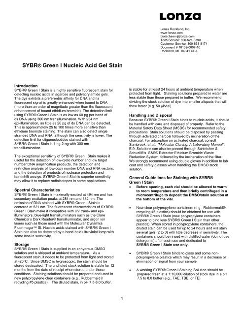
<strong>SYBR®</strong> <strong>Green</strong> I <strong>Nucleic</strong> <strong>Acid</strong> <strong>Gel</strong> <strong>Stain</strong><br />
Introduction<br />
<strong>SYBR®</strong> <strong>Green</strong> I <strong>Stain</strong> is a highly sensitive fluorescent stain for<br />
detecting nucleic acids in agarose and polyacrylamide gels.<br />
The dye exhibits a preferential affinity for DNA and its<br />
fluorescent signal is greatly enhanced when bound to DNA<br />
(more than an order of magnitude greater than the fluorescent<br />
enhancement of bound ethidium bromide). The detection limit<br />
using <strong>SYBR®</strong> <strong>Green</strong> I <strong>Stain</strong> is as low as 60 pg per band of<br />
ds DNA using 300 nm transillumination. With 254 nm<br />
epi-illumination, as little as 20 pg of ds DNA can be detected.<br />
This is approximately 25 to 100 times more sensitive than<br />
ethidium bromide staining. The stain can also detect single<br />
stranded DNA and RNA, although the sensitivity is lower. The<br />
detection limit for oligonucleotides stained with<br />
<strong>SYBR®</strong> <strong>Green</strong> I <strong>Stain</strong> is 1 ng-2 ng with 300 nm<br />
transillumination.<br />
The exceptional sensitivity of <strong>SYBR®</strong> <strong>Green</strong> I <strong>Stain</strong> makes it<br />
useful for the detection of low-cycle number and low target<br />
number DNA amplification products, the detection and<br />
restriction analysis of low-copy number DNA and RNA vectors,<br />
and the detection of products of nuclease protection and<br />
bandshift assays. <strong>SYBR®</strong> <strong>Green</strong> I <strong>Stain</strong>'s superior sensitivity<br />
may allow it to replace radioisotopes in some applications.<br />
Spectral Characteristics<br />
<strong>SYBR®</strong> <strong>Green</strong> I <strong>Stain</strong> is maximally excited at 494 nm and has<br />
secondary excitation peaks at 284 nm and 382 nm. The<br />
emission of DNA stained with <strong>SYBR®</strong> <strong>Green</strong> I <strong>Stain</strong> is<br />
centered at 521 nm. The fluorescent characteristics of <strong>SYBR®</strong><br />
<strong>Green</strong> I <strong>Stain</strong> make it compatible with UV trans- and epiilluminators,<br />
blue-light transilluminators such as the Clare<br />
Chemical’s Dark Reader® transilluminator, and argon ion<br />
lasers such as those used with the Molecular Dynamics<br />
FluorImager SI. <strong>Nucleic</strong> acids stained with <strong>SYBR®</strong> <strong>Green</strong> I<br />
<strong>Stain</strong> can also be detected by a hand-held ultraviolet lamp with<br />
some loss in sensitivity.<br />
Storage<br />
<strong>SYBR®</strong> <strong>Green</strong> I <strong>Stain</strong> is supplied in an anhydrous DMSO<br />
solution and is shipped at ambient temperature. As a<br />
fluorescent stain, it needs to be protected from light and stored<br />
at -20°C. Since DMSO is hygroscopic, the stain should be<br />
stored desiccated. The undiluted stock solution is stable for 12<br />
months from the date of receipt when stored under these<br />
conditions. <strong>Stain</strong>ing solutions should be prepared and used in<br />
new polypropylene clear containers (e.g., Rubbermaid®<br />
recycling #5 plastics). The diluted stain, in pH 7.5-8.0 buffer,<br />
1<br />
Lonza Rockland, Inc.<br />
www.lonza.com<br />
biotechserv@lonza.com<br />
Tech Service: 800-521-0390<br />
Customer Service: 800-638-8174<br />
Document # 18109-0807-10<br />
Rockland, ME 04841 USA<br />
is stable for at least 24 hours at ambient temperature when<br />
protected from light. <strong>Stain</strong>ing solutions prepared in water are<br />
less stable than those prepared in buffer. We recommend<br />
dividing the stock solution of dye into smaller aliquots that will<br />
thaw faster (e.g. 50 μl/vial).<br />
Handling and Disposal<br />
Because <strong>SYBR®</strong> <strong>Green</strong> I <strong>Stain</strong> binds to nucleic acids, it should<br />
be handled with care and disposed of properly. Refer to the<br />
Material Safety Data Sheet (MSDS) for recommended safety<br />
precautions. <strong>Stain</strong> solutions should be disposed by passing<br />
through activated charcoal followed by incineration of the<br />
charcoal. For adsorption on activated charcoal, consult<br />
Sambrook, et al., "Molecular Cloning: A Laboratory Manual”,<br />
E.9. Solutions can also be passed through Schleicher &<br />
Schuell®’s S&S® Extractor Ethidium Bromide Waste<br />
Reduction System, followed by the incineration of the filter.<br />
We strongly recommend using double gloves in addition to lab<br />
coat and safety glasses when handling the DMSO stock<br />
solution.<br />
General Guidelines for <strong>Stain</strong>ing with <strong>SYBR®</strong><br />
<strong>Green</strong> I <strong>Stain</strong><br />
• Before opening, each vial should be allowed to warm<br />
to room temperature and then briefly centrifuged in a<br />
microcentrifuge to deposit the DMSO/stain solution at<br />
the bottom of the vial.<br />
• New clear polypropylene containers (e.g., Rubbermaid®<br />
recycling #5 plastics) should be obtained for use with<br />
<strong>SYBR®</strong> <strong>Green</strong> I <strong>Stain</strong> (new polypropylene containers<br />
appear to bind less <strong>SYBR®</strong> <strong>Green</strong> I <strong>Stain</strong> than other<br />
plastics). When stored in polypropylene containers, the<br />
diluted stain can be used for up to 24 hours and will stain<br />
several gels (2 to 3) with little decrease in sensitivity. The<br />
containers should be rinsed with distilled water (do not use<br />
detergents) after each use and dedicated to<br />
<strong>SYBR®</strong> <strong>Green</strong> I <strong>Stain</strong> use only.<br />
• <strong>SYBR®</strong> <strong>Green</strong> I <strong>Stain</strong> binds to glass and some nonpolypropylene<br />
plastics which may result in a decrease or<br />
elimination of signal from your sample.<br />
• A working <strong>SYBR®</strong> <strong>Green</strong> I <strong>Stain</strong>ing Solution should be<br />
prepared fresh at a 1:10,000 dilution of stock dye in a pH<br />
7.5 to 8.0 buffer (e.g., TAE, TBE, or TE).
• Agarose gels should be cast no thicker than 4 mm. As gel<br />
thickness increases, diffusion of the dye into the gel is<br />
decreased lowering the detection of DNA.<br />
• For recording your results on film, we recommend using<br />
the <strong>SYBR®</strong> <strong>Green</strong> <strong>Gel</strong> <strong>Stain</strong> Photographic Filter (Catalog<br />
No. 50530). This filter is of deep yellow cellophane having an<br />
overall size of 3“ x 3” (75 mm x 75 mm). If this filter does not<br />
fit your camera, a Wratten® #15 or a Tiffen® #15 photographic<br />
filter of the appropriate diameter (purchased from the camera<br />
system manufacturer or a local photography shop) will work.<br />
The red/orange filter used to photograph ethidium bromide<br />
stained gels should not be used with gels stained with <strong>SYBR®</strong><br />
<strong>Green</strong> I <strong>Stain</strong>.<br />
<strong>Protocol</strong> for Post-staining <strong>Gel</strong>s<br />
Follow the steps below to stain DNA after electrophoresis<br />
1. Remove the concentrated stock solution of <strong>SYBR®</strong> <strong>Green</strong><br />
<strong>Stain</strong> from the freezer and allow the solution to thaw at<br />
room temperature.<br />
2. Spin the solution in a microcentrifuge to collect the dye at<br />
the bottom of the tube.<br />
3. Dilute the 10,000X concentrate to a 1X working solution<br />
(1 μl per 10 ml), in a pH 7.5-8.5 buffer, in a clear plastic<br />
polypropylene container. Prepare enough staining<br />
solution to just cover the top of the gel.<br />
4. Remove the gel from the electrophoresis chamber.<br />
5. Place the gel in staining solution.<br />
6. Gently agitate the gel at room temperature.<br />
7. <strong>Stain</strong> the gel for 15–30 minutes. The optimal staining time<br />
depends on the thickness of the gel, concentration of the<br />
agarose, and the fragment size to be detected. Longer<br />
staining times are required as gel thickness and agarose<br />
concentration increase.<br />
8. Remove the gel from the staining solution and view with a<br />
300 nm UV transilluminator, CCD camera or Clare<br />
Chemical’s Dark Reader® transilluminator. <strong>SYBR®</strong> <strong>Green</strong><br />
<strong>Stain</strong>ed <strong>Gel</strong>s do not require destaining. The dyes<br />
fluorescence yield is much greater when bound to DNA<br />
than when in solution.<br />
<strong>Stain</strong>ing vertical gels with <strong>SYBR®</strong> <strong>Green</strong> <strong>Stain</strong>s<br />
Incorporating <strong>SYBR®</strong> <strong>Green</strong> <strong>Stain</strong> into the gel or prestaining<br />
the DNA for use in a vertical format is not recommended. The<br />
dye binds to glass or plastic plates and DNA may show little to<br />
no signal. <strong>Gel</strong>s should be post-stained as described in the<br />
previous section.<br />
Follow this procedure when staining vertical gels<br />
with <strong>SYBR®</strong> <strong>Green</strong> <strong>Stain</strong><br />
1. Remove the concentrated stock solution of <strong>SYBR®</strong> <strong>Green</strong><br />
<strong>Stain</strong> from the freezer and allow the solution to thaw at<br />
room temperature.<br />
2. Spin the solution in a microcentrifuge to collect the dye at<br />
the bottom of the tube.<br />
3. Dilute the 10,000X concentrate to a 1X working solution, in<br />
a pH 7.5-8.5 buffer, in a clear plastic polypropylene<br />
container. Prepare enough staining solution to just cover<br />
the top of the gel.<br />
4. Remove the gel from the electrophoresis chamber.<br />
5. Open the cassette and leave the gel in place on one plate.<br />
6. Place the plate, gel side up in a staining container.<br />
7. Gently pour the stain over the surface of the gel.<br />
8. <strong>Stain</strong> the gel for 5-15 minutes.<br />
2<br />
9. View with a 300 nm UV transilluminator, CCD camera or<br />
Clare Chemical’s Dark Reader® transilluminator.<br />
<strong>SYBR®</strong> <strong>Green</strong> <strong>Stain</strong>ed <strong>Gel</strong>s do not require destaining.<br />
The dye’s fluorescence yield is much greater when bound<br />
to DNA than when in solution.<br />
<strong>Protocol</strong> for Adding Dye to Loading Buffer<br />
<strong>SYBR®</strong> <strong>Green</strong> I <strong>Stain</strong> can be added directly to the loading<br />
buffer at a final concentration of 1:1000. First prepare a 1:100<br />
dilution of <strong>SYBR®</strong> <strong>Green</strong> I <strong>Stain</strong> in high-quality anhydrous<br />
DMSO. The 1:100 dilution can be stored in the freezer and<br />
reused. Add 1 μl of this dilution to 9 μl-10 μl of your sample<br />
before loading.<br />
Photography<br />
<strong>Gel</strong>s stained with <strong>SYBR®</strong> <strong>Green</strong> I <strong>Stain</strong> exhibit negligible<br />
background fluorescence allowing long film exposures when<br />
detecting small amounts of DNA. When used with 300 nm<br />
transillumination and Polaroid® Type 57 Film, a 0.5-1 second<br />
exposure using an f-stop of 4.5 is adequate.<br />
In the photographs, the DNA will appear as bright bands<br />
against a gray gel. For Polaroid® Type 55 Film, a 15-45<br />
second exposure using an f-stop of 4.5 is adequate. When<br />
used with 254 nm epi-illumination (especially with a hand-held<br />
lamp), exposures of the order of 1-1.5 minutes may be<br />
required for maximal sensitivity. With 254 nm epi-illumination,<br />
the DNA will appear as bright bands against a black<br />
background. The <strong>SYBR®</strong> <strong>Green</strong> <strong>Stain</strong> Photographic Filter is<br />
recommended for all photography.<br />
Application Notes<br />
• <strong>SYBR®</strong> <strong>Green</strong> I <strong>Stain</strong> can be removed from doublestranded<br />
DNA by ethanol precipitation. Isopropanol<br />
precipitation is somewhat less effective at removing the<br />
dye; butanol extraction, chloroform extraction and phenol<br />
extraction do not remove the dye efficiently.<br />
• <strong>Gel</strong>s previously stained with ethidium bromide can<br />
subsequently be stained with <strong>SYBR®</strong> <strong>Green</strong> I <strong>Stain</strong><br />
following the standard protocol for post-staining. There will<br />
be some decrease in sensitivity when compared to a gel<br />
stained only with <strong>SYBR®</strong> <strong>Green</strong> I <strong>Stain</strong>.<br />
• Do not dilute the <strong>SYBR®</strong> <strong>Green</strong> I <strong>Stain</strong> stock solution in<br />
glass containers because <strong>SYBR®</strong> <strong>Green</strong> I <strong>Stain</strong> will bind<br />
to glass. Dilute the stock solution in a polypropylene<br />
container.<br />
• The inclusion of <strong>SYBR®</strong> <strong>Green</strong> I <strong>Stain</strong> in cesium chloride<br />
density gradient plasmid preparations is not<br />
recommended. The effect of the dye on the buoyant<br />
density of DNA is unknown.<br />
• <strong>SYBR®</strong> <strong>Green</strong> I <strong>Stain</strong> does not appear to interfere with<br />
enzymatic reactions.<br />
• We recommend the addition of 0.1% to 0.3% SDS in the<br />
prehybridization and hybridization solutions when<br />
performing Southern blots on gels stained with<br />
<strong>SYBR®</strong> <strong>Green</strong> I <strong>Stain</strong>.
• <strong>SYBR®</strong> <strong>Green</strong> I <strong>Stain</strong> bound to double-stranded DNA<br />
fluoresces green under UV transillumination. <strong>Gel</strong>s that<br />
contain DNA with single-stranded regions may fluoresce<br />
orange rather than green.<br />
• We do not recommend photographing gels with a 254 nm<br />
transilluminator. An outline of the UV light source can<br />
appear in photographs. A filter that will allow a 525 nm<br />
transmission and exclude other wavelengths (e.g., those<br />
in the infrared) is required.<br />
• <strong>SYBR®</strong> <strong>Green</strong> I <strong>Stain</strong> is not compatible with <strong>Gel</strong>Bond®<br />
Film or <strong>Gel</strong>Bond® PAG Film.<br />
Decontamination<br />
<strong>Stain</strong>ing solutions should be disposed of by passing through<br />
activated charcoal followed by incineration of the charcoal. For<br />
adsorption on activated charcoal, consult Sambrook, et al., pp.<br />
6.16 – 6.19, (1989). Solutions can also be passed through<br />
Schleicher & Schuell®’s S&S® Extractor Ethidium Bromide<br />
Waste Reduction System, followed by the incineration of the<br />
filter.<br />
References<br />
Schneeberger, C., et al. 1995. PCR Methods & App.<br />
4:234-238<br />
Ordering Information<br />
Catalog No. Description Size<br />
50512 <strong>SYBR®</strong> <strong>Green</strong> I <strong>Nucleic</strong> 2 x 500 μl<br />
<strong>Acid</strong> <strong>Gel</strong> <strong>Stain</strong><br />
50513 <strong>SYBR®</strong> <strong>Green</strong> I <strong>Nucleic</strong> 10 x 50 μl<br />
<strong>Acid</strong> <strong>Gel</strong> <strong>Stain</strong><br />
50530 <strong>SYBR®</strong> <strong>Green</strong> <strong>Gel</strong> <strong>Stain</strong> each<br />
Photographic Filer<br />
3” square (Wratten® #15)<br />
Related Products<br />
Agarose<br />
MDE® <strong>Gel</strong> Solution<br />
AccuGENE® Electrophoresis Buffers<br />
For More information contact Technical Service at<br />
(800) 521-0390 or visit our website at www.Lonza.com.<br />
Manufactured for Lonza Rockland, Inc. by Molecular Probes, Inc.<br />
Sold For Research Use Only.<br />
3<br />
<strong>Gel</strong>Bond is a trademark of FMC Corporation, SYBR is a trademark of<br />
Molecular Probes, Inc., FluorImager is a trademark of Molecular<br />
Dynamics, Inc., Schleicher & Schuell and S&S, are registered<br />
trademarks of Schleicher & Schuell, G.m.b.H. LTD, Dark Reader is a<br />
trademark of Clare Chemical Research, Inc., Polaroid is a trademark of<br />
Polaroid Corp., Rubbermaid is a trademark of Rubbermaid Prod.,<br />
Tiffen is a trademark of Tiffen Optical Co. Wratten is a trademark of<br />
Eastman Kodak Co. All other trademarks herein are marks of the<br />
Lonza Group or its affilites. SYBR <strong>Green</strong> <strong>Stain</strong> is covered by U.S.<br />
Patents 5,436,134 and 5,658,751.<br />
© 2007 Lonza Rockland, Inc.<br />
All rights reserved.


