Bioidentical Hormones - U.S. Senate Special Committee on Aging
Bioidentical Hormones - U.S. Senate Special Committee on Aging
Bioidentical Hormones - U.S. Senate Special Committee on Aging

You also want an ePaper? Increase the reach of your titles
YUMPU automatically turns print PDFs into web optimized ePapers that Google loves.
123<br />
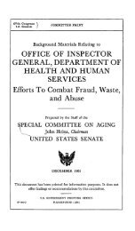
These are abstracts of journal science published in molecular<br />
biology <strong>on</strong> the mechanisms of fetal <strong>on</strong>cogenes and compounded<br />
horm<strong>on</strong>es by T.S. Wiley.<br />
1'3m.,r ,*-.. ..<br />
>: ~ a - ,, , j S -<br />
.. ~~~*1~n C.._ tab_ Sd 199 No-e;86:.60 .Li_ _n _ .-<br />
Progester<strong>on</strong>e Inhibits growth and Induces apoptosis in breast cancer cells: Inverse<br />
effects <strong>on</strong> Bci-2 and p53.<br />
* Formby B,<br />
* Wiley TS.,.<br />
Sansum Medical Research Institute, Santa Barbara, CA 93105, USA.<br />
Progester<strong>on</strong>e inhibits the proliferati<strong>on</strong> of normal breast epithelial cells In vivo, as<br />
well as breast cancer cells in vitro. But the biologic mechanism of this Inhibiti<strong>on</strong><br />
remains to be determined. We explored the possibility that an antiproliferative<br />
activity of progester<strong>on</strong>e In breast cancer cell lines Is due to its ability to Induce<br />
apoptosis. Since p53 and bd-2 genetically c<strong>on</strong>trol the apoptotic process, we<br />
investigated whether or not these genes could be involved in the progester<strong>on</strong>e-<br />
Induced apoptosis. We found a maximal 90 percent Inhibiti<strong>on</strong> of cell proliferati<strong>on</strong><br />
with T47-D breast cancer cells after exposure to 10 microM progester<strong>on</strong>e for 72<br />
hours. C<strong>on</strong>trol progester<strong>on</strong>e receptor negative MDA-231 cancer cells were<br />
unresp<strong>on</strong>sive to these two c<strong>on</strong>centrati<strong>on</strong>s of progester<strong>on</strong>e. After 24 hours of<br />
exposure to 10 mlcrM progester<strong>on</strong>e, cytufluorometric anaiysis of l-4/- breast<br />
cancer cells dem<strong>on</strong>strated 43 percent had underg<strong>on</strong>e apoptosis without signs of<br />
necrosis. After 72 hours of exposure to 10 microM progester<strong>on</strong>e, 48 percent of<br />
the cells had underg<strong>on</strong>e apoptosis and 40percent dem<strong>on</strong>strated "leaky'<br />
membranes. Untreated cancer cells did not undergo apoptosis. Evidence proving<br />
apoptosis was also dem<strong>on</strong>strated by fragmentati<strong>on</strong> of nudear DNA Into multiples<br />
of olig<strong>on</strong>ucleosomal fragments. After 24 hours of exposure to either 1 microM or<br />
10 mIcroM progester<strong>on</strong>e, the expressi<strong>on</strong> by T47-D cancer cells of bcl-2 was<br />
down-regulated, and that of p53 was up-regulated as detected by<br />
semiquantitative RT-PCR analysis. These results dem<strong>on</strong>strate that progester<strong>on</strong>e<br />
at a c<strong>on</strong>centrati<strong>on</strong> similar to that seen during the third trimester of pregnancy<br />
exhibited a str<strong>on</strong>g antiproliferative effect <strong>on</strong> at least two breast cancer cell lines.<br />
Apoptosis was induced In the progester<strong>on</strong>e receptor expressing T47-D breast<br />
cancer cells.<br />
PMID: 9846203 [PubMed - Indexed for MEDUNE]