A guide to the deep-water sponges of - NMFS Scientific Publications ...
A guide to the deep-water sponges of - NMFS Scientific Publications ...
A guide to the deep-water sponges of - NMFS Scientific Publications ...
- No tags were found...

You also want an ePaper? Increase the reach of your titles
YUMPU automatically turns print PDFs into web optimized ePapers that Google loves.
50 Pr<strong>of</strong>essional Paper <strong>NMFS</strong> 12<br />
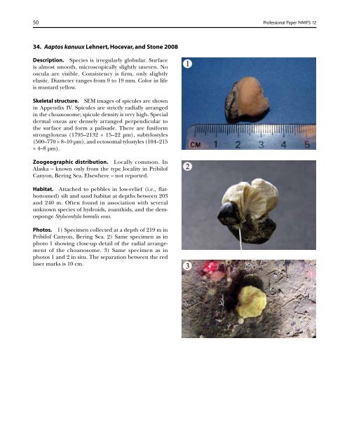
34. Aap<strong>to</strong>s kanuux Lehnert, Hocevar, and S<strong>to</strong>ne 2008<br />
Description. Species is irregularly globular. Surface<br />
is almost smooth, microscopically slightly uneven. No<br />
oscula are visible. Consistency is firm, only slightly<br />
elastic. Diameter ranges from 9 <strong>to</strong> 19 mm. Color in life<br />
is mustard yellow.<br />
Skeletal structure. SEM images <strong>of</strong> spicules are shown<br />
in Appendix IV. Spicules are strictly radially arranged<br />
in <strong>the</strong> choanosome; spicule density is very high. Special<br />
dermal oxeas are densely arranged perpendicular <strong>to</strong><br />
<strong>the</strong> surface and form a palisade. There are fusiform<br />
strongyloxeas (1795–2132 × 15–22 µm), subtylostyles<br />
(500–770 × 8–10 µm), and ec<strong>to</strong>somal tylostyles (104–215<br />
× 4–8 µm).<br />
Zoogeographic distribution. Locally common. In<br />
Alaska – known only from <strong>the</strong> type locality in Pribil<strong>of</strong><br />
Canyon, Bering Sea. Elsewhere – not reported.<br />
Habitat. Attached <strong>to</strong> pebbles in low-relief (i.e., flatbot<strong>to</strong>med)<br />
silt and sand habitat at depths between 203<br />
and 240 m. Often found in association with several<br />
unknown species <strong>of</strong> hydroids, zoanthids, and <strong>the</strong> demosponge<br />
Stylocordyla borealis eous.<br />
Pho<strong>to</strong>s. 1) Specimen collected at a depth <strong>of</strong> 219 m in<br />
Pribil<strong>of</strong> Canyon, Bering Sea. 2) Same specimen as in<br />
pho<strong>to</strong> 1 showing close-up detail <strong>of</strong> <strong>the</strong> radial arrangement<br />
<strong>of</strong> <strong>the</strong> choanosome. 3) Same specimen as in<br />
pho<strong>to</strong>s 1 and 2 in situ. The separation between <strong>the</strong> red<br />
laser marks is 10 cm.