A guide to the deep-water sponges of - NMFS Scientific Publications ...
A guide to the deep-water sponges of - NMFS Scientific Publications ...
A guide to the deep-water sponges of - NMFS Scientific Publications ...
- No tags were found...

You also want an ePaper? Increase the reach of your titles
YUMPU automatically turns print PDFs into web optimized ePapers that Google loves.
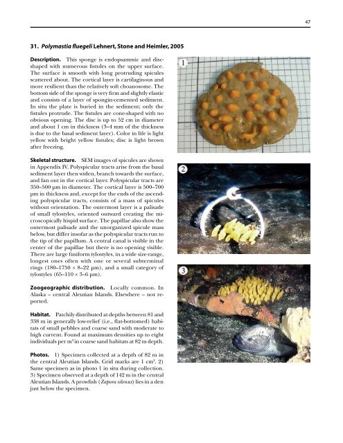
31. Polymastia fluegeli Lehnert, S<strong>to</strong>ne and Heimler, 2005<br />
Description. This sponge is endopsammic and discshaped<br />
with numerous fistules on <strong>the</strong> upper surface.<br />
The surface is smooth with long protruding spicules<br />
scattered about. The cortical layer is cartilaginous and<br />
more resilient than <strong>the</strong> relatively s<strong>of</strong>t choanosome. The<br />
bot<strong>to</strong>m side <strong>of</strong> <strong>the</strong> sponge is very firm and slightly elastic<br />
and consists <strong>of</strong> a layer <strong>of</strong> spongin-cemented sediment.<br />
In situ <strong>the</strong> plate is buried in <strong>the</strong> sediment; only <strong>the</strong><br />
fistules protrude. The fistules are cone-shaped with no<br />
obvious opening. The disc is up <strong>to</strong> 52 cm in diameter<br />
and about 1 cm in thickness (3–4 mm <strong>of</strong> <strong>the</strong> thickness<br />
is due <strong>to</strong> <strong>the</strong> basal sediment layer). Color in life is light<br />
yellow with bright yellow fistules; disc is light brown<br />
after freezing.<br />
Skeletal structure. SEM images <strong>of</strong> spicules are shown<br />
in Appendix IV. Polyspicular tracts arise from <strong>the</strong> basal<br />
sediment layer <strong>the</strong>n widen, branch <strong>to</strong>wards <strong>the</strong> surface,<br />
and fan out in <strong>the</strong> cortical layer. Polyspicular tracts are<br />
350–500 µm in diameter. The cortical layer is 500–700<br />
µm in thickness and, except for <strong>the</strong> ends <strong>of</strong> <strong>the</strong> ascending<br />
polyspicular tracts, consists <strong>of</strong> a mass <strong>of</strong> spicules<br />
without orientation. The outermost layer is a palisade<br />
<strong>of</strong> small tylostyles, oriented outward creating <strong>the</strong> microscopically<br />
hispid surface. The papillae also show <strong>the</strong><br />
outermost palisade and <strong>the</strong> unorganized spicule mass<br />
below, but differ ins<strong>of</strong>ar as <strong>the</strong> polyspicular tracts run <strong>to</strong><br />
<strong>the</strong> tip <strong>of</strong> <strong>the</strong> papillum. A central canal is visible in <strong>the</strong><br />
center <strong>of</strong> <strong>the</strong> papillae but <strong>the</strong>re is no opening visible.<br />
There are large fusiform tylostyles, in a wide size-range,<br />
longest ones <strong>of</strong>ten with one or several subterminal<br />
rings (180–1750 × 8–22 µm), and a small category <strong>of</strong><br />
tylostyles (65–110 × 3–6 µm).<br />
Zoogeographic distribution. Locally common. In<br />
Alaska – central Aleutian Islands. Elsewhere – not reported.<br />
Habitat. Patchily distributed at depths between 81 and<br />
338 m in generally low-relief (i.e., flat-bot<strong>to</strong>med) habitats<br />
<strong>of</strong> small pebbles and coarse sand with moderate <strong>to</strong><br />
high current. Found at maximum densities up <strong>to</strong> eight<br />
individuals per m 2 in coarse sand habitats at 82 m depth.<br />
Pho<strong>to</strong>s. 1) Specimen collected at a depth <strong>of</strong> 82 m in<br />
<strong>the</strong> central Aleutian Islands. Grid marks are 1 cm 2 . 2)<br />
Same specimen as in pho<strong>to</strong> 1 in situ during collection.<br />
3) Specimen observed at a depth <strong>of</strong> 142 m in <strong>the</strong> central<br />
Aleutian Islands. A prowfish (Zapora silenus) lies in a den<br />
just below <strong>the</strong> specimen.<br />
47