

W. Richard Bowen and Nidal Hilal 4
W. Richard Bowen and Nidal Hilal 4
W. Richard Bowen and Nidal Hilal 4
- No tags were found...

You also want an ePaper? Increase the reach of your titles
YUMPU automatically turns print PDFs into web optimized ePapers that Google loves.
198 7. MICRO/NANOENgINEERINg ANd AFM FOR CELLULAR SENSINg<br />
mechanical stress across the plasma membrane <strong>and</strong> convey the traction<br />
force that develops in the cytoskeleton to the ECM. Unbound integrins<br />
are mobile within the cell membrane <strong>and</strong> readily form clusters <strong>and</strong> focal<br />
adhesions in a tension-dependent manner [11]. Integrins also participate<br />
in other signalling transductions that regulate cell growth [12].<br />
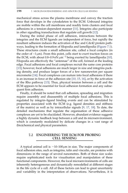
During the initial phase of cell adhesion, interactions between the<br />
integrins <strong>and</strong> the ECM lig<strong>and</strong>s are independent of force, but rapidly the<br />
resultant adhesion induces the activation of Rac <strong>and</strong> Cdc42 protein pathways,<br />
leading to the formation of filopodia <strong>and</strong> lamellipodia (Figure 7.1).<br />
These structures create a small adhesion site, called a focal complex (in<br />
the order of ~1 �m). From this point, cells start to exert traction forces on<br />
the ECM, with about 0.8–0.9 nN �m �2 being exerted by lamellipodia [10].<br />
Filopodia are effectively the “antennae” of the cell, formed at the leading<br />
edge. Focal adhesions <strong>and</strong> focal complexes recruit the same core proteins<br />
[13]; however, focal adhesions are much larger in size <strong>and</strong> integrins packing<br />
density, <strong>and</strong> produce larger forces of a few nanonewton per square<br />
micrometre [14]. Focal complexes can mature into focal adhesions if there<br />
is an increase in force at the adhesion site [10, 15, 16], or by the activation<br />
of the Rho pathway [13]. Thus, physical tension between a cell <strong>and</strong> the<br />
ECM appears to be essential for focal adhesion formation <strong>and</strong> any subsequent<br />
firm adhesion.<br />
Finally, it should be noted that cell adhesion, spreading <strong>and</strong> migration<br />
require assembly <strong>and</strong> disassembly of multiple focal adhesions. This is<br />
regulated by integrin–lig<strong>and</strong> binding events <strong>and</strong> can be stimulated by<br />
properties associated with the ECM (e.g. lig<strong>and</strong> densities <strong>and</strong> stiffness<br />
of the matrix) as well as by intracellular signals [9, 17, 18]. To date, the<br />
detailed mechanisms that regulate the organisation of these adhesive<br />
complexes are yet to be elucidated. However, abundant evidence suggests<br />
a highly dynamic feedback loop between a cell <strong>and</strong> its microenvironment,<br />
which is constantly modulated by delicate changes in a vast range of<br />
(bio)chemical <strong>and</strong> physical parameters.<br />
7.2 EngInEErIng tHE ECM For ProbIng<br />
CEll SEnSIng<br />
A typical animal cell is �10–100 �m in size. The major components of<br />
focal adhesion sites, such as integrins, talin <strong>and</strong> vinculin, are proteins with<br />
dimensions in the range of several nanometres. Both of these size scales<br />
require sophisticated tools for visualisation <strong>and</strong> manipulation of these<br />
functional components. However, the local microenvironments of cells are<br />
inherently heterogeneous <strong>and</strong> dynamically remodelled at different stages<br />
in the life cycle of a cell. All of these factors can lead to great uncertainty<br />
<strong>and</strong> variability in the interpretation of observations. Nevertheless, it is


