

89-91 - Polskie Stowarzyszenie Biomateriałów
89-91 - Polskie Stowarzyszenie Biomateriałów
89-91 - Polskie Stowarzyszenie Biomateriałów

Create successful ePaper yourself
Turn your PDF publications into a flip-book with our unique Google optimized e-Paper software.
influence of surface<br />
(nano)roughness on cell<br />
behaviour<br />
agnieszKa Piegat, Mirosława el fray*<br />
weSt PoMeranian univerSity of technoloGy in Szczecin,<br />
PolyMer inStitute,<br />
diviSion of bioMaterialS and MicrobioloGical technoloGieS,<br />
10 PulaSki Syt., 70-322 Szczecin, Poland<br />
Mailtol: Mirfray@zut.edu.Pl<br />
abstract<br />
The influence of surface (nano)roughness of soft<br />
elastomeric materials containing nanocrystalline TiO2<br />
on tissue cell behaviour after implantation tests was<br />
investigated. Addition of small amount (0.2wt.%) of<br />
TiO 2 into polymer matrix changed surface roughness<br />
giving material of well developed lamellar morphology<br />
and slightly diminished contact angle (wettability). This<br />
lamellar (fibrilar) morphology had strong influence on<br />
cell response after implantation into soft tissue indicating<br />
the absence of eosynophiles in tissue around<br />
implant.<br />
key words: nanocomposites; AFM, cantact angle,<br />
cell/tissue response<br />
[Engineering of Biomaterials, <strong>89</strong>-<strong>91</strong>, (2009), 257-258]<br />
introduction<br />
Biomaterial characterization in term of its biofunctionality<br />
must include such properties as mechanical stability,<br />
chemical structure, morphology, degradation profile and<br />
chemical character of the surface [1]. All these parameters<br />
and their interactions with biological system decide about<br />
the functionality and possibility of such materials to be used<br />
in specific applications. From the biological point of view,<br />
surface properties such as hydrophobicity/hydrophilicity,<br />
wettability, roughness and topography plays crucial role in<br />
cells and tissues response [2].<br />
Information about surface properties can be obtained using<br />
microscopic, spectroscopic or thermodynamic methods,<br />
depending from the requirements [3]. The goal of this study<br />
was to characterize the surface properties of polymeric<br />
nanocomposite films. Their contact angles, roughness and<br />
morphology and has been measured and analyzed with<br />
respect to cellular response after implantation test.<br />
experimental<br />
materials<br />
PET/DLA (poly(ethylene terephthalate)/dilinoleic acid)<br />
multiblock copolymers was prepared as the neat material<br />
at hard/soft segments weight ratio as 30/70wt%. Then, by<br />
adding 0.2wt% nanocrystalline TiO 2 during the synthesis<br />
step, PET/DLA-based nanocomposite was prepared as<br />
described in the authors’ previous work [4]. Thin films (60-<br />
160nm thickness) were obtained from polymers solution<br />
in chloroform by spin-coating on glass substrates (cover<br />
glasses φ=18mm).<br />
morphology<br />
AFM measurements were performed on the Nanoscope<br />
IV A (Veeco/Digital Instruments) AFM in tapping mode. The<br />
AFM was equipped with a dimension scanner - maximum<br />
scan size of 150x150µm.<br />
contact angle<br />
Contact angles measurements were carried out on spincoated<br />
samples using drop technique on a DataPhysisc,<br />
Contact Angle System OCA. During each measurement on<br />
the instrument, 15 points were collected from each polymer<br />
(three samples from each composition at 5 points). The contact<br />
angle was measured with ultra pure distilled water.<br />
implantation test<br />
Implantation test was performed according procedure<br />
described in details in [5]. Briefly, the PET/DLA and PET/<br />
DLA–0.2%TiO 2 used were small polymer rods, 10 to 12mm<br />
long, 3 to 5mm wide and 0.6 to 0.8mm thick. Polymer rods<br />
were implanted into the muscles of 30 Wistar rats weighing<br />
200–220g. Animal observations were performed for<br />
12 weeks and sacrificed with sodium pentobarbital in the<br />
amount 200mg (kg b.w.). The structure of histological slides<br />
was analyzed following the preparation of tissues with implanted<br />
polymers.<br />
results and discussion<br />
Addition of nanoparticles into polymer matrix is a simple<br />
method for modification of surface as well as bulk properties<br />
of polymeric materials. Depending from the particles<br />
character, they can act as osteoinductive or antimicrobial<br />
agents, or controlled drug delivery systems [6-8]. Song et<br />
al. [9] showed that new type of nanocomposite material,<br />
prepared by blending TiO 2 nanoparticles with PNIPAM-co-<br />
PS electrospinned fibers may find some new applications in<br />
field of bioanalysis or as directed drug carriers. El Fray and<br />
Piegat [4,10] showed that addition of small amount of TiO 2<br />
into thermoplastic elastomer matrix is easy way to control<br />
mechanical behavior and susceptibility to degradation of<br />
this type of materials.<br />
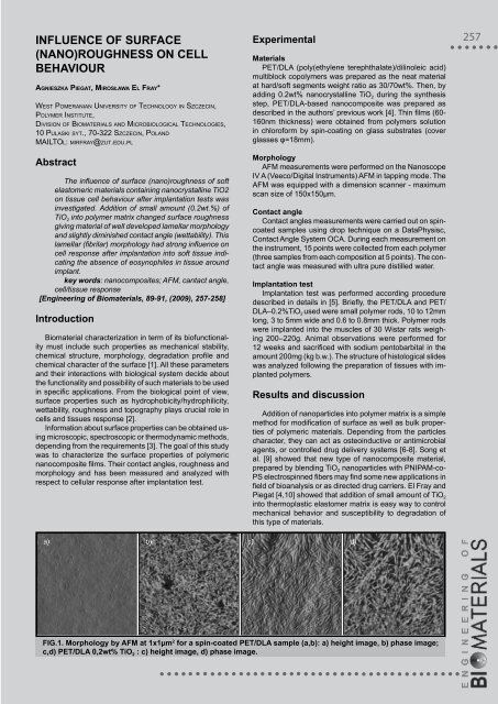
fig.1. morphology by afm at 1x1µm 2 for a spin-coated pet/dla sample (a,b): a) height image, b) phase image;<br />
c,d) pet/dla 0,2wt% tio 2 : c) height image, d) phase image.<br />
257


