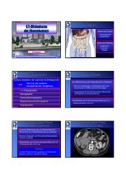
MDCT der Nasennebenhöhlen - Institut für Diagnostische und ...
MDCT der Nasennebenhöhlen - Institut für Diagnostische und ...
MDCT der Nasennebenhöhlen - Institut für Diagnostische und ...

Sie wollen auch ein ePaper? Erhöhen Sie die Reichweite Ihrer Titel.
YUMPU macht aus Druck-PDFs automatisch weboptimierte ePaper, die Google liebt.
Processus uncinatus<br />
Processus uncinatus<br />
• Hakenförmiger Auswuchs,<br />
Überbleibsel des unteren Teils des<br />
„first ethmoturbinal“<br />
• Variierende Anheftungen<br />
bestimmmen die Drainagemuster<br />
des Recessus<br />
frontalis<br />
<strong>Institut</strong> <strong>für</strong> <strong>Diagnostische</strong> <strong>und</strong> Interventionelle Radiologie, J.W. Goethe-Universität, Frankfurt<br />
Varianten: Concha bullosa, Haller Zelle<br />
Varianten: Concha bullosa, Haller Zelle<br />
� Primär nicht pathologisch – können aber<br />
Erkrankungen verursachen<br />
Concha bullosa<br />
Haller Zelle<br />
Inf<strong>und</strong>ibulum maxillare<br />
Sinus maxillaris<br />
<strong>Institut</strong> <strong>für</strong> <strong>Diagnostische</strong> <strong>und</strong> Interventionelle Radiologie, J.W. Goethe-Universität, Frankfurt<br />
„Low Ethmoid Roof“<br />
„Low Ethmoid Roof“<br />
• Falls nicht bekannt: Risikofaktor <strong>für</strong> intrakranielle Verletzungen<br />
bei FESS, spez. Ethmoidektomie<br />
• Keros Klassifikation:<br />
Typ I: Fossa olfactoria ist 1 - 3 mm tief (laterale Lamelle ist<br />
praktisch nicht vorhanden)<br />
Typ II: Fossa olfactoria ist 4 - 7 mm tief<br />
Typ III: Fossa olfactoria ist 8 - 16 mm tief<br />
<strong>Institut</strong> <strong>für</strong> <strong>Diagnostische</strong> <strong>und</strong> Interventionelle Radiologie, J.W. Goethe-Universität, Frankfurt<br />
Funktionale Anatomie:<br />
Ostiomeataler Komplex (OMC)<br />
Inf<strong>und</strong>ibulum ethmoidale:<br />
• Medial:<br />
Proc. uncinatus<br />
• Lateral:<br />
Lamina papyracea<br />
• Kranial:<br />
Boden <strong>der</strong> Bulla<br />
ethmoidalis<br />
<strong>Institut</strong> <strong>für</strong> <strong>Diagnostische</strong> <strong>und</strong> Interventionelle Radiologie, J.W. Goethe-Universität, Frankfurt<br />
Onodi Zelle<br />
Onodi Zelle<br />
• Seitliche <strong>und</strong> hintere Ausbuchtungen <strong>der</strong><br />
posterioren Ethmoidalzellen, weiten sich aus durch<br />
den Apex orbitalis <strong>und</strong> sind dem N. opticus<br />
benachbart: prädisponierte Verletzung bei FESS<br />
• In <strong>der</strong> axialen Ebene am besten darstellbar<br />
<strong>Institut</strong> <strong>für</strong> <strong>Diagnostische</strong> <strong>und</strong> Interventionelle Radiologie, J.W. Goethe-Universität, Frankfurt<br />
Klinische NNH-Diagnostik<br />
Schmerz, Blutung, Affektion<br />
MRT, CT Tumorverdacht: Obstruierte NNH<br />
Entzündung<br />
Therapie<br />
T1 - hypointens<br />
T2 - hyperintens<br />
MRT nativ<br />
T1 - hypointens<br />
T2 - mittleres Signal<br />
KM-MRT<br />
Lineare KM-Aufnahme Solide KM-Aufnahme<br />
Biopsie<br />
<strong>Institut</strong> <strong>für</strong> <strong>Diagnostische</strong> <strong>und</strong> Interventionelle Radiologie, J.W. Goethe-Universität, Frankfurt