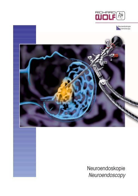
Neuroendoskopie Neuroendoscopy - Richard Wolf
Neuroendoskopie Neuroendoscopy - Richard Wolf
Neuroendoskopie Neuroendoscopy - Richard Wolf

Sie wollen auch ein ePaper? Erhöhen Sie die Reichweite Ihrer Titel.
YUMPU macht aus Druck-PDFs automatisch weboptimierte ePaper, die Google liebt.
<strong>Neuroendoskopie</strong><br />
<strong>Neuroendoscopy</strong><br />
<strong>Neuroendoskopie</strong><br />
<strong>Neuroendoscopy</strong>
<strong>Neuroendoskopie</strong><br />
<strong>Neuroendoscopy</strong><br />
Kinder-<strong>Neuroendoskopie</strong>-System<br />
Pediatric Neuroendoscope System<br />
nach Hopf / by Hopf
0<br />
1<br />
2<br />
3<br />
4<br />
5<br />
6<br />
7<br />
8<br />
9<br />
10<br />
11<br />
12<br />
13<br />
14<br />
15<br />
16<br />
Pediatric<br />
Neuroendoscope System<br />
by Hopf<br />
Medical Introduction<br />
Telescopes<br />
Sheaths, Articulated Arm<br />
Forceps, Scissors, Punches, Rongeurs<br />
Electrodes<br />
Bipolar Generator, HF Connecting Cables<br />
III.02<br />
Kinder-<br />
<strong>Neuroendoskopie</strong>-System<br />
nach Hopf<br />
Medizinische Einführung<br />
Optiken<br />
Schäfte, Gelenkarm<br />
Zangen, Scheren, Stanzen, Rongeure<br />
Elektroden<br />
Bipolar-Generator, HF-Anschlusskabel<br />
C 660.0<br />
<strong>Neuroendoskopie</strong><br />
<strong>Neuroendoscopy</strong>
Pediatric<br />
Neuroendoscope System<br />
by Hopf*<br />
Endoscopy has revolutionized<br />
neurosurgical treatment modalities<br />
in many ways. Today, many<br />
cases of hydrocephalus, cystic<br />
lesions, and other intraventricular<br />
pathologies are approached<br />
endoscopically.<br />
Therapeutic strategies are:<br />
❏ detection of pathologies that<br />
cannot be demonstrated by<br />
radiographic methods<br />
❏ restoration of free CSF pathways in hydrocephalus<br />
❏ resection or fenestration of cysts or other isolated compartments<br />
❏ resection or biopsy of intraventricular tumors<br />
❏ removal of blood clots<br />
❏ placement or elemination of ventricular catheters<br />
Requirements for modern endoscopes suitable for Neurosurgery are brilliant<br />
visualization and illumination, high-quality equipment for intricate<br />
surgical manipulations, small outer diameter of the endoscope, and an<br />
ergonomic and convenient way of handling the endoscope and the<br />
instruments in order to compete with minimally invasive microneurosurgery.<br />
Advantages of the Pediatric<br />
Neuroendoscope System<br />
The ''Pediatric Endoscope system''<br />
is highly adaptable and therefore<br />
suitable for a variety of surgical<br />
manipulations. In addition<br />
to its extraordinary small outer<br />
diameter and the convenient working<br />
length, it consists also of a<br />
simple but secure and reliable<br />
fixation device and a sophisticated<br />
insert for steering instruments<br />
around the corner. These characteristics make the Pediatric Endoscope<br />
system especially valuable for intraventricular endoscopic procedures in<br />
newborn and children, as well as in adults.<br />
Endoskopischer Blick auf den eröffneten Boden<br />
Endoscopic view of the fenestrated floor<br />
❏ small diameter - 4.4 x 3.3 mm or 6.1 x 4.8 mm<br />
❏ telescopes with direction of view 5°, 30° or 70°<br />
❏ different sheaths and working elements<br />
❏ insert for steering instruments around the corner<br />
❏ fixation device<br />
❏ variety of auxiliary instruments<br />
* Department of Neurosurgery - University of Mainz, Germany<br />
III.02<br />
Endoskopischer Blick auf das Foramen Monro<br />
Endoscopic view of foramen of Monro<br />
Kinder-<br />
<strong>Neuroendoskopie</strong>-System<br />
nach Hopf*<br />
Die Endoskopie hat die neurochirugischen Operationstechniken auf verschiedenartige<br />
Weise revolutioniert. Operationen zur Behandlung des<br />
Hydrocephalus, Cysten und anderen intraventrikulären Läsionen werden<br />
heutzutage immer häufiger endoskopisch durchgeführt.<br />
Therapeutische Strategien beinhalten:<br />
❏ Erkennung von radiologisch nicht darstellbaren Pathologien<br />
❏ Wiederherstellung der Liquorpassage beim Occlusionshydrocephalus<br />
❏ Resektion und Fenestration von Cysten oder kompartimentierten<br />
Ventrikelanteilen<br />
❏ Resektion und Biopsie intraventrikulärer Tumore<br />
❏ Extraktion von Blutkoageln<br />
❏ Platzierung und Entfernung von Ventrikelkathetern<br />
Endoskopischer Blick auf den Boden des dritten<br />
Ventrikels<br />
Endoscopic view of the floor of the third ventricle<br />
Anforderungen an moderne Endoskope<br />
für neurochirurgische Eingriffe<br />
sind brillante Bildqualität<br />
und Ausleuchtung, hochwertiges<br />
Instrumentarium für chirurgische<br />
Manipulationen, geringer Aussendurchmesser<br />
der Endoskope und<br />
ein ergonomisches und sicheres<br />
Handling, um dem Vergleich mit<br />
der minimal invasiven Mikroneurochirurgie<br />
standhalten zu können.<br />
Vorteile des Kinder-Neuroendoskop-Systems<br />
Das Kinder-Neuroendoskop-System bietet Einsatzmöglichkeiten für eine<br />
Vielzahl neurochirurgischer Operationen, da eine individuelle Gestaltung<br />
des Endoskops für die jeweilige Operation aufgrund verschiedener austauschbarer<br />
Komponenten ermöglicht wird. Neben dem ausserordentlich<br />
geringen Aussendurchmesser und der optimierten Arbeitslänge sind<br />
die einfache und sichere Fixierung sowie ein Einsatz zum Arbeiten "um<br />
die Ecke" weitere neue Merkmale. Diese Eigenschaften machen das<br />
Endoskopsystem im Einsatz gerade bei Neugeborenen und Kindern<br />
besonders wertvoll, es ist aber auch für den Einsatz bei Erwachsenen<br />
ideal.<br />
❏ kleiner Durchmesser der Schäfte von 4,4 x 3,3 mm oder 6,1 x 4,8 mm<br />
❏ Optiken mit 5°, 30° und 70° Blickrichtung<br />
❏ Einsätze für 1 x 6 Charr. oder 2 x 5 Charr. Instrumente<br />
❏ Steuereinsatz für das "Arbeiten um die Ecke"<br />
❏ Befestigungsadapter für Leila-Halterungssysteme<br />
❏ Vielzahl an speziellen endoskopischen Instrumenten<br />
* Department of Neurosurgery - University of Mainz, Germany<br />
C 660.001<br />
<strong>Neuroendoskopie</strong><br />
<strong>Neuroendoscopy</strong><br />
0
0<br />
Pediatric<br />
Neuroendoscope System<br />
Indications for Endoscopic Procedures<br />
Hydrocephalus<br />
Hydrocephalus<br />
The most frequently performed endosopic procedure is ''endoscopic Der häufigste endoskopische Eingriff ist die "endoskopische<br />
third ventriculostomy'' in patients with occlusive hydrocephalus. Here, Ventrikulostomie" bei Patienten mit occlusivem Hydrocephalus. Die<br />
a communication between the third ventricle<br />
Schaffung einer Öffnung im Boden des dritten<br />
and the prepontine cisterne through the floor<br />
Ventrikels zur präpontinen Zisterne ermög-<br />
of the third ventricle re-establishes physiololicht<br />
eine physiologische Liquor-Dynamik<br />
gical CSF pressure dynamics and enables a<br />
und ein Shunt-freies Leben für die Patienten<br />
shunt-free life for the patient (figure 1). An<br />
(Abb. 1). Der asymetrische Hydrocephalus<br />
additional indication for endoscopic surgery<br />
verursacht durch eine unilaterale Obstruktion<br />
is asymmetric hydrocephalus, caused by<br />
des Foramen Monro ist eine weitere<br />
unilateral obstruction of the foramen of<br />
Indikation für eine endoskopische Behand-<br />
Monro. A fenestration of the septum pellucilung.<br />
Die Fenestration des Septum pellucidum,<br />
the so-called "endoscopic pellucidodum,<br />
die sog. "endoskopische Pellucidototomy"<br />
restores a communication between<br />
mie" schafft eine Kommunikation beider<br />
both lateral ventricles and avoids shunting<br />
Seitenventrikel und kann so einen Shunt<br />
or, in case of impared CSF reabsorbtion,<br />
überflüssig machen oder im Falle einer redu-<br />
avoids a second shunt system. Furthermore, Abb. 1: Endoskopie des dritten Ventrikels<br />
zierten Liquorresorption zumindest die<br />
"endoscopic shunt placement" and endos- Fig. 1: Endoscopic third ventriculostomy<br />
Implantation eines zweiten Shuntsystems<br />
copic removal of adherent intracranial shunt<br />
vermeiden. Die endoskopische Shuntanlage<br />
catheters facilitates shunting procedures to a<br />
und die endoskopische Entfernung adhären-<br />
great degree and improves long-term function of the shunt system.<br />
ter Ventrikelkatheter erleichtert Shunt-Operationen wesentlich und verbessert<br />
die Langzeitfunktion der Shuntsysteme.<br />
Cystic Lesions<br />
Cysten<br />
Indications for endoscopic surgery in intracranial cystic lesions include Indikationen für endoskopische Eingriffe bei intraventrikulären Cysten<br />
colloid cysts, chorioid plexus cysts, and arachnoid cysts. The surgical beinhalten Colloidcysten, Cysten des Plexus chorioideus und Arach-<br />
goal is the removal or broad fenestration of<br />
noidalcysten. Der chirurgische Eingriff beruht<br />
the cyst wall. In the majority of arachnoid<br />
auf der Entfernung oder breiten Fenestrierung<br />
cysts, the cyst wall is adherent to the surro-<br />
der Cystenwand. Bei der Mehrzahl der<br />
unding structures and has a tough consis-<br />
Arachnoidalcysten ist die Cystenwand an<br />
tency. Therefore, "endoscopic cyst fenestra-<br />
den umgebenden Strukturen massiv<br />
tion" is the prefered method in order to crea-<br />
adhärent und hat eine derbe Konsistenz.<br />
te a wide communication between the cyst<br />
Deshalb ist die endoskopische Fenestrie-<br />
and the CSF-bearing spaces. Especially in<br />
rung der Cysten die bevorzugte Methode um<br />
suprasellar arachnoid cyst, endoscopic fene-<br />
eine breite Kommunikation zu erzielen.<br />
stration is the preferred treatment (figure 2).<br />
Hilfreich ist bei einer Vielzahl von Fällen der<br />
Einsatz von 2 Instrumenten zur gleichen Zeit,<br />
um z.B. die Cystenwand während der Fenestratiuon<br />
festzuhalten.<br />
Abb. 2: Endoskopische Fenestration einer Cyste<br />
Speziell bei suprasellären Arachnoidalcysten<br />
Fig. 2: Drawing of endoscopic cyst fenestration<br />
ist anerkannterweise die endoskopische<br />
Fenestration die Methode der Wahl (Abb. 2).<br />
III.02<br />
Kinder-<br />
<strong>Neuroendoskopie</strong>-System<br />
Indikationen für endoskopische Eingriffe<br />
C 660.002<br />
<strong>Neuroendoskopie</strong><br />
<strong>Neuroendoscopy</strong>
Pediatric<br />
Neuroendoscope System<br />
Indications for Endoscopic Procedures<br />
III.02<br />
Kinder-<br />
<strong>Neuroendoskopie</strong>-System<br />
Indikationen für endoskopische Eingriffe<br />
Intraventricular Tumors<br />
Intraventrikuläre Tumore<br />
Endoscopes have been used in different ways in patients with intraven- Bei Patienten mit intraventrikulären Tumoren können Endoskope auf<br />
tricular tumors. The most useful technique is "endoscopic biopsy".<br />
unterschiedliche Weise eingesetzt werden. Die am häufigsten ange-<br />
Other procedures include endoscopic tumor<br />
wandte Technik ist die endoskopische<br />
resection and endoscopic implantation of<br />
Biopsie. Dazu sind insbesondere Instrumen-<br />
radioactive material. The advantages of ente<br />
notwendig, die eine Gewinnung von ausdoscopic<br />
procedures for this indication are<br />
reichenden Mengen des pathologischen<br />
the ability to look and work around the cor-<br />
Gewebes und ggf. auch die Stillung kleiner<br />
ner and the brilliant visualization and illumi-<br />
Blutungen ermöglichen.<br />
nation of the structures in depth. Especially in<br />
Andere Konzepte beinhalten die Tumorpatients<br />
with hydrocephalus due to a<br />
Resektion oder endoskopische Implantation<br />
tumorous lesion of the posterior third ven-<br />
radioaktiven Materials. Einen entscheidenden<br />
tricle. Endoscopes enable definite treatment<br />
Vorteil in diesem Zusammenhang bringt bei<br />
of hydrocephalus and histological verificati-<br />
dem Kinder-Endoskop-System die Möglichon<br />
of tumors during the same procedure<br />
keit "um die Ecke" zu arbeiten. So kann bei<br />
through a single burrhole approach (figure 3).<br />
Patienten mit Occlusionshydrocephalus auf-<br />
Abb. 3: Endoskopischer Blick und Biopsie eines intraventrikulären grund eines Tumors im dritten Ventrikel<br />
Tumors während einer Ventrikulostomie durch ein Bohrloch (Pinealisregion, Aquäduktbereich) in einer<br />
Fig. 3: Drawing of endoscopic view and biopsy of the intraventricular<br />
tumor by third ventriculostomy through a single burrhole<br />
Sitzung über ein einziges Bohrloch (Abb. 3)<br />
die definitive Behandlung des Hydrocephalus<br />
durch eine endoskopische Ventrikulostomie<br />
und gleichzeiteig eine Biopsie des Tumors erfolgen, ohne dassein<br />
zusätzliches flexibles Endoskop mit bekanntermaßen schlechteren optischen<br />
Eigenschaften eingesetzt werden muss.<br />
C 660.003<br />
<strong>Neuroendoskopie</strong><br />
<strong>Neuroendoscopy</strong><br />
0
PANOVIEW Telescope<br />
VII.05<br />
0°<br />
8672.421<br />
8672.421/ .422/ .425<br />
185 mm NL / WL<br />
30°<br />
PANOVIEW telescope, Ø 2.7 mm,<br />
0° ................................................................................8672.421<br />
30° ..............................................................................8672.422<br />
70° ..............................................................................8672.425<br />
PANOVIEW-Optik<br />
8672.422<br />
70°<br />
8672.425<br />
PANOVIEW-Optik, Ø 2,7 mm,<br />
0° ................................................................................8672.421<br />
30° ..............................................................................8672.422<br />
70° ..............................................................................8672.425<br />
C 660.201<br />
<strong>Neuroendoskopie</strong><br />
<strong>Neuroendoscopy</strong><br />
2
Sheath for Infants Kleinkinder-Schaft<br />
Kleinkinder-Schaft, skaliert<br />
Sheath for infants, scaled<br />
inklusive / including:<br />
Obturator (8766.011)<br />
Obturator (8766.011)<br />
III.02<br />
Ø<br />
5.0 mm<br />
x<br />
3.8 mm<br />
NL<br />
WL<br />
146 mm<br />
Durchlaß<br />
Capacity<br />
1 mm<br />
(3 Charr./Fr.)<br />
Type<br />
Type<br />
8766.001<br />
C 660.301<br />
<strong>Neuroendoskopie</strong><br />
<strong>Neuroendoscopy</strong><br />
3
3<br />
Sheath for Children Kinder-Schaft<br />
Kinder-Schaft, skaliert<br />
Sheath for children, scaled<br />
inklusive / including:<br />
Obturator (8766.012)<br />
Obturator (8766.012)<br />
Arbeits-Einsatz mit 1 Instrumenten-Kanal<br />
Working insert with 1 instrument channel<br />
Arbeits-Einsatz mit 2 Instrumenten-Kanälen<br />
Working insert with 2 instrument channels<br />
III.02<br />
Ø<br />
6.2 mm<br />
x<br />
4.8 mm<br />
NL<br />
WL<br />
125 mm<br />
Durchlaß<br />
Capacity<br />
2 x 1.7 mm<br />
(2 x 5 Charr./Fr.)<br />
1 x 2 mm<br />
(1 x 6 Charr./Fr.)<br />
Arbeits-Einsatz passend in Kinder-Schaft (8766.002)<br />
Zum Einführen und Manipulieren von flex. Instrumenten bis 2 mm (6 Charr.)<br />
Working insert for use with sheath for children (8766.002)<br />
For guiding flexible instruments up to 2 mm (6 Fr.)<br />
Arbeits-Einsatz passend in Kinder-Schaft (8766.002)<br />
Zum Einführen und Manipulieren von 2 flex. Instrumenten bis 1,7 mm (5 Charr.)<br />
Working insert for use with sheath for children (8766.002)<br />
For guiding 2 flexible instruments up to 1.7 mm (5 Fr.)<br />
In Verbindung mit Einsatz<br />
In conjunction with insert<br />
8766.262<br />
8766.261<br />
Type<br />
Type<br />
8766.261<br />
Type<br />
Type<br />
8766.262<br />
Type<br />
Type<br />
8766.002<br />
C 660.302<br />
<strong>Neuroendoskopie</strong><br />
<strong>Neuroendoscopy</strong>
Sheath for Children<br />
with distal window<br />
Kinder-Schaft<br />
mit distalem Fenster, skaliert<br />
Sheath for children<br />
with distal window, scaled<br />
inklusive / including:<br />
Obturator (8766.012)<br />
Obturator (8766.012)<br />
Arbeits-Element<br />
Working element<br />
III.02<br />
Ø<br />
Kinder-Schaft<br />
mit distalem Fenster<br />
NL<br />
WL<br />
6.3 mm x 4.8 mm 125 mm 8766.003<br />
Arbeits-Element zur Steuerung von flexiblen Hilfsinstrumenten (1,7 mm / 5<br />
Charr.) in Verbindung mit Kinder-Schaft (8766.003) und Optiken<br />
(8672.421 / .422 / .423)<br />
Working element for controlling flexible auxiliary instruments (1.7 mm / 5<br />
Charr.) in conjunction with sheath for children (8766.003) and telescopes<br />
(8672.421 / .422 / .423)<br />
Type<br />
Type<br />
Type<br />
Type<br />
8766.202<br />
C 660.303<br />
<strong>Neuroendoskopie</strong><br />
<strong>Neuroendoscopy</strong><br />
3
3<br />
Articulated Arm,<br />
Clamp<br />
Gelenkarm (Leyhla)<br />
Articulated arm (Leyhla)<br />
hierzu / also:<br />
Spannklammer<br />
Clamp<br />
III.02<br />
Wir empfehlen 2 Gelenkarme.<br />
We recommend 2 articulated arms.<br />
Gelenkarm,<br />
Spannklammer<br />
Die Spannklammer dient in Verbindung mit dem Gelenkarm zur Halterung<br />
und Fixierung des Endoskops am OP-Tisch.<br />
Articulated arm and clamp for precise and controllable fixation of the<br />
system on the OR-table.<br />
Type<br />
Type<br />
8766.951<br />
8766.961<br />
C 660.304<br />
<strong>Neuroendoskopie</strong><br />
<strong>Neuroendoscopy</strong>
Flexible Forceps Flexible Zangen<br />
Fremdkörper-Faßzange, flexibel<br />
Foreign body grip forceps, flexible<br />
Fremdkörper-Faßzange, flexibel<br />
Foreign body grip forceps, flexible<br />
Probe-Exzisionszange, flexibel<br />
Biopsy forceps, flexible<br />
Probe-Exzisionszange, flexibel<br />
Biopsy forceps, flexible<br />
Ø<br />
1 mm<br />
(3 Charr./Fr.)<br />
1.7 mm<br />
(5 Charr./Fr.)<br />
1 mm<br />
(3 Charr./Fr.)<br />
1.7 mm<br />
(5 Charr./Fr.)<br />
NL<br />
WL<br />
260 mm<br />
365 mm<br />
260 mm<br />
315 mm<br />
Type<br />
Type<br />
828.03<br />
828.05<br />
829.03<br />
829.05<br />
VII.05 C 660.401<br />
<strong>Neuroendoskopie</strong><br />
<strong>Neuroendoscopy</strong><br />
4
34<br />
Flexible Forceps<br />
with overload protection<br />
Faßzange, flexibel<br />
Foreign body forceps, flexible<br />
Faßzange, flexibel<br />
Foreign body forceps, flexible<br />
Faßzange, flexibel<br />
Foreign body forceps, flexible<br />
Probe-Exzisionszange, flexibel<br />
Biopsy forceps, flexible<br />
Probe-Exzisionszange, flexibel<br />
Biopsy forceps, flexible<br />
Probe-Exzisionszange, flexibel<br />
Biopsy forceps, flexible<br />
VII.05<br />
Ø<br />
1 mm<br />
(3 Charr./Fr.)<br />
1.7 mm<br />
(5 Charr./Fr.)<br />
2.2 mm<br />
(7 Charr./Fr.)<br />
1 mm<br />
(3 Charr./Fr.)<br />
1.7 mm<br />
(5 Charr./Fr.)<br />
2.2 mm<br />
(7 Charr./Fr.)<br />
NL<br />
WL<br />
230 mm<br />
375 mm<br />
375 mm<br />
230 mm<br />
375 mm<br />
375 mm<br />
Flexible Zangen<br />
mit Überlastschutz<br />
Type<br />
Type<br />
8766.633<br />
8766.635<br />
8766.637<br />
8766.643<br />
8766.645<br />
8766.647<br />
C 660.402<br />
<strong>Neuroendoskopie</strong><br />
<strong>Neuroendoscopy</strong>
Bipolar Electrodes Bipolare Elektroden<br />
Bipolare Ringelektrode, flexibel<br />
Bipolar Ring electrode, flexible<br />
Bipolare Knopfelektrode, flexibel<br />
Bipolar button electrode, flexible<br />
Bipolare Stufen-Kugelelektrode, flexibel<br />
Bipolar stepped ball electrode, flexible<br />
Bipolare Nadelstufenelektrode, flexibel<br />
Bipolar stepped needle electrode, flexible<br />
III.02<br />
Ø<br />
2 mm<br />
(6 Charr./Fr.)<br />
NL<br />
WL<br />
400 mm<br />
also:<br />
Connecting part, bipolar, with EC-connector ........................8765.554<br />
Stückzahl<br />
Pieces<br />
5<br />
Type<br />
Type<br />
8765.613<br />
8765.621<br />
8765.612<br />
8765.614<br />
hierzu:<br />
Anschlußteil, bipolar, mit EU-Anschluß ...............................8765.554<br />
C 661.001<br />
<strong>Neuroendoskopie</strong><br />
<strong>Neuroendoscopy</strong><br />
10
10<br />
Button Electrodes Knopf-Elektroden<br />
Knopf-Elektrode<br />
Button electrode<br />
Knopf-Elektrode<br />
Button electrode<br />
Knopf-Elektrode<br />
Button electrode<br />
Nadel-Elektrode<br />
Needle electrode<br />
Haken-Elektrode<br />
Hook electrode<br />
III.02<br />
Ø<br />
1mm<br />
(3 Charr./Fr.)<br />
1.7 mm<br />
(5 Charr./Fr.)<br />
2 mm<br />
(6 Charr./Fr.)<br />
0.8 mm<br />
(2.4 Charr./Fr.)<br />
2 mm<br />
(6 Charr./Fr.)<br />
NL<br />
WL<br />
400 mm<br />
255 mm<br />
600 mm<br />
Type<br />
Type<br />
823.031<br />
823.05<br />
823.06<br />
824.03<br />
824.051<br />
C 661.002<br />
<strong>Neuroendoskopie</strong><br />
<strong>Neuroendoscopy</strong>
Bipolar Generator 2352 Bipolar-Generator 2352<br />
❏ Microprocessor monitoring of all control and safety functions<br />
❏ Continuously adjustable output power from 1 to 50 watts with two<br />
push buttons and digital display<br />
❏ All basic settings such as power, activation time and acoustic volume<br />
can be saved individually<br />
❏ Acoustic and optical indication when the generator is active<br />
❏ Evaluation of coagulation<br />
• acoustic<br />
the activity signal varies between 500 Hz and 150 Hz depending on<br />
the HF current flow<br />
• optical<br />
direct display of the HF current flow by an LED bar display<br />
VI.04<br />
The acoustic feedback, in particular, is extremely helpful for assess-<br />
ing and checking coagulation.<br />
❏ System test of the generator, cable and instrument by simply closing<br />
the jaws of the forceps and activating the foot-switch - without<br />
contaminating the instrument<br />
Bipolar generator<br />
including foot-switch,<br />
AP version (2030.103) and power cable, 3 m (2440.03),<br />
220-240 V a.c.,50/60 Hz ................................................2352.001<br />
100-127 V a.c.,50/60 Hz ................................................2352.002<br />
❏ Mikroprozessorgesteuerte Überwachung aller Bedien- und Sicherheitsfunktionen<br />
❏ Stufenlose Einstellung der Maximalleistung von 1 bis 50 Watt mittels<br />
zwei Drucktasten und digitaler Anzeige<br />
❏ Alle Grundeinstellungen wie Leistung, Aktivierungsdauer und Lautstärke<br />
können individuell abgespeichert werden.<br />
❏ Akustische und optische Betriebsanzeige bei aktiviertem Generator<br />
❏ Beurteilung des Koagulationsverlaufes<br />
• akustisch<br />
entsprechend des HF-Stromflusses variiert der Aktivitätston zwischen<br />
500 Hz und 150 Hz<br />
• optisch<br />
direkte Anzeige des HF-Stromflusses über LED-Leuchtbalken.<br />
Vor allem die akustische Rückmeldung stellt eine wesentliche Hilfe bei<br />
der Beurteilung und Kontrolle des Koagulationsverlaufes dar.<br />
❏ Systemtest von Generator, Kabel und Instrument durch einfaches<br />
Schließen des Zangen-Maulteils und Betätigen des Fußschalters -<br />
ohne Verlust der Sterilität<br />
Bipolar-Generator<br />
einschließlich Fußschalter,<br />
AP-Version (2030.103) und Netzkabel, 3 m (2440.03),<br />
220-240V ~, 50/60 Hz ....................................................2352.001<br />
100-127V ~, 50/60 Hz ....................................................2352.002<br />
C 661.601<br />
<strong>Neuroendoskopie</strong><br />
<strong>Neuroendoscopy</strong><br />
16
16<br />
Bipolar Generator 2352<br />
Technical Data<br />
VI.04<br />
Bipolar Generator 2352<br />
Technische Daten<br />
Schutzklasse nach VDE 0750 / IEC 601-1<br />
Class of protection complying with VDE 0750 / IEC 601-1<br />
Funkenentstörung nach VDE 0871 / B<br />
Interference suppression complying with VDE 0871 / B<br />
Klassifikation CF<br />
Classification<br />
Gruppe nach § 2 MedGV 1<br />
Group complyinng with § 2 MedGV<br />
Betriebstemperatur +10ºC bis 40ºC<br />
Operating temperature +10ºC to 40ºC<br />
Leistungsaufnahme VA Standby 5<br />
Power input VA max. 130<br />
Gewicht 5 kg<br />
Weight<br />
Abmessungen (B x H x T) 320 x 120 x 255 mm<br />
Dimensions (W x H x D)<br />
Bipolare Koagulation / Bipolar coagulation<br />
Form der HF-Spannung unmodulierte, sinusförmige Wechselspannung<br />
Type of HF voltage unmodulated, sine wave alternating voltage<br />
Nennfrequenz der HF-Spannung 350 kHz<br />
Rated frequency of HF voltage<br />
Spitzenwert der HF-Spannung max. 190 V im Leerlauf<br />
Peak value of HF voltage max. 190 V (idling)<br />
HF-Nennleistung 50 W bei 75<br />
Rated HF power<br />
HF-Leistungsbegrenzung (PHF Max.) von 1 W bis 50 W in 1 W-Stufen<br />
HF power limitation (PHF max.) from 1 W to 50 W in steps of 1 W<br />
Einstellung der HF-Leistungsbegrenzung über up/down-Tasten bzw. Testprogramm 1 für Grundeinstellung<br />
Setting the HF power limitation with up/down push-button or test programm 1 for basic setting<br />
Anzeige der HF-Leistungsbegrenzung 7-Segment-Anzeige, 2 Stellen<br />
Display of HF power limitation 7-segment display, 2-digit<br />
Genauigkeit der HF-Leistungsbegrenzung +/- 20%<br />
Accuracy of HF power limitation<br />
Aktivierung der Koagulation über Fußschalter<br />
Activation of coagulation by foot switch<br />
HF-Ausgangsbuchse 1, Typ Martin<br />
HF output socket 1, Martin type<br />
Akustische Anzeige des HF-Stroms 150 Hz - 500 Hz Tonfrequenz<br />
Acoustic display of HF current 150 Hz - 500 Hz acoustic frequency<br />
C 661.602<br />
<strong>Neuroendoskopie</strong><br />
<strong>Neuroendoscopy</strong>
HF Bipolar Connecting Cables HF-Bipolar Anschlusskabel<br />
Bipolar-Pinzettenanschluß<br />
Biplar tweezers connecting<br />
III.02<br />
Anschluß Instrument<br />
Instrument connecting<br />
ERBE / ACC / ICC / T<br />
2 pin Valleylab<br />
Anschluß Gerät<br />
Unit connecting<br />
WOLF / Martin / Berchthold / Aesculap<br />
Eschmann und andere Geräte mit 4 mm Stecker<br />
Eschmann and other devices with 4 mm connectors<br />
Länge<br />
Length<br />
3 m<br />
5 m<br />
3 m<br />
5 m<br />
3 m<br />
5 m<br />
3 m<br />
5 m<br />
Type<br />
Type<br />
8108.132<br />
8108.152<br />
8108.133<br />
8108.153<br />
8108.131<br />
8108.151<br />
8108.134<br />
8108.154<br />
C 661.603<br />
<strong>Neuroendoskopie</strong><br />
<strong>Neuroendoscopy</strong><br />
16
16<br />
HF monopolar connecting cable HF-Monopolar Anschlusskabel<br />
ERBE / ACC / ICC<br />
ERBE T-Serie<br />
Martin / Berchthold / Aesculap<br />
Bovie / Valleylab / Erbe Int.<br />
Eschmann und andere Geräte mit 4 mm Stecker<br />
Eschmann and other devices with 4 mm connectors<br />
III.02<br />
Anschluß Gerät<br />
Unit connecting<br />
Länge<br />
Length<br />
3 m<br />
5 m<br />
3 m<br />
5 m<br />
3 m<br />
5 m<br />
3 m<br />
5 m<br />
3 m<br />
5 m<br />
Type<br />
Type<br />
815.032<br />
815.052<br />
815.132<br />
815.152<br />
815.031<br />
815.051<br />
815.033<br />
815.053<br />
815.034<br />
815.054<br />
C 661.604<br />
<strong>Neuroendoskopie</strong><br />
<strong>Neuroendoscopy</strong>
<strong>Neuroendoskopie</strong><br />
<strong>Neuroendoscopy</strong><br />
Endoskopische Neurochirurgie<br />
Endoscopic Neurosurgery<br />
nach Caemaert / by Caemaert
0<br />
1<br />
2<br />
3<br />
4<br />
5<br />
6<br />
7<br />
8<br />
9<br />
10<br />
11<br />
12<br />
13<br />
14<br />
15<br />
16<br />
Endoscopic Neurosurgery<br />
by Caemaert<br />
Medical Introduction<br />
Telescopes, Sheaths<br />
Articulated Arm<br />
Flexible Forceps and Scissors<br />
Electrodes<br />
Bipolar Generator, HF Connecting Cables<br />
III.02<br />
Endoskopische Neurochirurgie<br />
nach Caemaert<br />
Medizinische Einführung<br />
Optiken, Schäfte<br />
Gelenkarm<br />
Flexible Zangen und Scheren<br />
Elektroden<br />
Bipolar-Generator, HF-Anschlusskabel<br />
C 670.0<br />
<strong>Neuroendoskopie</strong><br />
<strong>Neuroendoscopy</strong>
Endoscopic Neurosurgery<br />
by Caemaert<br />
Recent developments in neuroimaging have caused a renewed interest<br />
in endoscopic neurosurgery. Pathology suitable for endoscopic approach<br />
should be situated in the paraor intraventricular region, or<br />
should be a solitary cyst. C.T. and M.R.I. scans provide excellent visualization<br />
of these anomalies enabling a precise approach and planning<br />
strategy. In some cases, endoscopic treatment is optional is optional<br />
but in other cases it is highly advantageous over conventional and even<br />
blind stereotactic techniques. The operations can be performed through<br />
one single burr hole. In many cases the patient is allowed to leave the<br />
hospital the day after surgery.<br />
Only recently has suitable and specially designed cerebral instrumentation<br />
been developed. In 1986 Jacques Caemaert, from the department<br />
of neurosurgery of the University Hospital of Gent, Belgium, designed<br />
the first prototype of a new multipurpose neuroendoscope. The requirements<br />
for this instrument were a maximum outer diameter of<br />
6 mm, round in cross section to permit rotation around its own axis, a<br />
long rigid sheath suitable for free-hand introduction with any stereotactic<br />
frame, simultaneous visual control and operating possibilities, continuous<br />
irrigation via separate inlet and outlet channels which are themselves<br />
wide enough to take small auxiliary operating instruments. The<br />
<strong>Richard</strong> <strong>Wolf</strong> company produced this instrument and after a long series<br />
of trial operations and 5 different prototypes, the final neuroendoscope<br />
was created. The last two developments were the use of a small<br />
flexible endoscope able to pass through the working channel and the<br />
possibility of using different rigid telescopes with various viewing<br />
angles. These rigid telescopes can now be advanced in a second stage<br />
beyond the tip of the sheath to penetrate small openings or channels<br />
and to "look around the corner". Today’s challenge is to gain experience<br />
and increase surgical skills. Clinical trials should continuously support<br />
the development of new instruments that can be used through the<br />
endoscope.<br />
Indications<br />
intra- and paraventricular cysts<br />
❏ arachnoid cysts<br />
❏ ependymal cysts<br />
❏ colloid cysts<br />
❏ epidermoid cysts<br />
Hydrocephalus<br />
❏ third ventriculostomy<br />
❏ aqueduct obstruction<br />
❏ choroid plexus coagulation<br />
❏ septum pellucidum fenestration<br />
❏ shunts placement or correction<br />
Tumour<br />
❏ biopsy<br />
❏ removal<br />
❏ cystic tumours<br />
III.02<br />
Endoskopische Neurochirurgie<br />
nach Caemaert<br />
Die jüngsten Entwicklungen bildgebender Verfahren haben das Interesse an<br />
der endoskopischen Neurochirurgie wieder aufleben lassen. Endoskopische<br />
Verfahren eignen sich besonders, bei Krankheitsbildern im para- und<br />
intraventrikularen Bereich sowie bei Zysten. CT- und Kernspinnthomographie<br />
liefern ausgezeichnete Bilder dieser Anomalien, wodurch eine exakte<br />
Vorgehensplanung möglich wird. Nicht immer ist das endoskopische Verfahren<br />
die einzige Alternative, aber in manchen Fällen ist es den herkömmlichen<br />
und teilweise "blinden" stereotaktischen Techniken überlegen. Der<br />
Eingriff kann durch eine einziges Bohrloch hindurch erfolgen. Oft kann der<br />
Patient das Krankenhaus schon am Tag nach der Operation verlassen.<br />
Erst in jüngster Zeit sind geeignete, speziell für den Zerebralbereich konstruierte<br />
Instrumente entwickelt worden. Im Jahr 1986 beschrieb Jacques Caemaert<br />
von der Abteilung für Neurochirurgie in der Universitätsklinik Gent,<br />
Belgien, den ersten Prototyp eines neuen Mehrzweck-Neuroendoskops.<br />
Die Anforderungen an dieses Instrument waren: Maximaler Aussendurchmesser<br />
6 mm; kreisförmiger Querschnitt, um eine Drehung um seine eigene<br />
Achse zu ermöglichen; lange starre Hülse, geeignet zur freihändigen<br />
Einführung und mit jedem Stereotaxierahmen. Möglichkeit für simultane visuelle<br />
Kontrolle und Operation; ständige Spülung über separate Zu-, Ablauf-<br />
und Instrumentierkanäle, die selbst groß genug sind, um schlanke<br />
Zusatzinstrumente aufnehmen zu können. Die Firma <strong>Richard</strong> WOLF entwickelte<br />
dieses Instrument, und nachdem ein lange Reihe von Versuchsoperationen<br />
mit 5 unterschiedlichen Prototypen durchgeführt worden war,<br />
lag das Neuroendoskop in seiner endgültigen Form vor. Die letzten beiden<br />
Entwicklungsstufen waren durch die Verwendung eines schlanken, flexiblen<br />
Endoskops, das durch den Arbeitskanal geschoben werden konnte,<br />
und durch die Möglichkeit der Verwendung unterschiedlicher, starrer Endoskop-Optiken<br />
mit verschiedenen Blickrichtungen gekennzeichnet.<br />
Heute besteht die Aufgabe darin, Erfahrungen zu sammeln und neue, bessere<br />
chirurgische Fertigkeiten zu entwickeln. Durch klinische Versuche wird<br />
die Entwicklung neuer, zur Verwendung durch das Endoskop hindurch geeigneter<br />
Instrumente ständig simuliert.<br />
Indikationen<br />
Intra- und paraventrikulare Zysten<br />
❏ Arachnoidealzysten<br />
❏ Ependymzysten<br />
❏ Kolloidzysten<br />
❏ Epidermoidzysten<br />
Hydrozephalus<br />
❏ Ventrikulotomie<br />
❏ Aquäduktstenose<br />
❏ Koagulation des Plexus choroidei<br />
❏ Fenestration des Septum pelludicum<br />
❏ Einsetzen oder Korrigieren eines Shunts<br />
Tumore<br />
❏ Biopsie<br />
❏ Entfernung<br />
❏ Zystische Tumore<br />
C 670.001<br />
<strong>Neuroendoskopie</strong><br />
<strong>Neuroendoscopy</strong><br />
0
0<br />
Endoscopic Neurosurgery<br />
by Caemaert<br />
Operative Technique<br />
Whether a neuro-endoscope should be rigid or flexible is an irrelevant<br />
question. The real problem is to know when to use a rigid instrument<br />
and when a flexible one is preferable. In the vast majority of cases a rigid<br />
endoscope is perfectly suitable<br />
to reach all targets and to perform a<br />
wide variety of actions. The action<br />
radius is mostly wide enough, because<br />
of the 100° field of view, the<br />
5° angled lens at the tip of the rigid<br />
telescope, the possibility of rotating<br />
the endoscope around its own axis<br />
and the use of instruments with curved<br />
tips. In cases where a flexible<br />
endoscope is necessary we much<br />
prefer the use of a small flexible<br />
and steerable endoscope through<br />
the working channel of the rigid endoscope.<br />
This permits visual control<br />
of the movements of the flexible<br />
instrument. The problem when<br />
using a flexible endoscope alone is<br />
that "you see what you see but you<br />
don’t see what you do". The fornix<br />
might, for example, be damaged<br />
after entering the third ventricle<br />
through the foramen of Monro. Obviously two video cameras and two<br />
light cables are necessary. Direct visual control by the naked eye<br />
through the flexible endoscope is not advisable for sterility reasons.<br />
The cerebral endoscope can be introduced stereotactically or freehand.<br />
In many cases stereotactic guidance is used to reach the target after<br />
which the sheaths is detached from the stereotactic instrument carrier.<br />
The procedure is then continued free-hand and the stereotactic arc can<br />
be used as a support for the right hand of the assistant. It is very difficult<br />
to perform the delicate movements that are necessary during intraventricular<br />
interventions when the instrument is still attached to the stereotactic<br />
frame. In both cases, however, the angle of approach and the<br />
trajectory are of the utmost importance to be able to perform the planned<br />
operation.<br />
The use of the different instruments will be illustrated by endoscopic and<br />
X-ray pictures of several indications. The instruments that are actually<br />
available are listed separately. Today’s challenge is to expand the range<br />
of working instruments available and to extend the operative possibilities<br />
both safely but also as simply as possible.<br />
III.02<br />
Endoskopische Neurochirurgie<br />
nach Caemaert<br />
Operationstechniken<br />
Ob ein Neuroendoskop starr oder flexibel sein soll, ist die falsche Frage.<br />
Das wirkliche Problem liegt darin, zu wissen, wann ein flexibles Instrument<br />
vorzuziehen ist. In der weitaus überwiegenden Zahl der Fälle, ist<br />
ein starres Endoskop perfekt dazu<br />
geeignet, jeden Zielpunkt zu erreichen<br />
und eine Vielzahl von Tätigkeiten<br />
damit durchzuführen. Der Aktionsradius<br />
ist fast immer groß genug.<br />
Dies wird erreicht durch den<br />
Bildwinkel von 100°, durch die 5°-<br />
Ablenkung an der Optik, durch die<br />
Möglichkeit, Instrumente mit gebogener<br />
Spitze zu verwenden. Für die<br />
Fälle in denen ein flexibles Endoskop<br />
erforderlich ist, empfehlen wir,<br />
ein dünnes, flexibles und lenkbares<br />
Endoskop durch den Arbeitskanal<br />
des starren Endoskops hindurch zu<br />
verwenden. Dadurch erhält man eine<br />
Möglichkeit zur visuellen Kontrolle<br />
der Bewegungen des flexiblen<br />
Instruments. Das Problem bei der<br />
alleinigen Verwendung eines flexiblen<br />
Endoskops liegt darin, dass<br />
man "sieht, was man sieht, aber<br />
nicht sieht was man tut". Zum Beispiel kann der Fornix geschädigt werden,<br />
nachdem man durch das Foramen Monroi in den dritten Ventrikel<br />
gelangt ist. Es sind natürlich zwei Videokameras und zwei Bildübertragungssysteme<br />
erforderlich. Eine direkte visuelle Kontrolle mit dem unbewaffneten<br />
Auge durch das flexible Endoskop hindurch ist aus Gründen<br />
der Sterilität nicht empfehlenswert.<br />
Das Cerebral-Endoskop kann stereotaktisch oder freihändig eingeführt<br />
werden. In vielen Fällen wird die stereotaktische Führung benutzt, um<br />
den Zielbereich zu erreichen, danach wird die Halterung vom stereotaktischen<br />
Instrumententräger abgenommen. Das Verfahren wird dann<br />
freihändig fortgesetzt, und der Stereotaxierahmen kann als Unterstützung<br />
für die rechte Hand des Assistenten benutzt werden. Es ist äußerst<br />
schwierig, die feinfühligen Bewegungen, wie sie beim Einführen in einen<br />
Ventrikel nötig sind, zu machen, während das Instrument noch mit<br />
dem stereotaktischen Rahmen verbunden ist. In beiden Fällen sind der<br />
Winkel der Annäherung und die Wegkurve von äußerster Wichtigkeit für<br />
die Durchführung der geplanten Operation. Die Verwendung der verschiedenen<br />
Instrumente wird durch die folgenden Endoskop- und Röntgenbilder<br />
verschiedener Krankheitsbilder illustriert. Die lieferbaren Instrumente<br />
sind im Anschluss daran aufgeführt. Das Ziel muss heute sein,<br />
das Spektrum der Arbeitsinstrumente zu vergrößern und die Operationsmöglichkeiten<br />
zu erweitern, wobei sie so sicher, aber auch so einfach<br />
wie möglich sein sollten.<br />
C 670.002<br />
<strong>Neuroendoskopie</strong><br />
<strong>Neuroendoscopy</strong>
Endoscopic Neurosurgery<br />
by Caemaert*<br />
Einsatz eines Schneidlasers am Septum pellucidum<br />
The use of cutting laser on a septum pelludicum<br />
Verwendung eines flexiblen Endoskops, durch das Foramen<br />
Monro hindurchgeschoben, um eine Zyste und ihre Umgebung<br />
zu beobachten<br />
Use of the flexible endoscope advanced through the foramen<br />
of Monro to inspect the cyst and surroundings<br />
Öffnen einer Zystenwand mit einem Schneidlaser<br />
Opening of the cyst wall with a cutting laser<br />
III.02<br />
Endoskopische Neurochirurgie<br />
nach Caemaert*<br />
Laserkoagulation eines Blutgefäßes im Septum pellucidum<br />
Laser coagulation of the blood vessels in a septum pelludicum<br />
Entnehmen einer Probe mit einer Zange<br />
Grasping a specimen with forceps<br />
Laserkoagulation der Oberflächenblutgefäße<br />
Laser coagulation of the surface vessels<br />
These endo-photos were taken during neuroendoscopic procedures carried<br />
out by Prof. Dr. J. Caemaert*.<br />
*Prof. Dr. J. Caemaert, Department of Neurosurgery, University of Gent, Belgium<br />
Endoskopische Sicht eines Choristoms durch das Foramen<br />
Monro hindurch<br />
Endoscopic view of a choristoma through the foramen of<br />
Monro<br />
Bipolare Koagulation am Grund des dritten Ventrikels<br />
Bipolar coagulation of the floor ot the third ventricle<br />
Perforation und Dilatation mit einem Ballonkatheter<br />
Perforation and dilatation with a balloon catheter<br />
Diese endoskopischen Fotos wurden während neuroendoskopischer<br />
Behandlungen durch Prof. Dr. J. Caemaert* aufgenommen.<br />
*Prof. Dr. J. Caemaert, Abteilung für Neurochirurgie, Universität Gent, Belgien<br />
C 670.003<br />
<strong>Neuroendoskopie</strong><br />
<strong>Neuroendoscopy</strong><br />
0
PANOVIEW Telescope<br />
VII.05<br />
5°<br />
PANOVIEW telescope, Ø 2.7 mm,<br />
5° ................................................................................8959.431<br />
also:<br />
Sheath, Ø 6 mm,<br />
with working channel ø 2.2 mm (7 Charr),<br />
suction channel ø 1 mm and irrigation channel ø 1 mm,<br />
scaled ..........................................................................8765.001<br />
Aufnahme-Adapter für Stereotaxierahmen<br />
Adaptor for stereotactic frame<br />
8959.431<br />
336 mm NL / WL<br />
8765.001<br />
295 mm NL / WL<br />
LEK<br />
BRW<br />
PANOVIEW-Optik<br />
PANOVIEW-Optik, Ø 2,7 mm,<br />
5° ................................................................................8959.431<br />
hierzu:<br />
Schaft, Ø 6 mm,<br />
mit Arbeitskanal ø 2,2 mm (7 Charr.),<br />
Zu- und Ablaufkanal ø je 1 mm,<br />
skaliert ........................................................................8765.001<br />
Type<br />
Type<br />
8765.903<br />
8765.904<br />
C 670.201<br />
<strong>Neuroendoskopie</strong><br />
<strong>Neuroendoscopy</strong><br />
2
2<br />
PANOVIEW Telescope<br />
VII.05<br />
0°<br />
8672.421<br />
8672.421/ .422/ .425<br />
185 mm NL / WL<br />
30°<br />
8765.003<br />
128 mm NL / WL<br />
PANOVIEW telescope, Ø 2.7 mm,<br />
0° ................................................................................8672.421<br />
30° ..............................................................................8672.422<br />
70° ..............................................................................8672.425<br />
also:<br />
Sheath, Ø 6 mm,<br />
with working channel ø 2.2 mm,<br />
suction channel ø 1 mm and irrigation channel ø 1 mm,<br />
scaled ..........................................................................8765.003<br />
PANOVIEW-Optik<br />
8672.422<br />
70°<br />
8672.425<br />
PANOVIEW-Optik, Ø 2,7 mm,<br />
0° ................................................................................8672.421<br />
30° ..............................................................................8672.422<br />
70° ..............................................................................8672.425<br />
hierzu:<br />
Schaft, Ø 6 mm,<br />
mit Arbeitskanal ø 2,2 mm,<br />
Zu- und Ablaufkanal ø je 1 mm,<br />
skaliert ........................................................................8765.003<br />
C 670.202<br />
<strong>Neuroendoskopie</strong><br />
<strong>Neuroendoscopy</strong>
Articulated Arm,<br />
Clamp<br />
Gelenkarm (Leyhla)<br />
Articulated arm (Leyhla)<br />
hierzu / also:<br />
Spannklammer<br />
Clamp<br />
III.02<br />
Wir empfehlen 2 Gelenkarme.<br />
We recommend 2 articulated arms.<br />
Gelenkarm,<br />
Spannklammer<br />
Für Schäfte 8765.001/ .003:<br />
Die Spannklammer dient in Verbindung mit dem Gelenkarm zur Halterung<br />
und Fixierung des Endoskops am OP-Tisch.<br />
For sheaths 8765.001/ .003:<br />
Articulated arm and clamp for precise and controllable fixation of the<br />
system on the OR-table.<br />
Type<br />
Type<br />
8766.951<br />
8766.961<br />
C 670.301<br />
<strong>Neuroendoskopie</strong><br />
<strong>Neuroendoscopy</strong><br />
3
Flexible Forceps,<br />
Scissors<br />
Faßzange<br />
Grip forceps<br />
Probe-Exzisionszange<br />
Biopsy forceps<br />
Schere<br />
Scissors<br />
III.02<br />
Ø<br />
2.1 mm<br />
(7 Charr./Fr.)<br />
NL<br />
WL<br />
375 mm<br />
Flexible Zangen,<br />
Scheren<br />
In Verbindung mit Schaft<br />
In conjunction with sheath<br />
8765.001 8765.003<br />
Type<br />
Type<br />
828.07<br />
829.07<br />
830.07<br />
C 670.401<br />
<strong>Neuroendoskopie</strong><br />
<strong>Neuroendoscopy</strong><br />
4
4<br />
Flexible Forceps<br />
with overload protection<br />
Faßzange, flexibel<br />
Foreign body forceps, flexible<br />
Faßzange, flexibel<br />
Foreign body forceps, flexible<br />
Faßzange, flexibel<br />
Foreign body forceps, flexible<br />
Probe-Exzisionszange, flexibel<br />
Biopsy forceps, flexible<br />
Probe-Exzisionszange, flexibel<br />
Biopsy forceps, flexible<br />
Probe-Exzisionszange, flexibel<br />
Biopsy forceps, flexible<br />
VII.05<br />
Ø<br />
1 mm<br />
(3 Charr./Fr.)<br />
1.7 mm<br />
(5 Charr./Fr.)<br />
2.2 mm<br />
(7 Charr./Fr.)<br />
1 mm<br />
(3 Charr./Fr.)<br />
1.7 mm<br />
(5 Charr./Fr.)<br />
2.2 mm<br />
(7 Charr./Fr.)<br />
NL<br />
WL<br />
230 mm<br />
375 mm<br />
375 mm<br />
230 mm<br />
375 mm<br />
375 mm<br />
Flexible Zangen<br />
mit Überlastschutz<br />
Type<br />
Type<br />
8766.633<br />
8766.635<br />
8766.637<br />
8766.643<br />
8766.645<br />
8766.647<br />
C 670.402<br />
<strong>Neuroendoskopie</strong><br />
<strong>Neuroendoscopy</strong>
Bipolar Electrodes Bipolare Elektroden<br />
Bipolare Ringelektrode, flexibel<br />
Bipolar Ring electrode, flexible<br />
Bipolare Knopfelektrode, flexibel<br />
Bipolar button electrode, flexible<br />
Bipolare Stufen-Kugelelektrode, flexibel<br />
Bipolar stepped ball electrode, flexible<br />
Bipolare Nadelstufenelektrode, flexibel<br />
Bipolar stepped needle electrode, flexible<br />
III.02<br />
Ø<br />
2 mm<br />
(6 Charr./Fr.)<br />
NL<br />
WL<br />
400 mm<br />
also:<br />
Connecting part, bipolar, with EC-connector ........................8765.554<br />
Stückzahl<br />
Pieces<br />
5<br />
Type<br />
Type<br />
8765.613<br />
8765.621<br />
8765.612<br />
8765.614<br />
hierzu:<br />
Anschlußteil, bipolar, mit EU-Anschluß ...............................8765.554<br />
C 671.001<br />
<strong>Neuroendoskopie</strong><br />
<strong>Neuroendoscopy</strong><br />
10
10<br />
Button Electrodes,<br />
Hook Electrode<br />
Knopf-Elektrode<br />
Button electrode<br />
Knopf-Elektrode<br />
Button electrode<br />
Knopf-Elektrode<br />
Button electrode<br />
Haken-Elektrode<br />
Hook electrode<br />
III.02<br />
Ø<br />
1mm<br />
(3 Charr./Fr.)<br />
1.7 mm<br />
(5 Charr./Fr.)<br />
2 mm<br />
(6 Charr./Fr.)<br />
2 mm<br />
(6 Charr./Fr.)<br />
NL<br />
WL<br />
400 mm<br />
600 mm<br />
Knopf-Elektroden,<br />
Hakenelektrode<br />
Type<br />
Type<br />
823.031<br />
823.05<br />
823.06<br />
824.051<br />
C 671.002<br />
<strong>Neuroendoskopie</strong><br />
<strong>Neuroendoscopy</strong>
Bipolar Generator 2352 Bipolar-Generator 2352<br />
❏ Microprocessor monitoring of all control and safety functions<br />
❏ Continuously adjustable output power from 1 to 50 watts with two<br />
push buttons and digital display<br />
❏ All basic settings such as power, activation time and acoustic volume<br />
can be saved individually<br />
❏ Acoustic and optical indication when the generator is active<br />
❏ Evaluation of coagulation<br />
• acoustic<br />
the activity signal varies between 500 Hz and 150 Hz depending on<br />
the HF current flow<br />
• optical<br />
direct display of the HF current flow by an LED bar display<br />
VI.04<br />
The acoustic feedback, in particular, is extremely helpful for assess-<br />
ing and checking coagulation.<br />
❏ System test of the generator, cable and instrument by simply closing<br />
the jaws of the forceps and activating the foot-switch - without<br />
contaminating the instrument<br />
Bipolar generator<br />
including foot-switch,<br />
AP version (2030.103) and power cable, 3 m (2440.03),<br />
220-240 V a.c.,50/60 Hz ................................................2352.001<br />
100-127 V a.c.,50/60 Hz ................................................2352.002<br />
❏ Mikroprozessorgesteuerte Überwachung aller Bedien- und Sicherheitsfunktionen<br />
❏ Stufenlose Einstellung der Maximalleistung von 1 bis 50 Watt mittels<br />
zwei Drucktasten und digitaler Anzeige<br />
❏ Alle Grundeinstellungen wie Leistung, Aktivierungsdauer und Lautstärke<br />
können individuell abgespeichert werden.<br />
❏ Akustische und optische Betriebsanzeige bei aktiviertem Generator<br />
❏ Beurteilung des Koagulationsverlaufes<br />
• akustisch<br />
entsprechend des HF-Stromflusses variiert der Aktivitätston zwischen<br />
500 Hz und 150 Hz<br />
• optisch<br />
direkte Anzeige des HF-Stromflusses über LED-Leuchtbalken.<br />
Vor allem die akustische Rückmeldung stellt eine wesentliche Hilfe bei<br />
der Beurteilung und Kontrolle des Koagulationsverlaufes dar.<br />
❏ Systemtest von Generator, Kabel und Instrument durch einfaches<br />
Schließen des Zangen-Maulteils und Betätigen des Fußschalters -<br />
ohne Verlust der Sterilität<br />
Bipolar-Generator<br />
einschließlich Fußschalter,<br />
AP-Version (2030.103) und Netzkabel, 3 m (2440.03),<br />
220-240V ~, 50/60 Hz ....................................................2352.001<br />
100-127V ~, 50/60 Hz ....................................................2352.002<br />
C 671.601<br />
<strong>Neuroendoskopie</strong><br />
<strong>Neuroendoscopy</strong><br />
16<br />
17<br />
18
16<br />
Bipolar Generator 2352<br />
Technical Data<br />
VI.04<br />
Bipolar Generator 2352<br />
Technische Daten<br />
Schutzklasse nach VDE 0750 / IEC 601-1<br />
Class of protection complying with VDE 0750 / IEC 601-1<br />
Funkenentstörung nach VDE 0871 / B<br />
Interference suppression complying with VDE 0871 / B<br />
Klassifikation CF<br />
Classification<br />
Gruppe nach § 2 MedGV 1<br />
Group complyinng with § 2 MedGV<br />
Betriebstemperatur +10ºC bis 40ºC<br />
Operating temperature +10ºC to 40ºC<br />
Leistungsaufnahme VA Standby 5<br />
Power input VA max. 130<br />
Gewicht 5 kg<br />
Weight<br />
Abmessungen (B x H x T) 320 x 120 x 255 mm<br />
Dimensions (W x H x D)<br />
Bipolare Koagulation / Bipolar coagulation<br />
Form der HF-Spannung unmodulierte, sinusförmige Wechselspannung<br />
Type of HF voltage unmodulated, sine wave alternating voltage<br />
Nennfrequenz der HF-Spannung 350 kHz<br />
Rated frequency of HF voltage<br />
Spitzenwert der HF-Spannung max. 190 V im Leerlauf<br />
Peak value of HF voltage max. 190 V (idling)<br />
HF-Nennleistung 50 W bei 75<br />
Rated HF power<br />
HF-Leistungsbegrenzung (PHF Max.) von 1 W bis 50 W in 1 W-Stufen<br />
HF power limitation (PHF max.) from 1 W to 50 W in steps of 1 W<br />
Einstellung der HF-Leistungsbegrenzung über up/down-Tasten bzw. Testprogramm 1 für Grundeinstellung<br />
Setting the HF power limitation with up/down push-button or test programm 1 for basic setting<br />
Anzeige der HF-Leistungsbegrenzung 7-Segment-Anzeige, 2 Stellen<br />
Display of HF power limitation 7-segment display, 2-digit<br />
Genauigkeit der HF-Leistungsbegrenzung +/- 20%<br />
Accuracy of HF power limitation<br />
Aktivierung der Koagulation über Fußschalter<br />
Activation of coagulation by foot switch<br />
HF-Ausgangsbuchse 1, Typ Martin<br />
HF output socket 1, Martin type<br />
Akustische Anzeige des HF-Stroms 150 Hz - 500 Hz Tonfrequenz<br />
Acoustic display of HF current 150 Hz - 500 Hz acoustic frequency<br />
C 671.602<br />
<strong>Neuroendoskopie</strong><br />
<strong>Neuroendoscopy</strong>
HF Bipolar Connecting Cables HF-Bipolar Anschlusskabel<br />
Bipolar-Pinzettenanschluß<br />
Biplar tweezers connecting<br />
III.02<br />
Anschluß Instrument<br />
Instrument connecting<br />
ERBE / ACC / ICC / T<br />
2 pin Valleylab<br />
Anschluß Gerät<br />
Unit connecting<br />
WOLF / Martin / Berchthold / Aesculap<br />
Eschmann und andere Geräte mit 4 mm Stecker<br />
Eschmann and other devices with 4 mm connectors<br />
Länge<br />
Length<br />
3 m<br />
5 m<br />
3 m<br />
5 m<br />
3 m<br />
5 m<br />
3 m<br />
5 m<br />
Type<br />
Type<br />
8108.132<br />
8108.152<br />
8108.133<br />
8108.153<br />
8108.131<br />
8108.151<br />
8108.134<br />
8108.154<br />
C 671.603<br />
<strong>Neuroendoskopie</strong><br />
<strong>Neuroendoscopy</strong><br />
16
16<br />
HF monopolar connecting cable HF-Monopolar Anschlusskabel<br />
ERBE / ACC / ICC<br />
ERBE T-Serie<br />
Martin / Berchthold / Aesculap<br />
Bovie / Valleylab / Erbe Int.<br />
Eschmann und andere Geräte mit 4 mm Stecker<br />
Eschmann and other devices with 4 mm connectors<br />
III.02<br />
Anschluß Gerät<br />
Unit connecting<br />
Länge<br />
Length<br />
3 m<br />
5 m<br />
3 m<br />
5 m<br />
3 m<br />
5 m<br />
3 m<br />
5 m<br />
3 m<br />
5 m<br />
Type<br />
Type<br />
815.032<br />
815.052<br />
815.132<br />
815.152<br />
815.031<br />
815.051<br />
815.033<br />
815.053<br />
815.034<br />
815.054<br />
C 671.604<br />
<strong>Neuroendoskopie</strong><br />
<strong>Neuroendoscopy</strong>
EANS System<br />
Endoscopic Assisted Neuro Surgery<br />
VII.05<br />
0°<br />
8768.411<br />
25°<br />
Exoscope by Caemaert, Ø 2.7 mm,<br />
0° ................................................................................8768.411<br />
25° ..............................................................................8768.412<br />
70° ..............................................................................8768.415<br />
8768.411 / .412 / .415<br />
190 mm NL / WL<br />
EANS diagnosis sheath, capacity 2.7 mm<br />
with 1 irrigation and suction cock ....................................8768.011<br />
EANS diagnosis sheath, capacity 2.7 mm<br />
with 1 irrigation cock ......................................................8768.021<br />
8768.011 / .021<br />
142 mm NL / WL<br />
EANS-System<br />
Endoscopic Assisted Neuro Surgery<br />
8768.412<br />
70°<br />
8768.415<br />
Exoskop nach Caemaert, Ø 2,7 mm,<br />
0° ................................................................................8768.411<br />
25° ..............................................................................8768.412<br />
70° ..............................................................................8768.415<br />
EANS-Diagnose-Schaft, Durchlaß 2,7 mm<br />
mit 1 Zu- und Ablaufhahn................................................8768.011<br />
EANS-Diagnose-Schaft, Durchlaß 2,7 mm<br />
mit 1 Zulaufhahn ............................................................8768.021<br />
C 680.201<br />
<strong>Neuroendoskopie</strong><br />
<strong>Neuroendoscopy</strong><br />
2
Flexible Endoscopes Flexible Endoskope<br />
❏ Excellent visualization<br />
❏ Easy handling<br />
❏ Active deflection of 130° up and 130° down<br />
For 7223.001:<br />
Flexible biopsy forceps, ø 1 mm . . . . . . . . . . . . . . . . . . . . 7223.60<br />
Flexible grasping forceps, ø 1 mm . . . . . . . . . . . . . . . . . . . 7223.65<br />
VII.05<br />
7224.001<br />
Flexibles Endoskop<br />
einschließlich: Dichtigkeitstester (163.903) und Gassteriventil<br />
(163.904)<br />
Flexible Endoscope<br />
including: water tightness tester (163.903) and gas sterilisation<br />
valve (163.904)<br />
Außen-ø<br />
Outer ø<br />
3.5 mm<br />
(10.5 Charr./Fr.)<br />
7223.001<br />
❏ Exzellente Bildwiedergabe<br />
❏ Einfache Handhabung<br />
❏ Aktive Abwinkelung von 130° auf und 130° ab<br />
Nutzlänge<br />
Working length<br />
300 mm<br />
Instrumentierkanal<br />
Instrument channel<br />
1.1 mm<br />
(3.3 Charr./Fr.)<br />
Type<br />
Type<br />
7223.001<br />
- 7224.001<br />
Zu 7223.001:<br />
Flexible Probe-Exzissionszange, ø 1 mm . . . . . . . . . . . . . . 7223.60<br />
Flexible Greifzange, ø 1 mm . . . . . . . . . . . . . . . . . . . . . . . 7223.65<br />
C 680.204<br />
<strong>Neuroendoskopie</strong><br />
<strong>Neuroendoscopy</strong><br />
2