Seminar for PhD students - Max-Planck-Institut für biophysikalische ...
Seminar for PhD students - Max-Planck-Institut für biophysikalische ...
Seminar for PhD students - Max-Planck-Institut für biophysikalische ...

Sie wollen auch ein ePaper? Erhöhen Sie die Reichweite Ihrer Titel.
YUMPU macht aus Druck-PDFs automatisch weboptimierte ePaper, die Google liebt.
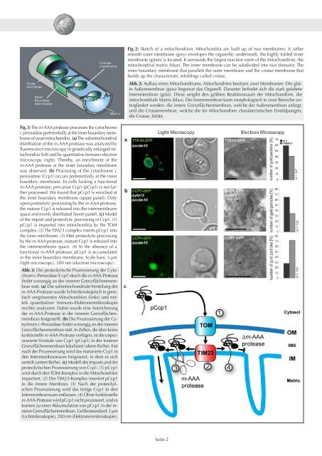
Fig. 3: The m-AAA protease processes the cytochrome<br />
c peroxidase preferentially at the inner boundary membrane<br />
of yeast mitochondria. (a) The submitochondrial<br />
distribution of the m-AAA protease was analyzed by<br />
fluorescence microscopy in genetically enlarged mitochondria<br />
(left) and by quantitative immuno-electron<br />
microscopy (right). Thereby, an enrichment of the<br />
m-AAA protease at the inner boundary membrane<br />
was observed. (b) Processing of the cytochrome c<br />
peroxidase (Ccp1) occurs preferentially at the inner<br />
boundary membrane. In cells lacking a functional<br />
m-AAA protease, precursor Ccp1 (pCcp1) is not further<br />
processed. We found that pCcp1 is enriched at<br />
the inner boundary membrane (upper panel). Only<br />
upon proteolytic processing by the m-AAA protease,<br />
the mature Ccp1 is released into the intermembrane<br />
space and evenly distributed (lower panel). (c) Model<br />
of the import and proteolytic processing of Ccp1. (1)<br />
pCcp1 is imported into mitochondria by the TOM<br />
complex. (2) The TIM23 complex inserts pCcp1 into<br />
the inner membrane. (3) After proteolytic processing<br />
by the m-AAA protease, mature Ccp1 is released into<br />
the intermembrane space. (4) In the absence of a<br />
functional m-AAA protease, pCcp1 is accumulated<br />
in the inner boundary membrane. Scale bars: 3 µm<br />
(light microscopy), 200 nm (electron microscopy).<br />
Abb. 3: Die proteolytische Prozessierung der Cytochrom<br />
c-Peroxidase (Ccp1) durch die m-AAA-Protease<br />
findet vorrangig an der inneren Grenzflächenmembran<br />
statt. (a) Die submitochondriale Verteilung der<br />
m-AAA-Protease wurde lichtmikroskopisch in genetisch<br />
vergrösserten Mitochondrien (links) und mittels<br />
quantitativer Immuno-Elektronenmikroskopie<br />
(rechts) analysiert. Dabei wurde eine Anreicherung<br />
der m-AAA-Protease in der inneren Grenzflächenmembran<br />
festgestellt. (b) Die Prozessierung der Cytochrom<br />
c-Peroxidase findet vorrangig an der inneren<br />
Grenzflächenmembran statt. In Zellen, die über keine<br />
funktionelle m-AAA-Protease verfügen, ist die unprozessierte<br />
Vorstufe von Ccp1 (pCcp1) in der inneren<br />
Grenzflächenmembran lokalisiert (obere Reihe). Erst<br />
nach der Prozessierung wird das maturierte Ccp1 in<br />
den Intermembranraum freigesetzt, in dem es sich<br />
verteilt (untere Reihe). (c) Modell des Imports und der<br />
proteolytischen Prozessierung von Ccp1. (1) pCcp1<br />
wird durch den TOM-Komplex in die Mitochondrien<br />
importiert. (2) Der TIM23-Komplex inseriert pCcp1<br />
in die innere Membran. (3) Nach der proteolytischen<br />
Prozessierung wird das fertige Ccp1 in den<br />
Intermembranraum entlassen. (4) Ohne funktionelle<br />
m-AAA-Protease wird pCcp1 nicht prozessiert, und es<br />
kommt zu einer Akkumulation von pCcp1 in der inneren<br />
Grenzflächenmembran. Größenstandard: 3 µm<br />
(Lichtmikroskopie), 200 nm (Elektronenmikroskopie).<br />
Fig. 2: Sketch of a mitochondrion. Mitochondria are built up of two membranes: A rather<br />
smooth outer membrane (grey) envelopes the organelle; underneath, the highly folded inner<br />
membrane (green) is located. It surrounds the largest reaction room of the mitochondrion, the<br />
mitochondrial matrix (blue). The inner membrane can be subdivided into two domains: The<br />
inner boundary membrane that parallels the outer membrane and the cristae membrane that<br />
builds up the characteristic infoldings called cristae.<br />
Abb. 2: Aufbau eines Mitochondriums. Mitochondrien besitzen zwei Membranen: Die glatte<br />
Außenmembran (grau) begrenzt das Organell. Darunter befindet sich die stark gefaltete<br />
Innenmembran (grün). Diese umgibt den größten Reaktionsraum der Mitochondrien, die<br />
mitochondriale Matrix (blau). Die Innenmembran kann morphologisch in zwei Bereiche untergliedert<br />
werden: die innere Grenzflächenmembran, welche der Außenmembran anliegt,<br />
und die Cristaemembran, welche die <strong>für</strong> Mitochondrien charakteristischen Einstülpungen,<br />
die Cristae, bildet.<br />
Seite 2