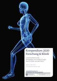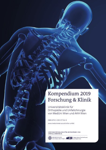
Kompendium 2019 Forschung & Klinik
Das Kompendium 2019 der Universitätsklinik für Orthopädie und Unfallchirurgie von MedUni Wien und AKH Wien (o. Univ.-Prof. R. Windhager) stellt einen umfassenden Überblick über die medizinsichen Leistungen und auch die umfangreichen Forschungsfelder dar. Die Veröffentlichungen zeigen die klinische Relevanz der einzelnen Forschungsrichtungen – seien es Daten über die Wirksamkeit von Orthesen des Sprunggelenkes, die Effizienz verschiedener Methoden der Behandlung von Schenkelhalsfrakturen, die Verbesserung der Diagnostik von Prothesenlockerungen oder die Ergebnisse einer neuen Behandlung des rezidivierenden Carpaltunnelsyndroms. Aber auch grundlegend neue und innovative Forschungsansätze führten bereits zu erfolgreichen Publikationen wie die Bedeutung von Galektinen oder der biochemischen Bildgebung bei der Discusdegeneration, der Wirksamkeit von Seleniumoxid zur antiinfektiven Beschichtung von Prothesenoberflächen oder der Verwendung von zellfreiem aurikulärem Knorpel als Matrix für die Knorpelregeneration.
Das Kompendium 2019 der Universitätsklinik für Orthopädie und Unfallchirurgie von MedUni Wien und AKH Wien (o. Univ.-Prof. R. Windhager) stellt einen umfassenden Überblick über die medizinsichen Leistungen und auch die umfangreichen Forschungsfelder dar. Die Veröffentlichungen zeigen die klinische Relevanz der einzelnen Forschungsrichtungen – seien es Daten über die Wirksamkeit von Orthesen des Sprunggelenkes, die Effizienz verschiedener Methoden der Behandlung von Schenkelhalsfrakturen, die Verbesserung der Diagnostik von Prothesenlockerungen oder die Ergebnisse einer neuen Behandlung des rezidivierenden Carpaltunnelsyndroms. Aber auch grundlegend neue und innovative Forschungsansätze führten bereits zu erfolgreichen Publikationen wie die Bedeutung von Galektinen oder der biochemischen Bildgebung bei der Discusdegeneration, der Wirksamkeit von Seleniumoxid zur antiinfektiven Beschichtung von Prothesenoberflächen oder der Verwendung von zellfreiem aurikulärem
Knorpel als Matrix für die Knorpelregeneration.

Sie wollen auch ein ePaper? Erhöhen Sie die Reichweite Ihrer Titel.
YUMPU macht aus Druck-PDFs automatisch weboptimierte ePaper, die Google liebt.
<strong>Kompendium</strong> <strong>2019</strong><br />
<strong>Forschung</strong> & <strong>Klinik</strong><br />
Universitätsklinik für<br />
Orthopädie und Unfallchirurgie<br />
von MedUni Wien und AKH Wien<br />
ISBN 978-3-200-07104-9<br />
www.meduniwien.ac.at/ortho-unfall
.O.R.E.<br />
I N S T I T U T E<br />
A4_99_MIKA_ADV_revVienna2020.indd 2 08.05.2020 12:10:05
Inhalt/Impressum<br />
3<br />
Die <strong>Klinik</strong><br />
4 Editorial<br />
6 Klinische Leistungen und Operationen<br />
9 Hohe Ambulanzfrequenz durch Spezialangebote<br />
10 Interview mit o. Univ.-Prof. Dr. Reinhard Windhager:<br />
„Spitzenforschung zum Wohl der Patientinnen und Patienten“<br />
14 Die Expertinnen und Experten auf einen Blick<br />
20 Ambulanzverzeichnis der Universitätsklinik<br />
für Orthopädie und Unfallchirurgie<br />
TOP-Studien<br />
Impressum:<br />
Herausgeber:<br />
Universitätsklinik für<br />
Orthopädie und<br />
Unfallchirurgie von MedUni<br />
Wien und AKH Wien<br />
o. Univ.-Prof. Dr. Reinhard<br />
Windhager<br />
Währinger Gürtel 18 – 20<br />
1090 Wien<br />
Redaktion & Gestaltung:<br />
Unlimited Media<br />
www.unlimitedmedia.at<br />
Lektorat: Sophie Hermann, BSc,<br />
Alexandra Lechner<br />
22 Das instabile Sprunggelenk richtig stabilisieren<br />
26 Prediction of lumbar disk herniation<br />
30 Zirconium nitride coating on orthopedic implant surfaces<br />
34 Glycobiology of the intervertebral disc:<br />
Towards a functional role of galectins in disc degeneration<br />
38 Intramedullary nails for the treatment<br />
of pertrochanteric fractures<br />
42 Hip screw in the treatment of undisplaced<br />
intracapsular neck fractures<br />
46 Superior image quality of total knee arthroplasty<br />
48 Modern cemented total knee arthroplasty design<br />
51 Surgery of true recurring median carpal tunnel syndrome<br />
54 Preoperative diagnosis methods of<br />
periprosthetic joint infections<br />
55 Diagnosis of native joint septic arthritis in adults<br />
56 Treatment of periprosthetic hip joint infection<br />
58 Imminent pulmonary complications in<br />
polytraumatised patients<br />
61 Assessment of sRAGE levels may improve diagnosis<br />
and risk evaluation in polytrauma care<br />
64 Auricular cartilage-derived biomimetic biomaterial<br />
for cartilage regeneration<br />
Fotos: shutterstock/Derya Draws,<br />
Unlimited Media, MedUni Wien/<br />
Christian Houdek,<br />
Druckerei: Copydruck KG<br />
Sandleitengasse 9 – 13<br />
1160 Wien<br />
Wien, Juli 2020<br />
ISBN 978-3-200-07104-9<br />
Publikationen<br />
68 Originalarbeiten <strong>2019</strong><br />
E-Book<br />
www.meduniwien.ac.at/ortho-unfall<br />
www.unlimitedmedia.at/orthopaedie<strong>2019</strong>
Editorial<br />
4<br />
Liebe Leserinnen und Leser!<br />
Zurückblickend auf das Jahr <strong>2019</strong> stimmt einen aus heutiger Sicht, einige<br />
Monate nach Beginn der weltweiten Pandemie, der Blick auf eine scheinbar<br />
intakte Welt nachdenklich und regt zu Reflexionen über unsere Aktivitäten in<br />
dieser gewohnten Umgebung an. Abgesehen von den Einschränkungen im klinischen<br />
Alltag kam es zu einer deutlichen Reduktion und schließlich Aufhebung<br />
der Kongresstätigkeit, die doch einen wesentlichen Teil unseres wissenschaftlichen<br />
Alltags eingenommen hat, galt es doch, die Aktivitäten der <strong>Klinik</strong> in den<br />
wissenschaftlichen Gesellschaften zu vertreten und gleichzeitig aktiv an einem<br />
internationalen Diskussionsprozess teilzunehmen.<br />
o. Univ.-Prof. Dr. Reinhard Windhager<br />
Dass die Finalisierung von Projekten bis hin zu Publikationen damals nicht zu<br />
kurz kam, ist aus heutiger Sicht, in der durch den Wegfall dieser Reiseaktivitäten<br />
ein deutlicher Zeitgewinn zu verzeichnen ist, schwer vorstellbar. Umso<br />
erfreulicher ist es, dass trotz dieser vielseitigen Verpflichtungen eine rege<br />
Publikationstätigkeit resultierte, die in Form der hinten angeführten Publikationsliste<br />
dokumentiert ist, in der die wichtigsten und repräsentativsten<br />
<strong>Forschung</strong>sergebnisse im Detail erläutert sind. Besonders erfreulich ist die<br />
rege Aktivität der <strong>Forschung</strong>scluster, die durch regelmäßige Treffen einen<br />
intensiven Diskussionsprozess entfacht haben, dessen Früchte erst in den<br />
nächsten Jahren nach Finalisierung dieser Projekte geerntet werden können.<br />
Die inten sive Aufarbeitung der laufenden Projekte und umsichtige Konzeption<br />
von mittel- bis langfristigen Projekten wird in allen <strong>Forschung</strong>sclustern verfolgt.<br />
Die bereits vorliegenden Veröffentlichungen zeigen die klinische Relevanz<br />
der einzelnen <strong>Forschung</strong>srichtungen – seien es Daten über die Wirksamkeit<br />
von Orthesen des Sprunggelenkes, die Effizienz verschiedener Methoden der<br />
Behandlung von Schenkelhalsfrakturen, die Verbesserung der Diagnostik von<br />
Prothesenlockerungen oder die Ergebnisse einer neuen Behandlung des rezidivierenden<br />
Carpaltunnelsyndroms. Aber auch grundlegend neue und innovative<br />
<strong>Forschung</strong>sansätze führten bereits zu erfolgreichen Publikationen wie die Bedeutung<br />
von Galektinen oder der biochemischen Bildgebung bei der Discusdegeneration,<br />
der Wirksamkeit von Seleniumoxid zur antiinfektiven Beschichtung<br />
von Prothesenoberflächen oder der Verwendung von zellfreiem aurikulärem<br />
Knorpel als Matrix für die Knorpelregeneration.<br />
An dieser Stelle sei auch allen Mitarbeiterinnen und Mitarbeitern gedankt, die<br />
zum Zustandekommen dieses Outputs beigetragen haben und mitunter nicht<br />
namentlich aufscheinen. Danken möchte ich auch all jenen für die aktive Wissensvermittlung<br />
in der studentischen Lehre, die dazu beigetragen haben, das<br />
Fach Orthopädie und Unfallchirurgie attraktiv darzustellen, was sich im großen<br />
Andrang für das klinisch praktische Jahr äußert.<br />
Ich wünsche Ihnen viel Spaß bei der Durchsicht dieser Lektüre!<br />
Ihr Reinhard Windhager
Ihr Rehabilitationszentrum für Orthopädie und Neurologie<br />
Wir richten Sie auf !<br />
Schwerpunkte:<br />
• aktivierte und chronische Abnützungserscheinungen<br />
der Gelenke (Arthrosen)<br />
• Erkrankungen der Wirbelsäule und Zustand<br />
nach operativen Eingriffen an der Wirbelsäule<br />
• Zustand nach arthroskopischen Eingriffen<br />
an Gelenken<br />
• Zustand nach Gelenksersatz<br />
(Endoprothesen)<br />
• Zustand nach offenen Operationen am<br />
Bewegungsapparat<br />
• orthopädisch-rehabilitative Schmerztherapie<br />
• Cerebrale Gefäßerkrankungen<br />
• Polyneuropathien<br />
• Bewegungsstörungen (z.B.Morbus Parkinson)<br />
• Neuromuskuläre Erkrankungen<br />
• Multiple Sklerose und ähnliche entzündliche<br />
Erkrankungen<br />
• Degenerative Veränderungen im Wirbelsäulenbereich<br />
mit neurologischer Symptomatik,<br />
chronische Schmerzsyndrome<br />
100% Einzelzimmer<br />
Modernster Diagnostik- und Therapiebereich<br />
www.rehawienbaumgarten.at<br />
Rehaklinik Wien Baumgarten Betriebs-GmbH, 1140 Wien, Reizenpfenninggasse 1, Tel: +43 1 41500-0
Zahlen und Fakten<br />
6<br />
Klinische Leistungen<br />
und Operationen<br />
Die Universitätsklinik für Orthopädie und Unfallchirurgie ist eine<br />
Großklinik mit einem umfassenden Leistungsspektrum. Es ist<br />
oberstes Ziel, den PatientInnen die bestmögliche medizinische<br />
Behandlung zukommen zu lassen. Bei den klinischen Leistungen<br />
und Operationen lag die Fallzahl, wie in den letzten Jahren, auch<br />
<strong>2019</strong> auf einem sehr hohen Niveau.<br />
Im Jahr <strong>2019</strong> wurden an der Universitätsklinik für Orthopädie und Unfallchirurgie<br />
insgesamt 7.039 Operationen durchgeführt. Im Jahr davor waren<br />
es 6.918. Diese Mehrzahl von 123 Operationen entspricht einer weiteren<br />
Steigerung von rund zwei Prozent. 2.945 Operationen fielen dabei in den<br />
Bereich der Orthopädie. Wobei hier betont werden muss, dass es sich zumeist<br />
um hochspezifische Eingriffe handelte, die an anderen Spitälern in<br />
Österreich gar nicht durchgeführt werden können. Es wurden 349 Hüftendoprothesen<br />
(HTEP), 343 Knieendoprothesen und 173 Fusionen der Wirbelsäule<br />
durchgeführt. Hinzu kamen im Jahr <strong>2019</strong> 435 Arthroskopien, 132<br />
Fußoperationen, 372 Tumorresektionen und 94 Osteosynthesen. Vor allem<br />
die Zahlen bei den Tumorresektionen stiegen im Vergleich zum Jahr 2018<br />
(278) stark an. 4.094 Operationen wurden an der Klinischen Abteilung für<br />
Unfallchirurgie durchgeführt. Hier wurden 6.729 PatientInnen stationär<br />
behandelt, wobei das Hauptaugenmerk auf der unfallchirurgischen Akutversorgung<br />
und Nachbehandlung lag.<br />
Klinische Leistung Orthopädie 2018 <strong>2019</strong><br />
Operationen gesamt 2.836 2.945<br />
unter anderem:<br />
Fusionen der Wirbelsäule 148 173<br />
Endoprothetische Versorgungen (gesamt) 760 731<br />
davon Hüftendoprothesen 373 349<br />
davon Knieendoprothesen 339 343<br />
Arthroskopien (alle Gelenke) 443 435<br />
Fußoperationen 127 132<br />
Tumorresektionen 278 372<br />
Osteosynthesen 70 94<br />
Klinische Leistung Unfallchirurgie<br />
Anzahl Operationen 4.080 4.094<br />
Stationäre Aufnahmen 7.057 6.729
Zahlen und Fakten<br />
9<br />
Hohe Ambulanzfrequenz<br />
durch Spezialangebote<br />
Die Universitätsklinik für Orthopädie und Unfallchirurgie versteht<br />
sich als universitäres Zentrum zur Diagnose, Therapie und<br />
Prävention von angeborenen und erworbenen Erkrankungen des<br />
Bewegungsapparates. Durch den hohen Grad an Spezialisierung<br />
und das große Angebot an Spezialambulanzen sind die Ambulanzen<br />
einer sehr starken Frequenz ausgesetzt. Oberstes Ziel ist<br />
es auch hier, den PatientInnen die bestmögliche medizinische<br />
Behandlung zukommen zu lassen.<br />
Die Klinische Abteilung für Unfallchirurgie der Medizinischen Universität Wien<br />
und des Universitätsklinikums AKH Wien gilt als weltweit anerkannte Instanz<br />
in der unfallchirurgischen Akutversorgung und Nachbehandlung. <strong>2019</strong> wurden<br />
71.216 Ambulanzbesuche in der Erstversorgung verzeichnet, hinzu kamen<br />
51.452 ambulante Kontrollen und 15.273 Versorgungen im Gipszimmer. In der<br />
Klinischen Abteilung für Orthopädie werden 21 höchst spezialisierte Ambulanzen<br />
auf internationalem Niveau angeboten. Insgesamt waren die 31.800<br />
Ambulanzbesuche im Jahr <strong>2019</strong> mit den Vorjahren vergleichbar (2018: 32.966;<br />
2017: 28.866). Die höchsten Frequenzen wiesen folgende Ambulanzen auf:<br />
Schmerztherapie (5.097), Tumororthopädie (3.738), Rheumaorthopädie (3.019),<br />
Kinderorthopädie (2.145), Wirbelsäule (1.718), Sportorthopädie (1.712), Endoprothetik<br />
(1.320), Sarkome (951) und die Fußambulanz (694).<br />
2018 <strong>2019</strong><br />
Ambulante Frequenzen Orthopädie (gesamt) 32.966 31.800<br />
Schmerztherapie 4.920 5.097<br />
Tumororthopädie 3.846 3.738<br />
Rheumaorthopädie 2.932 3.019<br />
Kinderorthopädie 2.277 2.145<br />
Wirbelsäule 2.002 1.718<br />
Sportorthopädie 1.718 1.712<br />
Endoprothetik 1.440 1.320<br />
Knochen- und Weichteilsarkome 1.045 951<br />
Fußambulanz 694<br />
Ambulante Frequenzen Unfallchirurgie<br />
Ambulante Frequenzen (ABF) Erstversorgung 73.911 71.216<br />
Ambulante Kontrollen (ABK) Nachbehandlung 55.387 51.452<br />
SchockraumpatientInnen 523 566<br />
Eingriffe in der Wundversorgung 4.871 4.675<br />
Versorgungen im Gipszimmer 16.570 15.273
<strong>Klinik</strong><br />
10<br />
Spitzenforschung zumWohl<br />
der Patientinnen und Patienten<br />
Die Universitätsklinik für Orthopädie und Unfallchirurgie von<br />
MedUni Wien und AKH Wien hat nicht nur einen Riesen anteil<br />
an der laufenden PatientenInnenversorgung sondern nimmt<br />
auch einen TOP-Platz in der internationalen Spitzen forschung<br />
ein. Im Interview erklärt o. Univ.-Prof. Dr. Reinhard Windhager,<br />
warum diese Spitzenforschung den Patientinnen und Patienten<br />
zugutekommt und welche Probleme es gibt.<br />
Ein Umstand, der auch durch verbesserte Operationstechniken, ein optimiertes<br />
perioperatives Management und eine damit verbundene Ausweitung der<br />
Operationsindikationen verstärkt wird. Eine weitere Veränderung, die von Jahr<br />
zu Jahr kontinuierlich ansteigt, ist die Zunahme an Eingriffen bei multimorbio.<br />
Univ.-Prof. Dr. Reinhard Windhager<br />
Die Universitätsklinik für Orthopädie und Unfallchirurgie ist<br />
eine der größten im AKH Wien. Welchen Anteil und welche Aufgaben<br />
übernimmt die <strong>Klinik</strong> in der Patientinnen- und Patientenversorgung?<br />
Mit knapp 7.000 Operationen und mehr als 160.000 Kontakten mit<br />
Patientinnen und Patienten pro Jahr liegt unsere <strong>Klinik</strong> im Spitzenbereich<br />
der Versorgung im Universitätsklinikum AKH Wien. Als tertiäres Versorgungszentrum<br />
kommen unserer <strong>Klinik</strong> überregionale Aufgaben zu, die sich<br />
in einer differenzierten Ambulanz struktur wiederspiegeln.<br />
Trotz dieses Versorgungsauftrages in der Spitzenmedizin, der eine Rundum-die-Uhr-Verfügbarkeit<br />
der wichtigsten Ressourcen verlangt, besteht die<br />
Gefahr, dass diese Ressourcen allzu sehr für Routine-Versorgungen, die auch<br />
in der Peripherie durchgeführt werden können, missbräuchlich verwendet<br />
werden und zu einer Überflutung der Strukturen führen. Unser Ziel ist es,<br />
hier entsprechend gegenzusteuern, um die Versorgung in der Spitzenmedizin<br />
nicht zu gefährden und den universitären Aufgaben gerecht zu werden.<br />
Bewegungsstörungen und Probleme beim Bewegungsapparat<br />
nehmen in der Gesellschaft immer mehr zu. Ist das auch in<br />
den Ambulanzen und an den OP-Zahlen abzulesen?<br />
Unsere Ressourcen sind, wie bereits erwähnt, begrenzt und den Aufgaben<br />
eines tertiären, universitären Versorgungszentrums angepasst, sodass eine<br />
Zunahme von Störungen im muskuloskelettalen Bereich keine unmittelbare<br />
Auswirkung auf die Zahlen hat. Sehr wohl hat aber die demografische Entwicklung<br />
einen Einfluss, insofern als die Überalterung zu einer Zunahme der<br />
Eingriffe bei hochbetagten Patientinnen und Patienten führt.
<strong>Klinik</strong><br />
11<br />
den Patientinnen und Patienten, die klar zu dem Aufgabengebiet eines tertiären<br />
Versorgungszentrums gehören. Dies betrifft in erster Linie Revisionsoperationen<br />
nach endoprothetischen Eingriffen und lebensbedrohliche Zustände<br />
wie Infektionen des muskuloskelettalen Systems.<br />
„Trotz des Versorgungsauftrages<br />
in der Spitzenmedizin, der eine Rundum-die-Uhr-Verfügbarkeit<br />
verlangt,<br />
besteht die Gefahr, dass diese<br />
Ressourcen allzu sehr für Routine-<br />
Versorgungen, die auch in der<br />
Peripherie durchgeführt werden<br />
können, missbräuchlich verwendet<br />
werden und zu einer Überflutung der<br />
Strukturen führen.“<br />
Reinhard Windhager<br />
Welche besonderen Leistungen und Operationen bietet die<br />
Universitätsklinik für Orthopädie und Unfallchirurgie an, die<br />
sonst kaum oder gar nicht in Österreich gemacht werden können?<br />
Hierzu zählen vor allem Eingriffe und Methoden, die an unserer <strong>Klinik</strong><br />
entwickelt wurden, wie zum Beispiel die Umkehrplastik bei bösartigen<br />
Knochentumoren, die so selten indiziert ist, dass eine extrem flache Lernkurve<br />
resultieren würde. Weitere Schwerpunkte sind komplexe Eingriffe am<br />
Becken, sei es auf Basis von neoplastischen Erkrankungen und Entzündungen<br />
oder aber Folgezustände nach fehlgeschlagenen Endoprothesen, bei<br />
denen mitunter große Beckenanteile rekonstruiert oder mittels individueller<br />
Prothesen, die im Metalldruckverfahren produziert werden, wiederhergestellt<br />
werden müssen. Ähnlich ist die Situation bei Eingriffen an der Wirbelsäule,<br />
wo durch enorme Fortschritte in der operativen Technik massive<br />
Fehlstellungen durch Korrekturosteotemien normalisiert werden können.<br />
Besondere Leistungen ergeben sich jedoch aus der interdisziplinären<br />
Verflechtung, sodass in Gebieten wie Infektionen des muskuloskelettalen<br />
Systems, Polytrauma-Versorgung oder Tumororthopädie enorme Leistungen<br />
resultieren.<br />
Die <strong>Forschung</strong>saktivitäten wurden in <strong>Forschung</strong>sclustern<br />
zusammengelegt. Hat sich das als zielführend erweisen?<br />
Diese Zusammenlegung und Bündelung, die bei mehr als 400 laufenden<br />
Projekten eine absolute Notwendigkeit war, hat sich als wahrer Boost für die<br />
<strong>Forschung</strong>saktivität erwiesen. Durch die Organisation in diesen Kleingruppen<br />
ist nicht nur der stetige Informationsfluss gewährleistet, sondern auch<br />
die Kommunikation durch regelmäßige Treffen gesichert. Die Eigenverantwortlichkeit<br />
in diesem Bereich führte auch zu einem großen Motivationsschub<br />
und zur Aufsetzung zahlreicher neuer Projekte, deren Früchte in den<br />
nächsten Jahren geerntet werden können. Um auch in Zukunft die Interaktivität<br />
dieser Cluster zu gewährleisten, werden regelmäßige, übergreifende<br />
Clustersitzungen abgehalten, um den Diskussionsprozess und damit die<br />
Stimulation weiterer Ideen aufrechtzuerhalten.<br />
Wie liegt die Universitätsklinik für Orthopädie und Unfallchirurgie<br />
bei der <strong>Forschung</strong> im internationalen Vergleich?<br />
Die Sichtbarkeit der Universitätsklinik für Orthopädie und Unfallchirurgie ist<br />
im internationalen Vergleich als sehr gut zu betrachten, zumal es unser Ziel<br />
ist, die qualitative <strong>Forschung</strong> einer quantitativ orientierten Publikationstätigkeit<br />
vorzuziehen. Im Besonderen betrifft dies klinisch relevante Fragestellungen<br />
und translationale <strong>Forschung</strong>, die international großen Anklang<br />
finden und auszugsweise in den folgenden Top-Publikationen wiedergegeben<br />
sind. Anerkennung findet die <strong>Forschung</strong> auch bei Bewerbung junger<br />
Kolleginnen und Kollegen um internationale Fellowships, bei denen ihnen<br />
die wissenschaftliche Erfahrung in den hoch kompetitiven Auswahlverfahren<br />
zugutekommen.
<strong>Klinik</strong><br />
12<br />
Wirkt sich die <strong>Forschung</strong> auch auf die Behandlungspfade,<br />
also auf die Routine im <strong>Klinik</strong>alltag aus?<br />
Da <strong>Forschung</strong>sfragen in erster Linie aus dem klinischen Alltag generiert werden<br />
und klinisch relevante Fragen darstellen, ist eine unmittelbare Auswirkung<br />
auf die Behandlungspfade nicht nur zu erwarten, sondern unabdingbar.<br />
Neben Fragestellungen und Ergebnissen, die unmittelbar in den klinischen<br />
Alltag Einzug finden, gibt es auch zahlreiche Projekte, die <strong>Forschung</strong>sansätze<br />
verfolgen, deren Auswirkungen sicherlich erst in wenigen Jahren zu<br />
erwarten sind. Dies betrifft in erster Linie die Arthroseforschung, aber auch<br />
Entwicklung von Verfahren zur Verhinderung von Infektionen auf Implantatoberflächen,<br />
Behandlung von Neuropathien und Auswirkungen auf das<br />
muskuloskelettale System oder Gewebeersatzverfahren.<br />
„Die Sichtbarkeit der Universitäts klinik<br />
für Orthopädie und Unfallchirurgie ist<br />
im internationalen Vergleich als sehr<br />
gut zu betrachten, zumal es unser Ziel<br />
ist, die qualitative <strong>Forschung</strong> einer<br />
quantitativ orientierten Publikationstätigkeit<br />
vorzuziehen.“<br />
Reinhard Windhager<br />
Welche Kooperationen gibt es mit Firmen, Institutionen<br />
oder auch anderen <strong>Klinik</strong>en?<br />
Kooperationen mit <strong>Klinik</strong>en und Institutionen gibt es in hoher und<br />
steigender Anzahl auf nationaler und internationaler Basis, die sich aus<br />
aktiver Beteiligung bei hochspezialisierten wissenschaftlichen Gesellschaften<br />
ergeben. Eine weitere Basis für die Etablierung von Kooperationen<br />
innerhalb der <strong>Klinik</strong> sind die interdisziplinären klinischen Boards, die<br />
nicht nur für klassische Gebiete wie die Tumororthopädie, sondern für alle<br />
wichtigen Bereiche, die auch teilweise in den <strong>Forschung</strong>sclustern abgebildet<br />
werden, existieren. So bestehen weitere Boards für Infektionen, Kinderorthopädie<br />
und Kindertraumatologie, rheumatologische Erkrankungen,<br />
Sport-Orthopädie und -Traumatologie sowie Wirbelsäulen- und andere Erkrankungen.<br />
Kooperationen mit Firmen werden in erster Linie dann forciert,<br />
wenn die Möglichkeit besteht, Drittmittelgelder über diese Zusammenarbeit<br />
einzuwerben.<br />
Ein wichtiger Teil ist auch die Ausbildung. Wie hat sich<br />
die Ausbildung bzw. das Curriculum verändert?<br />
Sowohl bei der prä- als auch bei der post-promotionellen Ausbildung<br />
sind Rahmenbedingungen vorgegeben, deren Umsetzung wir mit großem<br />
Engagement und Hingabe verfolgen. Die Qualität der studentischen Lehre<br />
wird nicht nur aus den Beurteilungen sichtbar, sondern zeigt sich auch im<br />
großen Interesse der Studentinnen und Studenten einen Teil ihres klinischen<br />
praktischen Jahres in der <strong>Klinik</strong> für Orthopädie und Traumatologie zu<br />
absolvieren.<br />
Neben der rein klinischen Ausbildung ist es uns ein besonderes Anliegen, den<br />
Zugang zu evidenzbasierter Medizin intensiv zu vermitteln und Wissensgenerierung<br />
durch <strong>Forschung</strong> zu stimulieren, was zur Folge hat, dass interessierte<br />
Kolleginnen und Kollegen bereits während des Studiums an zahlreichen wissenschaftlichen<br />
Projekten mitarbeiten oder aber ein PhD-Studium beginnen.<br />
Dies hat weiters zur Folge, dass auch in der post-promotionellen Ausbildung<br />
wissenschaftliches Arbeiten einen wichtigen Stellenwert erlangt, der sich<br />
darin äußert, dass die Zahl derer, die am Ende der Ausbildung die Habilitationskriterien<br />
aufweist, immer höher wird. Aus dieser frühen Heranführung an<br />
das wissenschaftliche Arbeiten während des Studiums resultiert auch eine<br />
deutliche Zunahme der Habilitationen, die in den letzten 10 Jahren beinahe<br />
so hoch war wie in den vorangegangenen 20 Jahren.
Kundenkompetenzzentrum mit<br />
Shop & Werkstätte:<br />
1210 Wien, Paukerwerkstraße 1c<br />
Tel.: 01/402 21 25-1000<br />
Email: office@ortoproban.at<br />
www.ortoproban.at<br />
1010 Wien, Wipplingerstraße 13 Tel.: 01/402 21 25-3050<br />
1040 Wien, Wiedner Hauptstraße 76 Tel.: 01/402 21 25-3070<br />
1080 Wien, Josefstädter Straße 33 Tel.: 01/402 21 25-3010<br />
1110 Wien, Simmeringer Hauptstr 101-103 Tel.: 01/402 21 25-3080<br />
1130 Wien, St. Veit-Gasse 56 Tel.: 01/402 21 25-3040<br />
1130 Wien, Speisinger Straße 109 Tel.: 01/402 21 25-3090<br />
1190 Wien, Heiligenstädter Straße 46-48 Tel.: 01/402 21 25-3060<br />
1210 Wien, Floridsdorfer Hauptstraße 45 Tel.: 01/402 21 25-3030<br />
1210 Wien, Brünner Straße 70 Tel.: 01/402 21 25-3100<br />
1230 Wien, Geßlgasse 5 Tel.: 01/402 21 25-3110<br />
1230 Wien, Anton-Baumgartner-Straße 44 Tel.: 01/402 21 25-3120<br />
2380 Perchtoldsdorf, Marktplatz 13 Tel.: 01/402 21 25-3130<br />
13x<br />
in Ihrer<br />
Nähe<br />
Ihr Orthopädietechniker mit<br />
über 100-jähriger Tradition<br />
Prothesen<br />
Mieder & Bandagen<br />
Orthesen & Apparate<br />
Kinderorthopädische Versorgungen<br />
Motorisierte Bewegungsschienen<br />
Rollstühle, Geh- & Alltagshilfen<br />
Produkte für die Pflege uvm.<br />
Unser Team von über 30 Orthopädietechnikern und Bandagisten kümmert sich gerne um die individuellen Anliegen der PatientInnen, sei es<br />
durch Anpassung oder individuelle Fertigung orthopädietechnischer Heilbehelfe und Hilfsmittel. Zusätzlich kümmern wir uns als Komplett-<br />
anbieter um die optimale und umfassende Versorgung der PatientInnen mit zB. Gehhilfen, Rollstühlen, div. Leihgeräten und Pflegeprodukten.<br />
ORTOPROBAN Leitner GmbH & Co. KG. Verrechnung mit allen Kassen. Lieferservice und Hausbesuche.
Das Ärzteteam<br />
14<br />
Die Expertinnen und<br />
Experten auf einen Blick<br />
Die Mitarbeiterinnen und Mitarbeiter der Universitätsklinik<br />
für Orthopädie und Unfallchirurgie im AKH Wien sind ein<br />
wichtiger Garant für die medizinisch optimale Behandlung.<br />
Aufgrund der exzellenten Verflechtung von <strong>Forschung</strong> und<br />
Praxis gelingt dies den rund 100 Ärztinnen und Ärzten bestens.<br />
Respektvoller Umgang miteinander, konstruktive Zusammenarbeit<br />
und zielorientierte, individuelle Fort- und Weiterbildung<br />
haben einen hohen Stellenwert.
Das Ärzteteam<br />
15<br />
o. Univ.-Prof. Dr. Reinhard Windhager Univ.-Prof. Dr. Alexander Giurea Univ.-Prof. in Dr. in Catharina Chiari, MSc<br />
Assoz. Prof. Priv.-Doz. Dr. Stefan Hajdu, MBA<br />
Assoz. Prof. in Priv.-Doz. in Dr. in Silke Aldrian<br />
Dr. Lukas Albrecht Dr. Jürgen Alphonsus Dr. in Anna Antoni Dr. Sebastian Apprich Univ.-Prof. in Dr. in Michaela<br />
Auer-Grumbach<br />
Mag. a Dr. in Rita Babeluk Dr. in Elena Batrina<br />
DI Dr. Emir Benca<br />
Dr. Harald Binder Mag. a Hannah Bischof<br />
Priv.-Doz. DDr. Christoph<br />
Böhler<br />
Dr. Robert Breuer Dr. Faris Brkic Dr. Alexander Bumberger Dr. Stephan Döring
Das Ärzteteam<br />
16<br />
Dr. in Nevenka Drmic Dr. Alexander Egkher Dr. Georg Fraberger Dr. Stephan Frenzel Assoz. Prof. Priv.-Doz. Dr.<br />
Philipp Funovics, MSc<br />
Ass.-Prof. Dr. Manfred<br />
Greitbauer<br />
Univ.-Prof. Dr. Josef Grohs Dr. Thomas Haider, PhD Dr. Gabriel Halát<br />
Dr. in Martina<br />
Hauser-Shinhan<br />
Dr. Stephan Heisinger<br />
Priv.-Doz. Dr. Gerhard<br />
Hobusch, MSc<br />
Assoz. Prof. Priv.-Doz. Dr.<br />
Marcus Hofbauer<br />
Dr. Florian Hofmann<br />
Dr. in Sabrina Holzer, BA<br />
Dr. in Laura Hruby, PhD Dr. Zhaohui Hu<br />
Dr. Michael Humenberger Dr. in Manuela Jaindl<br />
Dr. Fatmir Kabashi<br />
Dr. Georg Kaiser Priv.-Doz. Dr. Maximilian Dr. Richard Kellner Priv.-Doz. Dr. Alexander Kolb<br />
Kasparek, MSc<br />
Dr. Paul Kolbitsch
Das Ärzteteam<br />
17<br />
Dr. Ulrich Koller, MSc<br />
Univ.-Prof. in Dr. in Petra Krepler<br />
(karenziert)<br />
Dr. in Irena Krusche-Mandl<br />
Assoz. Prof. Priv.-Doz. Dr.<br />
Bernd Kubista, MSc<br />
Dr. in Roberta Laggner<br />
Dr. Nikolaus Lang, MSc<br />
Priv.-Doz. Dr. Richard<br />
Lass, MSc<br />
Assoz. Prof. Priv.-Doz. Dr.<br />
Johannes Leitgeb<br />
Dr. in Monika Luxl<br />
Ass.-Prof. Dr. Wolfgang<br />
Machold<br />
Dr. Bernhard Maier<br />
Dr. in Ulrike Marquart<br />
Dr. Michael Matzner<br />
Priv.-Doz. Dr. Lukas<br />
Negrin, PhD, MSc<br />
Dr. Arastoo Nia<br />
Dr. in Sylvia Nürnberger<br />
Dr. in Karin Pagano-Braun<br />
Ass.-Prof. Dr. Gholam<br />
Pajenda<br />
Ass.-Prof. Priv.-Doz. Dr.<br />
Joannis Panotopoulos<br />
DDr. Stephan Payr<br />
Dr. Stefan Plesser Dr. Domenik Popp Priv.-Doz. Dr.<br />
Dr. Gregor Rettl Ass.-Prof. Dr. Klaus-Dieter<br />
Stephan Puchner<br />
Schatz
Das Ärzteteam<br />
18<br />
Dr. in Eleonora Schneider Dr. Markus Schreiner Dr. Rupert Schuster<br />
Dr. Gilbert Schwarz<br />
Dr. Florian Sevelda, MSc<br />
Dr. in Irene Sigmund<br />
Ass.-Prof. Dr. Gobert<br />
Skrbensky<br />
Dr. Kevin Staats<br />
Dr. in Julia Starlinger, PhD<br />
Dr. in Beate Stelzeneder<br />
Dr. in Sandra Stenicka<br />
Priv.-Doz. Dr. Christoph<br />
Stihsen<br />
Ass.-Prof. Dr. Walter Stoik<br />
Dr. in Geraldine Sturz<br />
OÄ Dr. in Gerhild<br />
Thalhammer<br />
Dr. Thomas Tiefenböck, MSc<br />
Assoz. Prof. PD Mag. Dr.<br />
Stefan Tögel<br />
Dr. Klemens Vertesich<br />
Dr. Rainer Wagner<br />
Priv.-Doz. Dr. Wenzel<br />
Waldstein-Wartenberg<br />
Dr. in Valerie Weihs Priv.-Doz. Dr. Harald K.<br />
Widhalm<br />
Dr. in Madeleine Willegger<br />
ao. Univ.-Prof. Dr. Gerald E.<br />
Wozasek<br />
Dr. Lukas Zak
Das Ärzteteam<br />
19<br />
Weitere Teammitglieder (ohne Foto)<br />
(in alphabetischer Reihenfolge)<br />
Mag. a Marilena Bertacco, Dr. Sebastian Bachl, Mag. a Julia Bogensperger, Dr. in Miroslava Cernakova,<br />
Dr. in Britta Chocholka, Dr. in Theresia Dangl, Dr. Michél Dedeyan, Dr. in Emilia Eredansky, Mag. a Seyma Ergün,<br />
Dr. Jozsef-Tibor Erdös, Ass.-Prof. Dr. Martin Frossard, Dr. Markus Gregori, Dr. in Luiza Grünberger, ao. Univ.-Prof.<br />
Dr. Thomas Heinz, Dr. Florian Hofmann, Dr. Nikolaus Jantsch, Dr. in Anne Kleiner, Univ.-Prof. Dr. Richard Kdolsky,<br />
Dr. Timon Moftakhar, Dr. in Claudia Müller, Dr. Gergios Neophytou, Dr. in Sigrid Polzer, Dr. in Colleen Rentenberger,<br />
DI in Dr. in Anna Rienmüller, Dr. Philip Schefzig, Dr. Bernhard Springer, Ass.-Prof. in Dr. in Elisabeth Schwendenwein,<br />
Dr. Rainer Wagner, Dr. in Cornelia Zeitler, Mag. a Dominika Zurawaska<br />
Gastärzte und Beobachter im Jahr <strong>2019</strong><br />
Dr. Gruber, Österreich, 1. Januar – 30. April <strong>2019</strong>, Dr. Katshurio Hayashi, Japan, 9. Januar – 4. Februar <strong>2019</strong>,<br />
Zaur Mugutdinov, Russland, 4. – 27. Februar <strong>2019</strong>, Ognen Sheshoski, Macedonia, 4. – 28. März <strong>2019</strong>, Dr. Bojic<br />
Nikola, Bosnien und Herzegowina, 4. – 29. März <strong>2019</strong>, Jamal Al-Omari, Jordanien, 3. September 2018 – 31. März<br />
2020, Miodrag Vranjes, Serbia, 2. – 26. April <strong>2019</strong>, Dr. Galina Dubovik, Russland, 3. Juni – 31. Oktober <strong>2019</strong>,<br />
Dr. Firas Yassin, Österreich, 3. – 21. Juni <strong>2019</strong>, Dzmitry Bukach, Belarus, 3. – 27. September <strong>2019</strong>, Philip Hitov,<br />
Bulgaria, 3. – 20. Dezember <strong>2019</strong><br />
Fachkurzinformation Seite 74<br />
FOSFOMYCIN<br />
Fomicyt A5 Ins_V01_06/2020. FKI S.<br />
A5q Fomicyt_ok.indd 1 29.06.20 12:48
Ambulanzen<br />
20<br />
Ambulanzverzeichnis der<br />
Universitätsklinik für<br />
Orthopädie und Unfallchirurgie<br />
Alle Spezialambulanzen der Klinischen Abteilung für Ortho pädie<br />
befinden sich auf Ebene 7D im AKH Wien unter dem grünen<br />
Bettenturm und werden als Bestellambulanz geführt (telefonische<br />
Terminvereinbarung unter +43/1/404 00-40800). Die Zuweisung<br />
der PatientInnen erfolgt durch die niedergelassenen FachärztInnen<br />
für Orthopädie. Die Spezialambulanzen der Klinischen Abteilung<br />
für Unfall chirurgie und die Nachbehandlung in der Unfallambulanz<br />
sind auf Ebene 6 angesiedelt (telefonische Terminvereinbarung<br />
unter +43/1/404 00-59380).<br />
Klinische Abteilung für Unfallchirurgie<br />
• Allgemeine Unfallambulanz – Erstversorgung<br />
Assoz. Prof. Priv.-Doz. Dr. Stefan Hajdu, MBA<br />
• Allgemeine Unfallambulanz – Nachbehandlung<br />
Assoz. Prof. Priv.-Doz. Dr. Stefan Hajdu, MBA<br />
• Ambulanz für Kindertraumatologie<br />
Assoz. Prof. Priv.-Doz. Dr. Stefan Hajdu, MBA,<br />
Ass.-Prof. in Dr. in Elisabeth Schwendenwein,<br />
Dr. in Manuela Jaindl, Dr. in Monika Luxl<br />
• Ambulanz für Handchirurgie<br />
Dr. in Gerhild Thalhammer,<br />
Assoz. Prof. Priv.-Doz. Dr. Stefan Hajdu, MBA<br />
Dr. in Irena Krusche-Mandl<br />
• Ambulanz für Hüftverletzungen und<br />
posttraumatische Hüftbeschwerden<br />
Dr. Alexander Egkher, Dr. Rupert Schuster,<br />
Assoz. Prof. Priv.-Doz Dr. Stefan Hajdu, MBA<br />
• Schulterambulanz<br />
OA Dr. Jozsef-Tibor Erdös,<br />
Assoz. Prof. Priv.-Doz. Dr. Stefan Hajdu, MBA<br />
• Ambulanz für traumatische Knorpelschäden<br />
Assoz. Prof. in Priv.-Doz. in Dr. in Silke Aldrian,<br />
Assoz. Prof. Priv.-Doz. Dr. Stefan Hajdu, MBA<br />
• Ambulanz für posttraumatische Deformitäten<br />
und Gliedmaßenrekonstruktion<br />
ao. Univ.-Prof. Dr. Gerald Wozasek,<br />
Assoz. Prof. Priv.-Doz. Dr. Stefan Hajdu, MBA<br />
• Ambulanz für Sportverletzungen<br />
Dr. Rupert Schuster,<br />
Assoz. Prof. Priv.-Doz. Dr. Stefan Hajdu, MBA<br />
• Ambulanz für posttraumatische<br />
Wirbelsäulenbeschwerden<br />
Ass.-Prof. Dr. Gholam Pajenda,<br />
Assoz. Prof. Priv.-Doz. Dr. Stefan Hajdu, MBA
Ambulanzen<br />
21<br />
Klinische Abteilung für Orthopädie<br />
• Endoprothesenzentrum der Maximalversorgung<br />
Leitung: o. Univ.-Prof. Dr. Reinhard Windhager<br />
Koordinator: Assoz. Prof. Priv.-Doz. Dr. Bernd<br />
Kubista, MSc<br />
• Neuromuskuläre Fußambulanz<br />
Univ.-Prof. in Dr. in Michaela Auer-Grumbach<br />
• Spezialambulanz für Endoprothetik<br />
Univ.-Prof. Dr. Alexander Giurea,<br />
Assoz. Prof. Priv.-Doz. Dr. Bernd Kubista, MSc<br />
• Spezialambulanz für Kinderorthopädie<br />
ao. Univ.-Prof. in Dr. in Catharina Chiari, MSc,<br />
Priv.-Doz. Dr. Alexander Kolb<br />
• Spezialambulanz für Rehabilitation und Prothetik<br />
Priv.-Doz. Dr. Gerhard Hobusch,<br />
Dr. Florian Sevelda, MSc<br />
• Spezialambulanz für Rheumaorthopädie<br />
Dr. Florian Sevelda, MSc,<br />
Priv.-Doz. Dr. Stephan Puchner, MSc<br />
• Spezialambulanz für Sportchirurgie Knie<br />
Dr. Ulrich Koller, Ass.-Prof. Dr. Klaus-Dieter Schatz<br />
• Spezialambulanz für Sportorthopädie<br />
Dr. Ulrich Koller, Ass.-Prof. Dr. Klaus-Dieter Schatz<br />
• Spezialambulanz für Tumororthopädie<br />
Assoz. Prof. Priv.-Doz. Dr. Philipp Funovics, MSc,<br />
Assoz. Prof. Priv.-Doz. Dr. Joannis Panotopoulos<br />
• Spezialambulanz für Extremitätendeformitäten<br />
ao. Univ.-Prof. in Dr. in Catharina Chiari, MSc,<br />
Dr. Alexander Kolb<br />
• Spezialambulanz für Fuß<br />
Priv.-Doz. Dr. Stephan Puchner, MSc<br />
• Spezialambulanz für orthopäd. Schmerztherapie<br />
ao. Univ.-Prof. Dr. Gerold Holzer, Dr. Michael Matzner<br />
• Spezialambulanz für Schulter/Ellbogen<br />
Dr. Ulrich Koller, Ass.-Prof. Dr. Klaus-Dieter Schatz<br />
• Spezialambulanz für Knorpelschäden<br />
ao. Univ.-Prof. in Dr. in Catharina Chiari, MSc,<br />
Priv.-Doz. Dr. Alexander Kolb<br />
• Spezialambulanz für Wirbelsäule<br />
ao. Univ.-Prof. in Dr. in Petra Krepler (karenziert),<br />
ao. Univ.-Prof. Dr. Josef Georg Grohs<br />
• Spezialambulanz für Skoliose und<br />
Wirbelsäulendeformitäten<br />
ao. Univ.-Prof. in Dr. in Petra Krepler (karenziert),<br />
ao. Univ.-Prof. Dr. Josef Georg Grohs<br />
• Spezialambulanz für Knie<br />
Univ.-Prof. Dr. Alexander Giurea,<br />
Assoz. Prof. Priv.-Doz. Dr. Bernd Kubista, MSc<br />
• Spezialambulanz für Hüfte<br />
Assoz. Prof. Priv.-Doz. Dr. Bernd Kubista, MSc,<br />
Univ.-Prof. Dr. Alexander Giurea<br />
• Spezialambulanz für Hand<br />
Dr. Florian Sevelda, MSc<br />
• Spezialambulanz für komplexe<br />
Revisionen und Extremitätenrekonstruktionen<br />
o. Univ.-Prof. Dr. Reinhard Windhager<br />
• Spezialambulanz für Klumpfuß<br />
ao. Univ.-Prof. in Dr. in Catharina Chiari, MSc,<br />
Priv.-Doz. Dr. Alexander Kolb<br />
• Spezialambulanz für Knochen- und<br />
Weichteilsarkome<br />
o. Univ.-Prof. Dr. Reinhard Windhager,<br />
Assoz. Prof. Priv.-Doz. Dr. Joannis Panotopoulos
TOP-Studien<br />
22<br />
Das instabile Sprunggelenk<br />
richtig stabilisieren<br />
Publikation:<br />
Benca E, Ziai P, Hirtler L, Schuh<br />
R, Zandieh S, Windhager R.<br />
Biomechanical evaluation of<br />
different ankle orthoses in a<br />
simulated lateral ankle sprain<br />
in two different modes.<br />
Scandinavian Journal of<br />
Medicine & Science in Sports<br />
Sprunggelenksorthesen werden nach Umknicktraumata<br />
post operativ oder zur prophylaktischen Stabilisierung des<br />
instabilen Sprunggelenks verwendet. Am „Adolf Lorenz Lab<br />
for Biomechnaics“ an der Universitäts klinik für Orthopädie und<br />
Unfallchirurgie wurden verschiedene Sprunggelenks orthesen<br />
auf ihre Stabilisierungsfähigkeit untersucht und das Ergebnis<br />
war überraschend: Das instabile Sprunggelenk ist häufig<br />
ungenügend stabilisiert.
TOP-Studien<br />
23<br />
Das Umknicktrauma ist bekanntlich eine der häufigsten Sportverletzungen,<br />
wobei das laterale Trauma die höchste Inzidenzrate hat. Die Wahrscheinlichkeit<br />
eines wiederholten Umknicktraumas liegt bei über 70 %, somit stellt eine<br />
vorangegangene Verletzung den höchsten Risikofaktor dar. Dabei wird der<br />
laterale Bandapparat überbeansprucht, was sukzessive Rupturen, insbesondere<br />
des Ligamentum fibulotalare anterius und Ligamentum fibulocalcaneare,<br />
zur Folge hat. Eine Sprunggelenksorthese soll das Gelenk vor exzessiven<br />
Belastungen schützen, insbesondere gegen Drehmomente in Inversion,<br />
Plantarflexion und Innenrotation.<br />
„Am verletzten Sprunggelenk konnten<br />
beim wiederholten Umknicktrauma<br />
von den fünf getesteten Orthesen<br />
lediglich zwei, nämlich AirGo und<br />
Air Stirrup, eine signifikante protektive<br />
Wirkung zeigen.“<br />
Emir Benca<br />
Testung verschiedener Sprunggelenksorthesen<br />
auf isolierte mechanische Stabilisierungswirkung<br />
Trotz der häufigen klinischen Anwendung verschiedener Orthesen fehlt<br />
es an kontrollierten Studien über ihre Stabilisierungswirkung, sodass es<br />
nicht möglich ist, eine klare Empfehlung abzugeben, ob ein bestimmtes<br />
Modell eine Präventivwirkung hat und in welchem Ausmaß.<br />
Am Adolf Lorenz Lab for Biomechanics (Leiter DI Dr. Emir Benca) unter<br />
Beteiligung anderer <strong>Klinik</strong>en und Zentren der Medizinischen Universität<br />
Wien wurden nun verschiedene Sprunggelenksorthesen untersucht mit<br />
dem Ziel, die isolierte mechanische Stabilisierungswirkung der einzelnen<br />
unten angeführten Modelle (Abbildung 1) zu quantifizieren:<br />
• AirGo Ankle Brace (DJO, LLC; Vista, CA, USA),<br />
• Air Stirrup Ankle Brace (DJO, LLC; Vista, CA, USA),<br />
• Dyna Ankle 50S1 (Otto Bock HealthCare GmbH; Duderstadt, Deutschland),<br />
• MalleoLoc (Bauerfeind AG; Zeulenroda-Triebes, Deutschland) und<br />
• Push Aequi (Push; Maastricht-Airport, Niederlande).<br />
Die Testung erfolgte an humanen anatomischen Unterschenkelpräparaten.<br />
Um die Belastungen bei einem Umknicktrauma zu simulieren, wurden die<br />
Präparate in Supinationsstellung mechanisch einer Innenrotation von 0°<br />
bis 40° ausgesetzt. Dabei wurde das Drehmoment, also der Widerstand gegen<br />
die Innenrotation, welches im Sprunggelenk generiert wird, gemessen.<br />
Die Testung erfolgte im ersten Zyklus am gesunden Sprunggelenk. Danach<br />
wurde das Ligamentum fibulotalare anterius durchtrennt, um das typische<br />
Verletzungsmuster nach einem Umknicktrauma zu simulieren, gefolgt vom<br />
zweiten Testzyklus. Anschließend wurden die Präparate in randomisierter<br />
Reihenfolge mit den oben angeführten Orthesen stabilisiert und weiteren<br />
Testzyklen unterzogen. Die Auswertung erfolgte als prozentuelle Änderung<br />
des Innen rotationsmoments relativ zum intakten Zustand (Abbildung 2).<br />
Abbildung 1: Sprunggelenksorthesen, welche einer biomechanischen Untersuchung unterzogen wurden:<br />
(A) AirGo, (B) Air Stirrup, (C) Dyna Ankle 50S1, (D) MalleoLoc, und (E) Push Aequi.
TOP-Studien<br />
24<br />
Abbildung 2:<br />
Innenrotationsmomentverhältnisse als<br />
Funktion der Stabilisierung. Die Grundlinie<br />
repräsentiert den intakten und nicht<br />
stabilisierten Zustand. Die Belastung<br />
erfolgte quasistatisch mit 0,5°/s.<br />
Die Werte sind als Mittelwert ± Standardabweichung<br />
angegeben.<br />
DI Dr. Emir Benca<br />
Autor:<br />
Emir Benca absolvierte 2011<br />
das Maschinenbaustudium<br />
an der TU Wien. Im Jahr 2017<br />
promovierte er an der Medizinischen<br />
Universität Wien<br />
im Doktoratsstudium Applied<br />
Medical Science. Er leitet das<br />
Adolf Lorenz Lab for Biomechanics<br />
und koordiniert das<br />
Rapid-Prototyping-<strong>Forschung</strong>scluster<br />
an der Universitätsklinik<br />
für Orthopädie und Unfallchirurgie.<br />
Seine <strong>Forschung</strong>s- und<br />
Lehrschwerpunkte umfassen<br />
experimentelle und numerische<br />
Methoden in der Biomechanik,<br />
Evaluierung von Implantaten<br />
und Operationstechniken sowie<br />
additive Fertigung (3D-Druck).<br />
Studienergebnisse<br />
Die Ergebnisse der Studie zeigen, wie erwartet, einen signifikanten Abfall<br />
im Widerstandsmoment gegen zunehmende Innenrotation in Supination<br />
nach der Durchtrennung des Ligamentum fibulotalare anterius. Am verletzten<br />
Sprunggelenk konnten beim wiederholten Umknicktrauma von den<br />
fünf getesteten Orthesen lediglich zwei, nämlich AirGo und Air Stirrup, eine<br />
signifikante protektive Wirkung zeigen. Dabei ist das ähnliche Design der<br />
Stabilisierungselemente der zwei genannten Orthesen für die höhere mechanische<br />
Effektivität verantwortlich. Diese umschließen großflächig medial<br />
und lateral das Sprunggelenk. Zusätzliche Luftpolster erlauben eine gute<br />
Anpassung und Adhäsion an das Sprunggelenk. Die Stabilisierung durch die<br />
Aequi Orthese mit einem Stabilisierungselement lateral liegt im Bereich des<br />
physiologischen Sprunggelenks. Dyna Ankle und MalleoLoc gewährleisten<br />
kaum eine Stabilisierung des Sprunggelenks beim wiederholtem Umknicktrauma<br />
(
Arthrex Ankle Fusion Plates<br />
A Comprehensive System for Ankle Fusion Management<br />
■ Anatomically contoured plates for anterior,<br />
lateral, and posterior approaches<br />
■ Various lengths available<br />
■ Multiple compression options<br />
■ Specific joint preparation instrumentation<br />
Minimally invasive anterior tibiotalar plate<br />
Short talar neck anterior tibiotalar plate<br />
Anterior tibiotalar plate<br />
Lateral tibiotalar plate<br />
Lateral tibiotalocalcaneal plate<br />
Posterior tibiotalocalcaneal plate<br />
www.arthrex.com<br />
© Arthrex GmbH, 2020. All rights reserved.<br />
AD2-80016-EN_D_Ankle_Fusion_Plating_System_A4_210x279_A5.indd 1 14.05.20 11:30
TOP-Studien<br />
26<br />
Prediction of lumbar<br />
disk herniation<br />
Degenerative disk disease (DDD) is a common cause of low back<br />
pain (LBP), which in turn is among the leading causes for years<br />
lived with disability. In DDD, gradual loss of hydration of the<br />
nucleus pulposus and decreasing structural integrity of the annulus<br />
fibrosus lead to a loss of height of intervertebral disks (IVDs)<br />
and the risk of subsequent development of annular fissures<br />
and herniation.<br />
„The main finding of this study is that<br />
quantitative MRI using T 2<br />
mapping of<br />
the IVDs may be able to assess the risk<br />
of future lumbar disk herniation and<br />
correlates with disease burden.“<br />
Markus Schreiner<br />
Magnetic resonance imaging (MRI) plays a key role in the assessment<br />
of symptomatic patients, who do not respond to conservative treatment of<br />
low back pain or suffer from neurological symptoms 1 . However, only weak<br />
correlations between clinical symptoms and radiological findings have been<br />
reported, as about one third of the population aged 50 years or older suffers<br />
from at least one herniated disk while being asymptomatic 2 . Furthermore,<br />
morphological MRI is inherently observer dependent and does not provide<br />
insights into ultrastructural and compositional changes of the IVD, which<br />
reflect the degenerative process.<br />
Study:<br />
Raudner M, Schreiner MM, Juras<br />
V, Weber M, Stelzeneder D,<br />
Kronnerwetter C, Windhager R,<br />
Trattnig S.<br />
Prediction of Lumbar Disk Herniation<br />
and Clinical Outcome<br />
Using Quantitative Magnetic<br />
Resonance Imaging: A 5-Year<br />
Follow-Up Study.<br />
Invest Radiol. <strong>2019</strong> Mar; 54(3):<br />
183-189.<br />
Quantitative MRI, however, such as T 2<br />
mapping, bears the promise of objective<br />
and reproducible assessment of changes in IVD ultrastructure and composition,<br />
in particular the water content, as well as organization of the collagen<br />
network. T 2<br />
mapping has already been employed for the assessment of<br />
intervertebral disks 3 , but longitudinal studies on patients suffering from LBP<br />
are scarce and so far the predictive value of changes in T 2<br />
relaxation times<br />
caused by structural changes in the nucleus pulposus and annulus fibrosus<br />
remain unknown. For this reason the purpose of this study was to assess the<br />
T 2<br />
relaxation times of different parts of the intervertebral disks in patients<br />
suffering from low back pain, and evaluate the changes of the T 2<br />
relaxation<br />
times over time as well as the potential predictive value of T 2<br />
values regarding<br />
future disk herniation and clinical outcome at a five-year follow-up.<br />
Materials and Methods<br />
This study was designed as a follow-up study of a previous examination, which<br />
took place from 2008 to 2009 and included 66 patients, each suffering from<br />
low back pain without radicular symptoms or previous surgery. At baseline,<br />
LBP and ages between 15 and 65 years served as inclusion criteria. Neurological<br />
deficits of the lower limbs, radicular pain, a body mass index greater<br />
than 30, previous surgical interventions involving the lumbar disks, scoliosis<br />
with a Cobb angle exceeding 15 degrees, recent disk herniation (
TOP-Studien<br />
27<br />
Figure 2:<br />
A 55-year-old female patient with an RMDQ 11 and<br />
VAS of 8 at follow-up due to a collpased L4/L5 disk<br />
at follow-up (right) with a baseline (left) T2 of the<br />
nucleus pulposus of 65.5 ms and a baseline NPAF<br />
ratio of 61.3, indicating that the disk was at risk.<br />
and contraindications for MRI served as exclusion criteria at baseline. All<br />
patients of the first study were invited back for the follow-up examination.<br />
A surgical procedure that affected a lumbar intervertebral disk during the<br />
follow-up period served as additional exclusion criteria.<br />
Figure 1:<br />
Schematic illustration of the evaluation scheme<br />
of intervertebral disks using manual ROI (region of<br />
interest) placement. The T2 mean of the posterior<br />
annular region (red posterior 10 %; ROI 6 – labeled<br />
as PAF10) is divided by the mean nucleus pulposus<br />
value (mean of ROI2, 3 and 4) and multiplied by 100<br />
resulting in the NPAF ratio. This depicts the PAF10<br />
as a % of the disk’s nucleus pulposus and serves<br />
as a general marker of disk health and integrity.<br />
(reproduced with curtesy of Raudner et al.)<br />
Twenty-five patients (13 male, 12 female) underwent the follow-up examination.<br />
Mean age at baseline was 40.0 years (range 24 – 64 years). The<br />
mean follow-up time was 5.2 years (range, 4.4 – 5.8 years). All patients were<br />
examined using a 3T MR scanner (Tim Trio, Siemens Healthcare, Erlangen,<br />
Germany) and a spine array coil. A sagittal multi-echo spin-echo sequence<br />
was used for T 2<br />
mapping at baseline and follow-up (six echoes; TE 13.8, 27.6,<br />
41.4, 55.2, 69.0, 82.8 ms; TR 1200m; slice thickness 3mm, in-plane resolution<br />
256x256). For morphological imaging, standard sagittal T1- and sagittal,<br />
axial, and coronal T 2<br />
-weighted sequences were used. Radiological classification<br />
of DDD was performed according to Pfirrmann et al 4 . Herniation<br />
was described according to Fardon et al 5 .<br />
T 2<br />
maps were assessed using a manual ROI (region of interest) by subdividing<br />
the IVD into 20 % regions of the sagittal diameter. The inner 60 % represented<br />
the nucleus pulposus, the outer 20 % anteriorly and posteriorly represented<br />
the annulus fibrosus. The most posterior ROI was further subdivided and the<br />
posterior 10 % was labelled as posterior annulus fibrosus (PAF10) (Figure 1).<br />
For a subsequent statistical analysis, a dimensionless variable was calculated<br />
by dividing the mean T 2<br />
relaxation time of the PAF10 by the mean T 2<br />
relaxation<br />
time of the nucleus pulposus multiplied by 100. Statistical analysis was<br />
performed using SPSS 22.0 (SPSS Inc., Armonk, NY, USA). A p-value
TOP-Studien<br />
28<br />
follow-up were compared using a paired Student‘s t-test. To assess differences<br />
in T 2<br />
values between Pfirrmann scores, one-way analysis of variance (ANOVA)<br />
was used with an additionally calculated correlation (Spearman‘s rho). Receiver<br />
operating characteristic (ROC) curves were calculated to assess the predictive<br />
and discriminative potential of a given variable. Spearman’s rho was used to<br />
correlate clinical end points (Roland Morris Disability Questionnaire [RMDQ]<br />
and visual analogue scale [VAS]) with the minimum NP T 2<br />
and maximum NPAF<br />
and PAF10 T 2<br />
. A p-value of 0.05 or less was considered significant.<br />
Results<br />
From baseline to follow-up the mean T 2<br />
relaxation times of the NP decreased<br />
from 107.7 to 102.5 milliseconds (p = 0.005). Eleven additional herniations<br />
were detected at follow-up in addition to the 22 herniations at baseline. NP<br />
T 2<br />
and the NPAF ratio differed significantly between different Pfirrmann<br />
scores (p = 0.001). Significant difference regarding NPAF ratio was observed<br />
between disks with and without herniation at follow-up (p < 0.001).<br />
Dr. Markus Schreiner<br />
Author:<br />
Markus Schreiner is a resident<br />
at the Department of Orthopedics<br />
and Trauma Surgery at the<br />
Medical University of Vienna<br />
since 2016 and a member of the<br />
biomedical MR- imaging Cluster<br />
of orthopedic disorders. Since<br />
<strong>2019</strong> he coordinates together<br />
with Sebastian Apprich M.D.<br />
the scientific collaborations<br />
between the Orthopedic department<br />
and the High Field MR<br />
Centre (Prof. Siegfried Trattnig)<br />
at the Medical University of<br />
Vienna. After obtaining his M.D.<br />
from the Medical University of<br />
Vienna in 2014, Markus Schreiner<br />
started his PhD and spent<br />
five months at Mayo Clinic for a<br />
research traineeship. Within the<br />
biomedical MR-imaging cluster,<br />
his research focuses on the development<br />
and implementation<br />
of MR imaging techniques used<br />
for qualitative and quantitative<br />
assessment of cartilage, tendons,<br />
and intervertebral discs,<br />
as well as clinical projects on<br />
cartilage repair surgery.<br />
At baseline, NP T 2<br />
also differed significantly (all p < 0.001) between disks with<br />
and without herniation. Also, the baseline T 2<br />
values of disks with new herniations<br />
at follow-up differed significantly from disks without herniation at<br />
baseline and follow-up. The bNPAF showed an area under the curve (AUC) of<br />
0.893 in a receiver operating curve (ROC) curve for the detection of herniation<br />
at follow-up when excluding baseline herniation cases. At a cut off of 44.1 %<br />
and higher for the bNPAF, 100.0 % sensitivity and 76.5 % specificity were<br />
calculated. Furthermore, a significant correlation between baseline NPT2<br />
(r = −0.202; p = 0.030) as well as baseline NPAF (r = 0.233; p = 0.012) with<br />
RMDQ was observed.<br />
Conclusion<br />
The main finding of this study is that quantitative MRI using T2 mapping of<br />
the IVDs may be able to assess the risk of future lumbar disk herniation and<br />
correlates with disease burden. Both the NP T2 and the NPAF ratio differed<br />
significantly between disks with and without de novo herniation at follow-up.<br />
This corroborates the assumption that morphological changes such as<br />
fissures and herniations might be preceded by ultrastructural changes,<br />
which remain elusive to morphological MRI but are reflected in a change<br />
in T2 relaxation times of affected IVDs. However, additional studies are<br />
needed to confirm these findings in larger cohorts.<br />
References:<br />
1<br />
Deyo RA, Mirza SK (2016) CLINICAL PRACTICE. Herniated Lumbar Intervertebral Disk. N Engl J Med<br />
374:1763-1772<br />
2<br />
Brinjikji W, Luetmer PH, Comstock B et al (2015) Systematic literature review of imaging features<br />
of spinal degeneration in asymptomatic populations. AJNR Am J Neuroradiol 36:811-816<br />
3<br />
Stelzeneder D, Welsch GH, Kovacs BK et al (2012) Quantitative T2 evaluation at 3.0T compared to<br />
morphological grading of the lumbar intervertebral disc: a standardized evaluation approach in<br />
patients with low back pain. Eur J Radiol 81:324-330<br />
4<br />
Pfirrmann CW, Metzdorf A, Zanetti M, Hodler J, Boos N (2001) Magnetic resonance classification of<br />
lumbar intervertebral disc degeneration. Spine (Phila Pa 1976) 26:1873-1878<br />
5<br />
Fardon DF, Williams AL, Dohring EJ, Murtagh FR, Gabriel Rothman SL, Sze GK (2014) Lumbar disc<br />
nomenclature: version 2.0: Recommendations of the combined task forces of the North American<br />
Spine Society, the American Society of Spine Radiology and the American Society of Neuroradiology.<br />
Spine J 14:2525-2545
Spherox<br />
ist das einzig<br />
EMA-zugelassene<br />
ATMP zur<br />
Behandlung von<br />
Knorpelschäden.<br />
Spherox<br />
erzeugt<br />
hyalin-ähnlichen<br />
Knorpel.<br />
Spherox<br />
repariert<br />
Knorpeldefekte<br />
im Knie bis<br />
10 cm².<br />
Spherox<br />
ist 100% autolog<br />
und frei von<br />
Zusatzstoffen.<br />
ERSCHAFFE<br />
DIE BÜHNE FÜR<br />
PERSONALISIERTE HEILUNG<br />
www.haemo-pharma.at
TOP-Studien<br />
30<br />
Zirconium nitride coating on<br />
orthopedic implant surfaces<br />
Staphylococcus epidermidis is the most common bacterium<br />
in periprosthetic implant infection. Because of its ability to<br />
produce biofilms and persist on the surface of medical devices,<br />
removal of an infected implant and wide debridement of the<br />
infected tissue are required in most patients. Various surface<br />
modifications of endoprosthetic implants have been developed<br />
to prevent bacterial attachment, which should also show a<br />
beneficial impact on implant osseointegration.<br />
„Low toxicity, favorable corrosion<br />
resistance, and outstanding bio compatibility<br />
make zirconium nitride<br />
a promising candidate for the<br />
reduction of bacterial attachment<br />
on implant surface.“<br />
Kevin Staats<br />
Titanium alloys offer good osseointegration and biocompatibility,<br />
unfortunately have a comparatively low resistance to tribocorrosion.<br />
Implant steel demonstrates favorable mechanical properties,<br />
however its biocompatibility is lower compared to other materials<br />
because of its nickel content. Cobalt-chromium-molybdenum alloy<br />
shows the highest stiffness and a high corrosion resistance, but a<br />
limited biocompatibility due to allergenic components.<br />
Rough surfaces, such as corundum-blasted structures, promote implant<br />
engraftment into the bone, but may also support bacterial adhesion.<br />
Therefore the aim of this study was to investigate the effect of a high<br />
tribologic-resistant 2.5-µm zirconium nitride (Zr) top coat on an antiallergic<br />
multilayer ceramic-covered cobalt-chromium-molybdenum surface<br />
regarding the formation of S. epidermidis biofilm, compared with other<br />
commonly used smooth and rough orthopedic implant surface materials.<br />
Study:<br />
Pilz M, Staats K, Tobudic S,<br />
Assadian O, Presterl E,<br />
Windhager R, Holinka J.<br />
Zirconium Nitride Coating<br />
Reduced Staphylococcus epidermidis<br />
Biofilm Formation on<br />
Orthopaedic Implant Surfaces:<br />
An In Vitro Study.<br />
Clin Orthop Relat Res. <strong>2019</strong> Feb;<br />
477(2):461-466 (4,154).<br />
Bacterial isolates and implant infection<br />
Twenty S. epidermidis strains were isolated from orthopedic patients<br />
undergoing debridement and resection arthroplasties performed for acute<br />
peri prosthetic joint infection. Ten S. epidermidis strains were collected<br />
from the skin of healthy volunteers and as reference strain S. epidermidis<br />
(DSM 3269) was used. In total, 31 S. epidermidis strains were tested against<br />
five different surface discs each, repeated in triplicates. T<br />
The following surface discs were used: (1) a multilayer antiallergic surface<br />
with a multilayer ceramic-covered CoCrMo surface and a high tribologicresistant<br />
2.5-µm Zr nitride top coat; (2) a cobalt-chromium-molybdenum<br />
alloy; (3) a titanium alloy; (4) a titanium alloy with a corundum-blasted<br />
rough surface; and (5) a stainless steel implant with a corundum-blasted<br />
rough surface.
TOP-Studien<br />
31<br />
Biofilm formation on orthopedic surface discs<br />
For biofilm formation tests, surface discs were incubated for 24 hours<br />
with suspensions of overnight cultures of the bacterial isolates. Biofilm<br />
formation was measured by counting the number of colony-forming<br />
units (CFUs) and staining the biofilms. After a 24-hour incubation,<br />
discs were sonicated in fresh PBS with a routine laboratory water bath<br />
sonicator. Serial dilutions (1:10 up to 1:1,000) were made and 20 µL<br />
of these dilutions were streaked on agar plates, following standard<br />
bacteriological practice.<br />
After incubation for 24 hours at ambient air, colonies were counted and<br />
multiplied by the dilution factor calculating the number of CFUs/mL.<br />
For the quantification of biofilm formation discs were stained with 0.1 %<br />
Safranin-O solution and photographed. To quantify the percentage of the<br />
biofilm overgrowth area on discs the obtained images were analyzed using<br />
the open-source JAVA image processing software ImageJ 1.45r.<br />
This study demonstrated that the multilayer, ceramic-covered CoCrMo surface with a Zr nitride 2.5-µm top coat had less biofilm formation compared<br />
with other commonly used surface materials for orthopedic implants.
TOP-Studien<br />
32<br />
Results: evaluation of colony-forming units<br />
The lowest bacterial count was found in the CoCrMo + Zr surface disc<br />
(6.6 x 10⁴ CFU/mL ± 4.6 x 10⁴ SD) followed by the CoCrMo surface<br />
(1.1 x 10⁵ CFU/mL ± 1.9 x 10⁵ SD), the titanium surface (1.4 x 10⁵ CFU/mL ±<br />
1.8 x 10⁵ SD), the rough titanium surface (2.1 x 10⁵ CFU/mL ± 3.0 x 10⁵ SD),<br />
and the rough stainless steel surface (2.7 x 10⁵ CFU/mL ± 3.8 x 10⁵ SD).<br />
The mean CFU counts were fewer for the CoCrMo + Zr surface disc compared<br />
with the rough stainless steel surface (mean difference 2.0 x 10⁵, p = 0.021),<br />
the rough titanium surface (mean difference 1.4 x 10⁵, p = 0.002), and the<br />
smooth titanium surface (mean difference 7.0 x 10⁴, p = 0.016). Compared<br />
with the CoCrMo surface, the Zr nitride-coated discs showed no differences<br />
in bacterial growth.<br />
Dr. Kevin Staats<br />
Author:<br />
Kevin Staats started his residency<br />
at the Department of<br />
Orthopedics and Trauma Surgery<br />
at the Medical University<br />
of Vienna in 2016. His research<br />
interests are total joint and<br />
revision arthroplasty. He is currently<br />
finishing his PhD-thesis<br />
which covers the anti microbial<br />
effect of electrochemically<br />
modified titanium surfaces.<br />
Dr. Staats had the opportunity<br />
to complete a research fellowship<br />
at the Hospital of Special<br />
Surgery, New York City. There<br />
he assessed initial implant<br />
instability in an animal model.<br />
Dr. Staats is currently an extended<br />
board member of the<br />
Austrian Orthopedic Society as<br />
the representative for Austrian<br />
orthopedic residents.<br />
Results: quantification of biofilm formation<br />
The results of biofilm formation quantification show that the mean covered<br />
area of the surface of the CoCrMo + Zr discs was 19 % (± 16 SD; confidence<br />
interval [CI], 12-25), which is lower than for the other surfaces: CoCrMo (35 %<br />
± 23 SD, CI, 26-44; mean difference 16, p = 0.001, CI, 6.48-38.59), titanium alloy<br />
(46 % ± 20 SD, CI, 38-54; mean difference 27, p < 0.0001, CI, 11.44-43.55),<br />
rough titanium alloy (66 % ± 23 SD, CI, 57-75; mean difference 47, p < 0.0001,<br />
CI, 31.21-63.32), and rough stainless steel (58 % ± 18 SD, CI, 50-65; mean<br />
difference 39, p < 0.0001, CI, 22.63-54.74). The CoCrMo surface additionally<br />
showed lower biofilm formation compared with the steel corundum-blasted<br />
(mean difference 31; p = 0.001; CI, 6.48-38.59) and titanium alloy corundumblasted<br />
(mean difference 31; p < 0.0001; CI, 15.06-47.17) surfaces. Furthermore,<br />
the titanium alloy surface was less overgrown with biofilm compared<br />
with the titanium alloy corundum-blasted (mean difference 20; p = 0.006; CI,<br />
-35.83 to -3.72) surface.<br />
Discussion and conclusion<br />
Several factors are necessary for a successful outcome of prosthetic<br />
implantation. Besides important characteristics such as biocompatibility,<br />
stiffness, and osseointegration, bacterial adherence to implants –<br />
a process necessary for biofilm formation – may be a further key factor.<br />
Bacteria within a biofilm are less susceptible to systemic antibiotics than<br />
planktonic bacteria due to the protective glycocalyx of biofilms. The drug-free<br />
decrease of bacterial adhesion to medical devices represents an attractive<br />
method in the prevention of biofilm formation, especially in the view of<br />
increasing bacterial resistance against multiple antibiotics.<br />
This study demonstrated that the multilayer, ceramic-covered CoCrMo<br />
surface with a Zr nitride 2.5-µm top coat had less biofilm formation<br />
compared with other commonly used surface materials for orthopedic<br />
implants. Additionally, fewer CFUs were growing on agar plates after<br />
sonication of the Zr nitride-coated surface compared to both rough<br />
surface samples and the smooth titanium surface. Low toxicity,<br />
favorable corrosion resistance, and outstanding biocompatibility<br />
make Zr nitride a promising candidate for the reduction of bacterial<br />
attachment on implant surface.
WE LENGTHEN,<br />
TRANSPORT AND<br />
GROW BONE.<br />
Precice offers an easy to use, proven<br />
solution with predictable and better<br />
patient outcomes compared to external<br />
fixation, including patient satisfaction,<br />
fewer complications and the ability to<br />
weight bear earlier.<br />
Cherry_Med Medical Solutions GmbH<br />
Erdbergstraße 166, 1030 Wien<br />
office@cherrymed.at<br />
+43 (1) 403 89 56-137<br />
www.cherrymed.at<br />
www.heintel.at
TOP-Studien<br />
34<br />
Glycobiology of the intervertebral<br />
disc: Towards a functional role<br />
of galectins in disc degeneration<br />
„At our lab we aim to comprehensively<br />
investigate the biological relevance of<br />
glycan-galectin interactions for the onset<br />
and progression of degenerative joint<br />
diseases. Starting our work on clinical<br />
samples of knee osteoarthritis (OA),<br />
we identified Galectin-1, Galectin-3 and<br />
Galectin-8 – three galectins with highly<br />
different protein architecture – as potent<br />
triggers of functional disease markers,<br />
driving the degradation of the extracellular<br />
matrix of articular cartilage in<br />
OA conditions2-4.“<br />
Stefan Toegel<br />
Study:<br />
Elshamly M, Kinslechner K,<br />
Grohs JG, Weinmann D, Walzer<br />
SM, Windhager R, Gabius HJ,<br />
Toegel S.<br />
Galectins-1 and -3 in Human<br />
Intervertebral Disc Degeneration:<br />
Non-uniform Distribution<br />
Profiles and Activation of<br />
Disease Markers Involving<br />
NF-kB by Galectin-1.<br />
J Orthop Res. <strong>2019</strong> Oct;<br />
37(10):2204-2216.<br />
In <strong>2019</strong>, researchers of the ‚Karl Chiari Lab for Orthopaedic<br />
Biology‘ coordinated a scientific project that resulted in the publication<br />
of an article entitled „Galectins‐1 and ‐3 in Human Intervertebral<br />
Disc Degeneration: Non‐Uniform Distribution Profiles<br />
and Activation of Disease Markers Involving NF‐κB by Galectin‐1“<br />
in the Journal of Orthopaedic Research 1 . The work is the result of<br />
a fruitful interdisciplinary cooperation between basic researchers,<br />
glycobiologists, clinical scientists and orthopedic surgeons.<br />
Equal collaboration within this project is reflected by the shared first authorship<br />
of orthopedic and trauma surgeon Dr. Mahmoud Elshamly, and molecular<br />
biologist Katharina M. Pichler, PhD (née Kinslechner). In acknowledgement<br />
of the excellent quality of the research, Dr. Mahmoud Elshamly received the<br />
Scientific Award 2020 of the Austrian Spine Society (Figure 1, page 36).<br />
For several years, the study of glycobiology has been a major research focus<br />
of the ‚Karl Chiari Lab for Orthopaedic Biology‘, one of the three research laboratories<br />
of the Department of Orthopedics and Trauma Surgery. Essentially,<br />
glycobiology investigates the functional role of glycans, which originates from<br />
their interplay with glycan-binding proteins, the so-called lectins. Galectins<br />
are a family of galactose-binding lectins and have been known as molecular<br />
switches for many cellular events such as cell differentiation, metastasis or<br />
inflammation.<br />
At our lab we aim to comprehensively investigate the biological relevance of<br />
glycan-galectin interactions for the onset and progression of degenerative<br />
joint diseases. Starting our work on clinical samples of knee osteoarthritis<br />
(OA), we identified Galectin-1, Galectin-3 and Galectin-8 – three galectins<br />
with highly different protein architecture – as potent triggers of functional<br />
disease markers, driving the degradation of the extracellular matrix of articular<br />
cartilage in OA conditions 2-4 .<br />
Based on this experience, basic researchers joined forces with orthopedic<br />
surgeons and developed the idea of translating the concept of glycobiology<br />
into the pathophysiology of intervertebral disc (IVD) degeneration. In fact,<br />
degeneration of the IVD is a well-documented factor in low back pain,<br />
a condition that places an enormous burden on the individual patient and
TOP-Studien<br />
35<br />
Figure 2: Immunopositivity for Galectin-1 (brown)<br />
of Nucleus pulposus cells in the degenerated IVD.<br />
the socioeconomic status of our society, but the molecular mechanisms that<br />
underlie its onset and progression are not yet precisely defined.<br />
The study included clinical specimens from 23 patients with spondylo chondrosis,<br />
eight patients with spondylolisthesis, and seven patients with spinal<br />
deformity, all characterized by patients’ age, sex, the Pfirrmann grades of<br />
IVDs, as well as the morphological status evaluated by T2‐weighted MRI data.<br />
Based on histological staining with Hematoxylin/Eosin and Safranin O, the<br />
histopathological score of IVD degeneration was graded. Supporting the suita -<br />
bility of the specimens for further analyses, a moderately strong correlation<br />
(r = 0.783, p < 0.0001) between the histological score and the Pfirrmann grade<br />
was found.<br />
Immunopositivity for Galectin-1 (Figure 2) and Galectin-3 of the specimens<br />
was determined and graded using a labelling index (LI). Staining profiles for<br />
both galectins showed different distribution patterns in serial sections, an<br />
indication for non‐redundant functionalities. Comparison of total Galectin‐1<br />
LI scores or total Galectin-3 LI scores revealed no statistically significant<br />
difference between the three patient groups (p > 0.05). However, correlation<br />
analyses of all specimens, irrespective of the assigned study group, revealed
TOP-Studien<br />
36<br />
Figure 1: Dr. Mahmoud Elshamly<br />
receives the Scientific Award 2020 by<br />
the Austrian Spine Society.<br />
a moderate correlation between total Galectin‐1 LI and histological scores<br />
(r = 0.531, p < 0.001) and a mild correlation between the total Galectin‐1 LI<br />
score and the Pfirrmann grade (r = 0.387, p = 0.022). Similarly, a moderate<br />
degree of correlation of total Galectin‐3 LI scores with histological scores<br />
(r=0.564, p
FRESH<br />
AIR<br />
FRESH<br />
PERSPECTIVE<br />
Remove smoke in less<br />
than 30 seconds from<br />
the peritoneal cavity. 1<br />
The Valleylab <br />
Laparoscopic Smoke<br />
Evacuation System:<br />
effectively and<br />
efficiently removes<br />
surgical smoke from the<br />
OR during procedures. 2†<br />
RapidVac Smoke Evacuator<br />
Telescopic Smoke Evacuation Pencil<br />
WHY PUT THEM AT RISK?<br />
CHOOSE A SMOKE-FREE OR.<br />
† 9 of 10 surgeons surveyed after use agreed.<br />
1. Based on Buffalo Filter report #PR- 18001 rev A, Filter visualization. Feb. 28, 2018.<br />
2. Based on internal test report #RE00139506 rev A, Bourbon: Valleylab laparoscopic<br />
smoke evacuation system nurses and surgeons claims report. March 12, 2018.<br />
© 2020 Medtronic. All rights reserved. Medtronic, Medtronic logo and Further, Together<br />
are trademarks of Medtronic. 19-weu-smoke-evac-fresh-perspec-poster-3252029, AT 2020-07<br />
ad (appr COV14601 A3Poster) Smoke Evac-A4-mb.indd 1 16.07.2020 09:26:55
TOP-Studien<br />
38<br />
Intramedullary nails<br />
for the treatment of<br />
pertrochanteric fractures<br />
„Due to their 20 % larger diameter,<br />
the PFNA blade and Gamma with<br />
U-Blade RC lag screw seems to be<br />
appropriate solutions for patients<br />
with osteoporotic bone and unstable<br />
AO/OTA 31.A2.1-3 fractures.“<br />
Nikolaus Lang<br />
Trochanteric femoral fractures are one of the most common injuries<br />
in the elderly population and the incidence will only increase due<br />
to demographic changes in the next years 1 . An optimal operative<br />
treatment is immanent regarding the typical geriatric patient,<br />
benefiting from early mobilization and weight bearing in order<br />
to avoid immobility-related complications 1,2 . The best fixation<br />
method remains debated.<br />
An overall tendency is the use of cephalomedullary implants 2,4 . The Gamma<br />
Nail ® (Stryker Trauma Germany) and PFNA ® (DePuy Synthes Germany) are<br />
the most used intramedullary nails for the treatment of pertrochanteric<br />
fractures. For both implants, the migration of the femoral head into varus<br />
and retroversion, and the subsequent cut-out of the lag screw/blade, is the<br />
most common mechanical complication with an incidence of 0 – 7 % 5 – 7 .<br />
Study:<br />
Lang NW, Breuer R, Beiglboeck<br />
H, Munteanu A, Hajdu S, Windhager<br />
R, Widhalm HK.<br />
Migration of the lag screw after<br />
intramedullary treatment<br />
of AO 31.A2 pertrochanteric<br />
fractures does not result in<br />
higher incidence of cut-outs,<br />
regardless of which implant<br />
was used. A comparison of<br />
Gamma Nail with and without<br />
U-Blade and PFNA.<br />
J. Clin. Med. <strong>2019</strong>, 8, 615;<br />
doi:10.3390/jcm8050615.<br />
Poor bone quality in the head and neck of the femur requires an exact positioning<br />
of the head screw in the center-center or low-center position with a<br />
small tip-apex distance (TAD) 8,9 . Non-cylindrical and more blade-like head<br />
fixation devices compared to screws have been developed to prevent/ reduce<br />
the failure rate 10,11 . None of these implants have yet shown superiority<br />
regarding prevention of undesired rotational movement and consequent cut<br />
out 12–14 . Thus, the aim of the study was to assess (1) the cut-out rate,<br />
(2) the migration of the lag screw/ blade, and (3) the implant failure in geriatric<br />
patients with unstable AO/OTA 31.A2.1-3 fractures treated with PFNA ® ,<br />
Gamma3 ® or Gamma3 ® with U-Blade (RC) lag screw.<br />
Materials & methods<br />
Inclusion criteria for this study were unstable OTA/AO 31A2.1 – 3 femur<br />
fractures treated with Gamma3 ® Nail (Stryker Trauma Germany), either with<br />
standard lag screws or with the U-Blade (RC) Lag Screw (Stryker Trauma<br />
Germany), or Proximal Femur Nail Antirotation (PFNA ® , Depuy Synthes<br />
Germany). Patients with pathological fractures as well as patients treated<br />
with a long cephalomedullary nail were excluded. Data was retrieved from<br />
our department’s database and completed by chart reviews. All fractures<br />
were classified by using the AO/OTA system on standard radiographs 15 .<br />
Minimum follow-up was 6 months when general fracture healing was<br />
obtained, mean follow-up was 10.8 ± 4.1 months.
TOP-Studien<br />
39<br />
The modified Parker’s ratio was used<br />
to categorize the placement of the<br />
lag screw in the femoral head in<br />
antero-posterior and lateral plane.<br />
A total of 237 patients were eligible for the assessment with a mean age<br />
of 81.9 ± 10.5 years and a minimum follow-up of 6 months. A total of 82<br />
patients were treated with a standard Gamma3 ® (Gamma), 69 patients<br />
with a Gamma3 ® with an additional U-Blade (RC) lag screw (U-Blade) and<br />
86 patients were treated with a PFNA ® (PFNA).<br />
Cut-out was defined as extrusion of the lag screw by more than 1 mm from<br />
the femoral head 24 . The modified Parker’s ratio was used to categorize the<br />
placement of the lag screw in the femoral head in antero-posterior and<br />
lateral plane 16 – 18 and was measured using intraoperative radiographs at 6<br />
and 12 months post-operatively 24 . The likelihood of cut-out was assessed by<br />
the calcar referenced tip-apex distance (calTAD) and the tip apex distance<br />
(TAD) on postoperative radiographs 19 . The Parker Mobility Score was used<br />
to estimate all patients’ mobility pre-operatively, at discharge from hospital,<br />
and at the last follow-up 20 . Furthermore, all implant-related failures and<br />
patient-related complications were evaluated. Quantitative data was compared<br />
between the three groups and within each group (standard lag screw/U-Blade<br />
(RC) Lag screw, PFNA blade) using a one-way ANOVA, the student’s t-test or<br />
the Mann Whitney U test. Qualitative data analyses were performed utilising<br />
the χ²-analysis. Statistical significance was set at α = 0.05. Multiple regression<br />
analysis with a 95 % confidence interval was used to examine the independent<br />
associations of various demographic and injury-related factors.<br />
Results<br />
All implants showed low cut-out rates with 1.2 % (n=1) in the PFNA group<br />
and 2.9 % (n=2) and 3.7 % (n=3) in the Gamma group and U-blade group,<br />
respectively. The single cut-out after osteosynthesis with a PFNA happened<br />
6 weeks post-operatively. In the Gamma nail population, we observed three<br />
patients sustaining a cut out within 3 -, one cut-out was observed 3- 16<br />
weeks following index surgery. Among those treated with Gamma Nail +<br />
U-Blade, the first of two cut-outs happened 14 weeks post index surgery and<br />
the second 14 months post-operatively, complicated by concomitant femoral<br />
head necrosis. Both the mean TAD and calTAD among those who suffered
TOP-Studien<br />
40<br />
from cut-out was higher than those who were spared this complication,<br />
20.0mm and 15.3mm vs 18.5mm and 13.1mm, respectively (p=n.s.).<br />
However, in a subgroup analysis of patients suffering a cut-out, the patient<br />
in the PFNA group was the only one that showed a significantly higher TAD<br />
(32.5mm p
TOP-Studien<br />
41<br />
References:<br />
1<br />
Sandmann G, Biberthaler P. Pertrochanteric femoral fractures in the elderly. Unfallchirurg<br />
2015;118:447–62. doi:10.1007/s00113-015-0007-x.<br />
2<br />
Baumgaertner M, Socci A, Casemyr N, Leslie M. Implant options for the treatment of intertrochanteric<br />
fractures of the hip: rationale, evidence, and recommendations. Bone Jt J 2017;99:128–33.<br />
doi:10.1302/0301-620X.99B1.BJJ-2016-0134.R1.<br />
3<br />
Jun L, Augusto R, Sousa M, Zamboni C, Durigan JR, Hungria JS, et al. Late postoperative analysis<br />
of the tip-apex distance ( TAD ) in pertrochanteric fractures : is there an accommodation of the<br />
implant within the bone ? Injury 2017;48:54–6. doi:10.1016/S0020-1383(17)30776-3.<br />
4<br />
Niu E, Ms AY, Harris AHS, Bishop J. Which Fixation Device is Preferred for Surgical Treatment of Intertrochanteric<br />
Hip Fractures in the United States ? A Survey of Orthopaedic Surgeons. Clin Orthop<br />
Relat Res 2015;473:3647–55. doi:10.1007/s11999-015-4469-5.<br />
5<br />
Hoffmann S, Paetzold R, Stephan D, Püschel K, Buehren V, Augat P. Biomechanical Evaluation of<br />
Interlocking Lag Screw Design in Intramedullary Nailing of Unstable Pertrochanteric Fractures. J<br />
Orthop Trauma 2013;27:483–90. doi:10.1097/BOT.0b013e3182a1f54b.<br />
6<br />
Zhang H, Zhu X, Pei G, Zeng X, Zhang N, Xu P, et al. A retrospective analysis of the InterTan nail<br />
and proximal femoral nail anti-rotation in the treatment of intertrochanteric fractures in elderly<br />
patients with osteoporosis : a minimum follow-up of 3 years. J Orthop Surg Res 2017;12:1–8.<br />
doi:10.1186/s13018-017-0648-2.<br />
7<br />
Bojan A, Beimel C, Speitling A, Taglang G, Ekholm C, Jönsson A. 3066 consecutive Gamma Nails . 12<br />
years experience at a single centre. BMC Musculoskelet Disord 2010;11:1–10. doi:10.1186/1471-<br />
2474-11-133.<br />
8<br />
Geller JA, Saifi C, Morrison TA. Tip-apex distance of intramedullary devices as a predictor of cutout<br />
failure in the treatment of peritrochanteric elderly hip fractures. Int Orthop 2010;34:719–22.<br />
doi:10.1007/s00264-009-0837-7.<br />
9<br />
Aicale R, Maffulli N. Greater rate of cephalic screw mobilisation following proximal femoral nailing<br />
in hip fractures with a tip – apex distance ( TAD ) and a calcar referenced TAD greater than 25mm. J<br />
Orthop Surg Res 2018;13:1–9. doi:10.1186/s13018-018-0814-1.<br />
10<br />
Chinzei N, Hiranaka T, Niikura T, Tsuji M. Comparison of the Sliding and Femoral Head Rotation<br />
among Three Different Femoral Head Fixation Devices for Trochanteric Fractures. Clin Orthop<br />
Surg 2015;7:291–7. doi:10.4055/cios.2015.7.3.291.<br />
11<br />
Lenich A, Vester H, Nerlich M, Mayr E, Stöckle U, Füchtmeier B. Clinical comparison of the second<br />
and third generation of intramedullary devices for trochanteric fractures of the hip — Blade vs<br />
screw. Injury 2010;41:1292–6. doi:10.1016/j.injury.2010.07.499.<br />
12<br />
Queally J, Harris E, Handoll H, Parker M. Intramedullary nails for extracapsular hip fractures in<br />
adults (Review). Cochrane Database Syst Rev 2014. doi:10.1002/14651858.CD004961.pub4.www.<br />
cochranelibrary.com.<br />
13<br />
Lang NW, Arthold C, Joestl J, Gormasz A, Boesmueller S, Hajdu S, et al. Does an additional antirotation<br />
U-Blade ( RC ) lag screw improve treatment of AO / OTA 31 A1-3 fractures with gamma 3<br />
nail? Injury 2016;47:2733–8. doi:10.1016/j.injury.2016.10.032.<br />
14<br />
Ma K, Wang X, Luan F, Xu H, Fang Y, Min J, et al. Proximal femoral nails antirotation , Gamma nails<br />
, and dynamic hip screws for fixation of intertrochanteric fractures of femur: A meta-analysis.<br />
Orthop Traumatol Surg Res 2014;100:859–66. doi:10.1016/j.otsr.2014.07.023.<br />
15<br />
Mueller M. Classification and international AO-documentation of femur fractures. Unfallheilkunde<br />
1980;83:251–9.<br />
Dr. Nikolaus Lang, MSc<br />
Author:<br />
Nikolaus Lang is specialist in<br />
orthopedics and traumatology.<br />
His research focus is pertrochanteric<br />
femoral fractures.<br />
16Parker M. Cutting-out of the dynamic hip screw related to its position. J Bone Joint Surg Br<br />
1992;74:625.<br />
17<br />
Cleveland M, Bosworth DM, Thompson FR, Wilson HJJ, Ishizuka T. A ten-year analysis of intertrochanteric<br />
fractures of the femur. J Bone Jt Surgery Am Vol 1959;41:1399–408.<br />
18<br />
Parmar V, Kumar S, Aster A, Harper WH. Review of methods to quantify lag screw placement in hip<br />
fracture fixation. Acta Orthopædica Belgica 2005;3:260–3.<br />
19<br />
Kashigar A, Vincent A, Gunton MJ, Safir O, Kuzyk PRT. Predictors of failure for cephalomedullary<br />
nailing of proximal femoral fractures. Bone Jt J 2014;96:1029–34. doi:10.1302/0301-<br />
620X.96B8.33644.<br />
20<br />
Parker MJ, Palmer CR. A new mobility score for predicting mortality after hip fractures. J Bone<br />
Joint Surg Br 1993;75:797–8.
TOP-Studien<br />
42<br />
Hip screw in the treatment<br />
of undisplaced intracapsular<br />
neck fractures<br />
Due to the demographic development of an increasing elderly<br />
population, the treatment of the most common fracture among<br />
this age group, the intracapsular femoral neck fracture, is of<br />
ever-growing importance. The frequency of age-associated<br />
bone fractures is expected to rise as life expectancy continues<br />
to increase.<br />
Study:<br />
Widhalm HK 1 , Arnhold R 1 ,<br />
Beiglböck H 1 , Munteanu A 2 ,<br />
Lang NW 1 , Hajdu S 1 .<br />
1<br />
Department of Orthopedics<br />
and Traumatology, Clinical<br />
Division of Traumatology,<br />
Medical University of Vienna,<br />
Waehringer Guertel 18 – 20,<br />
A-1090 Vienna, Austria.<br />
2<br />
Medical School, University<br />
College London, London<br />
WC1E 6BT, UK.<br />
A Comparison of Dynamic Hip<br />
Screw and Two Cannulated<br />
Screws in the Treatment of<br />
Undisplaced Intracapsular<br />
Neck Fractures – Two-Year<br />
Follow-Up of 453 Patients.<br />
J Clin Med. <strong>2019</strong> Oct 12;8(10).<br />
pii: E1670. doi: 10.3390/<br />
jcm8101670.<br />
The principle factor responsible for this trend is the age-related bone<br />
demineralisation of the proximal femur compounded by the high prevalence<br />
of vitamin D deficiency. Cumulatively, these factors are responsible<br />
for approximately 90 % of femoral neck fractures. Considering the rising<br />
need for surgical treatment of intracapsular femoral fractures, as well<br />
as the range of osteosynthetic therapies, it is imperative to assess the<br />
quality of different care options to ensure optimal treatment.<br />
Today’s most popular head-preserving methods include the dynamic hip<br />
screw (DHS) and the percutaneous screw fixation using three cannulated<br />
screws. In this retrospective study we examine outcomes of intracapsular<br />
hip fractures treated using a DHS (DePuy Synthes Companies, Zuchwil,<br />
Switzerland) compared to those treated with double cannulated screw<br />
fixation (CSFN) (Sanatmetal Kft, Eger, Hungary), similar to the Manninger<br />
screw. The aim of the study was to compare DHS, the gold standard<br />
procedure, to CSFN in patients with undisplaced medial femoral neck<br />
fractures. The primary outcomes recorded were rates of revision surgery<br />
and the incidence of complications.<br />
Material & methods<br />
For this retrospective cohort study, we examined the data from our clinical<br />
database containing details of all patients who were taken to hospital with<br />
an undisplaced medial femoral neck fracture between January 2005 and<br />
December 2013 (n = 453). Details regarding the accident, surgical reports,<br />
electronically recorded patient data, and information about the inpatient<br />
stay were documented. Those who were treated with a DHS or CSFN were<br />
considered for further analysis. Follow-up studies, including x-rays and<br />
clinical examinations were carried out for all included patients over a period<br />
of at least 24 months.
TOP-Studien<br />
43<br />
DHS – Dynamic hip screw<br />
CSFN - cannulated screw fixation<br />
Exclusion criteria included age (
TOP-Studien<br />
44<br />
Primary outcomes<br />
A total of 64 (14.1 %) cases required revision surgery. No statistical<br />
difference was determined between the revision rates among CSFN<br />
and DHS patients (13.1 % (n = 26) vs. 15.0 % (n = 38); p = 0.565).<br />
Overall, 78 (17.2 %) patients suffered from one or more complications<br />
during the observation period.<br />
Although there is a trend towards fewer complications in the CSFN group<br />
compared to the DHS group (13.1 % (n = 26) vs. 20.5 % (n = 52) (p = 0.038),<br />
AVN was recorded as the most frequent complication within the study<br />
population (34 cases; 7.5 %).<br />
Furthermore, the incidence was almost double among DHS patients (9.4 %<br />
(n = 24) vs. 5.0 % (n = 10); p = 0.076). Similarly, the second most common<br />
adverse event, cut-out, was observed in 27 patients in total (6.0 %), and also<br />
doubled in the DHS population (7.5 % (n = 19) vs. 4.0 % (n = 8); p = 0.123).<br />
Secondary outcomes<br />
The mean operative time for the fixation with two cannulated screws was<br />
significantly shorter by approximately 13 min (53.35 min vs. 40.77 min,<br />
p < 0.0001). Analysis of external factors showed an association between<br />
increased alcohol consumption and a significant increase in the incidence<br />
of AVN in comparison with people without alcohol consumption respectively<br />
(18.6 % [n = 8] vs. 8.7 % [n = 41], p = 0.035) but no significant effect of<br />
cigarette smoking (p = 0.132). Although 118 (26.0 %) patients died within<br />
the first 24 months following primary surgery, neither implant significantly<br />
impacted survival outcomes (p = 0.170).<br />
Priv.-Doz. Dr. Harald K. Widhalm<br />
Author:<br />
Harald K. Widhalm: Medical<br />
Specialist for Trauma tology<br />
(since 2011)<br />
Medical Specialist for Orthopedics<br />
and Traumatology<br />
(since 2018)<br />
Research Fellowship at the<br />
Department of Orthopedic Surgery,<br />
Pittsburgh, US (2013/14)<br />
Clinical and Scientific main<br />
focus:<br />
• Polytrauma Management<br />
• Traumatic Brain Injuries<br />
• Sports medicine<br />
• Gait analysis<br />
• Hip fractures<br />
• Obesity & nutrition<br />
With regard to the death rates within the 2 operation groups, mortality rates<br />
of 2.8 % (n=7) after 1 month and 26.8 % (n=68) after 2 years were observed<br />
in the DHS group, while mortality rates of 1.5 % (n=3) and 25.1 % (n=50) were<br />
recorded in the CSFN group.<br />
Conclusion<br />
In conclusion, our data shows a trend supporting the preferential use of<br />
the CSFN over the DHS in terms of complication rates but lacks statistical<br />
significance. Likewise, it is associated with smaller burden on the patient<br />
characterized by a significantly shorter duration of surgery and a briefer<br />
length of hospital stay.<br />
Resume<br />
No significant difference was observed between the revision rates of DHS<br />
and CSFN. According to the complication rate, the advantage in favour<br />
of the CSFN was not significant. Excessive consumption of alcohol was<br />
associated with an increased incidence of avascular necrosis.<br />
The choice of implant showed no significant impact on rates of revision<br />
surgery and complications. In terms of socioeconomic factors, the<br />
fixation with two cannulated screws was more favourable, making it<br />
the more cost-effective and less stressful method.
Integriertes Patientenversorgungskonzept zur MACT-<br />
Gelenksknorpelrekonstruktion<br />
Exakte Indikationsstellung<br />
Indikations- und Durchführungs empfehlungen<br />
der Arbeits gemeinschaft „Geweberegeneration<br />
und Gewebe ersatz“ zur Autologen<br />
Chondrozyten-Transplanta tion (ACT). Behrens<br />
P, Bosch U, Bruns J, Erggelet C, Esenwein SA, Gaissmaier<br />
C, Krackhardt T, Löhnert J, Marlovits S, Meenen NM,<br />
Mollenhauer J, Nehrer S, Niethard FU, Nöth U, Perka C,<br />
Richter W, Schäfer D, Schneider U, Steinwachs M, Weise K<br />
(2004). Z Orthop 142: 529-539<br />
Hohe Zelldichte und hohe Zellvitalität<br />
des Implantats<br />
Effect of cell seeding concentration on the quality<br />
of tissue engineered constructs loaded with adult<br />
human articular chondrocytes. Concaro S, Nick lasson E,<br />
Ellowsson L, Lindahl A, Brittberg M, Gatenholm P (2008).<br />
J Tissue Eng Regen Med<br />
Flexible Anpassung des Implantats bei<br />
der Trans plantation und Wahrung einer<br />
homogenen Zellmatrix<br />
Stabilization of fibrin-chondrocyte constructs for<br />
cartilage reconstruction. Meinhart J, Fussenegger M, Höbling W<br />
(1999), Ann Plast Surg 42(6): 673-678<br />
KnorpelbiobsatEntnahme<br />
Zellkultur züchten<br />
MACTTransplantation<br />
Seit 2001 züchtet das Institut<br />
für Gewebe- und Organrekonstruk<br />
tion, kurz igo®,<br />
autologe Zell kul turen für die<br />
Knorpel zell rekon struk tion für mehr als 1.500 Patienten<br />
erfolgreich an.<br />
igo® ist ein privates biopharmazeutisches Unternehmen<br />
und Pionier auf dem Gebiet der Zellkulturtechnik<br />
und des Tissue Engineerings und aktiv<br />
beteiligt an Entwicklung neuer Heilmethoden. Als<br />
österreichisches Unternehmen glänzen wir mit<br />
der besonderen räumlichen Nähe und dem direkten<br />
Kontakt zu unseren Kunden.<br />
Informieren Sie sich unter www.igor.at<br />
Hohe Compliance des Patienten: Die für<br />
die Nachtherapie verwendeten Geräte,<br />
CPM Schiene sowie HPM Gerät verfügen<br />
über eine ComplianceÜberwachung. Wenn<br />
der Patient hier compliant ist, kann man<br />
davon ausgehen, dass er auch bei der<br />
Physio therapie konsequent mitarbeitet, was<br />
für den Erfolg der Therapie wesentlich ist.<br />
Low frequency EMF regulates chondrocyte<br />
differentiation and expression of matrix proteins.<br />
Ciombor DM, Lester G, Aaron RK, Neame P, Caterson B (2002).<br />
J Orthop Res, Vol. 20(1): 40-50<br />
Postoperative Therapie<br />
Die Kontrollierte Nachtherapie: wird<br />
durch das Netz werk von geschulten Physiotherapeuten<br />
unterstützt<br />
MACTLangzeittherapie<br />
Autologous chondrocyte implantation postoperative<br />
care and rehabilitation. Hambly K, Bobic V, Wondrasch B,<br />
VanAssche D, Marlovits S (2006). Science and Practice.<br />
Am J Sports Med 34(6): 1020-1038<br />
A prospective, randomized comparison of traditional<br />
and accelerated approaches to postoperative<br />
rehabilitation following autologous chondrocyte<br />
implantation: 2-year clinical outcomes. Jay R. Ebert,<br />
William B. Robertson, David G. Lloyd, M.H. Zheng, David J. Wood,<br />
Timothy Ackland (2010). Catilage 1(3)180-187<br />
Wissenschaftliche Publikationen<br />
mit igor ChondroSystems<br />
Clinical experience with matrix associated<br />
autologous chondrocyte transplantation<br />
(MACT). Ramadani F, Orthner E, Kitzler B,<br />
Wallner B, Burghuber C, Fußenegger M, Meinhart J<br />
(2005). Revista de ortopedie si traumatologie 1(6):<br />
106-110<br />
Transplantation of chondrocytes – longtime<br />
experiments. Prof. Dr. Lars Peterson (2006)<br />
Ergebnis nach ca. 18 Monaten<br />
Quelle:<br />
<strong>Klinik</strong> Diakonissen<br />
Schladming<br />
Seit 2006 läuft das Koopera ti onsprojekt Leomed/<br />
Igor der integrierten Patientenver sorgung für<br />
Knor pel rekon struktion.<br />
Nach Implantation unterstützt Leomed die<br />
Patienten bei der Organisation der Nachtherapie.<br />
Informieren Sie sich unter www.leomed.at<br />
Leomed Gesamtkonzept 150318.indd 1 25.04.2018 21:16
TOP-Studien<br />
46<br />
Superior CT image qualtiy and<br />
diagnostic assessment of total<br />
knee arthroplasty<br />
„The current study found, that<br />
dual-energy CT with mono-energetic<br />
imaging and the utilization of full<br />
titanium or ceramic TKAs reduce metal<br />
artifacts and provide superior image<br />
quality of the bone-prosthesis interface.“<br />
Maximilian Kasparek<br />
Study:<br />
Kasparek M, Töpker M, Lazar M,<br />
Weber M, Mang T , Apfaltrer P,<br />
Kubista B, Windhager R, Ringl H.<br />
Dual-energy CT and ceramic or<br />
titanium prostheses material<br />
reduce CT artifacts and provide<br />
superior image quality of total<br />
knee arthroplasty.<br />
Knee Surg Sports Traumatol<br />
Arthrosc. <strong>2019</strong> May; 27(5):<br />
1552-1561.<br />
Computed tomography (CT) is used for assessment of suspicious<br />
loosening or osteolytic lesions in total knee arthroplasty (TKA),<br />
however in the past the diagnostic value of CT was limited due<br />
to metal artifacts 1 – 3 . The main artifacts that appear on CT in the<br />
presence of metallic implants are streak artifacts (thick dark lines<br />
around metal implants) and blooming artifacts (virtual<br />
enlargement of metal implants) due to beam-hardening and<br />
partial volume effects 4, 5 .<br />
The modern technology of dual-energy CT obtains two CT data sets simultaneously<br />
with a tube voltage of 80kV for assessment of soft tissue and a<br />
tube voltage of 140 kV for assessment of bones as well as metal-implants 6, 7 .<br />
Dual-energy CT images can be post-processed by calculating virtual<br />
mono-energetic images to reduce metal artifacts 8 . Moreover, the material<br />
of metallic implants has a strong influence on the extent of metal artifacts<br />
in CT imaging. In general, Cobalt-Chromium (CoCr) TKAs are used. In patients<br />
with a history of nickel allergy full titanium, full ceramic, or ceramic alloy<br />
coated TKAs are used. Preliminary results showed that the extent of metal<br />
artifacts varies considerably between the different materials. The aim of<br />
this study was to compare CT image quality of single-energy CT and dualenergy<br />
CT and to determine the influence of different TKAs materials on the<br />
extent of metal artifacts.<br />
In a phantom set-up eight femoral component pairs were scanned with<br />
single-energy CT protocols and dual-energy CT protocols with postprocessing<br />
mono-energetic image reconstruction. The femoral component<br />
of the conventional TKAs of each pair was made of CoCr and the anti-allergic<br />
version consisted of full titanium, full ceramic, or were coated with a ceramic<br />
alloy. CT image quality assessment included quantitative measurement<br />
of blooming artifacts and streak artifacts as well as qualitative analysis<br />
of the bone-prosthesis interface.<br />
Results<br />
Dual-energy CT protocols had fewer blooming artifacts and streak artifacts<br />
compared to single-energy CT protocols. The image quality of all dualenergy<br />
CT protocols at the bone/prosthesis interface was superior compared
TOP-Studien<br />
47<br />
Figure 1: CT images of all TKAs pairs (Kasparek,<br />
Ringl et al. KSSTA <strong>2019</strong> 9 ). It is shown, that the full<br />
titanium (LCS ® -TiNi and NexGen ® Tivanium ® LPS)<br />
and the full ceramic TKA (BPK-S Ceramic Knee)<br />
had fewer metal artifacts and superior image<br />
quality of the bone-prosthesis interface compared<br />
to the standard CoCr TKA.<br />
Figure 2: The extent of streak artifacts of all dual-energy CT (DE 1-7) and<br />
single-energy CT (SE 8 and 9) protocols as a mean over all prostheses is<br />
presented (Kasparek, Ringl et al. KSSTA <strong>2019</strong> 9 ). All dual-energy CT protocols<br />
had less streak artifacts in comparisons to the single-energy CT protocols.<br />
to the single energy CT protocols. Full titanium and full ceramic TKAs had<br />
significantly fewer blooming artifacts and streak artifacts compared to the<br />
standard CoCr TKAs. Assessment of the bone/prosthesis interface revealed<br />
superior results for full titanium and ceramic TKAs.<br />
Conclusions<br />
The current study found that dual-energy CT with mono-energetic imaging<br />
and the utilization of full titanium or ceramic TKAs can reduce blooming<br />
artifacts as well as streak artifacts and provide superior image quality of the<br />
bone-prosthesis interface.<br />
References<br />
1. Kurmis TP, Kurmis AP, Campbell DG, Slavotinek JP. Pre-surgical radiologic identification of periprosthetic<br />
osteolytic lesions around TKRs: a pre-clinical investigation of diagnostic accuracy.<br />
J Orthop Surg Res. 2008;3:47. Epub 2008/10/07.<br />
2. Reish TG, Clarke HD, Scuderi GR, Math KR, Scott WN. Use of multi-detector computed tomography<br />
for the detection of periprosthetic osteolysis in total knee arthroplasty. J Knee Surg. 2006<br />
Oct;19(4):259-64. Epub 2006/11/04.<br />
Priv.-Doz. Dr. Maximilian Kasparek, MSc<br />
Author:<br />
Maximilian Kasparek has been<br />
working at the Department of<br />
Orthopedics and Trauma Surgery<br />
at the Medical University<br />
of Vienna since 2014. In 2016 he<br />
underwent a research fellowship<br />
at the Hospital for Special<br />
Surgery in New York, USA. His<br />
research focuses on hip and<br />
knee arthroplasty.<br />
3. White LM, Buckwalter KA. Technical considerations: CT and MR imaging in the postoperative<br />
orthopedic patient. Semin Musculoskelet Radiol. 2002 Mar;6(1):5-17. Epub 2002/03/28.<br />
4. Barrett JF, Keat N. Artifacts in CT: recognition and avoidance. Radiographics. 2004 Nov-<br />
Dec;24(6):1679-91. Epub 2004/11/13.<br />
5. Tan S, Soulez G, Diez Martinez P, Larrivee S, Stevens LM, Goussard Y, et al. Coronary Stent Artifact<br />
Reduction with an Edge-Enhancing Reconstruction Kernel - A Prospective Cross-Sectional Study<br />
with 256-Slice CT. PLoS One. 2016;11(4):e0154292. Epub 2016/04/30.<br />
6. Bamberg F, Dierks A, Nikolaou K, Reiser MF, Becker CR, Johnson TR. Metal artifact reduction<br />
by dual energy computed tomography using monoenergetic extrapolation. Eur Radiol. 2011<br />
Jul;21(7):1424-9. Epub 2011/01/21.<br />
7. Johnson TR, Krauss B, Sedlmair M, Grasruck M, Bruder H, Morhard D, et al. Material differentiation<br />
by dual energy CT: initial experience. Eur Radiol. 2007 Jun;17(6):1510-7. Epub 2006/12/08.<br />
8. Lewis M, Reid K, Toms AP. Reducing the effects of metal artefact using high keV monoenergetic<br />
reconstruction of dual energy CT (DECT) in hip replacements. Skeletal Radiol. 2013 Feb;<br />
42(2):275-82. Epub 2012/06/12.<br />
9. Kasparek MF, Topker M, Lazar M, Weber M, Kasparek M, Mang T, et al. Dual-energy CT and ceramic<br />
or titanium prostheses material reduce CT artifacts and provide superior image quality of total knee<br />
arthroplasty. Knee Surg Sports Traumatol Arthrosc. <strong>2019</strong> May;27(5):1552-61. Epub 2018/06/09.
TOP-Studien<br />
48<br />
Modern cemented total<br />
knee arthroplasty design<br />
„We were able to detect a higher<br />
incidence of radiolucencies with<br />
the Attune system compared to its<br />
predecessor the PFC Sigma. The<br />
majority of radiolucent lines occurred<br />
in the cement-implant interface of the<br />
medial baseplate, the anterior aspect of<br />
the tibial keel, and the posterior flange<br />
of the femoral component.“<br />
Kevin Staats<br />
Designs for Total Knee Replacement (TKR) systems undergo<br />
constant changes from generation to generation to improve<br />
the restoration of natural knee kinematics. Given that still<br />
around 20 % of patients undergoing TKR remain dissatisfied,<br />
implant designs have to further evolve. However, new designs<br />
need to be examined meticulously for advantages and/or<br />
disadvantages in kinematics, implant survivorship and<br />
radiological and patient reported outcomes.<br />
With the introduction of knee TKR designs and the evolution of new polyethylene<br />
technology, the incidences of excessive body wear and osteolysis<br />
have decreased. However, aseptic loosening still remains the most common<br />
cause for long-term revision, whereas periprosthetic infection has become<br />
the most common reason for revision surgery within the first two years.<br />
Radiographic evaluation of newly introduced TKR is crucial for the detection<br />
of radiolucencies, since the appearance of radiolucent lines might represent<br />
early signs of subsequent implant loosening.<br />
The purpose of the presented study was to evaluate the radiographic shortterm<br />
results of a recently introduced TKR system and compare it with the<br />
results from its predecessor.<br />
Study:<br />
Staats K, Wannmacher T, Weihs<br />
V, Koller U, Kubista B, Windhager<br />
R.<br />
Modern cemented total knee<br />
arthroplasty design shows<br />
a higher incidence of radiolucent<br />
lines compared to its<br />
predecessor.<br />
Knee Surgery, Sports Traumatology,<br />
Arthroscopy (KSSTA)<br />
(<strong>2019</strong>) 27: 1148-1155.<br />
Patients and methods<br />
We retrospectively reviewed 529 primary cemented TKRs in 481 patients,<br />
who underwent implantation between 2014 and 2017. In 52 % of the cases<br />
(n=276) the Attune system (DePuy Synthes, Warsaw, IN) was used. In the<br />
remaining cases (n=253) its predecessor PFC Sigma (DePuy Synthes,<br />
Warsaw, IN) was used. Patients from both groups did not differ in preopera tive<br />
patients’ basic demographics.<br />
A tourniquet was used in all cases and was inflated after preparation of<br />
the distal femur and proximal tibia. Subsequently, the bone was irrigated<br />
with 0.9 % saline with a pulsatile lavage. The cement was applied with cement<br />
gun pressurisation on to the bone of the proximal tibia and the tibial<br />
implant as well. The tibial component was inserted and impacted. Femoral<br />
component was inserted in the same manner.<br />
During curing time, the leg was placed in extension without following any sort<br />
of protocol. Watertight closure was performed after the cement was completely<br />
cured, the tourniquet was deflated, and pulsatile lavage was performed.
TOP-Studien<br />
49<br />
Figure 1:<br />
Example of radiolucent lines detected in the Attune cohort: (A) Preoperative a.p. and lateral radiographs of a female patient with osteoarthritis<br />
and varus deformity; (B) 6 weeks postoperative follow-up already revealing radiolucent lines at the medial aspect of the tibial keel and medial<br />
baseplate in the a.p.-view and at the anterior aspect of the keel in the lateral view (arrows); at 6 months and 12 months postoperative the<br />
radiolucencies were also detectable but without progression.<br />
Postoperative radiographic evaluation involved a long leg standing x-ray and<br />
a.p. and lateral standing x-ray. Radiographs were obtained at day of surgery,<br />
at day of discharge, 6 weeks, 6 months, and 12 months postoperatively and<br />
afterwards at an annual follow-up.<br />
The anonymised radiographs were read by two blinded independent readers<br />
in two consecutive rounds 4 weeks apart. We evaluated the radiographs<br />
according to the Modern Knee Society Radiographic Evaluation System.<br />
Radiolucencies were defined as radiolucent intervals either between the<br />
implant – cement (I/C) or the cement – bone (C/B) interface.<br />
Results<br />
Patients from the Attune-cohort did not differ in BMI and pre- and postoperative<br />
mechanical axis compared to the PFC Sigma-cohort. At the 12<br />
months postoperative follow-up, we found a significant higher incidence<br />
of radiolucencies in the Attune-cohort (35.1 %; n=97) compared to the PFC<br />
Sigma-cohort (7.5 %; n=19) (p
TOP-Studien<br />
50<br />
Figure 2:<br />
Incidence of radiolucent lines between the<br />
modern TKR design (Attune, ATT) and its<br />
predecessor (PFC Sigma, PFC).<br />
Dr. Kevin Staats<br />
Author:<br />
Kevin Staats started his residency<br />
at the Department of<br />
Orthopedics and Trauma Surgery<br />
at the Medical University of<br />
Vienna in 2016. His research interests<br />
are total joint and revision<br />
arthroplasty. He is currently<br />
finishing his PhD-thesis which<br />
covers the antimicrobial effect<br />
of electrochemically modified<br />
titanium surfaces. Dr. Staats<br />
had the opportunity to complete<br />
a research fellowship at the<br />
Hospital of Special Surgery, New<br />
York City. There he assessed<br />
initial implant instability in an<br />
animal model.<br />
Dr. Staats is currently an extended<br />
board member of the<br />
Austrian Orthopedic Society as<br />
the representative for Austrian<br />
orthopedic residents.<br />
Discussion<br />
We were able to detect a higher incidence of radiolucencies with the Attune<br />
system compared to its predecessor the PFC Sigma. The majority of radiolucent<br />
lines occurred in the cement-implant interface of the medial baseplate,<br />
the anterior aspect of the tibial keel, and the posterior flange of the<br />
femoral component.<br />
The reasons for the occurrence of radiolucencies at the tibial side remain<br />
to a certain extent a matter of speculation but need further elucidation.<br />
We believe that the combination of a high-viscosity cement and a keel punch<br />
that includes at least 1mm cement mantle during tibia preparation might<br />
be one explanation. In the PFC Sigma cohort we always used a keel punch<br />
for uncemented press-fit implantation although all tibial components were<br />
cemented. Additionally, we believe that a possible movement during the<br />
curing phase of the cement (especially (hyper-)extension of the knee) might<br />
lead to an increased force of the anterior aspect of the tibial baseplate<br />
causing the keel tilting dorsally. Due to these reasons we encourage surgeons<br />
to cement tibial and femoral component in two separate steps.<br />
Despite these radiographic findings, we were not able to detect any differences<br />
in the revision-free survival between both groups in this short-term<br />
follow-up. However, our findings should keep surgeons alert of possible<br />
pitfalls that might occur during implantation. In the meantime, the company<br />
has introduced a revised tibial component with additional cement pockets<br />
and optimized surface conditions. We are already preparing future studies<br />
to assess if the adapted tibial component is able to eliminate those described<br />
shortcomings.
TOP-Studien<br />
Surgery of true recurring median<br />
carpal tunnel syndrome<br />
51<br />
Operative treatment using the synovial flap by Wulle plus<br />
integument enlargement can lead to a high patient’s satisfaction<br />
and improved functionality in patients with true recurring<br />
median carpal tunnel syndrome.<br />
Median carpal tunnel syndrome (CTS) is the most common nerve compression<br />
syndrome often leading to surgery commonly considered to be easy. Still large<br />
series of nerve releases to treat this syndrome reveal complication rates up<br />
to 25 % resulting in revision surgery up to 12 %.<br />
But what is next if symptoms recur?<br />
True recurring CTS is caused by median nerve fibrosis. The nerve is often<br />
trapped in fibrous scar tissue resulting from the repeated disintegration<br />
of the surrounding gliding tissue impeding the nerve to glide properly when<br />
moving the wrist – thus patients perceive pain as the nerve is continually<br />
stretched and compressed.<br />
Study:<br />
Payr S 1 , Tiefenboeck TM 1 ,<br />
Moser V 2 , Turkof E 2 .<br />
1<br />
Department of Orthopaedics<br />
and Trauma Surgery, Department<br />
of Trauma Surgery<br />
Medical University of Vienna,<br />
1180 Vienna, Austria.<br />
2<br />
University Clinic of Vienna,<br />
Department of Gynaecolgy and<br />
Obstetrics, General Hospital<br />
of Vienna, Austria.<br />
Surgery of True Recurring Median<br />
Carpal Tunnel Syndrome<br />
with Synovial Flap by Wulle<br />
Plus Integument Enlargement<br />
Leads to a High Patient’s<br />
Satisfaction and Improved<br />
Functionality.<br />
J Clin Med. <strong>2019</strong> Dec 1;8(12).<br />
pii: E2094. doi: 10.3390/<br />
jcm8122094.<br />
The main goal of treating recurring CTS – in addition to neurolysis and<br />
decompression of the median nerve – must be the avoidance of postoperative<br />
reoccurring fibrosis. For this it is important not to place the nerve into<br />
a fibrotic wound bed and not to perform wound closure under pressure. The<br />
idea of embedding the median nerve into a well-vascularised tissue in order<br />
to prevent recurrent scarring was first introduced by Brown and Flynn with<br />
an abdominal pedicle flap in 1973.<br />
Synovial flap by Wulle<br />
Nowadays it is common to complete secondary neurolysis with any type of flap<br />
coverage of the median nerve. In literature many different flaps (hypothenar<br />
fad pad flap, pronator quadratus muscle flap, the abductor digiti minimi muscle<br />
flap, palmaris brevis turnover flap, posterior interosseous artery island flap,<br />
and more) are described.<br />
Ideally, the nerve is wrapped in a well-vascularised tissue that surrounds the<br />
nerve completely and additionally provides the properties of a lubricant.<br />
The synovial gliding tissue flap by Wulle is such a well vascularised tissue.<br />
If primary wound closure exerts too much pressure on the median carpal<br />
tunnel, it is preferable to perform an integument enlargement according to<br />
Millesi. In 14 patients 15 nerves (both hands of one patient previously had<br />
been operated) were considered to suffer from true recurrent median carpal<br />
tunnel syndrome according to Wulle with a mean symptom free interval of
TOP-Studien<br />
52<br />
Figure 1:<br />
Enwrapping the median nerve<br />
with the synovial flap from the common<br />
synovial bag of the flexor tendons.<br />
Figure 2:<br />
Surgery completed with integument<br />
enlargement by Z-plasty and<br />
split thickness skin graft.<br />
over 8 months. All patients underwent electrophysiological testing, clinical<br />
and subjective assessment (VAS-score and Levine test function-score)<br />
pre- and postoperatively.<br />
„In our given sample the synovial gliding<br />
tissue flap, combined with an integument<br />
enlargement – if necessary –, revealed<br />
to be an excellent tool to treat true<br />
recurring CTS.“<br />
Stephan Payr<br />
The clinical evaluation included Tinel’s sign, Phalen and reversed Phalen sign,<br />
paraesthesia in the peripheral area of the median nerve, and the presence of<br />
night pain, which leads to waking up during night time and typically increases<br />
when patients shake their hand.<br />
Operative technique<br />
All interventions were performed by the same qualified hand surgeon experienced<br />
in the synovial-flap by Wulle using a standardised technique. A short<br />
description of the operative technique is given in the following. In surgery,<br />
all 15 nerves were found to be surrounded by extensive fibrous tissue within<br />
the carpal canal. After preparation and neurolysis of the median nerve, the<br />
gliding tissue flap was dissected from the common synovial bag of flexor<br />
tendons and wrapped around the nerve (Fig. 1). Next an integument enlar-
TOP-Studien<br />
53<br />
gement was performed in order to avoid increased pressure on the nerve.<br />
Therefore, a Z-plasty was carried out to achieve a transverse scar, and the<br />
exposed flap was covered distally and proximally to the Z-plasty by splitthickness<br />
skin graft (Fig. 2). Postoperatively patients were given a splint for<br />
6 days. Full motion was restored in all patients after 12-14 days. A more<br />
detailed explanation of the operative technique is given in the original article.<br />
In all 15 hands a significant improvement regarding pain and sensation was<br />
reported. Furthermore, preoperative night pain disappeared postoperatively<br />
in all hands. The improvements of function and pain were highly significant,<br />
and patients were extremely satisfied with the results 12 months postoperative.<br />
Electrophysiologically, only the distal latency improved significantly.<br />
Further, the split thickness skin graft was never subject of complaint by any<br />
patient, and therefore the revision of the site was never requested.<br />
Critics argue that the gliding tissue flap by Wulle is not the ideal method to<br />
treat recurring CTS, as it provides insufficient padding and inadequate size.<br />
However, a constricted median nerve does not need protection against pressure,<br />
because the exertion of pressure upon nerves must be avoided during<br />
surgery anyway.<br />
In addition, the paramount property of a suitable flap should be to provide<br />
good gliding of the median nerve – not particularly shock absorption.<br />
Regarding the postulated inadequate size we have to say that according<br />
to our personal experience, the observed area of fibrosis of the median<br />
nerve within the carpal canal showed a variable length of approximately<br />
1.5-5.5 cm. Considering that the length of the synovial bag is much longer,<br />
this cited inconvenience did not occur in our hands.<br />
Given this, we regard the synovial tissue flap to be suitable in all cases of<br />
recurring CTS. Moreover, the obtained clinical results emphasise the suitability<br />
of the synovial flap. It is important to mention that the fact that only two<br />
patients regained normative values of their distal latency is not contradictory<br />
to our clinical results. Nerve conduction velocity studies often reveal residual<br />
slowing of distal sensory latency even after a successful carpal tunnel surgery,<br />
especially in chronic cases. Therefore, we find the subjective patient’s satisfaction<br />
12 months postoperative far more representative than the electrophysiological<br />
findings.<br />
DDr. Stephan Payr<br />
Author:<br />
Specialist for trauma surgery.<br />
Member of the paramedic<br />
ambulance for paediatric<br />
traumatology team. Scientific<br />
main focus: clinical paediatric<br />
traumatology, experimental<br />
trauma surgery, and basic<br />
research focusing on cartilage-,<br />
fat stem-, and bone cells.<br />
Treatment of Recurring CTS<br />
The synovial flap features additional properties preferable for the treatment<br />
of recurring CTS:<br />
• The synovial flap is the only flap large enough to fully enwrap the exposed<br />
median nerve and is therefore effectively protecting the nerve against<br />
sprouting fibrotic fibres.<br />
• Donor site morbidity is minimal leading to a satisfactory cosmetic outcome,<br />
especially when primary wound closure is possible.<br />
• Dissection of the synovial flap is quick and simple, the learning curve short,<br />
and the complication rate very low (based on personal observation).<br />
In our given sample the synovial gliding tissue flap, combined with an integument<br />
enlargement - if necessary -, revealed to be an excellent tool to treat<br />
true recurring CTS.
TOP-Studien<br />
54<br />
Preoperative diagnosis methods<br />
of periprosthetic joint infections<br />
„Frozen section yielded a very good<br />
intraoperative performance and is<br />
as reliable as permanent section and<br />
therefore can be used as confirmatory<br />
criterion in diagnosing PJI. If unavailable,<br />
the highly specific alpha defensin<br />
lateral flow test can be recommended,<br />
but only as suggestive criterion.“<br />
Irene Katharina Sigmund<br />
The preoperative diagnosis of periprosthetic joint infections (PJI)<br />
can be very challenging, especially when caused by low virulence<br />
microorganisms. Confirmatory clinical signs such as sinus tract<br />
and visible purulent discharge are often absent, and the results<br />
of the preoperative test methods can be ambiguous, complicating<br />
a reliable diagnosis (septic versus aseptic failure). In such cases<br />
an accurate intraoperative test method is substantial for decision<br />
making. Therefore, the diagnostic values of two intraoperative<br />
screening tests, the novel alpha defensin lateral flow test and the<br />
established frozen section, were assessed.<br />
In this prospective study patients with an indicated revision surgery after<br />
a total hip or knee replacement between 2016 and 2018 were included.<br />
During the revision surgery, tissue samples were collected for frozen section.<br />
The samples were processed and analysed by one of our three specialised<br />
pathologists. The presence of synovial fluid alpha defensin was determined<br />
qualitatively by using the lateral flow test intraoperatively. The Musculoskeletal<br />
Infection Society (MSIS, 2014) criteria were used for infection<br />
definition. The comparison between these two test methods was based<br />
on receiver operating characteristic analysis.<br />
Study:<br />
Sigmund IK, Holinka J, Lang S,<br />
Stenicka S, Staats K, Hobusch<br />
G, Kubista B, Windhager R.<br />
A comparative study of intraoperative<br />
frozen section and<br />
alpha defensin lateral flow<br />
test in the diagnosis of periprosthetic<br />
joint infection.<br />
Acta Orthop. <strong>2019</strong> Apr;<br />
90(2): 105-110. doi:<br />
0.1080/17453674.<strong>2019</strong>.15671.<br />
Results<br />
Twenty-nine (29 %) of the included 101 patients were postoperatively classified<br />
as septic. Of these, only eight (n=8/29, 28 %) could be positively defined as PJI<br />
preoperatively. Of the remaining 21 infections (72 %), 17 (81 %) were detected<br />
by frozen section and 13 (62 %) by the alpha defensin lateral flow test.<br />
The overall percentage agreement between frozen and permanent section<br />
was 99 % (95 % CI: 98 -100) and the Cohen’s kappa coefficient was 0.97<br />
(0.94 – 1.0). The sensitivity, specificity, and AUC of frozen section was 86 %<br />
(95 % CI: 69 – 95), 93 % (84 -97), and 0.9 (0.8- 1.0); and of alpha defensin<br />
69 % (52 – 83), 94 % (86 – 98), and 0.8 (0.7 – 0.9), respectively. There was a<br />
statistically significant difference between the AUCs of frozen section and<br />
the alpha defensin test (p=0.006).<br />
Conclusion<br />
Frozen section yielded a very good intraoperative performance and is as<br />
reliable as permanent section and therefore can be used as confirmatory<br />
criterion in diagnosing PJI. If unavailable, the highly specific alpha defensin<br />
lateral flow test can be recommended, but only as suggestive criterion.
TOP-Studien<br />
55<br />
Diagnosis of native joint<br />
septic arthritis in adults<br />
Inadequate and delayed surgical and antimicrobial treatment of<br />
native joint septic arthritis can result in irreversible destruction<br />
of the joint, disability, septic shock, and even death. Therefore,<br />
an early and accurate diagnosis is essential. For ideal antimicrobial<br />
therapy, the causative pathogen and its susceptibility needs to be<br />
identified. Although the conventional culture is the current<br />
benchmark, the weakness of this method is the relatively long<br />
timespan elapsing until results are available.<br />
While high virulence organisms can already be observed after one day, low<br />
virulence microorganisms such as coagulase negative staphylococci or<br />
Propionibacterium spp. can need up to 14 days for identification. Due to this<br />
disadvantage, attention has been paid to PCR analysis for offering a rapid<br />
turnaround time and high accuracy in recent years. In this prospective study,<br />
the performance of a fully automated multiplex PCR system in patients with<br />
a native joint septic arthritis was evaluated under clinical conditions.<br />
The synovial fluid of 72 patients (mean age 64 years) with clinical features of<br />
septic arthritis (48 knees, 11 hips, 8 shoulders, 2 ankles, 2 elbows, 1 sternoclavicular<br />
joint) was analysed by conventional microbiological analysis<br />
(phenotypical analysis) and mPCR (genotypical analysis). A joint was defined<br />
as septic based on modified Newman criteria.<br />
Dr. Irene Katharina Sigmund<br />
Study:<br />
Sigmund IK, Holinka J, Sevelda F,<br />
Staats K, Heisinger S, Kubista<br />
B, McNally MA, Windhager R.<br />
Performance of automated<br />
multiplex polymerase chain<br />
reaction (mPCR) using synovial<br />
fluid in the diagnosis of<br />
native joint septic arthritis in<br />
adults.<br />
Bone Joint J. <strong>2019</strong><br />
Mar;101-B(3):288-296. doi:<br />
10.1302/0301-620X.101B3.<br />
BJJ-2018-0868.R1.<br />
A total of 42 patients were classified as having an infection. In the septic group,<br />
a higher leucocyte count and percentage of polymorphonuclear neutrophils<br />
were observed (p
TOP-Studien<br />
56<br />
Treatment of periprosthetic<br />
hip joint infection<br />
The most commonly used surgical treatment strategy for periprosthetic<br />
hip infections (PHI) is a two-stage revision with or<br />
without the use of an antibiotic-loaded polymethylmethacrylate<br />
(PMMA) bone cement spacer. The chosen strategy depends<br />
largely on surgical experience and tradition.<br />
Study:<br />
Sigmund IK, Winkler T, Önder N,<br />
Perka C, Renz N, Trampuz A.<br />
Complications of Resection<br />
Arthroplasty in Two-Stage<br />
Revision for the Treatment<br />
of Periprosthetic Hip Joint<br />
Infection.<br />
J Clin Med. <strong>2019</strong> Dec<br />
16;8(12):2224. doi: 10.3390/<br />
jcm8122224.<br />
Author:<br />
Irene Katharina Sigmund has<br />
been a specialist in orthopedics<br />
and traumatology at the Medical<br />
University of Vienna since 2020.<br />
Her different areas of research<br />
focus on the diagnosis and<br />
therapy of periprosthetic joint<br />
infections as well as osteomyelitis<br />
and fracture-induced<br />
conditions.<br />
Complications associated with a cement spacer during the prosthesis-free<br />
interval are well reported in the literature. However, little data exists in the<br />
literature about complications associated with resection arthroplasty during<br />
this interval. Therefore, we investigated the intra- and postoperative complications<br />
during and after two stage revision using resection arthroplasty<br />
between ex- and reimplantation. In this retrospective cohort study,<br />
93 patients treated with a two-stage revision using resection arthroplasty<br />
for PHI from 2008 to 2014 were eligible for inclusion. According to the length<br />
of the prosthesis-free interval (10 weeks (group 2; n=44), the cohort was stratified into two groups. Local<br />
and systemic complications during explantation, prosthesis-free interval,<br />
reimplantation, and after reimplantation were documented.<br />
Results & Conclusion<br />
In 71 patients (76 %), a total of 146 local complications were documented.<br />
Blood loss during reimplantation [n=25] and during explantation [n=23],<br />
persistent infection during the prosthesis-free interval [n=16], leg length<br />
discrepancy [n=13], and reinfection [n=9] were the most common complications.<br />
Wound healing disorders after reimplantation were more often<br />
reported in group 2 (p=0.009). Overall, less complications were observed in<br />
group 1 after reimplantation (p=0.008). A higher incidence of complications<br />
(p=0.008), periprosthetic bone fractures (p=0.05) and blood loss (p=0.039)<br />
was seen in increasing acetabular bone defects (Paprosky). The revision-free<br />
survival probability was 85.7 % (95 % CI: 76 – 96) at 12 months in group 1,<br />
and 74.4 % (61 – 88) in group 2. The infection-free survival rate at 12 months<br />
was 93.9 % (87 – 100) in group 1, and 90.9 % (82 - 99) in group 2. Twenty-two<br />
(24 %) patients experienced 26 systemic complications (group 1=12, group<br />
2=14). The most common complications were allergic reactions to antibiotics,<br />
cardiovascular events and thromboembolic events.<br />
Patients with a shorter interval (group 1) show a higher revision-free and infection-free<br />
survival rate and experienced less complications after reimplantation.<br />
However, complications associated with resection arthroplasty during<br />
the prosthesis-free interval are common. Therefore, the use of a Girdlestone<br />
hip between the two stages should be evaluated carefully.
RAX,<br />
DACHSTEIN,<br />
ASTHMA,<br />
GROSSGLOCKNER.<br />
Schwere Krankheiten<br />
sind nicht mehr das Ende.<br />
Nur mit Ihrer Hilfe kann das Zentrum für<br />
Präzisionsmedizin in Wien errichtet werden,<br />
damit Unheilbares heilbar wird.<br />
Infos und Spenden auf zpm.at<br />
zpm.<br />
zentrum für<br />
präzisionsmedizin
TOP-Studien<br />
58<br />
Imminent pulmonary<br />
complications in<br />
polytraumatised patients<br />
We hypothesised that serum Interleukin-33 (IL-33) may qualify<br />
for predicting pulmonary complications in polytraumatised<br />
patients. At completion of our prospective blood serum analysis,<br />
we were able to state that succeeding tissue damage, IL-33<br />
is abundantly released immediately after multiple injuries occur.<br />
Elevated IL-33 levels in individuals experiencing both PLI<br />
and ARDS were observed, leading to the presumption that IL-33<br />
release after trauma seems to promote ARDS and might serve as<br />
an early solid indicator of impending death in a polytraumatised<br />
organism.<br />
Polytraumatised patients suffering from a concomitant severe chest trauma<br />
are at high risk of developing an acute respiratory distress syndrome (ARDS)<br />
and/or pneumonia. Damage control surgery appears to be the most reasonable<br />
treatment strategy, as these patient’s physiology is considered vulnerable<br />
to a sustained inflammatory response from immediate surgery (second hit).<br />
Providing decision-making aids concerning the initiation of distinct therapeutic<br />
pathways was the impetus of our biomarker research.<br />
Study:<br />
Halát G, Haider T, Dedeyan M,<br />
Heinz T, Hajdu S, Negrin L.<br />
IL-33 and Its Increased Serum<br />
Levels as an Alarmin for Imminent<br />
Pulmonary Complications<br />
in Polytraumatized Patients.<br />
World Journal of Emergency<br />
Surgery <strong>2019</strong>; 14: 36<br />
Interleukin-33 (IL-33), a fundamental activator of the immune system,<br />
is a member of the IL-1 family. It is constitutively expressed most notably by<br />
vascular endothelial cells and epithelial cells of human barrier tissues.<br />
Shorter mature forms are processed in the extracellular space and exhibit<br />
an up to 30 times greater biological activity than nuclear IL-33, and are<br />
inactivated within two hours in the extracellular environment. Various<br />
types of immune cells seem to be activated by IL-33 acting as an endogenous<br />
alarm signal (alarmin) indicating tissue damage or stress. Both<br />
molecular forms of IL-33 act specifically through the transmembrane<br />
receptor ST2L, expressed restrictedly on the surface of Th2 cells and<br />
mast cells. Whereas ST2L mediates the inflammatory effect of IL-33, sST2,<br />
a short, soluble isoform, exerts immunosuppressive activity inducing a<br />
direct anti-inflammatory effect.<br />
The objective of our study was to analyse serum IL-33 levels in severely<br />
injured patients and to investigate its potential role as an alarmin in<br />
polytrauma care. We were further interested whether IL-33 is involved in<br />
pulmonary complications commonly observed in this patient collective.
TOP-Studien<br />
59<br />
Figure 1: Illustration of<br />
release/secretion and<br />
inactivation mechanism<br />
of IL-33.<br />
„Considering our findings, we advocate<br />
the option of a damage control approach<br />
for patients with elevated IL33 serum<br />
levels in the acute setting of polytrauma.“<br />
Gabriel Halát<br />
Methods<br />
We included 130 polytrauma victims [Injury Severity Score (ISS) ≥ 16; age ≥<br />
18 years] directly admitted to our level I trauma centre within one hour after<br />
trauma in the time period from 2011 to 2015 into our study population. All of<br />
them were transferred to the intensive care unit after initial treatment due<br />
to a compromised medical condition surviving for at least 24 hours.<br />
For the biomarker serum level assessment, one separating gel tube was<br />
collected at admission and on day 2 after trauma. Serum levels of IL-33 were<br />
measured using an enzyme-linked immunosorbent assay. Mean values were<br />
calculated from sample triplets. To obtain a reference value, only one blood<br />
sample was taken from 10 age-matched, healthy individuals.<br />
Computed tomography scans performed at admission revealed parenchymal<br />
lung injury (PLI). ARDS was diagnosed according to the Berlin definition,<br />
which is based on clinical and radiographic data. Clinical and laboratory<br />
criteria to identify hospital acquired pneumonia were applied.<br />
Results<br />
The median age of our patient collective was 39 years, 90 patients were male<br />
and 40 were female. The median ISS was 30 and a thoracic trauma was present<br />
in 118 patients. ARDS onset was observed within 2 days after admission in<br />
32 of the affected 42 patients, whereas first signs of pneumonia were distinct<br />
on day 4 after admission, this even being the case in patients with primary<br />
ARDS onset. In all individuals suffering from both pneumonia and ARDS,<br />
pneumonia was diagnosed after the occurrence of ARDS. During hospitalisation,<br />
five patients died after a median timespan of 3 days (range, 1–24);<br />
cause of death in three cases was ARDS and in one case multiorgan failure<br />
and traumatic brain injury. In all our patients initial IL-33 levels were higher<br />
than the reference value of 0.25 pg/mL provided by our healthy controls. In<br />
order to identify the individuals presenting with the highest IL-33 levels at<br />
admission, we subdivided our polytraumatised patient collective according to<br />
the parameters PLI and ARDS, resulting in four groups: „-PLI-ARDS“ combing<br />
patients without PLI and ARDS, „-PLI+ARDS“ denoting those without PLI and<br />
suffering from ARDS as well as „+PLI-ARDS“ and „+PLI+ARDS“ including
TOP-Studien<br />
60<br />
Figure 2:<br />
Distribution of initial IL-33 in<br />
survivers and deceased.<br />
Dr. Gabriel Halát<br />
Author:<br />
Gabriel Halát started his career<br />
at the Trauma Surgery Department<br />
of the Medical University<br />
of Vienna in 2009. 2015 he was<br />
promoted to the position of a<br />
consultant. His major focus in<br />
clinical practice involves treatment<br />
of sports injuries, hand<br />
surgery and polytrauma care.<br />
He is also actively involved in<br />
academic teaching, organising<br />
lectures within the medical<br />
curriculum, as well as giving extracurricular<br />
presentations. His<br />
research activity covers biomarker<br />
research in polytrauma care<br />
and hand biomechanics. For his<br />
recent achievements in developing<br />
an innovative flexor tendon<br />
avulsion repair technique, he<br />
was awarded with national and<br />
international research prizes.<br />
those sustaining PLI and not developed/developing ARDS. Significant differences<br />
could be revealed between the „-PLI-ARDS“ and the „+PLI+ARDS“<br />
group (p=0.009) as well as between the „+PLI-ARDS“ and the „+PLI+ARDS“<br />
group (p=0.029). Significant differences between the „-PLI-ARDS“ and the<br />
„+PLI+ARDS“ group (p=0.023) as well as between the „+PLI-ARDS“ and the<br />
„+PLI+ARDS“ (p=0.039) were detected.<br />
Since ARDS was diagnosed in 76.2 % of the affected patients up to day 2,<br />
solely initial IL-33 levels may serve as a predictor of ARDS in clinical practice<br />
with a cut-off value of 4.77 pg/mL. Initial IL-33 levels were 2.6 times higher in<br />
deceased patients with a cut-off value of 11.19 pg/mL. However, no significant<br />
difference could be revealed on day 2 in this regard. Initial IL-33 levels<br />
were solely significantly correlated to ISS, duration of mechanical ventilation,<br />
and hospitalisation period, whereas day2-levels of IL-33 were only significantly<br />
correlated to the duration of mechanical ventilation.<br />
Conclusions<br />
Indicating the amount of damaged structural cells in the event of a barrier<br />
breach by mechanical impact, IL-33 can be considered as an alarmin in the<br />
response to polytrauma, as serum IL-33 levels increased in all of our patients<br />
immediately after they had sustained multiple injuries. The highest volume of<br />
necrotic epithelial lung cells in individuals experiencing both PLI and ARDS<br />
assumingly led to particularly high initial IL-33 levels. Despite the identification<br />
of IL-33 release after trauma as an initial event in the inflammatory process<br />
of direct ARDS, initial IL-33 levels did not provide sufficient statistical reliability<br />
as a predictor of ARDS in polytrauma care. Nevertheless, considering our<br />
findings, we advocate the option of a damage control approach for patients<br />
with elevated IL33 serum levels in the acute setting of polytrauma. Furthermore,<br />
our results unveil initial IL-33 levels as a potential indicator of impending<br />
death in poly traumatised patients already at admission. Being aware of the<br />
indicative character of our cut-off value due to the small number of fatalities<br />
in our patient collective, we encourage the idea of large multi-centre trials in<br />
order to implement a reliable cut-off value.
Assessment of sRAGE levels<br />
may improve diagnosis and risk<br />
evaluation in polytrauma care<br />
TOP-Studien<br />
61<br />
Our previous research on the soluble receptor of advanced<br />
glycation end products (sRAGE) suggested that serum sRAGE<br />
levels may reflect the evolution of lung damage during hospitalisation<br />
in polytraumatised patients. Thus, the objective of<br />
our follow-up study was to reveal the time course of sRAGE<br />
levels over the first three posttraumatic weeks, focusing on<br />
the severity of thoracic trauma and the development of acute<br />
respiratory distress syndrome (ARDS) and/or pneumonia.<br />
„In the near future, biomarkers<br />
will con tribute significantly to<br />
the improvement of personalised<br />
accident-surgery for polytrauma<br />
patients.“<br />
Lukas L. Negrin<br />
Twenty-eight consecutive polytraumatised patients (1) not less than 18 years<br />
of age (2), with an Injury Severity Score (ISS) of at least 16, (3) who suffered<br />
blunt thoracic trauma (Abbreviated Injury Scale (AIS) Thorax > 1), (4) who were<br />
directly admitted to our level I trauma centre, (5) who were transferred to the<br />
intensive care unit (ICU) after initial treatment, and (6) who survived their<br />
injury were enrolled in this prospective study, which was approved by the local<br />
ethics committee. Blood samples for sRAGE analysis using Luminex multianalyte<br />
technology were taken at admission (day 0) and on days 1, 3, 5, 7,<br />
10, 14, and 21 during hospitalisation, provided that the patient consented.<br />
Study:<br />
Negrin LL, Ristl R, Halát G,<br />
Heinz T, Hajdu S.<br />
The impact of polytrauma on<br />
sRAGE levels: evidence and<br />
statistical analysis of temporal<br />
variations.<br />
World J Emerg Surg. <strong>2019</strong><br />
Mar 18;14:13. doi: 10.1186/<br />
s13017-019-0233-6.<br />
eCollection <strong>2019</strong>.<br />
Ten healthy adults, each having responded to our call for volunteers,<br />
were combined to our control group. Only one blood sample was taken<br />
from them. Student’s t-test, Mann-Whitney U-test, Chi-square test,<br />
and minP permutation test due to Westfall and Young were performed<br />
to reveal significant differences (p
TOP-Studien<br />
62<br />
Figure 1:<br />
Black bold line: mean time course of<br />
sRAGE levels within the first 21 posttraumatic<br />
days. Dotted line and dashed lines:<br />
mean sRAGE level of the healthy control<br />
group (3404 ± 237 pg/mL) and its range<br />
(2245–4292 pg/mL)<br />
first signs were clearly distinct not earlier than on day 4 after admission.<br />
Secondary surgery was performed in nine polytrauma victims.<br />
Figure 1 shows the mean time course of sRAGE levels (black bold line) within<br />
the first 21 posttraumatic days. The mean sRAGE level of the healthy control<br />
group (3404 ± 237 pg/mL) is used as a reference value, indicated by the<br />
dotted line, whereas its range (2245 – 4292 pg/mL) is displayed by the two<br />
dashed lines. In the study group, the mean sRAGE level at admission amounts<br />
to 7032 ± 1443 pg/mL, being twice as high than the reference value, falling<br />
into its range within three days, where it remains for the entire study period.<br />
Additionally, Figure 1 reveals an almost continuous decline of individual sRAGE<br />
levels (grey lines) within the first five posttraumatic days in all our patients.<br />
According to our objective, we subdivided our patients into two groups,<br />
„Thorax 0“, combining ten patients with mild thoracic trauma (AISThorax < 3)<br />
and „Thorax 1“, comprising 18 patients with severe thoracic trauma<br />
(AISThorax < 3). „Thorax 1“ versus „Thorax 0“ did not significantly differ in<br />
length of stay in the ICU, inpatient stay, ventilation time, and the incidence<br />
of ARDS and pneumonia. Not surprisingly, the number of lung contusions<br />
and the ISS value were significantly higher in the „Thorax 1“-group. The time<br />
courses of „Thorax 1“ and „Thorax 0“ are presented in Figure 2.<br />
At admission, the mean sRAGE level was higher in „Thorax 1“ compared to<br />
„Thorax 0“ (8812 ± 2134 pg/mL versus 3829 ± 1559 pg/mL; p=0.035). How ever,<br />
the very next day a significant difference in sRAGE levels between the two<br />
groups could not be detected anymore.<br />
Interestingly, neither the development of ARDS and/or pneumonia, nor the<br />
necessity of secondary surgery resulted in significant differences in sRAGE<br />
levels between the subgroups, combining patients with and without the<br />
particular complication, at any time point.
TOP-Studien<br />
63<br />
Figure 2:<br />
The time courses of<br />
„Thorax 1“ and „Thorax 0“<br />
Priv.-Doz. Dr. Lukas Negrin, PhD, MSc<br />
Author:<br />
Lukas Negrin has been working<br />
as a specialist in trauma<br />
surgery at the University Clinic<br />
for Orthopedics and Trauma<br />
Surgery since 2016. He completed<br />
his habilitation in 2018<br />
and deepened his knowledge<br />
through numerous stays abroad.<br />
His main research focus are<br />
severely injured and multi-traumatised<br />
patients, having already<br />
drawn his interest early on<br />
during his career. Currently he<br />
is head of the interdisciplinary<br />
working group „Biomarker<br />
for polytrauma“, head of the<br />
re search cluster „Poly- and<br />
cranial brain trauma“, and deputy<br />
head of the working group<br />
„Polytrauma“ at the Austrian<br />
Society for Trauma Surgery.<br />
sRAGE levels as decision-making tool<br />
Lung contusions and lacerations are caused by a strong direct force to the<br />
thoracic cage, which is immediately transmitted to the lung parenchyma,<br />
resulting in the disruption of the epithelial and endothelial cell lining<br />
of the lung, alveolar haemorrhage, and interstitial extravasations of red<br />
cells and plasma. The damaged tissue activates systemic innate immune<br />
mechanisms with high local concentrations of proinflammatory mediators<br />
that stimulate chemotaxis and recruit neutrophils to the injured lung,<br />
resulting in an increase in cell membrane permeability and protein-rich<br />
alveolar edema, which might lead to the development of direct ARDS.<br />
The strength of the direct mechanical impact to the chest determines the<br />
severity of the thoracic injury, which is reflected in the AISThorax value.<br />
The high sRAGE levels detected in polytrauma victims shortly after lung<br />
injury, their clear temporal course within the first five posttraumatic days,<br />
and their significant decline from day 0 to day 1 suggest that the mechanical<br />
deterioration of the epithelium and endothelium temporarily triggers the<br />
release of an abundant amount of sRAGE that seems to be promptly cleared<br />
by the kidneys. As the development of direct ARDS affected neither value<br />
nor course of sRAGE levels within the first five days after the polytrauma<br />
occurred, the alternative hypotheses, suggesting that stimuli generated<br />
during the subsequent pulmonary inflammatory response might trigger<br />
the sRAGE release, has to be rejected.<br />
sRAGE levels assessed immediately after hospital admission seem to<br />
be a potential marker for the vehemence of impacts against the chest.<br />
If implemented in routine clinical practice, they might serve as an additional<br />
tool in diagnosis and risk evaluation and thus might help the trauma team<br />
in their decision-making on the appropriate treatment strategy in polytrauma<br />
victims.
TOP-Studien<br />
64<br />
Auricular cartilage-derived<br />
biomimetic biomaterial<br />
for cartilage regeneration<br />
„AuriScaff is the first cartilage matrix<br />
that can be uniformly repopulated,<br />
providing an authentic environment for<br />
the cells, having a higher load bearing<br />
capacity than existing biomaterials in<br />
use for cartilage regeneration.“<br />
Sylvia Nürnberger<br />
In cartilage regenerative medicine, biomaterial-based therapy<br />
plays a significant role for the formation of new articular<br />
cartilage. However, currently available biomaterials have suboptimal<br />
properties for this task, which may be the reason for<br />
various post-operative problems. A new biomaterial concept<br />
based on decellularized auricular cartilage is intended to<br />
improve cartilage regeneration and its reliability.<br />
Over 20 years after the introduction of cell-based cartilage regeneration<br />
by Mats Brittberg in the 1990s 1 and after several generations of biomaterial-based<br />
strategies, the regain of functionality of injured joints is still<br />
not completely satisfactory. Although there is a significant improvement<br />
in clinical symptoms, it is often not possible to return to sports, especially<br />
for athletic patients. Graft failure occurs in up to 20 % of cases, which often<br />
necessitates a revision surgery 2, 3 . Furthermore, long-term studies reveal a<br />
later deterioration of the symptoms after an initial improvement for up to two<br />
years 4 .<br />
Study:<br />
Nürnberger S, Schneider C,<br />
van Osch GVM, Keibl C, Rieder<br />
B, Monforte X, Teuschl AH,<br />
Mühleder S, Holnthoner W,<br />
Schädl B, Gahleitner C, Redl H,<br />
Wolbank S.<br />
Repopulation of an auricular<br />
cartilage scaffold, AuriScaff,<br />
perforated with an enzyme<br />
combination.<br />
Acta Biomater. <strong>2019</strong> Mar 1;<br />
86:207-222.<br />
While a new postoperative trauma or missing compliance of the patient to<br />
perform physiotherapy are obvious reasons for graft failure, there is no clear<br />
explanation for inadequate regeneration or late deterioration. Defects larger<br />
than 2 cm 2 are particularly problematic because of increasing stress on the<br />
defect region and surrounding cartilage, which makes regeneration even<br />
more difficult. However, especially in these large defects, the quality of the<br />
regenerated tissue is all the more important for proper functionality and<br />
clinical success.<br />
Shortcomings of current cartilage regeneration methods<br />
Even in successfully treated cases endoscopic examinations frequently show<br />
a rougher surface of the treated area and thus incomplete regeneration<br />
associated with inappropriate force distribution and sliding properties 5 .<br />
The few available biopsies from defect regions of successfully treated<br />
patients indicate that also in deeper regions, regenerated tissues do not<br />
show the characteristics of mature cartilage. Even if the intensity of the<br />
histological stains suggests a high proportion of type II collagen, the tissue<br />
is fibrous and does not contain sufficient glycosaminoglycans („masked”).
TOP-Studien<br />
65<br />
Possible reasons for an inadequate development of the regenerated tissue<br />
are the cell quality 6 and/or biomaterials 7 . While the cell quality can only be<br />
influenced to a limited extent when the patient’s (autologous) cells are used,<br />
the biomaterials represent a possibility for patient-independent improvement.<br />
The biomaterial, also called „scaffold”, has the task of distri buting<br />
the introduced cells inside the defect, stabilising and protecting them<br />
from mechanical influences 8, 9 . At best, the biomaterials should also promote<br />
cartilage formation. However, scaffolds currently available for cartilage regeneration<br />
in clinics are weak, have a suboptimal random architecture,<br />
and do not promote chondrogenesis.<br />
Figure 1: Development of the new biomaterial AuriScaff from auricular bovine cartilage: A) Auricular cartilage is harvested by removal of skin and perichondrium<br />
from bovine ears. B, C) Histological cross sections showing elastic fibres in red, stretching throughout the cartilage matrix. D) After enzymatic<br />
removal of the elastic fibres and the cells a hyaline matrix with empty channels and lacuna remains. E) Via the channels, reseeded cells can migrate into<br />
the dense matrix. F) Schematic drawing of a mouse model with subcutaneous implanted (bovine) osteochondral cylinders bearing experimental defects.<br />
G) Histological cross section though a cylinder with a defect filled with two layers of AuriScaff. H) Collagen type II immunohisto chemistry showing differentiated,<br />
newly formed tissue (dark brown staining) within the channels (arrow) and lacuna (arrowhead) of the AuriScaff matrix.
TOP-Studien<br />
66<br />
Figure 2: Schematic drawing of the possible clinical application strategies of AuriScaff. After the production of AuriScaff by removal of elastic<br />
fibres and bovine cells, the biomaterial is available off the shelf. It can either be seeded with donor cells ex vivo and pre-cultivated before<br />
implantation, or combined with bone marrow cells from iliac crest or subchondral bone (microfracture) and optionally also with chondrocytes.<br />
The solution – the new biomaterial AuriScaff<br />
In order to develop an improved biomaterial for cartilage regeneration, we have<br />
developed a concept for the use of decellularized cartilage. The challenge<br />
was to repopulate the dense matrix after decellularization to enable the use<br />
of autologous patient cells. This was achieved by taking advantage of the<br />
structural properties of elastic (auricular, bovine) cartilage. Elastic cartilage<br />
is basically hyaline cartilage, which is interwoven with elastic fibres. The<br />
selective enzymatic removal of these elastic fibres resulted in a hyaline<br />
matrix that was provided with countless microchannels in the size of cells.<br />
Via these channels, cells were able to migrate through the dense matrix<br />
and colonise it evenly, and thus were immediately embedded in an authentic<br />
cartilage environment (Figure 1). The colonisation of this biomaterial (AuriScaff)<br />
was possible by reseeding with cells in culture as well as by simultaneous<br />
application of cells and scaffolds into experimental cartilage defects in bovine<br />
osteochondral cylinders; this means that both, one-stage and two-stage<br />
procedures, are possible in the clinical setup. To obtain initial information<br />
about the in vivo behaviour of the biomaterials, the osteochondral cylinders<br />
with experimental defects in the thickness of human cartilage were implanted<br />
subcutaneously in mice.<br />
The use of immunosuppressed (athymic) animals made it possible to use<br />
human and bovine cells as well as cylinders. In this setting, bovine (easy<br />
differentiating) and human chondrocytes from elderly donors (poorly diffe-
TOP-Studien<br />
67<br />
rentiating) as well as co-cultures of chondrocytes with adipose derived stem<br />
cells (as an alternative cell source) differentiated without the addition of<br />
stimuli such as mechanical or hydrostatic compression or growth factors.<br />
Type II collagen had formed in the empty channels of the removed elastic<br />
fibres and lacunae of the original chondrocytes (Figure 1). The collagen fibrils<br />
were oriented along the channels and thus in the natural, vertical direction.<br />
AuriScaff therefore not only has an adequate composition and structure itself,<br />
but also promotes differentiation and new matrix formation in a physiological<br />
orientation. In addition to the structural properties, the mechanical strength<br />
of AuriScaff was three times higher than that of previous biomaterials,<br />
suggesting that AuriScaff is also suitable for situation in the stressed joint.<br />
Due to the promising results of the new biomaterial (patent WO<strong>2019</strong> / 07307)<br />
in the unloaded small animal model, the next step will be orthotopic testing<br />
in a large, loaded animal model. In the clinic, AuriScaff should be applicable<br />
according to current methods, either „off the shelf” for intraoperative combination<br />
with cells, or with pre-cultivation (Figure 2). AuriScaff is the first<br />
cartilage matrix that can be uniformly repopulated, providing an authentic<br />
environment for the cells, having a higher load bearing capacity than existing<br />
biomaterials in use for cartilage regeneration.<br />
Dr. in Sylvia Nürnberger<br />
Author:<br />
Sylvia Nürnberger is a biologist<br />
and head of the working group<br />
for experimental cartilage regeneration<br />
in the trauma surgery<br />
research laboratory with a<br />
special focus on biomaterials<br />
and alternative cell therapies.<br />
She developed and patented<br />
several biomaterials and cell<br />
culture systems based on natural<br />
materials and is particularly<br />
concerned with transnational<br />
research to implement fundamental<br />
knowledge and developments<br />
right down to the clinical<br />
area, making them accessible<br />
to the medical infrastructure<br />
and the patients.<br />
References<br />
1<br />
Brittberg M, Lindahl A, Nilsson A, Ohlsson C, Isaksson O, Peterson L. Treatment of deep cartilage<br />
defects in the knee with autologous chondrocyte transplantation. N Engl J Med 1994; 331: 889-<br />
895.<br />
2<br />
Wylie JD, Hartley MK, Kapron AL, Aoki SK, Maak TG. Failures and Reoperations After Matrix-<br />
Assisted Cartilage Repair of the Knee: A Systematic Review. Arthroscopy 2016; 32: 386-392.<br />
3<br />
Kon E, Filardo G, Condello V, Collarile M, Di Martino A, Zorzi C, et al. Second-generation autologous<br />
chondrocyte implantation: results in patients older than 40 years. Am J Sports Med 2011; 39:<br />
1668-1675.<br />
4. Aldrian S, Zak L, Wondrasch B, Albrecht C, Stelzeneder B, Binder H, et al. Clinical and radiological<br />
long-term outcomes after matrix-induced autologous chondrocyte transplantation: a prospective<br />
follow-up at a minimum of 10 years. Am J Sports Med 2014; 42: 2680-2688.<br />
5. Grad S, Loparic M, Peter R, Stolz M, Aebi U, Alini M. Sliding motion modulates stiffness and<br />
friction coefficient at the surface of tissue engineered cartilage. Osteoarthritis Cartilage 2012;<br />
20: 288-295.<br />
6. Albrecht C, Tichy B, Zak L, Aldrian S, Nurnberger S, Marlovits S. Influence of cell differentiation<br />
and IL-1beta expression on clinical outcomes after matrix-associated chondrocyte transplantation.<br />
Am J Sports Med 2014; 42: 59-69.<br />
7<br />
Nuernberger S, Cyran N, Albrecht C, Redl H, Vécsei V, Marlovits S. The influence of scaffold architecture<br />
on chondrocyte distribution and behavior in matrix-associated chondrocyte transplantation<br />
grafts. Biomaterials 2011; 32: 1032-1040.<br />
8<br />
Brittberg M. Cell carriers as the next generation of cell therapy for cartilage repair: a review of the<br />
matrix-induced autologous chondrocyte implantation procedure. The American journal of sports<br />
medicine 2010; 38: 1259-1271.<br />
9<br />
Marlovits S, Zeller P, Singer P, Resinger C, Vecsei V. Cartilage repair: generations of autologous<br />
chondrocyte transplantation. Eur J Radiol 2006; 57: 24-31.
Publikationen<br />
68<br />
Originalarbeiten <strong>2019</strong><br />
Der Impact Factor ist eine errechnete Zahl, deren Höhe<br />
den Einfluss einer wissenschaftlichen Fachzeitschrift<br />
wiedergibt. Er gibt an, wie häufig im Durchschnitt ein<br />
in dieser Zeitschrift veröffentlichter Artikel von anderen<br />
wissenschaftlichen Artikeln pro Jahr zitiert wird. Die<br />
ersten 20 Prozent der Zeitschriften des Fachgebietes im<br />
Journal Citation Reports (geordnet nach Höhe des Impact<br />
Factors) sind Top-Journale. Die zwischen 20 und 60<br />
Prozent liegenden Zeitschriften gelten als Standard-<br />
Journale. Folgend sind hier die Top- und Standard-<br />
Publika tionen des vergangenen Jahres angeführt.
Publikationen<br />
69<br />
Universitätsklinik für<br />
Orthopädie und Unfallchirurgie<br />
Top-Publikationen <strong>2019</strong><br />
Antoni A, Schwendenwein E, Binder H, Schauperl<br />
M, Datler P, Hajdu S. Delayed Intracranial Hemorrhage<br />
in Patients with Head Trauma and Antithrombotic<br />
Therapy. J Clin Med. <strong>2019</strong> Nov:8(11):1780. doi:<br />
10.3390/jcm8111780 (5.688)<br />
Benca E, Ziai P, Hirtler L, Schuh R, Zandieh S,<br />
Windhager R., Biomechanical evaluation of different<br />
ankle orthoses in a simulated lateral ankle<br />
sprain in two different modes. Scand J Med Sci<br />
Sports. <strong>2019</strong> May 6 (3.631)<br />
Breuer R, Unterrainer A, Komjati M, Tiefenboeck<br />
TM, Trieb K, Pirkl C. Minimally Invasive AC Joint<br />
Reconstruction System (MINAR®) in Modified<br />
Triple-Button Technique for the Treatment of<br />
Acute AC Joint Dislocation. J Clin Med. <strong>2019</strong> Oct<br />
15;8(10):1683 doi: 10.3390/jcm8101683 (5.688)<br />
Bonelli M, Von Dalwigk K, Platzer A, Olmos Calvo I,<br />
Hayer S, Niederreiter B, Holinka J, Sevelda F, Pap T,<br />
Steiner G; Superti-Furga G, Smolen J, Kiener H,<br />
Karonitsch T. IRF1 is critical for the TNF-driven interferon<br />
response in rheumatoid fibroblast-like synoviocytes.<br />
Experimental and Molecular Medicine.<br />
<strong>2019</strong>; 51. S. 75 (4.743)<br />
Elshamly M, Kinslechner K, Grohs JG, Weinmann<br />
D, Walzer SM, Windhager R, Gabius HJ, Toegel<br />
S.: Galectins-1 and -3 in Human Intervertebral<br />
Disc Degeneration: Non-uniform Distribution<br />
Profiles and Activation of Disease Markers Involving<br />
NF-kB by Galectin-1. J Orthop Res. <strong>2019</strong><br />
Oct;37(10):2204-2216. (2.728)<br />
Grgurevic L, Oppermann H, Pecin M, Erjavec I,<br />
Capak H, Pauk M, Karlovic S, Kufner V, Lipar M,<br />
Bubic Spoljar J, Bordukalo-Niksic T, Maticic D,<br />
Peric M, Windhager R, Sampath TK, Vukicevic S.,<br />
Recombinant Human Bone Morphogenetic Protein<br />
6 Delivered Within Autologous Blood Coagulum Restores<br />
Critical Size Segmental Defects of Ulna in<br />
Rabbits. JBMR Plus. 2018 Nov 5;3(5):e10085. doi:<br />
10.1002/jbm4.10085. (5.711)<br />
Hager B, Walzer SM, Deligianni X, Bieri O, Berg A,<br />
Schreiner MM, Zalaudek M, Windhager R, Trattnig<br />
S, Juras V. Orientation dependence and decay<br />
characteristics of T-2 * relaxation in the human<br />
meniscus studied with 7 Tesla MR microscopy and<br />
compared to histology. Magn Reson Med. <strong>2019</strong><br />
Feb;81(2):921-933 (3.858)<br />
Haidar M, Asselbergh B, Adriaenssens E, De Winter<br />
V, Timmermans JP, Auer-Grumbach M, Juneja M,<br />
Timmerman V. Neuropathy-causing mutations in<br />
HSPB1 impair autophagy by disturbing the formation<br />
of SQSTM1/p62 bodies. Autophagy <strong>2019</strong>;<br />
15(6): 1051-1068. (11,059) (11.059)<br />
Halát G, Haider T, Dedeyan M, Heinz T, Hajdu S,<br />
Negrin L. IL-33 and Its Increased Serum Levels<br />
as an Alarmin for Imminent Pulmonary Complications<br />
in Polytraumatized Patients. World Journal of<br />
Emergency Surgery <strong>2019</strong>; 14: 36 (3.798)<br />
Hofbauer M, Soldati F, Bartoliucci F, Trattnig S,<br />
Fu F, Denti M: Hamstring Tendon Autografts do Not<br />
Show Complete Graft Maturity 6 Months Postoperatively<br />
after Anterior Cruciate Ligament Reconstruction.<br />
Knee Surg Sports Traumatol Arthrosc.<br />
<strong>2019</strong> Jan:27(1):130-136 (3.149)<br />
Humenberger M, Stockinger M, Siller-Matula J,<br />
Kettner S, Hajdu S: Impact of antiplatelet therapies on<br />
patients outcome in osteosynthetic surgery of proximal<br />
femoral fractures J. Clin. Med. <strong>2019</strong>, 8(12), (2.176)<br />
Joestl J, Lang NW, Kleiner A, Platzer P, Aldrian S.<br />
The Importance of Sex Differences on Outcome after<br />
Major Trauma: Clinical Outcome in Women Versus<br />
Men. J Clin Med. <strong>2019</strong> Aug 20;8(8). pii: E1263.<br />
doi: 10.3390/jcm8081263.(5.688)<br />
Kasparek MF, Töpker M, Lazar M, Weber M, Kasparek<br />
M, Mang T, Apfaltrer P, Kubista B, Windhager<br />
R, Ringl H: Dual-energy CT and ceramic or titanium<br />
prostheses material reduce CT artifacts and provide<br />
superior image quality of total knee arthroplasty.<br />
Knee Surgery Sports Traumatology Arthroscopy,<br />
<strong>2019</strong> May:27(5):1552-1561(3.149)<br />
Krusche-Mandl I, Kaider A, Starlinger J, Preschitz<br />
M, Schuster R, Kefurt R, Marhofer P, Kasparek M,<br />
Hajdu S, Sator-Katzenschlager S. Implementation
Publikationen<br />
70<br />
of Electrical Auricular Acupuncture and Low<br />
Frequency Modulated Electric Current Therapy in Pain<br />
Management of Patients with Knee Osteoarthritis: A<br />
Randomized Pilot Trial. J Clin Med. <strong>2019</strong> Aug15;8(8).<br />
pii: E1229. doi: 10.3390/jcm8081229. (5.688)<br />
Lang NW, Breuer R, Beiglboeck H, Munteanu A,<br />
Hajdu S, Windhager R, Widhalm HK. Migration<br />
of the Lag Screw after Intramedullary Treatment<br />
of AO/OTA 31.A2.1-3 Pertrochanteric Fractures<br />
Does Not Result in Higher Incidence of Cut-Outs,<br />
Regardless of Which Implant Was Used: A Comparison<br />
of Gamma Nail with and without U-Blade<br />
(RC) Lag Screw and Proximal Femur Nail Antirotation<br />
(PFNA). J Clin Med. <strong>2019</strong> May 7;8(5):615. doi:<br />
10.3390/jcm8050615 (5.688)<br />
Löffler MT, Jacob A, Valentinitsch A, Rienmüller A,<br />
Zimmer C, Ryang YM, Baum T, Kirschke JS. Improved<br />
prediction of incident vertebral fractures using<br />
opportunistic QCT compared to DXA. European<br />
Radiology (<strong>2019</strong>) 29:4980–4989 (3.962)<br />
Marcucci G, Beltrami G, Tamburini A, Body JJ,<br />
Confavreux CB, Hadji P, Holzer G, Kendler D, Napoli<br />
N, Pierroz DD, Rizzoli R, Brandi ML. Bone health<br />
in childhood cancer: review of the literature and<br />
recommendations for the management of bone<br />
health in childhood cancer survivors. Ann Oncol.<br />
<strong>2019</strong> Jun 1;30(6):908-920 (14.196)<br />
Merle C, Innmann MM, Waldstein W, Pegg EC,<br />
Aldinger PR, Gill HS, Murray DW, Grammatopoulos<br />
G. High Variability of Acetabular Offset in<br />
Primary Hip Osteoarthritis Influences Acetabular Reaming<br />
– A Computed Tomography-Based Anatomic<br />
Study. J Arthroplasty. <strong>2019</strong> Aug;34(8):1808-1814.<br />
doi: 10.1016/ (3.524)<br />
Negrin L, Ristl R, Halat G, Heinz T, Hajdu S. The<br />
Impact of Polytrauma on sRAGE Levels: Evidence<br />
and Statistical Analysis of Temporal Variations. World<br />
Journal of Emergency Surgery <strong>2019</strong>; 14: 13 (3.798)<br />
Niederdöckl J, Simon A, Schnaubelt S, Schütz<br />
N, Laggner R, Sulzgruber P, Spiel A, Herkner H,<br />
Laggner A. Cardiac biomarkers predict mortality<br />
in emergency patients presenting with atrial<br />
fibrillation. Heart :105(6):482-488 10.1136/heartjnl-2018-313145<br />
(5.082)<br />
Nürnberger S, Schneider C, van Osch GVM, Keibl C,<br />
Rieder B, Monforte X, Teuschl AH, Mühleder S, Holnthoner<br />
W, Schädl B, Gahleitner C, Redl H, Wolbank<br />
S: Repopulation of an auricular cartilage scaffold,<br />
AuriScaff, perforated with an enzyme combination.<br />
Acta Biomater. <strong>2019</strong> Mar 1;86:207-222. (6.638)<br />
Nürnberger S, Lindner C, Maier J, Strohmeier K,<br />
Wurzer C, Slezak P, Suessner S, Holnthoner W, Redl<br />
H, Wolbank S, Priglinger E. Adipose Tissue-derived<br />
Therapeutic Cells in their Natural Environment<br />
as Novel Autologous Cell Therapy Strategy: The<br />
Microtissue-Stromal Vascular Fraction. Eur Cell<br />
Mat <strong>2019</strong> 22 (37) p113-133. doi: 10.22203/eCM.<br />
v037a08. (3.682)<br />
Ostermann RC, Lang NW, Joestl J, Pauzenberger L,<br />
Tiefenboeck TM, Platzer P. Fractures of the Humeral<br />
Shaft with Primary Radial Nerve Palsy: Do Injury<br />
Mechanism, Fracture Type, or Treatment Influence<br />
Nerve Recovery? J Clin Med. <strong>2019</strong> Nov 14;8(11). pii:<br />
E1969. doi: 10.3390/jcm8111969. (5.688)<br />
Payr S, Tiefenboeck TM, Moser V, Turkof E.<br />
Surgery of True Recurring Median Carpal Tunnel<br />
Syndrome with Synovial Flap by Wulle Plus<br />
Integument Enlargement Leads to a High<br />
Patient’s Satisfaction and Improved Functionality.<br />
J Clin Med. <strong>2019</strong> Dec 1;8(12). pii: E2094. doi:<br />
10.3390/jcm8122094. (5.688)<br />
Petis SM, Kubista B, Hartzler R, Abdel MP, Berry DJ.<br />
Polyethylene Liner and Femoral Head Exchange<br />
in Total Hip Arthroplasty Factors Associated with<br />
Long-Term Success and Failure. Journal of Bone<br />
and Joint Surgery – A – American Volumes. <strong>2019</strong>;<br />
101(5): 421-428. (4.716)<br />
Pilz M, Staats K, Tobudic S, Assadian O, Presterl<br />
E, Windhager R, Holinka J. Zirconium Nitride<br />
Coating Reduced Staphylococcus epidermidis<br />
Biofilm Formation on Orthopaedic Implant Surfaces:<br />
An In Vitro Study. Clin Orthop Relat Res. <strong>2019</strong><br />
Feb;477(2):461-466 (4.154)<br />
Rattay TW, Lindig T, Baets J, Smets K, Deconinck<br />
T, Söhn AS, Hörtnagel K, Eckstein KN, Wiethoff<br />
S, Reichbauer J, Döbler-Neumann M(10), Krägeloh-Mann<br />
I(10), Auer-Grumbach M, Plecko B,<br />
Münchau A, Wilken B, Janauschek M, Giese AK,
Publikationen<br />
71<br />
De Bleecker JL, Ortibus E, Debyser M, Lopez de Munain<br />
A, Pujol A, Bassi MT, D’Angelo MG, De Jonghe P, Züchner<br />
S, Bauer P, Schöls L, Schüle R. FAHN/SPG35: a<br />
narrow phenotypic spectrum across disease classifications.<br />
Brain. <strong>2019</strong> Jun 1;142(6):1561-1572. (11.814)<br />
Raudner M, Schreiner MM, Juras V, Weber M,<br />
Stelzeneder D, Kronnerwetter C, Windhager R,<br />
Trattnig S. Prediction of Lumbar Disk Herniation<br />
and Clinical Outcome Using Quantitative Magnetic<br />
Resonance Imaging: A 5-Year Follow-Up Study.<br />
Invest Radiol. <strong>2019</strong> Mar;54(3):183-189 (6.091)<br />
Rienmüller A, Krieg SM, Schmidt FA, Meyer EL,<br />
Meyer B. Reoperation rates and risk factors for<br />
revision 4 years after dynamic stabilization of the<br />
lumbar spine. The Spine Journal. <strong>2019</strong>; 19(1): 113-<br />
120 (3,196) (3.169)<br />
Sigmund IK, Winkler T, Önder N, Perka C, Renz N,<br />
Trampuz A. Complications of Resection Arthroplasty<br />
in Two Stage Revision for the Treatment of<br />
Periprosthetic Hip Joint Infection. J Clin Med <strong>2019</strong>,<br />
8, 2224. doi:10.3390/jcm8122224. (5.688)<br />
Sigmund IK, Holinka J, Sevelda F, Staats K, Heisinger<br />
S, Kubista B, McNally MA, Windhager R.<br />
Performance of automated multiplex polymerase<br />
chain reaction (mPCR) using synovial fluid in the<br />
diagnosis of native joint septic arthritis in adults.<br />
Bone Joint J, <strong>2019</strong> Mar;101-B(3):288-296. doi:<br />
10.1302/0301-620X.101B3.BJJ-2018-0868.R1.<br />
(4.301)<br />
Sigmund IK, Holinka J, Lang S, Stenicka S,<br />
Staats K, Hobusch G, Kubista B, Windhager R.<br />
A comparative study of intraoperative frozen<br />
section and alpha defensin lateral flow test in<br />
the dia gnosis of periprosthetic joint infection.<br />
Acta orthopaedica. <strong>2019</strong> Apr;90(2):105-110. doi:<br />
10.1080/17453674.<strong>2019</strong>.1567153. (3.217)<br />
Smolle MA, Sande MV, Callegaro D, Wunder J, Hayes<br />
A, Leitner L, Bergovec M, Tunn PU, van Praag V, Fiocco<br />
M, Panotopoulos J, Willegger M, Windhager R,<br />
Dijkstra SPD, van Houdt WJ, Riedl JM, Stotz M,<br />
Gerger A, Pichler M, Stöger H, Liegl-Atzwanger B,<br />
Smolle J, Andreou D, Leithner A, Gronchi A, Haas<br />
RL, SzkanderaJ. Individualizing Follow-Up Strategies<br />
in High-Grade Soft Tissue Sarcoma with Flexible<br />
Parametric Competing Risk Regression Models.<br />
Cancers (Basel). <strong>2019</strong> Dec 21;12(1). pii: E47.<br />
doi: 10.3390/cancers12010047. (6.162)<br />
Staats K, Wannmacher T, Weihs V, Koller U,<br />
Kubista B, Windhager R. Modern cemented total<br />
knee arthroplasty design shows a higher incidence<br />
of radiolucent lines compared to its predecessor.<br />
Knee Surgery Sports Traumatology Arthroscopy<br />
:27(4):1148-1155 10.1007/s00167-018-5130-0<br />
(3.149)<br />
Starlinger J, Kaiser G, Thomas A, Sarahrudi K. The<br />
impact of nonosteogenic factors on the expression<br />
of osteoprotegerin and RANKL during human fracture<br />
healing. Bone & Joint Research :8(7):349-356<br />
10.1302/2046-3758.87.bjr-2018-0116.r3 (3.652)<br />
Tiefenboeck TM, Payr S, Bajenov O, Koch T,<br />
Komjati M, Sarahrudi K. Effect of two (shortterm)<br />
storage methods on load to failure testing of<br />
murine bone tissue. Sci Rep. <strong>2019</strong> Apr 11;9(1):5961.<br />
doi: 10.1038/s41598-019-42476-4. (4.011)<br />
Tiefenboeck TM, Hirtler L, Winnisch M, Binder H,<br />
Koch T, Komjati M, Hofbauer M, Ostermann RC.<br />
The buried knot technique for all inside graft link<br />
preparation leads to superior biomechanical graft<br />
link stability. Sci Rep. <strong>2019</strong> Feb 6;9(1):1488. doi:<br />
10.1038/s41598-018-38150-w. (4.011)<br />
Widhalm HK, Arnhold R, Beiglböck H, Munteanu<br />
A, Lang NW, Hajdu S. A Comparison of Dynamic<br />
Hip Screw and Two Cannulated Screws in the<br />
Treatment of Undisplaced Intracapsular Neck<br />
Fractures-Two-Year Follow-Up of 453 Patients.<br />
J Clin Med. <strong>2019</strong> Oct 12;8(10). pii: E1670. doi:<br />
10.3390/jcm8101670. (5.688)<br />
Wu J, Ma S, Sandhoff R, Ming Y, Hotz-Wagenblatt A,<br />
Timmerman V, Bonello-Palot N, Schlotter-Weigel B,<br />
Auer-Grumbach M, Seeman P, Loescher WN, Reindl<br />
M, Weiss F, Mah E, Weisshaar N, Madi A, Mohr K,<br />
Schlimbach T, Cardenas RMH. Velasco, Koeppel J,<br />
Gruenschlaeger F, Mueller L, Baumeister M, Bruegger<br />
B, Schmitt M, Wabnitz G, Samstag Y, Cui G. Loss<br />
of Neurological Disease HSAN-I-Associated Gene<br />
SPTLC2 Impairs CD8(+) T Cell Responses to Infection<br />
by Inhibiting T Cell Metabolic Fitness. Immunity.<br />
<strong>2019</strong>, 50 (5): 1218 (21.522)
Publikationen<br />
72<br />
Standard-Publikationen <strong>2019</strong><br />
Benca E, Synek A, Amini M, Kainberger F, Hirtler<br />
L, Windhager R, Mayr W, Pahr D. QCT-based finite<br />
element prediction of pathologic fractures in proximal<br />
femora with metastatic lesions. Scientific<br />
Reports. <strong>2019</strong>; 9: 10305 (4.011)<br />
Blazekovic R, Planinc M, Catic J, Baric D,<br />
Unic D, Manojlovic L, Holzer S. Immunoglobulin<br />
G4 Inflammatory Aortic Aneurysm Mimicking<br />
Acute Aortic Syndrome. Ann Thorac Surg.<br />
<strong>2019</strong>;108(3):e179-e181. doi:10.1016/j.athoracsur.<strong>2019</strong>.01.029<br />
(3.919)<br />
Brandstetter B, Von Dalwigk K, Platzer A, Niederreiter<br />
B, Kartnig F, Fischer A, Vladimer G, Byrne R,<br />
Sevelda F, Holinka J, Pap T, Steiner G, Superti-<br />
Furga G, Smolen J, Kiener H, Karonitsch T. FOXO3 is<br />
involved in the tumor necrosis factor-driven inflammatory<br />
response in fibroblast-like synoviocytes.<br />
LABORATORY INVESTIGATION. <strong>2019</strong>; 99(5): 648 –<br />
658 (3.684)<br />
Bumberger A, Clauser P, Kolta M, Kapetas P;<br />
Bernathova M; Helbich T, Pinker K, Baltzer PAT. Can<br />
we predict lesion detection rates in second-look<br />
ultrasound of MRI-detected breast lesions? A systematic<br />
analysis. Eur J Radiol. <strong>2019</strong> Apr;113:96-<br />
100. doi: 10.1016/j.ejrad.<strong>2019</strong>.02.008. (2.948)<br />
Datler H, Vogel A, Kerndl M, Baumgartinger C, Musiejovsky<br />
L, Makivic N, Frech S, Niederreiter B,<br />
Haider T, Pühringer M, Brunner JS, Sharif O, Schabbauer<br />
G. PI3K Activity in Dendritic Cells Exerts<br />
Paradoxical Effects During Autoimmune Inflammation.<br />
Mol Immunol <strong>2019</strong> Jul:111: 32-42 (3.064)<br />
Gajdošík M, Hingerl L, Škoch A, Freudenthaler A,<br />
Krumpolec P, Ukropec J, Ukropcová B, Šedivý P,<br />
Hájek M, Itariu BK, Maier B, Baumgartner-Parzer<br />
S, Krebs M, Trattnig S, Krššák M: Ultralong TE In<br />
Vivo 1 H MR Spectroscopy of Omega-3 Fatty Acids<br />
in Subcutaneous Adipose Tissue at 7 T. J Magn<br />
Reson Imaging. <strong>2019</strong> Jul:50(1):71-82 (3.732)<br />
Gregori M, Zott S, Hajdu S, Braunsteiner. Preserving<br />
the radial head in comminuted Mason type III fractures<br />
without fixation to the radial shaft: a mid-term<br />
clinical and radiographic follow-up study. Journal<br />
of Shoulder and Elbow Surgery: 28(11):2215-2224<br />
10.1016/j.jse.<strong>2019</strong>.07.036 (2.865)<br />
Haider T, Simader E, Glück O, Ankersmit H, Heinz T,<br />
Hajdu S, Negrin L. Systemic release of heat-shock<br />
protein 27 and 70 following severe trauma. Scientific<br />
Reports Jul 3:9(1):9595-- 10.1038/s41598-<br />
019-46034-w (4.011)<br />
Haider T, Wozasek GE. Repeated Intramedullary<br />
Stabilization Following Failed Telescopic Nail<br />
Lengthening – An Appropriate Treatment Strategy.<br />
Injury. <strong>2019</strong>:50(11):2016-2064 (1.834)<br />
Heimel P, Swiadek N, Slezak P, Kerbl M,<br />
Schneider C, Nürnberger S, Redl H, Teuschl A,<br />
Hercher D. Iodine-Enhanced Micro-CT Imaging<br />
of Soft Tissue on the Example of Peripheral<br />
Nerve Regeneration. Contrast Media &<br />
Molecular Imaging <strong>2019</strong> Mar 27;<strong>2019</strong>:7483745--<br />
10.1155/<strong>2019</strong>/7483745 (1.984)<br />
Hobusch GM, Fellinger K, Schoster T, Lang S,<br />
Windhager R, Sabeti-Aschraf M. Ultrasound of horizontal<br />
instability of the acromioclavicular joint:<br />
A simple and reliable test based on a cadaveric<br />
study. Wien Klin Wochenschr. <strong>2019</strong> Feb;131(3-<br />
4):81-86 (1.170)<br />
Hruby LA, Sturma A, Aszmann OC. Surface Electromyographic<br />
Biofeedback as a Rehabilitation<br />
Tool for Patients with Global Brachial Plexus Injury<br />
Receiving Bionic Reconstruction. J Vis Exp. <strong>2019</strong><br />
Sep 28;(151). doi: 10.3791/59839. (1.108)<br />
Innmann MM, Hasberg S, Waldstein W, Grammatopoulos<br />
G, Gill HS, Pegg EC, Aldinger PR,<br />
Merle C. Are there clinically relevant anatomical<br />
differences of the proximal femur in patients with<br />
mild dysplastic and primary hip osteoarthritis?<br />
A CT-based matched-pairs cohort study. Clin<br />
Radiol. <strong>2019</strong> Nov;74(11):896.e17-896.e22. doi:<br />
10.1016/j.crad.<strong>2019</strong>.06.032. (2.082)<br />
Juras V, Schreiner M, Laurent D, Zbýň Š, Mlynarik V,<br />
Szomolanyi P, Hager B, Scotti C, Goldhahn J, Heule<br />
R, Bieri O, Trattnig S, The comparison of the performance<br />
of 3 T and 7 T T2 mapping for untreated<br />
low-grade cartilage lesions. Magn Reson Imaging.<br />
<strong>2019</strong> Jan;55:86-92. (2.112)
Publikationen<br />
73<br />
Kau T, Hametner S, Endmayr V, Deistung A, Prihoda<br />
M, Haimburger E, Menard C, Haider T, Höftberger R,<br />
Robinson S, Reichenbach JR, Lassmann H, Traxler H,<br />
Trattnig S, Grabner G: Microvessels may Confound<br />
the „Swallow Tail Sign” in Normal Aged Midbrains:<br />
A Post-mortem 7 T SW-MRI Study. J Neuroimaging.<br />
<strong>2019</strong> Jan;29(1):65-69. doi: 10.1111/jon.12576 (2.080)<br />
Keilani M, Palma S, Crevenna R, Gaudart C, Hasenöhrl<br />
T, Reschl M, Huto N, Hajdu S, Widhalm HK:<br />
Functional outcome after recurrent patellar dislocation:<br />
Comparison of two surgical techniques-<br />
Medial patellofemoral ligament reconstruction<br />
(MPFL) vs. Elmslie Trillat procedure. WiKliWo <strong>2019</strong><br />
Dec;131(23-24):614-619 doi: 10.1007/s00508-019-<br />
01570-3 (1.170)<br />
Kolb A, Isak V, Hobusch GM, Chiari C, Windhager<br />
R. Distal femoral varus osteotomy: results of the<br />
lateral open-wedge technique without bone grafting<br />
<strong>2019</strong> Oct;43(10):2315-2322. doi: 10.1007/<br />
s00264-018-4216-0. (2.384)<br />
Lang NW, Schwihla I, Weihs V, Kasparek M, Joestl<br />
J, Hajdu S, Sarahrudi K. Survival rate and Outcome<br />
of extracorporeal life support (ECLS) for treatment<br />
of acute cardiorespiratory failure in trauma<br />
patients. Sci Rep. <strong>2019</strong> Sep 9;9(1):12902. doi:<br />
10.1038/s41598-019-49346-z. (4.011)<br />
Nemecek E, Chiari C, Valentinitsch A, Kainberger F,<br />
Hobusch G, Kolb A, Hirtler L, Trost C, Vukicevic S,<br />
Windhager R. Analysis and quantification of bone<br />
healing after open wedge high tibial osteotomy.<br />
Wien Klin Wochenschr. <strong>2019</strong> Sep 9. doi: 10.1007/<br />
s00508-019-01541-8. (1.170)<br />
Rameder P, Schmidt R, Machold W, Tiefenboeck<br />
TM, Bukaty A, Huf W, Boesmueller S. Epidemiology,<br />
treatment and outcome after compartment syndrome<br />
of the thigh in 69 cases – Experiences from<br />
a level I trauma centre. Injury. <strong>2019</strong> Jun;50(6):1242-<br />
1246. doi: 10.1016/j.injury.<strong>2019</strong>.04.001. (1.834)<br />
Rueckl K, Alcaide DJ, Springer B, Rueckl S, Kasparek<br />
MF, Boettner F. Intraoperative measurement of cup<br />
inclination using fluoroscopy requires a correction<br />
factor. Arch Orthop Trauma Surg. <strong>2019</strong><br />
Nov;139(11):1511-1517 doi: 10.1007/s00402-<br />
019-03168-w. (1.973)<br />
Salminger S, Vujaklija I, Sturma A, Hasenöhrl T,<br />
Roche A, Mayer J, Hruby L, Aszmann O. Functional<br />
Outcome Scores With Standard Myoelectric Prostheses<br />
in Below-Elbow Amputees. American Journal of<br />
Physical Medicine and Rehabilitation :98(2):125-129<br />
10.1097/phm.0000000000001031 (1.908)<br />
Schwarz G, Hirtler L. Ectopic tendons of the pectoralis<br />
minor muscle as cause for shoulder pain<br />
and motion inhibition-Explaining clinically important<br />
variabilities through phylogenesis. PLoS One:<br />
<strong>2019</strong> Jun 21;14(6):e0218715 -- 10.1371/journal.<br />
pone.0218715 (2.776)<br />
Sigmund IK, Windhager R, Sevelda F, Staats K,<br />
Puchner S, Stenicka S, Thalhammer F, Holinka<br />
J. Multiplex PCR Unyvero i60 ITI application improves<br />
detection of low-virulent microorganisms<br />
in periprosthetic joint infections. Int Orthop <strong>2019</strong><br />
Aug:43(8):1891-1898 (2.384)<br />
Simader E, Budinsky L, Helbich T, Sherif C, Höftberger<br />
R, Kasprian G, Raunegger T, Hacker P,<br />
Ankersmit H, Beer L, Haider T. Subarachnoid hemorrhage<br />
in rats – Visualizing blood distribution<br />
in vivo using gadolinium-enhanced magnetic<br />
resonance imaging: Technical note. Journal of<br />
Neuroscience Methods : <strong>2019</strong> Sep 1;325:108370<br />
10.1016/j.jneumeth.<strong>2019</strong>.108370 (2.785)<br />
Sturma A, Schuhfried O, Hasenoehrl T, Ambrozy C,<br />
Salminger S, Hruby LA, Mayer JA, Götz-Neumann<br />
K, Crevenna R, Pinter MM, Aszmann OC . The longterm<br />
effects of an implantable drop foot stimulator<br />
on gait in hemiparetic patients. PLoS One.<br />
<strong>2019</strong> Apr 17;14(4):e0214991. doi: 10.1371/journal.<br />
pone.0214991. eCollection <strong>2019</strong>. (2.776)<br />
Sturma A, Hruby LA, Farina D, Aszmann OC. Structured<br />
Motor Rehabilitation After Selective Nerve Transfers. J<br />
Vis Exp. <strong>2019</strong> Aug 15;(150). doi: 10.3791/59840. (1.108)<br />
Tey S, Shahrizaila N, Drew AP, Samulong S, Goh<br />
KJ, Battaloglu E, Atkinson D, Parman Y, Jordanova<br />
A, Chung KW, Choi, BO, Li YC, Auer-Grumbach<br />
M, Nicholson G, Kennerson M, Ahmad-Annuar A.<br />
Linkage analysis and whole exome sequencing<br />
reveals AHNAK2 as a novel genetic cause for<br />
autosomal recessive CMT in a Malaysian family.<br />
Neurogenetics. <strong>2019</strong>; 20(3): 117 – 127 (3.017)
Publikationen<br />
74<br />
van Essen TA, den Boogert HF, Cnossen MC, de<br />
Ruiter GCW, Haitsma I, Polinder S, Steyerberg<br />
EW, Menon D, Maas AIR, Lingsma HF, Peul WC;<br />
CENTER-TBI Investigators and Participants (Antoni<br />
A, Binder H, Schwendenwein E). Variation<br />
in neurosurgical management of traumatic<br />
brain injury: a survey in 68 centers participating<br />
in the CENTER-TBI study. Acta Neurochir. <strong>2019</strong><br />
Mar:161(3):435-449 (1.834)<br />
Vande Vyvere T, Wilms G, Claes L, Martin Leon F,<br />
Nieboer D, Verheyden J, van den Hauwe L, Pullens<br />
P, Maas AIR, Parizel PM: Central versus Local Radiological<br />
Reading of Acute Computed Tomography<br />
Characteristics in Multi-Center Traumatic<br />
Brain Injury Research. Collaborative European<br />
NeuroTrauma Effectiveness Research in Traumatic<br />
Brain Injury (CENTER-TBI) Investigators<br />
and Participants (Antoni A, Binder H, Schwendenwein<br />
E). J Neurotrauma. <strong>2019</strong> Apr:36(7):1080-<br />
1092 (3.756)<br />
Vertesich K, Puchner SE, Staats K, Schreiner<br />
M, Hipfl C, Kubista B, Holinka J, Windhager R.:<br />
Distal Femoral Reconstruction Following Failed<br />
Total Knee Arthroplasty is Accompanied with Risk<br />
for Complication and Reduced Joint Function, BMC<br />
Musculoskelet Disord. <strong>2019</strong> Jan 31;20(1):47 (2.002)<br />
von Skrbensky G, Mühlbacher K, Benca E, Kolb A,<br />
Windhager R, Reischl G, Reinisch G. Evaluation of<br />
Aerosol Electrospray Analysis of Metal-on-Metal<br />
Wear Particles from Simulated Total Joint Replacement.<br />
Sensors (Basel). <strong>2019</strong> Aug 30;19(17).<br />
pii: E3751 (3.031)<br />
Supplement/Abstract <strong>2019</strong><br />
Auer-Grumbach M, Duca F, Rettl R, Binder Ch,<br />
Dalos D, Aschauer S, Gattermeier M, Loescher W,<br />
Lampl Ch, Rauschka H, Poelzl G, Zimprich F, Kovacs<br />
GG, Agis H, Bonderman D. Genetic profiles of<br />
patients with hereditary transthyretin amyloidosis<br />
in Austria. Wiener Klinische Wochenschrift <strong>2019</strong>;<br />
131: 352-353 (1.170)<br />
Kurzfassung der Fachinformation<br />
Bezeichnung des Arzneimittels: Fomicyt 40 mg/ml Pulver zur Herstellung einer Infusionslösung. Qualitative<br />
und quantitative Zusammensetzung: Ein ml der Infusionslösung enthält 40 mg Fosfomycin. Jede Durchstechflasche<br />
mit 2,69 g Pulver enthält 2,64 g Fosfomycin-Dinatrium (entsprechend 2 g Fosfomycin und 0,64 g Natrium)<br />
zur Lösung in 50 ml Lösungsmittel. Jede Durchstechflasche mit 5,38 g Pulver enthält 5,28 g Fosfomycin-Dinatrium<br />
(entsprechend 4 g Fosfomycin und 1,28 g Natrium) zur Lösung in 100 ml Lösungsmittel. Jede Durchstechflasche<br />
mit 10,76 g Pulver enthält 10,56 g Fosfomycin-Dinatrium (entsprechend 8 g Fosfomycin und 2,56 g Natrium)<br />
zur Lösung in 200 ml Lösungsmittel. Vollständige Auflistung der sonstigen Bestandteile, siehe Abschnitt 6.1 Liste<br />
der sonstigen Bestandteile. Anwendungsgebiete: Fosfomycin wird angewendet zur Behandlung der folgenden<br />
Infektionen bei Erwachsenen, Jugendlichen und Kindern einschließlich Neugeborenen (siehe Abschnitt Pharmakodynamische<br />
Eigenschaften): – Osteomyelitis, – Komplizierte Harnwegsinfektionen, – Nosokomiale Infektionen der<br />
unteren Atemwege, – Bakterielle Meningitis, – Bakteriämie, bei der eine Assoziation mit einer der oben aufgeführten<br />
Infektionen gesichert ist oder vermutet wird. Fosfomycin ist nur anzuwenden, wenn die Anwendung anderer<br />
antibakterieller Wirkstoffe, die üblicherweise zur Initialbehandlung der oben aufgeführten Infektionen empfohlen<br />
werden, als nicht geeignet erachtet wird oder wenn solche alternativen antibakteriellen Wirkstoffe keine Wirksamkeit<br />
gezeigt haben. Informationen zur Kombination mit anderen Antibiotika, siehe Abschnitte Besondere<br />
Warnhinweise und Vorsichtsmaßnahmen für die Anwendung und Abschnitt Pharmakodynamische Eigenschaften.<br />
Die allgemein anerkannten Empfehlungen für den angemessenen Gebrauch von antibakteriellen Substanzen<br />
sind zu berücksichtigen. Gegenanzeigen: Überempfindlichkeit gegen den Wirkstoff Fosfomycin, oder einen der<br />
sonstigen Bestandteile. Pharmakotherapeutische Gruppe: Antibiotika zur systemischen Anwendung, andere<br />
Antibiotika. ATC-Code: J01XX01, Liste der sonstigen Bestandteile: Bernsteinsäure. Inhaber der Zulassung:<br />
INFECTOPHARM Arzneimittel und Consilium GmbH, Von-Humboldt-Str. 1, 64646 Heppenheim, Deutschland. Verschreibungspflicht/Apothekenpflicht:<br />
Rezept- und apothekenpflichtig, wiederholte Abgabe verboten. Weitere<br />
Informationen zu den Abschnitten Besondere Warnhinweise und Vorsichtsmaßnahmen für die Anwendung,<br />
Wechselwirkungen mit anderen Arzneimitteln und sonstige Wechselwirkungen, Schwangerschaft und Stillzeit und<br />
Nebenwirkungen entnehmen Sie bitte der veröffentlichten Fachinformation. Stand der Information: Juli <strong>2019</strong>
Publikationen<br />
75<br />
Cuomo P, Scoccianti G, Schiavo A, Muratori F, Matera<br />
D, Kukushkina M, Funovics P, Lingitz MT,<br />
Windhager R, Dijkstra S, Jasper J, Muller D, Kaiser<br />
D, Perlaky T, Campanacci DA. Operative versus<br />
non-operative approach in aggressive fibromatosis:<br />
preliminary data from a multicentric study.<br />
32nd Annual Meeting of the European Musculo<br />
Skeletal Oncology Society; MAY 15-17, <strong>2019</strong>; Florence,<br />
Italy<br />
Elshamly M, MD, Toegel S, Windhager R, Grohs JG.<br />
Beeinflussung des abnormalen sagittalen Profils<br />
auf den klinischen Erfolg nach einer konservativen<br />
Behandlung der AO A4 und A3 thorakolumbalen<br />
Wirbelsäulenfraktur im Vergleich zu A1 Fraktur –<br />
Eine retrospective Studie. ÖGO, Wels 22.4.<strong>2019</strong><br />
Funovics P, Vertesich K, Schmid-Stino H, Schreiner<br />
M, Hobusch G, Panotopoulos J, Windhager R,<br />
Challenging factors in the treatment of synovial<br />
sarcoma. 32nd Annual Meeting of the European<br />
Musculo Skeletal Oncology Society; MAY 15-17,<br />
<strong>2019</strong>; Florence, Italy<br />
Grupp T, Giurea A: Biotribology of a vitamin E blended<br />
polyethylene for total hip arthroplasty – influence<br />
of ageing,articulation and backside wear in<br />
comparison to retrieved acetabular inserts. 79 AOA<br />
6. – 10.10.<strong>2019</strong> Canberra, Australien<br />
Giurea A, Fraberger G, Kolbitsch P, Lass R, Schneider<br />
E, Kubista B, Windhager R, The impact of personality<br />
traits on the outcome of total knee arthroplasty.<br />
79. AOA 6.-10.10.<strong>2019</strong> Canberra, Australien<br />
Hecker-Nolting S, Wirth T, Gosheger G, Antal I,<br />
Dürr HR, Exner U, Gerß J, Hardes J, Hillmann A,<br />
Kager L, Kühne T, Reichardt P, Sorg B, Tunn PU,<br />
Windhager R, Bielack S, Is salvage of limb salvage<br />
justified for locally recurrent osteosarcoma?<br />
A report by the Cooperative Osteosarcoma Study<br />
Group COSS. 32nd Annual Meeting of the European<br />
Musculo Skeletal Oncology Society; MAY 15-<br />
17, <strong>2019</strong>; Florence, Italy<br />
Hofer C, Trost C, Salzer M, Funovics PT, Windhager<br />
R, Hobusch GM, Pain sensations are key limitations<br />
in long-term survivors after major amputation<br />
for primary malignant bone tumors, 32nd<br />
Annual Meeting of the European Musculo Skeletal<br />
Oncology Society; MAY 15-17, <strong>2019</strong>; Florence, Italy<br />
Hofer C; Trost C, Panotopoulos J, Funovics TP,<br />
Windhager R, Hobusch GM, Using means of transport<br />
and walking aids after treatment for primary<br />
malignant bone tumors in the lower extremity – A<br />
minimum follow-up of 20 years. 32nd Annual Meeting<br />
of the European Musculo Skeletal Oncology<br />
Society; MAY 15-17, <strong>2019</strong>; Florence, Italy<br />
Leithner A, Smolle MA, Demmer G, Trost C, Friesenbichler<br />
J, Windhager R, Bergovec M, Hobusch GH,<br />
Treatment and complications of sarcomas of the foot<br />
and ankle region. Is early amputation justified? 32nd<br />
Annual Meeting of the European Musculo Skeletal<br />
Oncology Society; MAY 15-17, <strong>2019</strong>; Florence, Italy<br />
Palmerini E, Leithner A, Windhager R, Gosheger G,<br />
Boye, Laitinen M, Hardes J, Traub F, Jutte P, Willegger<br />
M, Casanova J, Setola E, Righi A, Picci P, Donati<br />
DM, Ferrari S. Angiosarcoma of bone: A European<br />
Muscoloskeletal Oncology Society (EMSOS) multicenter,<br />
retrospective study. Journal of Clinical<br />
Oncology <strong>2019</strong> 37:15_suppl, 11046-11046<br />
Smolle MA, van de Sande M, Callegaro D, Wunder J,<br />
Hayes AJ, Leitner L, Bergovec M, Tunn PU, van Praag<br />
V, Panotopoulos J, Willegger M, Windhager R, Riedl<br />
JM, Pichler M, Stotz M, Gerger A, Stöger H, Liegl-Atzwanger<br />
B, Smolle J, Leithner A, Haas RL, Szkandera<br />
J. Advancing soft-tissue sarcoma aftercare using<br />
flexible parametric survival models. 32nd Annual<br />
Meeting of the European Musculo Skeletal Oncology<br />
Society; MAY 15 – 17, <strong>2019</strong>; Florence, Italy<br />
Staals E, Sambri A, Mascard E , Abudu A , Gikas P,<br />
van de Sande M, Dierselhuis E, Jutte P, Campanacci<br />
D, Muratori F, Gilg M, Leithner AS, Windhager R,<br />
Gortzak Y, Funovics Ph, Schinhan M, Vyrva O, Manfrini<br />
M, Donati D, Abstract 2471 Preliminary results<br />
from EMSOS study on expandable distal femoral<br />
megaprostheses, 32nd Annual Meeting of the European<br />
Musculo Skeletal Oncology Society; May<br />
15-17, <strong>2019</strong>; Florence, Italy<br />
Tögel S, Kinslechner K, Elshamly M, Weinmann D,<br />
Walzer S, Grohs J, Gabius HJ, Windhager R. Galectin-1<br />
and Galectin-3 in the Human Intervertebral
Publikationen<br />
76<br />
Disc: Evidence for Different Localization Patterns<br />
and Gene Activation Profiles. Osteoarthritis and<br />
Cartilage. <strong>2019</strong>; 27: S464- S465<br />
Trost C, Hofer Ch, Stamm T, Funovics P, Windhager<br />
R, Hobusch G. Cross-cultural adaption, translation<br />
and validation of the Toronto Extremity Salvage<br />
Score (TESS) for patients with bone and soft tissue<br />
sarcoma in German speaking countries. 32nd Annual<br />
Meeting of the European Musculo Skeletal<br />
Oncology Society; MAY 15-17, <strong>2019</strong>; Florence, Italy<br />
Sonstige Publikationen und Substandard <strong>2019</strong><br />
Bumberger A, Koller U, Hofbauer M, Tiefenböck<br />
T, Hajdu S, Windhager R, Waldstein W. Rampenläsionen<br />
bei Verletzungen des vorderen Kreuzbands–<br />
eine systematische Übersichtsarbeit. Jatros<br />
Orthopädie und Traumatologie, Rheumatologie<br />
5/<strong>2019</strong>: 75<br />
Czerny C, Chiari C, Nöbauer-Huhmann I: Labral<br />
pathologies of the hip joint, Radiologe. <strong>2019</strong> Mar;<br />
59(3):234-241<br />
Chiari C.; Checklist Hip Dysplasia. Sports Orthopaedics<br />
and Traumatology. <strong>2019</strong>; 35(2): 178-181 (0.230)<br />
Döring K, Sevelda F, Windhager R, Puchner S. Diabetische<br />
Arthropathie, Diabetes Forum 04/<strong>2019</strong><br />
Grohs J, Urach F. Das sagittale Profil der Wirbelsäule<br />
/The sagittal profile of the Spine. Die Wirbelsäule<br />
<strong>2019</strong>; 03(03): 182-188 DOI: 10.1055/a-0850-3393<br />
Halát G, Negrin L, Unger, Streicher, Erhart J, Platzer<br />
P, Hajdu S. Vorstellung und biomechanische Evaluierung<br />
einer neuen chirurgischen Refixationstechnik<br />
bei knöchernem Ausriss der FDP Sehne. Jatros<br />
Unfallchirurgie und Sporttraumatologie 05/<strong>2019</strong>,<br />
76<br />
Hitzl W, Heisinger S, Hobusch GM, Frank K, Cotofana<br />
S: Projected numbers of primary total knee replacement<br />
in Austria from 2015-2075.Orthopade,<br />
<strong>2019</strong>; 48(2):144-149<br />
Hobusch GM. Osseointegration: Implantate für<br />
mehr Lebensqualität. Jatros Orthopädie & Traumatologie<br />
Rheumatologie 3 / <strong>2019</strong>: S.18-19<br />
Kolb A, Patsch J, Vogl WD, Benca E, Stelzeneder<br />
D, Windhager R, Hofstätter J. The role of the subchondral<br />
layer in osteonecrosis of the femoral<br />
head: analysis based on HR-QCT in comparison to<br />
MRI findings. Acta Radiologica <strong>2019</strong>; 60(4): 501-<br />
508<br />
Kubista B, Puchner S. Resektions- und Rekonstruktionsprothesen<br />
am proximalen Femur. Jatros<br />
Orthopädie & Traumatologie Rheumatologie<br />
3/<strong>2019</strong>: S.16-17<br />
Kubista B, Apprich S, Windhager R, Wenn eine Reparatur<br />
nicht mehr sinnvoll ist: Mega prothesen als<br />
Ersatz für defekten Knochen. Jatros Orthopädie &<br />
Traumatologie Rheuma tologie 1 / <strong>2019</strong>. S. 16-19.<br />
Nemecek E, Chiari C, Valentinitsch A, et al. Analysis<br />
and quantification of bone healing after open<br />
wedge high tibial osteotomy [published correction<br />
appears in Wien Klin Wochenschr. <strong>2019</strong> Oct 22;:].<br />
Wien Klin Wochenschr. <strong>2019</strong>;131(23-24):587-598.<br />
doi:10.1007/s00508-019-01541-8<br />
Schuh R, Willegger M, Korrekturosteotomien –<br />
Vielleicht zu selten durchgeführt? Fuß- & Sprunggelenk.<br />
<strong>2019</strong> Volume 17:1, 21-27<br />
Staats K. Windhager R.: Der fehlgeschlagene Prothesenwechsel:<br />
Was kommt dann? Jatros Orthopädie<br />
& Traumatologie Rheumatologie 3/<strong>2019</strong>:<br />
S.13-15<br />
Sabater-Martos M, Sigmund IK, Loizou C, McNally<br />
MA. Surgical Treatment and Outcomes of Calcaneal<br />
Osteomyelitis in Adults: A Systematic Review. JBJI,<br />
<strong>2019</strong> May, 4(3): 146-154. doi: 10.7150/jbji.34452.<br />
Scotti C, Laurent D, Juras V, Mlynarik V, Schreiner M,<br />
Szomolanyi P, Zbyn S, Goldhahn J, Haber H, Kubiak<br />
E, Roubenoff R, Marlovits S, Schieker M, Trattnig<br />
S. Longitudinal Assessment of Cartilage Composition<br />
by High-Field Mri in Patients with Low-Grade<br />
Knee Cartilage Injury. Osteoarthritis and Cartilage:<br />
27(-):S335-S335 10.1016/j.joca.<strong>2019</strong>.02.740
Publikationen<br />
77<br />
Sigmund IK, McNally MA. Diagnosis of bone and joint<br />
infections. Orthopaedics and Trauma, <strong>2019</strong> June 1; 33(3)<br />
P144-152 doi.org/10.1016/j.mporth.<strong>2019</strong>.03.001.<br />
Steiner E, Breuer R, Kazianka L, Wewalka M,<br />
Stimpfl T, Reiter B, Holst JJ, Miholic J. Gastric<br />
Emptying and Distal Gastrectomy Independently<br />
Enhance Postprandial Glucagon-Like Peptide-1<br />
Release After a Mixed Meal and Improve Glycemic<br />
Control in Subjects Having Undergone Pancreaticoduodenectomy.<br />
Pancreas. <strong>2019</strong> Aug:48(7):953-<br />
957. doi: 10.1097/MPA.0000000000001361<br />
Staats K, Kubista B, Windhager R. [Challenges in<br />
Primary Total Hip Arthroplasty]. Z Orthop Unfall.<br />
<strong>2019</strong> Aug;157(4):445-460. doi: 10.1055/a-0736<br />
3649. German<br />
Sun J, Boiadjieva-Scherzer T, Kronberger H, Staats<br />
K, Holinka J, Windhager R. Surface modification of<br />
Ti6Al4V alloy for implants by anodization and electrodeposition.<br />
AIMS Materials Science, 6(5): 713–729.<br />
Trattnig S, Raudner M, Schreiner M, Roemer F,<br />
Bohndorf K. Biochemische Knorpeldiagnostik –<br />
Update <strong>2019</strong> [Biochemical cartilage imaging-update<br />
<strong>2019</strong>]. Radiologe. <strong>2019</strong>;59(8):742-749.<br />
Wiegele M, Schoechl H, Haushofer A, Ortler M,<br />
Leitgeb J, Kwasny O, Beer R, Ay C, Schaden E.<br />
Diagnostic and therapeutic approach in adult<br />
patients with traumatic brain injury receiving<br />
oral anticoagulant therapy: an Austrian interdisciplinary<br />
consensus statement. Critical Care :<br />
23(-):62-- 10.1186/s13054-019-2352-6<br />
Windhager R, Funovics P, Panotopoulos J, Hobusch<br />
G, Schinhan M. Growing prostheses after<br />
sarcoma resection in children and adolescents,<br />
Orthopäde. <strong>2019</strong>; 48 (7): 563-571. (0.631)<br />
Zak L, Tiefenboeck TM, Wozasek GE. Innovative<br />
Technique in Extended Intramedullary Humeral<br />
Lengthening: A Case Report. JBJS Case Connect.<br />
<strong>2019</strong> Jul-Sep;9(3):e0174. doi: 10.2106/JBJS.<br />
CC.18.00174. PMID: 31425164<br />
Zak L, Tiefenboeck TM, Wozasek GE. Traumatic periimplant<br />
fracture after nail arthrodesis of the knee<br />
in a limb reconstruction case. Trauma Case Rep. <strong>2019</strong><br />
Feb 8;20:100173. doi: 10.1016/j.tcr.<strong>2019</strong>.100173.<br />
eCollection <strong>2019</strong> Apr. PMID: 30788406<br />
Buchbeiträge <strong>2019</strong><br />
Benca E, Biomechanik. In: Bewegung und Leistung.<br />
Stress und Schmerz. Editors: Kainberger F, Bobacz<br />
K, Pretterklieber M, Windhager R. Facultas, <strong>2019</strong>.<br />
13. Auflage. ISBN: 978-3-7089-1891-4.<br />
Bobacz K, Toegel S, Kainberger F. Degenerative Erkrankungen<br />
der peripheren Gelenke. In: Bewegung<br />
und Leistung. Stress und Schmerz. Editors: Kainberger<br />
F, Bobacz K, Pretterklieber M, Windhager R. Facultas,<br />
<strong>2019</strong>. 13. Auflage. ISBN: 978-3-7089-1891-4.<br />
Chiari C, (Co-Author), Klinische Aspekte und Risikofaktoren<br />
des Knorpelschadens, in: Knorpel<br />
und Arthrose im Sport: 5. GOTS-Expertenmeeting<br />
Krems (Donau), Österreich, Nehrer S, Valderrabano<br />
V, Engelhardt M (Hrsg). Verlag Vopelius; Auflage: 1<br />
(24. Juni <strong>2019</strong>)<br />
Grohs J, Bobacz K, Chiari C, Crevenna R, Greitbauer,<br />
Starlinger J, Kolb A, Rienmüller A, Willegger M,<br />
Kainberger F, Hoffmann K, Anamnese und klinische<br />
Untersuchung des Muskel-Skelett-Systems<br />
in „Bewegung und Leistung. Stress und Schmerz“<br />
(F. Kainberger, K. Bobacz, M. Pretterklieber, R.Windhager)<br />
13. Auflage, <strong>2019</strong><br />
Hobusch GM, Windhager R, Chapter: Chondrosarcoma<br />
of the Pelvis in: Angelini A, Rocca G, Ruggieri<br />
P, (Eds.) Surgery of Pelvic Bone Tumors, Springer<br />
Nature <strong>2019</strong><br />
Hobusch G, Co-Autor Lehrbuch Block 21 Manual<br />
Medizin, Facultas 13. Auflage <strong>2019</strong> ISBN 978-3-<br />
7089-1891-4: Bewegung und Leistung (Degenerative<br />
Erkrankungen der Sehnen und Faszien (Kainberger,<br />
Hobusch, Crevenna)<br />
Panotopoulos J, Co-Autor Lehrbuch Block 21<br />
Manual Medizin, Facultas 13. Auflage <strong>2019</strong> ISBN<br />
978-3-7089-1891-4: Bewegung und Leistung
Publikationen<br />
78<br />
(Knochentumoren; Kainberger, Hintner, Panotopoulos<br />
S.188-192)<br />
Panotopoulos J, Co-Autor Lehrbuch Block 21 Manual<br />
Medizin, Facultas 13. Auflage <strong>2019</strong> ISBN 978-<br />
3-7089-1891-4: Bewegung und Leistung (Weichteiltumoren;<br />
Kainberger, Hintner, Panotopoulos<br />
S.204-206)<br />
Windhager R (Hrsg): Bewegung und Leistung. Stress<br />
und Schmerz, Aufl. 13, facultas Universitätsverlag<br />
Dipolomarbeiten/Dissertationen <strong>2019</strong><br />
Auer-Grumbach M/Clemens Reichl: „Whole Exome<br />
Sequencing bei axonalen Formen der Charcot-Marie-Tooth<br />
Erkrankung“.<br />
Aldrian S, Nürnberger S: David Paul Miosga. Donor<br />
variability of the chondrogenic potential of passaged<br />
chondrocytes and adipose-derived stromal<br />
cells 17.01.<strong>2019</strong><br />
Albrecht C: Clemens Atteneder. Influence of bone<br />
morphogenetic protein receptor type II on the<br />
de-/redifferentiation process of human articular<br />
chondrocytes 24.09.<strong>2019</strong><br />
Antoni A, Hajdu S: Lukas Friedrich Wedrich. Management<br />
des Schädel-Hirn-Traumas unter Therapie<br />
mit direkten oralen Antikoagulantien – eine<br />
retrospektive Studie 28.05.<strong>2019</strong><br />
Benca E/Stephan Listabarth: „Epidemiology of<br />
running injuries: The Vienna retrospective study”.<br />
Böhler Ch, Habilitation 05/<strong>2019</strong>: „Chirurgische<br />
Therapie primärer Gelenksinfektionen“.<br />
Böhler Ch, Benca E/Florian Kolarik: „Operative Behandlung<br />
von Symphysensprengungen”.<br />
Chiari C, Willegger M/Maria Wallner: „Ergebnisse<br />
der modifizierten subcapitalen Dunn Osteotomie<br />
bei moderater bis schwerer Epiphyseolysis capitis<br />
femoris“.<br />
Grohs J/Daniela Schmidt: „Korrektur der Adoleszentenskoliose<br />
– Translation und Derotation“.<br />
Grohs J/Stefan Aspalter: „Clinical outcome and<br />
local profile after lumbar spine fusion surgery“.<br />
Grohs J/Marlene Lindorfer: „Possible change in<br />
demographic and surgical data of patients with<br />
planned spine surgery over a period of 5 years and<br />
evaluation of quality of life parameters“.<br />
Haider T, Hajdu S: Jonas Wardell Liebe. Comparison<br />
of two volar plating systems for distal radius<br />
fractures – A prospective randomised clinical trial.<br />
22.08.<strong>2019</strong><br />
Haider T, Hajdu S: Tobias Niederecker. Retrospektive<br />
Evaluation von Behandlung und Therapieerfolg<br />
von Rückenmarksverletzungen in einem Level 1<br />
Traumacenter. 29.11.2018<br />
Haider T, Wozasek GE: Julian Leopold Hochpöchler.<br />
Evaluation of femoral shortening and leg<br />
length discrepancy following internal fixation of<br />
femoral neck fractures in non-geriatric patients<br />
11.01.<strong>2019</strong><br />
Haider T, Wozasek GE: Jakob Schnabel. Long-term<br />
clinical outcome of non-geriatric patients after<br />
femoral neck fractures – Correlation with femoral<br />
shortening 11.01.<strong>2019</strong><br />
Heinz T, Weihs V: Viola Maria Iduna Habermayer.<br />
Der Einfluss von Komorbiditäten auf das Outcome<br />
von polytraumatisierten PatientInnen. Eine<br />
retrospektive Datenanalyse von 400 polytraumatisierten<br />
PatientInnen zwischen 2012 und 2015.<br />
18.10.2018<br />
Hobusch G/Christoph Hofer: „Funktionelle Langzeitergebnisse<br />
und Sportaktivität nach Amputation<br />
aufgrund maligner Erkrankung der unteren<br />
Extremität“.<br />
Hofbauer M: Viktoria Schlossarek. Evaluierung<br />
der Verletzungsschwere, Therapie und Mortalität<br />
von polytraumatisierten Kindern in einem Level<br />
I Trauma Center Ein Vergleich zweier Dekaden<br />
27.12.2018
Publikationen<br />
79<br />
Hofbauer M, Tiefenböck T: Katharina Udvari.<br />
Einfluss des Sehnendurchmessers auf die Rekonstruktion<br />
des vorderen Kreuzbandes – eine Studie<br />
am anatomischen Präparat 31.05.<strong>2019</strong><br />
Humenberger M, Hajdu S: Susan Hamida Pajenda.<br />
In-vivo testing of a low-alloyed biodegradable magnesium<br />
alloy – a pilot study 11.02.<strong>2019</strong><br />
Kdolsky R, Winnisch M: Florian Hruska. Das geriatrische<br />
Polytrauma – Einflussfaktoren auf das<br />
Outcome 14.05.<strong>2019</strong><br />
Lang N, Platzer P: Katja Tamara Posa. Evaluierung<br />
von Patienten, die mit dem unzementiertem Braun<br />
Excia ® Hüft System nach Schenkelhalsfraktur oder<br />
Osteoarthrose zwischen Jänner 2014 und Dezember<br />
2015 behandelt wurden – Eine retrospektive Studie<br />
12.08.<strong>2019</strong><br />
Nuernberger S (Co-Supervisor): Sarah Spitz. Enter<br />
the Matrix. Studying the Impact of Matrix Type and<br />
Stiffness Based on the Redifferentiation Behavior of<br />
Primary Human Chondrocytes. TU Wien 22.11.2018<br />
Stoik W, Widhalm HK: Walter Max Stefan Golubich.<br />
Die Therapie der medialen Schenkelhalsfraktur<br />
an der Universitätsklinik für Unfallchirurgie<br />
der Medizinischen Universität Wien in den<br />
Jahren 2002 – 2007 mit besonderem Fokus auf<br />
hüftkopf erhaltende Osteosynthesen und der Entwicklung<br />
einer avaskulären Hüftkopfnekrose in<br />
Abhängigkeit des Operationszeitpunktes und des<br />
Implantates, 31.05.<strong>2019</strong><br />
Tiefenböck T (Co-Supervisor): Johannes Zeilinger.<br />
Armplexusläsion nach Schulterluxation: Epidemiologie,<br />
Diagnostik, Therapie und Outcome – Eine<br />
multizentrische, retrospektive Datenauswertung<br />
28.03.<strong>2019</strong><br />
Toegel S (Co-Supervisor), Johannes Fiebrich.<br />
Biological activity of Galectin-4 in osteoarthritic<br />
chondrocytes. University of Vienna, <strong>2019</strong>.<br />
Toegel S (Co-Supervisor), Natalia Udovicic. Biological<br />
activity of hydrogen sulfide-releasing compounds in<br />
osteoarthritic chondrocytes and synoviocytes University<br />
of Vienna, <strong>2019</strong><br />
Waldstein W/Katharina Borst: The Influence of<br />
Patient Specific Knowledge and Resilience after<br />
Primary Total Hip and Knee Arthroplasty on the<br />
Postoperative Functional Outcome. April <strong>2019</strong><br />
Walzer S, Hobusch G/Dominikus Huber: „Dkk3 in<br />
Articular Cartilage”.<br />
Widhalm HK: Jannike Josefine Horns. Evaluation<br />
des klinischen Outcomes nach Vorderer Kreuzband<br />
Verletzung: Vergleich zweier OP Zeitpunkte,<br />
Akuteingriff vs. Eingriff im Intervall Evaluation of<br />
the clinical outcome after anterior cruciate ligament<br />
injury: early versus delayed reconstructio<br />
20.08.<strong>2019</strong><br />
Widhalm HK, Reschl M: Nadine Huto. Analysis of<br />
gait pattern after Roux-Elmslie-Trillat treatment<br />
in patients after recurrent patella dislocation<br />
12.08.<strong>2019</strong><br />
Widhalm HK: Christopher Robert Kopp. Traumatic<br />
Cranial Nerve Injuries Management and Outcome<br />
in Craniofacial Fractures 19.02.<strong>2019</strong><br />
Zak L, Albrecht C: Anne Kleiner. Matrix assoziierte<br />
autologe Chondrozytentransplantation am Kniegelenk<br />
mit dem Igor Matrixfleece- eine retrospektive<br />
Datenanalyse 31.05.<strong>2019</strong><br />
Preise <strong>2019</strong><br />
Apprich S: Poster Preis, Modular Megaprostheses<br />
In The Treatment Of Periprosthetic Fractures Of<br />
The Femur; Sebastian Apprich, Markus Schreiner,<br />
Kevin Staats, Christoph Böhler, Reinhard Windhager),<br />
20th EFORT Congress in Lisbon, Portugal,<br />
05. – 07 Juni <strong>2019</strong>.<br />
Babeluk R: Preis für <strong>Forschung</strong>sförderung d. Österr.<br />
Gesellschaft für Orthopädie und orthopädische<br />
Chirurgie Österreichische Gesellschaft für<br />
Orthopädie und orthopädische Chirurgie<br />
Benca E: ViCEM (Vienna Center for Engineering<br />
in Medicine), Best PhD Thesis Award
Publikationen<br />
80<br />
Böhler C: 3.Posterpreis, 12. Endoprothetikkongress<br />
in Berlin, 14. – 16. Februar <strong>2019</strong><br />
Haider T Open-Access Publication Förderung<br />
Österreichische Gesellschaft für Unfallchirurgie<br />
(ÖGU)<br />
Halát G Grand Rounds Award „Docere cum Laude”<br />
Medizinische Universtiät Wien – Grand Rounds<br />
Halát G Hull Deformity Course SICOT Award – Best<br />
Oral<br />
Halát G Wissenschaftspreis der ÖGU:<br />
Beste experimentelle Arbeit<br />
Kasparek M, Gütesiegel des Theodor Billroth-<br />
Preises, Wiener Ärztekammer<br />
Kasparek M, Emanuel-Trojan-Posterpreis ÖGU<br />
Kasparek M, Wissenschaftlicher Preis der<br />
Österreichischen Gesellschaft für Wirbelsäulenchirurgie<br />
<strong>2019</strong><br />
Kdolsky R, Lorenz Böhler Medaille ÖGU –<br />
Österreichische Gesellschaft für Unfallchirurgie<br />
Tiefenböck T, SIGASCOT Fellowship –<br />
17.11.<strong>2019</strong> – 30.11.<strong>2019</strong><br />
Drittmitellfinanzierte Projekte <strong>2019</strong><br />
Projektleitung: Auer-Grumbach M.<br />
Titel: Studien Amyloid-Polyneuropathien<br />
Geldgeber: Österr. Nationalbank<br />
Zuteilungsdatum: 01.10.2016<br />
Laufzeit: 01.10.2016 – 29.02.2020<br />
Vorraussichtliche Fertigstellung des Projekts:<br />
29.02.2020<br />
Gesamtsumme: 89.000 Euro<br />
Projektleitung: Chiari C.<br />
Titel: Hyalofast<br />
Geldgeber: Anika Therapeutics<br />
Zuteilungsdatum: 01.12.2017<br />
Laufzeit: 01.12.2017 – 31.12.2021<br />
Vorraussichtliche Fertigstellung<br />
des Projekts: 31.12.2021<br />
Gesamtsumme: 66.000 Euro<br />
Projektleitung: Hobusch G.<br />
Titel: AkuKnie<br />
Geldgeber: Bürgermeisterfonds<br />
Zuteilungsdatum: 01.10.2014<br />
Laufzeit: 01.10.2014 – 30.04.<strong>2019</strong><br />
Vorraussichtliche Fertigstellung<br />
des Projekts: 30.04.<strong>2019</strong><br />
Gesamtsumme: 18.654<br />
Projektleitung: Humenberger M<br />
Magnesiumbasierte biodegradierbare<br />
intramedulläre Spannosteosynthese Mg Bio-ISOS<br />
Start: 2018<br />
Laufzeit: bis Dezember <strong>2019</strong><br />
Gesamtsumme: 81.500 Euro<br />
Projektleitung: Kolb A.<br />
Titel: SonoNavigation<br />
Geldgeber: Bürgermeisterfonds<br />
Zuteilungsdatum: 01.10.2016<br />
Laufzeit: 01.10.2016 – 31.12.2020<br />
Vorraussichtliche Fertigstellung des<br />
Projekts: 31.12.2020<br />
Gesamtsumme: 5.000,00 Euro<br />
Projektleitung: Negrin L<br />
Welche Schlussfolgerungen lässt der zeitliche<br />
Verlauf von Biomarkerspiegeln, die im Blut von<br />
Polytrauma-Patienten erhoben werden, zu? –<br />
Eine Pilotstudie<br />
Förderung der Österreichischen<br />
Gesellschaft für Unfallchirurgie<br />
Start: Jänner <strong>2019</strong><br />
Laufzeit 48 Monate<br />
Projektlaufzeit: 01.<strong>2019</strong>- 12.2022<br />
Gesamtfördersumme 10.000 Euro<br />
Projektleitung: Nürnberger S<br />
Biological skin plug system –<br />
Characterization of the attachment cement in ticks<br />
Zuteilungsdatum: 28.04.2016<br />
Laufzeit: bis 27.04.2021<br />
Gesamtsumme: 351.254,40 Euro<br />
(davon MedUniWien: 196.702 Euro)
Publikationen<br />
81<br />
Projektleitung: Nürnberger S<br />
Cartilage for Cartilage regeneration: Laser<br />
engraved decellularized cartilage as biomaterial<br />
for defect treatment<br />
FFG Bridge 1 Projekt<br />
Bewilligung: Juni <strong>2019</strong>, Laufzeit 36 Monate<br />
Projektlaufzeit: 01.10.<strong>2019</strong>- 30.09.2022<br />
Gesamtfördersumme 317.936 Euro<br />
(UCO 40.748 Euro)<br />
Projektleitung: Nürnberger S<br />
Laserbasierte Methode zur Wiederbesiedelung<br />
von dezellularisierter Knorpelmatrix<br />
(LaserScaffold) für die Knorpelregeneration<br />
Lorenz Böhler Fonds<br />
Bewilligung Mai <strong>2019</strong><br />
Projektlaufzeit: 01.09.<strong>2019</strong> – 28.02.2021<br />
Lauzzeit 18 Monate<br />
Gesamtfördersumme 33.464 Euro<br />
Projektleitung: Toegel S.<br />
Titel: Identifizierung von Galektinrezeptoren<br />
in arthrotischen Knorpelzellen<br />
Geldgeber: Johnson&Johnson<br />
Zuteilungsdatum: 01.08.<strong>2019</strong><br />
Laufzeit: 18 Monate<br />
Vorraussichtliche Fertigstellung des Projekts:<br />
Ende 2021<br />
Gesamtsumme: 75.000 Euro<br />
Projektleitung: Widhalm HK<br />
The Clinical Relevance of micro RNAs in Mild<br />
Traumatic Brain Injury – A Pilot Study<br />
Thema: Micro RNA Diagnostik bei<br />
Schädelhirntrauma<br />
Geldgeber: Bürgermeisterfonds der Stadt Wien<br />
Zuteilungsdatum: 1.12.2015<br />
Laufzeit in Monaten: 60 Monate<br />
Vorausichtl. Fertigstellung des Projekts: Ende 2020<br />
Gesamtsummer: 13.000 Euro<br />
Projektleitung: Windhager R.<br />
Titel: Allofit IT with HXPE in Total Hip Arthroplasty<br />
Geldgeber: Zimmer Biomet Austria GmbH<br />
Zuteilungsdatum: 30.05.2012<br />
Laufzeit: 30.05.2012 – 31.01.2024<br />
Vorraussichtliche Fertigstellung des Projekts:<br />
31.01.2024<br />
Gesamtsumme: 156.800 Euro<br />
Projektleitung: Windhager R.<br />
Titel: Kurz-, mittel- und langfristige<br />
Haltbarkeit von Attune primären<br />
Knie- Totalendoprothesen<br />
Geldgeber: DePuy Synthes<br />
Zuteilungsdatum: 15.02.2013<br />
Laufzeit: 15.02.2013 – 15.12.2029<br />
Vorraussichtliche Fertigstellung des Projekts:<br />
15.12.2029<br />
Gesamtsumme: 52.767 Euro<br />
Projektleitung: Windhager R.<br />
Titel: OSTEOproSPINE<br />
Geldgeber: EU H2020<br />
Zuteilungsdatum: 01.01.2018<br />
Laufzeit: 01.01.2018 – 31.12.2022<br />
Vorraussichtliche Fertigstellung des Projekts:<br />
31.12.2022<br />
Gesamtsumme: 740.000 Euro<br />
Projektleitung: Windhager R.<br />
Titel: Educational Grant<br />
Geldgeber: Johnson&Johnson<br />
Zuteilungsdatum: 02.05.2018<br />
Laufzeit: 02.05.2018 – 31.12.2020<br />
Vorraussichtliche Fertigstellung des Projekts:<br />
31.12.2020<br />
Gesamtsumme: 16.500 Euro<br />
Projektleitung: Windhager R.<br />
Titel: Ausbildungsstipendium/<br />
Funktionalität des Galektin-Netzwerks<br />
in der arthrotischen Knorpeldegeneration<br />
Geldgeber: Johnson & Johnson<br />
Zuteilungsdatum: 01.07.2018<br />
Laufzeit: 01.07.2018 – 31.12.2020<br />
Vorraussichtliche Fertigstellung des Projekts:<br />
31.12.2020<br />
Gesamtsumme: 30.000 Euro<br />
Projektleitung: Windhager R.<br />
Titel: Lopain-Studie<br />
Geldgeber: Mestex<br />
Zuteilungsdatum: 27.06.2018<br />
Laufzeit: 27.06.2018 – 28.06.2020<br />
Vorraussichtliche Fertigstellung des Projekts:<br />
28.06.2020<br />
Gesamtsumme: 62.000 Euro
Publikationen<br />
82<br />
Projektleitung: Windhager R.<br />
Titel: ACTIS<br />
Geldgeber: Johnson&Johnson/DePuyProducts,Inc.<br />
Zuteilungsdatum: 01.12.2010<br />
Laufzeit: 01.12.2010 – 31.12.2022<br />
Vorraussichtliche Fertigstellung des Projekts:<br />
31.12.2022<br />
Gesamtsumme: 100.000 Euro<br />
Projektleitung: Zak L<br />
A randomized, placebo-controlled, patient and<br />
investigator blinded, single dose, Proof of Concept<br />
study investigating the safety, tolerability and<br />
preliminary efficacy of intra articular LNA043 in<br />
regenerating the articular cartilage of the knee<br />
at donor sites in patients undergoing autologous<br />
chondrocyte implantation<br />
Zuteilungsdatum: 10.04.2017, Laufzeit: bis 11.<strong>2019</strong><br />
Beantragte Summe: 20.452 Euro<br />
Vorsitze bei Tagungen/<br />
Wissenschaftlichge Leitungen <strong>2019</strong><br />
Aldrian S: Vorsitz. Verletzungen und Verletzungsfolgen<br />
im Kindes- und Jugendalter – Session 1 68.<br />
Fortbildungsveranstaltung der ÖGU: Verletzungen<br />
und Verletzungsfolgen im Kindes- und Jugendalter,<br />
08. – 09. November <strong>2019</strong>, Wien<br />
Aldrian S: Vorsitz. Verletzungen und Verletzungsfolgen<br />
im Kindes- und Jugendalter – Session 3 68.<br />
Fortbildungsveranstaltung der ÖGU: Verletzungen<br />
und Verletzungsfolgen im Kindes- und Jugendalter,<br />
08. – 09. November <strong>2019</strong>, Wien<br />
Aldrian S: Vorsitz. Multiligamentäre Läsionen und<br />
hinteres Kreuzband 55. Jahrestagung der ÖGU, 03.<br />
– 05. Oktober <strong>2019</strong>, Salzburg, Österreich<br />
Aldrian S: Vorsitz. Session 5 Cartilage/Imaging<br />
Austrian Cluster for Tissue Regeneration Annual<br />
Meeting, 11. – 12.03.<strong>2019</strong>, Wien, Österreich<br />
Aldrian S: 68. Fortbildungsveranstaltung der ÖGU:<br />
Verletzungen und Verletzungsfolgen im Kindesund<br />
Jugendalter, 08. – 09. November <strong>2019</strong>, Wien<br />
Antoni A: Vorsitz: „Wine & Cheese“ Diskussionsforum,<br />
Sitzung Junges Forum ÖGU, 55. Jahrestagung<br />
der ÖGU, 03. – 05. Oktober <strong>2019</strong>, Salzburg,<br />
Österreich<br />
Chiari C, Kolb A, Willegger M: Ausbildungskurs<br />
der Medizinischen Universität Wien – Sonografie<br />
der Säuglingshüfte nach Graf, 9.-10.Mai <strong>2019</strong> &<br />
17. – 18.Oktober <strong>2019</strong><br />
Chiari C: ÖGO <strong>2019</strong>, 22. – 24. Mai <strong>2019</strong>, Wels.<br />
Vorsitz Sitzung Kinderorthopädie.<br />
Chiari C: AE Masterkurs Gelenkerhaltende Hüftchirurgie<br />
Dreiländerkurs 8.-9.11.2020 Dresden,<br />
Deutschland. Vorsitz Block IV: Ergebnisse und<br />
Komplikationen<br />
Chiari C: Masterlehrgang Advanced Orthopedics<br />
and Traumatology – Aktuelle Konzepte der<br />
Ortho pädie und Traumatologie (MSc)“ Modul 11<br />
„Extre mitätenkorrektur, Kinderorthopädie und<br />
-traumatologie, Osteotomien“, 21.-23.11.<strong>2019</strong>,<br />
Donau-Unive rsität Krems.<br />
Frenzel S: Vorsitz. Sitzung Junges Forum ÖGU 55.<br />
Jahrestagung der ÖGU, 03. – 05. Oktober <strong>2019</strong>,<br />
Salzburg, Österreich<br />
Frenzel S: Vorsitz. Meniskus und sekundäre<br />
Stabilisatoren 55. Jahrestagung der ÖGU, 03. – 05.<br />
Oktober <strong>2019</strong>, Salzburg, Österreich<br />
Giurea A: Vorsitz, Balancing des primär komplexen<br />
Varusknies. Cases and Evidence in Hip and<br />
Knee Atrhroplasty Symposium 29 – 30 8. <strong>2019</strong><br />
Wien<br />
Grohs J: Vorsitz Komplikationen der Wirbelsäulenchirurgie<br />
DKOU 25.10.<strong>2019</strong> Berlin<br />
Grohs J: Vorsitz, Die Wirbelsäule, das mythische<br />
Organ. Österr. Gesellschaft für Wirbelsäulenchirurgie<br />
14.11.<strong>2019</strong> Wien<br />
Grohs J: Wissenschafliche Leitung: „Viel Wirbel um<br />
die Säule“ 20. Symposium der Österreichischen<br />
Gesellschaft für Wirbelsäulenchirurgie, 26.01.<strong>2019</strong><br />
Technisches Museum Wien
Publikationen<br />
83<br />
Haider T: Vorsitz. Gelenkserhaltende Eingriffe 55.<br />
Jahrestagung der ÖGU, 03. – 05. Oktober <strong>2019</strong>,<br />
Salzburg, Österreich<br />
Hajdu, S: Vorsitz. Traumakurs Alpbach AIOD 7.<br />
Traumakurs Alpbach, 13.05. – 17.05.<strong>2019</strong>, Alpbach,<br />
Österreich<br />
Hajdu, S: Vorsitz. Kopf und Hals im Fokus –<br />
Session 1 3. Interdisziplinäres Polytraumasymposium<br />
– Kopf und Hals im Fokus, 24. Mai <strong>2019</strong>,<br />
Wien<br />
Hajdu, S: Vorsitz. Sitzung III: Periprothetische Frakturen<br />
Johnson & Johnson Medical Products GmbH<br />
University Day, 11.05.<strong>2019</strong>, St. Pölten, Österreich<br />
Heinz T: Vorsitz. Kopf und Hals im Fokus – Session<br />
2 3. Interdisziplinäres Polytrauma-symposium –<br />
Kopf und Hals im Fokus, 24. Mai <strong>2019</strong>, Wien<br />
Humenberger M: Vorsitz. Indikationen zur Implantatentfernung<br />
– Session 2 67. Fortbildungsveranstaltung<br />
der ÖGU: Indikationen zur Implantatentfernung,<br />
14.. – 15. Juni <strong>2019</strong>, Wien<br />
Humenberger M: Vorsitz. Postervorträge II<br />
5. Jahrestagung der ÖGU, 03.. – 05. Oktober <strong>2019</strong>,<br />
Salzburg, Österreich<br />
Humenberger M: Vorsitz. Postervorträge I 55.<br />
Jahrestagung der ÖGU, 03.. – 05. Oktober <strong>2019</strong>,<br />
Salzburg, Österreich<br />
Humenberger M: Vorsitz. Fortbildung des Jungen<br />
Forums der ÖGU: Clavicula und Humerus – das<br />
Wichtigste aus AssistentInnensicht, 26.06.<strong>2019</strong>,<br />
Wien<br />
Jaindl M: Vorsitz. Verletzungen und Verletzungsfolgen<br />
im Kindes- und Jugendalter – Session 2<br />
68. Fortbildungsveranstaltung der ÖGU: Verletzungen<br />
und Verletzungsfolgen im Kindes- und<br />
Jugend lter, 08.. – 09. November <strong>2019</strong>, Wien<br />
Kasparek M: Vorsitz. Abstracts Arthroplasty<br />
International DKOU – Deutscher Kongress für<br />
Orthopädie und Unfallchirurgie, 22.. – 25. Oktober<br />
<strong>2019</strong>, Berlin, Deutschland<br />
Kdolsky R: Vorsitz. Free Papers 8 – Skeletal Trauma<br />
and Bone Infections – Upper and Lower Extremity<br />
20th European Congress of Trauma & Emergency<br />
Surgery, 05. – 07. Mai <strong>2019</strong>, Prag, Tschechien<br />
Kdolsky R: Vorsitz. Skeletal Trauma Foot and Ankle<br />
20th European Congress of Trauma & Emergency<br />
Surgery, 05. – 07. Mai <strong>2019</strong>, Prag, Tschechien<br />
Kdolsky R: Vorsitz. Hintere Schulterluxation 66.<br />
AOTrauma Symposium – Grenzfälle der operativen<br />
/konservativen Therapie. Ist die operative Therapie<br />
wirklich besser?, 30. – 31. Januar <strong>2019</strong>, Aigen im<br />
Ennstal<br />
Kdolsky R: Vorsitz. Knieendoprothetik 55. Jahrestagung<br />
der ÖGU, 03. – 05. Oktober <strong>2019</strong>, Salzburg,<br />
Österreich<br />
Leitgeb J: Vorsitz. Kopf und Hals im Fokus – Session<br />
2 3. Interdisziplinäres Polytrauma-symposium<br />
– Kopf und Hals im Fokus, 24. Mai <strong>2019</strong>, Wien<br />
Negrin L: Vorsitz. Kopf und Hals im Fokus – Session<br />
3 3. Interdisziplinäres Polytrauma-symposium<br />
– Kopf und Hals im Fokus, 24. Mai <strong>2019</strong>, Wien<br />
Negrin L: Wissenschaftliche Leitung: 35. Sportärztewoche,<br />
27.11. – 01.12.<strong>2019</strong>, Kaprun, Österreich<br />
Nürnberger S: Vorsitz. (Bio)adhesives – COST ENBA<br />
CA15216 TERMIS European Chapter Meeting <strong>2019</strong>,<br />
27.- 31. Mai <strong>2019</strong>, Rhodes, Greece<br />
Nürnberger S: Vorsitz. Session 5 Cartilage/Imaging<br />
Austrian Cluster for Tissue Regeneration Annual<br />
Meeting, 11. – 12.03.<strong>2019</strong>, Wien, Österreich<br />
Pajenda G: Vorsitz. Becken- & Azetabulumverletzungen<br />
– Session 4 66. Fortbildungsver-anstaltung<br />
der ÖGU: Becken- & Azetabulum-verletzungen<br />
am 08. – 09. März <strong>2019</strong>, Wien<br />
Pajenda G: Vorsitz. Arbeitskreis Wirbelsäule / Österreichische<br />
Gesellschaft für Wirbelsäulen-chirurgie<br />
/ AOSpine Management von peri- und<br />
intraoperativen Komplikationen in der Wirbelsäulentraumatologie<br />
55. Jahrestagung der ÖGU, 03. –<br />
05. Oktober <strong>2019</strong>, Salzburg, Österreich
Publikationen<br />
84<br />
Pajenda G: Wissenschaftliche Leitung. 66. Fortbildungsveranstaltung<br />
der ÖGU: Becken- & Azetabulum-verletzungen<br />
am 08. – 09. März <strong>2019</strong>,<br />
Wien<br />
Toegel S: Gutachter für die Deutsche <strong>Forschung</strong>sgemeinschaft<br />
(DFG).<br />
Toegel S: Referent für die Österreichische Akademie<br />
der Wissenschaften (OeAW), Förderprogramm<br />
DOC<br />
Toegel S: Basic Lecture: Osteoarthritis biology.<br />
#850.701, Vo. Study: N790 Doctoral Programme<br />
of Applied Medical Science. Thematic programme:<br />
Regeneration of Bones and Joints.<br />
Toegel S: SSM 3-Projektstudie (Wahlpflichtteil).<br />
#808.008, SK. Study: N202 Human medicine.<br />
Toegel S: Practical Seminar, #850.965, Se. Study:<br />
N790 Doctoral Programme of Applied Medical<br />
Science. Thematic programme: Regeneration of<br />
Bones and Joints.<br />
Widhalm HK: Vorsitz. AOTrauma Osteosynthese<br />
Kurs AOTrauma ORP Kurs, 19.06.<strong>2019</strong>, Wien, Österreich<br />
Widhalm HK: Vorsitz. Cartilage Fundamentals The<br />
7th Timisoara International Knee and Arthroscopy<br />
Course, 20.09.<strong>2019</strong> – 22.09.<strong>2019</strong>, Timisoara, Rumänien<br />
Widhalm HK: Vorsitz. Trauma and Emergency Surgery<br />
Salzburg Weill Cornell Seminar in Trauma<br />
and Emergency Surgery, 13.01.<strong>2019</strong> – 19.01.<strong>2019</strong>,<br />
Salzburg, Österreich<br />
Schuh R/Willegger M: Wintertagung Österreichische<br />
Gesellschaft für Fußchirurgie 29.11.<strong>2019</strong> –<br />
Der Fersenschmerz – unterer Fersenschmerz<br />
Windhager R, Arscherl R: Vorsitz, Sitzung 4, Knie<br />
Revision, EKB <strong>2019</strong>, 14. Februar <strong>2019</strong>, Berlin<br />
Windhager R, Gotterbaum T: Wissenschaftliche<br />
Leitung: AE-Masterkurs Hüfte, Wien, 3.–4. Oktober<br />
<strong>2019</strong><br />
Windhager R, Thielemann F, Thorey F: Wissenschaftliche<br />
Leitung, AE-Masterkurs Gelenkerhaltende<br />
Hüftchirurgie, Dresden 8.-9. November <strong>2019</strong><br />
Windhager R: Wissenschaftliche Leitung, Excellence<br />
in Hip Arthroplasty<br />
HÜFT ENDOPROTHETIK KURS mit dem Actis Hüftschaft,<br />
Wien, 07. – 08. November <strong>2019</strong><br />
Präsidentschaften und<br />
Funktionen bei Journalen 2049<br />
Benca E, Reviewer Annals of Biomedical Engineering<br />
Benca E, Reviewer ASME Journal of Biomechanical<br />
Engineering<br />
Benca E, Reviewer Computer Methods in<br />
Biomechanics and Biomedical Engineering<br />
Benca E, Reviewer International Journal for<br />
Banking, Engineering and Transplantation of Cells<br />
and Tissues<br />
Benca E, Reviewer Incorporating Advances in<br />
Tissue Banking<br />
Benca E, Reviewer International Journal of<br />
Environmental Research and Public Health<br />
Benca E, Reviewer Journal of Clinical Medicine<br />
Benca E, Reviewer Journal of Science in Sport<br />
and Exercise<br />
Benca E, Reviewer Sensors<br />
Böhler Ch, Reviewer Bone and Joint Journal<br />
Böhler Ch, Reviewer Journal of Bone Oncology<br />
Böhler Ch, Reviewer BMJ Open<br />
Böhler Ch, Reviewer Rheumatology<br />
Chiari C, Rubrikherausgeberin CME zertifizierte<br />
Fortbildungen in Der Orthopäde<br />
Chiari C, Mitglied der Ethikkommission der<br />
Medizinischen Universität Wien<br />
Chiari C, Generalsekretärin der ÖGO<br />
Chiari C, UEMS Delegierte Section Orthopaedics<br />
and Traumatology<br />
Chiari C, Board Member EPOS<br />
Frenzel S: Reviewer Plos ONE<br />
Giurea A, Guest Editor in Acta Chirurgica Austriaca,<br />
Giurea A, Wissenschaftlicher Beirat bei Iatros
Publikationen<br />
85<br />
Giurea A, Kolumne in: Sport in Wien, Echo Verlag<br />
Wien<br />
Giurea A, Kolumne in: Running, Echo Verlag Wien<br />
Grohs J, Corresponding Member der North American<br />
Spine Society (NASS)<br />
Grohs J, Beirat der Österreichischen Gesellschaft<br />
für Wirbelsäulenchirurgie<br />
Grohs J, Preiskommittee Otto Kraupp Preis<br />
18.6.<strong>2019</strong><br />
Haider T: Reviewer Injury<br />
Haider T: Reviewer BMJ Open<br />
Hauser-Schinhan M, Reviewer: American Journal<br />
of Sports Medicine<br />
Humenberger M: Nichtständiger Beirat der der<br />
Österreichischen Gesellschaft für Unfallchirurgie<br />
2016-<strong>2019</strong><br />
Humenberger M: Leiter der Zukunftskommission<br />
der Österreichischen Gesellschaft für Unfallchirurgie<br />
2016-<strong>2019</strong><br />
Humenberger M: Mitglied des Geschäftsführenden<br />
Vorstandes der Österreichischen Gesellschaft<br />
für Unfallchirurgie 2016-<strong>2019</strong><br />
Humenberger M: Reviewer Injury<br />
Humenberger M: Reviewer Thrombosis and Haemostasis<br />
– International Journal for Vascular<br />
Biology and Medicine<br />
Humenberger M: Reviewer Plos ONE<br />
Kasparek M: Reviewer The Journal of Arthroplasty<br />
Kdolsky R: Kassier AOTrauma Österreich<br />
Kdolsky R: Chair Section „Skeletal Trauma and<br />
Sports Medicine” European Society of Trauma and<br />
Emergency Medicine<br />
Kdolsky R: Editorial Board Problems in Traumatology<br />
and Osteosynthesis<br />
Kdolsky R: Reviewer Wiener Medizinische Wochenschrift<br />
Lass R, Editorial Board: Austin Journal of Orthopedics&<br />
Rheumatology<br />
Lass R, Editorial Board: World Journal of Orthopedics<br />
Lass R, Reviewer: Journal of Orthopedic Research<br />
Lass R, Reviewer Bone & Joint Research<br />
Lass R, Reviewer BMC Muskuloskeletal Disorders<br />
Lass R, Reviewer Journal of Clinical Medicine<br />
Lass R, Reviewer Diagnostic Microbiology<br />
Lass R, Reviewer European Journal of Clinical<br />
Microbiology & Infectious Diseases<br />
Lass R, Reviewer Journal of Functional Biomaterials<br />
Negrin L: Editorial Board Member Journal of<br />
Trauma & Treatment<br />
Negrin L: Editorial Board Member Journal of<br />
Clinical Trials<br />
Negrin L: Reviewer Journal of the American<br />
Academy of Orthopaedic Surgeons<br />
Negrin L: Reviewer Journal of Orthopaedic Surgery<br />
and Research<br />
Negrin L: Reviewer PLoS One<br />
Nürnberger S: Reviewer Cartilage<br />
Nürnberger S: Reviewer The Bone & Joint Journal<br />
Panotopoulos J, Mitglied des Wissenschaftlichen<br />
Beirates der Österreichischen Hämophiliegesellschaft<br />
(ÖHG)<br />
Panotopoulos J, Reviewer „Haemophilia“,<br />
Panotopoulos J, Reviewer „Journal of Thrombosis<br />
and Haemostasis“<br />
Panotopoulos J, Reviewer „International Orthopaedics“<br />
Panotopoulos J, Reviewer Bone and Joint Journal<br />
Panotopoulos J, Reviewer CORR<br />
Panotopoulos J, Rechnungsprüfer AMSOS<br />
Starlinger J: Reviewer International Orthopaedics<br />
Starlinger J: Reviewer Scientific Reports<br />
Stelzeneder B: Reviewer European Radiology<br />
Tiefenböck TM: Reviewer Journal of Orthopedic<br />
Surgery and Research<br />
Tiefenböck TM: Reviewer Plos ONE<br />
Toegel S, Reviewer: Arthritis Research & Therapy<br />
Toegel S, Reviewer: BBA-Molecular basis of disease<br />
Toegel S, Reviewer Inflammation<br />
Toegel S, Reviewer Osteoarthritis and Cartilage’<br />
Waldstein W: Reviewer, Archives of Orthopaedics<br />
Trauma Surgery<br />
Widhalm HK: Reviewer American Journal of Sports<br />
Medicine
Publikationen<br />
86<br />
Widhalm HK: Reviewer Journal of Clinical Medicine<br />
Widhalm HK: Reviewer Journal of Clinical<br />
Neuroscience<br />
Widhalm HK: Reviewer Journal of Neurotrauma<br />
Widhalm HK: Reviewer Orthopaedic Journal of<br />
Sports Medicine<br />
Widhalm HK: Reviewer Wiener Klinische Wochenschrift<br />
Willegger M, Reviewer: International Orthopaedics<br />
Willegger M, Reviewer: BMC Musculoskeletal<br />
Disorders<br />
Willegger M, Reviewer: Journal of Pediatric<br />
infectious diseases<br />
Windhager R, Ehrenmitglied: Italienische Gesellschaft<br />
für Orthopädie und Traumatologie (SIOT)<br />
Windhager R, Vorstandsmitglied ÖGO<br />
Windhager R, Mitglied Foundation Council AFOR<br />
Windhager R, Präsident von AMSOS – Austrian<br />
Musculoskeletal Oncology Society. (Österreichische<br />
Gesellschaft für Tumororthopädie) 2013 – 2017<br />
Windhager R, Mitglied des Wissenschaftlichen<br />
Komitees der ÖGO<br />
Windhager R, Mitglied der MedUni Ethikkommission<br />
ab 1.3.2010<br />
Windhager R, Mitglied des Vereins zur Förderung<br />
von Wissenschaft und <strong>Forschung</strong> in den neuen<br />
Universitätskliniken am AKH der Stadt Wien<br />
Windhager R, Ordentliches Mitglied der Europäischen<br />
Akademie der Wissenschaften und Künste<br />
März, 2013<br />
Windhager R, Mitglied des wissenschaftlichen<br />
Beirats. Verein für Jugendliche und junge Erwachsene<br />
mit onkologischen Erkrankungen. Juni 2013<br />
Windhager R, Mitglied der AE-Akademie (Arbeitsgemeinschaft<br />
Endoprothetik) ab 06.12.2013<br />
Windhager R, Mitglied des Advisory Board,<br />
Comprehensive Cancer Center, Graz ab Juli 2014<br />
Windhager R, Korrespondierendes Mitglied der<br />
Deutsche Gesellschaft für Orthopädie und orthopädische<br />
Chirurgie (DGOOC) ab Oktober 2016<br />
Windhager R, Mitherausgeber der Z Orthop<br />
Windhager R, Mitglied des wissenschaftlichen<br />
Beirates der Zeitschrift „Der Orthopäde“<br />
Windhager R, Mitglied des wissenschaftlichen<br />
Beirates der Zeitschrift „Arzt + Patient“<br />
Windhager R, Editorial Board Member, Journal of<br />
Orthopaedic Translation<br />
Windhager R, Reviewer Clinical Orthopaedics and<br />
Related Research<br />
Windhager R, Reviewer Annals Journal & Bulletin<br />
Journal<br />
Windhager R, Reviewer Wiener Medizinische Wochenschrift<br />
Windhager R, Reviewer memo magazine of european<br />
medical oncology<br />
Windhager R, Reviewer Knee Surgery, Sports Traumatology,<br />
Arthroscopy<br />
Windhager R, Reviewer: Orthopedic Research and<br />
Reviews<br />
Windhager R, Reviewer The Bone & Joint Journal<br />
Windhager R, Reviewer, Journal of Orthopaedic<br />
Translation<br />
Windhager R, The Knee<br />
Windhager R, Associate Editor/Advisory Editor:<br />
Zeitschrift für Orthopädie und Unfallchirurgie<br />
Windhager R, Reviewer European Journal of Cancer<br />
Windhager R, Reviewer Journal of Orthopaedic<br />
Research<br />
Windhager R, Reviewer International Orthopaedics<br />
Windhager R, Reviewer European Journal of Paediatrics<br />
Windhager R, Reviewer Orthopädie und<br />
Unfallchirurgie<br />
Windhager R, Reviewer Der Orthopäde<br />
Windhager R, Reviewer Acta Biomaterialia<br />
Windhager R, Reviewer Collegium Antropologicum<br />
ab 2013<br />
Absolvierte Kurse und Kongresse <strong>2019</strong><br />
Klinische Abteilung für Orthopädie<br />
Apprich S, AE-Masterkurs Gelenkerhaltende<br />
Hüftchirurgie – Dreiländerkurs <strong>2019</strong> – Dresden<br />
8.11.19 – 09.11.19<br />
Apprich S, 5 June <strong>2019</strong> – 07 June <strong>2019</strong>, 20th EFORT<br />
Annual Congress Lisbon <strong>2019</strong>, Portugal<br />
Böhler CH, AE-Masterkurs Hüfte.Wien,<br />
03. – 04.10.<strong>2019</strong>
Publikationen<br />
87<br />
Böhler Ch, Endoprothetikkongress <strong>2019</strong>, 14.–16.<br />
Februar <strong>2019</strong>, Berlin, Deutschland<br />
Böhler Ch, EFORT <strong>2019</strong>, 5 June <strong>2019</strong> – 07 June<br />
<strong>2019</strong>, 20 th EFORT Annual Congress Lisbon <strong>2019</strong>.<br />
Lisbon, Portugal.<br />
Böhler Ch: DKOU <strong>2019</strong>, 20. bis 23. Oktober 2020,<br />
Berlin, Deutschland<br />
Böhler Ch, ÖGO <strong>2019</strong>, 2. – 24. Mai <strong>2019</strong>, Wels<br />
Chiari C, EPOS Kongress 2029, 3. – 6. April <strong>2019</strong>,<br />
Tel Aviv, Israel<br />
Chiari C, 17. – 18.5.<strong>2019</strong> UEMS Meeting Rotterdam,<br />
Niederlande<br />
Chiari C, ÖGO <strong>2019</strong>, 2. – 24. Mai <strong>2019</strong>, Wels<br />
Chiari C, 19. – 20.9.<strong>2019</strong> Shark Screw Anwendertreffen<br />
Linz<br />
Chiari C, AE Masterkurs Gelenkerhaltende Hüftchirurgie<br />
Dreiländerkurs 8. – 9.11.2020 Dresden,<br />
Deutschland<br />
Chiari C, ICRS World Congress, 5-8.10.<strong>2019</strong>, Vancouver<br />
Canada<br />
Chiari C, UEMS Meeting Wien, 15. – 16.11.<strong>2019</strong><br />
Chiari C, Masterlehrgang Advanced Orthopedics<br />
and Traumatology – Aktuelle Konzepte der Orthopädie<br />
und Traumatologie (MSc)“, Modul 11 „Extremitätenkorrektur,<br />
Kinderorthopädie- und traumatologie,<br />
Osteotomien“, 21. – 23.11.<strong>2019</strong><br />
Chiari C, ICRS Focus Meeting Wien, 21.22.11.<strong>2019</strong><br />
Döring K: Sonographie der Säuglingshüfte nach<br />
Graf /Grund- und Aufbaukurs, 17. – 18.10.<strong>2019</strong>,<br />
Wien<br />
Groh JG, AOSpine Advanced Course – Anterior Spine<br />
Surgery and Approaches. Innsbruck, Austria<br />
10. – 11. Sep 2018<br />
Grohs JG, Eurospine, Helsinki 15. – 18.10.<strong>2019</strong><br />
Grohs JG, DKOU, 24. – 25.10.<strong>2019</strong> Berlin<br />
Grohs JG, DWG 28. – 30.11.<strong>2019</strong> München<br />
Grohs JG, Strahlenschutz 28.2.<strong>2019</strong>, Wien<br />
Grohs JG, Eussab 18.3.<strong>2019</strong> München und Eussab<br />
17.10.<strong>2019</strong> Helsinki<br />
Grohs JG, Wirbelsäulensymposium, Mondsee<br />
12. – 13.6.<strong>2019</strong><br />
Grohs JG, AOSpine Advanced Course-Anterior<br />
Spine Surgery and Approaches Salzburg, Austria<br />
5. – 6.9.<strong>2019</strong><br />
Hauser-Schinhan M, ICRS Focus Meeting & Surgical<br />
Skills Course Vienna 21. – 22.Nov.<br />
Hauser-Schinhan M, GOTS Kongress Salzburg<br />
27. – 29.Juni<br />
Kinslechner K, Research Documentation & Analysis<br />
(RDA) – Einführungsseminar 1: Dokumentation<br />
in Registern und Arbeiten mit Registerdaten<br />
(29.1.<strong>2019</strong>)<br />
Kinslechner K, Research Documentation & Analysis<br />
(RDA) – Einführungsseminar 2: Arbeiten mit<br />
Registerdaten (30.1.<strong>2019</strong>)<br />
Kinslechner K, Arbeitsrecht für WissenschafterInnen<br />
(19.3.<strong>2019</strong>)<br />
Staats K, ISOLS <strong>2019</strong> Athen September <strong>2019</strong>,<br />
Staats K, AE Masterkurs Hüfte Wien Oktober <strong>2019</strong>,<br />
Staats K, Research Fellowship Hospital for Special<br />
Surgery, New York City Feb.-Aug. <strong>2019</strong><br />
Stihsen Ch, AOSpine Advanced Level Specimen<br />
Course – Anterior Spine Surgery and Approaches,<br />
Austria, Salzburg, 5.9. – 6.9.<strong>2019</strong><br />
Stihsen Ch, VCF treatment training session, Toulouse,<br />
France, 26.9. – 27.9.<strong>2019</strong><br />
Stihsen Ch, Strahlenschutz Grundausbildung-<br />
Medizin Teil I, Seibersdorf Laboratories,<br />
7.10. – 9.10.<strong>2019</strong>
Publikationen<br />
88<br />
Trost C: Datenschutz Basisschulung, 12.11.<strong>2019</strong>,<br />
MedUni Wien<br />
Trost C: Datenschutz in der <strong>Forschung</strong>, 11.11.<strong>2019</strong>,<br />
MedUni Wien<br />
Trost C: Einsatz der Software nQuery zur Fallzahlplanung<br />
in klinischen Studien, MedUni Wien<br />
Trost: Will patient involvement in the outcome research<br />
lead to empowerment and better health<br />
outcomes? 23. September <strong>2019</strong>, MedUni Wien<br />
Trost C: Führen von Teams – Herausforderungen<br />
und Gestaltungsmöglichkeiten, 28.10.<strong>2019</strong>,<br />
MedUni Wien<br />
Willeger M: Wiener Handkurs Basis – AUVA,<br />
<strong>Forschung</strong>s- und Verwaltungszentrum, Juni <strong>2019</strong><br />
Willegger M: Annual Meeting EPOS – European<br />
Paediatric Orthopaedic Society, 3.-6.April <strong>2019</strong><br />
Tel Aviv, Israel<br />
Panotopoulos J ÖGO: Formularbeginn 35. Jahrestagung<br />
der Österreichischen Gesellschaft für<br />
Orthopädie und orthopädische Chirurgie 22. – 24.<br />
Mai <strong>2019</strong> Messe Wels – Halle 21, Messeplatz 1,<br />
4600 Wels<br />
Panotopoulos J Wiener Radiologisches Symposium<br />
<strong>2019</strong>, 7. – 9. November <strong>2019</strong> Börsensäle Wien<br />
Abgehaltene Kurse und Kongresse<br />
Klinische Abteilung für Unfallchirurgie<br />
Antoni A: 29th Cooperative Course Polytrauma<br />
Management, 4.5.<strong>2019</strong><br />
Antoni A: 20th European Congress of Trauma &<br />
Emergency Surgery, 5. – 7. 5.<strong>2019</strong><br />
Antoni A: 55. ÖGU Jahrestagung, 3. – 5.10.<strong>2019</strong><br />
Antoni A: 40th Orthopedic World Congress,<br />
4. – 7.12.<strong>2019</strong><br />
Bumberger A: 54. AO Trauma Kurs: Zugangswege &<br />
Osteosynthesen, Graz, Februar <strong>2019</strong><br />
Bumberger A: 35. Jahrestagung der Österreichischen<br />
Gesellschaft für Orthopädie, Wels, Mai <strong>2019</strong><br />
Bumberger A: 55. Jahrestagung der Österreichischen<br />
Gesellschaft für Unfallchirurgie, Salzburg,<br />
Oktober <strong>2019</strong><br />
Frenzel S: ECTES <strong>2019</strong> (Prag, CZ)<br />
Frenzel S: OEGO-Jahreskongress <strong>2019</strong> (Wels, Österreich)<br />
Frenzel S: 4. ASSH Jahreskongress <strong>2019</strong> (Las<br />
Vegas, USA)<br />
Frenzel S: OEGU Jahreskongress <strong>2019</strong> (Salzburg, AT)<br />
Frenzel S: Organisation: Workshop Junges Forum:<br />
„Grundlagen der Diagnostik und Therapie des verletzten<br />
Kniegelenkes“ OEGU Jahreskongress <strong>2019</strong><br />
(Salzburg, Österreich)<br />
Frenzel S: DKOU Jahreskongress <strong>2019</strong> (Berlin, DE)<br />
Frenzel S: SICOT Jahreskongress <strong>2019</strong> (Muscat,<br />
Oman)<br />
Hajdu S: Tagungsorganisation: 3. Interdisziplinäres<br />
Polytraumasymposium – Kopf und Hals im Fokus,<br />
24. Mai <strong>2019</strong>, Wien<br />
Halát G: Kongress: 33. Deutsch-Österreichisch-Schweizer<br />
Kongress für Sporttraumatologie<br />
& Sportmedizin, Veranstalter: Deutsche Akademie<br />
für Sportmedizin, 09.02.<strong>2019</strong> – 16.02.<strong>2019</strong>,<br />
Seefeld in Tirol, Österreich<br />
Halát G: Kurs: XIII. Wrist surgery Course, All about<br />
Osteoarthritis, Wrist Arthroscopy (IRCAD)<br />
Veranstalter: Orthopädische <strong>Klinik</strong> Markgröningen,<br />
Deutschland, 28.03.<strong>2019</strong> – 30.03.<strong>2019</strong>, Markgröningen,<br />
Deutschland<br />
Halát G: Kurs: Leistungsphysiologisch-Internistisch-Pädiatrischer<br />
Grundkurs, Veranstalter: Ärztekammer<br />
für Kärnten und ÖGSMP, 12.04.<strong>2019</strong> –<br />
14.04.<strong>2019</strong>, Bad Kleinkircheim, Österreich<br />
Halát G: Kongress: 14th IFSSH & 11th IFSHT Handchirurgie<br />
Kongress, Veranstalter: International<br />
Federation of Societies for Surgery of the Hand,<br />
17.06.<strong>2019</strong> – 21.06.<strong>2019</strong>, Berlin, Deutschland<br />
Halát G: Veranstaltung: Grand Rounds Unfallchirurgie,<br />
Vortrag: „Unfallchirurgie – Polytrauma“,<br />
Veranstalter: Medizinische Universität Wien,<br />
07.01.<strong>2019</strong>, Wien, Österreich<br />
Halát G: Veranstaltung: Wahlfach: „Das Polytrauma<br />
– Ein mehrfachverletzter Organismus“, Organisation<br />
& Leitung: Dr. Halát, Medizinische Universität<br />
Wien, 09/<strong>2019</strong> – 01/2020, Wien, Österreich<br />
Halát G: Pflege, Diagnostik und Therapie, Vortrag:<br />
„Notfallmanagement in der unfallchirurgischen Notaufmahme“,<br />
Veranstalter: Wiener Krankenanstaltenverbund,<br />
Akademie für Fortbildungen und Sonderausbildungen<br />
– Bereich Pflege, 16.09.<strong>2019</strong>, Wien.
Publikationen<br />
89<br />
Heinz T: Tagungsorganisation: 3. Interdisziplinäres<br />
Polytraumasymposium – Kopf und Hals im Fokus,<br />
24. Mai <strong>2019</strong>, Wien<br />
Hruby L: Wiener Handkurs – Basis, Wien, Februar<br />
<strong>2019</strong><br />
Hruby L: AIOD Traumakurs, Alpbach, Mai <strong>2019</strong><br />
Hruby L: „Sommerkurs“ Arthroskopie, Sylt, Oktober<br />
<strong>2019</strong><br />
Humenberger M: ÖGU Jahrestagung <strong>2019</strong>, Salzburg,<br />
Österreich<br />
Humenberger M: SICOT Jahreskongress <strong>2019</strong>,<br />
Muscat, Oman<br />
Humenberger M: Organisation: Fortbildung des<br />
Jungen Forums der ÖGU: Clavicula und Humerus,<br />
26.06.<strong>2019</strong>, Wien<br />
Kdolsky R: Salzburg Weill-Cornell Seminar: Trauma<br />
and Emergency Surgery, Open Medical Institute,<br />
Austrian American Foundation, Salzburg. 13.-19.<br />
JAN <strong>2019</strong><br />
Kdolsky R: 66. AO Trauma Symposium: „Grenzfälle<br />
der operative/konservativen Therapie. Ist die operative<br />
Therapie wirklich besser?“ Aigen/Ennstal.<br />
30. – 31. JAN <strong>2019</strong><br />
Kdolsky R: 20th European Congress for Trauma<br />
and Emergency Surgery, Prag. 5. – 7. MAI <strong>2019</strong><br />
Kdolsky R: 55. Jahrestagung der Österreichischen<br />
Gesellschaft für Unfallchirurgie, 3. – 5. 10 <strong>2019</strong>,<br />
Salzburg, Österreich,<br />
Kleiner A: Öffentliche Sitzung des Arbeitskreises<br />
Knie der ÖGU, 11.01.<strong>2019</strong><br />
Kleiner A: 66. ÖGU Fortbildung „Becken- und Azetabulumverletzungen“,<br />
08.03.<strong>2019</strong><br />
Kleiner A: 3. Interdisziplinäres Polytraumasymposium<br />
„Kopf und Hals im Fokus“, 24.05.<strong>2019</strong><br />
Kleiner A: 8. Fortbildungsseminar der ÖGOuT (Block<br />
8), 03.06.<strong>2019</strong><br />
Kleiner A: International Congress of Concepts and<br />
Innovations in Knee Surgery <strong>2019</strong>, 28.06.<strong>2019</strong><br />
Kleiner A: 55. ÖGU Jahrestagung „Knie &Kniegelenksnahe<br />
Strukturen“, 03.10.<strong>2019</strong><br />
Laggner R: Wiener Handkurs – Basis, Wien, Februar<br />
<strong>2019</strong><br />
Laggner R: AIOD Traumakurs, Alpbach, Mai <strong>2019</strong><br />
Laggner R: AO Spine Principles Course, Mai <strong>2019</strong><br />
Laggner R: „Sommerkurs“ Arthroskopie, Sylt, Oktober<br />
<strong>2019</strong><br />
Leitgeb J: Tagungsorganisation: 3. Interdisziplinäres<br />
Polytraumasymposium – Kopf und Hals im Fokus,<br />
24. Mai <strong>2019</strong>, Wien<br />
Negrin L: Tagungsorganisation: 3. Interdisziplinäres<br />
Polytraumasymposium – Kopf und Hals im<br />
Fokus, 24. Mai <strong>2019</strong>, Wien<br />
Negrin L: Vorlesung Wahlfach Polytrauma, WS –<br />
<strong>2019</strong>, LV – Nr.: 739.003 der Medizinischen Universität<br />
Wien, Definition des Polytraumas<br />
Negrin L: Austrian Knee Osteotomie Course;<br />
12.Salzburg, Österreich, 13. Dezember <strong>2019</strong>; Innsbruck<br />
Negrin L: Minimal invasive Hüfttotalendoprothetik<br />
– Versorgung mit dem ANA.NOVA ® Hüftsystem; 7. –<br />
8. November <strong>2019</strong>; Salzburg, Österreich<br />
Negrin L: 55. Jahrestagung der ÖGU – Knie und<br />
Kniegelenksnahe Strukturen; 3. – 5. Oktober <strong>2019</strong>;<br />
Sazburg<br />
Negrin L: AOTrauma Europe Masters Course –<br />
Damage Control (Course with Anatomical Specimen);<br />
25. – 26. Juli <strong>2019</strong>; Cape Town, Südafrika<br />
Negrin L: 34. Jahreskongress der GOTS; 27. – 29.<br />
Juni <strong>2019</strong>; Salzburg, Österreich<br />
Negrin L: Masterclass Komplexe Frakturen; 10. Mai<br />
<strong>2019</strong>; Wien<br />
Negrin L: WSI MX/PX-Titan ® Expertise; 10. April<br />
<strong>2019</strong>; Wien<br />
Negrin L: Planning and Achieving Spinal Balance<br />
Didactic Training Program; 5. – 6. April <strong>2019</strong>; Wien<br />
Negrin L: 2. GOTS-Treffen Österreich; 28. – 31. März<br />
<strong>2019</strong>; Hopfgarten im Brixental<br />
Negrin L: Öffentliche Jubiläumssitzung des ÖGU<br />
Arbeitskreises Knie – 20 Jahre Arbeitskreis Knie;<br />
11. Jänner <strong>2019</strong>; Wien<br />
Nürnberger S: PE-Seminar „Biostatistik II: Statistisches<br />
Testen und Regressionsanalyse“<br />
Nürnberger S: PE-Seminar „Datenschutz Vertiefung<br />
für ForscherInnen“<br />
Nürnberger S: PE-Seminar „Biostatistik I:SPSS:<br />
Dateneingabe und deskriptive Statistik“<br />
Nürnberger S: PE-Seminar „Erfolgreich präsentieren“<br />
Nürnberger S: Basis Seminar I im Senior Mentoring<br />
Programm
Publikationen<br />
90<br />
Schuster R: AO Trauma ORP Kurs – AO Trauma,<br />
Wien, 19.6.<strong>2019</strong><br />
Starlinger J: Tagungsorganisation: 8. ÖGOuT Fortbildungsseminar<br />
„Polytrauma/Notfallmedizin/<br />
Becken”, 03. – 08. Juni <strong>2019</strong><br />
Tiefenböck T: ESSKA Speciality Days Madrid,<br />
November <strong>2019</strong><br />
Tiefenböck T: Ultraschallgezielte Infiltration der<br />
Wirbelsäule und der große Gelenke, April <strong>2019</strong><br />
Speising<br />
Tiefenböck T: Notarztkurs – Eisenstadt Mai <strong>2019</strong><br />
Tiefenböck T: Notarztrefrescher – Notarzt-<br />
Refresher kurs Sportliche Fallstricke Februar <strong>2019</strong><br />
Tiefenböck T: Sportärztetage Grado Juni <strong>2019</strong><br />
Widhalm HK: Konsensusmeeting „Das Knie-<br />
Jubiläums sitzung 20 Jahre Arbeitskreis Knie“ der<br />
ÖGU: AUVA Wien, 11.1.<strong>2019</strong><br />
Widhalm HK: Weill Cornell Seminar: Trauma &<br />
Emergency Surgery: Austrian American Foundation<br />
(AAF) Salzburg, Schloss Arenberg AUT:<br />
Co-Chair, 13. – 19.1.<strong>2019</strong><br />
Widhalm HK: ÖGU-Zukunftskommissions Tagung,<br />
AUVA Meidling, 22.-23.2.<strong>2019</strong><br />
Widhalm HK: 66. ÖGU-Fortbildung – Beckenund<br />
Azetabulumverletzungen, AUVA Wien,<br />
08. – 09.3.<strong>2019</strong><br />
Widhalm HK: Notarzt-Refresherkurs – St.Gilgener<br />
Notfalltage, St.Gilgen, 04. – 05.5.<strong>2019</strong><br />
Widhalm HK: Jahrestagung der Österr. Gesellschaft<br />
für Orthopädie, Wels, Österreich 22. – 24.5.<strong>2019</strong><br />
TEACH, Trauma & Extremities, Academic Course<br />
of Healthcare, 6th, European TCC – Trauma Case<br />
Controversies, Krakau, Polen, 13. – 14.6.<strong>2019</strong><br />
Widhalm HK: AO Trauma ORP Kurs – AO Trauma,<br />
Wien, 19.6.<strong>2019</strong><br />
Widhalm HK: TNT-Polytrauma Jahresmeeting,<br />
Göttingen, Deutschland, 12. – 14.9.<strong>2019</strong><br />
Widhalm HK: 7th, International Knee Course, ESS-<br />
KA, Timisoara, Rumänien, 19. – 22.9.<strong>2019</strong><br />
Widhalm HK: 55. Jahrestagung der Österr.Gesellschaft<br />
für Unfallchirurgie,“Knie „Salzburg, 03. – 05.10.<strong>2019</strong><br />
Widhalm HK: Arthroskopie-Workshop: „Knorpelregeneration<br />
mit Hyalofast /Spherox – Fa Haemopharma<br />
, Anatomisches Institut Wien, 21.11.<strong>2019</strong><br />
Widhalm HK: ICRS Meeting, „See What You Can Do –<br />
Imaging, Diagnosis, Treatment”, Wien, 21. – 22.11.<strong>2019</strong><br />
Widhalm HK: Allograft on Tour – Rekonstruktion<br />
und Revision mit Sehnen- u. Knochenallografts<br />
Innsbruck (12/19), 4. – 5.12.<strong>2019</strong><br />
Zak L: Sitzung des Arbeitskreises Knie der ÖGU,<br />
Wien, 11.1.<strong>2019</strong><br />
Zak L: Abschlusskurs der Deutschen Assoziation<br />
für Fuß und Sprunggelenk, Münster,<br />
Zak L: Deutschland, 25.1.-26.1.<strong>2019</strong><br />
Zak L: Grundkurs OTP 2, Wien, 23.2.-24.2.<strong>2019</strong><br />
Zak L: Syngo.via Advanced Imaging Workshop CT,<br />
Wien, 8.3.<strong>2019</strong><br />
Zak L: Arthrex – ACP on Tour, Wien, 20.3.<strong>2019</strong><br />
Zak L: 3. Polytraumasymposium Wien, 24.5.<strong>2019</strong><br />
Zak L: International Congress of Concepts and<br />
Innovations in Knee Surgery <strong>2019</strong>, Vienna,<br />
28. – 29.6.<strong>2019</strong><br />
Zak L: Precourse of the 4th Combined Congress of<br />
the ASAMI-BR & ILLRS societies, 27.8.<strong>2019</strong><br />
Zak L: 4th Combined Congress of the ASAMI-BR &<br />
ILLRS societies, 28.8.-30.8.<strong>2019</strong><br />
Zak L: Precice Kurs Speising, Wien, Österreich,<br />
17.9. – 18.9.<strong>2019</strong><br />
Zak L: Masterclass – Infection, Stolzalpe,<br />
Österreich, 25. – 27.9.<strong>2019</strong><br />
Zak L: Arthrex Lab – Fuß + Sprunggelenk,<br />
München, Deutschland, 28.9.<strong>2019</strong><br />
Zak L: Stoßwellenkurs, Graz, Österreich, 12.10.<strong>2019</strong><br />
Zak L: Arthrex Mobile Lab – Knie, Lilienfeld,<br />
Österreich, 15.10.<strong>2019</strong><br />
Zak L: ICRS Focus Meeting – Imagning, Wien,<br />
21.11.-22.11.<strong>2019</strong><br />
Zak L: DIGEST Stoßwellen Zertifizierungskurs<br />
Modul 2 obere Extremität, Wien, 29.11.<strong>2019</strong><br />
Zak L: DIGEST Stoßwellen Zertifizierungskurs<br />
Modul 5 Haut, Wien, 30.11.<strong>2019</strong><br />
Zak L: Kurs Umstellungsosteotomie Hochrum,<br />
Innsbruck, Österreich, 12.12.-13.12.<strong>2019</strong><br />
Zeitler C: Mitorganisation als „Fellow“:<br />
22. GOTS-Treffen Österreich 28. – 30.03.<strong>2019</strong>,<br />
Hopfgarten im Brixental, Tirol, Österreich<br />
Zeitler C: Winterlehrgang, International Diploma in<br />
Mountain Medicine 06. – 12.04.<strong>2019</strong>, Franz-Senn<br />
Hütte, Tirol, Österreich<br />
Zeitler C: OTP III Grundkurs, Diplom für Sportmedizin<br />
09. – 11.05.<strong>2019</strong>, Podersdorf, Burgenland, Österreich<br />
Zeitler C: Organisation: Satellitensymposium am<br />
OTP III Grundkurs für Mitglieder der GOTS Young
Publikationen<br />
91<br />
Academy „Schädel und Wirbelsäule im Sport“,<br />
10. – 11.05.<strong>2019</strong><br />
Zeitler C: Organisation: Pre-Days für Studenten der<br />
GOTS Young Academy am GOTS Jahreskongress<br />
mit Vorträgen und Praxisworkshops, 27.06.<strong>2019</strong><br />
Zeitler C: GOTS Jahreskongress 27. – 29.06.<strong>2019</strong>,<br />
Salzburg, Österreich<br />
Zeitler C: AGA-Kongress 12. – 14.09.<strong>2019</strong>, Mannheim,<br />
Deutschland<br />
Zeitler C: Sommerlehrgang, International Diploma<br />
in Mountain Medicine 21. – 27.09.<strong>2019</strong>, Franz-<br />
Senn Hütte, Tirol, Österreich<br />
Zeitler C: ÖGU Jahrestagung 03. – 05.10.<strong>2019</strong>,<br />
Salzburg, Österreich<br />
Vorträge <strong>2019</strong><br />
Aldrian S: Kniebinnenverletzungen 68. Fortbildungsveranstaltung<br />
der ÖGU: Verletzungen und<br />
Verletzungsfolgen im Kindes- und Jugendalter,<br />
08. – 09. November <strong>2019</strong>, Wien<br />
Aldrian S: Eingeladener Vortrag: Versorgung von<br />
supracondylären Femurfrakturen nach<br />
Kniegelenksersatz: Platte vs. Nagel Johnson &<br />
Johnson Medical Products GmbH University Day,<br />
11.05.<strong>2019</strong>, St. Pölten, Österreich<br />
Benca E, 2nd ViCEM Biennial Meeting. November<br />
<strong>2019</strong>. Wien, Österreich. Fracture load prediction in<br />
patients with metastatic lesions in the femur using<br />
finite element analysis<br />
Benca E, St. Listabarth, F.K.J. Flock, E. Pablik, C.<br />
Fischer, S.M. Walzer, R. Dorotka, R. Windhager, P.<br />
Ziai. 25th Congress of the European Society of Biomechanics.<br />
Juli <strong>2019</strong>. Wien, Österreich. Analysis of<br />
running related injuries: The Vienna Study<br />
Binder H: Klinische Datenevaluierung nach Matrix-Assoziierter<br />
Autologer Knorpelzell-transplantation<br />
(MACT) – Eine retrospektive Studie 55.<br />
Jahrestagung der ÖGU, 03. – 05. Oktober <strong>2019</strong>,<br />
Salzburg, Österreich<br />
Böhler Ch, „Rheumatoid Arthritis Disease Activity<br />
And The Risk Of Aseptic Arthroplasty Loosening”<br />
EFORT <strong>2019</strong><br />
Böhler Ch, Risikofaktoren für Reinfektionen nach<br />
septischer Arthritis“ DKOU <strong>2019</strong><br />
Böhler Ch, „Rheumatoide Arthritis und das Risiko für<br />
aseptische Endoprothesenlockerung“ DKOU <strong>2019</strong><br />
Böhler Ch, „Rheumatoide Arthritis und das Risiko<br />
für aseptische Endoprothesenlockerung“ 35. Jahrestagung<br />
der Österreichischen Gesellschaft für<br />
Orthopädie und orthopädische Chirurgie, 22. – 24.<br />
Mai <strong>2019</strong> Wels<br />
Braunsteiner T: Pipkin Frakturen – Diagnostik und<br />
operative Versorgung 66. Fortbildungs-veranstaltung<br />
der ÖGU: Becken- & Azetabulumverletzungen<br />
am 08. – 09. März <strong>2019</strong>, Wien<br />
Breuer R: Minimally Invasive AC Joint Reconstruction<br />
System (Minar ® ) for acute AC joint dislocation<br />
35. Jahrestagung Österreichische Gesellschaft für<br />
Orthopädie und orthopädische Chirurgie, 22. – 24.<br />
Mai <strong>2019</strong>, Wels, Österreich<br />
Breuer R: Management of bilateral high grade<br />
trochlea dysplasia in a patient with Scott Taor Syndrome<br />
– A case report 55. Jahrestagung der ÖGU,<br />
03. – 05. Oktober <strong>2019</strong>, Salzburg, Österreich<br />
Brikic F: Transcutaneous Bone-Conduction Implant:<br />
A Single-Center Experience 41st Turkish<br />
National Otorhinolaryngology Congress, <strong>2019</strong> November<br />
13 – 17, Antalya, Türkei<br />
Bumberger A: Rampenläsionen bei Rupturen des<br />
vorderen Kreuzbands – eine systematische Übersichtsarbeit<br />
55. Jahrestagung der ÖGU, 03. – 05.<br />
Oktober <strong>2019</strong>, Salzburg, Österreich<br />
Bumberger A: Ramp Lesions in the ACL-deficient<br />
Knee- A Systematic Review of the Literature 35.<br />
Jahrestagung Österreichische Gesellschaft für<br />
Orthopädie und orthopädische Chirurgie, 22. – 24.<br />
Mai <strong>2019</strong>, Wels, Österreich
Publikationen<br />
92<br />
Chiari C, Keynote Lecture: Fehlbildungen der unteren<br />
Extremität, 35. Jahrestagung der Österreichischen<br />
Gesellschaft für Orthopädie und orthopädische<br />
Chirurgie, 22. – 24. Mai <strong>2019</strong> Wels<br />
Chiari C, Osteochondritis dissecans, 20.9.<strong>2019</strong><br />
Shark Screw Anwendertreffen, Linz, Österreich<br />
Chiari C, Grugrevic L; Valentinitsch A, Nemecek E,<br />
Staats K, Schreiner M, Trost C, Bordukalo-Niksic<br />
T, Kainberger F, Milosevic M; Marrtinovic S, Peric<br />
M, Vukicevic S, Windhager R. Vortrag: Autologous<br />
blood coagulum containing recombinant human<br />
BMP6 accelerates bone healing in a phase I/II<br />
study of patients with HTO. ICRS World Congress,<br />
5-8.10.<strong>2019</strong>, Vancouver, Kanada<br />
Chiari C, Nemecek E, Valentinitsch A, Kainberger F,<br />
Hobusch G, Kolb A, Hirtler L, Trost C, Vukicevic S,<br />
Windhager R. Vortrag: Analysis and quantification<br />
of bone healing after open wedge high tibial osteotomy.<br />
ICRS World Congress, 5-8.10.<strong>2019</strong>, Vancouver<br />
Kanada<br />
Chiari C, Vortrag: Ergebnisse nach Korrektur der<br />
Dysplasie, AE-Masterkurs Gelenkerhaltende Hüftchirurgie,<br />
Dresden 8. – 9. November <strong>2019</strong><br />
Chiari C: Vortrag, Gelenkersatz nach vorheriger Osteotomie,<br />
AE-Masterkurs Gelenkerhaltende Hüftchirurgie,<br />
Dresden 8. – 9. November <strong>2019</strong><br />
Chiari C, Masterlehrgang Advanced Orthopedics<br />
and Traumatology – Aktuelle Konzepte der<br />
Orthopädie und Traumatologie (MSc)“, Modul 11<br />
„Extremi tätenkorrektur, Kinderorthopädie- und<br />
traumatologie, Osteotomien“, 21. – 23.11.<strong>2019</strong><br />
Vorträge: Beinachsenvermessung, Deformitätenanalyse,<br />
Wachstumsprognose. DDH: Epidemiologie,<br />
Screening, wissenschaftliche Basis. Behandlung<br />
der SCFE und spezielle Techniken und<br />
Komplikationen.<br />
Chiari C, ICRS Focus Meeting Wien, 21.22.11.<strong>2019</strong><br />
Vortrag: Cartilage damage in the adolescence.<br />
Frenzel S: Periprothetische Femurfrakturen Inzidenz,<br />
Klassifikationsprobleme und der Vorschlag<br />
einer modifizierten Klassifikation 35. Jahrestagung<br />
Österreichische Gesellschaft für Orthopädie<br />
und orthopädische Chirurgie, 22. – 24. Mai <strong>2019</strong>,<br />
Wels, Österreich“<br />
Frenzel S: Update Junges Forum 67. Fortbildungsveranstaltung<br />
der ÖGU: Indikationen zur Implantatentfernung,<br />
14. – 15. Juni <strong>2019</strong>, Wien<br />
Frenzel S: Update Junges Forum 68. Fortbildungsveranstaltung<br />
der ÖGU: Verletzungen und Verletzungsfolgen<br />
im Kindes- und Jugendalter, 08. – 09.<br />
November <strong>2019</strong>, Wien<br />
Frenzel S: Begrüßung und Eröffnung der Sitzung<br />
55. Jahrestagung der ÖGU, 03. – 05. Oktober <strong>2019</strong>,<br />
Salzburg, Österreich<br />
Frenzel S: Facharztausbildung „Orthopädie &<br />
Traumatologie“: Überblick über häufige Fragen<br />
und Probleme 55. Jahrestagung der ÖGU, 03. – 05.<br />
Oktober <strong>2019</strong>, Salzburg, Österreich<br />
Frenzel S: Eingeladener Vortrag: ORIF in tibial<br />
plateau fractures- planning and strategy<br />
40th SICOT Orthopaedic World Congress,<br />
04.12.<strong>2019</strong> – 07.12.<strong>2019</strong>, Muscat, Oman<br />
Frenzel S: Eingeladener Vortrag: Emergency<br />
room requirements- the Vienna experience<br />
40th SICOT Orthopaedic World Congress,<br />
04.12.<strong>2019</strong> – 07.12.<strong>2019</strong>, Muscat, Oman<br />
Frenzel S: Eingeladener Vortrag: Cooperative Course<br />
Polytrauma Management: Strategies and treatment<br />
of brain injuries 40th SICOT Orthopaedic<br />
World Congress, 04.12.<strong>2019</strong> – 07.12.<strong>2019</strong>, Muscat,<br />
Oman<br />
Frenzel S: Eingeladener Vortrag: Cooperative Course<br />
Polytrauma Management (Satellitenprogramm):<br />
Casediscussion: Polytrauma with severe pelvic injuries<br />
DKOU – Deutscher Kongress für Orthopädie<br />
und Unfallchirurgie, 22. – 25. Oktober <strong>2019</strong>, Berlin,<br />
Deutschland<br />
Frenzel S: Eingeladener Vortrag: Treatment and<br />
strategies in traumatic brain injuries 20th European<br />
Congress of Trauma & Emergency Surgery – Polytraumakurs,<br />
04. Mai <strong>2019</strong>, Prag, Tschechien
Publikationen<br />
93<br />
Frossard M: Internistische Vorbereitung des geriatrischen<br />
Patienten 66. Fortbildungs-veranstaltung<br />
der ÖGU: Becken- & Azetabulum-verletzungen am<br />
08. – 09. März <strong>2019</strong>, Wien<br />
Funovics P, Vertesich K, Schmid-Stino H, Schreiner<br />
M, Hobusch GM, Panotopoulos J, Windhager R,<br />
Challenging factors in the treatment of synovial sarcoma.<br />
39th EMSOS Meeting Florence May 15-17.<br />
Funovics P, Vortrag: Behandlung pathologischer<br />
Femurfrakturen. Jahrestagung der AMSOS, 22.<br />
November, Innsbruck; Österreich<br />
Funovics P, Moderation: Knochenmetastasen und<br />
Behandlungsmöglichkeiten 1. Jahrestagung der<br />
AMSOS, 22. November, Innsbruck; Österreich<br />
Funovics P, Ongological and surgical outcome in<br />
synovial sarcoma – A retrospective single-center<br />
outcome study in 98 patients, 35. Jahrestagung der<br />
Österreichischen Gesellschaft für Orthopädie und<br />
orthopädische Chirurgie, 22. – 24. Mai <strong>2019</strong> Wels<br />
Gegori M: Begleitverletzungen bei Beckenringfrakturen<br />
66. Fortbildungsveranstaltung der ÖGU:<br />
Becken- & Azetabulum-verletzungen am 08. – 09.<br />
März <strong>2019</strong>, Wien<br />
Giurea A, Planung und Ergebnisanalyse bei<br />
HTEP. ÖGO Ausbildungsseminar Endoprothetik<br />
28.2. – 1.3. <strong>2019</strong><br />
Giurea A, Zugangswege HTEP. ÖGO Ausbildungsseminar<br />
Endoprothetik. 28.2. – 1.3. <strong>2019</strong> Wien<br />
Giurea A, Planung Femur-first und Tibia-first<br />
Technik bei KTEP. ÖGO Ausbildungsseminar<br />
Endoprothetik 28.2. – 1.3. <strong>2019</strong> Wien<br />
Giurea A, Zugangswege HTEP. ÖGO Ausbildungsseminar<br />
Endoprothetik. 28.2. – 1.3. <strong>2019</strong> Wien<br />
Giurea A, Planung Femur-first und Tibia-first<br />
Technik bei KTEP. ÖGO Ausbildungsseminar<br />
Endoprothetik 28.2. – 1.3. <strong>2019</strong> Wien<br />
Giurea A, Zugangswege bei KTEP. ÖGO, Ausbildungsseminar<br />
Endoprothetik 28.2. – 1.3.<strong>2019</strong><br />
Wien<br />
Giurea A, Varusgonarthrose, Osteotomie vs Schlittenprothese.<br />
Cases and Evidence in Hip and Knee<br />
Atrhroplasty Symposiuum 29. – 30 8. <strong>2019</strong> Wien<br />
Grohs JG, Wer braucht ein sagittales Profil? 20.<br />
Symposium der Österr. Ges. für Wirbelsäulenchirurgie,<br />
Wien 26.1.<strong>2019</strong><br />
Grohs JG, Osteoporotische Frakturen und Mb<br />
Bechterew. Masterstudium Krems 21.3.<strong>2019</strong><br />
Grohs JG, Lehre an der Univ.-<strong>Klinik</strong> f. Orthop u. Unfallchirurgie.<br />
Klausur 12.4.<strong>2019</strong><br />
Grohs JG, <strong>Forschung</strong>scluster Wirbelsäule an der<br />
Univ.-<strong>Klinik</strong> f. Orthop u. Unfallchirurgie. Klausur<br />
12.4.<strong>2019</strong><br />
Grohs JG, Belastungsmuster in verschiedenen<br />
Sportarten. ULG Public Health, 7.5.<strong>2019</strong><br />
Grohs JG, Überlastungsschäden. ULG Public<br />
Health, 7.5.<strong>2019</strong><br />
Grohs JG, Tumore und Metastasen der Wirbelsäule.<br />
AOSpine-Symposium Anthering 24.-25.5.<strong>2019</strong><br />
Grohs JG, Management des Patienten in der Notfallambulanz.<br />
AOSpine-Symposium Anthering<br />
24. – 25.5.<strong>2019</strong><br />
Grohs JG, Degenerative Disc Disease. AOSpine-<br />
Symposium Anthering 24. – 25.5.<strong>2019</strong><br />
Grohs JG, Tutorenschulung für OSCE 9.5.<strong>2019</strong><br />
Grohs JG, Profil der HWS, Wirbelsäulensymposium,<br />
Mondsee 12. – 13.6.<strong>2019</strong><br />
Grohs JG, Current Concepts: Sagittales Profil,<br />
MedUni Wien 26.6.<strong>2019</strong><br />
Grohs JG, Case discussion, AOSpine Advanced<br />
Course-Anterior Spine Surgery and Approaches<br />
Salzburg, Austria 5.-6.9.<strong>2019</strong><br />
Grohs JG, Workshop. AOSpine Advanced Course-Anterior<br />
Spine Surgery and Approaches,<br />
Salzburg, Österreich, 5. – 6.9.<strong>2019</strong>
Publikationen<br />
94<br />
Grohs JG, Indikationen zur OP. Die Wirbelsäule, das<br />
mythische Organ. Österr. Ges. f. Wirbelsäulenchirurgie<br />
14.11.<strong>2019</strong> Wien<br />
Grohs JG, Indikationen zur OP. Die Wirbelsäule, das<br />
mythische Organ. Österr. Ges. f. Wirbelsäulenchirurgie<br />
22.11.<strong>2019</strong> Salzburg, Österreich<br />
Hajdu S: Eingeladener Vortrag: Hüftnahe periprothetische<br />
Frakturen Manninger Jenö Zentralinstitut<br />
für Traumatologie, Semmelweis Universität<br />
Budapest, 10.09.<strong>2019</strong>, Budapest, Ungarn<br />
Hajdu S: Eingeladener Vortrag: Tibiaschaftfrakturen<br />
AIOD 7. Traumakurs Alpbach,<br />
13.05 – 17.05.<strong>2019</strong>, Alpbach, Österreich<br />
Hajdu S: Eingeladener Vortrag: Periprothetische<br />
Frakturen Vancouver Typ B2: Schaftwechsel,<br />
vs. Plattenosteosynthese Johnson & Johnson<br />
Medical Products GmbH University Day,<br />
11.05.<strong>2019</strong>, St. Pölten, Österreich<br />
Halát G: Introduction of a new repair technique in<br />
bony avulsion of the FDP tendon: A biomechanical<br />
study 55. Jahrestagung der ÖGU, 03. – 05. Oktober<br />
<strong>2019</strong>, Salzburg, Österreich<br />
Halát G: Introduction of an innovative repair<br />
technique in type IV FDP tendon avulsions<br />
40th SICOT Orthopaedic World Congress,<br />
04.12.<strong>2019</strong> – 07.12.<strong>2019</strong>, Muscat, Oman<br />
Halát G: Eingeladener Vortrag: Clamshell – Thorakotomie<br />
30. NÖ Notärztetagung, 18.05.<strong>2019</strong>,<br />
Krems, Österreich<br />
Halát G: Eingeladener Vortrag: Verletzungen der<br />
Hand und der Finger bei Sportlern 1. Arthrex<br />
Future Group Meeting – Distal Extremity,<br />
24.10.<strong>2019</strong>, Salzburg, Österreich<br />
Halát G: Eingeladener Vortrag: Verletzungen der<br />
Hand im Spitzensport 35. Sportärztewoche und 7.<br />
GIZ-Sportphysiowoche, 27.11.<strong>2019</strong> – 01.12.<strong>2019</strong>,<br />
Kaprun, Österreich<br />
Heinz T: Geriatric polytrauma patients: experiences<br />
from a level 1 trauma center 40th SICOT Orthopaedic<br />
World Congress, 04.12.<strong>2019</strong> – 07.12.<strong>2019</strong>,<br />
Muscat, Oman<br />
Hobusch GM, Vortrag auf Einladung. Konservative<br />
Therapie der Kox- und Gonarthrose-<br />
Was hilft? 12. Endoprothetikkongress Berlin<br />
14. – 16.02.<strong>2019</strong>.<br />
Hobusch GM, Hofer C, Funovics PT, Lang N, Windhager<br />
R, Modifizierter, gewichteter Aktivitäts score<br />
basierend auf den Empfehlungen von Tumororthopäden<br />
könnte ein objektives Werkzeug für die<br />
Messung der Sportaktivität nach Mega prothetik<br />
sein, 35. Jahrestagung der Österreichischen<br />
Gesellschaft für Orthopädie und orthopädische<br />
Chirurgie, 22. – 24. Mai <strong>2019</strong> Wels<br />
Hobusch GM, Joyce MJ, Keusch F, Windhager R,<br />
Modular-Replacement-Sports Survey (Mo-Re-<br />
Sport) unter EMSOS-, ISOLS- und MSTS-Mitgliedern,<br />
35. Jahrestagung der Österreichischen<br />
Gesellschaft für Orthopädie und orthopädische<br />
Chirurgie, 22. – 24. Mai <strong>2019</strong> Wels<br />
Hobusch GM, Hegelmaier AV, Windhager R, Über<br />
den Zusammenhang zwischen veränderter<br />
postoperativer Sportfähigkeit und der Lebensqualität<br />
(SF-36) nach Therapie von Knochen- und<br />
Weichteilsarkomen. ÖGO Wels 22. – 24.5.<strong>2019</strong>.<br />
Hobusch G, Joyce MJ, Keusch F, Windhager R,<br />
Modular-Replacement-Sports Survey (MoReSport),<br />
ISOLS Athen 11. – 14.September <strong>2019</strong><br />
Hobusch GM, geladener Vortrag: Sport nach<br />
Tumor endoprothesen. Was ist sinnvoll? Jahrestagung<br />
der AMSOS, 22. November in Innsbruck<br />
Hobusch GM, Moderation, lash session 7: complications<br />
in tumor surgery, functional results and<br />
quality of life, isols Athen 11. – 14.September <strong>2019</strong><br />
Hofbauer M: Quantitative, multiparametrische<br />
MR-Bildgebung nach vorderer Kreuzbandrekonstruktion:<br />
Evaluation der Ligamentisierungsphase<br />
und des korrespondierenden Risikos einer Reruptur<br />
nach VKB-Ersatz mit Hamstringsehnen 55.<br />
Jahrestagung der ÖGU, 03. Reruptur 05. Oktober<br />
<strong>2019</strong>, Salzburg, Österreich
Publikationen<br />
95<br />
Humenberger M: Resorbierbare Implantate in der<br />
Traumatologie – Status quo und zukünftige Anwendungsmöglichkeiten<br />
67. Fortbildungsveranstaltung<br />
der ÖGU: Indikationen zur Implantatentfernung,<br />
14. – 15. Juni <strong>2019</strong>, Wien<br />
Humenberger M: Update Junges Forum 66.<br />
Fortbildungsveranstaltung der ÖGU: Becken- &<br />
Azeta bulumverletzungen am 08. – 09. März <strong>2019</strong>,<br />
Wien<br />
Humenberger M: Bericht aus der Zukunftskommission<br />
55. Jahrestagung der ÖGU, 03. – 05. Oktober<br />
<strong>2019</strong>, Salzburg, Österreich<br />
Humenberger M: Ana.Nova Fortbildung Mödling:<br />
Vortrag zum Thema Hüftendoprothetik<br />
Humenberger M: Clavicula und Humerus – das<br />
Wichtigste aus AssistentInnensicht , Fortbildung<br />
des Jungen Forums der ÖGU: Clavicula und Humerus,<br />
26.06.<strong>2019</strong>, Wien<br />
Humenberger M: Sportverletzungen des Beckens<br />
und ihre operative Versorgung OTP 2 Kurs, Auf der<br />
Schmelz, 23.02.-24.02.<strong>2019</strong>, Wien, Österreich<br />
Humenberger M: AC-Verletzungen AIOD 7. Traumakurs<br />
Alpbach, 13.05. – 17.05.<strong>2019</strong>, Alpbach, Österreich<br />
Humenberger M: Tutor Sawbone-Workshop AIOD<br />
7. Traumakurs Alpbach, 13.05. – 17.05.<strong>2019</strong>, Alpbach,<br />
Österreich<br />
Humenberger M: JFÖGU: AC-Verletzungen<br />
Humenberger M: ÖGU Fortbildung: Bioresorbierbare<br />
Implantate in der Traumatologie<br />
Humenberger M: Next-Doc-Karrieremesse an der<br />
MUW: Vortrag „Unfallchirurgie – vom Kreuzband<br />
zum Polytrauma – Spezialist und Generalist“<br />
Jaindl M: Epicondyläre und Transcondyläre Humerusfrakturen<br />
68. Fortbildungsveranstaltung der<br />
ÖGU: Verletzungen und Verletzungsfolgen im Kindes-<br />
und Jugendalter, 08. – 09. November <strong>2019</strong>, Wien<br />
Jaindl M: Schaftfrakturen an der oberen Extremität:<br />
Korrekturpotential, tolerable Dislokationen<br />
und Komplikationen Johnson & Johnson Medical<br />
Products GmbH University Day, 11.05.<strong>2019</strong>,<br />
St. Pölten, Österreich<br />
Kasparek M: Reconstruction techniques after intercalary<br />
tumour resection of the femur HSS Seminar<br />
Bone and Joint Surgery 27. Jänner – 02. Februar<br />
<strong>2019</strong>, Salzburg, Österreich<br />
Kdolsky R: Lorenz-Böhler-Vorlesung: Das Fach<br />
Unfallchirurgie – Lorenz Böhler und die heutige<br />
Situation 55. Jahrestagung der ÖGU, 03. – 05. Oktober<br />
<strong>2019</strong>, Salzburg, Österreich<br />
Kdolsky R: Eingeladener Vortrag: Management<br />
of Multiple Injured Patients Salzburg Weill Cornell<br />
Seminar in Trauma and Emergency Surgery,<br />
13.01.<strong>2019</strong> – 19.01.<strong>2019</strong>, Salzburg, Österreich<br />
Kdolsky R: Eingeladener Vortrag: Damage Control<br />
Surgery/ Early Total Care Salzburg Weill Cornell<br />
Seminar in Trauma and Emergency Surgery,<br />
13.01.<strong>2019</strong> – 19.01.<strong>2019</strong>, Salzburg, Österreich<br />
Kdolsky R: Eingeladener Vortrag: Achillodynie<br />
vs.Chronic Partial Tears of Achilles Tendon 20th<br />
European Congress of Trauma & Emergency Surgery,<br />
05. – 07. Mai <strong>2019</strong>, Prag, Tschechien<br />
Kdolsky R: Eingeladener Vortrag: SH-Fraktur bei über<br />
60-Jährigen: pro Verschraubung 66. AOTrauma Symposium<br />
– Grenzfälle der operativen /konservativen<br />
Therapie. Ist die operative Therapie wirklich besser?,<br />
30. – 31. Januar <strong>2019</strong>, Aigen im Ennstal, Österreich<br />
Kdolsky R: Eingeladener Vortrag: Speichenkopffraktur:<br />
pro ORIF 66. AOTrauma Symposium –<br />
Grenzfälle der operativen /konservativen Therapie.<br />
Ist die operative Therapie wirklich besser?, 30. – 31.<br />
Januar <strong>2019</strong>, Aigen im Ennstal, Österreich<br />
Kdolsky R: Eingeladener Vortrag: Olecranonfraktur<br />
beim über 80-Jährigen: pro operativ, 66. AOTrauma<br />
Symposium – Grenzfälle der operativen /<br />
konservativen Therapie. Ist die operative Therapie<br />
wirklich besser?, 30. – 31.<strong>2019</strong>, Aigen im Ennstal,<br />
Österreich
Publikationen<br />
96<br />
Kdolsky R: Eingeladener Vortrag: Lorenz Böhler –<br />
Vorlesung : Lorenz Böhler und das Fach Unfallchirurgie<br />
55. Jahrestagung der ÖGU, 03. – 05. Oktober<br />
<strong>2019</strong>, Salzburg, Österreich<br />
Kubista B, DDH, Hüftluxation und TEP nach Beckenosteotomien,<br />
AE-Masterkurs Hüfte, Wien, 3. – 4.<br />
Oktober <strong>2019</strong><br />
Kubista B, Implantatentfernung in der Revision –<br />
Tipps und Tricks! AE-Masterkurs Hüfte, Wien, 3 – 4.<br />
Oktober <strong>2019</strong><br />
Kubista B, Isolierter Kopf und Inlaywechsel bei Hüft<br />
Tep Revisionen – Faktoren für Langzeiterfolg, 35. Jahrestagung<br />
Österreichische Gesellschaft für Orthopädie<br />
und orthopädische Chirurgie 22. – 24. Mai <strong>2019</strong><br />
Krepler P, Spondylodiszitis – warum konservativ,<br />
„Viel Wirbel um die Säule“ 20. Symposium der Österreichische<br />
Gesellschaft für Wirbelsäulenchirurgie,<br />
26.01.<strong>2019</strong> Technisches Museum Wien<br />
Lass R, Pro und contra – Osteosynthese versus<br />
Endoprothetik bei Frakturen, 1. Greifswalder Knieund<br />
Hüftsymposium, 29.30. März, Greifswald,<br />
Deutschland<br />
Lass R, Total Hip Arthroplasty using Imageless<br />
Computer Assisted Navigation – 2-year Follow-up<br />
of a Prospective Randomized Study35.Jahrestagung<br />
der ÖGO, Wels, 22. – 24. Mai <strong>2019</strong><br />
Lass R, invited lecture: Proximale Femurfraktur –<br />
Osteosynthese oder Endoprothese? AE-Masterkurs<br />
Hüfte, Wien, 3. – 4. Oktober <strong>2019</strong><br />
Lass R, invited lecture: Einsatz von Megaprothesen<br />
in der Revisionsendoprothetik, AE-Masterkurs<br />
Hüfte, Wien, 3. – 4. Oktober <strong>2019</strong><br />
Lass R, Pseudotumours in small-head metal-on-metal<br />
total hip arthroplasties at a minimum follow-up<br />
of 20 years. A concise follow-up of a previous report,<br />
40th SICOT Orthopaedic World Congress, Oman, 4.-<br />
7. Dezember <strong>2019</strong><br />
Lass R, invited lecture: Fallpräsentation „Interessante<br />
Fälle aus meiner <strong>Klinik</strong>“ 1 st European Medacta<br />
Kinematic Alignment Summit, 12.-13. Dezember<br />
<strong>2019</strong>, Hannover,<br />
Leithner A, Smolle MA, Demmer G, Trost C, Friesenbichler<br />
J, Windhager R, Bergovec M, Hobusch<br />
G, Treatment and complications of sarcomas of the<br />
foot and ankle region. Is early amputation justified?<br />
39th EMSOS Meeting Florence May 15-17<br />
Leitgeb J: Eingeladener Vortrag: Surgical Management<br />
of Traumatic Brain Injuries Salzburg Weill<br />
Cornell Seminar in Trauma and Emergency Surgery,<br />
13.01. – 19.01.<strong>2019</strong>, Salzburg, Österreich<br />
Luxl M: Becken- und Azetabulumfraktur im Kindesalter<br />
66. Fortbildungsveranstaltung der ÖGU:<br />
Becken- & Azetabulum-verletzungen am 08. – 09.<br />
März <strong>2019</strong>, Wien<br />
Luxl M: Kindesmisshandlung 68. Fortbildungsveranstaltung<br />
der ÖGU: Verletzungen und Verletzungsfolgen<br />
im Kindes- und Jugendalter, 08. – 09.<br />
November <strong>2019</strong>, Wien<br />
Matzner M, Sagittale Balance und Ergebnisse<br />
nach Wirbelsäulenchirurgie; Lehrgang Advanced<br />
Orthopedics and Traumatology – Aktuelle Konzepte<br />
der Orthopädie und Traumatologie (MSc)“ Modul<br />
7 „Wirbelsäulenchirurgie in der Orthopädie und<br />
Traumatologie“, 22. März <strong>2019</strong>, Donau Universität<br />
Krems, Österreich<br />
Matzner M, Diskussion eines Gutachtens; 20.<br />
Symposium der Österreichische Gesellschaft für<br />
Wirbelsäulenchirurgie, 26.01.<strong>2019</strong> Technisches<br />
Museum Wien<br />
Matzner M, Diskussion eines Gutachtens, Jahrestagung<br />
Österreichische Gesellschaft für Wirbelsäulenchirurgie,<br />
26.1.<strong>2019</strong> Wien.<br />
Negrin L: Eingeladener Vortrag: 8. ÖGOuT Fortbildungsseminar<br />
„Polytrauma“. Zugänge Thorax,<br />
Abdomen & „Access Large Vessels“. 05. – 06. Juni<br />
<strong>2019</strong>, Wien<br />
Negrin L: Eingeladener Vortrag: Interaktionsforum<br />
Mental in Form, Körperliche Fitness, 05.10.<strong>2019</strong>,<br />
Wien
Publikationen<br />
97<br />
Nöbauer-Huhmann IM, Panotopoulos J Vortrag:<br />
„Knochentumore: Sunburst and Rain Drops“,<br />
Wiener Radiologisches Symposium <strong>2019</strong>, 7. – 9.<br />
November <strong>2019</strong>, Börsensäle Wien<br />
Nürnberger S: Development of decellularized<br />
cartilage for use as scaffold material in cartilage<br />
tissue engineering & Cell therapy approaches,<br />
01. – 05. Juli <strong>2019</strong>, Erasmus + Workshop on Biomechanics,<br />
Heidelberg, Deutschland<br />
Pajenda G: Keynote-Speaker: Versorgung von<br />
Wirbel säulenverletzungen mit Rückenmarksbeteiligung:<br />
„State of the Art” und wohin gehen wir?<br />
Johnson & Johnson Medical Products GmbH University<br />
Day, 11.05.<strong>2019</strong>, St. Pölten, Österreich<br />
Pajenda G: Emanuel Trojans Weg in die moderne<br />
Wirbelsäulentraumatologie 100. Geburtstag von<br />
Emanuel Trojan, 24. Jänner <strong>2019</strong>, Wien<br />
Pajenda G: Konservative Behandlung der Becken<br />
und Azetabulumfrakturen 66. Fortbildungsveranstaltung<br />
der ÖGU: Becken- & Azetabulum-verletzungen<br />
am 08. – 09. März <strong>2019</strong>, Wien<br />
Panotopoulos J,., Nemecek E., Willegger M., Hobusch<br />
G., Funovics P., Windhager R.: Evaluierung des<br />
CRP als unabhängiger prognostischer Faktor für<br />
dedifferenzierte Chondrosarkome (ÖGO Wels <strong>2019</strong>)<br />
Puchner S, Martelanz L., Barousch W., Willinger B.,<br />
Holinka J., Windhager R.: Entzündungsparameter<br />
und mikrobielle Prothesenanhaftung bei periprothetischen<br />
Infektionen, 35. Jahrestagung Österreichische<br />
Gesellschaft für Orthopädie und orthopädische<br />
Chirurgie 22. – 24. Mai <strong>2019</strong><br />
Reschl M: Geriatric Trauma Salzburg Weill Cornell<br />
Seminar in Trauma and Emergency Surgery,<br />
13.01.<strong>2019</strong> – 19.01.<strong>2019</strong>, Salzburg, Österreich<br />
Reschl M: Teamwork in Polytrauma Management<br />
Salzburg Weill Cornell Seminar in Trauma and<br />
Emergency Surgery, 13.01.<strong>2019</strong> – 19.01.<strong>2019</strong>,<br />
Salzburg, Österreich<br />
Rienmüller A, Präsentation, Profil der HWS bei<br />
anteriorer Fusion, „20. Symposium der Österreichischen<br />
Gesellschaft für Wirbelsäulenchirurgie“,<br />
26.01.<strong>2019</strong> ,Technisches Museum Wien<br />
Schatz K, Keynote Lecture: Update schaftfreie<br />
Schulterprothetik, 35. Jahrestagung Österreichische<br />
Gesellschaft für Orthopädie und orthopädische<br />
Chirurgie, 22. – 24. Mai <strong>2019</strong>, Wels,<br />
Österreich<br />
Schatz K, Update schaftfreie Schulterprothetik,<br />
35. Jahrestagung Österreichische Gesellschaft für<br />
Orthopädie und orthopädische Chirurgie 22. – 24.<br />
Mai <strong>2019</strong><br />
Schwarz G: Anatomic considerations in humeral<br />
lengthening 4th Combined Congress of the ASA-<br />
MI-BR & ILLRS societies, 27. – 30. August, Liverpool,<br />
UK<br />
Sevelda F, Fink B.: Endoprothetik Komplikation<br />
/ Revision, Einseitiger Wechsel von septischen<br />
Schulterprothesen nach einem standardisierten<br />
Algorithmus. „ 35. Jahrestagung Österreichische<br />
Gesellschaft für Orthopädie und orthopädische<br />
Chirurgie 22. – 24. Mai <strong>2019</strong>, Wels, Österreich<br />
Sigmund IK: The value of serum inflammatory markers<br />
in the diagnosis of fracture related infections<br />
38th Annual Meeting of the European Bone and<br />
Joint Infection Society, 12. – 14. September <strong>2019</strong>,<br />
Antwerp, Belgien<br />
Sigmund IK: A comparison of intraoperative frozen<br />
section and alpha defensin lateral flow test for diagnosing<br />
periprosthetic joint infection Oxford Bone<br />
Infection Conference <strong>2019</strong>, 21. – 22. März <strong>2019</strong>,<br />
Oxford, UK<br />
Sigmund IK: Diagnostik bei periprothetischen<br />
Gelenksinfektionen 35. Jahrestagung Österreichische<br />
Gesellschaft für Orthopädie und orthopädische<br />
Chirurgie, 22. – 24. Mai <strong>2019</strong>, Wels,<br />
Österreich<br />
Sigmund IK: Der Wert des intraoperativen Gefrierschnittes<br />
in der Diagnostik von periprothetischen<br />
Gelenksinfektionen DKOU – Deutscher Kongress<br />
für Orthopädie und Unfallchirurgie, 22.-25. Oktober<br />
<strong>2019</strong>, Berlin, Deutschland
Publikationen<br />
98<br />
Sigmund IK: Eingeladener Vortrag: Diagnosis in<br />
periprosthetic joint infection: a comparison of<br />
criteria Symposium of orthopedic septic revision,<br />
26.01.<strong>2019</strong>, Graz, Österreich<br />
Sigmund IK: Eingeladener Vortrag: Treatment of<br />
osteomyelitis with resorbable cement Symposium<br />
of orthopedic septic revision, 26.01.<strong>2019</strong>, Graz, Österreich<br />
Staats K, Long-term survival analysis of cemented<br />
vs. cementless distal femur replacement reveals<br />
different outcomes between oncological and<br />
non-oncological patients, ISOLS <strong>2019</strong> Athen, September<br />
<strong>2019</strong><br />
Starlinger J: Management of Spine Injuries Salzburg<br />
Weill Cornell Seminar in Trauma and Emergency<br />
Surgery, 13.01.<strong>2019</strong> – 19.01.<strong>2019</strong>, Salzburg,<br />
Österreich<br />
Starlinger J: Management of Ruptures of the<br />
Lien Salzburg Weill Cornell Seminar in Trauma<br />
and Emergency Surgery, 13.01.<strong>2019</strong> – 19.01.<strong>2019</strong>,<br />
Salzburg, Österreich<br />
Stenicka S, Hanreich C, Kubista B, Giurea A, Sigmund<br />
I, Windhager R, Kotz R, Lass R, Zementfreie<br />
Hüfttotalendoprothesen mit neuartiger Oberfläche<br />
für eine verbesserte Osteointegration – Minimum<br />
Follow-up von 5 Jahren, 35. Jahrestagung<br />
Österreichische Gesellschaft für Orthopädie und<br />
orthopädische Chirurgie 22. – 24. Mai <strong>2019</strong>, Wels,<br />
Österreich<br />
Stihsen Ch, Springer B, Funovics Ph, Grohs J,<br />
Krepler P, Windhager R: Oral Presentation Surgical<br />
Treatment Of Spinal Metastases – Results Of A<br />
Large Single Centre Study, Global Spine Congress,<br />
Toronto, Kanada, 17.5.<strong>2019</strong><br />
Tiefenböck TM: 2 Vergleich zwischen Männern und<br />
Frauen nach Vorderer Kreuzband-rekonstruktion<br />
mit der All-Inside Methode 55. Jahrestagung der<br />
ÖGU, 03. – 05. Oktober <strong>2019</strong>, Salzburg, Österreich<br />
Tiefenböck TM: Biomechanischer Vergleich zweier<br />
unterschiedlicher Bandpräparations-techniken<br />
für die Rekonstruktion des vorderen Kreuzbandes<br />
– eine anatomische Studie 55. Jahrestagung der<br />
ÖGU, 03. – 05. Oktober <strong>2019</strong>, Salzburg, Österreich<br />
Thalhammer G: Mid- and Longterm Results after<br />
Transosseous TFCC Refixation. 14th IFSSH 11th<br />
IFSHT Triennial Congress, 17. – 21. Juni, Berlin,<br />
Deutschland<br />
Toegel S, Update: Glycobiology in the field of joint<br />
disorders. Oral presentation at the 6th Scientific<br />
Meeting of the Ludwig Boltzmann Institute for<br />
Arthritis and Rehabilitation, Salzburg, Österreich,<br />
11. – 12.3.<strong>2019</strong>.<br />
Toegel S, Hayer S, Gabius, HJ, Windhager R. The<br />
glycobiological role of galectins in the pathogenesis<br />
of degenerative joint diseases. Invited lecture at<br />
the 47. Kongress der Deutschen Gesellschaft für<br />
Rheumatologie, Dresden, Germany, 4-7 September<br />
<strong>2019</strong>.<br />
Toegel S, Update: The glycobiology of osteoarthritis.<br />
Oral presentation at the 7th Scientific<br />
Meeting of the Ludwig Boltzmann Institute for<br />
Arthritis and Rehabilitation, Graz, Österreich,<br />
25 – 26.9.<strong>2019</strong>.<br />
Toegel S, Galektin-1, -3 und -8 induzieren arthroserelevante<br />
Mechanismen in Knorpelzellen via<br />
NF-kB. Oral presentation at the DKOU <strong>2019</strong>, Berlin,<br />
Deutschland, 22-25.10.<strong>2019</strong>.<br />
Toegel S, The glyobiology of OA: Recent progress<br />
and future perspectives. Oral presentation at the<br />
Scientific Advisory Board Meeting, Ludwig Boltzmann<br />
Institute for Arthritis and Rehabilitation,<br />
Wien, 25.11.<strong>2019</strong>.<br />
Toegel S, Die Arthrose und das angeborene Immunsystem<br />
– Einfluss auf die Pathogenese der Erkrankung.<br />
Invited lecture at the ÖGR Jahrestagung<br />
<strong>2019</strong>, Vienna, Austria, 28. – 30.11.<strong>2019</strong>.<br />
Toegel S, Immunsystem und Arthrose – Einfluss<br />
auf die Pathogenese und Ansatz für zielgerichtete<br />
Therapien? Oral presentation at the Morgenfortbildung<br />
der Klinischen Abteilungen für Orthopädie<br />
und Unfallchirurgie, Medizinische Universität<br />
Wien, 4.12.<strong>2019</strong>.
Publikationen<br />
99<br />
Vertesich K, Assesment & Enhancement of Osseointegration<br />
in a Clinically Relevant, Cementless<br />
Murine Arthroplasty Model, <strong>2019</strong> Salzburg HSS<br />
Seminar in Bone and Joint Surgery Sunday 30. Januar<br />
<strong>2019</strong><br />
Waldstein W, Limited value of MRI in the preoperative<br />
assessment of lateral-compartment cartilage<br />
in potential candidates for medial UKA. European<br />
Knee Society, Valencia, 02. – 03. Mai <strong>2019</strong><br />
Weihs VB: Prevalence and influence of comorbidities<br />
in polytrauma patients on survival rates: experiences<br />
of a level 1 trauma center40th SICOT Orthopaedic<br />
World Congress, 04.12.<strong>2019</strong> – 07.12.<strong>2019</strong>,<br />
Muscat, Oman<br />
Widhalm HK: Robots are the way of the future in<br />
knee replacement The 7th Timisoara International<br />
Knee and Arthroscopy Course, 20.09.<strong>2019</strong> –<br />
22.09.<strong>2019</strong>, Timisoara, Rumänien<br />
Widhalm HK: OCD, SIF, SONK lesions The 7th<br />
Timisoara International Knee and Arthroscopy<br />
Course, 20.09.<strong>2019</strong> – 22.09.<strong>2019</strong>, Timisoara, Rumänien<br />
Widhalm HK: Evaluation des klinischen Outcomes<br />
nach vorderer Kreuzband Rekonstruktion unter<br />
Verwendung eines Allograft Sehnentransplantats<br />
35. Jahrestagung Österreichische Gesellschaft für<br />
Orthopädie und orthopädische Chirurgie, 22. – 24.<br />
Mai <strong>2019</strong>, Wels, Österreich<br />
Widhalm HK: Eingeladener Vortrag: Hip fractures<br />
TEACH, Trauma & Extremities, Academic Course<br />
of Healthcare, 6th, European TCC – Trauma Case<br />
Controversies, 13. – 14. Juni <strong>2019</strong>, Krakau, Polen<br />
Widhalm HK: Eingeladener Vortrag: Einsatz von Allografts<br />
in der Kreuzbandchirurgie – Erste Erfahrungen<br />
Allograft on Tour – Rekonstruktion und Revision<br />
mit Sehnen- u. Knochen-allografts, 11.<strong>2019</strong>,<br />
Innsbruck, Österreich<br />
Widhalm HK: Eingeladener Vortrag: Cartilage Fundamentals<br />
The 7th Timisoara International Knee<br />
and Arthroscopy Course, 20.09.<strong>2019</strong> – 22.09.<strong>2019</strong>,<br />
Timisoara, Rumänien<br />
Widhalm HK: Eingeladener Vortrag: Sports Related<br />
Injuries Salzburg Weill Cornell Seminar in Trauma<br />
and Emergency Surgery, 13.01.<strong>2019</strong> – 19.01.<strong>2019</strong>,<br />
Salzburg, Österreich<br />
Widhalm HK: Eingeladener Vortrag: Management<br />
of Hip Fractures Salzburg Weill Cornell<br />
Seminar in Trauma and Emergency Surgery,<br />
13.01.<strong>2019</strong> – 19.01.<strong>2019</strong>, Salzburg, Österreich<br />
Widhalm HK: Eingeladener Vortrag: Polytrauma<br />
Strategies Salzburg Weill Cornell Seminar in Trauma<br />
and Emergency Surgery, 13.01.<strong>2019</strong> – 19.01.<strong>2019</strong>,<br />
Salzburg, Österreich<br />
Willegger M, Versorgungsmöglichkeiten der MTP 1<br />
Arthrodese, DAF Arthrodesen Operationskurs 14.-<br />
15.Februar <strong>2019</strong>, Anatomie Innsbruck, Tirol<br />
Willegger M, Anatomie der das OSG stabilisierenden<br />
Bänder und Sehnen 15. Klausurtagung<br />
zur Konsensfindung der Österreichischen Gesellschaft<br />
für Fußchirurgie 14.Juni <strong>2019</strong>, Mondsee,<br />
Österreich<br />
Willegger M, Bildgebung bei Sehnenaffektionen<br />
an Fuß/Sprunggelenk, DAF Sehnenchirurgie Operationskurs<br />
21. – 22.September <strong>2019</strong>, Anatomie<br />
Wien<br />
Willegger M, Prinzipien der Sehnennaht und<br />
Sehnenverankerung, DAF Sehnenchirurgie Operationskurs<br />
21. – 22.September <strong>2019</strong>, Anatomie<br />
Wien<br />
Willegger M, Anatomie der Fersenregion, Wintertagung<br />
Österreichische Gesellschaft für Fußchirurgie–<br />
Der Fersenschmerz, 29.November <strong>2019</strong> Wien<br />
Willegger M, Wachstum, MSc Lehrgang: Advanced<br />
Orthopedics and Traumatology Modul 11 „Extremitätenkorrektur,<br />
Kinderorthopädie- und traumatologie,<br />
Osteotomien“ , 21. – 23.11.<strong>2019</strong>, Donauuniversität<br />
Krems<br />
Windhager R, Vortrag, Tumor: Reconstruction After<br />
Tumor Resection in the Extremities, <strong>2019</strong> Salzburg<br />
HSS Seminar in Bone and Joint Surgery, Salzburg<br />
30.01.<strong>2019</strong>
Publikationen<br />
100<br />
Windhager R, Vortrag, Tumor: Management of<br />
Bone Metastases, <strong>2019</strong> Salzburg HSS Seminar in<br />
Bone and Joint Surgery, Salzburg 30.01.<strong>2019</strong><br />
Windhager R, Vortrag, Tumor Related Fellows’ Case<br />
Presentations, <strong>2019</strong> Salzburg HSS Seminar in<br />
Bone and Joint Surgery, Salzburg 30.01.<strong>2019</strong><br />
Windhager R, Vortrag, Sleeves – Wann macht der<br />
Einsatz Sinn? EKB <strong>2019</strong>, 14. Februar <strong>2019</strong>, Berlin<br />
Windhager R, Vortrag, Hüftschaft – Mit oder ohne<br />
Kragen? EKB <strong>2019</strong>, 16. Februar <strong>2019</strong>, Berlin<br />
Windhager R, Vortrag; Spondylitis, Tumor und Rheuma<br />
der Wirbelsäule, Lehrgang Advanced Orthopedics and<br />
Traumatology, 22.3.<strong>2019</strong>, Donau Universität Krems<br />
Windhager R, Vortrag, Protheses in Skeletal Reconstruction,<br />
XXXII Course on MUSCULOSKELETAL<br />
PATHOLOGY, Rizzoli Orthopaedic Institute, 8. – 12.<br />
April, Bologna, Italien<br />
Windhager R, Vortrag, Prosthesis design for the<br />
AA hip, Anterior Approach to the Hip, Instructional<br />
Course, Johnson & Johnson Institute, 6. Mai <strong>2019</strong>,<br />
Hamburg<br />
Windhager R, Vortrag, The history and evolution<br />
of megaprostheses, EMSOS <strong>2019</strong>, 15. – 17.05, Florenz,<br />
Italien<br />
Windhager R, Keynote Lecture: Aktuelles Management<br />
von Knochenmetastasen, 35. Jahrestagung<br />
der Österreichischen Gesellschaft für Orthopädie<br />
und orthopädische Chirurgie, 22. – 24. Mai <strong>2019</strong> Wels<br />
Windhager R, Vortrag: Die pathologische proximale<br />
Femur Fraktur – Diagnostik und Versorgung,<br />
AE-Masterkurs Hüfte, Wien, 3. – 4. Oktober <strong>2019</strong><br />
Windhager R, Vortrag: Der fehlgeschlagene Wechsel,<br />
was kommt dann? AE-Masterkurs Hüfte, Wien,<br />
3. – 4. Oktober <strong>2019</strong><br />
Windhager R, Vortrag, Actis: Design, Surgical<br />
Technique and early Results, Excellence in Hip Arthroplasty,<br />
Hüft Endoprothetik Kurs mit dem Actis<br />
Hüftschaft, Wien, 07. – 08. November <strong>2019</strong><br />
Windhager R, Moderation: „Unterscheidung von<br />
primär maligne Knochentumore und Metastasen“.<br />
Jahrestagung der AMSOS, 22. November in Innsbruck,<br />
Österreich<br />
Windhager R, Vortrag: „Behandlung Vertebraler<br />
Metastasen – Chirurgischer Algorithmus: Vertebroplastie<br />
bis Korporektomie“. Jahrestagung der<br />
AMSOS, 22. November in Innsbruck, Österreich<br />
Wozasek GE: Renailing after telescopic lengthening<br />
4th Combined Congress of the ASAMI-BR &<br />
ILLRS societies, 27. – 30. August, Liverpool, UK<br />
Wozasek GE: Introduction to the humeral lengthening<br />
symposium-what is new? 4th Combined<br />
Congress of the ASAMI-BR & ILLRS societies,<br />
27. – 30. August, Liverpool, UK<br />
Wozasek GE: Eingeladener Vortrag: Nail Arthrodesis<br />
in Severe Knee Injuries Salzburg Weill Cornell<br />
Seminar in Trauma and Emergency Surgery,<br />
13.01.<strong>2019</strong> – 19.01.<strong>2019</strong>, Salzburg, Österreich<br />
Wozasek GE: Eingeladener Vortrag: The Principals<br />
and Beauty of Intramedullary Lengthening Salzburg<br />
Weill Cornell Seminar in Trauma and Emergency<br />
Surgery, 13.01. – 19.01.<strong>2019</strong>, Salzburg, Österreich<br />
Wozasek GE: Eingeladener Vortrag: Dealing with<br />
Non-Unions and Mal-Unions Salzburg Weill Cornell<br />
Seminar in Trauma and Emergency Surgery,<br />
13.01. – 19.01.<strong>2019</strong>, Salzburg, Österreich<br />
Zak L: The Impact of non-osteogenic factors on the<br />
expression of OPG and RANKL during human fracture<br />
healing” 4th Combined Congress of the ASA-<br />
MI-BR & ILLRS societies, 27. – 30. August, Liverpool,<br />
UK<br />
Zak L: Age influence on callus formation in bone<br />
lengthening 4th Combined Congress of the ASA-<br />
MI-BR & ILLRS societies, 27. – 30. August, Liverpool,<br />
UK<br />
Zak L: Uncommon treatment concepts in humeral<br />
lengthening 4th Combined Congress of the ASAMI-BR<br />
& ILLRS societies, 27. – 30. August, Liverpool, UK
Publikationen<br />
101<br />
Zak L: Eingeladener Vortrag: Operative Therapie<br />
von degenerativen Schäden – Knie (Knorpel, Meniskus,<br />
Achsfehlstellungen,….) OTP 2 Kurs, Auf der<br />
Schmelz, 23.02.-24.02.<strong>2019</strong>, Wien, Österreich<br />
Zeitler C: Fallpräsentation „VKB-Ruptur“ im Rahmen<br />
der UUPS Sitzung am 22. GOTS-Treffen, 28.-<br />
30.03.<strong>2019</strong>, Österreich<br />
Zeitler C: Basics zu Wirbelsäulenverletzungen und<br />
Schädel-Hirn-Trauma Satellitensymposium am<br />
OTP III Grundkurs für Mitglieder der GOTS Young<br />
Academy „Schädel und Wirbelsäule im Sport“, 10.<br />
– 11.05.<strong>2019</strong><br />
Zeitler C: Athletenbetreuung AGA Students Day<br />
im Rahmen des AGA Kongresses in Mannheim,<br />
Deutschland, 11.09.<strong>2019</strong><br />
Zeitler C: Tutorentätigkeit – Untersuchungstechniken<br />
der oberen Exremität AGA Students Day<br />
im Rahmen des AGA Kongresses in Mannheim,<br />
Deutschland, 11.09.<strong>2019</strong><br />
Poster <strong>2019</strong><br />
Antoni A, Wedrich L, Schauperl M, Sigmund I,<br />
Schwendenwein E, Leitgeb J, Hajdu S: Management<br />
of traumatic brain injury in patients with<br />
DOAC therapy– are the new oral anticoagulants<br />
really safer? 40th SICOT Orthopaedic World Congress,<br />
04.12. – 07.12.<strong>2019</strong>, Muscat, Oman<br />
Böhler C, Weimann P, Alasti F, Smolen J, Windhager<br />
R, Aletaha D, Poster: Rheumatoide Arthritis und<br />
das Risiko für aseptische Lockerung nach Totalendoprothesenimplantation,<br />
P 25, EKB <strong>2019</strong>, 14. –<br />
16. Februar <strong>2019</strong>, Berlin<br />
Böhler C, „Risk Factors For Reinfection In Patients<br />
With Septic Arthritis“, 20th EFORT Congress in Lisbon,<br />
Portugal, 05. – 07 Juni <strong>2019</strong>.<br />
Bumberger A, Hofbauer M, Koller U, Tiefenboeck<br />
T, Hajdu S, Windhager R, Waldstein W, Rampenlaesionen<br />
bei Rupturen des vorderen Kreuzbands-<br />
Eine systematische Uebersichtsarbeit. 36. AGA<br />
Kongress, 12 – 14. September <strong>2019</strong>, Mannheim<br />
Elshamly M, Toegel S, Pachucki A, Windhager R,<br />
Grohs JG, Einfluss des sagittalen Profils auf den<br />
klinischen Erfolg nach einer konservativen Behandlung<br />
der AO A4 und A3 Fraktur im Vergleich<br />
zur A1 Fraktur der thorakolumbalen Wirbelsäule<br />
– Eine retrospective Studie“ 35. Jahrestagung Österreichische<br />
Gesellschaft für Orthopädie und orthopädische<br />
Chirurgie 22. – 24. Mai <strong>2019</strong>, Wels<br />
Engel B, Suppan J, Nürnberger S, Marchetti-<br />
Deschmann M: An insight into the bioadhesives of<br />
the ixodid ticks Amblyomma hebraeum and Dermacentor<br />
marginatus TERMIS European Chapter<br />
Meeting <strong>2019</strong>, 27.- 31. Mai <strong>2019</strong>, Rhodes, Greece<br />
Fürsatz M, Gahleitner C, Schädl B, Redl H, Nürnberger<br />
S: An inducer system of autonomous pellet<br />
formation for chondrogenic differentiation 15.<br />
YSA PhD Symposium, 13. – 14. Juni <strong>2019</strong>, Wien,<br />
Österreich<br />
Fürsatz M, Gahleitner C, Schädl B, Redl H, Nürnberger<br />
S: A culturing system for induction of autonomous<br />
pellet formation for chondrogenic differentiation<br />
TERMIS European Chapter Meeting<br />
<strong>2019</strong>, 27. – 31. Mai <strong>2019</strong>, Rhodes, Greece<br />
Funovics P, Vertesich K, Schreiner M, Puchner S,<br />
Amann G, Hofstaetter JG, Windhager R, Ongological<br />
and surgical outcome in synovial sarcoma – A<br />
retrospective single-center outcome study in 98<br />
patients, 35. Jahrestagung Österreichische Gesellschaft<br />
für Orthopädie und orthopädische Chirurgie<br />
22. – 24. Mai <strong>2019</strong>, Wels<br />
Hobusch GM, Hegelmaier AV, Windhager R, Rehabilitation,<br />
Der Zusammenhang zwischen veränderter<br />
postoperativer Sportfähigkeit und der Lebensqualität<br />
(SF-36) nach Therapie von Knochen- und<br />
Weichteilsarkomen, 35. Jahrestagung Österreichische<br />
Gesellschaft für Orthopädie und orthopädische<br />
Chirurgie 22. – 24. Mai <strong>2019</strong>, Wels<br />
Hobusch GM, Keusch F, Joyce M, Windhager R,<br />
Modular-Replacement-Sports Survey (MoRe-<br />
Sport) unter EMSOS-, ISOLS- und MSTS-Mitgliedern,<br />
35. Jahrestagung Österreichische
Publikationen<br />
102<br />
Gesellschaft für Orthopädie und orthopädische<br />
Chirurgie 22. – 24. Mai <strong>2019</strong>, Wels<br />
Hobusch GM, Hofer C, Funovics P, Lang N, Windhager<br />
R, Ein modifizierter gewichteter Aktivitätsscore<br />
basierend auf den Empfehlungen von<br />
Tumororthopäden könnte ein objektives Werkzeug<br />
für die Messung der Sportaktivität nach einer tumor-megaprosthetischen<br />
Behandlung sein, 35.<br />
Jahrestagung Österreichische Gesellschaft für Orthopädie<br />
und orthopädische Chirurgie 22. – 24. Mai<br />
<strong>2019</strong>, Wels<br />
Kinslechner K, Kenn M, Weinmann D, Walzer SM,<br />
Windhager R, Schreiner W, Gabius HJ, Toegel S,<br />
Combined activity of Galectin-1, -3, -and -8 drives<br />
functional disease markers in osteroarthritic<br />
chondrocytes via NF-kB. OARSI, Toronto, Kanada,<br />
3. – 5. Mai <strong>2019</strong><br />
Kasparek M, Benca E, Willegger M, Boettner F,<br />
Hirtler L, Zandieh S, Holinka J, Windhager R, Schuh<br />
R: Proximale Chevron Osteotomie im Vergleich mit<br />
der modifizierten Lapidus Arthrodese: Eine biomechanische<br />
Studie DKOU – Deutscher Kongress für<br />
Orthopädie und Unfallchirurgie, 22. – 25. Oktober<br />
<strong>2019</strong>, Berlin, Deutschland<br />
Kasparek M, Töpker M, Lazar M, Weber M, Kasparek<br />
M, Mang T, Apfaltrer P, Kubista B, Windhager<br />
R, Ringl H: Metallartefaktreduktion in der CT-Bildgebung<br />
durch Anwendung von Keramik- und Titan-<br />
Knieendoprothesen sowie Dual-Energy CT<br />
55. Jahrestagung der ÖGU, 03. – 05. Oktober <strong>2019</strong>,<br />
Salzburg, Österreich<br />
Lass R, Kolbitsch P, Nöbauer-Huhmann I, Giurea A,<br />
Kubista B, Windhager R, Pseudotumors in smallhead<br />
metal-on-metal total hip arthroplasties at<br />
a minimum follow-up of 20 years. A concise follow-up<br />
of a previous report, 35. Jahrestagung Österreichische<br />
Gesellschaft für Orthopädie und orthopädische<br />
Chirurgie 22. – 24. Mai <strong>2019</strong>, Wels<br />
Lass R, Olischar B, Giurea A, Kubista B, Waldhoer<br />
T, Windhager R, Total Hip Arthroplasty using<br />
Imageless Computer Assisted Navigation, 2-Year<br />
Follow-up of a Prospective Randomized Study, 35.<br />
Jahrestagung Österreichische Gesellschaft für<br />
Orthopädie und orthopädische Chirurgie 22. – 24.<br />
Mai <strong>2019</strong>, Wels<br />
Lass R, Mid-Term follow up einer zementfreien<br />
Hüfttotalendoprothese für verbesserte Osteointegration,<br />
22.-25. Oktober <strong>2019</strong>, DKOU <strong>2019</strong>, Berlin,<br />
Deutschland<br />
Nürnberger S, Schneider C, Rieder B, Teuschl A,<br />
Zehetner J, Keibl C, Redl H, Wolbank S: Decellularized<br />
Articular Cartilage Repopulation Supported<br />
by Laser Engraving Technologies ICRS World Congress<br />
<strong>2019</strong> – International Cartilage Regeneration<br />
& Joint Preservation Society, 05. – 08. Oktober <strong>2019</strong><br />
Vancouver, Kanada<br />
Panotopoulos J, Nemecek E, Willegger M, Hobusch<br />
G, Funovics P, Windhager R, Evaluierung<br />
des CRP als unabhängiger prognostischer Faktor<br />
für dedifferenzierte Chondrosarkome, 35. Jahrestagung<br />
Österreichische Gesellschaft für Orthopädie<br />
und orthopädische Chirurgie 22. – 24. Mai <strong>2019</strong>,<br />
Wels<br />
Pichler K, Elshamly M, Weinmann D, Walzer SM,<br />
Grohs JG, Windhager R, Gabius HJ, Toegel S, Galectin-1<br />
and Galectin-3 in the human intervertebral<br />
disc: Evidence for different localization patterns<br />
and gene activation profiles. Scientific Advisory<br />
Board Meeting, Ludwig Boltzmann Institute for Arthritis<br />
and Rehabilitation, Vienna, Austria, 26. November<br />
<strong>2019</strong>.<br />
Plesser S, Chocholka B, Hartmann S, Aldrian S,<br />
Haider T: Retrospektive Evaluierung periprothetischer<br />
Frakturen in Assoziation mit einer Knie-Endoprothese<br />
– Ein mono-zentrischer Erfahrungsbericht<br />
55. Jahrestagung der ÖGU, 03. – 05. Oktober<br />
<strong>2019</strong>, Salzburg, Österreich<br />
Puchner S, Martelanz L, Barousch W, Willinger B,<br />
Holinka J, Windhager R, Entzündungsparameter<br />
und mikrobielle Prothesenanhaftung bei periprothetischen<br />
Infektionen, 35. Jahrestagung Österreichische<br />
Gesellschaft für Orthopädie und orthopädische<br />
Chirurgie 22. – 24. Mai <strong>2019</strong>, Wels<br />
Scheipl S, Tauber S, Friesenbichler J, Windhager<br />
R, Leithner A, Ranner G, Epiphysäre Hyperplasie
Publikationen<br />
103<br />
bedingt durch ein ektopes Ossifikationszentrum<br />
in der distalen Tibia: Präsentation eines pädiatrischen<br />
Falles mit 13-jähriger radiologischer<br />
Verlaufsbeobachtung, 35. Jahrestagung Österreichische<br />
Gesellschaft für Orthopädie und orthopädische<br />
Chirurgie 22. – 24. Mai <strong>2019</strong>, Wels<br />
Schindler M, Windhager R, Sevelda F, Rezidivfreies<br />
Überleben nach Synovektomie und anschließender<br />
Radiosynoviorthese bei Synovitis des Kniegelenks,<br />
35. Jahrestagung Österreichische Gesellschaft für<br />
Orthopädie und orthopädische Chirurgie 22. – 24.<br />
Mai <strong>2019</strong>, Wels<br />
Sigmund IK, Winkler T, Önder N, Perka C, Renz N,<br />
Trampuz A: Complications of resection arthroplasty<br />
during and after two-stage revision for periprosthetic<br />
hip infection (phi) 40th SICOT Orthopaedic<br />
World Congress, 04.12.<strong>2019</strong> – 07.12.<strong>2019</strong>, Muscat,<br />
Oman<br />
Stenicka S, Hanreich C, Sigmund IK, Kubista B., Giurea<br />
A., Windhager R., Lass R. Mid-Term follow up<br />
einer zementfreien Hüfttotalendoprothese für verbesserte<br />
Osteointegration 1‘+3‘. DKOU <strong>2019</strong>, Berlin,<br />
22. – 25. Oktober.<br />
Suppan J, Fürsatz M, Engel B, Marchetti-Deschmann<br />
M, Mears L, Valtiner M, Andriotis O, Thurner<br />
P, Nürnberger S: Structual and histochemical properties<br />
of the attachment cement from Dermacentor<br />
marginatus TERMIS European Chapter Meeting<br />
<strong>2019</strong>, 27.- 31. Mai <strong>2019</strong>, Rhodes, Greece<br />
Thalhammer G, Haider T, Beer L, Metz-Schimmerl<br />
S, Liebe J, Erhart J: Does Sparing of the Superficial<br />
Part of the Pronator Quadratus Lead to Improved<br />
Grip Strength Following Volar Plating of Distal Radius<br />
Fractures? – A Randomized Prospective Clinical<br />
Trial with Sonography-based Morphometric<br />
Evaluation, 74. Annual Meeting of the American<br />
Society for the Surgery of the Hand 5.-7. September<br />
<strong>2019</strong>, Las Vegas, USA<br />
Tiefenböck TM, Hirtler L, Koch T, Hofbauer M, Ostermann<br />
R: Beeinflusst die Verwendung eines größeren<br />
Durchmessers des Nahtmaterials die die<br />
mechanische Stabilität des Sehnenkonstruktes<br />
für die Rekonstruktion des vorderen Kreuzbandes<br />
– eine biomechanische Studie 55. Jahrestagung<br />
der ÖGU, 03. – 05. Oktober <strong>2019</strong>, Salzburg, Österreich<br />
Toegel S, Elshamly M, Kinslechner K, Weinmann D,<br />
Walzer SM, Grohs JG, Gabius HJ, Windhager R, Galectin-1<br />
and galectin-3 in the human intervertebral<br />
disc: evidence for different localization patterns<br />
and gene activation profiles. Poster presentation<br />
at the OARSI World Congress on Osteoarthritis,<br />
Toronto, Canada, 2-5 May <strong>2019</strong>.<br />
Trost C, Hofer C, Stamm T, Funovics PT, Windhager<br />
R, Hobusch GM, Cross-Cultural Adaption, Translation<br />
and Validation of the Toronto Extremity Salvage<br />
Score (TESS) for patients with bone and soft<br />
tissue sarcoma in German speaking countries,<br />
EMSOS <strong>2019</strong>, 15. – 17.05 Florenz, Italien<br />
Varga R, Holinka H, Sevelda F, Windhager R, Puchner<br />
S, Eine retrospektive Datenanalyse zur Evaluierung<br />
der Leukozyten-markierten Szintigraphie als<br />
Werkzeug in der Infektdiagnostik bei zweiseitigem<br />
Prothesenwechsel. 35. Jahrestagung Österreichische<br />
Gesellschaft für Orthopädie und orthopädische<br />
Chirurgie 22. – 24. Mai <strong>2019</strong>, Wels, Österreich<br />
Vertesich K, Niu Y, Ji G, Turajane K, Mun S, Xu R,<br />
Lopez J, Thomson A, Windhager R; Parkmin KH,<br />
Ivashkiv LB, Greenblatt MB, Bostrom MP, Yang X,<br />
Alendronate Enhances Osseointegration in a Murine<br />
Implant Model, Orthopedic Research Society,<br />
<strong>2019</strong> Annual Meeting, 2.-5. Februar <strong>2019</strong>, Austin,<br />
TX, USA<br />
Widhalm HK, Röggla V, Schett B, Reschl M, Hajdu S:<br />
Korrelation zwischen Transplantatgröße und anthropometrischen<br />
Daten vor der Rekonstruktion des<br />
vorderen Kreuzbandes 55. Jahrestagung der ÖGU,<br />
03. – 05. Oktober <strong>2019</strong>, Salzburg, Österreich<br />
Widhalm HK, Weissinger M: Evaluation des klinischen<br />
Outcomes nach Einsatz von Allografts bei<br />
der Rekonstruktion des vorderen Kreuzbandes in<br />
Österreich 55. Jahrestagung der ÖGU, 03. – 05. Oktober<br />
<strong>2019</strong>, Salzburg, Österreich<br />
Wozasek GE, Zak L, Tiefenböck T: Extended intramedullary<br />
lengthening 4th Combined Congress of
Publikationen<br />
104<br />
the ASAMI-BR & ILLRS societies, 27. – 30. August,<br />
Liverpool, UK<br />
Wozasek GE, Schwarz G: Anatomical considerations<br />
in intramedullary humeral lengthening 4th<br />
Combined Congress of the ASAMI-BR & ILLRS societies,<br />
27. – 30. August, Liverpool, UK<br />
Zak L, Kleiner A, Albrecht C, Aldrian S: Matrix-Associated<br />
Autologous Chondrocyte-Transplantation<br />
(MACT) at the knee joint using the Igor Matrixfleece<br />
ICRS Focus Meeting-Vienna <strong>2019</strong>, 21. – 22. November<br />
<strong>2019</strong>, Wien, Österreich<br />
Patente <strong>2019</strong><br />
Kolb A: Electronic Transducer Guiding Device for<br />
the Sonographic Screening for Developmental<br />
Dysplasia of the Hip<br />
Nürnberger S: EP19175047.0 Cell spheroid production<br />
in 2D culture systems<br />
Zak L, Kleiner A, Albrecht C, Aldrian S: Matrix-assoziierte<br />
autologe Chondrozytentrans-plantation<br />
(MACT) am Kniegelenk mit dem Igor® Matrixfleece<br />
– 2-Jahres Ergebnisse 55. Jahrestagung der ÖGU,<br />
03. – 05. Oktober <strong>2019</strong>, Salzburg, Österreich<br />
Gastärzte/Beobachter <strong>2019</strong><br />
Dr. GruberObserver, Österreich,<br />
1. Jänner – 30. April <strong>2019</strong><br />
Dr. Katshurio HayashiObserver, Japan,<br />
9. Januar – 4. Februar<br />
Zaur MugutdinovObserver OMI, Russland<br />
04.-27. Februar <strong>2019</strong><br />
Ognen SheshoskiObserver OMI, Macedonia<br />
04.-28. März <strong>2019</strong><br />
Dr Bojic NikolaObserver, Bosnien und Herzegowina<br />
04. – 29.03.<strong>2019</strong>,<br />
Jamal Al-OmariFellow, Jordanien<br />
3.9.2018 – 30.8.<strong>2019</strong>, 1.09.<strong>2019</strong> – 31.03.2002<br />
Miodrag VranjesObserver OMI, Serbia<br />
02. – 26. April <strong>2019</strong><br />
Dr. Galina Dubovik, Observer, Russland<br />
03.Juni – 31. Oktober <strong>2019</strong><br />
Dr. Firas Yassin, Observer, Österreich<br />
03. – 21. Juni <strong>2019</strong><br />
Dzmitry BukachObserver OMI, Belarus<br />
03. – 27. September <strong>2019</strong><br />
Philip HitovObserver OMI, Bulgaria<br />
03.-20. Dezember <strong>2019</strong><br />
E-Book:<br />
Die komplette Liste aller Veröffentlichungen<br />
ist parallel zu diesem<br />
<strong>Kompendium</strong> auch digital im E-Book<br />
unter diesen Links nachzuschlagen:<br />
www.meduniwien.ac.at/ortho-unfall oder<br />
www.unlimitedmedia.at/orthopaedie<strong>2019</strong>
PureVUE Visualisierungssystem<br />
Der entscheidende Unterschied –<br />
für Ihre Visualisierung von Erfolg<br />
• Hervorragende Bildqualität und kristallklares Echtzeit-Video<br />
• Umfassendes und einfach einzurichtendes Bild- und Fallmanagement<br />
• Verminderter Platzbedarf durch kompaktes Design<br />
• Hoher Bedienkomfort aufgrund intuitiver Benutzeroberfläche<br />
• Flexible Konfigurationsmöglichkeiten<br />
• Breite Auswahl an Zubehör für unbesorgtes Auf- und Umrüsten<br />
www.depuysynthes.com<br />
Vertrieb in Deutschland:<br />
Johnson & Johnson Medical GmbH<br />
Geschäftsbereich<br />
DePuy Synthes Mitek Sports Medicine<br />
Hummelsbütteler Steindamm 71<br />
22851 Norderstedt, Germany<br />
Tel.: +49 (0) 40 5297-5200<br />
Vertrieb in Österreich:<br />
Johnson & Johnson Medical Products GmbH<br />
Vorgartenstrasse 206B<br />
1020 Wien, Austria<br />
Tel.: +43 (0) 1 36025-0<br />
©DePuySynthes Deutschland 2017 · Alle Rechte geschützt. · DSUM-MTK-00027 05/18
E-Mail<br />
Web<br />
·