original articletesting were obtained after secondary enucleation orduring endoresection. After enucleation, the globe wascut along the meridian opposite the tumor base. A smallsample (2 × 2 × 2 mm 3 ) was cut from the tumor apex, putinto BSS, and sent for cytogenetic testing. Endoresectionwas performed by one surgeon (Andreas Wedrich) usinga standard 3-port 20-gauge vitrectomy system withoutsystemic hypotension [ 21 ]. Tumor material was aspiratedinto a 10-ml syringe and sent for cytogenetic testingimmediately after surgery.DNA isolation and amplificationDNA was extracted by the means of the Qiagen Mini Kit(Qiagen, Vienna, Austria) according to the manufacturer’sinstructions. In cases where not enough tumor materialwas available, cells were applied onto a polyethyleneterephthalate (PET) membrane-covered microscopeslide (Zeiss, Austria). Isolation of the cells of interest wascarried out using a laser microdissection and pressurecatapulting system (LMPC; P.A.L.M., Zeiss, Austria). Thecells were selected and directly catapulted into the cap ofa 200-μl Eppendorf tube containing 10 μl of digestion mix.Whole-genome amplification of the DNA was performedusing the GenomePlex Single Cell Whole GenomeAmplification Kit (#WGA4; Sigma-Aldrich, Germany).After purification using the GenElute PCR Clean-up Kit(#NA1020; Sigma-Aldrich, UK), DNA concentration wasdetermined by a Nanodrop spectrophotometer. AmplifiedDNA was stored at ‒ 20 °C.Array CGHArray CGH was carried out using a commercially availablewhole-genome oligonucleotide microarray platform(Human Genome CGH 44B and 60K Microarray Kit,Agilent Technologies, Santa Clara, CA). As a referenceDNA, commercially available male DNA was used (Promega,Madison, WI, USA), and in case of amplified testDNA, amplified reference DNA was used. Samples werelabeled with the Bioprime Array CGH Genomic LabelingSystem (Invitrogen, Carlsberg, CA, USA) accordingto the manufacturer’s instructions. Briefly, 250–500 ng oftest DNA and reference DNA were differentially labeledwith dCTP-Cy5 or dCTP-Cy3 (GE Healthcare Corp., Piscataway,NJ, USA). Further steps were performed accordingto the manufacturer’s protocol (version 6.0; http://www.agilent.com ). Slides were scanned using Agilent’smicroarray scanner G2505B (Agilent Technologies), andimages were analyzed using Feature Extraction and DNAWorkbench software 5.0.14.ResultsIn total, 15 patients had post-radiotherapy genetic analysis,either after Ruthenium-106 plaque brachytherapy(5 patients) or after Gamma-Knife radiotherapy (10patients). In ten cases, only post-radiotherapy testingwas available; 5 patients had genetic analysis done beforeand after radiotherapy.Radiation dose was 100 Gy to the tumor apex forbrachytherapy and 30 Gy [50 % isodose encompassingthe PTV (planning target volume)] for Gamma-Kniferadiosurgery in all cases.In total, median time between radiotherapy and postradiationcytogenetic testing for all 15 patients was 154(range: 14–879) days . Five patients showed concurrentmonosomy 3 and gains of chromosome 8, two showedmonosomy 3 only, four showed only gains of chromosome8, and four showed no changes of chromosome 3or 8. The median observation time after radiotherapy forthe eight patients without monosomy 3 was 365 (range:40–1270) days, and as expected, none of these patientsdeveloped liver metastases. The median follow-up timefor the seven patients with monosomy 3 was 733 (range:368–1063) days. One of those seven patients developedliver metastasis 14 months after initial treatment.Pre- and post-radiotherapy testing: before radiotherapy,two patients had transscleral fine-needle aspirationbiopsy (before Ru-106 brachytherapy), and inthree patients, biopsy of the melanoma was obtainedby 23-gauge transvitreal biopsy (before Gamma-Kniferadiosurgery). In four of those melanomas, we observedboth loss of chromosome 3 and gain of the long arm ofchromosome 8. One melanoma had only a gain of 8q(Table 1 ). Median time between radiotherapy and postradiationgenetic analysis in those five cases was 76(range: 34–526) days.The comparison between pre- and post-treatmentresults revealed unchanged status of chromosomes 3 and8 in all cases (Table 2 , Fig. 1 ). However, in two cases (case1 and case 5), the breakpoints identified on chromosome8q were slightly different before and after radiotherapy.Post-radiotherapy testing only: 10 patients who didnot have pre-operative genetic testing un<strong>der</strong>wentendoresection (6 cases) or enucleation (1 case) afterGamma-Knife radiosurgery, or endoresection afterbrachytherapy (3 cases), and asked for cytogeneticanalysis of the irradiated tumor. Array CGH analysis wassuccessfully performed on the post-radiation materialbetween 14 and 879 days after radiotherapy (median: 347days) and allowed to establish the copy number status ofchromosomes 3, 8, and other chromosomes. Of those 10melanomas, 3 showed monosomy 3, 4 showed gains ofchromosome 8, 1 melanoma showed both changes, andin 4 cases, chromosomal status was normal.ConclusionCytogenetic testing of uveal melanoma has advancedfrom a research tool to a prognostic test used in dailyclinical routine [ 10 ]. Until now, genetic testing has beenperformed and published on specimens obtained fromenucleated eyes or on biopsies taken before radiotherapy288 Genetic analysis of uveal melanoma by array comparative genomic hybridization before and after radiotherapy 1 3
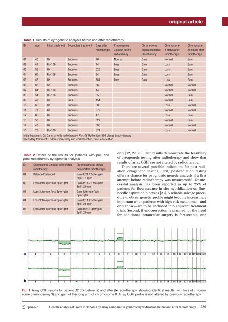
original articleTable 1 Results of cytogenetic analysis before and after radiotherapyID Age Initial treatment Secondary treatment Days afterradiotherapyChromosome3 status beforeradiotherapyChromosome8q status beforeradiotherapyChromosome3 status afterradiotherapyChromosome8q status afterradiotherapy01 46 GK Endores 76 Normal Gain Normal Gain02 49 Ru-106 Endores 74 Loss Gain Loss Gain03 58 GK Endores 526 Loss Gain Loss Gain04 65 Ru-106 Endores 34 Loss Gain Loss Gain05 49 GK Endores 281 Loss Gain Loss Gain06 88 GK Endores 56 Normal Normal07 64 Ru-106 Endores 14 Normal Normal08 54 Ru-106 Endores 55 Normal Gain09 47 GK Enuc 154 Normal Gain10 66 GK Endores 585 Loss Normal11 77 GK Endores 879 Normal Normal12 86 GK Endores 47 Loss Gain13 55 GK Endores 322 Normal Gain14 48 GK Endores 386 Normal Normal15 78 Ru-106 Endores 371 Loss NormalInitial treatment: GK Gamma-Knife radiotherapy, Ru-106 Ruthenium-106 plaque brachytherapySecondary treatment: Endores vitrectomy and endoresection, Enuc enucleationTable 2 Details of the results for patients with pre- andpost-radiotherapy cytogenetic analysisIDChromosome 3 status before/afterradiotherapyChromosome 8q statusbefore/after radiotherapy01 Balanced/balanced Gain 8q21.12-qter/gain8q13.13-qter02 Loss 3pter-qter/loss 3pter-qter Gain 8q11.21-qter/gain8q11.21-qter03 Loss 3pter-qter/loss 3pter-qter Gain 8pter-qter/gain8pter-qter04 Loss 3pter-qter/loss 3pter-qter Gain 8p11.21-qter/gain8p11.21-qter05 Loss 3pter-qter/loss 3pter-qter Gain 8p23.1-qter/gain8p11.21-qteronly [ 12 , 22 , 23 ]. Our results demonstrate the feasibilityof cytogenetic testing after radiotherapy and show thatresults of array CGH are not altered by radiotherapy.There are several possible indications for post-radiationcytogenetic testing. First, post-radiation testingoffers a chance for prognostic genetic analysis if a firstattempt before radiotherapy was unsuccessful. Unsuccessfulanalysis has been reported in up to 25 % ofpatients for fluorescence in situ hybridization on fineneedleaspiration biopsies [ 23 ]. A reliable salvage procedureto obtain genetic profile might become increasinglyimportant when patients with high-risk melanoma—andonly those—are to be included into adjuvant treatmenttrials. Second, if endoresection is planned, or the needfor additional intraocular surgery is foreseeable, oneFig. 1 Array CGH results for patient 02 (ID) before ( a ) and after ( b ) radiotherapy, showing identical results, with loss of chromosome3 (monosomy 3) and gain of the long arm of chromosome 8. Array CGH profile is not altered by previous radiotherapy1 3Genetic analysis of uveal melanoma by array comparative genomic hybridization before and after radiotherapy 289