Funktionelle Anatomie der Rinderklaue - Vetion.de
Funktionelle Anatomie der Rinderklaue - Vetion.de
Funktionelle Anatomie der Rinderklaue - Vetion.de

Erfolgreiche ePaper selbst erstellen
Machen Sie aus Ihren PDF Publikationen ein blätterbares Flipbook mit unserer einzigartigen Google optimierten e-Paper Software.
6<br />
Summary<br />
It is essential for claw trimming to have a thorough un<strong><strong>de</strong>r</strong>standing of the bovine claw’s anatomy as well as the<br />
horn formation and its disturbances. In a well-illustrated contribution the authors <strong>de</strong>al with the functional anatomy<br />
of the claws in cattle. The digital skin is formed by three well-known layers: subcutis, corium and epi<strong><strong>de</strong>r</strong>mis. In<br />
general the subcutis serves as a coarse layer of connective tissue allowing skin displacement. In the claw, however,<br />
the subcutaneous tissue is absent in some areas in or<strong><strong>de</strong>r</strong> to attach the superimposed layers firmly to the skeleton.<br />
The corium consists of the stratum reticulare and the stratum papillare. The papillae are assigned to enlarge the<br />
<strong><strong>de</strong>r</strong>mal surface in or<strong><strong>de</strong>r</strong> to nourish the epi<strong><strong>de</strong>r</strong>mis sufficiently. Any disturbance of the blood circulation (inflammation,<br />
in<strong>de</strong>ntation) influences the quality of horn formation negatively. The phenomena of soft and hard keratinisation<br />
are explained. The quality of the horn <strong>de</strong>pends on genetic influences, feeding (disturbances, strain on the<br />
intermediary metabolism), nutrition of the epi<strong><strong>de</strong>r</strong>mis (by the corium) and the rate of horn formation. The strain<br />
caused by faeces and urine or slurry as well as urea on the claws is mentioned briefly. The authors give a <strong>de</strong>tailed<br />
explanation of the horn structure which can be divi<strong>de</strong>d into tubular and lamellar horn.<br />
The epi<strong><strong>de</strong>r</strong>mal horn capsule comprises the claw plate and the ground surface. The claw plate itself can be divi<strong>de</strong>d<br />
into the horn of the periople, the coronary and the wall horn, the ground surface into the sole and bulb horn. The<br />
functional structure of the toe is also ma<strong>de</strong> up of the perioplic, the coronary, the wall, the sole and the bulb<br />
segment. The anatomy and trimming of the <strong>de</strong>w claws is briefly discussed.<br />
Finally the properties of the segments of the toe are summarized in a synoptic table.<br />
Key words: cattle, claw, functional anatomy, subcutis, corium, epi<strong><strong>de</strong>r</strong>mis, keratinisation, quality of horn, structure<br />
of horn, tubular horn, lamellar horn, perioplic horn, coronary horn, wall horn, sole horn, bulb horn, <strong>de</strong>w claws<br />
Einleitung<br />
ür das Verständnis von Klauenerkrankungen<br />
und <strong><strong>de</strong>r</strong>en Entstehung<br />
sowie für die richtige Klauenpflege<br />
ist es von großer Be<strong>de</strong>utung,<br />
die normale <strong>Anatomie</strong> und Funktion<br />
<strong><strong>de</strong>r</strong> Klaue zu kennen. So soll dieser<br />
Übersichtsartikel die funktionelle <strong>Anatomie</strong><br />
<strong><strong>de</strong>r</strong> Klauen <strong>de</strong>s Rin<strong>de</strong>s darstellen. 1<br />
F<br />
Zum Zehenendorgan (Abb. 1 a/b) im<br />
weiteren Sinne (landläufig “Klaue”)<br />
zählen alle Strukturen, die im Hornschuh<br />
eingeschlossen sind (Abb. 2):<br />
• die Knochen: distaler Abschnitt <strong>de</strong>s<br />
Kronbeins (-/Krb), das Klauenbein<br />
(-/Klb) und das Klauensesambein<br />
(-/Sb),<br />
• das Klauengelenk<br />
(-/weiße Pfeilspitzen),<br />
• die Endsehnen <strong><strong>de</strong>r</strong> Zehenstrecker<br />
und die tiefe Beugesehne (-/TB) mit<br />
<strong><strong>de</strong>r</strong> Bursa podotrochlearis<br />
(-/schwarze Pfeilspitzen),<br />
• die Haut mit Unterhaut (-/Uh), Le<strong><strong>de</strong>r</strong>haut<br />
(-/Lh), Oberhaut (-/Oh).<br />
GROSSTIERPRAXIS 6/2001<br />
Für die Klauenpflege ist jedoch vor allem<br />
ein Verständnis <strong><strong>de</strong>r</strong> <strong>Anatomie</strong> <strong>de</strong>s<br />
Hornschuhs und <strong><strong>de</strong>r</strong> Hornbildung sowie<br />
ihrer Störungen vonnöten. Deswegen<br />
wird auf die Darstellung <strong><strong>de</strong>r</strong><br />
ersten drei Punkte verzichtet.<br />
Haut im Bereich <strong><strong>de</strong>r</strong> Zehen. Die Haut<br />
an <strong>de</strong>n Zehen zeigt bis zu <strong>de</strong>n Klauen<br />
<strong>de</strong>n dreischichtigen Aufbau aus Unterhaut<br />
(Subkutis), Le<strong><strong>de</strong>r</strong>haut (Korium,<br />
Dermis) und Oberhaut (Epi<strong><strong>de</strong>r</strong>mis).<br />
An <strong>de</strong>n Klauen selbst sind diese<br />
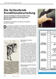
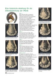
Abb. 1. Klauen eines Mastbullen, Deutsches Fleckvieh, ca. 20 Monate, rechter<br />
Vor<strong><strong>de</strong>r</strong>fuß. a) dorsale Ansicht, b) palmare Ansicht.<br />
1) Im Text wird nur <strong><strong>de</strong>r</strong> Ausdruck „palmar“ verwen<strong>de</strong>t. Diese Angabe bezieht sich primär nur auf die Schultergliedmaße. Wenn im Text nicht an<strong><strong>de</strong>r</strong>s<br />
erwähnt, gelten die jeweiligen Aussagen auch für die Beckengliedmaße.